Note: Descriptions are shown in the official language in which they were submitted.
CA 03064163 2019-11-19
WO 2018/224441 PCT/EP2018/064630
NOVEL ANTI-CD3 ANTIBODIES
FIELD OF THE INVENTION
[0001] The present invention relates to novel antibodies that are specific for
human
CD3, in particular for the CD3E domain.
BACKGROUND OF THE INVENTION
[0002] This invention relates to novel anti-CD3 antibodies, in particular
antibodies
directed against the CD3E domain, which combine high affinity with high
potency, and
in particular novel antibodies with an improved specificity and cross-
reactivity profile.
[0003] The T cell receptor or TCR is a molecule found on the surface of T
lymphocytes (or T cells) that is responsible for recognizing antigens bound to
major
histocompatibility complex (MHC) molecules on the surface of antigen
presenting
cells (APC). The binding between TCR and antigen is of relatively low
affinity. When
the TCR engages with antigen and MHC, the T lymphocyte is activated through a
series of biochemical events mediated by associated enzymes, co-receptors,
specialized accessory molecules, and activated or released transcription
factors.
[0004] The TCR is associated with other molecules like CD3, which possesses
three
distinct chains (y, 6, and E) in mammals, and either a 2 (CD247) chain or a q
chain.
These accessory molecules have transmembrane regions and are vital to
propagating the signal from the TCR into the cell; the cytoplasmic tail of the
TCR is
extremely short, making it unlikely to participate in signaling. The CD3- and -
chains,
together with the TCR, form what is known as the T cell receptor complex.
[0005] CD3E is a type I transmembrane protein expressed on the surface of
certain T
cells. It participates in the T cell receptor (TCR) complex and interacts with
other
CA 03064163 2019-11-19
WO 2018/224441 PCT/EP2018/064630
domains of this complex. One of these interaction partners is CD3y, which
binds to
CD3E in a 1:1 stoichiometry (De la Hera et al, J. Exp.Med.1991; 173: 7-17). It
is
believed that binding of the TCR to the MHC-peptide complex on the surface of
an
antigen presenting cell (APC) and subsequent movement of the T cell along the
APC
leads to a certain rotation of the TCR complex resulting in a dislocation of
CD3E and
CD3y relative to each other, which is required for efficient TCR signaling and
therefore activation of T-cells. Certain antibodies against CD3E have been
demonstrated to induce TCR signaling while others did not. TCR-activating
antibodies typically bind to an exposed epitope on CD3E, whereas some non-
stimulatory antibodies have been demonstrated to bind to the interface between
CD3E and CD3y, or to concomitantly bind to CD3E and CD3y, thus possibly
interfering with the relative displacement of CD3E and CD3y (Kim et al,
JBC.2009;
284: 31028-31037).
[0006] It is well established that peptide-MHC complexes bind TCR with low
affinity
and fast off-rate (Matsui et al, Science.1991; 254: 1788-1791; Weber et al,
Nature.1992; 356: 793-796). It has been suggested that this low affinity is
instrumental to allow a few peptide-MHC complexes to serially trigger many
TCRs
(Valitutti et al, Nature.1995; 375: 148-151) by repeated binding and
dissociation. This
serial triggering is critical to sustain signaling over time, allowing T cells
to eventually
reach the activation threshold (Valitutti et al, lmmunol. Today. 1997; 18: 299-
304;
Lanzavecchia et al, Cell. 1999; 96: 1-4). This notion is supported by the
finding that,
when compared to peptide-MHC complexes, high-affinity anti-CD3 antibodies do
not
efficiently stimulate T cells, since they trigger TCR with a 1:1 stoichiometry
(Viola et
al, Science 1996; 273: 104-106), suggesting that low-affinity antibodies may
be more
effective in stimulating T cells via TCR signaling because of their ability to
repeatedly
dissociate and re-bind to CD3E. Indeed, in a direct comparison of three
derivatives of
the anti-CD3E antibody TR66, which all bind with different affinities, wild-
type TR66
having an intermediate affinity showed best efficacy in T cell activation when
compared to its derivatives that have either higher or lower affinities
(Bortoletto et al,
J. Immuno.2002;32:3102-3107). Thus, a KD at around that of TR66 is ideal for
the
stimulation of T cells. The affinity of TR66 has been determined by use of
surface-
plasmon resonance (SPR) technology as well as by flow-cytometry, yielding
equilibrium dissociation constants of 2.6 x 10⁻⁷ M (Moore et al, Blood.2011;
117:
2
CA 03064163 2019-11-19
WO 2018/224441 PCT/EP2018/064630
4542-4551) and 1.0 x 10⁻⁷ M (Amann et al, Cancer Res. 2008; 68: 143-151),
respectively. In line with this, it has been recommended to use anti-CD3
antibodies
with an affinity of less than 10-8 M (US 7,112,324), and the T cell-
stimulatory
antibodies that have been published for human therapeutic use, bind with
affinities to
human CD3E in the same range. Therefore, according to the theory of serial TCR
triggering and in agreement with published results for anti-CD3E antibodies,
monoclonal antibodies with affinities significantly better than the ones
published are
not expected to be more potent stimulators of T cells, but in contrast are
expected to
be weaker activators.
[0007] Some of the published antibodies against CD3E have been generated via
immunization of animals with T cell preparations and subsequent isolation of
monoclonal antibodies by the so-called hybridoma procedure. The weakness of
this
approach is that the unselective immune response against various antigens of
foreign
(human) T cells in the animal, on one hand, and the poor efficiency of the
hybridoma
procedure on the other hand, decrease the probability to identify monoclonal
antibodies with T cell-stimulatory activity, also because these agonistic
antibodies
may represent a minority in the entirety of anti-CD3E antibodies. Immunization
with a
linear peptide spanning the targeted epitope increases the selectivity of the
immune
response, may, however, result in antibodies that do not recognize the native
full-
length CD3E or that may exert non-optimal TCR stimulation.
[0008] For the immunization of animals with other type-I transmembrane
proteins it
has been particularly useful to use the purified extracellular domain (ECD).
However,
purified ECD of CD3E tends to aggregate, and aggregates may have an altered
structure as compared to the native protein. Further this approach may
preferentially
lead to antibodies binding to the interface between CD3E and CD3y. In
contrast, the
complex of CD3E and CD3y produced as a single-chain protein, connected by a
flexible peptide linker, can be purified in a monomeric fraction and in its
native
conformation (Kim et al, JMB.2000; 302: 899-916). Immunization of animals with
such a CD3E/y single-chain protein may however lead to antibodies
concomitantly
binding to CD3E and CD3y, which would result in antagonistic effects.
3
CA 03064163 2019-11-19
WO 2018/224441 PCT/EP2018/064630
[0009] Several antibodies directed against human CD3E have been developed in
the
past.
[0010] Monoclonal antibody SP34 is a murine antibody that cross-reacts with
non-
human primate CD3, and that is also capable of inducing cell proliferation on
both
human and non-human primate PBMCs (Pessano et al., The T3/T cell receptor
complex: antigenic distinction between the two 20 kD T3 (T38 and T3E)
subunits.
EMBO J 4 (1985) 337-344).
[0011] WO 2007/042261 and WO 2008/119567, both assigned to Micromet (now
Amgen Research (Munich)), disclose cross-reactive binders directed against the
epitopes FSEXE and QDGNE, respectively, in CD3E. In opposition proceedings
filed
by several opponents against granted European patent EP 2 155 783 (based on
the
regional phase of WO 2008/119567), it is submitted that SP34 is binding to
epitope
QDGNE as well.
[0012] WO 2014/191113 disclose cross-reactive binders directed against a novel
epitope at the N-terminus of CD3E, wherein said epitope comprises amino acid
residue N4 as residue that is critical for binding, and wherein said epitope
further
comprises amino acid residue E6 as residue that is involved in binding. It
could be
shown that these antibodies exhibit both high affinity and high potency.
However,
while it could additionally be shown in WO 2014/191113 that the antibodies
disclosed
in the application are cross-reactive with CD3 from non-human primates in
vitro,
cross-reactivity could not be shown to cynomolgous CD3 in a cellular context.
Thus,
these antibodies are of rather limited use with respect to the preclinical
development
of pharmaceutical products comprising an anti-CD3 antibody.
[0013] Thus, there remained still a large unmet need to develop novel CD3
binding
molecules, in particular novel anti-CD3 antibodies, which exhibit the desired
affinity
and potency profile, but which additionally are cross-reactive with other
species, in
particular with non-human primates such as cynomolgus monkeys, both in vitro
and
in a cellular context.
4
CA 03064163 2019-11-19
WO 2018/224441 PCT/EP2018/064630
SUMMARY OF THE INVENTION
[0014] The present invention addresses the above needs and provides novel
antibodies that are specific for human CD3, in particular antibodies specific
for the
CD3E domain. The solution provided by the present invention, i.e. CD3-binding
molecules, in particular anti-CD3 antibodies obtained by peptide immunization
of
rabbits and screening of affinity matured memory B-cells, and in particular
CD3-
binding molecules, in particular anti-CD3 antibodies, in particular antibodies
specific
for the CD3E domain, with the required cross-reactivity profile, has so far
not been
achieved or suggested by the prior art. Novel CD3 antibodies of the present
invention
exhibit the desired affinity and potency profile, are cross-reactive with
other species,
in particular with non-human primates such as cynomolgus monkeys, both in
vitro
and in a cellular context. In addition, the antibodies of the present
invention have
favorable biophysical properties, such as quality, stability or solubility,
for example as
defined by the percentage of antibody in monomer form and thermal unfolding
determined by Differential Scanning Fluorimetry (DSF).
[0015] In a first aspect, the present invention relates to an antibody or
functional
fragment thereof, which is specific for human CD3, comprising:
a variable light chain, wherein the variable light chain comprises, from N-
terminus to
C-terminus, the regions LFW1-LCDR1-LFW2-LCDR2-LFW3-LCDR3-LFW4, wherein
each LFW designates a light chain framework region, and each LCDR designates a
light chain complementarity-determining region, and wherein said LCDRs
together
exhibit at least 90 % sequence identity to the corresponding LCDRs taken from
the
VL sequence according to SEQ ID NO: 4;
and
a variable heavy chain, wherein the variable light chain comprises, from N-
terminus
to C-terminus, the regions HFW1-HCDR1-HFW2-HCDR2-HFW3-HCDR3-HFW4,
wherein each HFW designates a heavy chain framework region, and each HCDR
designates a heavy chain complementarity-determining region, and wherein said
HCDRs together exhibit at least 90 % sequence identity to the corresponding
HCDRs
taken from the VH sequence according to SEQ ID NO: 8.
[0016] In a second aspect, the present invention relates to a multispecific
polypeptide
comprising the antibody of the present invention or functional fragment
thereof.
CA 03064163 2019-11-19
WO 2018/224441 PCT/EP2018/064630
[0017] In a third aspect, the present invention relates to a pharmaceutical
composition comprising the antibody or functional fragment thereof of the
present
invention, or the multispecific polypeptide of the present invention, and a
pharmaceutically acceptable carrier and/or excipient.
[0018] In a fourth aspect, the present invention relates the antibody or
functional
fragment thereof of the present invention, or the multispecific polypeptide of
the
present invention for use as a medicament.
[0019] In a fifth aspect, the present invention relates to a nucleic acid
sequence or a
collection of nucleic acid sequences encoding the antibody or functional
fragment
thereof of the present invention.
[0020] In a sixth aspect, the present invention relates to a vector or a
collection of
vectors comprising the nucleic acid sequence or the collection of nucleic acid
sequences of the present invention.
[0021] In a seventh aspect, the present invention relates to a host cell,
particularly an
expression host cell, comprising the nucleic acid sequence or the collection
of nucleic
acid sequences of the present invention, or the vector or collection of
vectors of the
present invention.
[0022] In an eighth aspect, the present invention relates to a method for
producing
the antibody or functional fragment thereof of the present invention,
comprising the
step of expressing the nucleic acid sequence or the collection of nucleic acid
sequences of the present invention, or the vector or collection of vectors of
the
present invention, or the host cell, particularly the expression host cell, of
the present
invention.
[0023] In a ninth aspect, the present invention relates to a method of
generating a
multispecific construct, comprising the step of cloning, in one or more steps,
one or
more nucleic acid sequences encoding the antibody or functional fragment
thereof
according to the present invention, into a multispecific construct comprising
a nucleic
acid sequence encoding at least a second binding domain or a fragment thereof,
and,
6
CA 03064163 2019-11-19
WO 2018/224441 PCT/EP2018/064630
optionally, a nucleic acid sequence encoding one or more additional binding
domains
or fragments thereof.
BRIEF DESCRIPTION OF THE DRAWINGS
[0024] Figure 1 shows monomer content of each sample determined by SE-HPLC
initially (d0) and over a period of 28 days of storage at 4 C and at a
concentration of
mg/mL.
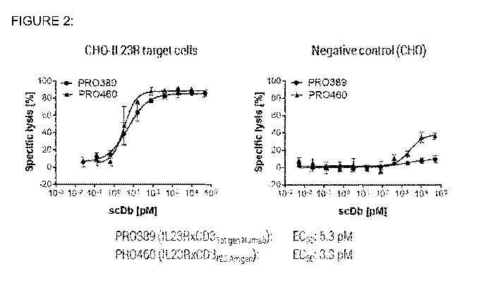
[0025] Figure 2 shows the T-cell mediated target cell depletion induced by
PR0460
(IL23RxCD3i2c Amgen) and PR0389 (IL23RxCD31st gen Numab) using human PBMCs.
The left panel shows cell lysis of target-expressing cells, while the right
panel shows
cell lysis of target-negative cells. The numerical value of the half maximal
effective
concentration (EC50) for the molecules in the presence of target-expressing
cells is
depicted below the graphs.
[0026] Figure 3 shows the T-cell activation of the molecules PR0460
(IL23RxCD312c
Amgen) and PR0389 (IL23RxCD31st gen Numab) determined by the FC assay. The
left
panel shows activation in the presence of target-expressing cells, while the
right
panel shows activation in the presence of target-negative cells.
[0027] Figure 4 shows the T-cell mediated target cell depletion induced by
PR0624
(IL23RxCID2
¨2nd gen Numab) and PR0389 (IL23RxCD31st gen Numab) using human PBMCs.
The left panel shows cell lysis of target-expressing cells, while the right
panel shows
cell lysis of target-negative cells. The numerical value of the half maximal
effective
concentration (EC50) for the molecules in the presence of target-expressing
cells is
depicted below the graphs.
[0028] Figure 5 shows the T-cell activation of the molecules PR0624
(IL23RxCID2
¨2nd
gen Numab) and PR0389 (IL23RxCD31st gen Numab) determined by the FC assay. The
left
panel shows activation in the presence of target-expressing cells, while the
right
panel shows activation in the presence of target-negative cells.
[0029] Figure 6 shows the T-cell activation of the molecules PR0624
(IL23RxCID2
¨2nd
gen Numab) and PR0389 (IL23RxCD31st gen Numab) determined by the NFAT reporter
gene assay. The left panel shows activation in the presence of target-
expressing
7
CA 03064163 2019-11-19
WO 2018/224441 PCT/EP2018/064630
cells, while the right panel shows activation in the presence of target-
negative cells.
The numerical value of the half maximal effective concentration (EC50) for the
molecules in the presence of target-expressing cells is depicted below the
graphs.
[0030] Figure 7 shows the T-cell mediated target cell depletion induced by
PR0624
(IL23RxCID2
¨2nd gen Numab) and PR0389 (IL23RxCD31st gen Numab) using cynomolgus
PBMCs. The left panel shows cell lysis in of target-expressing cells, while
the right
panel shows cell lysis of target-negative cells. The numerical value of the
half
maximal effective concentration (EC50) for the molecules in the presence of
target-
expressing cells is depicted below the graphs.
[0031] Figure 8 shows the T-cell mediated target cell depletion induced by
PR0957
(HER2xCID2
¨2nd gen Numab) and PR0956 (HER2xCD3i2c Amgen) using human PBMCs
after 16 hours (A) and after 40 hours (B). The left panel shows cell lysis of
target-
expressing cells, while the right panel shows cell lysis of target-negative
cells. The
numerical value of the half maximal effective concentration (EC50) for the
molecules
in the presence of target-expressing cells is depicted below the graphs.
[0032] Figure 9 shows the T-cell activation of the molecules PR0957 (HER2xCD2
¨2nd
gen Numab) and PR0956 (HER2xCD3i2c Amgen) determined by the FC assay after 16
hours (A) and after 40 hours (B). The left panel shows activation in presence
of
target-expressing cells, while the right panel shows activation in the
presence of
target-negative cells
DETAILED DESCRIPTION OF THE INVENTION
[0033] The present disclosure relates to novel antibodies that are specific
for human
CD3, in particular antibodies specific for the CD3E domain.
[0034] Unless defined otherwise, all technical and scientific terms used
herein have
the same meaning as commonly understood by those of ordinary skill in the art
to
which this invention pertains.
8
CA 03064163 2019-11-19
WO 2018/224441 PCT/EP2018/064630
[0035] The terms "comprising" and "including" are used herein in their open-
ended
and non-limiting sense unless otherwise noted. With respect to such latter
embodiments, the term "comprising" thus includes the narrower term "consisting
of".
[0036] The terms "a" and "an" and "the" and similar references in the context
of
describing the invention (especially in the context of the following claims)
are to be
construed to cover both the singular and the plural, unless otherwise
indicated herein
or clearly contradicted by context. For example, the term "a cell" includes a
plurality
of cells, including mixtures thereof. Where the plural form is used for
compounds,
salts, and the like, this is taken to mean also a single compound, salt, or
the like.
[0037] In a first aspect, the present invention relates to an antibody or
functional
fragment thereof, which is specific for human CD3, comprising:
(a) a variable light chain, wherein the variable light chain comprises, from N-
terminus
to C-terminus, the regions LFW1-LCDR1-LFW2-LCDR2-LFW3-LCDR3-LFW4,
wherein each LFW designates a light chain framework region, and each LCDR
designates a light chain complementarity-determining region, and wherein said
LCDRs together exhibit at least 60, 70, 80, 90, 91, 92, 93, 94, 95, 96, 97, 98
or 99
percent sequence identity, preferably at least 90 % sequence identity, to the
corresponding LCDRs taken from the VL sequence according to SEQ ID NO: 4; and
(b) a variable heavy chain, wherein the variable light chain comprises, from N-
terminus to C-terminus, the regions HFW1-HCDR1-HFW2-HCDR2-HFW3-HCDR3-
HFW4, wherein each HFW designates a heavy chain framework region, and each
HCDR designates a heavy chain complementarity-determining region, and wherein
said HCDRs together exhibit at least 60, 70, 80, 90, 91, 92, 93, 94, 95, 96,
97, 98 or
99 percent sequence identity, preferably at least 90 % sequence identity, to
the
corresponding HCDRs taken from the VH sequence according to SEQ ID NO: 8.
[0038] In the context of the present invention, the term "antibody" is used as
a
synonym for "immunoglobulin" (Ig), which is defined as a protein belonging to
the
class IgG, IgM, IgE, IgA, IgY or IgD (or any subclass thereof), and includes
all
conventionally known antibodies. A naturally occurring "antibody" is a
glycoprotein
comprising at least two heavy (H) chains and two light (L) chains inter-
connected by
disulfide bonds. Each heavy chain is comprised of a heavy chain variable
region
(abbreviated herein as VH) and a heavy chain constant region. The heavy chain
9
CA 03064163 2019-11-19
WO 2018/224441 PCT/EP2018/064630
constant region is comprised of three domains, CH1, CH2 and CH3. Each light
chain
is comprised of a light chain variable region (abbreviated herein as VL) and a
light
chain constant region. The light chain constant region is comprised of one
domain,
CL. The VH and VL regions can be further subdivided into regions of
hypervariability,
termed complementarity determining regions (CDRs), interspersed with regions
that
are more conserved, termed framework regions (FWs). Each VH and VL is
composed of three CDRs and four FWs arranged from amino-terminus to carboxy-
terminus in the following order: FW1-CDR1-FW2-CDR2-FW3-CDR3-FW4. The
variable regions of the heavy and light chains contain a binding domain that
interacts
with an antigen.
[0039] The term "antibody fragment" refers to at least one portion of an
intact
antibody, or recombinant variants thereof, and the term "functional fragment"
or
"functional antibody fragment" or "antigen-binding fragment" refers to an
antibody
fragment comprising at least an antigen-binding domain, e.g., that part of the
variable
region of an intact antibody, that is sufficient to confer recognition and
specific
binding of the functional antibody fragment to a target, such as the antigenic
determinant of an antigen. Examples of functional antibody fragments include,
but
are not limited to, Fab, Fab', F(ab')2, and Fv fragments, scFv antibody
fragments,
linear antibodies, single domain antibodies such as sdAb (either VL or VH),
camelid
VHH domains, and multi-specific molecules formed from antibody fragments such
as
a bivalent fragment comprising two or more, e.g., two, Fab fragments linked by
a
disulfide bridge at the hinge region, or two or more, e.g., two isolated CDR
or other
epitope binding fragments of an antibody linked. In one embodiment, the
functional
fragment of the invention is a scFv. An antibody fragment can also be
incorporated
into single domain antibodies, maxibodies, minibodies, nanobodies,
intrabodies,
diabodies, triabodies, tetrabodies, v-NAR and bis-scFv (see, e.g., Hollinger
and
Hudson, Nature Biotechnology 23:1126-1136, 2005). Antibody fragments can also
be
grafted into scaffolds based on polypeptides such as a fibronectin type III
(Fn3) (see
U.S. Patent No.: 6,703,199, which describes fibronectin polypeptide
minibodies). An
"antigen-binding region" or "antigen-binding domain" of an antibody typically
is found
in one or more hypervariable region(s) of an antibody, i.e., the CDR1, CDR2,
and/or
CDR3 regions; however, the variable "framework" regions can also play an
important
role in antigen binding, such as by providing a scaffold for the CDRs. The
constant
CA 03064163 2019-11-19
WO 2018/224441 PCT/EP2018/064630
regions of the antibodies may mediate the binding of the immunoglobulin to
host
tissues or factors, including various cells of the immune system (e.g.,
effector cells)
and the first component (Clq) of the classical complement system. The term
"antibody", as used herein, includes for example, monoclonal antibodies,
humanized
antibodies, or chimeric antibodies. The antibodies can be of any isotype
(e.g., IgG,
IgE, IgM, IgD, IgA and IgY), class (e.g., IgG1, IgG2, IgG3, IgG4, IgA1 and
IgA2) or
subclass.
[0040] The "Complementarity Determining Regions" ("CDRs") are amino acid
sequences with boundaries determined using any of a number of well-known
schemes, including those described by Kabat et al. (1991), "Sequences of
Proteins of
Immunological Interest," 5th Ed. Public Health Service, National Institutes of
Health,
Bethesda, MD ("Kabat" numbering scheme), Al-Lazikani et al., (1997) JMB 273,
927-
948 ("Chothia" numbering scheme) and ImMunoGenTics (IMGT) numbering (Lefranc,
M.-P., The Immunologist, 7, 132-136 (1999); Lefranc, M.-P. et al., Dev. Comp.
Immunol., 27, 55-77 (2003) ("IMGT" numbering scheme). For example, for classic
formats, under Kabat, the CDR amino acid residues in the heavy chain variable
domain (VH) are numbered 31-35 (HCDR1), 50-65 (HCDR2), and 95-102 (HCDR3);
and the CDR amino acid residues in the light chain variable domain (VL) are
numbered 24-34 (LCDR1), 50-56 (LCDR2), and 89-97 (LCDR3). Under Chothia the
CDR amino acids in the VH are numbered 26-32 (HCDR1), 52-56 (HCDR2), and 95-
102 (HCDR3); and the amino acid residues in VL are numbered 24-34 (LCDR1), 50-
56 (LCDR2), and 89-97 (LCDR3). By combining the CDR definitions of both Kabat
and Chothia, the CDRs consist of amino acid residues 26-35 (HCDR1), 50-65
(HCDR2), and 95-102 (HCDR3) in human VH and amino acid residues 24-34
(LCDR1), 50-56 (LCDR2), and 89-97 (LCDR3) in human VL. Under IMGT the CDR
amino acid residues in the VH are numbered approximately 26-35 (HCDR1), 51-57
(HCDR2) and 93-102 (HCDR3), and the CDR amino acid residues in the VL are
numbered approximately 27-32 (LCDR1), 50-52 (LCDR2), and 89-97 (LCDR3)
(numbering according to "Kabat"). Under IMGT, the CDRs of an antibody can be
determined using the program IMGT/DomainGap Align.
[0041] In the context of the present invention, the numbering system suggested
by
Honegger & PlOckthun ("AHo numbering") is used (Honegger & PlOckthun, J. Mol.
Biol. 309 (2001) 657-670), unless specifically mentioned otherwise.
Furthermore, the
11
CA 03064163 2019-11-19
WO 2018/224441 PCT/EP2018/064630
following residues are defined as CDRs: LCDR1 (also referred to as CDR-L1):
L24-
L42; LCDR2 (also referred to as CDR-L2): L58-L72; LCDR3 (also referred to as
CDR-L3): L107-L138; HCDR1 (also referred to as CDR-H1): H27-H42; HCDR2 (also
referred to as CDR-H2): H57-H76; HCDR3 (also referred to as CDR-H3): H108-
H138. For the sake of clarity, the numbering system according to Honegger &
PlOckthun takes the length diversity into account that is found in naturally
occurring
antibodies, both in the different VH and VL subfamilies and, in particular, in
the
CDRs, and provides for gaps in the sequences. Thus, in a given antibody
variable
domain usually not all positions 1 to 149 will be occupied by an amino acid
residue.
[0042] Preferably, the "antigen-binding region" comprises at least amino acid
residues 4 to 138 of the variable light (VL) chain and 5 to 138 of the
variable heavy
(VH) chain (in each case numbering according to Honegger & PlOckthun), more
preferably amino acid residues 3 to 144 of VL and 4 to 144 of VH, and
particularly
preferred are the complete VL and VH chains (amino acid positions 1 to 149 of
VL
and 1 to 149 of VH). The framework regions and CDRs are indicated in the
sequences shown in Table 1. A preferred class of immunoglobulins for use in
the
present invention is IgG. "Functional fragments" of the invention include the
domain
of a F(ab1)2 fragment, a Fab fragment, Fv and scFv. The F(ab1)2 or Fab may be
engineered to minimize or completely remove the intermolecular disulphide
interactions that occur between the CH1 and CL domains. The antibodies or
functional fragments thereof of the present invention may be part of bi- or
multifunctional polypeptides, as further described in Sections [0076] to
[0111].
[0043] The following terms are used to describe the sequence relationships
between
two or more polynucleotide or amino acid sequences: "sequence identity" or
"percentage of sequence identity". The term "sequence identity" as used herein
is
determined by calculating the maximum number of amino acid residues that are
identical between two polypeptide sequences, wherein gaps and/or insertions
may be
factored in to allow for the largest degree of sequence overlap. For example,
two
100mer polypeptides that are fully identical have a sequence identity of 100
/0. When
they differ by a single mutation, or when one polypeptide contains a deletion
of one
amino acid, the sequence identity is 99 % (99 out of 100 positions being
identical). In
other words, the "percentage of sequence identity" is calculated by comparing
two
optimally aligned sequences over the window of comparison, determining the
number
12
CA 03064163 2019-11-19
WO 2018/224441 PCT/EP2018/064630
of positions at which the identical nucleic acid base (e.g., A, T, C, G, U or
I) or amino
acid residue occurs in both sequences to yield the number of matched
positions,
dividing the number of matched positions by the total number of positions in
the
comparison window (i.e., the window size), and multiplying the result by 100
to yield
the percentage of sequence identity. The "sequence similarity" is the degree
of
resemblance between two sequences when they are compared. Where necessary or
desired, optimal alignment of sequences for comparison can be conducted, for
example, by the local homology algorithm of Smith and Waterman (Adv. Appl.
Math.
2:482 (1981)), by the homology alignment algorithm of Needleman and Wunsch (J.
Mol. Biol. 48:443-53 (1970)), by the search for similarity method of Pearson
and
Lipman (Proc. Natl. Acad. Sci. USA 85:2444-48 (1988)), by computerized
implementations of these algorithms (e.g., GAP, BESTFIT, FASTA, and TFASTA in
the Wisconsin Genetics Software Package, Genetics Computer Group, 575 Science
Dr., Madison, Wis.), or by visual inspection. (See generally Ausubel et al.
(eds.),
Current Protocols in Molecular Biology, 4th ed., John Wiley and Sons, New York
(1999)). Unless indicated otherwise herein, the degree of sequence similarity
referred
to herein is determined by utilization of Dayhoff PAM matrix (M.O. Dayhoff, R.
Schwartz, B.C. Orcutt: A model of Evolutionary Change in Proteins, pages 345-
352;
in: Atlas of protein sequence and structure, National Biomedical Research
Foundation, 1979).
[0044] The term "amino acid" refers to naturally occurring and synthetic amino
acids,
as well as amino acid analogs and amino acid mimetics that function in a
manner
similar to the naturally occurring amino acids. Naturally occurring amino
acids are
those encoded by the genetic code, as well as those amino acids that are later
modified, e.g., hydroxyproline, gamma-carboxyglutamate, and 0-phosphoserine.
The
terms "polypeptide" and "protein" are used interchangeably herein to refer to
a
polymer of amino acid residues. The terms apply to amino acid polymers in
which
one or more amino acid residue is an artificial chemical mimetic of a
corresponding
naturally occurring amino acid, as well as to naturally occurring amino acid
polymers
and non-naturally occurring amino acid polymer. Unless otherwise indicated, a
particular polypeptide sequence also implicitly encompasses conservatively
modified
variants thereof.
13
CA 03064163 2019-11-19
WO 2018/224441 PCT/EP2018/064630
[0045] The term "binding specificity" as used herein refers to the ability of
an
individual antibody combining site to react with one antigenic determinant and
not
with a different antigenic determinant. As used herein, a binding molecule is
"specific
to/for", "specifically recognizes", or "specifically binds to" a target, such
as for
example human CD3, when such binding molecule is able to discriminate between
such target biomolecule and one or more reference molecule(s), since binding
specificity is not an absolute, but a relative property. In its most general
form (and
when no defined reference is mentioned), "specific binding" is referring to
the ability
of the binding molecule to discriminate between the target biomolecule of
interest and
an unrelated biomolecule, as determined, for example, in accordance with a
specificity assay methods known in the art. Such methods comprise, but are not
limited to Western blots, ELISA, RIA, ECL, IRMA, SPR (Surface plasmon
resonance)
tests and peptide scans. For example, a standard ELISA assay can be carried
out.
The scoring may be carried out by standard colour development (e.g. secondary
antibody with horseradish peroxide and tetramethyl benzidine with hydrogen
peroxide). The reaction in certain wells is scored by the optical density, for
example,
at 450 nm. Typical background (= negative reaction) may be about 0.1 OD;
typical
positive reaction may be about 1 OD. This means the ratio between a positive
and a
negative score can be 10-fold or higher. In a further example, an SPR assay
can be
carried out, wherein at least 10-fold, preferably at least 100-fold difference
between a
background and signal indicates on specific binding. Typically, determination
of
binding specificity is performed by using not a single reference biomolecule,
but a set
of about three to five unrelated biomolecules, such as milk powder,
transferrin or the
like. The antibody of the invention or functional fragment thereof has a
binding
specificity for human CD3, preferably to human CD3E.
[0046] In one embodiment, the antibody of the invention or functional fragment
thereof has a binding specificity for human CD3 and to non-chimpanzee primate
CD3. As evident to the person skilled in the art, it is not excluded from the
scope of
the invention that antibody of the invention or functional fragment thereof
exhibiting
cross-species specificity as defined herein may also bind, e.g., to chimpanzee
CD3.
On the other hand, it is apparent that antibody of the invention or functional
fragment
thereof which only bind to human CD3, but not to non-chimpanzee primate CD3,
are
excluded from the scope of the invention. This applies mutatis mutandis to
binding
14
CA 03064163 2019-11-19
WO 2018/224441 PCT/EP2018/064630
domains which only bind to non-chimpanzee primate CD3, but not to human CD3,
such as e.g. those of monoclonal antibody FN-18.
[0047] As used herein, a 'non chimpanzee primate" or "non chimp primate" or
grammatical variants thereof refers to any primate other than chimpanzee, i.e.
other
than an animal of belonging to the genus Pan, and including the species Pan
paniscus and Pan troglodytes, also known as Anthropopithecus troglodytes or
Simia
satyrus. A "primate", "primate species", "primates" or grammatical variants
thereof
denote/s an order of eutherian mammals divided into the two suborders of
prosimians
and anthropoids and comprising man, apes, monkeys and lemurs. Specifically,
"primates" as used herein comprises the suborder Strepsirrhini (non-tarsier
prosimians), including the infraorder Lemuriformes (itself including the
superfamilies
Cheirogaleoidea and Lemuroidea), the infraorder Chiromyiformes (itself
including the
family Daubentoniidae) and the infraorder Lorisiformes (itself including the
families
Lorisidae and Galagidae). "Primates" as used herein also comprises the
suborder
Haplorrhini, including the infraorder Tarsiiformes (itself including the
family Tarsiidae),
the infraorder Simiiformes (itself including the Platyrrhini, or New World
monkeys,
and the Catarrhini, including the Cercopithecidea, or Old- World Monkeys).
Most
preferred is Macaca fascicularis (also known as Cynomolgus monkey and,
therefore,
in the Examples named "Cynomolgus"). Suitably, the antibody of the invention
or
functional fragment thereof has a binding specificity for human CD3 and for
cynomolgus CD3.
[0048] Further, depending on the context, the term "specific binding" may also
refer to
the ability of a binding molecule to discriminate between the target
biomolecule and
one or more closely related biomolecule(s), which are used as reference
points, such
as, for example, CD3 molecules from a different species, e.g. murine CD3.
Additionally, "specific binding" may relate to the ability of a binding
molecule to
discriminate between different parts of its target antigen, e.g. different
domains,
regions or epitopes of the target biomolecule, in particular the CD3E domain,
or
between one or more key amino acid residues or stretches of amino acid
residues of
the target biomolecule.
[0049] In one embodiment, the antibody of the invention or functional fragment
thereof has a binding specificity for human CD3E.
CA 03064163 2019-11-19
WO 2018/224441 PCT/EP2018/064630
[0050] The term "CD3" refers to a molecule expressed as part of the T cell
receptor
and has the meaning as typically ascribed to it in the prior art. In human, it
encompasses individual or independently combined CD3 subunits CD3 epsilon, CD3
delta, and CD3 gamma. The non-chimpanzee primate CD3 antigens as referred to
herein are, for example, Macaca fascicularis CD3 and Macaca mulatto CD3. In
Macaca fascicularis, it encompasses CD3 epsilon FN-18 negative and CD3 epsilon
FN-18 positive, CD3 gamma and CD3 delta. In Macaca mulatto, it encompasses CD3
epsilon, CD3 gamma and CD3 delta.
[0051] Preferably, said CD3 as used herein specifically relates to CD3E. The
term
"human CD3E" refers in particular to human CD3E with the GenBank Accession
No.NM 000733, UniProt ID number P07766 reproduced herein as SEQ ID NO: 19,
or a variant thereof. The CD3E "FN-18 negative" of Macaca fascicularis (i.e.
CD3E not
recognized by monoclonal antibody FN-18 due to a polymorphism as set forth
above)
is indicated in GenBank Accession No. AB073994. The CD3E "FN-18 positive" of
Macaca fascicularis (i.e. CD3 epsilon recognized by monoclonal antibody FN-18)
is
indicated in GenBank Accession No. AB073993.
[0052] The human CD3 gamma is indicated in GenBank Accession No. NM 000073.
The human CD3 delta is indicated in GenBank Accession No. NM 000732. The CD3
gamma of Macaca fascicularis is indicated in GenBank Accession No. AB073992.
The CD3 delta of Macaca fascicularis is indicated in GenBank Accession No.
AB073991.
[0053] Suitably, the antibodies of the invention or functional fragments
thereof target
human and cynomoglous (Macaca fascicularis) CD3E.
[0054] Suitably, the antibody of the invention is a monoclonal antibody. The
term
"monoclonal antibody" or "monoclonal antibody composition" as used herein
refers to
antibodies that are substantially identical to amino acid sequence or are
derived from
the same genetic source. A monoclonal antibody composition displays a binding
specificity and affinity for a particular epitope, or binding specificities
and affinities for
specific epitopes.
16
CA 03064163 2019-11-19
WO 2018/224441 PCT/EP2018/064630
[0055] In the context of the present invention, the term "epitope" refers to
that part of
a given target biomolecule that is required for specific binding between the
target
biomolecule and a binding molecule. An epitope may be continuous, i.e. formed
by
adjacent structural elements present in the target biomolecule, or
discontinuous, i.e.
formed by structural elements that are at different positions in the primary
sequence
of the target biomolecule, such as in the amino acid sequence of a protein as
target,
but in close proximity in the three-dimensional structure, which the target
biomolecule
adopts, such as in the bodily fluid.
[0056] Antibodies of the invention include, but are not limited to, the
chimeric, human
and humanized antibodies.
[0057] The term "chimeric antibody" (or functional fragment thereof) is an
antibody
molecule (or functional fragment thereof) in which (a) the constant region, or
a portion
thereof, is altered, replaced or exchanged so that the antigen-binding site
(variable
region) is linked to a constant region of a different or altered class,
effector function
and/or species, or an entirely different molecule which confers new properties
to the
chimeric antibody, e.g., an enzyme, toxin, hormone, growth factor, drug, etc.;
or (b)
the variable region, or a portion thereof, is altered, replaced or exchanged
with a
variable region having a different or altered antigen specificity. For
example, a mouse
antibody can be modified by replacing its constant region with the constant
region
from a human immunoglobulin. Due to the replacement with a human constant
region, the chimeric antibody can retain its specificity in recognizing the
antigen,
while having reduced antigenicity in human as compared to the original mouse
antibody.
[0058] A "humanized" antibody (or functional fragment thereof), as used
herein, is an
antibody (or functional fragment thereof) that retains the reactivity of a non-
human
antibody while being less immunogenic in humans. This can be achieved, for
instance, by retaining the non-human CDR regions and replacing the remaining
parts
of the antibody with their human counterparts (i.e., the constant region as
well as the
framework portions of the variable region). Additional framework region
modifications
may be made within the human framework sequences as well as within the CDR
sequences derived from the germline of another mammalian species. The
humanized
17
CA 03064163 2019-11-19
WO 2018/224441 PCT/EP2018/064630
antibodies of the invention may include amino acid residues not encoded by
human
sequences (e.g., mutations introduced by random or site-specific mutagenesis
in vitro
or by somatic mutation in vivo, or a conservative substitution to promote
stability or
manufacturing). See, e.g., Morrison et al., Proc. Natl. Acad. Sci. USA,
81:6851-6855,
1984; Morrison and 0i, Adv. Immunol., 44:65-92, 1988; Verhoeyen et al.,
Science,
239: 1534-1536, 1988; PadIan, Molec. lmmun., 28:489-498, 1991; and PadIan,
Molec. lmmun., 31: 169-217, 1994. Other examples of antibody engineering
technology include, but are not limited to Xoma technology disclosed in U.S.
Pat. No.
5,766,886.
[0059] The term "recombinant humanized antibody", as used herein, includes all
humanized antibodies of the invention that are prepared, expressed, created or
isolated by recombinant means, such as antibodies isolated from an animal
(e.g. a
mouse); antibodies expressed using a recombinant expression vector transfected
into a host cell, antibodies isolated from a recombinant, combinatorial human
antibody library, or antibodies prepared, expressed, created or isolated by
any other
means that involves splicing of human immunoglobulin gene sequences to other
DNA sequences. Such recombinant human antibodies have variable and constant
regions (if present) derived from human germline immunoglobulin sequence. Such
antibodies can, however, be subjected to in vitro mutagenesis (or, when an
animal
transgenic for human Ig sequences is used, in vivo somatic mutagenesis) and
thus
the amino acid sequences of the VH (antibody heavy chain variable region) and
VL
(antibody light chain variable region) of the recombinant antibodies are
sequences
that, while derived from and related to human germline VH and VL sequences,
may
not naturally exist within the human antibody germline.
[0060] Suitably, the antibody or functional fragment of the present invention
is an
artificial or an isolated antibody or functional fragment thereof. The term
"artificial
antibody", as used herein, means an antibody or functional fragment thereof,
which,
by virtue of its origin or manipulation: (i) is present in a host cell as the
expression
product of a portion of an expression vector, or (ii) is linked to a protein
or other
chemical moiety other than that to which it is linked in nature, or (iii) does
not occur in
nature. The term "isolated antibody", as used herein, refers to an antibody
expressed
in a host cell and purified away from associated proteins, as by gel
chromatography.
The term "isolated antibody" also refers to antibody that is substantially
free of other
18
CA 03064163 2019-11-19
WO 2018/224441 PCT/EP2018/064630
antibodies having different antigenic specificities (e.g., an isolated
antibody that
specifically binds to human CD3 is substantially free of antibodies that
specifically
bind antigens other than human CD3). An isolated antibody that specifically
binds
human CD3 may, however, have cross-reactivity to other antigens, such as CD3
molecules from other species (e.g., non-human primate and/or rodent CD3).
Moreover, an isolated antibody may be substantially free of other cellular
material
and/or chemicals.
[0061] In one embodiment, the present invention relates to an antibody or
functional
fragment thereof comprising (a) an LCDR1 as set forth in SEQ ID NO: 1; (b) an
LCDR2 as set forth in SEQ ID NO: 2; (c) an LCDR3 as set forth in SEQ ID NO: 3;
(d)
an HCDR1 as set forth in SEQ ID NO: 5; (e) an HCDR2 as set forth in SEQ ID NO:
6;
and (f) an HCDR3 as set forth in SEQ ID NO: 7.
[0062] The antibody of the invention or functional fragment thereof comprises
a
variable heavy chain (VH) domain and a variable light chain (VL) domain. In
the
context of the present invention the terms "VH" (variable heavy chain), "VK"
and "VA"
refer to families of antibody heavy and light chain sequences that are grouped
according to sequence identity and homology. Methods for the determination of
sequence homologies, for example by using a homology search matrix such as
BLOSUM (Henikoff, S. & Henikoff, J. G., Proc. Natl. Acad. Sci. USA 89 (1992)
10915-10919), and methods for the grouping of sequences according to
homologies
are well known to one of ordinary skill in the art. For VH, VK and VA
different
subfamilies can be identified, as shown, for example, in Knappik et al., J.
Mol. Biol.
296 (2000) 57-86, which groups VH in VH1A, VH1B and VH2 to VH6, VK in VK1 to
VK4 and VA in VA1 to VA3. In vivo, antibody VK chains, VA chains, and VH
chains are
the result of the random rearrangement of germline K chain V and J segments,
germline A chain V and J segments, and heavy chain V, D and J segments,
respectively. To which subfamily a given antibody variable chain belongs is
determined by the corresponding V segment, and in particular by the framework
regions FW1 to FW3. Thus, any VH sequence that is characterized in the present
application by a particular set of framework regions HFW1 to HFW3 only, may be
combined with any HFW4 sequence, for example an HFW4 sequence taken from
one of the heavy chain germline J segments, or an HFW4 sequence taken from a
19
CA 03064163 2019-11-19
WO 2018/224441
PCT/EP2018/064630
rearranged VH sequence. In particular embodiments, the HFW4 sequence is
WGQGTLVTVSS.
[0063] Suitably, the present invention provides an antibody or functional
fragment
thereof that specifically binds CD3 (e.g., human CD3 protein, in particular
human
CD3c), wherein said antibody or functional fragment thereof comprises a VH4 or
VH3
domain, preferably VH3 domain.
[0064] Suitably, the present invention provides an antibody or functional
fragment
thereof that specifically binds CD3 (e.g., human CD3 protein, in particular
human
CD3c), wherein said antibody or functional fragment thereof comprises (i) VK
frameworks FW1, FW2 and FW3, particularly VK1 or VK3 frameworks, preferably
VK1
frameworks FW1 to FW3, and (ii) a framework FW4, which is selected from a VK
FW4, particularly VK1 FW4, VK3 FW4, and a VA FW4. Suitable VK1 FW1 to FW3
exhibit at least 60, 70, 80, 90 percent sequence identity, preferably at least
90%
sequence identity, to the corresponding framework regions taken from the VK1
sequence according to SEQ ID NO: 4. Suitable VK1 FW4 exhibits at least 60, 70,
80,
90 percent sequence identity, preferably at least 90% sequence identity, to
the
corresponding FW4 taken from the VK1 sequence according to SEQ ID NO: 4.
Suitably, VK1 FW4 is the FW4 taken from the VK1 sequence according to SEQ ID
NO: 4. Suitable VA FW4 is as set forth in SEQ ID NO: 17 or SEQ ID NO: 18. In
one
embodiment the present invention provides an antibody or a functional fragment
thereof that specifically binds CD3 (e.g., human CD3 protein, in particular
human
CD3c), wherein said antibody or functional fragment thereof comprises VA FW4
comprising the amino acid sequence having at least 60, 70, 80, 90 percent
identity an
amino acid sequence selected from any of SEQ ID NO: 17 to SEQ ID NO: 18,
preferably to SEQ ID NO: 17.
[0065] In a particular embodiment, said variable light chain is a VK1 light
chain, and/or
said variable heavy chain is a VH3 chain. In another particular embodiment,
said
variable light chain is a chimeric light chain, comprising VK framework
regions I to III
and a VA framework region IV. In one embodiment, light chain is a chimeric
light
chain, comprising:
(i) the LCDR1, LCDR2, and LCDR3 sequences of SEQ ID NOs: 1, 2, and 3,
respectively;
CA 03064163 2019-11-19
WO 2018/224441 PCT/EP2018/064630
(ii) human VK framework regions FW1 to FW3, particularly human Vk1
framework regions FW1 to FW3;
(iii) FW4, which is selected from (a) a human VA germ line sequence for FW4,
particularly a VA germ line sequence selected from the SEQ ID NO: 17
and SEQ ID NO: 18, preferably SEQ ID NO: 17; and (b) a VA-based
sequence, which has one or two mutations, particularly one mutation,
compared to the closest human VA germ line sequence for FW4
comprising an amino acid sequence selected from the SEQ ID NO: 17 and
SEQ ID NO: 18, preferably SEQ ID NO: 17.
[0066] In one embodiment, said variable light chain exhibits at least 60, 70,
80, 90,
91, 92, 93, 94, 95, 96, 97, 98 or 99 % sequence identity to the amino acid
sequence
according to SEQ ID NO: 4, and/or wherein said variable heavy chain exhibits
at
least 60, 70, 80, 90, 91, 92, 93, 94, 95, 96, 97, 98 or 99 % sequence identity
to the
amino acid sequence according to SEQ ID NO: 8. In a particular embodiment,
said
variable light chain exhibits at least 90 % sequence identity to the amino
acid
sequence according to SEQ ID NO: 4, and/or wherein said variable heavy chain
exhibits at least 90 % sequence identity to the amino acid sequence according
to
SEQ ID NO: 8.
[0067] Suitably, the present invention relates to an antibody or functional
fragment
thereof, which is specific for human CD3, in particular human CD3E, comprising
a
variable light chain, wherein said variable light chain exhibits at least 90 %
sequence
identity to the amino acid sequence according to SEQ ID NO: 4 and wherein said
variable light chain comprises an Arginine (R) or a Lysine (K) at the light
chain amino
acid position 54 (AHo numbering), preferably an Arginine (R). In a further
embodiment, said variable light chain further comprises a Glutamine (Q) at the
light
chain amino acid position 50 (AHo numbering) and/or a Serine (S) at the light
chain
amino acid position 51 (AHo numbering), and optionally a Phenylalanine (F) at
the
light chain amino acid position 44 (AHo numbering) or a Glutamine (Q) at the
light
chain amino acid position 88 (AHo numbering) or a Histidine (H) at the light
chain
amino acid position 88 (AHo numbering). In a specific embodiment, the present
invention relates to an antibody or functional fragment thereof, which is
specific for
human CD3, in particular human CD3E, comprising a variable light chain,
wherein
said variable light chain exhibits at least 90 % sequence identity to the
amino acid
21
CA 03064163 2019-11-19
WO 2018/224441 PCT/EP2018/064630
sequence according to SEQ ID NO: 4 and wherein said variable light chain
comprises an Arginine (R) at the light chain amino acid position 54 (AHo
numbering),
a Glutamine (Q) at the light chain amino acid position 50 (AHo numbering), a
Serine
(S) at the light chain amino acid position 51 (AHo numbering), and a
Phenylalanine
(F) at the light chain amino acid position 44 (AHo numbering).
[0068] In yet another embodiment, the present invention relates to an antibody
or
functional fragment thereof, which is specific for human CD3, in particular
human
CD3E, comprising a variable light chain, wherein said variable light chain
exhibits at
least 90 % sequence identity to the amino acid sequence according to SEQ ID
NO: 4
and wherein said variable light chain comprises an Arginine (R) at the light
chain
amino acid position 54 (AHo numbering), and a Phenylalanine (F) at the light
chain
amino acid position 44 (AHo numbering).
[0069] Suitably, the present invention relates to an antibody or functional
fragment
thereof, which is specific for human CD3, in particular human CD3E, comprising
a
variable heavy chain, wherein said variable heavy chain exhibits at least 90 %
sequence identity to the amino acid sequence according to SEQ ID NO: 8 and
wherein said variable heavy chain comprises at least one, e.g. at least two,
preferably at least three, of the following amino acids selected from the list
consisting
of an Alanine (A) at the heavy chain amino acid position 53 (AHo numbering), a
Threonine (T) at the heavy chain amino acid position 103 (AHo numbering), and
a
Phenylalanine (F) at the heavy chain amino acid position 105 (AHo numbering).
In
one embodiment said variable heavy chain exhibits at least 90 % sequence
identity
to the amino acid sequence according to SEQ ID NO: 8 and comprises an Alanine
(A) at the heavy chain amino acid position 53 (AHo numbering), a Threonine (T)
at
the heavy chain amino acid position 103 (AHo numbering), and a Phenylalanine
(F)
at the heavy chain amino acid position 105 (AHo numbering).
[0070] In one embodiment, the present invention relates to the antibody of the
invention or functional fragment thereof comprising a variable light chain
comprising
the amino acid sequence of SEQ ID NO: 4 or a conservatively modified variant
thereof, and a variable heavy chain comprising the amino acid sequence of SEQ
ID
NO: 8 or a conservatively modified variant thereof.
22
CA 03064163 2019-11-19
WO 2018/224441 PCT/EP2018/064630
[0071] The term "conservatively modified variant" or "conservative variants"
applies to
both amino acid and nucleic acid sequences. With respect to particular nucleic
acid
sequences, conservatively modified variants refer to those nucleic acids which
encode identical or essentially identical amino acid sequences, or where the
nucleic
acid does not encode an amino acid sequence, to essentially identical
sequences.
Because of the degeneracy of the genetic code, a large number of functionally
identical nucleic acids encode any given protein. For instance, the codons
GCA,
GCC, GCG and CCU all encode the amino acid alanine. Thus, at every position
where an alanine is specified by a codon, the codon can be altered to any of
the
corresponding codons described without altering the encoded polypeptide. Such
nucleic acid variations are "silent variations", which are one species of
conservatively
modified variations. Every nucleic acid sequence herein which encodes a
polypeptide
also describes every possible silent variation of the nucleic acid. One of
skill will
recognize that each codon in a nucleic acid (except AUG, which is ordinarily
the only
codon for methionine, and TGG, which is ordinarily the only codon for
tryptophan)
can be modified to yield a functionally identical molecule. Accordingly, each
silent
variation of a nucleic acid that encodes a polypeptide is implicit in each
described
sequence.
[0072] For polypeptide sequences, "conservatively modified variants" or
"conservative
variants" include individual substitutions, deletions or additions to a
polypeptide
sequence which result in the substitution of an amino acid with a chemically
similar
amino acid. Conservative substitution tables providing functionally similar
amino
acids are well known in the art. Such conservatively modified variants are in
addition
to and do not exclude polymorphic variants, interspecies homologs, and alleles
of the
invention. The following eight groups contain amino acids that are
conservative
substitutions for one another: 1) Alanine (A), Glycine (G); 2) Aspartic acid
(D),
Glutamic acid (E); 3) Asparagine (N), Glutamine (Q); 4) Arginine (R), Lysine
(K); 5)
lsoleucine (I), Leucine (L), Methionine (M), Valine (V); 6) Phenylalanine (F),
Tyrosine
(Y), Tryptophan (W); 7) Serine (S), Threonine (T); and 8) Cysteine (C),
Methionine
(M) (see, e.g., Creighton, Proteins (1984)). In one embodiment, the term
"conservative sequence modifications" are used to refer to amino acid
modifications
that do not significantly affect or alter the binding characteristics of the
antibody
containing the amino acid sequence.
23
CA 03064163 2019-11-19
WO 2018/224441 PCT/EP2018/064630
[0073] In a preferred embodiment, the antibody of the invention or functional
fragment
thereof comprises a variable light chain comprising the amino acid sequence of
SEQ
ID NO: 4, and a variable heavy chain comprising the amino acid sequence of SEQ
ID
NO: 8.
[0074] In one embodiment of the present invention, the isolated antibody or
functional
fragment thereof is selected from: an IgG antibody, a Fab and an scFv
fragment.
Suitably, the antibody of the invention or functional fragment thereof is scFv
antibody
fragment. "Single-chain Fv" or "scFv" or "sFv" antibody fragments comprise the
VH
and VL domains of an antibody, wherein these domains are present in a single
polypeptide chain. Generally, the Fv polypeptide further comprises a
polypeptide
linker between the VH and VL domains, which enables the sFy to form the
desired
structure for target binding (see, for example, PlOckthun, The Pharmacology of
Monoclonal Antibodies, vol. 113, Rosenburg and Moore eds., Springer-Verlag,
New
York, 1994, pp. 269-315).
[0075] Suitably, the antibody of the invention or functional fragment thereof
is an
scFv. In particular embodiments, said functional fragment is an scFv format
comprising the linker according to SEQ ID NO: 15. Suitably, the antibody of
the
invention or functional fragment thereof is an scFv comprising at least one
amino acid
sequence selected from the list consisting of SEQ ID NOs: 4, 8, 29, 30, 32,
33, 35,
36, 37, 38, 39, 40, 41, and 42. Suitably, the antibody of the invention or
functional
fragment thereof is an scFv comprising the amino acid sequence selected from
the
list consisting of SEQ ID NOs: 29, 30, 32, 33, 35, 36, 37, 38, 39, 40, 41, and
42.
[0076] The term "affinity" as used herein refers to the strength of the sum of
total
noncovalent interactions between a single binding site or a molecule, e.g., an
antibody or a functional fragment thereof, and its binding partner, e.g., an
antigen.
Unless indicated otherwise, as used herein, "binding affinity" refers to
intrinsic binding
affinity which reflects 1:1 interaction between members of a binding pair,
e.g.,
interaction of a single antibody binding domain and its antigen. The affinity
can
generally be represented by the dissociation constant (KD). Affinity can be
measured
by common methods known in the art, including those described herein.
24
CA 03064163 2019-11-19
WO 2018/224441 PCT/EP2018/064630
[0077] In a suitable embodiment, the antibody of the invention or functional
fragment
thereof may have a KD to human CD3 and/or to cynomolgous CD3 of between 0.1 to
100 nM, 0.1 to 90 nM, 0.1 to 80 nM, 0.1 to 70 nM, 0.1 to 60 nM, 0.5 to 50 nM,
0.5 to
40 nM, 0.5 to 30 nM, 0.5 to 20 nM, 0.5 to 10 nM, 0.5 to 9 nM, 0.5 to 8 nM, 0.5
to 7
nM, 0.5 to 6 nM, 0.5 to 5 nM, particularly as measured by surface plasmon
resonance. In a suitable embodiment, the antibody of the invention or
functional
fragment thereof may have a KD to human CD3 and/or to cynomolgous CD3 of less
than approximately 100 nM, less than approximately 90 nM, less than
approximately
80 nM, less than approximately 70 nM, less than approximately 60 nM, less than
approximately 55 nM, less than approximately 40 nM, less than approximately 45
nM,
less than approximately 40 nM, less than approximately 35 nM, less than
approximately 30 nM, less than approximately 25 nM, less than 20 nM, less than
approximately 15 nM, less than approximately 10 nM, less than approximately 9
nM,
less than approximately 8 nM, less than approximately 7 nM, less than
approximately
6 nM, less than approximately 5 nM, less than approximately 4 nM, less than
approximately 3 nM, less than 2 nM, less than 1 nM, particularly as measured
by
surface plasmon resonance. Suitably, the antibody of the invention or
functional
fragment thereof has a KD to human CD3 and/or to cynomolgous CD3 of less than
10
nM, preferably less than 6 nM, particularly as measured by surface plasmon
resonance.
[0078] In a particular embodiment, the antibody of the invention or the
functional
fragment thereof, is characterized by one or more of the following parameters:
(i) a KD value for the binding to human CD3 of less than 40 nM, particularly
less than
nM, more particularly less than 6 nM, particularly as measured by surface
plasmon resonance, more particularly as determined by the method shown in
Example 2.1;
(ii) a KD value for the binding to cynomolgous CD3 of less than 20 nM,
particularly
less than 10 nM, more particularly less than 5 nM, particularly as measured by
surface plasmon resonance, more particularly as determined by the method shown
in
Example 2.1; and
(iii) an average midpoint of thermal unfolding temperature (Tm) exceeding at
least
60 C, particularly at least 65 C, more particularly at least 68 C, when
expressed in
the scFv (single chain variable fragment format) antibody format, as
determined by
CA 03064163 2019-11-19
WO 2018/224441 PCT/EP2018/064630
differential scanning fluorimetry (DSF) as described earlier (Egan, et al.,
MAbs, 9(1)
(2017), 68-84; Niesen, et al., Nature Protocols, 2(9) (2007) 2212-2221) in
particular
when samples are diluted in five phosphate-citrate buffers at pH values
ranging from
3.5 to 7.5 and containing 0.15-0.25 M NaCI, particularly 0.15 M NaCI. The
midpoint of
transition for the thermal unfolding of the scFv constructs is determined by
Differential
Scanning Fluorimetry using the fluorescence dye SYPRO Orange (see Wong &
Raleigh, Protein Science 25 (2016) 1834-1840). Samples in relevant excipient
conditions are prepared at a final protein concentration of 50 lig m1-1 by
spiking in
stock excipients that are prepared in relevant buffer. For a buffer scouting
experiment
samples are diluted in final scFv buffers with different pH values (pH 3.4,
4.4, 5.4, 6.4
and 7.2) containing a final concentration of 5x SYPRO Orange in a total
volume of
100 1. Along with the unknown samples the scFv DSF reference is measured as
internal control. Twenty-five microliters of prepared samples are added in
triplicate to
white-walled AB gene PCR plates. The assay is performed in a qPCR machine used
as a thermal cycler, and the fluorescence emission is detected using the
software's
custom dye calibration routine. The PCR plate containing the test samples is
subjected to a temperature ramp from 25 C to 96 C in increments of 1 C with 30
s
pauses after each temperature increment. The total assay time is about two
hours.
The Tm is calculated by the software GraphPad Prism using a mathematical
second
derivative method to calculate the inflection point of the curve. The reported
Tm is an
average of three measurements. In a particular embodiment, the determination
of Tm
is performed as described in Example 2.2, wherein a sample is diluted in
phosphate-
citrate buffer at a pH value of 6.4, which contains 0.25 M NaCI
[0079] In one aspect, the present invention relates to a multispecific
molecule, e.g.,
bispecific molecule, trispecific molecule, tetraspecific, pentaspecific,
hexaspecific
molecule, or a multivalent molecule, e.g., bivalent, trivalent, tetravalent,
pentavalent,
hexavalent molecule, comprising the antibody of the invention or functional
fragment
thereof. In a particular embodiment, the multispecific molecule is a
multispecific
polypeptide. In a particular embodiment, the multivalent molecule is a
multivalent
polypeptide.
[0080] The term "bispecific antibody" or "bispecific polypeptide", as used
herein,
refers to an antibody that binds to two different epitopes, in particular two
different
epitopes on two different targets. The term "trispecific antibody" or
"trispecific
26
CA 03064163 2019-11-19
WO 2018/224441 PCT/EP2018/064630
polypeptide", as used herein, refers to an antibody that binds to three
different
epitopes, in particular three different epitopes on three different targets.
[0081] An antibody of the invention, or functional fragment thereof, can be
derivatized
or linked to another functional molecule, e.g., another peptide or protein
(e.g.,
another antibody or ligand for a receptor) to generate a multispecific
molecule (e.g.,
multispecific polypeptide) that binds to at least two binding sites and/or
different
target molecules. The antibody of the invention may in fact be derivatized or
linked to
more than one other functional molecule to generate multispecific molecules
(e.g.,
multispecific polypeptides) that bind to more than two different binding sites
and/or
target molecules. To create a multispecific molecule of the invention, an
antibody of
the invention can be functionally linked (e.g., by chemical coupling, genetic
fusion,
noncovalent association or otherwise) to one or more other binding molecules,
such
as another antibody, antibody fragment, peptide or binding mimetic, such that
a
multispecific molecule results.
[0082] In one embodiment, the multispecific polypeptide of the present
invention
comprises: (a) the antibody of the invention or functional fragment thereof,
and (b) at
least one binding domain that binds to a different target than CD3.
[0083] The terms "binding domain", "antigen-binding fragment thereof",
"antigen
binding portion" or "functional fragment" of an antibody, and the like, as
used herein,
refer to one or more fragments of an intact antibody that retain the ability
to
specifically bind to a given antigen (e.g., CD3, IL23R, HER2, HSA). Antigen
binding
functions of an antibody can be performed by fragments of an intact antibody.
In
some embodiments, a binding domain of a multispecific antibody of the present
invention is selected from the group consisting of a Fab fragment, a
monovalent
fragment consisting of the VL, VH, CL and CHI domains; a F(ab')2 fragment, a
bivalent fragment comprising two Fab fragments linked by a disulfide bridge at
the
hinge region; an Fd fragment consisting of the VH and CHI domains; an Fv
fragment
consisting of the VL and VH domains of a single arm of an antibody; a single
domain
antibody (dAb) fragment (Ward et al., 1989 Nature 341:544-546), which consists
of a
VH domain; an isolated complementarity determining region (CDR), dsFv, a scAb,
STAB, a single domain antibody (sdAb or dAb), a single domain heavy chain
antibody, and a single domain light chain antibody, a VHH, a VNAR, single
domain
27
CA 03064163 2019-11-19
WO 2018/224441 PCT/EP2018/064630
antibodies based on the VNAR structure from shark, and binding domains based
on
alternative scaffolds including but limited to ankyrin-based domains,
fynomers,
avimers, anticalins, fibronectins, and binding sites being built into constant
regions of
antibodies (e.g. f-star technology). Suitably, a binding domain of the present
invention
is a single-chain Fv fragment (scFv) or single antibody variable domains. In a
preferred embodiment, a binding domain of the present invention is a single-
chain Fv
fragment (scFv).
[0084] Suitably, the multispecific polypeptide of the present invention
comprises: (a)
the antibody of the invention or functional fragment thereof, and (b) at least
one,
preferably one, tumor associated antigen (TAA) binding domain.
[0085] In one embodiment, the multispecific polypeptide of the present
invention
comprises: (a) the antibody of the invention or functional fragment thereof,
and (b) at
least one, preferably one, IL23R-binding domain.
[0086] Interleukin (IL)-23 is a heterodimeric cytokine comprised of two
protein
subunits, designated p40 and p19 for their approximate molecular weights. The
p40
protein is shared between IL-12 and IL-23, whereas the p19 protein subunit is
unique
to IL-23. IL-23 signals through a two-chain receptor complex consisting of the
IL-12
receptor beta-1 (IL-12R[31) chain, which binds to p40, and a unique IL-23
receptor
chain (IL23R), which confers IL-23-specific intracellular signaling. The term
"IL23R"
refers in particular to human IL23R with UniProt ID number Q5VWK5.
[0087] In some embodiments, the IL23R-binding domain is derived from a
monoclonal antibody or antibody fragment.
[0088] A suitable IL23R-binding domain is specific for human IL23R and
comprises: a
variable light chain, wherein the variable light chain comprises, from N-
terminus to C-
terminus, the regions LFW1-LCDR1-LFW2-LCDR2-LFW3-LCDR3-LFW4, wherein
each LFW designates a light chain framework region, and each LCDR designates a
light chain complementarity-determining region, and wherein said LCDRs
together
exhibit at least 90 % sequence identity to the corresponding LCDRs taken from
the
VL sequence according to SEQ ID NO: 11; and a variable heavy chain, wherein
the
variable light chain comprises, from N-terminus to C-terminus, the regions
HFW1-
28
CA 03064163 2019-11-19
WO 2018/224441 PCT/EP2018/064630
HCDR1-HFW2-HCDR2-HFW3-HCDR3-HFW4, wherein each HFW designates a
heavy chain framework region, and each HCDR designates a heavy chain
complementarity-determining region, and wherein said HCDRs together exhibit at
least 90 % sequence identity to the corresponding HCDRs taken from the VH
sequence according to SEQ ID NO: 12. Suitably, the IL23R-binding domain of the
present invention comprises (i) an LCDR1, LCDR2, LCDR3 as set forth in the
corresponding CDR regions taken from the VL sequence according to SEQ ID NO:
11, and (ii) an HCDR1, HCDR2, HCDR3 as set forth in the corresponding CDR
regions taken from the VH sequence according to SEQ ID NO: 12.
[0089] In one embodiment, said variable light chain of the IL23R-binding
domain
exhibits at least 90 % sequence identity to the amino acid sequence according
to
SEQ ID NO: 11, and/or said variable heavy chain of the IL23R-binding domain
exhibits at least 90 % sequence identity to the amino acid sequence according
to
SEQ ID NO: 12. In a specific embodiment, the IL23R-binding domain of the
present
invention comprises: (i) a variable light chain exhibiting at least 90 %
sequence
identity to the amino acid sequence according to SEQ ID NO: 11, wherein said
variable light chain comprises an LCDR1, LCDR2, LCDR3 as set forth in the
corresponding CDR regions taken from the VL sequence according to SEQ ID NO:
11, and/or (ii) a variable heavy chain exhibiting at least 90 % sequence
identity to the
amino acid sequence according to SEQ ID NO: 12, wherein said variable heavy
chain comprises an HCDR1, HCDR2, HCDR3 as set forth in the corresponding CDR
regions taken from the VH sequence according to SEQ ID NO: 12. In a specific
embodiment, said variable light chain of the IL23R-binding domain comprises
the
amino acid sequence according to SEQ ID NO: 11 or a conservatively modified
variant thereof, and/or said variable heavy chain of the IL23R-binding domain
comprises the amino acid sequence according to SEQ ID NO: 12 or a
conservatively
modified variant thereof. In a more specific embodiment, said variable light
chain of
the IL23R-binding domain comprises the amino acid sequence according to SEQ ID
NO: 11, and/or said variable heavy chain of the IL23R-binding domain comprises
the
amino acid sequence according to SEQ ID NO: 12.
[0090] In particular embodiments, said IL23R-binding domain is in an scFv
format
comprising the linker according to SEQ ID NO: 15.
29
CA 03064163 2019-11-19
WO 2018/224441 PCT/EP2018/064630
[0091] In one embodiment, the multispecific polypeptide of the present
invention
comprises: (a) the antibody of the invention or functional fragment thereof,
and (b) at
least one IL23R-binding domain, and wherein said multispecific polypeptide
comprises the amino acid sequence having at least 60, 70, 80, 90 percent
identity to
the amino acid sequence SEQ ID NO: 26, preferably wherein said multispecific
polypeptide comprises the amino acid sequence according to SEQ ID NO: 26.
[0092] In one embodiment, the multispecific polypeptide of the present
invention
comprises: (a) the antibody of the invention or functional fragment thereof,
and (b) at
least one, preferably one, HER2-binding domain.
[0093] The term "HER2" refers in particular to human HER2 with UniProt ID
number
Q5VWK5.
[0094] In some embodiments, the HER2-binding domain is derived from a
monoclonal antibody or antibody fragment.
[0095] A suitable HER2-binding domain is specific for human HER2 and
comprises: a
variable light chain, wherein the variable light chain comprises, from N-
terminus to C-
terminus, the regions LFW1-LCDR1-LFW2-LCDR2-LFW3-LCDR3-LFW4, wherein
each LFW designates a light chain framework region, and each LCDR designates a
light chain complementarity-determining region, and wherein said LCDRs
together
exhibit at least 90 % sequence identity to the corresponding LCDRs taken from
the
VL sequence according to SEQ ID NO: 20; and a variable heavy chain, wherein
the
variable light chain comprises, from N-terminus to C-terminus, the regions
HFW1-
HCDR1-HFW2-HCDR2-HFW3-HCDR3-HFW4, wherein each HFW designates a
heavy chain framework region, and each HCDR designates a heavy chain
complementarity-determining region, and wherein said HCDRs together exhibit at
least 90 % sequence identity to the corresponding HCDRs taken from the VH
sequence according to SEQ ID NO: 21. Suitably, the HER2-binding domain of the
present invention comprises (i) an LCDR1, LCDR2, LCDR3 as set forth in the
corresponding CDR regions taken from the VL sequence according to SEQ ID NO:
20, and (ii) an HCDR1, HCDR2, HCDR3 as set forth in the corresponding CDR
regions taken from the VH sequence according to SEQ ID NO: 21.
CA 03064163 2019-11-19
WO 2018/224441 PCT/EP2018/064630
[0096] In one embodiment, said variable light chain of the HER2-binding domain
exhibits at least 90 % sequence identity to the amino acid sequence according
to
SEQ ID NO: 20, and/or said variable heavy chain of the HER2-binding domain
exhibits at least 90 % sequence identity to the amino acid sequence according
to
SEQ ID NO: 21. In a specific embodiment, the HER2-binding domain of the
present
invention comprises: (i) a variable light chain exhibiting at least 90 %
sequence
identity to the amino acid sequence according to SEQ ID NO: 20, wherein said
variable light chain comprises an LCDR1, LCDR2, LCDR3 as set forth in the
corresponding CDR regions taken from the VL sequence according to SEQ ID NO:
20, and/or (ii) a variable heavy chain exhibiting at least 90 % sequence
identity to the
amino acid sequence according to SEQ ID NO: 21, wherein said variable heavy
chain comprises an HCDR1, HCDR2, HCDR3 as set forth in the corresponding CDR
regions taken from the VH sequence according to SEQ ID NO: 21. In a specific
embodiment, said variable light chain of the HER2-binding domain comprises the
amino acid sequence according to SEQ ID NO: 20 or a conservatively modified
variant thereof, and/or said variable heavy chain of the HER2 binding domain
comprises the amino acid sequence according to SEQ ID NO: 21 or a
conservatively
modified variant thereof. In a more specific embodiment, said variable light
chain of
the HER2-binding domain comprises the amino acid sequence according to SEQ ID
NO: 20, and/or said variable heavy chain of the HER2 binding domain comprises
the
amino acid sequence according to SEQ ID NO: 21.
[0097] In particular embodiments, said HER2-binding domain is in an scFv
format
comprising the linker according to SEQ ID NO: 15.
[0098] In a specific embodiment, the HER2-binding domain of the present
invention is
trastuzumab or functional fragment thereof. In one embodiment, the
multispecific
polypeptide of the present invention comprises: (a) the antibody of the
invention or
functional fragment thereof, and (b) at least one HER2-binding domain, and
wherein
said multispecific polypeptide comprises the amino acid sequence having at
least 60,
70, 80, 90 percent identity to the amino acid sequence SEQ ID NO: 27,
preferably
wherein said multispecific polypeptide comprises the amino acid sequence
according
to SEQ ID NO: 27.
31
CA 03064163 2019-11-19
WO 2018/224441 PCT/EP2018/064630
[0099] In a further embodiment, the multispecific polypeptide of the present
invention
may comprise a further binding domain having a specificity to human serum
albumin.
In one embodiment, the multispecific polypeptide of the present invention
comprises:
(a) the antibody of the invention or functional fragment thereof, (b) at least
one,
preferably one, IL23R-binding domain, and (c) at least one, preferably one,
HSA-
binding domain. In one embodiment, the multispecific polypeptide of the
present
invention comprises: (a) the antibody of the invention or functional fragment
thereof,
(b) at least one, preferably one, HER2-binding domain, and (c) at least one,
preferably one, HSA-binding domain.
[00100] The term "HSA" refers in particular to human serum albumin with
UniProt ID number P02768, or a variant thereof. Human Serum Albumin (HSA) is
66.4 kDa abundant protein in human serum (50 % of total protein) composing of
585
amino acids (Sugio, Protein Eng, Vol. 12, 1999, 439-446). Multifunctional HSA
protein is associated with its structure that allowed to bind and transport a
number of
metabolizes such as fatty acids, metal ions, bilirubin and some drugs (Fanali,
Molecular Aspects of Medicine, Vol. 33, 2012, 209-290). HSA concentration in
serum
is around 3.5-5 g/dL. Albumin-binding antibodies and fragments thereof may be
used
for example, for extending the in vivo serum half-life of drugs or proteins
conjugated
thereto.
[00101] In some embodiments, the HSA-binding domain is derived from a
monoclonal antibody or antibody fragment.
[00102] Suitable HSA-binding domains for use in the multispecific
polypeptide
of the invention are binding domains provided in the present disclosure. The
HSA-
binding domains of the invention include, but are not limited to, the
humanized
monoclonal antibodies whose sequences are listed in Table 1.
[00103] A suitable HSA-binding domain is specific for human HSA and
comprises: a variable light chain exhibiting at least 90 % sequence identity
to the
amino acid sequence according to SEQ ID NO: 22, and/or a variable heavy chain
exhibiting at least 90 % sequence identity to the amino acid sequence
according to
SEQ ID NO: 23. In a specific embodiment, the HSA-binding domain of the present
invention comprises: (i) a variable light chain exhibiting at least 90 %
sequence
32
CA 03064163 2019-11-19
WO 2018/224441 PCT/EP2018/064630
identity to the amino acid sequence according to SEQ ID NO: 22, wherein said
variable light chain comprises an LCDR1, LCDR2, LCDR3 as set forth in the
corresponding CDR regions taken from the VL sequence according to SEQ ID NO:
22, and/or (ii) a variable heavy chain exhibiting at least 90 % sequence
identity to the
amino acid sequence according to SEQ ID NO: 23, wherein said variable heavy
chain comprises an HCDR1, HCDR2, HCDR3 as set forth in the corresponding CDR
regions taken from the VH sequence according to SEQ ID NO: 23. In a more
specific
embodiment, said variable light chain of the HSA-binding domain comprises the
amino acid sequence according to SEQ ID NO: 22, and/or said variable heavy
chain
of the HSA-binding domain comprises the amino acid sequence according to SEQ
ID
NO: 23. Another suitable HSA-binding domain specific for human HSA comprises:
a
variable light chain exhibiting at least 90 % sequence identity to the amino
acid
sequence according to SEQ ID NO: 24, and/or a variable heavy chain exhibiting
at
least 90 % sequence identity to the amino acid sequence according to SEQ ID
NO:
25. In a specific embodiment, the HSA-binding domain of the present invention
comprises: (i) a variable light chain exhibiting at least 90 % sequence
identity to the
amino acid sequence according to SEQ ID NO: 24, wherein said variable light
chain
comprises an LCDR1, LCDR2, LCDR3 as set forth in the corresponding CDR regions
taken from the VL sequence according to SEQ ID NO: 24, and/or (ii) a variable
heavy
chain exhibiting at least 90 % sequence identity to the amino acid sequence
according to SEQ ID NO: 25, wherein said variable heavy chain comprises an
HCDR1, HCDR2, HCDR3 as set forth in the corresponding CDR regions taken from
the VH sequence according to SEQ ID NO: 25. In a specific embodiment, said
variable light chain of the HSA-binding domain comprises the amino acid
sequence
according to SEQ ID NO: 24 or a conservatively modified variant thereof,
and/or said
variable heavy chain of the HSA-binding domain comprises the amino acid
sequence
according to SEQ ID NO: 25 or a conservatively modified variant thereof. In a
more
specific embodiment, said variable light chain of the HSA-binding domain
comprises
the amino acid sequence according to SEQ ID NO: 24, and/or said variable heavy
chain of the HSA-binding domain comprises the amino acid sequence according to
SEQ ID NO: 25.
[00104] In particular embodiments, said HSA-binding domain is in an scFv
format comprising the linker according to SEQ ID NO: 15.
33
CA 03064163 2019-11-19
WO 2018/224441 PCT/EP2018/064630
[00105] Suitably, the multispecific polypeptide of the present invention
is in an
antibody format selected from any suitable multispecific, e.g. bispecific,
format known
in the art, including, by way of non-limiting example, formats based on a
single-chain
diabody (scDb), a tandem scDb (Tandab), a linear dimeric scDb (LD-scDb), a
circular
dimeric scDb (CD-scDb), a bispecific T-cell engager (BiTE; tandem di-scFv), a
tandem tri-scFv, a tribody (Fab-(scFv)2) or bibody (Fab-(scFv)1), Fab, Fab-
Fv2,
Morrison (IgG CH3-scFv fusion (Morrison L) or IgG CL-scFv fusion (Morrison
H)),
triabody, scDb-scFv, bispecific Fab2, di-miniantibody, tetrabody, scFv-Fc-scFv
fusion,
scFv-HSA-scFv fusion, di-diabody, DVD-Ig, COVD, IgG-scFab, scFab-dsscFv, Fv2-
Fc, IgG-scFv fusions, such as bsAb (scFv linked to C-terminus of light chain),
Bs1Ab
(scFv linked to N-terminus of light chain), Bs2Ab (scFv linked to N-terminus
of heavy
chain), Bs3Ab (scFv linked to C-terminus of heavy chain), Ts1Ab (scFv linked
to N-
terminus of both heavy chain and light chain), Ts2Ab (dsscFv linked to C-
terminus of
heavy chain), and Knob-into-Hole antibodies (KiHs) (bispecific IgGs prepared
by the
KiH technology), a MATCH (described in W02016/0202457; Egan T., et al., mAbs 9
(2017) 68-84) and DuoBodies (bispecific IgGs prepared by the Duobody
technology)
(MAbs. 2017 Feb/Mar;9(2):182-212. doi: 10.1080/19420862.2016.1268307).
Particularly suitable for use herein is a single-chain diabody (scDb), in
particular a
bispecific monomeric scDb, or scDb-scFv, in particular trispecific monomeric
scDb-
scFv.
[00106] The term "diabodies" refers to antibody fragments with two antigen-
binding sites, which fragments comprise a VH connected to VL in the same
polypeptide chain (VH-VL). By using a linker that is too short to allow
pairing between
the two domains on the same chain, the domains are forced to pair with the
complementary domains of another chain to create two antigen-binding sites.
Diabodies may be bivalent or bispecific. Diabodies are described more fully
in, for
example, EP 404097, WO 1993/01161, Hudson et al., Nat. Med. 9:129-134 (2003),
and Hollinger et al., Proc. Natl. Acad. Sci. USA 90: 6444-6448 (1993).
Triabodies and
tetrabodies are also described in Hudson et al., Nat. Med. 9:129-134 (2003).
[00107] The bispecific scDb, in particular the bispecific monomeric scDb,
particularly comprises two variable heavy chain domains (VH) or fragments
thereof
and two variable light chain domains (VL) or fragments thereof connected by
linkers
34
CA 03064163 2019-11-19
WO 2018/224441 PCT/EP2018/064630
L1, L2 and L3 in the order VHA-L1-VLB-L2-VHB-L3-VLA, VHA-L1-VHB-L2-VLB-L3-
VLA, VLA-L1-VLB-L2-VHB-L3-VHA, VLA-L1-VHB-L2-VLB-L3-VHA, VHB-L1-VLA-L2-
VHA-L3-VLB, VHB-L1-VHA-L2-VLA-L3-VLB, VLB-L1-VLA-L2-VHA-L3-VHB or VLB-
L1-VHA-L2-VLA-L3-VHB, wherein the VLA and VHA domains jointly form the antigen
binding site for the first antigen, and VLB and VHB jointly form the antigen
binding
site for the second antigen.
[00108] The linker L1 particularly is a peptide of 2-10 amino acids, more
particularly 3-7 amino acids, and most particularly 5 amino acids, and linker
L3
particularly is a peptide of 1-10 amino acids, more particularly 2-7 amino
acids, and
most particularly 5 amino acids. In particular embodiments, L1 and L3 are both
GGGGS (SEQ ID NO: 16). The middle linker L2 particularly is a peptide of 10-40
amino acids, more particularly 15-30 amino acids, and most particularly 20-25
amino
acids. In particular embodiments, L2 is (00005)4 (SEQ ID NO: 15).
[00109] In one embodiment of the present invention, the multispecific
polypeptide comprising the antibody of the invention or functional fragment
thereof is
a multispecific and/or multivalent antibody in a MATCH format described in WO
2016/0202457; Egan T., et al., mAbs 9 (2017) 68-84.
[00110] The bispecific, bivalent, multispecific and/or multivalent
constructs of
the present invention can be produced using any convenient antibody
manufacturing
method known in the art (see, e.g., Fischer, N. & Leger, 0., Pathobiology 74
(2007)
3-14 with regard to the production of bispecific constructs; Hornig, N. &
Farber-
Schwarz, A., Methods Mol. Biol. 907 (2012)713-727, and WO 99/57150 with regard
to bispecific diabodies and tandem scFvs). Specific examples of suitable
methods for
the preparation of the bispecific construct of the present invention further
include,
inter alia, the Genmab (see Labrijn et al., Proc. Natl. Acad. Sci. USA 110
(2013)
5145-5150) and Merus (see de Kruif et al., Biotechnol. Bioeng. 106 (2010) 741-
750)
technologies. Methods for production of bispecific antibodies comprising a
functional
antibody Fc part are also known in the art (see, e.g., Zhu et al., Cancer
Lett. 86
(1994) 127-134); and Suresh et al., Methods Enzymol. 121 (1986) 210-228).
[00111] These methods typically involve the generation of monoclonal
antibodies, for example by means of fusing myeloma cells with the spleen cells
from
CA 03064163 2019-11-19
WO 2018/224441 PCT/EP2018/064630
a mouse that has been immunized with the desired antigen using the hybridoma
technology (see, e.g., Yokoyama et al., Curr. Protoc. lmmunol. Chapter 2, Unit
2.5,
2006) or by means of recombinant antibody engineering (repertoire cloning or
phage
display/yeast display) (see, e.g., Chames & Baty, FEMS Microbiol. Letters 189
(2000)
1-8), and the combination of the antigen-binding domains or fragments or parts
thereof of two different monoclonal antibodies to give a bispecific construct
using
known molecular cloning techniques.
[00112] In particular embodiments, the multispecific polypeptide further
comprises one or more polypeptide linkers.
[00113] In particular embodiments, said multispecific polypeptide is a
monomeric polypeptide, particularly a monomeric polypeptide wherein the
antibody
or functional fragment thereof according to the present invention is an scFv
antibody
fragment linked via a linker to a second binding domain, particularly wherein
said
second binding domain is a second scFv antibody fragment.
[00114] In particular embodiments, said multispecific polypeptide is a
dimeric
polypeptide, particularly a dimeric polypeptide, wherein the association of
the two
polypeptides is caused by the association of complementary VL and VH domains
of
antibody fragments comprised in said multispecific polypeptide. In particular
such
embodiments, the multispecific polypeptide is a multispecific antibody
construct in
accordance with the teaching of WO 2016/202457. In particular other
embodiments,
the multispecific polypeptide is a single-chain diabody construct (scDb). In
particular
other embodiments, the multispecific polypeptide is a Fab-(scFv) n construct
(n being
an integer selected from 1, 2, 3, or 4) that employs a heterodimeric assembly
of a
Fab fragment consisting of VL-CL and VH-CH1 with either constant domain
forming a
scaffold, to which one or more scFv fragments are attached via flexible
linkers.
[00115] In a third aspect, the present invention relates to a
pharmaceutical
composition comprising the antibody or functional fragment thereof of the
present
invention or the multispecific polypeptide of the present invention, and a
pharmaceutically acceptable carrier and/or excipient.
[00116] The phrase "pharmaceutically acceptable" refers to those
compounds,
materials, compositions, and/or dosage forms which are, within the scope of
sound
36
CA 03064163 2019-11-19
WO 2018/224441 PCT/EP2018/064630
medical judgment, suitable for use in contact with the tissues of human beings
or
animals without excessive toxicity, irritation, allergic response, or other
problem or
complication, commensurate with a reasonable benefit/risk ratio.
[00117] Pharmaceutical compositions in accordance with the present
disclosure
may further routinely contain pharmaceutically acceptable concentrations of
salt,
buffering agents, preservatives, supplementary immune potentiating agents such
as
adjuvants and cytokines and optionally other therapeutic agents. The
composition
may also include antioxidants and/or preservatives. As antioxidants may be
mentioned thiol derivatives (e.g. thioglycerol, cysteine, acetylcysteine,
cystine,
dithioerythreitol, dithiothreitol, glutathione), tocopherols, butylated
hydroxyanisole,
butylated hydroxytoluene, sulfurous acid salts (e.g. sodium sulfate, sodium
bisulfite,
acetone sodium bisulfite, sodium metabisulfite, sodium sulfite, sodium
formaldehyde
sulfoxylate, sodium thiosulfate) and nordihydroguaiaretic acid. Suitable
preservatives
may for instance be phenol, chlorobutanol, benzylalcohol, methyl paraben,
propyl
paraben, benzalkonium chloride and cetylpyridinium chloride.
[00118] In particular embodiments provided herein, said antibodies or
functional
fragments thereof can be isolated, prepared, expressed, or created by
recombinant
means, such as antibodies expressed using a recombinant expression vector
transfected into a host cell, antibodies isolated from a recombinant,
combinatorial
antibody library, or antibodies prepared, expressed, created or isolated by
any other
means that involves creation, e.g., via synthesis, genetic engineering of DNA
sequences that encode human immunoglobulin sequences, or splicing of sequences
that encode human immunoglobulins, e.g., human immunoglobulin gene sequences,
to other such sequences.
[00119] Thus, in a fourth aspect, the present invention relates to a
nucleic acid
sequence or a collection of nucleic acid sequences encoding the antibody or
functional fragment thereof of the present invention.
[00120] The term "nucleic acid" is used herein interchangeably with the
term
"polynucleotide" and refers to deoxyribonucleotides or ribonucleotides and
polymers
thereof in either single- or double-stranded form. The term encompasses
nucleic
acids containing known nucleotide analogs or modified backbone residues or
37
CA 03064163 2019-11-19
WO 2018/224441 PCT/EP2018/064630
linkages, which are synthetic, naturally occurring, and non-naturally
occurring, which
have similar binding properties as the reference nucleic acid, and which are
metabolized in a manner similar to the reference nucleotides. Examples of such
analogs include, without limitation, phosphorothioates, phosphoramidates,
methyl
phosphonates, chiral-methyl phosphorates, 2-0-methyl ribonucleotides, peptide-
nucleic acids (PNAs). Unless otherwise indicated, a particular nucleic acid
sequence
also implicitly encompasses conservatively modified variants thereof (e.g.,
degenerate codon substitutions) and complementary sequences, as well as the
sequence explicitly indicated. Specifically, as detailed below, degenerate
codon
substitutions may be achieved by generating sequences in which the third
position of
one or more selected (or all) codons is substituted with mixed-base and/or
deoxyinosine residues (Batzer et al., Nucleic Acid Res. 19:5081, 1991; Ohtsuka
et
al., J. Biol. Chem. 260:2605-2608, 1985; and Rossolini et al., Mol. Cell.
Probes 8:91-
98, 1994).
[00121] In a fifth aspect, the present invention relates to a vector or a
collection
of vectors comprising the nucleic acid sequence or a collection of nucleic
acid
sequences of the present invention. The term "vector" or "expression vector"
means a
polynucleotide, most commonly a DNA plasmid, comprising nucleotide sequences
encoding the antibodies of the invention or a fragment thereof for recombinant
expression in host cells, preferably in mammalian cells. A vector may, or may
not, be
able to replicate in a cell. Once a polynucleotide encoding variable heavy
and/or
variable light chain of an antibody, or fragment thereof described herein has
been
obtained, the vector for the production of the antibody molecule can be
produced by
recombinant DNA technology using techniques well-known in the art. Thus,
methods
for preparing a protein by expressing a polynucleotide containing an antibody
encoding nucleotide sequence are described herein. Methods which are well
known
to those skilled in the art can be used to construct expression vectors
containing
antibody coding sequences and appropriate transcriptional and translational
control
signals. These methods include, for example, in vitro recombinant DNA
techniques,
synthetic techniques, and in vivo genetic recombination.
[00122] An expression vector can be transferred to a host cell by
conventional
techniques and the resulting cells can then be cultured by conventional
techniques to
38
CA 03064163 2019-11-19
WO 2018/224441 PCT/EP2018/064630
produce an antibody described herein or a fragment thereof. Thus, the present
invention relates to a host cell, particularly an expression host cell,
comprising the
nucleic acid sequence or the collection of nucleic acid sequences of the
present
invention, or the vector or collection of vectors of the present invention. In
certain
embodiments, a host cell contains a vector comprising a polynucleotide
encoding
both the variable heavy chain and variable light chain of the antibody of the
invention,
or a fragment thereof. In specific embodiments, a host cell contains two
different
vectors, a first vector comprising a polynucleotide encoding a variable heavy
chain of
said antibody, or a fragment thereof, and a second vector comprising a
polynucleotide encoding a variable light chain of said antibody, or a fragment
thereof.
In other embodiments, a first host cell comprises a first vector comprising a
polynucleotide encoding a variable heavy chain of said antibody, or a fragment
thereof, and a second host cell comprises a second vector comprising a
polynucleotide encoding a variable light chain of said antibody, or a
functional
fragment thereof.
[00123] Methods for the humanization of rabbit antibodies or rodent
antibodies
are well known to anyone of ordinary skill in the art (see, for example,
Borras, J Biol
Chem. 2010 Mar 19;285(12):9054-66; Rader et al, The FASEB Journal, express
article 10.1096/fj.02-0281fje, published online October 18, 2002; Yu et al
(2010) A
Humanized Anti-VEGF Rabbit Monoclonal Antibody Inhibits Angiogenesis and
Blocks
Tumor Growth in Xenograft Models. PLoS ONE 5(2): e9072.
doi:10.1371/journal.pone.0009072). The immunization of the rabbits or rodents
may
be performed with the antigen of interest as such, such as a protein, or, in
the case of
peptide or protein antigens, by DNA immunization of a rabbit with a nucleic
acid, e.g.
a plasmid, encoding the peptides or proteins of interest.
[00124] In a sixth aspect, the present invention relates to a host cell,
particularly
an expression host cell, comprising the nucleic acid sequence or the
collection of
nucleic acid sequences of the present invention, or the vector or collection
of vectors
of the present invention.
[00125] The term "host cell" refers to a cell into which an expression
vector has
been introduced. It should be understood that such terms are intended to refer
not
only to the particular subject cell but to the progeny of such a cell. Because
certain
39
CA 03064163 2019-11-19
WO 2018/224441 PCT/EP2018/064630
modifications may occur in succeeding generations due to either mutation or
environmental influences, such progeny may not, in fact, be identical to the
parent
cell, but are still included within the scope of the term "host cell" as used
herein.
[00126] In a seventh aspect, the present invention relates to a method for
producing the antibody or functional fragment thereof of the present
invention,
comprising the step of expressing the nucleic acid sequence or the collection
of
nucleic acid sequences of the present invention, or the vector or collection
of vectors
of the present invention, or the host cell, particularly the expression host
cell, of the
present invention.
[00127] In an eighth aspect, the present invention relates to a method of
generating a multispecific construct, comprising the step of cloning, in one
or more
steps, one or more nucleic acid sequences encoding the antibody or functional
fragment thereof according to the present invention, into a multispecific
construct
comprising a nucleic acid sequence encoding at least a second binding domain
or a
fragment thereof, and, optionally, a nucleic acid sequence encoding one or
more
additional binding domains or fragments thereof.
[00128] In particular embodiments of the eighth aspect, the second binding
domain is a second antibody or functional fragment thereof.
[00129] In particular embodiments, at least one of said optional,
additional
binding domains is present, particularly wherein said additional binding
domain is a
third antibody or functional fragment thereof.
[00130] In another aspect, the present invention relates to the antibody
of the
invention or functional fragment thereof, or the multispecific polypeptide
comprising
said antibody or functional fragment thereof, or the composition of the
invention for
use as a medicament.
[00131] In another aspect, the present invention relates to the antibody
of the
present invention or functional fragment thereof, or the multispecific
polypeptide
comprising said antibody or functional fragment thereof, or the composition of
the
invention for use in a manufacture of a medicament.
CA 03064163 2019-11-19
WO 2018/224441 PCT/EP2018/064630
[00132] In one aspect, the present invention relates to the antibody of
the
present invention or functional fragment thereof, or the multispecific
polypeptide
comprising said antibody or functional fragment thereof, or the composition of
the
invention for use in treating a cancer, an inflammatory and/or autoimmune
disease in
a subject in need thereof.
[00133] In another aspect, the present invention relates to use of the
antibody of
the present invention or functional fragment thereof, or the multispecific
polypeptide
comprising said antibody or functional fragment thereof, or the composition of
the
invention to treat a cancer, an inflammatory and/or autoimmune disease in a
subject
in need thereof.
[00134] In a further aspect, the present invention relates to use of the
antibody
of the present invention or functional fragment thereof, or the multispecific
polypeptide comprising said antibody or functional fragment thereof, or the
composition of the invention in the manufacture of a medicament for treatment
of a
cancer, an inflammatory and/or autoimmune disease, in a subject in need
thereof.
[00135] In one aspect, the present invention provides a method of treating
a
cancer, an inflammatory and/or autoimmune disease in a subject in need thereof
comprising administering to the subject a therapeutically effective amount of
the
antibody of the present invention or functional fragment thereof, or the
multispecific
polypeptide comprising said antibody or functional fragment thereof, or the
composition of the invention.
[00136] The term "subject" includes human and non-human animals. Non-
human animals include all vertebrates, e.g., mammals and non-mammals, such as
non-human primates, sheep, dog, cow, chickens, amphibians, and reptiles.
Except
when noted, the terms "patient" or "subject" are used herein interchangeably.
[00137] The terms "treatment", "treating", "treat", "treated", and the
like, as used
herein, refer to obtaining a desired pharmacologic and/or physiologic effect.
The
effect may be therapeutic in terms of a partial or complete cure for a disease
and/or
41
CA 03064163 2019-11-19
WO 2018/224441 PCT/EP2018/064630
adverse effect attributable to the disease or delaying the disease
progression.
"Treatment", as used herein, covers any treatment of a disease in a mammal,
e.g., in
a human, and includes: (a) inhibiting the disease, i.e., arresting its
development; and
(c) relieving the disease, i.e., causing regression of the disease.
[00138] The term "therapeutically effective amount" or "efficacious
amount"
refers to the amount of an agent that, when administered to a mammal or other
subject for treating a disease, is sufficient to effect such treatment for the
disease.
The "therapeutically effective amount" will vary depending on the agent, the
disease
and its severity and the age, weight, etc., of the subject to be treated.
[00139] The inflammatory and/or autoimmune disease may be rheumatoid
arthritis, ankylosing spondylitis, psoriasis, psoriatic arthritis, ulcerative
colitis, Crohn's
disease, systemic lupus erythematosus, juvenile diabetes, autoimmune uveitis,
and
multiple sclerosis, Parkinson's disease, Alzheimer's disease, and ischemia-
reperfusion injury.
[00140] The term "cancer" refers to a disease characterized by the rapid
and
uncontrolled growth of aberrant cells. Cancer cells can spread locally or
through the
bloodstream and lymphatic system to other parts of the body. The terms "tumor"
and
"cancer" are used interchangeably herein, e.g., both terms encompass solid and
liquid, e.g., diffuse or circulating, tumors. As used herein, the term
"cancer" or "tumor"
includes premalignant, as well as malignant cancers and tumors.
[00141] The cancer to be treated includes, but is not limited to,
colorectal
cancer, lung cancer, breast cancer, nasopharyngeal cancer, oral cancer,
esophageal
cancer, pancreatic cancer, B-cell lymphomas, and T-cell lymphomas, including
adult
T-cell lymphoma leukemia (ATLL), acute myeloid lymphoma (AML), diffuse large B-
cell lymphoma (DLBCL), follicular lymphoma (FL), pediatric acute lymphoblastic
lymphoma (B-ALL), angioimmunoblastic T-cell lymphoma (AITL), anaplastic large-
cell
lymphoma (ALCL), T-/natural killer-cell lymphomas, and peripheral T-cell
lymphoma
(PTCL). In particular embodiments the cancer is selected from colorectal
cancer, lung
cancer, breast cancer, nasopharyngeal cancer, oral cancer, esophageal cancer,
B-
cell lymphomas, and T-cell lymphomas such as adult T-cell lymphoma leukemia
42
CA 03064163 2019-11-19
WO 2018/224441 PCT/EP2018/064630
(ATLL), angioimmunoblastic T-cell lymphoma (AITL), anaplastic large-cell
lymphoma
(ALCL), T-/natural killer-cell lymphomas, and peripheral T-cell lymphoma
(PTCL).
EXAMPLES
[00142] The following examples illustrate the invention without limiting
its scope.
Example 1: Selection and Humanization
[00143] For the Lead Candidate generation of the CD3E binding domain six
rabbit monoclonal antibody clones were selected.
[00144] The humanization of the selected clone comprised the transfer of
the
rabbit CDRs onto a scFv acceptor framework of the VK1/VH3 type as described in
WO 2014/206561. In this process the amino acid sequences of the six CDR
regions
were identified on the donor sequence (rabbit mAb) and grafted into the
acceptor
scaffold sequence. For the selection the scFvs were constructed in a VL-Linker-
VH
arrangement. An example would be the combination of SEQ ID NOs: 4, 15 and 8
from N-terminus to C-terminus of the protein chain.
[00145] Additional amino acids from the rabbit donor in certain framework
positions, which have been described to potentially influence CDR positioning
and
thus antigen binding (Borras et al., 2010; J. Biol. Chem., 285:9054-9066) were
included in the final constructs. Table 2 shows the changes made for clone 28-
21-
D09 (numbering according to AHo). A comparison of the characterization data
for
these constructs revealed a significant advantage over the CDR grafting alone.
[00146] Once the in silico construct design described in the previous
section
was completed the corresponding genes were synthesized and bacterial
expression
vectors were constructed. The sequence of the expression constructs was
confirmed
on the level of the DNA and the constructs were manufactured according to
generic
expression and purification protocols.
[00147] The heterologous expression of the proteins was performed in
E.co/ias
insoluble inclusion bodies. The expression culture was inoculated with an
43
CA 03064163 2019-11-19
WO 2018/224441 PCT/EP2018/064630
exponentially growing starting culture. The cultivation was performed in shake
flasks
in an orbital shaker using commercially available rich media. The cells were
grown to
a defined 0D600 of 2 and induced by overnight expression with 1 mM Isopropyl
[3-D-
1-thiogalactopyranoside (IPTG). At the end of fermentation the cells were
harvested
by centrifugation and homogenized by sonication. At this point the expression
level of
the different constructs was determined by SDS-PAGE analysis of the cell
lysate.
The inclusion bodies were isolated from the homogenized cell pellet by a
centrifugation protocol that included several washing steps to remove cell
debris and
other host cell impurities. The purified inclusion bodies were solubilized in
a
denaturing buffer (100 mM Tris/HCI pH 8.0, 6 M Gdn-HCI, 2 mM EDTA) and the
scFvs were refolded by a scalable refolding protocol that generated milligram
amounts of natively folded, monomeric scFv. A standardized protocol was
employed
to purify the scFvs, which included the following steps. The product after
refolding
was captured by an affinity chromatography employing Capto L agarose (GE
Healthcare) to yield the purified scFvs. Lead candidates that met the affinity
and
potency criteria in initial testing were further purified by a polishing size-
exclusion
chromatography using a HiLoad Superdex75 column (GE Healthcare). Subsequent
to the purification protocol the proteins were formulated in a buffered saline
solution
and characterized.
Example 2: Characterization of humanized scFvs
2.1 Affinity to human and cynomolgus CD3E
[00148] Affinity of the humanized scFvs to human and cynomolgus CD3E was
determined by SPR measurements using a T200 device (Biacore, General
Electric).
CD3E was directly coupled to a CM5 sensor chip (Biacore, General Electric)
using
amine coupling chemistry. After performing a regeneration scouting and surface
performance test to find best assay conditions, a scFv dose response was
measured
and obtained binding curves were double-referenced (empty reference channel
and
zero analyte injection) and fitted using the 1:1 Langmuir model to retrieve
kinetic
parameters. The assay was run in a 1 X PBS-Tween buffer at pH 7.4.
44
CA 03064163 2019-11-19
WO 2018/224441 PCT/EP2018/064630
[00149] The SPR experiment shows the effect of adding structural residues
from the rabbit donor IgG onto the humanized sequence (see Table 3). The
construct
28-21-D09-sc03, which contains only the rabbit CDRs, exhibits no detectable
binding
to the target. All clones containing an arginine or lysine on position 54 of
the VL do
exhibit some binding to the target. Based on the SPR data the domains from
construct 28-21-D09-sc04 were chosen for further in-vitro characterization in
the
scDb format.
2.2 Thermal unfolding
[00150] The midpoint of transition for the thermal unfolding of the tested
scFv
constructs was determined by Differential Scanning Fluorimetry (DSF),
essentially as
described by Niesen (Niesen et al., Nat Protoc. 2 (2007) 2212-21). The DSF
assay is
performed in a qPCR machine (e.g. MX3005p, Agilent Technologies). The samples
were diluted in buffer (citrate-phosphate pH 6.4, 0.25 M NaCI) containing a
final
concentration of 5x SYPRO orange in a total volume of 25 L. In a buffer
scouting
experiment the pH dependence of the unfolding temperature was determined and
comparable pH characteristics were observed for all constructs. Samples were
measured in triplicates and a temperature ramp from 25-96 C was programmed.
The
fluorescence signal was acquired and the raw data were analyzed with the Graph
Pad
Prism (GraphPad Software Inc.).
[00151] The thermal unfolding of all functional humanized scFv constructs
was
analyzed by DSF as described above. While all constructs showed a midpoint of
unfolding above 60 C (see Table 4), there appears to be no correlation of high
conformational stability and high affinity (compare to Table 3).
2.3. Storage Stability
[00152] Initial monomer content of each sample was determined by size
exclusion high-performance liquid chromatography (SE-HPLC) (d0). Samples were
passed through a ShodexTM (Showa Denko) KW402.5-4F column with running buffer
(250 mM NaCI, 50 mM Na0Ac, pH 6.0; at a flow rate of 0.35 mL/min. Eluted
protein
was detected by absorbance at 280 nm. To calculate the percentage of monomeric
protein, area under the curve peaking at the monomer retention time was
divided by
CA 03064163 2019-11-19
WO 2018/224441 PCT/EP2018/064630
the total area under curves not attributable to the sample matrix. The samples
were
stored at 4 C and analyzed repeatedly over a period of 28 days (Figure 1) at a
concentration of 10 mg/mL.
Example 3: Generation of a single-chain diabody (scDb) format
[00153] For the functional characterization of the CD3 domain the selected
domains were incorporated into the single-chain diabody format in order to
test the
domains potential to induce T-cell activation and target cell depletion.
[00154] The selected CD3 domain was incorporated into the single-chain
diabody format either together with an IL23R-binding domain (anti-IL23R clone
14-
11-D07, see SEQ ID NOs: 11 and 12; PR0624, see SEQ ID NO: 26) or with a HER2-
binding domain (trastuzumab, see SEQ ID Nos: 20 and 21; PR0957, see SEQ ID
NO: 27). In addition to the VL and VH domains from clone 28-21-D09 also VL and
VH
domains of two other published CD3 binders were tested (anti-CD3 clone 09-24-
H09-
sc10: see WO 2014/191113 and SEQ ID NOs: 9 and 10; anti-CD3 clone I2C: see US
2010/0150918 and SEQ ID NOs: 13 and 14).
[00155] The construct design in the single-chain diabody (scDb) format was
performed as described previously (Holliger et al., "Diabodies": small
bivalent and
bispecific antibody fragments. Proc. Natl. Acad. Sci. U.S.A. 90, 6444-6448).
In short,
the variable domains as listed in Table 1 were arranged in an VLA-L1-VHB-L2-
VLB-
L3-VHA fashion (see Table 5), where L1 and L3 are short G45 linkers (SEQ ID
NO:
16) and L2 is a long (G45)4 linker (SEQ ID NO: 15).
[00156] The nucleotide sequences were de novo synthesized and cloned into
an adapted vector for E.coli expression that is based on a pET26b(+) backbone
(Novagen). The expression construct was transformed into the E.coli strain
BL12
(DE3) (Novagen) and the cells were cultivated in 2YT medium (Sambrook, J., et
al.,
Molecular Cloning: A Laboratory Manual) as a starting culture. Expression
cultures
were inoculated and incubated in shake flasks at 37 C and 200 rpm. Once an
0D600
of 1 had been reached protein expression was induced by the addition of IPTG
at a
final concentration of 0.5 mM. After overnight expression, the cells were
harvested by
46
CA 03064163 2019-11-19
WO 2018/224441 PCT/EP2018/064630
centrifugation at 4,000 g. For the preparation of inclusion bodies, the cell
pellet was
resuspended in IB Resuspension Buffer (50 mM Tris-HCI pH 7.5, 100 mM NaCI, 5
mM EDTA, 0.5% Triton X-100). The cell slurry was supplemented with 1 mM DTT,
0.1 mg/mL lysozyme, 10 mM leupeptin, 100 1.1.M PMSF and 1 1.1.M pepstatin.
Cells
were lysed by 3 cycles of ultrasonic homogenization while being cooled on ice.
Subsequently 0.01 mg/mL DNAse was added and the homogenate was incubated at
room temperature for 20 min. The inclusion bodies (lBs) were sedimented by
centrifugation at 15,000 g and 4 C. The lBs were resuspended in IB
Resuspension
buffer and homogenized by sonication before another centrifugation. In total a
minimum of three washing steps with IB Resuspension Buffer were performed and
subsequently two washes with IB Wash Buffer (50 mM Tris-HCI pH 7.5, 100 mM
NaCI, 5 mM EDTA) to yield the final lBs.
[00157] For protein refolding the isolated lBs were resuspended in
Solubilization
Buffer (100 mM Tris/HCI pH 8.0, 6 M Gdn-HCI, 2 mM EDTA) in a ratio of 5 mL per
g
of wet lBs. The solubilization was incubated for 30 min at room temperature
until DTT
was added at a final concentration of 20 mM and the incubation was continued
for
another 30 min. After the solubilization was completed the solution was
cleared by 10
min centrifugation at 21,500 g and 4 C. The refolding was performed by rapid
dilution
at a final protein concentration of 0.3 g/L of the solubilized protein in
Refolding Buffer
(typically: 100 mM Tris-HCI pH 8.0, 5.0 M urea, 5 mM cysteine,1 mM cystine).
The
refolding reaction was routinely incubated for a minimum of 14 h. The
resulting
protein solution was cleared by 10 min centrifugation at 8,500 g and 4 C. The
refolded protein was purified by affinity chromatography on Capto L resin (GE
Healthcare). The isolated monomer fraction was analyzed by size-exclusion
HPLC,
SDS-PAGE for purity and UV/Vis spectroscopy for protein content. Buffer was
exchange into native buffer (50 mM citrate-phosphate pH 6.4, 200 mM NaCI) by
dialysis. The protein concentrations were adjusted to the intended value for
the
stability analysis.
Example 4: Functional characterization of the single-chain diabody (scDb)
constructs
4.1 T-cell activation by NFAT reporter gene assay
47
CA 03064163 2019-11-19
WO 2018/224441 PCT/EP2018/064630
[00158] The T-cell activation was tested in an NFAT assay. The Jurkat NFAT
reporter T cell line expresses the luciferase reporter gene under control of
the NFAT
(nuclear factor of activated T-cells) response elements from the IL-2 promoter
(GloResponseTM NFAT-1uc2 Jurkat Cells). The transcription factor NFAT is
activated
upon cross-linking of CD3e and induces a number of genes involved in T cell
activation. In this system, cross-linking of CD3e induces expression of the
luciferase
reporter gene. Transgenic IL-23R- (Figures 2 to 7) or HER2- (Figures 8 to 9)
expressing Chinese Hamster Ovary (CHO-K1) cells were used as target cells and
the
parental CHO-K1 cell line was used as a negative control cell line. 25,000
viable
target cells diluted in 50 I assay medium (RPMI 1640, 10 % FCS) were plated
in
white flat bottom 96-well plates. Then, 25 I of 4 times concentrated test
proteins
diluted in assay medium were added to appropriate wells. Finally, 25 I of
assay
medium containing 50,000 Jurkat cells was added to each well and plates were
incubated first at RT for 10 min with gentle agitation and then transferred to
37 C, 5
% CO2 for 5 h. In order to detect luciferase activity, one step luciferase
assay kit
(Amsbio) was used according to manufacturer's instructions. Briefly, at the
end of the
incubation times, luciferase reagent substrate was mixed with the luciferase
reagent
buffer and 50 I were added to each well and plates were incubated for 15 min
in the
dark at RT. Plates were read with the Flexstation III multi-mode microplate
reader
(Molecular Devices).
4.2 Cytotox assay (T-cell driven target cell depletion)
Blood cells fractionation:
[00159] Peripheral blood mononuclear cells (PBMC) were isolated from fresh
blood of healthy volunteers or healthy cynomolgus monkeys using the lymphocyte
separation medium Lymphoprep (Stemcell technologies) according to
manufacturer's
instructions. Briefly, blood was diluted 1:2 with human PBMC isolation buffer
(PBS, 2
% FCS, 2 mM EDTA) or cynomolgus PBMCs isolation buffer (PBS, 5% FCS, 2 mM
EDTA) and applied to Leucosep tubes containing recommended amount of
Lymphoprep medium. LeucoSep tubes were centrifuged 30 min at 800 g (human
blood) or 2,000 g (cynomolgus blood) without brakes at RT. Then, the cell
layer
containing PBMCs was collected and washed twice with human or cynomolgus
PBMCs isolation buffer and red blood cells were lysed using red blood cells
lysis
48
CA 03064163 2019-11-19
WO 2018/224441 PCT/EP2018/064630
buffer for 5 min at RT. Isolated human and cynomolgus cells were then washed
once
with their respective isolation buffer and once with assay medium (RPMI-1640,
10%
FCS). After platelet removal, isolated PBMCs were resuspended in assay medium
at
a density 3x1 0 6 viable cells per ml.
Flow cytometry-based In vitro cytotoxicity assay (FC assay) and CD8+ T cells
activation:
[00160] For anti-I L23RxCD3e bispecific constructs transgenic IL-23R-
expressing Chinese Hamster Ovary (CHO-K1) cells were used as target cells and
the
parental CHO-K1 cell line was used as a negative control cell line (Figures 2
to 7).
For anti-HER2xanti-CD3e bispecific constructs transgenic HER2 expressing
Chinese
Hamster Ovary (CHO-K1) cells were used (Figures 8 to 9). 5,000 viable target
cells
previously labelled with PKH67 and diluted in 75 I of assay medium (RPMI-
1640,
% FCS) were added to 96-well plates. Next, 25 I of 6 times concentrated test
proteins diluted in assay medium were added to appropriate wells. Then, in
order to
have an E:T ratio of 30:1, 150,000 viable effector cells (PBMCs) diluted in 50
I
assay medium were added to each well and plates were mixed on a nutating mixer
at
RT prior to their incubation at 37 C, 5 % CO2. After 16 h, cells were
trypsinized,
resuspended in staining buffer (PBS, 2 % BCS, 2 mM EDTA) and transferred into
non-binding plates.
[00161] Cells were stained for different markers as CD69, CD8, CD4, CD11c
and Annexin-V. For analysis, the focus is on apoptotic and dead target cells
and
activated CD8+ T cells. Thereby, target cells are identified by green
fluorescence
(PKH67) and their viability is analyzed by Annexin-V APC. Effector cells (CD8+
cells)
were identified by detecting CD8 on their surface (anti-CD8 PerCP-Cy5.5).
Activation
of CD8+ T cells is finally detected by quantification of CD69 expression (anti-
CD69
PE). CD4 is used to better discriminate CD8+ and CD4+ T cells. CD11c is used
to
mark monocytes and dendritic cells and exclude them. For each marker except
Annexin-V antibodies are incubated 30 minutes at RT under gentle agitation.
Cells
are washed once with staining buffer, once with Annexin binding buffer and
Annexin-
V staining is carried on for 30 minutes at RT under agitation. Cells are
washed once
with Annexin-V binding buffer and flow cytometry analysis was done on a
Novocyte
Flow Cytometer.
49
CA 03064163 2019-11-19
WO 2018/224441
PCT/EP2018/064630
[00162] The percentage of specific target cells lysis is calculated
according to
the following equation:
Viability target cells of sample I
Specific lysis of target cells [in %] = [1 ____________________________ x
100
average viability of control samples
[00163] The percentage of activated CD8+ T cells corresponds to the
proportion
of CD69+ CD8+ T cells.
Results
Comparison of specificity of T-cell activation
[00164] Comparison of specific target cell lysis of target expressing
cells
mediated by PR0389 (IL23RxCD31st gen Numab) and PR0460 (IL23RxCD3i2c Amgen)
shows similar potency with an EC50 of 5.3 and 3.3 pM, respectively (see Figure
2).
Also the attainable maximum level of lysis is identical for these two
proteins. In
combination with antigen-negative cells, however a difference manifests at
higher
concentrations of the molecules, where PR0460 (IL23RxCD312c Amgen) causes a
partial depletion of the negative control cells (Figure 2). Quantification of
the effector
cell activation in the wells of the cytotoxicity assay by flow cytometry (FC)
is in line
with the results of the specific lysis. For the conditions which include
target-positive
cells a dose response is also observed for the activation of the effector
cells. These
results reveal a similar EC50 of the effector cell activation, while the
PR0389
(IL23RxCD3ist gen Numab) apparently reaches a lower plateau for the maximal
response
(see Figure 3). For the wells with the target-negative cells an apparent
difference of
the EC50 for the unspecific activation distinguishes the two molecules. The
administration of higher concentrations of both molecules leads to an increase
of
activated cells, even in the absence of target (Figure 3). However, this
effect is
observed at about 25-fold lower concentrations of the molecule PR0460
(IL23RxCD3i2c Amgen). Thus, the data provides evidence that the anti-CD3
domain in
the PR0389 (IL23RxCD31st gen Numab) displays more specific T-cell activation
in a
head-to-head comparison to the anti-CD3 domain of PR0460 (IL23RxCD312c Amgen)
using the same format and target-binding domain.
[00165] The specific target cell depletion of the anti-CD3 domains in
PR0389
(I L23RxCD31st gen Numab) and PR0624 (IL23RxCID2
¨2nd gen Numab) were compared in the
CA 03064163 2019-11-19
WO 2018/224441 PCT/EP2018/064630
cytotoxicity assay using human PBMCs. The two scDbs showed nearly identical
properties in terms of target cell depletion of target-positive cells and lack
of target
cell depletion of target-negative cells (see Figure 4). The quantification of
the effector
cell activation in the wells of the cytotoxicity assay by flow cytometry (FC)
is in line
with the results of the specific lysis. For the conditions which include
target-positive
cells a dose response is also observed for the activation of the effector
cells, with
both molecules resulting in similar responses. In the conditions containing
target-
negative cells an increase in activated effector cells is only observed at the
highest
condition tested (see Figure 5).
[00166] In a confirmatory experiment, the potency of the molecules PR0624
(IL23RxCID2
¨2nd gen Numab) and PR0389 (IL23RxCD3ist gen Numab) for T-cell activation
was tested using the NFAT reporter gene assay. In the experiment, the nearly
identical potency to induce T-cell activation in presence of antigen-positive
cells was
confirmed (see Figure 6). While the EC50 is shifted by about 8-fold to higher
concentrations compared to the cell lysis (see Figure 4), the data is in line
with the
activation data from the same experiment (see Figure 5), which also shows an
apparently 5-fold higher EC50. In combination with antigen negative cells both
molecules show only at the highest tested concentration of 50 nM an increase
of the
activation signal over baseline.
[00167] In conclusion, the results above show that the anti-CD3 domains
incorporated in PR0624 (IL23RxCID2
¨2nd gen Numab) and PR0389 (IL23RxCD3ist gen
Numab) behave identical in terms of in-vitro potency and specificity to induce
both,
target cell depletion and T-cell activation. In comparison, while the anti-CD3
domain
of PR0460 (IL23RxCD312c Amgen) shows in the identical molecular background a
comparable potency to activate T-cells in the presence of target cells and
mediate
their specific depletion, the PR0460 (IL23RxCD312c Amgen) also shows an
increased
activation of T-cells in absence of target cells, which even causes cell lysis
of target-
negative cells.
[00168] In addition, comparison of specific target cell lysis of HER2-
target-
expressing cells mediated by PR0957 (HER2xCID2
¨2nd gen Numab) and PR0956
(HER2xCD3i2c Amgen) was carried out. The comparison of specific target cell
lysis of
HER2-expressing target cells mediated by PR0957 (HER2xCD2
¨2nd gen Numab) and
51
CA 03064163 2019-11-19
WO 2018/224441 PCT/EP2018/064630
PR0956 (HER2xCD3i2c Amgen) shows approximately 10-fold higher potency of
P R0957 (HER2xCID2
¨2nd gen Numab) after 16 hours and approximately 3 to 4-fold higher
potency after 40 hours (see Figure 8A and 8B). After 40 hours both molecules
reach
the identical levels of specific lysis of target cells. Quantification of the
effector cell
activation in the wells of the cytotoxicity assay by flow cytometry (FC) is in
line with
the results of the specific lysis. For the conditions which include target-
positive cells a
dose response is also observed for the activation of the effector cells. These
results
reveal a similar EC50 of the effector cell activation for PR0957 (HER2xCD2
¨2nd gen
Numab) and PR0956 (HER2xCD3i2c Amgen). However, for the wells with the target-
negative cells an apparent difference of the EC50 for the unspecific
activation
distinguishes PR0957 (HER2xCID2
¨2nd gen Numab) and PR0956 (HER2xCD3i2c Amgen).
The administration of higher concentrations of both molecules leads to an
increase of
activated cells, even in the absence of target. However, this effect is
observed at
about 25-fold lower concentrations of the molecule PR0956 (HER2xCD3i2c Amgen)
(see Figure 9A and 9B). Moreover, when the anti-CD3 domain is tested in
combination with an anti-HER2 binding domain, PR0957 (HER2xCD2
¨2nd gen Numab)
shows superior potency and specificity to induce both, target cell depletion
and T-cell
activation compared to P R0956 (HER2xCD3i2c Amgen).
4.3 Crossreactivity of PR0624 (IL23RxCD32nd gen Numab) and PRO 389
(IL23RxCD31s1 gen Numab) to Cynomolgus monkey
[00169] An important feature of the anti-CD3E binding domain I2C
(US 2010/0150918) is the cross-species reactivity to human and non-chimpanzee
primates, which offers an advantage in the pre-clinical development of derived
therapeutics. Both anti-CD3E domains incorporated in PR0624 (IL23RxCID2
¨2nd gen
Numab) and PR0389 (IL23RxCD31st gen Numab) bind to recombinant human and cyno
CD3E in SPR measurements (Table 3 and WO 2014/191113). However, the reactivity
of the domains of clone 09-24-H09, which are incorporated in PR0389
(I L23RxCD31st gen Numab), was not maintained in the cellular context of
plasma-
membrane bound CD3E. In contrast, the domains derived from clone 28-21-D09,
present in PR0624 (IL23RxCID2
¨2nd gen Numab), display conserved species-reactivity
also in cellular assays. These differential features have been characterized
by
cytotox assays using cynomolgus PBMCs as effector cells.
52
CA 03064163 2019-11-19
WO 2018/224441 PCT/EP2018/064630
[00170] The data from the cytotox assay with cyno PBMCs (see Figure 7)
show
a similar potency of PR0624 (IL23RxCID2
¨2nd gen Numab) to induce specific target cell
depletion in comparison to using human effector cells (see Figure 4). In
addition, also
in the context of target-negative cells there is no unspecific lysis of cells
observed.
The analysis of PR0389 (IL23RxCD31st gen Numab) on the other hand shows no
depletion of target cells by the cyno effector cells. Further characterization
of the
domains revealed the absence of cell binding for PR0389 (IL23RxCD31st gen
Numab) to
cyno CD3E expressing cells.
[00171] In conclusion, the surprising finding that the 09-24-H09 domains
do not
display relevant cross-species reactivity to human and non-chimpanzee primates
results in only the domains 28-21-D09 to offer both, an advantage in
specificity over
the current state of the art and the desired feature of cross-species
reactivity.
53
Table 1: Sequence listing (CDR residues shown in bold and italic letters
o
Sequence ID Description Sequence
w
=
(SEQ ID)
.
oe
1 LCD R1 QSSQSVFSNNYLA
w
4.
28-21-D09-sc04
4.
4.
..
2 LCD R2 SASTLAS
28-21-D09-sc04
3 LCD R3 LGSYACSSADCYV
28-21-D09-sc04
4 Anti-CD3 VL D I QMTQS PSSLSASVG D RVTITC QSSQSVFSNNYLAW
FQQKPGQSPKRLIYSASTLASGVPSR
28-21-D09-sc04 FSGSGSGTD FTLTISSLQP E D FATYYCL GSYACSSADCYVFGTGTKVTVLG
HCD R1 GFSLSSYDMS
P
28-21-D09-sc04
-
0
6 HCD R2 ASYASGPTYYASWAKG
.
,
4. 28-21-D09-sc04
0
7 HCD R3 RGGWTGTSHSN I
,
,
,
28-21-D09-sc04
,
,
,
8 Anti-CD3 VH EVQLVESG GG LVQ PG GS LRLSCAAS GFSLSS
YDMSWVRQAP G KG LAW I GASYASGPTYYAS .
28-21-D09-sc04 WAKGRFTISRDNSKNTVYLQMNSLRAEDT ATYFCARGGWTGTSHSNNVGQGTLVTVSS
9 Anti-CD3 VL D I QMTQS PSSLSASVG D RVTITC
QSSESVYNNKRLSWYQQKP G KA P KLLIY TASSLASGVPSR
09-24-H09-sc10 FSGSGSGTDFTLTISSLQPEDFATYYC QGEFTCSNADCFTF GT GTKVTVLG
Anti-CD3 VH EVQLVESG GG LVQ PG GS LRLSCAAS GFPLSS YAM/VVVRQA PG KG LEW I
G MILRAGNIYYASW
09-24-H09-sc10 VKGR FTI S R D NS KNTVYLQM NS LRAE DTAVYYCARRHYNREG
YPIGIGDLWGQGTLVTVSS
.0
11 Anti-I L23R VL
DIQMTQSPSSLSASVGDRVTITCQASEN/YSFLAWYQQKPGKAPKWYSASKLAAGVPSRFSG n
,-i
14-11-D07-sc03 SGSGTDFTLTISSLQPEDFATYYCQQTNRYSNPD/YNVFGTGTKVTVLG
m
.0
12 Anti-I L23 R VH EVQLVESG GG LVQ PG GS LRLSCAAS
GIDFNSNYYMCVVVRQA PG KG LEW I G CIYVGSHVNTYY 6J
14-11-D07-sc03 ANWAKGR FTI S R D NS KNTVYLQM NSLRA E DTAVYYCA TSGSSVL
YFKFWGQGTLVTVSS oe
,
13 Anti-CD3 VL QTVVTQ E PS LTVS PG GTVTLTC GSSTGA
VTSGNYPNVVVQQKPGQAP RG LI G GTKFLAPGTP A .12
I2C RFSGSLLGGKAALTLSGVQP ED EAEYYC VL
WYSNRWVFGGGTKLTVL ,..4
=
14 Anti-CD3 VH
EVOLVESGGGLVQPGGSLKLSCAASGFTFNKYAMNVVVRQAPGKGLEWVARIRSKYNNYATY
120 YADSVKDRFTISRDDSKNTAYLQMNNLKTEDTAVYYCV
RHGNFGNSYISYWA YWGQGTLVTV g
ss
w
=
15 Linker L2 GGGGSGGGGSGGGGSGGGGS
.
oe
16 Linker L1/L3 GGGGS
w
4,.
4,.
17 VA germline- FGTGTKVTVLG
based FR4 (Ski 7)
18 VA germ line- FGGGTKLTVLG
based FR4 (Ski 2)
19 Human CD3c
MQSGTHWRVLGLCLLSVGVWGQDGNEEMGGITQTPYKVSISGTTVILTCPQYPGSEILWQHN
DKNIGGDEDDKNIGSDEDHLSLKEFSELEQSGYYVCYP RGSKPEDANFYLYLRARVCENCMEM
DVMSVATIVIVDICITGGLLLLVYYWSKN RKAKAKPVTRGAGAGG RQRGQNKERP PPVPN P DYE
PIRKGQRDLYSGLNQRRI
p
20 H ER2 VL DIQMTQSPSSLSASVG
DRVTITCRASQDVNTAVAWYQQKPGKAPKLLIYSASFLYSGVPSRFS .
u,
GSRSGTDFTLTISSLQPEDFATYYCQQHYTTPPTFGTGTKVTVLG
.
,
21 HER2 VH
EVOLVESGGGLVQPGGSLRLSCAASGFNIKDTYIHWVRQAPGKGLEWVARIYPTNGYTRYADS
1'
, VKG R FTISADTSKNTAYLQM NSLRA EDTAVYYCSRWGGDGFYAMDYWGQGTLVTVSS
,
22 HSA VL DIQMTQSPSSLSASVG
DRVTITCQSSESVYSNNQLSWYQQKPGQPPKLLIYDASDLASGVPSR
19-01-H04-sc03 FSGSGSGTDFTLTISSLQPEDFATYYCAGGFSSSSDTAFGGGTKLTVLG
23 HSA VH
EVQLVESGGGLVQPGGSLRLSCAASGFSLSSNAMGWVRQAPGKGLEYIGIISVGGFTYYASW
19-01-H04-sc03 AKGRFTISRDNSKNTVYLQMNSLRAEDTATYFCARDRHGGDSSGAFYLWGQGTLVTVSS
24 HSA VL
DVVMTQSPSSLSASVGDRVTITCQASQIISSRSAWYQQKPGQPPKLLIYQASKLASGVPSRFS .0
23-13-A01-sc03 GSGSGTDFTLTISSLQPEDFATYYCQCTYIDSNFGAFGGGTKLTVLG
n
,-i
25 HSA VH
EVQLVESGGGLVQPGGSLRLSCAASGFSFSSSYWICWVRQAPGKGLEWVGCVFTGDGTTYY m
.o
23-13-A01-sc03 ASWAKGRFTISRDNSKNTVYLQMNSLRAEDTATYFCARPVSVYYYGMDLWGQGTLVTVSS 6'
26 P R0624
DIQMTQSPSSLSASVGDRVTITCQASENIYSFLAWYQQKPGKAPKLLIYSASKLAAGVPSRFSG -`6-
(28-21-D09- SGSGTDFTLTISSLQPEDFATYYCQQTN RYSN
PDIYNVFGTGTKVTVLGGGGGSEVQLVESGG .12
5c04/14-11-D07- GLVQPGGSLRLSCAASG FSLSSYDMSWVRQAPGKGLAWIGASYASG PTYYASWAKG
RFTISR `,t
sc03) DNSKNTVYLQMNSLRAEDTATYFCARGGWTGTSHSN
IWGQGTLVTVSSGGGGSGGGGSGG
GGSGGGGSDIQMTQSPSSLSASVG DRVTITCQSSQSVFSNNYLAWFQQKPGQSPKRLIYSAS
TLASGVPSRFSGSGSGTDFTLTISSLQPEDFATYYCLGSYACSSADCYVFGTGTKVTVLGGGG 2
GSEVQLVESGGGLVQPGGSLRLSCAASGIDFNSNYYMCWVRQAPGKGLEWIGCIYVGSHVNT
YYANWAKGRFTISRDNSKNTVYLQMNSLRAEDTAVYYCATSGSSVLYFKFWGQGTLVTVSS
t-:-
w
4,.
4,.
4,.
27 P R0957 DIQMTQSPSSLSASVG
DRVTITCRASQDVNTAVAWYQQKPGKAPKLLIYSASFLYSGVPSRFSG
(28-21-D09-
SRSGTDFTLTISSLQPEDFATYYCQQHYTTPPTFGTGTKVTVLGGGGGSEVQLVESGGGLVQP
sc04/anti-H ER2) GGSLRLSCAASG FSLSSYDMSWVRQAPGKGLAWIGASYASG PTYYASWAKG
RFTISRDNSKN
TVYLQMNSLRAEDTATYFCARGGWTGTSHSNIWGQGTLVTVSSGGGGSGGGGSGGGGSGG
GGSDIQMTQSPSSLSASVG DRVTITCQSSQSVFSNNYLAWFQQKPGQSPKRLIYSASTLASGV
PSRFSGSGSGTDFTLTISSLQP EDFATYYCLGSYACSSADCYVFGTGTKVTVLGGGGGSEVQL
VESGGGLVQPGGSLRLSCAASGFNIKDTYIHWVRQAPGKGLEWVARIYPTNGYTRYADSVKG
RFTISADTSKNTAYLQMNSLRAEDTAVYYCSRWGG DG FYAMDYWGQGTLVTVSS
P
u, 28 28-21-D09-sc14 DIQMTQSPSSLSASVG
DRVTITCQSSQSVFSNNYLAWYQQKPGQSPKLLIYSASTLASGVPSRF .
,
c,
(PRO718)
SGSGSGTDFTLTISSLQPEDFATYYCLGSYACSSADCYVFGTGTKVTVLGGGGGSGGGGSGG
,
GGSGGGGSEVQLVESGGGLVQPGGSLRLSCAASGFSLSSYDMSWVRQAPGKGLEWIGASYA
' ,
,
SG PTYYASWAKG RFTISRDNSKNTVYLQMNSLRAEDTATYFCARGGWTGTSHSN IWGQGTLV
,
TVSS
29 28-21-D09-sc15 DIQMTQSPSSLSASVG
DRVTITCQSSQSVFSNNYLAWYQQKPGQSPKRLIYSASTLASGVPSR
(PRO719)
FSGSGSGTHFTLTISSLQPEDFATYYCLGSYACSSADCYVFGTGTKVTVLGGGGGSGGGGSG
GGGSGGGGSEVQLVESGGGLVQPGGSLRLSCAASGFSLSSYDMSWVRQAPGKGLEWIGASY
ASG PTYYASWAKG RFTISRDNSKNTVYLQMNSLRAEDTATYFCARGGWTGTSHSN IWGQGTL
VTVSS
30 28-21-D09-sc16 DIQMTQSPSSLSASVG
DRVTITCQSSQSVFSNNYLAWYQQKPGQSPKRLIYSASTLASGVPSR 'A
(PR0720)
FSGSGSGTDFTLTISSLQPEDFATYYCLGSYACSSADCYVFGTGTKVTVLGGGGGSGGGGSG
GGGSGGGGSEVQLVESGGGLVQPGGSLRLSCAASGFSLSSYDMSWVRQAPGKGLEWIGASY
ASG PTYYASWAKG RFTISRDNSKNTVYLQMNSLRAEDTATYFCARGGWTGTSHSN IWGQGTL re
VTVSS
-a
c,
4,.
31 28-21-D09-sc17 DIQMTQSPSSLSASVG
DRVTITCQSSQSVFSNNYLAWYQQKPGQSPKLLIYSASTLASGVPSRF ,7:4
=
(PR0721)
SGSGSGTHFTLTISSLQPEDFATYYCLGSYACSSADCYVFGTGTKVTVLGGGGGSGGGGSGG
GGSGGGGSEVQLVESGGGLVQPGGSLRLSCAASGFSLSSYDMSWVRQAPGKGLEWIGASYA
o
SG PTYYASWAKG RFTISRDNSKNTVYLQMNSLRAEDTATYFCARGGWTGTSHSN IWGQGTLV
õ
TVSS
..
oe
32 28-21-D09-sc18 DIQMTQSPSSLSASVG
DRVTITCQSSQSVFSNNYLAWYQQKPGQSPKRLIYSASTLASGVPSR
w
4.
(PR0722)
FSGSGSGTHFTLTISSLQPEDFATYYCLGSYACSSADCYVFGTGTKVTVLGGGGGSGGGGSG
4.
4.
..
GGGSGGGGSEVQLVESGGGLVQPGGSLRLSCAASGFSLSSYDMSWVRQAPGKGLEWIGASY
ASG PTYYASWAKG RFTISRDNSKNTVYLQMNSLRAEDTAVYYCARGGWTGTSHSN IWGQGTL
VTVSS
33 28-21-D09-sc19 DIQMTQSPSSLSASVG
DRVTITCQSSQSVFSNNYLAWYQQKPGQSPKRLIYSASTLASGVPSR
(PR0723)
FSGSGSGTDFTLTISSLQPEDFATYYCLGSYACSSADCYVFGTGTKVTVLGGGGGSGGGGSG
GGGSGGGGSEVQLVESGGGLVQPGGSLRLSCAASGFSLSSYDMSWVRQAPGKGLEWIGASY
ASG PTYYASWAKG RFTISRDNSKNTVYLQMNSLRAEDTAVYYCARGGWTGTSHSN IWGQGTL
VTVSS
P
34 28-21-D09-sc20 DIQMTQSPSSLSASVG
DRVTITCQSSQSVFSNNYLAWYQQKPGQSPKLLIYSASTLASGVPSRF
.0
u, (P R0724)
SGSGSGTHFTLTISSLQPEDFATYYCLGSYACSSADCYVFGTGTKVTVLGGGGGSGGGGSGG
,
-4
GGSGGGGSEVQLVESGGGLVQPGGSLRLSCAASGFSLSSYDMSWVRQAPGKGLEWIGASYA
0"
,
SG PTYYASWAKG RFTISRDNSKNTVYLQMNSLRAEDTAVYYCARGGWTGTSHSN IWGQGTLV
.
' ,
,
TVSS
,
,
35 28-21-D09-sc21 DIQMTQSPSSLSASVG
DRVTITCQSSQSVFSNNYLAWYQQKPGQSPKRLIYSASTLASGVPSR
(PRO801)
FSGSGSGTDFTLTISSLQPEDFATYYCLGSYACSSADCYVFGTGTKVTVLGGGGGSGGGGSG
GGGSGGGGSEVQLVESGGGLVQPGGSLRLSCAASGFSLSSYDMSWVRQAPGKGLAWIGASY
ASG PTYYASWAKG RFTISRDNSKNTVYLQMNSLRAEDTAVYYCARGGWTGTSHSN IWGQGTL
VTVSS
36 28-21-D09-sc22 DIQMTQSPSSLSASVG
DRVTITCQSSQSVFSNNYLAWFQQKPGQSPKRLIYSASTLASGVPSR .o
(PR0802)
FSGSGSGTDFTLTISSLQPEDFATYYCLGSYACSSADCYVFGTGTKVTVLGGGGGSGGGGSG
n
,-i
GGGSGGGGSEVQLVESGGGLVQPGGSLRLSCAASGFSLSSYDMSWVRQAPGKGLEWIGASY
m
ASG PTYYASWAKG RFTISRDNSKNTVYLQMNSLRAEDTAVYYCARGGWTGTSHSN IWGQGTL
'6.5
VTVSS
oe
'a
37 28-21-D09-sc23 DIQMTQSPSSLSASVG
DRVTITCQSSQSVFSNNYLAWYQQKPGKAPKRLIYSASTLASGVPSRF
.1.`
c.,
(PR0803)
SGSGSGTDFTLTISSLQPEDFATYYCLGSYACSSADCYVFGTGTKVTVLGGGGGSGGGGSGG
(44
0
GGSGGGGSEVQLVESGGGLVQPGGSLRLSCAASGFSLSSYDMSWVRQAPGKGLAWIGASYA
o
SGPTYYASWAKGRFTISRDNSKNTVYLQMNSLRAEDTAVYYCARGGWTGTSHSNIWGQGTLV
õ
TVSS
.
oe
38 28-21-D09-sc24
DIQMTQSPSSLSASVGDRVTITCQSSQSVFSNNYLAWYQQKPGQSPKKLIYSASTLASGVPSR -t:-
w
4,.
(PR0804)
FSGSGSGTDFTLTISSLQPEDFATYYCLGSYACSSADCYVFGTGTKVTVLGGGGGSGGGGSG
4,.
GGGSGGGGSEVQLVESGGGLVQPGGSLRLSCAASGFSLSSYDMSWVRQAPGKGLAWIGASY
ASGPTYYASWAKGRFTISRDNSKNTVYLQMNSLRAEDTAVYYCARGGWTGTSHSNIWGQGTL
VTVSS
39 28-21-D09-sc25
DIQMTQSPSSLSASVGDRVTITCQSSQSVFSNNYLAWYQQKPGQSPKRLIYSASTLASGVPSR
(PR0805)
FSGSGSGTDFTLTISSLQPEDFATYYCLGSYACSSADCYVFGTGTKVTVLGGGGGSGGGGSG
GGGSGGGGSEVQLVESGGGLVQPGGSLRLSCAASGFSLSSYDMSWVRQAPGKGLAWIGASY
ASGPTYYASWAKGRFTISRDNSKNTVYLQMNSLRAEDTATYFCARGGWTGTSHSNIWGQGTL
VTVSS
P
40 28-21-D09-sc26
DIQMTQSPSSLSASVGDRVTITCQSSQSVFSNNYLAWYQQKPGQSPKRLIYSASTLASGVPSR
.
u, (PR0806)
FSGSGSGTQFTLTISSLQPEDFATYYCLGSYACSSADCYVFGTGTKVTVLGGGGGSGGGGSG
,
oe
GGGSGGGGSEVQLVESGGGLVQPGGSLRLSCAASGFSLSSYDMSWVRQAPGKGLAWIGASY
" ,
ASGPTYYASWAKGRFTISRDNSKNTVYLQMNSLRAEDTAVYYCARGGWTGTSHSNIWGQGTL
,
,
VTVSS
41 28-21-D09-sc27
DIQMTQSPSSLSASVGDRVTITCQSSQSVFSNNYLAWYQQKPGQSPKRLIYSASTLASGVPSR
(PR0807)
FSGSGSGTDFTLTISSLQPEDFATYYCLGSYACSSADCYVFGTGTKVTVLGGGGGSGGGGSG
GGGSGGGGSEVQLVESGGGLVQPGGSLRLSCAVSGFSLSSYDMSWVRQAPGKGLAWIGASY
ASGPTYYASWAKGRFTISRDNSKNTVYLQMNSLRAEDTAVYYCARGGWTGTSHSNIWGQGTL
VTVSS
42 28-21-D09-sc28
DIQMTQSPSSLSASVGDRVTITCQSSQSVFSNNYLAWFQQKPGKAPKRLIYSASTLASGVPSRF .0
(PR0868)
SGSGSGTDFTLTISSLQPEDFATYYCLGSYACSSADCYVFGTGTKVTVLGGGGGSGGGGSGG
n
,-i
GGSGGGGSEVQLVESGGGLVQPGGSLRLSCAASGFSLSSYDMSWVRQAPGKGLEWIGASYA
m
.o
SGPTYYASWAKGRFTISRDNSKNTVYLQMNSLRAEDTAVYYCARGGWTGTSHSNIWGQGTLV
6J
TVSS
oe
'a
c,
4,.
c,
,..,
=
Table 2: Rabbit residues grafted in addition to the CDR regions (numbering
according to AHo) for clone 28-21-D09
1 1
c)
I
N
,-,
Construct Structural residues grafted VI
Structural residues grafted VH 7'e
N
28-21-D09-sc28 Y44F L54R
N
4..
4..
28-21-D09-sc04 Y44F K50Q A515 L54R
E53A V103T Y105F 4..
..,
28-21-D09-sc22 Y44F K500 A515 L54R
28-21-D09-sc23 L54R
E53A
28-21-D09-sc26 K50Q A515 L54R 088Q
E53A
28-21-D09-sc21 K500 A515 L54R
E53A
28-21-D09-sc15 K500 A515 L54R D88H
V103T Y105F
28-21-D09-sc18 K500 A515 L54R D88H
g
28-21-D09-sc27 K500 A515 L54R A25V
E53A 0
0
õ
V. 28-21-D09-sc16 K500 A51S L54R
V1031 Y105F -
,..7..
,õ
28-21-D09-sc19 K50Q A515 L54R
.,
,
28-21-D09-sc24 K50Q. A515 L54K
E53A -
-
,
-
28-21-D09-sc14 K500 A515
V103T Y105F '
28-21-D09-sc17 K500 A515 D88H
V103T Y105F
28-21-D09-sc20 K500 A515 D88H
28-21-D09-sc03
9:1
en
i-3
q
o
CO
a
0.,
.4.
0.,
t.,
=
CA 03064163 2019-11-19
WO 2018/224441 PCT/EP2018/064630
Table 3: Affinity data for anti-CD3 scFvs for human and cyno CD3c, determined
by SPR
Affinity
Human Affinity Cyno
Construct KD [M] KD [M]
28-21-D09-sc28 4.32E-09 not available
28-21-D09-sc04 5.52E-09 4.78E-09
28-21-D09-sc22 5.66E-09 not available
28-21-D09-sc23 9.65E-09 not available
28-21-D09-sc26 1.08E-08 not available
28-21-D09-sc21 1.18E-08 not available
28-21-D09-sc15 1.47E-08 1.37E-08
28-21-D09-sc18 1.55E-08 1.42E-08
28-21-D09-sc27 1.61E-08 not available
28-21-D09-sc16 1.76E-08 1.66E-08
28-21-D09-sc19 2.10E-08 1.95E-08
28-21-D09-sc24 3.39E-08 not available
28-21-D09-sc14 no binding no binding
28-21-D09-sc17 no binding no binding
28-21-D09-sc20 no binding no binding
28-21-D09-sc03 no binding no binding
CA 03064163 2019-11-19
WO 2018/224441 PCT/EP2018/064630
Table 4: Thermal unfolding data of the various aCD3 scFv constructs
Construct Tm
28-21-D09-sc28 61.9
28-21-D09-scO4 68.1
28-21-D09-sc22 69.5
28-21-D09-sc23 64.7
28-21-D09-sc26 65.1
28-21-D09-sc21 64.9
28-21-D09-sc15 62.9
28-21-D09-sc18 63.8
28-21-D09-sc27 C1.3
28-21-D09-sclii 71.3
28-21-D09-sc19 76.0
28-21-D09-sc24 60.7
28-21-DOS-sc25
28-21-D09-sc14 69.5
28-21-D09-sc17 70,7
28-21-DO9-sc20 69.4
28-21-D09-sc03
Table 5: Anti-CD3/anti-IL23R diabody and anti-CD3/anti-HER2 diabody
constructs
scDb VLA Linker VHB Linker VLB Linker VHA
construct Li L2 L3
SEQ ID NO:
PR0624 11 16 8 15 4 16 12
PR0389 11 16 10 15 9 16 12
PR0460 11 16 14 15 13 16 12
PR0956 20 16 14 15 13 16 21
61
CA 03064163 2019-11-19
WO 2018/224441 PCT/EP2018/064630
P R0957 20 16 8 15 4 16 21
* * * **
[00172] The present invention is not to be limited in scope by the
specific
embodiments described herein. Indeed, various modifications of the invention
in
addition to those described herein will become apparent to those skilled in
the art
from the foregoing description. Such modifications are intended to fall within
the
scope of the appended claims.
[00173] To the extent possible under the respective patent law, all
patents,
applications, publications, test methods, literature, and other materials
cited herein
are hereby incorporated by reference.
62