Note: Descriptions are shown in the official language in which they were submitted.
CA 03064534 2019-11-21
WO 2018/217058 PCT/KR2018/005955
1
Description
Title of Invention: ANTI-HUMAN INTERLEUKIN-2 AN-
TIBODIES AND USES THEREOF
Technical Field
[1] The present invention relates to an antibody that binds to human
interleukin-2
(hIL-2), and more particularly to an anti-hIL-2 antibody that binds
specifically to a
particular epitope of hIL-2, thereby inhibiting the binding of the hIL-2 to
CD25.
[2]
Background Art
[31 Interleukin-2 (IL-2) is a pleiotropic cytokine that plays an essential
role in the
survival, expansion and function of various lymphocytes including Treg (Foxp3+
CD4+
regulatory T) cells, natural killer cells (NK cells) and the like, which
express IL-2
receptor. Interleukin-2 receptor (IL-2R) is present as high-affinity IL-2
receptor
(IL-2R) and low-affinity IL-2 receptor (IL-2R) depending on its affinity. The
high-
affinity IL-2 receptor consists of three chains, IL-2Ryc (CD132), IL-2R3
(CD122) and
IL-2Ra (CD25), and the low-affinity IL-2 receptor consists only of IL-2Ryc and
IL-
2R3 chains (Boyman, 0., et al., Nat Rev Immunol, 2012. 12(3): p. 180-90).
[4] Since IL-2 stimulates CD8+ T cells and NK cells with anti-tumor
activity, it was
clinically used in the US and Europe in the 1990s for the treatment of
metastatic
melanoma and metastatic renal cancer (Rosenberg, S.A., J Immunol, 2014.
192(12): p.
5451-8). However, IL-2 therapy was effective in only less than 10% of cancer
patients
who received the therapy, and involved serious side effects. This is because
IL-2 ad-
ministered has a very short half-life in vivo and CD8 T cells and NK cells
with anti-
tumor activity express the low-affinity IL-2 receptor, and thus administration
of a large
amount of IL-2 is required. For this reason, serious diseases of multiple
organs are
caused by vascular leak syndrome and hypotension (Lotze, M.T., et al., J
Immunol,
1985. 134(1): p. 157-66, Schwartz, R.N., et al., Oncology (Williston Park),
2002.
16(11 Suppl 13): p. 11-20). Another problem is that IL-2 administration
induces a
strong expansion of Treg cells that express the high-affinity IL-2 receptor
and that
inhibit anti-tumor immunity mediated by CD8+ T cells and NK cells
(Brandenburg, S.,
et al., Eur J Immunol, 2008. 38(6): p. 1643-53; Facciabene, A., et al., Cancer
Res,
2012. 72(9): p. 2162-71). A method for overcoming these disadvantages of IL-2
therapy is to extend the in vivo half-life of IL-2 and, at the same time,
selectively
activate the CD8+ T cells and NK cells that express the low-affinity IL-2
receptor.
There have been many attempts to do this, but there has been little success
(Arenas-Ramirez, N., et al., Sci Transl Med, 2016. 8(367): p. 367ra166).
CA 03064534 2019-11-21
WO 2018/217058 PCT/KR2018/005955
2
[51 Recently, modification of the amino acid residues of IL-2 that binds
to the high-
affinity IL-2 receptor has been proposed as a solution. However, this method
has a
limitation in that it can provide a modified IL-2 that has immunogenicity or
suscep-
tibility to proteases that degrade an artificially introduced amino acid
sequence (Levin,
A.M., et al., Nature, 2012. 484(7395): p. 529-33).
[6] Accordingly, the present inventors have made extensive efforts to
develop a method
that extends the in vivo half-life of IL-2 without causing an unnatural
modification of
IL-2, and at the same time, selectively activates the CD8+ T cells and NK
cells that
express the low-affinity IL-2 receptor. As a result, the present inventors
have found
that, when an anti-IL-2 monoclonal antibody (mAb) having a particular
specificity is
bound to IL-2, it selectively inhibits the binding of IL-2 to the high-
affinity IL-2
receptor, thereby completing the present invention.
[71
[81 The information disclosed in the Background Art section is only for
the enhancement
of understanding of the background of the present invention, and therefore may
not
contain information that forms a prior art that would already be known to a
person of
ordinary skill in the art.
[91
[10] DISCLOSURE OF INVENTION
[11] Technical Problem
[12] It is an object of the present invention to provide an anti-hIL-2
antibody or antigen-
binding fragment thereof, which binds specifically to human interleukin-2 (hIL-
2), and
inhibits the binding of the hIL-2 to CD25.
[13] Another object of the present invention is to provide a nucleic acid
encoding the anti-
hIL-2 antibody or antigen-binding fragment thereof, a vector comprising the
nucleic
acid, a cell transformed with the vector, and a method of producing an anti-
hIL-2
antibody or antigen-binding fragment thereof using the same.
[14] Still another object of the present invention is to provide a
composition and treatment
method for preventing or treating cancer, which comprises the anti-hIL-2
antibody or
antigen-binding fragment thereof as an active ingredient.
[15] Yet another object of the present invention is to provide a bispecific
antibody or
antibody-drug conjugate comprising the anti-hIL-2 antibody or antigen-binding
fragment thereof, and a composition and treatment method for preventing or
treating
cancer, which comprises the bispecific antibody or antibody-drug conjugate as
an
active ingredient.
[16] A further object of the present invention is to provide a co-
administration com-
position and treatment method for cancer treatment, which comprises the anti-
hIL-2
antibody or antigen-binding fragment thereof and an immune checkpoint
inhibitor.
CA 03064534 2019-11-21
WO 2018/217058 PCT/KR2018/005955
3
[17]
[18] Technical Solution
[19] To achieve the above object, the present invention provides an anti-
hIL-2 antibody or
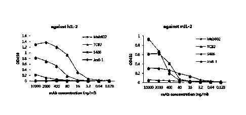
antigen-binding fragment thereof that comprises: a heavy-chain variable region
comprising a heavy-chain CDR1 comprising an amino acid sequence of SEQ ID NO:
11, a heavy-chain CDR2 comprising an amino acid sequence of SEQ ID NO: 12, and
a
heavy-chain CDR3 comprising an amino acid sequence of SEQ ID NO: 13; and a
light-chain variable region comprising a light-chain CDR1 comprising an amino
acid
sequence of SEQ ID NO: 14, a light-chain CDR2 comprising an amino acid
sequence
of SEQ ID NO: 15, and a light-chain CDR3 comprising an amino acid sequence of
SEQ ID NO: 16.
[20] The present invention also provides a nucleic acid encoding the anti-
hIL-2 antibody
or antigen-binding fragment thereof, a vector comprising the nucleic acid, a
cell
transformed with the vector, and a method of producing an anti-hIL-2 antibody
or
antigen-binding fragment thereof using the same.
[21] The present invention also provides a complex in which the anti-hIL-2
antibody or
antigen-binding fragment thereof is bound to hIL-2.
[22] The present invention also provides a composition and treatment method
for
preventing or treating cancer, which comprises the anti-hIL-2 antibody or
antigen-
binding fragment thereof as an active ingredient.
[23] The present invention also provides a bispecific antibody or antibody-
drug conjugate
comprising the anti-hIL-2 antibody or antigen-binding fragment thereof, and a
com-
position and treatment method for preventing or treating cancer, which
comprises the
bispecific antibody or antibody-drug conjugate as an active ingredient.
[24] The present invention also provides a co-administration composition
and treatment
method for cancer treatment, which comprises the anti-hIL-2 antibody or
antigen-
binding fragment thereof and an immune checkpoint inhibitor.
[25] The present invention also provides the use of the anti-hIL-2 antibody
or antigen-
binding fragment thereof for the prevention or treatment of cancer.
[26] The present invention also provides the use of the anti-hIL-2 antibody
or antigen-
binding fragment thereof for the preparation of a medicine for the prevention
or
treatment of cancer.
[27] The present invention also provides a composition for enhancing
vaccine efficacy,
which comprises the anti-hIL-2 antibody or antigen-binding fragment thereof as
an
active ingredient.
[28]
Brief Description of Drawings
CA 03064534 2019-11-21
WO 2018/217058 PCT/KR2018/005955
4
[29] FIG. 1 shows the results of testing the binding specificity of a TCB2
monoclonal
antibody against hIL-2.
[30] FIG. 2 shows the in vivo immunostimulatory effect of a hIL-2/TCB2
complex. FIG.
2A shows the results of analyzing the frequency of immune cells; FIG. 2B shows
the
results of analyzing the expression of CD44 and CD62L in CD4 and CD8 T cells;
FIG.
2C shows the results of experimental statistical analysis; and FIG. 2D shows
the effect
of a hIL-2/MAB602 or hIL-2/TCB2 complex on expansion of immune cells and the
results of experimental statistical analysis (**p <0.01, ***p <0.001 (unpaired
t test)).
[31] FIG. 3 shows surface plasmon resonance curves obtained using Biacore
T100 for the
affinities of anti-hIL-2 mAbs for hIL-2.
[32] FIG. 4 shows the effect of a hIL-2/TCB2 complex against a solid tumor
(***p
<0.001 (Two way ANOVA for day 12, unpaired t test for day 14)).
[33] FIG. 5 shows the effect of TCB2 mAb against a metastatic tumor (***p
<0.001
(unpaired t test)).
[34] FIG. 6 shows the anti-tumor effect of a combination of a hIL-2/TCB2
complex and
tumor peptide therapy in B6F10 melanoma models (***p <0.001 (Two way
ANOVA)).
[35] FIG. 7 shows the anti-tumor effect of a combination of a hIL-2/TCB2
complex and
an anti-CTLA-4 antibody in CT26 tumor models (Balb/C colon cancer) (**p <0.01
(Two way ANOVA for day 17, unpaired t test for day 24)).
[36] FIG. 8 shows the anti-tumor effect of a combination of a hIL-2/TCB2
complex and
an anti-PD-1 antibody in MC38 tumor models (B6 colon cancer) (*p <0.05, **p
<0.01
(Two way ANOVA for day 19, unpaired t test for day 21)).
[37] FIG. 9 shows the in vivo immunostimulatory of a hIL-2/hnTCB2 complex
and the
results of experimental statistical analysis.
[38] FIG. 10 shows the anti-tumor effect of a combination of a hIL-2/hnTCB2
complex
and an anti-PD-1 antibody in MC38 tumor models (B6 colon cancer) (*p <0.05,
**p
<0.01 (Two way ANOVA for day 19 and 22, unpaired t test for day 25)).
[39]
[40] Best Mode For Carrying Out The Invention
[41] Unless otherwise defined, all technical and scientific terms used
herein have the same
meaning as commonly understood by one of ordinary skill in the art to which
the
present disclosure belongs. In general, the nomenclature used herein is well
known and
commonly used in the art.
[42]
[43] In the present invention, efforts have been made to develop a method
that extends the
in vivo half-life of IL-2 without causing an unnatural modification of IL-2,
and at the
same time, selectively activates the CD8+ T cells and NK cells that express
the low-
CA 03064534 2019-11-21
WO 2018/217058 PCT/KR2018/005955
affinity IL-2 receptor. As a result, it has been found that, when an anti-IL-2
monoclonal antibody (mAb) having a particular specificity is bound to IL-2, it
se-
lectively inhibits the binding of IL-2 to the high-affinity IL-2 receptor.
[44]
[45] In one aspect, the present invention is directed to an anti-hIL-2
antibody (referred to
as "TCB2" in the specification) or antigen-binding fragment thereof, which
binds
specifically to human interleukin-2 (hIL-2) and inhibits the binding of the
hIL-2 to
CD25.
[46] As used herein, the term "human interleukin-2 (hIL-2)" refers to a 133-
amino-acid
protein (15.4 kDa) having no substantial sequence homology with any other
factors.
[47] As used herein, the term "CD25" refers to the IL-2Ra chain of IL-2
receptor. The IL-
2 receptor is present as high-affinity IL-2 receptor (IL-2R) and low-affinity
IL-2
receptor (IL-2R) depending on its affinity, and CD25 is a chain that is not
present in
the low-affinity IL-2 receptor and is present only in the high-affinity IL-2
receptor.
[48] The term "antibody" as used in the invention refers to a substance
produced by the
stimulus of an antigen in immune system and its kinds are not particularly
limited.
Lately, the antibodies have been widely used for treating diseases. As the
antibodies
are very stable in vivo as well as in vitro and have a long half-life, they
are favorable
for mass expression and production. Also, since the antibody has intrinsically
a dimer
structure, it has a fairly high avidity. An intact antibody has a structure
with two full-
length light chains and two full-length heavy chains, and each light chain is
linked to
each heavy chain via a disulfide bond. The constant region of an antibody is
divided
into a heavy chain constant region and a light chain constant region, and the
heavy
chain constant region has gamma (y), mu ([1), alpha (a), delta (8) and epsilon
(E) types,
and has gammal (y1), gamma2 (y2), gamma3 (y3), gamma4 (y4), alphal (al) and
a1pha2 (a2) as its subclass. The light chain constant region has kappa (lc)
and lambda
(X) types.
[49] The antibody in the invention may include an animal-derived antibody,
a chimeric
antibody, a humanized antibody, or a fully human antibody. An animal-derived
antibody which is produced by immunizing an animal with a desired antigen may
generally trigger an immune rejection response when administered to humans for
treatment purpose, and a chimeric antibody has been developed to suppress such
immune rejection response. A chimeric antibody is formed by replacing the
constant
region of an animal-derived antibody, which is a cause of an anti-isotype
response,
with the constant region of a human antibody using genetic engineering
methods. The
chimeric antibody has considerably improved anti-isotype response in
comparison with
animal-derived antibodies, but animal-derived amino acids are still present in
its
variable regions and thus it still contains potential side effects resulting
from an anti-
CA 03064534 2019-11-21
WO 2018/217058
PCT/KR2018/005955
6
idiotypic response. It is a humanized antibody that has been thus developed to
improve
such side effects. This is manufactured by grafting CDRs (complementarity de-
termining regions) which, of the variable regions of a chimeric antibody, have
an
important role in antigen binding into a human antibody framework.
[501 A "humanized antibody" as used herein includes a humanized light
chain variable
domain immunoglobulin and a humanized heavy chain variable domain im-
munoglobulin. The humanized antibody may include a constant region partially
or
wholly derived from (including synthetic analogs) one or more human gene
sequence.
A humanized antibody is expected to bind to the same target antigen as a donor
antibody which supplied the CDRs. Typically, all segments or portions of the
humanized antibody or immunoglobulin, with the exception of the CDRs, are sub-
stantially identical or substantially homologous to corresponding segments or
portions
of naturally occurring or consensus human immunoglobulin sequences. It is
important
in CDR grafting technology for manufacturing a humanized antibody to select an
optimized human antibody which can receive best the CDR of an animal-derived
antibody and for this, utilization of antibody database, analysis of crystal
structure,
molecule modeling technology, etc. are employed. However, although the CDR of
an
animal-derived antibody is grafted into an optimized human antibody framework,
there
are a considerable number of cases where antigen binding affinity is not
preserved
because there are amino acids which affect antigen binding while being
positioned at
the framework of the animal-derived antibody. In this regard, it may be
necessary to
apply an additional antibody engineering technology for restoring antigen
binding
affinity.
[51] As used herein, the term "monoclonal antibody (mAb)" has the same
meaning as
commonly used in the technical field to the present invention pertains, and
means an
antibody that recognizes a single epitope on an antigen to which it binds.
This contrasts
with a polyclonal antibody which is a collection of different antibodies that
bind to the
same antigen but bind to different epitopes of the antigen. For this reason, a
single
antigen molecule can be bound simultaneously by multiple polyclonal
antibodies, but a
particular monoclonal antibody specific for the antigen can be bound by only
one
molecule. After being bound by the single monoclonal antibody molecule, the
bound
epitope is blocked, and thus can no longer be bound by other monoclonal
antibodies.
The monoclonal nature of antibodies is particularly suitable for use as
therapeutic
agents. This is because these antibodies are single, homologous molecular
species, and
thus can be very well characterized, can be produced reproducibly, and
purified. These
factors make it possible to produce products whose biological activity can be
predicted
with a very high level of accuracy. These factors are particularly important,
because
these molecules must obtain permission from authorities for therapeutic
administration
CA 03064534 2019-11-21
WO 2018/217058 PCT/KR2018/005955
7
to mammals, particularly humans.
[521 The term "heavy chain" as used herein may be interpreted to include a
full-length
heavy chain including a variable region domain VH including an amino acid
sequence
having a variable region sequence sufficient to confer antigen-specificity,
three
constant region domains CH1, CH2 and CH3, and a hinge, and a fragment thereof.
Also, the term "light chain" as used herein may be interpreted to include a
full-length
light chain including a variable region domain VL including an amino acid
sequence
having a variable region sequence sufficient to confer antigen-specificity and
a
constant region domain CL, and a fragment thereof.
[531 In the present invention, the anti-hIL-2 antibody or antigen-binding
fragment thereof
may comprise: a heavy-chain variable region comprising an amino acid sequence
selected from the group consisting of SEQ ID NOS: 3, 23, 28, 32, and 34; and a
light-
chain variable region comprising an amino acid sequence selected from the
group
consisting of SEQ ID NOS: 4, 24, 26, and 30. Preferably, the anti-hIL-2
antibody or
antigen-binding fragment thereof may comprise: a heavy-chain variable region
of SEQ
ID NO: 3 and a light-chain variable region of SEQ ID NO: 4; a heavy-chain
variable
region of SEQ ID NO: 23 and a light-chain variable region of SEQ ID NO: 24; a
heavy-chain variable region of SEQ ID NO: 28 and a light-chain variable region
of
SEQ ID NO: 26; a heavy-chain variable region of SEQ ID NO: 32 and a light-
chain
variable region of SEQ ID NO: 30; or a heavy-chain variable region of SEQ ID
NO: 34
and a light-chain variable region of SEQ ID NO: 30.
[541 As used herein, the term "complementarity determining region (CDR)"
refers to the
amino acid sequence of the hypervariable region of the heavy chain or light
chain of
immunoglobulin. Each of the heavy and light chains may comprise three CDRs
(i.e., a
heavy chain CDR1, a heavy chain CDR2, and a heavy chain CDR3; and a light
chain
CDR1, a light chain CDR2, and a light chain CDR3). The CDR may provide
important
contact residues for the binding of the antibody to an antigen or an epitope.
[551 In the present invention, the anti-hIL-2 antibody or antigen-binding
fragment thereof
may comprise: a heavy-chain variable region comprising a heavy-chain CDR1
comprising a DNA sequence of SEQ ID NO: 5, a heavy-chain CDR2 comprising a
DNA sequence of SEQ ID NO: 6, and a heavy-chain CDR3 comprising a DNA
sequence of SEQ ID NO: 7; and a light-chain variable region comprising a light-
chain
CDR1 comprising a DNA sequence of SEQ ID NO: 8, a light-chain CDR2 comprising
a DNA sequence of SEQ ID NO: 9, and a light-chain CDR3 comprising a DNA
sequence of SEQ ID NO: 10.
[561 In the present invention, the anti-hIL-2 antibody or antigen-binding
fragment thereof
may comprise: a heavy-chain variable region comprising a heavy-chain CDR1
comprising an amino acid sequence of SEQ ID NO: 11, a heavy-chain CDR2
CA 03064534 2019-11-21
WO 2018/217058 PCT/KR2018/005955
8
comprising an amino acid sequence of SEQ ID NO: 12, and a heavy-chain CDR3
comprising an amino acid sequence of SEQ ID NO: 13; and a light-chain variable
region comprising a light-chain CDR1 comprising an amino acid sequence of SEQ
ID
NO: 14, a light-chain CDR2 comprising an amino acid sequence of SEQ ID NO: 15,
and a light-chain CDR3 comprising an amino acid sequence of SEQ ID NO: 16.
[571 As used herein, the term "specifically binding" has the same meaning
as generally
known to a person of ordinary skill in the art, indicating that an antigen and
an
antibody specifically interact with each other to lead to an immunological
response. In
the present invention, the human monoclonal antibody or its fragment has the
ability to
discriminate human IL-2 (hIL-2) from several other potential antigens. The dis-
crimination is achieved such that the monoclonal antibody or its fragment
binds only
or to a significant extent to hIL-2 as a potential binding partner in a pool
of multiple
different antigens. In this regard, "bind to a significant extent to hIL-2"
means that
hIL-2 as a potential binding partner in a pool of a plurality of equally
accessible
different antigens binds with an affinity at least 10-fold, preferably 50-
fold, preferably
100-fold higher than antigens other than hIL-2.
[581 As used herein, the term "antigen-binding fragment," which is a
fragment of the full
structure of an immunoglobulin, refers to some of a polypeptide including a
portion to
which an antigen can bind. For example, it may be a scFv, a (scFv)2, a Fab, a
Fab' or a
F(ab')2, but is not limited thereto. Among the above antigen-binding
fragments, a Fab,
which is a structure having the light chain and heavy chain variable regions,
the light
chain constant region, and the heavy chain first constant region (CHI), has
one antigen
binding site. A Fab' differs from the Fab in that the Fab' has a hinge region
including at
least one cysteine residue at the C-terminal of the heavy chain CH1 domain. A
F(ab')2
is produced when cysteine residues at the hinge region of Fab' are joined by a
disulfide
bond. A Fv is a minimal antibody fragment, having only heavy chain variable
regions
and light chain variable regions, and a recombinant technique for producing
the Fv
fragment is well known in the art. A two-chain Fv may have a structure in
which heavy
chain variable regions are linked to light chain variable regions by a non-
covalent
bond, and a single-chain Fv may generally form a dimer structure as in the two-
chain
Fv, wherein heavy chain variable regions are covalently bound to light chain
variable
regions via a peptide linker or the heavy and light chain variable regions are
directly
linked to each other at the C-terminals thereof. The linker may be a peptide
linker
including 1 to 100 or 2 to 50 any amino acids, and proper sequences thereof
have been
known in the art. The antigen-binding fragment may be obtained using a
protease (for
example, a whole antibody can be digested with papain to obtain Fab fragments,
or can
be digested with pepsin to obtain F(ab')2 fragments), or may be prepared by a
genetic
recombinant technique. The antigen-binding fragment of the antibody of the
present
CA 03064534 2019-11-21
WO 2018/217058 PCT/KR2018/005955
9
invention may be a fragment including one or more CRDs.
[59] In the present invention, the anti-hIL-2 antibody or antigen-binding
fragment thereof
may induce expansion of CD8+ T cells and NK cells. In an example of the
present
invention, it was found that the anti-hIL-2 antibody according to the present
invention
induced activation of CD8+ T cells and NK cells and induced little expansion
of Treg
cells.
[60]
[61] In another aspect, the present invention is directed to a nucleic acid
encoding the
anti-hIL-2 antibody or the antigen-binding fragment thereof.
[62] In the present invention, the nucleic acid encoding the anti-hIL-2
antibody or
antigen-binding fragment thereof may comprise a sequence of SEQ ID NO: 1, SEQ
ID
NO: 2, SEQ ID NO: 25, SEQ ID NO: 27, SEQ ID NO: 29, SEQ ID NO: 31 or SEQ ID
NO: 33. Specifically, the nucleic acid encoding the heavy chain of the
antibody
according to the present invention may comprise a sequence of SEQ ID NO: 27,
31 or
33, and/or the nucleic acid encoding the light chain of the antibody according
to the
present invention may comprise a sequence of SEQ ID NO: 2, 25 or 29.
[63] The antibody or antigen-binding fragment thereof of the present
invention may be re-
combinantly produced by isolating the nucleic acid encoding an antibody or
antigen-
binding fragment thereof. The nucleic acid is isolated and inserted into a
replicable
vector to result in further cloning (amplification of DNA) or further
expression.
[64] As used herein, the term "Nucleic acid" has a broad meaning including
DNA (gDNA
and cDNA) and RNA molecules. Nucleotides, basic elements of nucleic acids,
include
natural nucleotides as well as analogues in which sugar or base sites are
modified. The
sequence of the nucleic acid encoding the heavy and light chain variable
regions of the
present invention may be modified. Such modifications include the addition,
deletion,
or non-conservative substitution or conservative substitution of nucleotides.
[65] The nucleic acid of the present invention is interpreted to include a
nucleotide
sequence that exhibits substantial identity to the nucleotide sequence. The
substantial
identity means a nucleotide sequence showing at least 80% homology, more
preferably
at least 90% homology, and most preferably at least 95% homology by aligning
the nu-
cleotide sequence of the present invention with any other sequence as much as
possible
and analyzing the aligned sequence using algorithms commonly used in the art.
[66] The DNA encoding the antibody can be easily separated or synthesized
using con-
ventional procedures (for example, using an oligonucleotide probe capable of
specifically binding to DNA encoding the heavy chain and the light chain of
the
antibody).
[67] In still another aspect, the present invention is directed to a
recombinant vector
including the nucleic acid.
CA 03064534 2019-11-21
WO 2018/217058 PCT/KR2018/005955
[68] Many vectors are available. Vector components generally include, but
are not limited
to, one or more of the following: a signal sequence, an origin of replication,
one or
more marker genes, an enhancer element, a promoter, and a transcription
termination
sequence.
[69] The term "vector" as used herein, includes a plasmid vector; a cosmid
vector; a bac-
teriophage vector; and a viral vector, e.g., an adenovirus vector, retroviral
vectors, and
adeno-associated viral vectors as a mean for expressing a target gene in a
host cell. The
nucleic acid encoding the antibody in the vector is operably linked to a
promoter.
[70] As used herein, the term "operably linked" refers to a functional
linkage between a
nucleic acid expression control sequence (e.g., an array of promoter, signal
sequence,
or transcription regulation factor binding site) and another nucleic acid
sequence, and
thus the control sequence controls the transcription and/or translation of the
other
nucleic acid sequence.
[71] When a prokaryotic cell is used as a host, a strong promoter capable
of promoting
transcription (such as a tac promoter, lac promoter, lacUV5 promoter, 1pp
promoter,
pLX promoter, pRX promoter, rac5 promoter, amp promoter, recA promoter, SP6
promoter, trp promoter, and T7 promoter), a ribosome binding site for
initiation of
translation, and a transcription/translation termination sequence are
generally included.
Further, for example, when a eukaryotic cell is used as a host, a promoter
derived from
a genome of a mammalian cell (e.g., a metallothionein promoter, aP-actin
promoter, a
human hemoglobin promoter and a human muscle creatine promoter) or a promoter
derived from an mammalian virus (e.g., adenovirus late promoter, vaccinia
virus 7.5K
promoter, SV40 promoter, cytomegalovirus (CMV) promoter, HSV tk promoter,
mouse mammary tumor virus (MMTV) promoter, HIV LTR promoter, epstein ban
virus (EBV) promoter of moloney virus and Rous sarcoma virus (RSV) promoter)
can
be used, and generally have a polyadenylation sequence as a transcription
termination
sequence.
[72] Optionally, the vector may be fused with another sequence in order to
facilitate pu-
rification of an antibody expressed therefrom. Fused sequences include, for
example,
glutathione S-transferase (Pharmacia, USA), maltose binding protein (NEB,
USA),
FLAG (IBI, USA), and 6x His (hexahistidine; Quiagen, USA).
[73] The vector includes an antibiotic resistance gene commonly used in the
art as a
selective marker, and may include, for example, genes having resistance to
ampicillin,
gentamicin, carbenicillin, chloramphenicol, streptomycin, kanamycin,
geneticin,
neomycin, and tetracycline.
[74] In yet another aspect, the present invention is directed to a cell
transformed with the
recombinant vector. Cells used to produce the antibody of the present
invention may be
prokaryotic cells, yeasts, or other higher eukaryotic cells, but are not
limited thereto.
CA 03064534 2019-11-21
WO 2018/217058 PCT/KR2018/005955
11
[75] In the present invention, as the transformed cell, the prokaryotic
host cell can be
used, for example, a strain belonging to the genus Bacillus such as
Escherichia coli,
Bacillus subtilis, and Bacillus thuringiensis, Streptomyces, Pseudomonas (for
example,
Pseudomonas putida), Proteus mirabilis, and Staphylococcus (for example,
Staphy-
lococcus carnosus).
[76] Meanwhile, interest in animal cells is greatest, and an example of a
useful host cell
line may be, but is not limited thereto, COS-7, BHK, CHO, CHOK1, DXB-11, DG-
44,
CH0/-DHFR, CV1, COS-7, HEK293, BHK, TM4, VERO, HELA, MDCK, BRL 3A,
W138, Hep G2, SK-Hep, MMT, TRI, MRC 5, FS4, 3T3, RIN, A549, PC12, K562,
PER.C6, SP2/0, NS-0, U205, or HT1080.
[77]
[78] In a further aspect, the present invention is directed to a method of
producing an anti-
hIL-2 antibody or antigen-binding fragment thereof, comprising culturing the
cell,
thereby expressing the anti-hIL-2 antibody or antigen-binding fragment thereof
according to the present invention.
[79] The cells can be cultured in various media. Commercially available
media can be
used as a culture medium without limitation. All other essential supplements
known to
those skilled in the art may be included in the appropriate concentrations.
Culturing
conditions, e.g., temperature and pH have already been used with the selected
host
cells for expression, which will be apparent to those skilled in the art.
[80] When the antibody or antigen-binding fragment thereof is recovered,
impurities can
be removed, e.g., by centrifugation or ultrafiltration, and the resultant can
be purified,
for example, by affinity chromatography. Additional purification techniques
may be
used, such as anion or cation exchange chromatography, hydrophobic interaction
chro-
matography, and hydroxyl apatite chromatography.
[81]
[82] In a still further aspect, the present invention is directed to a
complex in which an
anti-hIL-2 antibody or antigen-binding fragment thereof is bound to hIL-2.
[83]
[84] In a yet further aspect, the present invention is directed to an
antibody-drug conjugate
(ADC) comprising a drug conjugated to the anti-hIL-2 antibody or antigen-
binding
fragment thereof.
[85] An antibody-drug conjugate (ADC) requires that the anticancer drug
should be stably
bound to the antibody before the anticancer drug is delivered to target cancer
cells. The
drug delivered to the target should be released from the antibody and should
induce
death of the target cells. To this end, the drug should be stably bound to the
antibody
and, at the same time, should have enough cytotoxicity to induce death of the
target
cells when being released from the antibody.
CA 03064534 2019-11-21
WO 2018/217058 PCT/KR2018/005955
12
[86] In the present invention, the anti-hIL-2 antibody or antigen-binding
fragment thereof
and cytotoxic substances including drugs such as anticancer drugs may be
linked to
each other by, for example, a covalent bond, a peptide bond or the like, so
that they
may be used as conjugates or fusion proteins (where cytotoxic substances
and/or
labeling substances are proteins). The cytotoxic substance may be any
substance
having toxicity against cancer cells, particularly solid cancer cells, and may
be one or
more selected from the group consisting of, but not limited to, radioisotopes,
cytotoxic
compounds (small molecules), cytotoxic proteins, anticancer agents, and the
like. The
cytotoxic proteins may be one or more selected from the group consisting of,
but not
limited to, ricin, saporin, gelonin, momordin, debouganin, diphtheria toxin,
and
pseudomonas toxin. The radioisotopes may be one or more selected from the
group
consisting of, but not limited to, 1311, 188Rh, and 90Y. The cytotoxic
compounds may
be one or more selected from the group consisting of, but not limited to,
duocarmycin,
monomethyl auristatin E (MMAE), monomethyl auristatin F (MMAF),
N2'-deacetyl-N2'-(3-mercapto-1-oxopropyl)maytansine (DM1), and
PBD(Pyrrolobenzodiazepine) dimer.
[87] In the present invention, the antibody-drug conjugate may be obtained
according to a
technique well known in the technical field to which the present invention
pertains.
[88] In the present invention, the antibody-drug conjugate may be one in
which the
antibody or antigen-binding fragment thereof is bound to the drug by a linker.
[89] In the present invention, the linker may be a cleavable linker or a
non-cleavable
linker.
[90] The linker is a region that connects between anti-hIL-2 antibody and
the drug. For
example, the linker is configured such that it is cleavable under
intracellular
conditions, that is, the drug can be released from the antibody through
cleavage of the
linker in an intracellular environment.
[91] The linker can be cleaved by a cleaving agent present in an
intracellular environment,
for example, lysosome or endosome. The linker may be a peptide linker that can
be
cleaved by intracellular peptidase or protease enzyme, for example, lysosome
or
endosome protease. Generally, the peptide linker has a length of at least two
amino
acids. The cleaving agents may include cathepsin B, cathepsin D, and plasmin,
and are
capable of hydrolyzing the peptide to enable the drug to be released into
target cells.
The peptide linker can be cleaved by thiol-dependent protease cathepsin B
which is
highly expressed in cancer tissue. For example, the linker that is used in the
present
invention may be a Phe-Leu or Gly-Phe-Leu-Gly linker. In addition, the peptide
linker
may also be a Val-Cit or Phe-Lys linker which is cleavable by, for example,
intra-
cellular protease.
[92] In the present invention, the cleavable linker is pH-sensitive, i.e.,
sensitive to hy-
CA 03064534 2019-11-21
WO 2018/217058 PCT/KR2018/005955
13
drolysis at certain pH values. Typically, the pH-sensitive linker is
hydrolyzable under
acidic conditions. For example, an acid-labile linker that is hydrolyzable in
the
lysosome (e.g., a hydrazone, semicarbazone, thiosemicarbazone, cis-aconitic
amide,
orthoester, acetal, ketal, or the like) can be used.
[93] The linker is cleavable under reducing conditions (e.g., a disulfide
linker). A variety
of disulfide linkers can be formed using SATA (N-succinimidyl-S-
acetylthioacetate),
SPDP (N-succinimidy1-3- (2-pyridyldithio)propionate), SPDB
(N-succinimidy1-3-(2-pyridyldithio)butyrate) and SMPT (N- succinimidyl-oxy-
carbonyl-alpha-methyl-alpha-(2-pyridyl-dithio)toluene).
[94] In the present invention, the drug and/or the drug-linker may be
conjugated randomly
through the lysine of the antibody or may be conjugated through a cysteine
which is
exposed when a disulfide bond chain is reduced. In some cases, the linker-drug
may be
bound through a cysteine present in a genetically engineered tag, for example,
a
peptide or a protein. The genetically engineered tag, for example, a peptide
or a
protein, may include an amino acid motif that may be recognized by, for
example,
isoprenoid transferase. The above-described peptide or protein has a deletion
at the
carboxy terminus of the peptide or protein, or has an addition at the carboxy
(C)
terminus of the peptide or protein through covalent bonding to a spacer unit.
The
peptide or the protein may be covalently bonded directly to the amino acid
motif or
may be linked to the amino acid motif by covalent bonding to a spacer unit.
The amino
acid spacer unit is composed of 1 to 20 amino acids, and is preferably a
glycine unit.
[95] The linker may include a beta-glucuronide linker which is recognized
and hy-
drolyzed by P-glucuronidase which is present in lysosomes or is highly
expressed in
some tumor cells. Unlike a peptide linker, the beta-glucuronide linker has an
advantage
in that it has high hydrophilicity, and thus can increase the solubility of an
antibody-
drug conjugate when it is bound to a highly hydrophobic drug.
[96] In addition, the linker may be a non-cleavable linker. In this case,
the drug may be
released through only a single step (antibody hydrolysis), thus producing, for
example,
an amino acid-linker-drug conjugate. This type of linker may be thioether or
maleimi-
docaproyl, and may maintain its stability in blood.
[97] In the present invention, the drug may be a chemotherapeutic agent,
toxin, micro
RNA (miRNA), siRNA, shRNA, or radioisotope. The drug that is a formulation ex-
hibiting a pharmacological effect may be conjugated to the antibody.
[98] The chemotherapeutic agent may be a cytotoxic agent or an immune
checkpoint
inhibitor. Specifically, the chemotherapeutic agent may include a
chemotherapeutic
agent capable of functioning as a microtubulin inhibitor, a mitotic inhibitor,
a topoi-
somerase inhibitor, or a DNA intercalator. In addition, the chemotherapeutic
agent may
include an immunomodulatory compound, an anticancer agent, an antiviral agent,
an
CA 03064534 2019-11-21
WO 2018/217058 PCT/KR2018/005955
14
antibacterial agent, an antifungal agent, an antiparasitic agent, or a
combination
thereof.
[99] The drug may be one or more selected from the group consisting of, but
not limited
to, for example, maytansinoid, auristatin, aminopterin, actinomycin,
bleomycin, tal-
isomycin, camptothecin, N8-acetyl spermidine, 1-(2-chloroethyl)-1,2-
methylsulfonyl
hydrazide, esperamycin, etoposide, 6-mercaptopurine, dolastatin, tricotecene,
calicheamycin, taxol, taxane, paclitaxel, docetaxel, methotrexate,
vincristine, vin-
blastine, doxorubicin, melphalan, mitomycin A, mitomycin C, chlorambucil, duo-
carmycin, L-asparaginase, mercaptopurine, thioguanine, hydroxyurea,
cytarabine, cy-
clophosphamide, ifosfamide, nitrosourea, cisplatin, carboplatin, mitomycin,
dacarbazine, procarbazine, topotecan, nitrogen mustard, cytoxan, etoposide,
5-fluorouracil, bischloroethylnitrosourea (BCNU), irinotecan, camptothecin,
bleomycin, idarubicin, daunorubicin, dactinomycin, plicamycin, mitoxantrone,
as-
paraginase, vinorelbine, chlorambucil, melphalan, carmustine, lomustine,
busulfan,
treosulfan, decarbazine, etoposide, teniposide, topotecan, 9-
aminocamptothecin,
crisnatol, mitomycin C, trimetrexate, mycophenolic acid, tiazofurin,
ribavirin,
5-ethyny1-1-beta-dribofuranosylimidazole-4-carboxamide (EICAR), hydroxyurea,
de-
feroxamine, floxuridine, doxifluridine, raltitrexed, cytarabine (ara C),
cytosine ara-
binoside, fludarabine, tamoxifen, raloxifene, megestrol, goserelin, leuprolide
acetate,
flutamide, bicalutamide, EB1089, CB1093, KH1060, verteporfin, phthalocyanine,
pho-
tosensitizer Pe4, demethoxy-hypocrellin A, interferon-a, interferon-y, tumor
necrosis
factor, gemcitabine, velcade, revamid, thalamid, lovastatin,
1-methyl-4-phenylpyridiniumion, staurosporine, actinomycin D, dactinomycin,
bleomycin A2, bleomycin B2, peplomycin, epirubicin, pirarubicin, zorubicin, mi-
toxantrone, verapamil and thapsigargin, nuclease, and toxins derived from
bacteria or
animals/plants.
[100] In the present invention, the drug may include one or more
nucleophilic groups
selected from the group consisting of amine, thiol, hydroxyl, hydrazide,
oxime,
hydrazine, thiosemicarbazone, hydrazine carboxylate and aryl hydrazide groups,
which
can react to form covalent bonds with the linker and the electrophilic group
on a linker
reagent.
[101]
[102] In another further aspect, the present invention is directed to a
bispecific antibody
comprising the anti-hIL-2 antibody or antigen-binding fragment thereof.
[103] In the present invention, the bispecific antibody means an antibody
form in which
one of the two arms of the antibody comprises the anti-hIL-2 antibody or
antigen-
binding fragment thereof according to the present invention, and the other arm
comprises either an antibody specific for an antigen other than hIL-2,
preferably a
CA 03064534 2019-11-21
WO 2018/217058 PCT/KR2018/005955
cancer-related antigen or an immune checkpoint protein antigen, or an antibody
or
antigen-binding fragment thereof which binds specifically to an immune
effector cell-
related antigen.
[104] The antigen to which the antibody other than the anti-hIL-2 antibody
included in the
bispecific antibody binds is a cancer-related antigen or an immune checkpoint
protein
antigen, which may be selected from among Her2, EGFR, VEGF, VEGF-R, CD-20,
MUC16, CD30, CD33, CD52, 4-1BB, TIM3, PD-1, PD-L1, CTLA4, BTLA4, EphB2,
E-selectin, EpCam, CEA, PSMA, PSA, ERB3, c-MET, and the like, and the immune
effector cell-related antigen may be selected from among, but not limited to,
TCR/
CD3, CD16 (FcyRIIIa), CD28, CD28, CD44, CD56, CD69, CD64 (FcyRI), CD89,
CD11b/CD18 (CR3), and the like.
[105]
[106] In another still further aspect, the present invention is directed to
a composition for
preventing or treating cancer, which comprises the anti-hIL-2 antibody or
antigen-
binding fragment thereof as an active ingredient.
[107] In another yet further aspect, the present invention is directed to a
composition for
preventing or treating cancer, which comprises the bispecific antibody or
antibody-
drug conjugate as an active ingredient.
[108] "Cancer" refers to a condition in which cells proliferate abnormally
and excessively
due to a problem in the function of regulating the normal division,
differentiation and
death of the cells, and invade the surrounding tissues and organs, thereby
forming a
mass and destroying or deforming the existing structures. "Solid cancer"
refers to a
cancer which has features distinguishable from those of blood cancer and which
is
composed of a mass caused by abnormal growth of cells in various solid organs,
including bladder, breast, intestines, kidneys, lungs, brain, esophagus,
gallbladder,
ovary, pancreas, stomach, cervix, thyroid, prostate, skin and the like.
"Metastatic
cancer" is caused by the metastasis of cancer cells, separated from a primary
cancer
site, to another site through blood, lymphatic vessels or the like, and
proliferation of
the metastasized cancer cells. The composition of the present invention can be
used for
the prevention or treatment of solid cancers and/or metastatic cancers. The
composition
of the present invention may be used for the prevention or treatment of, but
not limited
to, for example, skin cancer, breast cancer, colorectal cancer, kidney cancer,
lung
cancer, liver cancer, brain cancer, esophageal cancer, gallbladder cancer,
ovarian
cancer, pancreatic cancer, stomach cancer, uterine cervical cancer, thyroid
cancer,
prostate cancer, and bladder cancer, but is not limited thereto.
[109] As used herein, the term "preventing/prevention" refers to all
actions that inhibit the
metastasis, growth, and the like of cancers or delay the onset of cancers by
admin-
istering the composition. As used herein, the term "treating/treatment" refers
to any
CA 03064534 2019-11-21
WO 2018/217058 PCT/KR2018/005955
16
action resulting in improvements in symptoms of cancers or the beneficial
alteration of
cancers owing to the administration of the composition.
[110] The composition of the present invention may further comprise a
pharmaceutically
acceptable carrier. The carrier that is typically used in the formulation of
drugs may be
one or more selected from the group consisting of, but not limited to,
lactose, dextrose,
sucrose, sorbitol, mannitol, starch, gum acacia, calcium phosphate, alginate,
gelatin,
calcium silicate, microcrystalline cellulose, polyvinylpyrrolidone, cellulose,
water,
syrups, methyl cellulose, methylhydroxybenzoate, propylhydroxybenzoate, talc,
magnesium stearate, and mineral oil. In addition, the composition may further
comprise one or more selected from the group consisting of excipients,
lubricants,
wetting agents, sweeteners, aromatics, emulsifiers, suspensions, and
preservatives.
[111] The composition or pharmaceutical composition of the antibody may be
administered
orally or parenterally. Such a parenteral administration includes intravenous
injection,
subcutaneous injection, intramuscular injection, intraperitoneal injection,
endothelial
administration, topical administration, nasal administration, intrapulmonary
admin-
istration, intrarectal administration, etc. Because a protein or peptide is
digested when
administered orally, it is preferred that a composition for oral
administration may be
formulated to coat an active substance or to be protected against degradation
in
stomach. Also, the composition may be administered by any device which can
transport active substances to target cells.
[112] The content of the anti-hIL-2 antibody (TCB2 mAb) in the composition
may vary
depending on various factors such as formulation method, administration
method, age,
body weight, sex or pathological condition of the patient, diet,
administration time, ad-
ministration interval, administration route, excretion rate and reaction
sensitivity. For
example, a daily administration dosage of the anti-hIL-2 antibody (TCB2 mAb)
may
be in the range from 0.001 to 1,000 mg/kg, specifically 0.01 to 100 mg/kg,
more
specifically 0.1 to 50 mg/kg, but is not limited thereto. The effective dose
for single ad-
ministration of the anti-hIL-2 antibody (TCB2 mAb) may be formulated as one
for-
mulation in a unit-dose form or formulated in an appropriate amount, or
prepared by
injecting into a multiple-dose vial. The "pharmaceutically effective dose" as
used
herein may refer to the content or the dose of an active ingredient capable of
exhibiting
a desired a pharmacological effect, and can be determined variously depending
on
various factors such as formulation method, administration method, age, body
weight,
sex or pathological condition of the patient, diet, administration time,
administration
interval, administration route, excretion rate and reaction sensitivity.
[113] The composition may be formulated with pharmaceutically acceptable
carriers and/or
excipients according to a method that can be easily carried out by a person
having an
ordinary skill in the art to which the present invention pertains, and may be
provided in
CA 03064534 2019-11-21
WO 2018/217058 PCT/KR2018/005955
17
a unit-dose form or enclosed in a multiple-dose vial. Here, the formulation of
the com-
position may be in the form of a solution, a suspension, syrup or an emulsion
in oily or
aqueous medium, or may be extracts, powders, granules, tablets or capsules,
and may
further include a dispersion agent or a stabilizer. Also, the composition may
be ad-
ministered individually or in combination with other therapeutic agents.
[114] In particular, the composition including the anti-hIL-2 antibody
(TCB2 mAb)
includes an antibody, and thus may be formulated into immuno liposome.
Liposome
including an antibody may be prepared according to a method well known in the
pertinent art. The immuno liposome is a lipid composition including phos-
phatidylcholine, cholesterol and polyethyleneglycol-derived phos-
phatidylethanolamine, and may be prepared by reverse phase evaporation method.
For
example, a Fab' fragment of the antibody may be conjugated to liposome through
disulfide exchange reaction.
[115]
[116] In another yet further aspect, the present invention is directed to a
co-administration
composition for cancer treatment, which comprises the anti-hIL-2 antibody or
antigen-
binding fragment thereof and an immune checkpoint inhibitor.
[117] In the present invention, the immune checkpoint inhibitor (also,
called "checkpoint
inhibitor") may be an anti-CTLA-4 antibody or an anti-PD-1 antibody, but is
not
limited thereto.
[118] As used herein, the term "co-administration" (also, called
"combination") means that
the anti-hIL-2 antibody or antigen-binding fragment thereof and the immune
checkpoint inhibitor may be administered simultaneously, sequentially, or in
reverse
order, and the anti-hIL-2 antibody or antigen-binding fragment thereof and the
immune
checkpoint inhibitor may be administered in a combination of appropriate
effective
amounts of the active ingredients within the range determined by those skilled
in the
art.
[119] In an example of the present invention, it was found that when the
anti-CTLA-4 or
anti-PD-1 antibody and the anti-hIL-2 antibody according to the present
invention are
treated sequentially, the growth of tumor cells is further suppressed.
[120] The co-administration composition includes the anti-hIL-2 antibody,
and the
components related thereto are the same as the components included in the
above-
described composition for preventing or treating cancer. Thus, the description
of each
constitution applies equally to the co-administration composition.
[121]
[122] In another yet further aspect, the present invention is directed to a
method for
prevention and/or treatment of cancer, which comprises a step of administering
to a
patient a therapeutically effective amount of the anti-hIL-2 antibody or
antigen-binding
CA 03064534 2019-11-21
WO 2018/217058 PCT/KR2018/005955
18
fragment thereof, the bispecific antibody or the antibody-drug conjugate.
[123] The composition of the present invention may be administered as an
individual
therapeutic agent or in combination with other therapeutic agents, and may be
ad-
ministered sequentially or simultaneously with conventional therapeutic
agents.
[124] Any anticancer drug, for example, cisplatin, has side effects such as
cachexia,
sarcopenia, muscle wasting, bone wasting or involuntary body weight loss.
Thus, the
present invention may include a composition or a cancer treatment method,
which
treats cancer while preventing, minimizing or lowering the severity, frequency
or oc-
currence of cachexia, sarcopenia, muscle wasting, bone wasting or involuntary
body
weight loss.
[125] The method comprises a step of administering a pharmaceutical
composition
comprising an effective amount of the anti-hIL-2 antibody of the present
invention in
combination with at least one anticancer agent. In particular embodiments, the
present
invention includes a method which treats cancer while preventing, minimizing
or
lowering the severity, frequency or occurrence of cachexia, sarcopenia, muscle
wasting, bone wasting or involuntary body weight loss, the method comprising a
step
of administering to a patient a pharmaceutical composition comprising an
effective
amount of the anti-hIL-2 antibody of the present invention in combination with
one or
more anticancer agents known to induce or increase the severity, frequency or
oc-
currence of cachexia, sarcopenia, muscle wasting, bone wasting or involuntary
body
weight loss.
[126] In another yet further aspect, the present invention is directed to a
method for treating
cancer, which comprises a step of co-administering a composition comprising
the anti-
hIL-2 antibody or antigen-binding fragment thereof with an immune checkpoint
inhibitor.
[127] In the method for treating cancer according to the present invention,
the anti-hIL-2
antibody or antigen-binding fragment thereof and the immune checkpoint
inhibitor
may be administered simultaneously, sequentially, or in reverse order.
Preferably, the
method may comprise the steps of: (A) treating with an immune checkpoint
inhibitor;
and (B) treating with the anti-hIL-2 antibody or antigen-binding fragment
thereof, but
is not limited thereto.
[128] The immune checkpoint inhibitor may be an anti-CTLA-4 antibody or an
anti-PD-1
antibody, but is not limited thereto. The method for treating cancer includes
the com-
position comprising the anti-hIL-2 antibody, and the components related
thereto are the
same as the components included in the above-described composition. Thus, the
de-
scription of each constitution applies equally to the method of treating
cancer by co-
administration.
11291
CA 03064534 2019-11-21
WO 2018/217058 PCT/KR2018/005955
19
[130] In another yet further aspect, the present invention is directed to
the use of the anti-
hIL-2 antibody or antigen-binding fragment thereof for the prevention or
treatment of
cancer.
[131] In another yet further aspect, the present invention is directed to
the use of the anti-
hIL-2 antibody or antigen-binding fragment thereof for the preparation of a
medicine
for the prevention or treatment of cancer.
[132]
[133] In another yet further aspect, the present invention is directed to a
composition for
enhancing vaccine efficacy, which comprises the anti-hIL-2 antibody or antigen-
binding fragment thereof as an active ingredient.
[134] As used herein, the term "vaccine" refers to a biological agent
containing an antigen
that immunizes a living body, and means an immunogenic or antigenic substance
that
produces immunity in vivo by its administration to humans or animals in order
to
prevent infection.
[135]
[136] Examples
[137] Hereinafter, the present invention will be described in further
detail with reference to
examples. It will be obvious to a person having ordinary skill in the art that
these
examples are for illustrative purposes only and are not to be construed to
limit the
scope of the present invention.
[138]
[139] Example 1: Experiment on the Binding Specificity of TCB2 Monoclonal
Antibody
against hIL-2
[140] In vivo mouse models were used to evaluate the therapeutic efficacy
of a hIL-
2/TCB2 mAb complex. For this reason, in order to examine whether TCB2 mAb
shows cross-reactivity with mouse IL-2 (mIL-2), the binding specificity TCB2
mAb
against hIL-2 was tested. First, the splenocytes of BALB/c mice immunized 3-4
times
with hIL-2 over several weeks were fused with SP/2 myeloma cells. When the
hybridoma colony was visualized, the culture supernatant was subjected to
ELISA. 5
[Tim' of hIL-2 or mIL-2 was added to and mixed with PBS, and a total of 50 [11
of the
mixture was coated on an ELISA plate. Next, 200 [11 of 10% FBS was added to
the
PBS and incubated at room temperature for 30 minutes in order to prevent non-
specific
binding, and a titrated dose of the monoclonal antibody was incubated for 30
minutes.
The binding of the monoclonal antibody to the coated hIL-2 or mIL-2 was
detected
with anti-mouse IgG HRP or anti-rat IgG HRP. In each step, the plate was
washed 3-5
times with 200 [11 of PBS. As positive controls, commercially available
monoclonal an-
tibodies were used. As a positive control for hIL-2, Mab602 was used, and as
positive
controls for mIL-2, JES6-1 and 54B6 were used.
CA 03064534 2019-11-21
WO 2018/217058 PCT/KR2018/005955
[141] As a result, Mab602 used as the positive control for hIL-2 showed low
cross-re-
activity, whereas TCB2 mAb showed no cross-reactivity with mIL-2 (FIG. 1).
Thus, it
could be seen that TCB2 mAb did specifically bind only to hIL-2.
[142]
[143] Example 2: In Vivo Immunostimulatory Effect of hIL-2/TCB2 Complex
[144] MAB602, a previously reported mouse anti-hIL-2 mAb, stimulated human
CD8+ T
cells in humanized mice, thus demonstrating the efficacy of a hIL-2/mAb
complex for
anticancer immunotherapy in clinical applications. However, the sequence of
the CDR
region of MAB602 was not published, and it is unclear whether MAB602 is an
antibody which has a maximum anticancer effect when used as a hIL-2/anti-hIL-2
mAb complex. Thus, it was attempted to develop an excellent hIL-2 mAb that
induces
the maximum activation of CD8+ T cells and NK cells and the minimum expansion
of
Treg cells.
[145] On days 0, 1, 2 and 3, a hIL-2/TCB2 mAb (0.8[1g/8 [ig) complex was
injected into
B6 mice, and on day 5, the extent of cell expansion of splenic CD8+ T cells
and Treg
cells was analyzed. The hIL-2/TCB2 complex minimized expansion of Treg cells
and
CD4 T cells, but induced a strong expansion of CD8+ T cells and NK cells (FIG.
2).
Specifically, when the hIL-2/TCB2 mAb complex were injected, memory phenotype
(MP) CD8+ T cells were about 59-fold expanded, and the expanded MP CD8+ T
cells
constituted the majority of CD8+ T cells. NK cells were also 18-fold expanded,
but
Treg cells were only about 5-fold expanded, which was lower than the extent of
expansion of CD8+ T cells and NK cells. The effective ratio of MP CD8+ T cells
to
expanded Treg cells was 970% for the hIL-2/TCB2 mAb complex. Therefore, it can
be
seen that TCB2 mAb is a monoclonal antibody that selectively stimulates CD8+ T
cells
and NK cells, not Treg cells.
[146] In addition, the effective ratio of MP CD8+ T cells to expanded Treg
cells was 970%
for the hIL-2/TCB2 mAb complex, but 530% for the hIL-2/MAB602 complex (FIG.
2D). Thus, TCB2 is a monoclonal antibody superior to MAB602.
[147]
[148] Example 3: Analysis of the Affinity of TCB2 for hIL-2
[149] The selective stimulation of CD8+ T cells and NK cells by the TCB2
antibody
requires that the antibody be bound to the epitope of hIL-2. Since the epitope
of hIL-2
is also recognized by high-affinity IL-2R (CD25), TCB2 is likely to bind to
hIL-2 near
a site to which the IL-2Ra chain binds. Since MAB602 is also likely to bind to
hIL-2
near a site to which the IL-2Ra chain binds, TCB2 was analyzed competitively
with
MAB602 in order to observe the specificity of TCB2 which is an anti-hIL-2 mAb.
Another anti-hIL-2 mAb (5344.111), which is available commercially and known
to
bind to an epitope different from an epitope to which MAB602 binds, was used
as a
CA 03064534 2019-11-21
WO 2018/217058 PCT/KR2018/005955
21
control.
[150] For detection of hIL-2, sandwich ELISA was used. 900 RU (Rmax=90) of
anti-hIL-2
clones were immobilized on a CMS chip by amine coupling. A 2-fold dilution
(100
nM) of hIL-2 was allowed to flow on the chip at a rate of 10 [il/min for 3
minutes, and
then dissociation of the hIL-2 was monitored for 10 minutes.
[151] From the competitive analysis, it was found that TCB2 competed with
MAB602. It
was shown that, due to its specificity, TCB2 mAb did not compete with
5344.111, but
completed with MAB602. As a result, it was confirmed that TCB2 had a higher
affinity for human IL-2 than other anti-hIL-2 mAbs (FIG. 3).
[152]
[153] Example 4: Anti-tumor Effect of hIL-2/TCB2 Complex
[154] Example 4-1: Effect of TCB2 mAb against Solid Tumor
[155] In order to demonstrate the clinical usefulness of TCB2 mAb against a
solid tumor,
lx106B16F10 melanoma cells were injected subcutaneously into B6 mice, and then
PBS, hIL-2 (0.8 [ig) alone or the hIL-2/TCB2 (0.8 [ig/8 [ig) complex was
injected on
days 4 to 7. Next, tumor progression was monitored for 7 days.
[156] As a result, inhibition of solid tumor growth had a correlation with
the magnitude of
cytokine-induced expansion of CD8+ T cells and NK cells (FIG. 4). The hIL-
2/TCB2
mAb complex inhibited tumor growth better than hIL-2 alone.
[157]
[158] Example 4-2: Effect of TCB2 mAb against Metastatic Tumor
[159] In order to demonstrate the clinical usefulness of TCB2 mAb against a
metastatic
tumor, 3x10⁵B16F10 melanoma cells were injected intravenously into B6 mice. 7
Days after tumor injection, hIL-2 alone (0.8 [ig) or the hIL-2/TCB2 (0.8 [i/8
[ig)
complex was injected from day 7 to day 10. On day 18, the number of pulmonary
tumor nodules was measured.
[160] As a result, inhibition of the hIL-2/TCB2 well inhibited of pulmonary
tumor nodules,
unlike hIL-2 (FIG. 5). Thus, it can be see that TCB2 mAb has a potent
anticancer
effect when used as the hIL-2/TCB2 mAb complex.
[161]
[162] Example 5: Analysis of the Effect of Combination of hIL-2/TCB2
Complex and
Other Anticancer Therapies
[163] Anticancer therapies, which are currently developed worldwide,
include a method
that immunizes patients with a tumor neo-antigen, and a method that uses
checkpoint
inhibitors such as anti-CTLA-4 antibodies or anti-PD-1 antibodies. In this
Example,
whether the hIL-2/TCB2 complex can be used in combination with these
anticancer
therapies was analyzed.
11641
CA 03064534 2019-11-21
WO 2018/217058 PCT/KR2018/005955
22
[165] Example 5-1: Effect of Combination of hIL-2/TCB2 Complex and
Anticancer
Therapy Based on Neo-Antigen
[166] In order to test the compatibility of the hIL-2/TCB2 complex with neo-
antigen-based
therapy, 1x10⁶B16F10 cells were injected subcutaneously into B6 mice on day 0.
Next, PBS or a mixture of TRP2 peptide (100 [ig) and Poly I:C (100 [ig) was
injected
on days 3 and 7. The hIL-2/TCB2 complex (0.8 [ig/8 [ig) was injected in two
rounds of
four daily injections on days 4 to 7 and days 11 to 14. Next, tumor
progression was
monitored for 5 days.
[167] As a result, injection of the hIL-2/TCB2 complex and the neo-antigen-
based therapy
inhibited the growth of the B16F10 tumor to similar extents. However, when the
mice
were co-treated with the hIL-2/TCB2 complex and the neo-antigen-based therapy,
tumor growth was more inhibited (FIG. 6). Thus, it can be seen that the hIL-
2/TCB2
complex can be used in combination with the neo-antigen-based therapy.
[168]
[169] Example 5-2: Effect of Combination of hIL-2/TCB2 Complex and
Checkpoint
Inhibitor
[170] To test whether the hIL-2/TCB2 complex can be used in combination
with
checkpoint inhibitors, CT26 (Balb/C colon cancer and MC38 (B6 colon cancer)
models were used. After treatment with the hIL-2/TCB2 complex in combination
with
anti-CTLA-4 antibody or anti-PD-1 antibody or treatment with each of these an-
tibodies, tumor growth was observed.
[171] For an experiment in which mice were treated with the hIL-2/TCB2
complex in com-
bination with the anti-CTLA-4 antibody, 5x10⁵ CT26 cells were injected subcu-
taneously into Balb/C mice (day 0), and the anti-CTLA-4 antibody (100 [ig) was
injected three times at 3-day intervals from day 7. The hIL-2/TCB2 complex
(0.8 [ig/8
[ig) was injected once a day from day 8 to day 11 (four times). As a result,
the anti-
CTLA-4 antibody strongly inhibited growth of the CT26 tumor, and the tumor was
rejected in 33% of the mice. In the mice injected with the hIL-2/TCB2 complex,
tumor
growth was less inhibited than that in the mice injected with the anti-CTLA-4
antibody. However, when the mice were treated with the anti-CTLA-4 antibody
com-
bination with the hIL-2/TCB2 complex, tumor growth was more inhibited than
treatment with the anti-CTLA-4 antibody, and the tumor was rejected in 63% of
the
mice (FIG. 7).
[172] For an experiment in which mice were treated with the hIL-2/TCB2
complex in com-
bination with the anti-PD-1 antibody, 5x10⁵ MC38 cells were injected
subcutaneously
into B6 mice (day 0). Then, the anti-PD-1 antibody (100 [ig) was injected
three times
at 3-day intervals from day 7, and the hIL-2/TCB2 complex (1.5 [ig/15 [ig) was
injected once a day from day 8 to day 11 (four times). As a result, treatment
with the
CA 03064534 2019-11-21
WO 2018/217058 PCT/KR2018/005955
23
anti-PD-1 antibody was not effective in delaying tumor growth (the anti-PD-1
antibody
was used at a dose lower than the optimum dose), but treatment with the hIL-
2/TCB2
complex strongly inhibited the growth of the MC38 tumor and rejected the tumor
in
37% of the mice. When the hIL-2/TCB2 complex and the anti-PD-1 antibody were
injected together, the tumor was rejected in 100% of the mice (FIG. 8).
[173]
[174] Example 5-3: Effect on Memory Response Acquisition in Immune
Anticancer
Therapy with hIL-2/TCB2 Complex
[175] In order to examine whether mice that rejected a tumor would acquire
a memory
response to the same tumor, 5x10⁵ MC38 cells were injected into naive B6 mice
(that
have never been inoculated with a tumor) or the mice that rejected the tumor
by hIL-
2/TCB2 in Example 5-2 (day 25). The MC38 tumor grew rapidly in the naive B6
mice
injected with it, but it did not grow in the mice that rejected the tumor
(FIG. 8). This
suggests that immunotherapy with the hIL-2/TCB2 complex is particularly
helpful in
preventing cancer recurrence in patients.
[176]
[177] Taking these results together, it can be seen that the hIL-2/TCB2
complex may be
used in combination with checkpoint inhibitors such as anti-CTLA-4 antibody or
anti-
PD-1 antibody and is more effective when used in combination with these
checkpoint
inhibitors.
[178]
[179] Example 6: Sequencing of TCB2 Monoclonal Antibody
[180] The complementarity determining region (CDR) of TCB2 mAb was
sequenced
(Tables 1 to 3).
[181]
CA 03064534 2019-11-21
WO 2018/217058
PCT/KR2018/005955
24
[182] [Table 1]
DNA sequence and amino Acid Sequence of variable region of TCB2 antibody
Heavy chain Light. chain
DNA GAGGTGCAACTGCAGCAGTCTGGGG GACATTGTGATGACCCAGTCTCC
sequence CTGAGCTGGCAAGACCTGGGGCTTC AGCATCCCTGTCCATGGCTATAG
of AGTGAAGTTGTCCTGCAAGGCTTCTGAGAA.AAAGTCACCATCAGATGC
variable GGCTACACCTTTACTACCTACTGGA ATAACCAGCACTGATATTGATGA
region of TTCAGTGGGTGAAACAGAGGCCTGG TGATATGAACTGGTACCAGCAGA
TCB2 ACAGGGTCTGGAATGGATTGGGGCT AGCCAGGGGAACCTCCTAAGCTC
ATTTATCCTGGAGATGGTGATACTA CTTM-rTCAGAAGGCAATACTCT
GGTACATTCAGAATTTCAAGGGCAlk TCGTCCTGGAGTCCCATCCCGAT
GGCCACATTGACTGCAGATAAATCC TCTCCAGCAGTGGCTATGGTACA
TCCAGCACAGCCTACATGCAACTCA GATTTTGT1.-i-rfACAATTGAAAA
GCAGCTTGGCATCTGAGGACTCTGC CATGCTCTCAGAAGATGTTGCAG
GGTCTATTACTGTGCAAGATCCCTG ATTACTACTGTTTGCAAACT, GAT
GCAACTCGGGGCTTCTATGCTATGG AACTTGCCGTACACGTTCGGAGG
ACTACTGGGGTCAAGGAACCTCAGT GGGGACCAAGCTGGAAATAAAA
CACCGTCTCCTCA (SEQ ID NO: 2)
(SEQ ID NO: 1)
Amino Acid EVQLQQSGAELARPGASVKLSCKAS DIVMTQSPASLSMAIGEKVTIRC
Sequence GYTFTTYWIQWVKQRPGQGEWIGAI ITSTDIDDDMNWYQQKPGEPPICL
of YPGDGDTRYIQNFICGICATLTADKSS LISEGNTLRPGVPSRFSSSGYGr
variable STAYMQLSSLASEDSAVYYCARSLA DFVFTIENMLSEDVADYYCLQSD
region of TRGFYAMDT4GQGTSVTVSS NLPYTFGGGT1CLEIK
TCB2 (SEQ ID NO: 3) (SEQ ID NO: 4)
[183]
CA 03064534 2019-11-21
WO 2018/217058 PCTXR2018/005955
[184] [Tab1e2]
CDR DNA sequence of TCB2 antibody
Variable CDR DNA sequence SEQ ID
region NO:
Heavy chain CDR1 ACCTACTGGATTCAG 5
CDR2 GCTATTTATCCTGGAGATGGTGATACTAGGTAC 6
ATTCAGAATTTCAAGGGC
CDR3 TCCCTGGCAACTCGGGGCTTCTATGCTATGGAC 7
TAC
Light chain CORI ATAACCAGCACTGATATTGATGATGATATGAAC 8
CDR2 GAAGGCAATACTCTTCGTCCT 9
CDR3 TTGCAAAGTGATAACTTGCCGTACACG 10
[185]
[186] [Table 3]
CDR amino acid sequence of TCB2 antibody
Variable CDR Amino acid sequence SEQ ID
region NO:
Heavy chain CDR1 TYWIQ 11
CDR2 AIYPGDGDTRYIQNFKG 12
CDR3 SLATRGFYAMDY 13
Light chain CDR1 ITSTDIDDDMN ,14
CDR2 EGNTLRP 15
CDR3 LQSDNLPYT 16
[187]
[188] It can be seen that the amino acid sequence of TCB2 differs from that
of Naral
(Table 4) which is an anti-hIL-2 mAb antibody recently developed by Onur
Boyman
and Natalia Ramirez (WO 2016005950 Al). The CDR similarities between TCB2 and
Naral are 40%, 52.94% and 8.33% for heavy-chain CDRs 1 to 3, respectively, and
33.33%, 14.28% and 55.55% for light-chain CDRs 1 to 3 (Table 5).
[189]
CA 03064534 2019-11-21
WO 2018/217058 PCT/KR2018/005955
26
[190] [Table 4]
CDR amino acid sequence of Nara1 antibody
Variable CDR Amino acid sequence SEQ ID
region NO:
Heavy chain CDR1 NYLIE 17
CDR2 VINPGSGGTNYNEKFKG 18
CDR3 WRGDGYYAYFDV 19
Light chain CDR1 KASQSVDYDGDSYMN 20
CDR2 AASNLES 21
CDR3 QQSNEDPYT 22
[191]
[192] [Table 5]
Comparison of CDR amino acid sequence
between TCB2 and Naral antibodies
Variable CDR Number of the same Length of amino Similar
region i residues of Naral and acid of Nara' ity (%)
TCB2
TCB2 CDR1 2 5 ,40
Heavy 1CDR2 9 17 52.94
chain 'CDR3 1 12 8.33
TCB2 CDR1 5 15 33 . 33
Light 1CDR2 1 7 14.28
chain CDR3 5 9 55.55
[193]
[194] Based on the sequencing data, the Fab region of TCB2 mAb was cloned
into an IgG2
expression vector. The amino acid sequence of the cloned vector is shown in
Table 6
below.
11951
CA 03064534 2019-11-21
WO 2018/217058 PCT/KR2018/005955
27
[196] [Table 6]
Amino acid sequence of human chimeric TCB2
Amino acid sequence
Heavy EVQLQQSGAELARPGASVKLSCKASGYTFTTYWIQWVKQRPGQGLEWIGAIYP
chain GDGDTRYTQNFKGKATLTADKSSSTAYMQLSSLASEDSAVYYCARSLATRGFY
AMDYWGQGTSVTVSSASTKGPSVFPLAPCSRSTSESTAALGCLVKDYFPEPVT
VSWNSGALTSGVHTFPAVLQSSGLYSLSSVVTVPSSNFGTQTYTCNVDHKPSN
TKVDKTVERKCCVECPPCPAPPVAGPSVFLFPPKPKDTLMISRTPEVTCVVVD
VSHEDPEVQFNWYVDGVEVHNAKTKPREEQFNSTFRVVSVLTVVHQDWLNGKE
YKCKVSNKGLPAPIEKTISKTKGQPREPQVYTLPPSREEMTKNQVSLTCLVKG
FYPSDIAVEWESNGQPENNYKTTPPMLDSDGSFFLYSKLTVDKSRWQQGNVFS
CSVMHEALHNHYTQKSLSLSPGK
(SEQ ID NO: 23)
Light DIVMTQSBASLSMAIGEKVTIRCITSTDIDDDMNWYQQKPGEPPKLLISEGNT
chain LRPGVPSRFSSSGYGTDFVFTIENMLSEDVADYYCLQSDNLPYTFGGGTKLEI
KRTVAAPSVFIFPPSDEQLKSGTASVVCLLNNFYPREAKVQWKVDNALQSGNS
QESVTEQDSKDSTYSLSSTLTLSKADYEKHKVYACEVTHQGLSSPVTKSFNRG
EC
(SEQ ID NO: 24)
[197]
[198] Example 7: Humanized TCB2 Antibody
[199] In order to reduce the host immune response to mouse IgG, TCB2 mAb
was
humanized and expressed with human IgG1 Fc (Table 7). The CDR of mouse TCB2
(mTCB2) was introduced into the variable region of human IgG. Then, for an in
vivo
experiment, three humanized TCB2 (hnTCB2) mAb clones (VH1 + VL2, VH2 + VL2,
and AH03463 (VL03463 + VH03463)) having the highest affinity were selected
(Table 8).
[200]
CA 03064534 2019-11-21
WO 2018/217058 PCT/KR2018/005955
28
[201] [Table 7]
DNA sequence and amino acid sequence of
variable region of humanized TCB2
DNA sequence Amino acid
sequence
VL03463 Light GACATTCAGATGACCCAGAGCCCTTCCAGCC DIQMTQSPSSLSA
Chain TGAGCGCCAGCGTCGGGGACAGAGTGACCAT SVGDRVTITC ITS
TACCTGCATTACCTCCACAGACATTGACGAT TDIDDDNINWYQQK
GACATGAACTGGTACCAGCAGAAGCCAGGGA PGKAPRILIYEGN
AAGCCCCCAAGCTGCTGATCTATGAGGGAAA TLRPGVPSRFSSS
TACTCTGCGGCCCGGCGTGCCTAGCAGATTC GSGTDFTFTISSL
AGCTCCTCTGGCTCTGGGACCGATTTCACCT QPEDIATYYCLQS
TTACAATCAGTTCACTGCAGCCCGAAGACAT DNLPYTFGGGTKIA
TGCTACATACTATTGCCTGCAGAGCGACAAC EIK
CTGCCTTACACCTTCGGGGGAGGGACCAAAC (SEQ ID NO:
TGGAAATCAAA 26)
(SEQ ID NO: 25)
CA 03064534 2019-11-21
WO 2018/217058 PCT/KR2018/005955
29
[202]
VH03463 Heavy GAAGTGCAGCTGGTGCAGAGCGGAGCAGAAG EVQLVQSGAEVKK
Chain TGAAAAAGCCTGGGGCAAGCGTGAAGGTGTC PGASVKVSCKASG
CTGTAAAGCAAGCGGATATACATTCACCACA YTFTTYWIQWVKQ
TACTGGATCCAGTGGGTGAAGCAGGCACCAG APGQGLEWMGAIY
GACAGGGACTGGAGTGGATGGGAGCAATCTA PGDGDTRYIQNFK
CCCTGGAGACGGCGATACACGATATATTCAG GRVTMTRDTSTST
AACTTCAAAGGCCGGGTGACTATGACCAGAG VYMELSSLRSEDT
ACACATCTACTAGTACCGTCTATATGGAGCT AVYYCARSLATRG
GAGCTCCCTGAGGAGCGAAGATACCGCTGTC FYAMDYWGQGTLV
TACTATTGCGCCCGCTCTCTGGCTACAAGAG TVSS
GGTTCTACGCTATGGATTATTGGGGACAGGG (SEQ ID NO:
GACACTGGTCACCGTCAGCAGC 28)
(SEQ ID NO: 27)
[203]
VL2 Light GACATCGTGATGACCCAGAGCCCCAGTTCCC DIVMTQSPSSLSA
Chain TGAGCGCCAGCGTCGGAGACAGAGTGACTAT SVGDRVTIRCITS
TAGGTGTATTACTTCCACAGATATTGACGAT TDIDDDMNWYQQK
GACATGAACTGGTACCAGCAGAAGCCAGGCA PGKAPKLLISEGN
AAGCCCCCAAGCTGCTGATCAGCGAGGGAAA TLRPGVPSRFSGS
TACTCTGCGACCAGGAGTGCCTTCTAGATTC GYGTDFTFTISSL
TCTGGCAGTGGGTATGGAACCGATTTCACCT QPEDIADYYCLQS
TTACAATCAGCTCCCTGCAGCCCGAAGATAT DNLPYTFGGGTEL
TGCTGACTACTATTGCCTGCAGAGCGATAAC EIK
CTGCCATACACCTTCGGCGGGGGGACCAAAC (SEQ ID NO:
TGGAAATCAAA 30)
(SEQ ID NO: 29)
CA 03064534 2019-11-21
WO 2018/217058 PCT/KR2018/005955
[204]
Viii Heavy CAGGTGCAGCTGGTCCAGTCAGGAGCAGAAG QVQLVQSGAEVKK
Chain TCAAGAAGCCCGGAGCAAGCGTCAAAGTGTC PGASVKVSCKASG
ATGCAAAGCAAGCGGATATACATTTACCACA YTFTTYWIQWVRQ
TACTGGATCCAGTGGGTGCGACAGGCACCAG APGQGLEWMGAIY
GACAGGGACTGGAGTGGATGGGAGCAATCTA PGDGDTRYIQNFK
CCCTGGAGACGGCGATACAAGATATATTCAG GRVTMTRDTSTST
AACTTCAAGGGCCGGGTGACTATGACCAGAG VYMELSSLRSEDT
ACACATCTACTAGTACCGTCTATATGGAGCT AVYYCARSLATRG,
GAGCTCCCTGAGGAGCGAAGATACCGCTGTC FYAMDYWGQGTLV
TACTATTGCGCCCGCTCTCTGGCTACAAGGG TVSS
GGTTCTACGCAATGGATTACTGGGGGCAGGG (SEQ ID NO:
GACACTGGTCACCGTCTCATCA 32)
(SEQ ID NO: 31)
[205]
VH2 Heavy CAGGTCCAGCTGGTCCAGAGCGGAGCCGAGG QVQLVQSGAEVEK
Chain TGAAGAAGCCCGGAGCAAGCGTCAAACTGTC PGASVELSCKASG
ATGCAAGGCAAGCGGATACACTTTCACCACA YTFTTYWIQWVKQ
TACTGGATCCAGTGGGTGAAGCAGGCACCAG APGQGLEWIGAIY
GACAGGGACTGGAGTGGATCGGAGCAATCTA PGDGDTRYIQNFK
CCCTGGAGACGGCGATACACGGTATATTCAG GRVTMTADTSTST
AACTTCAAAGGCAGAGTGACTATGACCGCTG VYMELSSLRSEDT
ACACATCTACTAGTACCGTCTATATGGAGCT AVYYCARSLATRG
GAGCTCCCTGAGGAGCGAAGATACCGCCGTC FYAMDYWGQGTLV
TACTATTGCGCCCGGTCTCTGGCTACAAGGG TVSS
GCTTTTATGCTATGGATTATTGGGGACAGGG (SEQ ID NO:
CACACTGGTCACCGTCTCATCT 34)
(SEQ ID NO: 33)
[206]
[207] Residues in the amino acid sequence of VL03463, which were different
from those in
VL2, were underlined. For comparison of the sequence of the heavy-chain
region,
CA 03064534 2019-11-21
WO 2018/217058 PCT/KR2018/005955
31
residues in VH2 and VH03463, which were different from those in VH1, were un-
derlined. VL2 was used together with VH1 or VH2 to express two different
humanized
TCB2 antibodies (VL2 + VH1 or VL2 + VH2).
[208]
[209] [Table 81
Affinity of humanized TCB2 for bile-2
Relative
similarity of
mAbs Ka(1/Ms) Kd(l/s) KD(M) hbmanized
TCB2 to
chimeric TCB2
Chimeric 2.27E+07 1.63E-03 7.17E-11
Set
Viii + VL2 1.89E+07 1.97E-03 1.04E-10 68.9%
1
VH2 + VL2 1.68E+07 4.63E-03 2.75E-10 26%
Set Chimeric 2.29E+07 1.41E-03 6.16E-11
2 A1103463 2.11E+07 4.18E-03 1.98E-10 31%
[210]
[211] To compare the immune cell activation function between original mouse
TCB2,
human chimeric TCB2 (hcTCB2) and humanized TCB2 (hnTCB2), hIL-2 was allowed
to form complexes with different TCB2s (mouse TCB2 (mTCB2), hcTCB2, and
hnTCB2). Each of the complexes was injected into B6 mice once a day from day 0
to
day 3 (four times), and on day 5, the splenic immune cells were analyzed by
flow
cytometry. As a result, it was shown that the affinity of hnTCB2 was slightly
lower
than that of mTCB2 or hcTCB2 (Table 8), but the function of hnTCB2 to activate
immune cells was similar to that of mTCB2 (FIG. 9; VL2+VH2 was not indicated
due
to its low functionality). Thus, it was demonstrated that mTCB2 was
successfully
humanized.
[212]
[213] In order to examine whether hnTCB2 has anticancer activity in
addition to the
function of activating immune cells, 5x10⁵ MC38 cells were injected
subcutaneously
into B6 mice on day 0, and anti-PD-1 antibody (200 [ig) was injected three
times at
3-day intervals from day 7. Next, the hIL-2/hnTCB2 (VL2+ VH1, 1.5 [ig/15 [t)
CA 03064534 2019-11-21
WO 2018/217058 PCT/KR2018/005955
32
complex was injected once a day from day 8 to day 11 (four times), and then
growth of
the MC38 tumor was observed. As a result, growth of the tumor was delayed even
by
treatment with a high concentration of the anti-PD-1 antibody alone, but when
the mice
were treated with the hIL-2/hnTCB2 complex, growth of the MC38 tumor was
strongly inhibited to a level similar to a level shown in treatment with the
hIL-
2/mTCB2 complex, and the tumor was rejected in 40% of the mice. When the hIL-
2/hnTCB2 or hIL-2/mTCB2 complex was injected together with the anti-PD-1
antibody, the tumor was rejected in 85% of the mice (FIG. 10). Thus, it was
confirmed
that the function of original mTCB2 was conserved in humanized TCB2.
[214]
Industrial Applicability
[215] The anti-hIL-2 antibody of the present invention binds specifically
to a particular
epitope of hIL-2, thereby inhibiting the binding of the hIL-2 to CD25, thereby
minimizing expansion of Treg cells. In addition, it stimulates the CD8+ T
cells and NK
cells that exhibit anti-tumor activity. Thus, the anti-hIL-2 antibody of the
present
invention is useful as a new anticancer therapeutic agent.
[216]
[217] Although the present invention has been described in detail with
reference to the
specific features, it will be apparent to those skilled in the art that this
description is
only for a preferred embodiment and does not limit the scope of the present
invention.
Thus, the substantial scope of the present invention will be defined by the
appended
claims and equivalents thereof.
[218]
Sequence Listing Free Text
[219] Attached the electric file.