Note: Descriptions are shown in the official language in which they were submitted.
CA 03064872 2019-11-25
WO 2018/218232 PCT/US2018/034783
MINIMALLY INVASIVE GLAUCOMA SURGERY DEVICES, SYSTEMS, AND
ASSOCIATED METHODS
CROSS REFERENCE TO RELATED APPLICATIONS
[0001] This application claims priority to U.S. Provisional Patent Application
No. 62/511,887,
filed May 26, 2017 which is hereby incorporated in its entirety by reference.
FIELD OF THE INVENTION
[0002] The subject matter described herein relates generally to systems,
methods, and devices for
maintaining a gonioprism in a fixed position with respect to an eye during a
surgical or other
procedure.
BACKGROUND OF THE INVENTION
[0003] Currently, there is a great deal of clinical interest, including
research and development, in
the use of very small, intraocularly implantable devices for the treatment of
glaucoma. These
devices generally fall into a particular category of devices and are
collectively referred to as
Minimally Invasive Glaucoma Surgery (MIGS) devices. Presently, at least four
devices have been
approved for use by the United States Food and Drug Administration. The first
is the iStent, which
is manufacturer by Glaukos and is placed in the ocular trabecular meshwork.
The second is the
Cy-pass, which is manufactured by Alcon and is placed in the ocular supra-
choroidal space. The
third is the iTrack, manufactured by Ellex, which is a micro-catheter used to
dilate Schlemm's
canal. The fourth is the Xen Gel Stent, manufactured by Allergan, that helps
to create a filtration
pathway from the anterior chamber, through the sclera, and into the
subconjunctival space. Each
of these devices is designed to improve aqueous fluid out-flow and to reduce
intraocular pressure.
These devices are surgically placed in an area within the eye called "the
angle."
[0004] The angle is an area located within the anterior chamber of the eye
where the cornea and
the iris join 360 degrees around the periphery of the iris and cornea. This
area of the anterior
chamber is located under the peripheral corneal area and cannot be seen or
otherwise visualized
by looking directly at the eye. Therefore, in order to visualize the angle, a
physician must be able
to look around this peripheral corneal area, similar to looking around a
corner. Devices that have
1
CA 03064872 2019-11-25
WO 2018/218232 PCT/US2018/034783
been employed to perform this action include small hand held optical prisms,
referred to as
gonioprisms.
[0005] Gonioprisms are devices that are used during medical procedures,
especially on the eye, to
view obscured or hidden structures by providing angular views around
intermediate anatomical
structures. They generally provide a field of view of anterior ocular chamber
structures and
anterior chamber angles during procedures that provide for implantation of
devices, application of
lasers, and other surgical manipulation of structures in the eye, including
goniotomy. Gonioprisms
must be correctly positioned for effective use. Various examples of prior art
gonioprism
positioning tools have been developed and most of these require that they be
held by hand during
a surgical procedure, usually by the surgeon. These tools are usually held in
the surgeon's hand
and must be maintained in a particular position to correctly view the desired
structures.
[0006] While some gonioprism positioning tools include extensions, flanges,
handles, or other
structures designed to help maintain position with the ocular globe during
surgical procedures,
they can be unwieldy and may introduce an increased surgeon manipulation of
the device to
achieve the desired visualization effects. Additionally, most of these tools
require the surgeon to
maintain a particular amount of contact pressure with the patient's cornea or
other ocular
structures, which can be challenging. Where contact pressure by the surgeon is
too light, the
interface between the gonioprism lens and the surface of the cornea may be
lost, and the surgeon
will no longer be able to see the desired location or structures. Where
contact pressure by the
surgeon is too great, the cornea may crinkle or fold into Descemet' s membrane
in the cornea,
resulting in the surgeon no longer being able to see the desired location or
structures. Even experts
in the field that specialize in these types of ocular surgery procedures may
struggle with positioning
related challenges. Poor visualization as a result of positioning problems is
known to be one of
the primary impediments to successful ocular surgery procedures.
[0007] A number of examples of pertinent prior art gonioprisms and positioning
tools exist. One
example is U.S. Patent No. 7,125,119, which describes a standard gonioprism
with a contact lens
to fit on a cornea specifically for laser procedures like SLT. Another example
is WIPO Publication
No. 99/20171, which describes a contact lens with a vacuum to maintain contact
with an eye
surface for vitreoretinal surgery. This lens has an access port to allow the
introduction of
instruments into the posterior portion of the eye behind the lens, but the
access port does not pass
through a vacuum element area. WIPO Publication No. 92/07501 describes a
contact lens that
2
CA 03064872 2019-11-25
WO 2018/218232 PCT/US2018/034783
provides a wide field of vision for retinal ophthalmoscopy. U.S. Patent No.
5,046,836 describes a
contact lens for retinal indirect ophthalmoscopy. U.S. Patent No. 5,200,773
describes a contact
lens for retinal indirect ophthalmoscopy. U.S. Patent No. 5,886,812 describes
a contact lens
connected to a microscope for retinal indirect ophthalmoscopy. U.S. Patent
Pub. No.
2012/0257167A1 describes a hand-held gonioscope, including a prism with a
handle. U.S. Patent
No. 8,070,290 describes another hand held gonioscope, including a prism with a
handle. U.S.
Patent No. 7,419,262 describes yet another hand held gonioscope including a
prism with a handle.
Each of these prior art disclosures is incorporated herein by reference.
However, each these
devices lack the specific features and do not provide the benefits of the
embodiments described
herein.
[0008] It is therefore desirable to provide improved systems, devices, and
methods that allow a
gonioprism to maintain optimal positioning with respect to a corneal location
by selectively
applying a safe and effective amount of vacuum pressure to an external eye
surface which can
improve hands-free visualization of ocular structures through the gonioprism
and allow access to
structures within the anterior chamber.
SUMMARY OF THE INVENTION
[0009] Disclosed are systems, devices, and methods that maintain optimal
positioning of a
gonioprism with respect to a corneal location. In various embodiments, this is
achieved by
selectively applying a safe and effective amount of vacuum pressure to an
external eye surface and
results in improved, hands-free visualization of ocular structures through the
gonioprism.
[0010] These systems, devices, and methods include the use of vacuum docking
of a gonioprism
to an external eye surface that provides a removable fixation to the eye and
allows a physician to
accurately and effectively treat parts of the eye, including the cornea. In
some embodiments,
gonioprisms can be removably or detachably coupled with a vacuum dock, while
in others, they
may be fixedly coupled. Vacuum mechanisms can include active or passive
pumping mechanisms,
vacuum syringes including one or more valves, and others in various
embodiments.
BRIEF DESCRIPTION OF THE DRAWINGS
[0011] Illustrated in the accompanying drawing(s) is at least one of the best
mode embodiments
of the present invention.
3
CA 03064872 2019-11-25
WO 2018/218232 PCT/US2018/034783
[0012] FIG. 1A shows an example embodiment of an anatomical diagram of an eye
cross section
with a reference key.
[0013] FIG. 1B shows an example embodiment of an intracorneal angle diagram.
[0014] FIG. 2 shows an example embodiment of a prior art tool for maintaining
a fixed position
of a gonioprism.
[0015] FIGs. 3A-3B show an example embodiment of a prior art gonioprism.
[0016] FIG. 4 shows an example embodiment of a gonioprism separate from a
removable vacuum
docking device.
[0017] FIG. 5 shows an example embodiment of a gonioprism and removable vacuum
docking
device after being coupled.
[0018] FIG. 6 shows an example embodiment of a gonioprism and removable vacuum
docking
device coupled.
[0019] FIG. 7A shows a perspective view of an example embodiment of a
gonioprism and vacuum
docking device coupled together.
[0020] FIG. 7B shows a cross-sectional view of an example embodiment of a
gonioprism and
vacuum docking device coupled together.
[0021] FIG. 7C shows a bottom view of an example embodiment of a gonioprism
and vacuum
docking device coupled together.
[0022] FIG. 7D shows a perspective view of an example embodiment of a
gonioprism and vacuum
docking device coupled together.
[0023] FIG. 8A shows a perspective view of an example embodiment of an upright
gonioprism
and vacuum docking device coupled together.
[0024] FIGs. 8B and 8C show a cross-sectional view of an example embodiment of
an upright
gonioprism and vacuum docking device coupled together.
DETAILED DESCRIPTION
[0025] Before the present subject matter is described in detail, it is to be
understood that this
disclosure is not limited to the particular embodiments described, as such may
vary. It should also
be understood that the terminology used herein is for the purpose of
describing particular
embodiments only, and is not intended to be limiting, since the scope of the
present disclosure will
be limited only by the appended claims.
4
CA 03064872 2019-11-25
WO 2018/218232 PCT/US2018/034783
[0026] Disclosed herein are systems, devices and methods for improved, hands-
free visualization
of intra-ocular structures through a gonioprism during medical procedures. In
various
embodiments, these can include standard, modified, or customized vacuum docks
that, when
coupled with gonioprisms and engaged, maintain a fixed position with respect
to a coupled eye
surface location. As such, they can remain fixed to the eye for delicate
procedures in which
physicians or surgeons would benefit from having full use of both hands
without having to
constantly maintain a gonioprism in position using one hand.
[0027] Example embodiments disclosed herein included gonioprisms that are
removable or
detachable using screwing, clamping, latching, or other mechanisms with a
vacuum docking
station. In various embodiments, gonioprism docking devices can include one or
more disposable
components. In some embodiments, these components can be reusable if properly
sterilized.
[0028] In some embodiments, other docking functionality can also be included.
This can be done
in combination with or as substitution for vacuum docking functionality in
various embodiments.
To elaborate, mechanical docking with one or more mechanical structures can be
provided for in
some embodiments. This can include docking with a speculum, with sutures, with
lighting
components, with sensors, with measurement components, and with others, as
appropriate. In
some embodiments, this can be performed during a pre-procedure process, while
in other
embodiments, it can be performed during a procedure.
[0029] In some embodiments, systems, devices, and methods can include an
apparatus that makes
and maintains contact a portion of a patient's cornea and not another portion
of the eye, and which
provide access to the anterior chamber.
[0030] FIG. 1A shows an example embodiment of an eye anatomy cross-sectional
diagram 100A
showing a cornea and sclera interface. As shown in the example embodiment, a
location where
the cornea and sclera interface can include anatomical features including the
iridocorneal angle.
[0031] FIG. 1B shows an example embodiment of an intracorneal angle diagram
100B. As shown
in the example embodiment, Schlemm's canal can be located above the Trabecular
meshwork and
allow for Trabecular outflow. Ligamentous insertions of the ciliary muscled
can be coupled with
the Trabecular meshwork and uveoscleral outflow can occur between the anterior
chamber and the
ciliary muscle.
[0032] FIG. 2 shows an example embodiment of a prior art tool 200 for
maintaining a fixed
position of a gonioprism. As shown in the example embodiment, the tool can
include a handle
CA 03064872 2019-11-25
WO 2018/218232 PCT/US2018/034783
210 that is coupled at a distal end 220 with a gonioprism 230. This can be
held in position with
an exterior ocular surface 240 to provide the advantages of viewing ocular
structures through the
gonioprism lens that would be otherwise hidden based on anatomical intraocular
arrangements.
[0033] FIGs. 3A-3B show example embodiments 300A and 300B of a prior art
gonioprism. As
shown a proximal surface or lens 320A or 320B of the gonioprism 310A or 310B
can be concave,
convex, or flat, while a distal surface will generally be flat concave to
accommodate the convex
structure of the eye. The gonioprism 310A or 310B can be removably or
permanently coupled
with an exterior housing 330 that is opaque and does not allow light from the
sides of the
gonioprism to enter and interfere with the structures that are desired for
viewing during a
procedure.
[0034] FIG. 4 shows an example embodiment 400 of a gonioprism 410 separate
from a removable
vacuum docking device 420. As shown in the example embodiment, a gonioprism
410 can be a
steady state gonioprism, although in other embodiments, the gonioprism may
have more than one
state. This gonioprism 410 can be removably or permanently docked with the
vacuum docking
device 420 using a docking mechanism. This can be a screwing mechanism with
grooves, a
latching mechanism, or other mechanisms as appropriate. As such, removable
docking 420 can
be slidably engaged, rotatably engaged, or achieved using various other types
of engagement based
on component arrangement.
[0035] To elaborate, these mechanisms can generally be considered docking
mechanisms,
whereby a gonioprism is adapted for coupling with a docking device using the
docking mechanism.
These docking mechanisms can allow for docking before or during a procedure.
Additionally, in
some embodiments these docking mechanisms can allow for orientation and
manipulation of
docked gonioprisms to desired orientations after docking.
[0036] As shown, one or more vents or channels 430 in a portion of the
gonioprism can allow
ingress, egress, or both from ocular surfaces. For example, balanced salt
solutions ("BSS") that
are used to help irritate the eye, saline solution, and other fluids can be
moved through the vent
430 to assist in the procedure or allow natural fluid flow with respect to
normal eye functioning.
Thus, vents 430 can provide an interface with the vacuum docking device as a
gonioprism is
rotated or otherwise coupled into an operable position with respect to the
vacuum docking device
420.
6
CA 03064872 2019-11-25
WO 2018/218232 PCT/US2018/034783
[0037] Also shown in the example embodiment is a vacuum hose 440. This hose
440 can be
removable or permanently fixed to the vacuum docking device 410 at a vacuum
hose interface 450
and, when a vacuum is coupled at a distal hose end, the proximal hose end will
draw fluid, such
as air, through the hose. This can operate to seal the gonioprism 420 to the
docking device 410 in
some embodiments. In some embodiments, it can operate to seal the docking
device 420 and
gonioprism 410 to an ocular surface.
[0038] Additionally, it should be understood that in various embodiments,
seals can prevent fluid
movement between one or more components, and may be slidably or otherwise
engaged between
components. For example, one or more rubber rings can be provided at the
interface between the
gonioprism and the vacuum docking device.
[0039] FIG. 5 shows an example embodiment 500 of a gonioprism 520 and
removable vacuum
docking device 510 after being coupled. As shown in the example embodiment, a
skirt 530 can
operably engage an eye surface and can be permanently or removably coupled
with a docking
device base. Docking device 510 base can be generally cylindrical and can have
a hollow or solid
interior space defined by a circumferential wall. Skirt 530 can be cone shaped
with a narrower
radius end near docking device base and wider radius end that terminates in a
circumferential ring
that can engage an ocular surface. Skirt 530 and base can be coupled at a hard-
plastic ring to base
using adhesives or other appropriate coupling mechanisms.
[0040] Skirt 530 can be an elastomeric material in various embodiments. As
such it can be soft,
pliable plastic, moderate plastic, or slightly harder plastic as appropriate.
Gonioprisms 520 can
include a lens 540 made of polished glass or molded and polished plastic
material that is about 11
mm to about 12.5 mm or 13 mm. Vacuum docking device base can generally be a
hard material
shell that is operable to create and sustain a vacuum when engaged.
[0041] Vacuum docking device skirt 530 and base can be opaque in some
embodiments, such that
they do not allow light to penetrate through their surfaces. Gonioprism lens
540 is generally
transparent and allows light to pass through a distal and proximal end in one
or both direction in
order to view the subject material below. Lighting in many embodiments is
provided by a
microscope, while in some embodiments, ambient lighting in the operation room
can be sufficient.
Also, in some embodiments, lighting mechanisms that are coupled with the
docking device 510 or
gonioprism 530 can be provided. These may include one or more lighting
elements, such as
LED' s, that are powered using one or more power sources, such as removable or
permanently
7
CA 03064872 2019-11-25
WO 2018/218232 PCT/US2018/034783
attached batteries or power cables. It should be understood that on and off
switches or buttons can
allow for their associated effects.
[0042] In some embodiments, docking device skirt 530 can include a pre-
operative treatment that
aids in creating an effective vacuum seal, protects the ocular surface from
damage, or performs
some other functionality.
[0043] As shown in the example embodiment, the gonioprism 520 can detachably
or removably
couple with the vacuum docking device 510. The vacuum docking device 510 can
include a
vacuum skirt 530 that removably couples with at least a portion of a sclera of
an eye for stability.
Vacuums contemplated herein are generally operable to perform the functions
described herein
without requiring an excessive amount of vacuum pressure that may cause injury
to the patient.
Vacuum devices providing suction can be integrated with vacuum docking devices
in some
embodiments. Once a vacuum has been engaged, the vacuum docking device
generally remains
fixed with respect to its position with respect to its engaged eye surface. In
some embodiments,
the skirt and device can be slightly moved or repositioned, even after the
vacuum has been
engaged.
[0044] Although generally described herein are various vacuum docking
mechanisms, other
docking mechanisms can be provided in various embodiments. For example,
additional
mechanical docking with one or more mechanical structures can be provided for
in some
embodiments, including: docking with tools such as a speculum, with sutures,
with lighting
components, with sensors, with measurement components, and with others, as
appropriate. In
some embodiments, this can be performed during a pre-procedure process, while
in other
embodiments, it can be performed during a procedure.
[0045] FIG. 6 shows an example embodiment 600 of a gonioprism 610 and
removable vacuum
docking device 620 coupled. As shown, the vacuum docking device 620 can
include an opening
or port 630 that extends through a portion of the docking device 620 and is
defined and separated
from an exterior area by at least one wall. In the example embodiment, this is
a cylindrically
shaped hole that can extend from a treatment surface of the eye to the area
above the docking
device 620 and defined by a circumferential wall. This opening 630 can allow
access to the
treatment surface at the corneal interface to allow access to a corneal
incision location, with a
vacuum seal. Here, the opening 630 is through the skirt, although it can also
be through the
docking device body in some embodiments. The opening 630 can be about 3 mm
wide at its
8
CA 03064872 2019-11-25
WO 2018/218232 PCT/US2018/034783
diameter in some embodiments with a depth or length of about 1.5 mm. The
opening 630 is
generally located at an anterior location of the skirt.
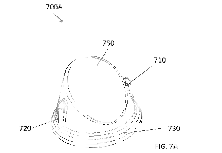
[0046] FIGs. 7A-7D illustrate various embodiments 700A, 700B, 700C, and 700D
of a gonioprism
710 with a vacuum docking device 720 coupled that may be detachable from each
other. As
opposed to a vacuum hose, in some embodiments, the gonioprism 710 may have one
or more
baffles and one or more openings 740 at the bottom. The pressing of the
baffles may create a
vacuum within the skirt caused to the suction and expulsion of air through the
bottom opening 740
that would allow it to adhere to one location as described above. The
gonioprism 710 may comprise
one or more push buttons 720 that may allow to break the vacuum seal so that
the gonioprism may
be adjusted to a different location. The gonioprism 710 may comprise one or
more lens 750. The
gonioprism 710 may include a single spherical radius that lays on the cornea.
In some
embodiments, the gonioprism 710 may have a port/opening, as described above,
or any such
possibility that would allow access to the anterior chamber. In some
embodiments, the contact
area of the gonioprism 710 and the skirt 730 may be small enough that no
port/opening may be
required. The gonioprism 710 and the skirt 730 may be a unitary piece or
detachable.
[0047] FIGs. 8A-8C illustrate a hands-free upright gonioprism 810 that allows
for the user to look
straight down without titling the patient's head or the microscope to see the
Trabecular meshwork.
The gonioprism 710 may be permanently or detachably attached to a skirt 840.
The gonioprism
810 may have two or more mirrors 820 and 830 or optical stacks or any other
mechanism on any
location on the gonioprism 810 such as the walls 860 and 870 that may allow
light 850 to refract
or reflect in a way that the patient's head or the microscope need not be
titled. The walls 860 and
870 may be conveniently tilted to achieve optimal visibility without having to
tilt the microscope
or the patient's head. The gonioprism 810 may have vacuum mechanisms as
described in previous
embodiments. In some embodiments, the gonioprism 810 may have a port/opening,
as described
above, or any such possibility that would allow access to the anterior
chamber. In some
embodiments, the contact area of the gonioprism 810 and the skirt 840 may be
small enough that
no port/opening may be required. As illustrated in FIG. 8C, the light 880 may
refract/reflect
through multiple optical material stacks or other mechanism on any location so
that mirrors on the
walls may not be needed for the surgeon to see the Trabecular meshwork.
directly while the patient
lays upright.
9
CA 03064872 2019-11-25
WO 2018/218232 PCT/US2018/034783
[0048] While the embodiments herein describe a gonioprism, the gonioprism may
be replaced or
supplemented by any other optical device, including but not limited to
surgical contact lenses,
Retinal Vitrectomy Lenses, Indirect Contact Surgical Lenses, Aspheric Macular
Lenses.
Additionally, the vacuum pressure (or other sealing pressures) being
applied/exerted on both the
skirt and the cornea, in some embodiments, the vacuum pressure (or other
sealing pressures) may
be applied/exerted to the skirt only and not the cornea to reduce pressure
induced folds in the
cornea which may tend to reduce visualization. Although shown as an
port/opening in one or more
components in the device, in some embodiments the port/opening may not be
configured as shown
in the figures. For example, in some embodiments the skirt may not be
completely circumferential
and instead may include one or more walls defining a triangular portion or
slice where procedures
can be executed. As such, a port can be defined by part of a discontinuation
of a circumferential
surface or wall. Thus, in some embodiments, ports do not involve nearby
engagement of the
vacuum docking system.
[0049] Additionally, in some embodiments a vacuum dock does not engage an
entire area below
a docking device or an entire area below a particular component of a docking
device. For example,
where a skirt is circumferential in nature, a vacuum pump and docking device
configuration may
not create a vacuum seal within the entire circumference of the skirt.
Instead, it may engage an
ocular surface at one or more specific points to create the vacuum seal and
maintain device
positioning using suction.
[0050] Although not shown in the example embodiment, in some embodiments
automatic digital
or analog vacuum gauges can be included that display vacuum pressure present
within the interior
of the docking device when in use. As such, these gauges can be coupled with
and influenced by
sensors, which are not shown.
[0051] As shown in the example embodiment, the gonioprism can be rotated with
respect to the
vacuum docking device, which, in some embodiments, can occur during a
procedure. Although
not shown, upward and downward or proximal and distal movement of the
gonioprism can be
actuated using a screw, lever, or other appropriate mechanism.
[0052] As used herein and in the appended claims, the singular forms "a",
"an", and "the" include
plural referents unless the context clearly dictates otherwise.
[0053] The publications discussed herein are provided solely for their
disclosure prior to the filing
date of the present application. Nothing herein is to be construed as an
admission that the present
CA 03064872 2019-11-25
WO 2018/218232 PCT/US2018/034783
disclosure is not entitled to antedate such publication by virtue of prior
disclosure. Further, the
dates of publication provided may be different from the actual publication
dates which may need
to be independently confirmed.
[0054] It should be noted that all features, elements, components, functions,
and steps described
with respect to any embodiment provided herein are intended to be freely
combinable and
substitutable with those from any other embodiment. If a certain feature,
element, component,
function, or step is described with respect to only one embodiment, then it
should be understood
that that feature, element, component, function, or step can be used with
every other embodiment
described herein unless explicitly stated otherwise. This paragraph therefore
serves as antecedent
basis and written support for the introduction of claims, at any time, that
combine features,
elements, components, functions, and steps from different embodiments, or that
substitute features,
elements, components, functions, and steps from one embodiment with those of
another, even if
the following description does not explicitly state, in a particular instance,
that such combinations
or substitutions are possible. It is explicitly acknowledged that express
recitation of every possible
combination and substitution is overly burdensome, especially given that the
permissibility of each
and every such combination and substitution will be readily recognized by
those of ordinary skill
in the art.
[0055] In many instances entities are described herein as being coupled to
other entities. It should
be understood that the terms "coupled" and "connected" (or any of their forms)
are used
interchangeably herein and, in both cases, are generic to the direct coupling
of two entities (without
any non-negligible (e.g., parasitic) intervening entities) and the indirect
coupling of two entities
(with one or more non-negligible intervening entities). Where entities are
shown as being directly
coupled together or described as coupled together without description of any
intervening entity, it
should be understood that those entities can be indirectly coupled together as
well unless the
context clearly dictates otherwise.
[0056] While the embodiments are susceptible to various modifications and
alternative forms,
specific examples thereof have been shown in the drawings and are herein
described in detail. It
should be understood, however, that these embodiments are not to be limited to
the particular form
disclosed, but to the contrary, these embodiments are to cover all
modifications, equivalents, and
alternatives falling within the spirit of the disclosure. Furthermore, any
features, functions, steps,
or elements of the embodiments may be recited in or added to the claims, as
well as negative
11
CA 03064872 2019-11-25
WO 2018/218232 PCT/US2018/034783
limitations that define the inventive scope of the claims by features,
functions, steps, or elements
that are not within that scope.
12