Note: Descriptions are shown in the official language in which they were submitted.
CA 03066247 2019-12-04
WO 2018/226907 PCT/US2018/036375
FACTORY-ON-A-CHIP FOR PRODUCTION OF BIOLOGICALLY DERIVED
MEDICINES/BIOPHARMACEUTICALS/BIOLOGICS/ B IO THERAPEUTIC S
GOVERNMENT RIGHTS IN INVENTION
[001] This invention was made with government support under Grant Number
N66001-13-
C-4023 awarded by the Defense Advanced Research Projects Agency (DARPA). The
government has certain rights in the invention.
CROSS REFERENCE TO RELATED APPLICATION
[002] The present application claims priority to U. S Provisional Patent
Application No.
62/516,161 filed on June 7, 2017, the contents of which are hereby
incorporated by reference
herein.
[003] Field of the Invention
[004] The present invention relates to protein manufacturing and, more
particularly, to an
integrated microfluidic bioprocessing system for on-demand production or
manufacturing of
proteins for point-of-care delivery.
BACKGROUND THE INVENTION
[005] Production of biologically-derived medicines or biotherapeutics involves
a large scale
(>10,000 L) process chain which includes large volume separation,
purification, formulation,
1-3
packaging and distribution. The major cost is in the maintenance of living
organism from
which these biotheraputics are harvested and the cold chain required to keep
the product stable
until it reaches the patient. To counter the complexities and expense of
maintaining living
organisms for biotherapeutics, recent efforts have seen the use of cellular
extracts as a source
4
for biomanufacuring. This has helped reduce production time from weeks to a
matter of hours.
These extracts contain a majority of the cellular machinery that are capable
of producing
4
properly folded and functional active biotherapeutics. Recently, cell extract
from different
biosystems (Mammalian Chinese Hampster Ovary (CHO) cells, yeast and E.coli)
have become
CA 03066247 2019-12-04
WO 2018/226907 PCT/US2018/036375
commercially available. The availability of cell-free extracts has made
miniaturization and
5-8
automation of protein purification a possibility.
However, the miniaturization and
automation still remain immature, some of these lack a purification chain and
the protein yield
is low, hence may not be well suited for point-of-care applications.
[006] The manufacturing process for biotherapeutics relies heavily on large-
scale
fermentation batches that require frequent monitoring to ensure robustness and
product quality.
However, as personalized medicines and single-use device technologies are
becoming
increasingly important, there is a growing need for flexible, scalable,
affordable and portable
systems that offer manufacturing options.
[007] Thus, there is a need to provide for a new portable platform for
manufacturing
biotherapeutics at the point-of-care wherein the portable platform would
operate in mobile
units (e.g. ambulance), patient bed-sides, pharmacies, resource limited areas,
acute
emergencies and battlefields.
SUMMARY OF THE INVENTION
[008] The present invention provides for a fully integrated microfluidic
system capable of
producing single-dose amounts of biotherapeutics at the point-of-care wherein
protein
production, purification and product harvest are all integrated as a single
microfluidic device
which is portable and capable of continuous-flow production of biotherapeutics
at the
microscale using a cell-free reaction system.
[009] In one aspect the present invention provides for a portable "factory-on-
a-chip"
comprising three primary components, wherein the components comprise a
bioreactor unit, a
mixer/debubbler and purification unit, wherein the purification unit comprises
a multiplicity of
chromatography columns. This setup will serve as a personalized medical device
kit with the
ability to prepare small quantities of biotherapeutics on-demand.
[0010] In yet another aspect, the present invention provides for a factory-on-
a-chip
microfluidic device comprising:
2
CA 03066247 2019-12-04
WO 2018/226907 PCT/US2018/036375
(i) a microfluidic bioreactor unit equipped with a continuous collection
channel for
synthesizing a crude protein in a reaction within the microfluidic bioreactor;
(ii) a microfluidic mixer/de-bubbler unit communicatively connected to the
microfluidic bioreactor unit to dilute the crude protein and remove any air
bubbles during
mixing; and
(iii) a microfluidic purification unit communicatively connected to the
microfluidic
mixer/de-bubbler unit comprising at least one purification column for
capturing the crude
protein and providing a purified protein, wherein the purification unit is
preferably connected
to sensors for monitoring pH, ionic strength, UV-Vis absorbance, fluorescence,
light scatter
and or circular dichroism for testing of the purified protein. Protein
analysis is preferably
conducted in an analytical module by at least one process analytical
technology (PAT) sensor
to analyze and monitored pH, ionic strength, UV-Vis absorbance, fluorescence,
light scatter,
and/or circular dichroism.
[0011] Preferably, units (i), (ii) and (ii) are stacked together to form a
single unit having a
dimensional length of about 100mm to 150 mm and a width perpendicular to the
length of
about 40 mm to about 90 mm.
[0012] In some embodiments, the mixer/de-bubbler comprises a porous membrane
to eliminate
bubbles and an addition of at least one microfluidic valve to optimize flow.
The microfluidic
valves may be integrated either as part of the chip or as an external
component within a process
channel to ensure that the process flow is effectively controlled
[0013] In a further aspect, the present invention provides for an integrated
device comprising
a reactor, mixer and purification chip connected together as one platform
chip. For in-line
quality control additional sensors are include along the production line of
the process including
sensors to measure pressure, temperature, pH, dissolved oxygen sensor and/or
UV detector to
produce a scalable amount of a therapeutical protein for point of care
administration.
[0014] The factory-on-a-chip microfluidic device of the present invention
preferably has from
about 4 to 8 purification micro-columns positioned in the microfluidic
purification unit. The
purification micro-columns comprise microscale channels for moving a volume
ranging from
about 25 ¨ 200 L. The microscale channels comprise chromatography resin for
capturing the
3
CA 03066247 2019-12-04
WO 2018/226907 PCT/US2018/036375
crude protein. Preferably the chromatography resin is an immobilized metal
affinity resin
and/or an ion exchange resin. Further the purification micro-columns
accommodate solutions
for an elution buffer for harvesting the purified protein. In one embodiment,
the micro-columns
are fabricated of three polymeric layers comprising a top layer, a middle
layer comprising the
microscale channels and a base plate. Preferably, the top layer is about 1 to
about 2 mm thick,
the middle layer about 0.75 to about 1.25 mm comprising the a micro-channel to
accommodate
chromatography resin and the base plate is about 1 to about 2 mm.
[0015] The microfluidic bioreactor comprises cell extracts and reagents for
expression of the
crude protein. Such cell extracts comprise a combination of cytoplasmic and/or
nuclear
components from cells comprising reactants for protein synthesis,
transcription, translation,
DNA replication.
[0016] The integrated device may further comprise a processor for controlling
and/or
monitoring timing, temperature and other parameters necessary for optimizing
the production
and purification of the synthesized proteins to provide a sufficient amount of
or a therapeutic
dosage of the synthesized protein. Such length of time in the microfluidic
bioreactor and/or
purification unit may be used to affect the potency and/or activity of the
synthesized protein.
[0017] In another aspect, the present invention provides for method of
preparing and
administering a therapeutic protein on demand to a subject, the method
comprising:
(a) synthesizing the therapeutic protein with a microfluidic factory on a
chip comprising:
(i) a microfluidic bioreactor unit equipped with a continuous collection
channel for a
synthesizing a crude therapeutic protein in a reaction within the microfluidic
bioreactor
and at least one process analytical technology (PAT) sensor (pH/dissolved-
oxygen/redox) for monitoring conditions during the reaction;
(ii) a microfluidic mixer/de-bubbler unit communicatively connected to the
microfluidic bioreactor to dilute the crude therapeutic protein and remove any
air
bubbles during mixing; and
(iii) a microfluidic purification unit communicatively connected to the
microfluidic
mixer/de-bubbler unit comprising at least one purification column capturing
the crude
therapeutic protein and providing a purified therapeutic protein, wherein the
microfluidic purification unit is preferably connected to sensors for
monitoring pH,
4
CA 03066247 2019-12-04
WO 2018/226907 PCT/US2018/036375
ionic strength, UV-Vis absorbance, fluorescence, light scatter and or circular
dichroism
of the purified therapeutic protein; and
(b) administering the purified therapeutic protein to the subject in a
sufficient amount of time
to maintain the viability of the purified therapeutic protein.
[0018] In another aspect, the present invention provides for on-demand
production of a
therapeutic protein, wherein the therapeutic protein exhibits increased
potency due to the timely
synthesis and substantially immediate delivery of protein. Preferably, the
newly synthesized
proteins are delivered to a patient within one hour, to one day, to two weeks.
Any refrigeration
is at a temperature above freezing from 0 to 6 C. Any freezing of the proteins
is preferably a
single event with temperatures ranging from about -2 C to about -10 C.
[0019] Additional advantages, aspects, and features of the invention will be
set forth in part in
the description which follows and in part will become apparent to those having
ordinary skill
in the art upon examination of the following or may be learned from practice
of the invention.
The aspects and advantages of the invention may be realized and attained as
particularly
pointed out in the appended claims.
BRIEF DESCRIPTION OF THE FIGURES
[0020] Figure 1 shows a 3-D image of the Factory-on-a-chip.
[0021] Figure 2 shows external and internal components of the device. Figure
2A shows an
external box for inserting the bioreactor unit consisting of the reactor
cassette and product vial.
Figure 2B shows a bioreactor cassette which holds the fully integrated
microfluidic chip and
sensors. The disposable Bioreactor cassette contains the lyophilized cell
extracts and reagents
needed for expression and the microfluidics for purification of the desired
therapeutic protein.
The non-disposable box, the size of a video cassette player considered to be
an analytical
module (150), contains the necessary pumps, buffers for purification and
analytics for real time
quality control wherein testing analytics in the analytic module comprises at
least one process
analytical technology (PAT) sensor for monitoring pH, pressure, temperature,
dissolved-
oxygen, redox conditions, ionic strength, UV-Vis absorbance, fluorescence,
light scatter,
and/or circular dichroism.
CA 03066247 2019-12-04
WO 2018/226907 PCT/US2018/036375
[0022] The bioreactor cassette is inserted into the box and within a few
hours, the G-CSF will
be deposited in the product vial available for immediate delivery to the
patient.
[0023] Figure 3 shows the bioreactor cassette pieces which show (from top to
bottom) the
casing, fluid connectors, microfluidic chips and PAT sensors for real time
monitoring of the
bioprocess.
[0024] Figure 4 shows again the Factory-on-a-chip device which contains the
bioreactor,
mixer/debubbler and purification unit.
[0025] Figure 5 shows the process chain showing the following (I) Protein
expression: GFP
expression is imaged after 4h at 30 C on a shaker-incubator in the bioreactor.
(II) Protein
capture: GFP, post expression, was collected, diluted and passed through an
immobilized metal
affinity chromatography resin (IMAC). The IMAC resin was packed inside a
multiple column
microfluidic channel. The captured protein is seen in lane 2 of the high
sensitivity silver stain
gel. (III) Protein purification: the eluted sample from the IMAC capture step
was then passed
through an ion-exchange resin (Q-Sepharose FF). The sample is seen in lane 3
has lesser
impurity bands compared to those observed in lane 2. Lane 3 bands are also
comparable to the
purified GFP standards purchased from Thermo ScientificInc.
[0026] Figure 6 shows one design of a multiple column microfluidic
chromatography system
applicable for the device of the present invention.
[0027] Figure 7 shows another design of a multiple column microfluidic
chromatography
system applicable for the device of the present invention.
[0028] Figure 8 shows the purification product using the microfluidic
Chromatography chip of
Figure 6. Photo 1. Image taken after loading the HisPur Cobalt resin (Tolan
beads): The flow
was generated manually using a 3mL syringe. Photo 2. Image taken after loading
with lmL
diluted GFP-Harvest (5X dilution of raw lysate) and then flowing through a
wash buffer (3mL)
(5mM imidazole, 1xPBS, pH 7.2). Photo 3(A) Image taken half way through
elution step.
(With a total of 3mL elution buffer: divided into two collection vials of
about 1.5 mL each)
Photo 3(B) Elution buffer (150 mM imidazole, 1xPBS, pH 7.2). Image taken post
elution. Most
of the protein elutes in the first 1.5 mL election fraction and did not leave
much protein in the
6
CA 03066247 2019-12-04
WO 2018/226907 PCT/US2018/036375
column. Photo 4. Flow through from stage 2. It is believed that controlled
pumping the lysate
into the column at an efficiently monitored flow rate would improve the
binding. The other
option would be to improve the packing efficiency of the channels.
[0029] Figure 9 shows the purification product using the microfluidic
Chromatography chip of
Figure 7. Photo 1. Image taken after loading the HisPur Cobalt resin (Tolan
beads): The flow
was generated manually using a 3mL syringe. Photo 2. Image taken after loading
with lmL
diluted GFP-Harvest (5X dilution of raw lysate) and the then flowing through a
wash buffer
(3mL) (5mM imidazole, 1xPBS, pH 7.2). Photo 3(A) Image taken half way through
elution
step. (With a total of 3mL elution buffer: divided into two collection vials
of about 1.5 mL
each). Photo 3B Elution buffer (150 mM imidazole, 1xPBS, pH 7.2). Image taken
post elution.
Most of the protein elutes in the first 1.5 mL election fraction and did not
leave much protein
in the column. Photo 4 shows GFP flow through from stage 2. Slightly better
controlled
pumping of the lysate into the column with a monitored flow rate slightly
improved the binding
of GFP. The first pass flow through was also recirculated once more through
the column which
improved the efficiency of binding.
[0030] Figure 10 show a device design sketch; A) 3D sketch in designed using
SketchUp Pro.
B) These columns are made up of three layers of polymethyl methacrylate
(PMMA); bottom
base plate layer (each 1 mm thick), middle channel layer and top inlet/outlet
layer (1.5 mm
thick). The top layer contains a larger circular slot towards the outlet for
PTFE frits. PTFE
frits were added post bonding. This array consists of 5 columns of 100 tL
volume. C) This
picture of the customizable microscale column device ( Col) shows an array of
columns with
varied resin capacities (25 ¨ 200 from left to right) displaying the
versatility of this system.
[0031] Figure 11 shows acetone injections for column validations. A) 1%
Acetone injections
performed on each of the different volume (25 - 200 l.L) columns, where the
flow rate was 0.2
mL/min. B) 1% Acetone injections performed on each 1004, column, where the
flow rate
was 0.5 mL/min. These validation experiments demonstrate the manufacturing
consistency
across tested columns. Col validation chart lists the theoretical plates and
asymmetrical ratios
for His-cobalt columns tested at different flow rates. C) Table presents the
micro-column
validation data for different column volumes, showing theoretical plates and
asymmetrical ratio
calculated using the HPLC software. Methods adopted for the column connections
to the
HPLC and other lab methods are shown and explained in Figure 17.
7
CA 03066247 2019-12-04
WO 2018/226907 PCT/US2018/036375
[0032] Figure 12 A) 3D design of the one-frit column, where the PTFE frit is
placed at the
channel outlet. B) 3D design of the two-frit columns, with a PTFE frit placed
at the channel
inlet and outlet.
[0033] Figure 12 Cont. shows the computational model results are plotted for
comparisons
between C) One-frit columns, D) One-frit versus the two-fit columns. Through
these models
the one-frit system produced slightly better profiles compared to the two-frit
channels, E)
Different frit thicknesses (1 to 2 mm) were tested by computational modeling,
which revealed
that column performance is dependent on frit thicknesses.
[0034] Figure 13 shows binding and elution profiles for G-CSF purification on
multi-volume
arrayed Col device. A) G-CSF binding peaks observed in Cols loaded with 0.3
mL of G-
CSF harvest. B) G-CSF elution peaks observed as the elution buffer is
introduced into the
columns. These are elution peaks seen for the 0.3 mL harvest; each individual
run showed
sharp peaks of protein. Tested on different sets of columns. The Col is
connected to the
HPLC system like a conventional column setup (Figure 17, show the image of
setup). C) Silver
stained SDS-PAGE gel images, where two chips A and B were used to purify 0.3
mL G-CSF
harvest. The harvest and elution band are consistent between repeats. D)
Western blots show
the G-CSF protein bands for each of the eluted samples, harvest and blank
(without DNA), and
values are presented in Table E.
[0035] Figure 14 shows binding, wash and elution profiles for G-CSF
purification on the single
volume Col device. A) G-CSF binding peaks observed in Cols loaded with 0.3
mL of G-
CSF harvest. B) The following step involves washing the column to remove any
impurities
during the binding step. Wash peaks observed on the HPLC. C) G-CSF elution
peaks observed
as the elution buffer is introduced into the columns. These are elution peaks
seen for the 0.3
mL harvest set where 4 individual runs showed sharp peaks of protein. D) G-CSF
elution peaks
observed as the elution buffer is introduced into the columns. These are
elution peaks seen for
the 0.5 mL harvest set where 3 individual runs showed sharp peaks of protein.
E) Shows a
comparison between G-CSF elution peaks between the Col and lmL ThermoFisher
Scientific
(Thermo) column. The Col has a much sharper peak compared to the Thermo
column.
Collected volume for the Col was 0.5 mL vs 2.3 mL for the Thermo column. The
Col is
connected to the HPLC system like a conventional column setup (image in Figure
17). F)
8
CA 03066247 2019-12-04
WO 2018/226907 PCT/US2018/036375
Silver stained SDS-PAGE gel images, for 0.3 mL G-C SF harvest, elutions
showing consistency
between repeats. Also compared the lmL harvest samples tested in both the Col
and Thermo
pre-packed column. G) 0.5 mL G-CSF harvest, elutions showing consistency
between repeats
and slight impurities are noticed on the Thermo pre-packed column in
comparison to the Col.
Arrows mark the impurities seen only within the Thermo column sample. H)
Provides the
concentrations of protein post purification for each of the tested harvest
volumes.
[0036] Figure 15 shows that the method for bonding PMMA chips was standardized
and
provides a rapid prototyping technique for mass production of Col. A) PMMA
chips were
doused in 100% ethanol, then sandwiched together between aluminum plates and
silicone
sheets. This sandwiched set is then gently placed between two heated platens.
B) The platens
are custom fitted to a Carver Press (Carver Hydraulic Press Model M), which
squeezes the
platens together. C) The controller box regulates the temperature on each
heated platen, the
set point temperature is 80 C. D) The pressure gauge is at 2500 psi.
[0037] Figure 16 shows the placing PTFE frit inserts and resin packing
methods. A) Process
for adding PTFE frits post bonding. 1) Frits were simply placed into the slot
post-bonding, 2)
Luer lock fittings were glued (cyanoacrylate 2075) on top to hold the frits in
place, 3) Glued
devices set overnight and stored in a clean cabinet until packed. B) The
column packing was
optimized to work specifically for Col designs. The process flow schematic
has the following
steps. 1) 20% ethanol (10 mL syringe) was pushed through the device (0.5
mL/min flow rate),
this removes any air-bubbles in the device. The changes in pressure are
monitored in real time
while His-beads were packed at a constant flow rate of 0.5mL/min. After bead
loading, 10 ¨
15 column volumes of 20% ethanol were pushed through the packed device at 0.5
mL/min, to
ensure the tight packing of beads. Post packing, devices were stored at 4 C
until used for
validation and purification experiments.
[0038] Figure 17 shows images of the device setup. A) The Col is assembled in-
line with the
LabsmithTM microfluidic platform and the Senserion flow sensor, which enables
monitoring
bead packing parameters in real-time. (Process flow is described in Figure
11). B) Column is
attached to the HPLC to implement a pre-saturation wash, with 10mM imidazole
buffer at
0.5mL/min, prior to loading the protein. Protein was loaded on the column
using an externally
connected syringe pump at a rate of 0.2 mL/min. This was followed by a wash
step to remove
impurities and finally an elution step to collect the protein for subsequent
protein analysis.
9
CA 03066247 2019-12-04
WO 2018/226907 PCT/US2018/036375
Protein analysis is preferably conducted in an analytical module by at least
one PAT sensor to
analyze and monitored pH, ionic strength, UV-Vis absorbance, fluorescence,
light scatter,
and/or circular dichroism.
[0039] Figure 18 shows bead packing pressure and flow rate measurements in
real time. 1 - 2
mL ethanol (10 mL syringe) was pushed through the device (0.5 mL/min flow
rate) to wet the
surface and remove any air bubbles prior to adding the beads. The pressures
and fluidic flow
were monitored in real time while His-beads collect inside the column. A)
Packing pressures
were recorded to be between 20 - 40 kPa (-3-6 psi) with operation pressures
reaching a
maximum of 50 kPas (-7.2 psi); when B) flow rates maintained at around
0.5mL/min. 10¨ 15
column volumes of 20% ethanol was pushed through the packed device at 0.5
mL/min, to
ensure the tight packing of beads.
DETAILED DESCRIPTION OF THE INVENTION
[0040] The present invention is particularly suited for the on-demand
manufacturing of
therapeutic proteins that are suitable for on-demand synthesis and for direct
delivery to a
patient. Therefore, the present invention will be primarily described and
illustrated in
connection with the manufacturing of therapeutic proteins. However, the
present invention can
also be used to manufacture any type of protein, including toxic proteins,
proteins with
radiolabeled amino acids, unnatural amino acids, etc. Further, the present
invention is
particularly suited for the on-demand manufacturing of proteins using cell-
free expression, and
thus the present invention will be described primarily in the context of cell-
free protein
expression. However, the present invention can also be used in connection with
cell-based
protein expression.
[0041] Definitions
[0042] "Microfluidic chip" means at least one microfluidic channel etched or
molded into a
material (e.g., glass, silicon or polymers such PDMS (polydimethylsiloxane)
and polymethyl
methacrylate (PMMA). The micro-channels are connected together in order to
achieve a
desired feature (e.g., mix, pump, sort, or control the biochemical
environment). The "cross-
sectional dimension" of the channel is measured perpendicular to the direction
of fluid flow
within the channel. Thus, some or all of the fluid channels in microfluidic
embodiments of the
CA 03066247 2019-12-04
WO 2018/226907 PCT/US2018/036375
invention may have maximum cross-sectional dimensions less than 2 mm, and in
certain cases,
less than 1 mm. In one set of embodiments, all fluid channels containing
embodiments of the
invention are microfluidic or have a largest cross sectional dimension of no
more than 2 mm
or 1 mm. In certain embodiments, the fluid channels may be formed in part by a
single
component (e.g. an etched substrate or molded unit). Of course, larger
channels, tubes,
chambers, reservoirs, etc. can be used to store fluids and/or deliver fluids
to various
components or systems of the invention.
[0043] "Comprise(s)," "include(s)," "having," "has," "can," "contain(s)," and
variants thereof,
as used herein, are intended to be open-ended transitional phrases, terms, or
words that do not
preclude the possibility of additional acts or structures. The singular forms
"a," "and" and "the"
include plural references unless the context clearly dictates otherwise. The
present disclosure
also contemplates other embodiments "comprising," "consisting of' and
"consisting essentially
of," the embodiments or elements presented herein, whether explicitly set
forth or not.
[0044] The term "cell-free" as used herein refers to an "in vitro" combination
of reactants
capable of performing reactions occurring in a cellular environment, in a
mixture where the
reactants are comprised outside the cellular environment. Cell-free systems,
by definition, do
not include whole cells capable of replicating but its components are
typically derived from a
cell and comprise a combination of cytoplasmic and/or nuclear components from
cells
comprising reactants for protein synthesis, transcription, translation, DNA
replication and/or
additional biological reactions occurring in a cellular environment
identifiable by a person
skilled in the art.
[0045] "Affinity" and "binding affinity" as used interchangeably herein refer
to the tendency
or strength of binding of the binding member to the analyte. For example, the
binding affinity
may be represented by the equilibrium dissociation constant (KD), the
dissociation rate (kd), or
the association rate (ka).
[0046] "Label" or "detectable label" as used interchangeably herein refers to
a moiety attached
to a specific binding member or analyte to render the reaction between the
specific binding
member and the analyte detectable, and the specific binding member or analyte
so labeled is
referred to as "detectably labeled." A label can produce a signal that is
detectable by visual or
instrumental means. Various labels include: (i) a tag attached to a specific
binding member or
11
CA 03066247 2019-12-04
WO 2018/226907 PCT/US2018/036375
analyte by a cleavable linker; or (ii) signal-producing substance, such as
chromagens,
fluorescent compounds, enzymes, chemiluminescent compounds, radioactive
compounds, and
the like. Representative examples of labels include moieties that produce
light, e.g., acridinium
compounds, and moieties that produce fluorescence, e.g., fluorescein. Other
labels are
described herein. In this regard, the moiety, itself, may not be detectable
but may become
detectable upon reaction with yet another moiety. Use of the term "detectably
labeled" is
intended to encompass such labeling.
[0047] "Microparticle(s)" and "microbead(s)" are used interchangeably herein
and refer to a
microbead or microparticle that is allowed to occupy or settle in an array of
wells, such as, for
example, in an array of wells in a detection module. The microparticle and
microbead may
contain at least one specific binding member that binds to an analyte of
interest and at least one
detectable label. Alternatively, the microparticle and microbead may
containing a first specific
binding member that binds to the analyte and a second specific binding member
that also binds
to the analyte and contains at least one detectable label.
[0048] Protein production, purification and product harvest are all integrated
as a single
microfluidic device, referred to as a 'Factory-on-a-chip' as shown in Figure
1. The Factory-
on-a-chip microfluidic device includes a microfluidic bioreactor (100)
equipped with a
continuous collection channel for the target biotherapeutic and at least one
PAT sensors
(including pH, dissolved-oxygen, redox, ionic strength, UV-Vis absorbance,
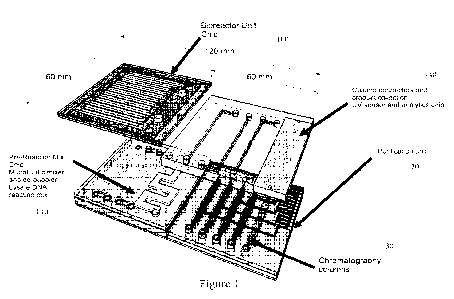
fluorescence,
light scatter, and/or circular dichroism) during the reaction, a microfluidic
mixer/de-bubbler
unit (110) is communicatively connected to the bioreactor to dilute the crude
protein harvest
and get rid of any air bubbles during the mixing process. Initial fabrication
tests for the
mixer/de-bubbler were successfully achieved using a porous membrane which is
able to
eliminate bubbles. Figures 2 and 3 provide for additional components for
enclosing the factory
on a chip unit including an external box, device holder, integrated sensors,
etc. This setup will
serve as a personalized medical device kit with the ability to prepare small
quantities of
biotherapeutics.
[0049] The porous membrane used in the mixer/de-bubbler can be fabricated from
any porous
polymeric material that reduces bubbles including, polyester, polypropylene,
nylon,
fluorocarbon polymers such as polytetrafluoroethylene, polyethylene, and
polysulfone, and
composites comprising one or more of such materials.
12
CA 03066247 2019-12-04
WO 2018/226907 PCT/US2018/036375
[0050] Microfluidic purification unit (120) in Figure 1 is communicatively
connected to the
microfluidic mixer/de-bubbler unit mixer device and contain a modular chip
based purification
column or columns for protein capture, buffer-exchange and polishing the
protein harvest.
Chromatography resins are included in the chromatography columns (130) and
selected for
chromatography resin packing efficiency and column efficiency. Product
collection from the
columns is collected in chip (140). Notably the purification module can be
connected to an
analytical module (150, Figure 2) for product characterization wherein
conditions and analysis
of the produced product in both the purification module and analytical module
can be
monitored and determined by sensors including pH, ionic strength, UV-Vis
absorbance,
fluorescence, light scatter, and/or circular dichroism.
[0051] "Chromatography resin" refers herein to a solid phase that selectively
or preferentially
binds one or more proteins from the source liquid. In the practice of the
invention, such
"chromatography resins" can be selected from any of the groups of resins
commonly described
as affinity, ion exchange and ion capture resins. The resins need only possess
a chemistry or
an associated ligand that will selectively or preferentially capture a
substance of interest from
the source liquid. Useful chromatography resins typically comprise a support
and one or more
ligand(s) bound thereto that provide(s) the selective or preferential binding
capability for the
target substance(s) of interest. Useful supports include, by way of
illustrative example,
polysaccharides such as agarose and cellulose, organic polymers such as
polyacrylamide,
methylmethacrylate, and polystyrene-divinylbenzene copolymers such as for
example
Amberlite resin, commercially available from Rohm & Haas Chemical Co.,
Philadelphia, PA.
It should be recognized that although the term "resin" is commonly used in the
art of
chromatography, it is not intended herein to imply that only organic
substrates are suitable for
resin substrate use, since inorganic support materials such as metals, silica
and glasses have
utility as well. In the practice of the present invention, the resin may be in
the form of beads
which are generally spherical, or alternatively the resin may be usefully
provide in particulate
or divided forms having other regular shapes or irregular shapes. The resin
may be of porous
or nonporous character, and the resin may be compressible or incompressible.
Preferred resins
will be physically and chemically resilient to the conditions employed in the
purification
process including pumping, temperatures, pH, and other aspects of the liquids
employed. The
13
CA 03066247 2019-12-04
WO 2018/226907 PCT/US2018/036375
resin as employed in the practice of the present invention is preferably of
regular generally
spherical shape, nonporous and incompressible.
[0052] "Affinity chromatography resin" or "affinity resin" refers to a
chromatography resin
that comprises a solid support or substrate with affinity ligands bound to its
surfaces.
Illustrative, non-limiting examples of suitable affinity chromatography resins
include spherical
beads with affinity ligands bound to the bead surfaces, wherein the beads are
formed of
cellulose, poly-styrene-divinylbenzene copolymer, polymethylmethacrylate, or
other suitable
material.
[0053] Ion exchange chromatography resin" or "ion exchange resin" refers to a
solid support
to which are covalently bound ligands that bear a positive or negative charge,
and which thus
has free counterions available for exchange with ions in a solution with which
the ion exchange
resin is contacted.
[0054] "Cation exchange resins" refers to an ion exchange resin with
covalently bound
negatively charged ligands, and which thus has free cations for exchange with
cations in a
solution with which the resin is contacted. A wide variety of cation exchange
resins, for
example, those wherein the covalently bound groups are carboxylate or
sulfonate, are known
in the art. Commercially available cation exchange resins include CMC-
cellulose, SP-
Sephadex , and Fast S-Sepharose (the latter two being commercially available
from
Pharmacia).
[0055] "Anion exchange resins" refers to an ion exchange resin with covalently
bound
positively charged groups, such as quaternary amino groups. Commercially
available anion
exchange resins include DEAE cellulose, QAE Sephadex , and Fast Q Sepharose
(the latter
two being commercially available from Pharmacia).
[0056] Figure 5 shows effective results using an immobilized metal affinity
resin and an ion
exchange resin. Immobilized metal affinity chromatography (IMAC) is a
specialized variant
of affinity chromatography where the proteins or peptides are separated
according to their
affinity for metal ions that have been immobilized by chelation to an
insoluble matrix. At pH
values around neutral, the amino acids histidine, tryptophan, and cysteine
form complexes with
14
CA 03066247 2019-12-04
WO 2018/226907 PCT/US2018/036375
the chelated metal ions (e.g., Zn2+, Cu2+, Cd2+, Hg2+, Co2+, Ni2+, and Fe2+).
This
technique is especially suited for purifying recombinant proteins as poly-
histidine fusions and
for membrane proteins and protein aggregates where detergents or high-ionic-
strength buffers
are required.
[0057] Figures 6 and 7 shows two different types of multiple column
microfluidic
chromatography systems. Figure 6 provides for a system including check valves
and a spin
column frit used as a collection chamber. Figure 7 shows that the system is
connected to an
inlet and outlet for controlling the lysate into the system.
[0058] Figures 8 and 9 shows the results of using the multiple column
microfluidic
chromatography systems of Figures 6 and 7 respectively. The results shown in
Figure 9 show
that controlled flow of the lysate containing the proteins into the columns
provides for
increased binding of the proteins to the chromatography resin. Also
recirculation is beneficial
for recapturing product.
[0059] Protein Expression in In Vivo and Cell-Free Systems
[0060] A protein is expressed in three main steps: replication, transcription
and translation.
DNA multiplies to make multiple copies by a process called replication.
Transcription occurs
when the double-stranded DNA is unwound to allow the binding of RNA polymerase
producing messenger RNA (mRNA). Transcription is regulated at various levels
by activators
and repressors, and also by chromatin structure in eukaryotes. In prokaryotes,
no special post-
transcriptional modification of mRNA is required. However, in eukaryotes, mRNA
is further
processed to remove introns (splicing), to add a 'cap' (M7 methyl-guanosine)
at the 5' end and
to add multiple adenosine ribonucleotides at the 3' end of mRNA to generate a
poly(A) tail.
The modified mRNA is then translated.
[0061] The translation or protein synthesis is also a multi-step process with
Initiation,
Elongation and Termination steps and is similar in both prokaryotes and
eukaryotes. The
difference is that in eukaryotes, proteins may undergo post-translational
modifications, such as
phosphorylation or glycosylation. The translation process requires cellular
components such
as ribosomes, transfer RNAs (tRNA), mRNA and protein factors as well as small
molecules
like amino acids, ATP, GTP and other cofactors.
CA 03066247 2019-12-04
WO 2018/226907 PCT/US2018/036375
[0062] The difference between in vivo and in vitro (cell-free) protein
expression is that in cell-
free expression, the cell wall and the nuclei are no longer present.
[0063] Cell-Free Protein Expression
[0064] To obtain the cell extract for cell-free protein expression, cells
(E.coli, wheat germ,
mammalian cells) are subjected to cell lysis followed by separation of the
cell wall and nuclear
DNA. The desired protein is synthesized by adding a DNA or mRNA template into
the cell
extract together with a reaction mix comprising of biological extracts and/or
defined reagents.
The reaction mix is comprised of amino acids, nucleotides, co-factors, enzymes
and other
reagents that are necessary for the synthesis, e.g. ribosomes, tRNA,
polymerases,
transcriptional factors, etc. When DNA is used as template (i.e. linked
reaction), it is first
transcribed to mRNA. Alternatively mRNA could also be used directly for
translation.
[0065] The template for cell-free protein synthesis can be either mRNA or DNA.
Translation
of stabilized mRNA or combined transcription and translation converts stored
information into
a desired protein. The combined system, generally utilized in E. coli systems,
continuously
generates mRNA from a DNA template with a recognizable promoter. Either
endogenous
RNA polymerase is used, or an exogenous phage RNA polymerase, typically T7 or
SP6, is
added directly to the reaction mixture. Alternatively, mRNA can be continually
amplified by
inserting the message into a template for QB replicase, an RNA dependent RNA
polymerase.
Purified mRNA is generally stabilized by chemical modification before it is
added to the
reaction mixture. Nucleases can be removed from extracts to help stabilize
mRNA levels. The
template can encode for any particular gene of interest.
[0066] Salts, particularly those that are biologically relevant, such as
manganese, potassium or
ammonium, may also be added. The pH of the reaction is generally run between
pH 6-9. The
temperature of the reaction is generally between 20 C and 40 C. These ranges
may be
extended.
[0067] In addition to the above components such as cell-free extract, genetic
template, and
amino acids, other materials specifically required for protein synthesis may
be added to the
reaction. These materials may include salts, polymeric compounds, cyclic AMP,
inhibitors for
16
CA 03066247 2019-12-04
WO 2018/226907 PCT/US2018/036375
protein or nucleic acid degrading enzymes, inhibitors or regulators of protein
synthesis,
oxidation/reduction adjusters, non-denaturing surfactants, buffer components,
spermine,
spermidine, etc.
[0068] The salts preferably include potassium, magnesium, ammonium and
manganese salts
of acetic acid or sulfuric acid, and some of these may have amino acids as a
counter anion. The
polymeric compounds may be polyethylene glycol, dextran, diethyl aminoethyl
dextran,
quaternary aminoethyl and aminoethyl dextran, etc. The oxidation/reduction
adjuster may be
dithiothreitol (DTT), ascorbic acid, glutathione and/or their oxides. Further
DTT may be used
as a stabilizer to stabilize enzymes and other proteins, especially if some
enzymes and proteins
possess free sulfhydryl groups. Also, a non-denaturing surfactant such as
Triton X-100 may
be used at a concentration of 0-0.5 M. Spermine and spermidine may be used for
improving
protein synthetic ability, and cAMP may be used as a gene expression
regulator.
[0069] Synthesized product is usually accumulated in the bioreactor unit wand
then is isolated
and purified according to the methods of the present invention for protein
purification. The
amount of protein produced in a translation reaction can be measured in
various fashions. One
method relies on the availability of an assay that measures the activity of
the particular protein
being translated. Examples of assays for measuring protein activity are a
luciferase assay
system and a chloramphenicol acetyl transferase assay system. These assays
measure the
amount of functionally active protein produced from the translation reaction.
Importantly,
activity assays will not measure full length protein that is inactive due to
improper protein
folding or lack of other post translational modifications necessary for
protein activity. As used
herein, the term "activity" refers to a functional activity or activities of a
peptide or portion
thereof associated with a full-length (complete) protein. Functional
activities include, but are
not limited to, catalytic or enzymatic activity, antigenicity (ability to bind
or compete with a
polypeptide for binding to an anti-polypeptide antibody), immunogenicity,
ability to form
multimers, and the ability to specifically bind to a receptor or ligand for
the polypeptide.
Preferably, the activity of produced proteins retain at least 55%, 60%, 65%,
70%, 80%, 85%,
90%, 95% or more of the initial activity for at least 3 days at a temperature
from about 0 C to
30 C.
[0070] Another method of measuring the amount of protein produced in a
combined in vitro
transcription and translation reactions is to perform the reactions using a
known quantity of
17
CA 03066247 2019-12-04
WO 2018/226907 PCT/US2018/036375
radiolabeled amino acid such as 35S-methionine or "C-leucine and subsequently
measuring the
amount of radiolabeled amino acid incorporated into the newly translated
protein.
Incorporation assays will measure the amount of radiolabeled amino acids in
all proteins
produced in an in vitro translation reaction including truncated protein
products.
[0071] Biomolecules for Protein Expression
[0072] The following biomolecules are preferably used for protein expression.
To carry out a
protein expression reaction, energy components and amino acids are supplied
externally and
may include, but not limited to the following components:
A genetic template for the target protein (mRNA or DNA) expression;
T7 RNA polymerases for mRNA transcription;
9 Translation factors (initiation, elongation and termination);
20 aminoacyl-tRNA synthetases (ARSes) for esterification of a specific amino
acid to
form an aminoacyl-tRNA;
Methionyl-tRNA transformylase transfers hydroxymethyl-, formyl-groups;
Creatine kinase converts ATP to ADP;
Myokinase catalyzes the inter conversion of adenine nucleotides;
Pyrophosphatase are acid anhydride hydrolases that act upon diphosphate bonds;
4 nucleoside triphosphates (ATP, GTP, CTP, TTP) for DNA formation;
Creatine phosphate which serves as a reserve of high-energy phosphates for
rapid
mobilization;
10-formy1-5,6,7,8-tetrahydrofolate for the formylation of the methionyl
initiator tRNA
(fMet-tRNA);
20 amino acids for protein synthesis;
Ribosomes for polypeptide translation;
46 tRNAs in protein synthesis; and
Cellular components which assist in proper protein folding.
[0073] Some of the proteins that may be expressed by the present invention for
on-demand
production may include, but not limited to, adrenocorticotropic hormone
peptides,
adrenomedullin peptides, allatostatin peptides, amylin peptides, amyloid beta-
protein fragment
peptides, angiotensin peptides, antibiotic peptides, antigenic polypeptides,
anti-microbial
peptides, apoptosis related peptides, atrial natriuretic peptides, bag cell
peptides, bombesin
18
CA 03066247 2019-12-04
WO 2018/226907 PCT/US2018/036375
peptides, bone GLA peptides, bradykinin peptides, brain natriuretic peptides,
C-peptides, C-
type natriuretic peptides, calcitonin peptides, calcitonin gene related
peptides, CART peptides,
casomorphin peptides, chemotactic peptides, cholecystokinin peptides, colony-
stimulating
factor peptides, corticortropin releasing factor peptides, cortistatin
peptides, cytokine peptides,
dermorphin peptides, dynorphin peptides, endorphin peptides, endothelin
peptides, ETa
receptor antagonist peptides, ETh receptor antagonist peptides, enkephalin
peptides,
fibronectin peptides, galanin peptides, gastrin peptides, glucagon peptides,
Gn-RH associated
peptides, growth factor peptides, growth hormone peptides, GTP-binding protein
fragment
peptides, guanylin peptides, inhibin peptides, insulin peptides, interleukin
peptides, laminin
peptides, leptin peptides, leucokinin peptides, luteinizing hormone-releasing
hormone
peptides, mastoparan peptides, mast cell degranulating peptides, melanocyte
stimulating
hormone peptides, morphiceptin peptides, motilin peptides, neuro-peptides,
neuropeptide Y
peptides, neurotropic factor peptides, orexin peptides, opioid peptides,
oxytocin peptides,
PACAP peptides, pancreastatin peptides, pancreatic polypeptides, parathyroid
hormone
peptides, parathyroid hormone-related peptides, peptide T peptides, prolactin-
releasing
peptides, peptide YY peptides, renin substrate peptides, secretin peptides,
somatostatin
peptides, substance P peptides, tachykinin peptides, thyrotropin-releasing
hormone peptides,
toxin peptides, vasoactive intestinal peptides, vasopressin peptides, and
virus related peptides.
[0074] There is certainly a need for optimization and process development
ability at the
microscale to help reduce cost of reagents and speed up biotherapeutic
manufacturing for
translation into the clinic. 9 Microfluidic devices have offered a platform
that could potentially
serve this need, where less material is utilized to achieve similar end goals
and may allow for
exploring novel approaches. 10,11 The inherent scale enables the feasibility
of developing
portable, disposable and modular chromatographic systems, where various
chromatographic
processes can be integrated into a single device. 12 Such versatile and
modular devices could
be plugged in-line with other scale compatible devices for characterization
and screening of
proteins.
[0075] The combination of chromatographic techniques and microfluidics has
been reported
for different purposes, proteins-on-demand, proteomic investigations,
biomarker detection,
nucleic acid investigation, and rapid optimization of separation techniques.
9'13-17 Millet et al
13 have shown the modular microfluidics platform for protein purification
demonstrating the
use of affinity beads and size exclusion chromatography. However, conventional
microfluidic
19
CA 03066247 2019-12-04
WO 2018/226907 PCT/US2018/036375
device manufacturing is expensive, laborious and impossible without proper
access to
microfabrication facilities or machines. Also, the inherent scale of
microfluidic devices
currently used for chromatography may not currently be practical, but are
potentially scalable.
11,14,15,18 There is a possibility for multiplexing with the current
microscale technologies, but
this still requires much effort towards usability. 11 Most of all,
microfluidic devices in most
cases are focusing on integrating with current HPLC machines or mass
spectrometry machines.
[0076] The present invention provides for versatile, customizable, robust, low-
cost, and easily
manufacturable chromatography columns for rapid screening of therapeutic
quality protein
purification. The reported scale addresses a huge gap in the current market
between large (1mL
¨ 1 L) columns and very small (0.1 - 10 L) low to high pressure microfluidic
columns. The
microscale column ( Col ranging from 25 ¨ 200 L) device described here is
equipped to
accommodate any affinity-based resin and serves as a universally compatible
microfluidic unit
for any system. These devices offer the ability to reduce reagent use,
comparable protein
purity, higher throughput, and low dead volumes, compared to conventional
columns in the
market.' The technology described herein provides a solution for quick
prototyping of
microscale columns for quick process development and optimization for affinity-
based
purification. As an example application, affinity Hi s-Pur cobalt-NTA
(ThermoFisherScientific
Inc.) resin and columns were utilized for on-chip characterization and
purification of
granulocyte colony stimulating factor (G-CSF) protein, expressed using the
cell-free CHO-IVT
system.
[0077] Design considerations.
[0078] Most chromatographic methods rely heavily on the device geometries,
geometric
phases, and high-pressure separations.
However, the advent of microchips for
chromatographic separations entails potential benefits and the planar geometry
has not stopped
the evolution in chromatographic screening methods in such systems. The planar
format is the
dominating format in the microfluidic separation devices, due to the ease of
fabrication and
design. 10,11 The planar format is a result of available machining tools used
to fabricate micro-
devices, even though this may not be an ideal situation for high-pressure
operations of pressure
driven separations. The chemical interactions between resin and protein are
dominant in this
situation and hence may be less dependent on the geometric design, but is not
completely
CA 03066247 2019-12-04
WO 2018/226907 PCT/US2018/036375
independent of channel geometry.29 To determine the optimal design parameters,
the present
invention focused on column arrays consisting of varied channel thicknesses
and volumes.
Devices were fabricated in polymethyl methacrylate (PMMA) substrates, off the
shelf fittings
(i.e. Luer lock fittings and PEEK fittings), PTFE frits and metal affinity
chromatography resin
(Figure 10).
[0079] PMMA is a sturdy thermoplastic that is often the plastic of choice for
microfluidic
purposes due to its good acid/base/solvent resistance, and excellent optical
properties. 30 The
bonding method described herein was adapted from a previously described method
23, where
the method of bonding involves solvent (ethanol) bonding at temperatures of 80
- 85 C. When
using such temperature and solvent conditions, the bonding is irreversible and
has shown to be
mechanically sturdy at high operating pressures. 23'31'32 Techniques using
PMMA are relatively
simple to implement in any laboratory setting and hence devices can be quickly
prototyped.
Another major consideration when designing chromatography columns is the
retention of
chromatography resin within the separation channel. To ensure proper retention
of affinity
beads inside the column, off-the shelf PTFE frits were bonded towards the
outlet end of the
columns. Such frits are commonly used in chromatography during the packing
protocols.
There are two main iterations of Col discussed herein, one chip was designed
to bear varied
volumes of resin (from 25 ¨ 200 L) and the other chip bore 5 channels of 100
L volume.
These two iterations of chips demonstrate the versatility and customizability
of this system,
thus providing quick solutions for process optimization. Cols were packed
using the
LabSmith Inc. setup, where the pressure and flow rate was monitored in real-
time. Labsmith
Inc. system provides an easily customizable platform and an easy interface for
resin packing
along with pressure and flow rate measurements. This is the advantage with the
device
presented herein, as well as its adaptability. Packing pressures were recorded
to be between 20
- 40 kPa (-3-6 psi) with operation pressures reaching a maximum of 50 kPas (-
7.2 psi).
[0080] Column performance and computational modeling.
[0081] Column validations included testing the packing efficiency, theoretical
plates, and
protein purification profiles on a conventional HPLC. Post packing, it is
often necessary to
test the integrity of the resin bed to confirm the quality and consistency of
the chromatographic
operations.33 Several measurements are used to qualify a column; these
parameters are number
of theoretical plates for a column and asymmetrical ratio between the two
sections of a
21
CA 03066247 2019-12-04
WO 2018/226907 PCT/US2018/036375
chromatographic peak. The most common type of test signal applied is a pulse
test function,
where a small volume of a tracer molecule is added to the buffer flowing
through the column
The peak broadening over the column is measured using height equivalent to a
theoretical
plate (HETP) and peak symmetry also described by an asymmetric ratio (As).
These
parameters were tested using 1% acetone injections where the peak shape and
theoretical plates
were calculated from UV profiles from pulse tests (Figure 11). These measured
profiles were
compared to conventional off the shelf lmL columns. Acetone injection tests
revealed a trend
where an increase in flow rate decreased the theoretical plates for all tested
column volumes.
However, an increase in column volume did not result in a significant change.
Although the
theoretical plate numbers in Cols (31.5 12.6 plates measured through the
HPLC software)
seemed close to the range of conventional columns (-50 plates), the Col peak
shapes seemed
much sharper. The measurement of the asymmetric ratio (As), between the
ascending and
descending portions of the acetone peaks at 10% of its peak height is another
standard method
used to determine column performance and packing efficiency. Col peak
asymmetrical ratios
were measured to be 1.5 0.1, compared to the conventional lmL column peak
ratios to be
around 0.88. The ideal asymmetry peak ratio is 1, however, a typical
acceptable range is
between 0.8 < A < 1.8.33'35 Notably the Cols found here in fall within this
range, which
suggests a positive relationship to conventional column performance. In
addition, COMSOL
multiphysics modeling and fluidic simulations successfully substantiate the
experimental
Cols parameters using equation 1-9, explained before. Figure 12 A illustrates
the designed
geometry and finite element mesh for a Cols with a single frit located at the
outlet of the
column. Figure 12B is a similar illustration of the same column but with frits
located at the
inlet and outlet of the column. In Figure 12 C, the COMSOL modeling results
for three various
sizes of the column with a single frit at the outlet were plotted. Figure 12D,
represents the
comparison between the single frit versus the two frits micro-columns. By
subtracting the peak
variance from extra-column sources and the feed variance contribution, the
modeling results
are seen to be in good agreement with the experimental tests using 1% acetone.
The calculated
variance of experimental results was 0.00587 min2 compared to the
theoretically modelled
variance of 0.00371 min2 (Calculated through theoretical modeling, using Eq. 8
and 9, shown
below). Computational modeling also proved useful in understanding how to
improve the
column performance by changing the frit thickness parameters. Modeled data
(Figure 12E)
revealed that a smaller frit thickness of 0.5 mm might further improve the
performance
compared to the 1.5 mm which is currently being used. However, a larger 2 mm
frit thickness
22
CA 03066247 2019-12-04
WO 2018/226907 PCT/US2018/036375
leads to further tailing of the column peaks.
[0082] This indicates that the assumption used in the modeling that the column
permeability
and porosity were uniform inside column is valid. However, the lower end of
the peak width
generally provides a more symmetrical appearance of the peak and efforts are
currently
underway to improve the packing efficiency to reduce plate heights. In
addition to the peak
performance, protein purification efficiency was tested for Granulocyte colony
stimulating
factor (G-CSF) (Figure 13 and Figure 14). G-CSF using Cols, resulted in
similar or slightly
better protein purity (93%) observed compared with conventional 1 mL column
(90%)
(Figure 13). Cols provide an additional advantage due to their customizable
size by
considerably reducing impurities. Since affinity resin binding sites might be
overwhelmed
with protein of interest, the suggestion would be to fine tune the column
capacity based on the
known protein concentration and attain improved purity.
Through literature, most
chromatography columns are range from less than 10 L resin volume
(microfluidic 11_1336),
most of which are not very compatible with a regular HPLC machine or above 1
mL resin
volume at the other end of the spectrum.2022 The fabrication and manufacturing
is often
expensive and fabrication methods are not easily accessible to most research
labs. This
presents a huge gap in this area of research for a low-cost, customizable and
versatile screening
toolkit for protein purification in a workable range that is compatible with
conventional
HPLCs. To address this gap, Col arrays were designed herein that are capable
of holding a
volume of affinity resin between 25 ¨200 L which can easily be customized for
a set amount
of protein. Table 1, highlight the performance of Cols compared with
conventional lmL
columns. Using the Col array, the user is provided with customizable resin
capacities that
could match the protein concentration (Table 1). Customization can also save
on considerably
large amounts of buffer and run time for optimization experiments. The amount
of buffer used
in this study for Cols was 10-fold less compared to conventional methods
(e.g. 10 mL of wash
and elution buffer is needed for the conventional IMAC columns, whereas for
the Col, only
needed lmL of each buffer was necessary) (Table 1). Purification times were
reduced to 10 -
20 min (total purification run-time) from a typical run-time of 1-2 h.
Potentially, such devices
could be incorporated into a research or industry setting, where a newly
discovered therapeutic
or research grade protein is rapidly optimized at low-cost. An added advantage
over current
methods is that Col devices contain HPLC compatible fittings and potentially
can be used in
tandem with all HPLC systems that use PEEK fittings (with 10-32 UNF taps or
Luer locks).
23
CA 03066247 2019-12-04
WO 2018/226907 PCT/US2018/036375
[0083] The present invention provides for the development of versatile
microfluidic platforms
for early-stage optimization of therapeutic protein purification. Devices are
compatible with
most HPLC fittings making them possible to use with any generic chromatography
instruments.
In addition, it is important to highlight that the manufacturing process is
less expensive than
conventional methods but with a resulting product of comparable performance.
The sample
purity and column efficiency of the Cols is comparable to conventional
columns. These
customizable devices address a niche area for protein purification and process
automation.
Besides protein capture with affinity resins, this microscale device can also
be adapted for
various other biomolecular separation systems, such as ion exchange, size
exclusion and buffer
exchange chromatography by choosing the appropriate resin, column design, and
volume
necessary for optimal conditions. These columns can find use in applications
in various use
cases such as biopharmaceutical drug development and point-of-care device.
[0084] Experimental section
[0085] Materials.
[0086] PTFE fit (20 m PTFE frits, Omnifit Catalogue# OMNI006FR-06-20); HPLC
to luer
fittings (10-32 female to male luer fitting, IDEX, Catalogue# P-656), His-Pur
IMAC resin
(HisPur cobalt resin, Catalogue#89966, ThermoFisher Scientific), PMMA (Astra
Product,
Clarex0, PMMA sheets, lmm and 1.5mm); CHO cell-free IVT system (Thermo
Scientific,
MD, Catalog# CCS1031), 10kDa MWCO Slide-A-Lyzer, 0.5 mL ¨ 3 mL capacity
cassette
(Thermo cassette, Thermo Scientific, Catalogue #66380); Luer lock caps (Female
luer cap,
polycarbonate, Cole parmer, #SC-45501-28), luer lock plug (Male luer lock
plug,
polycarbonate, #EW-45504-56), PTFE tubing (Cole Parmer 1/32" ID x 1/16"0D,
25ft/pk,
#EW-06407-41), Ethanol, (Fisher Scientific, #04-355-451, lgal. 200 proof);
Labsmith
components for 1/16" ID, pressure sensor starter package for uPS Pressure
sensor: uPS0800 ¨800kPa abs. range.
[0087] Device design.
[0088] 2D designs sketched in Corel draw were printed on PMMA sheets using a
CO2 laser
printer CO2 laser (Laser diode wavelength 630-680nm, max output is 5mW, class
laser 3R laser
24
CA 03066247 2019-12-04
WO 2018/226907 PCT/US2018/036375
product, 2.0 lens module). Prior to bonding, each printed PMMA layer was
rinsed with DI
water and dried with kim-wipes, then cleaned using isopropanol wipes. The mico-
Columns
(.iCol) were made up of three PMMA layers, top inlet outlet layer (1.5 mm
thick), middle
channel and a base plate (each 1 mm thick). The design consists of the top 1.5
mm thick
PMNIA layer that has a large circular slot (6 mm diameter) towards the outlet
end (meant for
PTFE fits), middle 1 mm thick PMNIA layer bearing the micro-channel to
accommodate
chromatography resin and bottom 1.5 mm PMNIA base plate. Two device designs
were tested
here, one had an array of microscale channels consisting of different volumes
(25 ¨ 200 L)
and the other had 5 microscale channels consisting of one volume (100 L), as
shown in Figure
10.
[0089] Thermal solvent bonding method.
[0090] Temperature regulated metal plates were custom fit to the top and
bottom surfaces of a
Carver press (Carver Hydraulic Press Model M). Prior to device bonding these
were pre-
heated to 80 C. Each plate had a temperature controller managed by an external
relay unit
responsible for maintaining the temperature. Aluminum plates and silicon
sheets were pre-
heated to 80 C. Devices were sandwiched between aluminum plates, heated to 80
C for 10min.
The process and apparatus used is shown and described in Figure 15. The
solvent bonding
using ethanol was adopted and modified from a previously published articles by
Al-Adhami et
al. 23'24 The device apparatus was then removed and allowed to cool at room
temperature. Each
PTFE frit is 6mm in diameter and 1.5 mm thick and fits perfectly into the
designated slot.
PTFE frits were simply placed inside each of its reserved slots. Luer lock cap
fittings were
glued in place to hold the frits within each slot. Prior to attaching luer
caps to the device, a
hole was drilled through each of these fittings using a 2.5 mm titanium drill
bit (drill bit
McMaster #39, titanium nitride kit) fixed to a DaytonTM 16" drill press. The
drilled luer lock
fittings were cleaned with DI water and ethanol, air dried, and then glued to
the inlet/outlets of
each device. The luer fittings enabled the connection of the Col to the HPLC
fittings, via the
PEEK (luer to 10/32) fittings as shown and discussed in Figures 16 and 17.
Devices were
stored in a clean and sterile environment until used.
[0091] IMAC resin packing.
CA 03066247 2019-12-04
WO 2018/226907 PCT/US2018/036375
[0092] Resin packing protocol was specially developed to accommodate Col
devices. For
this setup, two 10 mL BD syringes were required (fixed onto a BASi syringe
holder, BAS),
1/16" inner diameter PTFE tubing, Omnifit 3-way valve (Omnifit, Sigma Aldrich,
Supelco,
56140-U), Labsmith pressure sensors, Sensirion flow sensor and a 4.0 psi
check valve at
the outlet. Procedure was as follows: lmL of His-Pur cobalt beads were
resuspended into 40
mL of DI water in a conical (50 mL) tube. The mixture was gently shaken before
being filled
into a 10 mL loading syringe. 1 - 2 mL ethanol (10 mL syringe) was pushed
through the device
(0.5 mL/min flow rate) to wet the surface and remove any air-bubbles prior to
adding the beads.
(Apparatus and setup explained in Figure 11 B and Figure 12 A). The pressures
and fluidic
flow are monitored in real time while His-beads accumulate inside the column
as shown in
Figure 18. 10 ¨ 15 column volumes of 20% ethanol were pushed through the
packed device at
0.5 mL/min, this ensured the tight packing of beads. Post packing, devices
were stored at 4 C
until used for validation and purification experiments.
[0093] Column validations on HPLC.
[0094] Column validation (packing efficiency, theoretical plates, pressure and
flow rate
profiles) were performed on an UltiMate 3000 HPLC system (ThermoFisher
Scientific). The
Col performance was compared with the conventional lmL columns (Thermo
Scientific His-
Pur). 1% solution of acetone in 20% ethanol (v/v) injections was used to
validate the packing
efficiency on the HPLC (See Figure 14 and data presented in Table 1) A
standard solutions
from 50ug ¨ 400ug was the range used to determine a linear range. All columns
were validated,
tested, and cleared for use prior to protein purifications. Graphical analysis
and plots were
prepared using GraphPad Prism 7.
[0095] Computational modeling and simulations.
[0096] Computational modeling and fluidic flow simulations were conducted
using COMSOL
Multiphysics. Simulations were conducted for the Cols (length 27 mm and the
width of 0.98
mm), where the model consisted of six connected cylinders, one of which
represented the PTFE
frit at the outlet (for the one-frit design) and two of which represented the
frits at the inlet and
outlet (for the two-frit design) of the column. The fluid flow profiles within
the liquid-filled
domains of the micro-column were determined by solving the Navier-Stokes
equation for
incompressible flow given as follows:
26
CA 03066247 2019-12-04
WO 2018/226907 PCT/US2018/036375
p(U. Vs)ist = ¨V. [¨pT + + (V )T1t (1)
In Eq. 1 ,u denotes the dynamic viscosity, it is the fluid velocity in the
liquid-filled domain, p
is the fluid density, and p is the pressure. Alternatively, the Brinkman
equation shown by Eq.
2 was used to determine the flow profiles in the particulate bed:
= [¨p7 + (11/1 + OT)1 (2)
a
In Eq. 2, k denotes the permeability of the column and a is its porosity.
The boundary conditions for Eqs. 1 and 2 are as follows:
(i) Inlet velocity: = uo
(ii) No slip condition at the column
wall: ü = 0
(i0) Outlet gauge pressure: p = 0
The mass transport of solute species i in the non-porous domains was
determined by solving
the following two equations:
adi ¨ ¨
V¨ + .Ni = 0 (3)
ot
Ni= ¨(De)VCi+ id1 (4)
In Eqs. 3 and 4, C, is the concentration of species i in the fluid, Kt, is the
molar flux of species
i, and De is the diffusion coefficient.
[0097] To account for the mass transport of solute species i in the
particulate bed, the combined
effect of convective diffusion and dispersion in the interparticle fluid and
diffusion in the
particles was determined by solving Eqs. 5 and 6:
act ¨
(5)
ot
_ _L r,$)urt _L,T;Sri
p " (6)
In Eqs. 5 and 6, C/ indicates the interstitial concentration of species i
(i.e. the concentration of
species i in the interparticle fluid), Nis is the superficial molar flux of
species i, itis is the
superficial fluid velocity and DD is the superficial dispersion coefficient
diagonal tensor. Note
that the term "superficial" denotes a quantity evaluated per unit volume of
particulate bed or
per unit cross-sectional area of particulate bed. The Peclet numbers of 20 and
0.5 were used to
determine the axial and radial components of the dispersion coefficient
tensor, respectively.
25,26
27
CA 03066247 2019-12-04
WO 2018/226907 PCT/US2018/036375
[0098] In Eq. 5, R,s is the superficial adsorption rate that is determined by
assuming a parabolic
concentration profile inside the particle. This assumption results in a Linear
Driving Force
(LDF) approximation described as follows:
60* Di,particle
= (1 ¨ a) (qt ¨ f(CO) (7)
dp
where D,, particle is the diffusion coefficient of species i in the particle,
dp is the particle diameter,
qi is the average concentration in the particle, andf(C/) is the equilibrium
value of qi for a given
value of C/. The initial concentration of zero for C/ was assumed and Eq. 1-7
were solved
simultaneously together with the boundary conditions mentioned above for the
case of a
rectangular solute injection volume.
[0099] To compare the performance of the Cols, the number of theoretical
plates (N) was
calculated based on the Foley-Dorsey equation as follows:
1.83(tR/w0.5)2
N = (8)
(7)o.5 ¨ 0.7
where tR is the retention time at the peak maximum, 11 ) 0.5 is the peak width
at the 50% peak
height and (B/A)0.5 is the asymmetry factor at the 50% peak height.
[00100] The variance (a2) was then calculated according to the Eq. 9:
t 2
2 R
Cr ¨ ¨ (9)
[00101] The approach used in this study for modeling the mass transport
within the
micro-column has two advantages compared with previous similar studies.25-26
First, non-
linear adsorption equilibrium can be included in the modeling using the LDF
approximation
for species transport, as opposed to the use solely of linear equilibrium as
considered in
previous models, and second the dispersion coefficient has been defined
separately for the axial
and radial directions inside the column, which makes the modeling results more
realistic since
these dispersion coefficients typically vary by an order of magnitude or more.
[00102] In vitro protein expression (IVT) system.
[00103] The IVT system has three components: (a) the commercially
available CHO
cell-free lysate; (b) the reaction mixture consisting of ingredients needed
for the transcription
28
CA 03066247 2019-12-04
WO 2018/226907 PCT/US2018/036375
and translation of the target gene and (c) the dialysis buffer, which contains
reaction
supplements and energy regenerating material required to support protein
expression in a
continuous exchange system. The IVT system uses a 10 kDa MWCO Slide-A-Lyzer,
0.5 mL
¨ 3 mL capacity cassette as a modified bioreactor device. It provides a
constant supply of
energy-regenerating substrates to maintain the reaction while removing toxic
byproducts.
Procedure of preparing the reaction mix was adopted from previously published
data 27'28 and
slightly modified as follows: 1 mL vial of IVT CHO lysate is thawed and
reconstituted with
435 tL nuclease free water, 5 tL of GADD34myc, 400 tL of reaction mix (with
DTT) and
finally 160 tL (containing 80[tg) solution of protein (GFP) DNA,
sequentially). The total
reaction mix of 2 mL is split evenly between two 3.0 mL capacity dialysis
cassettes. This
provides an increased surface to volume ratio between the reaction mix and
dialysis buffer.
Cassettes are sealed inside the dialysis bag and placed inside an orbital
shaker incubator for 6
h at 30 C and 150 rpm (Sartorius shaker incubator, Certomat BS-1, Sartorius).
[00104] Protein purification.
[00105] Purification of G-CSF were performed on the HPLC (UltiMate 3000
HPLC
system, ThermoFisher Scientific). Prior to loading protein, columns were
saturated with wash
buffer 1 (prepared in 1X Phosphate buffered saline (PBS) contains 10mM of
Imidazole (pH
adjusted to 7.4)) for 15 column volumes (CVs) at 0.5 l.L/min flow rate. After
which, GCSF
was loaded on the column using a syringe pump, at a flow rate of 0.2 mL/min.
Post loading,
the impurities were washed of the columns using wash buffer 2 (prepared in 1X
PBS contains
40mM of Imidazole and 300 mM Sodium chloride (NaCl) (pH adjusted to 7.4) for
10 CVs at
0.5 mL/min. Finally, the protein was eluted out (elution buffer was prepared
in 1X PBS
contains 200 mM of Imidazole (pH adjusted to 7.4) at 0.5 mL/min. The total
eluted volume
collected from the Col was 0.5 mL compared to 2.3 - 2.5 mL of sample
collected from the
lmL Thermo columns. The eluted samples were analyzed by silver stained SDS-
PAGE gels
to verify the extent of impurities within each of the repeats. From the silver
stains, there is
evidence of purity and consistency between repeats for the 0.3 and 0.5 mL G-
CSF harvest
samples (Table 2). In addition, the western blots indicate the presence of
protein of interest G-
CSF and show the consistency in the band intensity between samples. The 660
assays provided
an idea about the consistent amounts collected from each Col.
[00106] Protein analysis.
29
CA 03066247 2019-12-04
WO 2018/226907 PCT/US2018/036375
[00107] Western blot:
[00108] Samples were diluted in phosphate buffered saline and glycerol
(PBS, pH 7.4).
In a fresh 1.5 mL Eppendorf tube, 15-18 tL of PBS+glycerol solution was
aliquoted and to
this a 2-5 tL of sample was mixed together. This was then treated with 6 tL of
5X diluted
Laemmli buffer dye, then boiled at 100 C for 5 minutes, then loaded to a pre-
cast 4-20%
Criterion XGT gel and run at 250V for 30 min, with a pre-run of 10 min. After
gel has been
run, the cassette is cracked, and the gel is transferred into the blotting
apparatus immersed in
lx Tris-Glycine transfer buffer). This helps transfer the proteins onto a
nitrocellulose
membrane (Bio-rad, Cat. #1620233). Once removed from the apparatus proteins
are left in 20
mL blocking buffer overnight with an anti-G-C SF primary antibody.
[00109] Primary antibody (Rabbit anti-G-CSF, Abcam, Cat. #9691) at a
concentration
of 1:3000 to 20 mL blocking buffer was added to the blocking buffer and left
overnight. The
following day this was removed, and the blot was washed with a solution of PBS
containing
0.1% Tween (PBST). Fresh blocking buffer (20 mL) was then added with a
complementary
HRP-conjugated secondary antibody (Goat Anti-Rabbit HRP, Abcam, Cat. #ab6721)
at a
concentration of 1:3000 and left mixing for 1 h. The blot was subsequently
washed with PBST
a couple of times. Finally, a chemiluminescent substrate (Thermo Scientific,
Cat. #34075) was
added to the blot and imaged using a ThermoScientific myECLTM Imager.
[00110] Silver stains.
[00111] Protein gels were prepared similar the western blot protocol. The
Silver staining
was performed on purified G-CSF samples using a ProteoSilverTM plus silver
stain kit (Sigma-
Aldrich, cat. #PROTSIL2). Criterion TGXTm precast midi protein gel (4-20%)
(Bio-Rad, cat.
#1656001) was used for these silver staining experiments following standard
protocol with a
CriterionTM electrophoresis cell (Bio-Rad, cat. no. #5671093). was used.
Known
concentrations of G-CSF (Life Technologies,) were loaded as a standard
reference for
determining the presence of purified protein of interest. Percent purity was
determined using
image analysis software by taking the ratio of the area of the known, lowest
detectable G-CSF-
His band vs. the total area, where the total area is equal to the area of the
lowest detectable G-
CSF-His band + area of impurities in an overloaded gel.
CA 03066247 2019-12-04
WO 2018/226907 PCT/US2018/036375
[00112] 660nm assay. Analysis was done using Pierce 660nm protein assay
kit (Thermo
Scientific, Cat. #22660) following standard protocol. BSA standard solutions
50¨ 1000 ug/mL
were used for determining the concentrations of sample protein.
31
CA 03066247 2019-12-04
WO 2018/226907 PCT/US2018/036375
[00114] References
[00115] The references cited herein are incorporated by references herein
for all
purposes.
1. Farid, S. S. Process economics of industrial monoclonal antibody
manufacture.
Chromatogr. B Anal. Technol. Biomed. Life Sci. 848, 8-18 (2007).
2. Conner, J. et al. The Biomanufacturing of Biotechnology Products.
Biotechnology Entrepreneurship: Starting, Managing, and Leading Biotech
Companies (Elsevier, 2014). doi:10.1016/B978-0-12-404730-3.00026-9
3. Brodel, A. K., Sonnabend, A. & Kubick, S. Cell-free protein expression
based
on extracts from CHO cells. Biotechnol. Bioeng. 111, 25-36 (2014).
4. Hodgman, E. & Jewett, M. Cell-free Synthetic Biology: Thinking Outside
the
Cell. Metab Eng. 14, 261-269 (2013).
5. Perez-Pinera, P. et al. Synthetic biology and microbioreactor platforms
for
programmable production of biologics at the point-of-care. Nat. Commun. 7,
12211
(2016).
6. Jackson, K., Jin, S. & Fan, Z. H. Optimization of a miniaturized fluid
array
device for cell- free protein synthesis. Biotechnol. Bioeng. 112, 2459-2467
(2015).
7. Millet, L. J., Lucheon, J. D., Standaert, R. F., Retterer, S. T. &
Doktycz, M. J.
Modular microfluidics for point-of-care protein purifications. Lab Chip 15,
1799-1811
(2015).
8. Rao, G., Kostov, Y., Tolosa, L., Ge, X. & Frey, D. D. System and method
for
production of on-demand proteins in a portable unit for point of care
delivery.
U520160222341A1 BioMod Patent 2016 (2016).
9. Pinto, I. F. et al. A regenerable microfluidic device with integrated
valves and
thin-film photodiodes for rapid optimization of chromatography conditions.
Sensors
and Actuators B: Chemical In press (2017). doi:10.1016/j.snb.2017.09.167
10. Kutter, J. P. Liquid phase chromatography on microchips. I Chromatogr.
A
1221, 72-82 (2012).
11. Grinias, J. P. & Kennedy, R. T. Advances in and prospects of microchip
liquid
chromatography. TrAC - Trends in Analytical Chemistry 81, 110-117 (2016).
12. Gaspar, A., Piyasena, M. E. & Gomez, F. A. Fabrication of fritless
chromatographic microchips packed with conventional reversed-phase silica
particles.
32
CA 03066247 2019-12-04
WO 2018/226907 PCT/US2018/036375
Anal. Chem. 79,7906-7909 (2007).
13. Millet, L. J., Lucheon, J. D., Standout, R. F., Retterer, S. T. &
Doktycz, M. J.
Modular microfluidics for point-of-care protein purifications. Lab Chip
15,1799-1811
(2015).
14. Xie, J., Miao, Y., Shih, J., Tai, Y. C. & Lee, T. D. Microfluidic
platform for
liquid chromatography-tandem mass spectrometry analyses of complex peptide
mixtures. Anal. Chem. 77,6947-6953 (2005).
15. Lazar, I. M., Trisiripisal, P. & Sarvaiya, H. A. Microfluidic liquid
chromatography system for proteomic applications and biomarker screening.
Anal.
Chem. 78,5513-5524 (2006).
16. Huft, J., Haynes, C. A. & Hansen, C. L. Microfluidic integration of
parallel
solid-phase liquid chromatography. Anal. Chem. 85, 2999-3005 (2013).
17. Pinto, I. F. et al. Integration of Photosensors in a Nano-liter Scale
Chromatography Column for the Online Monitoring of Adsorption/Desorption
Kinetics
of a Fluorophore-labeled Monoclonal Antibody. Procedia Eng. 168, 1426-1429
(2016).
18. Malmstadt, N., Yager, P., Hoffman, A. S. & Stayton, P. S. A smart
microfluidic
affinity chromatography matrix composed of poly(N-isopropylacrylamide)-coated
beads. Anal. Chem. 75,2943-2949 (2003).
19. Clontech Laboratories. TALON Metal Affinity Resins User Manual. TALON
Metal Affinity Resins User Manual 1, 1-15 (2004).
20. Clontech Laboratories. Protein Purification Products. Protein
Purification
Products Manual 1-7 (2011).
21. Thermo Fisher Scientific. ThermoFisher Scientific - Tools and reagents
for
recombinant protein purification Introduction. ThermoFisher Sci. - Tools
reagents
Recomb. protein Purif. 1-15 (2017).
22. Healthcare, G. Affinity Chromatography Vol.2 Tagged Proteins. Affinity
Chromatography ¨ Tagged Proteins Affinity Chromatography Tagged Proteins GE
Healthcare 2, 1-284 (2017).
23. Al-Adhami, M., Tilahun, D., Rao, G. & Kostov, Y. Optical sensor for
rapid
microbial detection. Adv. Environ. Chem. Biol. Sens. Technol. XIII 9862, 1-6
(2016).
24. Al-Adhami, M., Andar, A., Tan, E., Rao, G. & Kostov, Y. A solvent-based
method to fabricate PMNIA microfluidic devices. Chips tips RSC Nov, Published
online
(2017).
33
CA 03066247 2019-12-04
WO 2018/226907 PCT/US2018/036375
25. Guo, H. & Frey, D. D. Interpreting the difference between conventional
and bi-
directional plate-height measurements in liquid chromatography. I Chromatogr.
A
1217, 6214-6229 (2010).
26. Bunner, B., Kromidas, A., Kele, M. & Neue, U. Simulation of
chromatographic
band transport. Excerpt from Proc. COMSOL Con! - Bost. 1-7 (2008).
27. Perialber-Johnstone, C. et at. Optimizing cell-free protein expression
in CHO:
Assessing small molecule mass transfer effects in various reactor
configurations.
Biotechnol. Bioeng. (2017). doi:10.1002/bit.26282
28. Tran, K. et at. Cell-free production of a therapeutic protein:
Expression,
purification, and characterization of recombinant streptokinase using a CHO
lysate.
Biotechnol. Bioeng. 115, 92-102 (2018).
29. Rusmini, F., Zhong, Z. & Feij en, J. Protein immobilization strategies
for protein
biochips. Biomacromolecules 8, 1775-1789 (2007).
30. Tsao, C. W. & DeVoe, D. L. Bonding of thermoplastic polymer
microfluidics.
Microfluid. Nanofluidics 6, 1-16 (2009).
31. Bamshad, A., Nikfarj am, A. & Khaleghi, H. A new simple and fast
thermally-
solvent assisted method to bond PMMA¨PMMA in micro-fluidics devices.
Micromechanics Microengineering 26, 65017 (2016).
32. Ogilvie, I. R. G. et at. Solvent processing of PMMA and COC chips for
bonding
devices with optical quality surfaces. 14th Int. Con! Miniaturized Syst. Chem.
Life Sci.
1244-1246 (2010). doi:10.1088/0960-1317/20/6/065016
33. Hagel, L., Jagschies, G. & Sofer, G. Handbook of Process
Chromatography.
Handbook of Process Chromatography (2008). doi : 10.1016/B978-012374023 -
6.50010-5
34. GE Healthcare Bio-Sciences. Constant Flow Packing Method. GE Healthcare
Life Sciences - Methods Application Notes 29-0017-95, 1-4 (2011).
35. GE Healthcare Life Sciences. Application note: 28-9372-07 AA 'Column
efficiency testing'. GE Healthcare Life Sciences -Methods Application Notes 28-
9372-
7,1-6 (2010).
36. Chan, A. S., Danquah, M. K., Agyei, D., Hartley, P. G. & Zhu, Y. A
simple
microfluidic chip design for fundamental bioseparation. I Anal. Methods Chem.
2014,
(2014) .175457.
34
CA 03066247 2019-12-04
WO 2018/226907
PCT/US2018/036375
Table 1
Column conditions Col Thermo column
(volume 25 - 200 tit.)
,
Binding capacity -1 nig -10nig
Volume 0.025 - 0,1 mL 1 mi.
Wash buffer 1 (15 CV wash) 0.38 - 1,5.1111 15 m
\Nash buffer 2 (10 CV wash) 0.25 - 1 mt... 10 m L
Eluted volume 0.25 1 ml 2.5 niL
Total purification time 10 - 20 min 2 h
Theoretical plates (for flow rates 31.5 12.6 - 50
between 0.1 - 0.5 mljrnin)
Asymmetrical ratio (for flow rates
between 0.10.5 mI/mm)
...........
...........
Manufacturer CAST, UMBC Pierce-ThermoFisher
Scientific
Cost of each device $ 5 .... 15 $ 30 -50
Table 2
Column type Expression n Silver Wester Protein Area under
(mi.) stains n Blots Concentration the curve
= (ptg/ral.) 660
nm assay
.................... ___________________________________________________
...................
taCol 0.3 4 + 71.48 7.07 40.95 5.84
iiiiiiiil'illIVVVVVVVVVVVVVVVVVVVVVVVVVVVVVVVVIlibilliiiilf111111111:11114,1111
111111111111111111111111111111111111iiiidiiikililililililiriiilitailign,
Cal 0.5 (blank) 1 - 1.25