Note: Descriptions are shown in the official language in which they were submitted.
CA 03066799 2019-12-10
WO 2018/227244
PCT/AU2018/050584
1
METHOD FOR TREATING A SIDE EFFECT OF IMMUNOTHERAPY
FIELD
The invention relates to treating a side effect of
immunotherapy.
BACKGROUND
Immunotherapy is a biological therapy designed to improve a
subject's native immune system to combat disease. Commonly,
immunotherapy refers to cancer immunotherapy.
Established cancer immunotherapy includes cytokine therapy,
whereas developing areas of cancer immunotherapy include checkpoint
inhibitors, innate immune stimulators, and antibody conjugates.
Immunotherapy is demonstrating impressive responses in pre-
clinical and clinical trials and the field is undergoing rapid
expansion.
Despite its promise, immunotherapy is not without side
effects and significant risk. Observed side effects include cytokine
release syndrome (CRS) that may be related to macrophage activation
syndrome (MAS), on-target, off-cancer effects leading to outcomes
similar to graft-versus-host disease (GVHD) and B cell aplasia,
tumour lysis syndrome (TLS), neurotoxicity such as cerebral oedema,
and anaphylaxis caused by a subject's IgG response to non-human
antigens.
CRS has been treated with standard supportive therapies,
including steroids. However, steroids may affect T cells' activity
or proliferation in the subject. Another therapy for CRS has been
administration of inhibitors of pro-inflammatory cytokines that are
elevated in CRS. Tocilizumab, an anti-interleukin 6 (IL-6) receptor
antibody, and etanercept, a tumour necrosis factor (TNF) inhibitor,
have been used to treat CRS.
B cell aplasia, resulting in reduced antibody production, has
been treated with intravenous immunoglobulin to prevent infection.
TLS has been managed by standard supportive therapy,
including hydration, diuresis, administration of allopurinol and
recombinant urate oxidase, and haemodialysis as required.
Although these side effects have been managed with varying
levels of success, they have not been entirely successful with
CA 03066799 2019-12-10
WO 2018/227244
PCT/AU2018/050584
2
adverse events occurring regularly, and even subject deaths
occurring in a number of clinical trials.
Clearly an improved prophylactic and/or therapy for side
effects of immunotherapy is required.
It is to be understood that if any prior art publication is
referred to herein, such reference does not constitute an admission
that the publication forms part of the common general knowledge in
the art in Australia or any other country.
SUMMARY
A first aspect provides a method for treating a side effect
of immunotherapy, the method comprising administering a mesenchymal
stem cell (MSC) to a subject who has undergone or is undergoing
immunotherapy.
An alternative or additional embodiment of the first aspect
provides use of a mesenchymal stem cell (MSC) in the manufacture of
a medicament for treating a side effect of immunotherapy in a
subject who has undergone or is undergoing immunotherapy.
A further alternative or additional embodiment of the first
aspect provides a mesenchymal stem cell (MSC) for use in treating a
side effect of immunotherapy in a subject who has undergone or is
undergoing immunotherapy.
In one embodiment, the MSC has a
CD73+CD105+CD9O+CD146+CD44+CD1O+CD31-CD45- phenotype.
In one embodiment, the MSC expresses miR-145-5p, miR-181b-5p,
and miR-214-3p, but not miR-127-3p and miR-299-5p.
In one embodiment, treating comprises administering to the
subject about 1x10⁶ to about 1x10⁷ MSCs per kg body weight.
In one embodiment, treating comprises administering the
MSC(s) within 24 hours after observing a side effect of
immunotherapy.
In one embodiment, treating comprises administering the
MSC(s) before, during or after immunotherapy. In one embodiment,
treating comprises administering the MSC(s) after immunotherapy. In
one embodiment, treating comprises administering the MSC(s) within
24 hours to 72 hours after immunotherapy.
In one embodiment, the side effect or symptom is: cytokine
release syndrome (CRS), optionally release of IL-6, interferon-y
CA 03066799 2019-12-10
WO 2018/227244
PCT/AU2018/050584
3
(IFN-y), TNF, IL-2, IL-2-receptor a, IL-8, IL-10, or granulocyte
macrophage colony-stimulating factor (GMCSF); macrophage activation
syndrome (MAS); an on-target, off-cancer effect, optionally B cell
aplasia; tumour lysis syndrome (TLS); neurotoxicity, optionally
cerebral oedema; or anaphylaxis.
In one embodiment, the immunotherapy is for treating: a
lymphoma; a leukaemia; a melanoma; an epithelial cancer; or a
sarcoma.
In one embodiment, the immunotherapy is for treating: diffuse
large B cell lymphoma (DLBCL); Hodgkin lymphoma; non-Hodgkin
lymphoma (NHL); a non-Hodgkin B, T or NK cell lymphoma; primary
mediastinal B cell lymphoma (PMBCL); transformed follicular lymphoma
(TFL); mantle cell lymphoma (MCL); multiple myeloma (MM); chronic
lymphocytic leukaemia (CLL); acute myeloid leukaemia (AML); or acute
lymphoblastic leukaemia (ALL).
In one embodiment, the immunotherapy is a checkpoint
inhibitor, a bispecific T cell engager, a stimulator of interferon
genes agonist, a RIG I like receptor agonist, a Toll-like receptor
agonist, a cytokine, an antibody-cytokine fusion protein, or an
antibody-drug conjugate.
In one embodiment, the subject is mammalian, optionally
human.
A second aspect provides a therapeutic composition for
treating, ameliorating, or reducing a side effect of immunotherapy
in a mammalian subject, wherein said therapeutic composition
comprises a mesenchymal stem cell (MSC), wherein the MSC is made by
a method comprising:
(a) culturing a primitive mesoderm cell in a mesenchymal-
colony forming medium (M-CFM) comprising LiC1 and FGF2, but
excluding PDGF, under normoxic conditions for sufficient time for a
mesenchymal colony to form; and
(b) culturing the mesenchymal colony of (a) adherently to
produce the MSC,
wherein the MSC of (b) expresses miR-145-5p, miR-181b-5p, and
miR-214-3p, but not miR-127-3p and miR-299-5p, and/or has phenotype
CD73+CD105+CD9O+CD146+CD44+CD1O+CD31-CD45-.
A third aspect provides a container comprising a MSC that
expresses miR-145-5p, miR-181b-5p, and miR-214-3p, but not miR-127-
CA 03066799 2019-12-10
WO 2018/227244
PCT/AU2018/050584
4
3p and miR-299-5p, and/or has phenotype
CD73 CD105 CD9O+CD146 CD44 CD10 CD31-CD45-.
A fourth aspect provides a container comprising the
therapeutic composition of the second aspect.
BRIEF DESCRIPTION OF THE FIGURES
Figure 1 is a schematic representation of the experimental
design of Example 14.
Figure 2 is a graph showing the rectal temperature of control
and test mice of Example 14.
Figure 3 is a graph showing clinical score of control and
test mice of Example 14. 0 = Normal activity; 1 = Normal activity,
piloerection, tiptoe gait; 2 = Hunched, reduced activity but still
mobile; 3 = Hypomotile, but mobile when prompted; 4 = Moribund,
euthanized.
Figure 4 is a set of graphs showing percent mouse CD45+
cells, percent human CD45+ cells, CD4+ cells as a percent of human
CD45+ cells, and CD8+ cells as a percent of human CD45+ cells in
peripheral blood of mice of Example 14.
Figure 5 is a set of graphs showing CD69 expression on human
CD4+ T cells in peripheral blood of mice of Example 14.
Figure 6 is a set of graphs showing CD69 expression on human
CD8+ T cells in peripheral blood of mice of Example 14.
Figure 7 is a set of graphs showing percent mouse CD45+
cells, percent human CD45+ cells, CD4+ cells as a percent of human
CD45+ cells, and CD8+ cells as a percent of human CD45+ cells in
spleen of mice of Example 14.
Figure 8 is a set of graphs showing CD69 expression on human
CD4+ T cells in spleen of mice of Example 14.
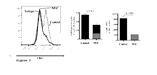
Figure 9 is a set of graphs showing CD69 expression on human
CD8+ T cells in spleen of mice of Example 14.
DETAILED DESCRIPTION
Unless defined otherwise in this specification, technical and
scientific terms used herein have the same meaning as commonly
understood by the person skilled in the art to which this invention
belongs and by reference to published texts.
CA 03066799 2019-12-10
WO 2018/227244
PCT/AU2018/050584
It is to be noted that the term "a" or "an" refers to one or
more, for example, "a MSC" is understood to represent one or more
MSCs, including a population of MSCs. As such, the terms "a" or
"an", "one or more," and "at least one" may be used interchangeably
5 herein.
In the claims which follow and in the description of the
invention, except where the context requires otherwise due to
express language or necessary implication, the word "comprise" or
variations such as "comprises" or "comprising" is used in an
inclusive sense, i.e. to specify the presence of the stated
features, but not to preclude the presence or addition of further
features in various embodiments of the invention.
The term "about" as used herein contemplates a range of
values for a given number of 25% the magnitude of that number. In
other embodiments, the term "about" contemplates a range of values
for a given number of 20%, 15%, 10%, 5%, 4%, 3%, 2%, or 1%
the magnitude of that number. For example, in one embodiment, "about
3 grams" indicates a value of 2.7 grams to 3.3 grams (i.e. 3 grams
10%), and the like.
Similarly, the timing or duration of events may be varied by
at least 25%. For example, while a particular event may be disclosed
in one embodiment as lasting one day, the event may last for more or
less than one day. For example, "one day" may include a period of
about 18 hours to about 30 hours. In other embodiments, periods of
time may vary by 20%, 15%, 10%, 5%, 4%, 3%, 2%, or 1% of
that period of time.
As used herein, "immunotherapy" includes, but is not limited
to, treating a subject with: a checkpoint inhibitor; a bispecific T
cell engager; a stimulator of interferon genes; a RIG I like
receptor; a toll-like receptor; an antibody-cytokine fusion protein;
a cytokine; or an antibody-drug conjugate.
Immunotherapies need not be used singly, and may be used in
combination. For example, two checkpoint inhibitors may be combined,
e.g. nivolumab and pembrolizumab or nivolumab and ipilimumab, or a
checkpoint inhibitor may be combined with a conventional cancer
therapy, e.g. radiotherapy or chemotherapy.
As used herein, "antibody" is used broadly and refers to an
antigen binding molecule. Thus, the term "antibody" includes an
CA 03066799 2019-12-10
WO 2018/227244
PCT/AU2018/050584
6
immunoglobulin, such as IgA, IgD, IgE, IgG, IgM, IgY or IgW, a
fragment, such as Fab, (Fab')2, scFv, scFv-Fc, a minibody, a
diabody, a single domain antibody (sdAb or nanobody), a bispecific
antibody, a multispecific antibody, and an antibody mimetic, such as
an affibody, affilin, affimer, affitin, alphabody, anticalin, avimer
DARPin, fynomer, Kunitz domain peptide and monobody. sdAbs include
camelid antibodies and IgNARs from cartilaginous fish. An antibody
may be polyclonal or monoclonal (mAb). An antibody may be chimeric,
humanized or human.
Antibody production is well-known in the art and includes
hybridoma, phage display, single B cell culture, and single B cell
amplification technologies, for example. Chimeric and humanised
antibodies may be produced using recombinant techniques. Human
antibodies may be produced using phage display technology or
transgenic animals such as transgenic mice, platforms for which are
available commercially. Once an antibody with appropriate
specificity has been identified and its sequence determined, the
antibody may be produced recombinantly, for example in cell culture,
for example CHO cell culture, as is known in the art.
As used herein a "side effect" includes a "symptom" and both
terms refer to an undesired or adverse effect of immunotherapy,
determined either qualitatively, i.e. undesired in any form, or
quantitatively, undesired above or below a specific threshold. Such
a symptom may also be referred to as an "adverse symptom" to
distinguish an effect from a necessary or desired effect of
immunotherapy. A side effect or symptom of immunotherapy may also be
referred to as an "adverse event", an "immune-mediated adverse
event", or an "immune-related adverse event".
Checkpoint inhibitors
Checkpoints are proteins that negatively regulate T cell
immune responses. To date, two checkpoints have been identified:
cytotoxic T lymphocyte antigen-4 (CTLA-4 or CD152) and programmed
death-1 (PD-1 or CD279). PD-1 is activated by programmed death-
ligand 1 (PD-L1) and programmed death-ligand 2 (PD-L2). Inhibition
of checkpoints or their ligands abrogates the negative regulation of
T cells and shifts the immune response toward T cell activation.
Ipilimumab is a human anti-CTLA-4 mAb. Ipilimumab may be
administered at 3 mg/kg every 2 weeks or 3 weeks or at 10 mg/kg
CA 03066799 2019-12-10
WO 2018/227244
PCT/AU2018/050584
7
every 2 weeks, for example. Ipilimumab may be used to treat
melanoma, non-small cell lung carcinoma (NSCLC), small cell lung
cancer (SCLC), bladder cancer, and prostate cancer, for example,
although clinical trials assessing ipilimumab for treating many more
cancers are underway. Ipilimumab may be used in combination with
other agents, for example nivolumab, bavituximab, dacarbazine, IL-2
or gp100.
Side effects or symptoms of ipilimumab include pruritus,
rash, vitiligo, diarrhoea, colitis, increased ALT, increased AST,
hepatitis, hypothyroidism, hypopituitarism, hypophysitis, adrenal
insufficiency, increased thyrotropin, decreased corticotropin, acute
inflammatory demyelinating polyneuropathy, ascending motor
paralysis, and myasthenia gravis, for example.
Nivolumab is a human IgG4 anti-PD-1 mAb. Nivolumab may be
administered at 3 mg/kg every 2 weeks, for example. Nivolumab may be
used to treat melanoma, metastatic melanoma, metastatic squamous
NSCLC, renal cell carcinoma, and bladder cancer, for example,
although clinical trials assessing nivolumab for treating many more
cancers are underway. Nivolumab may be used in combination with
other agents, for example ipilimumab or pembrolizumab.
Side effects or symptoms of nivolumab include pruritus, rash,
vitiligo, diarrhoea, colitis, increased ALT, increased AST,
increased bilirubin, pneumonitis, increased serum creatinine, renal
failure, hypothyroidism, hyperthyroidism, increased TSH, diabetes,
hypophysitis, adrenal insufficiency, and fatigue, for example.
Pembrolizumab is a humanised anti-PD-1 mAb. Pembrolizumab may
be administered at 2 mg/kg or 10 mg/kg every 2 weeks or 3 weeks, for
example. Pembrolizumab may be used to treat melanoma, metastatic
melanoma, metastatic NSCLC, head and neck squamous cell carcinoma
(HNSCC), and Hodgkin's lymphoma, for example. Clinical trials
assessing pembrolizumab for treating many more cancers, such as
breast cancer, gastric cancer, and urothelial cancer, for example,
are underway. Pembrolizumab may be used in combination with other
agents, for example nivolumab, talimogene laherparepvec, dabrafenib
plus trametinib, or ipilimumab.
Side effects or symptoms of pembrolizumab include pruritus,
rash, rash maculopapular, dermatitis acneiform, diarrhoea, colitis,
hepatitis, increased ALT, increased AST, dyspnea, pneumonitis,
CA 03066799 2019-12-10
WO 2018/227244
PCT/AU2018/050584
8
hypothyroidism, hyperthyroidism, increased amylase, pancreatitis,
arthralgia, uveitis, pyrexia, and fatigue, for example.
Another anti-PD-1 antibody in development is BGB-A317,
whereas anti-PD-Li antibodies include atezolizumab, avelumab and
durvalumab. Also in development is an anti-PD-Li affimer.
Bispecific T cell engagers (BiTEs)
A bispecific T cell engager (BiTE) is a type of bispecific
antibody that in a single protein links an scEv targeting a T cell
surface antigen, for example CD3s, to an scEv targeting a surface
antigen on a cancer cell. BiTEs are manufactured using recombinant
techniques as known in the art.
The first in class BiTE is blinatumomab, which is a
bispecific antibody directed to CD3s and CD19, and generally may be
used to treat B cell malignancies, such as acute lymphoblastic
leukaemia (ALL), non-Hodgkin's lymphoma (NHL), diffuse large B cell
lymphoma (DLBCL), and chronic lymphocytic leukaemia (CLL), primary
mediastinal B cell lymphoma (PMBCL), transformed follicular lymphoma
(TFL), multiple myeloma (MM), mantle cell lymphoma (MCL), and acute
myeloid leukaemia (AML). Blinatumomab may be administered at 5 pg/
m2/ d to 60 pg/ m2/ d, for example 5, 15 or 60 pg/ m2/ d.
The most common side effects of blinatumomab are CRS,
pyrexia, fatigue, headache and weight change. CRS would be expected
for most BiTEs.
Because BiTE is a platform technology, other cancer targets
are possible, some of which include CD20, CD33, Epithelial cell
adhesion molecule (EpCAM), carcinoembryonic antigen (CEA), prostate
specific membrane antigen (PSMA), human epidermal growth factor
receptor 2 (HER2), epidermal growth factor receptor (EGFR), Ephrin
type-A receptor 2 (EphA2), mucin 1 (MUC1), and melanoma-associated
chondroitin sulfate proteoglycan (MCSP).
Stimulator of interferon genes (STING)
Stimulator of interferon genes (STING, also known as TMEM173)
is a signalling molecule that binds and is activated by cyclic
dinucleotides, such as cyclic GMP-AMP and cyclic di-AMP, which are
produced in response to DNA entering the cytosol. STING also binds
double-stranded DNA. Upon activation, STING leads to IRE3-mediated
and NE-KB-mediated transcription of type I interferons (IENs) and
inflammatory cytokines such as TNF, IL-4, and IL-6, which in turn
CA 03066799 2019-12-10
WO 2018/227244
PCT/AU2018/050584
9
cause cell death and promotes dendritic cell, natural killer and
CD8 T cell function. Amongst other cells, STING is present in
intratumoural dendritic cells.
Cyclic dinucleotides may be produced biosynthetically/
enzymatically as is known in the art, although this is weighted
towards naturally-occurring cyclic dinucleotides. Otherwise, cyclic
dinucleotides may be produced chemically by nucleotide cyclization
or by stereospecific base insertion on a cyclic bis(3'-5')-sugar
phosphate as is known in the art. Both solution-phase and solid-
support syntheses have been developed and are known in the art.
Cyclic dinucleotides, including synthetic dithio mixed-
linkage cyclic dinucleotides, for example, are under development as
immunotherapy for cancer. Agents under development include ML RR-52
CDA and MIW815, and SB 11285. Cyclic dinucleotides may be combined
with other therapies such as radiotherapy, chemotherapy, checkpoint
inhibitors, or lethally irradiated GM-CSF-secreting tumour cell
cells (STINGVAX).
STING agonists may be used to treat melanoma and colon
cancer, for example.
RIG I like receptors (RLRs)
RIG I like receptors include retinoic acid inducible gene I
(RIG-I or DDX58), melanoma differentiation-associated protein 5
(MDA5 or IFIH1), and Laboratory of Genetics and Physiology 2 (LGP2
or DHX58), which are cytosolic RNA sensors.
RIG-I typically recognizes 5'-triphosphorylated RNA (5'-ppp-
RNA or 3pRNA) and short double stranded RNA, and is dependent on
functional LGP2. MDA5 typically recognises double stranded RNA
longer than 2000 nucleotides, and is also dependent on functional
LGP2. LGP2 itself cannot induce signalling, but is required for RIG-
I-mediated and MDA5-mediated responses.
Upon activation, RIG-I and MDA5 lead to IRF1-, IRF3-, IRF7-
and NFKB-mediated expression of IFNs and inflammatory cytokines,
which that can directly act an tumour cells as well as activate
T cells and natural killer cells.
RLR ligands may be used for immunotherapy and include 5'-ppp-
siRNAs, which act in a sequence independent manner via the RLR
pathway as well as in a sequence dependent manner via the RNA
interference (RNAi) pathway, as well as the hemagglutinating virus
CA 03066799 2019-12-10
WO 2018/227244
PCT/AU2018/050584
of Japan envelope (HVJ-E) vector, and polyinosinic: polycytidylic
acid (poly I:C).
RLR activation may be used to treat melanoma, pancreatic
cancer, prostate cancer, glioma, malignant pleural mesothelioma
5 (MPM), and ovarian cancer, for example.
Toll-like receptors (TLRs)
To date, ten human toll-like receptors (TLRs) have been
identified, each with a different ligand specificity: TLR2
recognises lipoproteins and peptidoglycans; TLR3 recognises viral
10 double stranded RNA and poly I:C; TLR4 recognises
lipopolysaccharides (LPS); TLR5 recognises bacterial flagellin;
TLR7/8 recognises single stranded RNA; TLR9 recognises CpG-
containing oligodeoxynucleotides (CpG-ODN); and TLR2/6, TLR2, and
TLR4 recognise the matrix proteoglycan versican and heat-shock
proteins (HSPs).
A number of these ligands have been approved for cancer
immunotherapy, including TLR2/4 agonists Bacillus Calmette-Guerin
(BCG) for treating bladder cancer, TLR4 agonist monophosphoryl lipid
A (MPL)for treating cervical cancer, and TLR7 agonist imiquimod for
treating breast cancer. Coley toxin and extract of larix leptolepis
(ELL) are also approved for immunotherapy.
Other TLR ligands are being developed for immunotherapy, for
instance, TLR5 agonist flagellin-derived CBLB502 (Entolimod) for
treating advanced solid tumours and hepatoma, TLR7 agonist 852A for
treating melanoma and hematologic malignancy, and TLR3 agonist poly
I:C/ polyinosinic-polycytidylic acid stabilised with polylysine and
carboxymethylcellulose (poly-ICLC) and TLR9 agonist synthetic CpG-
ODN as cancer vaccine adjuvants for treating multiple cancer types
including glioblastoma.
Thus, TLR ligands may be used to treat bladder cancer, breast
cancer, cervical cancer, glioblastoma, hematologic malignancy,
hepatoma, melanoma, and solid tumours, for example.
Cytokines
IL-2 is used clinically as an immunotherapy to improve the
anticancer efficacy of cytotoxic T cells. IL-2 has also been used to
promote ex vivo T cell expansion for adoptive cell transfer (ACT).
Other cytokines in use or development for immunotherapy include
IL-7, IL-12, IL-15, IL-18, IL-21, IFNa, IFNp, IFNy, granulocyte-
CA 03066799 2019-12-10
WO 2018/227244
PCT/AU2018/050584
11
macrophage colony stimulating factor (GM-CSF), and TNF. Cytokines
may be produced recombinantly as known in the art.
Cytokines may be used to treat melanoma, renal cell
carcinoma, lymphoma, B cell lymphoma, follicular lymphoma, hairy
cell leukaemia, sarcoma, hepatitis B/C, chronic granulomatous
disease, malignant osteoporosis, breast cancer, prostate cancer,
sarcoma, Ewing's sarcoma, Kaposi's sarcoma, neuroblastoma, mycosis
fungoides, head and neck cancer, AML, lung cancer, ovarian cancer,
chronic myeloid leukaemia (CML), CLL, and neutropenia, for example.
Often, however, cytokines fail to reach a therapeutic
concentration at the tumour site because they have no means for
preferential trafficking and a very short initial serum half-life
(minutes to hours). Also, cytokines have severe side effects
including systemic inflammation and vascular leak syndrome. Other
side effects include fever, chill, malaise, hypotension, organ
dysfunction, and cytopenias.
Antibody-cytokine fusion proteins
In view of the limitations of free cytokines in
immunotherapy, improvements have been made by recombinantly fusing
cytokines to antibodies of desired antigen specificity. Not only do
such antibodies traffic cytokines to the tumour site of action, the
antibodies may exert their own immunotherapeutic effects via
antibody-dependent cellular cytotoxicity (ADCC) and complement-
dependent cytotoxicity (CDC).
Antibody-cytokine fusion proteins include: hu14.18-IL-2, a
humanised anti-GD2 IgG fused to IL-2, for treating neuroblastoma;
DI-Leu16-IL2, a de-immunised and mutated anti-CD20 IgG fused to
IL-2, for treating B cell malignancies; L19-IL2, a high affinity
anti-fibronectin extradomain B diabody fused to IL-2, for treating
lymphomas, metastatic melanoma, renal cell carcinoma and solid
tumours; anti-CD20-IL-21, an anti-CD20 IgG fused to IL-21, for
treating DLBCL and MCL; BC1-IL12, an anti-human fibronectin isoform
B-FN (extradomain B) IgG, for treating malignant melanoma and RCC;
L19-INFa, an anti-fibronectin extradomain B human scFv fused to TNF,
for treating melanoma, solid tumours, colorectal cancer; anti-CD20-
IFNa, a chimeric anti-CD20 IgG fused to IFNa, for treating B cell
lymphomas and leukaemias; anti-CD20-tetrameric hIFNo( (20-2b-2b),
humanised anti-CD20 IgG veltuzumab fused to four molecules of
CA 03066799 2019-12-10
WO 2018/227244
PCT/AU2018/050584
12
IFNo(2b, for treating B cell lymphomas; F16-IL2, an anti-tenascin C
extradomain Al human scFv fused to IL-2, for treating breast cancer,
lung cancer, Merkel cell carcinoma (MCC), glioblastoma, AML and
solid tumours; NHS-IL2LT, a mouse-human chimeric antibody directed
against DNA released by necrotic tumour cells fused to two molecules
of IL-2, for treating solid tumours, NHL and NSCLC; NHS-IL12, a
mouse-human chimeric antibody directed against DNA released by
necrotic tumour cells fused to two molecules of IL-12, for treating
renal cell carcinoma, Kaposi sarcoma, T cell lymphoma, and NHL;
C2-2b-2b, a humanised anti-HLA-DR IgG fused to four molecules of
IFNo(2b, for treating haematopoietic cancers, B cell lymphomas and
leukaemias, and MM; and huKS-IL2, an anti-epithelial cell adhesion
molecule (EpCAM or KS) IgG fused to IL-2, for treating ovarian
cancer, prostate cancer, colorectal cancer and NSCLC.
Side effects of hu14.18-IL-2 resembled those of IL-2
immunotherapy and included capillary leak, hypoxia, elevated
transaminases, hyperbilirubinemia, hypotension, and renal
insufficiency.
Antibody-drug conjugates (ADCs)
Anti-cancer drugs may be conjugated to antibodies in order to
specifically target the drug to cancer cells. This approach is
particularly useful in delivering anti-cancer drugs that exert
cytotoxicity at concentrations much below standard chemotherapeutic
drugs and thus are too toxic to administer in their free form. Chief
amongst these are microtubule inhibitors such as maytansinoids and
auristatins. Alternatively, the drug may be a radionuclide.
The antibodies of ADCs are commonly, but not necessarily,
IgGs, often IgGl.
Drug, including radionuclide, production is known in the art.
The antibody and drug may be conjugated using a linker, which
may be cleavable or non-cleavable. The linker may be cleavable once
the ADC is internalised by a cancer cell, but stable in the
circulation prior to internalisation. However, once cleaved, a drug
may escape the targeted cell and attack non-cancer bystander cells.
A non-cleavable linker is intended to retain the ADC within the
cell.
Methods for conjugation are known in the art. Cleavable
linker chemistry may employ disulfides, hydrazones or peptides,
CA 03066799 2019-12-10
WO 2018/227244
PCT/AU2018/050584
13
whereas non-cleavable linker chemistry may employ thioethers.
Antibodies may be engineered to manipulate linker chemistry, for
example cysteine amino acid residues may be manipulated to modify
the number and/or position of sulfhydryl groups available for linker
chemistry.
Two ADCs, brentuximab vedotin targeting CD30 and trastuzumab
emtansine targeting HER2, are approved for clinical use, and many
ADCs are in development, including inotuzumab ozogamicin (CD22),
gemtuzumab ozogamicin (anti-CD33), ABT-414 (anti-EGFR),
glembatumumab vedotin (anti-gpNMB), labetuzumab govitecan (anti-
CEACAM5), sacituzumab govitecan (anti-TROP2, EGP1), lifastuzumab
vedotin (anti-NaPi2b), indusatumab vedotin (anti-GCC), polatuzumab
vedotin (anti-CD79b), pinatuzumab vedotin (anti-CD22), PSMA ADC
(anti-PSMA), coltuximab ravtansine (anti-CD19), BMS-986148 (anti-
MSLN), indatuximab ravtansine (anti-CD138, syndecan 1), milatuzumab
doxorubicin (anti-CD74), MLN2704 (anti-PSMA), SAR408701 (anti-
CEACAM5), rovalpituzumab tesirine (anti-DLL3), ABBV-399, AGS-16C3F
(anti-ENPP3), ASG-22ME (anti-nectin 4), AGS67E (anti-CD37), AMG 172
(anti-CD27), AMG 595 (anti-EGFRvIII), AGS-15E (anti-SLTRK6),
BAY1129980 (anti-C4.4a), BAY1187982 (anti-FGFR2), anetumab
ravtansine (anti-mesothelin), GSK2857916 (anti-BCMA), tisotumab
vedotin (anti-TF), IMGN289 (anti-EGFR), IMGN529 (anti-CD37),
mirvetuximab soravtansine (anti-FOLR1), L0P628 (anti-c-KIT), PCA062
(anti-p-cadherin), BMS936561 (anti-CD70), MEDI-547 (anti-EphA2),
.. PF-06263507 (anti-5T4), PF-06647020, PF-06647263 (anti-ephrin A),
PF-06664178 (anti-TROP2), RG7450 (anti-STEAP1), RG7458 (anti-MUC16),
RG7598, SAR566658 (anti-CA6), SGN-CD19A (anti-CD19), SGN-CD33A
(anti-CD33), SGN-CD70A (anti-CD70), SGN-LIV1A (anti-LIV1), and
trastuzumab vc-seco (anti-HER2).
Examples of ADCs comprising radionuclides include Y"-
ibritumomab tiuxetan and I131-tositumomab.
ADCs may be used to treat haematological cancers and solid
tumours, for example, breast cancer, melanoma, lung cancer, SCLC,
pancreatic cancer, colorectal cancer, ovarian cancer, endometrial
cancer, cervical cancer, prostate cancer, mesothelioma, bladder
cancer, RCC, liver carcinoma, gastric cancer, NSCLC, glioblastoma,
head and neck cancer, oesophageal cancer, Hodgkin's lymphoma, NHL,
anaplastic large cell lymphoma, ALL, DLBCL, multiple myeloma, CLL,
CA 03066799 2019-12-10
WO 2018/227244
PCT/AU2018/050584
14
and AML.
ADCs may be administered at a dose of 0.05 mg/kg to 16 mg/kg,
for example.
Side effects of ADCs include thrombocytopenia, neutropenia,
ocular toxicity, rash, typhlitis, nausea, dyspnea, liver toxicity,
mucositis, anaemia, neuropathy, capillary leak syndrome, and
diarrhoea.
Indications
Immunotherapy may be used to treat acute lymphoblastic
leukaemia (ALL), acute myeloid leukaemia (AML), anaplastic large
cell lymphoma, B cell lymphomas or leukaemias, B cell malignancies,
bladder cancer, breast cancer, cervical cancer, chronic
granulomatous disease, chronic lymphocytic leukaemia (CLL), chronic
myeloid leukaemia (CML), colon cancer, colorectal cancer, diffuse
large B cell lymphoma (DLBCL), endometrial cancer, Ewing's sarcoma,
follicular lymphoma, gastric cancer, glioblastoma, glioma,
haematological cancers, haematopoietic cancers, hairy cell
leukaemia, head and neck cancer, head and neck squamous cell
carcinoma, haematologic malignancy, hepatitis B/C, hepatoma,
Hodgkin's lymphoma, Kaposi's sarcoma, liver carcinoma, lung cancer,
lymphomas, malignant melanoma, malignant osteoporosis, malignant
pleural mesothelioma (MPM), mantle cell lymphoma (MCL), Merkel cell
carcinoma (MCC), melanoma, mesothelioma, multiple myeloma (MM),
mycosis fungoides, neuroblastoma, neutropenia, non-Hodgkin's
lymphoma (NHL), non-small cell lung carcinoma (NSCLC), metastatic
melanoma, metastatic NSCLC, metastatic squamous NSCLC, oesophageal
cancer, ovarian cancer, pancreatic cancer, primary mediastinal B
cell lymphoma (PMBCL), renal cell carcinoma, sarcoma, small cell
lung cancer (SCLC), solid tumours, T cell lymphoma, transformed
follicular lymphoma (TFL), or urothelial cancer, for example.
Side effects and symptoms
Cytokine-release syndrome (CRS) is a serious side effect of
immunotherapy. CRS is thought to result from proliferating T cells
that release large quantities of cytokines, including IL-6, IFN-y,
TNF, IL-2, IL-2-receptor a, IL-8, IL-10, and GMCSF.
Symptoms of CRS include: high fever, malaise, fatigue,
myalgia, nausea, anorexia, tachycardia/ hypotension, capillary leak,
cardiac dysfunction, renal impairment, hepatic failure, and
CA 03066799 2019-12-10
WO 2018/227244
PCT/AU2018/050584
disseminated intravascular coagulation.
Thus, subjects with CRS may experience any one or more of
fever, cardiovascular symptoms including tachycardia, hypotension,
arrhythmias, decreased cardiac ejection fraction, pulmonary symptoms
5 including oedema, hypoxia, dyspnoea, and pneumonitis, acute renal
injury usually caused by reduced renal perfusion, hepatic and
gastrointestinal symptoms including elevated serum transaminases and
bilirubin, diarrhoea, colitis, nausea, and abdominal pain,
hematologic symptoms including cytopenias such as grade 3-4 anaemia,
10 thrombocytopenia, leukopenia, neutropenia, and lymphopenia,
derangements of coagulation including prolongation of the
prothrombin time and activated partial thromboplastin time (PTT), D-
dimer elevation, low fibrinogen, disseminated intravascular
coagulation, macrophage activation syndrome (MAS), haemorrhage,
15 B-cell aplasia, and hypogammaglobulinemia, infectious diseases
including bacteremia, Salmonella, urinary tract infections, viral
infections such as influenza, respiratory syncytial virus, and
herpes zoster virus, musculoskeletal symptoms including elevated
creatine kinase, myalgias and weakness, neurological symptoms
including delirium, confusion, and seizure.
MAS overlaps clinically with CRS with subjects potentially
experiencing hepatosplenomegaly, lymphadenopathy, pancytopenia,
liver dysfunction, disseminated intravascular coagulation,
hypofibrinogenemia, hyperferritinemia, and hypertriglyceridemia.
Like CRS, subjects with MAS exhibit elevated levels of cytokines,
including IFN-y and GMCSF.
On-target, off-cancer effects may lead to outcomes similar to
GVHD and B cell aplasia, which is caused when the target cancer
antigen is expressed endogenously on other healthy/ normal cells
types.
For instance, B cell aplasia occurs because anti-CD19
antibodies also target normal B cells that express CD19. The
consequence of B cell aplasia is a reduced capacity to fight
infection because of hypoimmunoglobulinemia. Intravenous
immunoglobulin replacement therapy is used to prevent infection.
Another side effect of immunotherapy is TLS, which occurs
when the contents of cells are released as a result of therapy
causing cell death, most often with lymphoma and leukaemia. TLS is
CA 03066799 2019-12-10
WO 2018/227244
PCT/AU2018/050584
16
characterised by blood ion and metabolite imbalance, and symptoms
include nausea, vomiting, acute uric acid nephropathy, acute kidney
failure, seizures, cardiac arrhythmias, and death.
Neurotoxicity may result from immunotherapy and symptoms may
include cerebral oedema, delirium, hallucinations, dysphasia,
akinetic mutism, headache, confusion, alterations in wakefulness,
ataxia, apraxia, facial nerve palsy, tremor, dysmetria, and seizure.
Anaphylaxis can arise from non-host proteins, such as murine-
derived proteins forming part of the immunotherapy, e.g. chimeric or
humanised mAb.
Subjects undergoing immunotherapy as disclosed herein may
experience one or more side effects or symptoms including anaemia,
aphasia, arrhythmia, arthralgia, back pain, blood and bone marrow
disorders, blood and lymphatic system disorders, cardiac disorders,
-- chills, coagulation disorders, colitis, confused state,
constitutional symptoms, cough, decreased appetite, diarrhoea,
disorientation, dizziness, dyspnea, encephalopathy, fatigue, fever,
gastrointestinal disorders, general cardiovascular disorders,
haemorrhage, hepatic disorders, hyperglycaemia, hypokalaemia,
hypothyroidism, increased ALT, increased AST, increased C-reactive
protein, infection febrile neutropenia, leukopenia, malaise,
abnormal metabolic laboratory-testing results, metabolism nutrition
disorders, mucosal inflammation, musculoskeletal disorders, myalgia,
nausea, nervous system disorders, neurologic disorders, neutropenia,
-- oedema, pain, palmar-plantar erythrodysesthesia, paresthesia,
pneumonia, pruritus, pulmonary disorders, pyrexia, rash, renal
genitourinary disorders, respiratory disorders, skin and
subcutaneous tissue disorders, somnolence, speech disorders, sweats,
thoracic mediastinal disorders, thrombocytopenia, tremor, tumour
flare, tumour lysis syndrome, vascular disorders, and vomiting.
In turn, any of the side effects or symptoms listed above may
be indicative of CRS, MAS, TLS, on-target, off-cancer effects,
neurotoxicity, or anaphylaxis, for instance.
Management of side-effects
In general, side-effects of immunotherapy are managed with
standard supportive therapy for any presenting symptoms. However,
given the myriad side effects and symptoms that may occur as a
result of immunotherapy, multiple supportive therapies may be
CA 03066799 2019-12-10
WO 2018/227244
PCT/AU2018/050584
17
required simultaneously. The mainstay of supportive therapy is
steroids, for example dexamethasone, although more recently anti-
cytokine therapies have been used to treat CRS, for example
etanercept, an anti-TNF molecule, and tocilizumab, an anti-IL-6
receptor antibody.
Recommendations have been made that upon presentation of a
grade 3 or grade 4 adverse event, immunotherapy should be
discontinued permanently.
Mesenchymal stem cells
Accordingly, the invention provides an improved therapy for
reducing the number, severity and duration of side effects caused by
immunotherapy, by administration of mesenchymal stem cells (MSCs).
MSCs exert their effects through their immunomodulatory properties,
so for many side effects and symptoms, MSCs are able to act directly
at the immunogenic cause of the side effect or symptom.
MSCs secrete bioactive molecules such as cytokines,
chemokines and growth factors and have the ability to modulate the
immune system. MSCs have been shown to facilitate regeneration and
effects on the immune system without relying upon engraftment. In
other words, the MSCs themselves do not necessarily become
incorporated into the host - rather, they exert their effects and
are then eliminated within a short period of time. However, MSCs may
be engrafted.
As used herein, "mesenchymal stem cell" or "MSC" refers to a
particular type of stem cell that may be isolated from a wide range
of tissues, including bone marrow, adipose tissue (fat), placenta
and umbilical cord blood. Alternatively, MSCs may be produced from
pluripotent stem cells (PSCs). MSCs are also known as "mesenchymal
stromal cells". Alternatively or additionally, MSC refers to an MSC
as defined by the Mesenchymal and Tissue Stem Cell Committee of the
International Society for Cellular Therapy: (1) MSCs must be
plastic-adherent when maintained in standard culture conditions; (2)
MSCs must express CD105, CD73 and CD90, and lack expression of CD45,
CD34, CD14 or CD11b, CD79a1pha or CD19 and HLA-DR surface molecules;
(3) MSCs must differentiate to osteoblasts, adipocytes and
chondroblasts in vitro.
Production of MSCs from PSCs is described in international
patent application no. PCT/AU2017/050228 filed 14 March 2017, which
CA 03066799 2019-12-10
WO 2018/227244
PCT/AU2018/050584
18
is incorporated in full by this cross-reference, and is described in
Examples 1 and 2.
MSCs have been shown to exert immunomodulatory activities
against T cells, B cells, dendritic cells, macrophages, and natural
killer cells. While not wishing to be bound by theory, the
underlying mechanisms may comprise immunomodulatory mediators, for
example nitric oxide, indoleamine 2,3, dioxygenase, prostaglandin
E2, tumour necrosis factor-inducible gene 6 protein, CCL-2, and
PD-Li. These mediators are expressed at a low level until
stimulated, for example by an inflammatory cytokines, such as IFNy,
TNF, and IL-17.
In one embodiment, MSCs are pre-treated prior to
administration. Pre-treatment may be with a growth factor or by gene
editing, for example, where a growth factor may prime the MSC and
gene editing may confer a new or improved, e.g. more potent,
therapeutic property on the MSC.
As used herein, "pluripotent stem cell" or "PSC" refers to a
cell that has the ability to reproduce itself indefinitely, and to
differentiate into any other cell type. There are two main types of
PSC: embryonic stem cells (ESCs) and induced pluripotent stem cells
(iPSCs).
As used herein, "embryonic stem cell" or "ESC" refers to a
cell isolated from a five to seven day-old embryo donated with
consent by subjects who have completed in vitro fertilisation
therapy, and have surplus embryos. The use of ESCs has been hindered
to some extent by ethical concerns about the extraction of cells
from human embryos.
Suitable human PSCs include H1 and H9 human embryonic stem
cells.
As used herein, "induced pluripotent stem cell" or "iPSC"
refers to an ESC-like cell derived from adult cells. iPSCs have very
similar characteristics to ESCs, but avoid the ethical concerns
associated with ESCs, since iPSCs are not derived from embryos.
Instead, iPSCs are typically derived from fully differentiated adult
.. cells that have been "reprogrammed" back into a pluripotent state.
Suitable human iPSCs include, but are not limited to, iPSC
19-9-7T, MIRJT6i-mND1-4 and MIRJT7i-mND2-0 derived from fibroblasts
and iPSC BM119-9 derived from bone marrow mononuclear cells. Other
CA 03066799 2019-12-10
WO 2018/227244
PCT/AU2018/050584
19
suitable iPSCs may be obtained from Cellular Dynamics International
(CDI) of Madison, WI, USA.
In one embodiment, MSCs used according to the invention are
formed from primitive mesodermal cells. The primitive mesoderm cells
may have mesenchymoangioblast (MCA) potential. The primitive
mesoderm cells may have a Emfflin-KDR APLNR-'PDGFRalpha- phenotype. In
one embodiment, MSCs used according to the invention are formed from
linKDRAPLNRPDGFRalpha primitive mesoderm cells with MCA
potential.
A primitive mesoderm cell may be differentiated from a PSC,
for example an iPSC, by culturing the PSC in a differentiation
medium comprising FGF2, BMP4, Activin A, and LiC1 under hypoxic
conditions for about two days to form the primitive mesoderm cell.
Thus also disclosed is a method of differentiating a
pluripotent stem cell (PSC) into a mesenchymal stem cell (MSC), the
method comprising:
(a) culturing the PSC in a differentiation medium
comprising FGF2, BMP4, Activin A, and LiC1 under hypoxic conditions
for about two days to form a primitive mesoderm cell;
(b) replacing the differentiation medium of (a) with a
mesenchymal colony forming medium (M-CFM) comprising LiC1 and FGF2,
but excluding PDGF;
(c) culturing the primitive mesoderm cell of (b) in the
M-CFM of (b) under normoxic conditions for sufficient time for a
mesenchymal colony to form; and
(d) culturing the mesenchymal colony of (c) adherently to
produce the MSC.
In some embodiments, the concentration in the differentiation
medium of: BMP4 is about 10 ng/mL to about 250 mg/mL; FGF2 is about
5 ng/mL to about 50 ng/mL; activin A is about 1 ng/mL to about 15
ng/mL; and LiC1 is about 1 mM to about 2 mM. In one embodiment, the
differentiation medium comprises about 50 ng/mL BMP4; about 50 ng/mL
FGF2; about 1.5 ng/mL activin A; and about 2 mM LiCl.
As used herein, "mesenchymoangioblast" and 'MCA" refers to a
common or bipotential mesenchymal cell and endothelial cell
precursor.
As used herein, "Emillin--KDR'APLNRRDGFRalphaf primitive mesoderm
cell with MCA potential" refers to a cell expressing typical
CA 03066799 2019-12-10
WO 2018/227244
PCT/AU2018/050584
primitive streak and lateral plate/ extraembryonic mesoderm genes.
These cells have potential to form MCA and hemangioblast colonies in
serum-free medium in response to fibroblast growth factor 2 (FGF2).
When cultured according to example 2, these cells become MSCs.
5 The term E'ffain- denotes lack of expression of CD31,
VE-cadherin endothelial markers, CD73 and CD105 mesenchymal/
endothelial markers, and CD43 and CD45 hematopoietic markers.
In one embodiment, MSCs used according to the invention
exhibit a CD73+CD105+CD9O+CD146+CD44+CD1O+CD31-CD45- phenotype.
10 Although not explicitly indicated, this phenotype conforms to the
Mesenchymal and Tissue Stem Cell Committee of the International
Society for Cellular Therapy definition of MSCs.
In one embodiment, MSCs used according to the invention
express each of the microRNAs miR-145-5p, miR-181b-5p, and miR-214-
15 3p, but not miR-127-3p and miR-299-5p.
MSCs possess "immunomodulatory activities", which may be
assessed in vitro as the capacity of a MSC to suppress proliferation
of T helper (CD4) lymphocytes. Immunomodulatory activities may be
quantified in vitro relative to a reference, for example as
20 determined using an ImmunoPotency Assay.
A suitable ImmunoPotency Assay uses an irradiated test MSC
(e.g. iPSC-MSC produced according to the method disclosed herein)
and an irradiated reference sample MSC, which are plated separately
at various concentrations with carboxyfluorescein succinimidyl
ester-labelled leukocytes purified from healthy donor peripheral
blood. T helper (CD4) lymphocytes that represent a subset of the
reference sample are stimulated by adding CD3 and CD28 antibodies.
CD4 labelled T cells are enumerated using flow cytometry to assess T
cell proliferation. IC50 values are reported as a function of the
reference sample. A higher IC50 value indicates a greater magnitude
of suppression of proliferation of T helper (CD4) lymphocytes and
thus is indicative of superior T immunomodulatory properties.
MSC samples are irradiated prior to use in this assay to eliminate
the confounding factor of their proliferative potential.
It will be appreciated by the person skilled in the art that
the exact manner of administering to a subject a therapeutically
effective amount of MSCs for treating a side effect or symptom of
immunotherapy in a subject will be at the discretion of the medical
CA 03066799 2019-12-10
WO 2018/227244
PCT/AU2018/050584
21
practitioner. The mode of administration, including dose,
combination with other agents, timing and frequency of
administration, and the like, may be affected by the subject's
condition and history.
The MSC may be administered as a therapeutic composition. As
used herein, the term "therapeutic composition" refers to a
composition comprising an MSC or population of MSCs as described
herein that has been formulated for administration to a subject. The
MSC may be formulated in and/or a therapeutic composition may
comprise an excipient, carrier, buffer or other additive that
facilitates administration of the MSC to a subject. Preferably, the
therapeutic composition is sterile. In one embodiment, the
therapeutic composition is pyrogen-free.
In one embodiment, an MSC of the disclosure, for example a
MSC that expresses miR-145-5p, miR-181b-5p, and miR-214-3p, but not
miR-127-3p and miR-299-5p, and/or has phenotype CD73+CD105+CD90+
CD146+CD44+CD1O+CD31-CD45-, or a therapeutic composition of the
disclosure is provided in a container, preferably a sterile
container, preferably a pyrogen-free container. In one embodiment,
the container is suitable for bolus administration, for example, a
syringe. In another embodiment, the container is suitable for
infusion, for example, an infusion bag.
The MSC will be formulated, dosed, and administered in a
fashion consistent with good medical practice. Factors for
consideration in this context include the particular type of
disorder being treated and anticipated side effects or symptoms, the
particular subject being treated, the clinical condition of the
subject, the site of administration, the method of administration,
the scheduling of administration, and other factors known to medical
practitioners. The therapeutically effective amount of the MSC to be
administered will be governed by such considerations.
Doses of MSCs may range from about 103 cells/m2 to about
1010 cells/m2, for example about 106 cells/m2 to about 2x10³ cells/m2,
or about 103 cells/m2, about 5x10³ cells/m2, about 104 cells/m2, about
5x10⁴ cells/m2, about 105 cells/m2, about 5x10⁵ cells/m2, about 106
cells/m2, about 5x10⁶ cells/m2, about 107 cells/m2, about 5x10⁷
cells/m2, about 103 cells/m2, about 5x10⁹ cells/m2, about
109 cells/m2, about 5x10⁹ cells/m2, about 1010 cells/m2, or about
CA 03066799 2019-12-10
WO 2018/227244
PCT/AU2018/050584
22
5x101 cells/m2.
Doses of MSCs may range from about 103 cells/kg to about
101 cells/kg, for example about 106 cells/kg to about 2x10³
cells/kg, or about 103 cells/kg, about 5x10³ cells/kg, about
104 cells/kg, about 5x10⁴ cells/kg, about 105 cells/kg, about
5x10⁵ cells/kg, about 106 cells/kg, about 5x10⁶ cells/kg, about
107 cells/kg, about 5x10⁷ cells/kg, about 103 cells/kg, about
5x10³ cells/kg, about 109 cells/kg, about 5x10⁹ cells/kg, about
101 cells/kg, or about 5x101 cells/kg.
Doses of MSCs may range from about 103 cells to about
1010 cells, for example about 106 cells to about 2x10³ cells, or
about 103 cells, about 5x10³ cells, about 104 cells, about
5x10⁴ cells, about 105 cells, about 5x10⁵ cells, about 106 cells,
about 5x10⁶ cells, about 107 cells, about 5x10⁷ cells, about
103 cells, about 5x10³ cells, about 109 cells, about 5x10⁹ cells,
about 1010 cells, or about 5x101 cells.
The MSCs may be administered in a single dose, a split dose,
or in multiple doses. The MSCs may be administered more than once in
a treatment cycle.
The MSCs may be administered to a subject systemically or
peripherally by any suitable method, for example by routes including
intravenous (IV), intra-arterial, intramuscular, intraperitoneal,
intracerobrospinal, intracranial, subcutaneous (SC), intra-
articular, intrasynovial, intrathecal, intracoronary,
transendocardial, surgical implantation, topical and inhalation
(e.g. intrapulmonary) routes. Most preferably, the MSCs are
administered IV. MSCs may be administered in combination with a
scaffold of biocompatible material.
The MSCs may be administered to the subject before, during or
after immunotherapy. In one embodiment, MSCs are administered during
inflammation. Accordingly, in one embodiment, MSCs are administered
after immunotherapy has started, optionally after inflammation has
commenced and/or pro-inflammatory cytokine release has commenced or
increased relative to a control, for example relative to pre-
administration of the immunotherapy. Without wishing to be bound by
theory, it is thought that most benefit will be gained by
administering the MSCs after immunotherapy. This is because
immunotherapy is intrinsically an immune/ inflammatory response,
CA 03066799 2019-12-10
WO 2018/227244
PCT/AU2018/050584
23
whereas MSCs exert immunomodulatory and anti-inflammatory effects.
Thus, administering MSCs before, during or too early after
immunotherapy may dampen the effect of the immunotherapy. Despite
this theory, the invention is not restricted to such.
However, this apparent paradox is understood and accepted by
those skilled in the art. For example, a primary treatment for CRS
caused by immunotherapy is administration of steroids, which are
profoundly immunosuppressive. Advantages of MSCs, for example, may
include local immunomodulation versus systemic immunosuppression by
steroids, lack of persistence in the body, providing a further line
of defence in subjects who fail to respond to steroids or other
immunosuppressive therapies, reduced toxicity and increased
specificity versus steroids, and self-regulation by MSCs versus
steroids. Reduced toxicity, increased specificity, and self-
regulation are related, and by self-regulation, it is meant that
MSCs are thought to have a capacity to reduce their immunomodulatory
activity as the immune response of the side effect or symptom of
immunotherapy dissipates, whereas steroids for example must be
withdrawn by the physician, with an ensuing period of half-lives
before the steroid concentration drops below the therapeutic
concentration.
In one embodiment, treating comprises administering the
MSC(s) within 24 hours after observing a side effect of
immunotherapy. In another embodiment, the MSCs may be administered
to the subject receiving immunotherapy about 7 days, about 6 days,
about 5 days, about 4 days, about 72 hours, about 48 hours, about
36 hours, about 24 hours, about 16 hours, about 12 hours, about
8 hours, about 4 hours, about 3 hours, about 2 hours, about 60 min,
about 45 min, about 30 min, about 15 min, or about 5 min after
observing a side effect of immunotherapy.
Accordingly, in one embodiment, the MSCs may be administered
to the subject receiving immunotherapy about 1 week after
immunotherapy. In another embodiment, the MSCs may be administered
to the subject receiving immunotherapy about 5 min after
immunotherapy. In another embodiment, the MSCs may be administered
to the subject receiving immunotherapy about 6 days, about 5 days,
about 4 days, about 72 hours, about 48 hours, about 36 hours, about
24 hours, about 16 hours, about 12 hours, about 8 hours, about 4
CA 03066799 2019-12-10
WO 2018/227244
PCT/AU2018/050584
24
hours, about 2 hours, about 60 min, about 45 min, about 30 min,
about 15 min, or about 5 min after immunotherapy. In one embodiment,
the MSCs may be administered to the subject receiving immunotherapy
within about 24 hours to about 72 hours after immunotherapy.
In one embodiment, the MSCs may be administered to the
subject receiving immunotherapy about 1 week before immunotherapy.
In another embodiment, the MSCs may be administered to the subject
receiving immunotherapy about 5 min before immunotherapy. In another
embodiment, the MSCs may be administered to the subject receiving
immunotherapy about 6 days, about 5 days, about 4 days, about 72
hours, about 48 hours, about 36 hours, about 24 hours, about 16
hours, about 12 hours, about 8 hours, about 4 hours, about 2 hours,
about 60 min, about 45 min, about 30 min, or about 15 min before
immunotherapy.
In another embodiment, the MSCs may be administered to the
subject receiving immunotherapy at about the same time as or during
immunotherapy.
As used herein, "immunotherapy" when used in the context of
"before", "during", "undergoing", "after", "undergone" and similar
means before, during, undergoing, after, or undergone administration
of the immunotherapeutic agent, for example a checkpoint inhibitor,
a bispecific T cell engager, a stimulator of interferon genes
agonist, a RIG I like receptor agonist, a Toll-like receptor
agonist, a cytokine, an antibody-cytokine fusion protein, or an
antibody-drug conjugate. As used herein, "undergoing" includes
subjects who will undergo immunotherapy, but are yet to be
administered the immunotherapy.
The term "therapeutically effective amount" refers to an
amount of MSCs effective to treat a side effect or symptom of
immunotherapy in a subject.
The terms "treat", "treating" or "treatment" refer to both
therapeutic treatment and prophylactic or preventative measures,
wherein the aim is to prevent, reduce, or ameliorate a side effect
or symptom of immunotherapy in a subject or slow down (lessen)
progression of a side effect or symptom of immunotherapy in a
subject. Subjects in need of treatment include those already with
the side effect or symptom of immunotherapy as well as those in
CA 03066799 2019-12-10
WO 2018/227244
PCT/AU2018/050584
which the side effect or symptom of immunotherapy is to be prevented
or ameliorated.
The terms "preventing", "prevention", "preventative" or
"prophylactic" refers to keeping from occurring, or to hinder,
5 defend from, or protect from the occurrence of a side effect or
symptom of immunotherapy. A subject in need of prevention may be
prone to develop the side effect or symptom of immunotherapy.
The term "ameliorate" or "amelioration" refers to a decrease,
reduction or elimination of a side effect or symptom of
10 immunotherapy.
A side effect or symptom of immunotherapy may be quantified.
A side effect or symptom of immunotherapy may be quantified on a
semi-quantitative scale, for example 0 to 5, where 0 represents
absence, 1 to 4 represent identifiable increases in severity, and 5
15 represents maximum severity. Clinical trials often use a 1 to 5
scale where: 1 represents a mild adverse event (side effect); 2
represents a moderate adverse event (side effect); 3 represents a
severe adverse event (side effect); 4 represents a life-threatening
or disabling adverse event (side effect); and 5 represents death
20 related to adverse event (side effect). Alternatively, a side effect
or symptom of immunotherapy may be quantified as a binary event,
i.e. presence or absence, 0 or 1. Other semi-quantitative scales
will be readily apparent to the person skilled in the art. In
another embodiment, a side effect or symptom of immunotherapy may be
25 quantified on a quantitative scale, for instance: mass per volume
such as mass of cytokine per volume of tissue fluid; temperature;
duration; rate; enzyme activity; oxygen saturation; and so on.
The person skilled in the art will readily understand how to
assess and quantify any side effect or symptom of immunotherapy, and
be able to do so without difficulty or undue burden. For example,
the person skilled in the art will be able to measure: a cytokine
concentration in plasma or serum; temperature (fever); heart rate
(tachycardia); blood pressure (hypotension); cardiac dysfunction;
renal impairment; serum or plasma enzyme concentrations (hepatic
function); and so on.
Any quantification of a side effect or symptom of
immunotherapy may be compared to a control, for example a healthy
control subject receiving neither immunotherapy nor MSCs, an
CA 03066799 2019-12-10
WO 2018/227244
PCT/AU2018/050584
26
affected control subject receiving immunotherapy, but not treated
with MSCs, or a population.
Treating a side effect or symptom of immunotherapy by
administering a MSC may be about a 1% decrease, about a 2% decrease,
about a 3% decrease, about a 4% decrease, about a 5% decrease, about
a 6% decrease, about a 7% decrease, about an 8% decrease, about a 9%
decrease, about a 10% decrease, about a 20% decrease, about a 30%
decrease, about a 40% decrease, about a 50% decrease, about a 60%
decrease, about a 70% decrease, about an 80% decrease, about a 90%
decrease, about a 100%, or greater decrease in the side effect or
symptom of immunotherapy. Alternatively, treating a side effect or
symptom of immunotherapy may be about a 2-fold, about a 3-fold,
about a 4-fold, about a 5-fold, about a 6-fold, about a 7-fold,
about an 8-fold, about a 9-fold, about a 10-fold, or more decrease
in the side effect or symptom of immunotherapy. It follows that
"less severe side effects" refers to such a decrease in the side
effect or symptom of immunotherapy.
As used herein, the term "subject" may refer to a mammal. The
mammal may be a primate, particularly a human, or may be a domestic,
zoo, or companion animal. Although it is particularly contemplated
that the method disclosed herein is suitable for medical treatment
of humans, it is also applicable to veterinary treatment, including
treatment of domestic animals such as horses, cattle and sheep,
companion animals such as dogs and cats, or zoo animals such as
felids, canids, bovids and ungulates.
The following examples assist in describing the invention,
which is not to be limited to such examples.
EXAMPLES
Example 1. Reagents for MSC production
Table 1. Reagents
Description Vendor / Cat # or Ref #
DMEM/F12 Base Medium Invitrogen / A1516901
E8 supplement Invitrogen / A1517101
vitronectin Life Technologies / A14700
collagen IV Sigma / C5533
H-1152 ROCK Inhibitor EMD Millipore / 555550
Y27632 dihydrochloride ROCK
Tocris / 1254
Inhibitor
CA 03066799 2019-12-10
WO 2018/227244
PCT/AU2018/050584
27
Description Vendor / Cat # or Ref #
FGF2
Waisman Biomanufacturing / WC-
FGF2-FP
human endothelial-SFM Life Technologies / 11111-044
stemline II hematopoietic stem cell
Sigma / S0192
expansion medium
GLUTAMAX Invitrogen / 35050-061
insulin Sigma / 19278
lithium chloride (LiC1) Sigma / L4408
collagen I solution Sigma / C2249
fibronectin Life Technologies / 33016-015
DMEM/F12 Invitrogen / 11330032
recombinant human BMP4 Peprotech / 120-05E1
activin A Peprotech / 120-14E
Iscove's modified Dulbecco's medium Invitrogen / 12200036
(IMDM)
Ham's F12 nutrient mix Invitrogen / 21700075
sodium bicarbonate Sigma / S5761
L-ascorbic acid 2-phosphate Mg2+ Sigma / A8960
1-thioglycerol Sigma / M6145
sodium selenite Sigma / S5261
non-essential amino acids HyClone / 5H30853.01
chemically defined lipid Invitrogen / 11905031
concentrate
embryo transfer grade water Sigma / W1503
polyvinyl alcohol (PVA) MP Bio / 151-941-83
holo-transferrin Sigma / 10665
ES-CULT M3120 Stem Cell Technologies / 03120
STEMSPAN serum-free expansion Stem Cell Technologies / 09650
medium (SFEM)
L-ascorbic acid Sigma / A4544
Platelet-derived growth factor Peprotech / 110-14B
subunit B homodimer (PDGF-BB)
The reagents listed in Table 1 are known to the person
skilled in the art and have accepted compositions, for example IMDM
and Ham's F12. GLUTAMAX comprises L-alanyl-L-glutamine dipeptide,
usually supplied at 200 mM in 0.85% NaCl. GLUTAMAX releases
L-glutamine upon cleavage of the dipeptide bond by the cells being
cultured. Chemically defined lipid concentrate comprises arachidonic
acid 2 mg/L, cholesterol 220 mg/L, DL-alpha-tocopherol acetate
70 mg/L, linoleic acid 10 mg/L, linolenic acid 10 mg/L, myristic
acid 10 mg/L, oleic acid 10 mg/L, palmitic acid 10 mg/L, palmitoleic
acid 10 mg/L, pluronic F-68 90 g/L, stearic acid 10 mg/L, TWEEN 80
2.2 g/L, and ethyl alcohol. H-1152 and Y27632 are highly potent,
CA 03066799 2019-12-10
WO 2018/227244 PCT/AU2018/050584
28
cell-permeable, selective ROCK (Rho-associated coiled coil forming
protein serine/threonine kinase) inhibitors.
Table 2. IF6S medium (10X concentration)
10X IF6S Quantity Final
Concentration
IMDM 1 package, 5X
powder for 1 L
Ham's F12 nutrient mix 1 package, 5X
powder for 1 L
sodium bicarbonate 4.2 g 21 mg/mL
L-ascorbic acid 2-phosphate Mg2+ 128 mg 640 pg/mL
1-thioglycerol 80 pL 4.6 mM
sodium selenite (0.7 mg/mL solution) 24 pL 84 ng/mL
GLUTAMAX 20 mL 10X
non-essential amino acids 20 mL 10X
chemically defined lipid concentrate 4 mL 10X
embryo transfer grade water To 200 mL NA
Table 3. IF9S medium (1X concentration; based on IF6S)
IF9S Quantity Final
Concentration
IF6S 5 mL 1X
polyvinyl alcohol (PVA; 20 mg/mL 25 mL 10 mg/mL
solution)
holo-transferrin (10.6 mg/mL 50 pL 10.6 pg/mL
solution)
insulin 100 pL 20 pg/mL
embryo transfer grade water To 50 mL NA
Table 4. Differentiation medium (1X concentration; based on
IF9S)
Differentiation Medium Quantity Final
Concentration
IF9S 36 mL 1X
FGF2 1.8 pg 50 ng/mL
LiC1 (2M solution) 36 pL 2mM
BMP4 (100 pg/mL solution) 18 pL 50 ng/mL
Activin A (10 mg/mL solution) 5.4 pL 1.5 ng/mL
Table 5. Mesenchymal colony forming medium (1X concentration)
Mesenchymal colony forming medium Quantity Final
(M-CFM) Concentration
ES-CULT M3120 40 mL 40%
STEMSPAN SFEM 30 mL 30%
human endothelial-SFM 30 mL 30%
GLUTAMAX 1 mL 1X
L-ascorbic acid (250 mM solution) 100 pL 250 pM
LiC1 (2M solution) 50 pL 1 mM
1-thioglycerol (100 mM solution) 100 pL 100 pM
FGF2 600 ng 20 ng/mL
CA 03066799 2019-12-10
WT12018/227244 PCT/AU2018/050584
29
Table 6. Mesenchymal serum-free expansion medium (1X
concentration)
Mesenchymal serum-free expansion Quantity Final
medium (M-SFEM) Concentration
human endothelial-SFM 5 L 50%
STEMLINE II HSFM 5 L 50%
GLUTAMAX 100 mL 1X
1-thioglycerol 87 pL 100 pM
FGF2 100 pg 10 ng/mL
Example 2. Protocol for differentiating human PSC into MSC
1. Thawed iPSCs in E8 Complete Medium (DMEM/F12 Base Medium + E8
Supplement) + 1 pM H1152 on Vitronectin coated (0.5 pg/ckg)
plastic ware. Incubated plated iPSCs at 37 C, 5% CO2, 20% 02
(normoxic).
2. Expanded iPSCs three passages in E8 Complete Medium (without
ROCK inhibitor) on Vitronectin coated (0.5 pg/ckg) plastic
ware and incubated at 37 C, 5% CO2, 20% 02 (normoxic) prior to
initiating differentiation process.
3. Harvested and seeded iPSCs as single cells/small colonies at
5x10³ cells/ckg on Collagen IV coated (0.5 pg/ckg) plastic
ware in E8 Complete Medium + 10 pM Y27632 and incubated at
37 C, 5% CO2, 20% 02 (normoxic) for 24 h.
4. Replaced E8 Complete Medium + 10 pM Y27632 with
Differentiation Medium and incubated at 37 C, 5% CO2, 5% 02
(hypoxic) for 48 h to produce primitive mesoderm cells.
5. Harvested colony forming primitive mesoderm cells from
Differentiation Medium adherent culture as a single cell
suspension, transferred to M-CFM suspension culture and
incubated at 37 C, 5% CO2, 20% 02 (normoxic) for 12 days,
until mesenchymal colonies formed.
6. Harvested and seeded mesenchymal colonies on
Fibronectin/Collagen I coated (0.67 pg/ckg Fibronectin,
1.2 pg /ckg Collagen I) plastic ware in M-SFEM and incubated
at 37 C, 5% CO2, 20% 02 (normoxic) for 3 days to produce MSCs
(Passage 0).
7. Harvested colonies and seeded as single cells (Passage 1) at
1.3x10⁴ cells/ckg on Fibronectin/Collagen 1 coated plastic
ware in M-SFEM and incubated at 37 C, 5L% CO2, 20% 02
(normoxic) for 3 days.
CA 03066799 2019-12-10
WO 2018/227244
PCT/AU2018/050584
8. Harvested and seeded as single cells (Passage 2) at
1.3x10⁴ cells/ckg on Fibronectin/Collagen 1 coated plastic
ware in M-SFEM and incubated at 37 C, 5% CO2, 20% 02
(normoxic) for 3 days.
5 9. Harvested and seeded as single cells (Passage 3) at
1.3x10⁴ cells/ckg on Fibronectin/Collagen 1 coated plastic
ware in M-SFEM and incubated at 37 C, 5% CO2, 20% 02
(normoxic) for 3 days.
10. Harvested and seeded as single cells (Passage 4) at
10 1.3x10⁴ cells/ckg on Fibronectin/Collagen 1 coated plastic
ware in M-SFEM and incubated at 37 C, 5% CO2, 20% 02
(normoxic) for 3 days.
11. Harvested and seeded as single cells (Passage 5) at
1.3x10⁴ cells/ckg on Fibronectin/Collagen 1 coated plastic
15 ware in M-SFEM and incubated at 37 C, 5% CO2, 20% 02
(normoxic) for 3 days.
12. Harvested as single cells and froze final product.
Two experiments (TC-A-96 and DAD-V-90) were executed to
investigate the impact of supplementing M-CFM with PDGF-BB
20 (10 ng/mL) and/or LiC1 (1 mM) on T cell suppression of iPSC-MSCs. T
cell suppression was evaluated generated using Waisman
Biomanufacturing's ImmunoPotency Assay (IPA).
As outlined in Table 7, the following combinations of
platelet-derived growth factor (PDGF) and LiC1 were evaluated:
25 PDGF+/LiC1+, PDGF-/LiC1-, PDGF+/LiC1- and PDGF-/LiC1+. Note that two
different Dnegl seed densities (5x10³ cells/ckg and 1x10⁴ cells/ckg)
and two different concentrations of activin A (AA) in the
Differentiation Medium (1X AA = 15ng/mL and 0.1X AA = 1.5ng/mL) were
compared in the TC-A-96 experiment. A single Dnegl seed density
30 .. (5x10³ cells/ckg) and activin A concentration (1.5 ng/mL) were used
in the DAD-V-90 experiment. Also note that a single leukopak (LPK7)
was used in the first IPA (IPA 1) and two leukopaks (LPK7 and LPK8)
were used in the second IPA (IPA 2).
This assay is designed to assess the degree to which each MSC
line can suppress the proliferation of T helper (CD4) lymphocytes.
Cryopreserved MSCs were tested using cryopreserved leukocytes
purified from the peripheral blood of healthy individuals
(peripheral blood mononucleocyte cells (PBMC) derived from Leucopaks
CA 03066799 2019-12-10
WO 2018/227244
PCT/AU2018/050584
31
(LPK)). As such, LPK cell population variation is expected from
donor to donor. Each MSC test sample was tested against the PMBC
from two different individuals for clinical grade material with the
option to limit testing to a single PMBC sample for research grade
material. The assay for each MSC test sample was run in conjunction
with a reference standard MSC line to ensure assay integrity/
reproducibility and to normalize test samples. The assay is
described in Bloom et al. Cytotherapy, 2015, 17(2):140-51.
In brief, test MSCs were exposed to 21 Gy of gamma
irradiation. In a 48-well tissue culture plate 4x10⁵, 2x10⁵, 4x10⁴,
and 2x10⁴ irradiated MSCs were plated into individual wells. PMBC
were separately labelled with carboxyfluorescein succinimidyl ester.
Labelled PMBC cells are plated at 4x10⁵ cells per well containing
the MSCs above. This results in titrated PBMC:MSC ratios of 1:1,
1:0.5, 1:0.1, and 1:0.05. An additional well was plated with
stimulated PBMCs alone, another with MSCs alone, and another 1:0.05
ratio without stimulation, all which serve as controls.
Subsequently, T cell-stimulatory monoclonal antibodies, anti-human
CD3-epilson and anti-human CD28 (R&D Systems, Inc., Minneapolis,
MN), were added to each well.
On day four of culture, cells were harvested from individual
wells. Cells from each well were incubated with allophycocyanin-
labelled anti-human CD4. CD4+ cells were then analysed for
proliferation via carboxyfluorescein intensity using a flow
cytometer. The MSC alone control served to gate out MSCs from co-
culture wells. The PBMC alone control served as the positive control
for maximum T cell proliferation against which the degree of MSC
mediated suppression is measured. The non-stimulated 1:0.05 ratio
well was used to generate a negative control gate against which
proliferation was measured.
From test sample ratios a best fit curve was used to generate
IC50 values. The IC50 values were normalized to the reference
standard (IC50 Ref Std/IC50 Test Sample). This normalized IC50
yields larger values for more potent (more suppressive) samples and
smaller values for less potent samples.
Results
IC50 data presented in Table 7 show that M-CFM supplemented
with LiC1, but excluding PDGF (i.e. PDGF-/LiCl+) was optimal for
CA 03066799 2019-12-10
WO 2018/227244 PCT/AU2018/050584
32
differentiating iPSCs to produce iPSC-MSCs that are
immunomodulatory. Furthermore, a lower concentration of activin A
also improved the immunosuppression of iPSC-MSCs.
Table 7. ImmunoPotency Assay
IC50 IC50
(LPK8) Sample PDGF LiC1 Activin A Seed Density (D2)
(LPK7)
NA NS TC-A-96-B3 + 0.1X (1.5ng/mL) 5x10³ cells/cm2
NA 0.17 TC-A-96-B1 + 1X (15ng/mL) 5x10³ cells/cm2
NA 0.17 DAD-V-90-4 + 0.1X (1.5ng/mL) 5x10³ cells/cm2
NA 0.19 TC-A-96-D3 + 0.1X (1.5ng/mL) 1x10⁴ cells/cm2
NA 0.36 DAD-V-90-2 + 0.1X (1.5ng/mL) 5x10³ cells/cm2
NA 0.57 DAD-V-90-1 - 0.1X (1.5ng/mL) 5x10³ cells/cm2
0.39 0.54 TC-A-96-B2 - 1X (15ng/mL) 5x10³ cells/cm2
0.37 0.58 TC-A-96-D2 - 1X (15ng/mL) 1x10⁴ cells/cm2
0.69 0.93 DAD-V-90-3 - 0.1X (1.5ng/mL) 5x10³ cells/cm2
NA not applicable, NS not suppressive
MSCs produced according to this example exhibit a
CD73+CD105+CD9O+CD146+CD44+CD1O+CD31-CD45- phenotype.
Example 3. MSC microRNA analysis
The MSC produced according to Example 2 underwent analysis
against a microRNA (miRNA) microarray comprising 1194 miRNAs and a
proprietary miRNA panel consisting of miR-127-3p, miR-145-5p, miR-
181b-5p, miR-214-3p, miR-299-5p, validated against 71 MSC samples
and 94 non-MSC samples.
The MSC produced according to Example 2 expressed each of
miR-145-5p, miR-181b-5p, and miR-214-3p, but not miR-127-3p and miR-
299-5p.
Example 4. Alternative immunopotency assay 1
Immunopotency of MSCs will be evaluated as follows: human
PBMCs from various donors are pooled (to minimise inter-individual
variability in immune response) in phosphate-buffered saline and
stained with carboxyfluorescein succinimidyl ester (CFSE, 2 TIM) for
15 minutes at 37 C in the dark, at a cell density of 2 x 10⁷
PBMCs/mL. The reaction will be stopped by adding an equal amount of
RPMI-1640 medium supplemented with 10 % human blood group AB serum.
3 x 10⁵ CFSE labelled PBMCs resuspended in RPMI-1640 medium
supplemented with 10 % pooled human platelet lysate, 2 IU/mL
preservative-free heparin (Biochrom), 2 mM L-glutamine, 10 mM (4-(2-
hydroxyethyl)-1-piperazineethanesulfonic acid ) (HEPES; Gibco), 100
CA 03066799 2019-12-10
WO 2018/227244
PCT/AU2018/050584
33
IU/mL penicillin (Sigma) and 100 pg/mL streptomycin (Sigma) will be
then plated per well in triplicate in 96-well flat-bottomed plates
(Corning). T-cell proliferation will be determined using a Gallios
10-color flow cytometer and the Kaluza G1.0 software (both Coulter).
Viable 7-aminoactinomycin-D-excluding (7-AAD-; BD Pharmingen) CD3-
APC+ (eBioscience) T cells will be analysed after 4 to 7 days.
Proliferation kinetics and population distribution will be analysed
using Modfit 4.1 software (Verity).
Example 5. Alternative immunopotency assay 2
Immunopotency of MSCs will be evaluated as follows: T helper
(CD4) lymphocytes will be stained with CellTrace violet (CTV;
Invitrogen) according to the manufacturer's instructions and then
stimulated with anti-CD3/CD28-coated beads (Dynabeads, Invitrogen)
at a T-cell/bead ratio of 5:1 in 96-well U-bottomed plates.
Responder CD4 T cells will be then incubated with irradiated (at 100
Gy) Karpas 299 cells (K299 cells; Sigma) as a reference standard, or
MSCs. The co-cultured cells will be incubated at 37 C in 5% CO2 in
RPMI-1640 medium for 72 h. The cells will be then washed with
AnnexinV binding buffer (BD Biosciences) and stained with Annexin V-
fluorescein isothiocyanate or APC (BD Biosciences) for 15 min in the
dark at room temperature. After this incubation, the cells will be
stained with propidium iodide (PI) (Molecular Probes) and then data
immediately acquired on a LSRII Fortessa (BD Biosciences). Collected
data will be analysed with the use of FlowJo software (version
8.8.6; Tree Star). The viability is measured by the population of
Annexin V-negative and PI-negative T cells. This proportion of
viable cells will be analysed for CTV dim (% proliferation).
Suppression of T-cell proliferation will be calculated by means of
the equation: % Suppression = 100 - (a/b * 100), where a is the
percentage proliferation in the presence of suppressor cells and b
is the percentage proliferation in the absence of suppressor cells.
Example 6. MSCs treat side effects of checkpoint inhibitor
therapy for untreated melanoma
Subjects with histologically confirmed stage III
(unresectable) or stage IV melanoma and who have not received prior
systemic treatment for advanced disease will be divided randomly
into two groups. Each group will be infused IV with 3 mg/kg
nivolumab every 2 weeks. Patients will be monitored intermittently
CA 03066799 2019-12-10
WO 2018/227244
PCT/AU2018/050584
34
for response (progression-free survival), and continuously for
toxicity and side effects.
In one group of subjects, each subject will be infused IV
with 1x10⁶ to 1x10⁷ MSCs per kg body weight within 24 hours of
observing a side effect after infusing that subject with nivolumab.
In all subjects, progression-free survival is anticipated to
be 6.9 months (anticipated 95% CI 4.3 to 9.5 months), an objective
response rate is anticipated to be 44% (anticipated 95% CI 38 to
50%), and a complete response rate is anticipated to be 9%.
Treatment-related side effects of any grade are anticipated
in 82% of subjects in the group receiving nivolumab alone, including
diarrhoea (19% expected), fatigue (34$ expected), pruritus (19%
expected), rash (26% expected), nausea (13% expected), pyrexia (6%
expected), decreased appetite (11% expected), increased ALT (4$
expected), increased AST (4% expected), vomiting (6$ expected),
hypothyroidism (9$ expected), colitis (1% expected), arthralgia (8%
expected), and dyspnea (5% expected).
Subjects who are infused with MSCs are expected to exhibit
less severe and possibly fewer side effects compared with subjects
not treated with MSCs.
Example 7. MSCs treat side effects of bispecific T-cell
engager therapy for relapsed/refractory DLBCL
Subjects with relapsed/refractory DLBCL will be divided
randomly into two groups. Both groups will receive a stepwise dose
of blinatumomab, with 9 mg/d in week 1, 28 mg/d in week 2, and
112 mg/d thereafter. Blinatumomab will be administered by continuous
IV infusion for 8 weeks (cycle 1), 4 weeks without treatment, then
treatment for 4 weeks (cycle 2).
Subjects will be monitored intermittently for response, and
continuously for toxicity and side effects. Response will include
overall response rate at week 10 after two cycles of blinatumomab,
complete response rate, duration of response, progression-free
survival, overall survival, and the incidence and severity of side
effects.
Overall response rate will be determined by independent
central radiologic assessment according to the Cheson revised
response criteria for malignant lymphomas, including contrast-
enhanced computed tomography at week 10 and fluorodeoxyglucose
CA 03066799 2019-12-10
WO 2018/227244
PCT/AU2018/050584
positron emission tomography (PET) examination at week 11.
In one group of subjects, each subject will be infused IV
with 1x10⁶ to 1x10⁷ MSCs per kg body weight within 24 hours of
observing a side effect after infusing that subject with
5 blinatumomab.
A complete response is anticipated in 19% of subjects. The
overall response rate amongst all subjects is anticipated to be 35%.
Subjects with refractory disease at baseline are expected to have
overall response rate of 19%. Subjects with relapsed disease at
10 baseline are expected to have overall response rate of 67%. The
median duration of response is expected to be 11.6 months overall.
In all subjects, median progression-free survival is
anticipated to be 3.7 months, with a median follow-up of 15.0
months. In all subjects, median overall survival is anticipated to
15 be 5.0 months, with a median follow-up of 11.7 months.
Anticipated side effects in subjects not administered MSCs
include tremor (48% expected), pyrexia (44% expected), fatigue (26%
expected), oedema (26% expected), thrombocytopenia (22% expected),
pneumonia (22% expected), diarrhoea (22% expected), leukopenia (17%
20 expected), increased C-reactive protein (17% expected),
hyperglycaemia (17% expected), speech disorder (17% expected), cough
(17% expected), back pain (17% expected), hypokalaemia (17%
expected), dizziness (13'6 expected), encephalopathy (13% expected),
aphasia (9% expected), somnolence (9% expected), disorientation (9%
25 expected), confused state (9% expected), and paresthesia (9%
expected).
Subjects who are infused with MSCs are expected to exhibit
less severe and possibly fewer side effects compared with subjects
not treated with MSCs.
30 Example 8. MSCs treat side effects of antibody-cytokine fusion
therapy for metastatic breast cancer
Subjects with metastatic breast cancer will be divided
randomly into two groups. Both groups will receive IV F16-IL2
25 million international units (MIU) in combination with doxorubicin
35 25 mg/m2. F16-IL2 is a human scFv specific for the Al domain of
tenascin-C, named F16, and fused to human cytokine IL2.
Each treatment cycle will comprise F16-IL2 and doxorubicin
administration on days 1, 8 and 15 followed by 13 d rest. Tumour
CA 03066799 2019-12-10
WO 2018/227244
PCT/AU2018/050584
36
assessments will be performed according to Response Evaluation
Criteria In Solid Tumours (RECIST).
Subjects will be monitored intermittently for response, and
continuously for toxicity and side effects. Response will include
objective response rate and progression-free survival.
In one group of subjects, each subject will be infused IV
with 1x10⁶ to 1x10⁷ MSCs per kg body weight within 24 hours of
observing a side effect after administering the F16-IL2 to that
subject.
After 8 weeks (2 treatment cycles), the disease control rate
is expected to be 67% in all subjects. In all subjects, median
progression-free survival is expected to be 125 d and median overall
survival is expected to be 351 d.
Anticipated side effects in subjects not administered MSCs
include blood and lymphatic system disorders (at least 50%
expected), cardiac disorders (7% expected), gastrointestinal
disorders (at least 67% expected), metabolism and nutrition
disorders (14% expected), nervous system disorders (7% expected),
renal and urinary disorders (7% expected), respiratory, thoracic and
mediastinal disorders (7% expected), skin and subcutaneous tissue
disorders (at least 20% expected), and vascular disorders (7%
expected).
Subjects who are infused with MSCs are expected to exhibit
less severe and possibly fewer side effects compared with subjects
not treated with MSCs.
Example 9. MSCs treat side effects of cytokine therapy for
advanced melanoma
Subjects with stage IV or locally advanced stage III cutaneous
melanoma and expressing of HLA*A0201 (to allow presentation of the
peptide vaccine to T cells) will be assigned randomly to one of two
groups. Once per cycle, subjects will receive 1 mg of gp100:209-
217(210M) (amino acid sequence IMDQVPFSV) plus Freund's incomplete
adjuvant (Montanide ISA-51) (the peptide vaccine) SC, followed by
IL-2 (720 000 IU/kg) IV. IL-2 will be administered every 8 hours as
bolus IV.
Each subject will be treated with IL-2, as tolerated, up to a
maximum of 12 doses per cycle. Each cycle of treatment will be
repeated every 3 weeks, with 1 extra week added after every two
CA 03066799 2019-12-10
WO 2018/227244
PCT/AU2018/050584
37
cycles to allow for evaluation of the response.
Subjects will be monitored intermittently for response, and
continuously for toxicity and side effects. Response will include
clinical response and progression-free survival.
In one group of subjects, each subject will be infused IV
with 1x10⁶ to 1x10⁷ MSCs per kg body weight within 24 hours of
observing a side effect after administering IL2 to that subject.
The response rate in both groups is anticipated to be 20%.
Centrally verified overall clinical response in both groups is
expected to be 16%. Progression-free survival in both groups is
expected to be 2.2 months (expected 95% CI, 1.7 to 3.9 months). In
both groups, the median overall survival is expected to be
17.8 months (expected 95% CI, 11.9 to 25.8 months).
Anticipated side effects in subjects not administered MSCs
include blood or bone marrow (48% expected), general cardiovascular
(36% expected), arrhythmia (19$ expected), coagulation (4%
expected), constitutional symptoms (28% expected), skin (7%
expected), gastrointestinal (21% expected), haemorrhage (2%
expected), hepatic (40% expected), infection or febrile neutropenia
(8% expected), metabolic or laboratory-testing results (42%
expected), musculoskeletal (7% expected), neurologic (26% expected),
pulmonary (22% expected), pain (13% expected), renal or
genitourinary (19% expected), and tumour lysis syndrome or tumour
flare (2% expected).
Subjects who are infused with MSCs are expected to exhibit
less severe and possibly fewer side effects compared with subjects
not treated with MSCs.
Example 10. MSCs treat side effects of antibody-drug conjugate
therapy for HER2-positive advanced breast cancer
Subjects with HER2-positive advanced (unresectable, locally
advanced or metastatic) breast cancer will be divided randomly into
two groups. Both groups will receive trastuzumab emtansine 3.6 mg/kg
IV every 21 days.
Subjects will be monitored intermittently for response, and
continuously for toxicity and side effects. Response will include
progression-free survival, overall survival, and the objective
response rate. Progression will be assessed according to modified
RECIST.
CA 03066799 2019-12-10
WO 2018/227244
PCT/AU2018/050584
38
In one group of subjects, each subject will be infused IV
with 1x10⁶ to 1x10⁷ MSCs per kg body weight within 24 hours of
observing a side effect after administering trastuzumab emtansine to
that subject.
In both groups, median progression-free survival is expected
to be 9.6 months, and median overall survival is expected to be
30.9 months. The objective response rate is expected to be 44%. The
estimated 1 year survival rate is expected to be 85% and the
estimated 2 year survival rate is expected to be 65%.
Anticipated side effects in subjects not administered MSCs
include diarrhoea (23% expected), palmar-plantar erythrodysesthesia
(1% expected), vomiting (19% expected), neutropenia (6% expected),
hypokalaemia (9% expected), fatigue (35% expected), nausea (39%
expected), mucosal inflammation (7% expected), anaemia (10%
expected), elevated ALT (17% expected), elevated AST (22% expected),
and thrombocytopenia (28% expected).
Subjects who are infused with MSCs are expected to exhibit
less severe and possibly fewer side effects compared with subjects
not treated with MSCs.
Example 11. MSCs treat side effects of TLR agonist therapy for
hematological malignancy
Subjects with relapsed or refractory haematological malignancy
will be divided randomly into two groups. Both groups will receive
the TLR7 agonist 852A, a small-molecule imidazoquinoline, SC twice
weekly for 12 weeks. Subjects will start dosing at 0.6 mg/m2 twice
weekly and escalate by 0.2 mg/m2 after every 2 doses as tolerated to
a target dose of 1.2 mg/m2.
Subjects will be monitored intermittently for response,
toxicity and continuously for side effects. Response will be
assessed after every 8 doses (acute leukaemia) or every 12 doses
(multiple myeloma or lymphomas). Response will include complete
response, partial response, and stable disease.
In one group of subjects, each subject will be infused IV
with 1x10⁶ to 1x10⁷ MSCs per kg body weight within 24 hours of
observing a side effect after administering 852A to that subject.
The expected overall response rate in both groups is 18% at
1 year. The median survival in both groups is expected to be
3.5 months. A complete response of 6% is expected and a partial
CA 03066799 2019-12-10
WO 2018/227244
PCT/AU2018/050584
39
response of 6% is expected in both groups. Stable disease is
expected in 12% of subjects in both groups.
Anticipated side effects in subjects not administered MSCs
include nausea (86% expected), vomiting (18% expected), dyspnea (82%
expected), fever (71% expected), chills (82% expected), myalgia (76%
expected), sweats (94% expected), malaise (100% expected), oedema
(59% expected), cough (47% expected), and pain (12% expected).
Subjects who are infused with MSCs are expected to exhibit
less severe and possibly fewer side effects compared with subjects
not treated with MSCs.
Example 12. MSCs treat side effects of RLR agonist therapy for
pancreatic cancer
Subjects with pancreatic cancer will be divided randomly into
two groups. Both groups will receive IV 2.9 mg/kg of a bifunctional
ppp-siRNA that combines RIG-I activation with gene silencing of
TGF-Pi (ppp-TGF-P) twice weekly on days 1 and 4 over 6 weeks.
Subjects will be monitored intermittently for progression-free
survival, and continuously for toxicity and side effects, which are
expected to include increased ALT and leukopenia.
In one group of subjects, each subject will be infused IV
with 1x10⁶ to 1x10⁷ MSCs per kg body weight within 24 hours of
observing a side effect after administering ppp-TGF-p to that
subject.
Subjects who are infused with MSCs are expected to exhibit
less severe and possibly fewer side effects compared with subjects
not treated with MSCs.
Example 13. MSCs treat side effects of STING agonist therapy
for colon cancer
Subjects with colon cancer will be divided randomly into two
groups. Both groups will receive IV 5 mg/kg of cGAMP twice weekly on
days 1 and 4 over 6 weeks.
Subjects will be monitored intermittently for progression-free
survival, and continuously for toxicity and side effects.
In one group of subjects, each subject will be infused IV
with 1x10⁶ to 1x10⁷ MSCs per kg body weight within 24 hours of
observing a side effect after administering cGAMP to that subject.
Subjects who are infused with MSCs are expected to exhibit
less severe and possibly fewer side effects compared with subjects
CA 03066799 2019-12-10
WO 2018/227244
PCT/AU2018/050584
not treated with MSCs.
Example 14. Prevention or Treatment of Cytokine Release
Syndrome
This example uses NOD.Cg- prkdcscid 112,gtin/w3i/ SzJ (NSG) mice
5 that are severely immunodeficient, which allows these mice to be
humanized by engraftment and differentiation of peripheral blood
mononuclear cells (PBMC) resulting in high percentages of human CD4+
and CD8+ T cells in the peripheral blood and the spleens of the
mice. The OKT3 antibody binds to the human T cells and causes a
10 strong induction of human cytokines, thereby modelling CRS in
humans.
On day zero, 8- to 12-weeks-old female NOD.Cg-Prkdcscid
//2rgtm/w3i/SzJ (NSG) mice were injected intravenously through the
tail vein with 20x106 human PBMC(huPBMC). A schematic representation
15 of the experimental design is provided in Figure 3.
Frozen human PBMC were purchased from StemCell Technologies
and NSG mice were purchased from The Jackson Laboratory.
Frozen huPBMC samples were stored and thawed following the
manufacturer's instructions. Briefly, the vial of frozen cells was
20 thawed in a 37 C water bath, the outside of the vial was cleaned
with 70% ethanol, the cells were transferred to a 15mL conical tube
containing 10 mL of RPMI 10% FBS pre-warmed at 37 C, centrifuged at
1 500 rpm for 10 min, washed once with 10 mL of PBS and suspended in
1 mL of PBS for cell count by Trypan Blue dye exclusion. 20x106
25 huPBMC aliquots in 150 pl of PBS were prepared and kept on ice while
preparing the mice for injection.
Mice were placed in a cage and warmed for 2 to 3 minutes with
a lamp to induce dilatation of the tail vein. Next, mice were placed
in a mouse restrainer, the tails were cleaned with 70% ethanol, and
30 mice were injected through the tail vein with 20x106 huPBMC
administered with a 1 ml syringe, 27G needle. After the injection,
light pressure was applied to the site of the injection to prevent
bleeding. Mice were monitored daily for signs of disease until the
day of euthanasia.
35 10-12 days after huPBMC engraftment, mice were assigned to
either a control (n=2) or test (n=5) cohort. Control cohorts
received muromonab-CD3 (OKT3) antibody at a dose of 10 pg, via
intraperitoneal injection. OKT3 antibody is an anti-CD3 antibody
CA 03066799 2019-12-10
WO 2018/227244
PCT/AU2018/050584
41
used as an immunosuppressant agent to treat acute rejection after
organ transplant. OKT3 antibody may be purchased from commercial
suppliers such as abcam (catalog no. ab86883) or ThermoFisher
Scientific (catalog no. 14-0037-82) or other sources such as Walter
and Eliza Hall Institute's Antibody Facility. The control and test
cohort received OKT3 antibody via intraperitoneal injection 12 hours
after huPBMC administration. The test cohort received 2x10⁶ MSCs by
tail vein injection 5 hours before OKT3 administration (i.e. 7 hours
after huPBMC administration). In other examples, MSCs will be
administered at the same time as OKT3 administration or 1 h, 3 h,
5 h or 24 h after OKT3 administration (Figure 3).
Temperatures of mice were acquired 0, 1, 3, 5, and 24 hours
after administration of OKT3 antibody. Temperatures were taken using
a non-contact, infrared thermometer that has been calibrated against
a standard rectal thermometer to adjust for differences between
rectal and surface/skin temperatures.
Five hours after administration of OKT3 antibody, peripheral
blood samples were obtained via cheek vein puncture using a sterile
4 mm Goldenrod Animal Lancet or by withdrawing blood from the tail
vein.
Mice were sacrificed 5 or 24 hours after OKT3 administration,
depending on clinical score and body temperature. Peripheral blood
samples were obtained immediately upon euthanasia via cardiac
puncture, then spleens were harvested. Percent human CD45, CD4 and
CD8 T cells found in circulation and in spleens was determined by
standard flow cytometric staining and analysis. CD69 expression on
circulating and splenic CD4 and CD8 T cells was assessed by surface
staining and flow cytometric analysis.
Plasma samples collected at 5 and 24 hours after OKT3
administration will be evaluated for expression of IL-113, IL-2,
IL-6, IFNy, TNF, IL-10, and optionally IL-4 and IL-5.
In this model of CRS, test mice exhibited a higher rectal
temperature compared with control mice (Figure 4). Also, test mice
exhibited reduced clinical scores compared with test mice (Figure 5)
in this model of CRS.
No difference was observed between control and test mice in
the percentage of CD45+ cells in peripheral blood (Figure 6) or
spleen (Figure 9).
CA 03066799 2019-12-10
WO 2018/227244
PCT/AU2018/050584
42
However, CD69 expression was reduced in test mice compared
with control mice in human CD4+ cells in both peripheral blood
(Figure 7) and spleen (Figure 10) and in human CD8+ cells in both
peripheral blood (Figure 8) and spleen (Figure 11).
In view of reduced CD69 expression in human CD4+ cells and
human CD8+ cells of test mice relative to control mice, expression
of one or more of IL-113, IL-2, IL-6, IFNy, TNF, IL-10, IL-4 and IL-5
is expected to be reduced in test mice relative to control mice.