Note: Descriptions are shown in the official language in which they were submitted.
CA 03067244 2019-12-12
WO 2018/231871
PCT/US2018/037160
T-CELL ANTIGEN TARGETED CHIMERIC ANTIGEN RECEPTOR (CAR) AND
USES IN CELL THERAPIES
CROSS-REFERENCE TO RELATED APPLICATIONS
This application claims the benefit of U.S. Provisional Application No.
62/518,588 filed
June 12, 2017. The entirety of this application is hereby incorporated by
reference for all purposes.
STATEMENT REGARDING FEDERALLY SPONSORED RESEARCH OR
DEVELOPMENT
This invention was made with government support under grant 1R43CA192710-01
awarded by the National Institutes of Health. The government has certain
rights in the invention.
INCORPORATION-BY-REFERENCE OF MATERIAL SUBMITTED AS A TEXT FILE
VIA THE OFFICE ELECTRONIC FILING SYSTEM (EFS-WEB)
The Sequence Listing associated with this application is provided in text
format in lieu of
a paper copy, and is hereby incorporated by reference into the specification.
The name of the text
file containing the Sequence Listing is 17172PCT 5T25.txt. The text file is 11
KB, was created
on June 12, 2018, and is being submitted electronically via EFS-Web.
BACKGROUND
Adoptive transfer of genetically modified T cells is a promising approach for
generating
antitumor immune responses. Administering a preparative chemotherapy regimen
followed by
autologous T cells genetically engineered to express a chimeric antigen
receptor (CAR) that
recognized the B-cell antigen CD19 is reported to regress lymphoma.
Kochenderfer et al., Blood.
2010, 116(20):4099-102. However, the treatment of T-cell malignancies is
complicated by the
lack of a T-lymphoblast specific surface antigen. As a result, CAR T cells
generated to target
malignant T cells are at risk of fratricide, i.e., CAR T cell self-
destruction. Therefore, their
activation against targeted cancer T cells is compromised. Thus, there is a
need to identify
improved methods.
CD5 is a pan T-cell marker that is commonly overexpressed in most T-cell
malignancies.
CD5 expression by normal cells is believed to be restricted to thymocytes,
peripheral T cells, and
1
CA 03067244 2019-12-12
WO 2018/231871
PCT/US2018/037160
a minor subpopulation of B lymphocytes, called B-1 cells. Chen et al. report
preclinical targeting
of aggressive T-cell malignancies using anti-CD5 chimeric antigen receptor.
Leukemia, 2017,
31(10):2151-2160. This report indicates fratricide among the engineered CAR T
cells due to
inherent CD5 expression. See also Mamonkin et al., Blood. 2015, 126(8):983-92,
WO
2016/172606, WO 2016/138491, WO 2017/146767, and ClinicalTrials.gov Identifier
NCT03081910 entitled Autologous T-Cells Expressing a Second Generation CAR for
Treatment
of T-Cell Malignancies Expressing CD5 Antigen (MAGENTA).
References cited herein are not an admission of prior art.
SUMMARY
This disclosure relates to engineered cells, such as T-cells, comprising
targeted chimeric
antigen receptors. In certain embodiments, T-cell targeted chimeric antigen
receptors (CAR) are
expressed at higher levels when endogenous expression of a T-cell antigen is
knocked-down or
reduced in the T-cells. In certain embodiments, the engineered cells are
immunoregulatory cells
genetically modified to prevent or reduce T-cell antigen expression, or the
immunoregulatory cells
contain a nucleic acid that reduces or knocks-down T-cell mRNA expression,
under conditions
such that reduced expression of the T-cell antigen results in an increased
expression of a chimeric
antigen receptor compared to similarly situated immunoregulatory cells wherein
the expression of
the T-cell antigen is not altered or reduced. In certain embodiments, T-cell
antigens include, but
are not limited to, CD5, CD7 and CD3.
In certain embodiments, disclosure relates to engineered cells comprising T-
cells antigen
targeted chimeric antigen receptors. In certain embodiments, the engineered
cells are
immunoregulatory cells genetically modified to prevent or reduce T-cells
antigen expression or
the immunoregulatory cells contain a nucleic acid that reduces or knocks-down
T-cells antigen
mRNA expression. In certain embodiments, the disclosure relates to methods of
managing
conditions associated with abnormal T cell conditions, such as treating a T
cell malignancy
comprising administering engineered cells with T-cells antigen targeted
chimeric antigen receptors
(CARS), reducing natural T-cells antigen surface expression, to a subject
diagnosed with a T cell
malignancy. In certain embodiments, reduced expression of T-cells antigen
results in an increased
expression of a chimeric antigen receptor comprising a T-cells antigen
recognition domain on the
2
CA 03067244 2019-12-12
WO 2018/231871
PCT/US2018/037160
immunoregulatory cells, such as T-cells, compared to similarly situated
immunoregulatory cells
wherein the expression of T-cells antigen is not altered or reduced.
In certain embodiments, disclosure relates to engineered cells comprising CD5,
CD7 and/or
CD3 targeted chimeric antigen receptors. In certain embodiments, the
engineered cells are
immunoregulatory cells genetically modified to prevent or reduce CD5, CD7
and/or CD3
expression or the immunoregulatory cells contain a nucleic acid that reduces
or knocks-down CD5,
CD7 and/or CD3 mRNA expression. In certain embodiments, the disclosure relates
to methods of
managing conditions associated with abnormal T cell conditions, such as
treating a T cell
malignancy comprising administering engineered cells with CD5, CD7 and/or CD3
targeted
chimeric antigen receptors (CARS), reducing natural CD5, CD7 and/or CD3
surface expression,
to a subject diagnosed with a T cell malignancy. In certain embodiments,
reduced expression of
CD5, CD7 and/or CD3 results in an increased expression of a chimeric antigen
receptor comprising
a CD5, CD7 and/or CD3 antigen recognition domain on the immunoregulatory
cells, such as T-
cells, compared to similarly situated immunoregulatory cells wherein the
expression of CD5, CD7
and/or CD3 is not altered or reduced.
In certain embodiments, the present disclosure provides CD5, CD7 and/or CD3
targeted
chimeric antigen receptors (CARS) for hematologic malignancies, compositions
and methods of
use thereof In certain embodiment, the disclosure provides an engineered
chimeric antigen
receptor polypeptide, the polypeptide comprising: a signal peptide, a CD5, CD7
and/or CD3
antigen recognition domain, a hinge region, a transmembrane domain, at least
one co-stimulatory
domain, and a signaling domain.
In certain embodiments, the disclosure provides an engineered chimeric antigen
receptor
polypeptide or polynucleotide that encodes for a chimeric antigen receptor
polypeptide having an
antigen recognition domain selective for CD5, such as a CD5 targeted scFv. In
certain
embodiments, the CD5 targeted scFv has SEQ ID NO: 8, or variants thereof In
certain
embodiments, variants have greater than 99%, 98%, 97%, 96%, 95%, 94%, 93%,
92%, 91%, 90%,
89%, 88%, 87%, 86%, 85%, 84%, 83%, 82%, 81%, 80%, 79%, 78%, 77%, 76%, 75%,
74%, 73%,
72%, 71%, 70% or more identity to SEQ ID NO: 8. In certain embodiments
variants are 1 or 2
mutations, deletions, or insertions outside CDR1, CDR2, or CDR3 of the light
or heavy chain
variable region. In certain embodiments variants are 3 or 4 mutations,
deletions, or insertions
outside CDR1, CDR2, or CDR3 of the light or heavy chain variable region. In
certain embodiments
3
CA 03067244 2019-12-12
WO 2018/231871
PCT/US2018/037160
variants are 1 or 2 mutations, deletions, or insertions inside CDR1, CDR2, or
CDR3 of the light or
heavy chain variable region.
In another embodiment, the disclosure provides an engineered cell expressing
any of the
chimeric antigen receptor polynucleotides or polypeptides described above. In
certain
embodiments, the engineered cell is an immunoregulatory cell, such as a T-cell
or NK cell. In
certain embodiments, the T-cells are compositions of purified gamma delta T
cells, alpha beta T
cells, or combinations thereof
In certain embodiments, this disclosure relates to treating cancer comprising:
isolating
immunoregulatory cells, such as T-cells, from a subject; modifying the
isolated immunoregulatory
cells, such as T-cells, such that expression of a T-cell antigen is reduced;
inserting a vector or DNA
into the immunoregulatory cells, such as T-cells, wherein the vector of DNA
encodes and
expresses a chimeric antigen receptor comprising a T-cell antigen recognition
domain under
conditions such that the immunoregulatory cells, such as T-cells, express the
T-cell antigen
recognition domain providing transduced or engineered cells, such as
transduced or engineered T-
cells; and administering an effective amount of transduced or engineered
cells, to the subject,
optionally in combination with IL-2, to the subject.
In certain embodiments, this disclosure relates to treating cancer comprising:
isolating
immunoregulatory cells, such as T-cells, from a subject; modifying the
isolated immunoregulatory
cells, such as T-cells, such that expression of CD5, CD7 and/or CD3 is
reduced; inserting a vector
or DNA into the immunoregulatory cells, such as T-cells, wherein the vector or
DNA encodes and
expresses a chimeric antigen receptor comprising a CD5, CD7 and/or CD3 antigen
recognition
domain under conditions such that the immunoregulatory cells, such as T-cells,
express the CD5,
CD7 and/or CD3 antigen recognition domain providing transduced or engineered
cells, such as
transduced or engineered T-cells; and administering an effective amount of
transduced or
engineered cells, to the subject, optionally in combination with IL-2, to the
subject.
In certain embodiments, reduced expression of T-cell antigen results in an
increased
expression of a chimeric antigen receptor comprising a T-cell antigen
recognition domain on the
immunoregulatory cells, such as T-cells, compared to similarly situated
immunoregulatory cells
wherein the expression of T-cell antigen is not altered or reduced.
In certain embodiments, reduced expression of CD5, CD7 and/or CD3 results in
an
increased expression of a chimeric antigen receptor comprising a CD5, CD7
and/or CD3 antigen
4
CA 03067244 2019-12-12
WO 2018/231871
PCT/US2018/037160
recognition domain on the immunoregulatory cells, such as T-cells, compared to
similarly situated
immunoregulatory cells wherein the expression of CD5, CD7 and/or CD3 is not
altered or reduced.
In certain embodiments, modifying the isolated immunoregulatory cells, such as
T-cells,
such that expression of CD5, CD7 and/or CD3 is reduced comprises inserting a
vector or DNA
into the immunoregulatory cells, such as T-cells, wherein the vector or DNA
encodes and
expresses a Cas nuclease, e.g. Cas9, and a guide RNA that targets a sequence
for cleaving, nicking,
or blocking expression of the CD5, CD7 and/or CD3 gene or mRNA. In certain
embodiments, the
guide RNA comprises AGCGGTTGCAGAGACCCCAT (SEQ ID NO: 5) for targeting CD5.
In certain embodiments, modifying the isolated immunoregulatory cells, such as
T-cells,
such that expression of CD5, CD7 and/or CD3 is reduced comprises inserting
into the
immunoregulatory cells, such as T-cells, mRNA that encodes a Cas nuclease and
a guide RNA
that targets a sequence for cleaving, nicking, or blocking expression of the
CD5, CD7 and/or CD3
gene or mRNA.
In certain embodiments, guide RNA comprises
AGCGGTTGCAGAGACCCCAT (SEQ ID NO: 5) for targeting CD5.
In certain embodiments, modifying the immunoregulatory cells, such as T-cells,
such that
expression of CD5, CD7 and/or CD3 is reduced comprises inserting a vector or
mRNA into the
immunoregulatory cells, such as T-cells, wherein the vector or mRNA encodes
and expresses a
double stranded or short hairpin RNA capable of reducing CD5, CD7 and/or CD3
mRNA
expression. In certain embodiments, modifying the isolated immunoregulatory
cells, such as T-
cells, such that expression of CD5, CD7 and/or CD3 is reduced comprises
inserting double
stranded RNA oligonucleotides into the T-cells, e.g., into the cytosol,
wherein the RNA is capable
of reducing CD5, CD7 and/or CD3 mRNA expression by RNA interference (RNAi). In
certain
embodiments, engineered immunoregulatory cells, such as T-cells, are
considered to be expression
reduced CD5 modified cells as described above.
In certain embodiments, the T-cells are obtained from autologous peripheral
blood
lymphocytes (PBL) of the subject, e.g., isolated by leukapheresis.
In certain embodiments, administering an effective amount of transduced
immunoregulatory cells, such as T-cells, to the subject is after administering
a lymphodepleting
regimen to the subject. In certain embodiments, the lymphodepleting regimen is
non-
myeloablative or myeloablative. In certain embodiments, the lymphodepleting
regimen comprises
administering cyclophosphamide, fludarabine, or a combination thereof.
5
CA 03067244 2019-12-12
WO 2018/231871
PCT/US2018/037160
In certain embodiments, this disclosure relates to immunoregulatory cells,
such as T-cells,
comprising CD5, CD7 and/or CD3 targeted chimeric antigen receptors and uses
for targeting T-
cell malignancies by using CD5, CD7 and/or CD3 CRISPR-edited immunoregulatory
cell line,
such as T-cell lines. In certain embodiments, this disclosure relates to
immunoregulatory cells,
such as T-cells, comprising 1) an engineered chimeric antigen receptor
polypeptide, the
polypeptide comprising: a CD5, CD7 and/or CD3 antigen recognition domain, a
hinge region, a
transmembrane domain, at least one co-stimulatory domain, and a signaling
domain, and 2) CD5,
CD7 and/or CD3 gene which is deleted or mutated such that CD5, CD7 and/or CD3
surface
expression is reduced or eliminated. In certain embodiments, said CD5, CD7
and/or CD3 antigen
recognition domain comprises the binding portion or variable region of a
monoclonal antibody
that specifically binds CD5, CD7 and/or CD3, such as a CD5, CD7 and/or CD3
targeted scFv-
CAR.
In certain embodiments, this disclosure relates to knocking-out surface
expression of the
target antigen in CAR T cells using CRISPR-Cas9 genome editing. In certain
embodiments, the
disclosure contemplates CD5, CD7 and/or CD3 -CRISPR-edited T cells with
decreased self-
activation when expressing a CD5, CD7 and/or CD3 -CAR compared to that of CD5,
CD7 and/or
CD3-positive T cells.
In certain embodiments, this disclosure relates to reduced expression of CD5
in
immunoregulatory cells, such as T-cells, resulting in an increased expression
of a chimeric antigen
receptor comprising a CD5, CD7 and/or CD3 antigen recognition domain on the
immunoregulatory cells, such as T-cells, compared to immunoregulatory cells,
such as T-cells,
wherein the expression of CD5, CD7 and/or CD3 is not altered or reduced.
In certain embodiments, the CD5 antigen recognition domain is the homo sapiens
T-cell
surface glycoprotein CD5 isoform 1, e.g., comprises a polypeptide that is
selective for SEQ ID
NO:
1,
RL SWYDPDFQARLTRSNSKCQGQLEVYLKDGWHMVC SQ SWGRS SKQWEDPSQASKV
CQRLNCGVPLSLGPFLVTYTPQS SIICYGQLGSF SNCSHSRNDMCHSLGLTCLEPQKTTPP
T TRPPP TT TPEP TAPPRL QLVAQ SGGQHCAGVVEF YS GSLGGTI S YEAQDKTQDLENFL C
NNLQCGSFLKHLPETEAGRAQDPGEPREHQPLPIQWKIQNS SCT SLEHCFRKIKPQKSGR
VLALLCSGFQPKVQSRLVGGS SICEGTVEVRQGAQWAALCDSS SARSSLRWEEVCREQ
Q C GS VN S YRVLDAGDP T SRGLF CPHQKL S QCHELWERNSYCKKVFVTCQDPNP
6
CA 03067244 2019-12-12
WO 2018/231871
PCT/US2018/037160
In certain embodiments, the disclosure provides for a method of producing an
engineered
cell expressing a chimeric antigen receptor polypeptide or polynucleotide
having an antigen
recognition domain that specifically binds CD5, CD7 and/or CD3 and a CD5, CD7
and/or CD3
gene comprising a mutation, addition, or deletion such that CD5, CD7 and/or
CD3 is not expressed
on the engineered cells, or provided expression reduced CD5, CD7 and/or CD3
modified cells. In
certain embodiments, the method includes (i) providing peripheral blood cells
or cord blood cells;
(ii) introducing the aforementioned polynucleotide into the aforementioned
cells; (iii) expanding
the cells of step (ii); and isolating the cells of step (iii) to provide said
engineered cells or
expression reduced CD5, CD7 and/or CD3 modified cells. In certain embodiments,
the method
includes (i) providing peripheral blood cells or cord blood cells; (ii)
introducing the
aforementioned polypeptide or polynucleotide encoding CD5, CD7 and/or CD3
targeted chimeric
antigen receptor and optionally a Cas nuclease, e.g. Cas9, and gRNA that
targets the CD5, CD7
and/or CD3 gene or mRNA into the aforementioned cells; (iii) expanding the
cells of step (ii); and
isolating the cells of step (iii) to provide said engineered cells or
expression reduced CD5, CD7
and/or CD3 modified cells.
In certain embodiments, the disclosure provides a method of producing an
engineered cell,
or expression reduced CD5, CD7 and/or CD3 modified cells, expressing a
chimeric antigen
polypeptide or polynucleotide having an antigen recognition domain selective
for CD5, CD7
and/or CD3. In certain embodiments, the method includes (i) providing
placental cells, embryonic
.. stem cells, induced pluripotent stem cells, or hematopoietic stem cells;
(ii) introducing the
aforementioned polynucleotide, e.g., that encodes a CD5, CD7 and/or CD3
targeted scFv-CAR,
into the cells of step (i); (iii) expanding the cells of step (ii); and (iv)
isolating the cells of step (iii)
to provide said engineered cells, or expression reduced CD5, CD7 and/or CD3
modified cells.
In certain embodiments, the disclosure provides a method of reducing the
number of
immunoregulatory cells having CD5, CD7 and/or CD3 expressed on the surface of
the cells. The
method includes (i) contacting said immunoregulatory cells with an effective
amount of an
engineered cell, or expression reduced CD5, CD7 and/or CD3 modified cells,
expressing a CAR
polypeptide having a CD5, CD7 and/or CD3 antigen recognition domain; and (ii)
optionally,
assaying for the reduction in the number of immunoregulatory cells.
In one embodiment, the disclosure provides a method of treating a cell
proliferative disease.
The method includes administering to a patient in need thereof a
therapeutically effective amount
7
CA 03067244 2019-12-12
WO 2018/231871
PCT/US2018/037160
of an engineered cell, or expression reduced CD5, CD7 and/or CD3 modified
cells, expressing a
CAR polypeptide having a T-cell targeted antigen recognition domain, e.g.,
that encodes a CD5,
CD7 and/or CD3 scFv-CAR and optionally contains a Cas nuclease and gRNA that
targets the
CD5, CD7 and/or CD3 gene or mRNA expression.
In certain embodiments, the disclosure provides a method of treating an
autoimmune
disease. The method includes (i) administering to a patient in need thereof a
therapeutically
effective amount of an engineered cell, or expression reduced CD5, CD7 and/or
CD3 modified
cells, expressing a CAR polypeptide having a CD5, CD7 and/or CD3 targeted
antigen recognition
domain.
In certain embodiments, the disclosure provides engineered cells, or
expression reduced
CD5, CD7 and/or CD3 modified cells, expressing a CAR polypeptide having a CD5,
CD7 and/or
CD3 antigen recognition domain for use in the treatment of a cell
proliferative disease. The use
includes administering said engineered cells, or expression reduced CD5, CD7
and/or CD3
modified cells, or combinations thereof to a patient in need thereof
In some embodiments, CARs typically include at least one of intracellular
signaling, hinge
and/or transmembrane domains. First-generation CARs include CD3zeta as an
intracellular
signaling domain, whereas second-generation CARs include a single co-
stimulatory domain
derived from, for example, without limitation, CD28 or 4-IBB. Third generation
CARs include
two co-stimulatory domains, such as, without limitation, CD28, 4-1BB (also
known CD137) and
.. OX-40, and any other co-stimulatory molecules.
In some embodiments, a polynucleotide encoding a CAR having a CD5, CD7 and/or
CD3
antigen recognition domain is part of a gene in an expression cassette. In a
preferred embodiment,
the expressing gene or the cassette may include an accessory gene, gene
encoding a fluorescent
protein, or a tag or a part thereof. The accessory gene may be an inducible
suicide gene or a part
thereof, including, but not limited to, caspase 9 gene. The "suicide gene"
ablation approach
improves safety of the gene therapy and kills cells only when activated by a
specific compound or
a molecule. In some embodiments, the epitope tag is a c-myc tag, streptavidin-
binding peptide
(SBP), truncated EGFR gene (EGFRt) or a part or a combination thereof
8
CA 03067244 2019-12-12
WO 2018/231871
PCT/US2018/037160
BRIEF DESCRIPTION OF THE DRAWINGS
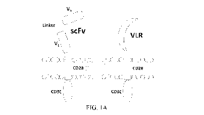
Figure 1A illustrates CAR structures containing the CD5-directed variable
lymphocyte
receptor (VLR) or single chain variable fragment (scFv). CAR structures with
CD28 containing a
scFv (left) or VLR (right) as the antigen recognition domain are shown.
Figure 1B illustrates the bicistronic transgene sequences used for expressing
enhanced
green fluorescent protein (eGFP) and the CD5-CARs using a P2A self-cleaving
sequence. It
includes a 5' long terminal repeat (LTR), human ubiquitin C promoter (hUBC),
eGFP sequence,
P2A sequence, an interleukin-2 signal peptide (IL-2 SP), the CD5-VLR (top) or
CD5-scFv
(bottom), a myc epitope tag, the CD28 region, the CD3zeta intracellular domain
and a 3' LTR.
Figure 2A shows western blot using anti-CD3zeta antibody on whole cell lysates
of NK-
92 cells shows the presence of CD5-VLR-CAR and CD5-scFv-CAR protein in the
sorted and
expanded cells. NK-92 cells were transduced with the eGFP-P2 A-CD5-scFv-CAR
lentiviral
vector and sorted for GFP expressing cells. After two rounds of sorting, an
enriched population of
CAR-expressing NK-92 cells was generated with 99% eGFP expression.
Figure 2B shows data where both CD5-CAR expressing NK-92 cells were mixed with
CD5-positive target cells Jurkat at various Effector: Target ratios and the
percent cytotoxicity was
measured by flow cytometry.
Figure 2C shows data for MOLT-4. CD5-CAR modified NK-92 cells showed a
significantly greater cytotoxicity (p < 0.01) against the CD5-positive Jurkat
and MOLT-4 cells
when compared to unmodified NK-92 cells in a 4 hour assay. This data indicates
NK-92 cell
mediated cytotoxicity against a CD5-positive T-ALL cell line using CD5-CARs.
Figure 2D shows data indicating no increase in cytotoxicity is seen when CD5-
CAR NK-
92 cells are cultured with CD5- negative 697 cells.
Figure 3A illustrates a method where Jurkat T cells were transduced with
lentiviral vectors
encoding either a scFv- or VLR-based CD5-CAR with co-expression of eGFP. The
Jurkat T cell
activation assay shows time points for measurement of T-cell activation and
Western blot analysis.
Figure 3B shows data on activation measured by surface CD69 expression four
days after
transduction increased as the amount of viral vector increased. Greater
activation was observed in
the CD5-VLR-CAR Jurkat group.
9
CA 03067244 2019-12-12
WO 2018/231871
PCT/US2018/037160
Figure 3C shows data on the percentage of activated cells was compared to the
vector copy
number (VCN) obtained for each transduced population of cells. The inset to
the figure defines
each group.
Figure 3D shows data on CD69 expression was measured 4 and 12 d after
transduction,
.. which showed activation decreased over time in both CD5-CAR expressing
Jurkat T cell groups.
Figure 4A shows data on CD5 knockout in Jurkat T cells using CRISPR-Cas9
genome
editing. CD5 expression, measure by flow cytometry, in Jurkat T cells five
days following mock
transfection or transfection with plasmid encoding Cas9 and one of three
different gRNA target
sequences. Histogram plots for CD5 expression in mock transfected and
transfected Jurkat T cells
are shown along a single axis.
Figure 4B shows an overlay image of histogram plots of CD5 expression in Naive
Jurkat
T cells and flow-sorted CD5-negative Jurkat T cells that were transfected with
the CD5-CRISPR
gRNA #2.
Figure 4C shows representative sequencing traces from Naive (top left)
CCTGCTGGGGATGCTGGGTGAGT (SEQ ID NO: 2) and sorted CD5-edited (top right)
CCGGTGGGGGGTGGGGGGGGA (SEQ ID NO: 3) Jurkat T cell genomic DNA PCR amplified
for CD5, sequence the gene from genomic DNA.
Figure 4D shows a TIDE analysis of the frequency of indels within the CD5 gene
after the
predicted break-site generated by Cas9. Results show 77% CD5-negative cells
were edited with
27% having a -1 deletion.
Figure 5A shows percentage of eGFP positive cells. CD5-edited CD5-CAR-modified
Jurkat T cells have reduced self-activation and increased CD5-CAR expression.
Naive (white) and
CD5-edited Jurkat T cells (black) were transduced with eGFP-P2 A-CD5-VLR-CAR,
eGFP-P2
A-CD5-scFv-CAR or control eGFP-P2 A-BCL-VLR-CAR lentiviral vectors at MOIs 1,
10 and
20. Polybrene was not used during transduction, which provided a greater
separation in
transduction efficiency between MOIs of 1 and 10. Transduction efficiency,
measured by eGFP-
positive cells, of each CAR vector at MOIs 1, 10 and 20 in both populations of
Jurkat T cells.
Figure 5B shows data on CD5 expression in both populations of Jurkat T cells
transduced
with each CAR vector at each MOI.
Figure 5C shows data on the activation was measured by monitoring CD69
expression and
transduction efficiency measured by eGFP expression. A correlation exists
between activation and
CA 03067244 2019-12-12
WO 2018/231871
PCT/US2018/037160
eGFP expression in CD5-CAR-transduced Jurkat T cells. Non-edited CD5-CAR-
modified cells
have increased T-cell activation compared to CD5-edited CD5-CAR-modified
cells.
Figure 5D shows western blots on whole cell lysates showing CD3zeta expression
in non-
edited Jurkat T cells (left) and CD5-edited Jurkat T cells (right) when
transduced with the VLR-
CAR vector. Endogenous CD3zeta is represented by the 18 kDa bands and CD3zeta
in the CAR
construct is represented by the 48, kDa band in the CD5-VLR-CAR construct.
eGFP, CD5 and
CD69 surface expression were measured by flow cytometry.
Figure 6A shows data indicating CD5-edited CD5-CAR-modified effector cells in
culture
with naive target T cells stimulates effector cell activation and target cell
down-regulation of CD5.
Naive and CD5-edited Jurkat T cells were transduced with eGFP-P2A-CD5-scFv-CAR
or eGFP-
P2A-CD5-VLR-CAR lentiviral vectors at MOI 5. Polybrene was not used during
transduction.
Target naive Jurkat T cells were labeled with VPD450. On day five post-
transduction, effector
cells were cultured with labeled target cells at E:T ratios 2:1, 1:1 and 1:5.
The cells were analyzed
by flow cytometry 24 hours later. White bars signify non-edited effector
cells; black bars signify
CD5-edited effector cells. Experiments were performed with three replicates
and error bars
represent standard deviation from the mean. Percent of baseline CD5 expression
in target Jurkat
T cells cultured with non-edited and CD5-edited effector Jurkat T cells
expressing the CD5-scFv-
CAR. CD5 expression in target cells cultured alone (gray bar) was used as
baseline and set at
100%.
Figure 6B shows data for the CD5-VLR-CAR.
Figure 6C shows data on T-cell activation of non-edited and CD5-edited
effector Jurkat T
cells expressing the CD5-scFv-CAR when cultured alone and in culture with
target Jurkat T cells.
Figure 6D shows data on CD5-VLR-CAR.
Figure 7A shows data on non-edited Jurkat T cells with CD5-scFv-CAR MOI 5.
Figure 7B shows data on CD5-edited Jurkat T cells with CD5-scFv-CAR MOI 5.
Figure 7C shows data indicating antigen editing results in an increase in CAR
expression.
Figure 8A shows Western blots of non-edited Jurkat T cells whole cell lysates.
Figure 8B shows Western blots of CD5-edited Jurkat T cells whole cell lysates.
Figure 8C show data on Western blot quantification indicating increased CAR
expression
in CD5-edited T cells.
11
CA 03067244 2019-12-12
WO 2018/231871
PCT/US2018/037160
DETAILED DISCUSSION
Before the present disclosure is described in greater detail, it is to be
understood that this
disclosure is not limited to particular embodiments described, and as such
may, of course, vary. It
is also to be understood that the terminology used herein is for the purpose
of describing particular
embodiments only, and is not intended to be limiting, since the scope of the
present disclosure will
be limited only by the appended claims.
Unless defined otherwise, all technical and scientific terms used herein have
the same
meaning as commonly understood by one of ordinary skill in the art to which
this disclosure
belongs. Although any methods and materials similar or equivalent to those
described herein can
also be used in the practice or testing of the present disclosure, the
preferred methods and materials
are now described.
All publications and patents cited in this specification are herein
incorporated by reference
as if each individual publication or patent were specifically and individually
indicated to be
incorporated by reference and are incorporated herein by reference to disclose
and describe the
methods and/or materials in connection with which the publications are cited.
The citation of any
publication is for its disclosure prior to the filing date and should not be
construed as an admission
that the present disclosure is not entitled to antedate such publication by
virtue of prior disclosure.
Further, the dates of publication provided could be different from the actual
publication dates that
may need to be independently confirmed.
As will be apparent to those of skill in the art upon reading this disclosure,
each of the
individual embodiments described and illustrated herein has discrete
components and features
which may be readily separated from or combined with the features of any of
the other several
embodiments without departing from the scope or spirit of the present
disclosure. Any recited
method can be carried out in the order of events recited or in any other order
that is logically
possible.
Embodiments of the present disclosure will employ, unless otherwise indicated,
techniques
of medicine, organic chemistry, biochemistry, molecular biology, pharmacology,
and the like,
which are within the skill of the art. Such techniques are explained fully in
the literature.
Prior to describing the various embodiments, the following definitions are
provided and
should be used unless otherwise indicated. Further, headings provided herein
are for convenience
only and do not interpret the scope or meaning of the claims.
12
CA 03067244 2019-12-12
WO 2018/231871
PCT/US2018/037160
It must be noted that, as used in the specification and the appended claims,
the singular
forms "a," "an," and "the" include plural referents unless the context clearly
dictates otherwise. In
this specification and in the claims that follow, reference will be made to a
number of terms that
shall be defined to have the following meanings unless a contrary intention is
apparent.
As used herein, the terms "treat" and "treating" are not limited to the case
where the subject
(e.g., patient) is cured and the disease is eradicated. Rather, embodiments,
of the present disclosure
also contemplate treatment that merely reduces symptoms, and/or delays disease
progression.
Unless the context requires otherwise, throughout the specification and claims
which
follow, the word "comprise" and variations thereof, such as, "comprises,"
"comprising"
"including," "containing," or "characterized by," are to be construed in an
open, inclusive sense,
that is, as "including, but not limited to" and does not exclude additional,
unrecited elements or
method steps. By contrast, the transitional phrase "consisting of' excludes
any element, step, or
ingredient not specified in the claim. The transitional phrase "consisting
essentially of' limits the
scope of a claim to the specified materials or steps "and those that do not
materially affect the basic
and novel characteristic(s)" of the claimed invention. In embodiments or
claims where the term
comprising is used as the transition phrase, such embodiments can also be
envisioned with
replacement of the term "comprising" with the terms "consisting of' or
"consisting essentially of."
The term "comprising" in reference to a peptide having an amino acid sequence
refers a
peptide that may contain additional N-terminal (amine end) or C-terminal
(carboxylic acid end)
amino acids, i.e., the term is intended to include the amino acid sequence
within a larger peptide.
The term "consisting of' in reference to a peptide having an amino acid
sequence refers a peptide
having the exact number of amino acids in the sequence and not more or having
not more than a
range of amino acids expressly specified in the claim. In certain embodiments,
the disclosure
contemplates that the "N-terminus of a peptide may consist of an amino acid
sequence," which
refers to the N-terminus of the peptide having the exact number of amino acids
in the sequence
and not more or having not more than a range of amino acids specified in the
claim however the
C-terminus may be connected to additional amino acids, e.g., as part of a
larger peptide. Similarly,
the disclosure contemplates that the "C-terminus of a peptide may consist of
an amino acid
sequence," which refers to the C-terminus of the peptide having the exact
number of amino acids
in the sequence and not more or having not more than a range of amino acids
specified in the claim
13
CA 03067244 2019-12-12
WO 2018/231871
PCT/US2018/037160
however the N-terminus may be connected to additional amino acids, e.g., as
part of a larger
peptide.
In certain embodiments, sequence "identity" refers to the number of exactly
matching
amino acids (expressed as a percentage) in a sequence alignment between two
sequences of the
alignment calculated using the number of identical positions divided by the
greater of the shortest
sequence or the number of equivalent positions excluding overhangs wherein
internal gaps are
counted as an equivalent position. For example, the polypeptides GGGGGG and
GGGGT have a
sequence identity of 4 out of 5 or 80%. For example, the polypeptides GGGPPP
and GGGAPPP
have a sequence identity of 6 out of 7 or 85%. In certain embodiments, any
recitation of sequence
identity expressed herein may be substituted for sequence similarity. Percent
"similarity" is used
to quantify the similarity between two sequences of the alignment. This method
is identical to
determining the identity except that certain amino acids do not have to be
identical to have a match.
Amino acids are classified as matches if they are among a group with similar
properties according
to the following amino acid groups: Aromatic - F Y W; hydrophobic-A V I L;
Charged positive:
R K H; Charged negative - D E; Polar - S T N Q. The amino acid groups are also
considered
conserved substitutions.
Chimeric Antigen Receptor Polypeptides
In certain embodiments, the disclosure provides a chimeric antigen receptor
(CAR)
polypeptide having a signal peptide, a T cell antigen recognition domain,
e.g., CD5, CD7, and/or
CD3 antigen recognition domain, a hinge region, a transmembrane domain, at
least one co-
stimulatory domain, and a signaling domain.
As used herein, the terms "peptide," "polypeptide," and "protein" are used
interchangeably,
and refer to a compound having amino acid residues covalently linked by
peptide bonds. A protein
or peptide must contain at least two amino acids, and no limitation is placed
on the maximum
number of amino acids. Polypeptides include any peptide or protein having two
or more amino
acids joined to each other by peptide bonds. As used herein, the term refers
to both short chains,
which also commonly are referred to in the art as peptides, oligopeptides, and
oligomers, for
example, and to longer chains, which generally are referred to in the art as
proteins, of which there
are many types.
14
CA 03067244 2019-12-12
WO 2018/231871
PCT/US2018/037160
"Polypeptides" include, for example, biologically active fragments,
substantially
homologous polypeptides, oligopeptides, homodimers, heterodimers, variants of
polypeptides,
modified polypeptides, derivatives, analogs, fusion proteins, among others.
The polypeptides
include natural peptides, recombinant peptides, synthetic peptides, or a
combination thereof
A "signal peptide" includes a peptide sequence that directs the transport and
localization
of the peptide and any attached polypeptide within a cell, e.g. to a certain
cell organelle (such as
the endoplasmic reticulum) and/or the cell surface. The signal peptide is a
peptide of any secreted
or transmembrane protein that directs the transport of the polypeptide of the
disclosure to the cell
membrane and cell surface, and provides correct localization of the
polypeptide of the present
disclosure. In particular, the signal peptide of the present disclosure
directs the polypeptide of the
present disclosure to the cellular membrane, wherein the extracellular portion
of the polypeptide
is displayed on the cell surface, the transmembrane portion spans the plasma
membrane, and the
active domain is in the cytoplasmic portion, or interior of the cell. In one
embodiment, the signal
peptide is cleaved after passage through the endoplasmic reticulum (ER), i.e.
is a cleavable signal
peptide. In an embodiment, the signal peptide is human protein of type I, II,
III, or IV. In an
embodiment, the signal peptide includes an immunoglobulin heavy chain signal
peptide.
The "antigen recognition domain" includes a polypeptide that is selective for
an antigen,
receptor, peptide ligand, or protein ligand of the target; or a polypeptide of
the target. In one
embodiment, the antigen recognition domain includes the binding portion or
variable region of a
monoclonal or polyclonal antibody directed against (selective for) the target.
In one embodiment,
the antigen recognition domain includes fragment antigen-binding fragment
(Fab). In another
embodiment, the antigen recognition domain includes a single-chain variable
fragment (scFV).
scFV is a fusion protein of the variable regions of the heavy (VH) and light
chains (VL) of
immunoglobulins, connected with a short linker peptide. In another embodiment,
the antigen
recognition domain includes ligands that engage their cognate receptor. In
another embodiment,
the antigen recognition domain is humanized. It is understood that the antigen
recognition domain
may include some variability within its sequence and still be selective for
the targets disclosed
herein. Therefore, it is contemplated that the polypeptide of the antigen
recognition domain may
be at least 95%, at least 90%, at least 80%, or at least 70% identical to the
antigen recognition
domain polypeptide disclosed herein and still be selective for the targets
described herein and be
within the scope of the disclosure.
CA 03067244 2019-12-12
WO 2018/231871
PCT/US2018/037160
The hinge region is a sequence positioned between for example, including, but
not limited
to, the chimeric antigen receptor, and at least one co- stimulatory domain and
a signaling domain.
The hinge sequence may be obtained including, for example, from any suitable
sequence from any
genus, including human or a part thereof Such hinge regions are known in the
art. In one
embodiment, the hinge region includes the hinge region of a human protein
including CD-8 alpha,
CD28, 4- IBB, 0X40, CD3-zeta, T cell receptor a or 0 chain, a CD3 zeta chain,
CD28, CD3s,
CD45, CD4, CD5, CD8, CD9, CD16, CD22, CD33, CD37, CD64, CD80, CD86, CD134,
CD137,
ICOS, CD154, functional derivatives thereof, and combinations thereof. In one
embodiment, the
hinge region includes the CD8 a hinge region. In some embodiments, the hinge
region includes
one selected from, but is not limited to, immunoglobulin (e.g. IgGl, IgG2,
IgG3, IgG4, and IgD).
The transmembrane domain includes a hydrophobic polypeptide that spans the
cellular
membrane. In particular, the transmembrane domain spans from one side of a
cell membrane
(extracellular) through to the other side of the cell membrane (intracellular
or cytoplasmic). The
transmembrane domain may be in the form of an alpha helix or a beta barrel, or
combinations
thereof. The transmembrane domain may include a polytopic protein, which has
many
transmembrane segments, each alpha-helical, beta sheets, or combinations
thereof. In one
embodiment, the transmembrane domain that naturally is associated with one of
the domains in
the CAR is used. In another embodiment, the transmembrane domain can be
selected or modified
by amino acid substitution to avoid binding of such domains to the
transmembrane domains of the
same or different surface membrane proteins to minimize interactions with
other members of the
receptor complex. For example, a transmembrane domain includes a transmembrane
domain of a
T-cell receptor a or 0 chain, a CD3 zeta chain, CD28, CD3s, CD45, CD4, CD5,
CD8, CD9, CD16,
CD22, CD33, CD37, CD64, CD80, CD86, CD134, CD137, ICOS, CD154, functional
derivatives
thereof, and combinations thereof. The artificially designed transmembrane
domain is a
.. polypeptide mainly comprising hydrophobic residues such as leucine and
valine. In one
embodiment, a triplet of phenylalanine, tryptophan and valine is found at each
end of the synthetic
transmembrane domain. In one embodiment, the transmembrane domain is the CD8
transmembrane domain. In another embodiment, the transmembrane domain is the
CD28
transmembrane domain. Such transmembrane domains are known in the art.
The signaling domain and co- stimulatory domain include polypeptides that
provide
activation of an immune cell to stimulate or activate at least some aspect of
the immune cell-
16
CA 03067244 2019-12-12
WO 2018/231871
PCT/US2018/037160
signaling pathway. In an embodiment, the signaling domain includes the
polypeptide of a
functional signaling domain of CD3 zeta, common FcR gamma (FCER1G), Fc gamma
RIIIA, FcR
beta (Fc Epsilon Rib), CD3 gamma, CD3 delta, CD3 epsilon, CD79a, CD79b, DNAX-
activating
protein 10 (DAP10), DNAX-activating protein 12 (DAP12), active fragments
thereof, functional
derivatives thereof, and combinations thereof Such signaling domains are known
in the art. In an
embodiment, the CAR polypeptide further includes one or more co-stimulatory
domains. In an
embodiment, the co-stimulatory domain is a functional signaling domain from a
protein including
0X40, CD27, CD28, CD30, CD40, PD-1, CD2, CD7, CD258, Natural killer Group 2
member C
(NKG2C), Natural killer Group 2 member D (NKG2D), B7-H3, a ligand that binds
to CD83,
ICAM-1, LFA-1 (CD1 la/CD 18), ICOS and 4-1BB (CD137), active fragments
thereof, functional
derivatives thereof, and combinations thereof
Polynucleotide encoding chimeric antigen receptor
The present disclosure further provides polynucleotides encoding the chimeric
antigen
receptor polypeptides described herein. The polynucleotide encoding the CAR is
prepared from
an amino acid sequence of the specified CAR by any conventional method. A base
sequence
encoding an amino acid sequence can be obtained from the aforementioned NCBI
RefSeq IDs or
accession numbers of GenBenk for an amino acid sequence of each domain, and
the nucleic acid
of the present disclosure can be prepared using a standard molecular
biological and/or chemical
procedure. For example, based on the base sequence, a polynucleotide can be
synthesized, and the
polynucleotide of the present disclosure can be prepared by combining DNA
fragments which are
obtained from a cDNA library using a polymerase chain reaction (PCR). In one
embodiment, the
polynucleotide disclosed herein is part of a gene, or an expression or cloning
cassette.
The term "polynucleotide" as used herein is defined as a chain of nucleotides.
Polynucleotide includes DNA and RNA. Furthermore, nucleic acids are polymers
of nucleotides.
Thus, nucleic acids and polynucleotides as used herein are interchangeable.
One skilled in the art
has the general knowledge that nucleic acids are polynucleotides, which can be
hydrolyzed into
the monomeric "nucleotides." The monomeric nucleotides can be hydrolyzed into
nucleosides. As
used herein polynucleotides include, but are not limited to, all nucleic acid
sequences which are
obtained by any means available in the art, including, without limitation,
recombinant means, i.e.,
17
CA 03067244 2019-12-12
WO 2018/231871
PCT/US2018/037160
the cloning of nucleic acid sequences from a recombinant library or a cell
genome, using ordinary
cloning technology and polymerase chain reaction (PCR), and the like, and by
synthetic means.
Polynucleotide vector
The polynucleotide described above can be cloned into a vector. A "vector" is
a
composition of matter which includes an isolated polynucleotide and which can
be used to deliver
the isolated polynucleotide to the interior of a cell. Numerous vectors are
known in the art
including, but not limited to, linear polynucleotides, polynucleotides
associated with ionic or
amphiphilic compounds, plasmids, phagemid, cosmid, and viruses. Viruses
include phages, phage
derivatives. Thus, the term "vector" includes an autonomously replicating
plasmid or a virus. The
term should also be construed to include non-plasmid and non-viral compounds
which facilitate
transfer of nucleic acid into cells, such as, for example, polylysine
compounds, liposomes, and the
like. Examples of viral vectors include, but are not limited to, adenoviral
vectors, adeno-associated
virus vectors, retroviral vectors, lentiviral vectors, and the like.
In one embodiment, vectors include cloning vectors, expression vectors,
replication
vectors, probe generation vectors, integration vectors, and sequencing
vectors. In an embodiment,
the vector is a viral vector. In an embodiment, the viral vector is a
retroviral vector or a lentiviral
vector. In an embodiment, the engineered cell is virally transduced to express
the polynucleotide
sequence.
A number of viral based systems have been developed for gene transfer into
mammalian
cells. For example, retroviruses provide a convenient platform for gene
delivery systems. A
selected gene can be inserted into a vector and packaged in retroviral
particles using techniques
known in the art. The recombinant virus can then be isolated and delivered to
cells of the subject
either in vivo or ex vivo. A number of retroviral systems are known in the
art. In some
embodiments, adenovirus vectors are used. A number of adenovirus vectors are
known in the art.
In one embodiment, lentivirus vectors are used. Viral vector technology is
well known in the art
and is described, for example, in Sambrook et al, (2001, Molecular Cloning: A
Laboratory Manual,
Cold Spring Harbor Laboratory, New York), and in other virology and molecular
biology manuals.
Viruses, which are useful as vectors include, but are not limited to,
retroviruses, adenoviruses,
adeno- associated viruses, herpes viruses, and lentiviruses. In general, a
suitable vector contains
an origin of replication functional in at least one organism, a promoter
sequence, convenient
18
CA 03067244 2019-12-12
WO 2018/231871
PCT/US2018/037160
restriction endonuclease sites, and one or more selectable markers, (e.g., WO
01/96584; WO
01/29058; and U.S, Pat. No. 6,326,193).
Expression of chimeric antigen receptor polynucleotide may be achieved using,
for
example, expression vectors including, but not limited to, at least one of a
SFFV or human
elongation factor 11 a (EF) promoter, CAG (chicken beta-actin promoter with
CMV enhancer)
promoter human elongation factor la (EF) promoter. Examples of less-strong/
lower-expressing
promoters utilized may include, but is not limited to, the simian virus 40
(SV40) early promoter,
cytomegalovirus (CMV) immediate-early promoter, Ubiquitin C (UBC) promoter,
and the
phosphoglycerate kinase 1 (PGK) promoter, or a part thereof Inducible
expression of chimeric
antigen receptor may be achieved using, for example, a tetracycline responsive
promoter,
including, but not limited to, TRE3GV (Tet-response element, including all
generations and
preferably, the 3rd generation), inducible promoter (Clontech Laboratories,
Mountain View, CA)
or a part or a combination thereof.
One example of a suitable promoter is the immediate early cytomegalovirus
(CMV)
promoter sequence. This promoter sequence is a strong constitutive promoter
sequence capable of
driving high levels of expression of any polynucleotide sequence operatively
linked thereto.
Another example of a suitable promoter is Elongation Growth Factor - 1 a (EF-
1 a). However,
other constitutive promoter sequences may also be used, including, but not
limited to the simian
virus 40 (SV40) early promoter, mouse mammary tumor virus (MMTV), human
immunodeficiency virus (HIV) long terminal repeat (LTR) promoter, MoMuLV
promoter, an
avian leukemia virus promoter, an Epstein-Barr virus immediate early promoter,
a Rous sarcoma
virus promoter, as well as human gene promoters such as, but not limited to,
the actin promoter,
the myosin promoter, the hemoglobin promoter, and the creatine kinase
promoter. Further, the
disclosure should not be limited to the use of constitutive promoters -
inducible promoters are also
contemplated as part of the disclosure. The use of an inducible promoter
provides a molecular
switch capable of turning on expression of the polynucleotide sequence which
it is operatively
linked when such expression is desired, or turning off the expression when
expression is not
desired. Examples of inducible promoters include, but are not limited to a
metallothionein
promoter, a glucocorticoid promoter, a progesterone promoter, and a
tetracycline promoter.
"Expression vector" refers to a vector comprising a recombinant polynucleotide
comprising expression control sequences operatively linked to a nucleotide
sequence to be
19
CA 03067244 2019-12-12
WO 2018/231871
PCT/US2018/037160
expressed. An expression vector includes sufficient cis- acting elements for
expression; other
elements for expression can be supplied by the host cell or in an in vitro
expression system.
Expression vectors include all those known in the art, such as cosmids,
plasmids (e.g., naked or
contained in liposomes) and viruses (e.g., lentiviruses, retroviruses,
adenoviruses, and adeno-
associated viruses) that incorporate the recombinant polynucleotide,
Additional promoter elements, e.g., enhancers, regulate the frequency of
transcriptional
initiation. Typically, these are located in the region 30-100 bp upstream of
the start site, although
a number of promoters have recently been shown to contain functional elements
downstream of
the start site as well. The spacing between promoter elements frequently is
flexible, so that
promoter function is preserved when elements are inverted or moved relative to
one another, in
the thymidine kinase (tk) promoter, the spacing between promoter elements can
be increased to 50
bp apart before activity begins to decline. Depending on the promoter, it
appears that individual
elements can function either cooperatively or independently to activate
transcription,
In order to assess the expression of a CAR polypeptide or portions thereof,
the expression
vector to be introduced into a cell can also contain either a selectable
marker gene or a reporter
gene or both to facilitate identification and selection of expressing cells
from the population of
cells sought to be transfected or infected through viral vectors, in other
aspects, the selectable
marker may be carried on a separate piece of DNA and used in a co-
transfection procedure. Both
selectable markers and reporter genes may be flanked with appropriate
regulatory sequences to
enable expression in the host cells. Useful selectable markers include, for
example, antibiotic -
resistance genes, such as neo and the like.
Reporter genes are used for identifying potentially transfected cells and for
evaluating the
functionality of regulatory sequences. In general, a reporter gene is a gene
that is not present in or
expressed by the recipient organism or tissue and that encodes a polypeptide
whose expression is
manifested by some easily detectable property, e.g., enzymatic activity.
Expression of the reporter
gene is assayed at a suitable time after the DNA has been introduced into the
recipient cells.
Suitable reporter genes may include genes encoding luciferase, beta-
galactosidase,
chloramphenicol acetyl transferase, secreted alkaline phosphatase, or the
green fluorescent protein
gene (e.g., Ui-Tei et al., 2000 FEBS Letters 479: 79-82). Suitable expression
systems are well
known and may be prepared using known techniques or obtained commercially. In
general, the
construct with the minimal 5' flanking region showing the highest level of
expression of reporter
CA 03067244 2019-12-12
WO 2018/231871
PCT/US2018/037160
gene is identified as the promoter. Such promoter regions may be linked to a
reporter gene and
used to evaluate agents for the ability to modulate promoter- driven
transcription.
Methods of introducing and expressing genes into a cell are known in the art.
In the context
of an expression vector, the vector can be readily introduced into a host
cell, e.g., mammalian,
bacterial, yeast, or insect cell by any method in the art. For example, the
expression vector can be
transferred into a host cell by physical, chemical, or biological means.
Physical methods for introducing a polynucleotide into a host cell include
calcium
phosphate precipitation, lipofection, particle bombardment, microinjection,
electroporation, and
the like. Methods for producing cells comprising vectors and/or exogenous
nucleic acids are well-
known in the art. See, for example, Sambrook et al. (2001, Molecular Cloning:
A Laboratory
Manual, Cold Spring Harbor Laboratory, New York). A preferred method for the
introduction of
a polynucleotide into a host cell is calcium phosphate transfection.
Biological methods for introducing a polynucleotide of interest into a host
cell include the
use of DNA and RNA vectors. Viral vectors, and especially retroviral vectors,
have become the
most widely used method for inserting genes into mammalian, e.g., human cells.
Other viral
vectors can be derived from lentivirus, poxviruses, herpes simplex virus I,
adenoviruses and adeno-
associated viruses, and the like. See, for example, U.S. Pat, Nos. 5,350,674
and 5,585,362.
Chemical means for introducing a polynucleotide into a host cell include
colloidal
dispersion systems, such as macromolecule complexes, nanocapsules,
microspheres, beads, and
lipid-based systems including oil-in-water emulsions, micelles, mixed
micelles, and liposomes.
An exemplary colloidal system for use as a delivery vehicle in vitro and in
vivo is a liposome (e.g.,
an artificial membrane vesicle). In the case where a non-viral delivery system
is utilized, an
exemplary delivery vehicle is a liposome. The use of lipid formulations is
contemplated for the
introduction of the nucleic acids into a host cell (in vitro, ex vivo or in
vivo). In another aspect, the
nucleic acid may be associated with a lipid. The nucleic acid associated with
a lipid may be
encapsulated in the aqueous interior of a liposome, interspersed within the
lipid bilayer of a
liposome, attached to a liposome via a linking molecule that is associated
with both the liposome
and the oligonucleotide, entrapped in a liposome, complexed with a liposome,
dispersed in a
solution containing a lipid, mixed with a lipid, combined with a lipid,
contained as a suspension in
a lipid, contained or complexed with a micelle, or otherwise associated with a
lipid. Lipid,
lipid/DNA or lipid/expression vector associated compositions are not limited
to any particular
21
CA 03067244 2019-12-12
WO 2018/231871
PCT/US2018/037160
structure in solution. For example, they may be present in a bilayer
structure, as micelles, or with
a "collapsed" structure. They may also simply be interspersed in a solution,
possibly forming
aggregates that are not uniform in size or shape. Lipids are fatty substances
which may be naturally
occurring or synthetic lipids. For example, lipids include the fatty droplets
that naturally occur in
the cytoplasm as well as the class of compounds which contain long-chain
aliphatic hydrocarbons
and their derivatives, such as fatty acids, alcohols, amines, amino alcohols,
and aldehydes.
Lipids suitable for use can be obtained from commercial sources. For example,
dimyristyi
phosphatidylcholine ("DMPC") can be obtained from Sigma, St. Louis, MO;
dicetyl phosphate
("DCP") can be obtained from K & K Laboratories (Plainview, NY); cholesterol
("Choi") can be
obtained from Calbiochem-Behring; dimyristyi phosphatidylglycerol ("DMPG") and
other lipids
may be obtained from Avanti Polar Lipids, Inc. (Birmingham, AL). Stock
solutions of lipids in
chloroform or chloroform/methanol can be stored at about -20 C. Chloroform is
used as the only
solvent since it is more readily evaporated than methanol.
"Liposome" is a generic term encompassing a variety of single and
multilamellar lipid
vehicles formed by the generation of enclosed lipid bilayers or aggregates.
Liposomes can be
characterized as having vesicular structures with a phospholipid bilayer
membrane and an inner
aqueous medium. Multilamellar liposomes have multiple lipid layers separated
by aqueous
medium. They form spontaneously when phospholipids are suspended in an excess
of aqueous
solution. The lipid components undergo self-rearrangement before the formation
of closed
structures and entrap water and dissolved solutes between the lipid bilayers
(Ghosh et al.,
Glycobiology 5, 505- 10). However, compositions that have different structures
in solution than
the normal vesicular structure are also encompassed. For example, the lipids
may assume a
micellar structure or merely exist as nonuniform aggregates of lipid
molecules. Also contemplated
are lipofectamine- nucleic acid complexes.
Regardless of the method used to introduce exogenous polynucleotides into a
host cell or
otherwise expose a cell to the polynucleotide of the present disclosure, in
order to confirm the
presence of the recombinant DNA sequence in the host cell, a variety of assays
may be performed.
Such assays include, for example, "molecular biological" assays well known to
those of skill in
the art, such as Southern and Northern blotting, RT-PCR and PCR; "biochemical"
assays, such as
detecting the presence or absence of a particular peptide, e.g., by
immunological means (ELISAs
22
CA 03067244 2019-12-12
WO 2018/231871
PCT/US2018/037160
and Western blots) or by assays described herein to identify agents falling
within the scope of the
disclosure.
Engineered Cell
In another embodiment, the disclosure provides an engineered cell expressing
the chimeric
antigen receptor polypeptide described above or polynucleotide encoding for
the same, and
described above. An "engineered cell" means any cell of any organism that is
modified,
transformed, or manipulated by addition or modification of a gene, a DNA or
RNA sequence, or
protein or polypeptide. Isolated cells, host cells, and genetically engineered
cells of the present
disclosure include isolated immune cells, such as NK cells and T cells that
contain the DNA or
RNA sequences encoding a chimeric antigen receptor or chimeric antigen
receptor complex and
express the chimeric receptor on the cell surface. Isolated host cells and
engineered cells may be
used, for example, for enhancing an NK cell activity or a T lymphocyte
activity, treatment of
cancer, and treatment of infectious diseases.
Any cell capable of expressing and/or capable of integrating the chimeric
antigen receptor
polypeptide, as disclosed herein, into its membrane may be used. In an
embodiment, the
engineered cell includes immunoregulatory cells. Immunoregulatory cells
include T-cells, such as
CD4 T-cells (Helper T-cells), CD8 T-cells (Cytotoxic T-cells, CTLs), and
memory T cells or
memory stem cell T cells. In another embodiment, T-cells include Natural
Killer T-cells (NK T-
cells). T cells comprise of CD4 and CD8 cells. CD4 is a glycoprotein present
on the surface of
immune cells such as T helper cells, important in T cell activation and
receptor for HIV. Some
monocytes or macrophages also express CD4. CD4 is also called OKT4. Cytotoxic
T cells are also
known as CD8+ T cells or CD8 T cells expressing CD8 glycoprotein at their
surfaces. These CD8+
T cells are activated once they are exposed to peptide antigens presented by
MHC class I. In an
embodiment, the engineered cell includes Natural Killer cells. Natural killer
cells are well known
in the art. In one embodiment, natural killer cells include cell lines, such
as NK-92 cells. Further
examples of NK cell lines include NKG, YT, NK-YS, HANK-1, YTS cells, and NKL
cells. NK
cells mediate anti-tumor effects without the risk of GvHD and are short-lived
relative to T-cells.
Accordingly, NK cells would be exhausted shortly after destroying cancer
cells, decreasing the
need for an inducible suicide gene on CAR constructs that would ablate the
modified cells.
23
CA 03067244 2019-12-12
WO 2018/231871
PCT/US2018/037160
In one embodiment, engineered cells, in particular allogeneic T cells obtained
from donors
can be modified to inactivate components of TCR (T cell receptor) involved in
MHC recognition.
As a result, TCR deficient T cells would not cause graft versus host disease
(GVHD).
T-antigen deficient T cells
T cell lymphomas or T cell leukemias express specific antigens, which may
represent
useful targets for these diseases. For instance, T cell lymphomas or leukemias
express CD5.
However, CD5 are also expressed in CAR T, but not NK cells, which offset their
ability of
targeting these antigens. The self-killing might occur in T cells armed with
CARs targeting any
one of these antigens. This makes generation of CARs targeting these antigens
difficult. Therefore,
it may be necessary to inactivate an endogenous antigen in a T cell when it is
used as a target to
arm CARs.
In another embodiment, the engineered cell is further modified to inactivate
cell surface
polypeptide to prevent engineered cells from acting on other engineered cells.
For example, the
endogenous CD5, CD7 and/or CD3 gene or gene expression of the engineered cells
may be
knocked out or inactivated. In another preferred embodiment, the engineered
cell is a T-cell having
the endogenous CD5, CD7 and/or CD3 gene knocked out or inactivated. In one
embodiment, the
engineered cell expressing a CAR having a CD5, CD7 and/or CD3 antigen
recognition domain
will have the gene expressing that antigen inactivated or knocked out. For
example, a T-cell having
a CD5, CD7 and/or CD3 CAR will have an inactivated or knocked out CD5, CD7
and/or CD3
antigen gene. Methods to knock out or inactivate genes are known. For example,
CRISPR/Cas9
system, zinc finger nuclease (ZFNs) and TALE nucleases (TALENs) and
meganucleases may be
used to knock out or inactivate the CD5, CD7 and/or CD3 gene or gene
expression of the
engineered cells.
Sources of Cells
The engineered cells may be obtained from peripheral blood, cord blood, bone
marrow,
tumor infiltrating lymphocytes, lymph node tissue, or thymus tissue. The host
cells may include
placental cells, embryonic stem cells, induced pluripotent stem cells, or
hematopoietic stem cells.
The cells may be obtained from humans, monkeys, chimpanzees, dogs, cats, mice,
rats, and
transgenic species thereof. The cells may be obtained from established cell
lines. The above cells
24
CA 03067244 2019-12-12
WO 2018/231871
PCT/US2018/037160
may be obtained by any known means. The cells may be autologous, syngeneic,
allogeneic, or
xenogeneic to the recipient of the engineered cells.
The term "autologous" refer to any material derived from the same individual
to whom it
is later to be re-introduced into the individual.
The term "allogeneic" refers to any material derived from a different animal
of the same
species as the individual to whom the material is introduced. Two or more
individuals are said to
be allogeneic to one another when the genes at one or more loci are not
identical. In some aspects,
allogeneic material from individuals of the same species may be sufficiently
unlike genetically to
interact antigenic ally.
The term "xenogeneic" refers to a graft derived from an animal of a different
species.
The term "syngeneic" refers to an extremely close genetic similarity or
identity especially
with respect to antigens or immunological reactions. Syngeneic systems include
for example,
models in which organs and cells (e.g. cancer cells and their non-cancerous
counterparts) come
from the same individual, and/or models in which the organs and cells come
from different
individual animals that are of the same inbred strain.
Suicide system
The engineered cells of the present disclosure may also include a suicide
system. Suicide
systems provide a mechanism whereby the engineered cell, as described above,
may be deactivated
or destroyed. Such a feature allows precise therapeutic control of any
treatments wherein the
engineered cells are used. As used herein, a suicide system provides a
mechanism by which the
cell having the suicide system can be deactivated or destroyed. Suicide
systems are well known in
the art.
In one embodiment, a suicide system includes a gene that can be
pharmacologically
activated to eliminate the containing cells as required. In specific aspects,
the suicide gene is not
immunogenic to the host harboring the polynucleotide or cell. In one example,
the suicide system
includes a gene that causes CD20 to be expressed on the cell surface of the
engineered cell.
Accordingly, administration of rituximab may be used to destroy the engineered
cell containing
the gene.
In some embodiments, the suicide system includes an epitope tag. Examples of
epitope
tags include a c-myc tag, streptavidin-binding peptide (SBP), and truncated
EGFR gene (EGFRt).
CA 03067244 2019-12-12
WO 2018/231871
PCT/US2018/037160
In this embodiment, the epitope tag is expressed in the engineered cell.
Accordingly,
administration of an antibody against the epitope tag may be used to destroy
the engineered cell
containing the gene.
In another embodiment, the suicide system includes a gene that causes
truncated epidermal
growth factor receptor to be expressed on the surface of the engineered cell.
Accordingly,
administration of cetuximab may be used to destroy the engineered cell
containing the gene. In
another embodiment, the suicide gene may include caspace 8 gene, caspase 9
gene, thymidine
kinase, cytosine deaminase (CD), or cytochrome P450. Examples of further
suicide systems
include those described by Jones et al. (Jones BS, Lamb LS, Goldman F and Di
Stasi A (2014)
.. Improving the safety of cell therapy products by suicide gene transfer.
Front. Pharmacol. 5:254),
which is herein incorporated by reference in its entirety.
Engineered CRISPR systems
Engineered CRISPR system can be used to induce genetic modifications, such as
highly
.. specific gene knockouts. CRISPR-Cas systems are native to bacteria and
provide adaptive
immunity against viruses and plasmids. Type-II CRISPR systems have a desirable
characteristic
in utilizing a single CRISPR associated (Cas) nuclease (specifically Cas9) in
a complex with the
appropriate guide RNAs (gRNAs). In bacteria, Cas9 guide RNAs comprise two
separate RNA
species: crRNA and tracrRNA. A target-specific CRISPR-activating RNA (crRNA)
directs the
.. Cas9/gRNA complex to bind and target a specific DNA sequence. The crRNA has
two functional
domains, a 5'-domain that is target specific and a 3'-domain that directs
binding of the crRNA to
the transactivating crRNA (tracrRNA). The tracrRNA is a longer, universal RNA
that binds the
crRNA and mediates binding of the gRNA complex to Cas9. The gRNA function can
also be
provided as an artificial single guide RNA (sgRNA), where the crRNA and
tracrRNA are fused
.. into a single species (see Jinek et al., Science, 337, 816-21, 2012). The
sgRNA format permits
transcription of a functional gRNA from a single transcription unit that can
be provided by a
double-stranded DNA (dsDNA) cassette containing a transcription promoter and
the sgRNA
sequence. In mammalian systems, these RNAs have been introduced by
transfection of DNA
cassettes containing RNA Pol III promoters (such as U6 or H1) driving RNA
transcription, viral
.. vectors, and single-stranded RNA following in vitro transcription (see Xu
et al., Appl Environ
Microbiol, 2014. 80(5):1544-52).
26
CA 03067244 2019-12-12
WO 2018/231871
PCT/US2018/037160
In the natural systems, a CRISPR associated (Cas) proteins then acts as an
nuclease to
cleave the targeted DNA sequence. The target sequence is identical to the
guide sequence, and also
contains a "protospacer-adjacent motif' (PAM) oligonucleotide adjacent and
downstream (3') to
the target region in order for the system to function. Among the known Cas
nucleases, such as
Cas9, S. pyogenes Cas9 has been widely reported.
Cas nucleases are typically large, multi-domain proteins containing two
distinct nuclease
domains. Point mutations can be introduced into Cas nucleases, such as Cas9,
to abolish nuclease
activity, resulting in a nuclease inactive Cas nuclease, such as Cas9, that
still retains its ability to
bind DNA in a gRNA-programmed manner. By creating Cas nuclease, such as Cas9,
fusion
proteins with protein domains that alter the rate of gene translation into
mRNA, e.g., transcription
factors and regulators, the CRISPR-cas system functions as a RNA guided gene
expression
controller.
Wild-type Cas9 proteins have two functional endonuclease domains, RuvC and
HNH. The
RuvC domain cleaves one strand of a double strand DNA and the HNH domain
cleaves another
strand. When the both domains are active, the Cas9 protein can generate the
DSB in genomic
DNA. Cas9 proteins having only one of the enzymatic activities have been
developed. Such Cas9
proteins cleave only one strand of the target DNA. For example, the RuvC and
HNH domains of
the Cas9 protein derived from Streptococcus pyogenes are inactivated by D 1 OA
and H840A
mutations, respectively. Naturally occurring mechanism can repair double
stranded or single
strand nicks; however, the repairs may result in addition or deletions to the
original sequences. If
both of the RuvC and HNH domains of the Cas9 protein are inactivated, the Cas9
may merely sit
and block transcription of the gene.
As used herein, the term "Cas nucleases, such as Cas9," means a protein having
an ability
to bind to a DNA molecule in the presence of gRNA, including Cas9 proteins
having both the
RuvC and HNH nuclease activities and Cas9 proteins lacking either one or both
of the nuclease
activities. The DNA-binding activity and nuclease activity of Cas nucleases,
such as Cas9, may be
measured, for example, by the method described in Sternberg et al., Nature,
507, 62-67 (2014).
Cas nucleases mRNA or Cas9 mRNA may be obtained by cloning a DNA coding an
amino
acid sequence of a desired Cas nuclease into a vector suitable for in vitro
transcription and
performing in vitro transcription. Vectors suitable for in vitro transcription
are known to those
skilled in the art. In vitro transcription vectors that contain a cloned DNA
encoding a Cas9 protein
27
CA 03067244 2019-12-12
WO 2018/231871
PCT/US2018/037160
are also known and include, for example, pT7-Cas9. Methods of in vitro
transcription are known
to those skilled in the art.
As used herein, the term "guide RNA" or "gRNA" refers to a synthetic RNA
having a
fusion of a guide sequence which hybridizes to a template strand of a double
stranded target
sequence having a PAM that is adjacent to it on the sense sequence and
"tracrRNA hybridizing
segment," e.g., segment derived from crRNA.
The tracrRNA hybridizing segment and tracrRNA may be linked together via a
linker, e.g.,
oligonucleotide linker or otherwise. Guide RNAs generally speaking comes in
different forms.
One form uses separate targeting guide RNA and a tracrRNA that hybridize
together to guide
targeting, and another, which uses a chimeric targeting guide RNA-tracrRNA
hybrid that links the
two separate RNAs in a single strand of RNA that forms a hairpin, referred to
as sgRNA. See also
Jinek et al., Science 2012; 337:816-821.
In the natural state, crRNA is responsible for sequence specificity of gRNA.
In
embodiments disclosed herein, the target sequence is selected so that the
sequence is present
immediately upstream of a protospacer adjacent motif (PAM) in a selected
double stranded nuclide
acid. The target sequence may be present in either strand of the genomic DNA.
However, in a
preferred embodiment of this disclosure, the gRNA comprises a sequence that is
identical to the
sense strand that is upstream from the PAM. Tools are available for selecting
a target sequence
and/or designing gRNA, and lists of target sequences which are predicted for
various genes in
various species may be obtained. For example, Feng Zhang lab's Target Finder,
Michael Boutros
lab's Target Finder (E-CRISP), RGEN Tools: Cas-OFFinder, CasFinder: Flexible
algorithm for
identifying specific Cas9 targets in genomes, and CRISPR Optimal Target
Finder, may be
mentioned and the entire contents thereof are incorporated herein by
reference.
Cas nucleases or Cas9 can bind to any DNA that has the PAM sequence. The exact
sequence of the PAM is dependent upon the bacterial species from which the Cas
nuclease or Cas9
is derived. One Cas9 protein is derived from Streptococcus pyogenes and the
corresponding PAM
sequence is NGG (SEQ ID NO: 4) present immediately downstream of the 3' end of
the target
sequence, wherein N represents any one of A, T/U, G, and C. PAM sequences of
various bacterial
species are known.
In bacteria, tracrRNA hybridizes to a part of gRNA to form a hairpin loop
structure. The
structure is recognized by Cas9 protein and a complex of crRNA, tracrRNA and
Cas9 protein is
28
CA 03067244 2019-12-12
WO 2018/231871
PCT/US2018/037160
formed. Thus, tracrRNA is responsible for the ability of gRNA to bind to Cas9
protein. tracrRNA
is derived from an endogenous bacterial RNA and has a sequence intrinsic to
the bacterial species.
tracrRNA derived from the bacterial species known to have a CRISPR system
listed above may
be used herein. Preferably, tracrRNA and Cas9 protein derived from the same
species are used.
gRNA may be obtained by cloning a DNA having a desired gRNA sequence into a
vector
suitable for in vitro transcription and performing in vitro transcription.
Vectors suitable for in vitro
transcription are known to those skilled in the art. In vitro transcription
vectors that comprise a
sequence corresponding to gRNA with no target sequence are also known in the
art. gRNA may
be obtained by inserting a synthesized oligonucleotide of a target sequence
into such vector and
performing in vitro transcription. Such vectors include, for example, pUC57-
sgRNA expression
vector, pCFD1-dU6: lgRNA, pCFD2-dU6:2gRNA pCFD3-dU6:3gRNA,
pCFD4-
U6:1 U6:3tandemgRNAs, pRB17, pMB60, DR274, 5P6-sgRNA-scaffold, pT7-gRNA,
DR274,
and pUC57-Simple-gRNA backbone available from Addgene, and pT7-Guide-IVT
available from
Origene. Methods of in vitro transcription are known to those skilled in the
art.
Method of making engineered cells
In one embodiment, the disclosure also provides methods of making the
engineered cells
described above. In this embodiment, the cells described above are obtained or
isolated. The cells
may be isolated by any known means. The cells include peripheral blood cells
or cord blood cells.
In another embodiment, the cells are placental cells, embryonic stem cells,
induced pluripotent
stem cells, or hematopoietic stem cells.
The polynucleotide encoding for the chimeric antigen receptor polypeptide
described
above is introduced into the peripheral blood cells or cord blood cells by any
known means. In one
example, the polynucleotide encoding for the chimeric antigen receptor
polypeptide described
above is introduced into the cell by way of viral vector.
The polynucleotide encoding for the chimeric antigen receptor polypeptide
described
above is introduced into the placental cells, embryonic stem cells, induced
pluripotent stem cells,
or hematopoietic stem cells by any known means. In one example, the
polynucleotide encoding
for the chimeric antigen receptor polypeptide described above is introduced
into the cell by way
of viral vector.
29
CA 03067244 2019-12-12
WO 2018/231871
PCT/US2018/037160
In other embodiments, the chimeric antigen receptor polynucleotide may be
constructed as
a transient RNA-modified "biodegradable derivatives". The RNA-modified
derivatives may be
electroporated into a T cell or NK cell. In a further embodiment, chimeric
antigen receptor
described herein may be constructed in a transposon system also called a
"Sleeping Beauty", which
integrates the chimeric antigen receptor polynucleotide into the host genome
without a viral vector.
Once the polynucleotide described above is introduced into the cell to provide
an
engineered cell, the engineered cells are expanded. The engineered cells
containing the
polynucleotide described above are expanded by any known means. The expanded
cells are
isolated by any known means to provide isolated engineered cells according to
the present
disclosure.
Methods of using
The disclosure provides methods to kill, reduce the number of, or deplete
immunoregulatory cells. In another embodiment, the disclosure provides a
method to kill, reduce
the number of, or deplete cells having CD5, CD7 and/or CD3. As used herein,
"reduce the number
of includes a reduction by at least 5%, at least 10%, at least 25%, at least
50%, at least 75%, at
least 80%, at least 90%, at least 99%, or 100%. As used herein, "deplete"
includes a reduction by
at least 75%, at least 80%, at least 90%, at least 99%, or 100%.
In one embodiment, the disclosure includes a method of reducing the number of
immunoregulatory cells having CD5, CD7 and/or CD3 by contacting the
immunoregulatory cells
with an effective amount of the engineered cells described above expressing a
chimeric antigen
receptor peptide having a CD5, CD7 and/or CD3 antigen recognition domain.
Optionally, the
reduction in the number of immunoregulatory cells having CD5, CD7 and/or CD3
may be
determined by any cell assay known in the art.
As used herein, the immunoregulatory cells may be in a patient, in cell
culture, or isolated.
As used herein, "patient" includes mammals. As used herein, the term "mammal"
refers to any
mammal, including, but not limited to, mammals of the order Rodentia, such as
mice and hamsters,
and mammals of the order Logomorpha, such as rabbits. The mammals may be from
the order
Carnivora, including Felines (cats) and Canines (dogs). The mammals may be
from the order
Artiodactyla, including Bovines (cows) and Swines (pigs) or of the order
Perssodactyla, including
CA 03067244 2019-12-12
WO 2018/231871
PCT/US2018/037160
Equines (horses). The mammals may be of the order Primates, Ceboids, or
Simoids (monkeys) or
of the order. Anthropoids (humans and apes). Preferably, the mammal is a
human.
In certain embodiments, the patient is a human 0 to 6 months old, 6 to 12
months old, 1 to
years old, 5 to 10 years old, 5 to 12 years old, 10 to 15 years old, 15 to 20
years old, 13 to 19
5 years old, 20 to 25 years old, 25 to 30 years old, 20 to 65 years old, 30
to 35 years old, 35 to 40
years old, 40 to 45 years old, 45 to 50 years old, 50 to 55 years old, 55 to
60 years old, 60 to 65
years old, 65 to 70 years old, 70 to 75 years old, 75 to 80 years old, 80 to
85 years old, 85 to 90
years old, 90 to 95 years old or 95 to 100 years old.
The terms "effective amount" and "therapeutically effective amount" of an
engineered cell
as used herein mean a sufficient amount of the engineered cell to provide the
desired therapeutic
or physiological or effect or outcome. Such, an effect or outcome includes
reduction or
amelioration of the symptoms of cellular disease. Undesirable effects, e.g.
side effects, are
sometimes manifested along with the desired therapeutic effect; hence, a
practitioner balances the
potential benefits against the potential risks in determining what an
appropriate "effective amount"
is. The exact amount required will vary from subject to subject, depending on
the species, age and
general condition of the subject, mode of administration and the like. Thus,
it may not be possible
to specify an exact "effective amount". However, an appropriate "effective
amount" in any
individual case may be determined by one of ordinary skill in the art using
only routine
experimentation. Generally, the engineered cell or engineered cells is/are
given in an amount and
under conditions sufficient to reduce proliferation of target cells.
In one embodiment, the disclosure includes a method of reducing the number of
immunoregulatory cells having a T-cell antigen such as CD5, CD7 and/or CD3 by
contacting the
immunoregulatory cells with an effective amount of the engineered cells
described above
expressing a chimeric antigen receptor peptide having a T-cell antigen antigen
recognition domain.
Optionally, the reduction in the number of immunoregulatory cells having T-
cell antigen may be
determined by any cell assay known in the art.
Method of treatment
In another embodiment, the disclosure provides methods for the treatment of a
cell
proliferative disease. The method includes administration of a therapeutically
effective amount of
the engineered cells described above to a patient in need thereof Cell
proliferative disease is any
31
CA 03067244 2019-12-12
WO 2018/231871
PCT/US2018/037160
one of cancer, neoplastic disease or any disease involving uncontrolled cell
proliferation (e.g.
formation of cell mass) without any differentiation of those cells into
specialized and different
cells. Cell proliferative diseases as also include a malignancy, or a
precancerous condition such as
a myelodysplasia syndrome or a preleukemia, or prelymphoma. With respect to
the disclosed
.. methods, the cancer can be any cancer, including any of acute lymphocytic
cancer, acute myeloid
leukemia, alveolar rhabdomyosarcoma, bladder cancer (e.g., bladder carcinoma),
bone cancer,
brain cancer (e.g., medulloblastoma), breast cancer, cancer of the anus, anal
canal, or anorectum,
cancer of the eye, cancer of the intrahepatic bile duct, cancer of the joints,
cancer of the neck,
gallbladder, or pleura, cancer of the nose, nasal cavity, or middle ear,
cancer of the oral cavity,
.. cancer of the vulva, chronic lymphocytic leukemia, chronic myeloid cancer,
colon cancer,
esophageal cancer, cervical cancer, fibrosarcoma, gastrointestinal carcinoid
tumor, head and neck
cancer (e.g., head and neck squamous cell carcinoma), Hodgkin lymphoma,
hypopharynx cancer,
kidney cancer, larynx cancer, leukemia, liquid tumors, liver cancer, lung
cancer (e.g., non-small
cell lung carcinoma), lymphoma, malignant mesothelioma, mastocytoma, melanoma,
multiple
.. myeloma, nasopharynx cancer, non-Hodgkin lymphoma, B -chronic lymphocytic
leukemia, hairy
cell leukemia, acute lymphoblastic leukemia (ALL), T-cell acute lymphocytic
leukemia, and
Burkitt's lymphoma, extranodal NK/T cell lymphoma, NK cell leukemia/lymphoma,
post-
transplant lymphoproliferative disorders, ovarian cancer, pancreatic cancer,
peritoneum,
omentum, and mesentery cancer, pharynx cancer, prostate cancer, rectal cancer,
renal cancer, skin
.. cancer, small intestine cancer, soft tissue cancer, solid tumors, stomach
cancer, testicular cancer,
thyroid cancer, and ureter cancer. Preferably, the cancer is a hematological
malignancy (e.g.,
leukemia or lymphoma, including but not limited to Hodgkin lymphoma, non-
Hodgkin lymphoma,
chronic lymphocytic leukemia, acute lymphocytic cancer, acute myeloid
leukemia, B ¨chronic
lymphocytic leukemia, hairy cell leukemia, acute lymphoblastic leukemia (ALL),
and Burkitt's
.. lymphoma), thymic carcinoma, diffuse large cell lymphoma, mantle cell
lymphoma, small
lymphocytic lymphoma (SLL), and chronic lymphoid leukemia(CLL), T-cell
lymphoma, and
peripheral T-cell lymphoma.
The disclosure provides a method for the treatment of acute organ rejection by
depletion
of T cells that are associated with a T-cell antigen such as CD5, CD7 and/or
CD3.
32
CA 03067244 2019-12-12
WO 2018/231871
PCT/US2018/037160
In one embodiment, the disclosure incudes a method for the treatment of acute
or chronic
graft versus host disease (GVHD) by depletion of T cells that are associated
with at least one of a
T-cell antigen such as CD5, CD7, or CD3.
In one embodiment, the disclosure incudes a method for the depletion or
reduction of donor
and host T cells using CAR T cells in vivo for stem cell transplant. This
could be accomplished by
administration of CAR T cells to a patient immediately before the infusion of
the bone marrow
stem cell graft.
The disclosure provides a method of immunotherapy as a conditioning or bridge-
to-
transplant strategy or stand-alone for the treatment of cell proliferative
diseases that are associated
with a T-cell antigen such as CD5, CD7 and/or CD3.
The disclosure provides a method for the treatment of cell proliferative
diseases that are
associated with a T-cell antigen such as CD5, CD7 and/or CD3.
In another embodiment, the disclosure provides a method for the treatment of
non-cancer
related diseases that are associated with the expression of a T-cell antigen
such as CD5, CD7 and/or
CD3.
In some embodiments, CAR having a T-cell antigen recognition domain for use in
the
treatment of a cell proliferative disease is combined with another anticancer
agent. In certain
embodiments, the anti-cancer agent selected from abemaciclib, abiraterone
acetate, methotrexate,
paclitaxel, adriamycin, acalabrutinib, brentuximab vedotin, ado-trastuzumab
emtansine,
aflibercept, afatinib, netupitant, palonosetron, imiquimod, aldesleukin,
alectinib, alemtuzumab,
pemetrexed di sodium, copanli sib, melphalan, brigatinib, chlorambucil,
amifostine, aminolevulinic
acid, anastrozole, apalutamide, aprepitant, pamidronate disodium, exemestane,
nelarabine, arsenic
trioxide, ofatumumab, atezolizumab, bevacizumab, avelumab, axicabtagene
ciloleucel, axitinib,
azacitidine, carmustine, belinostat, bendamustine, inotuzumab ozogamicin,
bevacizumab,
.. bexarotene, bicalutamide, bleomycin, blinatumomab, bortezomib, bosutinib,
brentuximab vedotin,
brigatinib, busulfan, irinotecan, capecitabine, fluorouracil, carboplatin,
carfilzomib, ceritinib,
daunorubicin, cetuximab, cisplatin, cladribine, cyclophosphamide, clofarabine,
cobimetinib,
cabozantinib-S-malate, dactinomycin, crizotinib, ifosfamide, ramucirumab,
cytarabine,
dabrafenib, dacarbazine, decitabine, daratumumab, dasatinib, defibrotide,
degarelix, denileukin
diftitox, denosumab, dexamethasone, dexrazoxane, dinutuximab, docetaxel,
doxorubicin,
durvalumab, rasburicase, epirubicin, elotuzumab, oxaliplatin, eltrombopag
olamine, enasidenib,
33
CA 03067244 2019-12-12
WO 2018/231871
PCT/US2018/037160
enzalutamide, eribulin, vismodegib, erlotinib, etoposide, everolimus,
raloxifene, toremifene,
panobinostat, fulvestrant, letrozole, filgrastim, fludarabine, flutamide,
pralatrexate, obinutuzumab,
gefitinib, gemcitabine, gemtuzumab ozogamicin, glucarpidase, goserelin,
propranolol,
trastuzumab, topotecan, palbociclib, ibritumomab tiuxetan, ibrutinib,
ponatinib, idarubicin,
idelali sib, imatinib, talimogene laherparepvec, ipilimumab, romidepsin,
ixabepilone, ixazomib,
ruxolitinib, cabazitaxel, palifermin, pembrolizumab, ribociclib,
tisagenlecleucel, lanreotide,
lapatinib, olaratumab, lenalidomide, lenvatinib, leucovorin, leuprolide,
lomustine, trifluridine,
olaparib, vincristine, procarbazine, mechlorethamine, megestrol, trametinib,
temozolomide,
methylnaltrexone bromide, midostaurin, mitomycin C, mitoxantrone, plerixafor,
vinorelbine,
necitumumab, neratinib, sorafenib, nilutamide, nilotinib, niraparib,
nivolumab, tamoxifen,
romiplostim, sonidegib, omacetaxine, pegaspargase, ondansetron, osimertinib,
panitumumab,
pazopanib, interferon alfa-2b, pertuzumab, pomalidomide, mercaptopurine,
regorafenib,
rituximab, rolapitant, rucaparib, siltuximab, sunitinib, thioguanine,
temsirolimus, thalidomide,
thiotepa, trabectedin, valrubicin, vandetanib, vinblastine, vemurafenib,
vorinostat, zoledronic acid,
or combinations thereof
In some embodiments, CAR having a T-cell antigen, e.g., CD5, CD7 and/or CD3
antigen
recognition domain for use in the treatment of a cell proliferative disease is
combined with a
checkpoint blockade, such as CTLA-4 and PD1/PD-Ll. In certain embodiments, the
chemotherapy
agent is an anti-PD-1, anti-CTLA4 antibody or combinations thereof, such as an
anti-CTLA4 (e.g.,
ipilimumab, tremelimumab) and anti-PD I (e.g., nivolumab, pembrolizumab,
atezolizumab,
avelumab, durvalumab). In certain embodiments, the method of administration is
in a subject
with a lymphodepleted environment. In certain embodiments, lymphodepleting
agents are, e.g.,
cyclophosphamide and fludarabine.
In some embodiments, CARs having a T-cell antigen, e.g., CD5, CD7 and/or CD3
antigen
recognition domain are used as a strategy to deepen, remove, reduce, resist
and/or prolong
responses to initial chemotherapy, or when combined with other adjunct
therapies. All available
adjunct therapies to treat or prevent the disease condition are considered to
be part of this disclosure
and are within the scope of the present disclosure
In another embodiment, administration of a CAR polypeptide having a T-cell
antigen, e.g.,
CD5, CD7 and/or CD3 antigen recognition domain is used to treat rheumatoid
arthritis. In another
embodiment, T-cell antigen, e.g., CD5, CD7 and/or CD3 -CAR may be used as a
prophylaxis for
34
CA 03067244 2019-12-12
WO 2018/231871
PCT/US2018/037160
graft- versus-host disease following bone marrow transplantation therapy (BMT)
therapy. In
another embodiment, T-cell antigen, e.g., CD5, CD7 and/or CD3-CAR may be used
to modify of
expression in treatment of autoimmune disorders and malignancies.
In some embodiments, the disclosure of engineered cell having a chimeric
antigen receptor
selective for CD5 may act as a bridge to bone marrow transplant for those
patients who are not
longer responding to chemotherapy or have minimal residual diseases and are
not eligible for bone
marrow transplant.
In particular embodiments, CD5, CD7 and/or CD3-CAR a T cell targets cells that
express
CD5, CD7 and/or CD3. Target cells may be, but is not limited to cancer cells,
such as T-cell
.. lymphoma or T-cell leukemia, precursor acute T-cell lymphoblastic
leukemia/lymphoma, B cell
chronic lymphocytic leukemia/small lymphocytic lymphoma, mantle cell lymphoma,
CD5, CD7
and/or CD3 positive diffuse large B cell lymphoma, and thymic carcinoma.
In one embodiment, CD5, CD7 and/or CD3-CAR may be used for treating non-
hematologic disorders including, but not limited to, rheumatoid arthritis,
graft- versus-host-disease
and autoimmune diseases.
The engineered or modified T cells may be expanded in the presence of IL-2
or/and both
IL-7 and IL-15, or using other molecules.
The introduction of CARs can be fulfilled before or after the inactivation of
CD5, CD7
and/or CD3 by expanding in vitro engineered T cells prior to administration to
a patient.
In some embodiments, CD5, CD7 and/or CD3 targeted CAR T cells are co-
administrated
with immunomodulatory drugs, such as, but not limited to CTLA-4 and PD-1/PD-L1
blockades,
or cytokines, such as IL-2 and IL12 or inhibitors of colony stimulating factor-
1 receptor (CSF1R),
such as FPA008.
In another embodiment, the disclosure provides a method of imparting, aiding,
increasing,
or boosting anti-leukemia or anti-lymphoma immunity.
The therapeutic agent including the engineered cell expressing the CAR as an
active
ingredient can be administered intradermally, intramuscularly, subcutaneously,
intraperitoneally,
intranasally, intraarterially, intravenously, intratumorally, or into an
afferent lymph vessel, by
parenteral administration, for example, by injection or infusion, although the
administration route
is not limited.
CA 03067244 2019-12-12
WO 2018/231871
PCT/US2018/037160
Any method of the disclosure may further includes the step of delivering to
the individual
an additional cancer therapy, such as surgery, radiation, hormone therapy,
chemotherapy,
immunotherapy, or a combination thereof. Chemotherapy includes, but is not
limited to, CHOP
(cyclophosphamide, doxorubicin, vincristie, prednisone), EPOCH (etoposide,
vincristine,
doxorubicin, cyclophosphamide, prednisone), or any other multidrug regimens.
In a preferred
embodiment, CD54 targeted CAR cells are utilized for treating or preventing a
residual disease
after stem cell transplant and/or chemotherapy.
In another embodiment, any method of the disclosure may further include
antiviral therapy,
cidofovir and interleukin-2, Cytarabine (also known as ARA-C) or natalizumab
treatment for MS
patients or efalizumab treatment for psoriasis patients or other treatments
for PML patients. In
further aspects, the T cells of the disclosure may be used in a treatment
regimen in combination
with chemotherapy, radiation, immunosuppressive agents, such as cyclosporin,
azathioprine,
methotrexate, mycophenolate, and FK506, antibodies, or other immunoablative
agents such as
CAMPATH, anti-CD3 antibodies or other antibody therapies, cytoxin,
fludarabine, cyclosporin,
FK506, rapamycin, mycophenolic acid, steroids, FR901228, cytokines, and
irradiation. Drugs that
inhibit either the calcium dependent phosphatase calcineurin (cyclosporine and
FK506) or inhibit
the p7056 kinase that is important for growth factor induced signaling
(rapamycin). (Liu et al.,
Cell 66:807-815, 1991; Henderson et al., Immun. 73:316-321, 1991; Bierer et
al., Curr. Opin.
Immun. 5:763-773, 1993) can also be used. In a further aspect, the cell
compositions of the present
disclosure are administered to a patient in conjunction with (e.g., before,
simultaneously or
following) bone marrow transplantation, T cell ablative therapy using either
chemotherapy agents
such as, fludarabine, external-beam radiation therapy ()CRT),
cyclophosphamide, or antibodies
such as OKT3 or CAMPATH. In one aspect, the cell compositions of the present
disclosure are
administered following B-cell ablative therapy such as agents that react with
CD20, e.g., Rituxan.
For example, in one embodiment, subjects may undergo standard treatment with
high dose
chemotherapy followed by peripheral blood stem cell transplantation. In
certain embodiments,
following the transplant, subjects receive an infusion of the expanded immune
cells of the present
disclosure. In an additional embodiment, expanded cells are administered
before or following
surgery.
The term "autoimmune disease" as used herein is defined as a disorder that
results from an
autoimmune response. An autoimmune disease is the result of an inappropriate
and excessive
36
CA 03067244 2019-12-12
WO 2018/231871
PCT/US2018/037160
response to a self-antigen. Examples of autoimmune diseases include but are
not limited to,
Addision's disease, alopecia greata, ankylosing spondylitis, autoimmune
hepatitis, autoimmune
parotitis, Crohn's disease, diabetes (Type 1), dystrophic epidermolysis
bullosa, epididymitis,
glomerulonephritis, Graves' disease, Guillain-Barr syndrome, Hashimoto's
disease, hemolytic
anemia, systemic lupus erythematosus, multiple sclerosis, myasthenia gravis,
pemphigus vulgaris,
psoriasis, rheumatic fever, rheumatoid arthritis, sarcoidosis, scleroderma,
Sjogren's syndrome,
spondyloarthropathies, thyroiditis, vasculitis, vitiligo, myxedema, pernicious
anemia, and
ulcerative colitis.
The present disclosure may be better understood with reference to the
examples, set forth
below. The following examples are put forth so as to provide those of ordinary
skill in the art with
a complete disclosure and description of how the compounds, compositions,
articles, devices
and/or methods claimed herein are made and evaluated, and are intended to be
purely exemplary
and are not intended to limit the disclosure. Following administration of the
delivery system for
treating, inhibiting, or preventing a cancer, the efficacy of the therapeutic
engineered cell can be
assessed in various ways well known to the skilled practitioner. For instance,
a therapeutic
engineered cell delivered in conjunction with the chemo-adjuvant is
efficacious in treating or
inhibiting a cancer in a subject by observing that the therapeutic engineered
cell reduces the cancer
cell load or prevents a further increase in cancer cell load. Cancer cell
loads can be measured by
methods that are known in the art, for example, using polymerase chain
reaction assays to detect
the presence of certain cancer cell nucleic acids or identification of certain
cancer cell markers in
the blood using, for example, an antibody assay to detect the presence of the
markers in a sample
(e.g., but not limited to, blood) from a subject or patient, or by measuring
the level of circulating
cancer cell antibody levels in the patient.
Development of chimeric antigen receptors targeting T-cell malignancies using
two
structurally different anti-CD5 antigen binding domains in NK and CRISPR-
edited T cell
lines
Patients with relapsed T-cell acute lymphoblastic leukemia or lymphoblastic
lymphoma
(T-ALL/T-LLy) have dismal outcomes, with mortality rates greater than 80%,
when treated with
chemotherapy alone. Allogeneic hematopoietic stem cell transplantation (HSCT)
offers the
greatest chance of cure in these patients. A recent study by the Center for
International Blood and
37
CA 03067244 2019-12-12
WO 2018/231871
PCT/US2018/037160
Marrow Transplant Research showed the 3-year overall survival (OS) with HSCT
is 48% for
patients able to achieve complete second remission (CR2) prior to
transplantation. For patients
with first relapse of T-ALL/LLy, achieving CR2 is the most important step
prior to HSCT, as
disease status at the time of transplantation remains the most important
factor associated with
overall survival. However, attaining clinical remission after relapse remains
the biggest
therapeutic challenge in T-cell disease, and most patients are unable to
receive transplantation
given the aggressive nature of relapsed disease. Thus, in order to maximize
and improve upon the
benefits of an allogeneic HSCT, there remains a need to develop newer
strategies to induce
remission in these relapsed patients.
CAR-based immunotherapy can play an important role by providing a sustained
remission
post-relapse, thereby acting as a bridge to stem cell transplantation. Unlike
CAR therapy in B-cell
malignancies, where sustained B-cell aplasia due to off-target toxicity can be
managed with
periodic intravenous immunoglobulin infusions, persistent T-cell aplasia
caused by T-cell-directed
CAR therapy would result in life threatening severe immunosuppression. Thus,
hematopoietic
stem cell transplantation (HSCT) to allow for immune reconstitution following
CAR T cell therapy
is a reasonable strategy.
Cytotoxicity and T-cell activation was demonstrated using an anti-CD5-VLR-CAR.
Using
CD5-CAR T cells with CRISPR-Cas9 genome editing is an approach to prevent
fratricide. Self-
activation of CD5-positive CD5-CAR-modified effector cells occurs due to
interactions with self
and neighboring CD5 antigens. Tests using both scFv- and VLR-based CD5-CARs
indicate that
this effect diminished over time as the average number of transgene copies per
cell decreased. One
approach to prevent effector cell activation in the absence of malignant cells
is to use CD5-
negative NK cells modified to express the anti-CD5 CARs. In vitro and in vivo
data indicate that
NK-92 cells modified to express CD5-CARs are effective in targeting a CD5-
positive T-cell
leukemia cell line. It is contemplated that persistence of NK-92 cells,
effector cell, optionally in
combination with IL-2, may include repeated dosing or by transitioning to
primary NK CD5-CAR
cells to enhanced anti-tumor efficacy in a T-cell leukemia mouse model.
Another approach is to knock out the target antigen from the effector cells
using genome-
editing. CD5-edited CD5-CAR-modified Jurkat T cells exhibit decreased self-
activation, yet
increased activation when cultured with target cells. CD5-edited effector
cells were significantly
more activated when in culture with target T cells compared to their initial
levels of activation in
38
CA 03067244 2019-12-12
WO 2018/231871
PCT/US2018/037160
culture alone. CD5-CAR expression in T cells results in down-regulation of
CD5. Interestingly,
data reported herein indicates that non-edited CAR-modified T cells have
decreased CD5-CAR
protein expression compared to CD5-edited CAR-modified T cells. This data is
shown in both
Jurkat T cells and primary T cells. Furthermore, in cultures with CD5-edited
effector cells and
target cells, effector cells interact more robustly with CD5 on target cells;
whereas CD5-positive,
non-edited effector T cells interact with CD5 antigen on both effector and
target cells, reducing
their potency. Overall, the data shows CD5-negative effector cells are
advantageous compared to
CD5-positive effector cells due to their decreased self-activation and
increased CAR expression.
CD5-edited effector cells have a greater effect on target cell CD5 expression.
The CD5-
VLR used in the CAR construct is an avidity-based antibody, with the
multimeric form of the VLR
antibody binding to human CD5 with a higher efficiency compared to the
monomeric form. The
scFv was derived from the murine H65 anti-human CD5 IgG antibody. It cannot be
concluded
from in vitro studies which CD5-CAR would be most advantageous, as both
demonstrate
substantial target cell association and effector cell activation. However, the
in vivo studies
indicated the VLR-CAR did not perform as well as the scFv-CAR.
NK-cells as effector cells and CD5 knockout in effector T cells, modified with
the CD5-
CARs have the potential to overcome the barriers of self-activation and
fratricide, which are issues
that are hampering the use of CAR therapies from being applied to the
treatment of T-cell
malignancies. One object of this disclosure is to provide a bridge to
allogenic transplantation for
.. relapsed patients. Strategies using CAR modified immunocompetent cells also
are contemplated
as therapeutics to attain long-term remission in these patients.
EXAMPLES
Construction of CD5-directed CARs
A CD5-VLR-CAR was generated using a VLR protein sequence shown to be specific
for
the CD5 antigen. The sequence for the CD5-scFv was generated using a published
humanized
murine immunoglobulin protein sequence. Studnicka et al., Human-engineered
monoclonal
antibodies retain full specific binding activity by preserving non-CDR
complementarity-
modulating residues. Protein Eng. 1994, 7(6):805-14. The cDNA sequence
designed to express
the scFv was codon optimized for human cell expression. The C-terminus of VH
was joined with
39
CA 03067244 2019-12-12
WO 2018/231871
PCT/US2018/037160
the N-terminus of VL using a 15 bp linker encoding a glycine and serine
pentapeptide repeat
(G4 S)3.
The entire CD5-scFy sequence totaled 720 bp compared to the shorter 570 bp CD5-
VLR
sequence. The two CD5 sequences were cloned into the CAR cassette, which is a
second
generation CAR composed of an N-terminal IL-2 signal peptide followed by the
CD5-VLR or -
scFV antigen binding domain, the transmembrane and intracellular domains of
CD28, and the
intracellular signaling domain of CD3zeta (Figure 1A). A bicistronic vector co-
expressing eGFP
and the CD5-CAR via a self-cleaving 2A peptide sequence (P2A) was used to
enable selection of
positively transduced cells by flow sorting (Figure 1B).
CD5 VLR CAR plasmid sequence: N-terminal IL-2 signal sequence followed by CD5
VLR
(bold), CD28 (bold), and CD3zeta
ATGTACAGGATGCAACTCCGTCTTGCATTGCACTAAGTCTTGCACTTGTCACG
AATTCGGGCGCGCCTTGTCCTTCACAGTGCTCCTGCAGCGGAACCGAGGTCCAT
TGTCAGAGAAAATCCCTGGCTTCAGTCCCTGCCGGAATCCCAACCACAACAAG
GGTGCTGTACCTGCACGTCAACGAGATTACTAAGTTCGAACCAGGAGTGTTTG
ACCGCCTGGTCAACCTGCAGCAGCTGTATCTGGGAGGAAATCAGCTGAGCGCC
CTGCCAGACGGCGTGTTCGATCGACTGACTCAGCTGACCAGACTGGATCTGTA
CAACAATCAGCTGACCGTGCTGCCTGCCGGGGTCTTTGACCGACTGGTGAATC
TGCAGACACTGGATCTGCACAACAATCAGCTGAAGTCTATCCCCAGAGGCGCA
TTCGACAACCTGAAAAGTCTGACCCATATTTGGCTGTTTGGGAATCCTTGGGAC
TGCGCCTGTAGCGATATCCTGTATCTGTCCGGATGGCTGGGACAGCATGCAGG
GAAAGAGCAGGGACAGGCTGTCTGCTCTGGCACCAACACACCCGTGCGGGCTGT
CACCGAGGCATCAACATCCCCATCAAAGTGTCCTGGCTACGTGGCAACAACCAGAT
CTGCTAGCGAGCAGAAGCTGATCAGCGAGGAGGACCTGGACAATGAGAAGAGCAA
TGGAACCATTATCCATGTGAAAGGGAAACACCTTTGTCCAAGTCCCCTATTTCC
CGGACCTTCTAAGCCCTTTTGGGTGCTGGTGGTGGTTGGTGGAGTCCTGGCTT
GCTATAGCTTGCTAGTAACAGTGGCCTTTATTATTTTCTGGGTGAGGAGTAAGA
GGAGCAGGCTCCTGCACAGTGACTACATGAACATGACTCCCAGGAGGCCTGGG
CCAACCCGCAAGCATTACCAGCCCTATGCCCCACCACGCGACTTCGCAGCCTA
TCGCTCCAGCAGGAGCGCAGACGCTCCCGCGTACCAGCAGGGCCAGAACCAGCTCT
ATAACGAGCTCAATCTAGGACGAAGAGAGGAGTACGATGTTTTGGACAAGAGACGT
CA 03067244 2019-12-12
WO 2018/231871
PCT/US2018/037160
GGCCGGGACCCTGAGATGGGAGGCAAGCCGAGAAGGAAGAACCCTCAGGAAGGCC
TGTACAATGAACTGCAGAAAGATAAGATGGCGGAGGCCTACAGTGAGATTGGGATG
AAAGGCGAGCGCCGGAGGGGCAAGGGGCACGATGGCCTTTACCAGGGTCTCAGTAC
AGCCACCAAGGACACCTACGACGCCCTTCACATGCAGGCCCTGCCTCCTCGCTGA
(SEQ ID NO: 6)
CD5 scFy CAR plasmid sequence: IL-2 signal sequence followed by CD5
scFv(bold),
CD28 (bold), and CD3zeta
ATGTACAGGATGCAACTCCTGTCTTGCATTGCACTAAGTCTTGCACTTGTCAC
GAAT TC GGGC GC GC C T GAAATTCAGTTGGTGCAAAGCGGAGGTGGCCTTGTGAA
GCCAGGAGGCAGTGTGCGAATTAGTTGTGCAGCCTCCGGTTACACGTTCACCA
ACTATGGCATGAACTGGGTGAGACAGGCCCCCGGCAAGGGGTTGGAATGGATG
GGCTGGATTAACACACATACGGGCGAACCGACATACGCCGACAGCTTTAAAGG
TCGATTTACTTTTAGCTTGGACGATTCCAAAAATACGGCATACCTGCAAATAAA
CTCACTGCGGGCAGAGGATACGGCCGTATATTTTTGTACGCGGAGAGGGTACG
ATTGGTACTTTGATGTCTGGGGACAGGGGACGACAGTAACCGTGTCTAGTGGC
GGGGGAGGATCAGGTGGTGGCGGTAGCGGTGGAGGTGGAAGTGATATCCAGA
TGACACAATCACCGAGTTCCCTGTCCGCGTCAGTAGGGGATCGGGTGACAATT
ACATGTAGAGCATCTCAAGACATCAATAGCTACCTGAGCTGGTTTCAGCAAAAG
CCCGGAAAAGCTCCGAAAACTCTGATTTATCGGGCCAATCGCCTTGAGTCTGG
GGTGCCAAGTAGATTTTCAGGCTCCGGGAGCGGGACGGACTATACGTTGACCA
TATCAAGTCTTCAGTACGAGGACTTCGGGATATACTATTGCCAACAGTACGATG
AGAGCCCGTGGACCTTCGGGGGTGGGACAAAGTTGGAGATCAAAGCTAGCGAG
CAGAAGCTGATCAGCGAGGAGGACCTGGACAATGAGAAGAGCAATGGAACCATT
ATCCATGTGAAAGGGAAACACCTTTGTCCAAGTCCCCTATTTCCCGGACCTTCT
AAGCCCTTTTGGGTGCTGGTGGTGGTTGGTGGAGTCCTGGCTTGCTATAGCTT
GCTAGTAACAGTGGCCTTTATTATTTTCTGGGTGAGGAGTAAGAGGAGCAGGC
TCCTGCACAGTGACTACATGAACATGACTCCCAGGAGGCCTGGGCCAACCCGC
AAGCATTACCAGCCCTATGCCCCACCACGCGACTTCGCAGCCTATCGCTCAGCA
GGAGC GCAGAC GCTCCC GC GTACCAGC AGGGCCAGAACCAGCTCTATAAC GAGCTC
AATCTAGGACGAAGAGAGGAGTACGATGTTTTGGACAAGAGACGTGGCCGGGACCC
41
CA 03067244 2019-12-12
WO 2018/231871
PCT/US2018/037160
TGAGATGGGAGGCAAGCCGAGAAGGAAGAACCCTCAGGAAGGCCTGTACAATGAA
CTGCAGAAAGATAAGATGGCGGAGGCCTACAGTGAGATTGGGATGAAAGGCGAGC
GCCGGAGGGGCAAGGGGCACGATGGCCTTTACCAGGGTCTCAGTACAGCCACCAAG
GACACCTACGACGCCCTTCACATGCAGGCCCTGCCTCCTCGC (SEQ ID NO: 7)
CD5 scFv (CDRs are bold)
EIQLVQ S GGGLVKP GGS VRI S CAA S GYTF TNYGMNWVRQAP GKGLEWMGWIN
THTGEPTYADSFKGRFTF SLDDSKNTAYLQINSLRAEDTAVYFCTRRGYDWYFDVWG
QGTTVTVS SGGGGSGGGGSGGGGSDIQMTQ SP S SL SASVGDRVTITCRASQDINSYL SW
FQQKPGKAPKTLIYRANRLESGVPSRF SGS GS GTDYTLTIS SLQYEDFGIYYCQQYDESP
WTFGGGTKLEIK (SEQ ID NO: 8)
CD5-CAR NK-cell mediated cytotoxicity
To demonstrate CAR-directed cytotoxicity, the well-characterized cytotoxic
human NK
cell line, NK-92, was used, which is an interleukin-2 (IL-2) dependent
immortalized cell line that
has maintained its cytotoxic capabilities. NK-92 cells do not display CD5 on
their surface, and
this allows for expression of the CD5-CAR without self-activation and
fratricidal killing of
transduced cells. To generate CD5-scFv-CAR expressing NK-92 cells, they were
transduced with
the bicistronic construct expressing eGFP and the CD5-scFv-CAR. Poor
transduction efficiency
(<5%) was observed after the initial lentiviral vector transduction. As with
the CD5-VLR-CAR-
expressing NK-92 cells, flow sorting was used to generate a CD5-scFv-CAR
expressing NK-92
cell line using eGFP as a selection marker for positively transduced cells.
After two rounds of flow
sorting for eGFP, a CD5-scFv-CAR expressing NK-92 population was generated
with 99% eGFP
expression. qPCR analysis demonstrated an average of 1.0 transduced gene
copy/cell in the sorted
and expanded cells. To confirm CD5-CAR expression in the flow sorted NK-92
cell lines, western
blot analysis was performed using a CD3zeta antibody. Bands of 48 and 55 kDa
were visible
corresponding to the CD5-VLR-CAR and CD5-scFv-CAR proteins respectively
(Figure 2A).
To assess their cytotoxic potential, CD5-CAR expressing NK-92 effector (E)
cells were
cultured with CD5-positive Jurkat and MOLT-4 T-cell leukemia target (T) cells
at varying E:T
ratios. The CD5-negative B-cell leukemia cell line, 697, was used as a
negative control. The target
cells were pre-labeled with the membrane dye PKH26, which allowed for easy
distinction from
42
CA 03067244 2019-12-12
WO 2018/231871
PCT/US2018/037160
the non-labeled effector cells using flow cytometry. Cytotoxicity was measured
via uptake of 7-
AAD, a marker for cell death, into target cells. A significant increase in
cytotoxicity was observed
with the CD5-CAR expressing NK-92 cells compared to Naive NK-92 cells, even at
the lowest
E:T ratios (Figure 2B and Figure 2C). Greater cytotoxicity was observed in the
CD5-scFv-CAR
group at the higher E:T ratios, however, the difference in cytotoxicity was
not significant between
the VLR-CAR and scFv-CAR at the lower 1:1 E:T ratio. No increase in
cytotoxicity was seen
when the CD5-CAR NK-92 cells were tested against the CD5-negative 697 cell
line (Figure 2D).
CD5 CAR-directed T-cell activation
In order to analyze the effect of CD5-CARs on T cells, the CD5-positive Jurkat
T-cell
leukemia line was transduced with the lentiviral vector encoding eGFP and a
CD5-CAR at MOIs
ranging from 1 to 20. To measure T-cell activation induced by engagement of
CD5-CARs with
CD5 on neighboring cells, surface expression of the T-cell activation marker,
CD69, was measured
by flow cytometry 4 and 12 d after transduction (Figure 3A). The degree of
activation correlated
with the transduction vector amount, with increasing activation in a dose
dependent manner.
Higher activation was observed in the CD5-VLR-CAR expressing Jurkat T cells
compared to those
expressing the CD5-scFv- CAR, and no activation was observed in eGFP negative
cells.
To confirm integration of the CD5-CAR transgene into the Jurkat T-cell genome,
proviral
vector copy number (VCN) was measured using quantitative PCR. Increases in VCN
were
correlated with increases in vector amount and increases in activation (Figure
3C). The CD5-VLR-
CAR Jurkat T cells had a higher VCN compared to the CD5-scFv-CAR cells at
corresponding
MOIs, which is likely the reason for the slightly higher activation observed
in the CD5-VLR-CAR
cells (Figure 3B). When comparing the activation between the two CD5-CAR-
modified cell
populations as a function of VCN, a linear correlation was found in both
groups (R2 = 0.91 for
CD5-VLR-CAR, R2 = 0.82 for CD5-scFv-CAR) and the CD5-scFv-CAR cells exhibited
higher
activation compared to the CD5-VLR-CAR cells (Figure 3C). As a means of
measuring CD5-CAR
protein expression in the transduced T cells, Western blot analysis was
performed on whole cell
lysates 9 d after transduction. CD5-CAR proteins were detected using an anti-
CD3zeta antibody.
Proteins of approximately 48 and 55 kDa were observed, which corresponded to
the predicted
sizes of the CD5-VLR-CAR and CD5-scFv-CAR, respectively, as well as an 18 kDa
band, which
corresponded to the molecular weight of the endogenous CD3zeta protein known
to be expressed
43
CA 03067244 2019-12-12
WO 2018/231871
PCT/US2018/037160
in Jurkat T cells. CAR expression increased in a vector MOI dependent manner.
On day 12 post-
transduction, activation and VCN were measured again in both CD5-CAR-
expressing Jurkat T-
cell populations. A decrease in VCN from day 4 to day 12 was observed, as was
a corresponding
decrease in CD69 expression (Figure 3D). Although this decrease in Jurkat T-
cell activation and
VCN can, in part, be due to pseudo transduction, it also likely results from
the faster proliferation
rate of non-modified cells compared to CD5-CAR expressing cells, as well as
from activation
induced cell death resulting from continuous activation of the transduced cell
population through
interactions with CD5 antigen on self and neighboring cells.
CD5 knockout in Jurkat T cells using CRISPR-Cas9 genome editing
To increase the effectiveness of anti-CD5-directed CAR T cells, CD5 expression
was
knocked out in Jurkat T cells using CRISPR-Cas9 genome editing. In T cells,
only full-length CD5
protein is expressed. However, in CD5-positive B cells, alternative splicing
of exon 1 results in an
alternate exon, termed exon 1B, that encodes a truncated, cytosolic CD5
protein. Targeting
sequences early in the gene, upstream of the splice site, may generate a non-
functional protein
product and avoid the alternative splicing event. Although T cells do not
express exon 1B naturally,
a balance between the expression of exon 1A and exon 1B has been implicated in
T cells, which
may occur if exons downstream to 1A are edited. Three gRNAs where generated
with different
targeting sequences within the first 100 bp of exon
1A to knockout CD5 expression. Each gRNA was expressed in conjunction with
Cas9,
derived from Streptococcus pyogenes, on a single plasmid.
Gene knockout with CRISPR technology may be accomplished by Cas9-mediated
dsDNA
or ssDNA breaks. After a break or nick, natural repair mechanisms, such as non-
homologous end
joining (NHEJ), frequently leads to deletions and insertions resulting
frameshifts disrupt the
transcription of the altered sequence. When using S. pyogenes Cas9, potential
target sites are both
[5'-20nt-NGG-31 and [5'-CCN-20nt-3], where N is any nucleotide. Thus, one can
target the
coding or template strand of DNA.
CD5 signal peptide (start of CD5 translation)
PAM and CD5 target sequence (bold)
44
CA 03067244 2019-12-12
WO 2018/231871
PCT/US2018/037160
ACCATGCCCATGGGGTCTCTGCAACCGCTGGCCACCTTGTACCTGCTGGGG
ATGCTGGTCGCTTCCTGCCTCGGACGGCT (SEQ ID NO: 10) . . .
CRISPR guide RNA and TracrRNA sequence #2: sequence include the U6 Promoter
followed by the CD5 target gRNA (bold), and TracrRNA¨ targets hybridization to
the coding
strand
GACTCTTCGCGATGTACGGGCCAGATATACGCGTAAGGTCGGGCAGGAAGAG
GGCCTATTTCCCATGATTCCTTCATATTTGCATATACGATACAAGGCTGTTAGAGAG
ATAATTAGAATTAATTTGACTGTAAACACAAAGATATTAGTACAAAATACGTGACGT
AGAAAGTAATAATTTCTTGGGTAGTTTGCAGTTTTAAAATTATGTTTTAAAATGGAC
TATCATATGCTTACCGTAACTTGAAAGTATTTCGATTTCTTGGCTTTATATATCTTGT
GGAAAGGACGAAACACCGAGCGGTTGCAGAGACCCCATGTTTTAGAGCTAGAAAT
AGCAAGTTAAAATAAGGCTAGTCCGTTATCAACTTGAAAAAGTGGCACCGAGTCGG
TGCTTTTTT (SEQ ID NO: 9)
Using nucleoporation, Naive Jurkat T cells were transfected with each CRISPR-
Cas9
construct and the percentage of CD5-negative cells five days after
transfection was determined.
CD5-CRISPR gRNA #2 yielded the greatest increase in CD5-negative Jurkat T
cells, resulting in
48% CD5-negative cells, compared to the mock transfected cells, which is a
clone that is naturally
15% CD5-negative. gRNA #1 and gRNA #3 resulted in 38% and 24% CD5-negative
cells,
respectively (Figure 4A). Using COSMID (CRISPR Off-target Sites with
Mismatches, Insertions,
and Deletions), a public webtool, one is able to identify sites within the
human genome that may
be targeted by the CRISPR system. Using the same search parameters, potential
off-target sites
were identified that could result from using gRNAs #1 and #2; gRNA #1 was
predicted to have
likely off-target sites in three genes, with one site being within the CD5
gene (separate location
from the intended target site), and gRNA #2 was predicted to have likely off-
target sites only
within the CD5 gene. Given the more efficient CD5 knockout and decreased
potential for off-
target binding, gRNA #2 was used in subsequent experiments.
CA 03067244 2019-12-12
WO 2018/231871
PCT/US2018/037160
Flow sorting allowed for the isolation and expansion of the population of CD5-
negative
Jurkat T cells from the mixed population of cells edited with CD5-CRISPR gRNA
#2. Only 2.1%
of sorted cells expressed CD5 (Figure 4B).
CD5-edited CAR-modified T cells have reduced self-activation and increased CD5-
CAR
expression
Naive and sorted CD5-CRISPR-edited Jurkat T cells were transduced with the
lentiviral
vectors encoding eGFP and the CD5-CARs. Additionally, a third lentiviral
vector encoding eGFP
and BCL-VLR-CAR was used as a negative control. BCL-VLR-CAR expressed in
Jurkat T cells
does not stimulate T-cell activation in the absence of BCL cells. It may be
that both CD5-CARs
would activate Naive Jurkat T cells to a greater degree than CD5-edited Jurkat
T cells, whereas
the BCL-VLR-CAR would stimulate low and equivalent levels of T-cell activation
in all Jurkat T
cells. eGFP expression was used as a marker of transduced Jurkat T cells to
identify the CAR
expressing population, and cells were transduced at MOIs of 1, 10 or 20. For
all three vectors,
there is an increase in eGFP-positive cells as the vector titer increases, and
this increase is similar
and consistent in both cell populations (Figure 5A). As the vector amount of
CD5-CAR increased,
there is a decrease in CD5 expression on non-edited Jurkat T cells (Figure
5B). This decrease is
most pronounced in CD5-scFv-CAR-modified Jurkat T cells. This effect was not
observed in
BCL-VLR-CAR-modified T cells indicating these results are a consequence of CD5-
CAR
expression. Furthermore, CD69 expression was compared to eGFP expression in
all cell groups.
A positive correlation was observed between eGFP expression and activation in
both the scFv-
and VLR-based CARs, as well as in edited and non-edited cells (Figure 5C). The
increase in
activation was dramatically dampened in CD5-edited cells expressing either the
CD5-VLR-CAR
or the CD5-scFv-CAR. The BCL-VLR-CAR only stimulated very low levels of T-cell
activation.
Western blot analysis using whole cell lysates collected 9 d after
transduction confirmed the
decrease in CD5 expression in
CD5-CAR-modified Jurkat T cells compared to Naive Jurkat T cells and BCL-CAR-
modified Jurkat T cells. Western blot analysis indicated CD5-edited Jurkat T
cells have lower
CD5 expression compared to non-edited cells for both transduced and non-
transduced cells. If the
decrease in CD5 levels is due to interaction between the CD5-CAR and the CD5
cell surface
protein then CD5-CAR levels may also be influenced by CD5 expression.
Therefore, cells with
46
CA 03067244 2019-12-12
WO 2018/231871
PCT/US2018/037160
lower CD5 expression levels will have increased CD5-CAR protein expression due
to reduced
interactions with the CD5 antigen. To test this, flow cytometry was ran using
a CD5-Fc fusion
protein consisting of the CD5 antigen fused to the Fc portion of an IgG.
Jurkat T cells were stained
with the CD5-Fc protein and then stained a second time using an anti-IgG Fc
antibody conjugated
to phycoerythrin (PE). The CD5-scFv-CAR-modified CD5-edited Jurkat T cells
bind CD5-Fc to
a greater degree than do CD5-scFv-CAR-modified non-edited Jurkat T cells. This
data also
indicated potential pseudo-transduction at day 4, however, CD5-Fc binding
decreases by day 8
and then appears to plateau. Significant differences are observed early after
transduction, however
they become less significant after CD5-CAR expression decreases in non-edited
cells and
normalizes. On day 8 post-transduction, 18.6% of non-edited Jurkat T cells
were bound to CD5-
Fc protein and eGFP, compared to 35.7% of CD5-edited Jurkat T cells.
Experiments in Jurkat T
cells serve as a basis for using primary T cells. Primary T cells were
expanded in media containing
IL-2 and IL-7, and using the same CRISPR-Cas9 system used in Jurkat T cells,
CD5 expression
was knocked out in 38.6% of our primary T cells. Non-edited and CD5-edited
primary T cells
were transduced with CD5-scFv-CAR lentiviral vector and measured eGFP and CD5-
Fc binding
by flow cytometry on day 9 post-transduction. Jurkat T cell data showed
increased percentage of
CD5-edited cells bound to CD5-Fc protein compared to non-edited cells, with
64.4% CD5-Fc-
bound CD5-edited cells, compared to 6.1% CD5- Fc-bound non-edited cells.
The difference in CD5-Fc binding to edited compared to non-edited cells could
be a result
of steric hindrance from CD5 binding the CAR on non-edited cells, blocking CD5-
Fc from binding
the CAR, as opposed to reduced CAR expression on these cells. To test this,
Western blot analysis
was performed on Jurkat whole cell lysates using a CD3zeta antibody to detect
endogenous
CD3zeta (18 kDa) and CD3zeta in the CAR constructs (48, 55, and 47 kDa in the
CD5-VLR-CAR,
CD5-scFv-CAR and BCL-VLR-CAR constructs, respectively). Using endogenous
CD3zeta as a
reference, the CD5-edited Jurkat T cells express both CD5-CARs at greater
levels compared to the
non-edited Jurkat T cells (Figure 5D). Furthermore, there is not an effect on
BCL-CAR expression
when comparing transduced cells with or without CD5 editing indicating non-
edited Jurkat T cells
have down-regulated CDS-CAR expression.
47
CA 03067244 2019-12-12
WO 2018/231871
PCT/US2018/037160
CD5-edited effector cells are efficiently stimulated by target T cells, which
down-regulate
CD5
Culturing CD5-CAR-modified effector cells with Naïve Jurkat T cells may result
in i) an
increase in non-edited effector CD5 expression because of competition between
CD5 expressed
on the CAR-modified cells and CD5 expressed on the target cells, ii) target
cell down-regulation
of CD5 expression and iii) increased activation of CD5-edited effector cells
compared to non-
edited cells. Non-edited and
CD5-edited Jurkat T cells were transduced with lentiviral vector encoding CD5-
scFv-CAR
or CD5-VLR-CAR at an MOI of 5. Flow cytometry five days after transduction
confirmed eGFP
expression, as well as a decrease in CD5 expression on the non-edited Jurkat T
cells. Naïve Jurkat
T cells were labeled with Violet Proliferation Dye 450 (VPD450) to distinguish
target cells from
effector cells, and subsequently cultured with the CAR-modified effector cells
at E:T ratios of 2:1,
1:1 and 1:5. After 24 hours, cells were collected and flow cytometry was used
to measure CD5
expression on the effector and target cells, as well as CD69 expression on the
effector cells. CD5
expression was low in effector cells in edited and non-edited transduced cells
when co-cultured
with target cells, showing there is little effect on CD5 expression on the
effector cells during co-
culture. To compare CD5 expression in the target cells, the level of CD5
expression in VPD450-
labeled Naïve Jurkat T cells cultured alone was set as the baseline CD5
expression in the target
cells. When in culture with CD5-scFv-CAR- (Figure 6A) and CD5-VLR-CAR modified
effector
cells (Figure 6B), CD5 expression decreased in the target cells, with a
greater decrease observed
in target cells cultured with the CD5-scFv-CAR-modified cells. At E:T ratios
of 2:1 and 1:1, there
is a significant difference in target cell CD5 expression between the groups
cultured with CD5-
edited CD5-scFv-CAR-effector cells (Figure 6A) and CD5-edited CD5-VLR-CAR-
effector cells
(Figure 6B). Additionally, significant differences in target cell CD5
expression were found at all
E:T ratios comparing the non-edited effector cell group and the CD5-edited
effector cell group.
However, at low E:T ratios (high percentage of target cells relative to
effector cells), the decrease
in CD5 expression was less pronounced (Figure 6A and Figure 6B, E:T ratio of
1:5 p = 0.028 and
p = 0.045 in CD5-scFv-CAR-effector cell cultures and CD5-VLR-CAR effector cell
cultures,
respectively). These results show CD5-edited CAR-modified effector T cells
have increased
association with the target cells compared to non-edited CAR-modified effector
T cells, which
results in the dramatic decrease in CD5 expression on the target cells. To
determine if there are
48
CA 03067244 2019-12-12
WO 2018/231871
PCT/US2018/037160
differences in effector cell activation, CD69 expression was measured. At all
E:T ratios, CD5-
edited CD5-scFv-CAR-(Figure 6C) and CD5-edited CD5-VLR-CAR-modified (Figure
6D)
effector T cells had a significant increase in activation compared to their
activation prior to culture
with naive target cells (Figure 6C and 6D). A control experiment measuring the
same parameters
using non-CAR-modified, CD5-edited effector cells demonstrated the cells alone
had no effect.
This data illustrates CD5-edited effector T cells have increased interactions
with target cells
compared to non-edited effector T cells, which results in an increase in
effector cell activation.
CD5-scFv-CAR NK-92 cells are superior to CD5-VLR-CAR NK-92 cells in delaying
disease
progression in a xenograft T-cell leukemia mouse model
To further compare the cytotoxic potential of the two CD5-CAR structures, the
efficacy of
the CD5-CAR expressing NK-92 cells were tested in a T-cell leukemia xenograft
mouse model.
Luciferase-expressing Jurkat T cells were used to establish the leukemia
model, which allowed for
monitoring of tumor burden using bioluminescence imaging. Treatment was
started seven days
after tumor injection. NK-92 cells were injected twice weekly for a total of 4
doses without IL-2
supplementation. The twice-weekly dosing regimen was based on our experiments
showing non-
irradiated NK-92 cells, in the absence of IL-2, do not persist in the
peripheral blood beyond three
days, and show no evidence of engraftment in the bone marrow. A significant
decrease in tumor
burden was evident in the CD5-scFv-CAR NK-92 treatment group at Day 21.
Significance for
multiple comparisons tests by Holm-Sidak method was shown for CD5-scFv-CAR vs
saline, and
CD5-scFv-CAR vs Naive NK-92 groups, but not for the CD5-scFv-CAR vs CD5-VLR-
CAR
group. A similar overall trend was observed at days 14 and 28 in terms of
disease burden; however,
the one-way ANOVA test was underpowered to compare all groups. Although only
modest effects
were observed, due to the cell dose and persistence of the NK-92 cells, the
scFv- CAR-treated
group had a significant advantage in survival compared to all three other
groups with a median
survival of 49 d compared to 40, 41, and 42 days for the saline, Naive NK-92
and CD5-VLR-CAR
NK-92 groups, respectively. In contrast, the CD5-VLR-CAR-NK-92 mice did not
exhibit a
significant survival advantage over the saline- and Naive NK-92-treated
groups.
49
CA 03067244 2019-12-12
WO 2018/231871
PCT/US2018/037160
Generation of CAR encoding lentiviral vector.
High titer, recombinant, self-inactivating (SIN) HIV lentiviral vector was
produced using
a four-plasmid system. The expression plasmid encoding the CD5-CAR constructs
and BCL-
VLR-CAR construct, as well as packaging plasmids containing the gag, pol, and
envelope (VSV-
g) genes were transiently transfected into HEK-293 T cells by calcium
phosphate transfection.
Cells were cultured in DMEM supplemented with 10% FBS and 1% penicillin
/streptomycin.
Twenty-four hours after transfection, the cell culture medium was replaced
with fresh medium. At
48 and 72 hours the vector supernatant was collected, filtered through a 0.22
mm filter and stored
at -80 C. After the final collection, the vector supernatant was pooled and
concentrated overnight
via centrifugation at 10,000 x g at 4 C. Pelleted vector was then re-
suspended in serum-free
StemPro media. Titering was performed on HEK-293 T cell genomic DNA using
quantitative
polymerase chain reaction (qPCR). Titers of the concentrated recombinant viral
vectors were about
1 x 10⁷ TU/mL.
Lentiviral vector transduction of cell lines.
Transduction of recombinant HIV-1-based lentiviral vector particles was
carried out by
incubating cells with vector in appropriate culture medium supplemented with 6
mg/mL polybrene,
unless otherwise stated. Twenty-four hours after transduction, culture medium
was replaced with
fresh medium. The transduced cells were then cultured for at least 3 d before
being used for
downstream applications. Jurkat T cells were transduced at multiplicity of
index (MOI) ranging
from 1 to 20.
Lentiviral vector spinoculation of primary T cells.
Transduction of recombinant HIV-1-based lentiviral vectors was carried out by
incubating
cells with vector in appropriate culture medium supplemented with 5 mg/mL
polybrene and then
centrifuged at 3000 RPM for 2.5 hours. Twenty-four hours after spinoculation,
culture medium
was replaced with fresh medium. The transduced cells were then cultured for at
least 3 d before
being used for downstream applications.
Transfection of Jurkat T cells and primary T cells.
Jurkat T cells and primary T cells were transfected using the Lonza
Nucleofector
CA 03067244 2019-12-12
WO 2018/231871
PCT/US2018/037160
2b Device and the Amaxa Cell Line Nucleofector Kit V or the Amaxa Human T Cell
Nucleofector kit, respectively, according to the manufacturer's protocol.
Cells were transfected
with 6 mg of a single plasmid CRISPR Cas9 system encoding both the guide RNA
(gRNA) and
Cas9. By day 5 post-transfection, the CD5 knockout was confirmed using BD
LSRII Flow
Cytometer.
Co-culture assay using CAR-modified effector T cells and Naïve target T cells.
Naïve and CD5-edited Jurkat T cells were transduced by incubating with high
titer,
recombinant, self-inactivating (SIN) lentiviral vectors encoding eGFP-P2A-CD5-
scFv-CAR or
eGFP-P2A-CD5-VLR-CAR at MOI 5. After 24 hours, culture medium was replaced
with fresh
medium. On day 5 after transduction, flow cytometry using BD LSRII Flow
Cytometer confirmed
eGFP expression. The same day, transduced cells were cultured with Naïve
Jurkat T cells labeled
with Violet Proliferation Dye 450 (VPD450) at effector (E) to target (T)
ratios of 2:1, 1:1 and 1:5.
The final concentration of each culture was 5 x 10⁵ cells/mL. Naïve Jurkat T
cells were labeled
according to the manufacturer's protocol. Flow cytometry was used to analyze
changes in CD5 on
the effector and target cells, as well as CD69 expression on effector cells at
24 hours after initiation
of the co-culture.
Generation of a T-cell leukemia murine xenograft model and treatment with CD5-
CAR
expressing NK-92 cells
NOD/SCID/IL2Rgnull (NSG) mice were purchased from The Jackson Laboratory (Bar
Harbor, ME) and were maintained in a specific pathogen-free environment. Mice
were cared for
according to the established principles of the Institutional Animal Care and
Use Committee
(IACUC) and all animal protocols were approved by the IACUC. A luciferase-
expressing Jurkat
T-cell leukemia cell line was kindly provided to us by Dr. Douglas Graham
(Atlanta, GA). To
determine the treatment dosing regimen with NK-92 cells, NSG mice were
injected with non-
irradiated CD5-scFv-CAR NK-92 cells without supplementation of IL-2 and
followed persistence
of the NK-92 cells over time. Mice were evaluated for evidence of NK-92 cells
by flow cytometry
in peripheral blood, bone marrow and spleen 1, 3, and 18 d post injection.
Based on results from
this experiment, a twice-weekly dosing regimen for non-irradiated NK-92 cells
without IL-2
supplementation was established. Seven- to nine-week-old NSG mice were then
intravenously
51
CA 03067244 2019-12-12
WO 2018/231871
PCT/US2018/037160
injected with 2 x 10⁶ luciferase-expressing Jurkat T cells on day 0 to
establish disease. Cells were
re-suspended in 100 uL phosphate buffered saline (PBS) prior to injection.
Treatment was started
on day 7 after tumor injection. There were four treatment groups; mice either
received PBS
(control), unmodified naive NK-92 cells, CD5-VLR-CAR NK-92 cells or CD5-scFv-
CAR NK-92
cells. For mice receiving cells, each treatment consisted of 107 NK-92 cells
re-suspended in 100
uL PBS administered intravenously via a retro-orbital injection. Each mouse
received 4 treatments
on days 7, 11, 14 and 18. Mice underwent in vivo bioluminescence imaging every
seven days to
monitor tumor burden. Animals were monitored frequently and were euthanized
upon signs of
leukemia progression (weight loss >20%, decreased activity, and/or hind limb
paralysis).
Increased VLR-CAR and scFv-CAR protein expression in CD5-edited T cells at all
MOIs
compared to that of non-edited Jurkat T cell
A non-edited and CD5-edited T cell line was transduced with an anti-CD5 scFv
CAR at an
MOI 5. Flow cytometry was used to measure GFP and CD5-Fc cellular surface
expression, which
is a measure of CAR expression, five days after transduction. At similar
levels of transduction, as
measured by GFP expression (Figures 7A shows non-edited cells and Figure 7B
shows edited T
cells), CD5-edited T cells demonstrate at least a 2-fold increase in CAR
expression compared to
that of non-edited T cells (Figure 7C). Additionally, whole cell lysates were
isolated from non-
edited and CD5-edited T cells that were transduced with CD5 VLR CAR and CD5
scFv CAR
lentiviral vectors at MOIs 1, 10 and 20. CAR protein expression was measured
by Western blot
using anti-CD3zeta antibody, which confirmed greater CAR protein expression in
CD5-edited T
cells compared to that in non-edited T cells. Endogenous CD3zeta is detected
at 18 kDa, and
CD3zeta in the CAR constructs are 48 and 55 kDa in the CD5 VLR CAR and CD5
scFv CAR,
respectively (Figures 8A and 8B). Quantification of the intensity of bands
relative to endogenous
CD3zeta shows increased VLR-CAR and scFv-CAR protein expression in CD5-edited
T cells at
all MOIs compared to that of non-edited Jurkat T cell (Figure 8C).
52