Note: Descriptions are shown in the official language in which they were submitted.
CA 03067605 2019-12-16
WO 2019/010383
PCT/US2018/041040
CHIMERIC ANTIGEN RECEPTORS WITH MUTATED CD28
COSTIMULATORY DOMAINS
CROSS-REFERENCE TO RELATED APPLICATIONS
This application claims benefit of U.S. Provisional Application No.
62/529,919,
filed July 7, 2017, which is hereby incorporated herein by reference in its
entirety.
SEQUENCE LISTING
This application contains a sequence listing filed in electronic form as an
ASCII.txt file entitled "320803_2160_Sequence_Listing_5T25" created on July 7,
2018. The content of the sequence listing is incorporated herein in its
entirety.
lo BACKGROUND
Surgery, radiation therapy, and chemotherapy have been the standard
accepted approaches for treatment of cancers including leukemia, solid tumors,
and
metastases. lmmunotherapy (sometimes called biological therapy, biotherapy, or
biological response modifier therapy), which uses the body's immune system,
either
directly or indirectly, to shrink or eradicate cancer has been studied for
many years
as an adjunct to conventional cancer therapy. It is believed that the human
immune
system is an untapped resource for cancer therapy and that effective treatment
can
be developed once the components of the immune system are properly harnessed.
A major advance for anti-cancer T cell therapy is the chimeric antigen
receptor (CAR), which is a single chain variable fragment (scFv) derived from
an
antibody fused to the signaling domains of a T cell receptor (TCR) (Davila,
M.L., et
al., Oncoimmunology, 2012. 1(9):1577-1583). The intracellular domain of a
first-
generation CAR includes only CD3, while second-generation CARs also include co-
stimulatory domains such as 0D28 or 41BB. These second-generation CAR
domains support highly-efficacious tumor killing in mice and led to the
clinical
evaluation of CAR T cell therapies in patients. The potential of CD19-targeted
CAR
T cells was confirmed by reports of complete remission rates of 90% for
patients with
B cell acute lymphoblastic leukemia (B-ALL) (Davila, M.L., et al., Sci Trans!
Med,
2014. 6(224):224ra25; Maude, S.L., et al., N Engl J Med, 2014. 371(16):1507-
17).
However, poor CAR T cell persistence and excessive T cell activation
contribute to
relapses and severe toxicities, respectively, and suggest a critical need to
understand CAR T cell biology (Gangadhar, T.C. and R.H. Vonderheide, Nat Rev
Olin Oncol, 2014. 11(2):91-9). Furthermore, relapses and toxicities have been
seen
1
CA 03067605 2019-12-16
WO 2019/010383
PCT/US2018/041040
with all second-generation CARs suggesting that the addition of co-stimulatory
domains to CARs improved efficacy, but at the cost of biologic complications.
SUMMARY
As disclosed herein, CAR T cells with a 0D28 co-stimulatory domain are
prone to low-level tonic signaling and an exhaustion phenotype in CM cells,
and
mutating certain 0D28 subdomains does not eliminate CAR signaling or cytokine
production. Therefore, refinement of second-generation CAR T cell function can
be
achieved by modulating 0D28 co-stimulation.
Disclosed herein are chimeric antigen receptor (CAR) polypeptides that can
lo be used with adoptive cell transfer to target and kill cancers. The
disclosed CARs
comprise a costimulatory signaling region comprising a mutated form of the
cytoplasmic domain of CD28 that enhances CAR-T cell function. In some
embodiments, the mutated form reduces CAR-T cell exhaustion. The CD28 domain
includes 3 intracellular subdomains (YMNM (SEQ ID NO:1), PRRP (SEQ ID NO:2),
and PYAP (SEQ ID NO:3)) that regulate signaling pathways post TCR-stimulation.
In
some embodiments, the disclosed CAR comprises mutation or deletion of one or
more of these subdomains that enhances CAR-T cell function, e.g. reducing CAR-
T
cell exhaustion. In some embodiments, the CAR polypeptides further comprise
one
or more deletions or mutations in CD3zeta and/or 41BB that enhance CAR T cell
function.
As with other CARs, the disclosed CAR polypeptides contain in an
ectodomain a ligand binding domain, such as an agent that can bind cancer
cells
expressing tumor associated antigen (TAA). The disclosed polypeptides can also
contain a transmembrane domain and an endodomain capable of activating an
immune effector cell. For example, the endodomain can contain an intracellular
signaling domain and optionally one or more co-stimulatory signaling regions.
The ligand binding domain, e.g. anti-TAA binding agent, is in some
embodiments an antibody fragment that specifically binds a TAA. For example,
the
antigen binding domain can be a Fab or a single-chain variable fragment (scFv)
of an
antibody that specifically binds a TAA. The anti-TAA binding agent is in some
embodiments an aptamer that specifically binds the TAA. For example, the anti-
TAA
binding agent can be a peptide aptamer selected from a random sequence pool
based on its ability to bind TAA. The anti-TAA binding agent can also be a
natural
ligand of TAA, or a variant and/or fragment thereof capable of binding the
TAA.
2
CA 03067605 2019-12-16
WO 2019/010383
PCT/US2018/041040
In some embodiments, the intracellular signaling domain is a CD3 zeta
(CD3) signaling domain. In some cases, the costimulatory signaling region
contains
1, 2, 3, or 4 cytoplasmic domains of one or more intracellular signaling
molecules.
Also disclosed are isolated nucleic acid sequences encoding the disclosed
CAR polypeptides, vectors comprising these isolated nucleic acids, and cells
containing these vectors. For example, the cell can be an immune effector cell
selected from the group consisting of an alpha-beta T cells, a gamma-delta T
cell, a
Natural Killer (NK) cells, a Natural Killer T (NKT) cell, a B cell, an innate
lymphoid cell
(ILC), a cytokine induced killer (CIK) cell, a cytotoxic T lymphocyte (CTL), a
lo lymphokine activated killer (LAK) cell, and a regulatory T cell.
In some embodiments, the cell exhibits an anti-tumor immunity when the
antigen binding domain of the CAR binds to the TAA on a tumor.
Also disclosed is a method of providing an anti-tumor immunity in a subject
with a TAA-expressing cancer that involves administering to the subject an
effective
amount of an immune effector cell genetically modified with a disclosed TAA-
specific
CAR comprising a mutated 0D28 co-stimulatory domain.
The details of one or more embodiments of the invention are set forth in the
accompanying drawings and the description below. Other features, objects, and
advantages of the invention will be apparent from the description and
drawings, and
from the claims.
DESCRIPTION OF DRAWINGS
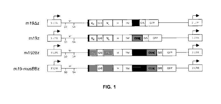
FIG. 1 is a schematic of anti-mouse CD19 CAR constructs. The retroviral
constructs include an anti-mCD19 scFy (VH/VL) with a CD8 hinge (H) and
transmembrane domain (TM) followed by 0D28 (m1928z) or 41BB (m19-musBBz)
and CD3 or CD3 alone (m19z). A negative control CAR includes no signaling
domains (m19,8,z). G/S is glycine-serine linker. The CAR is tagged to a
fluorescent
protein reporter (GFP or Cherry). LTR is longterminal repeat SD is splice-
donor, SA is
splice acceptor, and y is packaging signal.
FIG. 2A is a schematic of 0D28 subdomain mutations that support CAR
signaling in Nur77GFP derived CAR T cells. Shown are YMNM (SEQ ID NO:1), PRRP
(SEQ ID NO:2), and PYAP (SEQ ID NO:3) sequences replaced with AAAA (SEQ ID
NO:4). FIG. 2B is a bar graph showing Nur77GFP (percent GFP+) CD19-targeted
CAR
T cells stimulated with 3T3-mCD19. 24 hours after stimulation Nur773FP was
evaluated by flow cytometry.
FIGs. 3A and 3B are bar graphs showing IFN-y (Fig. 3A) and IL-2 (Fig. 3B)
secretion after CD28 subdomain mutant CD19-targeted CAR T cells were
stimulated
3
CA 03067605 2019-12-16
WO 2019/010383
PCT/US2018/041040
with 3T3 cells that express mouse CD19 (3T3-mCD19). FIGs. 30 and 3D are bar
graphs showing CM and EM subsets evaluated by flow cytometry.
FIG. 4 is a plot showing viability of 0D28 mutated CAR T cells and non-
mutated CAR T cells. CAR T cells were produced from wild type C57BL/6 mice and
cell viability was measured by trypan blue staining on an automated cell
counter
(BIO-RAD).
FIG. 5 is a plot showing proliferation of 0D28 mutant CAR T cells and non-
mutated CAR T cells. CAR T cells were produced from wild type C57BL/6 mice and
proliferation was evaluated by fold change from the initial cell number to
final cell
yield at Day 5.
FIGs. 6A to 6D are bar graphs showing IFN-y (Fig. 6A), IL-6 (Fig. 6B), IL-12
(Fig. 60), and TNF-a (Fig. 6D) produced by mutated and non-mutated CAR T
cells.
CART cells were activated with 3T3-mCD19 cells at a ratio of 10:1. After 24hr5
supernatants were harvested and cytokines measured by Luminex kit.
FIGs. 7A and 7B are graphs showing target cell killing by CD28 mutated and
non-mutated CAR T cells co-cultured with 3T3-mCD19 target cells at either a
10:1 or
1:6 ratio. Target cell killing was monitored by RTCA.
FIGs. 8A to 8H are plots showing B cell killing (Figs. 8A, 8C, 8E, 8G) and
CAR T cell counts (Figs. 8B, 8D, 8F, 8H) in vivo. 1x10⁶ CAR T cells (CD3+
CAR+)
.. were i.v. injected into cytoxan (300mg/kg) pretreated wild type C57BL/6
mice. At
weeks 1 (Figs. 8A, 8B), 2 (Figs. 8C, 8D), 4 (Figs. 8E, 8F), and 6 (Figs. 8G,
8H) mice
were bled and B cell and CAR T cell counts were analyzed by flow cytometry.
FIGs. 9A to 9D are plots showing B cell killing (Figs. 9A and 90) and CAR T
cell counts (Figs. 9B, and 9D) in bone marrow (Figs. 9A and 9B)) and spleen
(Figs.
90 and 9D). 1x10⁶ CAR T cells (CD3+ CAR+) were i.v. injected into cytoxan
(300mg/kg) pretreated wild type C57BL/6 mice. At week 8 bone marrow (BM) and
spleens were collected and B cell and CAR T cell counts were analyzed by flow
cytometry.
FIG. 10 is a graph showing survival of mice with Eu-ALL tumor after treatment
with CAR T cells. Six days after injection with Eu-ALL cells wild type C57BL/6
mice
were i.p. injected with cytoxan (300mg/kg) followed 1 day later with an i.v.
injection of
3x10⁵ CAR T cells.
FIGs. 11A and 11B are plots showing proliferation (Fig. 11A) and viability
(Fig.
11B) of CD28 mutated and non-mutated CAR T cells. CAR T cells were produced
from normal donor PBMCs and cell viability was measured by trypan blue
staining on
4
CA 03067605 2019-12-16
WO 2019/010383
PCT/US2018/041040
an automated cell counter (BIO-RAD). Proliferation was evaluated by fold
change
from the initial cell number to final cell yield at Day 5.
FIG. 12 is a graph showing human CAR T cells proliferation in vitro. 1x10⁵
human CAR T cells were co-cultured with 3T3-hCD19 cells at a ratio of 10:1 on
Day
5 and cell numbers were measured for 10 days.
FIG. 13 is a graph showing human mut06 CAR T cells kill as well as non-
mutated CAR T cells. CAR T cells were co-cultured with 3T3-hCD19 target cells
at a
5:1 ratio. Target cell killing was monitored by RTCA.
lo DETAILED DESCRIPTION
Disclosed herein are chimeric antigen receptors (CARs) that can specifically
recognize tumor-associated antigens (TAA) on cancers that comprise a mutated
form
of the cytoplasmic domain of 0D28 that reduce CAR-T cell exhaustion. Also
disclosed are immune effector cells, such as T cells or Natural Killer (NK)
cells, that
are engineered to express these CARs. Therefore, also disclosed are methods
for
providing an anti-tumor immunity in a subject with TAA-expressing cancers that
involves adoptive transfer of the disclosed immune effector cells engineered
to
express the disclosed CARs.
Chimeric antigen receptors (CAR) with mutated CD28 domains
CARs generally incorporate an antigen recognition domain from the single-
chain variable fragments (scFv) of a monoclonal antibody (mAb) with
transmembrane
signaling motifs involved in lymphocyte activation (Sadelain M, et al. Nat Rev
Cancer
2003 3:35-45). Disclosed herein is a chimeric antigen receptor (CAR) that can
be
that can be expressed in immune effector cells to enhance antitumor activity
against
cancers.
The disclosed CAR is generally made up of three domains: an ectodomain, a
transmembrane domain, and an endodomain. The ectodomain comprises the TAA-
binding region and is responsible for antigen recognition. It also optionally
contains a
signal peptide (SP) so that the CAR can be glycosylated and anchored in the
cell
membrane of the immune effector cell. The transmembrane domain (TD), is as its
name suggests, connects the ectodomain to the endodomain and resides within
the
cell membrane when expressed by a cell. The endodomain is the business end of
the
CAR that transmits an activation signal to the immune effector cell after
antigen
recognition. For example, the endodomain can contain an intracellular
signaling
domain (ISD) and a co-stimulatory signaling region (CSR).
5
CA 03067605 2019-12-16
WO 2019/010383
PCT/US2018/041040
The disclosed CARs have a CSR comprising a mutated form of 0D28 that
reduce CAR-T cell exhaustion. The 0D28 domain includes 3 intracellular
subdomains (YMNM (SEQ ID NO:1), PRRP (SEQ ID NO:2), and PYAP (SEQ ID
NO:3)) that regulate signaling pathways post TCR-stimulation. In some
embodiments, the disclosed CAR comprises mutation or deletion of one or more
of
these subdomains.
In some embodiments, the disclosed CAR is defined by the formula:
SP-TAA-HG-TM-CSR-lSD;
wherein "SP" represents an optional signal peptide,
lo wherein "TAA" represents a TAA-binding region,
wherein "HG" represents an optional hinge domain,
wherein "TM" represents a transmembrane domain,
wherein "CSR" represents the co-stimulatory signaling region,
wherein "ISD" represents an intracellular signaling domain, and
wherein "-" represents a peptide bond or linker.
Additional CAR constructs are described, for example, in Fresnak AD, et al.
Engineered T cells: the promise and challenges of cancer immunotherapy. Nat
Rev
Cancer. 2016 Aug 23;16(9):566-81, which is incorporated by reference in its
entirety
for the teaching of these CAR models.
For example, the CAR can be a TRUCK, Universal CAR, Self-driving CAR,
Armored CAR, Self-destruct CAR, Conditional CAR, Marked CAR, TenCAR, Dual
CAR, or sCAR.
TRUCKs (T cells redirected for universal cytokine killing) co-express a
chimeric antigen receptor (CAR) and an antitumor cytokine. Cytokine expression
may
be constitutive or induced by T cell activation. Targeted by CAR specificity,
localized
production of pro-inflammatory cytokines recruits endogenous immune cells to
tumor
sites and may potentiate an antitumor response.
Universal, allogeneic CAR T cells are engineered to no longer express
endogenous T cell receptor (TCR) and/or major histocompatibility complex (MHC)
molecules, thereby preventing graft-versus-host disease (GVHD) or rejection,
respectively.
Self-driving CARs co-express a CAR and a chemokine receptor, which binds
to a tumor ligand, thereby enhancing tumor homing.
CAR T cells engineered to be resistant to immunosuppression (Armored
CARs) may be genetically modified to no longer express various immune
checkpoint
molecules (for example, cytotoxic T lymphocyte-associated antigen 4 (CTLA4) or
6
CA 03067605 2019-12-16
WO 2019/010383
PCT/US2018/041040
programmed cell death protein 1 (PD1)), with an immune checkpoint switch
receptor,
or may be administered with a monoclonal antibody that blocks immune
checkpoint
signaling.
A self-destruct CAR may be designed using RNA delivered by electroporation
to encode the CAR. Alternatively, inducible apoptosis of the T cell may be
achieved
based on ganciclovir binding to thymidine kinase in gene-modified lymphocytes
or
the more recently described system of activation of human caspase 9 by a small-
molecule dimerizer.
A conditional CAR T cell is by default unresponsive, or switched 'off', until
the
lo addition of a small molecule to complete the circuit, enabling full
transduction of both
signal 1 and signal 2, thereby activating the CAR T cell. Alternatively, T
cells may be
engineered to express an adaptor-specific receptor with affinity for
subsequently
administered secondary antibodies directed at target antigen.
Marked CAR T cells express a CAR plus a tumor epitope to which an existing
monoclonal antibody agent binds. In the setting of intolerable adverse
effects,
administration of the monoclonal antibody clears the CAR T cells and
alleviates
symptoms with no additional off-tumor effects.
A tandem CAR (TanCAR) T cell expresses a single CAR consisting of two
linked single-chain variable fragments (scFvs) that have different affinities
fused to
intracellular co-stimulatory domain(s) and a CD3 domain. TanCAR T cell
activation
is achieved only when target cells co-express both targets.
A dual CAR T cell expresses two separate CARs with different ligand binding
targets; one CAR includes only the CD3 domain and the other CAR includes only
the co-stimulatory domain(s). Dual CAR T cell activation requires co-
expression of
both targets on the tumor.
A safety CAR (sCAR) consists of an extracellular scFv fused to an
intracellular inhibitory domain. sCAR T cells co-expressing a standard CAR
become
activated only when encountering target cells that possess the standard CAR
target
but lack the sCAR target.
The antigen recognition domain of the disclosed CAR is usually an scFv.
There are however many alternatives. An antigen recognition domain from native
T-
cell receptor (TCR) alpha and beta single chains have been described, as have
simple ectodomains (e.g. CD4 ectodomain to recognize HIV infected cells) and
more
exotic recognition components such as a linked cytokine (which leads to
recognition
of cells bearing the cytokine receptor). In fact almost anything that binds a
given
target with high affinity can be used as an antigen recognition region.
7
CA 03067605 2019-12-16
WO 2019/010383
PCT/US2018/041040
The endodomain is the business end of the CAR that after antigen recognition
transmits a signal to the immune effector cell, activating at least one of the
normal
effector functions of the immune effector cell. Effector function of a T cell,
for
example, may be cytolytic activity or helper activity including the secretion
of
cytokines. Therefore, the endodomain may comprise the "intracellular signaling
domain" of a T cell receptor (TCR) and optional co-receptors. While usually
the entire
intracellular signaling domain can be employed, in many cases it is not
necessary to
use the entire chain. To the extent that a truncated portion of the
intracellular
signaling domain is used, such truncated portion may be used in place of the
intact
lo chain as long as it transduces the effector function signal.
Cytoplasmic signaling sequences that regulate primary activation of the TCR
complex that act in a stimulatory manner may contain signaling motifs which
are
known as immunoreceptor tyrosine-based activation motifs (ITAMs). Examples of
ITAM containing cytoplasmic signaling sequences include those derived from
CD8,
CD3, CD3O, CD3y, CD3E, 0D32 (Fc gamma RIla), DAP10, DAP12, CD79a, CD79b,
FcyRly, FcyRIlly, FccRl13 (FCERIB), and FuRly (FCERIG).
In particular embodiments, the intracellular signaling domain is derived from
CD3 zeta (CD3) (TCR zeta, GenBank accno. BAG36664.1). T-cell surface
glycoprotein CD3 zeta (CD3) chain, also known as T-cell receptor T3 zeta chain
or
0D247 (Cluster of Differentiation 247), is a protein that in humans is encoded
by the
CD247 gene.
First-generation CARs typically had the intracellular domain from the CD3
chain, which is the primary transmitter of signals from endogenous TCRs.
Second-
generation CARs add intracellular signaling domains from various costimulatory
protein receptors (e.g., CD28, 41BB, ICOS) to the endodomain of the CAR to
provide
additional signals to the T cell. Preclinical studies have indicated that the
second
generation of CAR designs improves the antitumor activity of T cells. More
recent,
third-generation CARs combine multiple signaling domains to further augment
potency. T cells grafted with these CARs have demonstrated improved expansion,
activation, persistence, and tumor-eradicating efficiency independent of
costimulatory
receptor/ligand interaction (lmai C, et al. Leukemia 2004 18:676-84; Maher J,
et al.
Nat Biotechnol 2002 20:70-5).
For example, the endodomain of the CAR can be designed to comprise the
CD3 signaling domain by itself or combined with any other desired cytoplasmic
domain(s) useful in the context of the CAR of the invention. For example, the
cytoplasmic domain of the CAR can comprise a CD3 chain portion and a
8
CA 03067605 2019-12-16
WO 2019/010383
PCT/US2018/041040
costimulatory signaling region. The costimulatory signaling region refers to a
portion
of the CAR comprising the intracellular domain of a costimulatory molecule. A
costimulatory molecule is a cell surface molecule other than an antigen
receptor or
their ligands that is required for an efficient response of lymphocytes to an
antigen.
Examples of such molecules include 0D27, 0D28, 4-i BB (0D137), 0X40, CD30,
CD40, ICOS, lymphocyte function-associated antigen-1 (LFA-1), CD2, CD7, LIGHT,
NKG2C, B7-H3, and a ligand that specifically binds with 0D83, CD8, CD4, b2c,
CD80, 0D86, DAP10, DAP12, MyD88, BTNL3, and NKG2D. Thus, while the CAR is
exemplified primarily with 0D28 as the co-stimulatory signaling element, other
costimulatory elements can be used alone or in combination with other co-
stimulatory
signaling elements.
In some embodiments, the CAR comprises a hinge sequence. A hinge
sequence is a short sequence of amino acids that facilitates antibody
flexibility (see,
e.g., Woof et al., Nat. Rev. Immunol., 4(2): 89-99 (2004)). The hinge sequence
may
be positioned between the antigen recognition moiety and the transmembrane
domain. The hinge sequence can be any suitable sequence derived or obtained
from
any suitable molecule. In some embodiments, for example, the hinge sequence is
derived from a CD8a molecule or a 0D28 molecule.
The transmembrane domain may be derived either from a natural or from a
synthetic source. Where the source is natural, the domain may be derived from
any
membrane-bound or transmembrane protein. For example, the transmembrane
region may be derived from (i.e. comprise at least the transmembrane region(s)
of)
the alpha, beta or zeta chain of the T-cell receptor, 0D28, CD3 epsilon, 0D45,
CD4,
CD5, CD8 (e.g., CD8 alpha, CD8 beta), CD9, CD16, 0D22, 0D33, 0D37, 0D64,
CD80, 0D86, CD134, CD137, or CD154, KIRDS2, 0X40, CD2, 0D27, LFA-1
(CD11a, CD18) , ICOS (0D278) , 4-1BB (CD137) , GITR, CD40, BAFFR, HVEM
(LIGHTR) , SLAMF7, NKp80 (KLRF1) , CD160, CD19, IL2R beta, IL2R gamma, IL7R
a, ITGA1, VLA1, CD49a, ITGA4, IA4, CD49D, ITGA6, VLA-6, CD49f, ITGAD, CD11d,
ITGAE, CD103, ITGAL, CD11a, LFA-1, ITGAM, CD11b, ITGAX, CD11c, ITGB1,
0D29, ITGB2, CD18, LFA-1, ITGB7, TNFR2, DNAM1 (0D226) , SLAMF4 (0D244,
2B4) , 0D84, 0D96 (Tactile) , CEACAM1, CRTAM, Ly9 (0D229) , CD160 (BY55) ,
PSGL1, CD100 (SEMA4D) , SLAMF6 (NTB-A, Ly108) , SLAM (SLAMF1, CD150,
IP0-3) , BLAME (SLAMF8) , SELPLG (CD162) , LTBR, and PAG/Cbp. Alternatively
the transmembrane domain may be synthetic, in which case it will comprise
predominantly hydrophobic residues such as leucine and valine. In some cases,
a
triplet of phenylalanine, tryptophan and valine will be found at each end of a
synthetic
9
CA 03067605 2019-12-16
WO 2019/010383
PCT/US2018/041040
transmembrane domain. A short oligo- or polypeptide linker, such as between 2
and
amino acids in length, may form the linkage between the transmembrane domain
and the endoplasmic domain of the CAR.
In some embodiments, the CAR has more than one transmembrane domain,
5 which can be a repeat of the same transmembrane domain, or can be
different
transmembrane domains.
In some embodiments, the CAR is a multi-chain CAR, as described in
W02015/039523, which is incorporated by reference for this teaching. A multi-
chain
CAR can comprise separate extracellular ligand binding and signaling domains
in
10 different transmembrane polypeptides. The signaling domains can be
designed to
assemble in juxtamembrane position, which forms flexible architecture closer
to
natural receptors, that confers optimal signal transduction. For example, the
multi-
chain CAR can comprise a part of an FCERI alpha chain and a part of an FCERI
beta chain such that the FCERI chains spontaneously dimerize together to form
a
CAR.
Tables and 2 below provide some example combinations of TAA-binding
region, co-stimulatory signaling regions, and intracellular signaling domain
that can
occur in the disclosed CARs.
Table 1. Second Generation CARs
Co-stimulatory Signal
ScFv Signal Domain
TAA 0D28* CD8
TAA 0D28* CD3
TAA 0D28* CD3O
TAA 0D28* CD3y
TAA 0D28* CD3c
TAA 0D28* FcyRI-y
TAA 0D28* FcyRIII-y
TAA 0D28* FccRl13
TAA 0D28* FuRly
TAA 0D28* DAP10
TAA 0D28* DAP12
TAA 0D28* 0D32
TAA 0D28* CD79a
TAA 0D28* CD79b
0D28*= mutated 0D28 co-stimulatory domain as described herein
Table 2. Third Generation CARs
Co-stimulatory Co-stimulatory Signal
ScFv Signal Signal Domain
TAA 0D28* 0D28* CD8
TAA 0D28* 0D28* CD3
CA 03067605 2019-12-16
WO 2019/010383
PCT/US2018/041040
TAA 0D28* 0D28* CD3O
TAA 0D28* 0D28* CD3y
TAA 0D28* 0D28* CD3c
TAA 0D28* 0D28* FcyRI-y
TAA 0D28* 0D28* FcyRIII-y
TAA 0D28* 0D28* FccRl [3
TAA 0D28* 0D28* FuRly
TAA 0D28* 0D28* DAP10
TAA 0D28* 0D28* DAP12
TAA 0D28* 0D28* 0D32
TAA 0D28* 0D28* CD79a
TAA 0D28* 0D28* CD79b
TAA 0D28* CD8 CD8
TAA 0D28* CD8 CD3
TAA 0D28* CD8 CD3O
TAA 0D28* CD8 CD3y
TAA 0D28* CD8 CD3c
TAA 0D28* CD8 FcyRI-y
TAA 0D28* CD8 FcyRIII-y
TAA 0D28* CD8 FccRl [3
TAA 0D28* CD8 FuRly
TAA 0D28* CD8 DAP10
TAA 0D28* CD8 DAP12
TAA 0D28* CD8 0D32
TAA 0D28* CD8 CD79a
TAA 0D28* CD8 CD79b
TAA 0D28* CD4 CD8
TAA 0D28* CD4 CD3
TAA 0D28* CD4 CD3O
TAA 0D28* CD4 CD3y
TAA 0D28* CD4 CD3c
TAA 0D28* CD4 FcyRI-y
TAA 0D28* CD4 FcyRIII-y
TAA 0D28* CD4 FccRl [3
TAA 0D28* CD4 FuRly
TAA 0D28* CD4 DAP10
TAA 0D28* CD4 DAP12
TAA 0D28* CD4 0D32
TAA 0D28* CD4 CD79a
TAA 0D28* CD4 CD79b
TAA 0D28* b2c CD8
TAA 0D28* b2c CD3
TAA 0D28* b2c CD3O
TAA 0D28* b2c CD3y
TAA 0D28* b2c CD3c
TAA 0D28* b2c FcyRI-y
TAA 0D28* b2c FcyRIII-y
TAA 0D28* b2c FccRl [3
TAA 0D28* b2c FuRly
TAA 0D28* b2c DAP10
TAA 0D28* b2c DAP12
TAA 0D28* b2c 0D32
11
CA 03067605 2019-12-16
WO 2019/010383
PCT/US2018/041040
TAA 0D28* b2c CD79a
TAA 0D28* b2c CD79b
TAA 0D28* 0D137/41BB CD8
TAA 0D28* 0D137/41BB CD3
TAA 0D28* 0D137/41BB CD3O
TAA 0D28* 0D137/41BB CD3y
TAA 0D28* 0D137/41BB CD3c
TAA 0D28* 0D137/41BB FcyRI-y
TAA 0D28* 0D137/41BB FcyRI I I-y
TAA 0D28* 0D137/41BB FccRl [3
TAA 0D28* 0D137/41BB FuRly
TAA 0D28* 0D137/41BB DAP10
TAA 0D28* 0D137/41BB DAP12
TAA 0D28* 0D137/41BB 0D32
TAA 0D28* 0D137/41BB CD79a
TAA 0D28* 0D137/41BB CD79b
TAA 0D28* I COS CD8
TAA 0D28* I COS CD3
TAA 0D28* I COS CD3O
TAA 0D28* I COS CD3y
TAA 0D28* I COS CD3c
TAA 0D28* I COS FcyRI-y
TAA 0D28* I COS FcyRI I I-y
TAA 0D28* I COS FccRl [3
TAA 0D28* I COS FuRly
TAA 0D28* I COS DAP10
TAA 0D28* I COS DAP12
TAA 0D28* I COS 0D32
TAA 0D28* I COS CD79a
TAA 0D28* I COS CD79b
TAA 0D28* 0D27 CD8
TAA 0D28* 0D27 CD3
TAA 0D28* 0D27 CD3O
TAA 0D28* 0D27 CD3y
TAA 0D28* 0D27 CD3c
TAA 0D28* 0D27 FcyRI-y
TAA 0D28* 0D27 FcyRI I I-y
TAA 0D28* 0D27 FccRl [3
TAA 0D28* 0D27 FuRly
TAA 0D28* 0D27 DAP10
TAA 0D28* 0D27 DAP12
TAA 0D28* 0D27 0D32
TAA 0D28* 0D27 CD79a
TAA 0D28* 0D27 CD79b
TAA 0D28* CD286 CD8
TAA 0D28* CD286 CD3
TAA 0D28* CD286 CD3O
TAA 0D28* CD286 CD3y
TAA 0D28* CD286 CD3c
TAA 0D28* CD286 FcyRI-y
TAA 0D28* CD286 FcyRI I I-y
TAA 0D28* CD286 FccRl [3
12
CA 03067605 2019-12-16
WO 2019/010383
PCT/US2018/041040
TAA 0D28* CD286 FuRly
TAA 0D28* CD286 DAP10
TAA 0D28* CD286 DAP12
TAA 0D28* CD286 0D32
TAA 0D28* CD286 CD79a
TAA 0D28* CD286 CD79b
TAA 0D28* CD80 CD8
TAA 0D28* CD80 CD3
TAA 0D28* CD80 CD3O
TAA 0D28* CD80 CD3y
TAA 0D28* CD80 CD3c
TAA 0D28* CD80 FcyRI-y
TAA 0D28* CD80 FcyRIII-y
TAA 0D28* CD80 FccRl [3
TAA 0D28* CD80 FuRly
TAA 0D28* CD80 DAP10
TAA 0D28* CD80 DAP12
TAA 0D28* CD80 0D32
TAA 0D28* CD80 CD79a
TAA 0D28* CD80 CD79b
TAA 0D28* 0D86 CD8
TAA 0D28* 0D86 CD3
TAA 0D28* 0D86 CD3O
TAA 0D28* 0D86 CD3y
TAA 0D28* 0D86 CD3c
TAA 0D28* 0D86 FcyRI-y
TAA 0D28* 0D86 FcyRIII-y
TAA 0D28* 0D86 FccRl [3
TAA 0D28* 0D86 FuRly
TAA 0D28* 0D86 DAP10
TAA 0D28* 0D86 DAP12
TAA 0D28* 0D86 0D32
TAA 0D28* 0D86 CD79a
TAA 0D28* 0D86 CD79b
TAA 0D28* 0X40 CD8
TAA 0D28* 0X40 CD3
TAA 0D28* 0X40 CD3O
TAA 0D28* 0X40 CD3y
TAA 0D28* 0X40 CD3c
TAA 0D28* 0X40 FcyRI-y
TAA 0D28* 0X40 FcyRIII-y
TAA 0D28* 0X40 FccRl [3
TAA 0D28* 0X40 FuRly
TAA 0D28* 0X40 DAP10
TAA 0D28* 0X40 DAP12
TAA 0D28* 0X40 0D32
TAA 0D28* 0X40 CD79a
TAA 0D28* 0X40 CD79b
TAA 0D28* DAP10 CD8
TAA 0D28* DAP10 CD3
TAA 0D28* DAP10 CD3O
TAA 0D28* DAP10 CD3y
13
CA 03067605 2019-12-16
WO 2019/010383
PCT/US2018/041040
TAA 0D28* DAP10 CD3c
TAA 0D28* DAP10 FcyRI-y
TAA 0D28* DAP10 FcyRI I I-y
TAA 0D28* DAP10 FccRl [3
TAA 0D28* DAP10 FuRly
TAA 0D28* DAP10 DAP10
TAA 0D28* DAP10 DAP12
TAA 0D28* DAP10 0D32
TAA 0D28* DAP10 CD79a
TAA 0D28* DAP10 CD79b
TAA 0D28* DAP12 CD8
TAA 0D28* DAP12 CD3
TAA 0D28* DAP12 CD3O
TAA 0D28* DAP12 CD3y
TAA 0D28* DAP12 CD3c
TAA 0D28* DAP12 FcyRI-y
TAA 0D28* DAP12 FcyRI I I-y
TAA 0D28* DAP12 FccRl [3
TAA 0D28* DAP12 FuRly
TAA 0D28* DAP12 DAP10
TAA 0D28* DAP12 DAP12
TAA 0D28* DAP12 0D32
TAA 0D28* DAP12 CD79a
TAA 0D28* DAP12 CD79b
TAA 0D28* MyD88 CD8
TAA 0D28* MyD88 CD3
TAA 0D28* MyD88 CD3O
TAA 0D28* MyD88 CD3y
TAA 0D28* MyD88 CD3c
TAA 0D28* MyD88 FcyRI-y
TAA 0D28* MyD88 FcyRI I I-y
TAA 0D28* MyD88 FccRl [3
TAA 0D28* MyD88 FuRly
TAA 0D28* MyD88 DAP10
TAA 0D28* MyD88 DAP12
TAA 0D28* MyD88 0D32
TAA 0D28* MyD88 CD79a
TAA 0D28* MyD88 CD79b
TAA 0D28* CD7 CD8
TAA 0D28* CD7 CD3
TAA 0D28* CD7 CD3O
TAA 0D28* CD7 CD3y
TAA 0D28* CD7 CD3c
TAA 0D28* CD7 FcyRI-y
TAA 0D28* CD7 FcyRI I I-y
TAA 0D28* CD7 FccRl [3
TAA 0D28* CD7 FuRly
TAA 0D28* CD7 DAP10
TAA 0D28* CD7 DAP12
TAA 0D28* CD7 0D32
TAA 0D28* CD7 CD79a
TAA 0D28* CD7 CD79b
14
CA 03067605 2019-12-16
WO 2019/010383
PCT/US2018/041040
TAA 0D28* BTN L3 CD8
TAA 0D28* BTN L3 CD3
TAA 0D28* BTN L3 CD3O
TAA 0D28* BTN L3 CD3y
TAA 0D28* BTN L3 CD3c
TAA 0D28* BTN L3 FcyRI-y
TAA 0D28* BTN L3 FcyRIII-y
TAA 0D28* BTN L3 FccRl [3
TAA 0D28* BTN L3 FuRly
TAA 0D28* BTN L3 DAP10
TAA 0D28* BTN L3 DAP12
TAA 0D28* BTN L3 0D32
TAA 0D28* BTN L3 CD79a
TAA 0D28* BTN L3 CD79b
TAA 0D28* NKG2D CD8
TAA 0D28* NKG2D CD3
TAA 0D28* NKG2D CD3O
TAA 0D28* NKG2D CD3y
TAA 0D28* NKG2D CD3c
TAA 0D28* NKG2D FcyRI-y
TAA 0D28* NKG2D FcyRIII-y
TAA 0D28* NKG2D FccRl [3
TAA 0D28* NKG2D FuRly
TAA 0D28* NKG2D DAP10
TAA 0D28* NKG2D DAP12
TAA 0D28* NKG2D 0D32
TAA 0D28* NKG2D CD79a
TAA 0D28* NKG2D CD79b
TAA CD8 0D28* CD8
TAA CD8 0D28* CD3
TAA CD8 0D28* CD3O
TAA CD8 0D28* CD3y
TAA CD8 0D28* CD3c
TAA CD8 0D28* FcyRI-y
TAA CD8 0D28* FcyRIII-y
TAA CD8 0D28* FccRl [3
TAA CD8 0D28* FuRly
TAA CD8 0D28* DAP10
TAA CD8 0D28* DAP12
TAA CD8 0D28* 0D32
TAA CD8 0D28* CD79a
TAA CD8 0D28* CD79b
TAA CD4 0D28* CD8
TAA CD4 0D28* CD3
TAA CD4 0D28* CD3O
TAA CD4 0D28* CD3y
TAA CD4 0D28* CD3c
TAA CD4 0D28* FcyRI-y
TAA CD4 0D28* FcyRIII-y
TAA CD4 0D28* FccRl [3
TAA CD4 0D28* FuRly
TAA CD4 0D28* DAP10
CA 03067605 2019-12-16
WO 2019/010383
PCT/US2018/041040
TAA CD4 0D28* DAP12
TAA CD4 0D28* 0D32
TAA CD4 0D28* CD79a
TAA CD4 0D28* CD79b
TAA b2c 0D28* CD8
TAA b2c 0D28* CD3
TAA b2c 0D28* CD3O
TAA b2c 0D28* CD3y
TAA b2c 0D28* CD3c
TAA b2c 0D28* FcyRI-y
TAA b2c 0D28* FcyRI I I-y
TAA b2c 0D28* FccRl [3
TAA b2c 0D28* FuRly
TAA b2c 0D28* DAP10
TAA b2c 0D28* DAP12
TAA b2c 0D28* 0D32
TAA b2c 0D28* CD79a
TAA b2c 0D28* CD79b
TAA 0D137/41BB 0D28* CD8
TAA 0D137/41BB 0D28* CD3
TAA 0D137/41BB 0D28* CD3O
TAA 0D137/41BB 0D28* CD3y
TAA 0D137/41BB 0D28* CD3c
TAA 0D137/41BB 0D28* FcyRI-y
TAA 0D137/41BB 0D28* FcyRI I I-y
TAA 0D137/41BB 0D28* FccRl [3
TAA 0D137/41BB 0D28* FuRly
TAA 0D137/41BB 0D28* DAP10
TAA 0D137/41BB 0D28* DAP12
TAA 0D137/41BB 0D28* 0D32
TAA 0D137/41BB 0D28* CD79a
TAA 0D137/41BB 0D28* CD79b
TAA I COS 0D28* CD8
TAA I COS 0D28* CD3
TAA I COS 0D28* CD3O
TAA I COS 0D28* CD3y
TAA I COS 0D28* CD3c
TAA I COS 0D28* FcyRI-y
TAA I COS 0D28* FcyRI I I-y
TAA I COS 0D28* FccRl [3
TAA I COS 0D28* FuRly
TAA I COS 0D28* DAP10
TAA I COS 0D28* DAP12
TAA I COS 0D28* 0D32
TAA I COS 0D28* CD79a
TAA I COS 0D28* CD79b
TAA 0D27 0D28* CD8
TAA 0D27 0D28* CD3
TAA 0D27 0D28* CD3O
TAA 0D27 0D28* CD3y
TAA 0D27 0D28* CD3c
TAA 0D27 0D28* FcyRI-y
16
CA 03067605 2019-12-16
WO 2019/010383
PCT/US2018/041040
TAA 0D27 0D28* FcyRIII-y
TAA 0D27 0D28* FccRlf3
TAA 0D27 0D28* FccRly
TAA 0D27 0D28* DAP10
TAA 0D27 0D28* DAP12
TAA 0D27 0D28* 0D32
TAA 0D27 0D28* CD79a
TAA 0D27 0D28* CD79b
TAA CD286 0D28* CD8
TAA CD286 0D28* CD3
TAA CD286 0D28* CD3O
TAA CD286 0D28* CD3y
TAA CD286 0D28* CD3c
TAA CD286 0D28* FcyRI-y
TAA CD286 0D28* FcyRIII-y
TAA CD286 0D28* FccRlf3
TAA CD286 0D28* FccRly
TAA CD286 0D28* DAP10
TAA CD286 0D28* DAP12
TAA CD286 0D28* 0D32
TAA CD286 0D28* CD79a
TAA CD286 0D28* CD79b
TAA CD80 0D28* CD8
TAA CD80 0D28* CD3
TAA CD80 0D28* CD3O
TAA CD80 0D28* CD3y
TAA CD80 0D28* CD3c
TAA CD80 0D28* FcyRI-y
TAA CD80 0D28* FcyRIII-y
TAA CD80 0D28* FccRlf3
TAA CD80 0D28* FccRly
TAA CD80 0D28* DAP10
TAA CD80 0D28* DAP12
TAA CD80 0D28* 0D32
TAA CD80 0D28* CD79a
TAA CD80 0D28* CD79b
TAA 0D86 0D28* CD8
TAA 0D86 0D28* CD3
TAA 0D86 0D28* CD3O
TAA 0D86 0D28* CD3y
TAA 0D86 0D28* CD3c
TAA 0D86 0D28* FcyRI-y
TAA 0D86 0D28* FcyRIII-y
TAA 0D86 0D28* FccRlf3
TAA 0D86 0D28* FccRly
TAA 0D86 0D28* DAP10
TAA 0D86 0D28* DAP12
TAA 0D86 0D28* 0D32
TAA 0D86 0D28* CD79a
TAA 0D86 0D28* CD79b
TAA 0X40 0D28* CD8
TAA 0X40 0D28* CD3
17
CA 03067605 2019-12-16
WO 2019/010383
PCT/US2018/041040
TAA 0X40 0D28* CD3O
TAA 0X40 0D28* CD3y
TAA 0X40 0D28* CD3c
TAA 0X40 0D28* FcyRI-y
TAA 0X40 0D28* FcyRI I I-y
TAA 0X40 0D28* FccRl [3
TAA 0X40 0D28* FuRly
TAA 0X40 0D28* DAP10
TAA 0X40 0D28* DAP12
TAA 0X40 0D28* 0D32
TAA 0X40 0D28* CD79a
TAA 0X40 0D28* CD79b
TAA DAP10 0D28* CD8
TAA DAP10 0D28* CD3
TAA DAP10 0D28* CD3O
TAA DAP10 0D28* CD3y
TAA DAP10 0D28* CD3c
TAA DAP10 0D28* FcyRI-y
TAA DAP10 0D28* FcyRI I I-y
TAA DAP10 0D28* FccRl [3
TAA DAP10 0D28* FuRly
TAA DAP10 0D28* DAP10
TAA DAP10 0D28* DAP12
TAA DAP10 0D28* 0D32
TAA DAP10 0D28* CD79a
TAA DAP10 0D28* CD79b
TAA DAP12 0D28* CD8
TAA DAP12 0D28* CD3
TAA DAP12 0D28* CD3O
TAA DAP12 0D28* CD3y
TAA DAP12 0D28* CD3c
TAA DAP12 0D28* FcyRI-y
TAA DAP12 0D28* FcyRI I I-y
TAA DAP12 0D28* FccRl [3
TAA DAP12 0D28* FuRly
TAA DAP12 0D28* DAP10
TAA DAP12 0D28* DAP12
TAA DAP12 0D28* 0D32
TAA DAP12 0D28* CD79a
TAA DAP12 0D28* CD79b
TAA MyD88 0D28* CD8
TAA MyD88 0D28* CD3
TAA MyD88 0D28* CD3O
TAA MyD88 0D28* CD3y
TAA MyD88 0D28* CD3c
TAA MyD88 0D28* FcyRI-y
TAA MyD88 0D28* FcyRI I I-y
TAA MyD88 0D28* FccRl [3
TAA MyD88 0D28* FuRly
TAA MyD88 0D28* DAP10
TAA MyD88 0D28* DAP12
TAA MyD88 0D28* 0D32
18
CA 03067605 2019-12-16
WO 2019/010383
PCT/US2018/041040
TAA MyD88 0D28* CD79a
TAA MyD88 0D28* CD79b
TAA CD7 0D28* CD8
TAA CD7 0D28* CD3
TAA CD7 0D28* CD3O
TAA CD7 0D28* CD3y
TAA CD7 0D28* CD3c
TAA CD7 0D28* FcyRI-y
TAA CD7 0D28* FcyRIII-y
TAA CD7 0D28* FccRlf3
TAA CD7 0D28* FuRly
TAA CD7 0D28* DAP10
TAA CD7 0D28* DAP12
TAA CD7 0D28* 0D32
TAA CD7 0D28* CD79a
TAA CD7 0D28* CD79b
TAA BTNL3 0D28* CD8
TAA BTNL3 0D28* CD3
TAA BTNL3 0D28* CD3O
TAA BTNL3 0D28* CD3y
TAA BTNL3 0D28* CD3c
TAA BTNL3 0D28* FcyRI-y
TAA BTNL3 0D28* FcyRIII-y
TAA BTNL3 0D28* FccRlf3
TAA BTNL3 0D28* FuRly
TAA BTNL3 0D28* DAP10
TAA BTNL3 0D28* DAP12
TAA BTNL3 0D28* 0D32
TAA BTNL3 0D28* CD79a
TAA BTNL3 0D28* CD79b
TAA NKG2D 0D28* CD8
TAA NKG2D 0D28* CD3
TAA NKG2D 0D28* CD3O
TAA NKG2D 0D28* CD3y
TAA NKG2D 0D28* CD3c
TAA NKG2D 0D28* FcyRI-y
TAA NKG2D 0D28* FcyRIII-y
TAA NKG2D 0D28* FccRlf3
TAA NKG2D 0D28* FuRly
TAA NKG2D 0D28* DAP10
TAA NKG2D 0D28* DAP12
TAA NKG2D 0D28* 0D32
TAA NKG2D 0D28* CD79a
TAA NKG2D 0D28* CD79b
0D28*= mutated 0D28 co-stimulatory domain as described herein
In some embodiments, the anti-TAA binding agent is single chain variable
fragment (scFv) antibody. The affinity/specificity of an anti-TAA scFv is
driven in large
part by specific sequences within complementarity determining regions (CDRs)
in the
19
CA 03067605 2019-12-16
WO 2019/010383
PCT/US2018/041040
heavy (VH) and light (VL) chain. Each VH and VL sequence will have three CDRs
(CDR1, CDR2, CDR3).
In some cases, the anti-TAA binding agent is an affinity maturated scFv. In
some cases, the anti-TAA has a dissociation constant (KD) for the TAA that is
less
than 50 nM, 40 nM, 30 nM, 25 nM, 20 nM, 15 nM, or 10 nM.
In some embodiments, the anti-TAA binding agent is derived from natural
antibodies, such as monoclonal antibodies. In some cases, the antibody is
human. In
some cases, the antibody has undergone an alteration to render it less
immunogenic
when administered to humans. For example, the alteration comprises one or more
techniques selected from the group consisting of chimerization, humanization,
CDR-
grafting, deimmunization, and mutation of framework amino acids to correspond
to
the closest human germline sequence.
Tumor antigens are proteins that are produced by tumor cells that elicit an
immune response, particularly T-cell mediated immune responses. The additional
antigen binding domain can be an antibody or a natural ligand of the tumor
antigen.
The selection of the additional antigen binding domain will depend on the
particular
type of cancer to be treated. Tumor antigens are well known in the art and
include,
for example, a glioma-associated antigen, carcinoembryonic antigen (CEA),
EGFRvIll, IL-11Ra, IL-13Ra, EGFR, FAP, B7H3, Kit, CA LX, CS-1, MUC1, BCMA, bcr-
abl, HER2, 13-human chorionic gonadotropin, alphafetoprotein (AFP), ALK, CD19,
CD123, cyclin BI, lectin-reactive AFP, Fos-related antigen 1, ADRB3,
thyroglobulin,
EphA2, RAGE-1, RUI, RU2, 55X2, AKAP-4, LCK, OY-TESI, PAX5, SART3, CLL-1,
fucosyl GM1, GloboH, MN-CA IX, EPCAM, EVT6-AML, TGS5, human telomerase
reverse transcriptase, plysialic acid, PLAC1, RUI, RU2 (AS), intestinal
carboxyl
esterase, lewisY, sLe, LY6K, mut h5p70-2, M-CSF, MYCN, RhoC, TRP-2, CYPIBI,
BORIS, prostase, prostate-specific antigen (PSA), PAX3, PAP, NY-ESO-1, LAGE-
la,
LMP2, NCAM, p53, p53 mutant, Ras mutant, gp100, prostein, OR51E2, PANX3,
PSMA, PSCA, Her2/neu, hTERT, HMWMAA, HAVCR1, VEGFR2, PDGFR-beta,
survivin and telomerase, legumain, HPV E6,E7, sperm protein 17, SSEA-4,
tyrosinase, TARP, VVT1, prostate-carcinoma tumor antigen- 1 (PCTA-1), ML-IAP,
MAGE, MAGE-A1,MAD-CT-1, MAD-CT-2, MelanA/MART 1, XAGE1 , ELF2M, ERG
(TMPRSS2 ETS fusion gene), NA17, neutrophil elastase, sarcoma translocation
breakpoints, NY-BR-1, ephnnB2, CD20, CD22, CD24, CD30, CD33, CD38, CD44v6,
CD97, CD171, CD179a, androgen receptor, FAP, insulin growth factor (IGF)-I,
IGFII,
IGF-I receptor, GD2, o-acetyl-GD2, GD3, GM3, GPRC5D, GPR20, CXORF61, folate
receptor (FRa), folate receptor beta, ROR1, Flt3, TAG72, TN Ag, Tie 2, TEM1,
CA 03067605 2019-12-16
WO 2019/010383
PCT/US2018/041040
TEM7R, CLDN6, TSHR, UPK2, and mesothelin. In a preferred embodiment, the
tumor antigen is selected from the group consisting of folate receptor (FRa),
mesothelin, EGFRvIll, IL-13Ra, 0D123, 0D19, 0D33, BCMA, GD2, CLL-1, CA-1X,
MUCI, HER2, and any combination thereof.
Non-limiting examples of tumor antigens include the following: Differentiation
antigens such as tyrosinase, TRP-1, TRP-2 and tumor-specific multilineage
antigens
such as MAGE-1, MAGE-3, BAGE, GAGE-1, GAGE-2, pi 5; overexpressed
embryonic antigens such as CEA; overexpressed oncogenes and mutated tumor-
suppressor genes such as p53, Ras, HER-2/neu; unique tumor antigens resulting
from chromosomal translocations; such as BCR-ABL, E2A-PRL, H4-RET, IGH-IGK,
MYL-RAR; and viral antigens, such as the Epstein Barr virus antigens EBVA and
the
human papillomavirus (HPV) antigens E6 and E7. Other large, protein-based
antigens include TSP- 180, MAGE-4, MAGE-5, MAGE-6, RAGE, NY-ESO,
p185erbB2, p180erbB-3, c-met, nm- 23H1, PSA, CA 19-9, CA 72-4, CAM 17.1, NuMa,
K-ras, beta-Catenin, CDK4, Mum-1, p 15, p 16, 43-9F, 5T4, 791Tgp72, alpha-
fetoprotein, beta-HCG, BCA225, BTAA, CA 125, CA 15-3\CA 27.29\BCAA, CA 195,
CA 242, CA-50, CAM43, CD68\P1, CO-029, FGF-5, G250, Ga733\EpCAM, HTgp-
175, M344, MA-50, MG7-Ag, MOV18, NB/70K, NY-CO-1, RCASI, SDCCAG1 6, TA-
90\Mac-2 binding protein\cyclophilm C-associated protein, TAAL6, TAG72, TLP,
TPS, GPC3, MUC16, LMP1, EBMA-1, BARF-1, CS1, CD319, HER1, B7H6, Li CAM,
1L6, and MET.
Nucleic Acids and Vectors
Also disclosed are polynucleotides and polynucleotide vectors encoding the
disclosed CARs that allow expression of the CARs in the disclosed immune
effector
cells.
Nucleic acid sequences encoding the disclosed CARs, and regions thereof,
can be obtained using recombinant methods known in the art, such as, for
example
by screening libraries from cells expressing the gene, by deriving the gene
from a
vector known to include the same, or by isolating directly from cells and
tissues
containing the same, using standard techniques. Alternatively, the gene of
interest
can be produced synthetically, rather than cloned.
Expression of nucleic acids encoding CARs is typically achieved by operably
linking a nucleic acid encoding the CAR polypeptide to a promoter, and
incorporating
the construct into an expression vector. Typical cloning vectors contain
transcription
and translation terminators, initiation sequences, and promoters useful for
regulation
of the expression of the desired nucleic acid sequence.
21
CA 03067605 2019-12-16
WO 2019/010383
PCT/US2018/041040
The disclosed nucleic acid can be cloned into a number of types of vectors.
For example, the nucleic acid can be cloned into a vector including, but not
limited to
a plasmid, a phagemid, a phage derivative, an animal virus, and a cosmid.
Vectors of
particular interest include expression vectors, replication vectors, probe
generation
vectors, and sequencing vectors.
Further, the expression vector may be provided to a cell in the form of a
viral
vector. Viral vector technology is well known in the art and is described, for
example,
in Sambrook et al. (2001, Molecular Cloning: A Laboratory Manual, Cold Spring
Harbor Laboratory, New York), and in other virology and molecular biology
manuals.
lo Viruses, which are useful as vectors include, but are not limited to,
retroviruses,
adenoviruses, adeno-associated viruses, herpes viruses, and lentiviruses. In
general,
a suitable vector contains an origin of replication functional in at least one
organism,
a promoter sequence, convenient restriction endonuclease sites, and one or
more
selectable markers. In some embodimens, the polynucleotide vectors are
lentiviral or
retroviral vectors.
A number of viral based systems have been developed for gene transfer into
mammalian cells. For example, retroviruses provide a convenient platform for
gene
delivery systems. A selected gene can be inserted into a vector and packaged
in
retroviral particles using techniques known in the art. The recombinant virus
can then
be isolated and delivered to cells of the subject either in vivo or ex vivo.
One example of a suitable promoter is the immediate early cytomegalovirus
(CMV) promoter sequence. This promoter sequence is a strong constitutive
promoter
sequence capable of driving high levels of expression of any polynucleotide
sequence operatively linked thereto. Another example of a suitable promoter is
Elongation Growth Factor-1a (EF-1a). However, other constitutive promoter
sequences may also be used, including, but not limited to the simian virus 40
(5V40)
early promoter, MND (myeloproliferative sarcoma virus) promoter, mouse mammary
tumor virus (MMTV), human immunodeficiency virus (HIV) long terminal repeat
(LTR)
promoter, MoMuLV promoter, an avian leukemia virus promoter, an Epstein-Barr
virus immediate early promoter, a Rous sarcoma virus promoter, as well as
human
gene promoters such as, but not limited to, the actin promoter, the myosin
promoter,
the hemoglobin promoter, and the creatine kinase promoter. The promoter can
alternatively be an inducible promoter. Examples of inducible promoters
include, but
are not limited to a metallothionine promoter, a glucocorticoid promoter, a
progesterone promoter, and a tetracycline promoter.
22
CA 03067605 2019-12-16
WO 2019/010383
PCT/US2018/041040
Additional promoter elements, e.g., enhancers, regulate the frequency of
transcriptional initiation. Typically, these are located in the region 30-110
bp upstream
of the start site, although a number of promoters have recently been shown to
contain functional elements downstream of the start site as well. The spacing
between promoter elements frequently is flexible, so that promoter function is
preserved when elements are inverted or moved relative to one another.
In order to assess the expression of a CAR polypeptide or portions thereof,
the expression vector to be introduced into a cell can also contain either a
selectable
marker gene or a reporter gene or both to facilitate identification and
selection of
lo expressing cells from the population of cells sought to be transfected
or infected
through viral vectors. In other aspects, the selectable marker may be carried
on a
separate piece of DNA and used in a co-transfection procedure. Both selectable
markers and reporter genes may be flanked with appropriate regulatory
sequences to
enable expression in the host cells. Useful selectable markers include, for
example,
antibiotic-resistance genes.
Reporter genes are used for identifying potentially transfected cells and for
evaluating the functionality of regulatory sequences. In general, a reporter
gene is a
gene that is not present in or expressed by the recipient organism or tissue
and that
encodes a polypeptide whose expression is manifested by some easily detectable
property, e.g., enzymatic activity. Expression of the reporter gene is assayed
at a
suitable time after the DNA has been introduced into the recipient cells.
Suitable
reporter genes may include genes encoding luciferase, beta-galactosidase,
chloramphenicol acetyl transferase, secreted alkaline phosphatase, or the
green
fluorescent protein gene. Suitable expression systems are well known and may
be
prepared using known techniques or obtained commercially. In general, the
construct
with the minimal 5' flanking region showing the highest level of expression of
reporter
gene is identified as the promoter. Such promoter regions may be linked to a
reporter
gene and used to evaluate agents for the ability to modulate promoter-driven
transcription.
Methods of introducing and expressing genes into a cell are known in the art.
In the context of an expression vector, the vector can be readily introduced
into a
host cell, e.g., mammalian, bacterial, yeast, or insect cell by any method in
the art.
For example, the expression vector can be transferred into a host cell by
physical,
chemical, or biological means.
Physical methods for introducing a polynucleotide into a host cell include
calcium phosphate precipitation, lipofection, particle bombardment,
microinjection,
23
CA 03067605 2019-12-16
WO 2019/010383
PCT/US2018/041040
electroporation, and the like. Methods for producing cells comprising vectors
and/or
exogenous nucleic acids are well-known in the art. See, for example, Sambrook
et al.
(2001, Molecular Cloning: A Laboratory Manual, Cold Spring Harbor Laboratory,
New
York).
Biological methods for introducing a polynucleotide of interest into a host
cell
include the use of DNA and RNA vectors. Viral vectors, and especially
retroviral
vectors, have become the most widely used method for inserting genes into
mammalian, e.g., human cells.
Chemical means for introducing a polynucleotide into a host cell include
lo colloidal dispersion systems, such as macromolecule complexes,
nanocapsules,
microspheres, beads, and lipid-based systems including oil-in-water emulsions,
micelles, mixed micelles, and liposomes. An exemplary colloidal system for use
as a
delivery vehicle in vitro and in vivo is a liposome (e.g., an artificial
membrane
vesicle).
In the case where a non-viral delivery system is utilized, an exemplary
delivery vehicle is a liposome. In another aspect, the nucleic acid may be
associated
with a lipid. The nucleic acid associated with a lipid may be encapsulated in
the
aqueous interior of a liposome, interspersed within the lipid bilayer of a
liposome,
attached to a liposome via a linking molecule that is associated with both the
liposome and the oligonucleotide, entrapped in a liposome, complexed with a
liposome, dispersed in a solution containing a lipid, mixed with a lipid,
combined with
a lipid, contained as a suspension in a lipid, contained or complexed with a
micelle,
or otherwise associated with a lipid. Lipid, lipid/DNA or lipid/expression
vector
associated compositions are not limited to any particular structure in
solution. For
example, they may be present in a bilayer structure, as micelles, or with a
"collapsed"
structure. They may also simply be interspersed in a solution, possibly
forming
aggregates that are not uniform in size or shape. Lipids are fatty substances
which
may be naturally occurring or synthetic lipids. For example, lipids include
the fatty
droplets that naturally occur in the cytoplasm as well as the class of
compounds
which contain long-chain aliphatic hydrocarbons and their derivatives, such as
fatty
acids, alcohols, amines, amino alcohols, and aldehydes. Lipids suitable for
use can
be obtained from commercial sources. For example, dimyristyl
phosphatidylcholine
("DMPC") can be obtained from Sigma, St. Louis, Mo.; dicetyl phosphate ("DCP")
can
be obtained from K & K Laboratories (Plainview, N.Y.); cholesterol ("Choi")
can be
obtained from Calbiochem-Behring; dimyristyl phosphatidylglycerol ("DMPG") and
other lipids may be obtained from Avanti Polar Lipids, Inc, (Birmingham,
Ala.).
24
CA 03067605 2019-12-16
WO 2019/010383
PCT/US2018/041040
Immune effector cells
Also disclosed are immune effector cells that are engineered to express the
disclosed CARs (also referred to herein as "CAR-T cells." These cells are
preferably
obtained from the subject to be treated (i.e. are autologous). However, in
some
embodiments, immune effector cell lines or donor effector cells (allogeneic)
are used.
Immune effector cells can be obtained from a number of sources, including
peripheral blood mononuclear cells, bone marrow, lymph node tissue, cord
blood,
thymus tissue, tissue from a site of infection, ascites, pleural effusion,
spleen tissue,
and tumors. Immune effector cells can be obtained from blood collected from a
lo subject using any number of techniques known to the skilled artisan,
such as Ficoll TM
separation. For example, cells from the circulating blood of an individual may
be
obtained by apheresis. In some embodiments, immune effector cells are isolated
from peripheral blood lymphocytes by lysing the red blood cells and depleting
the
monocytes, for example, by centrifugation through a PERCOLLTM gradient or by
counterflow centrifugal elutriation. A specific subpopulation of immune
effector cells
can be further isolated by positive or negative selection techniques. For
example,
immune effector cells can be isolated using a combination of antibodies
directed to
surface markers unique to the positively selected cells, e.g., by incubation
with
antibody-conjugated beads for a time period sufficient for positive selection
of the
desired immune effector cells. Alternatively, enrichment of immune effector
cells
population can be accomplished by negative selection using a combination of
antibodies directed to surface markers unique to the negatively selected
cells.
In some embodiments, the immune effector cells comprise any leukocyte
involved in defending the body against infectious disease and foreign
materials. For
example, the immune effector cells can comprise lymphocytes, monocytes,
macrophages, dentritic cells, mast cells, neutrophils, basophils, eosinophils,
or any
combinations thereof. For example, the immune effector cells can comprise T
lymphocytes.
T cells or T lymphocytes can be distinguished from other lymphocytes, such
as B cells and natural killer cells (NK cells), by the presence of a T-cell
receptor
(TCR) on the cell surface. They are called T cells because they mature in the
thymus
(although some also mature in the tonsils). There are several subsets of T
cells, each
with a distinct function.
T helper cells (TH cells) assist other white blood cells in immunologic
processes, including maturation of B cells into plasma cells and memory B
cells, and
activation of cytotoxic T cells and macrophages. These cells are also known as
CD4+
CA 03067605 2019-12-16
WO 2019/010383
PCT/US2018/041040
T cells because they express the CD4 glycoprotein on their surface. Helper T
cells
become activated when they are presented with peptide antigens by MHC class II
molecules, which are expressed on the surface of antigen-presenting cells
(APCs).
Once activated, they divide rapidly and secrete small proteins called
cytokines that
regulate or assist in the active immune response. These cells can
differentiate into
one of several subtypes, including TH1, TH2, TH3, TH17, TH9, or TFH, which
secrete
different cytokines to facilitate a different type of immune response.
Cytotoxic T cells (Tc cells, or CTLs) destroy virally infected cells and tumor
cells, and are also implicated in transplant rejection. These cells are also
known as
lo CD8+ T cells since they express the CD8 glycoprotein at their surface.
These cells
recognize their targets by binding to antigen associated with MHC class I
molecules,
which are present on the surface of all nucleated cells. Through IL-10,
adenosine and
other molecules secreted by regulatory T cells, the CD8+ cells can be
inactivated to
an anergic state, which prevents autoimmune diseases.
Memory T cells are a subset of antigen-specific T cells that persist long-term
after an infection has resolved. They quickly expand to large numbers of
effector T
cells upon re-exposure to their cognate antigen, thus providing the immune
system
with "memory" against past infections. Memory cells may be either CD4+ or
CD8+.
Memory T cells typically express the cell surface protein CD45RO.
Regulatory T cells (Tree cells), formerly known as suppressor T cells, are
crucial for the maintenance of immunological tolerance. Their major role is to
shut
down T cell-mediated immunity toward the end of an immune reaction and to
suppress auto-reactive T cells that escaped the process of negative selection
in the
thymus. Two major classes of CD4+ Tree cells have been described ¨ naturally
occurring Tree cells and adaptive Tree cells.
Natural killer T (NKT) cells (not to be confused with natural killer (NK)
cells)
bridge the adaptive immune system with the innate immune system. Unlike
conventional T cells that recognize peptide antigens presented by major
histocompatibility complex (MHC) molecules, NKT cells recognize glycolipid
antigen
presented by a molecule called CD1d.
In some embodiments, the T cells comprise a mixture of CD4+ cells. In other
embodiments, the T cells are enriched for one or more subsets based on cell
surface
expression. For example, in some cases, the T comprise are cytotoxic CD8+ T
lymphocytes. In some embodiments, the T cells comprise y6 T cells, which
possess
a distinct T-cell receptor (TCR) having one y chain and one 6 chain instead of
a and
13 chains.
26
CA 03067605 2019-12-16
WO 2019/010383
PCT/US2018/041040
Natural-killer (NK) cells are CD56+CD3- large granular lymphocytes that can
kill virally infected and transformed cells, and constitute a critical
cellular subset of
the innate immune system (Godfrey J, et al. Leuk Lymphoma 2012 53:1666-1676).
Unlike cytotoxic CD8+ T lymphocytes, NK cells launch cytotoxicity against
tumor cells
without the requirement for prior sensitization, and can also eradicate MHC-I-
negative cells (Narni-Mancinelli E, et al. Int Immunol 2011 23:427-431). NK
cells are
safer effector cells, as they may avoid the potentially lethal complications
of cytokine
storms (Morgan RA, et al. Mol Ther 2010 18:843-851), tumor lysis syndrome
(Porter
DL, et al. N Engl J Med 2011 365:725-733), and on-target, off-tumor effects.
lo Although NK cells have a well-known role as killers of cancer cells, and
NK cell
impairment has been extensively documented as crucial for progression of MM
(Godfrey J, et al. Leuk Lymphoma 2012 53:1666-1676; Fauriat C, et al. Leukemia
2006 20:732-733), the means by which one might enhance NK cell-mediated anti-
MM activity has been largely unexplored prior to the disclosed CARs.
Therapeutic Methods
Immune effector cells expressing the disclosed CARs can elicit an anti-tumor
immune response against TAA-expressing cancer cells. The anti-tumor immune
response elicited by the disclosed CAR-modified immune effector cells may be
an
active or a passive immune response. In addition, the CAR-mediated immune
response may be part of an adoptive immunotherapy approach in which CAR-
modified immune effector cells induce an immune response specific to TAA.
Adoptive transfer of immune effector cells expressing chimeric antigen
receptors is a promising anti-cancer therapeutic. Following the collection of
a
patient's immune effector cells, the cells may be genetically engineered to
express
the disclosed CARs, then infused back into the patient.
The disclosed CAR-modified immune effector cells may be administered
either alone, or as a pharmaceutical composition in combination with diluents
and/or
with other components such as IL-2, IL-15, or other cytokines or cell
populations.
Briefly, pharmaceutical compositions may comprise a target cell population as
described herein, in combination with one or more pharmaceutically or
physiologically acceptable carriers, diluents or excipients. Such compositions
may
comprise buffers such as neutral buffered saline, phosphate buffered saline
and the
like; carbohydrates such as glucose, mannose, sucrose or dextrans, mannitol;
proteins; polypeptides or amino acids such as glycine; antioxidants; chelating
agents
such as EDTA or glutathione; adjuvants (e.g., aluminum hydroxide); and
preservatives. Compositions for use in the disclosed methods are in some
27
CA 03067605 2019-12-16
WO 2019/010383
PCT/US2018/041040
embodimetns formulated for intravenous administration. Pharmaceutical
compositions may be administered in any manner appropriate treat MM. The
quantity
and frequency of administration will be determined by such factors as the
condition of
the patient, and the severity of the patient's disease, although appropriate
dosages
may be determined by clinical trials.
When "an immunologically effective amount", "an anti-tumor effective
amount", "an tumor-inhibiting effective amount", or "therapeutic amount" is
indicated,
the precise amount of the compositions of the present invention to be
administered
can be determined by a physician with consideration of individual differences
in age,
lo weight, tumor size, extent of infection or metastasis, and condition of
the patient
(subject). It can generally be stated that a pharmaceutical composition
comprising
the T cells described herein may be administered at a dosage of 104 to 109
cells/kg
body weight, such as 105 to 106 cells/kg body weight, including all integer
values
within those ranges. T cell compositions may also be administered multiple
times at
these dosages. The cells can be administered by using infusion techniques that
are
commonly known in immunotherapy (see, e.g., Rosenberg et al., New Eng. J. of
Med. 319:1676, 1988). The optimal dosage and treatment regime for a particular
patient can readily be determined by one skilled in the art of medicine by
monitoring
the patient for signs of disease and adjusting the treatment accordingly.
In certain embodiments, it may be desired to administer activated T cells to a
subject and then subsequently re-draw blood (or have an apheresis performed),
activate T cells therefrom according to the disclosed methods, and reinfuse
the
patient with these activated and expanded T cells. This process can be carried
out
multiple times every few weeks. In certain embodiments, T cells can be
activated
from blood draws of from 10 cc to 400 cc. In certain embodiments, T cells are
activated from blood draws of 20 cc, 30 cc, 40 cc, 50 cc, 60 cc, 70 cc, 80 cc,
90 cc,
or 100 cc. Using this multiple blood draw/multiple reinfusion protocol may
serve to
select out certain populations of T cells.
The administration of the disclosed compositions may be carried out in any
convenient manner, including by injection, transfusion, or implantation. The
compositions described herein may be administered to a patient subcutaneously,
intradermally, intratumorally, intranodally, intramedullary, intramuscularly,
by
intravenous (i.v.) injection, or intraperitoneally. In some embodiments, the
disclosed
compositions are administered to a patient by intradermal or subcutaneous
injection.
In some embodiments, the disclosed compositions are administered by i.v.
injection.
28
CA 03067605 2019-12-16
WO 2019/010383
PCT/US2018/041040
The compositions may also be injected directly into a tumor, lymph node, or
site of
infection.
In certain embodiments, the disclosed CAR-modified immune effector cells
are administered to a patient in conjunction with (e.g., before,
simultaneously or
following) any number of relevant treatment modalities, including but not
limited to
thalidomide, dexamethasone, bortezomib, and lenalidomide. In further
embodiments,
the CAR-modified immune effector cells may be used in combination with
chemotherapy, radiation, immunosuppressive agents, such as cyclosporin,
azathioprine, methotrexate, mycophenolate, and FK506, antibodies, or other
lo immunoablative agents such as CAM PATH, anti-CD3 antibodies or other
antibody
therapies, cytoxin, fludaribine, cyclosporin, FK506, rapamycin, mycophenolic
acid,
steroids, FR901228, cytokines, and irradiation. In some embodiments, the CAR-
modified immune effector cells are administered to a patient in conjunction
with (e.g.,
before, simultaneously or following) bone marrow transplantation, T cell
ablative
therapy using either chemotherapy agents such as, fludarabine, external-beam
radiation therapy (XRT), cyclophosphamide, or antibodies such as OKT3 or
CAMPATH. In another embodiment, the cell compositions of the present invention
are administered following B-cell ablative therapy such as agents that react
with
CD20, e.g., Rituxan. For example, in some embodiments, subjects may undergo
standard treatment with high dose chemotherapy followed by peripheral blood
stem
cell transplantation. In certain embodiments, following the transplant,
subjects
receive an infusion of the expanded immune cells of the present invention. In
an
additional embodiment, expanded cells are administered before or following
surgery.
The cancer of the disclosed methods can be any TAA-expressing cell in a
subject undergoing unregulated growth, invasion, or metastasis. In some
aspects,
the cancer can be any neoplasm or tumor for which radiotherapy is currently
used.
Alternatively, the cancer can be a neoplasm or tumor that is not sufficiently
sensitive
to radiotherapy using standard methods. Thus, the cancer can be a sarcoma,
lymphoma, leukemia, carcinoma, blastoma, or germ cell tumor. A representative
but
non-limiting list of cancers that the disclosed compositions can be used to
treat
include lymphoma, B cell lymphoma, T cell lymphoma, mycosis fungoides,
Hodgkin's
Disease, myeloid leukemia, bladder cancer, brain cancer, nervous system
cancer,
head and neck cancer, squamous cell carcinoma of head and neck, kidney cancer,
lung cancers such as small cell lung cancer and non-small cell lung cancer,
neuroblastoma/glioblastoma, ovarian cancer, pancreatic cancer, prostate
cancer,
skin cancer, liver cancer, melanoma, squamous cell carcinomas of the mouth,
throat,
29
CA 03067605 2019-12-16
WO 2019/010383
PCT/US2018/041040
larynx, and lung, endometrial cancer, cervical cancer, cervical carcinoma,
breast
cancer, epithelial cancer, renal cancer, genitourinary cancer, pulmonary
cancer,
esophageal carcinoma, head and neck carcinoma, large bowel cancer,
hematopoietic cancers; testicular cancer; colon and rectal cancers, prostatic
cancer,
and pancreatic cancer.
The disclosed CARs can be used in combination with any compound, moiety
or group which has a cytotoxic or cytostatic effect. Drug moieties include
chemotherapeutic agents, which may function as microtubulin inhibitors,
mitosis
inhibitors, topoisomerase inhibitors, or DNA intercalators, and particularly
those
lo which are used for cancer therapy.
The disclosed CARs can be used in combination with a checkpoint inhibitor.
The two known inhibitory checkpoint pathways involve signaling through the
cytotoxic
T-lymphocyte antigen-4 (CTLA-4) and programmed-death 1 (PD-1) receptors. These
proteins are members of the 0D28-B7 family of cosignaling molecules that play
important roles throughout all stages of T cell function. The PD-1 receptor
(also
known as 0D279) is expressed on the surface of activated T cells. Its ligands,
PD-L1
(B7-H1; 0D274) and PD-L2 (B7-DC; 0D273), are expressed on the surface of APCs
such as dendritic cells or macrophages. PD-L1 is the predominant ligand, while
PD-
L2 has a much more restricted expression pattern. When the ligands bind to PD-
1,
an inhibitory signal is transmitted into the T cell, which reduces cytokine
production
and suppresses T-cell proliferation. Checkpoint inhibitors include, but are
not limited
to antibodies that block PD-1 (Nivolumab (BMS-936558 or MDX1106), CT-011, MK-
3475), PD-L1 (MDX-1105 (BMS-936559), MPDL3280A, MSB0010718C), PD-L2
(rHIgM12B7), CTLA-4 Opilimumab (MDX-010), Tremelimumab (CP-675,206)), IDO,
B7-H3 (MGA271), B7-H4, TIM3, LAG-3 (BMS-986016).
Human monoclonal antibodies to programmed death 1 (PD-1) and methods
for treating cancer using anti-PD-1 antibodies alone or in combination with
other
immunotherapeutics are described in U.S. Patent No. 8,008,449, which is
incorporated by reference for these antibodies. Anti-PD-L1 antibodies and uses
therefor are described in U.S. Patent No. 8,552,154, which is incorporated by
reference for these antibodies. Anticancer agent comprising anti-PD-1 antibody
or
anti-PD-L1 antibody are described in U.S. Patent No. 8,617,546, which is
incorporated by reference for these antibodies.
In some embodiments, the PDL1 inhibitor comprises an antibody that
specifically binds PDL1, such as BMS-936559 (Bristol-Myers Squibb) or
MPDL3280A
(Roche). In some embodiments, the PD1 inhibitor comprises an antibody that
CA 03067605 2019-12-16
WO 2019/010383
PCT/US2018/041040
specifically binds PD1, such as lambrolizumab (Merck), nivolumab (Bristol-
Myers
Squibb), or MEDI4736 (AstraZeneca). Human monoclonal antibodies to PD-1 and
methods for treating cancer using anti-PD-1 antibodies alone or in combination
with
other immunotherapeutics are described in U.S. Patent No. 8,008,449, which is
incorporated by reference for these antibodies. Anti-PD-L1 antibodies and uses
therefor are described in U.S. Patent No. 8,552,154, which is incorporated by
reference for these antibodies. Anticancer agent comprising anti-PD-1 antibody
or
anti-PD-L1 antibody are described in U.S. Patent No. 8,617,546, which is
incorporated by reference for these antibodies.
lo The disclosed CARs can be used in combination with other cancer
immunotherapies. There are two distinct types of immunotherapy: passive
immunotherapy uses components of the immune system to direct targeted
cytotoxic
activity against cancer cells, without necessarily initiating an immune
response in the
patient, while active immunotherapy actively triggers an endogenous immune
response. Passive strategies include the use of the monoclonal antibodies
(mAbs)
produced by B cells in response to a specific antigen. The development of
hybridoma
technology in the 19705 and the identification of tumor-specific antigens
permitted
the pharmaceutical development of mAbs that could specifically target tumor
cells for
destruction by the immune system. Thus far, mAbs have been the biggest success
story for immunotherapy; the top three best-selling anticancer drugs in 2012
were
mAbs. Among them is rituximab (Rituxan, Genentech), which binds to the CD20
protein that is highly expressed on the surface of B cell malignancies such as
non-
Hodgkin's lymphoma (NHL). Rituximab is approved by the FDA for the treatment
of
NHL and chronic lymphocytic leukemia (CLL) in combination with chemotherapy.
Another important mAb is trastuzumab (Herceptin; Genentech), which
revolutionized
the treatment of HER2 (human epidermal growth factor receptor 2)-positive
breast
cancer by targeting the expression of HER2.
Generating optimal "killer" CD8 T cell responses also requires T cell receptor
activation plus co-stimulation, which can be provided through ligation of
tumor
necrosis factor receptor family members, including 0X40 (CD134) and 4-i BB
(CD137). 0X40 is of particular interest as treatment with an activating
(agonist) anti-
0X40 mAb augments T cell differentiation and cytolytic function leading to
enhanced
anti-tumor immunity against a variety of tumors.
In some embodiments, such an additional therapeutic agent may be selected
from an antimetabolite, such as methotrexate, 6-mercaptopurine, 6-thioguanine,
31
CA 03067605 2019-12-16
WO 2019/010383
PCT/US2018/041040
cytarabine, fludarabine, 5-fluorouracil, decarbazine, hydroxyurea,
asparaginase,
gemcitabine or cladribine.
In some embodiments, such an additional therapeutic agent may be selected
from an alkylating agent, such as mechlorethamine, thioepa, chlorambucil,
melphalan, carmustine (BSNU), lomustine (CCNU), cyclophosphamide, busulfan,
dibromomannitol, streptozotocin, dacarbazine (DTIC), procarbazine, mitomycin
C,
cisplatin and other platinum derivatives, such as carboplatin .
In some embodiments, such an additional therapeutic agent may be selected
from an anti-mitotic agent, such as taxanes, for instance docetaxel, and
paclitaxel,
lo and vinca alkaloids, for instance vindesine, vincristine, vinblastine,
and vinorelbine.
In some embodiments, such an additional therapeutic agent may be selected
from a topoisomerase inhibitor, such as topotecan or irinotecan, or a
cytostatic drug,
such as etoposide and teniposide.
In some embodiments, such an additional therapeutic agent may be selected
from a growth factor inhibitor, such as an inhibitor of ErbBI (EGFR) (such as
an
EGFR antibody, e.g. zalutumumab, cetuximab, panitumumab or nimotuzumab or
other EGFR inhibitors, such as gefitinib or erlotinib), another inhibitor of
ErbB2
(HER2/neu) (such as a HER2 antibody, e.g. trastuzumab, trastuzumab-DM I or
pertuzumab) or an inhibitor of both EGFR and HER2, such as lapatinib).
In some embodiments, such an additional therapeutic agent may be selected
from a tyrosine kinase inhibitor, such as imatinib (Glivec, Gleevec STI571) or
lapatinib.
Therefore, in some embodiments, a disclosed antibody is used in combination
with ofatumumab, zanolimumab, daratumumab, ranibizumab, nimotuzumab,
panitumumab, hu806, daclizumab (Zenapax), basiliximab (Simulect), infliximab
(Remicade), adalimumab (Humira), natalizumab (Tysabri), omalizumab (Xolair),
efalizumab (Raptiva), and/or rituximab.
In some embodiments, a therapeutic agent for use in combination with a
CARs for treating the disorders as described above may be an anti-cancer
cytokine,
chemokine, or combination thereof. Examples of suitable cytokines and growth
factors include IFNy, IL-2, IL-4, IL-6, IL-7, IL-10, IL-12, IL-13, IL-15, IL-
18, IL-23, IL-
24, IL-27, IL-28a, IL-28b, IL-29, KGF, IFNa (e.g., INFa2b), IFN , GM-CSF,
CD4OL,
Flt3 ligand, stem cell factor, ancestim, and TNFa. Suitable chemokines may
include
Glu-Leu-Arg (ELR)- negative chemokines such as IP-10, MCP-3, MIG, and SDF-la
from the human CXC and C-C chemokine families. Suitable cytokines include
32
CA 03067605 2019-12-16
WO 2019/010383
PCT/US2018/041040
cytokine derivatives, cytokine variants, cytokine fragments, and cytokine
fusion
proteins.
In some embodiments, a therapeutic agent for use in combination with a
CARs for treating the disorders as described above may be a cell cycle
control/apoptosis regulator (or "regulating agent"). A cell cycle
control/apoptosis
regulator may include molecules that target and modulate cell cycle
control/apoptosis
regulators such as (i) cdc-25 (such as NSC 663284), (ii) cyclin-dependent
kinases
that overstimulate the cell cycle (such as flavopiridol (L868275, HM R1275), 7-
hydroxystaurosporine (UCN-01, KW-2401), and roscovitine (R-roscovitine,
lo CYC202)), and (iii) telomerase modulators (such as BIBR1532, SOT-095,
GRN163
and compositions described in for instance US 6,440,735 and US 6,713,055) .
Non-
limiting examples of molecules that interfere with apoptotic pathways include
TNF-
related apoptosis-inducing ligand (TRAI L)/apoptosis-2 ligand (Apo-2L),
antibodies
that activate TRAIL receptors, IFNs, and anti-sense BcI-2.
In some embodiments, a therapeutic agent for use in combination with a
CARs for treating the disorders as described above may be a hormonal
regulating
agent, such as agents useful for anti-androgen and anti-estrogen therapy.
Examples
of such hormonal regulating agents are tamoxifen, idoxifene, fulvestrant,
droloxifene,
toremifene, raloxifene, diethylstilbestrol, ethinyl estradiol/estinyl, an
antiandrogene
(such as flutaminde/eulexin), a progestin (such as such as hydroxyprogesterone
caproate, medroxy- progesterone/provera, megestrol acepate/megace), an
adrenocorticosteroid (such as hydrocortisone, prednisone), luteinizing hormone-
releasing hormone (and analogs thereof and other LHRH agonists such as
buserelin
and goserelin), an aromatase inhibitor (such as anastrazole/arimidex,
aminoglutethimide/cytraden, exemestane) or a hormone inhibitor (such as
octreotide/sandostatin).
In some embodiments, a therapeutic agent for use in combination with an
CARs for treating the disorders as described above may be an anti-cancer
nucleic
acid or an anti-cancer inhibitory RNA molecule.
Combined administration, as described above, may be simultaneous,
separate, or sequential. For simultaneous administration the agents may be
administered as one composition or as separate compositions, as appropriate.
In some embodiments, the disclosed CARs is administered in combination
with radiotherapy. Radiotherapy may comprise radiation or associated
administration
of radiopharmaceuticals to a patient is provided. The source of radiation may
be
either external or internal to the patient being treated (radiation treatment
may, for
33
CA 03067605 2019-12-16
WO 2019/010383
PCT/US2018/041040
example, be in the form of external beam radiation therapy (EBRT) or
brachytherapy
(BT)). Radioactive elements that may be used in practicing such methods
include,
e.g., radium, cesium-137, iridium-192, americium-241, gold-198, cobalt-57,
copper-
67, technetium-99, iodide-123, iodide-131, and indium-111.
In some embodiments, the disclosed CARs is administered in combination
with surgery.
CAR-T cells may be designed in several ways that enhance tumor cytotoxicity
and specificity, evade tumor immunosuppression, avoid host rejection, and
prolong
their therapeutic half-life. TRUCK (T-cells Redirected for Universal Cytokine
Killing) T
lo cells for example, possess a CAR but are also engineered to release
cytokines such
as IL-12 that promote tumor killing. Because these cells are designed to
release a
molecular payload upon activation of the CAR once localized to the tumor
environment, these CAR-T cells are sometimes also referred to as 'armored
CARs'.
Several cytokines as cancer therapies are being investigated both pre-
clinically and
clinically, and may also prove useful when similarly incorporated into a TRUCK
form
of CAR-T therapy. Among these include IL-2, IL-3. IL-4, IL-5, IL-6, IL-7, IL-
10, IL-12,
IL-13, IL-15, IL-18, M-CSF, GM-CSF, IFN-a, IFN-y, TNF-a, TRAIL, FLT3 ligand,
Lymphotactin, and TGF-13 (Dranoff 2004). "Self-driving" or "homing" CAR-T
cells are
engineered to express a chemokine receptor in addition to their CAR. As
certain
chemokines can be upregulated in tumors, incorporation of a chemokine receptor
aids in tumor trafficking to and infiltration by the adoptive T-cell, thereby
enhancing
both specificity and functionality of the CAR-T (Moon 2011). Universal CAR-T
cells
also possess a CAR, but are engineered such that they do not express
endogenous
TCR (T-cell receptor) or MHC (major histocompatibility complex) proteins.
Removal
of these two proteins from the signaling repertoire of the adoptive T-cell
therapy
prevents graft-versus-host-disease and rejection, respectively. Armored CAR-T
cells
are additionally so named for their ability to evade tumor immunosuppression
and
tumor-induced CAR-T hypofunction. These particular CAR-Ts possess a CAR, and
may be engineered to not express checkpoint inhibitors. Alternatively, these
CAR-Ts
can be co-administered with a monoclonal antibody (mAb) that blocks checkpoint
signaling. Administration of an anti-PDL1 antibody significantly restored the
killing
ability of CAR TILs (tumor infiltrating lymphocytes). While PD1-PDL1 and CTLA-
4-
CD80/CD86 signaling pathways have been investigated, it is possible to target
other
immune checkpoint signaling molecules in the design of an armored CAR-T
including
LAG-3, Tim-3, IDO-1, 2B4, and KIR. Other intracellular inhibitors of TILs
include
phosphatases (SHP1), ubiquitin-ligases (i.e., cbl-b), and kinases (i.e.,
diacylglycerol
34
CA 03067605 2019-12-16
WO 2019/010383
PCT/US2018/041040
kinase) . Armored CAR-Ts may also be engineered to express proteins or
receptors
that protect them against or make them resistant to the effects of tumor-
secreted
cytokines. For example, CTLs (cytotoxic T lymphocytes) transduced with the
double
negative form of the TGF-13 receptor are resistant to the immunosuppression by
lymphoma secreted TGF-13. These transduced cells showed notably increased
antitumor activity in vivo when compared to their control counterparts.
Tandem and dual CAR-T cells are unique in that they possess two distinct
antigen binding domains. A tandem CAR contains two sequential antigen binding
domains facing the extracellular environment connected to the intracellular
lo costimulatory and stimulatory domains. A dual CAR is engineered such
that one
extracellular antigen binding domain is connected to the intracellular
costimulatory
domain and a second, distinct extracellular antigen binding domain is
connected to
the intracellular stimulatory domain. Because the stimulatory and
costimulatory
domains are split between two separate antigen binding domains, dual CARs are
also referred to as "split CARs". In both tandem and dual CAR designs, binding
of
both antigen binding domains is necessary to allow signaling of the CAR
circuit in the
T-cell. Because these two CAR designs have binding affinities for different,
distinct
antigens, they are also referred to as "bi-specific" CARs.
One primary concern with CAR-T cells as a form of "living therapeutic" is
their
manipulability in vivo and their potential immune-stimulating side effects. To
better
control CAR-T therapy and prevent against unwanted side effects, a variety of
features have been engineered including off-switches, safety mechanisms, and
conditional control mechanisms. Both self-destruct and marked/tagged CAR-T
cells
for example, are engineered to have an "off-switch" that promotes clearance of
the
CAR-expressing T-cell. A self-destruct CAR-T contains a CAR, but is also
engineered to express a pro-apoptotic suicide gene or "elimination gene"
inducible
upon administration of an exogenous molecule. A variety of suicide genes may
be
employed for this purpose, including HSV-TK (herpes simplex virus thymidine
kinase), Fas, iCasp9 (inducible caspase 9), CD20, MYC tag, and truncated EGFR
(endothelial growth factor receptor). HSK for example, will convert the
prodrug
ganciclovir (GCV) into GCV-triphosphate that incorporates itself into
replicating DNA,
ultimately leading to cell death. iCasp9 is a chimeric protein containing
components
of FK506-binding protein that binds the small molecule AP1903, leading to
caspase 9
dimerization and apoptosis. A marked/tagged CAR-T cell however, is one that
possesses a CAR but also is engineered to express a selection marker.
Administration of a mAb against this selection marker will promote clearance
of the
CA 03067605 2019-12-16
WO 2019/010383
PCT/US2018/041040
CAR-T cell. Truncated EGFR is one such targetable antigen by the anti-EGFR
mAb,
and administration of cetuximab works to promotes elimination of the CAR-T
cell.
CARs created to have these features are also referred to as sCARs for
rswitchable
CARs', and RCARs for 'regulatable CARs'. A "safety CAR", also known as an
"inhibitory CAR" (iCAR), is engineered to express two antigen binding domains.
One
of these extracellular domains is directed against a tumor related antigen and
bound
to an intracellular costimulatory and stimulatory domain. The second
extracellular
antigen binding domain however is specific for normal tissue and bound to an
intracellular checkpoint domain such as CTLA4, PD1, or 0D45. Incorporation of
lo multiple intracellular inhibitory domains to the iCAR is also possible.
Some inhibitory
molecules that may provide these inhibitory domains include B7-H1, B7-1,
CD160,
PIH, 2B4, CEACAM (CEACAM-1. CEACAM-3, and/or CEACAM-5), LAG-3, TIGIT,
BTLA, LAIR1, and TGF[3-R. In the presence of normal tissue, stimulation of
this
second antigen binding domain will work to inhibit the CAR. It should be noted
that
due to this dual antigen specificity, iCARs are also a form of bi-specific CAR-
T cells.
The safety CAR-T engineering enhances specificity of the CAR-T cell for tumor
tissue, and is advantageous in situations where certain normal tissues may
express
very low levels of a tumor associated antigen that would lead to off target
effects with
a standard CAR (Morgan 2010). A conditional CAR-T cell expresses an
extracellular antigen binding domain connected to an intracellular
costimulatory
domain and a separate, intracellular costimulator. The costimulatory and
stimulatory
domain sequences are engineered in such a way that upon administration of an
exogenous molecule the resultant proteins will come together intracellularly
to
complete the CAR circuit. In this way, CAR-T activation can be modulated, and
possibly even 'fine-tuned' or personalized to a specific patient. Similar to a
dual CAR
design, the stimulatory and costimulatory domains are physically separated
when
inactive in the conditional CAR; for this reason these too are also referred
to as a
"split CAR".
In some embodiments, two or more of these engineered features may be
combined to create an enhanced, multifunctional CAR-T. For example, it is
possible
to create a CAR-T cell with either dual- or conditional- CAR design that also
releases
cytokines like a TRUCK. In some embodiments, a dual-conditional CAR-T cell
could
be made such that it expresses two CARs with two separate antigen binding
domains
against two distinct cancer antigens, each bound to their respective
costimulatory
domains. The costimulatory domain would only become functional with the
stimulatory domain after the activating molecule is administered. For this CAR-
T cell
36
CA 03067605 2019-12-16
WO 2019/010383
PCT/US2018/041040
to be effective the cancer must express both cancer antigens and the
activating
molecule must be administered to the patient; this design thereby
incorporating
features of both dual and conditional CAR-T cells.
Typically, CAR-T cells are created using a-13 T cells, however y-O T cells may
also be used. In some embodiments, the described CAR constructs, domains, and
engineered features used to generate CAR-T cells could similarly be employed
in the
generation of other types of CAR-expressing immune cells including NK (natural
killer) cells, B cells, mast cells, myeloid-derived phagocytes, and NKT cells.
Alternatively, a CAR-expressing cell may be created to have properties of both
T-cell
lo and NK cells. In an additional embodiment, the transduced with CARs may
be
autologous or allogeneic.
Several different methods for CAR expression may be used including
retroviral transduction (including y-retroviral), lentiviral transduction,
transposon/transposases (Sleeping Beauty and PiggyBac systems), and messenger
RNA transfer-mediated gene expression. Gene editing (gene insertion or gene
deletion/disruption) has become of increasing importance with respect to the
possibility for engineering CAR-T cells as well. CRISPR-Cas9, ZFN (zinc finger
nuclease), and TALEN (transcription activator like effector nuclease) systems
are
three potential methods through which CAR-T cells may be generated.
Definitions
The term "amino acid sequence" refers to a list of abbreviations, letters,
characters or words representing amino acid residues. The amino acid
abbreviations
used herein are conventional one letter codes for the amino acids and are
expressed
as follows: A, alanine; B, asparagine or aspartic acid; C, cysteine; D
aspartic acid; E,
glutamate, glutamic acid; F, phenylalanine; G, glycine; H histidine; I
isoleucine; K,
lysine; L, leucine; M, methionine; N, asparagine; P, proline; Q, glutamine; R,
arginine;
S, serine; T, threonine; V, valine; W, tryptophan; Y, tyrosine; Z, glutamine
or glutamic
acid.
The term "antibody" refers to an immunoglobulin, derivatives thereof which
maintain specific binding ability, and proteins having a binding domain which
is
homologous or largely homologous to an immunoglobulin binding domain. These
proteins may be derived from natural sources, or partly or wholly
synthetically
produced. An antibody may be monoclonal or polyclonal. The antibody may be a
member of any immunoglobulin class from any species, including any of the
human
classes: IgG, IgM, IgA, IgD, and IgE. In exemplary embodiments, antibodies
used
with the methods and compositions described herein are derivatives of the IgG
class.
37
CA 03067605 2019-12-16
WO 2019/010383
PCT/US2018/041040
In addition to intact immunoglobulin molecules, also included in the term
"antibodies"
are fragments or polymers of those immunoglobulin molecules, and human or
humanized versions of immunoglobulin molecules that selectively bind the
target
antigen.
The term "aptamer" refers to oligonucleic acid or peptide molecules that bind
to a specific target molecule. These molecules are generally selected from a
random
sequence pool. The selected aptamers are capable of adapting unique tertiary
structures and recognizing target molecules with high affinity and
specificity. A
"nucleic acid aptamer" is a DNA or RNA oligonucleic acid that binds to a
target
lo molecule via its conformation, and thereby inhibits or suppresses
functions of such
molecule. A nucleic acid aptamer may be constituted by DNA, RNA, or a
combination
thereof. A "peptide aptamer" is a combinatorial protein molecule with a
variable
peptide sequence inserted within a constant scaffold protein. Identification
of peptide
aptamers is typically performed under stringent yeast dihybrid conditions,
which
enhances the probability for the selected peptide aptamers to be stably
expressed
and correctly folded in an intracellular context.
The term "carrier" means a compound, composition, substance, or structure
that, when in combination with a compound or composition, aids or facilitates
preparation, storage, administration, delivery, effectiveness, selectivity, or
any other
feature of the compound or composition for its intended use or purpose. For
example, a carrier can be selected to minimize any degradation of the active
ingredient and to minimize any adverse side effects in the subject.
The term "chimeric molecule" refers to a single molecule created by joining
two or more molecules that exist separately in their native state. The single,
chimeric
molecule has the desired functionality of all of its constituent molecules.
One type of
chimeric molecules is a fusion protein.
The term "fusion protein" refers to a polypeptide formed by the joining of two
or more polypeptides through a peptide bond formed between the amino terminus
of
one polypeptide and the carboxyl terminus of another polypeptide. The fusion
protein
can be formed by the chemical coupling of the constituent polypeptides or it
can be
expressed as a single polypeptide from nucleic acid sequence encoding the
single
contiguous fusion protein. A single chain fusion protein is a fusion protein
having a
single contiguous polypeptide backbone. Fusion proteins can be prepared using
conventional techniques in molecular biology to join the two genes in frame
into a
single nucleic acid, and then expressing the nucleic acid in an appropriate
host cell
under conditions in which the fusion protein is produced.
38
CA 03067605 2019-12-16
WO 2019/010383
PCT/US2018/041040
The term "identity" refers to sequence identity between two nucleic acid
molecules or polypeptides. Identity can be determined by comparing a position
in
each sequence which may be aligned for purposes of comparison. When a position
in the compared sequence is occupied by the same base, then the molecules are
identical at that position. A degree of similarity or identity between nucleic
acid or
amino acid sequences is a function of the number of identical or matching
nucleotides at positions shared by the nucleic acid sequences. Various
alignment
algorithms and/or programs may be used to calculate the identity between two
sequences, including FASTA, or BLAST which are available as a part of the GCG
lo sequence analysis package (University of VVisconsin, Madison, Ws.), and
can be
used with, e.g., default setting. For example, polypeptides having at least
70%, 85%,
90%, 95%, 98% or 99% identity to specific polypeptides described herein and
preferably exhibiting substantially the same functions, as well as
polynucleotide
encoding such polypeptides, are contemplated. Unless otherwise indicated a
similarity score will be based on use of BLOSUM62. When BLASTP is used, the
percent similarity is based on the BLASTP positives score and the percent
sequence
identity is based on the BLASTP identities score. BLASTP "Identities" shows
the
number and fraction of total residues in the high scoring sequence pairs which
are
identical; and BLASTP "Positives" shows the number and fraction of residues
for
which the alignment scores have positive values and which are similar to each
other.
Amino acid sequences having these degrees of identity or similarity or any
intermediate degree of identity of similarity to the amino acid sequences
disclosed
herein are contemplated and encompassed by this disclosure. The polynucleotide
sequences of similar polypeptides are deduced using the genetic code and may
be
obtained by conventional means, in particular by reverse translating its amino
acid
sequence using the genetic code.
The term "nucleic acid" refers to a natural or synthetic molecule comprising a
single nucleotide or two or more nucleotides linked by a phosphate group at
the 3'
position of one nucleotide to the 5' end of another nucleotide. The nucleic
acid is not
limited by length, and thus the nucleic acid can include deoxyribonucleic acid
(DNA)
or ribonucleic acid (RNA).
The term "operably linked to" refers to the functional relationship of a
nucleic
acid with another nucleic acid sequence. Promoters, enhancers, transcriptional
and
translational stop sites, and other signal sequences are examples of nucleic
acid
sequences operably linked to other sequences. For example, operable linkage of
DNA to a transcriptional control element refers to the physical and functional
39
CA 03067605 2019-12-16
WO 2019/010383
PCT/US2018/041040
relationship between the DNA and promoter such that the transcription of such
DNA
is initiated from the promoter by an RNA polymerase that specifically
recognizes,
binds to and transcribes the DNA.
The terms "peptide," "protein," and "polypeptide" are used interchangeably to
refer to a natural or synthetic molecule comprising two or more amino acids
linked by
the carboxyl group of one amino acid to the alpha amino group of another.
The term "pharmaceutically acceptable" refers to those compounds,
materials, compositions, and/or dosage forms which are, within the scope of
sound
medical judgment, suitable for use in contact with the tissues of human beings
and
lo animals without excessive toxicity, irritation, allergic response, or
other problems or
complications commensurate with a reasonable benefit/risk ratio.
The term "protein domain" refers to a portion of a protein, portions of a
protein, or an entire protein showing structural integrity; this determination
may be
based on amino acid composition of a portion of a protein, portions of a
protein, or
the entire protein.
A "spacer" as used herein refers to a peptide that joins the proteins
comprising a fusion protein. Generally a spacer has no specific biological
activity
other than to join the proteins or to preserve some minimum distance or other
spatial
relationship between them. However, the constituent amino acids of a spacer
may be
selected to influence some property of the molecule such as the folding, net
charge,
or hydrophobicity of the molecule.
The term "specifically binds", as used herein, when referring to a polypeptide
(including antibodies) or receptor, refers to a binding reaction which is
determinative
of the presence of the protein or polypeptide or receptor in a heterogeneous
population of proteins and other biologics. Thus, under designated conditions
(e.g.
immunoassay conditions in the case of an antibody), a specified ligand or
antibody
"specifically binds" to its particular "target" (e.g. an antibody specifically
binds to an
endothelial antigen) when it does not bind in a significant amount to other
proteins
present in the sample or to other proteins to which the ligand or antibody may
come
in contact in an organism. Generally, a first molecule that "specifically
binds" a
second molecule has an affinity constant (Ka) greater than about 105 M-1
(e.g., 106
M-1, 107 M-1, 108 M-1, 10 M-1, 1010 M-1, 1011 M-1, and 1012 M-1 or more) with
that
second molecule.
The term "specifically deliver" as used herein refers to the preferential
association of a molecule with a cell or tissue bearing a particular target
molecule or
marker and not to cells or tissues lacking that target molecule. It is, of
course,
CA 03067605 2019-12-16
WO 2019/010383
PCT/US2018/041040
recognized that a certain degree of non-specific interaction may occur between
a
molecule and a non- target cell or tissue. Nevertheless, specific delivery,
may be
distinguished as mediated through specific recognition of the target molecule.
Typically specific delivery results in a much stronger association between the
delivered molecule and cells bearing the target molecule than between the
delivered
molecule and cells lacking the target molecule.
The term "subject" refers to any individual who is the target of
administration
or treatment. The subject can be a vertebrate, for example, a mammal. Thus,
the
subject can be a human or veterinary patient. The term "patient" refers to a
subject
lo under the treatment of a clinician, e.g., physician.
The term "therapeutically effective" refers to the amount of the composition
used is of sufficient quantity to ameliorate one or more causes or symptoms of
a
disease or disorder. Such amelioration only requires a reduction or
alteration, not
necessarily elimination.
The terms "transformation" and "transfection" mean the introduction of a
nucleic acid, e.g., an expression vector, into a recipient cell including
introduction of a
nucleic acid to the chromosomal DNA of said cell.
The term "treatment" refers to the medical management of a patient with the
intent to cure, ameliorate, stabilize, or prevent a disease, pathological
condition, or
disorder. This term includes active treatment, that is, treatment directed
specifically
toward the improvement of a disease, pathological condition, or disorder, and
also
includes causal treatment, that is, treatment directed toward removal of the
cause of
the associated disease, pathological condition, or disorder. In addition, this
term
includes palliative treatment, that is, treatment designed for the relief of
symptoms
rather than the curing of the disease, pathological condition, or disorder;
preventative
treatment, that is, treatment directed to minimizing or partially or
completely inhibiting
the development of the associated disease, pathological condition, or
disorder; and
supportive treatment, that is, treatment employed to supplement another
specific
therapy directed toward the improvement of the associated disease,
pathological
condition, or disorder.
The term "variant" refers to an amino acid or peptide sequence having
conservative amino acid substitutions, non-conservative amino acid
subsitutions (i.e.
a degenerate variant), substitutions within the wobble position of each codon
(i.e.
DNA and RNA) encoding an amino acid, amino acids added to the C-terminus of a
peptide, or a peptide having 60%, 70%, 80%, 90%, 95%, 96%, 97%, 98%, 99%
sequence identity to a reference sequence.
41
CA 03067605 2019-12-16
WO 2019/010383
PCT/US2018/041040
The term "vector" refers to a nucleic acid sequence capable of transporting
into a cell another nucleic acid to which the vector sequence has been linked.
The
term "expression vector" includes any vector, (e.g., a plasmid, cosmid or
phage
chromosome) containing a gene construct in a form suitable for expression by a
cell
(e.g., linked to a transcriptional control element).
A number of embodiments of the invention have been described.
Nevertheless, it will be understood that various modifications may be made
without
departing from the spirit and scope of the invention. Accordingly, other
embodiments
are within the scope of the following claims.
EXAMPLES
Example 1:
Fully murine anti-mouse CD19 constructs were developed to validate B-ALL
as a targeted for CD19-targeted CAR T cells (Davila et al., 2013 PLoS One
8:e61338). This included the 2nd generation m1928z CAR, which contains the
0D28
costimulatory domain (Fig. 1). The 0D28 co-stimulatory domain used in CAR T
cells
is from the cytoplasmic tail of 0D28, which has no intrinsic enzymatic
activity but
contains subdomains or motifs that regulate T cell signaling. The YMNM (SEQ ID
NO:1) motif is a binding site for the p85 subunit of PI3k and 0D28 co-
stimulation
supports PI3k activation leading to cell cycle progression, anti-apoptosis,
and cellular
metabolism (Rudd and Schneider, 2003 Nat Rev Immunol 3:544-556; Sasaki et al.,
2000 Science 287:1040-1046; Wang and Rudd, 2008 Trends Cell Biol 18:486-493).
Studies of mice with a mutated YMNM (SEQ ID NO:1) domain abrogate PI3k
signaling without other associated in vivo defects (Dodson et al., 2009). The
PYAP
(SEQ ID NO:3) motif of CD28 binds LCK resulting in T cell activation and IL2
production (Holdorf et al., 1999 J Exp Med 190:375-384; Kim et al., 1998 J
Biol
Chem 273:296-301). Other molecules with 5H3 domains, including Fyn, Grb2, and
GADS, also bind to the PYAP (SEQ ID NO:3) and PRRP (SEQ ID NO:2) motifs (Ellis
et al., 2000 J Immunol 164:5805-5814; Okkenhaug and Rottapel, 1998 J Biol Chem
273:21194-21202). Mice with mutations in the PYAP (SEQ ID NO:3) domain have
markedly impaired CD28-dependent proliferation, cytokine secretion, and
adaptive
immunity (Burr et al., 2001 J Immunol 166:5331-5335; Dodson et al., 2009 Mol
Cell
Biol 29:3710-3721; Friend et al., 2006 J Exp Med 203:2121-2133). Collectively,
these
results demonstrate that CD28 subdomains mediate distinct signaling and
functional
42
CA 03067605 2019-12-16
WO 2019/010383
PCT/US2018/041040
pathways and are differentially required for T cell function. In addition,
0D28 is
known to amplify low-level TCR signals (Acuto and Michel, 2003 Nat Rev Immunol
3:939-951) and data supports a model whereby low-level tonic CAR signaling is
amplified by 0D28 leading to CAR T cell exhaustion in vivo. It was unknown how
these subdomains and their signaling pathways regulate CAR-T cell function so
experiments were conducted to define their role and to design the optimal
signaling
construct for human translation. Therefore, a series of 7 mutants in the YMNM
(SEQ
ID NO:1), PRRP (SEQ ID NO:2), or PYAP (SEQ ID NO:3) subdomains were
designed (Fig. 2A). These CAR constructs include either null mutations of a
single
lo subdomain (Mut01-Mut03) or 2 mutated subdomains leaving only a single
functional
subdomain (Mut04-Mu06). A triple mutant (Mut07) was also created with all 3
subdomains mutated. The CD28Mut CARs were evaluated in T cells from Nur77GFP
mice and all supported CAR signaling was determined, although double mutants
(Mut04-Mut06) had lower Nur77GFP at levels similar to the 1st generation m19z
CAR
(Fig. 14B). Using these 0D28 mutants and other novel CAR designs the mechanism
of CAR T cell exhaustion was evaluate by 0D28 co-stimulation and methods to
reduce exhaustion to enhance persistence were identified.
To determine the regulatory role of the 0D28 co-stimulatory subdomains for
in vitro and in vivo function of CD19-targeted CAR T cells, how each subdomain
contributes to CAR T cell function was defined using 0D28 mutants (Fig. 2A).
The
cytokine production and memory subsets of the 0D28 mutant CAR T cells was
evaluated, demonstrating that double mutants (Mut04-06) have enhanced IFNy
secretion compared to single mutants (Mut01-03), while all 0D28 mutants
produce
similarly reduced levels of IL2 (Fig. 3A-B). CM and EM CAR T cell subsets were
largely preserved, but with variable effects such as Mut04 having decreased CM
and
increased EM subsets (Fig. 30-D). These results demonstrate differential
outcomes
in 0D28 mutants, which may be related to the differential induction of
transcriptional
pathways such as API, NFAT, or NFKB. These modifications can serve as
refinements in CAR design to improve response rates, reduce relapse relates,
and
most importantly, improve survival for patients with treatment-refractory
cancer.
Example 2:
To further evaluate the in vitro and in vivo function of CD28 mutated CAR T
cells mouse T cells were modified with the CARs and their impact on viability
(Figure
4) and proliferation (Figure 5) was determined. Secreted cytokine production
was
also characterized by creating CAR T cells from normal wild-type mice and
43
CA 03067605 2019-12-16
WO 2019/010383
PCT/US2018/041040
incubating with 3T3-mCD19 overnight and then secreted cytokine levels were
measured. Mut06 CAR T cells produce significantly less cytokines compared to
non-
mutated 1928z but significantly more cytokines compared to the first
generation CAR
(Figs. 6A to 6D). Target cytotoxicity was also characterized by creating CAR T
cells
from normal wild-type mice and incubating with 3T3-mCD19 and measuring target
killing by RTCA over 1 week. 0D28 mutant CAR T cells kill as well as non-
mutated
CAR T cells while at low E:T ratios mut06 killing is superior (Figs. 7A and
7B). 1x10⁶
CAR T cells (CD3+ CAR+) were i.v. injected into cytoxan (300mg/kg) pretreated
wild
type C57BLJ6 mice and then monitored for B cell killing and CAR T cell
persistence
lo by serial flow cytometry of blood. At weeks 1 (Figs. 8A, 8B), 2 (Figs.
80, 8D), 4 (Figs.
8E, 8F), and 6 (Figs. 8G, 8H) mice Mut06 and 1928z CAR T cells show similar B
cell
killing and CAR T cell counts in vivo (Figs. 8A to 8H, 9A to 9D). In vivo
killing of
leukemia by various CD19-targeted CAR T cells was also compared. Six days
after
injection with Eu-ALL cells wild type C57BLJ6 mice were i.p. injected with
cytoxan
(300mg/kg) followed 1 day later with an i.v. injection of 3x10⁶ CAR T cells.
Mice with
Eu-ALL tumor have significantly longer survival when given mut06 CAR T cells
compared to 1928z CRA T cells (Fig. 10). These observation were also extended
to
human 0D28 mutated, human CD19-targeted CAR T cells. Using T cells acquired
from healthy donors and modified with human CD19-targeted CAR T cells
constructs
viability, proliferation, and cytotoxicity were evaluated. Viability and
proliferation of
0D28 mutated human CAR T cells is similar to non-mutated CAR T cells at the
end
of production when assayed by trypan blue cell counting (Figs. 11A and 11B).
Also,
all human CAR T cells showed similar proliferation in vitro after stimulation
with 3T3
cells that express human CD19 (Fig. 12). Compared to non-mutated CAR T cells,
human mut06 CAR T cells kill 3T3-human CD19 cells similarly when by measured
by
RTCA (Fig. 13).
Unless defined otherwise, all technical and scientific terms used herein have
the same meanings as commonly understood by one of skill in the art to which
the
disclosed invention belongs. Publications cited herein and the materials for
which
they are cited are specifically incorporated by reference.
Those skilled in the art will recognize, or be able to ascertain using no more
than routine experimentation, many equivalents to the specific embodiments of
the
invention described herein. Such equivalents are intended to be encompassed by
the following claims.
44