Note: Descriptions are shown in the official language in which they were submitted.
CA 03067999 2019-12-19
WO 2018/236457
PCT/US2018/027446
1
DESCRIPTION
NEEDLELESS IV INJECTION PORT
TECHNICAL FIELD OF THE INVENTION
[0001] The present disclosure relates generally to medical intravenous
administration line
connectors. More particularly, this disclosure pertains to a needleless,
intermittent,
neutral fluid displacement injection ports for safe infusion of IV fluids,
antibiotics, lipids,
blood, blood components or drug products and/or blood aspiration in
intravenous and blood
administration therapy.
BACKGROUND OF THE INVENTION
[0002] In the mid-1980's, concern grew publically worldwide within the
healthcare
community for a new and potentially lethal virus called the Human
Immunodeficiency
Virus (HIV) which leads to AIDS (Acquired Immune Deficiency Syndrome). Prior
to the
AIDS epidemic, IV therapy and blood collection methods utilized hypodermic
syringes and
IV sets utilizing steel needles and latex injection ports to administer drugs
and IV fluids
along with blood collection samples. An accidental needle stick injury among
healthcare
providers was a common occurrence. Various viruses, fungi and bacterial
infections (i.e.
Hepatitis A, B, and C, Staphylococcus, Tuberculosis) could be transmitted to
the healthcare
provider via an accidental needle stick injury. Accidental punctures by
contaminated
needles can inject hazardous fluids into the body through the skin. There is
potential for
injection of hazardous drugs, but contact with infectious fluids, especially
blood, is by far
the greatest concern. Even small amounts of infectious fluid can spread
certain diseases
effectively through an accidental needle stick injury. The AIDS epidemic was
the catalyst
for change from high risk steel needles to needleless injection port devices
for intermittent
intravenous therapy and/or blood collection within the healthcare community.
[0003] Conventional "standalone" needleless injection ports include a body
having a first
portion that can be mated at one end to any patient's vascular access
catheter, IV extension
set, Huber needle set or IV bags and a second portion that can be mated to a
standard
syringe (without a steel hypodermic needle) or IV administration set (without
a steel
hypodermic needle) in order to infuse IV fluids, drugs, antibiotics, blood
products or other
fluids through the injection port and into the patient's bloodstream.
Conventional
standalone needleless injection ports can also have a second portion that can
be mated to a
CA 03067999 2019-12-19
WO 2018/236457
PCT/US2018/027446
2
blood collection device or syringe in order to aspirate blood samples from the
patient.
These conventional needleless injection ports can also be incorporated into an
IV pump set
or IV administration set in a Y-Injection Port configuration. Among the early
and
conventional needleless injection port internal fluid path designs introduced
into the market
since the early 1990's, many had the sole purpose to prevent accidental
needlestick injuries
for the healthcare provider.
[0004] Over the past 25 years, various conventional needleless injection ports
have been
introduced that utilize different functional design methods incorporating a
two-way
(infusion and aspiration capabilities), valve-type system for intermittent
fluid delivery or
aspiration. A combination of a resilient barrier(s) or seal(s) (i.e.
silicone), steel springs,
steel needles, steel blunt needles, and thermoplastic components have been
utilized in
conventional needleless injection ports.
[0005] The patient could receive antibiotics, normal saline/heparin, and other
drugs or
fluids through a standard syringe, or IV therapy through an IV administration
set/IV bag.
Blood samples are generally taken through a standard syringe or a blood
collection device
for chemical analysis. As the various fluid delivery medical devices are
coupled to the
injection port, the male-luer component of each of these fluid delivery
medical devices will
push down on the resilient barrier or seal to open the fluid pathway of the
injection port in
order to infuse fluids or draw blood samples through the injection port. Once
the infusion
or aspiration procedure is completed, the syringe, IV administration set, or
blood collection
device is removed from the injection port, the internal valve system reseals
with the intent
to prevent contamination from entering into the injection port fluid pathway
system and
potential catheter-related bloodstream infections (CR-BSIs).
[0006] Ever since needleless, intermittent injection ports were introduced to
the markets in
the early 1990's, two major patient safety issues have evolved; a significant
increase in
catheter-related bloodstream infections (CR-BSIs) and intraluminal thrombotic
catheter
occlusions (blood clots within the vascular-access catheter). Prior to
needleless injection
ports being introduced to the market in the early 1990's, CR-BSI's or
intraluminal
thrombotic catheter occlusions were not reported in medical journals when
utilizing steel
hypodermic needles and latex injection ports. It appears that needleless
injection ports
solved one major healthcare issue of eliminating accidental needlestick
injuries, but,
inadvertently created new patient safety issues.
[0007] Intravascular catheters play a central role in the care of critically
and chronically ill
patients; an estimated 7 million central venous catheters (CVCs) and
peripherally-inserted
CA 03067999 2019-12-19
WO 2018/236457
PCT/US2018/027446
3
central catheters (PICCs) and over 300 million peripheral IV catheters (Ply'
s) are inserted
in patients each year in the United States alone as an integral part of
today's patient care
paradigm. These devices allow the administration of, among other things,
parenteral
nutrition, antibiotics, pain medication and large fluid volumes as well as
provide access for
blood sampling and blood component delivery. However, more than 250,000
catheter-
related bloodstream infections (CR-BSI's) have been reported in medical
journals to occur
annually, with an estimated mortality rate of 12% to 25% (30,000 to 60,000 CR-
BSI
associated deaths every year in the United States). CR-BSI is not only one of
the highest
mortality infections in the hospital, but it also significantly increases
hospital length of stay,
with additional health care cost estimates of over S50,000 per occurrence
(over S12 billion
annually).
[0008] A second patient safety issue that has developed since the introduction
of
needleless injection ports is intraluminal thrombotic catheter occlusions, or
blood clots
within the vascular-access catheter. Catheter occlusion is defined as a
partial or complete
obstruction of the catheter lumen that limits or prevents the ability to
withdraw blood, flush
the catheter, and/or administer parenteral solutions or medications.
Characterized by the
inability to withdraw blood or infuse liquids, catheter occlusions occur in up
to 25% of all
CVCs and PICCs and are associated with interrupted intravascular therapy,
often requiring
either pharmacologic or even surgical approaches to restore catheter patency.
Any of these
events can negatively affect the patient's hospital experience. Discomfort
associated with
catheter restarts and IV site manipulation directly impacts the patient's
perception of quality
of care. Clinical complications associated with catheter occlusions can cost
significant time
and money and are also a critical factor in the overall patient care equation.
It has been
reported in the literature that typically 190 CVC/PICC catheters become
occluded due to
intraluminal thrombosis for every 1,500 catheters placed. Inability to access
the patient's
vascular system is not the only negative side effect of thrombus formation and
catheter
occlusion. Defined as a positive blood culture with clinical or
microbiological evidence
strongly implicating the catheter as the source of infection, catheter-related
bloodstream
infections (CR-BSIs) have been shown to have a strong correlation with the
presence of
catheter thrombi and fibrin sheaths in both animal and human studies. It is
surmised that an
intraluminal thrombosis may serve as a nidus for infections, perhaps due to
the blood fibrin
and biofilm depositions, thereby affecting the patient's health and increasing
hospital costs.
[0009] Conventional needleless injection ports may also have other functional
design
deficiencies that could contribute to the increase in the two critical
catheter care and
CA 03067999 2019-12-19
WO 2018/236457
PCT/US2018/027446
4
maintenance issues facing healthcare today; catheter-related bloodstream
infections (CR-
BSIs) and intraluminal thrombotic catheter occlusions.
[0010] Poorly designed septum seal integrity, large gaps or openings at the
critical outer
septum area (or entry point), could allow microbial contamination ingress into
the patient's
injection port fluid pathway. Additionally, septum surface designs could make
effective
disinfection of the septum surface very difficult prior to accessing the
needleless injection
port; which could lead to downstream contamination into the patient's
bloodstream. Most
conventional needleless injection ports have torturous fluid pathways within
their valve
system designs that exhibit dead spaces that are difficult to effectively
flush blood, air
bubbles, and/or critical drugs from the injection port. Entrapped blood,
within 24 hours,
could begin developing blood fibrin and biofilm colonies within the injection
port itself.
The blood fibrin buildup within the injection port fluid pathway dead spaces
can become a
food source for microorganisms. Many conventional needleless injection ports
with
torturous fluid pathway valve designs have multiple-moving valve components
within the
fluid pathway of the injection port. This leads to large priming volumes (the
amount of
fluid to fill the fluid pathway of the needleless injection port), which
increases the
possibility for dead spaces within the injection port fluid pathway. Also, the
majority of
conventional needleless injection ports on the market exhibit either a
negative or positive
fluid displacement functional feature that exhibits a reflux of the patient's
blood into the
catheter lumen immediately upon disconnecting a syringe or IV set from the
injection port
(Negative Fluid Displacement designs) or reflux of the patient's blood
immediately upon
connecting a syringe or IV set to the injection port (Positive-Pressure
Displacement
designs). Most needleless injection ports are accessed many times over the
life of the
product; typically the life cycle for a conventional injection port is up to
72 to 96 hours
before being replaced in an acute care hospital, and up to 7 days in a home
care setting. This
is due to a concern for potential infection and/or occlusion occurring. Each
time blood is
refluxed into the catheter lumen, blood fibrin develops on the inner wall of
the catheter.
The blood fibrin buildup contributes to intraluminal thrombotic catheter
occlusions and
becomes the food source for microorganisms coming down from the needleless
injection
port. The problems mentioned above can potentially be harmful to a patient or
otherwise
undesirably jeopardize the safety of the patient.
[0011] Additionally, the first and second portions of the injection port body
in many
conventional needleless injection ports are either sonically-welded or solvent-
bonded
together during the assembly process in manufacturing in order to firmly
connect the two
CA 03067999 2019-12-19
WO 2018/236457
PCT/US2018/027446
portions together and create an internal seal within the body. This
manufacturing process
can be difficult and time consuming, as well as costly.
[0012] What is needed, then, are improvements to a new needleless,
intermittent injection
port that is designed to reduce catheter-related bloodstream infections (CR-
BSIs) and
intraluminal thrombotic catheter occlusions, thereby, improving better patient
safety and
care.
BRIEF SUMMARY OF THE INVENTION
[0013] In one embodiment an injection port assembly includes a body having a
first
mating structure and a second mating structure configured to be coupled to the
first mating
structure. A resilient barrier is configured to be received within the body
and is
compressible from a less compressed first position in which fluid flow through
the injection
port assembly is blocked, to a more compressed second position in which fluid
flow through
the injection port assembly is permitted. The resilient barrier includes an
internal cavity.
When the resilient barrier is in a relaxed state, the internal cavity includes
a cavity nose
portion, a cavity sealing portion, and a cavity guide portion. The cavity nose
portion has a
cavity nose portion maximum inside diameter. The cavity sealing portion has a
cavity
sealing portion length, the cavity sealing portion having a cavity sealing
portion inside
diameter smaller than the cavity nose portion inside diameter along at least a
majority of the
cavity sealing portion length. The cavity guide portion is located on an
opposite side of the
cavity sealing portion from the cavity nose portion. The cavity guide portion
has a cavity
guide portion inside diameter greater than the cavity sealing portion inside
diameter. A
hollow cannula is coupled to the first mating structure and is configured to
be received
within the resilient barrier. The hollow cannula has a cannula distal end
portion configured
to extend through the resilient barrier when the resilient barrier is in the
more compressed
second position. The cannula distal end portion has at least one lateral
outlet window
having a window length less than the cavity sealing portion length. The
cannula distal end
portion includes a cannula nose located distally of the lateral outlet window
and configured
to be closely received in the cavity nose portion of the resilient barrier
when the resilient
barrier is in the less compressed first position. The cannula distal end
portion both distally
and proximally of the lateral outlet window has a cannula distal end portion
outside
diameter sufficiently greater than the cavity sealing portion inside diameter
such that when
the cannula is received in the resilient barrier with the cannula nose
received in the cavity
nose portion there is an interference fit between the cannula and the
resilient barrier
CA 03067999 2019-12-19
WO 2018/236457
PCT/US2018/027446
6
extending along the lateral outlet window and both proximally and distally of
the lateral
outlet window so that the cavity sealing portion of the resilient barrier
seals across the
lateral outlet window.
[0014] The cavity nose portion may be bulbous in shape and may have a semi-
spherical
distal end. The cavity nose portion may include a frusto-conical portion of
increasing
diameter in a proximal direction from the semi-spherical distal end to the
cavity nose
portion maximum inside diameter.
[001 5] The cavity sealing portion may include a frusto-conical portion of
increasing
diameter in a proximal direction from a cavity sealing portion minimum inside
diameter to a
cavity sealing portion maximum inside diameter.
[0016] The cavity guide portion may include a frusto-conical portion of
increasing
diameter in a proximal direction from the cavity sealing portion.
[001 7] The interference fit between the cannula and the resilient barrier may
extend into
the frusto-conical portion of the cavity guide portion.
[0018] The cavity guide portion may include a first frusto-conical portion of
increasing
diameter in a proximal direction adjacent the cavity sealing portion and a
second frusto-
conical portion adjacent the first frusto-conical portion, the second frusto-
conical portion
having a smaller included angle than the first frusto-conical portion.
[001 9] The interference fit between the cannula and the resilient barrier may
extend at
least about 0.010 inch proximally and distally of the outlet window.
[0020] The cannula and the resilient barrier may be configured such that the
interference
fit provides at least about 0.001 inch radial interference between the cannula
and the
resilient barrier. Optionally the interference fit may provide at least about
0.002 inch radial
interference. Optionally the interference fit may provide at least about 0.004
inch radial
interference. Optionally the interference fit may provide at least about 0.006
inch radial
interference.
[0021] The cannula nose may substantially fill the cavity nose portion when
the resilient
barrier is in the less compressed first position with the cannula nose closely
received in the
cavity nose portion.
[0022] When the resilient barrier is in a relaxed state the cavity sealing
portion inside
diameter may be smaller than the cavity nose portion maximum inside diameter
along the
entire cavity sealing portion length.
CA 03067999 2019-12-19
WO 2018/236457
PCT/US2018/027446
7
[0023] The at least one lateral outlet window in one embodiment includes two
diametrically
opposed outlet windows, and in another embodiment includes three
circumferentially equally
spaced outlet windows.
[0024] In another embodiment an injection port assembly includes a body having
a first
mating structure and a second mating structure configured to be coupled with
the first
mating structure. A resilient barrier may be received within the body and
compressible
between a less compressed first position in which fluid flow through the
injection port
assembly is blocked to a more compressed second position in which fluid flow
through the
injection port assembly is permitted. The resilient barrier includes an
internal cavity. A
hollow cannula is coupled to the first mating structure and configured to be
received within
the internal cavity of the resilient barrier. The hollow cannula may have a
longitudinal
central axis. The hollow cannula includes a cannula distal end portion
configured to extend
through the resilient barrier when the resilient barrier is in the more
compressed second
position. The cannula further includes at least one lateral outlet window
formed in the
cannula distal end portion, the at least one lateral outlet window having a
window width
perpendicular to the longitude central axis. An internal fluid passageway is
defined in the
hollow cannula and configured to communicate the at least one lateral outlet
window with a
fluid conduit connected to the first mating structure. The internal fluid
passageway may
have a non-circular cross section axially proximal from the window. The non-
circular cross
section may have a cross section area greater than a cross section area of a
circle of diameter
equal to the window width.
[0025] The at least one lateral outlet window may comprise two diametrically
opposed
outlet windows.
[0026] The two diametrically opposed outlet windows may be diametrically
spaced apart
by a window spacing.
[0027] The internal fluid passageway may extend laterally to each of the two
diametrically
opposed outlet windows and the non-circular cross section may have a first
lateral cross
section dimension at least equal to the window spacing immediately adjacent a
proximal
end of the windows.
[0028] The non-circular cross section immediately adjacent the proximal ends
of the
windows may have a second lateral cross section dimension perpendicular to the
first lateral
cross section dimension, which second lateral cross section dimension is at
least equal to the
window width.
CA 03067999 2019-12-19
WO 2018/236457
PCT/US2018/027446
8
[0029] The non-circular cross section of the internal fluid passageway may be
at least
partially defined between first and second generally parallel opposed interior
walls of the
hollow cannula.
[0030] The first and second interior walls may extend along a length of the
windows.
[0031] The hollow cannula may further include first and second diametrically
opposed
reinforcing ribs extending radially inward from the first and second opposed
interior walls,
respectively, along at least the length of the windows.
[0032] The first and second reinforcing ribs may continue proximally beyond
the length of
the windows further into the internal fluid passageway.
[0033] The at least one lateral outlet window may comprise three
circumferentially equally
spaced outlet windows.
[0034] The non-circular cross section of the internal fluid passageway may be
a three
lobed cross section.
[0035] The three lobed cross section may taper radially outward and extend
proximally
from the windows for a distance at least as long as the window length.
[0036] The at least one window may have a window length, and the non-circular
cross
section of the internal fluid passageway may extend proximally beyond the
length of the
window further into the internal fluid passageway by a further distance at
least as long as
the window length.
[0037] In another embodiment the injection port assembly may include a snap
lock feature
for locking the first and second mating structures together. The snap lock
feature may
include a first locking portion and a second locking portion. One of the first
and second
locking portions may include a locking edge and the other of the first and
second locking
portions may include a tapered locking surface. The locking edge is configured
to engage
the tapered locking surface to resist disengagement of the first and second
mating structures.
[0038] The locking edge may be defined by a substantially 90 degree corner.
[0039] The tapered locking surface may be a curved tapered locking surface.
[0040] The tapered locking surface may be defined on the second locking
portion of the
second mating structure, and the tapered locking surface may be a segmented
surface
defined on a plurality of stabilizing ring securement segments of the second
mating
structure.
[0041] The first and second locking portions may be configured such that a
force of at
least 30 pounds, and more preferably at least 40 pounds, is required to pull
apart the first
and second mating structures.
CA 03067999 2019-12-19
WO 2018/236457
PCT/US2018/027446
9
[0042] Numerous objects features and advantages of the present invention will
be readily
apparent to those skilled in the art upon a review of the following
description when taken in
conjunction with the accompanying drawings.
BRIEF DESCRIPTION OF THE DRAWINGS
[0043] Fig. 1 is an elevation cross section view of an embodiment of the
injection port
assembly.
[0044] Fig. 2 is an elevation view of the first mating structure of the body,
including the
hollow cannula.
[0045] Fig. 3 is an elevation cross section view of the first mating structure
and cannula
taken along line 2-2 of Fig. 2.
[0046] Fig. 4 is a top plan view of the first mating structure and cannula of
Fig. 2.
[0047] Fig. 5 is an elevation view of the first mating structure and cannula
of Fig. 2,
rotated 90 about the longitudinal axis as compared to Fig. 2.
[0048] Fig. 6 is an enlarged view of the distal end portion of the cannula
circled in Fig. 5.
[0049] Fig. 7 is a cross section view taken along line 7-7 of Fig. 5 showing
the non-
circular cross section of the internal fluid passageway of the cannula.
[0050] Fig. 8 is an elevation view of the resilient barrier of the injection
port assembly of
Fig. 1, with an internal cavity of the resilient barrier indicated in dashed
lines.
[0051] Fig. 9 is an enlarged elevation cross section view of the upper portion
of the
resilient barrier of Fig. 8.
[0052] Fig. 10 is an elevation view of the second mating structure or upper
body portion
of the injection port assembly of Fig. 1.
[0053] Fig. 11 is an elevation cross section view of the upper body portion of
Fig. 10.
[0054] Fig. 12 is an elevation view of a second embodiment of the lower body
portion
having three lateral outlet windows.
[0055] Figs. 12A-12D are cross section views taken along lines 12A-12A, 12B-
12B, 12C-
12C and 12D-12D of Fig. 12.
[0056] Fig. 13 is an enlarged view of the distal end portion of the cannula of
the lower
body portion of Fig. 12.
[0057] Fig. 14 is an elevation view of a second embodiment of the resilient
barrier, for use
with the lower body portion of Fig. 12.
[0058] Fig. 15 is an enlarged cross section view of the distal end portion of
the resilient
barrier of Fig. 14.
CA 03067999 2019-12-19
WO 2018/236457
PCT/US2018/027446
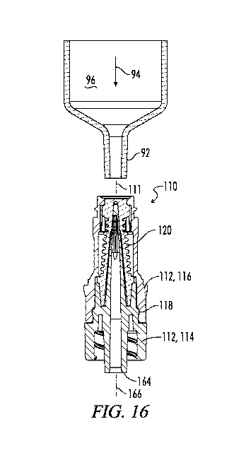
[0059] Fig. 16 is an elevation cross section view of an injection port
assembly using the
lower body portion and resilient member of Figs. 12 and 14 in the less
compressed position
of the resilient barrier. A syringe is shown in position about to be engaged
with the
injection port assembly.
[0060] Fig. 17 a view similar to Fig. 16, the syringe having been engaged with
the
injection port assembly to move the resilient barrier to its more compressed
second position
in which fluid flow through the injection port assembly is permitted.
[0061] Fig. 18 is an elevation view of a Y-site injection port assembly
including the
resilient member and cannula construction of Figs. 12-17.
[0062] Fig. 19 is an elevation cross section view of the Y-site injection port
assembly of
Fig. 18.
DETAILED DESCRIPTION
[0063] While the making and using of various embodiments of the present
invention are
discussed in detail below, it should be appreciated that the present invention
provides many
applicable inventive concepts that are embodied in a wide variety of specific
contexts. The
specific embodiments discussed herein are merely illustrative of specific ways
to make and
use the invention and do not delimit the scope of the invention.
[0064] The general arrangement of needleless IV injection ports and the
various usages
thereof in combination with other medical devices is described in greater
detail in pending
U.S. Patent Application 14/939,835 of Ryan entitled "Needleless, Intermittent,
Neutral
Displacement IV Injection Port" published as U.S. Patent Application
Publication No.
2016/0129235, the details of which are incorporated herein by reference.
Embodiment Of Figs. 1-11
[0065] Referring now to the drawings, and particularly to Fig. 1, a first
embodiment of an
injection port assembly is shown and generally designated by the numeral 10.
The injection
port assembly 10 has a longitudinal axis 11. The injection port assembly 10
includes a body
12 made up of a first mating structure 14 and a second mating structure 16.
The first mating
structure 14 may also be referred to as a lower body part 14, and the second
mating
structure 16 may also be referred to as an upper body part 16. The first and
second mating
structures 14 and 16 are coupled together by a snap lock feature 18.
CA 03067999 2019-12-19
WO 2018/236457
PCT/US2018/027446
11
[0066] The injection port assembly 10 further includes a resilient barrier 20
which is
configured to be received within the body 12 and which is compressible from a
less
compressed first position as seen for example in Fig. 1, in which fluid flow
through the
injection port assembly 10 is blocked, to a more compressed second position in
which fluid
flow through the injection port assembly 10 is permitted. It is noted that
Figs. 16 and 17
illustrate a similar less compressed first position and more compressed second
position for
the alternative embodiment of Figs. 12-17, and Figs. 16 and 17 are also
representative of the
change in shape of the resilient barrier 20 for the injection port assembly
10.
[0067] The details of construction of the first mating structure 14 are best
shown in Figs.
2-7. The details of construction of the second mating structure 16 are best
shown in Figs.
and 11. The details of construction of the resilient barrier 20 are best shown
in Figs. 8
and 9.
[0068] Fig. 1 shows the injection port assembly 10 in an assembled cross
section view
with the first and second mating structures 14 and 16 coupled together and
with the resilient
barrier 20 received within the body 12 between the first and second mating
structures 14
and 16.
[0069] As best seen in Figs. 8 and 9, the resilient barrier 20 includes an
internal cavity 22.
It will be appreciated that the resilient barrier 20 is formed from an
elastomeric material,
and is shown in Figs. 8 and 9 in its relaxed state in which the elastomeric
material is
relatively undeformed. It will also be appreciated that in Fig. 1 a hollow
cannula 24 of the
first mating structure 14 has been received in the internal cavity 22, thus
deforming portions
of the resilient barrier 20 radially outward so that the shape of the
resilient barrier 20 as seen
in Fig. 1, and particularly of its internal cavity 22, are different due to
the resilient
deformation thereof.
[0070] Referring now to Figs. 8 and 9 which show the resilient barrier 20 and
particularly
its internal cavity 22 in their relaxed state, the internal cavity 22 in this
relaxed state may be
described as including a cavity nose portion 26, a cavity sealing portion 28,
and a cavity
guide portion 30.
[0071] As shown in Fig. 9, the cavity nose portion 26 has a cavity nose
portion maximum
inside diameter 32.
[0072] The cavity sealing portion 28 may be described as having a cavity
sealing portion
length 34. The cavity sealing portion 28 has a minimum cavity sealing portion
inside
diameter 36 at its upper end and is slightly tapered to a maximum cavity
sealing portion
inside diameter 38 at its lower end. It is noted that the cavity sealing
portion 28 overall can
CA 03067999 2019-12-19
WO 2018/236457
PCT/US2018/027446
12
be described as having a cavity sealing portion inside diameter smaller than
the cavity nose
portion maximum inside diameter 32 along at least a majority of the cavity
sealing portion
length 34. The cavity sealing portion inside diameter may be smaller than the
cavity nose
portion maximum inside diameter 32 along substantially the entire cavity
sealing portion
length 34.
[0073] The cavity sealing portion 28 of internal cavity 22 may be described as
including a
frusto-conical portion of increasing diameter in a proximal direction which
increases from
cavity sealing portion minimum inside diameter 36 to cavity sealing portion
maximum
inside diameter 38.
[0074] The cavity nose portion 26 may be described as being bulbous in shape
as seen best
in Fig. 9, and having a semi-spherical distal end 54. The cavity nose portion
26 may be
further described as including a frusto-conical portion 56 of increasing
diameter in a
proximal direction from the semi-spherical distal end 54 to the cavity nose
portion
maximum inside diameter 32.
[0075] The cavity guide portion 30 is located on an opposite side of the
cavity sealing
portion 28 from the cavity nose portion 26. The cavity guide portion 30 tapers
radially
outward from the cavity sealing portion 28 and thus may be described as having
a cavity
guide portion inside diameter greater than the cavity sealing portion inside
diameter 38.
The cavity guide portion 30 may be further described as including a first
frusto-conical
portion 40 of increasing diameter in a proximal direction from the cavity
sealing portion 38,
and a second frusto-conical portion 42 adjacent the first frusto-conical
portion 40, the
second frusto-conical portion 42 having a smaller included angle than the
first frusto-
conical portion 40.
[0076] As previously noted, a hollow cannula 24 is coupled to the first mating
structure
14, and in the example illustrated, the hollow cannula 24 is integrally formed
with the first
mating structure 14. The hollow cannula 24 is configured to be received within
the resilient
barrier 20 as shown for example in Fig. 1.
[0077] The hollow cannula 24 includes a distal end portion 44 shown in
enlarged view in
Fig. 6. The distal end portion 44 is configured to extend through the
resilient barrier 20
when the resilient barrier 20 is in the more compressed second position. The
cannula distal
end portion 44 has at least one lateral outlet window 46 and in the example
shown has a pair
of lateral outlet windows 46 and 48.
[0078] As seen in Figs. 5 and 6, each of the lateral outlet windows 46, 48 has
a window
length 50 which is less than the cavity sealing portion length 34. As best
seen in Fig. 7, the
CA 03067999 2019-12-19
WO 2018/236457
PCT/US2018/027446
13
windows 46 and 48 also have a width 51 perpendicular to the longitudinal
central axis 11,
60 of the injection port assembly 10 and the cannula 24.
[0079] The cannula distal end portion 44 includes a cannula nose 52 located
distally of the
lateral outlet windows 46 and 48, and configured to be closely received in the
cavity nose
portion 26 of the resilient barrier 20 when the resilient barrier 20 is in the
less compressed
first position as shown in Fig. 1. The cannula nose may substantially fill the
cavity nose
portion when the resilient barrier is in the less compressed first position
with the cannula
nose closely received in the cavity nose portion. More particularly, the
cannula nose may
fill at least 90%, and more preferably at least 95%, of the cavity nose
portion 96 by volume.
[0080] The cannula distal end portion 44 has a cannula distal end portion
outside diameter
both distally and proximally of the lateral outlet windows 46 and 48, which
cannula distal
end portion outside diameter is sufficiently greater than the respective
inside diameters of
the cavity sealing portion 28 of internal cavity 22 of resilient barrier 20
when the cannula
nose 52 is received in the cannula nose portion 26 such that there is an
interference fit
between the cannula 24 and the resilient barrier 20. The interference fit
extends along the
lateral outlet windows 46 and 48 and both proximally and distally of the
lateral outlet
windows 46 and 48 so that the cavity sealing portion 28 of the resilient
barrier 20 seals
across the lateral outlet windows 46 and 48.
[0081] This is visualized in Fig. 1, wherein the relaxed position of the
cavity sealing
portion 28 of resilient barrier 20 is shown in dashed lines, and thus the
extent of radially
outward resilient deformation of the resilient barrier 20 by the cannula 24
received therein is
readily apparent and it is apparent that this radially deformed portion of the
cavity sealing
portion 28 of resilient barrier 20 extends both distally and proximally from
the lateral outlet
windows 46 and 48.
[0082] The area between the dashed line relaxed state representation 28 and
the solid line
position of cavity sealing portion 28 as seen in Fig. 1 may be described as an
interference fit
58 between the hollow cannula 24 and the resilient barrier 20. As is apparent
in Fig. 1, this
interference fit 58 between the cannula 24 and the resilient barrier 20
extends proximally
into the first frusto-conical portion 40 of the cavity guide portion 30 of
internal cavity 22 of
resilient barrier 20. The interference fit 58 may also be described as a
resilient interference
zone spanning the length of the lateral windows 46 and 48.
[0083] At any one cross section along the axis 11 of injection port assembly
10, the
interference fit 58 may be described as a radial interference which is
mathematically
determined by comparing the outside diameter of the cannula 24 to the inside
diameter of
CA 03067999 2019-12-19
WO 2018/236457
PCT/US2018/027446
14
the cavity sealing portion 28 in its relaxed state, and dividing that
difference by two to
provide the radial interference. Preferably the radial interference along the
interference fit
58 is at least about 0.001 inch, optionally at least about 0.002 inch,
optionally at least about
0.004 inch and optionally at least about 0.006 inch.
[0084] Preferably the interference fit 58 between the cannula 24 and the
resilient barrier 20
extends at least about 0.010 inch both proximally and distally from the
lateral outlet
windows 46 and 48.
Non-Circular Cross Section Fluid Passageway
[0085] The hollow cannula 24 has a longitudinal central axis 60 which is
coincident with
the central axis 11 of the injection port assembly 10.
[0086] The cannula 24 has an internal fluid passageway 62 defined therein as
best seen in
Fig. 3. The internal fluid passageway 62 communicates the lateral outlet
windows 46 and
48 with a proximal end of the first mating structure 14 which is configured to
communicate
with a fluid conduit 66 schematically illustrated in Fig. 3. The fluid conduit
66 may be
representative of any structure to which the injection port assembly 10 is to
be connected
for fluid flow therewith.
[0087] Fig. 7 shows a downward facing cross section of the cannula 24 taken
along line 7-
7 of Fig. 5 and shows that the internal fluid passageway 62 has a non-circular
cross section
axially proximal from the windows 46 and 48. This non-circular cross section
may be
described as having a cross sectional area greater than a cross section area
of a circle of
diameter equal to the window width 51.
[0088] The two outlet windows 46 and 48 may be described as being
diametrically
opposed as is best seen in Fig. 7, and as being diametrically spaced apart by
a window
spacing 68. The non-circular cross section internal fluid passageway 62 as
seen in Fig. 7
may be described as extending laterally to each of the two diametrically
opposed outlet
windows 46 and 48, and the non-circular cross section has a first lateral
cross section
dimension 70 at least equal to the window spacing 68 at an axial location
immediately
adjacent to a proximal end 72 of the windows 46 and 48.
[0089] The non-circular cross section of the internal fluid passageway 62 as
seen in Fig. 7
immediately adjacent the proximal ends 72 of windows 46 and 48 may also be
described as
having a second lateral cross section dimension 74 which is at least equal to
the window
width 51.
CA 03067999 2019-12-19
WO 2018/236457
PCT/US2018/027446
[0090] As is best visualized in Fig. 7, the non-circular cross section of the
internal fluid
passageway 62 may be described as being at least partially defined between
first and second
generally parallel opposed interior walls 76 and 78 of the hollow cannula 24.
[0091] The cannula 24 may further include first and second reinforcing ribs 80
and 82.
The ribs 80 and 82 may be described as first and second diametrically opposed
reinforcing
ribs 80 and 82 extending radially inwardly from the first and second opposed
interior walls
76 and 78, respectively.
[0092] This cross sectional shape of the internal fluid passageway 62 as
visually depicted
in Fig. 7 preferably extends along a non-circular cross section length 84
shown in Fig. 3.
As is apparent in Fig. 3, the non-circular cross section length 84 extends
along the window
length 50 and continues proximally beyond the window length 50 into the
internal
passageway 62 of cannula 24 by a further distance 86 at least as long as the
window length
50 and preferably longer than the window length 50.
[0093] Proximally of the non-circular cross sectional length 84, the internal
passageway 62
may transition into a circular cross section extending to the proximal end 64
of the first
mating structure 14.
[0094] The internal fluid passageway of non-circular cross section as depicted
for example
in Fig. 7 provides for increased fluid flow through the cannula 24 while
maintaining the
structural integrity of the cannula 24.
[0095] It will be appreciated by those skilled in the art that the typical
dimensions of the
cannula 24 are relatively small. For example, the cannula 24 may have an
outside diameter
88 adjacent its distal end of approximately 0.04 inch, and the window width 51
may for
example be approximately 0.026 inch. Thus if the internal fluid passageway 62
were of
completely circular cross section as was typical in the prior art, a circular
internal fluid
passageway 62 leading to the lateral windows 46 and 48 would typically have a
circular
cross section with a diameter of about 0.026 inch. By constructing the cannula
24 with the
non-circular cross sectional area depicted in Fig. 7 having a cross sectional
area greater than
a cross section of a circle of diameter equal to the window width 51,
increased fluid flow
through the cannula 24 for any given pressure of fluid supplied thereto is
provided.
Furthermore, due to the very small structures involved, the presence of the
reinforcing ribs
80 and 82 aids in maintaining structural integrity of the tip portion of the
cannula 24 around
the windows 46 and 48, while still allowing this greater cross section
internal passageway to
be provided.
CA 03067999 2019-12-19
WO 2018/236457
PCT/US2018/027446
16
Improved Snap Lock Feature
[0096] The snap lock feature 18 is improved over prior designs so as to
provide a
substantial increase in the tension force required to pull the first and
second mating
structures 14 and 16 apart after assembly.
[0097] Referring to Fig. 5, the first mating structure 14 includes a first
locking portion 18a
defined by a snap lock ring 300 having an outermost surface 301 defined
between a tapered
upper guiding surface 302 and a locking shoulder 304. The locking shoulder 304
is at
substantially 90 degrees to the outer surface 301 thus defining a relatively
sharp locking
edge 306. Located below the snap lock ring 300 is a stabilizing ring shelf
308.
[0098] The second mating structure 16 includes a second locking portion 18b
best seen in
Fig. 11. The second locking portion 18b includes a snap lock ring channel 310
in which
the snap lock ring 300 is to be received. Located below the snap lock ring
channel 310 is a
plurality of stabilizing ring securement segments 312 separated by gaps 314.
It can be seen
that the snap lock ring channel 310 is curved in cross-section and forms a
curved tapered
upper locking surface 316 on each of the stabilizing ring securement segments
312.
[0099] When the first and second mating structures 14 and 16 are snapped
together as seen
in Fig. 1, The sharp locking edge 306 of the snap lock ring 300 bites into the
curved tapered
upper locking surfaces 316 of the stabilizing ring securement segments 312 to
securely
prevent the first and second mating structures 14 and 16 from being pulled
back apart.
[00100] When the current design is compared to a snap lock feature like that
shown in
U.S. Patent Application Publication No. 2016/0129235 wherein the engaging
surfaces of the
snap lock ring and of the stabilizing ring securement segments are both
tapered at
complementary angles, a substantial increase in the force required to pull
apart the first and
second mating structures is provided. The required pull apart force was
increased from
about 14 pounds with the design of U.S. Patent Application Publication No.
2016/0129235
to about 54 pounds with the present design. The snap lock feature 18 can be
described as
having the first and second locking portions 18a and 18b configured such that
a force of at
least 30 pounds, and more preferably at least 40 pounds, is required to pull
apart the first
and second mating structures 14 and 16.
Embodiment Of Figs. 12-17
[00101] An alternative embodiment of an injection port assembly having three
lateral
outlet windows instead of two lateral outlet windows is shown in Figs. 12-17
and is
generally designated by the number 110.
CA 03067999 2019-12-19
WO 2018/236457
PCT/US2018/027446
17
[00102] The injection port assembly 10 has a longitudinal axis 111. The
injection port
assembly 110 is shown in assembled cross section in Fig. 16 and includes a
body 112 made
up of a first mating structure 114 and a second mating structure 116. The
first mating
structure 114 may also be referred to as a lower body part 114, and the second
mating
structure 116 may also be referred to as an upper body part 116. The first and
second
mating structures 114 and 116 are coupled together by a snap lock feature 118.
[00103] The injection port assembly 110 further includes a resilient barrier
120 which is
configured to be received within the body 112 and which is compressible from a
less
compressed first position as seen for example in Fig. 16, in which fluid flow
through the
injection port assembly 110 is blocked, to a more compressed second position
as seen for
example in Fig. 17, in which fluid flow through the injection port assembly
110 is
permitted.
[00104] The details of construction of the first mating structure 114 are best
shown in
Figs. 12-13. The details of construction of the second mating structure 116
are substantially
the same as was shown for the second mating structure 16 in Figs. 10 and 11.
The details of
construction of the resilient barrier 120 are best shown in Figs. 14 and 15.
[00105] Fig. 16 shows the injection port assembly 110 in an assembled cross
section view
with the first and second mating structures 114 and 116 coupled together and
with the
resilient barrier 120 received within the body 112 between the first and
second mating
structures 114 and 116.
[00106] As best seen in Figs. 14 and 15, the resilient barrier 120 includes an
internal
cavity 122. It will be appreciated that the resilient barrier 120 is formed
from an
elastomeric material, and is shown in Figs. 14 and 15 in its relaxed state in
which the
elastomeric material is relatively undeformed. It will also be appreciated
that in Fig. 16 a
hollow cannula 124 of the first mating structure 114 has been received in the
internal cavity
122, thus deforming portions of the resilient barrier 120 radially outward so
that the shape
of the resilient barrier 120 as seen in Fig. 16, and particularly of its
internal cavity 122, are
different due to the resilient deformation thereof.
[00107] Referring now to Figs. 14 and 15 which show the resilient barrier 120
and
particularly its internal cavity 122 in their relaxed state, the internal
cavity 122 in this
relaxed state may be described as including a cavity nose portion 126, a
cavity sealing
portion 128, and a cavity guide portion 130.
[00108] As shown in Fig. 15, the cavity nose portion 126 has a cavity nose
portion
maximum inside diameter 132.
CA 03067999 2019-12-19
WO 2018/236457
PCT/US2018/027446
18
[00109] The cavity sealing portion 128 may be described as having a cavity
sealing
portion length 134. The cavity sealing portion 128 has a minimum cavity
sealing portion
inside diameter 136 at its upper end and is slightly tapered to a maximum
cavity sealing
portion inside diameter 138 at its lower end. It is noted that the cavity
sealing portion 128
overall can be described as having a cavity sealing portion inside diameter
smaller than the
cavity nose portion maximum inside diameter 132 along at least a majority of
the cavity
sealing portion length 134. The cavity sealing portion inside diameter may be
smaller than
the cavity nose portion maximum inside diameter 132 along substantially the
entire cavity
sealing portion length 34.
[00110] The cavity sealing portion 128 of internal cavity 122 may be described
as
including a frusto-conical portion of increasing diameter in a proximal
direction which
increases from cavity sealing portion minimum inside diameter 136 to cavity
sealing portion
maximum inside diameter 138.
[00111] The cavity nose portion 126 may be described as being bulbous in shape
as seen
best in Fig. 15, and having a semi-spherical distal end 154. The cavity nose
portion 126
may be further described as including a frusto-conical portion 156 of
increasing diameter in
a proximal direction from the semi-spherical distal end 154 to the cavity nose
portion
maximum inside diameter 132.
[00112] The cavity guide portion 130 is located on an opposite side of the
cavity sealing
portion 128 from the cavity nose portion 126. The cavity guide portion 130
tapers radially
outward from the cavity sealing portion 128 and thus may be described as
having a cavity
guide portion inside diameter greater than the cavity sealing portion inside
diameter 138.
The cavity guide portion 130 may be further described as including a first
frusto-conical
portion 140 of increasing diameter in a proximal direction from the cavity
sealing portion
138, and a second frusto-conical portion 142 adjacent the first frusto-conical
portion 140,
the second frusto-conical portion 142 having a smaller included angle than the
first frusto-
conical portion 140.
[00113] As previously noted, a hollow cannula 124 is coupled to the first
mating structure
114, and in the example illustrated, the hollow cannula 124 is integrally
formed with the
first mating structure 114. The hollow cannula 124 is configured to be
received within the
resilient barrier 120 as shown for example in Fig. 16.
[00114] The hollow cannula 124 includes a distal end portion 144 shown in
enlarged view
in Fig. 13. The distal end portion 144 is configured to extend through the
resilient barrier
120 when the resilient barrier 120 is in the more compressed second position
of Fig. 17.
CA 03067999 2019-12-19
WO 2018/236457
PCT/US2018/027446
19
The cannula distal end portion 144 has at least one lateral outlet window 146
and in the
example shown has three lateral outlet windows 146, 147 and 148.
[00115] As seen in Figs. 12 and 13, each of the lateral outlet windows 146,
147 and 148
has a window length 150 which is less than the cavity sealing portion length
134. As best
seen in Fig. 12C, the windows 146, 147 and 148 also have a width 151
perpendicular to the
longitudinal central axis 111, 160 of the injection port assembly 110 and the
cannula 124.
[00116] The cannula distal end portion 144 includes a cannula nose 152 located
distally of
the lateral outlet windows 146, 147 and 148, and configured to be closely
received in the
cavity nose portion 126 of the resilient barrier 120 when the resilient
barrier 120 is in the
less compressed first position as shown in Fig. 16.
[00117] The cannula distal end portion 144 has a cannula distal end portion
outside
diameter both distally and proximally of the lateral outlet windows 146, 147
and 148, which
cannula distal end portion outside diameter is sufficiently greater than the
respective inside
diameters of the cavity sealing portion 128 of internal cavity 122 of
resilient barrier 120
when the cannula nose 152 is received in the cannula nose portion 126 such
that there is an
interference fit between the cannula 124 and the resilient barrier 120. The
interference fit
extends along the lateral outlet windows 146, 147 and 148 and both proximally
and distally
of the lateral outlet windows 146, 147 and 148 so that the cavity sealing
portion 128 of the
resilient barrier 120 seals across the lateral outlet windows 146, 147 and
148.
[00118] Preferably the radial interference along the interference fit is at
least about 0.001
inch, optionally at least about 0.002 inch, optionally at least about 0.004
inch and optionally
at least about 0.006 inch. Preferably the interference fit between the cannula
124 and the
resilient barrier 120 extends at least about 0.010 inch both proximally and
distally from the
lateral outlet windows 146, 147 and 148.
[00119] The hollow cannula 124 has a longitudinal central axis 160 which is
coincident
with the central axis 111 of the injection port assembly 110.
[00120] The cannula 124 has an internal fluid passageway 162 defined therein
as best seen
in Figs. 12A-12C. The internal fluid passageway 162 communicates the lateral
outlet
windows 146, 147 and 148 with a proximal end of the first mating structure 114
which is
configured to communicate with a fluid conduit 166 schematically illustrated
in Fig. 16.
The fluid conduit 166 may be representative of any structure to which the
injection port
assembly 110 is to be connected for fluid flow therewith.
[00121] Fig. 12D shows an upward facing cross section of the cannula 124 taken
along
line D-D of Fig. 12. Figs 12A, 12B and 12C show downward facing cross sections
of the
CA 03067999 2019-12-19
WO 2018/236457
PCT/US2018/027446
cannula 124 taken along lines A-A, B-B and C-C, respectively. Figs. 12A-12C
show that
the internal fluid passageway 162 has a non-circular cross section axially
proximal from the
windows 146, 147 and and 148. This non-circular cross section may be described
as having
a cross sectional area greater than a cross section area of a circle of
diameter equal to the
window width 151.
[00122] The three outlet windows 146, 147 and 148 may be described as being
equally
circumferentially spaced about the axis 160. The non-circular cross section
internal fluid
passageway 162 as seen in Figs. 12A-12D may be described as extending
laterally to each
of the outlet windows 146, 147 and 148.
[00123] The non-circular cross section of internal fluid passageway 162 may be
described
as a three lobed cross section. As can be seen in comparing Figs. 12A, 12B and
12C, the
three lobed cross section tapers radially outward. This cross sectional shape
of the internal
fluid passageway 62 as visually depicted in Figs. 12A-12C preferably extends
along a non-
circular cross section length 184 shown in Fig. 13. As is apparent in Fig. 13,
the non-
circular cross section length 184 extends along the window length 150 and
continues
proximally beyond the window length 150 into the internal passageway 162 of
cannula 124
by a further distance 186 at least as long as the window length 150 and
preferably longer
than the window length 150. Proximally of the non-circular cross sectional
length 184, the
internal passageway 162 may transition into a circular cross section extending
to the
proximal end 164 of the first mating structure 114.
Improved Performance
[00124] The provision of the interference fit 58 between the cannula 24 and
the resilient
barrier 20 of Figs. 1-11, and of the cannula 124 and resilient barrier 120 of
the embodiment
of Figs. 12-17, has provided substantially increased resistance to leaking due
to back
pressure within the injection port assemblies 10 and 110 as compared to a
similar prior
design of the assignee of the present invention as depicted in U.S. Patent
Application
Publication No. 2016/0129235.
[00125] For example, using the embodiment of Figs. 12-17, tests were run on
back
pressure resistance, flow rate and fluid displacement.
[00126] Average back pressure resistance has improved from 47 psi with the
previous
design to over 68 psi with the design depicted herein having the interference
fit. Testing was
done using standardized procedures wherein each sample was submerged in water
and
CA 03067999 2019-12-19
WO 2018/236457
PCT/US2018/027446
21
subjected to increased pressure until air bubbles were observed leaking from
the submerged
sample.
[00127] Average fluid flow rates at gravity increased from 48 mL/min for a
similar design
having a circular cross section internal fluid passageway, up to an average of
approximately
139 mL/min for the cross sectional area generally like that shown in Fig. 12A-
12C. In these
tests for a lot of 59 samples, flow rates ranged from a minimum of 127 mL/min
to a max of
155 mL/min for sterilized samples, and from a minimum of 127 mL/min to a max
of 163
mL/min for non-sterilized samples
[00128] Additionally, fluid reflux was measured at 0.00 mL with the embodiment
of Figs.
12-17.
Methods of Use
[00129] As depicted in Figs. 1 and 16 for the respective embodiments, the
upper and lower
parts 16, 116 and 14, 114 of the bodies 12, 112 are assembled with the
resilient barriers 20,
120 contained therein and with the cannula 24, 124 received with the internal
cavity 22, 122
of respective resilient barrier 20, 120. It will be appreciated that in the
assembled
arrangement as seen in Figs. 1 and 16, there may be a slight axial compression
of the
resilient barrier 20, 120 from its completely relaxed state. The position of
the resilient
barrier 20, 120 as depicted in Figs. 1 and 16 may be described as a less
axially compressed
first position in which fluid flow through the injection port assembly 10, 110
is blocked. It
will be appreciated that the distal end of the resilient barrier 20, 120 has a
precut slit 90, 190
formed therein through which the distal end portion 44, 144 of cannula 24, 124
will
protrude when the resilient barrier 20, 120 is moved to its more axially
compressed second
position like that shown in Fig. 17.
[00130] The injection port assembly 10, 110 may be connected to various
conduits and
medical devices so as to provide for intravenous injection into the patient's
body and for
collection of blood samples from the patient. The injection port assembly 10
may be
incorporated into an IV pump set or IV administration set in a Y-site
injection port
configuration. Figs. 18 and 19 for example, show a Y-site injection port
arrangement 210
utilizing the three window embodiment of Figs. 12-17.
[00131] As depicted in Fig. 16 and 17, the resilient barrier 120 may be moved
from its
closed first position to its open second position by engagement of the
injection port
CA 03067999 2019-12-19
WO 2018/236457
PCT/US2018/027446
22
assembly 110 by a male-luer slip syringe 92. Beginning in the closed position
of Fig. 16, as
is indicated by the arrow 94 the syringe 92 is pushed downward engaging the
distal end of
the resilient barrier 120 and forcing it downward relative to the cannula 124
so as to expose
the distal end portion 144 of cannula 124 thus allowing the lateral windows
such as 146,
147 and 148 to communicate with the interior 96 of syringe 92. This allows
fluids to be
injected into or withdrawn from the patient's blood stream.
[001 32] The resilient barrier 20, 120 may for example be formed of a silicone
rubber
material having a diameter in the range of from about 50 to about 70, and
preferably having
a diameter of about 60. The silicone rubber material may have a small amount
of phenyl oil
included therein to provide an internal lubricant when the resilient barrier
20, 120 slides
along the outer surface of the cannula 24, 124. The exterior surface of
cannula 24, 124 may
be treated to form a slightly roughened surface with irregularities on the
order of 0.001 inch
and may be lubricated with silicone oil to further aid in the movement of the
resilient barrier
20 between its closed and open positions of Figs. 16 and 17. These features
aid in allowing
the resilient barrier 20, 120 to substantially instantaneously snap back from
its open position
of Fig. 17 to its closed position of Fig. 16 upon removal of the syringe 92.
[001 33] Thus it is seen that the apparatus and methods of the present
invention readily
achieve the ends and advantages mentioned as well as those inherent therein.
While certain
preferred embodiments have been illustrated and described for purposes of the
present
disclosure, numerous changes in the arrangement and construction of parts and
steps will be
apparent to those skilled in the art, which changes are encompassed within the
scope and
spirit of the present invention as defined by the appended claims.