Note: Descriptions are shown in the official language in which they were submitted.
CA 03068938 2020-01-03
WO 2019/013838 PCT/US2018/014220
SCREENING PLATFORM TO IDENTIFY THERAPEUTIC DRUGS OR AGENTS
FOR TREATMENT OF ALZHEIMER'S DISEASE
RELATED APPLICATIONS
[0001] This application claims benefit under 35 U.S.C. 119(e) of the U.S.
Provisional
Application No. 62/530,125, filed July 8, 2017 and the U.S. Provisional
Application No.
62/535,589, filed July 21, 2017, the contents of each of which are
incorporated herein by reference
in their entireties.
TECHNICAL FIELD
[0002] This disclosure is related generally to methods and compositions for
screening
candidate substances for the prevention or treatment of neurodegenerative
disorders. The
disclosure also relates generally to compositions and methods for modulating
the function of
cells expressing CD33.
BACKGROUND
[0003] Neurodegenerative diseases are a serious, common and growing
worldwide
problem. These include Alzheimer's disease (AD), cognitive impairment,
Parkinson's
disease, dementia, schizophrenia, amyotrophic lateral sclerosis (ALS),
Huntington's disease
or multiple sclerosis. Particularly in the elderly, neurodegenerative diseases
cause suffering,
reduced quality of life and are a major predictor of mortality. Alzheimer's
disease (AD) is the
most common form of neurodegenerative disease in the elderly and with an
increasing
population of the elderly in the US and worldwide, AD is reaching epidemic
proportions. AD
is characterized by progressive dementia and personality dysfunction. The
abnormal
accumulation of amyloid plaques in the vicinity of degenerating neurons and
reactive
astrocytes is a pathological characteristic of AD.
[0004] Microglia are the brain resident immune cells, responsible for
clearing toxic
pathogens, such as amyloid-f3 (Abeta, A13). During AD progression, microglia
transition from
a neuroprotective/pro-phagocytic to a neurotoxic state, characterized by
increased secretion
of pro-inflammatory cytokines (Heneka et al. Nature Immunology, 2015). It was
found that
microglial receptor CD33 is a late-onset AD risk factor in a large family-
based GWAS
analysis (Bertram et al., Am. J. Hum. Genet., 2008). It was also discovered
that CD33
promoted amyloid pathology by inhibiting uptake and clearance of AP in
microglial cells
(Griciuc et al., Neuron, 2013).
[0005] Drugs such as donepezil and memantine treat the symptoms of AD but
they do not
treat the disease itself. A new drug, T-817MA
1
CA 03068938 2020-01-03
WO 2019/013838 PCT/US2018/014220
(1-{342-(1-Benzothiophen-5-yl)ethoxy]propy1}-3-azetidinol maleate) is in phase
1 and 2
clinical trials and it's neuroprotective effects against toxicity from Afl and
actions promoting
neurite outgrowth are well documented (Takamura et al., Neurobiol Aging,
2014). How these
drugs interact and modulate the activity of CD33 is unknown. Additionally,
there remains a
need to discover new treatments for neurodegenerative diseases such as AD. For
example,
rapid screening methods that can be applied to biologically relevant compounds
and
combinations of compounds, and their dose response would provide a significant
advance in
this field and lead to break-through therapies and drugs that can help
eradicate and control
these diseases.
SUMMARY
[0006] The present disclosure relates to the discovery of methods for
screening candidate
substances for the prevention or treatment of neurodegenerative disorders. The
disclosure
also relates to the discovery of the microglial receptor CD33 as a risk factor
in
neurodegenerative diseases and to compositions and methods for modulating the
function of
cells expressing CD33 as a screening assay and mechanistic probe.
[0007] In one aspect, there is provided a method for testing a therapeutic
efficacy of a
candidate substance for a prevention or treatment agent of a neurodegenerative
disorder.
Generally, the method comprises treating immune or immune-like cells
expressing full-length
human CD33, also referred to as CD33 expressing immune or immune-like cells
herein, with
a candidate substance at a relevant concentration, further treating said
treated CD33
expressing immune or immune-like cells with amyloid-0 (Abeta, AP) at a
relevant
concentration or lipopolysaccharide, and measuring intracellular levels of Afl
(for Afl treated
cells) or culture media levels of pro-inflammatory cytokines (for
lipopolysaccharide treated
cells). The CD33 expressing immune or immune-like cells optionally can be
cultured prior to
treatment with the candidate substance.
[0008] In embodiments of the method comprising treatment with AP, higher
levels of AP
in CD33 cells treated with the candidate substance, relative to a negative
control, indicate that
the tested candidate substance has therapeutic efficacy. In embodiments of the
method
comprising treatment with lipopolysaccharide, lower levels of one or more pro-
inflammatory
cytokines in the culture media of CD33 cells treated with the candidate
substance, relative to
a negative control, indicate that the tested candidate substance has
therapeutic efficacy.
[0009] In some embodiments, the method comprises treating CD33 expressing
immune
or immune-like cells with a candidate substance at a relevant concentration;
further treating
said treated CD33 expressing immune or immune-like cells with amyloid-0
(Abeta, AP) at a
2
CA 03068938 2020-01-03
WO 2019/013838 PCT/US2018/014220
relevant concentration; and measuring intracellular levels of AP in said CD33
expressing
cells, wherein higher levels of AP in CD33 cells treated with the candidate
substance, relative
to a negative control, indicate that the tested candidate substance has
therapeutic efficacy.
[0010] In some embodiments, the method comprises treating CD33 expressing
immune
or immune-like cells with a candidate substance at a relevant concentration;
further treating
said treated CD33 expressing immune or immune-like cells with
lipopolysaccharide; and
measuring levels of pro-inflammatory cytokines in culture media of said CD33
expressing
cells, wherein lower levels of pro-inflammatory cytokines in the culture media
of CD33 cells
treated with the candidate substance, relative to a negative control, indicate
that the tested
candidate substance has therapeutic efficacy.
[0011] In another aspect, there is provided a method for testing a binding
interaction of a
substance with human CD33. Generally, the method comprises treating said CD33
expressing
immune or immune-like cells with a candidate substance at a relevant
concentration; and
measuring a read-out of the binding interaction using a standard assay,
wherein a higher read-
out in CD33 cells treated with the substance, relative to a negative control,
indicate a binding
interaction. The CD33 expressing immune or immune-like cells optionally can be
cultured
prior to treatment with the candidate substance.
[0012] In some embodiments of the various aspects described herein, the
CD33
expressing immune or immune-like cells are microglial cells.
[0013] In some embodiments of the various aspects described herein, the
CD33
expressing immune or immune-like cells optionally can be cultured prior to
treatment with
the candidate substance.
[0014] The substances identified as having therapeutic efficacy or a
binding interaction
with CD33 can be used for modulating a function of a microglial cells. The
microglial cell
can be in vitro, in vivo, or ex vivo. Accordingly, in another aspect, provided
herein is a
method for modulating a function of a microglial cell in a subject. Generally,
the method
comprises administering to a patient or subject in need thereof a substance
identified by a method
described herein.
[0015] In some embodiments, the substance that is administered to the
subject is identified as
having therapeutic efficacy by a method described herein.
[0016] In some embodiments, the substance that is administered to the
subject is
1-(3-(2-(1-benzothiophen-5-yl)ethoxy)propyl)azetid in-3-ol or a salt thereof
3
CA 03068938 2020-01-03
WO 2019/013838 PCT/US2018/014220
[0017] In some embodiments, the method can be used for treating or
preventing a
neurodegenerative disease in a subject. For example, by administering to a
patient or subject in
need thereof a therapeutic dose or dosages of a substance identified by a
method described
herein. Exemplary neurodegenerative disorders include, but are not limited to,
Alzheimer's
disease, mild cognitive impairment, Parkinson's disease, dementia,
schizophrenia,
amyotrophic lateral sclerosis, Huntington's disease and multiple sclerosis.
For example, the
methods can be applied to Alzheimer's disease.
[0018] In another aspect, there is provided a pharmaceutical composition
for modulating
a function of microglial cells. Generally, the pharmaceutical composition
comprises
1-(3-(2-(1-benzothiophen-5-yl)ethoxy)propyl)azetidin-3-ol, or a salt thereof.
[0019] In some embodiments, the pharmaceutical composition further
comprises a
pharmaceutically acceptable excipient or carrier.
[0020] In some embodiments of the various aspects described herein, the
method
enhances phagocytosis or inhibits cytokine production of microglial cells.
[0021] In some embodiments of the various aspects described herein, the
compound is
1-(3-(2-(1-benzothiophen-5-yl)ethoxy)propyl)azetidin-3-ol maleate, also
referred to as T-
817MA herein.
[0022] Other features and advantages of the invention will be apparent from
the Detailed
Description, and from the Claims. Thus, other aspects of the invention are
described in the
following disclosure and are within the ambit of the invention.
BRIEF DESCRIPTION OF THE DRAWINGS
[0023] FIG. 1 shows the results as a bar graph from a lactate dehydrogenase
(LDH)
toxicity assay on naïve BV2 microglial cells after 5 hours of treatment with a
DMSO,
817MA, 817A11, and 614P. No statistically significant toxicity was seen at
these
concentrations.
[0024] FIG. 2 shows the results as a bar graph from an LDH toxicity assay
on wtCD33-
expressing microglial cells after 5 hours of treatment with a DMSO, 817MA,
817A11, and
614P. No statistically significant toxicity is seen at these concentrations.
[0025] FIG. 3 shows the results for an Abeta42 uptake assay into naïve BV2
microglial
cells as a line graph. The results for DMSO, 817MA, 817A11 and 614P into naïve
BV2
microglial cells is shown. Compounds 817MA and 817A11 have similar Abeta42
uptake
EC50s. Compound 614P has a higher EC5o.
[0026] FIG. 4 shows the results for an Abeta42 uptake assay into wt-CD33
expressing
BV2 cells as a line graph. The results for DMSO, 817MA, 817A11 and 614P into
wt-CD33
4
CA 03068938 2020-01-03
WO 2019/013838 PCT/US2018/014220
expressing BV2 cells is shown. Compounds 817MA and 817A11 have similar Abeta42
uptake EC5os. Compound 614P has a higher EC5o.
[0027] FIG. 5 shows the results for an Abeta40 uptake assay into naïve BV2
microglial
cells as a line graph. The results for DMSO, 817MA, 817A11 and 614P into naïve
BV2
microglial cells is shown. Compound 817A11 has a lower uptake EC5o than 817MA.
Compound 614P has a higher EC5o than both 817MA and 817A11.
[0028] FIG. 6 shows the results for an Abeta40 uptake assay into wt-CD33
expressing
BV2 cells as a line graph. The results for DMSO, 817MA, 817A11 and 614P into
wt-CD33
expressing BV2 cells is shown. Compounds 817A11 has a lower uptake EC5o than
817MA.
Compound 614P has a higher EC50 than both 817MA and 817A11.
[0029] FIG. 7 is a table showing LPS activation. Ten cytokines were
simultaneously
analyzed in microglial conditioned media. The cytokines KC/GRO, IL-6, IL-10
and TNF-a
showed detectable levels upon LPS activation. KC/GRO could only be detected in
undiluted
media. Cytokines IFN-y, IL-2, IL-5, IL-12p70, IL-10 and IL-4 were not
detected.
[0030] FIG. 8 shows the results for a first experiment for BV2 LPS
activation of TNFa as
a line graph. The graph shows the TNFa concentration production response as a
function of
DMSO, 817MA, 817A11 and 614P added concentrations. Compound 817MA is the only
compound effectively reducing TNFa production.
[0031] FIG. 9 shows the results for a second experiment for BV2 LPS
activation of TNFa
as a line graph. The graph shows the TNFa concentration production response as
a function
of DMSO, 817MA, 817A11 and 614P added concentrations. Compound 817MA is the
only
compound effectively reducing TNFa production.
[0032] FIG. 10 shows the results for a first experiment for BV2 LPS
activation of IL-6 as
a line graph. The graph shows the IL-6 concentration production response as a
function of
DMSO, 817MA, 817A11 and 614P added concentrations. Compound 817MA is the most
effective compound for reducing IL-6 production.
[0033] FIG. 11 shows the results for a second experiment for BV2 LPS
activation of IL-6
as a line graph. The graph shows the IL-6 concentration production response as
a function of
DMSO, 817MA, 817A11 and 614P added concentrations. Compound 817MA is the most
effective compound for reducing IL-6 production.
[0034] FIG. 12 shows the results for a first experiment for BV2 LPS
activation of IL-10
as a line graph. The graph shows the IL-10 concentration production response
as a function of
DMSO, 817MA, 817A11 and 614P added concentrations. Compound 817MA is the most
effective compound for reducing IL-10 production.
CA 03068938 2020-01-03
WO 2019/013838 PCT/US2018/014220
[0035] FIG. 13 shows the results for a second experiment for BV2 LPS
activation of IL-
as a line graph. The graph shows the IL-10 concentration production response
as a
function of DMSO, 817MA, 817A11 and 614P added concentrations. Compound 817MA
is
the most effective compound for reducing IL-10 production.
[0036] FIG. 14 shows the results for a first experiment for BV2 LPS
activation of
KC/GRO as a line graph. The graph shows the KC/GRO concentration production
response
as a function of DMSO, 817MA, 817A11 and 614P added concentrations.
[0037] FIG. 15 shows the results for a second experiment for BV2 LPS
activation of
KC/GRO as a line graph. The graph shows the KC/GRO concentration production
response
as a function of DMSO and 817MA added concentrations.
[0038] FIG. 16 shows the results in a bar graph form for data from a first
test in AD 3D
ReN cell culture system. The test is and LDH assay in HReN30-mGAP30 media
after one-
week treatment with test compounds DMSO, T-817MA (817MA), T-817A11(817A11) and
T-614 (614P). n is 3 or 4 for each cell line.
[0039] FIG. 17 shows the results in a bar graph form for data from a second
test in AD
3D ReN cell culture system. The test is an LDH assay in HReN30-mGAP10#D4 media
after
one-week treatment with test compounds DMSO, T-817MA (817MA), T-817A11(817A11)
and T-614 (614P). n is 3 or 4 for each cell line.
[0040] FIG. 18A-18H shows a series of bar graphs of data for soluble
(media) (horizontal
axis) and insoluble Abeta levels (vertical axis) after drug treatments. The
test compounds are
T-817MA (817MA), T-817A11(817A11) and T-614 (614P). FIG. 18A shows Abeta40 in
HReN30 (HReN-mGAP30) Media. FIG. 18B shows Abeta42 in HReN30 (HReN-mGAP30)
Media. FIG.18C shows Abeta40 in HReN30 (HReN-mGAP30) insoluble fraction.
FIG.18D
shows Abeta42 in HReN30 (HReN-mGAP30) insoluble fraction. FIG.18E to 18H are
ReN-
mGAP#D4 experiments. FIG.18E shows Abeta40 in ReN-mGAP10#D4 (ReN-mGAP#D4)
Media. FIG.18F shows Abeta42 in ReN-mGAP1O#D4 (ReN-mGAP#D4) Media. FIG.18G
shows Abeta40 in ReN-mGAP1O#D4 (ReN-mGAP#D4) insoluble fraction. FIG.18H shows
Abeta42 in ReN-mGAP10#D4 (ReN-mGAP#D4) insoluble fraction.
[0041] FIG.19A to19D shows a series of bar graphs of data for insoluble p-
tau and total
p-tau levels after drug treatments. The test compounds are DMSO, T-817MA
(817MA), T-
817A11(817A11) and T-614 (614P). FIG.19A andl9B are HReN-mGAP30 tests. FIG.
19A
shows pTau181 concentration (unit/mL) in HReN30 insoluble fraction. FIG. 19B
shows
pTau181 concentration (pg/mL) in HReN30 insoluble fraction. FIG. 19C and FIG.
19D are
ReN-mGAP#D4 tests. FIG. 19C shows pTau181 concentration (unit/mL) in ReN-
6
CA 03068938 2020-01-03
WO 2019/013838 PCT/US2018/014220
mGAP10#D4 insoluble fraction. FIG. 19D shows pTau181 concentration (pg/mL) in
ReN-
mGAP10#D4 insoluble fraction.
[0042] FIG. 20 shows a series of images in ReN-mGAP#D4 (4-week
differentiation).
[0043] FIG. 21 shows a series of images in HReN-mGAP30 (7-week
differentiation).
[0044] FIG. 22 shows a bar graph of data for sodium nitroprusside (SNP)
toxicity studies
in an AD 3D ReN cell culture system. The data is of WST-8 assay in ReN cells
without B27
after one-day treatment with SNP.
[0045] FIG. 23 shows a bar graph of data for a WST-8 assay in ReN cells
without B27
after four days of 817MA and one day of SNP treatment.
[0046] FIG. 24 shows a bar graph of data for a WST-8 assay (% viability) in
ReN cells
without B27 after four days of 817MA and one day of SNP treatment.
[0047] FIG. 25 show a bar graph of data for a toxicity (LDH) assay in
microglial cells.
The test compound is 817MA at various concentrations and includes a DMSO
control. The
50[tM concentration was excluded from the uptake analysis.
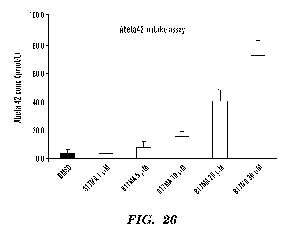
[0048] FIG. 26 shows a bar graph of data for an Abeta42 uptake assay. The
data confirms
uptake of Abeta42 by 817MA in microglial cells. EC50 is about 20 [tM following
24 hours of
compound pre-treatment.
[0049] FIG. 27A and 27B show exemplary time line diagrams for 817MA
treatments.
[0050] FIG. 28 shows a bar graph of data for cell viability to SNP
concentration at day
26.
[0051] FIG. 29 shows a bar graph of data for cell viability at selected
concentrations. n =
3 or 4 for each cell line.
[0052] FIG. 30A and 30B show bar graphs showing data for the 817MA effect
on SNP-
induced toxicity after 3 days (FIG. 30A) and after 3 weeks (FIG. 30B).
[0053] FIG. 31A and 31B show bar graphs showing data for the 817MA effect
on cell
viability (no SNP) for 3 days (FIG 31A) and for 3 weeks (FIG. 31B).
[0054] FIG. 32 shows a series of microscope images illustrating the SNP
effect on cells.
[0055] FIG. 33 shows a second series of microscope images illustrating the
SNP effect on
cells.
[0056] FIG. 34 shows a bar graph of data for the effect of a four-day
treatment with
817MA on SNP-induced toxicity in non-AD cells.
DETAILED DESCRIPTION
7
CA 03068938 2020-01-03
WO 2019/013838 PCT/US2018/014220
[0057] The present disclosure is based, in part, on the discovery of
valuable tools to study
potential drugs for the treatment of neurodegenerative diseases or disorders.
The disclosure is
also based in part on producing cells expressing microglial receptors that are
a risk factor in
late-onset AD and that lead to amyloid pathology. It has been found as
described herein that
such cells can be used for screening potential drug candidates as therapeutics
for
neurodegenerative disorders. The cells also provide insight into the mechanism
of drug
activity and to the discovery of important features (e.g., structure to
activity relationships) of
effective drug treatments. In addition, the disclosure provides compositions
that are effective
in the treatment of neurodegenerative disorders and their effect on the CD33
expressing cells.
[0058] Accordingly, in one embodiment, there is provided a method for
testing a
therapeutic efficacy of a candidate substance for a prevention or treatment
agent of a
neurodegenerative disorder. Generally, the method comprises treating immune or
immune-
like cells expressing full-length human CD33 with a candidate substance at a
relevant
concentration. The method also includes treating the treated CD33 expressing
immune or
immune-like cells with amyloid-f3 at a relevant concentration, and measuring
intracellular
levels of AP. In an alternative embodiment to the AP treatment, the method
includes treating
the CD33 expressing immune or immune-like cells with lipopolysaccharide and
measuring
the levels of pro-inflammatory cytokines in a cultured media of these cells.
The CD33
expressing immune or immune-like cells optionally can be cultured prior to
treatment (AP or
lipopolysaccharides) with the candidate substance.
[0059] A therapeutic refers to any drug, substance, compound, combination
of
compounds, treatment agent, or treatment therapy that is a used for the
purpose of alleviating
the symptoms of or curing a disease, condition or disorder. In some
embodiments the
therapeutic can be a test compound, for which the effectiveness of the
compound is not
known until the screening assay or test is completed.
[0060] Therapeutic efficacy relates to how effective a test compound (e.g.,
a substance,
compound, combination of compounds, treatment agent, or treatment therapy) is.
A test
compound having therapeutic efficacy means it is more effective for
alleviating the
symptoms of or curing a disease or condition as compared a control. In
addition, a high
therapeutic efficacy of a test compound means it is more effective for
alleviating the
symptoms of or curing a disease or condition as compared to another test
compound with a
lower therapeutic efficacy. For example, the control can be a compound that
has a lower
therapeutic efficacy than a test compound with therapeutic efficacy, and the
control is not as
effective in alleviating the symptoms of or curing a disease or condition. The
term "more
8
CA 03068938 2020-01-03
WO 2019/013838 PCT/US2018/014220
effective" can include that a lower dosage of the therapeutic provides the
same amount of
benefit, has fewer undesirable, harmful or toxic side effects, or the more
effective therapeutic
has additional benefits (e.g., health benefits, cost benefits), as compared to
the less effective
therapeutic.
[0061] Toxicity and therapeutic efficacy can be determined by standard
pharmaceutical
procedures in cell cultures or experimental animals, e.g., for determining the
LD50 (the dose
lethal to 50% of the population) and the ED50 (the dose therapeutically
effective in 50% of
the population). The dose ratio between toxic and therapeutic effects is the
therapeutic index
and it can be expressed as the ratio LD50/ED50. Compositions that exhibit
large therapeutic
indices are preferred. As used herein, the term ED denotes effective dose and
is used in
connection with animal models. The term EC denotes effective concentration and
is used in
connection with in vitro models.
[0062] As used herein a "control" is a drug, substance, compound, compounds
and/or test
condition with a known therapeutic effect, such as no therapeutic effect or
efficacy or some
specific amount of therapeutic effect of efficacy. As used herein, a "negative
control" can be
a control that has similar physical characteristics to the test therapeutic
composition but is
known to have no therapeutic effect. For example, the negative control can be
a solvent,
diluent or delivery agent that the test compound is dissolved/combined with
during the test,
but as the negative control the test compound is excluded in/from the solvent,
diluent or
delivery agent. For example, the test compound can be any one or more of a
solvent, DMSO,
water, alcohol, a micelle, vesicle, protein, polymer or complexing agent. A
"positive control"
can be a control for which there is a known therapeutic effect and would
therefore provide a
positive result in a test for that therapeutic effect.
[0063] As used herein, the term "candidate compound", "candidate
substance", "test
compound" or "test agent" refers to any compound, molecule or agent that is to
be tested. As
used herein, the terms, which are used interchangeably, refer to biological or
chemical
compounds such as simple or complex organic or inorganic molecules, small
molecules,
peptides, proteins, oligonucleotides, polynucleotides, carbohydrates, or
lipoproteins. A vast
array of compounds can be synthesized, for example oligomers, such as
oligopeptides and
oligonucleotides, and synthetic organic compounds based on various core
structures, and
these are also included in the terms noted above. In addition, various natural
sources can
provide compounds for screening, such as plant or animal extracts, and the
like. Compounds
can be tested singly or in combination with one another. Agents or candidate
compounds can
be randomly selected or rationally selected or designed. As used herein, an
agent or candidate
9
CA 03068938 2020-01-03
WO 2019/013838 PCT/US2018/014220
compound is said to be "randomly selected" when the agent is chosen randomly
without
considering the specific interaction between the agent and the target compound
or site. As
used herein, an agent is said to be "rationally selected or designed", when
the agent is chosen
on a nonrandom basis which takes into account the specific interaction between
the agent and
the target site and/or the conformation in connection with the agent's action.
In some
embodiments, the assays described herein can be used to guide a rational
design, for example,
by providing mechanistic insight into the efficacy of a test compound which is
feed in an
iterative fashion into a series of assays or screens while refining the
selection of test
compounds. A test compound can be a control compound.
[0064] As used herein, the term "small molecule" can refer to compounds
that are "natural
product-like," however, the term "small molecule" is not limited to "natural
product-like"
compounds. Rather, a small molecule is typically characterized in that it
contains several
carbon¨carbon bonds, and has a molecular weight more than about 50, but less
than about
5000 Daltons (5 kD). Preferably the small molecule has a molecular weight of
less than 3 kD,
still more preferably less than 2 kD, and most preferably less than 1 kD. In
some cases, it is
preferred that a small molecule have a molecular mass equal to or less than
700 Daltons.
[0065] Depending upon the particular embodiment being practiced, the test
compounds
can be provided free in solution, or may be attached to a carrier, or a solid
support, e.g.,
beads. A number of suitable solid supports may be employed for immobilization
of the test
compounds. Examples of suitable solid supports include agarose, cellulose,
dextran
(commercially available as, i.e., Sephadex, Sepharose) carboxymethyl
cellulose, polystyrene,
polyethylene glycol (PEG), filter paper, nitrocellulose, ion exchange resins,
plastic films,
polyaminemethylvinylether maleic acid copolymer, glass beads, amino acid
copolymer,
ethylene-maleic acid copolymer, nylon, silk, etc. Additionally, for the
methods described
herein, test compounds can be screened individually, or in groups. Group
screening is
particularly useful where hit rates for effective test compounds are expected
to be low such
that one would not expect more than one positive result for a given group.
Group screening is
also useful for determining hits that can act synergistically.
[0066] As used herein, the term "neurodegenerative disease" refers to a
varied assortment
of central nervous system disorders characterized by gradual and progressive
loss of neural
tissue and/or neural tissue function. A neurodegenerative disease is a class
of neurological
disorder or disease, and where the neurological disease is characterized by a
gradual and
progressive loss of neural tissue, and/or altered neurological function,
typically reduced
neurological function as a result of a gradual and progressive loss of neural
tissue. Examples
CA 03068938 2020-01-03
WO 2019/013838 PCT/US2018/014220
of neurodegenerative diseases include for example, but are not limited to,
Alzheimer's disease
(AD), Parkinson's disease (PD), Huntington's Disease, Amyotrophic Lateral
Sclerosis (ALS,
also termed Lou Gehrig's disease) and Multiple Sclerosis (MS), polyglutamine
expansion
disorders (e.g., HD, dentatorubropallidoluysian atrophy, Kennedy's disease
(also referred to
as spinobulbar muscular atrophy), spinocerebellar ataxia (e.g., type 1, type
2, type 3 (also
referred to as Machado-Joseph disease), type 6, type 7, and type 17)), other
trinucleotide
repeat expansion disorders (e.g., fragile X syndrome, fragile XE mental
retardation,
Friedreich's ataxia, myotonic dystrophy, spinocerebellar ataxia type 8, and
spinocerebellar
ataxia type 12), Alexander disease, Alper's disease, ataxia telangiectasia,
Batten disease (also
referred to as Spielmeyer-Vogt-Sjogren-Batten disease), Canavan disease,
Cockayne
syndrome, corticobasal degeneration, Creutzfeldt-Jakob disease, ischemia
stroke, Krabbe
disease, Lewy body dementia, multiple system atrophy, Pelizaeus-Merzbacher
disease, Pick's
disease, primary lateral sclerosis, Refsum's disease, Sandhoff disease,
Schilder's disease,
spinal cord injury, spinal muscular atrophy (SMA), SteeleRichardson-Olszewski
disease,
Tabes dorsalis, and the like. In some embodiments the disease is a subset of
these deseases
such as Alzheimer's disease, mild cognitive impairment, Parkinson's disease,
dementia,
schizophrenia, amyotrophic lateral sclerosis Huntington's disease or multiple
sclerosis. In
some other embodiments, the neurodegenerative disease is Alzheimer's disease.
[0067] As described herein "culturing cells" or "culturing a cell" refers
to growing cells
to increase their population. This can be done in a "cell culture medium"
(also referred to
herein as a "culture medium" or "medium") which as referred to herein is a
medium for
culturing cells containing nutrients that maintain cell viability and support
proliferation. The
cell culture medium can contain any of the following in an appropriate
combination: salt(s),
buffer(s), amino acids, glucose or other sugar(s), antibiotics, serum or serum
replacement,
and other components such as peptide growth factors, etc. Cell culture media
ordinarily used
for particular cell types are known to those skilled in the art. In addition,
for the assays and
testing purposes as described herein, a cell culture can be 2D cell culture,
such as a thin film
or monolayer, or a 3D cell culture. A wide variety of techniques currently
exist to culture
cells into 3D structures. Without limitations, these 3D cell culture models
can include
polymeric hard schaffolds, biologic scaffolds, micropatterened surface
microplates, hanging
drop microplates, spheroid microplates containing Ultra-Low Attachement
coatings or
microfluidic 3D cell cultures. In some embodiments a AD 3D ReN cell culture
system can be
utilized, for example, as described in A 3D human neural cell culture system
for modeling
Alzheimer 's disease, Y. H Kim et al., Nat Protoc. 2015 Jul; 10(7): 985-1006.
11
CA 03068938 2020-01-03
WO 2019/013838 PCT/US2018/014220
[0068] As used herein "immune" cells and "immune-like" cells are any of
various cells
that engulf, destroy or incapacitate pathogens. For example, cells that can
function in an
immune system by protecting against pathogens and aiding in tissue repair.
These include
white blood cells (e.g., leukocyts, white cell, white corpuscle), which are
produced in bone
marrow. Immune cells include neutrophiles, macophage, dendritic cells,
eosinophils,
basophils, lymphocytes, and monocytes¨and can be found in blood, lymph, and
other tissues.
These can also include "microglial cells" which are resident cells of the
central nervous
system. In some embodiments the immortalized murine microglial cell line BV-2
is used.
[0069] CD33 is a transmembrane myeloid specific member of the sialic acid-
binding
receptor family and is expressed highly on myeloid progenitor cells but at
much lower levels
in differentiated cells. Binding of sialic acid activates CD33, leading to
monocyte inhibition
via immunoreceptor tyrosine-based inhibitory motif domains. Human CD33 has two
tyrosine
residues in its cytoplasmic domain (Y340 and Y358). In some embodiments CD33
can
include the "full length" peptide. The amino acid sequence for CD33 is known
in the art and
is provided below for reference:
mp11111p11 wagalamdpn fwlqvqesvt vqeglcvlvp ctffhpipyy dknspvhgyw fregaiisrd
spvatnkldq
evqeetqgrf rllgdpsrnn cslsivdarr rdngsyffrm ergstkysyk spqlsvhvtd lthrpkilip
gtlepghskn
ltcsyswace qgtppifswl saaptslgpr tthssvliit prpqdhgtnl tcqvkfagag vttertiqln
vtyvpqnptt
gifpgdgsgk qetragvvhg aiggagvtal lalcicliff ivkthrrkaa rtavgrndth pttgsaspkh
qkksklhgpt
etsscsgaap tvemdeelhy aslnfhgmnp skdtsteyse vrtq (SEQ ID NO: 1). Optionally,
the
truncated peptides lacking the sialic acid binding domain can be used in some
embodiments.
[0070] In some embodiments the immortalized murine microglial cell line BV-
2 are used.
In some embodiments the BV-2 cells expressing full length human CD33 BV-2
cells are used
while in other embodiments BV-2 cells expressing CD33 lacking sialic acid
binding domain
are used. In some embodiment of the assays described herein cells can be an
isolated
population of a substantially pure cells.
[0071] As used herein "treating" a cell with a substance means to contact
the cell with the
substance for any amount of time. For example, combining the cell and compound
directly or
combining them in a medium such as solvents, buffers or other media. For
example, the
media can include a cell growth media, biological fluids such as cerebrospinal
fluid, blood or
plasma, or simulated biological fluids. The treatment can be for any amount of
time such as
for between 1 second and several days such as 60 or more days. For example,
treatment can
be for between one minute and 60 days, between 1 hour and 45 days, between 1
day and 30
days, for between 1 and 7 days, between 1 and 3 days. Treatment can also
include incubation
12
CA 03068938 2020-01-03
WO 2019/013838 PCT/US2018/014220
(e.g., at temperatures between 5 and 50 C, between 25 and 40 C, about 37 C),
mixing,
labeling, isolation, sonication, centrifugation, filtration, lyophilization
and irradiation
simultaneous with, prior to or following the treatment.
[0072] The test compound or therapeutic can be tested at any desired
concentration. As
used herein "relevant concentration" refers to a concentration that is close
to a concentration
that is expected to have an effect and can be formulated into a drug for
administration to a
subject. For example, the test compound can be tested at a final concentration
of from 0.01
nM to about 10 mM. Further, the test can be tested at 2 or more (e.g., 2, 3,
4, 5, 6, 7, 8, 9, 10
or more) different concentrations. This can be helpful if the test compound is
active only in a
range of concentration. When the test compound is tested at 2 or more
different
concentrations, the concentration difference can range from 10 ¨ 10,000 fold
(e.g., 10-5000
fold, 10-1000 fold, 10-500 fold, or 10-250 fold). In addition, two or more
different
compounds can be tested simultaneously or added sequentially in any
combination of order
and concentrations.
[0073] As used herein "amyloid-f3" or 13-Amyloid peptide," (AP or Abeta)
are a group of
peptides 36-43 amino acids in length that are the main component of the sticky
buildup called
amyloid plaques found in the brains of AD patients. The peptides can be
derived from
the amyloid precursor protein (APP), which is cleaved by beta secretase and
gamma
secretase to yield AP. The two major isoforms of AP are: the 42-residue Af342
(Abeta42) and
the 40-residue A1340 (Abeta40). Af342 has two extra residues at the C-terminus
as compared
to Af340. The amyloid plaques in Alzheimer's brains can consist of mostly
Af342 and some
plaques contain only Af342, even though vascular A1340 concentration is
several-fold more
than Af342. The AP as relates in the embodiments can be both of a natural or
synthetic form.
[0074] As used herein a "lipopolysaccharide" is a compound in which a lipid
molecule is
bound to a polysaccharide by a covalent bond. For example, Endotoxin which can
refer to
any cell-associated bacterial toxin or can refer to the complex associated
with the outer
membrane of Gram-negative pathogens such Escherichia colt, Salmonella,
Shigella,
P seudomonas, Neisseria, Haemophilus influenzae, Bordetella pertussis and
Vibrio cholera.
The term can refer to a molecule including a hydrophobic lipid section, a
hydrophilic core
polysaccharide, and a repeating hydrophilic 0-antigenic oliosaccharide side
chain. The lipid
section can be made up of a 3-glucosamine-(1¨>6)-glucosamine-1-phosphate base
with fatty
acid esters attached to both carbohydrates. The hydrophilic core
polysaccharide can include
an inner and outer core. the inner polysaccharide core typically contains
between 1 and 4
13
CA 03068938 2020-01-03
WO 2019/013838 PCT/US2018/014220
molecules of the KDO (3-deoxy-a-D-manno-octulosonic acid) attached to the
disaccharide
core. The KDO-containing inner core can also be modified with heptulose
(ketoheptose)
monosaccharides, the most common of which is L-glycero-a-D-manno-
heptopyranose. The
inner core glycan residues can be phosphorylated or modified with phosphate-
containing
groups, e.g., pyrophosphate or 2-aminoethylphosphate. The outer core of the
lipopolysaccharide can include more common hexoses, including glucose,
galactose, and N-
acetylglucosamine and can be structurally more diverse than the inner core.
The 0-antigen is
a repeating oligosaccharide unit typically comprised of two to six sugars.
[0075] As used herein "cytokines" refers to small proteins such as can be
released by
cells and effect the interactions and communications between cells. Cytokine
include
lymphokine (cytokines made by lymphocytes), monokine (cytokines made by
monocytes),
chemokine (cytokines with chemotactic activities), and interleukin (cytokines
made by one
leukocyte and acting on other leukocytes). Cytokines may act on the cells that
secrete them
(autocrine action), on nearby cells (paracrine action), or on distant cells
(endocrine action).
There are both pro-inflammatory cytokines (such as TNFa, ILl, IL6, IL8) and
anti-
inflammatory cytokines such as (TGF-0 and IL-10). In some embodiments
cytokines can
include, but are not limited to, IFN-y, IL-2, IL-5, IL-12p70, IL-10, IL-4,
KC/GRO, IL-6, IL-
and TNF-a.
[0076] By "measuring" as used herein such as in measuring intracellular
levels or
measuring levels can mean a qualitative, semi-quantitative, or quantitative
measurement
method. For example, a qualitative measurement can include the detection of
the presence or
absence of an indicator such as can be detected by the presence or absence of
a color in a
sample. In some embodiments, the qualitative measurement detects the presence
or absence
of A13 or pro-inflammatory cytokines, e.g., through an indicator molecule or
tag. A semi-
quantitative method can include the ranking of two or more samples for
example, from
highest to lowest-or more intense to less intense, with respect to a color
indicator. A
quantitative measurement can include a numerical value of the concentration of
an analyte in
the sample. In some embodiments the measurement provides the concentration of
Afl and
pro-inflammatory cytokines in the test sample. In some embodiments the
measurement is of
absorbance, fluorescence or % cell viability. For example, the enzyme-linked
immunosorbent
assay (ELISA) can be used for quantitating Afl such by using the Amyloid beta
40 Human
ELISA Kit or the Amyloid beta 42 Human ELISA Kit commercially available
(Thremo
Scientific). In some embodiments the methods include measuring the toxicity of
a test
compound. For example, the toxicity can be tested utilizing a cytotoxicity
(LDH) test, such as
14
CA 03068938 2020-01-03
WO 2019/013838 PCT/US2018/014220
a PierceTM LDH cytotoxicity Assay Kit (Thermo Scientific) or CytoTox-ONETm LDH
assay
(Promega, WI). In some embodiments, the methods include measurement on
cytokines.
ELISA can be used for measuring cytokines, as can variants of ELISA which use
a surface
such as an addressable bead (e.g., Luminex Mulitiplex Assays, Invitrogen-
thermofisher).
[0077] As used herein the "T-817" or "817" refers to the compound
1-(3-(2-(1-benzothiophen-5-yl)ethoxy)propyl)azetidin-3-ol. The compound "T-
817MA" or
"817MA" refers to edonerpic, or edonerpic maleate which is
1-(3-(2-(1-benzothiophen-5-yl)ethoxy)propyl)azetidin-3-ol maleate.
[0078] As used herein "T-817A11" or "817A11" refers tol-{342-(1-
benzothiophen-5-
yl)ethoxy]propionylIazetidin-3-ol.
[0079] As user herein "T-614P" and "614P" and refer the compound Iguratimod
having
the chemical name N-(3-formamido-4-oxo-6-phenoxy-4H-chromen-7-y1).
[0080] As used herein a "binding interaction" or "binding affinity" is a
quantitative or
qualitative measure of the strength of the binding interaction between two
substances such a
two proteins, a protein-small molecule, or a protein and a nucleic acid.
Binding affinity can
be measured and reported as the equilibrium dissociation constant (KD), which
is used to
evaluate and rank order strengths of bimolecular interactions. The smaller the
KD value, the
greater the binding affinity of the ligand for its target. The binding
affinity is influenced by
non-covalent intermolecular interactions such as hydrogen bonding,
electrostatic interactions,
hydrophobic and Van der Waals forces between the two molecules. A "standard
assay" refers
to an assay that is known in the art or could be routinely selected. Some
standard assays of
measuring binding affinity include ELISA, gel-shift assays, pull-down assays,
equilibrium
dialysis, analytical ultracentrifugation, cytometry, surface plasmon resonance
(SPR),
isothermal titration calorimetry and spectroscopic assays. The standard assays
provide a
"read-out" such as a number provided through an electronic media or printout
that relates to
the degree of, for example, a binding interaction directly or through a
calibration. The read-
out can also be as a color change that can be observed or measured, optionally
using a
microscope. The read-out can also be presented as a plotted data, such as a UV-
Vis emission,
fluorescence or absorbance.
[0081] Some embodiments include a pharmaceutical composition. As described
in detail
below, the pharmaceutical compositions can be specially formulated for
administration in
solid or liquid form, including those adapted for the following: (1) oral
administration, for
example, drenches (aqueous or non-aqueous solutions or suspensions), lozenges,
dragees,
capsules, pills, tablets (e.g., those targeted for buccal, sublingual, and
systemic absorption),
CA 03068938 2020-01-03
WO 2019/013838 PCT/US2018/014220
boluses, powders, granules, pastes for application to the tongue; (2)
parenteral administration,
for example, by subcutaneous, intramuscular, intravenous or epidural injection
as, for
example, a sterile solution or suspension, or sustained-release formulation;
(3) topical
application, for example, as a cream, ointment, or a controlled-release patch
or spray applied
to the skin; (4) intravaginally or intrarectally, for example, as a pessary,
cream or foam; (5)
sublingually; (6) ocularly; (7) transdermally; (8) transmucosally; or (9)
nasally. Additionally,
agents can be implanted into a patient or injected using a drug delivery
system. See, for
example, Urquhart, et al., Ann. Rev. Pharmacol. Toxicol. 24: 199-236 (1984);
Lewis, ed.
"Controlled Release of Pesticides and Pharmaceuticals" (Plenum Press, New
York, 1981);
U.S. Pat. No. 3,773,919; and U.S. Pat. No. 35 3,270,960.
[0082] As used here, the term "pharmaceutically acceptable" refers to those
compounds,
materials, compositions, and/or dosage forms which are, within the scope of
sound medical
judgment, suitable for use in contact with the tissues of human beings and
animals without
excessive toxicity, irritation, allergic response, or other problem or
complication,
commensurate with a reasonable benefit/risk ratio.
[0083] As used here, the term "pharmaceutically-acceptable carrier" means a
pharmaceutically-acceptable material, composition or vehicle, such as a liquid
or solid filler,
diluent, excipient, manufacturing aid (e.g., lubricant, talc magnesium,
calcium or zinc
stearate, or steric acid), or solvent encapsulating material, involved in
carrying or transporting
the subject compound from one organ, or portion of the body, to another organ,
or portion of
the body. Each carrier must be "acceptable" in the sense of being compatible
with the other
ingredients of the formulation and not injurious to the patient. Some examples
of materials
which can serve as pharmaceutically-acceptable carriers include: (1) sugars,
such as lactose,
glucose and sucrose; (2) starches, such as corn starch and potato starch; (3)
cellulose, and its
derivatives, such as sodium carboxymethyl cellulose, methylcellulose, ethyl
cellulose,
microcrystalline cellulose and cellulose acetate; (4) powdered tragacanth; (5)
malt; (6)
gelatin; (7) lubricating agents, such as magnesium stearate, sodium lauryl
sulfate and talc; (8)
excipients, such as cocoa butter and suppository waxes; (9) oils, such as
peanut oil,
cottonseed oil, safflower oil, sesame oil, olive oil, corn oil and soybean
oil; (10) glycols, such
as propylene glycol; (11) polyols, such as glycerin, sorbitol, mannitol and
polyethylene
glycol; (12) esters, such as ethyl oleate and ethyl laurate; (13) agar; (14)
buffering agents,
such as magnesium hydroxide and aluminum hydroxide; (15) alginic acid; (16)
pyrogen-free
water; (17) isotonic saline; (18) Ringer's solution; (19) ethyl alcohol; (20)
pH buffered
solutions; (21) polyesters, polycarbonates and/or polyanhydrides; (22) bulking
agents, such as
16
CA 03068938 2020-01-03
WO 2019/013838 PCT/US2018/014220
polypeptides and amino acids (23) serum component, such as serum albumin, HDL
and LDL;
(22) C2-C12 alcohols, such as ethanol; and (23) other non-toxic compatible
substances
employed in pharmaceutical formulations. Wetting agents, coloring agents,
release agents,
coating agents, sweetening agents, flavoring agents, perfuming agents,
preservative and
antioxidants can also be present in the formulation. The terms such as
"excipient", "carrier",
"pharmaceutically acceptable carrier" or the like are used interchangeably
herein.
[0084] Some embodiments include methods for modulating a function of
microglial cells.
In some embodiments the microglial cells are modulated in vitro such as in a
cell culture. In
other embodiments the cells are modulated in vivo, wherein the cells are in a
subject. In yet
other aspects the cells are modulated ex vivo, such as from a biopsy or sample
from a subject.
[0085] Microglial cells function as the primary immune cells of the central
nervous
system (CNS), and are similar to peripheral macrophages. Once activated, for
example as a
response to a pathogen or injury, they function as the major inflammatory cell
type in the
brain. The activated cells can function to rapidly change morphology,
proliferate and migrate
to the site of infection/injury where through phagocytosis they destroy
pathogens as well as
remove damaged cells. As part of their response function microglial cells can
also secrete
cytokines and chemokines, as well as prostaglandins, NO and reactive oxygen
species. By
releasing cytokines such as CC12 microglial cells are also important for
recruiting leucocytes
into the CNS. Microglia function also to interact with infiltrating T
lymphocytes and, thus,
mediate the immune response in the brain. As part of their function, they have
the capacity to
stimulate proliferation of both TH1- and TH2-CD4 positive T cells.
Additionally, they
function as an aid in the resolution of the inflammatory response, through the
production of
anti-inflammatory cytokines such as I1-10.
[0086] As used herein "phagocytosis" refers to process by which certain
cells (e.g.,
phagocytes) ingest or engulf other cells, cell fragments, a microorganism or
foreign particles.
For example, by the local infolding of the cell's membrane and protrusion of
its cytoplasm
around the fold until the material has been surrounded and engulfed by closure
of the
membrane and formation of a vacuole. This a characteristic of some types of
immune cells.
[0087] In some embodiments, the methods comprise administering to a patient
a
therapeutic dose or dosage of compositions that are identified as a possible
drug in the
disclosed assay. As used herein, the term "therapeutic dose" or
"therapeutically effective
amount" means that amount necessary, at least partly, to attain the desired
effect, or to delay
the onset of, inhibit the progression of, or halt altogether, the onset or
progression of the
particular disease or disorder being treated. This includes both therapeutic
and prophylactic
17
CA 03068938 2020-01-03
WO 2019/013838 PCT/US2018/014220
treatments. Such amounts will depend, of course, on the particular condition
being treated,
the severity of the condition and individual patient parameters including age,
physical
condition, size, weight and concurrent treatment. These factors are well known
to those of
ordinary skill in the art and can be addressed with no more than routine
experimentation.
[0088] As used herein, the term "administer" or "administering" refers to
the placement
of a composition into a subject by a method or route which results in at least
partial
localization of the composition at a desired site such that a desired effect
is produced. A
compound or composition described herein can be administered by any
appropriate route
known in the art including, but not limited to, oral or parenteral routes,
including intravenous,
intramuscular, subcutaneous, transdermal, airway (aerosol), pulmonary, nasal,
rectal, and
topical (including buccal and sublingual) administration. The compounds can be
administered
at very early stages of a disease, or before early onset, or after significant
progression. When
applied to an individual active ingredient, administered alone, the term
refers to that
ingredient alone. When applied to a combination, the term refers to combined
amounts of the
active ingredients that result in the therapeutic effect, whether administered
in combination,
serially or simultaneously.
[0089] Exemplary modes of administration include, but are not limited to,
injection,
infusion, instillation, inhalation, or ingestion. "Injection" includes,
without limitation,
intravenous, intramuscular, intraarterial, intrathecal, intraventricular,
intracapsular,
intraorbital, intracardiac, intradermal, intraperitoneal, transtracheal,
subcutaneous,
subcuticular, intraarticular, sub capsular, subarachnoid, intraspinal,
intracerebro spinal, and
intrasternal injection and infusion.
[0090] Some embodiments include the co-administration of compounds. This
can refer to
the administration of two or more compounds to a subject, wherein the two or
more
compounds can be administered simultaneously, or at different times, as long
as they work
additively or synergistically. The compounds can be administered in the same
formulation or
in separate formulations. When administered in separate formulations, the
compounds can be
administered within any time of each other. For example, the compounds can be
administered
within 24 hours, 12 hours, 6 hours, 5 hours, 4 hours, 3 hours, 2 hours, 1
hours, 45 minutes, 30
minute, 25 minutes, 20 minutes, 15 minutes, 10 minutes, 5 minutes or less of
each other.
When administered in separate formulations, any compound can be administered
first.
Additionally, co-administration does not require the different compounds to be
administered
by the same route, i.e., the components of the combination can be administered
to a subject
by the same or different routes of administration. As such, each can be
administered
18
CA 03068938 2020-01-03
WO 2019/013838 PCT/US2018/014220
independently or as a common dosage form. Similarly, the term "co-testing" can
refer to the
testing of two or more compounds in an assay, wherein the two or more
compounds can be
tested simultaneously, or at different times, for example, to determine if
they work additively
or synergistically. When two or more compounds are co-tested, it may not be
known
previously known if the compounds work synergistically and the testing can be
to determine
if a synergistic effect exists, if two compounds are compatible, if an
additive effect exists, if a
negative synergistic effect exists or any other combined effects exist.
[0091] As referred to herein, the screening assay or testing can be
performed in any
suitable container or apparatus available to one of skill in the art for cell
culturing. For
example, the assay can be performed in 24-, 96-, or 384- well plates. In one
embodiment, the
assay is performed in a 384-well plate.
[0092] In some embodiments, the screening method or testing is a high-
throughput
screening. High- throughput screening (HTS) is a method for scientific
experimentation that
uses robotics, data processing and control software, liquid handling devices,
and sensitive
detectors. High-Throughput Screening or HTS allows a researcher to quickly
conduct
millions of biochemical, genetic or pharmacological tests. High-Throughput
Screening are
well known to one skilled in the art, for example, those described in U. S.
Pat. Nos.
5,976,813; 6,472,144; 6,692,856; 6,824,982; and 7,091,048, and contents of
each of which is
herein incorporated by reference in its entirety.
[0093] HTS uses automation to run a screen of an assay against a library of
candidate
compounds. Typical HTS screening libraries or "decks" can contain from 100,000
to more
than 2,000,000 compounds.
[0094] The key labware or testing vessel of HTS is the microtiter plate: a
small container,
usually disposable and made of plastic, which features a grid of small, open
divots called
wells. Modern microplates for HTS generally have either 384, 1536, or 3456
wells. These are
all multiples of 96, reflecting the original 96 well microplate with 8 x 12
9mm spaced wells.
In some embodiments automation is utilized with the larger well plates, for
example having
24 or 96 well plates.
[0095] To prepare for an assay, the researcher fills each well of the plate
with the
appropriate reagents that he or she wishes to conduct the experiment with,
such as a cell.
After some incubation time has passed to allow the reagent to absorb, bind to,
or otherwise
react (or fail to react) with the compounds in the wells, measurements are
taken across all the
plate's wells, either manually or by a machine. Manual measurements are often
necessary
when the researcher is using microscopy to (for example) seek changes that a
computer could
19
CA 03068938 2020-01-03
WO 2019/013838 PCT/US2018/014220
not easily determine by itself. Otherwise, a specialized automated analysis
machine can run a
number of experiments on the wells such as colorimetric measurements,
radioactivity
counting, etc. In this case, the machine outputs the result of each experiment
as a grid of
numeric values, with each number mapping to the value obtained from a single
well. A high-
capacity analysis machine can measure dozens of plates in the space of a few
minutes like
this, generating thousands of experimental data points very quickly.
Some selected definitions
[0096] For convenience, certain terms employed herein, in the
specification, examples
and appended claims are collected herein. Unless stated otherwise, or implicit
from context,
the following terms and phrases include the meanings provided below. Unless
explicitly
stated otherwise, or apparent from context, the terms and phrases below do not
exclude the
meaning that the term or phrase has acquired in the art to which it pertains.
The definitions
are provided to aid in describing particular embodiments, and are not intended
to limit the
claimed invention, because the scope of the invention is limited only by the
claims. Further,
unless otherwise required by context, singular terms shall include pluralities
and plural terms
shall include the singular.
[0097] Unless defined otherwise, all technical and scientific terms used
herein have the
same meaning as those commonly understood to one of ordinary skill in the art
to which this
invention pertains. Although any known methods, devices, and materials may be
used in the
practice or testing of the invention, the methods, devices, and materials in
this regard are
described herein.
[0098] As used herein the term "comprising", "comprises", "includes" or
"including" is
used in reference to compositions, methods, and respective component(s)
thereof, that are
essential to the invention, yet open to the inclusion of unspecified elements,
whether essential
or not.
[0099] The singular terms "a," "an," and "the" include plural referents
unless context
clearly indicates otherwise. Similarly, the word "or" is intended to include
"and" unless the
context clearly indicates otherwise.
[00100] Other than in the operating examples, or where otherwise indicated,
all numbers
expressing quantities of ingredients or reaction conditions used herein should
be understood
as modified in all instances by the term "about." The term "about" when used
in connection
with percentages may mean 5% (e.g., 4%, 3%, 2%, or I%) of the value being
referred
to.
CA 03068938 2020-01-03
WO 2019/013838 PCT/US2018/014220
[00101] Although methods and materials similar or equivalent to those
described herein
can be used in the practice or testing of this disclosure, suitable methods
and materials are
described below. The abbreviation, "e.g." is derived from the Latin exempli
gratia, and is
used herein to indicate a non-limiting example. Thus, the abbreviation "e.g."
is synonymous
with the term "for example."
[00102] As used herein, the term "herein" is used to refer to the whole
disclosure and is
not meant to be restricted to a specific section or subsection of the
disclosure.
[00103] The terms "decrease", "reduced", "reduction", "decrease" or
"inhibit" are all used
herein generally to mean a decrease by a statistically significant amount.
However, for
avoidance of doubt, "reduced", "reduction" or "decrease" or "inhibit" means a
decrease by at
least at least 1% as compared to a reference level, for example decrease by at
least about10%,
or at least about 20%, or at least about 30%, or at least about 40%, or at
least about 50%, or at
least about 60%, or at least about 70%, or at least about 80%, or at least
about 90% or up to
and including a 100% decrease (e.g. absent level as compared to a reference
sample), or any
decrease between 1-100% as compared to a reference level.
[00104] The terms "increased" ,"increase" or "enhance" or "activate" are all
used herein to
generally mean an increase by a statically significant amount; for the
avoidance of any doubt,
the terms "increased", "increase" or "enhance" or "activate" means an increase
of at least 1%
as compared to a reference level, for example an increase of about10% as
compared to a
reference level, or of at least about 20%, or at least about 30%, or at least
about 40%, or at
least about 50%, or at least about 60%, or at least about 70%, or at least
about 80%, or at least
about 90% or up to and including a 100% increase or any increase between 1-
100% as
compared to a reference level, or at least about a 2-fold, or at least about a
3-fold, or at least
about a 4-fold, or at least about a 5-fold or at least about a 10-fold
increase, or any increase
between 2-fold and 10-fold or greater as compared to a reference level.
[00105] The term "statistically significant" or "significantly" refers to
statistical
significance and generally means at least two standard deviation (2SD) away
from a
reference level. The term refers to statistical evidence that there is a
difference. It is defined
as the probability of making a decision to reject the null hypothesis when the
null hypothesis
is actually true.
[00106] A "cell line" refers to a population of largely or substantially
identical cells that
has typically been derived from a single ancestor cell or from a defined
and/or substantially
identical population of ancestor cells. The cell line may have been or may be
capable of being
maintained in culture for an extended period (e.g., months, years, for an
unlimited period of
21
CA 03068938 2020-01-03
WO 2019/013838 PCT/US2018/014220
time). It may have undergone a spontaneous or induced process of
transformation conferring
an unlimited culture lifespan on the cells. Cell lines include all those cell
lines recognized in
the art as such. It will be appreciated that cells acquire mutations and
possibly epigenetic
changes over time such that at least some properties of individual cells of a
cell line may
differ with respect to each other.
[00107] An "isolated cell" as can be used herein refers to a cell that has
been removed
from an organism in which it was originally found or a descendant of such a
cell. Optionally
the cell has been cultured in vitro, e.g., in the presence of other cells.
Optionally the cell is
later introduced into a second organism or re-introduced into the organism
from which it (or
the cell from which it is descended) was isolated.
[00108] The term "isolated population" with respect to an isolated
population of cells as
used herein refers to a population of cells that has been removed and
separated from a mixed
or heterogeneous population of cells. In some embodiments, an isolated
population is a
substantially pure population of cells as compared to the heterogeneous
population from
which the cells were isolated or enriched from. In some embodiments, the
isolated population
is an isolated population of reprogrammed cells which is a substantially pure
population of
reprogrammed cells as compared to a heterogeneous population of cells
comprising
reprogrammed cells and cells from which the reprogrammed cells were derived.
[00109] A "substantially pure" cell population, can refer to a particular
cell population that
is at least about 75%, at least about 85%, at least about 90%, or at least
about 95% pure, with
respect to the cells making up a total cell population.
[00110] The disclosure is further illustrated by the following examples which
should not
be construed as limiting. The examples are illustrative only, and are not
intended to limit, in
any manner, any of the aspects described herein. The following examples do not
in any way
limit the invention.
EXAMPLES
General
[00111] As shown in the figures, compounds were selected and tested in
microglial and 3D
AD cell culture models at a range of concentrations, and over a period of time
ranging from
hours to days. The culture models were chosen to elucidate different stages
and biological
aspects of Alzheimer's disease. The toxicity of the compounds was assessed
analyzing the
cultures media with the commercially available assay (CytoTox-ONETm, Promega)
that
measures the release of LDH. Toxic concentrations were excluded from further
testing. For
microglial cultures, Af342 and A1340 uptake assays were carried forward for 2
to 24 hours
22
CA 03068938 2020-01-03
WO 2019/013838 PCT/US2018/014220
following 3 hours of compound pre-treatment. The experimental outcomes were
determined
by measuring increased internalized A042 and A040 levels by a commercially
available
ELISA assay (Wako Abeta42 ELISA kit). Alternatively, LPS-activation assays
were carried
forward for 3h following 3 hours of compound pre-treatment. The reduction of
toxic
cytokines released in the culture media was monitored.
[00112] Experimental Information
[00113] Naive BV2 Cells, or BV2 stably expressing wt-CD33, were seeded in 24-
well
plates at the density of 2.5x10E5 cells for BV2 and 4x10E5 cells for wt-CD33
clone, in
proliferating media. On the following day, cells were treated with compounds,
or DMSO as a
control, at different concentrations in proliferating media for 3 hours. For
Abeta42 and
Abeta40 uptake assays, cells were washed twice with 500 ilt/well of PBS and
treated with
compounds/DMSO in the presence of 300 nM Abeta peptide in DMEM media for 2
hours. At
the end of the 2 hour incubation, 150 !IL of media were collected from the 24-
well plates.
They were centrifuged at 4 C, 2500 rpm for 10 min, transferred to new plates
and used to
assess compounds toxicity with CytoTox-ONETm (LDH) assay. The remaining cells
in the
24-well plates were washed three times with 500 lL/well of cold PBS and lysed
with 50 !IL
of RIPA buffer supplemented with Complete EDTA-free protease inhibitors
cocktail,
HALTTm phosphatase inhibitor and 1,10-Phenantroline, while rocking for 20 min
at 4 C. The
Cells were centrifuged at 4 C, 13,500 rpm for 15 min and the supernatant was
transferred to
new microcentrifuge tubes. Protein concentrations in the lysate supernatants
were determined
with the PierceTM BCA protein assay kit. 2-3 pg/well of protein from the
lysates was
analyzed for Abeta42 uptake using the Wako Abeta42 ELISA kit. For Abeta40
uptake,
Cisbio Abeta40 HTRF assay was used.
[00114] Toxic compound concentrations were excluded from the Abeta42 and
Abeta40
analysis.
[00115] For microglial activation experiments, cells were treated with
compounds/DMSO
in the presence of li.tg/mL LPS in proliferating media for 3 hours. The media
was collected,
cleared of particulate and analyzed using MSD Proinflammatory Panel 1 cytokine
assay kit.
Testing of compound 817 MA
[00116] The protocol used for 24-hour pre-treatment testing in microglial
cells was as
follows.
[00117] On day zero the naive BV2 microglial cells were seeded in
proliferating media.
23
CA 03068938 2020-01-03
WO 2019/013838 PCT/US2018/014220
[00118] On day one the cells were then treated with 817MA, or DMSO as a
control, in
proliferating media for 24 hours, at concentrations ranging from 0 to 50 M.
[00119] On day two the cells were then washed twice with PBS and treated with
compounds/DMSO in the presence of 300 nM Abeta42 peptide in DMEM media for 2
hours.
The compounds toxicity was assessed in the media collected at the end of the
treatment with
CytoTox-ONETm (LDH) assay. The remaining cells were lysed with RIPA buffer
supplemented with protease and phosphatase inhibitors. Protein concentrations
were
determined with the PierceTM BCA protein assay kit. Normalized lysates were
analyzed for
Abeta42 uptake using the Wako Abeta42 ELISA kit.
LDH toxicity testing
[00120] Toxicity testing results is shown with reference to FIG.1 and 2. Both
figures show
the results as a bar graph for LDH toxicity. The test compounds are DMSO,
817MA,
817A11, and 614P. FIG. 1 shows the toxicity testing results on naive
microglial cells and
FIG. 2 shows the results on wtCD33-expressing microglial cells. In both cases
no statistically
significant toxicity is seen for the test compounds after a five- hour
treatment of the cells with
the compounds.
Abeta Uptake Assay
[00121] Abeta uptake assays with test compounds DMSO, 817MA, 817A11 and 614P
is
shown with reference to the results displayed by FIG. 3-6. The effect on the
uptake of
Abeta40 and Abeta42 into naive BV2 and wt-CD33 expressing BV2 cells as a
function of
concentration is assayed. FIG. 3 shows that 817MA and 817MA have a lower ECso
for the
uptake of Abeta40 and Abeta42 in all the cell types tested as compared to test
compound
614P. The assay also distinguishes between 817MA and 817A11. For example, FIG.
3 and 4
show that 817MA and 817A11 treated cells have similar Abeta42 uptake in naive
BV2 and
wt-CD33 expressing BV2 cells, while FIG. 5 and 6 show that 817A11 treated
cells have a
lower ECso Abeta40 uptake than 817MA treated cells in naive BV2 and wt-CD33
expressing
BV2 cells.
Detection and Reduction of Cytokines
[00122] FIG. 7 tabulates the results from an LPS activation testing of
microglial cells,
showing that cytokines KC/GRO, IL-6, IL-10 and TNF-a were detectable upon LPS
activation, although KC/GRO was only detectable in undiluted media. The 10
cytokines were
simultaneously analyzed in microglial conditioned media. FIG 8-15 show the
results for an
assay to test the effect of test compounds DMSO, 817MA, 817A11 and 614P on
cytokines
KC/GRO, IL-6, IL-10 and TNF-a. The assays show that BV2 cells treated with
817MA show
24
CA 03068938 2020-01-03
WO 2019/013838 PCT/US2018/014220
reduced concentrations of IL-6, IL-10 and TNF-a. The two sequential assays
determine
which if any cytokines are detected upon LPS activation, followed by
determination of the
reduction in detectable cytokines upon treatment/exposure to a test compound.
Testing in AD 3D Cell Culture
[00123] FIG. 16 shows the results in a bar graph form for data from a first
test in AD 3D
ReN cell culture system. The test is and LDH assay in HReN30-mGAP30 media
after one-
week treatment with test compounds DMSO, T-817MA (817MA), T-817A11(817A11) and
T-614 (614P). n is 3 or 4 for each cell line.
[00124] FIG. 17 shows the results in a bar graph form for data from a second
test in AD
3D ReN cell culture system. The test is an LDH assay in HReN30-mGAP10#D4 media
after
one-week treatment with test compounds DMSO, T-817MA (817MA), T-817A11(817A11)
and T-614 (614P). n is 3 or 4 for each cell line.
[00125] FIG 18A-18H shows a series of bar graphs of data for soluble (media)
(horizontal
axis) and insoluble Abeta levels (vertical axis) after drug treatments. The
test compounds are
T-817MA (817MA), T-817A11(817A11) and T-614 (614P). From left to right in each
of
these graphs each bar is for: DMSO (0.1%), T-817MA (0.3[tM), T-817MA (31.tm),
T-817A11
(0.3[tM), T-817A11 (3 M), DMSO (0.5%), T-614P (10[tM), T-614P (100[tM) and
GuHC1/differentiation media. FIG. 18A to FIG. 18D are HReN-mGAP30 experiments.
FIG.
18A shows Abeta40 in HReN30 (HReN-mGAP30) Media. FIG. 18B shows Abeta42 in
HReN30 (HReN-mGAP30) Media. FIG.18C shows Abeta40 in HReN30 (HReN-mGAP30)
insoluble fraction. FIG.18D shows Abeta42 in HReN30 (HReN-mGAP30) insoluble
fraction.
FIG.18E to FIG.18H are ReN-mGAP#D4 experiments. FIG.18E shows Abeta40 in ReN-
mGAP1O#D4 (ReN-mGAP#D4) Media. FIG.18F shows Abeta42 in ReN-mGAP1O#D4
(ReN-mGAP#D4) Media. FIG.18G shows Abeta40 in ReN-mGAP1O#D4 (ReN-mGAP#D4)
insoluble fraction. FIG.18H shows Abeta42 in ReN-mGAP1O#D4 (ReN-mGAP#D4)
insoluble fraction.
[00126] FIG.19A-19D shows a series of bar graphs of data for insoluble p-tau
and total p-
tau levels after drug treatments. The test compounds are DMSO, T-817MA
(817MA), T-
817A11(817A11) and T-614 (614P). From left to right in each of these graphs
the data is for:
DMSO (0.1%), T-817MA (0.3p,M), T-817MA (3p,m), T-817A11 (0.3p,M), T-817A11
(3[1,M),
DMSO (0.5%), T-614P (10[tM), T-614P (100[tM) and GuHC1. FIG. 19A and FIG. 19B
are
HReN-mGAP30 tests. FIG. 19A shows pTau181 concentration (unit/mL) in HReN30
insoluble fraction. FIG. 19B shows pTau181 concentration (pg/mL) in HReN30
insoluble
fraction. FIG. 19C and FIG. 19D are ReN-mGAP#D4 tests. FIG. 19C shows pTau181
CA 03068938 2020-01-03
WO 2019/013838 PCT/US2018/014220
concentration (unit/mL) in ReN-mGAP1O#D4 insoluble fraction. FIG. 19D shows
pTau181
concentration (pg/mL) in ReN-mGAP1O#D4 insoluble fraction.
[00127] FIG. 20 shows a series of images in ReN-mGAP#D4 (4-week
differentiation). The
horizontal rows from top to bottom show images for DMSO 3 M, 817MA 0.3 M,
817MA
3 M, 817A11 0.3[tM, 817A11 3 M, DMSO 100[tM, 614P iguratimod 10[tM and 614P
iguratimod 100[tM.
[00128] FIG. 21 shows a series of images in HReN-mGAP30 (7-week
differentiation). The
horizontal rows from top to bottom show images for DMSO 3 M, 817MA 0.3 M,
817MA
3 M, 817A11 0.3[tM, 817A11 3 M, DMSO 100[tM, 614P iguratimod 10[tM and 614P
iguratimod 100[tM.
[00129] FIG. 22 shows a bar graph of data for sodium nitroprusside (SNP)
toxicity studies
in an AD 3D ReN cell culture system. The data is of WST-8 assay in ReN cells
without B27
after one-day treatment with SNP. The 6 bars on the left are data of mixed-
clonal non-AD
cell line. The 6 bars on the right are data of single-clonal non-AD cell line.
n = 3 or 4 for each
cell line.
[00130] FIG. 23 shows a bar graph of data for a WST-8 assay in ReN cells
without B27
after four days of 817MA and one day of SNP treatment. The 20 bars on the left
are data of
mixed-clonal non-AD cell line. The 20 bars on the right are data of single-
clonal non-AD cell
line. n = 3 or 4 for each cell line. The concentrations for the treatments
from left to right for
the 20 bars on the left are as follows: ReN-G2 DMSO SNP OmM; ReN-G2 817MA 0.1
M
SNP OmM; ReN-G2 817MA 0.5[tM SNP OmM; ReN-G2 817MA l[tM SNP OmM; ReN-G2
817MA 3 .M SNP OmM; ReN-G2 DMSO SNP 4mM; ReN-G2 817MA 0.1 M SNP 4mM;
ReN-G2 817MA 0.5 M SNP 4mM; ReN-G2 817MA l[tM SNP 4mM; ReN-G2 817MA
3[1..M SNP 4mM; ReN-G2 DMSO SNP 5mM; ReN-G2 817MA 0.1 M SNP 5mM; ReN-G2
817MA 0.5 M SNP 5mM; ReN-G2 817MA l[tM SNP 5mM; ReN-G2 817MA 3[1..M SNP
5mM; ReN-G2 DMSO SNP 10mM; ReN-G2 817MA 0.1 M SNP 10mM; ReN-G2 817MA
0.5 M SNP 10mM; ReN-G2 817MA l[tM SNP 10mM; and ReN-G2 817MA 3[1..M SNP
10mM. The concentrations for the treatments from left to right for the 20 bars
on the right are
as follows: #G2B2 DMSO SNP OmM; #G2B2 817MA 0.1 M SNP OmM; #G2B2 817MA
0.5[1,M SNP OmM; #G2B2 817MA 1[1..M SNP OmM; #G2B2 817MA 3[1,M SNP OmM;
#G2B2 DMSO SNP 2mM; #G2B2 817MA 0.1[1..M SNP 2mM; #G2B2 817MA 0.5[1,M SNP
2mM; #G2B2 817MA 1 M SNP 2mM; #G2B2 817MA 3[1..M SNP 2mM; #G2B2 DMSO
SNP 3mM; #G2B2 817MA 0.111M SNP 3mM; #G2B2 817MA 0.5[tM SNP 3mM; #G2B2
817MA l[tM SNP 3mM; #G2B2 817MA 3[1,M SNP 3mM; #G2B2 DMSO SNP 10mM;
26
CA 03068938 2020-01-03
WO 2019/013838 PCT/US2018/014220
#G2B2 817MA 0.11.1õM SNP 10mM; #G2B2 817MA 0.511M SNP 10mM; #G2B2 817MA
11.1õM SNP 10mM; and #G2B2 817MA 311M SNP 10mM.
[00131] FIG. 24 shows a bar graph of data for a WST-8 assay (% viability) in
ReN cells
without B27 after four days of 817MA and one day of SNP treatment. The 4 bars
on the left
are data of mixed-clonal non-AD cell line ReN-G2 data. The 4 bars on the right
are data of
single-clonal non-AD cell line #G2B2 data. n = 3 or 4 for each cell line
Optimization of Drug Candidate
[00132] FIG. 25 shows that 817MA has low or no toxicity below about 50 tM, for
example at about 30 i.tM no statistically significant toxicity is seen as
compared to DMSO.
FIG. 26 shows that above about 1 tM Abeta42 uptake is greater than the DMSO
control
(e.g., above about 5, above about 10, above about 2011.M) in microglial cells.
The ECso is
about 20 i.tM following 24hrs of 817MA treatment. This example shows how
optimization of
drug concentration can be achieved by minimizing toxicity and maximizing Abeta
uptake.
SNP Assay in Brain 3D AD Model
[00133] FIG. 27A and 27B show exemplary time line diagrams for 817MA
treatments.
FIG. 27 A shows a 3-day 817MA treatment which includes: (day 0) cells plating,
differentiation starts; (day 25) 3-day 817MA treatment starts; (day 26) SNP
toxicity is tested
in a few untreated wells; (day 27) SNP assay is run in the presence of 817MA;
and (day 28)
experiment ends. FIG. 27 B shows a 3-week 817MA treatment which includes: (day
0) cells
plating, differentiation starts; (day 7) 3-week 2x/week 817MA treatment
starts; (day 26) SNP
toxicity is tested in a few untreated wells; (day 27) SNP assay is run in the
presence of
817MA; and (day 28) experiment ends. Non-AD cell lines used were mixed clonal
ReN-G2.
AD cell lines used were mixed clonal HReN30; single clonal mGAP#D4 and
mAP#E6F4.
The read-out assay was WST-8
[00134] FIG. 28 shows a bar graph of data for cell viability to SNP
concentration at day
26. Left to right are data for non-AD, AD mixed clonal, AD single clonal and
AD single
clonal.
[00135] FIG. 29 shows a bar graph of data for cell viability at selected
concentrations. Left
to right are data for non-AD, AD mixed clonal, AD single clonal and AD single
clonal. n = 3
or 4 for each cell line.
[00136] FIG. 30A and 30B show two bar graphs showing data for the 817MA effect
on
SNP-induced toxicity. FIG. 31A, shows data for 817MA 3 days. FIG. 31B, shows
data for
817MA 3 weeks. Left to right, in groups of four bars, are data for non-AD, AD
mixed clonal,
AD single clonal and AD single clonal. From left to right each bar is for: G2
DMSO + SNP
27
CA 03068938 2020-01-03
WO 2019/013838 PCT/US2018/014220
2.5mM; G2 817MA 0.1 M SNP 2.5mM; G2 817MA l[tM SNP 2.5mM; G2 817MA 3 M +
SNP 2.5mM; HReN30 DMSO + SNP 3mM; HReN30 817MA 0.1[tM SNP 3mM; HReN30
817MA l[tM SNP 3mM; HReN30 817MA 3 M + SNP 3mM; #D4 DMSO + SNP 2mM;
#D4 817MA 0.1[tM SNP 2mM; #D4 817MA l[tM SNP 2mM; #D4 817MA 3 M + SNP
2mM; #E6F4 DMSO + SNP 2mM; #E6F4 817MA 0.1[tM SNP 2mM; #E6F4 817MA l[tM
SNP 2mM; and #E6F4 817MA 3 M + SNP 2mM.
[00137] FIG. 31A and 31B show bar graphs showing data for the 817MA effect on
cell
viability: no SNP. FIG. 31A, shows data for 817MA 3 days. FIG. 31B, shows data
for
817MA 3 weeks. Left to right, in groups of four bars, are data for non-AD, AD
mixed clonal,
AD single clonal and AD single clonal. From left to right each bar is for: G2
DMSO + SNP
OmM; G2 817MA 0.1[tM SNP OmM; G2 817MA l[tM SNP OmM; G2 817MA 3 M + SNP
OmM; HReN30 DMSO + SNP OmM; HReN30 817MA 0.1[tM SNP OmM; HReN30 817MA
l[tM SNP OmM; HReN30 817MA 3 M + SNP OmM; #D4 DMSO + SNP OmM; #D4
817MA 0.1[tM SNP OmM; #D4 817MA l[tM SNP OmM; #D4 817MA 3 M + SNP OmM;
#E6F4 DMSO + SNP OmM; #E6F4 817MA 0.1[tM SNP OmM; #E6F4 817MA l[tM SNP
OmM; and #E6F4 817MA 3 M + SNP OmM.
[00138] FIG. 32 shows a series of microscope images illustrating the SNP
effect on cells.
The images are arranged in an array. The columns from left to right are of
DMSO, DMSO +
SNP, 817MA l[tM and 817MA+SNP. The rows from top to bottom are of ReN-G2,
HReN30
and mGAP#D4.
[00139] FIG. 33 shows a second series of microscope images illustrating the
SNP effect on
cells. The images are arranged in an array. The columns from left to right are
of DMSO,
DMSO + SNP, 817MA 3 M and 817MA+SNP. The rows from top to bottom are of ReN-
G2,
HReN30 and mGAP#D4.
[00140] FIG. 34 shows a bar graph of data for the effect of a four-day
treatment with
817MA on SNP-induced toxicity in non-AD cells. The 12 bars on the left are
data of mixed-
clonal non-AD cell line. The 12 bars on the right are data of single-clonal
non-AD cell line. n
= 3 or 4 for each cell line. From left to right each bar is for: ReN-G2 DMSO
SNP OmM;
ReN-G2 817MA l[tM SNP OmM; ReN-G2 817MA 5 M SNP OmM; ReN-G2 817MA 10[tM
SNP OmM; ReN-G2 DMSO SNP 4mM; ReN-G2 817MA l[tM SNP 4mM; ReN-G2 817MA
M SNP 4mM; ReN-G2 817MA 10[tM SNP 4mM; ReN-G2 DMSO SNP 5mM; ReN-G2
817MA l[tM SNP 5mM; ReN-G2 817MA 5 M SNP 5mM; ReN-G2 817MA 10[tM SNP
5mM; #G2B2 DMSO SNP OmM; #G2B2 817MA l[tM SNP OmM; #G2B2 817MA 5 M
SNP OmM; #G2B2 817MA 10[tM SNP OmM; #G2B2 DMSO SNP 2mM; #G2B2 817MA
28
CA 03068938 2020-01-03
WO 2019/013838 PCT/US2018/014220
1 M SNP 2mM; #G2B2 817MA 5 M SNP 2mM; #G2B2 817MA 10 M SNP 2mM; #G2B2
DMSO SNP 3mM; #G2B2 817MA 1 M SNP 3mM; #G2B2 817MA 5 M SNP 3mM; and
#G2B2 817MA 10 M SNP 3mM. n=3 or 4 for each cell line. *p<0.05, **p<0.01,
***p<0.001.
[00141] All patents and other publications identified in the specification
and examples are
expressly incorporated herein by reference for all purposes. These
publications are provided
solely for their disclosure prior to the filing date of the present
application. Nothing in this
regard should be construed as an admission that the inventors are not entitled
to antedate such
disclosure by virtue of prior invention or for any other reason. All
statements as to the date or
representation as to the contents of these documents is based on the
information available to
the applicants and does not constitute any admission as to the correctness of
the dates or
contents of these documents.
[00142] Although preferred embodiments have been depicted and described in
detail
herein, it will be apparent to those skilled in the relevant art that various
modifications,
additions, substitutions, and the like can be made without departing from the
spirit of the
invention and these are therefore considered to be within the scope of the
invention as
defined in the claims which follow. Further, to the extent not already
indicated, it will be
understood by those of ordinary skill in the art that any one of the various
embodiments
herein described and illustrated can be further modified to incorporate
features shown in any
of the other embodiments disclosed herein.
29