Note: Descriptions are shown in the official language in which they were submitted.
CA 03069381 2020-01-08
WO 2019/030587
PCT/IB2018/055081
SELF-ILLUMINATING MICROSURGICAL CANNULA DEVICE
TECHNICAL FIELD
[0001] This
application claims the benefit of priority of U.S. Provisional
Patent Application Serial No. 62/542,902 titled "Self-Illuminating
Microsurgical
Cannula Device", filed on August 9, 2017, whose inventors are Joshua Anderson,
James Y. Chon, Mark Harrison Farley and Paul R. Hallen, which is hereby
incorporated by reference in its entirety as though fully and completely set
forth
herein.
[0002] The
present disclosure is directed to cannulas for facilitating insertion
of instruments during microsurgical procedures and related methods of use.
BACKGROUND
[0003] A number
of different types of ophthalmic medical conditions can be
treated with microsurgical procedures that involve the insertion of
microsurgical
instruments into the human eye. For example, vitreo-retinal procedures may be
performed to treat conditions such as age-related macular degeneration (AMID),
diabetic retinopathy, diabetic vitreous hemorrhage, macular hole, retinal
detachment,
epiretinal membrane, cytomegalovirus (CMV) retinitis, and many other
ophthalmic
conditions. Vitrectomy procedures involve removing all or part of the vitreous
humor
from the eye. In such vitreo-retinal procedures, instruments are typically
inserted into
the eye through the pars plana into the posterior chamber of the eye. Other
ophthalmic
microsurgical procedures may involve insertion of instruments into the eye at
other
locations.
[0004] With
respect to posterior segment surgery, the instruments that may be
used include, for example, a vitreous cutter probe, a laser probe, or an
ultrasonic
fragmenter for cutting or fragmenting the tissue. Each instrument may be
connected to
a control console by a long air-pressure (pneumatic) line and/or power cable,
optical
cable, or flexible tubes for supplying an infusion fluid to the surgical site
or for
withdrawing or aspirating fluid and cut/fragmented tissue from the site. The
cutting,
1
CA 03069381 2020-01-08
WO 2019/030587
PCT/IB2018/055081
infusion, and aspiration functions of the instruments may be controlled by the
console
that not only provides power for the surgical instruments (e.g., a
reciprocating or
rotating cutting blade or an ultrasonically vibrated needle), but may also
control the
flow of infusion fluid and provide a source of vacuum (relative to atmosphere)
for the
aspiration of fluid and cut/fragmented tissue. The functions of the console
may be
controlled manually by the surgeon, e.g., through use of a foot-operated
switch or
proportional control.
[0005] In
ophthalmic microsurgical procedures, a surgeon may be required to
insert and withdraw an instrument multiple times, or to insert and withdraw
multiple
instruments. If the instruments directly contact an incision site, the
insertion,
manipulation, and withdrawal of the instruments can cause trauma to the eye.
In order
to minimize the need for making multiple incisions, to provide simple
insertion and
withdrawal of instruments, to reduce the chance for trauma at the incision
site, and to
promote healing, a surgeon may insert one or more cannulas into the eye, each
cannula serving as an entryway or entry port for instruments into the eye. A
typical
cannula is a small tube with an attached hub. The tube is inserted into the
eye, and the
hub acts as a stop limiting the advancement of the tube into the eye and
preventing the
tube from completely entering the eye. The hub may be stitched to the eye to
keep the
cannula in place. The cannula allows the surgeon to insert one or more
microsurgical
instruments through the tube into the eye. Examples of cannulas are disclosed
in U.S.
Patent No. 8,343,106 and U.S. Patent No. 8,679,064, the disclosures of which
are
hereby incorporated by reference herein.
[0006] It is
typically desirable to use the smallest size cannula suitable for the
instruments to be used, in order to minimize the size of the incision into the
eye.
Depending on the size of the incision, the incision may be small enough to
render a
resulting wound substantially self-healing, thereby eliminating the need to
employ
additional procedures to close the incision, such as sutures.
[0007] Multiple
cannulas may be inserted when, for example, it is desired to
use multiple instruments simultaneously. In some instances, it may be
desirable to use
cannulas of different sizes simultaneously, for example when it is desired to
use
instruments of different sizes simultaneously.
2
CA 03069381 2020-01-08
WO 2019/030587
PCT/IB2018/055081
[0008] To aid
it identifying which instruments go with which cannula, the
cannulas have in the past been color-coded, with the color indicating the size
associated with the cannula. For example, a 23 gauge cannula may have a hub
with a
first color (e.g., orange), a 25 gauge cannula may have a hub with a second
color (e.g.,
blue or teal), and a 27 gauge cannula may have a hub with a third color (e.g.,
purple).
In this way, after the cannulas are in place in the eye, when the surgeon
desires to
insert an instrument of a particular size into the eye, the surgeon can
identify the sizes
of the cannulas by their color in order to determine the appropriate cannula
into which
to insert the instrument.
SUMMARY
[0009] The
present disclosure is directed to improved cannulas for facilitating
insertion of instruments during microsurgical procedures.
[0010] During
ophthalmic surgical procedures, room lighting is often dim. It
can be difficult to see the cannulas that have been inserted into the eye, and
it can be
difficult to distinguish between different cannula colors. In the past, during
instrument
exchanges, surgeons often have needed to turn microscope illumination back on
to
provide sufficient visual contrast to guide instrument insertion.
[0011] In an
exemplary embodiment in accordance with the present
disclosure, a cannula device is provided to be self-illuminating. The self-
illumination
may be provided by phosphors in the cannula device, such as a phosphorescent
pigment used in manufacturing the cannula device. The cannula device comprises
a
cannula tube and a hub and optionally a sealing element. The phosphors or
phosphorescent pigment may be provided in one or more of the hub, tube, or
sealing
element. The phosphors or phosphorescent pigment may correspond to a color-
coding
of the cannula device associated with a size of the cannula tube.
[0012] In an
exemplary method, a first cannula device is provided with a self-
illuminating feature such as phosphors or a phosphorescent pigment in the hub,
tube,
or sealing element of the first cannula device. The phosphors or
phosphorescent
pigment may correspond to a color-coding of the first cannula device
associated with
a size of the first cannula device. The first cannula device is inserted into
an eye with
the tube providing a passage for one or more instruments into the eye. A
second
3
CA 03069381 2020-01-08
WO 2019/030587
PCT/IB2018/055081
cannula device may be provided with a self-illuminating feature such as
phosphors or
a phosphorescent pigment in the hub, tube, or sealing element of the second
cannula
device. The phosphors or a phosphorescent pigment may correspond to a color-
coding
of the second cannula device associated with a size of the second cannula
device,
wherein the size of the tube of the second cannula device is different from
the size of
the tube of the first cannula device, and the color of the second cannula
device is
different from the color of the first cannula device. The second cannula
device is
inserted into an eye with the tube providing a passage for one or more
instruments
into the eye.
[0013] The
foregoing general description and the following detailed
description are exemplary and explanatory in nature and are intended to
provide an
understanding of the present disclosure without limiting the scope of the
present
disclosure. In that regard, additional aspects, features, and advantages of
the present
disclosure will be apparent to one skilled in the art from the accompanying
drawings
and the following detailed description.
BRIEF DESCRIPTION OF THE DRAWINGS
[0014] The
accompanying drawings illustrate implementations of the devices
and methods disclosed herein and, together with the description, serve to
explain the
principles of the present disclosure.
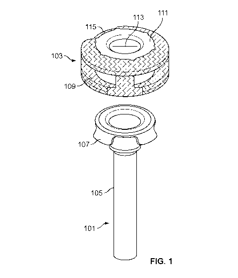
[0015] FIG. 1
illustrates a cannula tube and a self-illuminating, color-coded
overcap or hub, according to an embodiment.
[0016] FIG. 2A
illustrates a cannula device comprising the cannula tube and
hub of FIG. 1 assembled together.
[0017] FIG. 2B
illustrates a cannula device similar to the cannula device in
FIG. 2A but with a different size cannula tube and different color coding.
[0018] FIG. 2C
illustrates a cannula device similar to the cannula devices in
FIGS. 2A and 2B, but with a different size cannula tube and different color
coding.
[0019] FIG. 3A
illustrates a cannula device similar to the cannula device of
FIG. 2A, but with the self-illuminating color coding in the elastomeric seal.
4
CA 03069381 2020-01-08
WO 2019/030587
PCT/IB2018/055081
[0020] FIG. 3B illustrates a cannula device similar to the cannula device
in
FIG. 3A but with a different size cannula tube and different color coding.
[0021] FIG. 3C illustrates a cannula device similar to the cannula
devices in
FIGS. 3A and 3B, but with a different size cannula tube and different color
coding.
[0022] FIG. 4 illustrates the cannula device of FIG. 2A on a trocar
inserter.
[0023] FIG. 5 illustrates a flowchart of an embodiment of a method for
using a
self-illuminating cannula.
[0024] The accompanying drawings may be better understood by reference to
the following detailed description.
DETAILED DESCRIPTION
[0025] For the purposes of promoting an understanding of the principles
of the
present disclosure, reference will now be made to the implementations
illustrated in
the drawings, and specific language will be used to describe the same. It will
nevertheless be understood that no limitation of the scope of the disclosure
is
intended. Any alterations and further modifications to the described devices,
instruments, methods, and any further application of the principles of the
present
disclosure are fully contemplated as would normally occur to one skilled in
the art to
which the disclosure relates. In particular, it is fully contemplated that the
features,
components, and/or steps described with respect to one implementation may be
combined with the features, components, and/or steps described with respect to
other
implementations of the present disclosure. For simplicity, in some instances
the same
reference numbers are used throughout the drawings to refer to the same or
like parts.
[0026] FIG. 1 illustrates an embodiment of a trocar cannula 101 and an
overcap or hub 103. The cannula 101 may be configured for insertion into an
eye to
facilitate insertion and removal of instruments during surgery. The cannula
101 may
include a tube or shaft 105 capable of extending into the eye (e.g., through a
sclera,
conjunctiva, etc). In some embodiments, the cannula 101 may be manufactured
separately from the hub 103 and then attached to the hub 103. For example, the
cannula 101 may include one or more tabs 107 configured to engage
corresponding
CA 03069381 2020-01-08
WO 2019/030587
PCT/IB2018/055081
slots 109 in the hub 103 (e.g., the cannula 101 illustrated in FIG. 1 includes
four tabs
107 to engage four corresponding slots 109 in the hub 103). Other attachments
are
also contemplated. For example, the cannula 101 may include the slots, and the
hub
103 may include the tabs. In some embodiments, the cannula 101 may be attached
to
the hub 103 through adhesive, thermal bonding, etc. In other embodiments, the
cannula 101 may be manufactured in one piece with the hub 103.
[0027] In some
embodiments, a sealing element 111 may be coupled to the
hub 103 or may otherwise be arranged to selectively seal off the passage
through the
cannula 101 (e.g., the sealing element 111 may be disposed at least partially
between
the shaft 105 and the hub 109). The sealing element 111 may be made of an
elastomer
(e.g., silicone). As shown in FIG. 1, a surface of the sealing element 111 may
be
exposed on the hub 103. In some embodiments, the exposed surface of the
sealing
element 111 may include one or more slits 113 to allow passage of surgical
tools or
instruments into the cannula 101. In a relaxed condition of the sealing
element 111,
i.e., in the absence of a surgical instrument (or vent or other element), the
slit 113 of
the sealing element 111 is closed to inhibit fluid flow through the sealing
element 111
and thereby to seal off fluid passage through the cannula 101. A surgical
instrument
(or vent or other element) may be passed through the slit 113.
[0028] In some
embodiments, the sealing element 111 may be attached to the
hub 103 to inhibit rotation of the sealing element 111 relative to the hub
103. For
example, the sealing element 111 may be overmolded into a depression and one
or
more holes in the hub 103. In some embodiments, the sealing element 111 may
include a silicon wafer that is formed separately from the hub 103 and
inserted
between the hub 103 and the cannula 101 during assembly of the hub 103 onto
the
cannula 101. In such a case, the sealing element 111 may be attached to the
hub 103
and cannula 101 through a friction fit. Other attachments are also
contemplated (e.g.,
adhesive).
[0029] FIG. 2A
illustrates an embodiment of an entry port or cannula device
comprising the cannula 101 affixed to the hub 103 (e.g., after engagement of
the tabs
107 in respective slots 109). In some embodiments, the tab/slot interface may
prevent
rotation of the hub 103 relative to the cannula 101 (e.g., during insertion of
the
cannula 101 into the eye). In some embodiments, the tabs 107 may be configured
to
6
CA 03069381 2020-01-08
WO 2019/030587
PCT/IB2018/055081
permanently hold the hub 103 to the cannula 101 (such that the hub 103 may not
be
removed from the cannula 101 without destroying part of the cannula 101 and/or
hub
103). For example, the tabs 107 (and cannula 101) may be made of stainless
steel, and
the hub 103 may be made of plastic (e.g., polycarbonate). Other materials are
also
contemplated. The permanent hold between the hub 103 and the cannula 101 may
prevent inadvertent removal of the hub 103 from the cannula 101 during surgery
(e.g.,
vitreoretinal surgery).
[0030] As can
be seen in FIG. 2A, cannula devices as shown in FIG. 2A and
as otherwise disclosed herein in accordance with other embodiments comprise a
small
tube with a hub at the proximal end of the tube. The tube is inserted into the
eye, and
the hub acts as a stop limiting the advancement of the tube into the eye and
preventing
the tube from completely entering the eye. The hub may be stitched to the eye
to keep
the cannula in place.
[0031] Cannula
devices as shown in FIG. 2A and as otherwise disclosed
herein in accordance with other embodiments may be provided in different
sizes,
having different sizes of tubes for accommodating instruments of different
sizes.
Examples of cannula sizes suitable for ophthalmic surgical procedures include,
for
example, 20 gauge, 23 gauge, 25 gauge, 27 gauge, and others. The cannula 101
in
FIG. 2A may be, for example, a 23 gauge cannula.
[0032] To aid
it identifying the size of the cannula, and to assist in
determining which instruments go with which cannula, the cannula device may be
color-coded, with the color indicating the size associated with the cannula.
For
example, a 23 gauge cannula may have a first color, a 25 gauge cannula may
have a
second color, and a 27 gauge cannula may have a third color. The color-coding
may
be on any visible part of the cannula device, including the hub, sealing
element,
and/or tube. With the color-coding, after the cannulas are in place in the
eye, the
surgeon can identify the cannula sizes by their color in order to determine
the
appropriate cannula into which to insert an instrument.
[0033] With
prior devices, with dim lighting in the room of the surgical
procedure, it can be difficult for the surgeon to see the cannula devices
and/or their
color. This may necessitate turning on a microscope light or other light for
the
7
CA 03069381 2020-01-08
WO 2019/030587
PCT/IB2018/055081
surgeon to see the cannula devices for instrument exchanges, which can be
undesirable. For example, it may cause the surgeon's eyes to have to adjust to
the
brighter light and then readjust when the microscope light or other light is
turned off,
taking time during the procedure.
[0034] In
accordance with exemplary embodiments herein described, the
cannula device may be self-illuminating (e.g., at least partially made of a
self-
illuminating material). The self-illuminating material may be a material that,
for
example, contains a phosphorescent pigment. The self-illumination can help the
surgeon see the location of the cannula device and can help the surgeon see
any color
coding and thereby identify the size of the cannula device.
[0035] In the
exemplary embodiment of FIG. 2A, the hub 103 may be self-
illuminating, for example by being phosphorescent. The pigment used for color-
coding of the hub 103 may be a phosphorescent pigment. The cannula device of
FIG.
2A is color-coded with a first color (e.g., orange) to indicate the size of
the cannula,
e.g., 23 gauge.
[0036] FIG. 2B
illustrates a cannula device similar to the cannula device in
FIG. 2A but with a different size cannula tube and different color coding. In
the
exemplary embodiment of FIG. 2B, the hub 103 is also self-illuminating, for
example
by being phosphorescent. As with FIG. 2A, the pigment used for color-coding of
the
hub 103 may be a phosphorescent pigment. The cannula device of FIG. 2B is
color-
coded with a second color (e.g., blue or teal) to indicate the size of the
cannula, e.g.,
25 gauge.
[0037] FIG. 2C
illustrates a cannula device similar to the cannula devices in
FIGS. 2A and 2B, but with a different size cannula tube and different color
coding. In
the exemplary embodiment of FIG. 2C, the hub 103 is also self-illuminating,
for
example by being phosphorescent. As with FIGS. 2A and 2B, the pigment used for
color-coding of the hub 103 may be a phosphorescent pigment. The cannula
device of
FIG. 2C is color-coded with a third color (e.g., purple) to indicate the size
of the
cannula, e.g., 27 gauge.
[0038] FIGS.
3A, 3B, and 3C illustrate cannula devices similar to the cannula
devices of FIGS. 2A, 2B, and 2C, but with the self-illuminating color coding
in the
8
CA 03069381 2020-01-08
WO 2019/030587
PCT/IB2018/055081
elastomeric sealing element 111. In each of FIGS. 3A, 3B, and 3C, the sealing
element 111 is self-illuminating by being phosphorescent. The pigment used for
color-coding of the sealing element 111 may be a phosphorescent pigment. The
cannula device of FIG. 3A is color-coded with a first color (e.g., orange) to
indicate
the size of the cannula, e.g., 23 gauge. The cannula device of FIG. 3B is
color-coded
with a second color (e.g., blue or teal) to indicate the size of the cannula,
e.g., 25
gauge. The cannula device of FIG. 2C is color-coded with a third color (e.g.,
purple)
to indicate the size of the cannula, e.g., 27 gauge.
[0039] The self-
illumination feature may be incorporated in any visible part of
the cannula device, including the hub, sealing element, and/or tube. The self-
illumination provides a "glow-in-the-dark" effect whereby the cannula device
is
visible in the dim or dark surgical procedure room. This feature can
facilitate in-situ
visualization of the cannula devices without active illumination, ease of
instrument
insertion into the cannula devices, and/or verification of color-coding of the
cannula
devices to verify gauge size.
[0040] In
exemplary embodiments wherein the self-illumination feature is
provided by all or part of the cannula device being phosphorescent, the
phosphorescence may be excited to its luminous operating state under
microscope
illumination during normal surgical insertion of the cannula device into the
eye.
Alternatively, a separate light could be used to make the cannula device
phosphoresce
more. After activation by the microscope or otherwise, the phosphorescence of
the
cannula device emits a soft light, visible after the microscope illumination
or other
lighting is turned off. The phosphorescent light may be the gauge-specific
colored
light indicating the size of the cannula.
[0041] With the
self-illumination feature, surgical efficiency is promoted
without significant cost or complexity. The feature alleviates the need to
turn on
microscope illumination during the procedure in order for the surgeon to view
the in-
place cannula devices.
[0042] The
desired portion(s) of the cannula devices may be made
phosphorescent by incorporating suitable phosphors into the material used for
manufacturing to make the material self-illuminating. For example, the
phosphors
9
CA 03069381 2020-01-08
WO 2019/030587
PCT/IB2018/055081
may be mixed into the plastic used for making the hub and/or into the
elastomer (e.g.,
silicone) used for making the sealing element. Examples of phosphors include
zinc
sulfide and strontium aluminate. The phosphors can be energized by normal
light,
such as the light from a microscope or in a room, and the resulting glow can
last long
enough for the duration of the surgical procedure. The phosphors absorb light
when
energized and release the stored energy to glow when the lights are off or
dim.
Phosphors can be selected to give a desired color of visible light after being
activated.
The use of phosphorescent pigments and/or dopants can be chosen to give the
desired
illumination.
[0043] FIG. 4
illustrates an embodiment of a self-illuminating cannula device
on a trocar inserter 501. In some embodiments, the trocar inserter 501 may
include a
trocar blade 503 attached to a handle 505. In some embodiments, the handle 505
may
be made of plastic, and the blade 503 may be made of stainless steel. Other
materials
are also contemplated. The trocar blade 503 may extend past the end of the
shaft 105
of the cannula device and may include one or more sharp edges to pierce an eye
(e.g.,
pierce a hole through the sclera and into the vitreous body) for insertion of
the
cannula 101. In some embodiments, a guide 507 may fit into guide slot 115 of
the
cannula device to inhibit rotation of the cannula device relative to the
handle 505
during insertion of the cannula device into eye. In some embodiments, the
guide 507
may releasably engage the guide slot 115 such that when the trocar inserter
501 is
withdrawn from the cannula device, the guide 507 does not pull the cannula
device
out of the eye. For example, the guide 507 may frictionally engage the guide
slot 115
with a friction force that is less than a friction force exerted by the eye on
the external
sides of the cannula 101 when the cannula 101 is in the eye.
[0044] FIG. 5
illustrates a flowchart of an embodiment of a method for using a
self-illuminating cannula. The elements provided in the flowchart are
illustrative
only. Various provided elements may be omitted, additional elements may be
added,
and/or various elements may be performed in a different order than provided
below.
[0045] At 501,
a cannula device with a tube configured for entry of a surgical
instrument and a hub at a proximal end of the tube may be used. The cannula
device
may include a self-illuminating material (e.g., phosphors) that emits light
that
facilitates visualization of the cannula device. For example, the hub may
include a
CA 03069381 2020-01-08
WO 2019/030587
PCT/IB2018/055081
self-illuminating material. In some embodiments, the cannula device may
include a
sealing element configured to seal a passage through the tube when the sealing
element is in a relaxed condition. In some embodiments, the sealing element
may
include a self-illuminating material.
[0046] At 503,
the cannula device may be inserted at a desired location for
surgical entry of a surgical instrument.
[0047] At 505,
the cannula device may be illuminated to activate the
phosphors into an activated condition. The phosphors, when in an activated
condition, may emit a color-coded light corresponding to a size of the cannula
device.
[0048] In some
embodiments, at 507, a second cannula device may be used
(where the previously mentioned cannula device is a first cannula device). The
second cannula device may include a tube configured for entry of a second
surgical
instrument with a hub at a proximal end of the tube. The second cannula device
may
also be self-illuminating. In some embodiments, the first and second cannula
device
may be different sizes.
[0049] At 509,
the second cannula device may be inserted at a desired location
for surgical entry of the second surgical instrument.
[0050] At 511,
after inserting the second cannula device, the second cannula
device may emit light that facilitates visualization of the second cannula
device. In
some embodiments, the first cannula device may emit a light of a first color
corresponding to a size of the first cannula device and the second cannula
device may
emit a light of a second color corresponding to a size of the second cannula
device. In
some embodiments, the second color may be different from the first color.
[0051] Persons
of ordinary skill in the art will appreciate that the
implementations encompassed by the present disclosure are not limited to the
particular exemplary implementations described above. In that regard, although
illustrative implementations have been shown and described, a wide range of
modification, change, and substitution is contemplated in the foregoing
disclosure. It
is understood that such variations may be made to the foregoing without
departing
from the scope of the present disclosure. Accordingly, it is appropriate that
the
11
CA 03069381 2020-01-08
WO 2019/030587
PCT/IB2018/055081
appended claims be construed broadly and in a manner consistent with the
present
disclosure.
12