Note: Descriptions are shown in the official language in which they were submitted.
CA 03070775 2020-01-21
WO 2019/023385
PCT/US2018/043762
SYSTEM AND METHOD FOR POSITIONING A HEART VALVE
CROSS-REFERENCE TO RELATED APPLICATIONS
[0001] This application claims priority to U.S. Provisional Patent
Application No.
62/536,932, filed July 25, 2017, titled "SYSTEM AND METHOD FOR POSITIONING A
HEART VALVE," the entirety of which is incorporated by reference herein.
INCORPORATION BY REFERENCE
[0002] All publications and patent applications mentioned in this
specification are herein
incorporated by reference to the same extent as if each individual publication
or patent
application was specifically and individually indicated to be incorporated by
reference.
BACKGROUND
[0003] The mitral valve lies between the left atrium and the left
ventricle of the heart.
Various diseases can affect the function of the mitral valve, including
degenerative mitral valve
disease and mitral valve prolapse. These diseases can cause mitral stenosis,
in which the valve
fails to open fully and thereby obstructs blood flow, and/or mitral
insufficiency, in which the
mitral valve is incompetent and blood flows passively in the wrong direction.
[0004] Many patients with heart disease, such as problems with the
mitral valve, are
intolerant of the trauma associated with open-heart surgery. Age or advanced
illness may have
impaired the patient's ability to recover from the injury of an open-heart
procedure. Additionally,
the high costs associated with open-heart surgery and extra-corporeal
perfusion can make such
procedures prohibitive.
[0005] Patients in need of cardiac valve repair or cardiac valve
replacement can be served by
minimally invasive surgical techniques. In many minimally invasive procedures,
small devices
are manipulated within the patient's body under visualization from a live
imaging source like
ultrasound, fluoroscopy, or endoscopy. Minimally invasive cardiac procedures
are inherently
less traumatic than open procedures and may be performed without extra-
corporeal perfusion,
which carries a significant risk of procedural complications.
[0006] Prosthetic valve replacement procedures can be difficult, and
various factors are
generally taken into account when placing the valve. First, the prosthetic
valve should be placed
at the same or very nearly the same angle as the native valve. A valve that is
off axis could cause
turbulent blood flow and/or potential para-valvular leaks. Second, the
prosthetic valve should
ideally have concentricity. This means that the valve is placed in the same
center as the native
valve. An off center deployment or valve placement could affect the mechanism
of neighboring
- 1 -
CA 03070775 2020-01-21
WO 2019/023385
PCT/US2018/043762
valves or the heart's conductive system. Finally, the prosthetic valve should
be at the proper
depth within the patient's heart with respect to the location of the native
valve, as otherwise, the
prosthetic valve may interfere with the conductive nature of the heart as
well.
[0007] However, in general, trans-catheter mitral valve delivery can be
difficult because the
physician must control the placement of the valve in at least three degrees of
motion using at
least two different imaging modalities. It is common for the user to correctly
position the valve
in one degree of motion, but to then lose that position while trying to obtain
a correct position in
a second degree of motion. Similarly, it isn't unusual for the user to lose
position in an x-ray
imaged degree of motion when switching to ultrasound to obtain a correct
position in a second
degree of motion.
[0008] A safe and efficient system and method for replacement of a
cardiac valve that
addresses some or all of these concerns is described herein.
SUMMARY OF THE DISCLOSURE
[0009] Described herein are mechanical positioning aids that may be used to
assist a
physician in placing a mitral valve in the native valve orifice. The
positioning aid can maintain a
first position while allowing the user to adjust a second degree of motion to
obtain a second
position.
[0010] In general, in one embodiment, a system for delivering a medical
device to a heart
valve annulus includes a first imaging sensor, a second imaging sensor, a
delivery arm, and a
control system. The first imaging sensor is configured to be aligned such that
a view of the first
imaging sensor is along a primary plane of the heart valve annulus. The second
imaging sensor
is configured to be aligned such that a view of the second imaging sensor is
along a longitudinal
axis of the heart valve annulus. The delivery arm is configured to hold a
medical device delivery
system. The control system is configured to adjust the delivery arm to set an
angle of the
delivery system perpendicular to the primary plane using images from the first
imaging sensor or
adjust the delivery arm to center the delivery device in the heart valve
annulus using images from
the second imaging sensor.
[0011] This and other embodiments can include one or more of the
following features. The
control system can be further configured to adjust the delivery arm to insert
the delivery device
along the longitudinal axis prior to deploying the medical device. The control
system can be
configured to adjust the delivery arm to insert the delivery device along the
longitudinal axis
using images from the first imaging sensor. The control system can be
configured to do both
steps of adjusting the delivery arm to set an angle of the delivery system
perpendicular to the
primary plane using images from the first imaging sensor and adjusting the
delivery arm to
- 2 -
CA 03070775 2020-01-21
WO 2019/023385
PCT/US2018/043762
center the delivery device in the heart valve annulus using images from the
second imaging
sensor. The first imaging sensor can be an x-ray sensor. The second imaging
sensor can be an
ultrasound sensor. The heart valve annulus can be a mitral valve annulus. The
medical
instrument can be a prosthetic mitral valve. The delivery arm can include at
least six degrees of
freedom. The control system can be further configured to create a computed
pivot point for the
delivery device at a center of the heart valve annulus. The control system can
be further
configured to allow the robotic arm to travel along a virtual rail when
manually acted upon by a
user.
[0012] In general, in one embodiment, a method of delivering a medical
device to a heart
valve annulus includes: (1) aligning a first imaging sensor such that a view
of the first imaging
sensor is along a primary plane of the heart valve annulus; (2) aligning a
second imaging sensor
such that a view of the second imaging sensor is along a longitudinal axis of
the heart valve
annulus; (3) attaching a delivery system holding the medical device to a
delivery arm; (4)
adjusting the delivery arm to set an angle of the delivery system
perpendicular to the primary
plane using images from the first imaging sensor; (5) adjusting the delivery
arm to center the
delivery device in the heart valve annulus using images from the second
imaging sensor; and (6)
deploying the medical device from the delivery system into the heart valve
annulus.
[0013] This and other embodiments can include one or more of the
following features. The
method can further include adjusting the delivery arm to insert the delivery
device along the
longitudinal axis prior to deploying the medical device. Adjusting the
delivery arm to insert the
delivery device along the longitudinal axis can include using images from the
first imaging
sensor. Using images from the first imaging sensor can include aligning
markers on the delivery
device with the primary plane. The first imaging sensor can be an x-ray
sensor. The method can
further include inserting a coronary vein wire and injecting contrast dye to
identify the mitral
valve plane in images from the x-ray sensor. The second imaging sensor can be
an ultrasound
sensor. The heart valve annulus can be a mitral valve annulus. The medical
instrument can be a
prosthetic mitral valve. The delivery arm can include at least six degrees of
freedom. The
delivery arm can be a mechanical un-powered arm. The delivery arm can be a
robotic arm. At
least one of the steps of adjusting the delivery arm to set an angle or
adjusting the steps of
adjusting the delivery arm can be performed automatically by a control system
of the robotic
arm. The control system can be configured to create a computed pivot point for
the delivery
device at a center of the heart valve annulus. A control system of the robotic
arm can be
configured to allow the robotic arm to travel along a virtual rail when
manually acted upon by a
user.
- 3 -
CA 03070775 2020-01-21
WO 2019/023385
PCT/US2018/043762
BRIEF DESCRIPTION OF THE DRAWINGS
[0014] The novel features of the invention are set forth with
particularity in the claims that
follow. A better understanding of the features and advantages of the present
invention will be
obtained by reference to the following detailed description that sets forth
illustrative
embodiments, in which the principles of the invention are utilized, and the
accompanying
drawings of which:
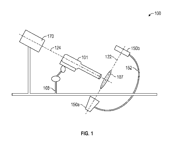
[0015] Figure 1 shows a valve positioning system.
[0016] Figure 2 is a flow chart of the method of delivering a mitral
valve using a valve
positioning system.
[0017] Figure 3A shows a delivery device with a mitral valve therein where
the delivery
device is not orthogonal to the native mitral valve orifice. Figure 3B shows a
delivery device
housing a mitral valve where the delivery device is orthogonal to the native
mitral valve.
[0018] Figure 4 shows a delivery device with a mitral valve therein
where the delivery
device is centered in the mitral valve annulus.
[0019] Figure 5A shows a delivery device with a mitral valve therein where
the delivery
device is aligned with the mitral valve plane. Figure 5B shows a close up of
the delivery device
and valve of Figure 5A.
[0020] Figure 6 shows a mechanical valve positioning arm.
[0021] Figure 7 shows a robotic valve positioning arm.
DETAILED DESCRIPTION
[0022] The valve positioning systems described herein can be used to
deliver and deploy a
wide variety of replacement heart valves, such as prosthetic valves adapted to
be minimally
invasively delivered. For example, the valve positioning systems described
herein can be
configured to be able to deliver and deploy a replacement heart valve, such as
a mitral valve, that
includes proximal and distal anchors.
[0023] Exemplary prosthetic valves that can be delivered and deployed
include the
expandable prosthetic valves described in App. No. 14/677,320, filed April 2,
2015, Publication
No. US 2016-0158000 Al, titled "REPLACEMENT CARDIAC VALVES AND METHODS
OF USE AND MANUFACTURE" in U.S. Pat. No 8,870,948, and in International Patent
Application No. PCT/U52016/032550, filed May 13, 2016, titled "REPLACEMENT
MURAL
VALVES," in U.S. Patent Application No. 16/012,666, filed June 19, 2018,
titled
"REPLACEMENT MITRAL VALVES," all of which are incorporated by reference
herein.
[0024] Further, the valve positioning systems described herein can be
used with the delivery
devices described, for example, in International Patent Application No.
PCT/U52016/032546,
- 4 -
CA 03070775 2020-01-21
WO 2019/023385
PCT/US2018/043762
filed May 13, 2016, titled "CARDIAC VALVE DELIVERY DEVICES AND SYSTEMS," U.S.
Provisional Patent Application No. 62/424,021, filed November 18, 2016, titled
"CARDIAC
VALVE DELIVERY DEVICES AND SYSTEMS," U.S. Provisional Patent Application No.
62/424,051, filed November 18, 2016, and titled "CARDIAC VALVE DELIVERY
DEVICES
AND SYSTEMS", and International Patent Application No. PCT/U52017/062045,
filed
November 16, 2017, and titled "CARDIAC VALVE DELIVERY DEVICES AND SYSTEMS",
the entireties of which are incorporated by reference herein.
[0025] A replacement heart valve, such as a mitral valve prosthesis, can
be delivered using
one of the valve positioning systems described herein to a cardiac valve
orifice, such as the
mitral valve, using minimally invasive techniques. In some embodiments, a
small incision can
be made in the patient's body, and the prosthesis can be passed through the
apex of the heart to,
for example, the mitral valve. This can be referred to as the transatrial
delivery approach. In
other embodiments, the prosthesis can be delivered through the venous system
and into the left
atrium through a transseptal puncture. A transseptal approach can impart size
limitations on the
delivery and thus the delivery profile of the replacement heart valve.
Additionally, a transseptal
approach can also impart certain flexibility requirements on the replacement
heart valve. For
both delivery approaches, the distal-most anchor can be delivered to the
ventricle while the
proximal-most anchor can be delivered to the atrium.
[0026] The valve positioning systems described herein can be used to
delivery a replacement
heart valve (e.g., via a delivery device) to the treatment site for
deployment.
[0027] Referring to Figure 1, in some embodiments, a valve positioning
system 100 can
include an arm 103 that is used to support a mitral valve delivery device 101
(which can be any
delivery device described, for example, in International Application No.
PCT/U52016/032546,
filed May 13, 2016, and titled "CARDIAC VALVE DELIVERY DEVICES AND SYSTEMS",
International Application No. PCT/U52017/037850, filed June 16, 2017, and
titled "CARDIAC
VALVE DELIVERY DEVICES AND SYSTEMS", and International Application No.
PCT/U52017/062045, filed November 16, 2017, and titled "CARDIAC VALVE DELIVERY
DEVICES AND SYSTEMS"). The valve positioning system 100 can further include
two
imaging modalities. The imaging modalities can be, for example, ultrasound or
x-ray. Further,
the two imaging modalities can be identical to one another (e.g., provide the
same type of
imaging) or different. As shown in Figure 1, for example, the imaging
modalities can include an
ultrasound sensor 170 and an x-ray sensor 150a,b (150a is the emitter while
150b is the detector,
which can be part of a c-arm 152). The x-ray sensor 150a,b can be positioned,
for example, such
that the x-ray image is taken down the plane 122 of the mitral valve 107
(i.e., on-edge with the
plane 122 of the mitral valve). This position of the x-ray sensor 150a,b can
allow orientation of
- 5 -
CA 03070775 2020-01-21
WO 2019/023385
PCT/US2018/043762
the delivery device 101 (and thus the mitral valve) orthogonal to the native
valve 107. Further,
the ultrasound sensor 170 can be positioned, for example, such that the
ultrasound image is taken
down the longitudinal axis 124 of the valve 107 (i.e., the axis that extends
through the center of
the valve 107). This position of the ultrasound sensor 170 can allow
positioning of the delivery
device 101 (and thus the mitral valve) in the center of the native valve 107.
[0028] A method of delivering a mitral valve to the mitral valve orifice
using the valve
positioning system of Figure 1 is shown in the flow chart 200 of Figure 2. To
begin, at step 221,
the first imaging modality (e.g., x-ray imaging sensor 150a,b) is maneuvered
so as to have a set
relationship with the native mitral valve 107 of the patient. For example, the
position of the x-
ray emitter 150a can be set such that the view of the x-ray imaging sensor
150a,b is on-edge to
the plane 122 of the native mitral valve 107. In some embodiments, contrast
injections and a
coronary vein wire can be used to better visualize the plane 122 of the mitral
valve 107 under x-
ray (and thus to provide for placement of the x-ray emitter 150a with
increased accuracy). When
contrast injections and a coronary vein wire are to be used, the coronary vein
wire can be placed
in the coronary sinus, which is positioned in the body along the same plane
122 as the native
mitral valve 107. After placing the wire in the coronary sinus, contrast can
be provided by
injecting radio-opaque dye into the ventricle underneath the mitral annulus.
The contrast
injection can circle under the annular plane, creating a semi-circular ring
that is visible
immediately underneath the mitral valve and parallel to the plane 122 of the
mitral valve
annulus. The image plane of the x-ray system 150a,b can then be rotated so as
to be
perpendicular to the mitral valve plane 122 (i.e., by making the ring of dye
appear to be as flat as
possible in the x-ray image). The final position of the x-ray emitter 150a
will advantageously be
relative to, and within, the coordinate system of the mitral valve 107. An
exemplary position of
the emitter 150a after the emitter 150a has been positioned within the mitral
valve coordinate
system is, for example, is LAO 16 Cranial 40 where LAO is a "Left Anterior
Oblique" angle
measured relative to the patient and floor in the patient's left hand to right
hand direction and
Cranial is measured in the direction of the patient's head in a head to foot
direction.
[0029] At step 222, the second imaging modality (e.g., ultrasound system
170) is set such
that the view is looking at the face of the mitral valve (i.e., along the axis
124). For example, a
para-sternal short-axis view at the level of the mitral valve can be used. If
a second x-ray system
is used as the second imaging modality rather than the ultrasound system 170,
the second x-ray
system can be positioned using the contrast injections and coronary vein wire
as described
above. The second the x-ray system may be moved into a position to be parallel
to the mitral
plane by rotating the x-ray system 150a,b until the footprint of the dye ring
image is maximized
in a circular perspective.
- 6 -
CA 03070775 2020-01-21
WO 2019/023385
PCT/US2018/043762
[0030] At step 223, the delivery device 101 is positioned into the heart
(e.g., through an atrial
purse-string) such that the replacement mitral valve is approximately at the
location of the native
mitral valve orifice 107.
[0031] At step 224, the delivery device 101 is attached to the
positioning arm 103.
[0032] At step 225, the positioning arm 103 is used to set the angle of the
delivery device
101 such that it is perpendicular to the plane 122 of the native valve 107
based upon the image
from the first imaging modality (e.g., the x-ray sensor 150a,b). Thus, the
delivery device 101
can be pivoted until it is orthogonal to the plane 122. In some embodiments,
the orthogonal
position can be confirmed because circumferential rings (e.g., made of
tantalum wire) on the
delivery device 101 can appear to be a closed, single line rather than an open
oval or circle in the
image from the first imaging modality (e.g., the x-ray sensor 150a,b) that is
aligned with the
mitral valve plane 122. For example, Figure 3A shows an x-ray image 300a of a
delivery device
301 housing a mitral valve 302. The delivery device 301 is not orthogonal to
the native mitral
valve orifice, as is indicated by the oval-shaped tantalum wire 315 in the x-
ray image 300a. In
contrast, Figure 3B shows an x-ray image 300b in which the delivery device 301
is orthogonal to
the native mitral valve, as is indicated by the line of tantalum wire 315 in
the 300b.
[0033] At step 226, the positioning arm 103 is used to center the
delivery device 101 in the
native mitral valve annulus based on the ultrasound image. For example, Figure
4 shows the
delivery device 101 centered in the annulus between the anterior mitral valve
leaflet 443 and the
posterior mitral valve leaflet 441. The left ventricular outflow tract 445 and
the right ventricle
447 can also be seen.
[0034] At step 227, the depth of insertion of the delivery device 103 is
set using the
positioning arm 103. In some embodiments, the x-ray image can be used to set
the depth by
aligning markers on the delivery device with the mitral plane 122. For
example, Figure 5A
shows x-ray images 500a, 500b, and 500c. The x-ray image 500a shows that the
waist 557 of the
mitral valve replacement 555 is below the mitral valve plane 122. The x-ray
image 500b shows
that the waist 557 of the mitral valve replacement 555 is above the mitral
valve plane 122.
Finally, x-ray image 500c shows that the waist 557 of the mitral valve
replacement 555 is
aligned with the mitral valve plane 122. Figure 5B shows markers 551a,b (e.g.,
rings) on the
delivery device 501 and markers 553 on the valve replacement 555 that can be
used to align the
delivery device with the mitral plane 122.
[0035] Once the depth, angle, and centrality of the mitral valve have
been set, then the valve
can be deployed (at step 228).
[0036] Referring to Figure 6, in some embodiments, the positioning arm
used herein can be a
mechanical un-powered arm 603 having an attachment mechanism 613 configured to
hold the
- 7 -
CA 03070775 2020-01-21
WO 2019/023385
PCT/US2018/043762
delivery device 601 and a plurality of locks or brakes 611a-d configured to
control the position
of the delivery device 601. The arm 603 can include, for example, mechanical
locks configured
to control the degrees of freedom of the device (i.e., the angular,
rotational, and/or axial positions
of the delivery device. The positioning arm 603 can include six degrees of
freedom (shown by
arrows 633a-f).
[0037] In one embodiment, following the steps of the flow chart 200 when
using the
positioning arm 603, the brake 611d can be released to pivot the delivery
device 601 to the
orthogonal position, e.g., using the rings 615 as they appear in the x-ray
image (for step 225).
Once the delivery device 601 is at the correct orthogonal position, the brake
611d can be locked
again. Further, brakes 611a and/or 611b can be released to allow the delivery
device 601 to be
centered (per step 226), and then the brakes 611a and/or 611b can be locked in
place. Finally,
with the first two positions held in place by the brakes 611a,b, and d, the
user can unlock the
brake 611c to allow the insertion depth to be set (at step 227).
[0038] Referring to Figure 7, in some embodiments, the valve positioning
arm used herein
can be a computer controlled, servo-mechanical robotic arm 703. The arm 703
can include a
base 779 and a mount 713 configured to mount the delivery device 701 thereto.
In some
embodiments, the arm 703 can have five to seven degrees of freedom for
positioning the mitral
valve delivery system. For example, arm 703 includes six movable (e.g.,
motorized) connections
or joints 777a-f that provide six degrees of freedom. An additional degree of
freedom can be
provided (e.g., to avoid collisions with other objects during use). Further,
the arm 703 can
include or be connected to a control system configured to move the robotic arm
703.
[0039] In one embodiment, the robotic arm 703 can include a 6 degree of
freedom (DOF)
force sensor mounted at or near the distal end of the arm 703. The user can
thus hold the robot
703, and control software in or connected to the robotic arm 703 can use
information from the
force sensor to follow the user's input and move the robotic arm 703 (and thus
the delivery
device 701). In one specific example, the force sensor(s) can be placed on or
into the distal
section of the robotic arm 703. The force sensor(s) can provide input into the
robotic control
systems to move multiple degrees of freedom in pre-programmed sequence. In
such an example,
the user can grasp the robotic arm 703 near the distal end and make a motion
as of insertion. The
force sensor(s) can then sense the applied force and direction, and the
robotic control system can
send output signal to the motors in the joints 777a-f such that the joints
777a-f can rotate in
concert to affect a straight-line movement.
[0040] In another embodiment, the robotic arm 703 can include a user
interface (button,
GUI, voice control) on or connected thereto that can be in communication with
the robotic
control system. Further, the robotic arm 703 can include an actuator (e.g.,
button) on or
- 8 -
CA 03070775 2020-01-21
WO 2019/023385
PCT/US2018/043762
connected thereto. In use, the user can actuate the actuator to "clutch" the
robotic arm 703 into
an "impedance" mode or "following" mode. The user can then position the robot
arm 703 so
that the tip of the delivery device 701 is just touching the surface of the
heart. At this position,
the user can input the position of the delivery device 701 (e.g., a "ready"
position) into the user
interface. The control system can then create a computed center or pivot point
at the surface of
the heart. This information may be used to reduce side-to-side motion at the
surface of the heart
to reduce the chance of trauma at the insertion point of the delivery device
701. Because the tip
of the delivery device 701 is in a rigid, encoded joint chain from the base of
the robot arm 703 to
the heart, touching the surface of the heart informs the robotic control
system of the position of
the insertion point relative to the robotic coordinate system at the base of
the robotic system.
When the system has knowledge of the insertion point, the system may treat
that point as a pivot
or remote center where the only motions of the delivery system 701 allowed are
rotations and
depth insertion (i.e., no side-to-side motions are allowed that may traumatize
the insertion point
are initiated by the system).
[0041] In one embodiment, to control the pivoted position of the robotic
arm 703 (i.e., such
that the delivery device 701 is orthogonal to the plane 122 of the mitral
valve orifice as at step
225), the user can input the angles of the first imaging modality (e.g., the x-
ray sensor 150a,b)
into the control system for the robotic arm 703 when the first imaging
modality is set such that
the view is on-edge to the mitral plane 122. Using the image (e.g., x-ray
data), the robotic arm
703 can automatically position the delivery device 701 perpendicular to the
plane 122 of the
native mitral valve 107. In another embodiment, the user can position the x-
ray arm 703
manually (or through controlled movement with a user interface) such that the
delivery device
701 is perpendicular to the native valve 107 based on the image, e.g., such
that the rings 715 on
the delivery device are closed as described above.
[0042] To ensure that the mitral valve replacement is positioned properly
within the center of
the native orifice (as at step 226), the robotic arm 703 can be switched to a
mode that allows the
delivery device to be centered in the mitral valve using the second imaging
modality (e.g.,
ultrasound). In this mode, the robotic arm can become a "virtual stir" that
maintains the depth
and orthogonality of the delivery device while allowing the user to adjust the
centering of the
delivery device.
[0043] To control the insertion depth (as at step 227), the user can
make an input to the
robotic control system of the robotic arm 703 that places the arm 703 in a
mode where the
delivery device is allowed to move along a "virtual rail." In other words, the
user may hold the
distal point of the robotic arm 703, and the arm 703 can follow the user's
input to move in or out
along an insertion axis orthogonal to the native mitral valve. In this
"virtual rail" mode, the
- 9 -
CA 03070775 2020-01-21
WO 2019/023385
PCT/US2018/043762
control system of the robotic arm 703 may be set to allow zero motion in
directions other than
the direction of insertion. Alternatively, the "virtual rail" may provide
almost no resistance in
the direction of insertion while providing some greater resistance to motion
in directions other
than insertion. In either case, if the user is not holding onto the robot arm
703 at the distal
position, the robot arm 703 can lock and hold position. While the virtual rail
is set, the insertion
depth can be controlled based on the first imaging modality (e.g., the image
from the x-ray
sensor).
[0044] In some embodiments, as the prosthetic valve is deployed from the
delivery device,
the user may wish to place the robotic arm 703 in a mode that allows the valve
to be tensioned
on the native tissue. This tension mode may be a "virtual rail" where the
robot allows the user to
move the delivery device 701 only in the insertion axis, or it may be an
active tensioning mode
where the robotic control system uses information from force sensors to apply
a constant, low
force in the insertion axis to keep the valve in contact with tissue as it is
deployed.
[0045] In another embodiment, all motions may be controlled robotically
rather than having
some motions of the robotic arm 703 controlled manually. In this mode, for
example, the user
cam use a joystick-style input to remotely position the delivery device
following the same steps.
The robot arm 703 can use force sensor information to limit applied forces to
reasonable levels,
for example three Newtons.
[0046] The robotic arm 703 can be, for example, any of the robotic arms
or systems
described in International Patent Publication No. WO 2010/040215, titled
"PORTABLE
ROBOTIC ARM," filed October 6, 2009, the entirety of which is incorporated by
reference
herein.
[0047] Although described for use with a mitral valve prosthetic, the
systems and methods
described herein can be used with a variety of different implantable devices,
including stents or
other valve prosthetics.
[0048] It should be understood that any elements described herein with
respect to one
embodiment can be substituted or combined with elements of any other
embodiment(s).
[0049] When a feature or element is herein referred to as being "on"
another feature or
element, it can be directly on the other feature or element or intervening
features and/or elements
may also be present. In contrast, when a feature or element is referred to as
being "directly on"
another feature or element, there are no intervening features or elements
present. It will also be
understood that, when a feature or element is referred to as being
"connected", "attached" or
"coupled" to another feature or element, it can be directly connected,
attached or coupled to the
other feature or element or intervening features or elements may be present.
In contrast, when a
feature or element is referred to as being "directly connected", "directly
attached" or "directly
- 10 -
CA 03070775 2020-01-21
WO 2019/023385
PCT/US2018/043762
coupled" to another feature or element, there are no intervening features or
elements present.
Although described or shown with respect to one embodiment, the features and
elements so
described or shown can apply to other embodiments. It will also be appreciated
by those of skill
in the art that references to a structure or feature that is disposed
"adjacent" another feature may
have portions that overlap or underlie the adjacent feature.
[0050] Terminology used herein is for the purpose of describing
particular embodiments
only and is not intended to be limiting of the invention. For example, as used
herein, the singular
forms "a", "an" and "the" are intended to include the plural forms as well,
unless the context
clearly indicates otherwise. It will be further understood that the terms
"comprises" and/or
"comprising," when used in this specification, specify the presence of stated
features, steps,
operations, elements, and/or components, but do not preclude the presence or
addition of one or
more other features, steps, operations, elements, components, and/or groups
thereof. As used
herein, the term "and/or" includes any and all combinations of one or more of
the associated
listed items and may be abbreviated as "/".
[0051] Spatially relative terms, such as "under", "below", "lower", "over",
"upper" and the
like, may be used herein for ease of description to describe one element or
feature's relationship
to another element(s) or feature(s) as illustrated in the figures. It will be
understood that the
spatially relative terms are intended to encompass different orientations of
the device in use or
operation in addition to the orientation depicted in the figures. For example,
if a device in the
.. figures is inverted, elements described as "under" or "beneath" other
elements or features would
then be oriented "over" the other elements or features. Thus, the exemplary
term "under" can
encompass both an orientation of over and under. The device may be otherwise
oriented (rotated
90 degrees or at other orientations) and the spatially relative descriptors
used herein interpreted
accordingly. Similarly, the terms "upwardly", "downwardly", "vertical",
"horizontal" and the like
are used herein for the purpose of explanation only unless specifically
indicated otherwise.
[0052] Although the terms "first" and "second" may be used herein to
describe various
features/elements (including steps), these features/elements should not be
limited by these terms,
unless the context indicates otherwise. These terms may be used to distinguish
one
feature/element from another feature/element. Thus, a first feature/element
discussed below
could be termed a second feature/element, and similarly, a second
feature/element discussed
below could be termed a first feature/element without departing from the
teachings of the present
invention.
[0053] Throughout this specification and the claims which follow, unless
the context
requires otherwise, the word "comprise", and variations such as "comprises"
and "comprising"
means various components can be co-jointly employed in the methods and
articles (e.g.,
-11-
CA 03070775 2020-01-21
WO 2019/023385
PCT/US2018/043762
compositions and apparatuses including device and methods). For example, the
term
"comprising" will be understood to imply the inclusion of any stated elements
or steps but not
the exclusion of any other elements or steps.
[0054] As used herein in the specification and claims, including as used
in the examples and
unless otherwise expressly specified, all numbers may be read as if prefaced
by the word "about"
or "approximately," even if the term does not expressly appear. The phrase
"about" or
"approximately" may be used when describing magnitude and/or position to
indicate that the
value and/or position described is within a reasonable expected range of
values and/or positions.
For example, a numeric value may have a value that is +/- 0.1% of the stated
value (or range of
values), +/- 1% of the stated value (or range of values), +/- 2% of the stated
value (or range of
values), +/- 5% of the stated value (or range of values), +/- 10% of the
stated value (or range of
values), etc. Any numerical range recited herein is intended to include all
sub-ranges subsumed
therein.
[0055] Although various illustrative embodiments are described above,
any of a number of
changes may be made to various embodiments without departing from the scope of
the invention
as described by the claims. For example, the order in which various described
method steps are
performed may often be changed in alternative embodiments, and in other
alternative
embodiments one or more method steps may be skipped altogether. Optional
features of various
device and system embodiments may be included in some embodiments and not in
others.
Therefore, the foregoing description is provided primarily for exemplary
purposes and should
not be interpreted to limit the scope of the invention as it is set forth in
the claims.
[0056] The examples and illustrations included herein show, by way of
illustration and not of
limitation, specific embodiments in which the subject matter may be practiced.
As mentioned,
other embodiments may be utilized and derived there from, such that structural
and logical
substitutions and changes may be made without departing from the scope of this
disclosure.
Such embodiments of the inventive subject matter may be referred to herein
individually or
collectively by the term "invention" merely for convenience and without
intending to voluntarily
limit the scope of this application to any single invention or inventive
concept, if more than one
is, in fact, disclosed. Thus, although specific embodiments have been
illustrated and described
herein, any arrangement calculated to achieve the same purpose may be
substituted for the
specific embodiments shown. This disclosure is intended to cover any and all
adaptations or
variations of various embodiments. Combinations of the above embodiments, and
other
embodiments not specifically described herein, will be apparent to those of
skill in the art upon
reviewing the above description.
- 12 -