Note: Descriptions are shown in the official language in which they were submitted.
CA 03071159 2020-01-24
WO 2019/023266 PCT/US2018/043539
COLLAGEN ENCAPSULATION OF INSULIN-PRODUCING CELLS
[0001] Pancreatic islets contain several types of cells, including beta cells
that produce the hormone
insulin. When the level of blood glucose, also called blood sugar, rises after
a meal, the pancreas
responds by releasing insulin into the bloodstream. Insulin helps cells
throughout the body absorb
glucose from the bloodstream and use it for energy.
[0002] It is estimated that about 1.25 million Americans, including both
children and adults, are
inflicted with type 1 diabetes. Diabetes also affects veterinary patients,
including dogs and cats, with
an incidence of 1 to 2 percent. Patients with type 1 diabetes have damaged
beta cells and therefore
require lifelong insulin therapy. Most require 2 or more injections of insulin
daily, with doses adjusted
on the basis of self-monitoring of blood glucose levels. Although such insulin
therapy is life-saving, it
provides inferior control compared to functional pancreatic islet cells and
does not eliminate chronic
complications that decrease quality of life, lead to premature death, and
account for a large percentage
of medical costs. Replacing beta cell function via cell-based therapies such
as islet transplantation or
transplantation of other insulin-producing cells is an attractive alternative
to standard-of-care
exogenous insulin administration or whole pancreas transplantation because of
reduced surgical risks
and impositions on patient quality of life.
[0003] At present, clinical islet transplantation for human patients involves
isolation of a large number
of islets from multiple human cadaveric donor pancreases followed by infusion
through the portal vein
into the liver where they become lodged. Although successes associated with
the Edmonton protocol
and a recent multi-center phase III clinical trial highlight the potential of
pancreatic islet
transplantation, a number of persistent obstacles preclude it from gaining
more widespread use.
[0004] One challenge is that there exists a limited supply of high quality
human donor islets for
transplant. Upon islet isolation and conventional in-vitro culture in
suspension, the insulin-producing
1
SUBSTITUTE SHEET (RULE 26)
CA 03071159 2020-01-24
WO 2019/023266 PCT/US2018/043539
beta cells undergo rapid cell death and lose their glucose-sensitive insulin
release function, which has
been attributed to the loss of critical cell-extracellular matrix
interactions, biophysical cues, and
vascularization. The lack of in-vitro culture and preservation strategies
requires the rapid use (<20
hours) of islets following isolation.
[0005] Another major challenge is that patients receiving transplanted islets
from another individual
are required to be on long-term systemic immune suppression drugs to help keep
the cells alive and
functioning after transplantation. Unfortunately, chronic immunosuppression
has significant risks and
side effects since it compromises the patient's natural defense and protection
mechanisms.
[0006] Most importantly, the longevity and function of transplanted islets
must be improved for
patients to achieve long-term insulin independence. It has been estimated that
the large majority of
transplanted islets (>60%) fail to engraft following transplantation. This has
been attributed to loss of
critical microenvironmental cues, insufficient supply of oxygen and nutrients,
and the rapid blood
mediated inflammatory reaction following islet transplantation.
[0007] Such findings have focused current translational research efforts
towards developing strategies
that i) prolong or preserve viability and function of isolated islets and
other insulin-producing cells in-
vitro and ii) support long-term insulin-producing cell replacement and
function without the need for
chronic immunosuppression.
[0008] Therefore, there remains a need to develop an insulin-producing cell
replacement strategy that
i) facilitates minimally invasive procedures for administration, biopsy, and
transplant removal; ii)
supports non-invasive monitoring; and iii) promotes long-term engraftment and
function of
replacement insulin-producing cells in absence of systemic immunosuppression.
An easy-to-
administer, insulin-producing cell transplantation therapy that returns long-
lasting blood glucose
control back to the patient in the absence of systemic immunosuppression and
fibrotic capsule
2
SUBSTITUTE SHEET (RULE 26)
CA 03071159 2020-01-24
WO 2019/023266 PCT/US2018/043539
formation has the potential to dramatically improve the quality of life,
health status, and life expectancy
for both human and veterinary patients with type 1 diabetes.
SUMMARY OF THE DISCLOSURE
[0009] In one aspect of the disclosure, compositions comprising collagen and
insulin-producing cells
in an aqueous medium are provided.
[0010] In another aspect of the disclosure, semi-solid collagen-insulin-
producing cell compositions are
provided.
[0011] In a further aspect of the disclosure, methods for treating metabolic
disorders comprising
administering compositions comprising collagen and insulin-producing cells to
mammals are provided.
[0012] In an additional aspect of the disclosure, methods for treating
metabolic disorders comprising
administering semi-solid compositions comprising collagen and insulin-
producing cells to mammals
are provided.
[0013] In yet an additional aspect of the disclosure, methods for lowering
blood glucose levels
comprising administering compositions comprising collagen and insulin-
producing cells to mammals
are provided.
[0014] In a further aspect of the disclosure, methods for controlling blood
glucose levels comprising
administering compositions comprising collagen and insulin-producing cells to
mammals are provided.
[0015] In yet an additional aspect of the disclosure, methods for lowering
blood glucose levels
comprising administering compositions comprising semi-solid compositions
comprising collagen and
insulin-producing cells to mammals are provided.
[0016] In a further aspect of the disclosure, methods for controlling blood
glucose levels comprising
administering semi-solid compositions comprising collagen and insulin-
producing cells to mammals
3
SUBSTITUTE SHEET (RULE 26)
CA 03071159 2020-01-24
WO 2019/023266 PCT/US2018/043539
are provided.
[0017] In a still further aspect of the disclosure, processes for making semi-
solid collagen-insulin-
producing cell compositions are provided comprising combining an acidic
collagen in an aqueous
medium with a self-assembly reagent, and adding islets to make semisolid
collagen-insulin-producing
cell compositions.
[0018] In yet an additional aspect of the disclosure, processes for implanting
a graft of collagen-
insulin-producing cell compositions into mammals are provided.
[0019] In a further aspect of the disclosure, stable collagen-insulin-
producing cell compositions are
provided.
[0020] In an additional aspect of the disclosure, methods for reversing
diabetes comprising
administering a collagen-insulin-producing cell composition to a mammal are
provided.
[0021] These and other features, aspects and advantages of the present
invention will become better
understood with reference to the following figures, associated descriptions
and claims.
BRIEF DESCRIPTION OF THE DRAWINGS
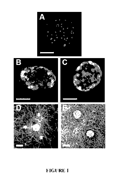
[0022] Figure 1. Mouse islets macroencapsulated in polymerized collagen
formulations show
sustained viability and fibril density-dependent traction forces following 14
days of in-vitro culture.
Representative images of calcein AM and propidium iodide stained islets after
14 days of culture in
liquid media suspension (A), macroencapsulated in polymerized collagen-1.5
mg/mL (B), or
macroencapsulated in polymerized collagen-3.0 mg/mL (C). Confocal reflection
images show islet-
induced collagen-fibril deformation (arrows) within polymerized collagen-1.5
mg/mL (D) but not
polymerized collagen-3.0 mg/mL (E). Scale bar = 50 p.m (A-C) and 100 pm (D,E)
[0023] Figure 2. Mouse islets macroencapsulated in polymerized collagen
formulations maintain
4
SUBSTITUTE SHEET (RULE 26)
CA 03071159 2020-01-24
WO 2019/023266 PCT/US2018/043539
multicellular cytoarchitecture following 14 days of in-vitro culture A,
Immunostaining shows islet
cytoarchitecture with insulin- and glucagon-positive cells within polymerized
collagen-1.5mg/mL and
polymerized collagen-3.0 mg/mL. By comparison, liquid media suspensions of
islets show
compromised insulin and glucagon staining patterns. Islet cell nuclei were
visualized with DRAQSTM.
Scale bar = 30 m.
[0024] Figure 3. Mouse islets macroencapsulated in polymerized collagen
formulations maintain
function following 14 days of in-vitro culture. Fourteen-day normalized
stimulation indices (mean SD;
n=8-12) as measured by glucose-stimulated insulin release. Values were
normalized to stimulation
indices for freshly isolated (day 0) islets. Asterisk indicates mean
stimulation index value for 14-day
suspension islets was significantly less than the value for freshly isolated
islets (p<0.05).
[0025] Figure 4. Non-fasting blood glucose levels (mean SD) following
subcutaneous injection and
in-situ macroencapsulation of C57BL/6J islets in various polymerized collagen
formulations (n=3 for
each formulation) within diabetic immunocompromised NOD. SCID mice as compared
to
subcutaneous injection of islets only (n=4) and polymerized collagen only
(n=3) control groups.
[0026] Figure 5. Histopathological and immunohistochemical analysis of
C57BL/6J islet explants in
polymerized collagen-1.5 mg/mL (A-D) 14 days following subcutaneous
transplantation in diabetic
immunocompromised NOD.SCID mice. Polymerized collagen-islet constructs were
readily
identifiable between the panniculus carnosus muscle and the skeletal muscle
facial layers. Masson's
trichrome (A) and H&E (B) stained sections indicated functional
vascularization of islets and no
evidence of polymerized fibrillar collagen degradation, inflammation, or
foreign body response. Islets
maintained multicellular cytoarchitecture with cells staining positively for
insulin and glucagon (D).
Cell nuclei were stained with DAPI (2-(4 Amidinopheny1)-1H-indole-6-
carboxamidine).
[0027] Figure 6. Histopathological and immunohistochemical analysis of
C57BL/6J islet explants in
SUBSTITUTE SHEET (RULE 26)
CA 03071159 2020-01-24
WO 2019/023266 PCT/US2018/043539
polymerized collagen-2.2 mg/mL (A-D) 14 days following subcutaneous
transplantation in diabetic
immunocompromised NOD.SCID mice. Polymerized collagen-islet constructs were
readily
identifiable between the panniculus carnosus muscle and the skeletal muscle
facial layers. Masson's
trichrome (A) and H&E (B) stained sections indicated functional
vascularization of islets and no
evidence of polymerized fibrillar collagen degradation, inflammation, or
foreign body response. Islets
maintained multicellular cytoarchitecture with cells staining positively for
insulin and glucagon (D).
Cell nuclei were stained with DAPI.
[0028] Figure 7. Histopathological and immunohistochemical analysis of
C57BL/6J islet explants in
polymerized collagen-3.0 mg/mL (A-D) 14 days following subcutaneous
transplantation in diabetic
immunocompromised NOD.SCID mice. Polymerized collagen-islet constructs were
readily
identifiable between the panniculus carnosus muscle and the skeletal muscle
facial layers. Masson's
trichrome (A) and H&E (B) stained sections indicated functional
vascularization of islets and no
evidence of polymerized fibrillar collagen degradation, inflammation, or
foreign body response. Islets
maintained multicellular cytoarchitecture with cells staining positively for
insulin and glucagon (D).
Cell nuclei were stained with DAPI.
[0029] Figure 8. Histopathological and immunohistochemical analysis of
C57BL/6J islet-only
explants (A-D) 14 days following subcutaneous transplantation in diabetic
immunocompromised
NOD. SCID mice. H&E stained sections (A-B) and immunohistochemical staining
for insulin and
glucagon (C) showed injection of islets in saline resulted in formation of a
large granuloma with loss
of normal multicellular morphology and protein expression.
[0030] Figure 9. In (A), non-fasting blood glucose levels (mean SD) following
subcutaneous in-situ
macroencapsulation of syngeneic islets in polymerized collagen-3.0 mg/mL (n=3)
within diabetic
C57BL/6J mice compared to islet only group (n=3). Mice receiving
macroencapsulated islets achieved
6
SUBSTITUTE SHEET (RULE 26)
CA 03071159 2020-01-24
WO 2019/023266 PCT/US2018/043539
normoglycemia within 24 hours following transplantation. Blood glucose
remained below the diabetic
threshold (<250 mg/dL; dashed line) throughout the 90-day study period. The
control group remained
diabetic throughout the study with widely varying blood glucose values.
Glucose tolerance test
(mean SD) and associated area under the curve (AUC; mean SD) analysis 15 (B,C)
and 90 (D,E) days
following transplantation demonstrated the capacity of islets + polymerized
collagen group but not
islets-only group to rapidly regulate blood glucose levels following glucose
injection. AUC values for
islets + polymerized collagen group were significantly (p<0.05) less than
those for islet only controls
at both time points
[0031] Figure 10. Histopathological analysis of polymerized collagen-
encapsulated (3.0 mg/mL),
syngeneic islets 90 days following subcutaneous transplantation within
diabetic mice (A and B). H&E
stained cross-sections showing polymerized collagen encapsulated islets within
the subcutaneous space
below the panniculus carnosus muscle (PCM). The fibrillar collagen matrix
formed by polymerized
collagen persisted and integrated with surrounding host tissues, with evidence
of functional
revascularization (panel B, black arrow). Encapsulated islets stained positive
for insulin and glucagon
(as indicated by asterisks) in (C). CD31 (white arrows) staining confirmed the
presence of endothelial
cells near islets co-stained to visualize insulin and nuclei in (D) and (E).
[0032] Figure 11. Histopathological analysis of explant 90-days following
subcutaneous
transplantation of islets only within diabetic mice showed evidence of
inflammatory-mediate
destruction and necrosis of islets.
[0033] Figure 12. Non-fasting blood glucose levels (mean SD) following
subcutaneous in-situ
macroencapsulation of allogeneic CD1 mouse islets in polymerized collagen-3.0
mg/mL (n=3) and
polymerized collagen-4.2 mg/mL (n=5) within diabetic C57BL/6J mice. Dashed
line at blood glucose
level of 250 mg/dL represents diabetic threshold.
7
SUBSTITUTE SHEET (RULE 26)
CA 03071159 2020-01-24
WO 2019/023266 PCT/US2018/043539
[0034] Figure 13. Histopathological analysis of polymerized collagen-
encapsulated (4.2 mg/mL),
allogeneic islets 60 days following subcutaneous transplantation. (A,B). H&E
stained cross-sections
showing encapsulated islets within the subcutaneous space below the panniculus
carnosus muscle
(PCM) with no evidence of a foreign body reaction against the polymerized
collagen or allogeneic
islets.
[0035] Figure 14. Immunostained explants of polymerized collagen-encapsulated
(4.2 mg/mL)
allogeneic islets 60 days following subcutaneous transplantation. Islets
stained positive for insulin and
glucagon (asterisk) are seen in (A), with CD31 (B-C; see arrows) staining
confirming the presence of
endothelial cells near islets co-stained for insulin and nuclei.
[0036] Figure 15. Human islets macroencapsulated in polymerized collagen show
preserved
morphology and function following 14-day in-vitro culture. A. Immunostaining
shows islet
cytoarchitecture with insulin- and glucagon-positive cells within polymerized
collagen-4.0 mg/mL.
Islet cell nuclei were visualized with DRAQSTM. Scale bar = 60 pm. B.
Stimulation index (mean+SE)
as measured by glucose-stimulated insulin release for untreated control islets
(day 0; n=8), 14-day
suspension islets (n=9), and 14-day polymerized collagen-islet constructs (4.0
mg/ml; n=8). Asterisk
indicates that the mean stimulation index value for 14-day suspension islets
was significantly less than
values for other two groups (p<0.05).
[0037] Figure 16. Histopathological analysis of human islet explants 9 days
following subcutaneous
transplantation in diabetic C57BL/6J mice (xenogenic model). Islets delivered
and encapsulated within
polymerizable collagen (2000 Pa) maintained their multicellular
cytoarchitecture (A-B). There was no
evidence of acute inflammation or foreign body response associated with the
islets or polymerized
collagen material.
[0038] Figure 17. Effect of mycophenolic acid (MPA) on islet viability when
incorporated as a local
8
SUBSTITUTE SHEET (RULE 26)
CA 03071159 2020-01-24
WO 2019/023266 PCT/US2018/043539
immunomodulatory agent within polymerizable collagen-islet suspensions.
Neutralized collagen
prepared at 1000 Pa (A,C,E) or 2000 Pa (B,D,F) was mixed with human islets
alone (control; A,B) or
human islets and either 0.01 mg/ml (C,D) or 1.0 mg/ml (E,F) MPA. The collagen-
islet-MPA
suspension was polymerized within a well-plate and cultured for 7 days. Islet
viability was detected
using calcein AM (live) and propidium iodide (dead; as indicated by small
round dots) and imaged via
confocal microscopy. Scale bar= 100 gm.
[0039] Figure 18. Effect of mycophenolate mofetil (MMF) on islet viability
when incorporated as a
local immunomodulatory agent within polymerizable collagen-islet suspensions.
Neutralized collagen
prepared at 1000 Pa (A,C,E) or 2000 Pa (B,D,F) was mixed with human islets
alone (control; A,B) or
human islets and either 0.01 mg/ml (C,D) or 1.0 mg/ml (E,F) MMF. The collagen-
islet-MMF
suspension was polymerized within a well-plate and cultured for 7 days. Islet
viability was detected
using calcein AM (live) and propidium iodide (dead; as indicated by small
round dots) and imaged via
confocal microscopy. Scale bar= 100 gm.
[0040] Figure 19. Confocal image of polymerized collagen-islet-endothelial
colony forming cell
(ECFC) construct following 7 days of culture. Mouse islets and human ECFCs
were suspended in
neutralized collagen solution and used to create polymerized collagen-islet-
ECFC constructs. Within
the polymerized collagen, ECFC underwent vasculogenesis forming vessel-
networks that interfaced
with nearby islets.
DETAILED DESCRIPTION
[0041] While the concepts of the present disclosure are illustrated and
described in detail in the figures
and the description herein, results in the figures and their description are
to be considered as exemplary
and not restrictive in character; it being understood that only the
illustrative embodiments are shown
and described and that all changes and modifications that come within the
spirit of the disclosure are
9
SUBSTITUTE SHEET (RULE 26)
CA 03071159 2020-01-24
WO 2019/023266 PCT/US2018/043539
desired to be protected. Unless defined otherwise, the scientific and
technology nomenclatures have
the same meaning as commonly understood by a person in the ordinary skill in
the art pertaining to this
disclosure.
[0042] This disclosure includes innovative therapies for diabetes and glucose
lowering and by creating
collagen-fibril microenvironments for improved i) islet survival, function,
and protection in vitro and
during transport and ii) islet survival, function, protection, delivery, and
engraftment in vivo. For
example, the cytoarchitecture and function of mouse and human islets can be
maintained in vitro
beyond 14 days when encapsulated within collagen compositions of the
disclosure. Further, mouse
islets, either syngeneic or allogeneic, in a collagen-suspension injected
subcutaneously self-assemble
in vivo and maintain normoglycemia in an established diabetic mouse model.
Transplanted islets
maintained responsiveness to a glucose tolerance test as well as their
characteristic multi-cellular
morphology with no associated inflammatory response.
[0043] In many embodiments of the disclosure, compositions comprising collagen
in an aqueous
medium with insulin-producing cells are provided. The collagen is usually type-
I collagen, and the
type-I collagen is typically oligomeric collagen. The collagen medium is
usually acidic and may be,
for example, a solution or a suspension. The compositions are typically
combined with a self-assembly
reagent, often in an aqueous solution, which tends to neutralize the aqueous
medium and enable self-
assembly and polymerization. The self-assembly reagent may raise the pH to,
for example, physiologic
pH and/or physiologic ionic strength. The compositions are often in the form
of an aqueous suspension
and when the temperature is raised, such as to physiologic temperature, the
rate of polymerization
increases. Islets or other insulin-producing cells may be added to the
neutralized collagen solution prior
to polymerization. Polymerization may be induced in vitro or, for example, by
injecting the
compositions herein in vivo such as into a mammal. To slow polymerization, one
may keep the
compositions at less than 10 C such as at about 4 C.
SUBSTITUTE SHEET (RULE 26)
CA 03071159 2020-01-24
WO 2019/023266 PCT/US2018/043539
[0044] The self-assembly reagent may comprise one or more of a buffer, base,
various salts and sugar
(for example, glucose). A particular example of a self-assembly reagent can be
found in Example 1.
[0045] The insulin-producing cells may be in the form of islets, such as
pancreatic islets. When
compared with the host mammal to which the compositions of the disclosure are
administered, the
islets may be autografts, allogeneic, or xenogeneic. Thus, for example, human
or porcine islets could
be used in a human patients with diabetes, representing allogeneic and
xenogeneic transplants,
respectively. As another example, canine or porcine islets could be used in a
dog with diabetes,
representing allogeneic and xenogeneic transplants, respectively. The islets
may be derived from
genetically modified animals. Typically, islets are taken from dogs, cats,
porcine animals, or humans.
[0046] The insulin-producing cells, including beta cells, may be stem-cell
derived insulin-producing
cells, and these may be derived induced pluripotent stem cells, embryonic stem
cells, or adult stem
cells. The insulin-producing cells may be progenitor derived or genetically
modified.
[0047] The stiffness of the polymerized collagen is related to the
concentration of collagen in the
composition. Typical concentrations of collagen range from about 0.5 mg/mL all
the way up to about
40 mg/mL of liquid medium of the of the composition which is often a
suspension. Other ranges
include between about 0.5 mg/mL and about 30 mg/mL, between about 1 mg/mL and
about 20 mg/mL,
between about 1 mg/mL and about 10 mg/mL, between about 1 mg/mL and about 5
mg/mL, between
about 1.5 mg/mL and about 5 mg/mL, between about 2 mg/mL and about 5 mg/mL,
between about 2.2
mg/mL and about 4.2 mg/mL, between about 3.0 mg/mL and about 4.2 mg/mL. In
many embodiments
the concentration of oligomer is either about 3 mg/mL or about 4.2 mg/mL.
[0048] Once polymerized, the collagen-insulin-producing cell compositions of
the disclosure typically
become semi-solids. Such compositions may be called collagen-insulin-producing
cell constructs or
collagen-islet constructs when the insulin-producing cells are islets. For
example, when suspensions
11
SUBSTITUTE SHEET (RULE 26)
CA 03071159 2020-01-24
WO 2019/023266 PCT/US2018/043539
or solutions of collagen-insulin-producing cells are injected in vivo, such as
subcutaneously into a
mammal, fibril networks of collagen form during the polymerization process
which tend to encapsulate
the insulin-producing cells. During the process, at least some and often
substantially all of the collagen
takes place in the formation of the fibril network. The composition which
comprises the insulin-
producing cells and the polymerized collagen (i.e., the fibril network) is a
semi-solid in that it is not
uniformly liquid or solid. Rather, it has both a solid phase (fibrils) and a
liquid phase (aqueous within
the body). The polymerized collagen is typically polymerized oligomeric
collagen such as type I-
oligomeric collagen.
[0049] The stiffness of the collagen compositions depends on the
concentration. Various stiffness
values of the compositions of the disclosure may be prepared. Such stiffness
values typically range
between about 40 Pa and about 2 MPa. Other ranges include between about 100 Pa
and about 1 I\IPa,
between about 200 Pa and about 1 I\IPa, between about 300 Pa and about 500
KPa, between about 500
Pa and about 100 KPa, between about 500 Pa and about 5 KPa, between about 800
Pa and about 3
KPa, between about 900 and about 2.5 KPa, and between about 1KPa and about 2
KPa. Specific
embodiments include about 1 KPa or about 2 KPa.
[0050] Unlike islet suspensions of the prior art, the insulin-producing cells
of the disclosure, such as
pancreatic islets, when present in the compositions of the disclosure, are
able to be sustained for over
14 days in vitro due to the protective nature of the collagen fibril network
surrounding the cells. For
example, figure 1 shows compositions of the disclosure made in accordance with
Example 2. Mouse
islets were combined with acidic stock collagen solutions made in accordance
with Example 1 and
polymerized with the self-assembly reagent of Example 1. The compositions so
polymerized were
evaluated after 14 days in a cultured medium and compared with islets which
were simply suspended
in a liquid medium. As shown in Figure 1A, such islets died, whereas islets
encapsulated in accordance
with compositions of the disclosure survived, for example, as set forth in
Figure 1B and Figure 1C.
12
SUBSTITUTE SHEET (RULE 26)
CA 03071159 2020-01-24
WO 2019/023266 PCT/US2018/043539
[0051] When formed in a mammal, such as after subcutaneous injection, or after
implantation, the
insulin-producing cells in the collagen compositions of the disclosure are
sustainable for well beyond
14 days such as for over 90 days. In addition, the compositions, when placed
in vivo, do not trigger a
foreign body response. Such a response would evidence itself by infiltrating
inflammatory cell
populations, such as neutrophils and macrophages, as well as formation of an
obvious fibrous capsule
around the fibrillar network, and histology shows no evidence of such
inflammatory cell populations
or fibrous capsule formation. Indeed, histology shows the integration of the
compositions of the
disclosure via vascularization of surrounding tissue in vivo. Further, if
there were a foreign body
response, the islets would cease to function and, as the Examples herein and
figures show, the contrary
occurs ¨ the islets survive and the islets continue to produce insulin to
maintain normal blood glucose
levels.
[0052] When placed in a mammal, the compositions of the disclosure are capable
of lowering blood
glucose levels to below the diabetic threshold on a mammalian species basis.
Indeed, the compositions
may be used to maintain normoglycemia. The compositions may also be used to
treat metabolic
disorder such as type 1 diabetes and indeed have been shown to reverse
diabetes within 24 hours of
administration as seen in Example 5 and Figure 12.
[0053] The compositions of the disclosure may further comprise an active
pharmaceutical ingredient,
such as an immunosuppressive agent. Examples of immunosuppressive agents
include mycophenolic
acid, mycophenolate mofetil, or a combination thereof. Compositions containing
such agents may be
dosed to a mammal in order to treat metabolic conditions for example. The
compositions of the
disclosure may further comprise non-islet cells. Examples of such cells
include endothelial colony-
forming cells. Such endothelial colony-forming cells may be, for example,
human endothelial colony-
forming cells. Such endothelial colony-forming cells may be vessel-forming.
13
SUBSTITUTE SHEET (RULE 26)
CA 03071159 2020-01-24
WO 2019/023266 PCT/US2018/043539
[0054] In other embodiments of the disclosure, collagen-solutions may be
combined with islets to form
suspensions that are injected in preclinical diabetic allogenic and syngeneic
mouse models. For
example, 500 islets, separated into 2 sites, were mixed with collagen
solutions at different
concentrations (3.2 mg/mL, 2.2 mg/mL, or 1.4 mg/mL which corresponds to 1000
Pa, 500 Pa, or 200
Pa, respectively) or saline and then injected into the subcutaneous space of
mice. In an allogenic
(C57BL/6J to NOD-SCID) model, mice that had collagen-islet constructs of the
disclosure with a
higher collagen concentration consistently lowered and maintained
normoglycemia (<300mg/dL) for
14 days after transplantation. Control mice that received islets mixed with
saline remained
hyperglycemic over the study period (>250mg/dL). Further testing of 1000 Pa
collagen-islet construct
in a syngeneic (C57BL/6J to C57BL/6J) model indicates that the mice lowered
and maintained
normoglycemia for more than 90 days (see Figure 9A). Histological analysis
showed mature
vasculature near transplanted islets and no evidence of a foreign body
response to the collagen-islet
construct. Immunofluorescence indicated viable islets that positively stained
for insulin and glucagon.
[0055] In other embodiments, human islets may be used to make collagen-islet
construct of the
disclosure as, for example, set forth in Example 6. Such constructs have been
shown to be stable in
vitro as seen in Example 6 and Figure 15. Human islets in collagen-islet
constructs of the disclosure
have been transplanted into diabetic mice without a foreign body response,
thus showing the
xenogeneic robustness of compositions of the disclosure, as seen in Example 7.
Example 8 is directed
collagen-insulin-producing cell compositions of the disclosure, such as
collagen-islet constructs, may
also be combined with one or more of the accessory cells or active
pharmaceutical ingredients.
[0056] Further, collagen-insulin-producing cell compositions of the disclosure
can be readily injected
into a well plate to polymerize and create polymerized collagen-encapsulated
insulin-producing-cell
constructs for long-term maintenance of cell morphology and function in vitro.
Such compositions may
also be injected subcutaneously in vivo for management of type 1 diabetes. In
many embodiments, the
14
SUBSTITUTE SHEET (RULE 26)
CA 03071159 2020-01-24
WO 2019/023266 PCT/US2018/043539
insulin-producing cells are islets.
[0057] Collagen encapsulation and delivery of insulin-producing cells such as
islets have several
advantages over conventional and emerging therapeutic strategies, including:
1) low cost encapsulation
biomaterial; 2) biomaterial elicits no foreign body response; 3) biomaterial
induces rapid
neovascularization, innervation, and tissue integration which supports islet
longevity and function; 4)
therapeutic insulin-producing cells such as islets provide superior
physiologic blood glucose
monitoring and maintenance; 5) supports local delivery of any necessary
immunomodulation agents;
6) may provide complete insulin independence; 7) may eliminate or reduce the
need for long-term
immunosuppression.
[0058]
EXAMPLES
[0059] Example 1. Preparation and Induction of Polymerization (Self-assembly)
of Collagen
Compositions
[0060] Type I oligomeric collagen was either isolated from porcine dermis
according to the procedure
outlined in Example 1 of U.S. Patent No. 8,084,055 or obtained in a
proprietary sterile formulation
from GeniPhys, LLC as medical grade, which were prepared following the general
procedures set forth
below.
[0061] Type I collagen oligomers are derived from the dermis of closed herd
pigs and prepared as
described previously (Bailey JL, Critser PJ, Whittington C, Kuske JL, Yoder
MC, Voytik-Harbin SL;
Collagen oligomers modulate physical and biological properties of three-
dimensional self-assembled
matrices, Biopolymers (2011) 95(2):77-93 and Kreger ST, Bell BJ, Bailey J,
Stites E, Kuske J, Waisner
B, Voytik-Harbin SL; Polymerization and matrix physical properties as
important design
considerations for soluble collagen formulations, Biopolymers (2010) 93(8):690-
707, both
SUBSTITUTE SHEET (RULE 26)
CA 03071159 2020-01-24
WO 2019/023266 PCT/US2018/043539
incorporated herein by reference). Prior to use, lyophilized type I oligomeric
collagen was dissolved in
0.01 N hydrochloric acid to form an acidic collagen solution. The acidic
collagen solution was then
rendered aseptic by filtration or treatment with chloroform. A Sirius Red
(Direct Red 80) assay is
used to determine collagen concentration. Oligomer formulations are
standardized based upon purity
as well as polymerization capacity according to the ASTM international
consensus standard F3089-14
(ASTM Standard F3089, 2014, "Standard Guide for Characterization and
Standardization of
Polymerizable Collagen-Based Products and Associated Collagen-Cell
Interactions", ASTM
International, West Conshohocken, PA, F3089-14, www.astm or . Polymerization
capacity is defined
by matrix shear storage modulus (G') as a function of collagen concentration
of the polymerization
reaction. Single-step self-assembly was performed with a 10X self-assembly
reagent prepared
according to the following recipe:
2 g KH2PO4 (FW 136.09)
30 11.5 g Na2HPO4 (FW 141.96)
2 g KC1 (FW 74.55)
g glucose
80 g NaC1 (FW 58.44) 20 ml 5N NaOH
All reagents are added to Milli-Q filtered water to achieve a finalized volume
of liter and sterile filtered
(0.22 p.m). One part 10X self-assembly reagent is then added to 9 parts acidic
collagen solution, which
initiates polymerization. In each example below collagen-islet encapsulations
were done with
oligomer.
[0062] Example 2. Mouse Islets Show Improved Viability, Cytoarchitecture, and
Function In-
vitro Following Creation of Polymerized Collagen-Islet Constructs
16
SUBSTITUTE SHEET (RULE 26)
CA 03071159 2020-01-24
WO 2019/023266 PCT/US2018/043539
[0063] Loss of critical microenvironmental cues upon islet isolation and
culture decreases islet
viability and function, thereby compromising islet engraftment and transplant
outcome. By
comparison, polymerizable collagen can improve survival and function of mouse
islets in vitro.
Polymerized collagen-islet constructs were created and cultured for periods of
time up to 14 days and
additional mouse islets were cultured using the conventional liquid suspension
format (see e.g.,
Methods of Human Islet Culture for Transplantation. Murdoch TB, McGhee-Wilson
D, Shapiro AMJ,
Lakey JRT. Cell Transplant. 2004 Sep; I3(6):605-618) for comparison with the
results herein
indicating improvements due to the collagen-islet constructs of the
disclosure.
[0064] Mouse Islets. Mouse pancreatic islets were isolated from 8- to 14-wk
old C57BL/6J mice
(Jackson Laboratory, Bar Harbor, ME) according to established methods. Islet
isolations were
approved by the Indiana University Institutional Animal Care and Use Committee
using AALAC
guidelines. Isolated islets were cultured suspended in liquid culture medium
consisting of RPMI 1640
medium supplemented with 10% fetal bovine serum (HyClone, ThermoFisher
Scientific, Waltham,
MA), 100 U/mL penicillin, and 100 1.1g/mL streptomycin (Sigma Aldrich, St.
Louis, MO) in a
humidified environment of 5% CO2 in air at 37 C prior to experimental use the
next day.
[0065] Collagen Encapsulation of Mouse Islets for In-Vitro Culture. To prepare
polymerized collagen
matrices at different stiffness (G') values, stock acidic collagen solutions
of Example 1 were diluted
with 0.01 N HCl so to achieve final collagen concentrations of 0.5-5 mg/mL in
the polymerization
reaction. These polymerization reaction concentrations yield polymerized
matrices with shear storage
modulus (G', Pa; also referred to generally as stiffness) from about 40 to
about 2500 Pa, as defined by
the collagen's polymerization capacity. The polymerization reaction was
initiated by adding 10X self-
assembly solution of Example 1 to the acidic collagen solution (9 parts acidic
collagen solution plus 1
part 10X self-assembly solution of Example 1), which neutralizes the) to form
a neutralized collagen
solution. Mouse islets were suspended in the neutralized collagen solution,
aliquoted into 96 well-
17
SUBSTITUTE SHEET (RULE 26)
CA 03071159 2020-01-24
WO 2019/023266 PCT/US2018/043539
plates (30 islets/100 t L; Cellvis, Sunnyvale, CA). The neutralized collagen
solutions (in the presence
or absence of cells) were maintained on ice (4 C) prior to warming to 37 C,
which induce rapid
polymerization. Immediately following polymerization, culture medium (RPMI
1640 medium
supplemented with 10% fetal bovine serum (HyClone, ThermoFisher Scientific,
Waltham, MA), 100
U/mL penicillin, and 100 [tg/mL streptomycin (Sigma Aldrich, St. Louis, MO))
was added, and the
collagen-islet constructs were cultured for up to 14 days with medium changes
made daily. For
comparison purposes, mouse islets were also cultured in a conventional
suspension format in liquid
medium.
[0066] Assessment of Islet Viability and Function Following In-Vitro Culture.
Islets cultured
suspended in either conventional liquid media or as polymerized collagen-islet
constructs of the
disclosure were treated with Calcein AM and propidium iodide (Molecular
Probes, Eugene, OR) for
live-dead determinations. Images were collected using laser scanning confocal
microscopy on an
Olympus IX81 inverted microscope adapted with Olympus Fluoview FV1000
(Olympus, Tokyo,
Japan). Image stacks of 40-100 ttm thickness with a 3 [tm step size were
obtained using a 20X air
objective, and z-projections were created using Imaris software (Bitplane,
Concord, MA).
[0067] Immunofluorescence was used to qualitatively assess islet
cytoarchitecture and protein
expression. Islets cultured suspended in liquid or as polymerized collagen-
islet constructs were fixed
in 3% paraformaldehyde (Mallinckrodt, Derbyshire, UK), permeabilized with 0.1%
Triton X-100
(Sigma Aldrich), and blocked with 1% bovine serum albumin (Jackson
ImmunoResearch, West Grove,
PA). Samples then were treated overnight at 4 C with primary guinea pig anti-
insulin (PA1-26938,
Invitrogen) and rabbit anti-glucagon antibodies (mouse islets: ab10988, Abcam,
Cambridge, MA;
human islets: 2760, Cell Signaling Technologies, Danvers, MA). Samples were
rinsed and then treated
with secondary antibodies (A11073, goat anti-guinea pig Alexa Fluor 488
conjugate and A11035, goat
anti-rabbit Alexa Fluor 546 conjugate, Life Technologies) overnight at 4 C.
After rinsing, samples
18
SUBSTITUTE SHEET (RULE 26)
CA 03071159 2020-01-24
WO 2019/023266 PCT/US2018/043539
were treated with DRAQ5T1 (Cell Signaling Technologies, Danvers, MA) to stain
nuclei.
[0068] Quantitative assessment of islet function was performed via glucose
stimulated insulin
secretion (GSIS) testing. Islets cultured suspended in liquid or as
polymerized collagen-islet constructs
were prepared within 24-well Transwell culture inserts (Corning, Kennebunk,
NM) for up to 14 days.
Basal insulin secretion was stimulated by incubating the samples for 1 hour
with 2.8 mM glucose in
Krebs Ringer buffer (0.1% BSA, 25 mM HEPES, 115 mM NaCl, 24 mM NaHC0.3,5 mM
KC1, 1 mM
MgC12=6H20, 2.5 mM CaC12=2H20). Following basal secretion, samples underwent
static incubation
for 1 hour each, with low (2.8 mM) followed by high (28 mM) glucose
concentrations in Krebs Ringer
buffer. Insulin secretion was measured using a STELLUX insulin enzyme-linked
immunosorbent
assay (ELISA) kit (Alpco, Salem, NH). Stimulation indices, which represent the
ratio of insulin
secreted with high glucose over insulin secreted with low glucose, were
calculated and normalized to
values obtained for freshly isolated (day 0) islets.
[0069] Summary of Findings. Islets cultured suspended in liquid medium
exhibited a significant loss
of viability, with the majority of islet cells showing moderate to significant
islet death over the 14-day
culture period (Figure 1A). Suspension islets also displayed compromised
insulin and glucagon
staining patterns (Figure 2) and a significant (p<0.05) decrease in function
over time, as measured by
GSIS (Figure 3). In contrast, islets encapsulated and cultured within
polymerized collagen of the
disclosure prepared at 1.5 mg/mL and 3.0 mg/mL maintained their viability
(Figure 1B-C), their multi-
cellular architecture with insulin- and glucagon-producing cells (Figure 2),
and glucose-sensitive
insulin secretion function (Figure 3) over the 14-day culture period. The
ability of islet cells to bind to
and exert contractile forces on the surrounding collagen-fibril matrix was
evident at both macroscopic
(construct contraction) and microscopic (islet-collagen fibril interactions)
levels (Figure 1D-E).
Although these islet-collagen interactions resulted in contraction of the
majority of 0.5 mg/mL
constructs, 1.5 mg/mL and 3.0 mg/mL constructs retained their volume and
showed progressively less
19
SUBSTITUTE SHEET (RULE 26)
CA 03071159 2020-01-24
WO 2019/023266 PCT/US2018/043539
cell-induced local fibril reorganization with increased concentration (Figure
1D-E).
[0070] Example 3. Subcutaneous Transplantation of Islets in Diabetic Mice:
Short-term Study
Using Allogeneic Model
[0071] Since encapsulation of mouse islets within polymerized collagen
constructs prolonged their
viability and function in-vitro, various polymerizable collagen formulations
were prepared by mixing
islets with neutralized collagen solutions to create collagen-islet
suspensions, followed by
subcutaneous injection of the collagen-islet suspension into streptozocin-
induced diabetic mice.
Immediately following injection, the collagen-islet suspension polymerized in
situ, forming a stable
and continuous collagen-fibril matrix that encapsulated and protected resident
islets within the
subcutaneous space. Allogeneic C57BL/6J mouse islets were injected into
NOD.CB17- Prkdc"id/J
mice, which exhibit inflammatory but not immune reactions mediated by T cells
and B cells. Function
of subcutaneously transplanted islets as well as the biocompatibility and
tissue response of the
polymerized collagen biomaterial were evaluated after 14 days and showed
positive effects compared
to control animals that received injections of polymerized collagen or islets
only (suspended in saline).
[0072] Mouse Islets. Mouse pancreatic islets were isolated from 8- to 14-wk
old C57BL/6J mice
(Jackson Laboratory, Bar Harbor, ME) according to methods of Stull and
coworkers. Islet isolations
were approved by the Indiana University Institutional Animal Care and Use
Committee using AALAC
guidelines. Isolated islets were incubated in RPMI 1640 medium supplemented
with 10% fetal bovine
serum (HyClone, ThermoFisher Scientific, Waltham, MA), 100 U/mL penicillin,
and 100 lig/mL
streptomycin (Sigma Aldrich, St. Louis, MO) in a humidified environment of 5%
CO2 in air at 37 C
prior to experimental use the next day.
[0073] Subcutaneous Islet Transplantation in Diabetic Mice (Allogeneic
Models). Mouse islet
transplantation procedures were approved by the Indiana University
Institutional Animal Care and Use
SUBSTITUTE SHEET (RULE 26)
CA 03071159 2020-01-24
WO 2019/023266 PCT/US2018/043539
Committee using AALAC guidelines. Male 8-wk to 14-wk old NOD,CB17- Prkdc/J
(allogeneic
model; Jackson Laboratories, Bar Harbor, ME) recipient mice were injected with
low dose
streptozotocin (55 mg/kg) for 5 days to chemically induce diabetes prior to
islet transplantation.
C57BL/6J islets were mixed with neutralized collagen solutions. Collagen-islet
suspensions were then
injected subcutaneously through small bore needles (26 1/2 G) into diabetic
mice. Each mouse received
2 injections, one positioned laterally on each side of the back, with
approximately 250 islets/500 lit
collagen (oligomer) solution neutralized with the self-assembly reagent of
Example 1 per site for a
total of 500 islets/mouse. Additional mice received 2 injections of islets
suspended in saline (islets
only; 250 islets/500 1AL saline/site) or neutralized collagen solution
(collagen only: 500 [it
collagen/site) for comparison. Non-fasting blood glucose was measured 3 times
per week after
transplantation. Diabetes was classified as two consecutive blood glucose
levels above 250 mg/dL.
[0074] Intraperitoneal Glucose Tolerance Test. Intraperitoneal glucose
tolerance tests (GTT) were
performed at specified times following subcutaneous transplantation to assess
islet responsiveness to
glucose challenges. Mice were fasted overnight, and then injected
intraperitoneal with 2 g/kg of 20%
glucose. Blood glucose levels were measured at baseline before injection (time
0) and 15, 30, 60, 90,
and 120 minutes following glucose injection.
[0075] In-Vivo Histology and Immunofluorescence. Injection sites and
surrounding tissues were
removed at specified timepoints and placed in 10% formalin before paraffin
embedding and sectioning.
Sections were stained with hematoxylin and eosin (H&E) and Masson's trichrome
(MTC). For
immunofluorescence, sections were deparaffinized, rehydrated, and stained with
primary guinea pig
anti-insulin (A0564, Dako, Santa Clara, CA) and rabbit anti-glucagon (sc-
13091, Santa Cruz
Biotechnology, Dallas, TX) at 1:500 dilutions. Sections were then treated with
secondary Alexa Fluor
488 goat anti-guinea pig (A11073, Life Technologies) and Alexa Fluor 568 goat
anti-rabbit (A11036,
Life Technologies) at 1:50 and 1:200 dilutions, respectively. For
identification of vascular
21
SUBSTITUTE SHEET (RULE 26)
CA 03071159 2020-01-24
WO 2019/023266 PCT/US2018/043539
endothelium, sections were stained with primary mouse anti-CD31/PECAM-1 (BBA7,
R&D Systems,
Minneapolis, MN) then treated with secondary Alexa Fluor 546 donkey anti-mouse
(A10036, Life
Technologies). Nuclei were counterstained with DRAQ5TM or DAPI.
[0076] Summary of Results. Initial pilot studies evaluated collagen solutions
at various concentrations
of 1.5, 2.2, and 3.0 mg/ml which correspond roughly to polymerized collagen
matrix stiffness values
200, 500, and 1000 Pa respectively based upon standardized polymerization
capacity. The
polymerization half-time for the collagen at body temperature (37 C) was
20.26+0.12 seconds,
16.28+0.03 seconds, and 16.35+0.11 seconds, as measured rheometrically for
collagen solutions at 1.5,
2.2, and 3.0 mg/mL, respectively. Prior to islet transplant, mean blood
glucose values for the animals
were 533+72 mg/dL. As shown in Figure 4, collagen 1000 Pa + islets ("Islets
and Oligomer 3.0 in
Figure 4") provided the most reproducible reversal of diabetes in all animals
(consistent maintenance
of blood glucose values below the diabetic threshold) and as well as highly
regulated blood glucose
levels (small standard deviation bars). For the collagen 1000 Pa + islet
group, non-fasting blood glucose
decreased below the diabetic level within 24 hours and remained below the
diabetic level for the entire
14 day study period (Figure 4). In contrast, both islet only and collagen 1000
Pa only control groups
remained hyperglycemic, above the diabetic level, for the entire 14-day study
(Figure 4). Comparing
histopathological results of Figures 5, 6, and 7, the most uniform
encapsulation of individual islets was
achieved with 1000 Pa (3 mg/mL) collagen, with islets maintaining their normal
morphology with
evidence of functional revascularization (Figure 7). Islet aggregates were
noted within 500 Pa (1.5
mg/mL) + islet and 200 Pa (2.2 mg/mL) + islet groups, suggestive of
insufficient fibril density for
encapsulation and/or inadequate mixing. Immunostaining of all polymerized
collagen + islet groups
confirmed a multicellular cytoarchitecture with both insulin- and glucagon-
producing cells (Figures
5D, 6D, and 7D). The collagen material, which polymerized in situ following
injection, appeared as
normal collagenous connective tissue with moderate fibroblast infiltration and
no evidence of
22
SUBSTITUTE SHEET (RULE 26)
CA 03071159 2020-01-24
WO 2019/023266 PCT/US2018/043539
inflammation or foreign body response (Figure 5). The fibrillar collagen
material was highly stable and
well-integrated within the subcutaneous space for all collagen + islet
(Figures 5-7) and collagen only
groups. In contrast, for the islet only group, large granulomatous regions
were observed with no
identifiable islets (Figure 8). Such findings are consistent with previous
reports that suggest that the
subcutaneous space is an inhospitable microenvironment for transplantation of
islets alone.
[0077] Example 4. Subcutaneous Transplantation of Islets in Diabetic Mice:
Long-term Study
Using Syngeneic Model
[0078] To assess long-term survival and function of collagen-encapsulated
islets following
subcutaneous delivery, C57BL/6J mouse islets were injected into C57BL/6J mice
(syngeneic
transplant).
Mouse Islets. Mouse pancreatic islets were isolated from 8- to 14-wk old
C57BL/6J mice (Jackson
Laboratory, Bar Harbor, ME) according to methods of Stull and coworkers. Islet
isolations were
approved by the Indiana University Institutional Animal Care and Use Committee
using AALAC
guidelines. Isolated islets were incubated in RPMI 1640 medium supplemented
with 10% fetal bovine
serum (HyClone, ThermoFisher Scientific, Waltham, MA), 100 U/mL penicillin,
and 100 [tg/mL
streptomycin (Sigma Aldrich, St. Louis, MO) in a humidified environment of 5%
CO2 in air at 37 C
prior to experimental use the next day.
[0079] Subcutaneous Islet Transplantation in Diabetic Mice (Syngeneic Model).
Mouse islet
transplantation procedures were approved by the Indiana University
Institutional Animal Care and Use
Committee using AALAC guidelines. Male 8-wk to 14-wk old C57BL/6J (syngeneic
model) recipient
mice were injected with low dose streptozotocin (55 mg/kg) for 5 days to
chemically induce diabetes
prior to islet transplantation. C57BL/6J islets were mixed with neutralized
collagen solutions.
Collagen-islet suspensions were then injected subcutaneously through small
bore needles (26 1/2 G)
23
SUBSTITUTE SHEET (RULE 26)
CA 03071159 2020-01-24
WO 2019/023266 PCT/US2018/043539
into diabetic mice. Each mouse received 2 injections, one positioned laterally
on each side of the back,
with approximately 250 islets/500 [EL collagen (oligomer) solution neutralized
with the self-assembly
reagent of Example 1 per site for a total of 500 islets/mouse. Additional mice
received 2 injections of
islets suspended in saline (islets only; 250 islets/500 [IL saline/site) or
neutralized collagen solution
(collagen only: 500 [IL collagen/site) for comparison. Non-fasting blood
glucose was measured 3 times
per week after transplantation. Diabetes was classified as two consecutive
blood glucose levels above
250 mg/dL.
[0080] Intraperitoneal Glucose Tolerance Test. Intraperitoneal glucose
tolerance tests (GTT) were
performed at specified times following subcutaneous transplantation to assess
islet responsiveness to
glucose challenges. Mice were fasted overnight, and then injected
intraperitoneally with 2 g/kg of 20%
glucose. Blood glucose levels were measured at baseline before injection (time
0) and 15, 30, 60, 90,
and 120 minutes following glucose injection.
[0081] In-Vivo Histology and Immunofluorescence. Injection sites and
surrounding tissues were
removed at specified timepoints and placed in 10% formalin before paraffin
embedding and sectioning.
Sections were stained with hematoxylin and eosin (H&E) and Masson's trichrome
(MTC). For
immunofluorescence, sections were deparaffinized, rehydrated, and stained with
primary guinea pig
anti-insulin (A0564, Dako, Santa Clara, CA) and rabbit anti-glucagon (sc-
13091, Santa Cruz
Biotechnology, Dallas, TX) at 1:500 dilutions. Sections were then treated with
secondary Alexa Fluor
488 goat anti-guinea pig (A11073, Life Technologies) and Alexa Fluor 568 goat
anti-rabbit (A11036,
Life Technologies) at 1:50 and 1:200 dilutions, respectively. Nuclei were
counterstained with
DRAQ5TM.
[0082] Summary of Results. Syngeneic mouse islets suspended in collagen 1000
Pa (3 mg/mL) and
injected subcutaneously within diabetic mice showed rapid engraftment and
function as indicated by
24
SUBSTITUTE SHEET (RULE 26)
CA 03071159 2020-01-24
WO 2019/023266 PCT/US2018/043539
rapid reversal of diabetes within 24 hours following transplantation in all
three recipient mice (Figure
9A). Blood glucose levels for these animals remained below the diabetic level
throughout the 90-day
study period with tight regulation as indicated by the small standard
deviation bars. In contrast, mice
transplanted with islets only remained diabetic with blood glucose values over
400 mg/dL (Figure 9A).
Intraperitoneal glucose tolerance testing (GTT) was performed on day 15
(Figures 9B and 9C) and 90
(Figures 9D and 9E) to further assess glucose responsiveness of transplanted
islets. Interestingly, mice
receiving collagen 1000 Pa + islets achieved normoglycemia 120 min post-
glucose injection while
blood glucose in islet only mice remained elevated above basal levels at both
15 and 90 day timepoints
(Figures 9B and 9D). Analysis of area under the curve (AUC) indicated that
values for mice receiving
collagen 1000 Pa + islets were significantly lower than those for islet only
mice at both timepoints
(p<0.05; Figures 9C and 9E). Histolopathologic and immunostaining analyses at
90 days showed
persistence of the collagen material with integration into the surrounding
subcutaneous tissue
compartment and no evidence of chronic inflammatory or foreign body response
(Figure 5A). Within
the collagen 1000 Pa, islets maintained their rounded, multicellular
architecture with evidence of a rich
capillary supply and insulin- and glucagon-positive cells (Figure 10A, B, and
C) and nearby patent
vasculature (Figure 10B) and CD-31 positive cells (Figures 10D and 10E). Islet
only explants showed
a robust foreign-body response marking graft failure (Figures 11A and 11B).
[0083] Example 5. Reversal of Diabetes with Immune Modulation Following In-
situ Oligomer
encapsulation of Allogeneic Islets
[0084] To assess function and immunoprotection of collagen-encapsulated islets
following
subcutaneous delivery, allogeneic transplantation studies were performed,
where CD1 mouse islets
were injected into diabetic C57BL/6J mice.
[0085] Mouse Islets. Mouse pancreatic islets were isolated from 8- to 14-wk
old CD1 mice (Jackson
Laboratory, Bar Harbor, ME) according to methods of Stull and coworkers. Islet
isolations were
SUBSTITUTE SHEET (RULE 26)
CA 03071159 2020-01-24
WO 2019/023266 PCT/US2018/043539
approved by the Indiana University Institutional Animal Care and Use Committee
using AALAC
guidelines. Isolated islets were incubated in RPMI 1640 medium supplemented
with 10% fetal bovine
serum (HyClone, ThermoFisher Scientific, Waltham, MA), 100 U/mL penicillin,
and 100 lag/mL
streptomycin (Sigma Aldrich, St. Louis, MO) in a humidified environment of 5%
CO2 in air at 37 C
prior to experimental use the next day.
[0086] Subcutaneous Islet Transplantation in Diabetic Mice (Allogeneic Model).
Mouse islet
transplantation procedures were approved by the Indiana University
Institutional Animal Care and Use
Committee using AALAC guidelines. Male 8-wk to 14-wk old C57BL/6J recipient
mice were injected
with low dose streptozotocin (55 mg/kg) for 5 days to chemically induce
diabetes prior to islet
transplantation. CD1 islets were mixed with collagen (oligomer) solution
neutralized with the self-
assembly reagent of Example 1 and the resulting collagen-islet suspensions
were then injected
subcutaneously through small bore needles (26 1/2 G) into diabetic mice. Each
mouse received 2
injections, one positioned laterally on each side of the back, with
approximately 250 islets/500 I,
collagen/site for a total of 500 islets/mouse. Non-fasting blood glucose was
measured 3 times per week
after transplantation. Diabetes was classified as two consecutive blood
glucose levels above 250
mg/dL.
[0087] In-Vivo Histology and Immunofluorescence. Injection sites and
surrounding tissues were
removed at specified timepoints and placed in 10% formalin before paraffin
embedding and sectioning.
Sections were stained with hematoxylin and eosin (H&E) and Masson's trichrome
(MTC). For
immunofluorescence, sections were deparaffinized, rehydrated, and stained with
primary guinea pig
anti-insulin (A0564, Dako, Santa Clara, CA) and rabbit anti-glucagon (sc-
13091, Santa Cruz
Biotechnology, Dallas, TX) at 1:500 dilutions. Sections were then treated with
secondary Alexa Fluor
488 goat anti-guinea pig (A11073, Life Technologies) and Alexa Fluor 568 goat
anti-rabbit (A11036,
Life Technologies) at 1:50 and 1:200 dilutions, respectively. Nuclei were
counterstained with
26
SUBSTITUTE SHEET (RULE 26)
CA 03071159 2020-01-24
WO 2019/023266 PCT/US2018/043539
DRAQ5TM,
[0088] Summary of Results. When the islets-collagen compositions were injected
subcutaneously,
polymerization occurred in vivo causing the islets to become macroencapsulated
in polymerized
collagen at 3 mg/mL. Blood glucose values declined within the first 24 hours;
however, animals
remained hyperglycemic (Figure 12). Hypothesizing that the level of immune
protection may be related
to the amount of oligomer (or fibril density), follow-up animals were
performed using an collagen
concentration of 4.2 mg/mL, which corresponds roughly to an oligomer stiffness
value of 2000 Pa and
polymerization half-time of 16.30+0.02 seconds. In this case, diabetes was
reversed within 24 hours
with blood glucose values gradually increasing above the diabetic threshold
after about 40 days (Figure
12). Histopathologic and immunostaining analyses of 60-day oligomer-islet
explants showed insulin-
and glucagon-positive islets surrounded by fibrillar collagen (Figure 13).
Interestingly, a subset of
islets showed no evidence of a foreign body response, while others showed very
mild inflammatory
infiltrate. In some instances, nearby vasculature was evident (Figure 14);
however, vascularization and
inflammation events did not appear to be correlated.
[0089] Example 6. Human Islets Show Improved Viability, Cytoarchitecture, and
Function In-
vitro Following Creation of Polymerized Collagen-Islet Constructs
[0090] Human Islets. Human pancreatic islets were obtained through the
Integrated Islet Distribution
Program (IIDP). Protocols for handling of human islets were approved by the
Purdue Institutional
Biosafety Committee. Islets were maintained overnight in Prodo Islet Media
(Recovery; Prodo
Laboratories, Aliso Viejo, CA) supplemented with 5% human AB serum (Prodo
Laboratories), 1%
Glutamine/Glutathione (Prodo Laboratories), and 100 U/mL penicillin, and 100
lig/mL streptomycin
(Sigma Aldrich) in a humidified environment of 5% CO2 in air at 37 C prior to
use in experiments.
[0091] Collagen Encapsulation of Human Islets for In-Vitro Culture. To prepare
polymerized collagen
27
SUBSTITUTE SHEET (RULE 26)
CA 03071159 2020-01-24
WO 2019/023266 PCT/US2018/043539
matrices at different stiffness (G') values, stock acidic collagen solutions
were diluted with 0.01 N HC1
so to achieve final collagen concentrations of 4 mg/mL in the polymerization
reaction. These
polymerization reaction concentrations yield polymerized matrices with shear
storage modulus (G',
Pa; also referred to generally as stiffness) of about 1500 Pa, as defined by
the collagen's polymerization
capacity. The polymerization reaction was initiated by adding 10X self-
assembly solution of Example
1 to the acidic collagen solution (9 parts acidic collagen solution and 1 part
10X self-assembly solution
of Example 1) to form a neutralized collagen solution. Human islets were
suspended in the neutralized
collagen solutions, aliquoted into 96 well-plates (30 islets/100
L; Cellvis, Sunnyvale, CA), and
allowed to polymerize at 37 C. Immediately following polymerization, culture
medium was added,
and the collagen-islet constructs were cultured for up to 14 days with medium
changes made daily. For
comparison purposes, human islets were also cultured in a conventional
suspension format in liquid
medium.
[0092] Assessment of Mouse and Human Islet Viability and Function Following In-
Vitro Culture. Islets
cultured suspended in liquid or as polymerized collagen-islet constructs were
treated with Calcein AM
and propidium iodide (Molecular Probes, Eugene, OR) for live-dead
determinations. Images were
collected using laser scanning confocal microscopy on an Olympus IX81 inverted
microscope adapted
with Olympus Fluoview FV1000 (Olympus, Tokyo, Japan), Image stacks of 40-100
gm thickness with
a 3 gm step size were obtained using a 20X air objective, and z-projections
were created using Imaris
software (Bitplane, Concord, MA). Individual islet viability was assessed
qualitatively.
Immunofluorescence was used to qualitatively assess islet cytoarchitecture and
function. Islets cultured
suspended in liquid or as polymerized collagen-islet constructs were fixed in
3% paraformaldehyde
(Mallinckrodt, Derbyshire, UK), permeabilized with 0.1% Triton X-100 (Sigma
Aldrich), and blocked
with 1% bovine serum albumin (Jackson ImmunoResearch, West Grove, PA). Samples
then were
treated overnight at 4 C with primary guinea pig anti-insulin (PA1-26938,
Invitrogen) and rabbit anti-
28
SUBSTITUTE SHEET (RULE 26)
CA 03071159 2020-01-24
WO 2019/023266 PCT/US2018/043539
glucagon antibodies (mouse islets: ab10988, Abcam, Cambridge, MA; human
islets: 2760, Cell
Signaling Technologies, Danvers, MA). Samples were rinsed and then treated
with secondary
antibodies (A11073, goat anti-guinea pig Alexa Fluor 488 conjugate and A11035,
goat anti-rabbit
Alexa Fluor 546 conjugate, Life Technologies) overnight at 4 C. After rinsing,
samples were treated
with DRAQ5Tm (Cell Signaling Technologies, Danvers, MA) to stain nuclei.
Quantitative assessment
of islet function was performed via glucose stimulated insulin secretion
(GSIS) testing. Islets cultured
suspended in liquid or as polymerized collagen-islet constructs were prepared
within 24-well Transwell
culture inserts (Corning, Kennebunk, ME) for up to 14 days. Basal insulin
secretion was stimulated by
incubating the samples for 1 hour with 2.8 mM glucose in Krebs Ringer buffer
(0.1% BSA, 25 mM
HEPES, 115 mM NaCl, 24 mM NaHCO3,5 mM KCl, 1 mM MgC12=6H20, 2.5 mM
CaC12=2H20).
Following basal secretion, samples underwent static incubation for 1 hour
each, with low (2.8 mM)
followed by high (28 mM) glucose concentrations in Krebs Ringer buffer.
Insulin secretion was
measured using a STELLUX insulin enzyme-linked immunosorbent assay (ELISA)
kit (Alpco,
Salem, NH). Values were reported as a stimulation index (SI), which represents
the ratio of insulin
secreted with high glucose (ng/mL) over insulin secreted with low glucose
stimulation (ng/mL).
[0093] Summary of Results. The in-vitro culture of collagen-encapsulated human
islets was found to
maintain human islet survival and function beyond 14 days. Immunostaining
revealed maintenance of
islet cytoarchitecture and phenotype with insulin- and glucagon-positive cells
(Figure 15A), with over
80% of islet showing minimal to no component cell death. Finally, islets
encapsulated and cultured for
14 days in 4.0 mg/mL collagen showed statistically similar (p>0.05) GSIS
function compared to Day
0 islets (Figure 15B). As expected, suspension islets showed poor viability
(>90% significant death)
and a significant decrease (p<0.05) in GSIS function following 14 days of
culture (Figure 15B).
[0094] Example 7. Subcutaneous Transplantation of Human Islets in Diabetic
Mice: Short-term
Xenogeneic Model
29
SUBSTITUTE SHEET (RULE 26)
CA 03071159 2020-01-24
WO 2019/023266 PCT/US2018/043539
[0095] Mice received two subcutaneous injections with 1000 islets
equivalents/500 ji L collagen
(oligomer) solution neutralized with the self-assembly reagent of Example 1
per site for a total of 2000
islet equivalents/mouse. Additional mice received two injections of islets
only (2000 islet
equivalents/500 [tL saline/site) for comparison. For this study, both collagen
1000 Pa (3.0 mg/mL) and
collagen 2000 Pa (4.2 mg/mL) formulations were applied.
[0096] Subcutaneous Islet Transplantation in Diabetic Mice (Xenogeneic Model).
Human islet
transplantation procedures were approved by the Indiana University
Institutional Animal Care and Use
Committee using AALAC guidelines. Male 8-wk to 14-wk old C57BL/6J (xenogeneic
model) recipient
mice were injected with low dose streptozotocin (55 mg/kg) for 5 days to
chemically induce diabetes
prior to islet transplantation. Human islets were mixed with neutralized
collagen solutions. Collagen-
islet suspensions were then injected subcutaneously through small bore needles
(26 1/2 G). Each mouse
received 2 injections, one positioned laterally on each side of the back, with
1000 islet equivalents/500
!IL collagen/site for a total of 2000 islet equivalents/mouse. Additional mice
received 2 injections of
islets suspended in saline (islets only; 2000 islet equivalents/500 I,
saline/site) for comparison. Non-
fasting blood glucose was measured 3 times per week after transplantation.
Diabetes was classified as
two consecutive blood glucose levels above 250 mg/dL.
[0097] In-Vivo Histology and Immunofluorescence. Injection sites and
surrounding tissues were
removed at specified timepoints and placed in 10% formalin before paraffin
embedding and sectioning.
Sections were stained with hematoxylin and eosin (H&E) and Masson's trichrome
(MTC). For
immunofluorescence, sections were deparaffinized, rehydrated, and stained with
primary guinea pig
anti-insulin (A0564, Dako, Santa Clara, CA) and rabbit anti-glucagon (sc-
13091, Santa Cruz
Biotechnology, Dallas, TX) at 1:500 dilutions. Sections were then treated with
secondary Alexa Fluor
488 goat anti-guinea pig (A11073, Life Technologies) and Alexa Fluor 568 goat
anti-rabbit (A11036,
Life Technologies) at 1:50 and 1:200 dilutions, respectively. Nuclei were
counterstained with
SUBSTITUTE SHEET (RULE 26)
CA 03071159 2020-01-24
WO 2019/023266 PCT/US2018/043539
DRAQ5TM,
[0098] Summary of Results. Although all mice maintained hyperglycemia for 9
days following
transplant, histopathological analysis showed that collagen 2000 Pa
encapsulated human islets were
viable and maintained their normal multicellular structure (Figure 10A-B).
There was no evidence of
an acute inflammatory reaction, and little to no neovascularization of islets
was observed at 9 days
(Figure 10A-B). Islet only explants showed evidence of islet degranulation and
destructive necrosis
similar to that observed with allogeneic and syngeneic transplantation.
[0099] Example 8. Incorporation of accessory cells as well as therapeutic
drugs in the collagen-
islet suspension for further augmentation of islet longevity and function.
[00100] The polymerizable collagen represents the first injectable islet
delivery vehicle that
undergoes in-situ self-assembly and encapsulation, it can be further adapted
by incorporating accessory
cell populations or therapeutic drugs to enhance islet longevity and function.
For example, islets, along
with vessel-forming endothelial colony forming cells, can be incorporated into
the collagen suspension
to achieve accelerated vascularization as well as improved longevity and
function. Alternatively,
immunomodulatory agents may be added to the collagen-islet suspension to
achieve enhanced local
immunoprotection of islets, thereby reducing or eliminating the need for
systemic immunosuppression.
Addition of Therapeutic Drugs. FDA-approved immunosuppressant mycophenolic
acid (MPA) in its
prodrug (mycophenolate mofetil; MMF) or active forms into the neutralized
collagen solution along
with human islets were incorporated into the oligomer constructs. The
polymerized collagen-islets
constructs were maintained in culture for 7 days to define how drug
concentration affected islet
viability over time. Stock MPA and MMF solutions (50 mg/mL) were prepared by
dissolution in
methanol and DMSO, respectively. MPA and MN/IF solutions were added to the
acidic collagen
solution of Example 1. The acidic collagen solution containing drugs was then
neutralized with self-
31
SUBSTITUTE SHEET (RULE 26)
CA 03071159 2020-01-24
WO 2019/023266 PCT/US2018/043539
assembly reagent to achieve 1000 Pa and 2000 Pa collagen and final drug
concentrations between 0
mg/mL and 1 mg/mL. Results showed that MPA (Figure 17) and MN/if (Figure 18)
had no observable
effect on islet viability when incorporated into islet-collagen constructs
compared to controls.
[00101] Addition of Accessory Cells. Early studies involved mixing mouse
islets and human-derived
endothelial colony forming cells (ECFC) within neutralized collagen solutions
for creation and in-vitro
culture of polymerized collagen-islet-ECFC constructs. After 7 days in
culture, ECFC formed vessel-
networks that appeared to interface with islets (Figure 19). Human ECFCs were
isolated from umbilical
cord blood and cultured as described previously. Ingram, D. A.; Mead, L. E.;
Tanaka, H.; Meade, V.;
Fenoglio, A.; Mortell, K.; Pollok, K.; Ferkowicz, M. J.; Gilley, D.; Yoder, M.
C. Identification of a
Novel Hierarchy of Endothelial Progenitor Cells Using Human Peripheral and
Umbilical Cord Blood.
Blood 2004, 104, 2752-2760. ECFCs were propagated in complete endothelial cell
growth medium
(EGM-2, Lonza, Walkersville, MD) supplemented with 10% fetal bovine serum
(HyClone,
ThermoFisher Scientific, Waltham, MA). Cells were grown and maintained in a
humidified
environment of 5% CO2 in air at 37 C. Mouse islets were isolated as described
in Example 1. Isolated
islets were encapsulated and cultured for 4 days with endothelial colony
forming cells (ECFCs) using
an multitissue interface format described in Buno KP, Chen X, Weibel JA,
Thiede SN, Garimella SV,
Yoder MC, Voytik-Harbin SL. In Vitro Multitissue Interface Model Supports
Rapid Vasculogenesis
and Mechanistic Study of Vascularization across Tissue Compartments, ACS Appl
Mater Interfaces,
2016 Aug 31;8(34):21848-60. 1 OuL spheres of oligomer ranging in concentration
from 1.5 mg/mL to
3.0 mg/mL were used to encapsulate mouse islets (10 islets/ sphere). The cell
density of ECFCs within
the sphere was 5 x 10⁵ cells/ mL. Then, the sphere was further embedded in a
surrounding oligomer
matrix (1.5 to 3.0 mg/mL; 250uL; 48-well plate) with an ECFC cell density of 3
x 106 cells/ mL.
Immunofluorescent staining was performed for further visualization of the
islet-ECFC constructs.
Phallodin (red) was used to distinguish all cells, UEA-1 Lectin (green) was
used to distinguish ECFCs,
32
SUBSTITUTE SHEET (RULE 26)
CA 03071159 2020-01-24
WO 2019/023266 PCT/US2018/043539
and DRAQ5 (blue) for nuclei.
[00102] The following clauses provide numerous embodiments and are non-
limiting:
[00103] Clause 1. A composition comprising collagen and insulin-producing
cells in an aqueous
medium.
[00104] Clause 2. The collagen-insulin-producing cell composition of clause 1,
wherein the collagen
is type I collagen.
[00105] Clause 3. The collagen-insulin producing cell composition of clause 1,
wherein the collagen
is oligomeric collagen.
[00106] Clause 4. The collagen-insulin-producing cell composition of clauses 1-
3, wherein the
composition further comprises a self-assembly reagent.
[00107] Clause 5. The collagen-insulin-producing cell composition of clause 4,
wherein the reagent
is in solution.
[00108] Clause 6. The collagen-insulin-producing cell composition of clauses 4-
5, wherein pH of
the composition is physiologic pH.
[00109] Clause 7. The collagen-insulin-producing cell composition of clauses 4-
6, wherein the ionic
strength of the composition is physiologic ionic strength.
1001101 Clause 8. The collagen-insulin-producing cell composition of clauses 4-
6, wherein the
composition is capable of polymerizing.
1001111 Clause 9. The collagen-insulin-producing cell composition of clauses 1-
8 in the form of a
suspension.
[00112] Clause 10. The collagen-insulin-producing cell composition of
clauses 1-9, wherein the
temperature of the composition is physiologic temperature.
33
SUBSTITUTE SHEET (RULE 26)
CA 03071159 2020-01-24
WO 2019/023266 PCT/US2018/043539
[00113] Clause 11. The collagen-insulin-producing cell composition of
clauses 1-10, wherein the
insulin-producing cells are islets.
[00114] Clause 12. The collagen-insulin-producing cell composition of
clause 11, wherein the islets
are pancreatic islets.
[00115] Clause 13. The collagen-insulin-producing cell composition of
clause 11, wherein the islets
are autograft islets.
[00116] Clause 14. The collagen-insulin-producing cell composition of
clause 11, wherein the islets
are allogeneic islets.
[00117] Clause 15. The collagen-insulin-producing cell composition of
clause 11, wherein the islets
are xenogeneic islets.
[00118] Clause 16. The collagen-insulin-producing cell composition of
clause 11, wherein the islets
are derived from a genetically modified animal.
[00119] Clause 17. The collagen-insulin-producing cell composition of
clauses 1-10, wherein the
insulin-producing cells are stem cell-derived insulin-producing cells.
[00120] Clause 18. The collagen-insulin-producing cell composition of clause
17, wherein the stem-
cell derived insulin-producing cells are derived from induced pluripotent stem
cells, embryonic stem
cells, or adult stem cells.
[00121] Clause 19. The collagen-insulin-producing cell composition of
clauses 1-18, wherein the
insulin-producing cells are progenitor-derived insulin-producing cells.
[00122] Clause 20. The collagen-insulin-producing cell composition of clauses
1-10, wherein the
insulin-producing stem cells are genetically modified.
[00123] Clause 21. The collagen-insulin-producing cell composition of
clauses 3-19, wherein the
34
SUBSTITUTE SHEET (RULE 26)
CA 03071159 2020-01-24
WO 2019/023266 PCT/US2018/043539
concentration of oligomeric collagen is between about 0.5 mg/mL and about 40
mg/mL.
[00124] Clause 22. The collagen-insulin-producing cell composition of clause
21, wherein the
concentration of oligomeric collagen is between about 0.5 mg/mL and 30 mg/mL.
[00125] Clause 23. The collagen-insulin-producing cell composition of clause
21, wherein the
concentration of oligomeric collagen is between about 1 mg/mL and about 21
mg/mL.
[00126] Clause 24. The collagen-insulin-producing cell composition of clause
21, wherein the
concentration of oligomeric collagen is between about 1 mg/mL and about 10
mg/mL.
[00127] Clause 25. The collagen-insulin-producing cell composition of clause
21, wherein the
concentration of oligomeric collagen is between about 1 mg/mL and about 5
mg/mL.
[00128] Clause 26. The collagen-insulin-producing cell composition of clause
21, wherein the
concentration of oligomeric collagen is between about 1.5 mg/mL and about 5
mg/mL.
[00129] Clause 27. The collagen-insulin-producing cell composition of clause
21, wherein the
concentration of oligomeric collagen is between about 1.5 mg/mL and about 4.2
mg/mL.
[00130] Clause 28. The collagen-insulin-producing cell composition of clause
21, wherein the
concentration of oligomeric collagen is between about 2.2 mg/mL and about 4.2
mg/mL.
[00131] Clause 29. The collagen-insulin-producing cell composition of clause
21, wherein the
concentration of oligomeric collagen is between about 3.0 mg/mL and about 4.2
mg/mL.
[00132] Clause 30. The collagen-insulin-producing cell composition of clause
21, wherein the
concentration of oligomeric collagen is about 3.0 mg/mL.
[00133] Clause 31. The collagen-insulin-producing cell composition of clause
21, wherein the
concentration of oligomeric collagen is about 4.2 mg/mL.
[001341 Clause 32. The collagen-insulin-producing cell composition of
clauses 1-31, wherein the
SUBSTITUTE SHEET (RULE 26)
CA 03071159 2020-01-24
WO 2019/023266 PCT/US2018/043539
composition is kept at less than 10 C.
[00135] Clause 33. The collagen-insulin-producing cell composition of
clauses 1-32, wherein the
composition is kept at about 4 C.
[00136] Clause 34. The collagen-insulin-producing cell composition of
clauses 1-31, wherein the
composition is kept at physiologic temperature.
[00137] Clause 35. The collagen-insulin-producing cell composition of clauses
4-34, wherein the
self-assembly reagent comprises a buffer.
[00138] Clause 36. The collagen-insulin-producing cell composition of clauses
4-25, wherein the
self-assembly reagent comprises a base.
[00139] Clause 37. The collagen-insulin-producing cell composition of clause 4-
36, wherein the
self-assembly reagent comprises a buffer.
[00140] Clause 38. The collagen-insulin-producing cell composition of clause
37, wherein the base
is NaOH and the buffer is KH2PO4 and Na2HPO4.
[00141] Clause 39. The collagen-insulin-producing cell composition of clause
38, wherein the
reagent further comprises NaCl, KC1, and a sugar.
[00142] Clause 40. The collagen-insulin-producing cell composition of clause
39, wherein the sugar
is glucose.
[00143] Clause 41. The collagen-insulin-producing cell composition of clauses
12-40, wherein the
pancreatic islets are from a mammal.
[00144] Clause 42. The collagen-insulin-producing cell composition of clause
41, wherein the
mammal is dog, cat, rodent, porcine, or human.
[00145] Clause 43. A collagen-insulin-producing cell composition.
36
SUBSTITUTE SHEET (RULE 26)
CA 03071159 2020-01-24
WO 2019/023266 PCT/US2018/043539
[00146] Clause 44. The collagen-insulin-producing cell composition of
clause 43 in semi-solid solid
form.
[00147] Clause 45. The collagen-insulin-producing cell composition of clauses
43-44, wherein the
collagen is polymerized collagen.
[00148] Clause 46. The collagen-insulin-producing cell composition of clause
45, wherein the
polymerized collagen is polymerized oligomeric collagen.
[00149] Clause 47. The collagen-insulin-producing cell composition of clause
46, wherein the
polymerized collagen is type I oligomeric collagen.
[00150] Clause 48. The collagen-insulin-producing cell composition of clauses
43-47, wherein at
least some portion of the collagen is a fibril network.
[00151] Clause 49. The collagen-insulin-producing cell composition of clauses
43-48, wherein
substantially all of the collagen is a fibril network.
[00152] Clause 50. The collagen-insulin-producing cell composition of clauses
43-49, wherein the
insulin-producing cells are islets.
[00153] Clause 51. The collagen-insulin-producing cell composition of
clause 50, wherein the islets
are pancreatic islets.
[00154] Clause 52. The collagen-insulin-producing cell composition of
clause 50, wherein the islets
are autograft islets.
[00155] Clause 53. The collagen-insulin-producing cell composition of
clause 50, wherein the islets
are allogeneic islets.
[00156] Clause 54. The collagen-insulin-producing cell composition of
clause 50, wherein the islets
are xenogeneic islets.
37
SUBSTITUTE SHEET (RULE 26)
CA 03071159 2020-01-24
WO 2019/023266 PCT/US2018/043539
[00157] Clause 55. The collagen-insulin-producing cell composition of
clause 50, wherein the islets
are derived from a genetically modified animal.
[00158] Clause 56. The collagen-insulin-producing cell composition of clauses
43-49, wherein the
insulin-producing cells are stem cell-derived insulin-producing cells.
[00159] Clause 57. The collagen-insulin-producing cell composition of clause
56, wherein the stem-
cell derived insulin-producing cells are derived from induced pluripotent stem
cells, embryonic stem
cells, or adult stem cells.
[00160] Clause 58. The collagen-insulin-producing cell composition of clauses
43-49, wherein the
insulin-producing cells are progenitor-derived insulin-producing cells.
[00161] Clause 59. The collagen-insulin-producing cell composition of clauses
43-49, wherein the
insulin-producing stem cells are genetically modified.
[00162] Clause 60. The collagen-insulin-producing cell composition of clauses
43-59, wherein the
collagen encapsulates the insulin-producing cells.
[00163] Clause 61. The collagen-insulin-producing cell composition of clauses
48-60, wherein the
fibrillar-network forms in vivo.
[00164] Clause 62. The collagen-insulin-producing cell composition of
clauses 43-61, capable of
sustaining islets in vivo for at least 14 days.
[00165] Clause 63. The collagen-insulin-producing cell composition of clause
62, capable of
sustaining islets in vivo for at least 90 days.
[00166] Clause 64. The collagen-insulin-producing cell composition of clauses
43-63, capable of
lowering blood glucose levels in a mammal to normoglycemia.
[00167] Clause 65. The collagen-insulin-producing cell composition of clauses
43-64 in a mammal.
38
SUBSTITUTE SHEET (RULE 26)
CA 03071159 2020-01-24
WO 2019/023266 PCT/US2018/043539
[00168] Clause 66. The collagen-insulin-producing cell composition of clause
65, wherein the
mammal is a human.
[00169] Clause 67. The collagen-insulin-producing cell composition of clauses
43-66, wherein the
stiffness of the collagen-insulin-producing cell composition is between about
40 Pa and about 2 MPa.
[00170] Clause 68. The collagen-insulin-producing cell composition of clause
67, wherein the
stiffness of the collagen-insulin-producing cell composition is between about
100 Pa and 1 MPa.
[00171] Clause 69. The collagen-insulin-producing cell composition of clause
67, wherein the
stiffness of the collagen-insulin-producing cell composition is between about
200 Pa and 1 MPa.
[00172] Clause 70. The collagen-insulin-producing cell composition of clause
67, wherein the
stiffness of the collagen-insulin-producing cell composition is between about
300 Pa and 500 KPa.
[00173] Clause 71. The collagen-insulin-producing cell composition of clause
67, wherein the
stiffness of the collagen-insulin-producing cell composition is between about
500 Pa and 100 KPa.
[00174] Clause 72. The collagen-insulin-producing cell composition of clause
67, wherein the
stiffness of the collagen-insulin-producing cell composition is between about
500 Pa and 5 KPa.
[00175] Clause 73. The collagen-insulin-producing cell composition of clause
67, wherein the
stiffness of the collagen-insulin-producing cell composition is between about
800 Pa and 3 KPa.
[00176] Clause 74. The collagen-insulin-producing cell composition of clause
67, wherein the
stiffness of the collagen-insulin-producing cell composition is between about
900 Pa and 2.5 KPa.
[00177] Clause 75. The collagen-insulin-producing cell composition of clause
67, wherein the
stiffness of the collagen-insulin-producing cell composition is between about
1 KPa and 2 KPa.
[00178] Clause 76. The collagen-insulin-producing cell composition of clause
67, wherein the
stiffness of the collagen-insulin-producing cell composition is about 1 Kpa.
39
SUBSTITUTE SHEET (RULE 26)
CA 03071159 2020-01-24
WO 2019/023266 PCT/US2018/043539
[00179] Clause 77. The collagen-insulin-producing cell composition of clause
67, wherein the
stiffness of the collagen-insulin-producing cell composition is about 2 KPa.
[00180] Clause 78. The collagen-insulin-producing cell composition of clauses
43-77 further
comprising an active pharmaceutical ingredient.
[00181] Clause 79. The collagen islet composition of clause 78, wherein the
active pharmaceutical
ingredient is an immunosuppressing agent.
[00182] Clause 80. The collagen-insulin-producing cell composition of clause
79, wherein the
immunosuppressing agent is mycophenolic acid, mycophenolate mofetil, or a
combination thereof.
[00183] Clause 81. The collagen-insulin-producing cell composition of
clauses 43-80, further
comprising non-islet cells.
[00184] Clause 82. The collagen-insulin-producing cell composition of clause
58, wherein the non-
islet cells are endothelial colony-forming cells.
[00185] Clause 83. A method of treating metabolic disorders by administering
to a patient in need
thereof a composition of clauses 43-82.
[00186] Clause 84. A method of lowering blood glucose by administering to a
patient in need thereof
a composition of clauses 43-82.
[00187] Clause 85. A process for making a solid collagen-insulin-producing
cell composition of
clauses 43-84 comprising combining an acidic type I collagen oligomer solution
with a self-assembly
reagent to make a combined solution; adding islets to the combined solution to
make a suspension to
form a semisolid collagen-insulin-producing cell composition.
[00188] Clause 86. The process of clause 85, wherein the suspension is heated.
[00189] Clause 87. The process of clause 86, wherein the heating is done by
administering the
SUBSTITUTE SHEET (RULE 26)
CA 03071159 2020-01-24
WO 2019/023266 PCT/US2018/043539
suspension to a mammal.
[00190] Clause 88. The process of clause 87, wherein the mammal is a human.
[00191] Clause 89. A method of administering collagen-insulin-producing cell
composition of
clauses 43-82, comprising implanting a graft of collagen-insulin-producing
cell composition into a
human.
[00192] Clause 90. A method of controlling blood glucose by administering to a
patient in need
thereof a composition of clauses 43-82.
[00193] Clause 91. The method of clause 83, wherein the metabolic disorder is
type 1 diabetes.
[00194] Clause 92. Stable collagen-insulin-producing cell composition of
clauses 43-82.
[00195] Clause 93. The collagen-insulin-producing cell composition of clause
92, wherein the
composition is stable in vitro for at least 14 days.
[00196] Clause 94. The method of clauses 83-89 wherein the collagen-insulin-
producing cell
composition is injected into a mammal subcutaneously.
[00197] Clause 95. The method of clauses 83-89 wherein the collagen-insulin-
producing cell
composition is implanted into a mammal.
[00198] Clause 96. The method of clauses 94-95 wherein the mammal is a dog,
cat, rodent, porcine,
or human.
[00199] Clause 97. The method of clause 96, wherein there is no foreign body
response.
[00200] Clause 98. The method of clause 96, wherein there is no visible
foreign body response.
[00201] Clause 99. The collagen-insulin-producing cell composition of clauses
43-82, wherein
when the composition is placed in vivo in a mammal, there is no foreign body
response.
41
SUBSTITUTE SHEET (RULE 26)
CA 03071159 2020-01-24
WO 2019/023266 PCT/US2018/043539
[00202] Clause 100. The collagen-insulin-producing cell composition of clauses
43-82, wherein
when the composition is placed in vivo in a mammal, there is no visible
foreign body response.
[00203] Clause 101. The collagen-insulin-producing cell composition of
clauses 43-82 in vivo in a
mammal, wherein the composition is capable of promoting vascularization of
tissue surrounding the
composition.
[00204] Clause 102. The collagen-insulin-producing cell composition of clauses
43-82 in vivo in a
mammal, wherein the composition promotes vascularization of tissue surrounding
the composition.
[00205] Clause 103. A method of reversing diabetes by administering to a
mammal with diabetes
an effective amount of a composition of clauses 43-82 and 99-102.
[00206] Clause 104. The method of clause 103, wherein the mammal is a dog,
cat, rodent, porcine,
or human.
[00207] Clause 105. The method of clauses 103-104, wherein the diabetes is
reversed within 24
hours of administration.
42
SUBSTITUTE SHEET (RULE 26)