Note: Descriptions are shown in the official language in which they were submitted.
CA 03071922 2020-02-03
WO 2019/023811
PCT/CA2018/050954
NANOPARTICLE PLATFORM FOR ANTIBODY AND VACCINE DELIVERY
Field
The present invention relates to nanoparticles. In particular, the present
invention
relates to nanoparticle subunit fusion proteins, vaccines comprising the
nanoparticles, and
related compositions and methods.
Background
Nanoparticles have contributed to advancements in various disciplines. Their
use has
the potential to confer targeted delivery; and allows the engineering of
ordered micro-arrays,
slow release and caged micro-environments for catalytic processes.
Nanoparticles can be synthesized from a variety of materials, including
polysaccharide, liposomes, or inorganic nanomaterial. However, these delivery
platforms are
associated with important limitations when fusing biomolecules, such as
reduced activity of
proteins due to harsh fabrication conditions, unwanted degradation products
and low
encapsulation efficiency. Inappropriate conditions or formulations can have
catastrophic
effects on structure, and thus inhibit desired function. For example, the
trimeric gp120
glycoprotein ¨ the most-heavily glycosylated known ¨ and antibody domains have
strict
buffer ranges to be optimally active.
Protein nanoparticles are an attractive alternative to the technologies above;
their
building blocks are amino acids and genetic engineering enables exquisite
control of
composition, molecular weight, and function. For the fabrication of
nanoparticles that contain
sensitive and metastable proteins, protein self-assembly is the method of
choice. Indeed,
self-assembled nanoparticles form under physiological conditions through non-
covalent
interactions and reliably yield uniform and often symmetric nanocapsules. Self-
assembling
protein nanoparticles possess three distinct surfaces that can all be tweaked
to convey
added functionalities: exterior, interior and inter-subunits surfaces.
Numerous reports exist for the fusion of peptides to self-assembling proteins.
Titanium or gold binding peptides can be used to selectively adhere
nanoparticles to these
metals. However, biological interactions often require ternary and quaternary
structure, and
thus folded proteins generally confer extended functions over peptides.
Moreover, 50% of
human proteins are estimated to be glycosylated, and these posttranslational
modifications
play a key role in upholding the protein structure, convey stability, and
provide function. Only
a few examples exist for the genetic fusion of glycoproteins to protein
nanoparticles.
1
CA 03071922 2020-02-03
WO 2019/023811
PCT/CA2018/050954
Nanocages decorated on their surface with antigens for use in vaccines have
been
described by, e.g., U.S. Patent No. 8,546,337, U.S. Patent Application
Publication No.
2015/0110825, International Patent Application Publication No. 2016/109792,
Kanekiyo et al.
(Nature, 2013, 499:102-106), and Sliepen et al. (Retrovirology, 2015, 12:82).
We have
previously shown that it is possible to genetically fuse cargo to the exterior
of the self-
assembling lumazine synthase protein for its multimeric (60meric) display
(Jardine et al.,
Science. 2013 May 10;340(6133):711-6).
International Patent Application Publication No. 2010/0222501 describe a
method for
making composite nanoparticles, in which a moiety such as an antibody can be
attached to
organic groups protruding from the surface of the nanoparticles.
Choe et al. (Materials 2016, 9(12):994) provide a review of several methods to
isolate
and target antibodies using smart biomaterials that mimic the binding of Fc-
receptors to
antibodies. Fc-binding peptides are applied e.g., to localize antibodies on
nanomaterials and
to increase the half-life of proteins in serum. In this review, recent
developments of Fc-
binding peptides are presented and their binding characteristics and diverse
applications are
discussed.
Khoshnejad et al. (Bioconjugate Chem., 2016, 27(3):628-637) describe a study
in
which monoclonal antibodies to ICAM-1 and PECAM-1 or their single chain
antigen-binding
fragments (scFv) were conjugated to ferritin nanoparticles. It is suggested
that ferritin
nanoparticles may provide a platform for targeting endothelial adhesion
molecules with
carriers in the 20 nm size range.
Kang et al. (Fourth International Conference on Multifunctional, Hybrid and
Nanomaterials, Poster programme, 2015, P1.048) describe a chimeric protein
nanocage of a
scFv variant of Trastuzumab and human ferritin.
Carter et al. (Science., 1992, 256(5053):105-7) show that engaging CD19 at the
same time as an antigen produces a heightened B cell response to that antigen.
A need exists for the development of a product, composition and/or method that
provides the public with a useful alternative.
Summary of the Invention
In accordance with an aspect, there is provided a fusion protein comprising:
a nanocage monomer; and
2
CA 03071922 2020-02-03
WO 2019/023811
PCT/CA2018/050954
an antibody or fragment thereof linked to the nanocage monomer, the antibody
or
fragment thereof comprising a first member of a binding pair;
wherein a plurality of the fusion proteins self-assemble to form a nanocage in
which a
plurality of the antibodies or fragments thereof decorate the exterior surface
of the
nanocage, whereby the first member of the binding pair is exposed for
interacting with a
second member of the binding pair.
In an aspect, the first member of the binding pair is a Fc portion of an
antibody or
fragment thereof and the second member of the binding pair is a Fc receptor.
In an aspect, the first member of the binding pair is an antigen-binding
epitope and
the second member of the binding pair is an antigen.
In an aspect, the nanocage comprises from about 3 to about 100 nanocage
monomers, such as 24 or 60 monomers.
In an aspect, the nanocage monomer is selected from ferritin, encapsulin, SOR,
lumazine synthase, pyruvate dehydrogenase, carboxysome, vault proteins, GroEL,
heat
shock protein, E2P, MS2 coat protein, fragments thereof, and variants thereof.
In an aspect, the fusion protein further comprises a linker between the
nanocage
monomer and the antibody or fragment thereof.
In an aspect, the linker is flexible or rigid and comprises from about 1 to
about 30
amino acid residues.
In an aspect, the linker comprises from about 8 to about 16 amino acid
residues.
In an aspect, the linker comprises a GGS repeat.
In an aspect, the linker comprises four GGS repeats.
In an aspect, the fusion protein further comprises the antigen.
In an aspect, the antigen comprises a repeat domain.
In an aspect, the antigen is a malaria antigen.
In an aspect, the antigen is a fragment of the malaria CSP protein.
In an aspect, the antigen is a fragment of the NANP repeat domain of the
malaria
CSP protein.
3
CA 03071922 2020-02-03
WO 2019/023811
PCT/CA2018/050954
In an aspect, the antigen comprises 5.5 NANP repeats.
In an aspect, the antigen is NPNANPNANPNANPNANPNANP.
In an aspect, the fusion protein is a Fc domain.
In an aspect, the antibody or fragment thereof is specific for a repeat
domain.
In an aspect, the antibody or fragment thereof is specific for a malaria
antigen.
In an aspect, the antibody or fragment thereof is specific for the malaria CSP
protein.
In an aspect, the antibody or fragment thereof is specific for the NANP repeat
domain
of the malaria CSP protein.
In an aspect, the antibody or fragment thereof comprises a sequence having at
least
90% sequence identity to the sequence:
QVQLVESGGGVVQPGRSLRLSCAASGFTFSNYGMHWVRQAPGKGLEWVAVIWDGSKKY
YADSVKGRFTISRDNSKNTLYLQMNSLRAEDTAVYYCARVRDSSDYYGDAFDIWGQGTMV
TVSS
or a fragment thereof.
In an aspect, the antibody or fragment thereof comprises the sequence:
QVQLVESGGGVVQPGRSLRLSCAASGFTFSNYGMHWVRQAPGKGLEWVAVIWDGSKKY
YADSVKGRFTISRDNSKNTLYLQMNSLRAEDTAVYYCARVRDSSDYYGDAFDIWGQGTMV
TVSS.
In an aspect, the antibody or fragment thereof consists of the sequence:
QVQLVESGGGVVQPGRSLRLSCAASGFTFSNYGMHWVRQAPGKGLEWVAVIWDGSKKY
YADSVKGRFTISRDNSKNTLYLQMNSLRAEDTAVYYCARVRDSSDYYGDAFDIWGQGTMV
TVSS.
In an aspect, the antibody or fragment thereof is specific for a tumour
antigen.
In an aspect, the antibody or fragment thereof is specific for an autoantigen.
In an aspect, the antibody or fragment thereof is specific for CD19, CD22,
CD79,
BCMA, or CD20.
In an aspect, the antibody or fragment thereof is specific for a target organ.
4
CA 03071922 2020-02-03
WO 2019/023811
PCT/CA2018/050954
In an aspect, the antibody or fragment thereof comprises a heavy chain and/or
light
chain of a Fab fragment.
In an aspect, the antibody or fragment thereof comprises a scFv.
In an aspect, the fusion protein further comprises a Fab light chain and/or
heavy
chain.
In an aspect, the fusion protein is in association with a separately produced
Fab light
chain and/or heavy chain.
In an aspect, the fusion protein further comprises a detectable moiety.
In an aspect, the detectable moiety is a fluorescent protein, such as GFP,
EGFP,
Ametrine, and/or a flavin-based fluorescent protein, such as a LOV-protein,
such as iLOV.
In accordance with an aspect, there is provided a nanocage comprising at least
one
fusion protein described herein.
In an aspect, each nanocage monomer comprises the fusion protein described
herein.
In an aspect, from about 20% to about 80% of the nanocage monomers comprise
the
fusion protein described herein.
In an aspect, the nanocage is multivalent.
In an aspect, the nanocage is carrying a cargo molecule, such as a
pharmaceutical
agent, a diagnostic agent, and/or an imaging agent.
In an aspect, the cargo molecule is a protein and is fused to the fusion
protein such
that the cargo molecule is contained in the nanocage internally.
In an aspect, the cargo molecule is a fluorescent protein, such as GFP, EGFP,
Ametrine, and/or a flavin-based fluorescent protein, such as a LOV-protein,
such as iLOV.
In an aspect, the cargo molecule is not fused to the fusion protein and is
contained in
the nanocage internally.
In an aspect, the cargo molecule is contained internally to provide T-cell
epitopes, but
optionally not B-cell epitopes.
5
CA 03071922 2020-02-03
WO 2019/023811
PCT/CA2018/050954
In an aspect, the cargo molecule is fused to the fusion protein and contained
internally to provide T-cell epitopes, but optionally not B-cell epitopes.
In an aspect, the cargo molecule is a small molecule, radioisotope, or
magnetic
particle.
In an aspect, the fusion protein further comprises an antigen on the surface.
In an aspect, the antigen is expressed as a fusion protein with a nanocage
monomer.
In accordance with an aspect, there is provided a vaccine comprising the
nanocage
of described herein.
In accordance with an aspect, there is provided a nucleic acid molecule
encoding the
fusion protein described herein.
In accordance with an aspect, there is provided a vector comprising the
nucleic acid
molecule described herein.
In accordance with an aspect, there is provided a host cell comprising the
vector of c
described herein and producing the fusion protein described herein.
In accordance with an aspect, there is provided a method of immunizing a
subject,
the method comprising administering the nanocage described herein or the
vaccine
described herein.
In accordance with an aspect, there is provided a method for treating and/or
preventing a disease or condition, the method comprising administering the
nanocage
described herein or the vaccine described herein.
In an aspect, the disease or condition is cancer, HIV, malaria, or an
autoimmune
disease.
In accordance with an aspect, there is provided a method for diagnostic
imaging, the
method comprising administering the nanocage described herein to a subject,
tissue, or
sample, wherein the nanocage comprises an diagnostic label, such as a
fluorescent protein
or magnetic imaging moiety, and imaging the subject, tissue, or sample.
In accordance with an aspect, there is provided a use of the fusion protein
described
herein or the nanocage described herein as a research tool, such as in FACS or
in an
ELISA.
6
CA 03071922 2020-02-03
WO 2019/023811
PCT/CA2018/050954
The novel features of the present invention will become apparent to those of
skill in
the art upon examination of the following detailed description of the
invention. It should be
understood, however, that the detailed description of the invention and the
specific examples
presented, while indicating certain aspects of the present invention, are
provided for
illustration purposes only because various changes and modifications within
the spirit and
scope of the invention will become apparent to those of skill in the art from
the detailed
description of the invention and claims that follow.
Brief Description of the Drawings
The present invention will be further understood from the following
description with
reference to the Figures, in which:
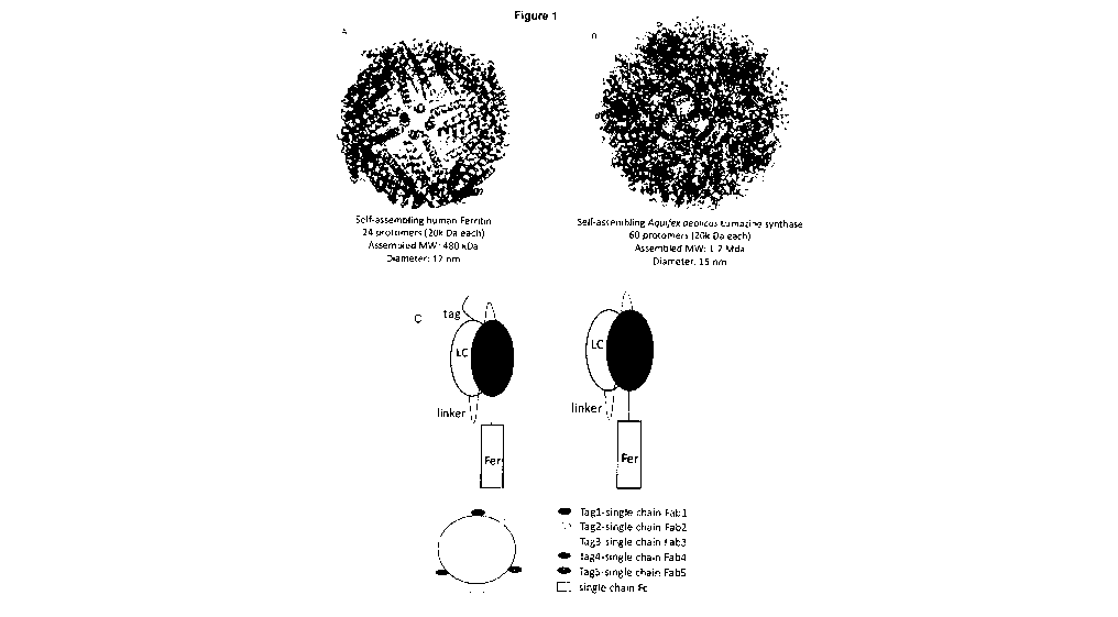
Figures 1A and 1B show naturally occurring self-assembling nanoparticle
backbones
of human Ferritin (Figure 1A) or Aquifex aeolicus Lumazine synthase (Figure
1B) described
herein; Figure 1C shows a schematic for generating single-chain Fab-ferritin
nanoparticles
that only require transfection of one plasmid.
Figure 2 shows a schematic of the constructs used to produce the antibody Fabs
expressing Ferritin (Figure 2A) or Lumazine synthase (Figure 2B) nanoparticles
of the
present invention. Figure 2C shows a schematic representation of an antibody
expression
nanoparticle described herein;
Figure 3 shows data representing the purity of the antibody Fab expressing
Ferritin
(Figure 3A) or Lumazine synthase (Figure 3B) nanoparticles produced in
accordance with
the methods of Figure 2A and 2B as determined by affinity chromatography and
SDS-PAGE
analysis;
Figure 4 shows electron micrographs of the antibody Fab expressing Ferritin
(Figure
4A) or Lumazine synthase (Figure 4B) nanoparticles described herein;
Figure 5 shows data representing the binding affinity of CD22 to antibody Fab
without
a nanoparticle backbone (Figure 5A), antibody Fab expressing Ferritin (Figure
5B) or
Lumazine synthase (Figure 5C) nanoparticles described herein;
Figure 6 shows data demonstrating the receptor mediated endocytosis of two
different antibody Fab expressing Ferritin nanoparticles (Figure 6A and 6B) as
compared to
the absence of endocytosis with a Ferritin nanoparticle alone (Figure 6C);
Figure 7 shows data representing the fluorescent capabilities of the Ferritin
nanoparticles described herein;
7
CA 03071922 2020-02-03
WO 2019/023811
PCT/CA2018/050954
Figure 8 shows the crystal structure of the interactions between a CSP NANP
repeat
domain antigen and two Fab antibody fragments;
Figure 9 shows schematic modeling of the interactions between a CSP NANP
repeat
domain antigen, and Fab antibody fragment, and a B cell;
Figure 10 shows the sequences of fusion proteins comprising a CSP NANP repeat
domain antigen fused to a Fab heavy chain with a linker of varying lengths;
Figure 11 shows data from size exclusion chromatography purification of the
fusion
proteins of Figure 10;
Figure 12 shows data from size exclusion chromatography purification of a wild-
type
antibody corresponding to the fusion proteins of Figure 10 but lacking the CSP
NANP repeat
domain and the linker;
Figure 13 shows CSP binding kinetics of the fusion proteins of Figure 10
compared
to binding by wild-type antibody;
Figure 14-16 show wild-type antibody binding affinity to the fusion proteins
of Figure
10;
Figure 17 shows schematic representations of the interaction between wild-type
antibody and the fusion proteins of Figure 10;
Figure 18 shows data from size exclusion chromatography coupled with multi-
angle
light scattering to determine absolute mass for the wild-type antibody and
fusion protein
interaction and a schematic representing the interaction of a B-cell
expressing an IgM
receptor specific for the fusion protein described herein;
Figure 19 shows nanoparticles engineered to co-display a-CD19 stimulating Fab
and
antigens as a vaccine platform;
Figure 20 shows that nanoparticles can be engineered for co-display of
stimulating
antibodies and antigens;
Figure 21 shows that bi-specific nanoparticles are well folded and display
high
density of Fabs and antigens;
Figure 22 shows that bi-specific nanoparticles are functional and bind as
expected;
8
CA 03071922 2020-02-03
WO 2019/023811
PCT/CA2018/050954
Figure 23 shows self-adjuvanted nanoparticles are capable of boosting Ca2+
dependent B cell activation in comparison to controls
Figure 24 shows folding, assembly, and elution data for a single-chain Fc
nanoparticle;
Figure 25 shows affinity maturation of high-affinity human PfCSP NANP
antibodies.
(A) Surface plasmon resonance (SPR) affinity and SHM of selected (labeled) VH3-
33/VK1-
5/KCDR3:8 (green) and non-VH3-33/VK1-5/KCDR3:8 anti-PfCSP antibodies (gray)
(9). (B to
D) Original and mutated antibodies. [(B) and (C)] PfCSP ELISA reactivity. (D)
Mean (bars) Pf
liver-cell traversal inhibition from two-to-four independent experiments
(symbols). **
significant (a = 0.01) for two-tailed Student's t test. (E) Silent (gray) and
replacement (red)
SHM (bars) in VH3-33A/K1-5 antibodies (n = 63). (F) Observed (obs) aa usage
compared to
baseline (base) model (22, 23). (G and H) Independent NANP3 SPR affinity
measurements
(dots) and mean (line). **significant (a = 0.01) and not significant (ns) for
Bonferroni multiple
comparisons test. (A), (B), and (C), one representative of at least two
independent
experiments.
Figure 26 shows affinity maturation drives homotypic repeat binding. (A to H)
1210
Fab/NANP5 co-crystal structure. (A) Superposition of the four NANP-bound Fabs.
(B)
Surface representation of the antigen¨antibody interaction. (C) Details of
core epitope
recognition by 1210. Black dashes indicate H-bonds. (D) Two 1210 Fabs in
complex with
NANP5. [(E) and (F)] Surface representation of Fab-B (E) and Fab-A (F).
Residues involved
in homotypic interactions are dark gray. [(G) and (H)] Details of homotypic
interactions.
Affinity matured residues are labeled in red. (I) Mean SEM KD determined by
isothermal
titration calorimetry (ITC). Dots represent measurements from at least three
independent
experiments. One-tailed Mann¨Whitney test: *P <o05, **P <0.01. (J) Size-
exclusion
chromatography coupled with multi-angle light scattering (SEC/MALS) for the
1210 Fab-
PfCSP complex. Red line indicates mean SD molar mass from two measurements.
(K) 2D
class averages for the 1210 Fab-PfCSP complex. Red arrows indicate individual
Fabs, red
lines indicate the binding angle observed in the crystal structure (D). Scale
bar, 10 nm.
Figure 27 shows NANP5 repeat binding by antibody 1210. A, The four 1210 Fabs
bound to 2 NANP5 peptides in the asymmetric unit of the 1210-NANP5 crystal
structure. B,
Superposition of the NPNA cadence of 580 (teal; (10)), 663 (green; (10)), 1210
(yellow) and
the unliganded peptide (purple; (12)) structures. The standard deviation in
the Phi and Psi
angles is shown. C, Superposition of 1210-NANP5 with the H.2140 / L.1210
chimeric Fab in
complex with a NANP3 peptide. The 1210 bound NANP5 peptide is colored yellow,
and the
chimeric Fab and NANP3-bound peptide are colored gray.
9
CA 03071922 2020-02-03
WO 2019/023811
PCT/CA2018/050954
Figure 28 shows effect of alanine exchange of residues H.Y52A and H.Y58 on
antigen binding. PfCSP and NANP5 ELISA reactivity of antibodies 1210, 2140,
2219 and
respective mutants with alanine exchanges at positions H.Y52A and H.Y58. One
out of three
representative experiments is shown.
Figure 29 shows 1210-NANP5 crystal structure. A, Detailed interactions of 1210
with
NANP5. Intermolecular H-bonds are colored as black dashes and intramolecular H-
bonds
are colored red. B, Unbiased electron density omit map (black mesh) contoured
to 1.0 a for
the NANP5 peptide bound to two 1210 Fabs. C, Elution profile of 1210-NANP5
examined by
SEC/MALS. The horizontal red line corresponds to the calculated molar mass for
two 1210
-- Fabs bound to NANP5. D, Elution profile of full-length PfCSP examined by
SEC/MALS. The
horizontal red line corresponds to the calculated molar mass of the eluting
antigen.
Figure 30 shows isothermal titration calorimetry of 1210 binding to NANP
repeat
peptides. A, B, Representative raw ITC data (top panel) and fitted binding
curves (bottom
panel) are shown for 1210, 1210_NS, 1210_YY and 1210_GL binding to NANP5 (A)
and
NANP3 (B). C, Summary of measured binding thermodynamic values for these
interactions
observed in (A) and (B). Mean SEM for at least three independent experiments
is reported.
Figure 31 shows binding avidity of 1210 and 1210_YY to PfCSP. Representative
biolayer interferometry sensorgrams (green), 1:1 model best fits (black) and
calculated
binding avidity for (A) 1210 IgG and (B) 1210_YY IgG binding to full length
PfCSP.
Figure 32 shows B cell activation and parasite inhibition. (A to D) NANP5-
induced
calcium signaling of 1210 and variants. [(A) and (B)] Reaction kinetic and
percent activated
cells (A), and overlay of median signal intensities (B) to 1 pg/mL NANP5 for
one of at least
six representative experiments. [(C) and (D)] Percent activated cells and
median activation
time after 1 pg/mL (C) (n = 6 or 7) and 0.1 pg/mL (D) (n = 3) NANP5. Symbols
indicate
independent experiments, lines and error bars indicate mean SD. **
significant (a = 0.01)
and not significant (ns) for Bonferroni multiple comparisons test. (E and F)
Parasite
inhibition. (E) Mean SD IC50 values from at least three independent
experiments for 1210
(black) and 2163 (brown) antibodies with indicated NANP3 affinities. No
significant
differences between IC50 values due to extensively overlapping confidence
intervals. (F)
Parasite-free mice after passive immunization with 30 pg or 100 pg of 1210 or
variants 24
hours before subcutaneous injection with Pb-PfCSP sporozoites. Data show one
(100 pg) or
two (30 pg) independent experiments with five mice per group. No significant
differences in
survival for 1210 variants (Mantel-Cox test).
CA 03071922 2020-02-03
WO 2019/023811
PCT/CA2018/050954
Figure 33 shows antibody mediated inhibition of Pf hepatocyte traversal. A, B,
Pf
hepatocyte traversal inhibition for 1210(A), 2163 (B), as well as the
indicated variants. The
IC50 values (in g/mL) and Hill coefficient (n) values and their standard
deviations are
indicated above each plot. C, NANP3 affinities and Hill coefficient for 1210
(black) and 2163
(brown) as well as the respective variants as shown in (A and B). Error bars
indicate
standard deviation.
Figure 34 shows antihomotypic affinity maturation in IGHV3-23-encoded PfCSP
NANP antibodies. (A) SPR affinity and SHM of 1450 out of all VH3-23A/k1-5
(green) and
non-VH3-23A/k1-5 anti-PfCSP antibodies (gray) (9). (B) Silent (gray) and
replacement (red)
SHM (bars) in VH3-23A/k1-5 antibodies (n = 100). (C to E) Fab 1450¨NANP5 co-
crystal
structure. Head-to-head binding mode (C), Fab¨Fab (D), and Fab¨NANP5 (E)
interactions.
Black dashes indicate H-bonds. Affinitymatured residues are colored according
to SHM aa
usage scheme and labeled in red. Observed (obs) aa usage compared to baseline
(base)
model (22, 23). (F) VH3-33/W1-5/KCDR3:8 or VH3-23A/k1-5 antibodies in total
memory B
cells (18) and CD19+CD27hiCD38hiplasmablasts (PB) and CD19+CD27+PfCSP-reactive
memory B cells (CSPmem) (8, 9). Dots represent subsamples of n = 1500
sequences.
Boxplots show median, standard deviation, max and min of the distribution. ***
significant (a
= 0.001) for two-tailed Student's t test. (G) Frequency of VH3-33A/k1-
5/KCDR3:8 and VH3-
23A/k1-5 antibodies among clonally expanded vs. singlet pooled PB and CSPmem
(9).
Figure 35 shows NANP5 repeat binding by antibodies 1450 and 580-gl. A, Surface
representation of the 1450 and 580-gl (PDB 6AZM, (10)) paratopes bound to
NANP5. B,
Detailed interactions of 1450 with NANP5. Intermolecular H-bonds are colored
as black
dashes and intramolecular H-bonds are colored red.
Figure 36 shows structure comparison of 1210 and the RTS,S vaccine-induced
NANP antibody 311 (encoded by IGHV3-33 and IGLV1-40). Similar antigen-binding
conformations are observed for recognition of the minimal NPNANPNANA repeat
epitope.
Analogous to the anti-homotypic mutation H.N56_K in 1210, 311 possesses
H.N56_R,
suggesting that it may also have undergone anti-homotypic affinity maturation.
A, 1210 Igk
chain is shown in teal, 1210 IgH chain is shown in green. B, 311 IgA chain is
shown in
brown, 311 IgH chain is shown in purple. NANP repeat antigens are shown in
pink. Mutated
residues are colored in yellow. AA-exchanges at positions H.31, H.50 and H.56
are
highlighted. C, D, Detailed representation of homotypic HCDR2 interactions
between 1210
(C) and 311 (D) Fabs binding neighboring repeat epitopes. For D the structure
of the 311-
NANP complex was duplicated and structurally aligned to both Fab-A and Fab-B
of the
1210_NANP5 complex. Affinity matured residues H.K56 (C, 1210 Fab) and H.R56
(D, 311
Fab, (11)) are labeled in red.
11
CA 03071922 2020-02-03
WO 2019/023811
PCT/CA2018/050954
Figure 37 shows IGHV3-33, IGHV3-30/IGHV3-30-3, IGHV3-30-5 gene frequency.
Frequency of IGHV3-33, IGHV3-30/IGHV3-30-3, IGHV3-30-5 germline gene segments
(8,9)
as determined by genomic sequencing of peripheral blood mononuclear cells.
Sequences
were assigned to the respective germline gene based on their CDR2 sequence as
shown in
Table 1.
Figure 38 shows that the malaria vaccine antigen (CSP-NANP5.5-linker-antibody)
elicits IgG titers that can recognize the full-length PfCSP antigen.
Figure 39 shows the activity/function of the elicited anti-PfCSP sera from the
immunizations in Figure 25.
Detailed Description of Certain Aspects
Definitions
Unless otherwise explained, all technical and scientific terms used herein
have the
same meaning as commonly understood by one of ordinary skill in the art to
which this
disclosure belongs. Definitions of common terms in molecular biology may be
found in
Benjamin Lewin, Genes V, published by Oxford University Press, 1994 (ISBN 0-19-
854287-
9); Kendrew et al. (eds.), The Encyclopedia of Molecular Biology, published by
Blackwell
Science Ltd., 1994 (ISBN 0-632-02182-9); and Robert A. Meyers (ed.), Molecular
Biology
and Biotechnology: a Comprehensive Desk Reference, published by VCH
Publishers, Inc.,
1995 (ISBN 1-56081-569-8). Although any methods and materials similar or
equivalent to
.. those described herein can be used in the practice for testing of the
present invention, the
typical materials and methods are described herein. In describing and claiming
the present
invention, the following terminology will be used.
It is also to be understood that the terminology used herein is for the
purpose of
describing particular aspects only, and is not intended to be limiting. Many
patent
applications, patents, and publications are referred to herein to assist in
understanding the
aspects described. Each of these references are incorporated herein by
reference in their
entirety.
In understanding the scope of the present application, the articles "a", "an",
"the", and
"said" are intended to mean that there are one or more of the elements.
Additionally, the
term "comprising" and its derivatives, as used herein, are intended to be open
ended terms
that specify the presence of the stated features, elements, components,
groups, integers,
and/or steps, but do not exclude the presence of other unstated features,
elements,
components, groups, integers and/or steps. The foregoing also applies to words
having
similar meanings such as the terms, "including", "having" and their
derivatives.
12
CA 03071922 2020-02-03
WO 2019/023811
PCT/CA2018/050954
It will be understood that any aspects described as "comprising" certain
components
may also "consist of" or "consist essentially of," wherein "consisting of" has
a closed-ended
or restrictive meaning and "consisting essentially of" means including the
components
specified but excluding other components except for materials present as
impurities,
unavoidable materials present as a result of processes used to provide the
components, and
components added for a purpose other than achieving the technical effect of
the invention.
For example, a composition defined using the phrase "consisting essentially
of"
encompasses any known acceptable additive, excipient, diluent, carrier, and
the like.
Typically, a composition consisting essentially of a set of components will
comprise less than
5% by weight, typically less than 3% by weight, more typically less than 1%,
and even more
typically less than 0.1% by weight of non-specified component(s).
It will be understood that any component defined herein as being included may
be
explicitly excluded from the claimed invention by way of proviso or negative
limitation.
In addition, all ranges given herein include the end of the ranges and also
any
intermediate range points, whether explicitly stated or not.
Terms of degree such as "substantially", "about" and "approximately" as used
herein
mean a reasonable amount of deviation of the modified term such that the end
result is not
significantly changed. These terms of degree should be construed as including
a deviation of
at least 5% of the modified term if this deviation would not negate the
meaning of the word
it modifies.
It is further to be understood that all base sizes or amino acid sizes, and
all molecular
weight or molecular mass values, given for nucleic acids or polypeptides are
approximate,
and are provided for description. Although methods and materials similar or
equivalent to
those described herein can be used in the practice or testing of this
disclosure, suitable
methods and materials are described below. The abbreviation, "e.g." is derived
from the
Latin exempli gratia, and is used herein to indicate a non-limiting example.
Thus, the
abbreviation "e.g." is synonymous with the term "for example." The word "or"
is intended to
include "and" unless the context clearly indicates otherwise.
The terms "protein nanoparticle" and "nanocage" are used interchangeably
herein
and refer to a multi-subunit, protein-based polyhedron shaped structure. The
subunits or
nanocage monomers are each composed of proteins or polypeptides (for example a
glycosylated polypeptide), and, optionally of single or multiple features of
the following:
nucleic acids, prosthetic groups, organic and inorganic compounds. Non-
limiting examples of
protein nanoparticles include ferritin nanoparticles (see, e.g., Zhang, Y.
Int. J. Mol. Sci.,
13
CA 03071922 2020-02-03
WO 2019/023811
PCT/CA2018/050954
12:5406-5421, 2011, incorporated by reference herein), encapsulin
nanoparticles (see, e.g.,
Sutter et al., Nature Struct, and Mol. Biol., 15:939-947, 2008, incorporated
by reference
herein), Sulfur Oxygenase Reductase (SOR) nanoparticles (see, e.g., Urich et
al., Science,
311 :996-1000, 2006, incorporated by reference herein), lumazine synthase
nanoparticles
(see, e.g., Zhang et al., J. Mol. Biol., 306: 1099-1114, 2001) or pyruvate
dehydrogenase
nanoparticles (see, e.g., Izard et al., PNAS 96: 1240-1245, 1999, incorporated
by reference
herein). Ferritin, encapsulin, SOR, lumazine synthase, and pyruvate
dehydrogenase are
monomeric proteins that self-assemble into a globular protein complexes that
in some cases
consists of 24, 60, 24, 60, and 60 protein subunits, respectively.
Carboxysome, vault
proteins, GroEL, heat shock protein, E2P and M52 coat protein also produce
nanocages are
contemplated for use herein. In addition, fully or partially synthetic self-
assembling
monomers are also contemplated for use herein.
A "vaccine" is a pharmaceutical composition that induces a prophylactic or
therapeutic immune response in a subject. In some cases, the immune response
is a
protective immune response. Typically, a vaccine induces an antigen-specific
immune
response to an antigen of a pathogen, for example a viral pathogen, or to a
cellular
constituent correlated with a pathological condition. A vaccine may include a
polynucleotide
(such as a nucleic acid encoding a disclosed antigen), a peptide or
polypeptide (such as a
disclosed antigen), a virus, a cell or one or more cellular constituents. In
one specific, non-
limiting example, a vaccine induces an immune response that reduces the
severity of the
symptoms associated with malaria infection and/or decreases the parasite load
compared to
a control. In another non-limiting example, a vaccine induces an immune
response that
reduces and/or prevents malaria infection compared to a control.
The term "antibody", also referred to in the art as "immunoglobulin" (Ig),
used herein
refers to a protein constructed from paired heavy and light polypeptide
chains; various Ig
isotypes exist, including IgA, IgD, IgE, IgG, such as IgGi, IgG2, IgG3, and
IgG4, and IgM. It
will be understood that the antibody may be from any species, including human,
mouse, rat,
monkey, llama, or shark. When an antibody is correctly folded, each chain
folds into a
number of distinct globular domains joined by more linear polypeptide
sequences. For
example, the immunoglobulin light chain folds into a variable (VL) and a
constant (CL)
domain, while the heavy chain folds into a variable (VH) and three constant
(CH, CH2, CH3)
domains. Interaction of the heavy and light chain variable domains (VH and VI)
results in the
formation of an antigen binding region (Fv). Each domain has a well-
established structure
familiar to those of skill in the art.
The light and heavy chain variable regions are responsible for binding the
target
antigen and can therefore show significant sequence diversity between
antibodies. The
14
CA 03071922 2020-02-03
WO 2019/023811
PCT/CA2018/050954
constant regions show less sequence diversity, and are responsible for binding
a number of
natural proteins to elicit important immunological events. The variable region
of an antibody
contains the antigen binding determinants of the molecule, and thus determines
the
specificity of an antibody for its target antigen. The majority of sequence
variability occurs in
six hypervariable regions, three each per variable heavy and light chain; the
hypervariable
regions combine to form the antigen-binding site, and contribute to binding
and recognition
of an antigenic determinant. The specificity and affinity of an antibody for
its antigen is
determined by the structure of the hypervariable regions, as well as their
size, shape and
chemistry of the surface they present to the antigen.
An "antibody fragment" as referred to herein may include any suitable antigen-
binding antibody fragment known in the art. The antibody fragment may be a
naturally-
occurring antibody fragment, or may be obtained by manipulation of a naturally-
occurring
antibody or by using recombinant methods. For example, an antibody fragment
may include,
but is not limited to a Fv, single-chain Fv (scFv; a molecule consisting of VL
and VH
connected with a peptide linker), Fc, single-chain Fc, Fab, F(a1:)2, single
domain antibody
(sdAb; a fragment composed of a single VL or VH), and multivalent
presentations of any of
these.
By the term "synthetic antibody" as used herein, is meant an antibody which is
generated using recombinant DNA technology. The term should also be construed
to mean
an antibody which has been generated by the synthesis of a DNA molecule
encoding the
antibody and which DNA molecule expresses an antibody protein, or an amino
acid
sequence specifying the antibody, wherein the DNA or amino acid sequence has
been
obtained using synthetic DNA or amino acid sequence technology which is
available and
well known in the art.
The term "epitope" refers to an antigenic determinant. An epitope is the
particular
chemical groups or peptide sequences on a molecule that are antigenic, that
is, that elicit a
specific immune response. An antibody specifically binds a particular
antigenic epitope, e.g.,
on a polypeptide. Epitopes can be formed both from contiguous amino acids or
noncontiguous amino acids juxtaposed by tertiary folding of a protein.
Epitopes formed from
contiguous amino acids are typically retained on exposure to denaturing
solvents whereas
epitopes formed by tertiary folding are typically lost on treatment with
denaturing solvents.
An epitope typically includes at least 3, and more usually, at least 5, about
9, about 11, or
about 8 to about 12 amino acids in a unique spatial conformation. Methods of
determining
spatial conformation of epitopes include, for example, x-ray crystallography
and 2-
dimensional nuclear magnetic resonance. See, e.g., "Epitope Mapping Protocols"
in
Methods in Molecular Biology, Vol. 66, Glenn E. Morris, Ed (1996).
CA 03071922 2020-02-03
WO 2019/023811
PCT/CA2018/050954
The term "antigen" as used herein is defined as a molecule that provokes an
immune
response. This immune response may involve either antibody production, or the
activation of
specific immunologically-competent cells, or both. The skilled artisan will
understand that
any macromolecule, including virtually all proteins or peptides, can serve as
an antigen.
Furthermore, antigens can be derived from recombinant or genomic DNA. A
skilled artisan
will understand that any DNA, which comprises a nucleotide sequence or a
partial nucleotide
sequence encoding a protein that elicits an immune response therefore encodes
an
"antigen" as that term is used herein. Furthermore, one skilled in the art
will understand that
an antigen need not be encoded solely by a full length nucleotide sequence of
a gene. It is
readily apparent that the aspects described herein include, but are not
limited to, the use of
partial nucleotide sequences of more than one gene and that these nucleotide
sequences
could be arranged in various combinations to elicit the desired immune
response. Moreover,
a skilled artisan will understand that an antigen need not be encoded by a
"gene" at all. It is
readily apparent that an antigen can be synthesized or can be derived from a
biological
sample. Such a biological sample can include, but is not limited to a tissue
sample, a cell, or
a biological fluid.
Thus, the compositions described herein may be suitable for protection or
treatment
of vertebrate subjects against a variety of disease states such as, for
example, viral,
bacterial, fungal or parasitic infections, cancer, and autoimmune disorders.
It is to be
.. recognized that these specific disease states have been referred to by way
of example only
and are not intended to be limiting.
Suitable antigens useful in combination with the compositions described herein
include any antigen as defined herein. Antigens are commercially available or
one of skill in
the art is capable of producing them. The antigen can be either a modified-
live or killed
microorganism, or a natural product purified from a microorganism or other
cell including, but
not limited to, tumor cell, a synthetic product, a genetically engineered
protein, peptide,
polysaccharide or similar product, or an allergen. The antigenic moiety can
also be a subunit
of a protein, peptide, polysaccharide or similar product. The antigen may also
be a genetic
antigen, i.e., DNA or RNA that engenders an immune response.
Representative of the antigens that can be used include, but are not limited
to,
natural, recombinant or synthetic products derived from viruses, bacteria,
fungi, parasites
and other infectious agents in addition to autoimmune diseases, hormones, or
tumor
antigens which might be used in prophylactic or therapeutic vaccines and
allergens. In one
embodiment, the antigen comprises virus-like particles (VLPs) from various
viruses such as
influenza, HIV, RSV, Newcastle disease virus (NDV) etc. See PCT/U52006/40862,
PCT/U52004/022001, U.S. Ser. No. 11/582,540, U.S. 60/799,343, U.S. 60/817,402,
U.S.
16
CA 03071922 2020-02-03
WO 2019/023811
PCT/CA2018/050954
60/859,240, all of which are herein incorporated by reference in their
entirety. In another
embodiment, the antigen comprises chimeric VLPs. "Chimeric VLPs" refer to VLPs
that
contain proteins, or portions thereof, from at least two different sources
(organisms). Usually,
one protein is derived from a virus that can drive the formation of VLPs from
host cells. Thus,
in one embodiment, said chimeric VLP comprises an RSV M protein. In another
embodiment, said chimeric VLP comprises a NDV M protein. In another
embodiment, said
chimeric VLP comprises an influenza virus M protein.
The viral or bacterial products can be components which the organism produced
by
enzymatic cleavage or can be components of the organism that were produced by
recombinant DNA techniques that are well known to those of ordinary skill in
the art.
Some specific examples of antigens are antigens derived from viral infections
caused
by hepatitis viruses A, B, C, D & E3, human immunodeficiency virus (HIV),
herpes viruses 1,
2, 6 & 7, cytomegalovirus, varicella zoster, papilloma virus, Epstein Barr
virus, para-influenza
viruses, adenoviruses, bunya viruses (e.g. hanta virus), coxsakie viruses,
picoma viruses,
rotaviruses, respiratory syncytial viruses, rhinoviruses, rubella virus,
papovavirus, mumps
virus, measles virus, polio virus (multiple types), adeno virus (multiple
types), parainfluenza
virus (multiple types), avian or pandemic influenza (various types), seasonal
influenza,
shipping fever virus, Western and Eastern equine encephalomyelitis, Japanese
B.
encephalomyelitis, Russian Spring Summer encephalomyelitis, hog cholera virus,
Newcastle
disease virus, fowl pox, rabies, feline and canine distemper and the like
viruses, slow brain
viruses, rous sarcoma virus (RSV), Papovaviridae, Parvoviridae,
Picornaviridae, Poxyiridae
(such as Smallpox or Vaccinia), Reoviridae (e.g., Rotavirus), Retroviridae
(HTLV-I, HTLV-II,
Lentivirus), and Togaviridae (e.g., Rubivirus). Viruses falling within these
families can cause
a variety of diseases or symptoms, including, but not limited to: arthritis,
bronchiollitis,
encephalitis, eye infections (e.g., conjunctivitis, keratitis), chronic
fatigue syndrome,
Japanese B encephalitis, Junin, Chikungunya, Rift Valley fever, yellow fever,
meningitis,
opportunistic infections (e.g., AIDS), pneumonia, Burkitt's Lymphoma,
chickenpox,
hemorrhagic fever, Measles, Mumps, Parainfluenza, Rabies, the common cold,
Polio,
leukemia, Rubella, sexually transmitted diseases, skin diseases (e.g.,
Kaposi's, warts), and
viremia.
The antigens may also be derived from bacterial and fungal infections for
example:
antigens derived from infections caused by Mycobacteria causing TB and
leprosy,
pneumocci, aerobic gram negative bacilli, mycoplasma, staphyloccocal
infections,
streptococcal infections, salmonellae and chlamydiae, B. pertussis, Leptospira
pomona, and
icterohaemorrhagiae. Specific embodiments comprise S. paratyphi A and B, C.
diphtheriae,
C. tetani, C. botulinum, C. perfringens, C. feseri and other gas gangrene
bacteria, B.
17
CA 03071922 2020-02-03
WO 2019/023811
PCT/CA2018/050954
anthracis, P. pestis, P. multocida, Neisseria meningitidis, N. gonorrheae,
Hemophilus
influenzae, Actinomyces (e.g., Norcardia), Acinetobacter, Bacillaceae (e.g.,
Bacillus
anthrasis), Bacteroides (e.g., Bacteroides fragilis), Blastomycosis,
Bordetella, Borrelia (e.g.,
Borrelia burgdorferi), BruceIla, Candidia, Cam pylobacter, Chlamydia,
Coccidioides,
Corynebacterium (e.g., Corynebacterium diptheriae), Cryptococcus,
Dermatocycoses, E. coil
(e.g., Enterotoxigenic E. coil and Enterohemorrhagic E. coli), Enterobacter
(e.g.
Enterobacter aerogenes), Enterobacteriaceae (Klebsiella, Salmonella (e.g.,
Salmonella
typhi, Salmonella enteritidis, Serratia, Yersinia, Shigella), Erysipelothrix,
Haemophilus (e.g.,
Haemophilus influenza type B), Helicobacter, Legionella (e.g., Legionella
pneumophila),
Leptospira, Listeria (e.g., Listeria monocytogenes), Myco plasma,
Mycobacterium (e.g.,
Mycobacterium leprae and Mycobacterium tuberculosis), Vibrio (e.g., Vibrio
cholerae),
Pasteurellacea, Proteus, Pseudomonas (e.g., Pseudomonas aeruginosa),
Rickettsiaceae,
Spirochetes (e.g., Treponema spp., Leptospira spp., Borrelia spp.), Shigella
spp.,
Meningiococcus, Pneumococcus and Streptococcus (e.g., Streptococcus pneumoniae
and
Groups A, B, and C Streptococci), Ureaplasmas, Treponema pollidum, and the
like;
Staphylococcus aureus, Plasmodium sp. (Pl. falciparum, Pl. vivax, etc.),
Aspergillus sp.,
Candida albicans, Pasteurella haemolytica, Corynebacterium diptheriae toxoid,
Meningococcal polysaccharide, Bordetella pertusis, Streptococcus pneumoniae
(pneumococcus) polysaccharide, Clostridium tetani toxoid, Mycobacterium bovis,
killed cells
of Salmonella typhi, Cryptococcus neoformans, and Aspergillus.
The antigens may also be derived from parasitic malaria, leishmaniasis,
trypanosomiasis, toxoplasmosis, schistosomiasis, filariasis malaria,
Amebiasis, Babesiosis,
Coccidiosis, Cryptosporidiosis, Dientamoebiasis, Dourine, Ectoparasitic,
Giardias,
Helminthiasis, Theileriasis, Trichomonas and Sporozoans (e.g., Plasmodium
virax,
Plasmodium fakiparium, Plasmodium malariae and Plasmodium ovale). These
parasites can
cause a variety of diseases or symptoms, including, but not limited to:
Scabies,
Trombiculiasis, eye infections, intestinal disease (e.g., dysentery,
giardiasis), liver disease,
lung disease, opportunistic infections (e.g., AIDS related), malaria,
pregnancy complications,
and toxoplasmosis.
Tumor-associated antigens suitable for use in compositions described herein
include
both mutated and non-mutated molecules which may be indicative of single tumor
type,
shared among several types of tumors, and/or exclusively expressed or
overexpressed in
tumor cells in comparison with normal cells. In addition to proteins and
glycoproteins, tumor-
specific patterns of expression of carbohydrates, gangliosides, glycolipids
and mucins have
also been documented. Exemplary tumor-associated antigens for use in the
subject cancer
vaccines include protein products of oncogenes, tumor suppressor genes and
other genes
18
CA 03071922 2020-02-03
WO 2019/023811
PCT/CA2018/050954
with mutations or rearrangements unique to tumor cells, reactivated embryonic
gene
products, oncofetal antigens, tissue-specific (but not tumor-specific)
differentiation antigens,
growth factor receptors, cell surface carbohydrate residues, foreign viral
proteins and a
number of other self proteins. Specific embodiments of tumor-associated
antigens include,
e.g., mutated antigens such as the protein products of the Ras p21
protooncogenes, tumor
suppressor p53 and HER-2/neu and BCR-ab1 oncogenes, as well as CDK4, MUM1,
Caspase 8, and Beta catenin; overexpressed antigens such as galectin 4,
galectin 9,
carbonic anhydrase, Aldolase A, PRAME, Her2/neu, ErbB-2 and KSA, oncofetal
antigens
such as alpha fetoprotein (AFP), human chorionic gonadotropin (hCG); self
antigens such as
carcinoembryonic antigen (CEA) and melanocyte differentiation antigens such as
Mart
1/MeIan A, gp100, gp75, Tyrosinase, TRP1 and TRP2; prostate associated
antigens such as
PSA, PAP, PSMA, PSM-P1 and PSM-P2; reactivated embryonic gene products such as
MAGE 1, MAGE 3, MAGE 4, GAGE 1, GAGE 2, BAGE, RAGE, and other cancer testis
antigens such as NY-ES01, 55X2 and SCP1; mucins such as Muc-1 and Muc-2;
.. gangliosides such as GM2, GD2 and GD3, neutral glycolipids and
glycoproteins such as
Lewis (y) and globo-H; and glycoproteins such as Tn, Thompson-Freidenreich
antigen (TF)
and sTn. Also included as tumor-associated antigens herein are whole cell and
tumor cell
lysates as well as immunogenic portions thereof, as well as immunoglobulin
idiotypes
expressed on monoclonal proliferations of B lymphocytes for use against B cell
lymphomas.
Tumor-associated antigens and their respective tumor cell targets include,
e.g., cytokeratins,
particularly cytokeratin 8, 18 and 19, as antigens for carcinoma. Epithelial
membrane antigen
(EMA), EphA1, EphA2, EphA3, EphA4, EphA5, EphA6, EphA7, EphA8, EphA10, EphB1,
EphB2, EphB3, EphB4, EphB6, human embryonic antigen (HEA-125), human milk fat
globules, MBr1, MBr8, Ber-EP4, 17-1A, C26 and T16 are also known carcinoma
antigens.
Desmin and muscle-specific actin are antigens of myogenic sarcomas. Placental
alkaline
phosphatase, beta-human chorionic gonadotropin, and alpha-fetoprotein are
antigens of
trophoblastic and germ cell tumors. Prostate specific antigen is an antigen of
prostatic
carcinomas, carcinoembryonic antigen of colon adenocarcinomas. HMB-45 is an
antigen of
melanomas. In cervical cancer, useful antigens could be encoded by human
papilloma virus.
Chromagranin-A and synaptophysin are antigens of neuroendocrine and
neuroectodermal
tumors. Of particular interest are aggressive tumors that form solid tumor
masses having
necrotic areas. The lysis of such necrotic cells is a rich source of antigens
for antigen-
presenting cells, and thus the subject therapy may find advantageous use in
conjunction with
conventional chemotherapy and/or radiation therapy. The antigens can be
derived from any
tumor or malignant cell line.
Antigens may also be derived from common allergens that cause allergies.
Allergens
include organic or inorganic materials derived from a variety of man-made or
natural sources
19
CA 03071922 2020-02-03
WO 2019/023811
PCT/CA2018/050954
such as plant materials, metals, ingredients in cosmetics or detergents,
latexes, or the like.
Classes of suitable allergens for use in the compositions and methods
described herein can
include, but are not limited to, pollens, animal dander, grasses, molds,
dusts, antibiotics,
stinging insect venoms, and a variety of environmental (including chemicals
and metals)
drug and food allergens. Common tree allergens include pollens from
cottonwood, popular,
ash, birch, maple, oak, elm, hickory, and pecan trees; common plant allergens
include those
from rye, ragweed, English plantain, sorrel-dock and pigweed; plant contact
allergens
include those from poison oak, poison ivy and nettles; common grass allergens
include
Timothy, Johnson, Bermuda, fescue and bluegrass allergens; common allergens
can also be
obtained from molds or fungi such as Altemaria, Fusarium, Hormodendrum,
Aspergillus,
Micropolyspora, Mucor and thermophilic actinomycetes; penicillin and
tetracycline are
common antibiotic allergens; epidermal allergens can be obtained from house or
organic
dusts (typically fungal in origin), from insects such as house mites
(dermalphagoides
pterosinyssis), or from animal sources such as feathers, and cat and dog
dander; common
food allergens include milk and cheese (diary), egg, wheat, nut (e.g.,
peanut), seafood (e.g.,
shellfish), pea, bean and gluten allergens; common environmental allergens
include metals
(nickel and gold), chemicals (formaldehyde, trinitrophenol and turpentine),
Latex, rubber,
fiber (cotton or wool), burlap, hair dye, cosmetic, detergent and perfume
allergens; common
drug allergens include local anesthetic and salicylate allergens; antibiotic
allergens include
penicillin and sulfonamide allergens; and common insect allergens include bee,
wasp and
ant venom, and cockroach calyx allergens. Particularly well characterized
allergens include,
but are not limited to, the major and cryptic epitopes of the Der pl allergen
(Hoyne et al.
(1994) Immunology 83, 190-195), bee venom phospholipase A2 (PLA) (Akdis et al.
(1996) J.
Clin. Invest. 98, 1676-1683), birch pollen allergen Bet v 1 (Bauer et al.
(1997) Clin. Exp.
Immunol. 107, 536-541), and the multi-epitopic recombinant grass allergen
rKBG8.3 (Cao et
al. (1997) Immunology 90, 46-51). These and other suitable allergens are
commercially
available and/or can be readily prepared as extracts following known
techniques.
The antigen may be in the form of purified or partially purified antigen and
can be
derived from any of the above antigens, an antigenic peptide, proteins that
are known and
available in the art, and others that can identified using conventional
techniques. The
antigens will typically be in the form in which their toxic or virulent
properties have been
reduced or destroyed and which when introduced into a suitable, will either
induce and
immune response against the specific microorganisms, extract, or products of
microorganisms used in the preparation of the antigen, or, in the case of
allergens, they will
aid in alleviating the symptoms of the allergy due to the specific allergen.
The antigens can
be used either singly or in combination; for example, multiple bacterial
antigens, multiple
viral antigens, multiple bacterial antigens, multiple parasitic antigens,
multiple bacterial, viral
CA 03071922 2020-02-03
WO 2019/023811
PCT/CA2018/050954
toxoids, multiple tumor antigens, multiple allergens or combinations of any of
the foregoing
products can be combined with adjuvant compositions to create a polyvalent
antigenic
composition and/or a vaccine. In the compositions described herein, the
antigen may be
antigen entrapped in, adsorbed to, or in an admixture with the vesicle
component of the
composition.
In one embodiment, suitable antigens for use with the compositions described
herein
include antigens which are poorly immunogenic, for example malaria antigens,
dengue
antigens and HIV antigens, or antigens intended to confer immunity against
pandemic
diseases, for example influenza antigens.
"Encoding" refers to the inherent property of specific sequences of
nucleotides in a
polynucleotide, such as a gene, a cDNA, or an mRNA, to serve as templates for
synthesis of
other polymers and macromolecules in biological processes having either a
defined
sequence of nucleotides (e.g., rRNA, tRNA and mRNA) or a defined sequence of
amino
acids and the biological properties resulting therefrom. Thus, a gene encodes
a protein if
transcription and translation of mRNA corresponding to that gene produces the
protein in a
cell or other biological system. Both the coding strand, the nucleotide
sequence of which is
identical to the mRNA sequence and is usually provided in sequence listings,
and the non-
coding strand, used as the template for transcription of a gene or cDNA, can
be referred to
as encoding the protein or other product of that gene or cDNA.
The term "expression" as used herein is defined as the transcription and/or
translation of a particular nucleotide sequence driven by its promoter.
"Isolated" means altered or removed from the natural state. For example, a
nucleic
acid or a peptide naturally present in a living animal is not "isolated," but
the same nucleic
acid or peptide partially or completely separated from the coexisting
materials of its natural
state is "isolated." An isolated nucleic acid or protein can exist in
substantially purified form,
or can exist in a non-native environment such as, for example, a host cell.
Unless otherwise specified, a "nucleotide sequence encoding an amino acid
sequence" includes all nucleotide sequences that are degenerate versions of
each other and
that encode the same amino acid sequence. The phrase nucleotide sequence that
encodes
a protein or an RNA may also include introns to the extent that the nucleotide
sequence
encoding the protein may in some version contain an intron(s).
By the term "modulating," as used herein, is meant mediating a detectable
increase
or decrease in the level of a response in a subject compared with the level of
a response in
the subject in the absence of a treatment or compound, and/or compared with
the level of a
21
CA 03071922 2020-02-03
WO 2019/023811
PCT/CA2018/050954
response in an otherwise identical but untreated subject. The term encompasses
perturbing
and/or affecting a native signal or response thereby mediating a beneficial
therapeutic
response in a subject, typically, a human.
The term "operably linked" refers to functional linkage between a regulatory
sequence and a heterologous nucleic acid sequence resulting in expression of
the latter. For
example, a first nucleic acid sequence is operably linked with a second
nucleic acid
sequence when the first nucleic acid sequence is placed in a functional
relationship with the
second nucleic acid sequence. For instance, a promoter is operably linked to a
coding
sequence if the promoter affects the transcription or expression of the coding
sequence.
Generally, operably linked DNA sequences are contiguous and, where necessary
to join two
protein coding regions, in the same reading frame.
"Parenteral" administration of an immunogenic composition includes, e.g.,
subcutaneous (s.c.), intravenous (i.v.), intramuscular (i.m.), or intrasternal
injection, or
infusion techniques.
The term "polynucleotide" as used herein is defined as a chain of nucleotides.
Furthermore, nucleic acids are polymers of nucleotides. Thus, nucleic acids
and
polynucleotides as used herein are interchangeable. One skilled in the art has
the general
knowledge that nucleic acids are polynucleotides, which can be hydrolyzed into
the
monomeric "nucleotides." The monomeric nucleotides can be hydrolyzed into
nucleosides.
As used herein polynucleotides include, but are not limited to, all nucleic
acid sequences
which are obtained by any means available in the art, including, without
limitation,
recombinant means, i.e., the cloning of nucleic acid sequences from a
recombinant library or
a cell genome, using ordinary cloning technology and PCR, and the like, and by
synthetic
means.
As used herein, the terms "peptide," "polypeptide," and "protein" are used
interchangeably, and refer to a compound comprised of amino acid residues
covalently
linked by peptide bonds. A protein or peptide must contain at least two amino
acids, and no
limitation is placed on the maximum number of amino acids that can comprise a
protein's or
peptide's sequence. Polypeptides include any peptide or protein comprising two
or more
amino acids joined to each other by peptide bonds. As used herein, the term
refers to both
short chains, which also commonly are referred to in the art as peptides,
oligopeptides and
oligomers, for example, and to longer chains, which generally are referred to
in the art as
proteins, of which there are many types. "Polypeptides" include, for example,
biologically
active fragments, substantially homologous polypeptides, oligopeptides,
homodimers,
heterodimers, variants of polypeptides, modified polypeptides, derivatives,
analogs, fusion
22
CA 03071922 2020-02-03
WO 2019/023811
PCT/CA2018/050954
proteins, among others. The polypeptides include natural peptides, recombinant
peptides,
synthetic peptides, or a combination thereof.
By the term "specifically binds," as used herein with respect to an antibody,
is meant
an antibody which recognizes a specific antigen, but does not substantially
recognize or bind
other molecules in a sample. For example, an antibody that specifically binds
to an antigen
from one species may also bind to that antigen from one or more species. But,
such cross-
species reactivity does not itself alter the classification of an antibody as
specific. In another
example, an antibody that specifically binds to an antigen may also bind to
different allelic
forms of the antigen. However, such cross reactivity does not itself alter the
classification of
-- an antibody as specific. In some instances, the terms "specific binding" or
"specifically
binding," can be used in reference to the interaction of an antibody, a
protein, or a peptide
with a second chemical species, to mean that the interaction is dependent upon
the
presence of a particular structure (e.g., an antigenic determinant or epitope)
on the chemical
species; for example, an antibody recognizes and binds to a specific protein
structure rather
-- than to proteins generally. If an antibody is specific for epitope "A', the
presence of a
molecule containing epitope A (or free, unlabeled A), in a reaction containing
labeled "A" and
the antibody, will reduce the amount of labeled A bound to the antibody.
The terms "therapeutically effective amount", "effective amount" or
"sufficient
amount" mean a quantity sufficient, when administered to a subject, including
a mammal, for
example a human, to achieve a desired result, for example an amount effective
to cause a
protective immune response. Effective amounts of the compounds described
herein may
vary according to factors such as the immunogen, age, sex, and weight of the
subject.
Dosage or treatment regimes may be adjusted to provide the optimum therapeutic
response,
as is understood by a skilled person. For example, administration of a
therapeutically
effective amount of the fusion proteins described herein is, in aspects,
sufficient to increase
immunity against a pathogen, such as Plasmodium. In other aspects,
administration of a
therapeutically effective amount of the fusion proteins described herein is
sufficient to treat a
disease or condition, such as cancer, HIV, malaria, or an autoimmune disease.
In still other
aspects, administration of a therapeutically effective amount of the fusion
proteins described
herein is sufficient to act as an adjuvant to increase effectiveness of a
vaccine.
Moreover, a treatment regime of a subject with a therapeutically effective
amount
may consist of a single administration, or alternatively comprise a series of
applications. The
length of the treatment period depends on a variety of factors, such as the
immunogen, the
age of the subject, the concentration of the agent, the responsiveness of the
patient to the
-- agent, or a combination thereof. It will also be appreciated that the
effective dosage of the
agent used for the treatment may increase or decrease over the course of a
particular
23
CA 03071922 2020-02-03
WO 2019/023811
PCT/CA2018/050954
treatment regime. Changes in dosage may result and become apparent by standard
diagnostic assays known in the art. The fusion proteins described herein may,
in aspects, be
administered before, during or after treatment with conventional therapies for
the disease or
disorder in question, such as malaria, HIV or cancer. For example, the fusion
proteins
described herein may find particular use in combination with immunotherapies
for treating
cancer.
The term "transfected" or "transformed" or "transduced" as used herein refers
to a
process by which exogenous nucleic acid is transferred or introduced into the
host cell. A
"transfected" or "transformed" or "transduced" cell is one which has been
transfected,
transformed or transduced with exogenous nucleic acid. The cell includes the
primary
subject cell and its progeny.
The phrase "under transcriptional control" or "operatively linked" as used
herein
means that the promoter is in the correct location and orientation in relation
to a
polynucleotide to control the initiation of transcription by RNA polymerase
and expression of
the polynucleotide.
A "vector" is a composition of matter which comprises an isolated nucleic acid
and
which can be used to deliver the isolated nucleic acid to the interior of a
cell. Numerous
vectors are known in the art including, but not limited to, linear
polynucleotides,
polynucleotides associated with ionic or amphiphilic compounds, plasmids, and
viruses.
Thus, the term "vector" includes an autonomously replicating plasmid or a
virus. The term
should also be construed to include non-plasmid and non-viral compounds which
facilitate
transfer of nucleic acid into cells, such as, for example, polylysine
compounds, liposomes,
and the like. Examples of viral vectors include, but are not limited to,
adenoviral vectors,
adeno-associated virus vectors, retroviral vectors, and the like.
The term "subject" as used herein refers to any member of the animal kingdom,
typically a mammal. The term "mammal" refers to any animal classified as a
mammal,
including humans, other higher primates, domestic and farm animals, and zoo,
sports, or pet
animals, such as dogs, cats, cattle, horses, sheep, pigs, goats, rabbits, etc.
Typically, the
mammal is human.
Administration "in combination with" one or more further therapeutic agents
includes
simultaneous (concurrent) and consecutive administration in any order.
The term "pharmaceutically acceptable" means that the compound or combination
of
compounds is compatible with the remaining ingredients of a formulation for
pharmaceutical
use, and that it is generally safe for administering to humans according to
established
24
CA 03071922 2020-02-03
WO 2019/023811
PCT/CA2018/050954
governmental standards, including those promulgated by the United States Food
and Drug
Administration.
The term "pharmaceutically acceptable carrier" includes, but is not limited to
solvents, dispersion media, coatings, antibacterial agents, antifungal agents,
isotonic and/or
absorption delaying agents and the like. The use of pharmaceutically
acceptable carriers is
well known.
The term "adjuvant" refers to a compound or mixture that is present in a
vaccine and
enhances the immune response to an antigen present in the vaccine. For
example, an
adjuvant may enhance the immune response to a polypeptide present in a vaccine
as
contemplated herein, or to an immunogenic fragment or variant thereof as
contemplated
herein. An adjuvant can serve as a tissue depot that slowly releases the
antigen and also as
a lymphoid system activator that non-specifically enhances the immune
response. Examples
of adjuvants which may be employed include MPL-TDM adjuvant (monophosphoryl
Lipid
A/synthetic trehalose dicorynomycolate, e.g., available from GSK Biologics).
Another
suitable adjuvant is the immunostimulatory adjuvant A5021/A502 (GSK). These
immunostimulatory adjuvants are formulated to give a strong T cell response
and include
QS-21, a saponin from Quillay saponaria, the TL4 ligand, a monophosphoryl
lipid A, together
in a lipid or liposomal carrier. Other adjuvants include, but are not limited
to, nonionic block
co-polymer adjuvants (e.g., CRL 1005), aluminum phosphates (e.g., AIPO<sub>4</sub>),
R-848 (a
Th1-like adjuvant), imiquimod, PAM3CYS, poly (I:C), loxoribine, BCG (bacille
Calmette-
Guerin) and Corynebacterium parvum, CpG oligodeoxynucleotides (ODN), cholera
toxin
derived antigens (e.g., CTA 1-DD), lipopolysaccharide adjuvants, complete
Freund's
adjuvant, incomplete Freund's adjuvant, saponin, mineral gels such as aluminum
hydroxide,
surface active substances such as lysolecithin, pluronic polyols, polyanions,
peptides, oil or
hydrocarbon emulsions in water (e.g., MF59 available from Novartis Vaccines or
Montanide
ISA 720), keyhole limpet hemocyanins, and dinitrophenol.
"Variants" are biologically active fusion proteins, antibodies, or fragments
thereof
having an amino acid sequence that differs from a comparator sequence by
virtue of an
insertion, deletion, modification and/or substitution of one or more amino
acid residues within
.. the comparative sequence. Variants generally have less than 100% sequence
identity with
the comparative sequence. Ordinarily, however, a biologically active variant
will have an
amino acid sequence with at least about 70% amino acid sequence identity with
the
comparative sequence, such as at least about 71%, 72%, 73%, 74%, 75%, 76%,
77%, 78%,
79%, 80%, 81%, 82%, 83%, 84%, 85%, 86%, 87%, 88%, 89%, 90%, 91%, 92%, 93%,
94%,
95%, 96%, 97%, 98%, or 99% sequence identity. The variants include peptide
fragments of
at least 10 amino acids that retain some level of the biological activity of
the comparator
CA 03071922 2020-02-03
WO 2019/023811
PCT/CA2018/050954
sequence. Variants also include polypeptides wherein one or more amino acid
residues are
added at the N- or C-terminus of, or within, the comparative sequence.
Variants also include
polypeptides where a number of amino acid residues are deleted and optionally
substituted
by one or more amino acid residues. Variants also may be covalently modified,
for example
by substitution with a moiety other than a naturally occurring amino acid or
by modifying an
amino acid residue to produce a non-naturally occurring amino acid.
"Percent amino acid sequence identity" is defined herein as the percentage of
amino
acid residues in the candidate sequence that are identical with the residues
in the sequence
of interest, such as the polypeptides of the invention, after aligning the
sequences and
introducing gaps, if necessary, to achieve the maximum percent sequence
identity, and not
considering any conservative substitutions as part of the sequence identity.
None of N-
terminal, C-terminal, or internal extensions, deletions or insertions into the
candidate
sequence shall be construed as affecting sequence identity or homology.
Methods and
computer programs for the alignment are well known in the art, such as
"BLAST".
"Active" or "activity" for the purposes herein refers to a biological and/or
an
immunological activity of the fusion proteins described herein, wherein
"biological" activity
refers to a biological function (either inhibitory or stimulatory) caused by
the fusion proteins.
The fusion proteins described herein may include modifications. Such
modifications
include, but are not limited to, conjugation to an effector molecule such as
an anti-malaria
agent or an adjuvant. Modifications further include, but are not limited to
conjugation to
detectable reporter moieties. Modifications that extend half-life (e.g.,
pegylation) are also
included. Proteins and non-protein agents may be conjugated to the fusion
proteins by
methods that are known in the art. Conjugation methods include direct linkage,
linkage via
covalently attached linkers, and specific binding pair members (e.g., avidin-
biotin). Such
methods include, for example, that described by Greenfield et al., Cancer
Research 50,
6600-6607 (1990), which is incorporated by reference herein and those
described by Amon
et al., Adv. Exp. Med. Biol. 303, 79-90 (1991) and by Kiseleva et al, Mol.
Biol. (USSR)25,
508-514 (1991), both of which are incorporated by reference herein.
Fusion Proteins
Described herein are fusion proteins. The fusion proteins comprise a nanocage
monomer and an antibody or fragment thereof linked to the nanocage monomer,
the
antibody or fragment thereof comprising an antigen-binding epitope. A
plurality of the fusion
proteins self-assemble to form a nanocage in which a plurality of the
antibodies or fragments
thereof decorate the exterior surface of the nanocage, whereby the antigen-
binding epitope
is exposed for interacting with an antigen.
26
CA 03071922 2020-02-03
WO 2019/023811
PCT/CA2018/050954
In other aspects, the fusion proteins comprise a nanocage monomer and an
antibody
or fragment thereof linked to the nanocage monomer, the antibody or fragment
thereof
comprising a Fc portion of an antibody or fragment thereof. A plurality of the
fusion proteins
self-assemble to form a nanocage in which a plurality of the antibodies or
fragments thereof
decorate the exterior surface of the nanocage, whereby Fc portion of an
antibody or
fragment thereof is exposed for interacting with a Fc receptor.
In typical aspects, the nanocage comprises from about 3 to about 100 nanocage
monomers, such as from about 3, 4, 5, 6, 7, 8, 9, 10, 12, 14, 16, 18, 20, 22,
24, 26, 28, 30,
32, 34, 36, 38, 40, 42, 44, 46, 48, 50, 52, 55, 56, 58, 60, 62, 64, 66, 68,
70, 72, 74, 76, 78,
80, 82, 84, 86, 88, 90, 92, 94, 96, or 98 to about 4, 5, 6, 7, 8, 9, 10, 12,
14, 16, 18, 20, 22,
24, 26, 28, 30, 32, 34, 36, 38, 40, 42, 44, 46, 48, 50, 52, 55, 56, 58, 60,
62, 64, 66, 68, 70,
72, 74, 76, 78, 80, 82, 84, 86, 88, 90, 92, 94, 96, 98, or 100 nanocage
monomers, such as
24 or 60 monomers. The nanocage monomer may be any known nanocage monomer,
natural, synthetic, or partly synthetic and is, in aspects, selected from
ferritin, encapsulin,
SOR, lumazine synthase, pyruvate dehydrogenase, carboxysome, vault proteins,
GroEL,
heat shock protein, E2P, MS2 coat protein, fragments thereof, and variants
thereof. Figure
1A and 1B show images of self-assembling nanocages.
In certain aspects, the fusion proteins described herein comprise a linker
between
the nanocage monomer and the antibody or fragment thereof. This linker allows
both the
.. nanocage monomer and the antibody or fragment thereof to adopt favourable
conformations
for self-assembly and antibody function, once the protein is expressed. The
linker may be
flexible or rigid.
The linker is generally long enough to impart some flexibility to the fusion
protein,
although it will be understood that linker length will vary depending upon the
nanocage
monomer and antibody sequences and the three-dimensional conformation of the
fusion
protein. Thus, the linker is typically from about 1 to about 30 amino acid
residues, such as
from about 1, 2, 3, 4, 5, 6, 7, 8, 9, 10, 11, 12, 13, 14, 15, 16, 17, 18, 19,
20, 21, 22, 23, 24,
25, 26, 27, 28, or 29 to about 2, 3, 4, 5, 6, 7, 8, 9, 10, 11, 12, 13, 14, 15,
16, 17, 18, 19, 20,
21, 22, 23, 24, 25, 26, 27, 28, 29, or 30 amino acid residues, such as from
about 8 to about
16 amino acid residues, such as 8, 10, or 12 amino acid residues.
The linker may be of any amino acid sequence that does not interfere with the
binding of the antigen to its antigen binding site on the antibody. In one
typical example, the
linker comprises a GGS repeat and, more typically, the linker comprises about
2, 3, 4, 5, or 6
GGS repeats, such as about 4 GGS repeats.
27
CA 03071922 2020-02-03
WO 2019/023811
PCT/CA2018/050954
Typically, the antibody comprises a heavy chain and/or light chain of a Fab
fragment,
although it will be understood that any antibody or fragment thereof, such as
one of those
listed above, may be used in the fusion proteins described herein. In other
typical aspects,
the antibody or fragment thereof comprises an scFv or an scFc.
In certain aspects, the fusion protein may further comprise an antigen. Such
aspects
are described explicitly in U.S. Application No. __ (Julien et al., filed
concurrently herewith, Docket No. 3206-5005), which is incorporated herein by
reference in
its entirety. Briefly, in such aspects, the antigen has at least a first and a
second antibody-
binding epitope; and an antibody or fragment thereof that is specific for at
least the first
antigen epitope. Binding of the antibody or fragment thereof to the first
antigen epitope
presents the second antigen epitope for binding to an antigen-binding moiety
and/or the first
antibody-binding epitope binds to the antibody or fragment thereof and wherein
said binding
presents said second antibody-binding epitope in the context of the antibody
or fragment
thereof.
In aspects, the antigen typically comprises a repeat domain. This facilitates
inclusion
of two identical antibody binding epitopes in a single entity. Of course, the
antigen, as has
been described above, may have different antibody binding epitopes, in which
case a repeat
domain would not be appropriate. In related aspects, the antibody is specific
for a repeat
domain.
In typical aspects, the antigen is a malaria antigen, such as a fragment of
the malaria
CSP protein. More typically, the antigen is a fragment of the NANP repeat
domain of the
malaria CSP protein and comprises 5.5 NANP repeats. In typical aspects, the
antigen is
NPNANPNANPNANPNANPNANP. In related aspects, the antibody is specific for a
malaria
antigen, such as the malaria CSP protein and, more typically, the NANP repeat
domain of
the malaria CSP protein.
It will be understood that the malaria CSP protein may have other repeating
amino
acids besides or in addition to NANP, including NPDP, NVDP, and NANA. These,
repeated
alone or in any combination with or without NANP, may form the antigen or part
of the
antigen and, for the sake of clarity, are encompassed by the phrase "NANP
repeat domain"
even if NANP is not present. The unique location of the NPDP motif at the
junction between
the N-terminal domain and the central repeat region is conserved in almost all
Pf isolates
(>99.8%) (Kisalu et al., 2018). In contrast, although an NANP-NVDP alternating
sequence is
generally located immediately after the NPDP motif, NANP motifs can be
repeated >40
times, and the length can differ widely between Pf field isolates. As such,
repeat-targeting
mAbs have been shown to bind many copies of their epitope even within a single
PfCSP
28
CA 03071922 2020-02-03
WO 2019/023811
PCT/CA2018/050954
molecule. mAbs MGG4 and CIS43 bind promiscuously to NPDP, NVDP, and NANP yet
also
demonstrate distinctive preferences for specific repeating motifs.
Importantly, the described
mAbs have the ability to engage the NPDP motif (termed the junctional epitope,
KQPADGNPDPNANPNVDPN), which may confer increased potency to inhibit Pf
sporozoites. The paratopes of mAbs MGG4 and CIS43 have the ability to
accommodate the
interchangeable nature of certain amino acids in the repeating motifs (NPDP
versus NVDP
versus NANP).
Typically, the antibody or fragment thereof comprises a sequence having at
least
90% sequence identity to the sequence:
QVQLVESGGGVVQPGRSLRLSCAASGFTFSNYGMHWVRQAPGKGLEWVAVIWDGSKKY
YADSVKGRFTISRDNSKNTLYLQMNSLRAEDTAVYYCARVRDSSDYYGDAFDIWGQGTMV
TVSS
or a fragment thereof, such as sequence having at least 91%, 92%, 93%, 94%,
95%,
96%, 97%, 98%, or 99% sequence identity, such as a sequence comprising or
consisting of
100% sequence identity to this sequence.
In other aspects, the antibody or fragment thereof is an anti-CD22 or anti-
CD19
antibody, such as Epratuzumab or Denintuzumab. In certain aspects, an antigen
may be co-
displayed on the surface of the nanocages, either as a separate subunit fusion
protein or
bound by any other known method to the surface of the nanocages. By co-
displaying an
antigen and an anti-CD19 antibody or fragment thereof, an adjuvant effect
specifically linked
to the antigen is provided. It is specifically contemplated that the antigen
being co-displayed
with anti-CD19 may be either bound to the nanoparticle surface directly or
indirectly and may
be displayed in the context of an antibody or fragment thereof as described
above.
In other aspects, the antibody or fragment thereof may be directed to any
antigen,
such as those listed above. Typically, the antigen is derived from a cancer or
an infectious
agent such as hepatitis A, B, C, HIV, mycobacteria, malaria pathogens, SARS
pathogens,
herpesvirus, influenzavirus, poliovirus or from bacterial pathogens such as
chlamydia and
mycobacteria, or from autoreactive B cells or any T cells for co-recruitment
and cytotoxic
killing.
Generally, the fusion protein described herein is associated with a Fab light
chain
and/or heavy chain, which may be produced separately or contiguously with the
fusion
protein.
29
CA 03071922 2020-02-03
WO 2019/023811
PCT/CA2018/050954
The fusion proteins described herein may alternatively find use as
therapeutics or
diagnostic agents. Thus, the antibody or fragment thereof in aspects may be
specific for a
tumour antigen or an autoantigen, for example.
A substantially identical sequence may comprise one or more conservative amino
acid mutations. It is known in the art that one or more conservative amino
acid mutations to
a reference sequence may yield a mutant peptide with no substantial change in
physiological, chemical, or functional properties compared to the reference
sequence; in
such a case, the reference and mutant sequences would be considered
"substantially
identical" polypeptides. Conservative amino acid mutation may include
addition, deletion, or
substitution of an amino acid; a conservative amino acid substitution is
defined herein as the
substitution of an amino acid residue for another amino acid residue with
similar chemical
properties (e.g. size, charge, or polarity).
In a non-limiting example, a conservative mutation may be an amino acid
substitution. Such a conservative amino acid substitution may substitute a
basic, neutral,
hydrophobic, or acidic amino acid for another of the same group. By the term
"basic amino
acid" it is meant hydrophilic amino acids having a side chain pK value of
greater than 7,
which are typically positively charged at physiological pH. Basic amino acids
include
histidine (His or H), arginine (Arg or R), and lysine (Lys or K). By the term
"neutral amino
acid" (also "polar amino acid"), it is meant hydrophilic amino acids having a
side chain that is
uncharged at physiological pH, but which has at least one bond in which the
pair of electrons
shared in common by two atoms is held more closely by one of the atoms. Polar
amino acids
include serine (Ser or S), threonine (Thr or T), cysteine (Cys or C), tyrosine
(Tyr or Y),
asparagine (Asn or N), and glutamine (Gin or Q). The term "hydrophobic amino
acid" (also
"non-polar amino acid") is meant to include amino acids exhibiting a
hydrophobicity of
greater than zero according to the normalized consensus hydrophobicity scale
of Eisenberg
(1984). Hydrophobic amino acids include proline (Pro or P), isoleucine (Ile or
l),
phenylalanine (Phe or F), valine (Val or V), leucine (Leu or L), tryptophan
(Trp or W),
methionine (Met or M), alanine (Ala or A), and glycine (Gly or G).
"Acidic amino acid" refers to hydrophilic amino acids having a side chain pK
value of
less than 7, which are typically negatively charged at physiological pH.
Acidic amino acids
include glutamate (Glu or E), and aspartate (Asp or D).
Sequence identity is used to evaluate the similarity of two sequences; it is
determined by calculating the percent of residues that are the same when the
two
sequences are aligned for maximum correspondence between residue positions.
Any known
method may be used to calculate sequence identity; for example, computer
software is
CA 03071922 2020-02-03
WO 2019/023811
PCT/CA2018/050954
available to calculate sequence identity. Without wishing to be limiting,
sequence identity can
be calculated by software such as NCB! BLAST2 service maintained by the Swiss
Institute
of Bioinformatics (and as found at ca.expasy.org/tools/blast/), BLAST-P, Blast-
N, or FASTA-
N, or any other appropriate software that is known in the art.
The substantially identical sequences of the present invention may be at least
85%
identical; in another example, the substantially identical sequences may be at
least 70, 75,
80, 85, 90, 95, 96, 97, 98, 99, or 100% (or any percentage there between)
identical at the
amino acid level to sequences described herein. In specific aspects, the
substantially
identical sequences retain the activity and specificity of the reference
sequence. In a non-
limiting embodiment, the difference in sequence identity may be due to
conservative amino
acid mutation(s).
The polypeptides or fusion proteins of the present invention may also comprise
additional sequences to aid in their expression, detection or purification.
Any such
sequences or tags known to those of skill in the art may be used. For example,
and without
wishing to be limiting, the fusion proteins may comprise a targeting or signal
sequence (for
example, but not limited to ompA), a detection tag, exemplary tag cassettes
include Strep
tag, or any variant thereof; see, e.g., U.S. Patent No. 7,981,632, His tag,
Flag tag having the
sequence motif DYKDDDDK, Xpress tag, Avi tag,Calmodulin tag, Polyglutamate
tag, HA tag,
Myc tag, Nus tag, S tag, SBP tag, Softag 1, Softag 3, V5 tag, CREB-binding
protein (CBP),
glutathione S-transferase (GST), maltose binding protein (MBP), green
fluorescent protein
(GFP), Thioredoxin tag, or any combination thereof; a purification tag (for
example, but not
limited to a His5 or His6), or a combination thereof.
In another example, the additional sequence may be a biotin recognition site
such as
that described by Cronan et al in WO 95/04069 or Voges et al in
WO/2004/076670. As is
also known to those of skill in the art, linker sequences may be used in
conjunction with the
additional sequences or tags.
More specifically, a tag cassette may comprises an extracellular component
that can
specifically bind to an antibody with high affinity or avidity. Within a
single chain fusion
protein structure, a tag cassette may be located (a) immediately amino-
terminal to a
connector region, (b) interposed between and connecting linker modules, (c)
immediately
carboxy-terminal to a binding domain, (d) interposed between and connecting a
binding
domain (e.g., scFv) to an effector domain, (e) interposed between and
connecting subunits
of a binding domain, or (f) at the amino-terminus of a single chain fusion
protein. In certain
embodiments, one or more junction amino acids may be disposed between and
connecting
a tag cassette with a hydrophobic portion, or disposed between and connecting
a tag
31
CA 03071922 2020-02-03
WO 2019/023811
PCT/CA2018/050954
cassette with a connector region, or disposed between and connecting a tag
cassette with a
linker module, or disposed between and connecting a tag cassette with a
binding domain.
Also encompassed herein are isolated or purified fusion proteins,
polypeptides, or
fragments thereof immobilized onto a surface using various methodologies; for
example, and
without wishing to be limiting, the polypeptides may be linked or coupled to
the surface via
His-tag coupling, biotin binding, covalent binding, adsorption, and the like.
The solid surface
may be any suitable surface, for example, but not limited to the well surface
of a microtiter
plate, channels of surface plasmon resonance (SPR) sensorchips, membranes,
beads (such
as magnetic-based or sepharose-based beads or other chromatography resin),
glass, a film,
or any other useful surface.
In other aspects, the fusion proteins may be linked to a cargo molecule; the
fusion
proteins may deliver the cargo molecule to a desired site and may be linked to
the cargo
molecule using any method known in the art (recombinant technology, chemical
conjugation,
chelation, etc.). The cargo molecule may be any type of molecule, such as a
therapeutic or
diagnostic agent. For example, and without wishing to be limiting in any
manner, the
therapeutic agent may be a radioisotope, which may be used for
radioimmunotherapy; a
toxin, such as an immunotoxin; a cytokine, such as an immunocytokine; a
cytotoxin; an
apoptosis inducer; an enzyme; an anti-cancer antibody for immunotherapy; or
any other
suitable therapeutic molecule known in the art. In the alternative, a
diagnostic agent may
include, but is by no means limited to a radioisotope, a paramagnetic label
such as
gadolinium or iron oxide, a fluorophore, a Near Infra-Red (NIR) fluorochrome
or dye (such as
Cy3, Cy5.5, Alexa680, Dylight680, or Dylight800), an affinity label (for
example biotin, avidin,
etc), fused to a detectable protein-based molecule, or any other suitable
agent that may be
detected by imaging methods. In a specific, non-limiting example, the fusion
protein may be
linked to a fluorescent agent such as FITC or may genetically be fused to the
Enhanced
Green Fluorescent Protein (EGFP).
In some aspects, the cargo molecule is a protein and is fused to the fusion
protein
such that the cargo molecule is contained in the nanocage internally. In other
aspects, the
cargo molecule is not fused to the fusion protein and is contained in the
nanocage internally.
The cargo molecule is typically a protein, a small molecule, a radioisotope,
or a magnetic
particle.
The fusion proteins described herein specifically bind to their targets.
Antibody
specificity, which refers to selective recognition of an antibody for a
particular epitope of an
antigen, of the antibodies or fragments described herein can be determined
based on affinity
.. and/or avidity. Affinity, represented by the equilibrium constant for the
dissociation of an
32
CA 03071922 2020-02-03
WO 2019/023811
PCT/CA2018/050954
antigen with an antibody (KD), measures the binding strength between an
antigenic
determinant (epitope) and an antibody binding site. Avidity is the measure of
the strength of
binding between an antibody with its antigen. Antibodies typically bind with a
KD of 10-5 to 10-
11 M. Any KD greater than 10-4 M is generally considered to indicate non-
specific binding.
The lesser the value of the KD, the stronger the binding strength between an
antigenic
determinant and the antibody binding site. In aspects, the antibodies
described herein have
a KD of less than 10-4 M, 10-5 M, 10-6 M, 10-7 M, 10-8 M, or 10-5 M.
Also described herein are nanocages comprising at least one fusion protein
described herein. It will be understood that the nanocages may self-assemble
from multiple
identical fusion proteins, from multiple different fusion proteins (and
therefore be multivalent),
from a combination of fusion proteins and wild-type proteins, and any
combination thereof.
For example, the nanocages may be decorated with at least one of the fusion
proteins
described herein in combination with at least one anti-cancer antibody for
immunotherapy. In
typical aspects, from about 20% to about 80% of the nanocage monomers comprise
the
fusion protein described herein.
Also described herein are nucleic acid molecules encoding the fusion proteins
and
polypeptides described herein, as well as vectors comprising the nucleic acid
molecules and
host cells comprising the vectors.
Polynucleotides encoding the fusion proteins described herein include
polynucleotides with nucleic acid sequences that are substantially the same as
the nucleic
acid sequences of the polynucleotides of the present invention. "Substantially
the same"
nucleic acid sequence is defined herein as a sequence with at least 70%, at
least 75%, at
least 80%, at least 85%, at least 90%, at least 91%, at least 92%, at least
93%, at least 94%,
at least 95% identity to another nucleic acid sequence when the two sequences
are
optimally aligned (with appropriate nucleotide insertions or deletions) and
compared to
determine exact matches of nucleotides between the two sequences.
Suitable sources of DNAs that encode fragments of antibodies include any cell,
such
as hybridomas and spleen cells, that express the full-length antibody. The
fragments may be
used by themselves as antibody equivalents, or may be recombined into
equivalents, as
described above. The DNA deletions and recombinations described in this
section may be
carried out by known methods, such as those described in the published patent
applications
listed above in the section entitled "Functional Equivalents of Antibodies"
and/or other
standard recombinant DNA techniques, such as those described below. Another
source of
DNAs are single chain antibodies produced from a phage display library, as is
known in the
art.
33
CA 03071922 2020-02-03
WO 2019/023811
PCT/CA2018/050954
Additionally, expression vectors are provided containing the polynucleotide
sequences previously described operably linked to an expression sequence, a
promoter and
an enhancer sequence. A variety of expression vectors for the efficient
synthesis of antibody
polypeptide in prokaryotic, such as bacteria and eukaryotic systems, including
but not limited
to yeast and mammalian cell culture systems have been developed. The vectors
of the
present invention can comprise segments of chromosomal, non-chromosomal and
synthetic
DNA sequences.
Any suitable expression vector can be used. For example, prokaryotic cloning
vectors include plasmids from E. coli, such as colEI, pCRI, pBR322, pMB9, pUC,
pKSM, and
RP4. Prokaryotic vectors also include derivatives of phage DNA such as MI3 and
other
filamentous single-stranded DNA phages. An example of a vector useful in yeast
is the 2p
plasmid. Suitable vectors for expression in mammalian cells include well-known
derivatives
of SV-40, adenovirus, retrovirus-derived DNA sequences and shuttle vectors
derived from
combination of functional mammalian vectors, such as those described above,
and
functional plasmids and phage DNA.
Additional eukaryotic expression vectors are known in the art (e.g., P J.
Southern &
P. Berg, J. Mol. Appl. Genet, 1:327-341 (1982); Subramani et al, Mol. Cell.
Biol, 1:854-864
(1981); Kaufinann & Sharp, "Amplification And Expression of Sequences
Cotransfected with
a Modular Dihydrofolate Reductase Complementary DNA Gene," J. Mol. Biol,
159:601-621
(1982); Kaufhiann & Sharp, Mol. Cell. Biol, 159:601-664 (1982); Scahill et
al., "Expression
And Characterization Of The Product Of A Human Immune Interferon DNA Gene In
Chinese
Hamster Ovary Cells," Proc. Nat'l Acad. Sci USA, 80:4654-4659 (1983); Urlaub &
Chasin,
Proc. Nat'l Acad. Sci USA, 77:4216-4220, (1980), all of which are incorporated
by reference
herein).
The expression vectors typically contain at least one expression control
sequence
that is operatively linked to the DNA sequence or fragment to be expressed.
The control
sequence is inserted in the vector in order to control and to regulate the
expression of the
cloned DNA sequence. Examples of useful expression control sequences are the
lac
system, the trp system, the tac system, the trc system, major operator and
promoter regions
of phage lambda, the control region of fd coat protein, the glycolytic
promoters of yeast, e.g.,
the promoter for 3-phosphoglycerate kinase, the promoters of yeast acid
phosphatase, e.g.,
Pho5, the promoters of the yeast alpha-mating factors, and promoters derived
from polyoma,
adenovirus, retrovirus, and simian virus, e.g., the early and late promoters
or 5V40, and
other sequences known to control the expression of genes of prokaryotic or
eukaryotic cells
and their viruses or combinations thereof.
34
CA 03071922 2020-02-03
WO 2019/023811
PCT/CA2018/050954
Also described herein are recombinant host cells containing the expression
vectors
previously described. The fusion proteins described herein can be expressed in
cell lines
other than in hybridomas. Nucleic acids, which comprise a sequence encoding a
polypeptide
according to the invention, can be used for transformation of a suitable
mammalian host cell.
Cell lines of particular preference are selected based on high level of
expression,
constitutive expression of protein of interest and minimal contamination from
host proteins.
Mammalian cell lines available as hosts for expression are well known in the
art and include
many immortalized cell lines, such as but not limited to, Chinese Hamster
Ovary (CHO)
cells, Baby Hamster Kidney (BHK) cells and many others. Suitable additional
eukaryotic
cells include yeast and other fungi. Useful prokaryotic hosts include, for
example, E. coli,
such as E. coli SG-936, E. coli HB 101, E. coli W3110, E. coli X1776, E. coli
X2282, E. coli
DHI, and E. coli MRC1, Pseudomonas, Bacillus, such as Bacillus subtilis, and
Streptomyces.
These present recombinant host cells can be used to produce fusion proteins by
culturing the cells under conditions permitting expression of the polypeptide
and purifying the
polypeptide from the host cell or medium surrounding the host cell. Targeting
of the
expressed polypeptide for secretion in the recombinant host cells can be
facilitated by
inserting a signal or secretory leader peptide-encoding sequence (See, Shokri
et al, (2003)
Appl Microbiol Biotechnol. 60(6): 654-664, Nielsen et al, Prot. Eng., 10:1-6
(1997); von
Heinje et al., Nucl. Acids Res., 14:4683-4690 (1986), all of which are
incorporated by
reference herein) at the 5 end of the antibody-encoding gene of interest.
These secretory
leader peptide elements can be derived from either prokaryotic or eukaryotic
sequences.
Accordingly suitably, secretory leader peptides are used, being amino acids
joined to the N-
terminal end of a polypeptide to direct movement of the polypeptide out of the
host cell
cytosol and secretion into the medium.
The fusion proteins described herein can be fused to additional amino acid
residues.
Such amino acid residues can be a peptide tag to facilitate isolation, for
example. Other
amino acid residues for homing of the antibodies to specific organs or tissues
are also
contemplated.
It will be understood that an Fab-nanocage can be generated by co-transfection
of
HC-ferritin and LC. Alternatively, single-chain Fab-ferritin nanocages can be
used that only
require transfection of one plasmid, as shown in Figure 1C. This can be done
with linkers of
different lengths between the LC and HC for example 60 or 70 amino acids. When
single-
chain Fabs are used, it can be ensured that the heavy chain and light chain
are paired. Tags
(e.g. Flag, HA, myc, His6x, Strep, etc.) can also be added at the N terminus
of the construct
or within the linker for ease of purification as described above. Further, a
tag system can be
CA 03071922 2020-02-03
WO 2019/023811
PCT/CA2018/050954
used to make sure many different Fabs are present on the same nanoparticle
using
serial/additive affinity chromatography steps when different Fab-nanoparticle
plasmids are
co-transfected. This provides multi-specificity to the nanoparticles. Protease
sites (e.g. TEV,
3C, etc.) can be inserted to cleave linkers and tags after expression and/or
purification, if
desired. An example of such a construct is for anti-HIV broadly neutralizing
Fab 10E8:
YELTQETGVSVALGRTVTITCRGDSLRSHYASWYQKKPGQAPILLFYGKNNRPSGVPDRFS
GSASGNRASLTISGAQAEDDAEYYCSSRDKSGSRLSVFGGGTKLTVLSQPKAAPSVTLFPP
SSEELQANKATLVCLISDFYPGAVTVAWKADSSPVKAGVETTTPSKQSNNKYAASSYLSLT
PEQWKSHRSYSCQVTHEGSTVEKTVAPTECGGSSGSGSGSTGENLYFQGSAGTTGTSAS
TSGYPYDVPDYAGGGGSAGGTATLEVLFQGPSSGSSSSGGTGEVQLVESGGGLVKPGGS
LRLSCSASGFDFDNAWMTWVRQPPGKGLEWVGRITGPGEGWSVDYAAPVEGRFTISRLN
SINFLYLEMNNLRMEDSGLYFCARTGKYYDFWSGYPPGEEYFQDWGRGTLVTVSSASTKG
PSVFPLAPSSKSTSGGTAALGCLVKDYFPEPVTVSWNSGALTSGVHTFPAVLQSSGLYSLS
SVVTVPSSSLGTQTYICNVNHKPSNTKVDKRVEPKSCSRGGGGGSGGSGGSGGSMSSQI
RQNYSTDVEAAVNSLVNLYLQASYTYLSLGFYFDRDDVALEGVSHFFRELAEEKREGYERL
LKMQNQRGGRALFQDIKKPAEDEWGKTPDAMKAAMALEKKLNQALLDLHALGSARTDPHL
CDFLETHFLDEEVKLIKKMGDHLTNLHRLGGPEAGLGEYLFERLTLRHD
In another aspect, described herein are methods of vaccinating subjects by
administering a therapeutically effective amount of the fusion proteins
described herein to a
mammal in need thereof, typically a young, juvenile, or neonatal mammal.
Therapeutically
effective means an amount effective to produce the desired therapeutic effect,
such as
providing a protective immune response against the antigen in question.
Any suitable method or route can be used to administer the fusion proteins and
vaccines described herein. Routes of administration include, for example,
oral, intravenous,
intraperitoneal, subcutaneous, or intramuscular administration.
It is understood that the fusion proteins described herein, where used in a
mammal
for the purpose of prophylaxis or treatment, will be administered in the form
of a composition
additionally comprising a pharmaceutically acceptable carrier. Suitable
pharmaceutically
acceptable carriers include, for example, one or more of water, saline,
phosphate buffered
saline, dextrose, glycerol, ethanol and the like, as well as combinations
thereof.
Pharmaceutically acceptable carriers may further comprise minor amounts of
auxiliary
substances such as wetting or emulsifying agents, preservatives or buffers,
which enhance
the shelf life or effectiveness of the binding proteins. The compositions of
the injection may,
as is well known in the art, be formulated so as to provide quick, sustained
or delayed
release of the active ingredient after administration to the mammal.
36
CA 03071922 2020-02-03
WO 2019/023811
PCT/CA2018/050954
Although human antibodies are particularly useful for administration to
humans, they
may be administered to other mammals as well. The term "mammal" as used herein
is
intended to include, but is not limited to, humans, laboratory animals,
domestic pets and
farm animals.
Also included herein are kits for vaccination, comprising a therapeutically or
prophylactically effective amount of a fusion protein described herein. The
kits can further
contain any suitable adjuvant for example. Kits may include instructions.
The above disclosure generally describes the present invention. A more
complete
understanding can be obtained by reference to the following specific examples.
These
examples are provided for purposes of illustration only, and are not intended
to be limiting
unless otherwise specified. Thus, the invention should in no way be construed
as being
limited to the following examples, but rather, should be construed to
encompass any and all
variations which become evident as a result of the teaching provided herein.
The following examples do not include detailed descriptions of conventional
methods, such as those employed in the construction of vectors and plasmids,
the insertion
of genes encoding polypeptides into such vectors and plasmids, or the
introduction of
plasmids into host cells. Such methods are well known to those of ordinary
skill in the art and
are described in numerous publications including Sambrook, J., Fritsch, E. F.
and Maniatis,
T. (1989), Molecular Cloning: A Laboratory Manual, 2nd edition, Cold Spring
Harbor
Laboratory Press, which is incorporated by reference herein.
Without further description, it is believed that one of ordinary skill in the
art can, using
the preceding description and the following illustrative examples, make and
utilize the
compounds of the present invention and practice the claimed methods. The
following
working examples therefore, specifically point out the typical aspects of the
present
invention, and are not to be construed as limiting in any way the remainder of
the disclosure.
Examples
Example 1: Construct design, cloning, expression and purification of
nanoparticles of
the present invention
Construct design and cloning
The amino acid sequence for human ferritin L chain (Uniprot: P02792) was
obtained,
and a 12 amino acid GGS4x linker was added to the N- and C-terminus. Upstream
of the N-
terminal linker, a StrepTag II affinity tag was added to facilitate affinity
purification, and AfIll
and Xbal restriction sites were added to facilitate downstream cloning.
Furthermore, Nhel
and Kpnl restriction sites were added downstream to the C-terminal linker
(Figure 2A).
37
CA 03071922 2020-02-03
WO 2019/023811
PCT/CA2018/050954
Similarly, the amino acid sequence of Therm otoga maritima lumazine synthase
(Uniprot:
Q9X2E5) was obtained and flanked with N-and C-terminal GGS4x linkers. Agel and
Nhel
restriction sites were added to the N-terminus of this construct, and AflII
and Xbal sites were
added to the C-terminus, followed by a StrepTag II (Figure 2B). Both
constructs were codon
optimized for human expression, synthesized, and cloned into pHLsec expression
vector.
The heavy chain of Epratuzumab Fab (Epratuzumab HC) or Denintuzumab Fab
(Denintuzumab HC) was cloned to the N-terminus of ferritin (Figure 2A) and
lumazine
synthase (Figure 2B) constructs using the above-mentioned restriction sites.
In addition,
eGFP and iLOV were cloned to the C-terminus of ferritin using Nhel and Kpnl
restriction
sites.
Expression and purification of nanoparticles
Fab HC-nanoparticle constructs, Fab LC and unconjugated nanoparticle (where
Fab
is either denintuzumab or epratuzumab as examples, and nanoparticle is either
ferritin
(Figure 2A) or lumazine synthase (Figure 2B)) were transiently co-transfected
into HEK293F
(Thermo Fisher Scientific) cells in a 1:1:1 ratio. Cells were split in 200 mL
cultures at 0.8 x
106 cells mL-1. 50 pg of DNA was filtered and mixed in a 1:1 ratio with
transfection reagent
FectoPRO (Polyplus Transfections), and incubated at room temperature for 10
min. The
DNA:FectoPRO solution was then added directly to the cells, and cells were
incubated at
37 C, 180 rpm, 8% CO2 in a Multitron Pro shaker (Infors HT) for 6-7 days.
Cells were harvested by centrifugation at 6,371 x g for 20 min, and
supernatants
were retained and filtered using a 0.22 pm Steritop filter (EMD Millipore).
Supernatants were
passed through a StrepTrap affinity column (GE Healthcare) at 4 mL min* The
column was
washed with 20 mM Tris pH 9.0, 150 mM NaCI, 1 mM EDTA buffer prior to elution
with
20mM Tris pH 9.0, 150 mM NaCI, 1mM EDTA, and 10 mM desthiobiotin. Fractions
containing eluted nanoparticles were pooled, concentrated, and separated on a
Superose 6
Increase size exclusion column (GE Healthcare) at 0.5 mL min-1 in 20 mM Tris
pH 9.0, 150
mM NaCI buffer to achieve size homogeneity. Data demonstrating the purity of
antibody Fab
expressing Ferritin (Figure 3A) and Lumazine synthase (Figure 3B)
nanoparticles is shown
by elution profiles (top) and western blots (bottom).
The same protocol was used to produce Ferritin-GFP/iLOV particles, with the
exception that HEK293F cells were transfected with the Ferritin-GFP/iLOV
constructs alone.
38
CA 03071922 2020-02-03
WO 2019/023811
PCT/CA2018/050954
Example 2: Negative stain electron microscopy of antibody Fab expressing
Ferritin
and Lumazine synthase nanoparticles
The production of electron micrographs of the antibody Fab expressing Ferritin
(Figure 4A) and Lumazine synthase (Figure 4B) nanoparticles of the present
invention is
described. Purified nanoparticles were stained with 2% uranyl formate. A
dataset consisting
of 20-50 images was collected manually with a field-emission FEI Tecnai F20
electron
microscope operating at 200 kV and an electron exposure of 30 e- A-2. Images
were
acquired with an Onus charge-coupled device (CCD) camera (Gatan Inc.) at a
calibrated
magnification of 34,483 x, resulting in a pixel size of 2.61 A at the specimen
and a defocus
range of approximately 0.75 to 2 pm was used. A total of ¨1,000 particle
images were
manually selected with EMAN2. 2D classification of particle images was
performed with 50
classes allowed.
Example 3: Binding affinities of antibody Fab expressing Ferritin and Lumazine
synthase nanoparticles
The binding affinities of epratuzumab Fab (Figure 5A), epratuzumab-ferritin
(Figure
5B) and epratuzumab-lumazine synthase (Figure 5C) to CD22 were measured by
biolayer
interferometry (BLI) using the Octet RED96 BLI system (Pall ForteBio). Ni-NTA
biosensors
were hydrated in lx kinetics buffer (IX PBS, pH 7.4, 0.002% Tween, 0.01% BSA)
and
loaded with 25 ng pL-1CD22 (Uniprot: P20273) for 300 s at 1,000 rpm.
Biosensors were then
transferred into wells containing lx kinetics buffer to baseline for 60 s
before being
transferred into wells containing a serial dilution of Fab/nanoparticles. The
180 s association
phase was subsequently followed by a 180 s dissociation step in lx kinetics.
Analysis was
performed using the Octet software, with a 1:1 fit model.
Example 4: Receptor mediated endocytosis of antibody expressing Fab
nanoparticles
Antibody-ferritin (Figure 6A and 6B) or ferritin alone (Figure 6C)
nanoparticles (0.5
mg m1-1¨ 1 mg mL-1) were labeled at a ratio of 10:1 v/v with Alexa Fluor-647
(4 mg mL-1)
(Thermo Fisher Scientific) for 1 h. Nanoparticles were then dialyzed over 8 h
in 2 L of lx
PBS, changing the dialysis buffer 3 times. 5 pg mL-1 of dialyzed, labeled
nanoparticles were
used to treat human Bjab cells (1x10⁶ cells mL-1) for 5, 10 or 30 min.
Following desired
internalization time, cells were washed 3 times and dispensed into a Lab-Tekll
chamber
(Nalge Nunc International). Images were captured using a WaveFX-XI spinning
disc
confocal microscope (Quorum Technologies) equipped with a 63x oil-immersion
objective
and an EM-CCD camera (Hamamatsu Photonics). Images of the center plane of the
cells
were acquired and images were processed and analyzed using Volocity software
(Improvision).
39
CA 03071922 2020-02-03
WO 2019/023811
PCT/CA2018/050954
Example 5. Nano particles can be made fluorescent by internal conjugation of
fluorescent protein
The cloning, expression, purification and EM is as described above. In
addition,
eGFP and iLOV were cloned to the C-terminus of ferritin using Nhel and Kpnl
restriction
sites. With respect to producing Ferritin-GFP/iLOV particles, the protocol is
as described
above, with the exception that HEK293F cells were transfected with the
Ferritin-GFP/iLOV
constructs alone. Staining was done as described above in Example 2.
Fluorescence of
Ferritin-GFP/iLOV nanoparticles was measured with a transilluminator at a
wavelength of
365 nm (Figure 7).
Example 6: CSP-NPNA5.5-linker-1210 fusion protein expression and purification
The fusion proteins were constructed and purified as follows. To begin, 5.5x
CSP
NPNA repeats followed by either an 8, 10 or 12 residue flexible GGS linker
were cloned at
the N-terminus of the 1210-HC-Fab sequence in a pcDNA3.4 TOPO expression
vector.
CSP-NPNA5.5-8x-1210 Fab (Figure 10A), CSP-NPNA5.5-10x-1210 Fab (Figure 10B)
and
CSP-NPNA5.5-12x-1210 Fab (Figure 10C) were produced by transient expression in
HEK293F cells by co-transfection with the 1210-LC gene (Figure 10D) in a
pcDNA3.4 TOPO
expression vector using the FectoPRO (Polyplus) transfection reagent.
Purification was done
via KappaSelect affinity chromatography (GE Healthcare). Fabs were further
purified by size
exclusion chromatography (Superdex 200 Increase 10/300 GL, GE Healthcare; see
Figures
11 and 12).
Example 7: Fusion proteins do not bind to CSP but are recognized and bound by
wild-
type antibodies
Determination of binding to CSP was conducted as follows. Biolayer
interferometry
(Octet RED96, ForteBio) experiments were conducted to determine if CSP-NPNA5.5-
linker-
1210 Fabs could recognize CSP, or whether the CSP binding site was occluded by
the
NPNA5.5 (Figure 13). Recombinant CSP was diluted to 10 pg/mL in kinetics
buffer (PBS, pH
7.4, 0.01 % (w/v) BSA, 0.002% Tween-20) and immobilized onto Ni-NTA (NTA)
biosensors
(ForteBio). Following establishment of a stable baseline with loaded ligand in
kinetics buffer,
biosensors were dipped into wells containing 1210 Fab, CSP-NPNA5.5-8x-1210
Fab, CSP-
NPNA5.5-10x-1210 Fab and CSP-NPNA5.5-12x-1210 Fab. Tips were then dipped back
into
kinetics buffer to monitor the dissociation rate.
Determination of binding affinity of wild-type antibodies for the fusion
proteins was
conducted using isothermal titration calorimetry (ITC) as follows.
Calorimetric titration
experiments were performed with an Auto-iTC200 MicroCalorimeter (MicroCal) at
25 C. Proteins were dialyzed against 20 mM Tris, 150 mM NaCI pH 8.0 overnight
at 4 C.
CA 03071922 2020-02-03
WO 2019/023811
PCT/CA2018/050954
CSP-NPNA5.5-8x-1210 Fab, CSP-NPNA5.5-10x-1210 Fab and CSP-NPNA5.5-12x-1210
Fab (10 pM) in the calorimetric cell was titrated with 1210 Fab (92 pM) in 15
successive
injections of 2.5 pl. The experimental data were analyzed according to a 1:1
binding model
in Origin 7.0 and shown in Figures 14-16.
Example 8: Size exclusion chromatography multi-angle light scattering (SEC-
MALS)
Determination of the absolute mass of the antibody-fusion protein interaction
was
conducted as follows. The 1210 Fab / CSP-NPNA5.5-linker-1210 Fab co-complexes
recovered from ITC were loaded on a Superdex 200 Increase 10/300 GL (GE
Healthcare),
coupled in-line on an AKTA Pure chromatography system (GE Healthcare) with the
following
calibrated detection systems: (i) MiniDawn Treos MALS detector (Wyatt); (ii)
quasielastic
light scattering (QELS) detector (Wyatt); and (iii) Optilab T-reX refractive
index (RI) detector
(Wyatt). Data processing was performed using ASTRA software (Wyatt) and shown
in Figure
18A.
Example 9. Antibody expressing Fab nanoparticles enhance B cell activation
when
co-displayed with antigens
BG505 Env SOSIP trimer was cloned to the N terminus of Ferritin using Agel and
Xbal restriction sites. The amino acid sequence of e0D-GT6 was obtained and
was added
to the C-terminus of lumazine synthase, separated by a GGS4x linker and an
Nhel restriction
site. A StrepTag II was added to the C-terminus of the construct to facilitate
affinity
purification. The entire construct was codon optimized for mammalian
expression,
synthesized and cloned into pHLsec expression vector using the restriction
enzymes Agel
and Xhol (Figure 19). Fab HC-nanoparticle, Fab LC and antigen-nanoparticle
(where Fab is
denintuzumab, antigen is either BG505 SOSIP or e0DGT6 and nanoparticle is
either ferritin
or lumazine synthase) were transiently co-transfected into HEK293F (Thermo
Fisher
Scientific) as described in previous examples above and purified using
identical protocols as
described above, with the exception that BG505 containing nanoparticles were
also purified
by Galanthus Nivalis Lectin (GNL) agarose affinity using by a 500 mM sodium
chloride wash
and 1 M a-methylmannoside elution. Negative stain electron microscopy was as
described in
previous examples. Biolayer interferometry was as described in previous
examples and used
CD19mVenus and VRC01 Fab as ligands coated to Ni-NTA and anti-human Fab
biosensors
to detect binding to denintuzumab-nanoparticles and BG505/e0DGT6-
nanoparticles,
respectively. For calcium flux assays (Figure 23), Bjab cells (1x10⁶ cells)
were incubated
with 1 pM of Fluo-4 dye (Life Technologies) in HBSS for 30 min. Cells were
washed twice
with 5 mL of 1X PBS, and resuspended in 500 pl of RPM I, on ice. Before
acquisition, cells
were warmed in a 37 C bath for 5 min and acquired on high for 30 s on the FITC
channel of
a BD LSR Fortess Cell Analyzer to establish baseline. Indicated amounts of
nanoparticles
41
CA 03071922 2020-02-03
WO 2019/023811
PCT/CA2018/050954
were then added to the cells and quickly mixed, following which data was
acquired for 5-10
min, or until signal returned to baseline. Data was analyzed in FlowJo to
establish mean
intensity, which was plotted over time.
Example 10: Single-chain Fc nanoparticle design
A single-chain Fc nanoparticle was designed using the following sequence,
where
bolding indicates the Fc domain, regular font indicates the linker, and
underlining represents
ferritin:
DKTHTCPPCPAPELLGGPSVFLFPPKPKDTLMISRTPEVTCVVVDVSHEDPEVKFNWYVD
GVEVHNAKTKPREEQYNSTYRVVSVLTVLHQDWLNGKEYKCKVSNKALPAPIEKTISKAK
GQPREPQVYTLPPSREEMTKNQVSLTCLVKGFYPSDIAVEWESNGQPENNYKTTPPVLDS
DGSFFLYSKLTVDKSRWQQGNVFSCSVMHEALHNHYTQKSLSLSPGKGGSSGSGSGST
GTSSSGTGTSAGTTGTSASTSGSGSGGGGGSGGGGSAGGTATAGASSGSGSSGSSSSG
GTGDKTHTCPPCPAPELLGGPSVFLFPPKPKDTLMISRTPEVTCVVVDVSHEDPEVKFNW
YVDGVEVHNAKTKPREEQYNSTYRVVSVLTVLHQDWLNGKEYKCKVSNKALPAPIEKTIS
KAKGQPREPQVYTLPPSREEMTKNQVSLTCLVKGFYPSDIAVEWESNGQPENNYKTTPP
VLDSDGSFFLYSKLTVDKSRWQQGNVFSCSVMHEALHNHYTQKSLSLSPGKSRGGGGG
SGGSGGSGGSMSSQIRQNYSTDVEAAVNSLVNLYLQASYTYLSLGFYFDRDDVALEGVSH
FFRELAEEKREGYERLLKMQNQRGGRALFQDIKKPAEDEWGKTPDAMKAAMALEKKLNQA
LLDLHALGSARTDPHLCDFLETHFLDEEVKLIKKMGDHLTNLHRLGGPEAGLGEYLFERLTL
RHD
The antibody Fc domain is correctly folded, as shown by its ability to bind to
a Protein
A column. Elution can be achieved by low pH or by 3 M MgCl2, as shown in
Figure 24. The
single-chain Fc nanoparticles are correctly assembled, as shown by a
monodisperse peak
on size exclusion chromatography.
Example 4: Antihomotypic affinity maturation improves human B cell responses
against a repetitive epitope
ABSTRACT
Affinity maturation selects B cells expressing somatically mutated antibody
variants
with improved antigen-binding properties to protect from invading pathogens.
We determined
the molecular mechanism underlying the clonal selection and affinity
maturation of human B
cells expressing protective antibodies against the circumsporozoite protein of
the malaria
parasite Plasmodium falciparum (PfCSP). We show in molecular detail that the
repetitive
nature of PfCSP facilitates direct homotypic interactions between two PfCSP
repeat-bound
monoclonal antibodies, thereby improving antigen affinity and B cell
activation. These data
42
CA 03071922 2020-02-03
WO 2019/023811
PCT/CA2018/050954
provide a mechanistic explanation for the strong selection of somatic
mutations that mediate
homotypic antibody interactions after repeated parasite exposure in humans.
Our findings
demonstrate a different mode of antigen-mediated affinity maturation to
improve antibody
responses to PfCSP and presumably other repetitive antigens.
MATERIALS AND METHODS
Genotyping
The study was approved by the ethics committee of the medical faculty and the
university clinics of the University of Tubingen and strictly adhered to Good
Clinical Practice
and the principles of the Declaration of Helsinki. The clinical trial from
which the samples
were obtained was registered under
https://clinicaltrials.gov/ct2/show/NCT02115516 and
number 2013-003900-38 in the EudraCT database and carried out under FDA IND
15862
and with approval of the Paul-Ehrlich-Institute (8, 9). Genomic DNA was
extracted from
whole blood. IGHV3 gene family segments were amplified using barcoded primers.
Amplicons were pooled and prepared for sequencing using the TruSeq PCR-free
library-
prep kit (Illumine). Sequencing was performed on a MiSeq sequencer using a 300-
300-bp
paired-end protocol. Sequencing reads were assembled using PandaSeq (24) and
assigned
to the donors by barcode identification.
Site-directed mutagenesis
Site-directed mutagenesis on the antibody encoding plasmids was performed
using
the Q5 sitedirected mutagenesis kit (Qiagen).
Antibody and Fab production
For IgG production, IGH and IGK variable regions were cloned into expression
vectors upstream of human IGK and IGG1 constant regions, respectively, as
previously
described (25). Recombinant monoclonal antibodies were expressed in HEK293F
cells
(ThermoFisher Scientific) and antibody concentrations of Protein G Sepharose
(GE
healthcare)-purified antibodies were determined by ELISA as previously
described (9, 10).
Fabs were generated by papain digestion of IgG, purified via Protein A
chromatography
followed by cation-exchange chromatography (MonoS, GE Healthcare) and size-
exclusion
chromatography (Superdex 200 Increase 10/300 GL, GE Healthcare). For ITC
studies, IGH
and IGK variable regions were cloned into pcDNA3.4 TOPO expression vectors
immediately
upstream of human IGK and CH1 constant regions, respectively. Fab were
transiently
expressed in HEK293F cells (ThermoFisher Scientific) and purified via
KappaSelect affinity
43
CA 03071922 2020-02-03
WO 2019/023811
PCT/CA2018/050954
chromatography (GE Healthcare) and sizeexclusion chromatography (Superdex 200
Increase 10/300 GL, GE Healthcare).
Antigen production
ELISAs were performed against NANP5 (Alpha Diagnostic International), NANP3
(PSL GmbH, Heidelberg) or PfCSP with an N-terminal truncation expressed in E.
coli as
previously described (10, 26). For BLI, SEC-MALS and single particle negative-
stain EM, full
length PfCSP (NF54 strain) was cloned into pcDNA3.4-TOPO for transient
expression in
HEK293F cells. PfCSP was purified via HisTrap Ni/NTA (GE Healthcare) and size-
exclusion
chromatography (Superdex 200 Increase 10/300 GL, GE Healthcare).
Surface plasmon resonance
Surface plasmon resonance measurements were performed on a BIACORE T200
instrument (GE Healthcare) docked with a series S sensor chip CMS (GE
Healthcare). Ten
millimolar HEPES with 150 mM NaCI at pH 7.4 was used as a running buffer as
described
(9). Anti-human IgG antibodies were immobilized on the chip using an amine-
coupling based
human antibody capture kit. Equal concentrations of sample antibody and
isotype control
were captured in the sample and the reference flow cells, respectively.
Running buffer was
injected for 20 min at a rate of 10 pL/min in order to stabilize the flow
cells. NANP3 at 0.015,
0.09, 0.55, 3.3, and 20 pM in running buffer was injected at a rate of 30
pL/min. The flow
cells were regenerated with 3 M MgCl2. The data were fit by steady-state
kinetic analysis
using the BIACORE T200 softwareV2Ø
Crystallization and structure determination
Purified 1210 and chimeric H.2140/K.1210 Fabs were concentrated to 12 mg/mL
and
diluted to 10 mg/mL with NANP5 (10 mg/mL) and NANP3 (10 mg/mL), respectively,
in a 1:5
molar ratio prior to crystallization trials. Purified 1450 Fab and NANP5 were
mixed in a 3:1
molar ratio and excess 1450 Fab was purified away via size-exclusion
chromatography
(Superdex 200 Increase 10/300 GL, GE Healthcare). Purified 1450-NANP5 was then
concentrated to 6 mg/mL prior to crystallization trials. 1210-NANP5 co-
crystals grew in 20%
(w/v) PEG 3350 and 0.2 M sodium citrate and were cryoprotected in 15% (w/v)
ethylene
glycol. Co-crystals of the chimeric H.2140/K.1210 Fab in complex with NANP3
grew in 20%
(w/v) PEG 4000, 0.6 M sodium chloride, and 0.1 M MES pH 6.5 and were
cryoprotected in
15% (w/v) glycerol. 1450-NANP5 co-crystals grew in 22.5% (w/v) PEG 3350 and
0.2 M di-
ammonium hydrogen citrate and were cryoprotected in 15% (w/v) ethylene glycol.
Data were
collected at the 08ID-1 beamline at the Canadian Light Source (CLS) or at the
23-ID
beamline at the Advanced Photon Source (APS), processed and scaled using XDS
(27). The
44
CA 03071922 2020-02-03
WO 2019/023811
PCT/CA2018/050954
structures were determined by molecular replacement using Phaser (28).
Refinement of the
structures was carried out using phenix.refine (29) and iterations of
refinement using Coot
(30). Software were accessed through SBGrid (31).
Isothermal titration calorimetry
Calorimetric titration experiments were performed with an Auto-iTC200
instrument
(Malvern) at 25 C. Proteins were dialyzed against 20 mM Tris pH 8.0 and 150 mM
sodium
chloride overnight at 4 C. NANP5 and NANP3 peptides were diluted in dialysis
buffer to 2-3
pM and added to the calorimetric cell, which was titrated with 1210, 1210_GL,
1210
H.D100Ymut_K.N92Ymut(1210_YY), and 1210_H.K56_Nrev_K.N93_Srev(1210_NS) Fabs
(100 pM) in 15 successive injections of 2.5 pl. Experiments were performed at
least three
times and the mean and standard error of the mean were reported (Fig. 30). The
experimental data were analyzed according to a 1:1 binding model by means of
Origin 7Ø
Statistical analysis was performed using a one-tailed Mann/Whitney test in
Prism.
Biolayer interferometry binding studies
BLI (Octet RED96, ForteBio) experiments were conducted to determine the
binding
avidity of 1210 and 1210_YY IgG for full length PfCSP. Full-length PfCSP was
diluted to 10
pg/mL in kinetics buffer (PBS, pH 7.4, 0.01 % (w/v) BSA, and 0.002% Tween20)
and
immobilized onto Ni/NTA (NTA) biosensors (ForteBio). Following the
establishment of a
stable baseline with loaded ligand in kinetics buffer, biosensors were dipped
into wells
containing twofold dilution series of IgG. Tips were then dipped back into
kinetics buffer to
monitor the dissociation rate. Kinetics data were analyzed using ForteBio's
Data Analysis
software 9.0, and curves were fitted to a 1:1 binding model.
Size-exclusion chromatography-multi-angle light scattering (SEC/MALS)
NANP5 peptide was co-complexed with a threefold molar excess of 1210 Fab and
loaded on a Superdex 200 Increase 10/300 GL (GE Healthcare), coupled in-line
to an AKTA
Pure chromatography system (GE Healthcare) with the following calibrated
detection
systems: (i) MiniDawn Treos MALS detector (Wyatt); (ii) Quasielastic light
scattering (QELS)
detector (Wyatt); and (iii) Optilab T-reX refractive index (RI) detector
(Wyatt). Three hundred
thirty micrograms of full-length PfCSP was loaded on a Superdex 200 Increase
10/300 GL
(GE Healthcare), coupled in-line with an Agilent Technologies 1260 Infinity II
HPLC with the
detection systems described above. Full-length PfCSP (5 pM) was co-complexed
with a 20-
fold molar excess of 1210 Fab (100 pM) and either 100 pL or 400 pL was loaded
on a
Superose 6 Increase 10/300 GL (GE Healthcare) in-line with an Agilent
Technologies 1260
CA 03071922 2020-02-03
WO 2019/023811
PCT/CA2018/050954
Infinity ll HPLC with the detection systems described above. Data processing
was performed
using the ASTRA software (Wyatt).
Negative-stain transmission electron microscopy
400 mesh Cu grids were coated with colloidon and a thin continuous layer of
carbon
was evaporated onto the grids. Carbon grids were glow discharged according to
standard
protocols. A 3-pL drop of co-complexed 1210 Fabs with full-length PfCSP was
applied to a
glow-discharged carbon grid. After 20 s, the grid was blotted and 3 pL of 1 %
(w/v) uranyl
formate solution was added three times for two lots of 5 s and a final 18 s,
with blots in
between. Data were collected on a FEI Tecnai 20 operated at 200 kV. One
hundred twenty
images were collected with a defocus value between 1 and 3 pm. Initially, a
total of 1080
particle images were manually selected with Relion 2.0 (32) and 2D
classification of particle
images was performed with 10 classes allowed. Subsequently, the best six 2D
classes
comprising 947 particle images were used for autopicking 13,146 particle
images from 120
micrographs and 2D classification was performed with 50 classes allowed.
Retroviral transduction of TKO-EST cells
Triple Rag2, AS, and SLP-65 TKO-EST deficient murine pre-B cells, which lack
endogenous BCR expression, were reconstituted with Ig heavy and light chain
genes via
retroviral transduction (33). For the generation of viral particles,
constructs encoding
complete IGHM and IGk variable regions were cloned into the pMIZCC and pMIZYN
vector
backbones (34). 1.8 x 10⁵Phoenix-Eco viral packaging cells per well were
seeded into six-
well culture plates in complete Iscove's modified Dulbecco's medium (IMDM,
including 5%
FCS, 2 mM Lglutamine, 0.5 mL p- m e rca pto et h a n o I , and
penicillin/streptomycin). Twenty-four
hours later, cells were transfected with 0.5 pg of heavy-chain and 0.5 pg of
light-chain
plasmid, in 100 pl of pure IMDM using 3 pl of GeneJuice reagent and incubated
for 48 hat
37 C and 8% CO2. Supernatants were harvested and viral particles were purified
using a
0.45-pm filter. 1 pl/mL of polybrene was added to the viral particle
suspension. In parallel, 2
x 105TKO-EST cells were transferred into a 1.5-mL tube and centrifuged (366 x
g, 4 C, 5
min). The supernatant was discarded and the cell pellet was resuspended in 800
pl of the
viral particle suspension. TKO-EST cells were spin-transduced at 366 x g and
37 C. After 3
h, the medium was replaced with fresh complete IMDM supplemented with IL-7 and
the cells
were seeded into six-well plates.
Ca2+flux measurement
Ca2+flux was measured as described in (33). After viral transduction, 1 x
106TKO-
EST cells were loaded for 45 min at 37 C with the calcium-sensitive dye Indo-1
AM
46
CA 03071922 2020-02-03
WO 2019/023811
PCT/CA2018/050954
(Molecular Probes). The Indo-1 staining solution was prepared by mixing 25 pl
of the Indo-1
stock solution (prepared by diluting 50 pg of Indo-1 in 25 pl of DMSO) with 25
pl pluronic
acid F-127 and 113 pl of FCS and incubated (5 min, darkness, RT). Indo-loaded
cells were
washed in 5 mL of 1% FCS IMDM, resuspended in 500 pl of 1% FCS IMDM and
transferred
into FACS tubes. Each sample was pre-warmed individually for 10 min at 37 C on
a hotplate
before measurement. After recording the Ca2+flux baseline on a LSR cytometer
for 30 s, 5
pl of the antigen solution containing 4-hydroxytamoxifen (4-0HT, final
concentration: 2 pM)
was added and the Ca2+flux in response to antigen was recorded for 6 min.
Surface Ig
expression in the different cell lines was comparable when measured in FACS by
binding of
anti-IgM and anti-IgK fluorescently labelled antibodies. Comparable
functionality of all cell
lines was confirmed upon stimulation with 4-0HT and the a-Igk antibody (1
pg/mL).
Pf traversal assay
Pf traversal assays were performed in 96-well-plate format as described (9,
10). In
brief, 75,000 Pf sporozoites obtained from female Anopheles coluzzii mosquito
salivary
glands were preincubated with different concentrations of monoclonal
antibodies for 30 min
before incubation with HC-04 human hepatocyte cells in the presence of 0.5
mg/mL
dextran/rhodamine (Molecular Probes). Untreated sporozoites and
dextran/rhodamine alone
were used as positive control and to determine the experimental background
signal,
respectively. Upon fixation with 1% paraformaldehyde (PFA), the percentage of
dextran-
positive (i.e., traversed cells) was measured using an LSR ll flow cytometer.
The
background signal was subtracted from all measurements. Traversal inhibition
was
determined based on the traversal rate observed for untreated sporozoites.
Data for each
antibody was pooled from at least three independent experiments and the
titration curve
fitted using a three-parametric Hill function.
Mouse immunizations and infections
All animal experiments were approved by LAGeSo, Berlin, Germany (H0027/12).
Immunizations and infections were performed as previously described (9, 10).
In brief, 8-
weekold C57BL/6 female mice (5 per group) were passively immunized
intraperitoneally with
100 pg or 30 pg of monoclonal human anti-PfCSP antibody or an isotype control
(mG053
(35)) in 100 pl of PBS. Twenty-four hours post passive immunization, mice were
infected
with 5,000 PfCSP transgenic Plasmodium berghei (Pb-PfCSP) (10) sporozoites by
subcutaneous injection at the tail base. Giemsa-stained blood smears were
analyzed daily
from day 3 to day 12 post-infection. At least 100 microscopic fields were
counted to declare
parasite positivity.
47
CA 03071922 2020-02-03
WO 2019/023811 PCT/CA2018/050954
RESULTS AND DISCUSSION
Sporozoites of the human malaria parasite Plasmodium falciparum (Pf) express a
surface protein, circumsporozoite protein (PfCSP), with an immunodominant
central NANP
repeat region (1-3). Antibodies against the repeat can mediate protection from
Pf infection in
animal models (4-6). However, anti-NANP antibody-mediated protection is not
readily
achieved through vaccination. Thus, the induction of protective PfCSP NANP
antibodies is a
major goal in preerythrocytic vaccine development (7). We recently showed that
the anti-
NANP PfCSP memory B cell response in Pf-naIve volunteers after repeated
exposure to live
Pf sporozoites under chloroquine prophylaxis matured predominantly through the
clonal
selection and expansion of potent Pf inhibitory IGHV3-33 and IGKV1-5-encoded
germline
antibodies with 8-amino-acid (aa)-long immunoglobulin (Ig) K complementarity
determining
region (CDR)3 (KCDR3:8) (8,9).
Here, we analyzed five representative germline or low-mutated antibodies with
reported affinities to a NANP 5-mer peptide (NANP5) between 10-6 and 10-9 M
(Fig. 25A and
Table 1) (9). Antigen binding was abrogated when the original Ig VK1-5 was
replaced by
VK2-28, or when the native Ig heavy (IgH) chains were paired with a VK1-5
light chain with 9-
aalong KCDR3 (Fig. 25B), demonstrating the importance of these specific Ig
gene features
in antigen recognition.
Table 1. VH3-33/VK1-5/K:8 antibody genes features.
Replace m e nt
iiAb IGHV !GM H C D R3 !GIN iGfq KC D R3 lsotype
Hill
2290 IGHV3-33 1G1-113 ARVQ DSEDYGGNS GA ET I IGKV1-5 IGKI4 QQY MYR- IGH114
0
1210 1611413-33 IGI-if3 A RVRDSSDYYGDA.FD1 IGKV1-5 IGKJ QQYNNYINT
leg.: 2 3
2163 IGHV3-33 1G1-114 ARVQTTTGGGSCCP FDY IGKV1-5
IGKI1 QQYNSYWT IGH114 0
2219 IGHV3-33 1G1-113 ARVQ DSEDYGGNS GVFD IGKV1-5
IGKI4 QQY MYR- IGH114 4 1
2140 IGHV3-33 iGi-if5 A KVG E GQVGDSSGYYDH IGKV1-5
IGKI5 QQYKSFINT IGHG1 4 1
All VH3-33/Vk1-5/KCDR3:8 antibodies were encoded by the IGHV3-33*01 allele
(9).
IGHV3-33*01 differs from three otherwise highly similar gene segments (IGHV3-
30, IGHV3-
30-3, and IGHV3-30-5) at position 52 of the IgH CDR (HCDR) 2, which strictly
encodes for a
tryptophan and not serine or arginine (Table 2 and Table 3). H.W52_5 and
H.W52_R
.. mutants of the selected antibodies, including a H.W52_A mutant in antibody
2140, and a
double mutant (H.W52_R, H.V5O_F) to mimic the IGHV3-30*02 and IGHV3-30-5*02
alleles,
all showed reduced PfCSP repeat reactivity associated with reduced in vitro
parasite
inhibitory activity (Fig. 25, C and D).
48
CA 03071922 2020-02-03
WO 2019/023811
PCT/CA2018/050954
Table 2. HCDR2 residues encoded by different IGHV3-33, IGHV3-30, IGHV3-30-3,
and
IGHV3-30-5 alleles.
Gene* Allele(s). 50 51 52 52A
IGHV3-33 01.02.03.04.06 V I W Y
fGHli3-3.3 C5 V I S V
01,0104.05.06_07.08_09,10.
.01-1113-30 V I S Y
11.12.13,14,15.16.17M.19
1GI-1113-30-3 01.02.03 V I S V
.011/3-30-5 01 V I S Y
'According to littplim..,:: iingt orgigenedtai.
49
CA 03071922 2020-02-03
WO 2019/023811
PCT/CA2018/050954
Table 3. Amino acid sequence of VH3-33, VH3-30, VH3-30-3, VH3-30-5
CLUSTAL 0{1.2.0 multiple sequence alignffent
A001943911Gm3.-33-011
WOLYESCGCVMGRSLRLSCAASGFUSSYGMHWVR.QAPaLLEWVAVIVIDGSNKYY 64
M9g66511GFIV3-33'421
Q4LYESCGGVMGRSLRLSCAASGFUSSYGMHWVRt)APaGLEWVAVIW/DGSNKYY 64
M7734511aV3.-33,031 WLASCGCVM,GRSLRLSCAASCFUSSYCKIWYWPGKGLEwYAVIVOGSNKYY
64
M773351IGHV3-33'441
OlgILASCGCM)PGRSLRLSCAASCFUSSYCMHWO.i)AKKGLEmAVIVOGSNKIY 64
M773341101,13-33'451
QIOLASGGGVYtVGRSLRLSCAASGFTBSYGMHWvRt)APaGLEmAVISYDGSNKYY 64
HMSS43611Gm3-33-061 OOLASCGCWPGRSLRLSCAASCFUSSYCKIWYWPaGLEwVAVIWYDGSNKYY
64
M83134 IGHV3.-34,41 OOLASCGCVYtWGRSLRLSCAASCFUSSYAMHWRQAPCKCLEMAvISYDCSNKYY
64
L26441 IGHV3-34'42 QIOLASCGGWOPGGSLRLSOUSGFUSSYGMHWRQAPGKCLEWVAFIRYDGSNKYY
64
Mgg663. IGHV3.-34,43 MLASCGCM)PGRSLRLSCAASCFUSSYCMHWO.i)APaGLEmAVISYDCSNKYY
64
L06615 IGHv3-30,13a
OOLASCGGVYtWGRSLRLSCAASGFTFSSYAMHAIMAPCKGLEWVAOSYDGSNKYY 64
M77323 IGHV3-34'45 QIOLASCGGWOPGRSLRLSCAASCFUSSYGMHWvWPGKGLEmAVISYDGSNKYY
64
L06613 IGHV3.-34,46
MLASCGCVY.CIPGRSLRLSCAASCFUSSYCMHWO.i)AKKGLEmAVISYDCSNKYY 64
L06614 IGHV3-34'47 Q4LYESGGGVMGRSLRLSCAASGFU5SYAMHWRQAPEACLEWVA1SY0G5NKYY
64
M62733 IGHV3.-34,48
OOLliDSCGCVWGRSLRLSCAASAFTFSSYAMHAIMAPCKCLEWVAOSYDCSNKYY 64
M77340 IGHV3.-34,4g OOLASCGCVYtWGRSLRLSCAASCFUSSYAMHWRQAPCKCLEWVAOSYDCSNKYY
64
M77326 Lail/3-34'14 QIOLASCGGWOPGRSLRLSCAASCFUSSYAMHWRQAPCAGLEWVAOSYDGSNKYY
64
M77331 IGHV3.-34$11 OlgILASCGCM)PGRSLRLSCAASCFUSSYAMHAMAPCKCLEWVAOSYDCSNKYY
64
M77338 IGHV3.-34'12
OOLASCGGVYtWGRSLRLSCAASGFTBSYGMHWvRt)APaGLEwliAvISYDGSNKYY 64
M77339 IGHV3-34'13 QIOLASCGGWOPGRSLRLSCAASCFUSSYGMHWvWPGKGLEmAVISYDGSNKYY
64
M77324 IGHv3-30,1a
OlgILASCGCVY.MGRSLRLSCAASCFUSSYAMHWRQAPCKCLEWVAOSYDCSNKYY 64
M77327 IGHV3-34'15 Q0LASGGGVYtVGRSLRLSCAASGFTFSSYAMHWAQAPEACLEWVAOSYDGSNKYY
64
M77328 IGHV3.-34,16 OOLASCGCWPGRSLRLSCAASCFUSSYAMHWROAPCKCLEWVAOSYDCSNKYY
64
M77329 IGHV3.-34,17 OOLASCGCVYtWGRSLRLSCAASCFUSSYAMHWRQAPCKCLEWVAOSYDCSNKYY
64
X92214 IGHV3-34'18 WOLYESCGGVWGRSLRLSOUSGFUSSYGMHWVROPGKCLEWVAVISYDGSNKYY
64
1.06616 IGHV3.-34$1g MLASCGCM)PGRSLRLSCAASCFUSSYCMHWO.i)APaGLEmAVISYDCSNKYY
64
AC2a4561IGH113-34-3411
Q4LASCGGVY01GRSLRLSCAASGFTBSYAMHWvR0APaGLEwVAVISYDGSNKYY 64
M7734.211UV3-34-3-021
QIOLASCGGWOPGRSLRLSCAASCFUSSYAMHWvWPGKGLEwVAVISYDGSNKYY 64
KC7139451IGHVI-34-3411
WOLASCGCVY.CIPGRSLRLSCAASCFUSSYAMHWRQAPCKCLEWVAOSYDCSNKYY 64
AC2aa4561IGH113-34-5411
Q4LASCGGVYtWGRSLRLSCAASGFTBSYGMHWvRt)APaGLEwliAvISYDGSNKYY 64
AC245.24311Gm3-30-5-421
OOLASCGCVM,GGSLRLSCAASCFUSSYCKIWYWPaGLEwVAFIRYDCSNKYY 64
A001943911Gm3.-33-011 ADSMGRFTISREINSKNTL/LTINSLRAEDTAYYYCAR 98
M9g66511GFIV3-33'421 ADSAKGRFTISMINSTOLFLQIINSLRAEDTATaCAR 98
M7734511UV3-33,431 ADSYKGRFTISREINSFATL/LQMNSLRAELITAwY/CAK 98
M773351IGHV3-33'441 ADSYKGRFTISRDNSKNTL/LQMNSLRAELITAvY/CAR 98
M7733411aV3-33'451 ADSYKGRFTISMINSKNTL/LTINSLRAEDTAYY/CAR 98
HMBSS43611Gm3-33-061 ADSYKOTTISREINSKNTL/LOMNSLRAEDTAYY/CAK 98
M83134 IGHV3-34,41 ADSYKGRFTISRDNSKNTL/LQMNSLRAELITAvY/CAR 98
L26441 IGHV3-34'42 ADSYKGRFTISREINSFATL/LQMNSLRAELITAwY/CAK 98
M1313663. IGHV3.-34$43 ACISYKGRFTISREINSKNTL/LQMNSLRAELITAvY/CAR 98
L06615 IGHv3-30,01 ADSYKGRFTISKINSKNTL/UMNSLRAELITAvY/CAR 98
M77323 IGHV3-34'45 ADSYKGRFTISREINSFATL/LQMNSLRAEGTAYYYCAR 98
L06613 IGHV3.-34,46 ADSYKGRFTISRDNSKOL/LQIINSLRAEDTAYY/CAR 98
L06614 IGHV3.-34'47 ADSYKGRFTISKINSKNTL/UMNSLRAELITAvY/CAR 98
M62733 IGHV3.-34,08 ADSYKOTTISREINSKNTL/LOMNSLRAELITAwY/CAR 98
M77340 IGHV3.-34,4g ADSYKGRFAISMINSKOL/WINSLRAEDTAYY/CAR 98
M77326 IGHV3-34'14 TDSYKGRFTISREINSKNTL/LTINSLRAEDTAYY/CAR 98
M77331 IGHV3.-34$11 ACISYKGRFTISREINSKNTLIUMNSLRAELITAvY/CAR 98
M77338 IGHV3.-34'12 ADSYKGRFTISKINSKNTL/UMNSLRAELITAvY/CAR 98
M77339 IGHV3-34'13 ADSYKGRFTISREINSKNRL/LQIINSLRAEDTAYY/CAR 98
M77324 IGHv3-30,1a ADSYKGRFTISRDNSKNTL/LQMNSLRAELITAvY/CAR 98
M77327 IGHV3.-34'15 ADSYKGRFTISKINSKNTL/LQMSSLRAEDTAYYYCAR 98
M77328 IGHV3.-34,16 ADSYKOTTISREINSKNTL/LOMNSLRAELITAwY/CAR 98
M77329 IGHV3.-34,17 ADSYKGRFTISRDNSKNTL/LQMNSLRAELITAvY/CAR 98
X92214 IGHV3-34'18 ADSYKGRFTISREINSFATL/LQMNSLRAELITAwY/CAK 98
L06616 IGHV3.-34,19 ADSYKOTTISREINSKNTL/LOMNSLRAELITAwY/CAR 98
AC2a14561IGH113-34-3411 ADSYKGRFTISMINSKOL/WINSLRAEDTAYY/CAR 98
M7734.211UV3-34-3-021 ADSYKGRFTISREINSKNTL/LTINSLRAEDTAYY/CAK 98
KC7139451IGHVI-34-3411 ADSYKGRFTISRDNSKNTL/LQMNSLRAELITAvY/CAR 98
AC2a14561IGH113-34-5411 ADSYKGRFTISKINSKNTL/UMNSLRAELITAvY/CAK 98
AC245.24311Gml-30-5-421 ADSYKOTTISREINSKNTL/LOMNSLRAELITAwY/CAK 98
. ................................ . .......
CA 03071922 2 02 0-02-03
WO 2019/023811
PCT/CA2018/050954
The majority of NANP-reactive VH3-33A/k1-5/KCDR3:8 B cells belonged to
clonally
expanded and somatic hypermutation (SHM)-diversified cell clusters with strong
selection for
replacement mutations in HCDR1 (H.S31) and HCDR2 (H.V50, H.N56), as well as
KCDR3
(K.S93), likely as a result of affinity maturation (Fig. 25, E and F) (9). The
introduction of
missing mutations (mut) or reversions (rev) at positions H.V50 and, to a
lesser extent, H.S31
revealed a role in binding to a minimal NANP3 peptide (10, 11) as demonstrated
for the
germline antibody 2163 and the low-mutated antibody 1210 (Fig. 25, G and H,
and table 4).
In contrast, exchanges at positions H.N56 and K.S93, either alone (1210_H.K56
Nrev,
1210_K.N93_Srev, 2163_H.N56_Kmut) or in combination (1210_NS, 2163_KN), showed
no
significant effect (Fig. 25, G and H, and Table 4). Thus, affinity maturation
to the repeat
explained the strong selection for only two of the four characteristic
replacement mutations in
VH3-33A/K1-5/KCDR3:8 anti-NANP antibodies.
Table 4. IgH and Igk amino acid sequence of 1210 and 2163 antibody variants
1219 variants DO GH,23- 33. DI Ir _____ 1-1 OALV _______________________
_____ D- IGH33.2.2
1 710 5i 0,01 ,R 5NNNYVOPNR 51 RI 5 CAS 5N FTR 55Y.5111SD/ ROA
PNKNI RsNiANI WYOGSHKYYNOSVKGR FT I RDPJK1JTI Yi om N51
PAFDTRYTICARVPV59Jr(GRAFDIKOGTMVP155
121.1) HVH LVE 5C.i.C.VVHPC.R LR L5 CAA 55F TF 5 YGM140/ RHA PULL
EWVAV /WOGS KYYADSVKGR FT I 5R DN 5 KN T LY LHM N5L RA ED TAV YY
EARVPIrS.SOYYGDA FO LWCHLTFIVTV5
121D H. VS O LVE
5GGGVVOPGR SLR L5 CAA 5G FTF S YGMHYD/ ROA PGKGL EWVA I1Y055 11SYYAO PAGER FT
I 5R DN 5KNT LY DOM N5L PA EDTAVYY [ARM 50YYGOA FO IW5OGTMVTV5
1 710 H.1131 5 VO LVE 5GGGVVOPG R 5 LR L5 C AA 5G FTF 5SYGMHWV ROA PGKGL
EWVAV IWY055 11SYYAO PAGER FT I 5R DN 5 KNT LY DOM N5 L PA EDTAVYY [ARM)
50YYGOA FO IW5OGTMVTV5
121.1) H. K56 0,01 VF 5NNNYVOPNR 51 RI 5 C4AFTF 5 YORK,' ROA PNKNI
FWVAVI WYOGSHISTYNOSVNGR FT I 5R DD 5 KNT I Y I OM N51 FDTAVTICARVPS)59)TIGON
FRI WNONTMVP/5 5
121D 163 5 HVH LVE 5C.i.C.VVHPC.R LR L5 CAA 55F TF 5 YGM140/ RHA
PULL EWVAV /WOGS KYYADSVKGR FT I 5R DN 5 KN T LY LHM N5L RA ED TAV YY
EARVPIrS.SOYYGDA FO LWCHLTFIVTV5
.1) .54 re''' N..13 5"."
Named 121.3 VO LVE 5GGGVVOPG R 5 LR L5 C AA 5G FTF 5 YGMHWV ROA PGKGL
EWVAV IWYOGANYYAO 5V1HER FT I 5R DN 5 KNT LY DOM N5 L PA EDTAVYY [ARM) 50YYGOA
FO IW5OGTMVTV5
.1) .filOur rc.r41)
Harped UN 0,01 VF 5NNNYVOPNR 51 RI 5 CAA 5N FTF 5 YORK,' ROA PNKNI
FWVAVIWYOGS ISTYNOSVNGR FT I 5R DD 5 KNT I Y I OM N51 RAFDTAVTICARVPS)55 rf
GOA FM WNONTMVP/5 5
MB variant s INVI-5=DtIKJLbL
1 710 NI D IHM TH 5P 5 TL 5A5Vi. D RV TI T GRA.505/-5.91LAW YHH
KPC. KA PK L LI WASS L ESC.VP S RF 5555T E FT LT I 55 LHP DD FAT Y YEHOYNSYWT
FLIX.T KVE I K
121 RE% 5G5GT
E FT LT I 55 LQP DD FATYYCQQYN YWT FGOGT KVE I K
121 H.VSO I" D IWITO 5P 5TL5A5VGDRVTIT CRA 5Q 51 5 5W LAWYQ0 KPG KA
PK L LI YNA 5, L E5GVP RE% 5G5GT E FT LT I 55 LQP DD FATYYCQQYN YWT FGOGT KVE
I K
1710 H.1131 5`'' D DOMTO 5P 5TI $.45,N D WTI T CRASOSI -55W DAWYOO KPN KS
PM I YNA591. ESNVP I RR 5N 5N5NT FT I T 551 =OPDDFRTYYEOPY14 YWTMWTIO/FIK
121.1) H. K56 D IHM TH 5P 5 TL 5A5Vi. D RV TI T GRA.505/-5.5WLAW YHH
KPC. KA PK L LI WAS, L ESC.VP L RF 5555T E FT LT I 55 LHP DD FAT Y YEHOYN YWT
FLIX.T KVE I K
121D N93 5r" D IWITO 5P 5TL5A5VGDRVTIT CRA 5Q 51 5 5W LAWYQ0 KPG KA
PK L LI YNA 5, L E5GVP RE% 5G5GT E FT LT I 55 LQP DD FATYYCQQYNSYWT FGOGT KVE
I K
1210 ..H93
Harped 1210 H5 D IWITO 5P 5TL5A5VGDRVTIT CRA 5Q 51 5 5W LAWYQ0 KPG KA PK
L LI YNA 5, L E5GVP RE% 5G5GT E FT LT I 55 LQP DD FATYYCQQYNSYWT FGOGT KVE I K
11.0 H.fil.5we Km)/ r.
D TOMTO 5P 5TI 545VNDR1TIT CRASOSI -55W DAWYOO KPN KS PM I YNA591. ESNVP I RR
5N 5N5NT FT I T 551 OP DD FATYYTOOY 'Off FGOGT KVF IF
Harr,d 11.0 ve
21.53 variant s rgll I GFIV3- 33. DI __ H. 51) D-
-I GM] 4. 02
213 OQLVEEVVOPRSLRLSCAAEFTF 5SYGMHWV ROA PGKGL EWVAV
IWYOGANYYAO PAGER FT I 5R DN 5 KNT LYLOMLPAEDTACV1TTTCCPFOQGTLVfllSS
2163 H. V51) In'rOVOLVESVVOFRSLRLSLMSF1F 55NGfING RDA PCKLL EWA L
IVAVI]GIF1I 5R DN SKNT LY LOM N5L Rh ED TAV YY LAIRVOTTTGGGSCC PFDASC.HC. T LV
TV 5 5
7153 H. $31 Al" ONO! VF 5GGG/V0PGR 51 RI 5 C4A5G FTF 5 YGPIDJV ROA PGKGI
FWV4VI HYOGSNISTYNOSVNGR FT I $R DD 5 KNT I Y I OM N51 RFDT4./YY CAR
VOTTTGGC5CC PF0'04GOGT I VT/55
213 H. IS Kn' VO LVE 5GGGVVOPGR 5 LR L5 C 5G FTF
55Y011141VROAPGKGLEWVAVIWYOG5 15-YYAO 5V.KGR FTI 5R DN 5KNT LY EOM N5 L
EDTAVYY CARVOTTTEGE SCC PFOT34GOUT LVT155
21=55 H .H5.5 K".. K.5.15 H`..
OVOI IF 5GGG/V0PGR 51 RI $ EPA 5G FTF 55YGPI1V
POCI RsNiAlf I WYOG5 KYYNOSVKGR FT I $R DN 5 KNT 1 YI OM N51 Fol
FDTRYTIEARVOTTTUGSCC PFP'OeIGOUPITY 5 5
Ramo 21.55
2163 variant s IqK IGICV1-5=03 __________________________
2163 D POMTO 5P 55L5A5VGDPVTIT CPR 5I55W LAWY.90 KPG KA PK L LI
MA 55 L E5GVP 5R F 5G 5G 5GT E FT LT I 55 LQP DD FATYY CQQYNSYWT FGHGT KVE I K
2163 H. V51) I" D IHM TH 5P 5 5L 5A5Vi. D RV TI T GRA.505/-5.91LAW 'MN
KPC. KA PK L LI WASS L ESC.VP 5R F 55 5C.T E FT LT I 55 LHP DD FAT YY
LOHYNSYWT FLIX.T KVE I K
7153 H . 531 N" D IWO 5P 551 5.45V5DPVTIT19.65051590 LAWYGOKP5 IG; PK! I I
YKA5'51.C5NVP 5R F 5N 5N 5NT F FT I T I 551 DD NUT! COOYMYYiT FNONT IR
7153 H PJ3 K" D IOMTO 5P 551 $.45VGDRVTIT ER11.50.51-5.5WLAWY=00KPICAPKI
I IYIV1551.E5g./P5RFW W507F71 TI 551 =OP DD FRTY/ EOPYlanff MI-ATP/FIN
21E3 J456 Pr. Sg fr..
Aldined 2163 KR D IHMTH 5P 5 5L 5A5VG D RVTI T CRA 50 51 5 5W LAWYHQ KPG
KA PK L LI YNA 55 L E5GVP 5R F 5G 5G 5GT E FT LT I 55 LQP DD FATYY CQQYN YWT
FGHGT KVE I K
grey: CDRs I orange: mutations at positions H.31. H.50. H.56 and K.93 I blue:
mutations at other positions I pink: mutations that restrict homotypic
interaction by steric hindrance
We next determined the co-crystal structure of the 1210 antigen-binding
fragment
(Fab) with NANP5 (Fig. 26, Fig. 27A, and Tables 5 to 7). The NANP core epitope
contained
a Type I p-turn and an elongated conformation (Fig. 26, A and C, and fig.
27B), similar to
NANP bound to a chimeric IgH 2140/Igk 1210 antibody and in line with previous
51
CA 03071922 2020-02-03
WO 2019/023811
PCT/CA2018/050954
observations (fig. 27C and tables 5 and 8) (10-14). Main-chain atoms in KCDR3
were
optimally positioned to mediate H-bonds with the repeat, likely contributing
to the strong
selection of 8-aa-long KCDR3s (Fig. 26, B and C, and tables 3, 6, and 11). VH3-
33 germline
residues mediated the majority of antigen contacts, notably H.V50 and H.W52
(the residue
uniquely encoded by IGHV3-33 alleles), as well as H.Y52A and H.Y58 in HCDR2
(table 6
and fig. 28) (15). Affinity maturation at H.V50 and H.S31 may be explained by
strengthened
van der Weals interactions with the repeat (Fig. 26C).
Table 5. Data collection and refinement statistics.
1210-NANP!. 21441-121.11-NANP., I 4511.0-NA_NP5
14aveleruz1h tA) 0.97949 1 .1)3327 1P.L..,7949
Space wroup C-2 P4.2.2 (.222.
Cell dimensions
a.b.e. (A) 21)6.1, 150.9. 134.7 043.1, 043.1, 157.2 51.6.
135.1.34.1.1
II- 911_ 94.04_911 90, '.)0, 'Ai Lfra 90. 90
Resolurion tAr 40-1213.3-3.21 40-1.X5 (1.95- I .S50 40-3.1
(3..F.-3.40
No. malccuk-s in ANC 2 1 1
No. unique abSeTialiOnS 67_565 (5,X91.10 47,923 16.923)
15.95012_4521
Mulliplicilv 3.S (3.) 12.6 t12.2) 3.914.110
14.7{63.30 4114175.50 31.1 (72.71
U. (J s.s (37.51 2.1) 122.30 16.5 (3M.61
<Ito I> S.9 (1.6) 2I.S t i.70 4.5 11.30
99.11157.00 99.9 (76.61 95.7 (19.61
Completeness CVO '19. 11114) 99.7 t9X.5) 92.1 (92.91
Refinement IiIics
Reflections (work) 65.564 45,843 15.1117
Reflections (tea) 2Ø111 2,1)ID 79f,
NO11.-hydrOgen alums 132n10 3.5,55 6,524
Macromolecule 13_369 1,361 6,524
V4aler 0 269 LP
LP lieleroalorn 241 ,q
__
ll..,..Ld !Flu.; 211 3. "04 7.9. ,10.9 - 4. 299
Rms deviations From idealits.
Band lengths (A) 0Ø1.14 41.014 1P.11.04
Band angle {') 0.95 1 .4S 1106
Hamachandran plot
Favoured regions CYO 96.3 96.S 95.3
Allowed regions {".451 3.7 3.2 4.7
B-factors (A)
14 ilson B-Vitlike 6)4 35 57
Average B-faciors S3 55 D
Average macromolecule S3 55 D
Average hatTORIOM 92 LCO,
Average water molecule - R 1 -
_. %/attics in parentheses refer to the highest resolution bin.
- Ehki E., I ha , - -7.11.... -.-.. I i .E..m -114.1
._ R.,..... = E..... [1.2lN I )]. : E..
^ = {E IF.. I - IF. / .... lE I IF, I) for all data
except as indicated in Footnote c.
., 5% of data were used for the Ft:r,, calculation
52
CA 03071922 2020-02-03
WO 2019/023811
PCT/CA2018/050954
Table 6. Table of contacts between NANP5 and 1210 Fabs.
NV.; P5 (BSA A2) interaction 1210-A 1210-B
41a2 (A-9 11-30)
Ala VDW H-Tyr58 H-Tyr100A
AlaN HB H-Tyr100A 11
Asn3 (A-43 B-10)
Asn VDW H-Tyr58, H-Trp52, H-Tyr100A
H-Tyr100A, H-Tyr100B
Pro4 (A-123 B-0)
Pro VDW K-Tyr94, K-Trp96,
H-Trp52, H-Tyr58
An (A-123 B-0)
Asn VDW K-Tyr91, K-Asn92,
K-Asn93, K-Tyr914,
K-Trp96, H-Tyr100A,
H-Tyr10013, H-Gly100C
As no,ii HB K-Tyr94\
NO2 HB K-Asn92u
Asn HB H-Gl v1000
41a6 (A-28 11-16)
Alai VDW H-Trp52, H-Tyr100A, H-Tyr100A
H-Tyr100B, H-Gly100C
Asn7 (A-72 B-0)
Mn VDW H-Tip52, H-Va195,
H-Ser98, H-Ser99,
H-Asp100, 1-1-Tyr100A,
H-Tyr100B, H-Gly100C
Mn" HB H-Tyr100Au
AsnW12 HB H-Ser98 , H-Asp100()
Asnu HB H-Trp52NEI
Pro8 (A-124 B-0)
Pro VDW H-Tyr32, H-Gly33,
H-His35, H-Va150,
H-I1e51, H-Trp52,
H-Tyr52A, H-Va195,
H-Gly100C
Pro HB H-Tyr52)0
Asn9 (A-108 B-0)
Mn VDW H-Asn31, H-Tyr32,
H-Gly33, H-Tyr52A,
H-Va195, H-Ar06,
H-Asp97, H-Ser98
Asnii61 HB H-G1y33Ni
Asn HB H-Va1959, H-Arg96 ,
53
CA 03071922 2020-02-03
WO 2019/023811
PCT/CA2018/050954
H-Asp97
Alan (A-73 B42)
Ala VDW H-Asn31, H-Tyr32, H-Tyr58
H-Tyr52A, H-Asp97,
H-Ser98
AlaN HB H-Asn31
Asn 1 1 (A-26 B-27)
Asn VDW H-Ser98, H-Ser99, H-Trp52, H-Tyr58,
H-Asp100, H-Tyr100A, H-Tyr100B
H-Tyr100B
Asnr.b:C HB H-Ser99{), H-Asp100
Pro12 (A-7 B-115)
Pro VDW H-Ser99 K-Tyr94, K-Trp96,
H-Trp52, H-Tyr58
Asn13 (A-17 B-121)
Asn 'VDW H-Ser99 K-Tyr91, K-Asn92,
K-Asn93, K-T yr94,
K-Trp96, H-Tyr100B,
H-Gly100C
mitom HB K-Tyr94N
Asnr*I2 HB K-Tyr94u
Mu HB H-G1y100CN
Ala14 (A-0 B-27)
Ala VDW H-Tr[352, H-Tyr100A,
H-Tyr 100B, H-Gly100C
Asn15 (A-0 B-71)
Asn VDW H-Trp52, H-Va195,
H-Arg96, H-Ser98,
H-Asp100, H-Tyr100A,
H-Tyr 100B, H-Gly100C
AsnN
HB H-Tyr100A
AsnN62 HB H-Ser98 , H-Asp100
Asn{)
HB H-Trp52 \Li
Prol6 (A-0 B-125)
Pro VDV' H-Asn31, H-Tyr32,
H-Gly33, H-Va150,
H-Ile51, H-Trp52,
H-Tyr52A, H-Va195,
H-Gly100C
Pro" HB H-Tyr52AN
Asn17 (A-0 B-107)
Asn VDW H-Asn31, H-Tyr32,
H-G1y33, H-Tyr52A,
H-Va195, H-Arg96,
H-Asp97, H-Ser98
Asn{'l HB H-Gly33
54
CA 03071922 2020-02-03
WO 2019/023811
PCT/CA2018/050954
AsnN62 ________________________________________________________________
HB H-Va195(), H-Arg96P,
H-Asp971
A1u18 (A-0 B-68)
Ala VDW H-Ser30, H-Asn31,
H-Tyr32, H-Tyr52A
HB H-Asn3 1 c)
Asn 19 (A-0 B-51)
Asn 'VDW H-Ser98, H-Ser99,
H-Asp100, H-Tyr100A
AsuN62 HB H-Ser99
HB: hydrogen bond (3.89 A cut-off)
VDW: Ivan der Wauls (5.0 A cu(-0f0
Table 7. Table of contacts between 1210 Fab-A and 1210 Fab-B.
1210-Fab (A) (BSA A2) interaction 1210-Fab (B)
H-Tyr52A (32)
H-Tyr 'VDW H-Lys56
H-Tyrel4 HB H-Lys.56Ng
H-Lys56 (30)
1-1-Lys VDW H-Tyr100A
H-Ser19 (54)
H-Ser VDW K-Asn92, K-Asn93, H-Tyr100B
H-Serc HB K-Asn92c), K-Asn93 61
H-Seru HB H-Tyr100Bc)"
H-Asp100 (45)
H-Asp VDW K-Ser30, K-Trp32, K7Asn92, H-Tyrl 00B
H-Asp HB K-Ser3e, K-Asn92 N62
H-Tyr100A (104)
H-Tyr 'VDW K-Trp32, H-100A sp, H-T!,T100A, H-Tyr100B
HB: hydrogen bond (3.89 A cut-off)
VDW: Ivan der Waal s (5.0 A cut-off)
CA 03071922 2020-02-03
WO 2019/023811
PCT/CA2018/050954
Table 8. Table of contacts between NANP3 and the chimeric H.2140/ K.1210 Fab.
NANP3 (BSA A2) Interaction H.2140 / K.1210 Fab
Ala2 (37)
Ala VDW H-Lys56, H-Tyr58
Asn3 (12)
Asn VDW K-Asn92, K-Asn93, H-Trp52, H-Tyr58
Asa 61 WMHB K-Asn92 , K-Asn939
Asn N62
WMHB H-Ser I 00CD1
Asa WMHB H-Tyr5SPH
Pro4 (117)
Pro VDW K-Asn93, K-Tyr94, K-Trp96, H-Ile50,
H-Trp52, H-Tyr58
Asn5 (132)
Mn VDW K-Tyr91, K-Asn92, K-Asn93, K-Tyr94,
K-Trp96, H-Asp100B, H-Ser100C, H-SerlOOD
Asn061 HB K-Tyr94N
Ase" FIB K-Tyr91 , L-Tyr94 , H-Ser1000
Asn HB H-Ser I OODN
Ala6 (14)
Ala VDW H-Tip52., H-SerlOOD
Asn7 (35)
Asn VDW H-Trp52, 1-1-61u97, H-Asp I 00B, H-Ser I
OOD
AsaNti2
WMHB H-G1u97 , H-Asp10080
Asn' HB H-Trp52:'' I
Pros (121)
Pro VDW H-Tyr32, H-61y33, H-lle50, H-lle5 I, H-
Trp52,
H-Tyr52A, H-Va195, H-SerlOOD
Pro FIB H-G1y33N, H-Tyr52AN
Asn9 (85)
Asa VDW H-Ser31, H-1yr32, H-Gly33, H-Tyr52A, H-
Val95, H-G1y96, H-61u97, H-SerlOOD
Asn HB H-G1y33N
Ase" WMHB H-G1u97 , H-SerlOOD Y
Ala10(62)
Ala VDW H-Ser30, H-Ser31, H-1yr32, H-Tyr52A
AlaN HB H-Ser3 I
HB: hydrogen bond (3.89 A cut-off)
WMHB: water-mediated hydrogen bond (3.89 A cut-off)
VDW: van der Wallis (5.0 A cut-off)
56
CA 03071922 2020-02-03
WO 2019/023811
PCT/CA2018/050954
Table 9. Table of contacts between NANP5 and 1450 Fabs
NANP5 (BSA A2) interaction 1450-A 1450-B
Asn I (A-64 B-0)
Asn V MN' K-Lys50, H-Phe9S
Ala2 (A-65 B-0)
Ala VDW K-Trp32, H-Phe98,
H-Cys99, H-Serl 00
Asn3 (A-132 B-0)
AsnVDW K-Tyr91, H-G1u95,
H-Gly96, H-61y97,
H-Phe98, H-Cys99,
H-Ser I 00, H-CyslOOD,
H-Tyr100E, H-Tyr100F,
H-Tyr100G
Asnr'l HB H-Phe98u
AsnN62 HB H-G1u95 H-Gly96{),
H-Phe98"
Pro4 (4-122 B-0)
Pro V DW K-Trp32, H-Tyr91,
H-Gly92, H-Cys100D,
H-Tyr100E
Pro" FIB H-Tyr100EN
Asn5 (A-45 B-18)
26.sn VDW H-Ser100A, H-Cys100D, H-Ser100
H-Thr100C, H-Tyr100E
Asn"1 HB H-Ser I ODA"(
mnN62 HB H-Ser10er
A1a6 (A-93 B-0)
Ala VDW H-Tyr58, H-Thr100C,
H-Cys100D, H-Tyr100E
LIB H-Thr100C"
Asn7 (A-118 B-17)
Asn V DW H-Thr100C H-Phe98, H-Ser100
Pro8 (A-3 11-30)
Pro VDW K-Lys50
Pro" HB K-Lys5ONE1
Asn9. (A-0 B-28)
Asn VDW IC-Lys50, H-Phe98
Alai (A-0 B-46)
Ala VDW H-Trp32, H-Phe98,
H-Ser100
Asn 1 1 (A-0 B-126)
Asn VDW H-Trp32, K-Tyr91,
H-G1u95, H-G1y96,
H-G1y97, H-Phe98,
57
CA 03071922 2020-02-03
WO 2019/023811
PCT/CA2018/050954
H-Cys99, H-Cys100D,
H-Tyr100E, H-Tyr100F,
H-Tyr100G
ANflr HB H-Phe9g
MriN32 HB H-G1u95NE2, H-Gly96 ,
H-Phe98
Pro12 (A-Li B-127)
Pro VDW K-Tip32, K-T91,
H-CyslOOD, H-Tyr100E
Pro HA H-Tyr100C'
Asn13 (A-31 B-50)
Asn H-Ser I 00, H-Ser I 00A H-Cys99, H-Ser100,
H-Ser I 00A, H-Thr100C,
H-CyslOOD, H-T _Yr I ODE
AgriNi32 HB H-SerlOe
AN 11 61 HB H-Ser100AN,
H-Serlatel
A1a14 (A-0 B-84)
Ala 'V DW H-Tyr58, H-Thr100C,
H-CyslOOD, H-T.yr I ODE
AlaN HA H-Tht-100Ca
Asu15 (A41 B-21:1)
Mu VDW H-T5, H-Tyr100E
Asn HA H-Tyr581H
Prol6 (A-0 B-31)
Pro 'V DW H-Asp56, H-Tyr58,
H-Thr100C
Asn17 (A-0 B-79)
Mn DW H-G1y55, H-Asp56,
H-Thr57, H-Tyr58
Mu" HA H-Asp56"62
HA H-Thr57\
AsuN62
HB H-Thr57()YI
HA: hydrogen bond (3.89 A cut-off)
VDW: van der Wallis (5.0 A cut-off)
Table 10. Table of contacts between 1450 Fab-A and 1450 Fab-B.
1450-Fab (A) (BSA A2) interaction 1450-Fab (B)
K-Asn30 (33)
Mn VDW K-Asn30
AsnN" HB K-Asn30(361
H-Ser100 (25)
Ser VDW H-Ser100, H-Ser100A
H-Ser100A (40)
Ser VDW H-Ser100, H-Ser100A
H-Ala100B (7)
Ala VDW H-Serl 00
HA: hydrogen bond (3.89 A cut-off)
VDW: van der Wallis (5.0 A cut-off)
58
CA 03071922 2020-02-03
WO 2019/023811 PCT/CA2018/050954
Table 11. BSA and contact summary for crystal structures.
Source Target H-bonds BSA (A2)
Molecule Molecule 1-1-Chain K-Chain Total H-Chain K-Chain Total
1210(A) NANP s 13 15 533 140 673
1210(B) NANPs 13
15 585 122 707
1210(A) 1210(B) 6 0 6 259 0 259
11.2140/K.1210 NANP3 7 3 10 439 130 569
1450(A) NANP5 0 369 149 518
1450 (B) NANP 13 1 14 460 141 601
1450(A) 1450 (B) 0 1 1 75 33 108
Notably, our crystal structure also revealed that two 1210 Fabs (designated
1210
Fab-A and Fab-B) bound to one NANP5 peptide in a head-to-head configuration at
a 1330
angle (Fig. 26D and fig. 29). This unique binding mode led to six homotypic
antibody¨
antibody H-bonds providing 263 A2of buried surface area (BSA) between the two
Fabs and
an additional ¨120 A2of BSA between the Fabs and the repeat (Fig. 26, E and F,
and tables
6, 7, and 11). Two highly selected mutations, H.N56_K and K.S93_N (Fig. 25, E
and F),
formed H-bonds with H.Y52A and H.S99 in the opposing Fab, thereby stabilizing
the head-
.. to-head configuration (Fig. 26, G and H). The 8-aa long KCDR3 optimally
contacted the
HCDR3 of the opposite 1210 molecule, providing another explanation for the
length
restriction in KCDR3.
To investigate homotypic interactions, we next measured the Fab affinity to
NANP5
and NANP3 for 1210, 1210_NS (which lacks the selected mutations involved in
homotypic
binding), a 1210 H.D100_Ymut/K.N92_Ymutmutant (1210_YY, designed to disrupt
head-to-
head binding through steric clashes), and 1210 germline (1210_GL) (Fig. 261
and fig. 30).
Compared to 1210, 1210_YY and 1210_NS showed significantly weaker affinity to
NANP5,
but not to NANP3, whereas 1210_GL was significantly worse at binding both
peptides (Fig.
261 and fig. 30) (16). These data suggest that only 1210 efficiently
recognized the repeat in a
high-affinity homotypic head-to-head binding configuration. An analysis of
full-length PfCSP
with 38 NANP repeats confirmed this hypothesis. Approximately twelve 1210 Fabs
bound
PfCSP and recognized the NANP repeat in a head-to-head binding configuration
similar to
the 1210 Fab-NANP5 crystal structure (Fig. 26, J and K, and fig. 29D) (11,
17). Furthermore,
1210_YY, with its restricted ability to engage in homotypic antibody
interactions, showed a
lower binding avidity to full-length PfCSP than 1210 (fig. 31). Thus, affinity
maturation selects
for mutations that improve homotypic antibody interactions, thereby indirectly
increasing
PfCSP NANP binding.
To better understand the selection of SHM at the cellular level, we measured
the
degree of B cell activation in response to NANP5 of transgenic B cell lines
expressing 1210
or variant B cell receptors (BCRs) (Fig. 32, A to D). BCR signaling was
delayed in cells
59
CA 03071922 2020-02-03
WO 2019/023811
PCT/CA2018/050954
expressing 1210_GL compared to 1210. This effect was even more pronounced in
1210_YY
mutant cells. As expected, 1210_V50Imut with high repeat affinity mediated
stronger signals
than 1210, especially with low antigen concentrations, whereas 1210_NS showed
no
significant differences (Fig. 32D). Thus, B cell activation is promoted by
both direct NANP
binding and homotypic antibody interactions. Despite a two-log difference in
NANP3 affinities
(Fig. 25, G and H) and the varied potential of these antibodies to engage in
homotypic
interactions, all showed similar capacities to inhibit Pf sporozoites in vitro
(Fig. 32E and fig.
33). Likewise, all antibodies conferred similar levels of dose-dependent
protection from the
development of blood-stage parasites after passive immunization in mice,
presumably due to
strong avidity effects (Fig. 32F). These data provide a mechanistic
explanation for the strong
in vivo selection of anti-homotypic antibody mutants by affinity maturation,
independently of
their protective efficacy as soluble antibodies.
VH3 antibodies dominate the anti-PfCSP memory response (9, 11, 14). In
addition to
VH3-33A/k1-5/KCDR3:8, we observed a cluster of highly mutated, affinity-
matured VH3-
23/W1-5 NANP-reactive memory B cell antibodies in our selection (Fig. 34, A
and B) (9).
Although the NANP5 binding mode of a representative VH3-23/W1-5 antibody,
1450, was
different from 1210, it also recognized NANP5 in a head-to-head configuration,
with HCDR3s
in direct juxtaposition and the affinity-matured K.N30 residues forming an H-
bond between
Fab-A and Fab-B (Fig. 34, C to E; fig. 35, A and B; and tables 5, 9, and 10).
Sequence
analysis of the VH3-23/Vk1-5 antibody cluster confirmed enrichment for aa
exchanges that
participate directly in antibody¨antigen interactions, antibody¨antibody
contacts, or favor a
1450 paratope conformation optimal for NANP epitope recognition (Fig. 34B).
After PfSPZ-CVac immunization of malaria-naïve individuals, ¨15% of PfCSP-
reactive memory B cells showed VH3-33A/k1-5/KCDR3:8 or VH3-23/W1-5 sequence
characteristics (Fig. 34F) (18). Furthermore these cells were strongly
enriched in the
expanded anti-PfCSP memory B cell pool compared to the non-expanded population
(Fig.
34G). Thus, anti-homotypic affinity maturation is observed after repeated Pf
sporozoite
exposure (9) in both low-mutated high-affinity VH3-33 antibodies, as well as
in lower-affinity
antibodies utilizing other gene combinations. This phenomenon also likely
takes place in B
cell responses elicited by RTS,S malaria vaccination (fig. 36) (11).
Thus, anti-homotypic affinity maturation, in addition to traditional
antibody¨antigen
affinity maturation, promotes the strong clonal expansion and competitive
selection of
PfCSP-reactive B cells in humans. Even in the absence of affinity maturation,
VH3-33/W1-
5/KCDR3:8 antibodies are moderate-to-strong NANP binders and potent Pf
inhibitors. This
critically depends on H.W52 in HCDR2. Because IGHV3-33 is located in a region
of
structural polymorphism of the IGH locus, haplotype frequencies, especially in
Pf-endemic
CA 03071922 2020-02-03
WO 2019/023811
PCT/CA2018/050954
areas, may determine the efficient induction of protective humoral anti-PfCSP
repeat
responses upon vaccination (19). Indeed, one donor in our study was IGHV3-33-
negative
(fig. 37). We propose that anti-homotypic affinity maturation may be a
generalizable property
of B cell responses if a repetitive antigen (malarial or other) brings two
antibodies into close
proximity to optimize binding and promote clustering of surface immunoglobulin
molecules
through homotypic interactions (20, 21).
REFERENCES
1. F. Zavala, A. H. Cochrane, E. H. Nardin, R. S. Nussenzweig, V. Nussenzweig,
Circumsporozoite proteins of malaria parasites contain a single immunodominant
region with two or more identical epitopes. J. Exp. Med. 157, 1947-1957
(1983).
2. J. B. Dame, J. L. Williams, T. F. McCutchan, J. L. Weber, R. A. Wirtz, W.
T. Hockmeyer,
W. L. Maloy, J. D. Haynes, I. Schneider, D. Roberts, G. S. Sanders, E. P.
Reddy, C.
L. Diggs, L. H. Miller, Structure of the gene encoding the immunodominant
surface
antigen on the sporozoite of the human malaria parasite Plasmodium falciparum.
Science 225, 593-599 (1984).
3. V. Enea, J. Ellis, F. Zavala, D. E. Arnot, A. Asavanich, A. Masuda, I.
Quakyi, R. S.
Nussenzweig, DNA cloning of Plasmodium falciparum circumsporozoite gene: Amino
acid sequence of repetitive epitope. Science 225, 628-630 (1984).
4. P. Potocnjak, N. Yoshida, R. S. Nussenzweig, V. Nussenzweig, Monovalent
fragments
(Fab) of monoclonal antibodies to a sporozoite surface antigen (Pb44) protect
mice
against malarial infection. J. Exp. Med. 151, 1504-1513 (1980).
5. N. Yoshida, R. S. Nussenzweig, P. Potocnjak, V. Nussenzweig, M. Aikawa,
Hybridoma
produces protective antibodies directed against the sporozoite stage of
malaria
parasite. Science 207, 71-73 (1980).
6. L. Foquet, C. C. Hermsen, G.-J. van Gemert, E. Van Braeckel, K. E. Weening,
R.
Sauerwein, P. Meuleman, G. Leroux-Roels, Vaccine-induced monoclonal antibodies
targeting circumsporozoite protein prevent Plasmodium falciparum infection. J.
Clin.
Invest. 124, 140-144 (2014).
7. E. M. Riley, V. A. Stewart, Immune mechanisms in malaria: New insights in
vaccine
development. Nat. Med. 19, 168-178 (2013).
8. B. Mordmuller, G. Surat, H. Lagler, S. Chakravarty, A. S. Ishizuka, A.
Lalremruata, M.
Gmeiner, J. J. Campo, M. Esen, A. J. Ruben, J. Held, C. L. Calle, J. B.
Mengue, T.
Gebru, J. Ibanez, M. Sulyok, E. R. James, P. F. Billingsley, K. C. Natasha, A.
Manoj,
T. Murshedkar, A. Gunasekera, A. G. Eappen, T. Li, R. E. Stafford, M. Li, P.
L.
Feigner, R. A. Seder, T. L. Richie, B. K. L. Sim, S. L. Hoffman, P. G.
Kremsner,
61
CA 03071922 2020-02-03
WO 2019/023811
PCT/CA2018/050954
Sterile protection against human malaria by chemoattenuated PfSPZ vaccine.
Nature
542, 445-449 (2017).
9. R. Murugan, L. Buchauer, G. Triller, C. Kreschel, G. Costa, G. Pidelaserra
Marti, K.
Imkeller, C. E. Busse, S. Chakravarty, B. K. L. Sim, S. L. Hoffman, E. A.
Levashina,
P. G. Kremsner, B. Mordmuller, T. Hofer, H. Wardemann, Clonal selection drives
protective memory B cell responses in controlled human malaria infection. Sci.
Immunol. 3, eaap8029 (2018).
10. G. Triller, S. W. Scally, G. Costa, M. Pissarev, C. Kreschel, A. Bosch, E.
Marois, B. K.
Sack, R. Murugan, A. M. Salman, C. J. Janse, S. M. Khan, S. H. I. Kappe, A. A.
Adegnika, B. Mordmuller, E. A. Levashina, J.-P. Julien, H. Wardemann, Natural
parasite exposure induces protective human anti-malarial antibodies. Immunity
47,
1197-1209.e10 (2017).
11. D. Oyen, J. L. Torres, U. Wille-Reece, C. F. Ockenhouse, D. Emerling, J.
Glanville, W.
Volkmuth, Y. Flores-Garcia, F. Zavala, A. B. Ward, C. R. King, I. A. Wilson,
Structural
basis for antibody recognition of the NANP repeats in Plasmodium falciparum
circumsporozoite protein. Proc. Natl. Acad. Sci. U.S.A. 114, E10438¨E10445
(2017).
12. A. Ghasparian, K. Moehle, A. Linden, J. A. Robinson, Crystal structure of
an
NPNArepeat motif from the circumsporozoite protein of the malaria parasite
Plasmodium falciparum. Chem. Commun. 14, 174-176 (2006).
13. N. K. Kisalu, A. H. Idris, C. Weidle, Y. Flores-Garcia, B. J. Flynn, B. K.
Sack, S. Murphy,
A. Schon, E. Freire, J. R. Francica, A. B. Miller, J. Gregory, S. March, H.-X.
Liao, B.
F. Haynes, K. Wiehe, A. M. Trama, K. 0. Saunders, M. A. Gladden, A. Monroe, M.
Bonsignori, M. Kanekiyo, A. K. Wheatley, A. B. McDermott, S. K. Farney, G.-Y.
Chuang, B. Zhang, N. Kc, S. Chakravarty, P. D. Kwong, P. Sinnis, S. N. Bhatia,
S. H.
I. Kappe, B. K. L. Sim, S. L. Hoffman, F. Zavala, M. Pancera, R. A. Seder, A
human
monoclonal antibody prevents malaria infection by targeting a new site of
vulnerability on the parasite. Nat. Med. 24, 408-416 (2018).
14. J. Tan, B. K. Sack, D. Oyen, I. Zenklusen, L. Piccoli, S. Barbieri, M.
Foglierini, C. S.
Fregni, J. Marcandalli, S. Jongo, S. Abdulla, L. Perez, G. Corradin, L.
Varani, F.
Sallusto, B. K. L. Sim, S. L. Hoffman, S. H. I. Kappe, C. Daubenberger, I. A.
Wilson,
A. Lanzavecchia, A public antibody lineage that potently inhibits malaria
infection
through dual binding to the circumsporozoite protein. Nat. Med. 24, 401-407
(2018).
15. The importance of H.Y52A and H.Y58 for repeat reactivity was confirmed by
alanine
mutations in antibodies 1210, 2140, and 2219 (fig. 29).
16. All antibodies recognized NANP5 and NANP3 with binding stoichiometries of
¨2 and ¨1,
respectively, demonstrating that NANP5 but not the shorter NANP3 enables
binding
of two Fabs.
62
CA 03071922 2020-02-03
WO 2019/023811
PCT/CA2018/050954
17. C. R. Fisher, H. J. Sutton, J. A. Kaczmarski, H. A. McNamara, B. Clifton,
J. Mitchell, Y.
Cai, J. N. Dups, N. J. D'Arcy, M. Singh, A. Chuah, T. S. Peat, C. J. Jackson,
I. A.
Cockburn, T-dependent B cell responses to Plasmodium induce antibodies that
form
a high-avidity multivalent complex with the circumsporozoite protein. PLOS
Pathog.
13, e1006469 (2017).
18. B. J. DeKosky, T. Kojima, A. Rodin, W. Charab, G. C. Ippolito, A. D.
Ellington, G.
Georgiou, In-depth determination and analysis of the human paired heavy- and
light-
chain antibody repertoire. Nat. Med. 21, 86-91 (2015).
19. C. T. Watson, K. M. Steinberg, J. Huddleston, R. L. Warren, M. Malig, J.
Schein, A. J.
Willsey, J. B. Joy, J. K. Scott, T. A. Graves, R. K. Wilson, R. A. Holt, E. E.
Eichler, F.
Breden, Complete haplotype sequence of the human immunoglobulin heavychain
variable, diversity, and joining genes and characterization of allelic and
copy-number
variation. Am. J. Hum. Genet. 92, 530-546 (2013).
20. T. Hattori, D. Lai, I. S. Dementieva, S. P. Montano, K. Kurosawa, Y.
Zheng, L. R. Akin, K.
M. Swist-Rosowska, A. T. Grzybowski, A. Koide, K. Krajewski, B. D. Strahl, N.
L.
Kelleher, A. J. Ruthenburg, S. Koide, Antigen clasping by two antigen-binding
sites of
an exceptionally specific antibody for histone methylation. Proc. Natl. Acad.
Sci.
U.S.A. 113, 2092-2097 (2016).
21. H. M. Davies, S. D. Nofal, E. J. McLaughlin, A. R. Osborne, Repetitive
sequences in
malaria parasite proteins. FEMS Microbiol. Rev. 41, 923-940 (2017).
22. G. Yaari, J. A. Vander Heiden, M. Uduman, D. Gadala-Maria, N. Gupta, J. N.
H. Stern,
K. C. O'Connor, D. A. Hafler, U. Laserson, F. Vigneault, S. H. Kleinstein,
Models of
somatic hypermutation targeting and substitution based on synonymous mutations
from high-throughput immunoglobulin sequencing data. Front. Immunol. 4, 358
(2013).
23. N. T. Gupta, J. A. Vander Heiden, M. Uduman, D. Gadala-Maria, G. Yaari, S.
H.
Kleinstein, Change-0: A toolkit for analyzing large-scale B cell
immunoglobulin
repertoire sequencing data. Bioinformatics 31, 3356-3358 (2015).
24. A. P. Masella, A. K. Bertram, J. M. Truszkowski, D. G. Brown, J. D.
Neufeld, PANDAseq:
Paired-end assembler for Illumine sequences. BMC Bioinformatics 13, 31 (2012).
25. T. Tiller, E. Meffre, S. Yurasov, M. Tsuiji, M. C. Nussenzweig, H.
Wardemann, Efficient
generation of monoclonal antibodies from single human B cells by single cell
RT-
PCR and expression vector cloning. J. Immunol. Methods 329, 112-124 (2008).
26. K. Tewari, B. J. Flynn, S. B. Boscardin, K. Kastenmueller, A. M. Salazar,
C. A. Anderson,
V. Soundarapandian, A. Ahumada, T. Keler, S. L. Hoffman, M. C. Nussenzweig, R.
M. Steinman, R. A. Seder, Poly(I:C) is an effective adjuvant for antibody and
multi-
functional CD4+ T cell responses to Plasmodium falciparum circumsporozoite
protein
(CSP) and aDEC-CSP in non human primates. Vaccine 28, 7256-7266 (2010).
63
CA 03071922 2020-02-03
WO 2019/023811
PCT/CA2018/050954
27. W. Kabsch, XDS. Acta Crystallogr. D 66, 125-132 (2010).
28. A. J. McCoy, R. W. Grosse-Kunstleve, P. D. Adams, M. D. Winn, L. C.
Storoni, R. J.
Read, Phaser crystallographic software. J. Appl. Crystallogr. 40, 658-674
(2007).
29. P. D. Adams, P. V. Afonine, G. Bunkoczi, V. B. Chen, I. W. Davis, N.
Echols, J. J.
Headd, L.-W. Hung, G. J. Kapral, R. W. Grosse-Kunstleve, A. J. McCoy, N. W.
Moriarty, R. Oeffner, R. J. Read, D. C. Richardson, J. S. Richardson, T. C.
Terwilliger, P. H. Zwart, PHENIX: A comprehensive Python-based system for
macromolecular structure solution. Acta Crystallogr. D 66, 213-221 (2010).
30. P. Emsley, B. Lohkamp, W. G. Scott, K. Cowtan, Features and development of
Coot.
Acta Crystallogr. D 66, 486-501 (2010).
31. A. Morin, B. Eisenbraun, J. Key, P. C. Sanschagrin, M. A. Timony, M.
Ottaviano, P. Sliz,
Collaboration gets the most out of software. Elife 2, e01456 (2013).
32. S. H. W. Scheres, A Bayesian view on cryo-EM structure determination. J.
Mol. Biol.
415, 406-418 (2012).
33. S. Meixlsperger, F. Kohler, T. Wossning, M. Reppel, M. Muschen, H. Jumaa,
Conventional light chains inhibit the autonomous signaling capacity of the B
cell
receptor. Immunity 26, 323-333 (2007).
34. F. Kohler, E. Hug, C. Eschbach, S. Meixlsperger, E. Hobeika, J. Kofer, H.
Wardemann,
H. Jumaa, Autoreactive B cell receptors mimic autonomous pre-B cell receptor
signaling and induce proliferation of early B cells. Immunity 29, 912-921
(2008).
35. H. Wardemann, S. Yurasov, A. Schaefer, J. W. Young, E. Meffre, M. C.
Nussenzweig,
Predominant autoantibody production by early human B cell precursors. Science
301, 1374-1377 (2003)
Example 5: Immunization Experiments
Figure 38 shows that the malaria vaccine antigen (CSP-NANP5.5-linker-antibody)
elicits IgG titers that can recognize the full-length PfCSP antigen. As
expected, the response
is boostable and increases through the three doses. In these two examples, the
malaria
vaccine is displayed on two different nanoparticles, one leading to stronger
immune
responses than the other. Figure 39 shows the activity/function of the
elicited anti-PfCSP
sera from the immunizations in Figure 38. This is measured in a sporozoite
traversal
inhibition assay. At a given sera dilution, the inhibitory activity varies
between 50 and 80%,
depending on how the malaria vaccine is presented on the nanoparticles. These
results
demonstrate that 1) the malaria vaccine described herein induces anti-malarial
immune
responses and that 2) the resulting immune sera has inhibitory capacity
against sporozoites.
64