Note: Descriptions are shown in the official language in which they were submitted.
CA 03072274 2023-07-10
WO 2018/148464 PCT/0S2018/017495
1
2 TITLE
3 INTRA-OPERATIVE RADIATION THERAPY CAPSULE WITH CYLINDRICAL
4 SHELL RADIATION CONTAINMENT SHUTTER SYSTEM
FIELD OF INVENTION
6 This invention relates to an improvement on prior U.S. and foreign
patents of radiation
7 cancer treatment by a mobile miniature capsule or cassette containing a
radioactive source
8 deployed internally to a patient which is robotically manipulated having
an openable
9 aperture to allow radiation emission to more precisely destroy tumors,
especially those on
organs, and to obtain a quality margin while not destroying underlying
healthy, essential
11 tissue. The key improvement is the use of a series of cylindrical
rotating shells about a
12 central axis interior to a capsule to occlude radiation and expose a
patient to radiation by a
13 capsule on the end of a robot arm with a suitable operating mechanism.
Cams as defined
14 later are proposed to be driven by a motor, or less likely, a solenoid.
The invention enables close-confines radiation therapy. The invention enables
the
16 practical use of intraoperative irradiation, with alpha, beta and
neutrons, x-ray, gamma or a
17 combination thereof.
18 The text and figures of US Patent 8,092,370 Direct Visualization Robotic
Intra-Operative
19 Radiation Therapy Applicator Device, and the text and figures of US Pat.
8,920,300 are
herein referred to including the electromechanical elements, means, and
21 methods of connection with a surgical robot. Large portions of prior
description from
22 those inventions in the '370 and '300 patents are repeated to illustrate
the basic principles
23 upon which this novel invention and its new elements are based. This
invention has a re-
24 designed shutter system to better attenuate radiation by a significant
multiple using
different elements which can be broadly used where a radiation source, is used
in a
26 patient for a radiation application device where compact size, highly
directional irradiation,
27 and limited leakage is of paramount importance using a series of
concentric cylinders
28 which when aligned form a uniform cone, and when offset correctly,
maximize occlusion
29 of radiation, and enable a compact arrangement with fewer air gaps.
SUMMARY
31 This invention proposes a robotic applicator device to be deployed
internally to a
32 patient having a capsule (also referred to as a cassette) and aperture
with a means of
Date Regue/Date Received 2023-07-10
CA 03072274 2020-02-06
WO 2018/148464 PCT/US2018/017495
2
2 alternately occluding and exposing a radioactive source through the
aperture. The
3 invention proposes some alternative shutter systems and retains the text
of prior
4 applications for contextual reference. The capsule and aperture will be
integrated with a
surgical robot to create a robotic IORT (intra-operative radiation therapy)
applicator device
6 as more fully described below. The capsule, radiation source, and IORT
applicator arm
7 would be integrated to enable a physician, physicist or technician to
interactively internally
8 view and select tissue for exposure to ionizing radiation in sufficient
quantities to deliver
9 therapeutic radiation doses to tissue, while avoiding exposure to
personnel. Via the robotic
manipulation device, the physician and physicist would remotely apply
radiation to not
11 only the tissue to be exposed, but also control the length of time of
the exposure. Control
12 means would be added to identify and calculate margin and depth of
tissue to be treated
13 and the proper radiation source or radioactive isotope (which can be any
particle emitter,
14 including neutron, x-ray, alpha, beta or gamma emitter) to obtain the
desired therapeutic
effects.
16 This invention described herein comprises the integration of a radiation
application
17 device with a surgical robotic machine for the purpose of allowing a
novel form of
18 radiotherapy treatment internally to a person having a cancer or other
neoplasm consisting
19 of one or more tumors by attaching and integrating a capsule containing
a radiation
producing isotope or x-ray or particle generator with an occlusive shielding
mechanism to
21 permit the introduction, visualization and aiming of a precise radiation
field to expose the
22 cancerous and benign tumors to a lethal dose of radiation under the
remote guidance of the
23 surgical robot systems. This invention will permit, under robotic
control, the selection of a
24 capsule, attachment to the surgical robotic arms and introduction of the
radiation into the
patient under direct and imaging guided visualization for the purpose of
exposing
26 cancerous tissues, intra-operatively to doses of radiation by exposing
the tumor cells to a
27 radiation field for an adequate amount of time to render them incapable
of further growth
28 and thus, limiting further growth of the diseased tumor cells.
29 As this invention is intended to be used intra-operatively, surgeons
skilled in the art
of cancer surgery, together with radiation oncologists and medical physicists
skilled in the
31 art of using and delivering radiation treatments will use the invention
cooperatively at the
32 time of surgical removal of the tumor and at subsequent intervals as may
be necessary to
CA 03072274 2020-02-06
WO 2018/148464 PCT/US2018/017495
3
2 deliver radiation treatments intra-operatively as part of a planned
surgical procedure to
3 deliver curative doses of radiation to tumors. The invention, using
imaging techniques
4 such as ultrasound, MRI, CT, PET or PET/CT or some combination of medical
imaging
guidance, a priori or contemporaneously with the surgical procedure to guide
and direct the
6 radiation oncologist in the correct and accurate placement of the
radiation field inside the
7 patient and timing of tissue exposures to produce a curative dose of
radiation without
8 delivering doses to uninvolved tissues to minimize, to the greatest
extent possible the
9 complications associated with radiation treatment and delivery. The
invention described
herein will allow the operator to identify neoplastic tissue (benign or
cancerous) of interest
11 to the operator via medical imaging as described above, real time
guidance via spatial
12 depiction of the key anatomical landmarks at the time of insertion of
the capsule for
13 irradiation intra-operatively, real-time depiction in 3-dimensions on
the imaging display
14 system of the precise position of the applicator through the surgical
robots positioning
reporting technologies and under direct visualization using visible light
techniques and
16 permit the operator to precisely position the intraoperative
radiotherapy capsule in such a
17 way, within the human body, using the surgical robotic manipulator arms
under remote
18 control of the robot by the physician, to deliver the proper type and
exposure of radiation
19 to the neoplastic tumors, thus enhancing the probability of curing
and/or better managing
the disease.
21 One optimal application would be a radiation ablation capsule mounted
onto an arm
22 of a da Vinci surgical robot machine. The preferred isotope for the
preferred mode is for
23 an Iridium-192 radioactive isotope that will emit radiation to eliminate
cancerous tissue.
24 The capsule would enable the radiation oncologist and surgeon, upon
completion of
surgery, to immediately, while the patient is under anesthesia, ablate
(bombard with
26 radiation) cancer sites internal to the patient, view them visually in
real time and examine
27 the tissue and determine if any further ablation is needed.
28
29
BACKGROUND
31 Traditionally, intraoperative radiation therapy has been delivered via
large,
32 cumbersome linear accelerators and via injections of radioactive
substances, both of which
CA 03072274 2020-02-06
WO 2018/148464 PCT/US2018/017495
4
2 can cause substantial collateral damage and resultant morbidity and have
not been shown
3 to substantially improve outcomes.
4 A significant and longstanding problem with many cancers, such as
ovarian cancer,
is that upon resection (surgery), it is difficult to obtain what is referred
to as a clear margin,
6 or optimal debulking, that is a complete surgical removal of all cancer,
including
7 microscopic cancer. As a result, residual cancer cells frequently remain,
and may (and
8 often do) break off from the primary cancer and migrate to other
locations which are
9 difficult to reach and destroy. Moreover, the other sites to which the
cancer cells may
migrate (metastasize), are often adjacent to and on sensitive organ tissue,
even if they have
11 not invaded the organ at the time of discovery. The metastatic cancer
cells will then begin
12 to grow using the local blood supply of the new site of involvement,
eventually
13 compromising organ function, and ultimately destroying the organ,
frequently resulting in
14 death.
Traditional external beam radiation therapy techniques frequently are
ineffective in
16 treating such localized metastases due to the relative toxicity of
radiation delivered to the
17 involved organ. A dose of radiation sufficient to destroy the cancer
will be likewise fatal to
18 the involved tissue or organ at issue due to the inability in the non-
operative setting to
19 deliver a specific dose to only the cancerous lesions. The inability of
external beam
radiotherapy to precisely target a small metastatic lesion is well documented
and relates to
21 a.) inability to visualize small lesions on CT/MR/PET with high
precision
22 b.) inability to identify and track organ motion in real time for
the period
23 needed to precisely target a small cancerous lesion
24 c.) inability to restrict the external beam dose using conventional,
conformal, IMRT,
cyberknife or tomography techniques to the cancerous lesions enough to deliver
sufficient
26 dose to the tumor without unacceptable normal organ damage.
27 The statistics supporting complete removal (i.e. optimal surgical
excision) are very
28 compelling. Research has demonstrated that for locally advanced ovarian
cancer, the
29 prognosis is dismal and for Stage III ovarian cancers, comprising 51% of
all ovarian cancer
cases, as an example, the five year survival rate for optimally debulked
cancers (no gross
31 residual disease apparent), is between 21% and 5%, and there has been
little change in
32 mortality in the last 25 years, despite advances in chemotherapy and
surgical techniques.
CA 03072274 2023-07-10
WO 2018/148464 PCT/0S2018/017495
2 [Gunderson].
3 The volume of residual disease is an important prognostic indicator
supported by
4 numerous studies demonstrating the value of cytoreductive surgery (i.e.
the complete
5 removal of all visible cancer cells), both in primary and secondary
procedures. That is, the
6 larger the volume of residual disease, the poorer the prognosis.
Cytoreductive procedures
7 have been shown to prolong progression free survival intervals and
overall survival for
8 patients with disease less than 1 cm remaining. For these patients,
treatment with
9 chemotherapeutic agents has been helpful, but ovarian cancer progression
and death
remains high. The value of reducing residual disease has been shown to be
important.
11 With no residual disease, median survival was 39 months, with <0.5 cm
residual disease,
12 median survival dropped to 29 months, with residual disease between 0.5
cm and 1.5 cm,
13 18 months and less than 11 months for residual disease greater than 1.5
cm. [Griffiths].
14 Radiation therapy is a well known treatment modality for neoplastic
(cancerous)
disease. Radiation therapy has been tried without success in treating
abdominal cancers in
16 general, due the inability to deliver dose specifically to sites of
residual disease without
17 producing unacceptable morbidity and mortality due to the highly
sensitive normal tissues
18 in the abdomen. Intraoperative radiation therapy has not been widely
adapted due to the
19 previous inability to precisely deliver radiation to tumors while
minimizing dose to normal
tissues.
21 Other attempts at delivering radioactive seeds include placing
catheters, but absent
22 a robotic arm device and the dose delivery apparatus contemplated in
this invention and the
23 real time dosimetry and source selection during the surgical procedures,
the delivery
24 methods are inflexible and cannot be precisely guided in the way that
the invention
proposes, and cannot be rapidly repositioned during the course of the
treatment. In other
26 words, once a catheter has been placed, it is fixed and immobile absent
a second operation,
27 while the proposed invention will allow immediate and precise
positioning at the time of
28 the surgery, allowing flexibility and precision unobtainable with the
traditional methods of
29 catheter placement.
This invention proposes to be integrated with recent technologies developed
and
31 owned by Intuitive Surgical, Inc., called the DaVincilm Robotic Surgery
Device, a form of
32 intra-operative robotic surgical device, and more generally to intra-
operative robotic
Date Regue/Date Received 2023-07-10
CA 03072274 2023-07-10
WO 2018/148464 PCT/0S2018/017495
6
2 surgical devices, including a Bright Lase Ultra Laser(TM) surgical laser
mad by QPC
3 Lasers of Sylvan, CA. . Examples of technology related to intra-operative
robotic surgical
4 devices can be found in "Performing cardiac surgery without
cardioplegia," Evans et al,
U.S. Pat. 6,468,265, Oct. 22, 2002; "Manipulator positioning linkage for
robotic surgery,"
6 Blumenkranz eta!, U.S. Pat. 6,246,200, June 12,2001, "Master having
redundant degrees
7 of freedom," Salisbury, Jr. eta!, U.S. Pat. 6,684,129, Jan. 27, 2004; and
devices
8 illustrating automated control such as "Minimally invasive surgical training
using robotics
9 and telecollaboration," Wang eta!, U.S. Pat. 7,413,565, August 19, 2008,
the descriptions
in which are referred to illustrate surgical robotic intra-operative surgical
11 devices and integrated surgical robotic intra-operative systems. The
field of radiation
12 oncology has changed markedly with the introduction of imaging based
radiation therapy
13 treatment planning in the early 1990s for external beam radiation
therapy. After
14 physically removing as much of the tumor as possible, at present, a
linear accelerator is
used to deliver a concentrated dosage of radiation directly onto the exposed
cancerous
16 tissue. An example of such a linear accelerator is the Intra0p Mobetron
electron linear
17 accelerator (registered trademark of Intra0p Medical Corp.), now
manufactured by Phillips
18 which which uses a linear acceleration radiation system. The
technologies that make this
19 possible have allowed the design of precision radiation fields to treat
cancers in ways that
were previously not possible, but have a clumsy aspect because of their size.
which renders
21 them unable to be precisely manipulated into a position where the
therapeutic beam can be
22 optimally aimed to provide maximum therapeutic advantage: i.e., the
targeting of high risk
23 tumor areas while avoiding dose to uninvolved tissue. This difficulty is
particularly
24 problematic in the treatment of abdominal cancers where tumors are often
on or near
radiation-sensitive vital organs. The radiation oncologist is not able to
manipulate an
26 external beam of rachation sufficiently to avoid collateral damage of
other healthy tissues
27 in the abdominal cavity.
28 There has been a long felt need to be able to precisely target cancers
and other
29 tumors in the intra-operative setting as well. The development of the
DaVinciTm style infra-
operative surgical device and like devices (also more generically referred to
as a "surgical
31 robot") creates a new avenue to exploit in the pursuit of this goal,
which avenue is the
Date Regue/Date Received 2023-07-10
CA 03072274 2020-02-06
WO 2018/148464 PCT/US2018/017495
7
2 subject of this invention.
3 For the purposes of this invention, a device which proposes to stabilize
the patient
4 and then robotically undertake surgery and treatment with the physician
operating at least
one robotic device or arm shall be referred to as a surgical robot. For the
purposes of this
6 invention, a surgical robot which uses the radiotherapy capsule or
cassette and related
7 guidance systems as an attachment to a robotic manipulator arm shall be
referred to as a
8 surgical robotic intra-operative radiation therapy device, or SRIORT.
9 This invention is unique in that the device allows the physician to
identify and
deliver a lethal radiation dose to one or more tumor sites at the time of
surgery in real time
11 under direct visualization. By contrast, under the present art, an
applicator is put in place
12 and at a later date and time post-operatively deliver radiation using
devices such as the
13 Mammositee balloon/catheter type devices or a flat square of material
containing
14 afterloading catheters through which a radioactive source may be placed
at a later date and
time.
16 As previously stated, intraoperative radiation post-surgical therapy and
therapy
17 during surgery have been delivered via large, cumbersome linear
accelerators and via
18 injections of radioactive substances, both of which can cause
substantial collateral damage
19 and resultant morbidity and have not been shown to substantially improve
outcomes.
Other approaches are inflexible and cannot be precisely guided in the way that
the
21 invention proposes, and cannot be rapidly repositioned during the course
of the treatment.
22 In other words, once a catheter has been placed, it is fixed and
immobile absent a second
23 operation, while the proposed invention will allow immediate and precise
positioning at
24 the time of the surgery, allowing flexibility and precision unobtainable
with the traditional
methods of catheter placement. An additional benefit is that the proposed
invention will
26 permit the introduction of intra-operative radiation therapy during a
closed laparoscopic
27 procedure rather than requiring an open procedure as is presently
required with linear
28 accelerator based intra-operative techniques.
29 This invention proposes a new addition to IORT that enables a much more
highly
specific targeted treatment of cancerous tissue and can direct radiation from
different
31 angles as needed to minimize vital organ damage while applying lethal
doses of radiation
32 localized to the cancerous lesion.
CA 03072274 2020-02-06
WO 2018/148464 PCT/US2018/017495
8
2 The SRIORT device will overcome disadvantages in the present art by
combining
3 the ability to deliver precise, robotically performed surgery using a
surgical robot, followed
4 by the ability, in the operating room, using the same surgical robot, to
attach the SRIORT
device containing a radioisotope with high specific activity and energy
characteristics,
6 combined with a movable aperture, aiming device and dosing and timing
logic which will
7 enable the delivery of radiation in a highly localized manner to treat
areas of known or
8 suspected residual disease while sparing normal tissue radiation dose,
thus creating a
9 substantial therapeutic advantage. This device will combine PET/CT/MR and
direct
imaging modalities, including video imaging, intraoperative ultrasonic
imaging, and tactile
11 response sensors to precisely identify the areas to be treated, the
depth of desired treatment
12 and the radiation dose needed.
13 As the SRIORT device will penult the intra-operative placement of a
radiation field
14 directly on a tumor site, in real time, without the need for an open
laparotomy as is the case
in conventional intraoperative radiotherapy, and at the same time the robotic
component
16 will permit the surgeon and radiation oncologist to safely place the
desired treatments in
17 real time in the operating room with minimal to no personnel exposure to
ionizing
18 radiation, this invention represents a dramatic step forward in the art
of radiation therapy.
19 It will eliminate the need for open surgery, utilize minimally invasive
surgery, and will
reduce the need for a second operation for traditional catheter based
brachytherapy.
21 The application of the invention also contemplates delivery of radiation
to what
22 have been viewed as "inoperable" cancers because of proximity to
critical tissue. This
23 invention enables stereotactical intervention by radiation in a precise
manner adjacent to
24 radiosensitive tissue not ordinarily amenable to radiation therapy
without lethal or
undesired consequences.
26 By way of further background, currently, intra-operative radiation
therapy has been
27 delivered via large, cumbersome linear accelerators. These have been
shown to
28 substantially improve outcomes, but have harsh side effects. For
ablation of internal tissue
29 by a linear accelerator, a patient has to be surgically open and due to
the large size and
heavy shielding requirements, the procedure is infrequently used or not
available. The
31 invention would permit frequent use and would function in conjunction
with an already
32 existing machine, the da Vinci Surgical System intra-operative surgical
robot produced
CA 03072274 2020-02-06
WO 2018/148464 PCT/US2018/017495
9
2 by Intuitive Surgical, Inc. of Sunnyvale, California. Side effects are
significantly reduced
3 because the capsule delivers precise radiation to tumor sites, while the
dose to normal
4 tissue is minimized.
Intra-operative radiation therapy contemplated in this invention is used
primarily to treat
6 tumors that cannot be completely removed surgically because of their
close proximity to
7 vital, healthy tissue.
8 This invention proposes a method and mechanism of controlling radiation
exposure
9 using a capsule to administer radiation to a patient to be mounted on an
arm, preferably a
robotic arm such as on and in coordination with a Da Vinci() Surgical Robot,
in order to
11 administer and control radiation exposure using a cylindrical shell
shutter mechanism
12 designed to minimize leakage from a centrally located radiation source,
while permitting
13 full exposure of irradiated subject material or space when the shutter
is opened. The
14 invention uses a different set of elements and structure from prior art
in order to achieve
homogeneous solid angle divergence of beam port from the radiation source and
virtually
16 eliminate significant voids with minimal radiation attenuation in the
voids. Using a series
17 of concentric cylindrical shells which each contain an offset conically-
shaped aperture
18 from that of an adjacent cylindrical shell, when the cylindrical shells
rotate into an "open"
19 position, the apertures form a smooth cone to an outer emission aperture
and expose the
radiation source to adjacent tissue. When rotated to be "closed" or "off', the
offset
21 apertures and shells occlude the source, preventing full-strength
radiation exposure and
22 minimizing radiation leakage.
23 The proposed shutter mechanism permits a very stable fixed location
source with
24 exposure controlled by exposing and occluding the source. It limits
leakage by preventing
a large gap in the radiation source to applicator surface at any given point
and augments
26 the effectiveness of a fail-safe mechanism by means of an automatic
closure mechanism
27 which will continue to function albeit at reduced effectiveness even if
one of the shells
28 jams, giving time to remove the compact applicator from the working
environment to a
29 safe area, which will minimize unplanned exposure.
Due to the nature of radiation, conventional shutter systems using single
plane
31 shutters must be large enough and have a high enough electron density
(high atomic
CA 03072274 2020-02-06
WO 2018/148464 PCT/US2018/017495
2 number or Z and material density) construction to prevent or minimize
radiation leakage
3 from byproduct material radiation sources.
4 Present methods of occluding a radiation source include moving the source
mechanically
5 away from the aperture to an area with increased shielding, or closing,
obstructing jaws.
6 However, such methods and devices may leave large air voids which create
a need for
7 shielding in order to compensate for the void to diminish radiation
exterior to the device.
8 This results in a larger than necessary device to accommodate extra
shielding. In addition,
9 due to the need for mechanical components to actuate and drive the
sliding shutter,
10 differential shielding is obtained resulting in uneven leakage or a
localized hot spot. The
11 sliding shutter approach also typically leaves a non-uniform path length
in the region the
12 shutter is parked when radiation is desired, which could result in
undesirable penumbra
13 effects due to partial excess transmission of radiation through the
thinner regions of the
14 shutter resulting in uneven irradiation to the subject material which
could cause
underexposure at the boundaries of the field, or necessitates even further
voids to avoid
16 penumbra. (Figure 15). Because the shutter must close the beam path
completely, and the
17 beam path must accommodate the shutter, there will be resulting air gaps
along the
18 radiation pathway which will create undesirable variances in the
radiation beam intensity at
19 the target, or, alternatively, require the device to be much larger than
anticipated (double
the length) to create a second position on the sliding shutter, containing the
desired
21 transmission pathway to insure a uniform distance through shielding
material with minimal
22 transmission path lengths through air or excess material.
23 OBJECTIVES OF THE INVENTION
24 A first objective of the invention is to enable non-surgical precise
improvement of
margins by intra-body irradiation which cannot be safely done by a human in
close
26 proximity to the capsule and tissue to be irradiated. The capsule must
be small enough to
27 be mobile within a human body cavity. The capsule should adequately
shield both patient
28 and surgeon from unwanted radiation exposure
29 A second objective is to enable visual examination of tissue adjacent to
surgically
removed tissue, and on a real-time basis, irradiate tissue that needs to be
eliminated, or
31 irradiate tissue to increase the margin from removed tissue.
32 A third objective is to enable removal of tissue to precise depths by
irradiation
CA 03072274 2023-07-10
WO 2018/148464 PCT/0S2018/017495
11
2 inside the patient's body, including while visually examining such
tissue, so that
3 "inoperable," meaning tissue that is radiosensitive, or dangerous to
excise, can be precisely
4 removed or avoided. The aperture through which radiation is emitted must
align
accurately with radiation source for successful exposure.
6 A fourth objective is to enable visualization and removal of small
lesions, including
7 those detected on CT/MR/PET, with high precision.
8 A fifth objective is to identify and track organ or tissue motion in
real time for the
9 period needed to precisely target a small cancerous lesion, and adjust
irradiation to
coordination with organ or tissue motion.
11 A sixth objective is to restrict irradiation to benign, malignant, or
cancerous lesions
12 enough to deliver sufficient dose to the tumor without unacceptable
noimal organ damage,
13 and avoid the imprecision and collateral damage from the inability to
restrict the external
14 beam dose using conventional, conformal, IMRT, cyberknife or tomography
techniques to
the precise lesion and desired margin. The radiation must produce a circular
pattern of a
16 known diameter through a conical hole with minimal penumbra. The doors
or shutters
17 must fully open and close for every cycle.
18 A seventh objective is to use the increased velocity and accuracy with
which a
19 surgical robot can move to minimize invasive time that would be required
and
simultaneously decrease unnecessary time of exposure to radiation.
Concurrently, a
21 smooth capsule surface prevents the capsule from snagging on any tissue
it comes in
22 contact with.
23 FIGURES
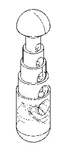
24 Figure IA shows a schematic of the capsule.
Figure 1B shows the relative positions of the body tissue with the tumor
nodule (an
26 example of 4 mm. depth is shown) which is being targeted disposed on
said tissue. A
27 simplified diagram of a shroud containing a locator mechanism is shown
over the tissue,
28 with the cassette (usually called a capsule in the description)
containing the radioactive
29 substance, and the general disposition of the capsule on a robotic arm,
also sometimes
referred to in the trade as a "instrument" which "instrument" on the DaVinciTM
surgical
Date Regue/Date Received 2023-07-10
CA 03072274 2023-07-10
WO 2018/148464 PCT/0S2018/017495
12
2 system is mounted on the DaVinciTm surgical system robot arm.
3 Figure 2 shows the detail of radioisotope loading
4 Figure 3 shows the position of the slide door, aperture disk and
radioisotope relative to the
capsule in various positions.
6 Figure 4 shows the subassembly detail of the front mount and back mount
and slide door
7 and rack_
8 Figure 5 shows the subassembly detail of the motor mounting plate
relative to the stepper
9 motors and the disposal of bevel gears on the shafts of the stepper
motors.
Figure 6 shows the subassembly detail and relative positions of the spur gear
and pinion
11 spear gear, and bevel gears mounted on the shafts and the relative
positions of the aperture
12 spur gear and spur gear.
13 Figure 7 shows the gross assembly detail of the end cap, motor mounting
plate, front
14 mount and back mount.
Figure 8 shows another general concept and captures the overall concept of the
invention.
16 Figure 9 shows a side view cross-section of a capsule with a movable
shutter with a push
17 rod moving the shutter against a spring.
18 Figure 10 is atop view of Figure 9.
19 Figure 111 a simplified view showing the geometry of Figure 9.
Figure 12 is another shutter design operated by a push rod.
21 Figure 13 is an end view of the capsule showing the multiple plate
design of Figure 14.
22 Figure 14 is a detail view of the multiple plate design operated by a
push rod moving the
23 plates constituting the shutter into moving plate receptacles.
24 Figure 15 is a top view looking down toward the aperture of the capsule
sowing the
relative location of the moving plates constituting the shutter to occlude the
radionuclide.
Date Regue/Date Received 2023-07-10
CA 03072274 2020-02-06
WO 2018/148464 PCT/US2018/017495
13
2 Figure 16 shows a cylindrical shell radiation shutter system showing
radiation source at
3 center, concentric shells with apertures aligned allowing exposure
through the outermost
4 beam port (emission aperture located on the capsule).
Figure 17 shows a cylindrical shell radiation shutter system showing source at
center with
6 all cylindrical shell apertures offset from each other thereby
obstructing the source and
7 shielding the radiation source from the exterior of the capsule.
8 Figure 18 shows a cylindrical shell radiation shutter system lateral view
showing shutters
9 open. The lines within the spreading cone are intended to generally
illustrate the
homogeneity of the spreading cone.
11 Figure 19 shows a cylindrical shell radiation shutter system showing
source at center with
12 all cylindrical shell apertures aligned with each other permitting
irradiation exterior to the
13 capsule along the beam path. The lines within the spreading cone are
intended to generally
14 illustrate the homogeneity of the spreading cone.
Figure 20 shows a top view of the cylindrical shell radiation shutter system
showing source
16 at center with all cylindrical shell apertures offset from each other
occluding the source and
17 preventing significant irradiation exterior to the capsule. The lines
appearing as a "T" are
18 meant to approximately illustrate the gradually widening cone from
cylindrical shell to
19 cylindrical shell which, when the cylindrical shells are aligned, will
result in the
homogeneity of the spreading cone.
21 Figure 21 shows a proposed gear plate: The gear plate in this figure
shows variable length
22 slots, all with one edge aligned in a series of 6 tracks in this
example, but could consist of
23 any reasonable number of tracks, corresponding with shutter thickness
desired and number
24 of shutters ( in this instance a number of six shutters has been
selected, but the number can
vary in a range of 1, 2, 3, 4, 5, 6, 7, 8, 9, 10 or integral numbers up to 20,
although after a
26 point the more shutters, the air gaps created and the less cost-
effective the device will be;
27 here the numbers of shutters is selected as six to illustrate the
invention (inner to outer,
28 which have been labeled A, B, C, D, E, F arbitrarily). The slots when
rotated clockwise
29 align the apertures with the case aperture, exposing the source. When
rotated counter-
CA 03072274 2020-02-06
WO 2018/148464 PCT/US2018/017495
14
2 clockwise, the shell apertures are not aligned with the source or each
other thereby
3 obstructing the source and shielding the radiation source from the
exterior of the capsule.
4 Figure 22 shows a Top Oblique View of the gear plate top oblique view
showing two
cylinders of six in this example. The other cylinders have been removed to
show the
6 underlying mechanism. Cylinder A (inner most) and Cylinder E (second from
outer most)
7 are shown with apertures aligned (open). The gear slots of varying
lengths are shown on
8 the gear plate.
9 Figure 23 shows an alternative gear plate showing raised drive surfaces
arranged in tracks,
which would mate with corresponding slots in each cylindrical shell shutter
component
11 causing variable motion and alignment. In this arrangement a cam plate
is used, each fixed
12 to each other, or alternatively independently rotatable with individual
stepping motors. In
13 this case, the raised surfaces would be either a fixed length with
variable independent
14 rotation or as shown with fixed rotation of all tracks.
Figure 24 shows a source rod and cap assembly with source in center of source
16 containment rod. The source (orange) is fixed in the central rod which
is inserted into the
17 cylindrical shutter system such that the source is precisely located at
the center of the
18 aperture beam ports. The lower tab on the source rod demonstrates one
means of locking
19 the capsule system into place, thus affixing the source in the desired
position.
Figure 25 shows two shutter cylinder components shown in position on gear
plate with
21 outer shell removed. The outer track gear slot is shown immediately
below the aperture of
22 the next inner shell. The innermost shell is shown in the center of the
gear plate assembly,
23 and the drive motor is shown attached to the gear plate below.
24 Figure 26 shows a gear plate assembled in the cylindrical shell
radiation shutter system
with a motor showing three cylindrical shells (A, C, E from inner to outer).
Not illustrated
26 in order to demonstrate the principle involved are cylindrical shells B,
D, and F. The
27 arrows show the shells aligned with the aperture. Note in the gear drive
beneath the gear
28 plate is a slot just above the motor. This slot is a space for
attachment of a mainspring
29 which will rotate the gear plate counterclockwise rapidly in the event
of a motor failure,
closing the shutters as a fail-safe mechanism.
CA 03072274 2020-02-06
WO 2018/148464 PCT/US2018/017495
2 Figure 27 shows a completed capsule with cylindrical shell radiation
shutter system, gear
3 plate and drive motor shown. The radiation source is at the very center
of the aperture
4 inside the inner most cylinder.
5 Figure 28 shows an exploded view showing individual cylindrical shutters
in alignment
6 with radiation shielding caps in place top and bottom. The gear plate is
visible below the
7 shutters.
8 Figure 29 shows a Side View of the exploded capsule containing the
cylindrical shell
9 radiation shutter system showing a) (toward the top) radiation shielding
cap above and a
10 radiation shielding cap below (toward the bottom of the figure), b) a
Side view of the
11 outermost emission aperture (beam port) (red) and c) the individual
cylindrical shutters
12 with ports (apertures) facing right in this illustration.
13 Figure 30 shows a final assembled capsule with cylindrical shell
radiation shutter system
14 showing radiation shielding cap in place (green), cylindrical shutters
(with every other
15 cylinder removed for clarity, gear plate drive mechanism, drive motor,
and a means for a
16 fail-safe closed position, here a mainspring emergency fail safe closing
mechanism (blue).
17 Figure 31 shows a linear slide shutter assembly showing radiation source
(green arrow),
18 desired beam path (red cone), shutter and area of shutter interference
with desired beam
19 path, creating penumbra effect with variance in irradiation dose on the
desired target in this
region. The salmon line shows the radiation shield gap necessary to
accommodate the
21 sliding shutter permitting undesired exposure when the irradiation is in
progress.
22 Similarly, the orange line shows an air gap in the field permitting
undesirable
23 inhomogeneity in the radiation field. The figure illustrates the use in
the present invention
24 of a different set of elements and structure in order to achieve
homogeneous solid angle
divergence of beam port from the radiation source and virtually eliminate
significant voids
26 with minimal radiation attenuation in the voids.
27 Figure 32 shows the radiation source geometry. There are two pertinent
geometries. The
28 radiation source geometry is associated with the radiation source
itself, and consists of a
29 "capsule central axis" from which all pertinent radiation distances are
measured (Distance
is from the radiation source in this illustration, for the embodiment of
emission from the
CA 03072274 2020-02-06
WO 2018/148464 PCT/US2018/017495
16
2 long end of the capsule it is the radiation source as one end of the
distance line). This is
3 depicted by the yellow arrow. The green arrow shows an off axis ray,
which is not on the
4 central axis. Note this geometry may be the same as the physical geometry
of the capsule,
or if desired may be independent of the capsule geometry, depending on
application
6 requirements.
7 Figure 33 shows a view along the capsule central axis of a means for a
fail-safe closed
8 position, here a coil-spring fail-safe mechanism
9 Figure 34 shows a view perpendicular to the capsule central axis of a
coil-spring fail-safe
mechanism engaged with respect to a tab, gear tooth or cam against a
cooperating tab, gear
11 tooth or cam such that if power is lost, the spring causes the
particular shutter or shutters
12 operated in cooperation with that particular shutter to occlude
radiation.
13 Figure 35 shows another view of the internal disposition of a coil-
spring fail-safe
14 mechanism.
Figure 36 shows a cross section of multiple coil-spring fail-safe mechanisms
disposed in
16 cooperation with a series of gear teeth or cams such that if power is
lost, the spring causes
17 the particular shutter or shutters operated in cooperation with that
particular shutter to
18 occlude radiation.
19 Figure 37 shows a means for a fail-safe closed position, here a leaf-
spring fail-safe
mechanism which is connected to a sample cylindrical shell with the
cylindrical shell being
21 driven open by the operation of the capsule to align the shells and
returning upon no longer
22 being driven to a rest/fail-safe position.
23 Figure 38, shows a means for a fail-safe closed position, here a
longitudinally acting spring
24 operating a cantilevered arm which is connected to a sample cylindrical
shell with the
cylindrical shell being driven open by the operation of the capsule to align
the shells and
26 returning upon no longer being driven to a rest/fail-safe position.
27 FURTHER BACKGROUND
28 The effective undesired dose outside of the beam path which traverses
necessary gaps in
29 shielding material is given by the foiniula
CA 03072274 2020-02-06
WO 2018/148464 PCT/US2018/017495
17
2 / = le(x) where la is the mass attenuation coefficient and x is the total
thickness of the
3 shielding material (less air gaps). As can be seen in above the beam port
in Figure 15, an
4 air gap must exist to accommodate closing the shutter, which reduces the
shielding
thickness with a consequent increase in undesired radiation leakage outside of
the desired
6 radiation field. Where x = 0 (within the beam path, there is no
attenuation from shielding.
7 Where there are air gaps in the shutter the value of x is reduced by the
path length of the air
8 gap resulting in differing shielding attenuation, increasing undesired
leakage and
9 restricting the beam port shapes to those shapes easily mechanically
accommodated.
Conversely, below the beam port, the shutter itself creates a variable air gap
due to
11 its physical characteristics, which will partially attenuate the beam
causing a variance in
12 beam intensity (penumbra) resulting in undesirable overdose/underdose at
the field edge.
13 This leaves the following potential dose in-homogeneity:
14 Source to device surface distance along the mechanical discontinuity:
2.8 cm.
Shielding Available (shielding ¨ mechanical space air gap): 1.4 cm
16 Which leaves a total shielding thickness of 2.8 cm ¨ 1.4 cm = 1.4 cm
17 If a maximum typical exposure time is 75 minutes with an unshielded
source of 177
18 cGy/minute, and lead is used in the device (for the purposes of this
example), the tenth
19 value layer (TVL) of lead is 0.6 cm.
(1.4cm)
________________________________ = 2.3TVL
(0.6cm)
21 The resulting dose attenuation is:
cGy _233 cGy
22 177 ¨min 10 = 0.83¨min If there were no air gap at all in the
shielding, the dose
23 attenuation would be improved to
(2.scm)
24 ¨(0.6cm) = 4.67TVL and the leakage will be reduced to
cGy _ cGy
177 ¨min 10 4'67 = 0.00378 ¨min Thus this air gap is significant and
inhomogeneities in
26 shielding can cause remarkable differences in radiation field doses. If
a typical desired
27 irradiation of 24 Gy is desired, the exposure time will be 24 Gy/1.77 Gy
= 14 min 6.6
28 seconds. The leakage from a shield with an air gap (discontinuous) will
be 0.83 cGy/min x
29 14.12 minutes = 11.7 cGy. With a continuous shield the undesired
exposure will be
CA 03072274 2020-02-06
WO 2018/148464 PCT/US2018/017495
18
2 reduced to 0.05 cGy. This the elimination shielding discontinuities has a
significant
3 impact on reduction in unwanted radiation dose.
4
DESCRIPTION OF THE INVENTION
6 The preferred mode of invention proposes to first select an
interchangeable
7 irradiating capsule with a shutter as set forth below. Based on the depth
and size of tissue
8 to be treated, a radiation source will be selected for placement in the
capsule and mounted
9 on the robotic arm of the SRIORT. The arm would then be moved to the
proper location
=for irradiation of the tissue, under direct visualization, with or without
assistance from
11 alternative imaging modalities or any combination of these.
12 Expanding on the above, the key invention components are:
13
14 = A radiation source
= A capsule mountable on an robot arm, or on an instrument connected to a
16 robot arm, with an emission aperture opening to a cavity containing the
radiation
17 source with certain control electronics and devices designed to be
connected to the
18 surgical robot and inserted into the patient's body through the
laparoscopic/surgical
19 robotic incisions
= For a lesion, tumor, tissue, or organ, a mechanism for displaying pre-
21 operative medical imaging, fused pre-operative medical imaging,
including CT,
22 MRI, Ultrasound, functional MRI, PET, PET/CT and nuclear medical
scanning in
23 the operating room in real time visible to the manipulation station of
the surgical
24 robot preferably on a video screen or computer monitor or other means
for display.
= A mechanism for identifying and tracking the real time coordinates of
the
26 radiation source capsule within the body and displaying the 3-
dimensional location
27 of the capsule on the pre-operative imaging with a projection of the
presently
28 programmed radiation field distribution on the images and a control
means such as
29 a general purpose computer to make real-time updates to the tissue
position relative
to the surgical robot, avoiding overdoses to desired tissue.
31 = A mechanism for tracking, visually, preferably on a video screen,
computer
CA 03072274 2020-02-06
WO 2018/148464 PCT/US2018/017495
19
2 monitor, or means for display the internal position of the capsule
within the body
3 and for advancement and positioning under direct visualization using
visible,
4 infrared and ultraviolet light or any combination of these.
= A mechanism for identifying the tumor, and tumor depth (using a
6 combination of the above or ultrasonic echoes)
7 = A mechanism for setting an aperture size, accepting a desired dose
and
8 calculating the exposure time based on the selected radiation source
physical
9 parameters and characteristics.
= A mechanism for activating the now properly positioned radioactive
source
11 in the cavity to deliver the desired radiation dose, and field size and
shape to the
12 desired volume of the tumor while preventing exposure to the operating
room
13 personnel. Normally this would mean an electromechanical actuator
opening a
14 closed shutter in the capsule. However, a mechanical connection could be
made so
that an actuator, such as a pin, in the surgical robot arm actually activates
the
16 shutter to open. A spirally opening and closing iris shutter of the
style used in a
17 camera, or a simple door mechanism can provide an adjustable aperture.
18 = A mechanism for identifying and tracking the real time coordinates
of the
19 radiation source capsule within the body and displaying the 3-
dimensional location
of the capsule on the pre-operative imaging, a post-radiation report to show
21 radiation field distribution on the images, on for instance, a video
screen, computer
22 monitor or means for display, and probable damage to irradiated tissue.
23
24 These components and mechanisms will be described in detail below.
The application of the invention would be as follows for cancers:
26 The physician would have pre-imaged the patient's body according to
standard
27 medical procedures to locate the tumor and any other areas of suspected
cancer activity,
28 sometimes known as "hot spots". These are areas that are identifiable in
a variety of
29 medical imaging modalities, including PET, CT, MRI and nuclear medicine
scans. The
physicians would have visually identified any other areas of suspected cancer
involvement
31 during the course of surgical intervention.
32 The physician will then make an incision in the abdomen and the SRIORT
is
CA 03072274 2020-02-06
WO 2018/148464 PCT/US2018/017495
2 activated. The SRIORT has a television camera mounted on a robotic arm.
The SRIORT
3 has accessories mounted on a robotic arm and are controlled by remote
control. The
4 surgical SRIORT is then used to incise the interior membranes and a
cutting implement is
5 used to perform a resection by the physician. The surgeon can cauterize
and clean as
6 needed and ultimately view the remaining tissue through the camera on the
SRIORT arm,
7 and in conjunction with medical imaging as described above, determine
what further areas
8 need radiation treatment.
9 In the case of ovarian cancer, when the maximum surgical debulking
possible has
10 been obtained, frequently, studs of disease remain which involve the
surface of the liver,
11 the diaphragm and areas of the bowel. It is not possible to treat these
areas generally with
12 external beam (whole abdominal radiation therapy), conventional
brachytherapy or loose
13 isotope therapy or conventional intraoperative radiation therapy using
accelerators due to
14 the inability to deliver a precisely enough targeted and sufficient dose
of radiation to
15 eliminate cancer metastases without causing substantial morbidity and
even mortality, or
16 exposing operating room personnel to unacceptably high exposures to
radiation.
17 Based on the depth of tissue desired to be penetrated and the desired
dose to be
18 delivered, a particular radiation source, which may be a radioisotope or
device generated
19 radiation (x-rays), of appropriate emission type, energy and strength
would be selected for
20 placement in the capsule on the SRIORT arm. This capsule would be either
permanently
21 mounted on the SRIORT arm or preferably would be an interchangeable
module to
22 accommodate differing physical characteristics of radiation sources. The
capsule must be
23 designed to balance size of the device with necessary shielding for both
direction and size
24 of radiation field and personnel protection from leakage radiation. The
capsule would then
be selected under robotic control from its storage location, mounted on the
arm of the
26 SRIORT and moved into the proper position inside the patient in the
proper location for
27 irradiation. The physician would then move the capsule and proposed beam
location to the
28 angle and desired beam angle to the lesion. The SRIORT has a camera
enabling direct
29 visualization of the lesion. An alternate imaging device, appropriate
for the tumor could be
used in addition to a camera, such as an ultrasound transducer or probe. A
laser could be
31 mounted to identify and illuminate the spot of radiation beam
application.
32 Traditional IORT using linear accelerators external to the body have
used doses in
CA 03072274 2020-02-06
WO 2018/148464 PCT/US2018/017495
21
2 the range of 10-20 Gy (Gy = gray = joule/kg energy deposited in matter by
ionizing
3 radiation). These doses can be delivered with a variety of devices and
isotopes, most
4 commonly those with high specific activity such as Ir-192 or Cs-137, or
more recently x-
ray diodes and solid state x-ray generators, can be used. In addition, other
emitters such as
6 Sr-90 (beta emitter with energy of 0.195 MeV). The table below gives
examples of
7 byproduct material and typical energies and half-lives.
8
Typical Isotope Emission/Energy Half Life
Cs-137 Gamma/662 keV 30 years
Ir-192 Gamma/442keV 70.2 days
Sr-90 Beta/195 keV 29 years
Cf-252 Neutron/fissile spectrum 2.6 years
9
11 Dose calculations
are given by the following formula for isotopes:
12
13 Dose =
(PAKR)(ISF)2 (Strength)(timeof exposure)
14
16 These sources and other sources will generally have activity in the
range of 5-10 Ci
17 (10 Ci=370 GBq). For example, to deliver 20 Gy to a depth of 5 mm (4 mm
+ 1 mm
18 margin) for the 4 mm tumor shown in Figure 1, from the applicator
capsule, assuming a 10
19 Ci source strength, using Iridium-192, which has a specific air KERMA
constant (PAKR =
(1.115 Gyxm2)
used to convert activity into dose, the following exposure would be required:
(GBqxhr)
21
(111.5cGy ¨ cm2) 1 lhr
22 2000cGy = (370G Bq) _______________________ )2 ( __
(GBq ¨ hr) ((0 .25cm) '60min)t
23
24 which yields an exposure time of 2000/11001 = 0.181 minutes or 10
seconds exposure,
assuming the above parameters. The quantity .25 cm. was selected in order to
have a
26 typical source to surface distance. Therefore, each lesion could be
treated in under 1
27 minute, with precise control of exposures, field placement and size
under real time
CA 03072274 2020-02-06
WO 2018/148464 PCT/US2018/017495
22
2 guidance in the operating room using the SRIORT.
3 Due to the absolute criticality of distance in this exposure range, to
delivered dose
4 per unit time, the capsule will have an independent electronic distance
measuring device
using optical ranging.
6 Where organ motion is a concern, the device can be placed at an
increased distance
7 such as 0.5 cm from the tumor at the physician's discretion. Adjustments
can be made to
8 accommodate organ motion or relative motion of the patient. For this
distance the above
9 calculation would yield an exposure time of 0.67 minutes or 40.2 seconds.
The exposure time would be electronically controlled with a dual timer backup
11 system whereby if the primary timer set time expires, then a backup
secondary timer will
12 engage and close the aperture to stop the radiation exposure. Both of
these timers will
13 have a clearly visual display at the operator's console with an alarm,
both visual and
14 audible when the cassette has radiation present and a second alarm both
visual and audio if
the cassette's control electronics fail to close the aperture (in the case of
a radioactive
16 source) or stop power to the radiation generator (in the case of an x-
ray diode device).
17 The cassette's radiation "safe" chamber and aperture is constructed with
radiation
18 shielding in mind. Since the device is capable of using both high and
low dose rate
19 sources, shielding is mandatory for several reasons, the most important
of which is to
protect patient tissue from stray radiation emission from the device and to
protect operating
21 room personnel while the device or radiation source is in transit.
22 The shielding calculations are based on using either depleted uranium,
lead or
23 tungsten. Due to its superior shielding characteristics, the preferred
shielding is uranium
24 since uranium shielding will be thinner and allow for a more compact
cassette which will
be easier to insert into a laparoscopic wound (1-3 cm) and manipulate under
robotic
26 control, once it is inserted into the body. A typical source size (based
on the Nucletron and
27 Varian sources presently in use), is 0.5 mm in diameter by 5 mm long. To
reduce the dose
28 to acceptable levels during the time the source is in the patient, for
this proposed
29 calculation example, an assumption is made that a procedure with the
source in the patient
could last up to an hour. During this time the source will be emitting
radiation and in the
31 medical therapeutic use of radiation 60 cGy of exposure during a
treatment can be
32 administered at low risk. Since operating room personnel exposure must
be kept lower
CA 03072274 2020-02-06
WO 2018/148464 PCT/US2018/017495
23
2 than this, additional external shielding will be placed around the
patient to meet ALARA
3 radiation safety limits. The robotic workstation can be placed physically
far from the
4 patient, further minimizing the need for external shielding. The
shielding calculation
equation is
6
37GBq 111.5cGycm2 1
7 10Cix _____
Ci GBq ¨ hr __________________________ x(10cm)2 =
412.55 cGy/hr
8
9 The 10 Ci is selected as the source strength. The quantity 37GBq per Ci
is a
conversion factor. Ten centimeters is a typically selected distance to the
patient body
11 surface for the purpose of radiation shielding calculation because the
average patient is
12 approximately 20cm. "thick." To reduce this dose rate to an acceptable
level, the dose
13 would be reduced to less than 60 cGy/hr or by a factor of approximately
1 or 2 tenth value
14 layers of shielding. The tenth value layer of depleted uranium for Ir-
192 is 6.5 mm so, 1.3
cm of depleted uranium will allow full shielding and reduce the leakage
exposure rate at 10
16 cm from 411 cGy/hr to 4.1 cGy/hr at 10 cm or 16 cGy/hr at 5 cm. If
tungsten were chosen,
17 the shielding thickness required will be approximately 22 mm.
18 Given the source size, shielding requirements, and necessary electronics
and
19 adaptors, the preferred mode would be that the final dimensions of the
cassette will be 4
cm in diameter x 5 cm long or 4 cm x 3 cm x 5 cm. For the cone portion, if a
cone is
21 desired, the divergence of the cone should match the outer diameter of
the tissue being
22 irradiated. The cone can be selected in shape to correspond to the tumor
shape. The cone
23 can be very short, if used at all, 3 to 4 mm. The cassette can have
varying cones mounted
24 on it to conform to irregular tumor shapes. This will give adequate
space to enclose a
source, associated visualization, measurement and control electronics and
mechanical
26 safety apparatus. In SI units the shielding calculation equation is:
27
1000mCi 4.111cGycm2 1
28 10Cix _______
Ci mCi ¨ hr _______________________ x(10cm)2 =
411 cGy/hr
29
The shutter would have a diameter of at least the maximum field size desired.
A cassette
31 designed with a shutter opening of up to two cm. would be the most that
would likely be
CA 03072274 2020-02-06
WO 2018/148464 PCT/US2018/017495
24
2 required. The collimation of the radiation is more likely determined by
the size of the
3 source, but the shutter size should be larger than the largest desired
collimation for a
4 particular treatment regime.
Another preferred mode is a slightly smaller capsule. Its shielding can be
increased
6 by leaving cavities in the capsule into which can be inserted tungsten or
depleted uranium
7 blocks or other very emission absorbing material. Those materials are
very difficult to
8 work or machine and are more easily used in pre formed shapes like blocks
or sheets. If
9 lead blocks are used and sealed in by relatively inert metals to body
materials, or even
sealed in by silicone or other material which does not give insult to body
tissue or react
11 with such tissue, this is a way to give lead functionality in the
capsule as a shield which
12 would otherwise be less desirable to insert in the body, even for a
short time. Lead is
13 easily worked and melted. By using those methods of shielding, less
expensive shielding
14 materials can be combined to lower the capsule cost.
A second mode of invention would use the cassette device as a positioning
system
16 only and for the delivery of radiation the device would have a transfer
tube connector
17 which would allow the use of existing High Dose Rate Remote afterloading
devices such
18 as the Nucletron HDR or Varian HDR device to provide the radiation
source. These
19 devices have an Ir-192 source similar to that described above which is
attached to a cable
and is positioned via transfer tubes which are attached to the HDR and the
SRIORT
21 cassette. This option would be available for institutions that have such
a device available
22 for interstitial radiotherapy. Other than the source delivery mechanism,
in this case, the
23 source is not an integrated part of the cassette, but rather delivered
once the device is
24 properly positioned. There are numerous disadvantages with this
arrangement which make
this less preferred than the self-contained system, most notably is that the
source is freely
26 radiating while it traverses the transfer tubes, which will require all
personnel to leave the
27 operating room, thus dramatically increasing the time it takes to do the
procedures.
28 The advantage of this device is that the device is small, easily
manipulated by the
29 SRIORT control systems, in real time, under direct visualization. This
enables the surgeon
and radiation oncologist to determine during the course of the operation areas
of residual
31 and unresectable disease and to deliver a dose of radiation precisely
and interactively to
32 sterilize the tumor. Because the capsule radiation source is orders of
magnitude smaller
CA 03072274 2020-02-06
WO 2018/148464 PCT/US2018/017495
2 than the conventional linear accelerator arms, it can be placed with high
precision within
3 the body and using articulating robotic "hands" holding the capsule in
place, the field can
4 be directed at the correct tumor site while inserted into the body
through the robotic
5 incisions.
6 Due to the potentially high activity sources in use, an emergency
aperture closing
7 mechanism incorporating both electronic and mechanical overrides would be
used in the
8 device. The system will also have fail safe mechanisms resulting in the
aperture defaulting
9 to the closed position absent electrical and mechanical signals to open
the shutter or expose
10 the aperture. In the case of x-ray generators, the fail-safe will not
permit current to flow to
11 the device except under direct positive command.
12 In addition this device, by virtue of having a shielded capsule with a
controllable
13 aperture, together with the articulated robotic "wrist" or "hand"
apparatus, allows precise
14 positioning of the radiation source prior to opening the aperture and
thus protecting normal
15 tissue from radiation until the device is positioned and verified. This
is a substantial
16 advance over the current methods of applying intraoperative radiation
therapy.
17 The purpose of using a shielded capsule is to minimize the damage to
tissue while
18 the capsule and the radiation source inside is in transit to the desired
location. The capsule
19 would be made of a high density shielding material such as lead,
tungsten or uranium and
20 the capsule would have a shutter covering an aperture through which
radiation particles
21 would be emitted. The shutter would also be of high density shielding
material such as
22 tungsten, but materials can be selected from those in the Berger &
Seltzer handbook which
23 contains data on mass energy attenuation coefficients sufficient to
provide appropriate and
24 necessary radiation protection. The capsule design will permit the
adaptation of
25 interchangeable shutters, much like the interchangeable lenses of a
camera.
26 The interchangeable capsule would be stored in a shielded storage
device, could be
27 sterilized by steam or gas sterilization as is traditionally used in the
operating room
28 environment. The radiation source would be extracted from the storage
pig, which is a
29 larger, well shielded storage chamber used to transport and store
radioactive source
material, usually build of lead or tungsten, immediately adjacent to the
patient in the
31 operating room which will minimize the exposure of any personnel and the
patient during
32 the capsule transit time. It would be impractical to shield all gamma
radiation from a
CA 03072274 2020-02-06
WO 2018/148464 PCT/US2018/017495
26
2 source emitting gamma rays, but the distance allowed by the robotically
assisted
3 intraoperative radiation therapy applicator coupled with a reasonable
amount of shielding
4 would allow the device to be used while minimizing exposure to personnel
to be in
conformance with NCRP limits of exposures to radiation workers. The device
will include
6 adequate shielding in the form of mobile shielding units installed in the
operating room to
7 protect operating personnel in accordance with the ALARA ¨ as low as
reasonably
8 achievable ¨ philosophy of radiation protection and well below the
accepted occupational
9 exposure limits for the planned procedures. Survey instruments will be
built into the
apparatus and workstations to measure and record total in-room exposures.
Mobile patient
11 shielding would be available, depending on the radioisotope, to shield
the patient,
12 preferably with an aperture for the surgical entry site only so that any
exposure of the
13 patient is minimized. That mobile patient shielding could be in the form
of one or a series
14 of hooded containers such as lead shields on mobile casters, or a one or
a series of lead
aprons.
16 The cassette could be designed to either have contacts connected to
internal wiring
17 that meet control contacts on the robotic arm, or the internal wiring of
the cassette can be
18 connected by a wire harness to the robotic arm. An alternative preferred
mode is a wireless
19 control mechanism, but the level of ionizing radiation can be
problematic.
For alpha or beta emitters, a lightweight capsule is possible. Under current
21 technology a particle accelerator cannot be used for effective
application of alpha particles,
22 protons, electrons or light ions, which at energies useful
therapeutically have a very short
23 path length, but within that path length are devastating to the
reproductive machinery of
24 cancer cells (DNA and cellular ability to repair fractured DNA). Alpha
particles and to a
lesser extent, beta particles emitted from radioisotopes are readily obtained
from a variety
26 of isotopes, as are gamma rays. [Berger and Selzer, Affix]
27 Alpha particles are considered high linear energy transfer (LET)
particles and
28 deliver substantive damage to DNA in the form of double stranded DNA
breaks, which are
29 very difficult for cells to repair properly. Gamma rays, and x rays, in
contrast are low
LET particles and operate by the generation of radiolysis of water generating
hydroxyl free
31 radicals in the vicinity of DNA causing single strand and double
stranded breaks following
32 a linear-quadratic curve of cell survival v. dose, culminating in a loss
of reproductive
CA 03072274 2020-02-06
WO 2018/148464 PCT/US2018/017495
27
2 integrity of the cancer cells. Likewise beta particles, though low in
linear energy transfer
3 can cause double stranded breaks and destroy DNA through clusters of
single stranded
4 breaks which can be made pennanent by oxygen fixation in non-hypoxic
environments.
The capsule mounted on the SRIORT arm enables an alpha or beta emitter to be
6 completely shielded from healthy tissue and to minimize transient damage
as the radiation
7 source is positioned at its intended target. Only on setting the aperture
to the desired beam
8 size, positioning the aperture in the correct location and desired angle
and opening the
9 shutter on the capsule will a beam of radiation be emitted through the
aperture in the
capsule in the desired direction to irradiate the lesion. In the case of an x-
ray generator, the
11 x-ray source will only be turned on when the above parameters are met.
12 As particle path length in tissue is very predictable, cancerous tissue
can be
13 destroyed with a much finer precision while minimizing damage to normal
tissue, such as
14 livers, kidneys and bowel. Sr-90 is a typical beta emitter which would
be deadly to tissue
without appropriate shielding, but when used in the proposed capsule could be
safely
16 directed to the targeted area. Likewise isotopes that emit alpha
particles, and gamma rays
17 or a source capable of developing x-rays can be used with appropriate
shielding design on
18 the capsule. The significant advantage of a beta emitter is enablement
by the invention of
19 a new technology of a very effective and predictable radiating isotope,
and the
miniaturization of the capsule because of reduction of bulk because shielding
is much
21 simpler. Any metal, or plastic such as lucite, with appropriate electron
stopping power as
22 set out in tables for a source available to a reasonably skilled
practitioner, such as the tables
23 in Berger & Seltzer, can be used for the shielding. Much smaller tumors
in much smaller
24 and confined spaces can be treated.
The capsule shutter could be simply the equivalent of a door occluding a
radiation
26 aperture. A preferred mode is to use an iris type aperture with a clam
shell outer cover.
27 The aperture can be opened to various diameters allowing the physician
to choose the size
28 of lesion to be treated and the surface area of the volume. A light
source can be disposed
29 on the exterior of the cassette for illumination inside the patient of
the tumor to be
irradiated. An alternate light source to act a as field light behind the
aperture through
31 which radiation will be emitted, but behind the iris would enable the
physician to continue
32 visible inspection of a lesion as he positions the device for maximum
coverage of the
CA 03072274 2020-02-06
WO 2018/148464 PCT/US2018/017495
28
2 tumor before the radiation source is opened by the clamshell. In
addition, this mode gives
3 redundant protection should one or the other of the apertures fail while
the device is in
4 place, thus allowing the device to be removed from the patient and safely
deposited in the
shielded pig until repairs can safely be made. A preferred light source is an
LED, fiber-
6 optic or solid state light emitter.
7 Upon completion of the treatment procedures, the SRIORT arm and
radiation
8 source can be remotely stored in the pig or appropriate storage device
where sterilization
9 and preparation for the next case can take place. For convenience sake,
the storage device
is preferably a table with a shielding container or pig on it. The storage
device would
11 likely have multiple pigs. The storage device including a shielding pig
is referred to as a
12 shielded source containment table, even if a closet or storage cabinet
is used. To insure
13 radiologic safety, each pig shall have a means of detecting radiation
presence to insure that
14 a source is present or absent from the pig. By regulation, that would
usually be a room
detector in the room, and/or a sensor inside the shielded source containment
table, such as
16 an ion chamber, electrometer or Geiger-Mueller type device.
17 In addition to a radiation source, other devices could also be mounted
with the unit,
18 including a laser or particle emission device and used adjuvantly for
tissue destruction.
19 This device is not limited to the carriage of radioactive sources, but
can also be used in
conjunction with x-ray diodes or other radiation sources.
21 Because a surgical robot can have more than one arm, the invention
enables more
22 than one capsule to stand ready in the shielded source containment table
so that should a
23 physician determine to select a different capsule during irradiation,
the capsule in present
24 use can be quickly withdrawn, its path of extraction memorized and an
new capsule with
the preferred radiation source inserted.
26 Another important variation on the preferred mode relates to the doors
or shutters
27 on the capsule (also referred to as a cassette). The electromechanical
movement could also
28 be accomplished by hydraulic means with push pull pressure by an
electrical signal to start
29 a pump and then the mechanical action of pumping fluid to move a door.
More
specifically, a small hydraulic pump could be used to move the shutter or
door, and a valve
31 or a second pump used to move the shutter or door back. Also, the doors
could be
32 operated against non-toxic gas pressure so that when pressure is applied
by a motor or
CA 03072274 2020-02-06
WO 2018/148464 PCT/US2018/017495
29
2 hydraulically, in the event of power loss, the door moves to the closed
position because the
3 gas presses against the lost pressure.
4 The robotic system could be designed to combine the elements into an
invention as
before except that the non-radiation emitting features and associated control
of emissions
6 are separable from the radiation emitting features and associated control
of emissions. By
7 the expression "associated control of emissions" is meant the one or more
doors or shutters
8 on the capsule containing the radioisotope. As further description, on an
effector arm or
9 robot arm, or attachable to it, there would be a standard set of tools
and carrier that would
be used repeatedly. That will be referred to as the shuttle capsule holder.
The capsule
11 holding the radioactive source would be designed with necessary
interconnects to control
12 the doors to the shuttle capsule holder.
13 On the shuttle capsule holder could be other apparatus including
tracking apparatus,
14 range finders, laser pointer or camera/visualization apparatus. More
specifically, those
could be a stand-off remote detection for determining the distance from the
radiation
16 source to the tissue being irradiated, an electronic distance measuring
device using optical
17 ranging for locating the distance between said tissue being irradiated
and said radioactive
18 source, a means for utilizing ultrasonic detection to determine tissue
depth, including
19 tumor depth to determine an applicable margin to be irradiated, and one
or more means for
direct visualization by remote display of tissue adjacent to said capsule,
which could be
21 simply a fiber optic cable to transmit the image to an extra corporal
screen or image
22 capture device to see the image. The advantage of the direct
visualization by remote
23 means of tissue adjacent to said capsule is to ascertain if all diseased
tissue has been
24 surgically removed and to ascertain if further irradiation is necessary.
The procedure would be that the shuttle capsule holder would be set up by a
26 technician and could have a range finder, light or laser pointer and
other features pointed in
27 U.S. App!. 60/973,545, and 61/098,225 and PCT Appl. PCT/US2008/077100
entitled
28 "DIRECT VISUALIZATION ROBOTIC INTRA-OPERATIVE RADIATION THERAPY
29 APPLICATOR DEVICE." That shuttle capsule holder would be mounted to the
robot, or
picked up by the robot, and then remotely carried to the capsule for
interconnection and
31 then the dual system inserted into the body for treatment. Among the
advantages are that
32 capsule sizes can be smaller depending on which isotope/energy/ emitter
is selected and/ or
CA 03072274 2020-02-06
WO 2018/148464 PCT/US2018/017495
2 .. it enables tighter procedures and the shuttle capsule system allows reuse
of the expensive
3 components more easily.
4 Advantages of this system include the ability to use the more expensive
parts
5 repeatedly and to vary the capsules more easily. Capsules can be varied
in size or
6 protection depending on the isotope involved and perhaps the size of the
incision. Also,
7 .. the capsule can be shipped alone to a laboratory or radiopharmacy for
reloading. The pig
8 in which the capsule is stored could be shipped, the capsule filled with
the hot
9 radioisotope, and the pig returned to the hospital or surgical center to
be opened by a robot
10 and the operation to proceed as described.
11 The radioisotope can be mounted in the capsule any number of ways
including
12 gluing it on a pin in the capsule, or holding it in a ring like a
circular gunsight or by
13 otherwise securing it inside the capsule. The location should be secure
and it is preferable
14 it be known or measured because it is preferable to know where the
radioisotope is exactly
15 compared to the tumor being ablated. The size of the isotope varies in
size and activity.
16 .. Generally the order of magnitude is between 5-10 mm. For special
purposes isotopes it
17 may larger or smaller.
18 Another way of accomplishing the pickup of the capsule by a robot arm is
to design
19 the capsule so that it has a spine of contacts on it. Preferably to the
rear of the capsule
20 opposite the opening through which the radioisotope is exposed would be
a group of
21 contacts for electrical connection and any fiberoptic connection.
Mounting them on a
22 spine appears to be the easiest mode. The robot arm would have on its
end a
23 .. corresponding set of contacts and any fiberoptic connection interior to
an alternately
24 opening and closing jaw, and the jaw would be mounted on and then
maneuvered on the
25 robot arm or end effector to align the jaw's contacts with the group of
contacts on the rear
26 of the capsule and be closed on the contacts to make positive contact.
The jaw could also
27 be designed to be the shuttle capsule holder referred to before and/or
to accomplish its
28 =functions. The jaw could be designed "inside out" particularly in the
shuttle capsule holder
29 design to have the contacts aligned inside a slot or aperture in the
capsule into which is
30 fitted a jaw which has the contacts on its exterior and is expanded to
make positive contact.
31 The jaw would preferably have material for attenuating radiation
emissions similar to that
32 of the capsule in order to preserve a relatively uniform radius of
radiation attenuating
CA 03072274 2020-02-06
WO 2018/148464 PCT/US2018/017495
31
2 material around the radioisotope.
3 Another preferred mode is to design a capsule in which is disposed a
plunger. This
4 mode would hold a "loose isotope", i.e. an unsealed radioactive source,
in solution or in a
form that is injectable into the tumor with a plunger from inside the capsule,
enabling the
6 use of a "hotter" isotope. This could be used with P-32 or 1-131 or alpha
or beta emitters,
7 including Y-90.
8
9
EXAMPLES OF APPLICATIONS OF PREFERRED MODE OF INVENTION
11 In the following two examples, a narrative description of how the SRIORT
device and
12 system will be used in actual practice. Several physicians will, of
necessity be directly
13 involved in these procedures due to the differences in training between
the specialties. The
14 key players in each case will be a surgeon and a radiation oncologist.
The surgeon will be
specifically trained in a pertinent area and the radiation oncologist is
trained in the
16 appropriate use, application and dosing of radiation for the treatment
of tumors. In
17 addition, a medical physicist, specifically trained in the use of
radiation sources in
18 conjunction with the radiation oncologist, must be available for the
planning of radiation
19 delivery using the SRIORT device.
21 Example: Abdominal Tumor (Ovarian Cancer Stage Illb)
22 Initially, the patient will be informed of the nature of the procedures
to be
23 performed in the treatment of the cancer. After being informed and after
the patient
24 acknowledges this information and gives her consent, the patient will be
taken to the
operating room and placed on the operating table in the supine position.
Following this the
26 patient will be anesthetized using general anesthesia supplied by the
anesthesiologist.
27 After adequate general anesthesia is instilled, the patient will be
examined under
28 anesthesia to determine, if possible, the extent of disease. Following
this, the patient will
29 be prepped and draped in the usual sterile fashion and a sub-umbilical
transverse incision
will be made extending approximately 1 ¨ 1.5 cm. Following this, a
laparoscopic trochar
31 with a TV camera in the bore will be advanced through the incision and
under direct
32 visualization into the peritoneal cavity. Following entry into the
abdomen, the abdomen
CA 03072274 2020-02-06
WO 2018/148464 PCT/US2018/017495
32
2 will be insufflated with carbon dioxide gas to distend the abdominal wall
away from the
3 intra-abdominal organs. Following this, again under direct visualization
via the TV
4 camera, a series of similar incisions will be made and trochars
introduced into the abdomen
which will allow the placement of robotic arms in the course of the surgery.
Once these
6 trochars are in place, the robotic actuating system will be placed into
position at the
7 operating table and the robotic arms will be placed in the ready
position. The physicians
8 will then move to the SRIORT control station, which will be located in
the operating room
9 behind a radiation shield of sufficient physical characteristics to
provide as low as
reasonably achievable radiation protection during the period of time that the
intra-operative
11 radiation device is in operation. The workstation will have
visualization system
12 originating from the robotic cameras placed in the patient, and
selectable views. The
13 control station will also have ergonomic robotic hand manipulators which
will allow the
14 physicians to move and manipulate the robotic arms in a natural way,
under the control of
computer and associated electronic circuitry.
16 The surgeon will then place the appropriate robotic arms into the
patient via the
17 previously placed trochars which will then be manipulated from the
control station to
18 perform the operation. The surgeon will generally use the robotic arms
to place suction
19 into areas of peritoneal fluid collections which will be sent to
pathology for microscopic
analysis for metastatic cancer cells. Following this, the abdomen will be
washed with
21 sterile water and that too will be collected and sent to pathology for
analysis.
22 From this point, the surgeon will perform the hysterectomy, bilateral
salpingo-
23 oopherectomy and pelvic and para-aortic lymph node dissections. Once
this part of the
24 procedure is complete, the surgeon will turn his attention to the
remainder of the abdomen.
Generally in locally advanced ovarian cancer, the omentum is also removed.
Following
26 this, the surgeon will inspect the remainder of the bowel using the
robotic devices and
27 cameras for further evidence of cancer. S/he will examine the bladder,
rectum, bowel,
28 peritoneal surfaces, the liver and the underside of the diaphragm. If
lesions are found, the
29 surgeon will resect, to the greatest extent possible, any visible
disease within the
peritoneum, using the robotic surgery system. During the debulking process,
the surgeon
31 using the SRIORT system will activate a marking device which will record
the spatial
32 coordinates of all sites of known or suspected cancer that has been
identified and/or
CA 03072274 2020-02-06
WO 2018/148464 PCT/US2018/017495
33
2 resected within the abdomen or surgical field. These coordinates will
then be available to
3 identify, post-operatively and in future procedures, potential locations
where further
4 radiation therapy might be considered for the treatment of microscopic
disease.
The marking device will consist of an electronic control which will signal the
6 control computers to record the present spatial position and settings of
the robotic arm,
7 viewing system and controls to, in essence, create a stored anatomical
"waypoint" allowing
8 the surgeon to select the location at some point in the future, display
the waypoint on the
9 operating room imaging monitors either alone or overlaid on the pre-
operative imaging.
This will allow the surgeon and the radiation oncologist to return to the area
of interest in
11 the patient for further study, irradiation or procedures. In addition,
the device will allow
12 the surgeon to place a gold seed marker in tissue to identify the
suspect tissue
13 radiologically at a future point, post-operatively. Adjustments could be
made to waypoints
14 during surgery to accommodate changes in position.
Once the surgeon has completed his work, the radiation oncologist, in
cooperation
16 with the surgeon will place on monitors in the operating theatre the pre-
operative medical
17 imaging, including, but not limited to computed tomography scans
(CT/CAT), positron
18 emission tomography scans (PET or PET/CT), magnetic resonance imaging
scans (MRI),
19 ultrasonic imaging and any other imaging techniques which may be helpful
in localizing
position and radiation within the patient. Once the surgeon and the radiation
oncologist
21 determine the sites to be irradiated, the radiation oncologist, in
consultation with the
22 medical physicist, the shielding equipment will be moved into place in
the operating
23 theatre to protect personnel necessary to the operation from the
radiation sources used in
24 the treatment of the lesions.
Following this, a cart containing the SRIORT robotic applicator arms capable
of
26 attaching cassettes containing the radiation sources, along with the
cassettes and radiation
27 sources will be brought into the operating theatre.
28 Once the radiation oncologist has selected the appropriate radiation
sources and
29 doses to be used in the treatment of lesions, the medical physicist will
pre-program the
SRIORT device using a separate computer workstation to identify the sources to
be used,
31 the beam size to be used and the depth of irradiation and doses of
radiation to be delivered.
32 Once these parameters have been programmed into the device, the delivery
of the radiation
CA 03072274 2020-02-06
WO 2018/148464 PCT/US2018/017495
34
2 can then proceed.
3 Typically, as is presently done, for instance in prostate seeding, once
the lesions are
4 marked, a simulation of the proposed procedure would be performed.
Techniques of
radiation simulation that are presently available would be incorporated in
programming of
6 a general purpose computer used in conjunction with the system.
7 The radiation oncologist will select the appropriate arm to be used and
will, using
8 the SRIORT device move the arm into position to extract the selected
cassette from the
9 radiation source storage cart (pig, in the case of a radionuclide
source). The cassette will
have electrical connections which will enable the cassette to identify itself
to the SRIORT
11 manipulator and hence back to the control station. The SRIORT will
compare the cassette
12 identification with the pre-programmed source selection and radiation
dose planning
13 previously done by the physicist to insure that the proper cassette has
been mounted with
14 the correct source. The source, while still in its shielded chamber
(pig) will then have its
aperture set to a specific set of sizes and each size will be measured to
verify the accuracy
16 of the aperture size controls prior to extraction. The shutter will then
be opened, as well to
17 expose a radiation detector to verify the source activity/strength
matches the predicted
18 values calculated and referenced in the pre-programmed controller. This
will allow the
19 radiation oncologist and the physicist to resolve any discrepancies
prior to actually
introducing the device into a patient.
21 Once verification of the planning and exposure parameters have taken
place, the
22 SRIORT control system will allow the physician to remove the cassette
and manipulate the
23 robotic arm carrying the cassette into position within the patient via
the appropriate trochar.
24 The cassette will also contain a locator transducer which will identify
its precise spatial
location within the operating theatre and more importantly within the patient.
This
26 location will also be transmitted to the imaging workstations containing
the medical
27 images and the location of the radiation source within the patient can
be depicted on the
28 operating room monitors, as well as directly visualized within the
patient on the SRIORT
29 vision system. While this will generally be done with visual spectrum of
light, it will also
be possible to map non-visual spectrum such as infrared spectra to the visible
spectrum to
31 allow the radiation oncologist to observe physiologic activity which
might not be
32 observable with ordinary visible light, thus enhancing the physician's
ability to identify and
CA 03072274 2020-02-06
WO 2018/148464 PCT/US2018/017495
2 treat areas of potential residual cancer and prevent recurrences.
3 Under these visualization schemas, the physician from the SRIORT control
station
4 will advance the radiation cassette into the proper position to deliver
the radiation to the
5 intended target. The radiation oncologist will then set an aperture size
appropriate to treat
6 the lesion, and then visually identify this aperture by means of a self
contained field light
7 which will replicate the actual radiation field through the aperture.
Comparing this field
8 light with the area of interest, the physician, in real time will make
fine adjustments to the
9 position of the source and aperture size to conform precisely to the area
to be irradiated.
10 The field light can be supplemented with an aiming laser device attached
to the cassette or
11 the SRIORT arm carrying the cassette.
12 Once this is done, the SRIORT will perform final exposure rate and time
13 calculations and the shutters will be opened, allowing the cassette's
radiation source to
14 irradiate the lesion to the dose and depth desired for proper disease
control. The radiation
15 oncologist will have the ability to review and examine directly by
manipulation of the
16 SRIORT to the previously stored coordinates of areas of interest, the
imaging studies and
17 via direct visible and extra-visual spectral mapping information.
18 This process will be repeated as many times as is necessary to properly
treat each
19 and every lesion identified for the best hope of permanent eradication
of the cancerous
20 lesions. In each case, the radiation oncologist and the medical
physicist will have the
21 ability to select from a variety of cassettes, the appropriate intra-
operative radiation
22 applicator for each lesion to be treated with radiation at the time of
the surgery and to
23 manipulate and program the sources in real time for the best possible
chance of cure of
24 cancer and neoplastic diseases.
25 In the case of other sites, such as the head and neck, brain or chest,
these
26 procedures described above will be equally applicable, with appropriate
modifications for
27 the site of disease. This SRIORT device will permit the use of radiation
to treat areas
28 previously untreatable intraoperatively due to the inability to position
accelerators
29 precisely. Other devices, such as Med-Tech's brachytherapy
intraoperative applicator, are
30 incapable of the precision necessary to spot treat lesions of interest
without causing
31 unacceptable morbidity for lesions located on or adjacent to
radiosensitive organs.
32 While the invention has focused on a procedure relating to incision
surgery and
CA 03072274 2020-02-06
WO 2018/148464 PCT/US2018/017495
36
2 resection of tissue, and follow-up by irradiation to achieve adequate
margins, the invention
3 is applicable to surgery where resection is deemed undesirable, such as
so-called
4 "inoperable cancers." These involve lesions which for instance are
adjacent to the aorta
where resection has too high a risk of mortality. This invention enables a
stand-off from a
6 critical vessel or organ, and use of irradiation, potentially in a step-
by-step manner, to
7 destroy tissue iteratively, avoiding physical contact with the
radiosensitive tissue, and/or
8 permitting healthy tissue to grow back.
9 Another variation is to utilize a sensor on a moving organ or in
conjunction with a
moving organ and coordinate the output from that sensor with the opening and
closing of
11 the shutter and aperture, and the positioning of the capsule. Thus, for
a lesion on heart
12 tissue, an EKG lead could be connected and integrated with a general
purpose computer so
13 that radiation exposure would be timed to only occur at certain points
in the relative
14 movement of tissue vis a vis the capsule. Alternatively, a range finder,
either visual,
optical, or ultrasonic, on the capsule could be coordinated with the aperture
so that
16 radiation exposure occurred only in certain distance ranges. This would
enable certain
17 heart and pulmonary-aortic lesions to be treated by a stand-off tissue
irradiation with
18 considerably less danger to a patient. The capsule could be moved in
conjunction with
19 rhythmic tissue movement.
The invention can be used, for example, in conjunction with intraparenchymal
21 lesions in the liver. The liver is radiosensitive tissue and the
intraparenchymal lesions are
22 not ordinarily amenable to radiation therapy without lethal
consequences.
23 The invention enables stereotactical radiosurgery type techniques where
the
24 physician can, in real time, determine the depth of effect of
irradiation, and make real time
adjustments in dosages, hopefully eliminating another invasion of the
patient's body.
26 The invention contemplates a means for positive attachment of the
capsule by
27 which is meant that the robot arm has a clasp, finger, bayonet, clamp or
slide mechanism to
28 positively lock the capsule, and further, has an electrical feedback
mechanism that operates
29 only when positive lock has occurred meaning the capsule is securely
attached to the robot
arm. A means for positive attachment also includes a surgical end effector as
defined in
31 U.S. Pat. 6,246,200 cited earlier.
32 The invention contemplates that other arms of the surgical robot may be
engaged in
CA 03072274 2020-02-06
WO 2018/148464 PCT/US2018/017495
37
2 surgery, or in tissue manipulation to facilitate entry of the capsule for
irradiation.
3 If multiple consoles are contemplated, prior art describes and this
invention would
4 use an arbitration mechanism to preferably give priority at all times to
the handling of the
capsule containing a radioactive substance absent a specific command to the
contrary.
6 Potentially a speech interface could be included to assist in direction
on pre-defined
7 axes, but it is important to remember of radiologic safety reasons, close
manual override
8 and control is needed.
9 While the preferred mode of electrical communication and control is a
physical
electrical connection and control by pins on the capsule against contacts on
the robot arm
11 or vice versa, another mode of invention is to use telecommunication
between the surgical
12 robot, or to the surgical robot, and/or telecommunication to the
capsule.
13 The term means for imaging is intended to include CT (computer
tomography),
14 MRI (magnetic resonance imaging), ultrasound or ultrasonic imaging;
functional MRI,
PET (positive emission tomography), PET/CT and nuclear medical scanning.
16 The term means for direct visualization or direct visualization includes
the use of
17 visible infrared and ultraviolet light or any combination of those to
enable direct
18 visualization.
19 Also proposed is the concept of placing two means of direct
visualization enabling
true internal stereoscopic visualization through more than one mounted means
for direct
21 visualization on the capsule.
22 The term means for direct visualization or direct visualization includes
the use of
23 visible, infrared and ultraviolet light or any combination of those to
enable direct
24 visualization, including an endoscope or a laparoscope.
Also proposed is the concept of placing stereoscopic endoscope or stereoscopic
26 laparoscope, meaning two means of direct visualization enabling true
internal stereoscopic
27 visualization through more than one mounted means for direct
visualization on the capsule.
28 The term "stand-off remote detection" includes radar and electric
signaling for
29 determining distance; in this invention the stand-off remote detection
is primarily intended
to determine the distance from the radiation source to the tissue being
irradiated, taking
31 into account the tare length of the radiation source to the edge of the
capsule, or the end of
32 the shroud if one is used. Other forms of stand-off remote detection are
also discussed
CA 03072274 2020-02-06
WO 2018/148464 PCT/US2018/017495
38
2 such as ultrasound and laser optical finders.
3 A fail-safe closed position means that if power is lost, particularly
power to operate
4 the shutter, the shutter closes occluding the aperture through which
radiation is being
emitted into the patient.
6 This invention describes a capsule containing a series of cylindrical
shells which contain
7 an aperture, which aperture may be conically shaped, or other shape such
as a square,
8 rectangle or ovoid, or any other pertinent geometry, which when the
cylindrical shells
9 having apertures are rotated into position by remotely operable means for
controlling the
shutters, preferably a mechanical means, and preferably a stepper motor, will
result in the
11 apertures be aligned to expose the radiation source to the outside of
the capsule and patient
12 tissue. When closed, the shells will rotate to occlude the source,
preventing full-strength
13 exposure and the apertures in the cylindrical shells will be offset from
one another
14 minimizing radiation leakage through the closed shutter apertures from
the radiation
source. By this means, a compact shutter mechanism can occlude a radiation
source
16 sufficiently to eliminate undesired full strength exposure and reduce
leakage from the
17 radiation source by distributing the apertures in the cylindrical shells
across a variety of
18 thin shells of a cylinder and offsetting them from one another when the
source is "off,"
19 thereby functioning as a shutter and being a compact shutter system.
The materials, elements, description, and methods described and illustrated in
the
21 Description of Figures, and the Figures themselves, are adopted by
reference into this
22 description of the invention. Figures 16-38 show various embodiment and
design details
23 of the invention using cylindrical shells to occlude the radiation
source. There could be
24 one cylindrical shell, but multiple shells have many more advantages as
will be explained.
This invention is contemplated as a significantly improved shuttering
mechanism
26 for the Direct Visualization Robotic Intra-Operative Radiation Therapy
Applicator Device
27 described in U.S. Patent 8,092,370, and more generally, surgical robots,
but will also find
28 application in other fixed source exposure systems where a reasonable
amount of leakage
29 is acceptable but compact devices are desired or necessary. Conventional
shutters consist
of large and bulky collimation systems which require heavy servo-motors to
drive shutters
31 into place. By using a cylindrical shell radiation shutter system also
referred to as a
32 cylindrical shutter system or a cylindrical shell shutter system, the
shutters can rapidly
CA 03072274 2020-02-06
WO 2018/148464
PCT/US2018/017495
39
2 open and close a source by spinning into position in the open and closed
position. This
3 invention describes an alternate means of control of radiation exposure
to ablate cancers
4 and other material(s) used in the radiation ablation capsule described in
US
2012/0016175A1 and US 8092370. Because a sliding shutter closure system
consisting of
6 a single thick sliding shutter may not fully occlude or alternatively
will create significant
7 radiation penumbra, or leave air voids with low radiation attenuation,
the invention
8 provides a better means of exposing and occluding the source is required.
The cylindrical
9 shell mechanism described overcomes this limitation. Mechanical gaps in a
sliding shutter
arrangement permit radiation leakage. The invention, by means of concentric
shutters
11 substantially reduces the release of undesired radiation by minimizing
such air gaps and
12 eliminating edge leakage. Where compact irradiation devices are
essential, this technique
13 will decrease the bulkiness of the device.
14 The
cylindrical radiation shutter system has a solid radiation source, generally a
photon emitter, but sources emitting other radiation, including beta, alpha
and neutron
16 particles are equally acceptable. In the capsule contemplated by this
invention, this
17 defined central axis of the capsule is preferably perpendicular to the
longitudinal longer
18 dimension of the capsule (referred to as the "capsule central axis" or
"defined capsule
19 central axis"). The other important axis is the axis from the central
point of the radiation
source to the outermost beam port (referred to as the "radiation central
axis"). The
21 concentric cylinders described in the next paragraph are preferably
perpendicular to a line
22 drawn from the radiation source on the capsule central axis to the
center of the opening of
23 the capsule through which radiation will be emitted (the "outermost beam
port") which
24 outermost beam port opens perpendicular to the radiation central axis,
but is located on the
side of the capsule (also referred to as the "outermost beam port") parallel
to the defined
26 capsule central axis. Alternatively, the defined capsule central axis
could be perpendicular
27 to the longitudinal longer dimension of the capsule, and the outermost
beam port can be on
28 one end of the central capsule axis. In that design, the concentric
cylinders described in the
29 next paragraph are preferably perpendicular to a line drawn from the
radiation source on
the central capsule axis to the center of the outermost beam port opening at
the end of the
31 capsule through which radiation will be emitted, and are also
perpendicular to the central
32 capsule axis. The alignment of the outermost beam port opening and
radiation central axis
CA 03072274 2020-02-06
WO 2018/148464 PCT/US2018/017495
2 could be at any desired angle from the capsule longitudinal central axis
so long as it does
3 not irradiate in a direction to damage the operating mechanism of the
capsule.
4 On that defined central capsule axis, in a preferred mode, surrounding
the central
5 radiation source are a series of thin concentric cylindrical shells (also
referred to as
6 "cylinders"), each with a cylindrical shell aperture cut into the
cylindrical shell. The
7 apertures depicted are circular and diverge with a solid angle divergence
from the source,
8 but could be designed with any number of shapes depending on the desired
projected
9 radiation field when radiation exposure is desired. These shapes could
include a round
10 shape as depicted, a square shape, a rectangular shape or any irregular
polygon for which a
11 divergent series can be cut in each of the shutter cylindrical shells.
The exit port of the
12 shutter can be any size, limited only by the size of the radiation
source, which is generally
13 an isotropic or nearly isotropic source. The calculations for irregular
shapes are
14 determined using a Sievert integral.
15 When the device is activated, the cylindrical shells are rotated such
that their
16 apertures are aligned, exposing the source and permitting radiation to
exit the source
17 through the aperture, while the remainder of the source is shielded by
the walls of the
18 shutter, and the cap thus permitting unidirectional and precise control
of irradiation at the
19 full intensity of the source. A traditional sliding or closing
obstructing jaw shutter can be
20 combined with the cylindrical shell.
21 When no radiation exposure is needed, (i. e. full-strength radiation is
occluded from
22 being emitted thought the outermost beam port to ablate issue, and is
"off' or "closed"),
23 the cylindrical shells are mechanically rotated such that there is no
overlap of any of the
24 cylindrical shell apertures. Thus, the radiation source is protected
with no air gaps in the
25 closed shutter assembly, reducing leakage of radiation. In the closed
position, the inner
26 cylinder is rotated sufficiently to offset the outer port opening, the
next shell (cylinder)
27 adjacent to the innermost shell is rotated further to insure no overlap
in the beam aperture
28 between the inner and second cylinders. The third cylinder is rotated
further so that its
29 aperture overlaps neither the first, nor the second, providing minimal
air gaps in the
30 shutters which will reduce radiation leakage through the shutter
mechanism. This is
31 repeated until all the shutter cylindrical shells have their apertures
offset from one another
CA 03072274 2020-02-06
WO 2018/148464 PCT/US2018/017495
41
2 and from the outer most beam port. The number of shells can be 1, 2, 3,
4, 5, 6, or any
3 integral number up to 20.
4 Each cylindrical shutter will contain a gear tooth affixed to at least
one edge,
preferably its lower edge. This tooth will fit into a gear plate with a slot
to cause selective
6 movement with the rotation of the gear plate.
7 The gear plate will consist of a number of concentric tracks,
(arbitrarily labeled a,
8 b, ...) each track of variable angular length which will move each
shutter a variable length
9 calculated to insure no overlap of the apertures of each of the
cylindrical shutters with each
other and with the outermost beam port (emission aperture) on the surface of
the capsule
11 when the cylindrical shell shutter system is "off' or "closed.". Each
track will have a
12 variable angular length slot and will correspond with each cylindrical
shell component of
13 the shutter which will move each shutter component a variable length
calculated to insure
14 no overlap of the apertures of each of the cylindrical shutters with
each other and with the
shell port and source port when no irradiation is desired. In each track,
there will be a
16 variable length arc to cause selective motion of each individual shutter
cylinder.
17 The gear tooth on each shutter will align with the aperture in such a
way so as to
18 cause all the apertures to align when the gear plate is rotated
clockwise, opening the port
19 by aligning all the apertures and exposing the source when irradiation
is desired.
When the gear plate is rotated counter clockwise, the first cylindrical
shutter A
21 (labeled arbitrarily A, B,. . . [up to the number of cylindrical shells]
corresponding to each
22 track on the gear plate) will turn away from the radiation source and
outermost beam port
23 on the surface of the capsule. Once shutter is fully turned away, the
next shell B with its
24 gear tooth set in a variable length slot on the next inner track (b) of
the gear plate will
engage and begin rotating away from the shell aperture (or radiation source
opening
26 described below). Once this shutter shell has turned far enough to
occlude the more interior
27 cylindrical shell aperture, the next inner shell C gear tooth will be
engaged by the next
28 inner most variable length track (c) on the gear plate and begin turning
away from the
29 outermost beam port and interior cylindrical shell apertures until all
cylindrical shells are
offset from each other and the outermost beam port. The source itself can be
situated in a
31 conical setting and the exterior of the cone facing the outside of the
capsule, termed a
32 radiation source opening, and in this variation, any interior
cylindrical shell would be
CA 03072274 2020-02-06
WO 2018/148464 PCT/US2018/017495
42
2 rotated away from the radiation source opening.
3 To open the shutter system, the gear plate will rotate in a clockwise
direction which
4 will bring all the gear teeth on each of the cylindrical shells to bring
the cylindrical shell
apertures into alignment and turn until the cylindrical shell apertures are
all facing the
6 outermost beam port and the radiation source or radiation source opening.
This will expose
7 the radiation source permitting un-attenuated radiation in the shape of
the apertures, which
8 are designed to account for a near point source, or any other arbitrarily
shaped source in
9 the geometry. Ideally, the cylindrical shell apertures are cut conically
cylindrical shell by
cylindrical shell with the conical shape widening toward the exterior of the
shell so that a
11 smooth 3-dimensional symmetric cone with a straight gradient from the
radiation source to
12 the exterior diameter of the outer emission aperture in the capsule is
created, minimizing
13 penumbra.
14 The gear plate is turned by a stepping motor with a direct drive shaft
affixed to the
center of the gear plate. The motor will turn the gear plate counter-clockwise
the correct
16 angular distance to fully close (offset the apertures) or turn the plate
clockwise to fully
17 open the cylindrical shell apertures such that their individual
apertures are all aligned with
18 the beam port, thus irradiating the desired target.
19 A mechanical failsafe mechanism to act as a means for a fail-safe closed
position is
incorporated into the cylindrical shell shutter system such that in the event
of a failure of
21 the drive motor, the failsafe will rapidly snap the shutters to the
closed position by turning
22 the gear plate counter-clockwise, causing the shutters to cease
alignment with the beam
23 port, offsetting each other and occluding the radiation source. This
spring will be a main-
24 spring which will be wound when the shutters are opened by the drive
motor. The motor
will hold the shutters open against the force of the spring with an idle
current. Should that
26 current fail, indicating a loss of power or control to the motor, the
spring will rotate the
27 gear plate under mechanical force, closing the shutters, permitting the
device to be safely
28 retrieved. Alternatively, a spring could be contained within a
circularly shaped groove and
29 stretched when the cylindrical shell shutter system is operated, and if
power fails, the
spring returns to its rest position and drags the cylindrical shells to offset
the apertures and
31 prevent full-strength radiation. Alternatively, a circular coil spring
similar to a clock
32 mainspring can be attached to a tab on for instance the outer shell and
a series of tabs
CA 03072274 2020-02-06
WO 2018/148464 PCT/US2018/017495
43
2 mounted on the interior shells that are engaged by that outer shell tab
so that when the
3 device fails or is not operated, the circular coil spring draws the outer
shell to its rest
4 position and engages the tab on the next interior shell which engages the
tab on the next
interior shell until the innermost shell is moved and all of the rest
positions leave the
6 apertures rotationally offset from each other.
7 The advantage of the present invention compared to a typical shutter
alternately
8 moving in one plane is that the air gaps or voids from the space vacated
by the linear
9 shutter are eliminated as are potential lines of radiation leakage.
The effective undesired dose outside of the beam path which traverses
necessary gaps in
11 shielding material is given by the formula
12
13 / = loe"") where t is the mass attenuation coefficient and x is the
total thickness of the
14 shielding material (less air gaps). As can be seen in above the beam
port in Figure 15, an
air gap must exist to accommodate closing the shutter, which reduces the
shielding
16 thickness with a consequent increase in undesired radiation leakage
outside of the desired
17 radiation field. Where x = 0 (within the beam path, there is no
attenuation from shielding.
18 Where there are air gaps in the shutter the value of x is reduced by the
path length of the air
19 gap resulting in differing shielding attenuation, increasing undesired
leakage and
restricting the beam port shapes to those shapes easily mechanically
accommodated.
21 Conversely, below the beam port, the shutter itself creates a variable
air gap due to
22 its physical characteristics, which will partially attenuate the beam
causing a variance in
23 beam intensity (penumbra) resulting in undesirable overdose/underdose at
the field edge.
24 This leaves the following potential dose in-homogeneity:
Source to device surface distance along the mechanical discontinuity: 2.8 cm.
26 Shielding Available (shielding ¨ mechanical space air gap): 1.4 cm
27 Which leaves a total shielding thickness of 2.8 cm ¨ 1.4 cm = 1.4 cm
28 If a maximum typical exposure time is 75 minutes with an unshielded
source of 177
29 cGy/rninute, and lead is used in the device (for the purposes of this
example), the tenth
value layer (TVL) of lead is 0.6 cm.
(1.4cm)
31 ________________________________ = 2.3TV L
(0.6cm)
CA 03072274 2020-02-06
WO 2018/148464
PCT/US2018/017495
44
2 .. The resulting dose attenuation is:
3 177 = 0.83 cGy 10_2.33 cGy
If there were no air gap at all in the shielding, the dose
mat mai
4 attenuation would be improved to
(2.0cm)
-(0.6cm) = 4.67TVL and the leakage will be reduced to
6 177 10-4.67
= 0.00378 Thus this air gap is significant and inhomogeneities in
7 shielding can cause remarkable differences in radiation field doses. If a
typical desired
8 irradiation of 24 Gy is desired, the exposure time will be 24 Gy/1.77 Gy
= 14 min 6.6
9 seconds. The leakage from a shield with an air gap (discontinuous) will
be 0.83 cGy/min x
14.12 minutes = 11.7 cGy. With a continuous shield the undesired exposure will
be
11 reduced to 0.05 cGy. This the elimination shielding discontinuities has
a significant
12 impact on reduction in unwanted radiation dose.
13
14
16 As an alternative, the shutter system base will contain a pair of
opposed pins
17 extending below the shutter system with its cylindrical shells which
will have a base that
18 rides on a circular cam plate and acts as a cam follower. The cam wheel
itself will have a
19 raised surface of varying lengths based on displacement of each
cylindrical shutter shell
from the radiation central axis. This surface length along each arc of the cam
will cause
21 each cylindrical shell to move a requisite arc length to avoid overlaps
of the apertures, thus
22 maximizing shielding and minimizing leakage. When the shutter is opened,
the cam will
23 rotate in the opposite direction and a second raised surface on the cam
plate will move the
24 shutter in the opposite direction to align the aperture openings and
expose the source,
permitting irradiation to take place.
26 Alternatively, a series of concentric cam plates can be utilized, with
placement of a
27 hole for a pin from each cylindrical shell shutter and an actuating
mechanism which could
28 consist of either individual drive motors or a single drive motor with a
cam shaft and keyed
29 slot in each of the cam plates permitting variable, independent
rotation. A complicated
variation which enables variance in treatment strength is to not move one of
the cylindrical
CA 03072274 2020-02-06
WO 2018/148464 PCT/US2018/017495
2 shells and in a calculating manner, reduce the otherwise full-strength
radiation directed to a
3 tissue in a patient.
4 Using this shutter system, a much less bulky and more compact radiation
source
5 shutter system can be developed. Present mechanisms require thick and
heavy single
6 sliding shutters. This improvement reduces the gaps in shielding
necessary to
7 accommodate a thick sliding shutter, minimizes air gaps which reduce
shielding
8 effectiveness and permits an overall more compact shutter system as well
as reduced
9 leakage from the space necessary to accommodate a thick shutter.
10 This alternative shutter mechanism also simplifies source loading and
unloading.
11 The source may now be contained as part of the capsule cap. (Figure 9).
With this loading
12 technique, the source rod is mounted centered on a cap with the
radiation source at the
13 opposite end of the source rod, and the radiation source is kept in a
receptacle with the cap
14 partly shielding the source when the source/cap assembly is retrieved to
insert into the
15 irradiation device. The centered source rod containing the source is
then inserted into the
16 cylindrical shutter assembly and locked into position. The device is now
ready to be used.
17 At the midpoint of the cylindrical shutters on that central capsule axis
is a solid core in the
18 capsule that fits with the capsule cap and is made larger in diameter
than the radiation
19 source so that the radiation source cannot by a straight line beam pass
interior to the first
20 moving cylindrical shell and exterior to the solid core. Thus the
radiation source is
21 shielded by the cap and source rod itself on one end along the central
capsule axis and by
22 the solid core on the other end.
23 An alternative embodiment is to orient the cylindrical shells
perpendicularly to the
24 capsule central axis and have the aperture through the capsule at the
end of the capsule
25 through which aperture from the cavity the patient is exposed to
radiation. The shells
26 preferably have a similar shape to the shape of the end of the capsule
to enable them to be
27 mounted more closely to the end, the gearing can be to the side of the
shells and the cams
28 can be used for positional guidance as opposed to be driven, but the
same principles apply
29 to calculations of the thickness of the shells. The loading would be
optimally from the side,
30 that is perpendicular to rather than parallel to the capsule central
axis with the radiation
31 source again generally centered among the cylindrical shells.
CA 03072274 2020-02-06
WO 2018/148464 PCT/US2018/017495
46
2 A camera can be mounted such as a small CMOS camera. A so-called
raspberry pi
3 embedded control or Arduino board using I2C logic can be utilized for
control. A Mouser
4 LI-5M05CMAF available from Mouser Electronics, Inc. of Mansfield Texas is
an example
that can be used.
6 Radiation shielding efficiency increases substantially and makes
practical new
7 compact irradiation devices which where heretofore larger and bulkier.
This is
8 demonstrated through the following calculations for uniform thickness
shells in an
9 example which would be used for human irradiation in an operating room:
11 Pertinent Example Dose Rate Calculations
12 192h.
13
14 Iridium-192 Physical Characteristics:
16 Air Kerma Rate Constant (F): (108/2Gym2)and the Pb TVL = 0.6 cm
(GBqxhr)
17
(i.p.Gym2) (i.ccycm2)
18 AAPM defines U as
hr hr
19
(low)
Simplifying the Air Kerma Rate Constant: r8 =
GBq
21 Note: Glasgow uses 111 for Ir-192.
22
23 Given this, the specific exposure rate at 2.5 cm source to surface
distance (SSD) for a 10
24 Ci (370 GBq) source is 110 cGy/min and the time of exposure to deliver a
24 Gy does is
about 21 minutes. To deliver 16 Gy takes about 15 minutes per site.
26
27 Leakage calculations:
28
29 1. Patient undesired exposure with shutter open and exposing tissue.
CA 03072274 2020-02-06
WO 2018/148464
PCT/US2018/017495
47
2 Exposure rate at 2 cm through full thickness shield of 2 cm of Pb
equivalent material.
3
4 370GBq (-1 (111cGycm2) ahr)
= 111 cGy /min. for unshielded exposure rate.
(2cm)2) (GBqhr) k(60min)
6 The Tenth Value layer thickness of Pb is 0.6 cm. The total shielding from
the capsule is 2
7 cm giving
8
( (1TV L)
9 2.0cm __________ = 3.33
V0.6cmPb)
11 The final leakage rate at the surface is
12
13 L = (111 cl 10-3.33 = 0.051 cGy /minute
min
14
Assuming a 24 Gy primary target irradiation for 5 sites, the maximum normal
tissue
16 exposure is approximately 75 minutes x 0.051 cGy/minute = 3.86 cGy,
which is an
17 acceptable dose.
18 Next, we must determine the maximum leakage at the surface for the
shutter
19 mechanism with the aperture closed. Assuming uniform shutter wall
thickness (worst case
scene) there will be a reduction in shielding due to the aperture air gap for
any given shell.
21 This will reduce the effective shielding thickness by 0.5 cm, giving a
shield path of 1.8 cm
22 instead of 2.0 cm of Pb (or equivalent) or 3.33 TVL. This yields an
exposure of 8.3 cGy
23 which is also reasonable, especially given the fact that when the shield
is closed and it is
24 highly unlikely that the applicator will remain in a fixed position for
75 minutes. Thus, the
___________________________________________________________________ design is
very reasonable. A typical exposure time for each site treated would be 75
26 ___________ minutes/5 = _15 minutes.
CA 03072274 2020-02-06
WO 2018/148464 PCT/US2018/017495
48
2 Finally, we must consider personnel exposure and ICRP occupational dose
limits.
3 The likely closest exposure point would be anesthesia personnel at the
head of the patient
4 or about 1 M from the abdominal cavity, and somewhat less in the thoracic
region. For the
purposes of discussion we will assume 1 meter Source to nearest OR personnel.
As the
6 OR will be a radiation control area, we will have all personnel badged
and trained. This
7 calculation assumes no lead apron shielding, although in practice, it is
likely that OR
8 personnel will don lead aprons, further reducing potential personnel
exposure.
9
Exposures to Occupied Space at 1 meter with capsule aperture closed:
11
12
1 (111cGycm2) ( (1hr) )
____________________________________________________ = 4.107 cGy/min.
13 370GBq((100cm)2) (GBqhr) (60min)
14
With 2 cm of Lead equivalent shielding, the exposure rate drops to
16
10_333 ((60min))
17 L = (4.07 = 0.114 cGy/hour at the surface of
the capsule.
min k hour
18
19 This is a raw value which does not account for tissue attenuation or
supplemental portable
shields which may be easily placed between the patient and the OR staff. Nor
does it
21 account for lead apron shielding which may be available for personnel.
Assuming a
22 typical irradiation procedure is 2 hours from the time the source is
loaded from the
23 container to the time it is returned to the container, for radiation
safety discussion
24 purposes, this exposure is 0.00228 Sv at 1 m.
26 The NRC occupational dose limits, (10 CFR 20.1201) permits an annual
limit of
27 0.05 Sv. Without additional shielding, a total of 17 cases could be
performed. In practice,
28 we try to achieve a much lower dose to personnel (ALARA). By moving
personnel to 2 m,
CA 03072274 2020-02-06
WO 2018/148464 PCT/US2018/017495
49
2 rotating OR staff and physicians, and/or placing a minimum of portable
shielding we
3 reduce this dose easily to very reasonable levels. At 2 meters from the
source, (6 feet) the
4 exposure for a lengthy procedure is reduced by 4 from 0.00228 Sv at 1 m
to 0.00057 Sv at
2 m. The annual limit would permit the same team to perform a total of 87
cases per year,
6 without additional shielding.
7
8 In practice, an additional 2 cm of Pb in a portable shield would reduce
this dose
9 much further, and it is highly unlikely that the source will be in use
for a full 2 hours /
procedure, thus the ability to consistently perform 1 or 2 cases per day is
readily achievable
11 without exceptional shielding.
12 Shielding advantage is given by the formula:
13
_( tien) px
14 / = /0 (¨)e[[ P jE
]
r 2
16 Where there are multiple attenuators of differing atomic composition in
the beam path, the
17 amount of attenuation, overall is a function of the sum of the
attenuators and the inverse
18 square of the distance from the emission source. Thus, the relative
attenuation for the
19 cylindrical shell shutter is:
21 [( gen)
1 =110 e_ P jnPxn1 1
r2
n=1
22
23 where I is the emitted radiation at a point of interest in space after
passing through layers
24 of materials of thickness x and having mass-energy attenuation
coefficients pip, p. being
the mass- energy attenuation coefficient for the shielding material, p being
the density of
26 each layer, with x the thickness of each layer of x and r is the
distance from the source at
27 the outer surface of the shell. .
CA 03072274 2020-02-06
WO 2018/148464 PCT/US2018/017495
2 When multiple attenuators are arranged in the radiation emission path,
the final
3 emission is dependent on the atomic number of the intervening absorbers
and
4 corresponding mass attenuation, and the inverse square of the distance.
In the simplified
5 drawing below a comparison is made to the relative increased shielding
efficiency by using
6 a cylindrical thin-shell shutter as compared with the more conventional
massive sliding
7 shutter:
8
9 Case 1: Mono-thickness massive shutter:
10 = Source at position (0,0,0)
11 = Shutter travel length 5 cm
12 = Shutter thickness 2 cm
13 = Source to shutter distance when closed ¨ 0 mm
14 = Source to shutter housing gap when closed = x sin 45 = 2.7 cm
15 = Thickness of attenuator at surface (housing thickness) = 5 mm
16
17 The total distance from the source to the closest point on the shutter
housing will be 3.2
18 cm. For a point source of initial intensity Io, a gamma emitter of
energy 0.4 MeV, and
19 tungsten shielding, the mass attenuation coefficient is 0.1925 cm^2/g,
and density is 19.25
20 g/cm^3. Air attenuation for this purpose can be neglected. Therefore the
path distance in
21 air is a proximately 2.7 and the attenuation available is from the
shutter housing alone,
22 which is 5 mm. This yields an attenuation of 0.0153.
23 With the cylindrical shutter, at the same position, the source will
benefit from the
24 full thickness of the material from which the cylindrical shutter is
made (in this example,
25 tungsten) giving an attenuation thickness of 3.2 cm instead of 0.5 cm,
and an attenuation
26 factor of 6.8 E-7, as opposed to 0.0153, or 6 orders of magnitude
better.
27 In the direct beam path, the effective loss of shielding is in line with
the source and the
28 apertures in each of the cylindrical shell shutter components, giving a
maximum air gap of
29 0.5 cm in the example shutter cylindrical thickness. The inverse square
distance is also in
30 operation, with the smallest aperture closest to the source, and the
largest furthest from the
31 source where the inverse square reduction in intensity is the greatest.
CA 03072274 2020-02-06
WO 2018/148464 PCT/US2018/017495
51
2 The radiation leakage path will contain full thickness shielding less
the thickness of
3 the shells which can be varied to meet shielding criteria. In addition,
this invention using
4 the cylindrical shells also permits a variety of aperture shapes
permitting a much more
flexible tailored design, in contrast to a single-piece, relatively-more-
massive sliding
6 shutter. The only accommodation required for novel shutter aperture
shapes is that the size
7 be small enough to avoid overlap of aperture windows in each of the
cylindrical shells
8 when they are in the closed position and the aperture cannot be so large
relative to the
9 source that the beam begins to form a penumbra.
Radiation intensity decreases by the square of the distance from the source.
11 Because of this, an alternate implementation is a cylindrical shell
shutter with varying
12 thicknesses of the shells instead of a monolithic uniform shell
thickness for each shell, with
13 the inner cylindrical shells being thicker where the aperture is
smaller. The aperture size is
14 a function of arc length and is given by the distance from the inner
shell wall from the
source and the ultimate size of the aperture. For the purposes of this
disclosure, we use 2
16 cm aperture size at a 2 cm given source-surface distance, requiring a
1.8 cm arc length on
17 the outer shutter shell, but the same concept applies at any aperture
size and device size.
18 This leaves an aperture angle of 52'. This will permit complete offsets
of each shell
19 aperture from one another.
An alternate implementation is to use equi-attenuation cylindrical shells,
rather than
21 equi-thickness shells. This is advantageous since the inner shells have
a smaller aperture
22 in each shell than outer shells, which will result in less radiation
reaching the outer shells
23 and a thinner outer shell may be feasible, as an equivalent radiation
shielding thickness is
24 less, due to the inverse square reduction in radiation present at the
inner surface of the
outer shells. If we desire to use equi-attenuation at each shell distance, the
calculation for
26 shell thickness changes, allowing outer shells that are further from the
source to be thinner
27 than the inner shells to achieve the same attenuation effect. Thus
radiation leakage is
28 reduced with this method over conventional massive aperture collimators
because the
29 shutter mechanism in conventional radiation beam collimation are massive
single piece or
sliding, full thickness "fingers." In the cylindrical shell shutter system
described in this
31 invention, the cylindrical shells where the aperture is smallest, close
to the radiation source
32 can be thicker, resulting in reduced radiation shielding requirements in
the next further out
CA 03072274 2020-02-06
WO 2018/148464 PCT/US2018/017495
52
2 cylindrical shell because of both inverse square reduction in field
intensity and the fact that
3 the aperture region in the inner cylindrical shell is relatively small
due to divergence
4 factors compared with each subsequent shell aperture. This results in a
much more
compact shutter mechanism with superior radiation leakage characteristics with
a relatively
6 simple mechanism. With this scheme, the only limit is the thinness of the
material which
7 will guarantee mechanical stability of the thin cylindrical shell. The
variable thickness,
8 equi-attenuation shell concept is given by:
9
x = ¨1 In (¨I r2)
it /0
11
12 Where (as above)
13 = I= desired leakage/acceptable leakage (Sv)
14 = Io= incident radiation intensity (Sv)
= r = radius from the radiation source (cm)
16 = p= density corrected mass attenuation coefficient, i.e. Ru/p)( p),
with units of cm-']
17
18 The cylindrical shell radiation shutter system with cylindrical shells,
situated within a
19 capsule, has additional advantages in that it permits the design of
compact, highly
directional radiation devices. A further advantage is that while the drawings
demonstrate a
21 conical aperture, the cylindrical shell shutter permits the design of
any convenient beam
22 port shape such as square, rectangular, ellipsoid, or any regular or
irregular polyhedron
23 necessary to meet the desired irradiation pattern, consistent with
source size and shape.
24 An alternative embodiment of the cylindrical shell shutter system would
include a
modified gear plate or turntable. The outermost shell would have a pair of
opposing teeth
26 which would insert into the gear plate as shown in figures 21-36,
particularly Figures 21,
27 32, 35 and 36. It could also include any even number of opposed gear
teeth. This will
28 preserve rotational stability of the outer shell during the opening and
closing process. The
29 outermost shell will contain on the opposite surface a drive tab which
will overlap the next
innermost shell. The next innermost shell will not have a gear plate surface
drive tooth,
CA 03072274 2020-02-06
WO 2018/148464 PCT/US2018/017495
53
2 but rather contain two tabs on the opposite surface which will be driven
by the outermost
3 shell. These will be offset so that when the device is rotated clockwise,
the tabs will cause
4 alignment with the beam port in the shutter shells and cause the device
to emit full strength
radiation. These tabs will be angularly offset such that in the clockwise
rotation then fully
6 engaged the shutter aperture will be open. When the outer shell is
rotated counter-
7 clockwise, the opposing tab will be engaged on the two shells and will
cause the outer shell
8 to rotate away from the beam port opening, and the upper tab will engage
the next
9 innermost shell after sufficient angular rotation of the outer shell has
occurred to then
move the next inner shell port away from the aperture, offset from the outer
shell port
11 causing occlusion. A second set of tabs on the next (2nd) innermost
shell will likewise
12 cause the next (31.d) innermost shell and so forth until all of the
inner shells are offset from
13 each other and the external aperture, fully occluding the radiation
source and attenuating
14 radiation to a safe and non-therapeutic level. In other words, the two
opposing teeth that
match to the gear plate and are fixed to it when turned in one direction and
opposite that
16 direction can have tabs or cams extending into the next shell to engage
it with a cam
17 follower to allow that shell to alternately open and close. As a
reasonably skilled
18 practitioner would recognize, this arrangement can be reversed so the
rotation occurs in
19 opposite directions to those just stated.
With this alternative method of implementation, the opportunity to create a
hollow path in
21 the device could be used to also further enhance radiation safety with
the use of remote
22 after-loading radiation equipment which could be used to insert a source
into the device
23 once it is placed in position and removed once the treatment delivery is
completed. This
24 additional implementation would allow "on the fly" changes in radiation
sources of
varying physical characteristics appropriate to the findings at the time of
surgery,
26 substituting one source for another without the need to remove the
device from the patient
27 and return it to its docking station for source exchange. Conversely and
less desirably, an
28 arrangement can be made whereby the cams can be arranged so tabs are
moved by the
29 means of remote control, usually a stepper motor and the cylindrical
shells are designed to
be moved as if they were the gear plate.
CA 03072274 2020-02-06
WO 2018/148464 PCT/US2018/017495
54
2 In another embodiment, one or more stepper motors can be used to direct
drive a
3 cylinder shell and with judicious use of cams and tabs overlapping to
another shell and
4 cam followers, one stepper motor can be used to direct drive more than
one shell.
The source could be afterloaded either through an access channel in the cap,
robotic
6 coupling or other path into the center of the cylindrical shell by means
of a flexible tube as
7 is used in conventional remote source afterloading of fixed catheters. A
disk type spring or
8 alternatively, a series of tension springs would insure positive
activation of the various
9 shutters for full open/close operation, thus providing an additional
redundancy to reduce
the probability of shutter activation failure.
11 A means for a fail-safe closed position means a fail-safe closed
position means that
12 if power is lost, particularly power to operate the shutter, the shutter
closes occluding the
13 aperture(s) through which radiation is being emitted into the patient.
14 Another mode of invention is a capsule inserted into a 1-3 centimeter
incision the
doctor will make in the patient. The invention preferably uses a combination
of doors
16 contained on and in the capsule. The first "door" is an aperture disk
which has a conical
17 aperture. The aperture disk rotates; when the conical aperture is
aligned with the isotope
18 opening, tissue is exposed to the radioisotope. As the conical opening
is rotated and no
19 longer aligned, the aperture is in the OFF position and tissue is
shielded from the isotope.
The second door is a sliding door exterior to the aperture disk that slides up
and down a
21 portion of the length of the capsule. This door also has a fail safe
mechanism which can be
22 a spring. In the event of power failure, the spring will force the door
closed to prevent
23 unwanted radiation exposure.
24 The capsule can be set up while the radioisotope is in a lead pig on a
table, and the capsule
can be moved by the surgical robot to pick up a cartridge containing the
26 radioisotope. Alternatively, the robot can pick up the capsule in which
the radioisotope is
27 mounted, with the capsule stored in the lead pig.
28 Using the present invention, a small capsule inside the patient
manipulated by a
29 robotic arm would minimize the collateral damage to healthy tissue that
is associated with
these procedures.
31 The da Vinci Surgical System is the robotic surgical device that is to
be used to
32 manipulate, control, and direct the radiation capsule while it is inside
the body cavity of a
CA 03072274 2020-02-06
WO 2018/148464 PCT/US2018/017495
2 human. A surgeon seated at a control station manipulates hand controls
with several
3 degrees of freedom. The surgeon can be shielded from a lead pig initially
holding the
4 capsule or radioisotope, and from the radiation. These hand controls of
the surgical robot
5 translate the surgeons hand motions into end-effector movement. Different
end effectors
6 are used for different purposes (i.e. gripping and grabbing tissue,
others for cutting,
7 sawing, sewing, or any other action that is conventionally performed by
the hand of a
8 surgeon). These end effectors of the da Vinci arms actually have a wider
range of motion
9 than the human wrist.
10 This invention mounted on to the da Vinci arms would be capable of
attacking
11 tumors from a variety of angles that simply could never be achieved
using standard linear
12 accelerator technology.
13 Additionally, in the present invention, the camera mounted in the
capsule provides
14 direct visualization to the radiation oncologist at the operating
station so that he can see, in
15 real time, what he is operating on, locating and attacking multiple
tumor sites in one
16 operation. The radiation oncologist can, therefore, more effectively
destroy cancerous
17 tissue in the body while dramatically reducing collateral damage to
healthy, vital tissue.
18 This capability is far superior to standard treatment methods today.
19 Another preferred way to implement the invention is to build a capsule
so it can be
20 assembled together. The oval-shaped capsule (1) has various parts. The
oval-shaped
21 capsule is generally made of a radioopaque or minimally radiotranslucent
material,
22 including a material selected from the group of radioopaque or minimally
radiotranslucent
23 materials, including gold, depleted uranium, tungsten or platinum (for
convenience sake,
24 the radioopaque or minimally radiotranslucent material is referred to by
the one word
25 "radioopaque"). Starting with a radioopaque slide door (2) (also called
a shutter) which
26 can be alternately opened and closed to occlude radiation through an
aperture opening in
27 the front mount, that door slides into a radioopaque front mount (3)
which has a front
28 mount trapezoidal channel (21). The slide door covers a rotating
radioopaque aperture
29 disk (4) which has one or more conical apertures (5) with central cone
axis for each
30 aperture. The slide door is shaped similarly to a trapezoid and slides
into and out of a front
31 mount trapezoidal channel (21) in the front mount and slides in the
spring guide (13)
32 mounted in the end cap. The slide door includes a toothed rack (20) that
will be connected
CA 03072274 2020-02-06
WO 2018/148464 PCT/US2018/017495
56
2 to it. The rack of the sliding door is mated with a pinion spur gear and
is driven by a
3 remotely operable means for controlling the shutter, preferably a stepper
motor. The
4 motion created by the motor-rack-and-pinion (one of stepper motors (11),
rack (20), and
pinion spur gear (22)) system slides the door up and down the length of the
capsule thereby
6 opening and closing the capsule.
7 The aperture disk can be rotated alternately to occlude radiation, and
in cooperation
8 with proper material selection for the capsule, can, in a three cm.
diameter capsule, occlude
9 radiation emitted from a radiation source (6), which is an isotope
disposed in a
radioopaque cartridge (8). The isotope is disposed towards the center of the
diameter of
11 the capsule and towards the longitudinal center of the capsule, and
interior to the aperture
12 disk (4) and cartridge (8). The aperture disk has the conical openings
widening toward the
13 exterior of the capsule and when the slide door is open, the front mount
aperture opening
14 designed to be wider than the cone cast by the conical opening in the
aperture disk. There
can be multiple conical openings in the aperture disk so that the capsule has
adjustable
16 aperture size. The radioopaque front mount (3) fits onto a radioopaque
back mount (7)
17 which together account for about one-half the outside shape of the
capsule. The isotope
18 which is the radiation source is optimally secured in an aperture at the
end of a
19 radioopaque cartridge (8) which fits into the back mount into a capsule
cartridge
aperture. The back mount can have a back mount emission aperture aligned with
and
21 adjacent to the aperture at the end of the cartridge through which
aperture radiation will be
22 emitted; that back mount emission aperture or the aperture at the end of
the radioopaque
23 cartridge will be covered and uncovered and the target tissue will be
alternately exposed or
24 occluded from cytotoxic levels of radiation aimed at the target tissue
depending on the
position of the aperture disk and sliding door. The radioisotope can be
mounted in a cavity
26 which is either interior to the capsule in an aperture at the end of the
radioopaque cartridge
27 (8), or in a cavity formed by the back mount emission aperture. Either
of these latter
28 cavities is referred to as an emission cavity. The combination of the
aperture opening in
29 the front mount, an open slide door, and the rotating aperture disk
allowing unattenuated
radioactive emission through the sliding door is referred to as the emitting
aperture. When
31 a conical aperture of the aperture disk is aligned with the aperture at
the end of the
32 cartridge, and the back mount emission aperture (if any) and the slide
door is opened, the
CA 03072274 2020-02-06
WO 2018/148464 PCT/US2018/017495
57
2 radioactive emissions of the isotope are emitted to the exterior
environment along an axis
3 perpendicular to the long axis of the oval capsule. The front and back
mounts are secured
4 to a radioopaque end cap (9) with the combination forming an oval
cylinder which is the
ultimate shape of the radioopaque capsule. The end cap has apertures to
contain two
6 remotely operable means for controlling the aperture disk, preferably
stepper motors (10)
7 and (11) (sometimes referred to as stepping motors) to operate the
aperture disk and the
8 slide door. The motors are disposed so that the central axis of the
rotating shaft of each
9 motor is parallel to the long axis of the capsule. The motors are secured
into a motor
mounting plate (12) in turn secured to the end cap (9) which combination
secures the
11 motors into the end cap. The end cap has a spring guide (13) which
guides a spring (14)
12 which exerts force against the slide door to force it closed in the
event of a power failure to
13 the capsule. The capsule, preferably by the end cap, is mounted on or
held by a surgical
14 robot and manipulated by the surgical robot.
Two bevel gears (15) mounted on the motor shafts drive bevel gears (16) on two
pin-shaped
16 motor shafts (17) mounted into the back mount and when the motor
operates, the bevel
17 gears on the motor shafts rotate the bevel gears on the shafts (17)
mounted into the back
18 mount. Through one set of bevel gears( one of (15) and one of (16)), one
stepper motor
19 (10) drives a spur gear (18) connected to an aperture spur gear (19)
fixed on the aperture
disk and causes the aperture disk (5) mounted on a shaft (23) mounted on the
back mount
21 to rotate to the desired position of the aperture disk and the conical
aperture of the aperture
22 disk. The other stepper motor (11), through the other set of bevel gears
(one of (15) and
23 one of (16)), drives one of the spur gears acting as a pinion (22) which
drives a toothed
24 rack (20) attached to the slide door (as stated, also called a shutter)
which causes the slide
door to move parallel to the long axis of the capsule to an open or closed
position of the
26 slide door.
27 For a capsule of 3 cm. diameter and approximately 6.5 cm. long, made of
tungsten,
28 =for an Iridium192 source, approximately 90% of radiation from the
radioisotope emitted
29 through the capsule can be eliminated when the slide door is closed and
the aperture disk
occludes the radiation source from the slide door.
31 The capsule can have a cartridge (8) that is removable and/or
interchangeable. The
32 cartridge can be secured with screws, or be placed so that a surgical
robot arm moves the
CA 03072274 2020-02-06
WO 2018/148464 PCT/US2018/017495
58
2 capsule into a capsule, and fits over the cartridge into a cartridge
aperture or hollow in the
3 back of the capsule. A linear actuator having an operating pin interior
to the capsule can be
4 moved to secure the cartridge by the pin. The preferred mode is to
operate the linear
actuator to move the pin out of the cartridge aperture or hollow, mate with
the cartridge
6 which has an isotope which cartridge is being stored in a lead pig for
radioactive storage,
7 and then turn off the power to the linear actuator. The benefit of this
mode is that if power
8 is lost to the capsule, the cartridge remains secured in the capsule.
9 In order to minimize power requirements, a linear actuator to secure the
sliding
door open while power is on could be used which is perpendicular to the axis
of movement
11 of the sliding door to secure the door open and to enable temporary
deactivation of the
12 stepper motor for the sliding door. If power was lost, a small spring
surrounding this linear
13 actuator or acting on this linear actuator would cause the actuator to
release the door and a
14 spring (14) would cause the door to shut and attenuate the radiation to
tissue. Another way
to minimize energy is to position a lever arm or linkage operating on a pivot
actuated by
16 the sliding action of the sliding door. The longer end of the lever is
moved by the sliding
17 door, and the travel of the shorter end of the lever is minimized. A
spring acts on the lever
18 and the spring is moved from its neutral or unsprung position by the
shorter lever arm.
19 When the sliding door is moved to its operating position, the sliding
door acts on the
longer end of the lever and the shorter end presses on a spring, minimizing
the travel of the
21 spring and the force to keep the sliding door open. A series of linkages
of levers could be
22 substituted for a lever on the same principle, and is included in the
term lever. A linear
23 actuator that operates if there is power to the capsule could be used to
lock the lever and
24 enable the stepper motor to be turned off. The remotely operable means
for controlling the
shutters can be a gear plate interacting with cams, or a series of gears
interacting with a
26 stepper motor and the shutters in any of the embodiments of this
invention.
27 The remotely operable means for controlling the shutters, preferably
stepper
28 motors, is controlled via a wiring harness or wireless interface from a
computer which will
29 communicate with a stepper motor controller, which controls a stepper
motor driver, which
ultimately sends electric current to the stepper motor (aided by a power
supply) which
31 collectively generate output from the stepper motors. Appropriate
stepper motors and
32 controllers which function well are available from McMaster-Carr Supply
Company, 600
CA 03072274 2020-02-06
WO 2018/148464 PCT/US2018/017495
59
2 N. County Line Rd., Elmhurst, Illinois 60126; for instance part number
AM1020-ww-ee
3 and its accompanying controller 6627T51. A stepper motor controller
conforming to
4 Stepper Motor NEMA 8 Controller Infoimation (R256 Controller with built-
in 256
Microstepping Driver) should accomplish the needed control. Computer software
can be
6 written so that the stepper motors will rotate incrementally until its
respective door has
7 moved to its desired position. A programming development environment such
as
8 LabViewTM software can be used for the controlling mechanism, in
particular version 7.1
9 or higher. Labview software is a software package developed by National
Instruments of
11500 N Mopac Expwy, Austin, TX 78759. In conjunction with PW-100-24 two R256
11 controllers, a personal computer, DB-9 cables with a 4 pin connector,
female-female DB-9
12 connector and an R232 card, serial to USB converter cable and connecting
cables and
13 wiring, the aperture disk and sliding door of the capsule can be
controlled and the interval
14 between opening and closing controlled. The wiring or control would be
preferably
controlled though the robot arm or could be wirelessly controlled. An
independent source
16 of electrical power could provided through the robot arm or distinct
from it, including from
17 a battery in the capsule. A so-called raspberry pi can be programmed to
control the stepper
18 motors.
19 A computer via software will control the stepper motors electrically
through
standard computer interface ports, such as a USB, USB2, serial port, parallel
port or other
21 well defined standard interface as identified by the IEEE or other
international standards
22 organizations. The computer software will be designed and built to
provide layered
23 approach to communications with the software and hardware operating in
concert, using a
24 simple loop control approach, an interrupt driven approach, or a
monolithic software kernel
or some combination of these. The software interfaces with device drivers
which translate
26 high level software instructions into stepper motor motion and
repetition instructions at the
27 hardware device level. These hardware instructions will then command
stepping motor
28 controller interfaces which will generate stepping motor holding
currents and stepping
29 motor pulses to cause the stepping motors to move in the desired
direction or to hold the
shutters in a desired position. Power for the interfaces and associated
hardware circuits
31 will come from an isolated power supply, and motor winding power will be
provided from
32 a stepper motor DC power supply applied and controlled by the stepper
motor controller as
CA 03072274 2020-02-06
WO 2018/148464 PCT/US2018/017495
2 programmed by the software. The power supply selected was from Lin
Engineering of
3 16245 Vineyard Blvd., Morgan Hill, California 95037, part number PW-100-
24, but
4 suitable power supplies are readily available and other parts can be
found from general
5 electronic and small parts manufacturers and distributors.
6 This design maintains good radiation shielding from all directions.
7 Also contemplated are a fiber optic camera, and a light source to
illuminate tissue. They
8 can be disposed in a variety of positions adjacent to the front mount
opening in the front
9 mount which opening is alternately occluded by the slide door.
10 Also contemplated is a circuit to operate an indicator light to verify
if the slide door
11 is open or closed.
12 Also contemplated are two line lasers that will form a cross showing the
center
13 target spot of the radiation emission when the door and disk expose the
target tissue. This
14 will allow the operator viewing through the camera to see where the
capsule is pointing.
15 A positioning device such as a range finder or ultrasound device is
contemplated.
16 Also contemplated is that if power is lost to the capsule, the
electromechanical
17 connection to the capsule would become flexible so that the robot arm,
or instrument, can
18 be used to extract the capsule through the incision or orifice through
which the capsule was
19 inserted.
20 Turning to specifics of further design of the shutter mechanism, Figure
9 shows a
21 simplified picture of the principle of a shutter mechanism. A push rod
(27), operated from
22 a surgical robot arm is designed to operate the shutter (31). The push
rod is best used
23 moving interior to a sleeve (28) of the robotic arm, shown in Figure 12.
The push rod, or
24 the shutter it is pushing or both, can work against one or more springs
(9) that is (are)
25 compressed when the shutter is open (29), seated in an aperture in the
capsule (9). The
26 spring gives a fail-safe mechanism to close the shutter if power is
lost. The movement of
27 the shutter can alternately open or occlude the radioisotope from
emitting radiation out
28 =from the shutter through the emission aperture (30) which will be
adjacent to the tumor
29 being irradiated. In Figure 10, a cross section, the push rod (27) has a
U-shape
30 surrounding the conical round aperture (30) and preferably shaped to
line up with the cone
31 of radiation. A stop (32) for the U-shape is shown, but is not required.
Figure 11 shows a
32 simplified view without all the internal mechanism. Figure 12 shows
another design for
CA 03072274 2020-02-06
WO 2018/148464 PCT/US2018/017495
61
2 the shape of the shutter (33). The distance AB can be optimized so that
the combination of
3 radiation occlusion from the sum of the void and the distance through the
capsule (9) can
4 be made close to equal to the sum of the void on the opposite side of the
aperture cone,
plus the distance CD plus the small amount of the capsule to secure the
shutter. Because it
6 is difficult to work, if additional shielding is desired, tungsten blocks
or tungsten shells can
7 be inserted in the capsule before it is assembled. A proposed location
for a tungsten shell
8 plate (34) covering some or most of the capsule, in this case shown as a
flat panel, can be
9 seen at the top of Figure 13.
Yet another design is shown in Figures 14, 15 and 13. The radioisotope is
11 numbered (35). A desired cone of radiation from the isotope to an
emission aperture (30)
12 is shown. Adjacent to, but not jutting into that cone of radiation when
open, is a series of
13 plates. They can all be operated from one side by a push rod (27), or be
operated
14 synchronously by levers or a gear mechanism from one or both sides. A
push road can
operate on the one or more levers to operate the shutters simultaneously from
an open
16 position to a closed position and vice versa. The one or more levers
simply have to be
17 positioned within the capsule to move the moving plate (37) the
necessary distance from
18 the open to closed position with the lever set so that upon the movement
of that necessary
19 distance, the motion of the lever is increased to act to close by the
distance necessary to
move a more outwardly plate (41) to its closed position and likewise with a
linkage to
21 close moving plate 45 from the open position to its closed position
covering the cone of
22 radiation. The push rod is shown interior to a push rod sleeve. The
plates preferably
23 alternate as follows. They can be referred to as multiple moving plates
to moving plate
24 receptacles. If from opposite sides, they are interleaving moving plates
to moving plate
receptacles. The first moving plate (37) is closest to the radioisotope for
this illustration.
26 It can be rounded to correspond to a first moving plate receiving
aperture. (36). The first
27 moving plate receptacle simply needs to correspond to the shape of the
end of the opposite
28 first moving plate (37) which will move toward it when it closes, but
when the shutter is
29 open, neither should infringe on the desired cone of radiation to the
aperture (30) to avoid
unnecessary penumbra. The first moving plate (37) , as with all the moving
plates to be
31 described, can be designed to work against a spring in the same vein as
in Figure 10
32 (spring not shown). A lever mechanism forcing the shutter closed can be
used so only
CA 03072274 2020-02-06
WO 2018/148464 PCT/US2018/017495
62
2 when the push rod is operated does the shutter open. This is intended to
be a fail-safe
3 mechanism. A small groove (38) is set in the first moving plate and the
top of the first
4 moving late receptacle to guide the next second moving plate (39). This
is shown in
Figure 14 and 15. It moves into a second moving plate receptacle (40). The
shape of the
6 end of the second moving plate (39) can be rounded, or can be
geometrically similar to the
7 shape of the first moving plate receptacle (36). The idea is that each
plate in a sense has an
8 opposite fitting characteristic to the underlying plate to better assure
no leakage and to
9 accomplish occlusion of radioactivity as much as possible when the
shutter composed of
the moving plates is closed. In this figure, the first and second moving
plates are designed
11 to move together, but they need not be joined, and the moving plates can
be set so as to
12 move from left to the right of the drawing, or alternately with the
first moving plate
13 moving from left to right and then the second moving plate moving from
right to left. In
14 the same vein with similar shapes, three more plates are shown, a third
moving plate (41)
which moves to a third moving plate receptacle (42), and a fourth moving plate
(43)
16 moving to a fourth moving plate receptacle (44). The third and fourth
moving plates here
17 are designed to work together and another groove is shown in the top of
the fourth moving
18 plate. There can be a groove (38) in each plate with a tongue (47) from
the upper plate
19 fitting into the groove. Finally, a fifth moving plate (45) into a fifth
moving plate
receptacle (46) is shown. Figure 13 shows the push rod 27. A tungsten shell
plate as in
21 number 34 in Figure 13 can be utilized.
22 Where the tungsten shell plate is to be located can be a cavity
surrounding some,
23 most or all of the capsule which can be sealable, and other materials
such as lead, depleted
24 uranium, tungsten as mentioned, or other highly radioactive absorbent
materials which are
either sometimes toxic or very difficult to melt or work can be inserted in
such a cavity.
26 The embodiments represented herein are only a few of the many
embodiments and
27 modifications that a practitioner reasonably skilled in the art could
make or use. The
28 invention is not limited to these embodiments. Alternative embodiments
and
29 modifications which would still be encompassed by the invention may be
made by those
skilled in the art, particularly in light of the foregoing teachings.
Therefore, the following
31 claims are intended to cover any alternative embodiments, modifications
or equivalents
32 which may be included within the spirit and scope of the invention as
claimed.