Note: Descriptions are shown in the official language in which they were submitted.
CA 03072311 2020-02-06
WO 2019/030684
PCT/IB2018/055961
SYRINGE ADAPTOR AND COMPLEMENTARY FLUID-PORT ADAPTOR
FIELD OF THE INVENTION
The present invention relates generally to the field of medical devices, and
specifically to
adaptors used to facilitate the transfer of fluids to and from a syringe.
BACKGROUND
US Patent 7,670,326 describes a syringe adapter element for use in a drug
mixing system,
including a housing element having a syringe port adapted for fluid connection
with a syringe and
a fluid port adapted for fluid connection with a fluid flow adapter element, a
needle and at least
one septum disposed in the housing element, the needle having a sealed
orientation wherein the at
least one septum blocks fluid flow through the needle and a fluid flow
orientation wherein the
needle punctures the at least one septum so as to permit fluid to flow through
the needle, and an
anti-separation device adjacent the fluid port, such that when the syringe
adapter element is
connected to a fluid flow adapter element, the anti-separation device applies
a force that acts
against separating the syringe adapter element from the fluid flow adapter
element.
PCT Publication WO/2008/129550 describes a method that allows contamination-
free
transfer of a liquid from one container to another and devices including
embodiments of a transfer
apparatus and adaptors that are used to carry out the method. By contamination-
free transfer of
liquid it is meant that during the transfer process there is no leakage of the
liquid or air
contaminated by the liquid or vapors of the liquid to the surroundings and
also that no contaminants
from the surroundings come into contact with the liquid. The disclosure is
particularly directed
towards providing an apparatus that is adapted to effect contamination-free
transfer of a hazardous
drug to and from any container equipped with a standard connector port.
US Patent 8,122,923, whose disclosure is incorporated herein by reference,
describes a
drug mixing system including at least one receptacle port adaptor adapted to
be inserted into a port
of a fluid receptacle, at least one syringe adaptor adapted to be attached to
a syringe and to the at
least one receptacle port adaptor and at least one vial adaptor adapted for
connection to a vial
containing a drug and adapted for connection to the at least one syringe
adaptor, the system being
characterized in that at least one of the receptacle port adaptor, the at
least one syringe adaptor and
the at least one vial adaptor being vented to the atmosphere in a manner which
prevents release to
the atmosphere of possibly harmful contents of the vial in a liquid, solid or
gaseous form.
PCT Publication WO/2016/199133 describes changes to components of fluid
transfer
1
CA 03072311 2020-02-06
WO 2019/030684
PCT/IB2018/055961
apparatuses comprising a first component, e.g. a syringe, a connector
component configured to
connect between the first component and an adapter component that is
configured to allow
connection of the connector component to a second component of the drug
transfer apparatus, e.g.
to a drug vial. The changes include, inter alia, changes to the sealing
elements that seal the
proximal end of the syringe, redesign of a septum holder inside the connector
component and
corresponding redesign of the housing of the connector component; changes to
the structure of the
end of the connector component that connects to the first component to allow
the first component
to swivel relative to the connector component; and changes to the design of
the adapter component
to a second component of the drug transfer apparatus to allow it to mate with
the redesigned
housing of the connector component.
PCT Publication WO/2016/147178 describes embodiments of septum holders for use
in
syringe connectors that are used to connect syringes to other elements of
liquid transfer
apparatuses. The septum holders comprise a septum holder body, at least one
resilient elongated
arm that terminates with a distal enlarged element attached to the sides of
the body, and a septum.
The septum holders of the invention are characterized in that they comprise at
least one bore that
is created in the septum or in an insert fixed in either the body of the
septum holder or in the septum
that functions as the seat of a needle valve and in that the septum is
attached to the bottom of the
body of the septum holder projecting downwards parallel to the at least one
elongated arm.
SUMMARY OF THE INVENTION
There is provided, in accordance with some embodiments of the present
invention, a
syringe adaptor for use with a fluid-port adaptor having an outer surface that
is shaped to define
one or more circumferential ridges. The syringe adaptor includes a body,
shaped to define a body
lumen having a variable radius that is larger at a distal portion of the body
lumen than at a more
proximal portion of the body lumen. The syringe adaptor further includes a
syringe fitting at a
proximal end of the body, configured to connect to a distal end of a syringe,
a needle extending
distally from the syringe fitting into the body lumen, and a septum housing
slidably disposed
within the body lumen. The septum housing includes a plurality of distally-
extending, radially-
flexible legs, each of the legs being shaped to define a series of teeth that
protrude radially inward,
and being configured to flex radially inward upon entering the more proximal
portion of the body
lumen such that the teeth are releasably lockable onto the circumferential
ridges. The syringe
adaptor further includes a septum, mounted inside the septum housing.
In some embodiments, a distal portion of an inside wall of the body is shaped
to define a
2
CA 03072311 2020-02-06
WO 2019/030684
PCT/IB2018/055961
plurality of grooves, the variable radius being larger at the distal portion
of the body lumen by
virtue of the grooves.
In some embodiments, the legs of the septum housing are aligned with
respective ones of
the grooves.
In some embodiments, an inside wall of the body is shaped to define a
plurality of grooves
having a variable depth that is larger at a distal portion of the inside wall
than at a more proximal
portion of the inside wall, the variable radius being larger at the distal
portion of the body lumen
by virtue of the variable depth of the grooves.
In some embodiments, the legs of the septum housing are disposed within
respective ones
.. of the grooves.
In some embodiments, when the legs are inside the distal portion of the body
lumen, a
distal opening of the needle is disposed proximally to at least a distal face
of the septum.
In some embodiments, when the legs are inside the distal portion of the body
lumen, the
distal opening of the needle is disposed inside of the septum.
In some embodiments, when the legs are inside at least part of the more
proximal portion
of the body lumen, the distal opening of the needle is disposed distally to a
distal face of the
septum.
In some embodiments, the legs are configured to expand radially outward upon
entering
the distal portion of the body lumen from the more proximal portion of the
body lumen, such that
the teeth become unlocked from the circumferential ridges.
In some embodiments, the syringe adaptor further includes a spring disposed
within the
body lumen between the syringe fitting and the septum housing, the spring
being configured to
bias the septum housing distally so as to facilitate locking of the teeth onto
the circumferential
ridges.
In some embodiments, each of the teeth is angled in a proximal direction.
In some embodiments, a distance between successive ones of the teeth is less
than 1 mm.
In some embodiments, the series of teeth includes at least three teeth.
In some embodiments, the series of teeth consists of two teeth.
There is further provided, in accordance with some embodiments of the present
invention,
a vial adaptor for use with a syringe adaptor that includes a septum housing,
the septum housing
3
CA 03072311 2020-02-06
WO 2019/030684
PCT/IB2018/055961
including a plurality of distally-extending, radially-flexible legs, each of
the legs being shaped to
define one or more teeth that protrude radially inward. The vial adaptor
includes a syringe-adaptor
connecting portion at a front of the vial adaptor. The syringe-adaptor
connecting portion includes
a cylindrical septum housing having an outer surface shaped to define a series
of circumferential
ridges, the circumferential ridges being configured to be releasably
connectable to the syringe
adaptor by virtue of the teeth releasably locking onto the circumferential
ridges. The syringe-
adaptor connecting portion further includes a septum, disposed at an opening
of the septum
housing so as to resiliently seal the opening, and a vial connecting portion
disposed behind the
syringe-adaptor connecting portion. The vial connecting portion includes a
rearwardly-extending
hollow spike configured to couple the vial adaptor to a vial by piercing a
vial closure disposed at
an opening of the vial. The syringe-adaptor connecting portion and the vial
connecting portion
are collectively shaped to define a passage extending from the hollow spike to
the opening of the
septum housing, so as to allow fluid flow between the spike and the opening of
the septum housing.
In some embodiments, the vial connecting portion further includes a
circumferential
arrangement of leaves surrounding the spike, the leaves being configured to
connect the vial
adaptor to the vial by grasping the vial.
In some embodiments, the leaves are configured to non-releasably connect the
vial adaptor
to the vial by grasping the vial.
In some embodiments, the spike is parallel to a vector that is normal to the
opening of the
septum housing.
In some embodiments, each of the circumferential ridges is angled away from
the opening
of the septum housing.
In some embodiments, the series of circumferential ridges includes at least
three
circumferential ridges.
In some embodiments, a distance between successive ones of the circumferential
ridges is
less than 1 mm.
In some embodiments, the syringe adaptor further includes a plurality of tabs,
and the
syringe-adaptor connecting portion is shaped to define a ledge located behind
the circumferential
ridges, the ledge being configured to receive the tabs during connection of
the vial adaptor to the
syringe adaptor.
There is further provided, in accordance with some embodiments of the present
invention,
a system that includes a syringe adaptor and a fluid-port adaptor. The syringe
adaptor includes a
4
CA 03072311 2020-02-06
WO 2019/030684
PCT/IB2018/055961
body, shaped to define a body lumen having a variable radius that is larger at
a distal portion of
the body lumen than at a more proximal portion of the body lumen, a syringe
fitting at a proximal
end of the body, configured to connect to a distal end of a syringe, and a
needle extending distally
from the syringe fitting into the body lumen. The syringe adaptor further
includes a syringe
adaptor septum housing slidably disposed within the body lumen, the syringe
adaptor septum
housing including a plurality of distally-extending, radially-flexible legs,
each of the legs being
shaped to define one or more teeth that protrude radially inward. The syringe
adaptor further
includes a septum, mounted inside the syringe adaptor septum housing. The
fluid-port adaptor
includes a fluid-port connecting portion, configured to connect the fluid-port
adaptor to a fluid-
port, and a syringe-adaptor connecting portion. The syringe-adaptor connecting
portion includes
a cylindrical septum housing having an outer surface shaped to define a series
of circumferential
ridges, the circumferential ridges being configured to releasably connect the
fluid-port adaptor to
the syringe adaptor by virtue of the teeth releasably locking onto the
circumferential ridges when
the legs are inside the more proximal portion of the body lumen, and a septum,
disposed at an
opening of the cylindrical septum housing so as to resiliently seal the
opening. The fluid-port
adaptor is shaped to define a passage extending from the fluid-port connecting
portion to the
syringe-adaptor connecting portion so as to allow fluid flow between the fluid-
port connecting
portion and the syringe-adaptor connecting portion.
There is further provided, in accordance with some embodiments of the present
invention,
a system that includes a syringe adaptor and a fluid-port adaptor. The syringe
adaptor includes a
body, shaped to define a body lumen having a variable radius that is larger at
a distal portion of
the body lumen than at a more proximal portion of the body lumen, a syringe
fitting at a proximal
end of the body, configured to connect to a distal end of a syringe, and a
needle extending distally
from the syringe fitting into the body lumen. The syringe adaptor further
includes a syringe
adaptor septum housing slidably disposed within the body lumen, the syringe
adaptor septum
housing including a plurality of distally-extending, radially-flexible legs,
each of the legs being
shaped to define a series of teeth that protrude radially inward. The syringe
adaptor further
includes a septum, mounted inside the syringe adaptor septum housing. The
fluid-port adaptor
includes a fluid-port connecting portion, configured to connect the fluid-port
adaptor to a fluid-
port, and a syringe-adaptor connecting portion. The syringe-adaptor connecting
portion includes
a cylindrical septum housing having an outer surface shaped to define one or
more circumferential
ridges, the circumferential ridges being configured to releasably connect the
fluid-port adaptor to
the syringe adaptor by virtue of the teeth releasably locking onto the
circumferential ridges when
the legs are inside the more proximal portion of the body lumen, and a septum,
disposed at an
5
CA 03072311 2020-02-06
WO 2019/030684
PCT/IB2018/055961
opening of the cylindrical septum housing so as to resiliently seal the
opening. The fluid-port
adaptor is shaped to define a passage extending from the fluid-port connecting
portion to the
syringe-adaptor connecting portion so as to allow fluid flow between the fluid-
port connecting
portion and the syringe-adaptor connecting portion.
There is further provided, in accordance with some embodiments of the present
invention,
a fluid-port adaptor for use with a syringe adaptor that includes a septum
housing, the septum
housing including a plurality of distally-extending, radially-flexible legs,
each of the legs being
shaped to define one or more teeth that protrude radially inward. The fluid-
port adaptor includes
a fluid-port connecting portion, configured to connect the fluid-port adaptor
to a fluid-port, and a
syringe-adaptor connecting portion. The syringe-adaptor connecting portion
includes a cylindrical
septum housing having an outer surface shaped to define a series of
circumferential ridges, the
circumferential ridges being configured to releasably connect the fluid-port
adaptor to the syringe
adaptor by virtue of the teeth releasably locking onto the circumferential
ridges, and a septum,
disposed at an opening of the septum housing so as to resiliently seal the
opening. The fluid-port
adaptor is shaped to define a passage extending from the fluid-port connecting
portion to the
syringe-adaptor connecting portion so as to allow fluid flow between the fluid-
port connecting
portion and the syringe-adaptor connecting portion.
In some embodiments, the fluid-port connecting portion includes a spike that
is configured
to couple the fluid-port adaptor to the fluid port by piercing a closure
disposed at an opening of
the fluid port.
In some embodiments, the fluid-port connecting portion includes a cylindrical
tube
configured to connect the fluid-port adaptor to the fluid port by passing over
the fluid port.
In some embodiments, the fluid-port connecting portion includes a cylindrical
tube
configured to connect the fluid-port adaptor to the fluid port by passing into
the fluid port.
In some embodiments, each of the circumferential ridges is angled away from
the opening
of the septum housing.
In some embodiments, the series of circumferential ridges includes at least
three
circumferential ridges.
In some embodiments, a distance between successive ones of the circumferential
ridges is
less than 1 mm.
In some embodiments, the fluid port is selected from the group consisting of:
a fluid port
of an intravenous (IV) bag, a fluid port of a vial, and a fluid port of an IV
cannula.
6
CA 03072311 2020-02-06
WO 2019/030684
PCT/IB2018/055961
There is further provided, in accordance with some embodiments of the present
invention,
a syringe adaptor. The syringe adaptor includes a body, shaped to define a
body lumen, a syringe
fitting at a proximal end of the body, configured to connect to a distal end
of a syringe, a needle
extending distally from the syringe fitting into the body lumen, and a septum
housing. The syringe
adaptor further includes an elastomeric septum mounted inside the septum
housing, the septum
including a proximal face, shaped to define an aperture having an aperture
diameter, and a distal
face. The septum is shaped to define a cavity, joined to the aperture, having
a cavity diameter that
is larger than the aperture diameter. The septum housing is configured to
slide proximally within
the body lumen such that a distal end of the needle contained in the cavity
passes through the distal
face of the septum.
In some embodiments, the cavity extends at least 5 mm from the proximal face
of the
septum.
In some embodiments, the distal face of the septum is disposed at least 7 mm
from the
proximal face of the septum.
In some embodiments, the aperture diameter is less than 1.5 mm.
In some embodiments, the cavity diameter is greater than 0.8 mm.
In some embodiments, the cavity diameter is 0.3-0.7 mm greater than a needle
diameter of
the needle.
The present invention will be more fully understood from the following
detailed
description of embodiments thereof, taken together with the drawings, in
which:
BRIEF DESCRIPTION OF THE DRAWINGS
Fig. 1 is a schematic illustration of a system comprising a syringe adaptor
and a
complementary fluid-port adaptor, in accordance with some embodiments of the
present invention;
Fig. 2 is a schematic exploded view of a syringe adaptor, in accordance with
some
embodiments of the present invention;
Fig. 3 is a schematic illustration of a septum housing holding a septum, in
accordance with
some embodiments of the present invention;
Fig. 4 is a schematic illustration of a distal portion of a syringe adaptor,
in accordance with
some embodiments of the present invention;
Figs. 5A-D are schematic illustrations collectively showing the coupling of a
syringe
7
CA 03072311 2020-02-06
WO 2019/030684
PCT/IB2018/055961
adaptor with a fluid-port adaptor, in accordance with some embodiments of the
present invention;
Fig. 6 is a schematic illustration of a septum, in accordance with some
embodiments of the
present invention;
Figs. 7A-B are schematic illustrations of an infusion-set adaptor, in
accordance with some
embodiments of the present invention; and
Figs. 8A-B are schematic illustrations of a spike port adaptor, in accordance
with some
embodiments of the present invention.
DETAILED DESCRIPTION OF EMBODIMENTS
OVERVIEW
Various medical applications call for the use of a syringe to transfer a fluid
to or from a
container. For example, in some applications, a syringe is used to draw fluid
from a fluid reservoir,
to pass the fluid into a vial that contains a powdered drug, and then,
following the mixing of the
fluid with the powdered drug to form a solution, to draw the solution from the
vial.
When used for such applications, the distal end of the syringe is attached,
via a Luer fitting
or other fitting, to a needle, which is shaped to define a lumen through which
the fluid may pass.
To transfer fluid to or from a container, such as a fluid reservoir or vial,
the needle is first passed
through a fluid port of the container. Next, the plunger of the syringe is
advanced or withdrawn,
as appropriate, such as to transfer the fluid to or from the container.
Finally, the needle is removed
from the fluid port.
A challenge, when performing a fluid transfer as described above, is that
exposure of the
distal end of the needle may pose a hazard to the user, e.g., due to possible
leakage of fluid from
the opening of the needle. Embodiments described herein address this
challenge, by providing a
fluid-port adaptor and a complementary syringe adaptor that comprises the
needle, which together
inhibit exposure of the distal end of the needle.
Before transferring fluid between a syringe and a container, the syringe
adaptor is coupled
to the distal end of the syringe, such that fluid communication is established
between the syringe
and the needle, and the fluid-port adaptor is coupled to the fluid port of the
container, such that
fluid communication is established between the container and the fluid-port
adaptor.
Subsequently, the syringe adaptor is coupled to the fluid-port adaptor. The
act of coupling the
syringe adaptor to the fluid-port adaptor causes the needle to pass into the
fluid-port adaptor, such
that fluid communication is established between the needle and the container.
Subsequently,
8
CA 03072311 2020-02-06
WO 2019/030684
PCT/IB2018/055961
following the transfer of fluid via the needle, the syringe adaptor remains
coupled to the fluid-port
adaptor while the needle is withdrawn from the container, such that the distal
end of the needle
remains unexposed throughout the withdrawal of the needle.
Typically, the syringe adaptor described herein comprises a syringe-adaptor
body, shaped
to define a body lumen having a variable radius that is larger at a distal
portion of the body lumen
than at a more proximal portion of the body lumen, and a syringe fitting at
the proximal end of the
syringe-adaptor body, the needle extending distally from the syringe fitting
into the body lumen.
The syringe adaptor further comprises a septum housing configured to slide,
longitudinally, within
the body lumen. The septum housing comprises a plurality of distally-
extending, radially-flexible
legs. Before the syringe adaptor is coupled to the fluid-port adaptor, these
legs are positioned
within the distal portion of the body lumen, where the legs extend to a radius
that is greater than
that of (i) the septum housing of the fluid-port adaptor, which is to be
inserted into the body lumen,
and (ii) the more proximal portion of the body lumen.
To couple the syringe adaptor with the fluid-port adaptor and simultaneously
advance the
needle into the fluid-port adaptor, the septum housing of the fluid-port
adaptor is inserted into the
body lumen, and a septum at the opening of this septum housing is pushed
against another septum
that is mounted inside the septum housing of the syringe adaptor. This pushing
action causes the
septum housing of the syringe adaptor to slide proximally within the body
lumen, such that the
radially-flexible legs enter the smaller-radius portion of the body lumen, and
are thus pushed
radially inward. As the legs are pushed radially inward, the legs grasp the
septum housing of the
fluid-port adaptor, as described in detail below. The proximal pushing of the
septum housing of
the syringe adaptor further causes the needle to pass through the distal face
of the syringe-adaptor
septum and through the fluid-port-adaptor septum, such that fluid may
subsequently be transferred
via the needle.
Subsequently, following the transfer of fluid, the complementary adaptors are
pulled away
from one another. This pulling action causes the septum housing (along with
the septum housing
of the fluid-port adaptor) to slide distally within the body lumen, while the
legs continue to grasp
the septum housing of the fluid-port adaptor. The legs release the septum
housing of the fluid-
port adaptor only upon reaching the larger-radius distal portion of the body
lumen, at which point
the distal end of the needle is safely contained within or proximally to the
syringe-adaptor septum.
Advantageously, the legs may grasp the fluid-port adaptor such that the fluid-
port-adaptor
septum remains tightly pressed against the syringe-adaptor septum, thus
inhibiting leakage of fluid
from between the septa. For example, the septum housing of the fluid-port
adaptor may be shaped
9
CA 03072311 2020-02-06
WO 2019/030684
PCT/IB2018/055961
to define a plurality of circumferential (e.g., circular) ridges, and each of
the legs may be shaped
to define one or more teeth configured to slide distally, but not proximally,
over the ridges. As
the septum housing of the syringe adaptor is proximally pushed within the
smaller-radius portion
of the body lumen, a spring disposed within the body lumen proximally to the
septum housing
applies a counterforce to the septum housing. This counterforce causes the
teeth to slide distally
along the ridges, until the two septa are tightly pressed against one another.
The teeth then remain
interlocked with the ridges, until the legs return to the larger-radius distal
portion of the body
lumen.
Embodiments described herein also include a particular septum for mounting
inside the
septum housing of the syringe adaptor. This septum comprises a proximal face
shaped to define
an aperture having a resting diameter that is less than that of the needle,
such that the needle
fittingly slides through the aperture. The septum further comprises a
relatively long cavity joined
to the aperture. The cavity is configured to contain the distal end of the
needle (or at least the
distal opening of the needle), along with any fluid that might escape from the
needle, when the
syringe adaptor is not coupled to the fluid-port adaptor. Moreover, typically,
the septum provides
an airtight seal around the needle opening, such that a user may be inhibited
from pushing the
plunger of the syringe when the syringe adaptor is not coupled to the fluid-
port adaptor.
Advantageously, the cavity is relatively wide, such that relatively little
friction is generated by the
sliding of the needle through the septum.
Before transferring fluid to or from the syringe, the needle first pierces the
distal face of
the septum. (For example, as described above, the needle may pierce the distal
face of the septum
by virtue of the proximal pushing of the septum housing of the syringe
adaptor.) Subsequently,
following the transfer of fluid, the needle slides proximally through the
septum, until the distal end
of the needle (or at least the distal opening of the needle) is disposed
within the cavity of the
.. septum. (For example, as described above, the needle may slide proximally
through the septum
by virtue of the distal sliding of the septum housing of the syringe adaptor
within the body lumen.)
The distal face of the septum then recloses, due to the elasticity of the
septum, while, at the other
end of the septum, the needle remains tightly contained by the proximal-face
aperture. There is
thus little chance of any fluid leaking from the septum, and, moreover, an
airtight seal around the
.. needle may be maintained.
GLOSSARY
The term "septum," as used herein, may refer to a resilient structure. A
septum may
comprise one or more walls or membranes that may be used to divide one
compartment from
CA 03072311 2020-02-06
WO 2019/030684
PCT/IB2018/055961
another compartment.
The term "lumen" may refer to any duct, cavity, or other hollow space, or to a
plurality of
conjoined hollow spaces. A lumen may have any suitable cross-sectional shape.
The terms "proximal" and "distal" are used to describe the relative positions
of elements
of the syringe adaptor, with reference to the point of view of the syringe to
which the syringe
adaptor is coupled. For example, a first element of the syringe adaptor that
is closer to the syringe
than a second element of the syringe adaptor is said to be more proximal than
the second element
of the syringe adaptor. So as not to cause any confusion, terms such as
"front," "rear," and
"behind," rather than the terms "proximal" and "distal," are used to describe
the relative positions
of elements of the fluid-port adaptor. The portion of the fluid-port adaptor
that couples with the
syringe adaptor is said to be at the "front" of the fluid-port adaptor, while
the portion of the fluid-
port adaptor that couples with the relevant fluid port is said to be at the
"rear" of the fluid-port
adaptor, "behind" (i.e., directly behind, or behind and off to the side from)
the front of the fluid-
port adaptor.
SYSTEM DESCRIPTION
Reference is initially made to Fig. 1, which is a schematic illustration of a
system 21
comprising a syringe adaptor 20 and a complementary fluid-port adaptor 38, in
accordance with
some embodiments of the present invention.
Syringe adaptor 20 comprises a body 22, shaped to define a body lumen. In Fig.
1, body
22 is transparent, so as to show various elements of the syringe adaptor
disposed within the body
lumen. These elements include a septum housing 24, which is slidably disposed
within the body
lumen, and a septum 26, which is mounted within septum housing 24. As further
described below
with reference to Fig. 3, septum housing 24 typically comprises a plurality of
distally-extending,
radially-flexible legs 34, each of which is shaped to define one or more, such
as a series of two,
three, or more, teeth that protrude radially inward from the legs.
Syringe adaptor 20 further comprises a syringe fitting 30 coupled to body 22
at the
proximal end of the body. Syringe fitting 30, which may comprise, for example,
a Luer fitting, is
configured to connect, releasably or non-releasably, to the distal end of a
syringe. A needle 32,
which is shaped to define a needle lumen, extends distally from the syringe
fitting into the lumen
of body 22, such that the connection of the syringe fitting to the syringe
establishes fluid
communication between the needle and the syringe. Needle 32 may comprise a
metal, a plastic,
and/or any other suitable material. Needle 32 may be a standard hypodermic
needle, as known in
11
CA 03072311 2020-02-06
WO 2019/030684
PCT/IB2018/055961
the art.
In some embodiments, syringe adaptor 20 further comprises a spring 28,
disposed at the
proximal end of the body lumen between the syringe fitting and the septum
housing. As shown,
needle 32 typically passes through spring 28.
Prior to the coupling of the syringe adaptor with the fluid-port adaptor, the
distal end of
the needle is disposed within, or proximally to, septum 26. The act of
coupling the syringe adaptor
to the fluid-port adaptor, however, causes needle 32 to pass through septum
26, and through
another septum 42 disposed at the opening of the fluid-port adaptor. Fluid may
then be transferred,
via the needle, through the fluid port, by retracting or advancing the plunger
of the syringe.
In the particular embodiment shown in Fig. 1, fluid-port adaptor 38, which is
used with
syringe adaptor 20, comprises a vial adaptor 39. (Below, either one of the
terms "fluid-port
adaptor" and "vial adaptor" may be used when referring to vial adaptor 39.)
Vial adaptor 39
comprises, at the front of the vial adaptor, a syringe-adaptor connecting
portion 40, configured to
releasably connect to syringe adaptor 20, and, behind syringe-adaptor
connecting portion 40, a vial
connecting portion 41, configured to connect to a vial. (Alternatively, it may
be said that vial
connecting portion 41 is configured to connect to the fluid port of the vial,
i.e., the portion of the
vial through which fluid flows, such as the top portion or neck portion of the
vial.) Typically, vial
adaptor 39 further comprises an intermediate portion 78, situated between
syringe-adaptor
connecting portion 40 and vial connecting portion 41. Intermediate portion 78
provides a passage
between syringe-adaptor connecting portion 40 and vial connecting portion 41,
such that fluid may
flow between the vial and the syringe adaptor.
In other embodiments, as further described below with reference to Figs. 7A-B
and Figs
8A-B, fluid-port adaptor 38 may be configured for releasable or non-releasable
connection to other
types of fluid ports. In such alternate embodiments, the fluid-port adaptor
may comprise other
types of fluid-port connecting portions, alternatively to vial connecting
portion 41.
Syringe-adaptor connecting portion 40 comprises a cylindrical septum housing
43 having
an outer surface shaped to define one or more, such as a series of two, three,
or more,
circumferential (e.g., circular) ridges 44, each of which runs, e.g., in a
closed loop, along the
circumference of septum housing 43. As further described below, e.g., with
reference to Fig. 5D,
ridges 44 are configured to releasably connect the vial adaptor to the syringe
adaptor by virtue of
the teeth that protrude from legs 34 releasably locking onto the ridges. (It
is emphasized that
ridges 44 are separate from each other, and are thus different from a
continuous helical threading.)
It is noted that, in this context, the term "cylindrical" does not indicate
that septum housing
12
CA 03072311 2020-02-06
WO 2019/030684
PCT/IB2018/055961
43 is necessarily perfectly cylindrical; rather, the shape of septum housing
43 may vary slightly
from that of a perfect cylinder. For example, the septum housing may comprise
a cylindrical main
body, along with one or more protrusions that protrude radially from the main
body. In some
embodiments, septum housing 43 is not cylindrical at all, but rather, has some
other suitable shape.
Syringe-adaptor connecting portion 40 further comprises septum 42, which is
disposed at
the front opening of septum housing 43 so as to resiliently seal the opening.
In some embodiments,
septum 42 protrudes from the opening of septum housing 43; in other
embodiments, septum 42
does not protrude from the opening.
As shown in the cross-sectional portion of Fig. 1, typically, a distance D2
between
successive ones of the ridges is less than one mm. Alternatively or
additionally, each of ridges 44
may be angled, rearwardly, away from the opening of the septum housing, i.e.,
toward intermediate
portion 78 and vial connecting portion 41. For example, each ridge may
comprise a first ridge-
surface 35 that slopes rearwardly and outwardly from the outer surface 31 of
the septum housing,
and a second ridge-surface 37 that is perpendicular to surface 31 of the
septum housing. (In such
embodiments, a longitudinal cross-section through the series of ridges 44, as
shown in Fig. 1,
reveals a sawtooth pattern.) The aforementioned small distance between
successive ridges, and/or
the aforementioned angling of the ridges, may facilitate the interlocking of
the ridges with the
teeth on legs 34.
In some embodiments, syringe-adaptor connecting portion 40 is shaped to define
a ledge
55, and body 22 is shaped to define a plurality of, such as a pair of, tabs
46, which facilitate the
mating of the syringe adaptor with fluid-port adaptor 38 by grasping onto
ledge 55, as described
below with reference to Fig. 5C. In particular, the grasping of the ledge by
tabs 46 prevents septum
housing 24 from sliding distally within the body lumen (and hence, from
sliding out of the body
lumen), e.g., due to a distal pushing force exerted on septum housing 24 by
spring 28. Following
the transfer of fluid, the user releases tabs 46, and the two adaptors are
then uncoupled from one
another.
Vial connecting portion 41 comprises a hollow spike 47 that extends rearwardly
from
intermediate portion 78 or syringe-adaptor connecting portion 40. Typically,
spike 47 is parallel
to a (hypothetical) vector 53 that is normal to the opening of the septum
housing. Spike 47 is
configured to couple vial adaptor 39 to a vial, by piercing a vial closure
(e.g., another septum)
disposed at an opening of the vial. Syringe-adaptor connecting portion 40 and
vial connecting
portion 41 are collectively shaped to define a passage 49 extending from spike
47 to the opening
of septum housing 43, so as to allow fluid flow between the spike and the
opening of the septum
13
CA 03072311 2020-02-06
WO 2019/030684
PCT/IB2018/055961
housing. For example, as shown in Fig. 1, a straight passage 49 may extend
from the aperture 92
at the rear end of spike 47 through the length of the spike, through
intermediate portion 78, and
through syringe-adaptor connecting portion 40. Hence, following the connection
of the vial
adaptor to the vial (via vial connecting portion 41) and to the syringe
adaptor (via syringe-adaptor
.. connecting portion 40), fluid may be transferred to or from the vial via
passage 49.
In some embodiments, vial connecting portion 41 further comprises a
circumferential
arrangement of leaves 51, which typically extend rearwardly from intermediate
portion 78,
surrounding spike 47. Leaves 51 are configured to connect (typically, non-
releasably) the vial
adaptor to the vial by grasping the vial, thus inhibiting an accidental
release of the vial adaptor
.. from the vial during a mixing operation.
THE SYRINGE ADAPTOR
Reference is now made to Fig. 2, which shows a schematic exploded view of
syringe
adaptor 20, and to Fig. 3, which is a schematic illustration of septum housing
24 holding septum
26, in accordance with some embodiments of the present invention.
In the particular embodiment shown in Fig. 2, septum housing 24 comprises a
distal piece
24a and a proximal piece 24b. To mount septum 26 within the septum housing, a
proximal portion
26c of the septum is inserted into a distally-protruding tubular section 25 of
proximal piece 24b,
and tubular section 25, along with the septum, is then inserted into distal
piece 24a. Alternatively,
the septum may be first inserted into distal piece 24a, and tubular section 25
may then be passed
.. over the septum and into the distal piece. In either case, by inserting
tubular section 25 into distal
piece 24a, the distal piece becomes coupled to proximal piece 24b.
Further to the insertion of tubular section 25 into distal piece 24a, the
coupling between
distal piece 24a and proximal piece 24b may be maintained by any suitable
mechanism. For
example, the two pieces may be non-releasably connected to one another, e.g.,
by welding or
.. gluing the two pieces together. Alternatively, the two pieces may be
releasably connected to one
another. For example, the distal piece may comprise proximally-protruding tabs
27, the proximal
ends of which lock into corresponding apertures in the head 29 of proximal
piece 24b that is
proximal to tubular section 25. Alternatively, the two pieces may be connected
by a snap
connection, threaded connection, force-fit connection, or any other suitable
type of connection.
Following the mounting of septum 26 into septum housing 24, a distal portion
26a of the
septum emerges from an aperture in distal piece 24a, as shown in Fig. 3.
During the coupling of
the syringe adaptor to the fluid-port adaptor, and while the two adaptors
remain coupled to one
14
CA 03072311 2020-02-06
WO 2019/030684
PCT/IB2018/055961
another, portion 26a (and in particular, the distal face of portion 26a) is in
contact with septum 42
of the fluid-port adaptor (Fig. 1). In some embodiments, as shown in Fig. 2, a
middle portion 26b
of the septum has an expanded diameter that is larger than the diameter of the
aperture in distal
piece 24a, such that the septum does not slip through this aperture.
In other embodiments, septum housing 24 does not comprise separate proximal
and distal
pieces; rather, septum housing 24 comprises a single, integrated piece having
proximal and distal
portions. Alternatively or additionally, instead of a single septum, the
syringe adaptor may
comprise two septa, one of these septa being disposed near the proximal end of
the septum housing,
and the other of these septa being disposed near the distal end of the septum
housing.
The septum housing, along with septum 26 mounted therein, is inserted into
body 22, such
that legs 34 are positioned within the distal portion of the body lumen.
Septum housing 24 is
configured to slide longitudinally within the lumen of body 22, such sliding
facilitating the
coupling of the syringe adaptor with the fluid-port adaptor, and the
subsequent uncoupling of the
two adaptors from one another.
Additionally, syringe fitting 30 is coupled to the proximal end of body 22,
such that needle
32 passes through an aperture in head 29 and into septum 26. While the syringe
adaptor is not
coupled to the fluid-port adaptor, the distal end of needle 32 (or at least
the distal opening of the
needle, which may be located at any portion of the distal end), is typically
positioned within a
cavity of septum 26, as further described below with reference to Fig. 6.
Alternatively, if syringe
adaptor comprises two septa, the distal end of needle 32 (or at least the
distal opening of the needle)
may be positioned between these two septa.
As previously described with reference to Fig. 1, septum housing 24 comprises
a plurality
of distally-extending, radially-flexible legs 34. (For example, as shown,
distal piece 24a may
comprise legs 34.) Each of legs 34 is shaped to define one or more, such as a
series of two, three,
or more, teeth 36, which protrude radially inward from the legs. Typically, as
shown, each tooth
36 is angled in the proximal direction, such that the tooth protrudes both
radially inward and in the
proximal direction. (As described above for ridges 44, the series of teeth may
thus define a
sawtooth pattern, when viewed in cross-section.) Alternatively or
additionally, the distance D1
between successive ones of the teeth may be less than one mm. One or both of
these properties
may facilitate the interlocking of teeth 36 with ridges 44 (Fig. 1).
Reference is now made to Fig. 4, which is a schematic illustration of the
distal portion of
syringe adaptor 20, in accordance with some embodiments of the present
invention. The right side
of Fig. 4 shows a head-on view of the distal portion of the syringe adaptor,
while the left side of
CA 03072311 2020-02-06
WO 2019/030684
PCT/IB2018/055961
Fig. 4 shows a corresponding longitudinal cross-section through the distal
portion of the syringe
adaptor.
Typically, the lumen of body 22 has a circular transverse cross-section, such
that the lumen
may accommodate the cylindrical septum housing of the fluid-port adaptor. As
noted above,
however, the shape of the septum housing of the fluid-port adaptor may differ
from that of a perfect
cylinder, e.g., by virtue of the septum housing comprising one or more radial
protrusions;
consequently, the cross-sectional shape of the lumen may also differ from a
perfect circle at one
or more locations along the longitudinal axis of body 22, such as to
accommodate these
protrusions. Moreover, as further described below, grooves may be formed in at
least part of the
inside wall of body 22, such that the cross-sectional shape of at least part
of the lumen might not
be perfectly circular.
Typically, the lumen of body 22 has a variable radius that is larger at the
distal portion of
the lumen than at a more proximal portion of the lumen. While the syringe
adaptor is not coupled
to the fluid-port adaptor, legs 34 are positioned inside the distal portion of
the body lumen, such
that legs 34 are free to expand radially outward to a radially-expanded
position. Subsequently, as
described below with reference to Fig. 5B, during the mating of the two
adaptors, septum housing
24 is pushed toward the proximal end of body 22. As the septum housing is
proximally pushed,
legs 34 are forced into the smaller-radius, more proximal portion of the body
lumen, such that the
legs are forced radially inward, and therefore engage ridges 44 (Fig. 1) of
the fluid-port adaptor.
While legs 34 are positioned inside the distal portion of the body lumen, the
distal opening
of the needle is disposed proximally to at least the distal face of septum 26.
For example, as
described above with reference to Fig. 2 and shown in Fig. 4, the distal
opening of the needle may
be disposed inside of the septum. In contrast, when the legs are inside at
least part of the more
proximal portion of the body lumen, the distal opening of the needle is
disposed distally to the
distal face of the septum.
In some embodiments, the distal portion of the inside wall of body 22 is
shaped to define
a plurality of grooves 50, such that the radius of the body lumen is larger at
the distal portion of
the body lumen by virtue of the grooves. For example, in Fig. 4, grooves 50
are at a radius R1,
i.e., a distance R1 from the center of the body lumen, while portions 52 of
the inside wall of body
22 lying between the grooves, along with the more proximal portion of the
inside wall that is not
shaped to define any grooves, are at a radius that is smaller than R 1 . The
septum housing is
inserted into the body lumen with legs 34 being aligned with grooves 50, such
that, when
positioned within the distal portion of the body lumen, legs 34 expand into
the grooves. As the
16
CA 03072311 2020-02-06
WO 2019/030684
PCT/IB2018/055961
septum housing is pushed proximally, the legs are forced out of the grooves,
and hence contract
radially inward.
In some embodiments, grooves 50 extend into the more proximal portion of body
22, but
are deeper in the distal portion of body 22 than in the more proximal portion
of the body. In other
.. words, in some embodiments, the inside wall of body 22 is shaped to define
a plurality of grooves
50 having a variable depth that is larger at the distal portion of the inside
wall than at the more
proximal portion of the inside wall. (The depth of the grooves may change
continuously or
discretely. As an example of the latter, the depth may have exactly two
values: a first, larger value
in the distal portion of the inside wall, and a second, smaller value in the
more proximal portion
.. of the inside wall.) The radius of the body lumen is thus larger at the
distal portion of the body
lumen by virtue of the variable depth of the grooves. An advantage of such
embodiments is that
it may be easier to properly align the septum housing within the body lumen,
given that the legs
of the septum housing may be disposed within the grooves even when the septum
housing is in
the more proximal portion of the body lumen.
COUPLING THE ADAPTORS TO ONE ANOTHER
Reference is now made to Figs. 5A-D, which are schematic illustrations
collectively
showing the coupling of syringe adaptor 20 with fluid-port adaptor 38, in
accordance with some
embodiments of the present invention.
Fig. 5A shows the beginning of the coupling process, whereby septum housing 43
enters
the body lumen of the syringe adaptor. Typically, prior to the beginning of
the coupling process
as depicted in Fig. 5A, the fluid-port adaptor is connected to a fluid port
(such as a vial), and the
syringe adaptor is connected to a syringe.
As indicated by a leftward-pointing arrow 66, the entrance of septum housing
43 into the
body lumen may be effected by passing body 22 over syringe-adaptor connecting
portion 40 while
.. the position of the fluid-port adaptor remains fixed. For example, if fluid-
port adaptor 38 is
coupled to a vial that sits on a horizontal surface (such that syringe-adaptor
connecting portion 40
points vertically upward), the syringe adaptor may be pushed downward, such
that body 22 passes
over syringe-adaptor connecting portion 40. Alternatively, as indicated by a
rightward-pointing
arrow 68, septum housing 43 may be inserted into body 22, while the position
of the syringe
adaptor remains fixed. Alternatively, both the syringe adaptor and the fluid-
port adaptor may be
moved towards one another.
Fig. 5B shows the same scenario shown in Fig. 5A. In Fig. 5B, however, body 22
is hidden,
17
CA 03072311 2020-02-06
WO 2019/030684
PCT/IB2018/055961
such as to show the various elements of syringe adaptor 20 contained within
the body lumen. It is
thus shown that in this initial configuration of the syringe adaptor, prior to
any proximal pushing
of septum housing 24, legs 34 are poised over septum housing 43 in a radially-
expanded
configuration, the distal end of needle 32 is contained within septum 26, and
spring 28 - which, as
shown in Fig. 5B, may extend all the way from syringe fitting 30 to septum
housing 24- is typically
uncompressed, or is minimally compressed.
As the insertion of syringe-adaptor connecting portion 40 (via movement of the
syringe
adaptor toward the fluid adaptor, and/or movement of the fluid-port adaptor
toward the syringe
adaptor) into body 22 continues, septum 42 of the fluid-port adaptor, which is
disposed at the
.. opening of septum housing 43, comes into contact with the distal face of
septum 26, and the fluid-
port adaptor then pushes septum housing 24 proximally, i.e., towards syringe
fitting 30, within the
body lumen. As septum housing 24 is proximally pushed, legs 34 enter the
narrower portion of
the body lumen, such that the legs are pushed radially inward.
As the fluid-port adaptor continues to proximally push septum housing 24,
spring 28
facilitates the locking of teeth 36 onto ridges 44 by biasing the septum
housing distally. In
particular, as spring 28 is compressed, spring 28 exerts a distal counterforce
(i.e., a force that
counters the proximal force exerted by the fluid-port adaptor) on septum
housing 24. This
counterforce causes teeth to slide distally over ridges 44, while septum 26 of
the syringe adaptor
and septum 42 of the fluid-port adaptor, by virtue of their compressibility,
are pressed tightly
together. A tight seal between the two septa is thus created, such as to
inhibit any leakage of fluid.
It is noted that an advantage of having multiple ridges on septum housing 43
and/or
multiple teeth on each leg 34 is that a tight seal between septum 26 and
septum 42 may be obtained
for various septum sizes, shapes, and compressibilities. In other words, due
to the presence of
multiple ridges and/or multiple teeth, it is likely that at least one tooth
and one ridge will interlock
with one another when the septa are pressed tightly together. On the other
hand, if there were only
a single tooth per leg 34 and a single ridge on septum housing 43, the single
tooth might not reach
the single ridge if septum 26 were to protrude a large distance from the
distal aperture in septum
housing 24 and septum 26 were not sufficiently compressible; conversely, if
septum 26 were to
protrude only a short distance and/or septum 26 were sufficiently
compressible, a tight seal
between the septa might not be attainable.
In some embodiments, syringe adaptor 20 does not comprise spring 28. In such
embodiments, teeth 36 do not slide along ridges 44; rather, upon legs 34
entering the narrower
portion of the body lumen, the legs close over the ridges, and then remain
locked in place, i.e.,
18
CA 03072311 2020-02-06
WO 2019/030684
PCT/IB2018/055961
each tooth that interlocks with a particular one of the ridges remains
interlocked with that ridge.
(Even in such embodiments, it is advantageous to have multiple ridges and/or
multiple teeth per
leg, in that having multiple ridges and/or multiple teeth facilitates using
system 21 with various
septum sizes and shapes.)
Fig. 5C shows the syringe adaptor and fluid-port adaptor coupled to one
another. For
clarity, an outer portion 70 of intermediate portion 78 is hidden, such as to
reveal ledge 55, which
is typically located behind the ridges. Ledge 55 is configured to receive tabs
46 during the
connection of the vial adaptor to the syringe adaptor. In particular, upon
septum housing 43 being
sufficiently advanced within the body lumen, tabs 46 lock onto ledge 55, thus
helping to prevent
septum housing 24 and septum housing 43 from sliding distally within the body
lumen. (It is noted
that septum housing 43 may continue to be advanced, even after tabs 46 lock
onto ledge 55. Such
further advancement may cause teeth 36 to continue to slide distally along
ridges 44, thus creating
a tighter seal between the two septa.)
Fig. 5D shows the same scenario shown in Fig. 5C. In Fig. 5D, however, portion
70 is
shown, and body 22 is not shown, such that teeth 36 are shown releasably
locked onto the ridges.
(It is noted that even after the user ceases to push the two adaptors
together, the teeth do not slide
proximally over the ridges; rather, the teeth remain locked onto the ridges,
as long as legs 34
remain within the narrower, more proximal portion of body 22.) As a further
result of the proximal
movement of septum housing 43 and septum housing 24 within the body lumen,
needle 32 passes
through both septa, such that the distal opening of the needle is disposed
within, or beyond, septum
housing 43, and within passage 49 (Fig. 1).
Following the transfer of fluid via the needle, tabs 46 are released from
ledge 55, such that
spring 28 expands, thus distally pushing septum housing 24. Alternatively or
additionally, the two
adaptors may be pulled away from one another, such that septum housing 43
distally pulls septum
housing 24 through the body lumen. In any case, upon legs 34 entering the
wider, distal portion
of the body lumen from the more proximal portion of the body lumen, the legs
expand radially
outward, such that the teeth become unlocked from the ridges. The two adaptors
may then be fully
separated.
THE SYRINGE-ADAPTOR SEPTUM
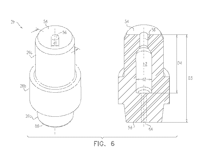
Reference is now made to Fig. 6, which is a schematic illustration of septum
26, in
accordance with some embodiments of the present invention. Septum 26, which
was briefly
described above with reference to Fig. 2, is described in detail hereinbelow,
with reference to Fig.
19
CA 03072311 2020-02-06
WO 2019/030684
PCT/IB2018/055961
6.
Septum 26, which may be made of a resilient material such as an elastomer,
comprises a
proximal face 54, a distal face 58, and a longitudinal body extending between
proximal face 54
and distal face 58. For example, as described above with reference to Fig. 2,
the longitudinal body
of the septum may comprise a proximal portion 26c, a wider middle portion 26b,
and a distal
portion 26a. Typically, distal face 58 is disposed at least 7 mm from proximal
face 54 of the
septum, i.e., the distance D3 between the proximal and distal faces of the
septum is at least 7 mm.
For example, distance D3 may be between 7 and 25 mm, such as between 10 and 20
mm, e.g.,
13.5 mm.
Proximal face 54 is shaped to define an aperture 56 having a diameter dl that
is less than
the diameter of the distal end of needle 32. For example, since a typical
needle diameter is 1.5
mm, diameter dl may be less than 1.5 mm. Due to the elastomeric properties of
the septum, upon
the needle passing through the aperture, the needle forces the aperture to
expand to a diameter that
is just large enough to accommodate the needle. The septum thus fittingly
slides over the needle,
and, subsequently to the sliding of the septum, proximal face 54 continues to
press against the
needle. There is thus little risk of any fluid escaping through the aperture.
Septum 26 is shaped to define a cavity 62 that is joined to aperture 56.
Typically, cavity
62 is relatively long; for example, the cavity may extend at least 5 mm from
the proximal face of
the septum, i.e., the distance D4 between proximal face 54 and the distal end
of the cavity may be
at least 5 mm. For example, distance D4 may be between 5 and 15 mm, such as 9
mm. The
relatively large length of cavity 62 facilitates housing the distal end of
needle 32 while the syringe
adaptor is not coupled to the fluid-port adaptor. Advantageously, the diameter
d2 of cavity 62 is
typically larger than diameter dl of the aperture; for example, diameter d2
may be greater than 0.8
mm, such as between 1 and 3 mm. (Typically, diameter d2 is 0.3-0.7 mm, such as
0.5 mm, greater
than the diameter of the distal end of needle 32; for example, for a needle
diameter of 1.5 mm, d2
may be 2 mm.) The relatively large width of cavity 62 generally reduces the
friction between the
needle and the septum as the needle passes through the septum.
As the septum housing slides proximally within the body lumen of the syringe
adaptor
during the coupling of the syringe adaptor with the fluid-port adaptor, the
distal end of the needle,
which was contained heretofore in cavity 62, pierces through a portion of the
septum that lies
between cavity 62 and distal face 58, thus creating a passage 64 through which
the needle passes.
Subsequently, fluid is transferred via the needle. Following the transfer of
fluid, during the
uncoupling of the two adaptors, the septum housing slides distally within the
body lumen, such
CA 03072311 2020-02-06
WO 2019/030684
PCT/IB2018/055961
that the distal end of the needle returns to cavity 62. As the needle is
withdrawn (via the distal
sliding of the septum housing), passage 64 closes due to the elasticity of
septum 26, such that the
distal end of the needle is sealed within the septum. Typically, the septum
provides an airtight
seal around the needle opening, such that the user is inhibited from
improperly using the syringe
while the syringe adaptor is not connected to the fluid port adaptor.
ALTERNATE FLUID-PORT ADAPTORS
The paragraphs below describe various fluid-port adaptors that may be used,
alternatively
to vial adaptor 39, with syringe adaptor 20. (It is noted that vial adaptor 39
may include any
suitable features of these fluid-port adaptors described below, mutatis
mutandis.)
Reference is first made to Figs. 7A-B, which are schematic illustration of an
infusion-set
adaptor 72, in accordance with some embodiments of the present invention. Fig.
7B shows a
longitudinal cross-section through adaptor 72, taken as indicated in Fig. 7A.
Infusion-set adaptor 72 is similar to vial adaptor 39, in that infusion-set
adaptor 72 is
configured to couple to syringe adaptor 20, generally as described above with
reference to the
preceding figures. Infusion-set adaptor 72 differs from vial adaptor 39,
however, in that infusion-
set adaptor 72 is configured for releasable connection to a fluid port (e.g.,
a side port) of an
intravenous (IV) cannula, e.g., at an injection site, rather than for
connection to a vial.
Similarly to vial adaptor 39, infusion-set adaptor 72 comprises, at its front
end, syringe-
adaptor connecting portion 40, which in turn comprises cylindrical septum
housing 43. Septum
42 may be sealingly mounted onto a seat 76 located near the opening of septum
housing 43, or
alternatively mounted within septum housing 43 in any other suitable manner.
The outer surface
of septum housing 43 is shaped to define one or more circumferential (e.g.,
circular) ridges 44,
and syringe-adaptor connecting portion 40 may be further shaped to define
ledge 55, as described
above with reference to the preceding figures.
Unlike vial adaptor 39, however, infusion-set adaptor 72 comprises, at its
rear end, a fluid-
port connecting portion comprising a cylindrical tube 74. Cylindrical tube 74
is configured to
connect infusion-set adaptor 72 to the fluid port of an IV cannula, by passing
into or over the fluid
port, e.g., as shown in Fig. 29 of US Patent 8,122,923, whose disclosure is
incorporated herein by
reference.
Typically, a cylindrical intermediate portion 78 is disposed between syringe-
adaptor
connecting portion 40 and cylindrical tube 74. Passage 49 extends axially
through tube 74,
intermediate portion 78, and syringe-adaptor connecting portion 40, thus
allowing fluid flow
21
CA 03072311 2020-02-06
WO 2019/030684
PCT/IB2018/055961
through infusion-set adaptor 72 when septum 42 is suitably pierced. In some
embodiments,
infusion-set adaptor 72 (excluding septum 42) is integrally formed, and is
side-to-side symmetric
along its central longitudinal axis 80.
Reference is now made to Figs. 8A-B, which are schematic illustrations of a
spike port
adaptor 82, in accordance with some embodiments of the present invention. Fig.
8B shows a
longitudinal cross-section through adaptor 82, taken as indicated in Fig. 8A.
Spike port adaptor 82 is similar to vial adaptor 39, in that spike port
adaptor 82 is
configured to couple to syringe adaptor 20, generally as described above with
reference to the
preceding figures. Spike port adaptor 82 differs from vial adaptor 39,
however, in that spike port
adaptor 82 is configured for releasable connection to a fluid port of an IV
bag (or "receptacle").
One commercial product that may be modified to embody spike port adaptor 82 is
the
Tevadaptor Spike Port Adaptor.
Similarly to vial adaptor 39, spike port adaptor 82 comprises syringe-adaptor
connecting
portion 40, which comprises cylindrical septum housing 43 shaped to define a
plurality of ridges
44, along with an intermediate portion 78 and a fluid-port connecting portion
comprising hollow
spike 47. In spike port adaptor 82, however, spike 47 extends obliquely to, or
perpendicularly to,
vector 53, which is normal to the opening of the septum housing. Spike 47
connects the spike port
adaptor to a fluid port of an IV bag, by piercing a closure disposed at the
opening of the fluid port,
as shown, for example, in Fig. 20 of US Patent 8,122,923, whose disclosure is
incorporated herein
by reference.
Typically, spike 47 is formed of plastic. Spike 47 comprises a main body
portion 88, which
is shaped to define a tapered end 90. In some embodiments, the outer surface
of main body portion
88 includes one or more finger grip surfaces (not shown). To further
facilitate the gripping of the
main body portion, the outer surface of the main body portion may be shaped to
define a plurality
of bumps or other protrusions, and/or may be coated with a coarse, grip-
enhancing coating.
In some embodiments, tapered end 90 is shaped to define two apertures: a first
aperture 92,
which communicates with passage 49 (which extends, e.g., in an L-shape, from
first aperture 92
to the opening of septum housing 43), and a second aperture 94, which
communicates with a
second passage 96, which in turn communicates with the lumen of a hollow
flexible tube 84
(described below). In some embodiments, passage 49 includes a side passage
100, which
terminates at a third aperture 104. Side passage 100 may be positioned, for
example, within
tapered end 90, or between tapered end 90 and syringe-adaptor connecting
portion 40. In such
embodiments, air may flow out of passage 49 via side passage 100 and third
aperture 104. Side
22
CA 03072311 2020-02-06
WO 2019/030684
PCT/IB2018/055961
passage 100 may be provided with a check valve 102, which enforces a
unidirectional, inward
flow of air therethrough. In particular, as fluid is drawn from the IV bag via
spike 47, air from the
ambient environment passes through check valve 102, through passage 49, and
into the IV bag.
In some embodiments, a hydrophobic membrane, which allows the flow of air
therethrough but
inhibits the flow of fluid therethrough, is positioned within side passage
100, e.g., adjacent to
check valve 102.
Tube 84, which is typically formed from plastic, is typically coupled to a
standard clamp
86, which is commercially available from various manufacturers, such as Qosina
of Italy. Tube
84 is coupled, at its front end, to intermediate portion 78 or to main body
portion 88 of spike 47,
such that the lumen of tube 84 is in fluid communication with second passage
96 (as shown in Fig.
8B) or with passage 49.
Typically, a sealing assembly 98 is attached to the rear end of tube 84.
Sealing assembly
98 is configured to seal tube 84 during use of the drug mixing device, and may
be removed from
tube 84 when the IV bag is connected directly to an infusion set spike for
infusion of the fluid
contained therein to a patient.
In some embodiments, instead of spike port adaptor 82 comprising tube 84,
intermediate
portion 78 or main body portion 88 may be shaped to define an outlet port
configured to receive
another spike element. Such an outlet port may, for example, be formed from an
elastomeric
element attached to intermediate portion 78 or main body portion 88. A
separate tube, having
another spike disposed at its front end, may then be inserted into the outlet
port, such as to establish
fluid communication between the tube and passage 49 and/or second passage 96.
More generally, the scope of the present invention includes fluid-port
adaptors configured
for connection to any suitable types of fluid ports, by virtue of comprising
any suitable types of
fluid-port connectors. For example, a fluid-port connector may comprise a
hollow male or female
threaded extension that may be screwed into, or over, a corresponding threaded
fluid port of a fluid
container.
It will be appreciated by persons skilled in the art that the present
invention is not limited
to what has been particularly shown and described hereinabove. Rather, the
scope of embodiments
of the present invention includes both combinations and subcombinations of the
various features
described hereinabove, as well as variations and modifications thereof that
are not in the prior art,
which would occur to persons skilled in the art upon reading the foregoing
description. Documents
incorporated by reference in the present patent application are to be
considered an integral part of
the application except that to the extent any terms are defined in these
incorporated documents in
23
CA 03072311 2020-02-06
WO 2019/030684 PCT/IB2018/055961
a manner that conflicts with the definitions made explicitly or implicitly in
the present
specification, only the definitions in the present specification should be
considered.
24