Note: Descriptions are shown in the official language in which they were submitted.
LIGHT BASED THERAPY DEVICES AND METHODS
BACKGROUND OF THE INVENTION
[0002] The present invention relates to designs, systems and methods of a
light
therapy device.
[0003] The concept of using light energy to treat human tissues has
emerged in
the last few decades. Radiation, UV, and broad spectrum light have all been
employed
therapeutically and efficaciously, enjoying wide acceptance in the medical
community. One version of this, called Low Level Light Therapy (LLLT), uses a
wide variety of wavelengths in the visible and near-infrared spectrum to
generate a
tissue response in a process that has become known as photobiomodulation. The
potential list of applications for LLLT is enormous: everything from dental
treatments
to pain control and accelerated wound healing has been studied with promising
results. Given the low incidence of side effects, ability to target specific
tissues, and
the relative ease of treatment, patient and physician acceptance and adoption
of these
technologies is justifiably growing.
[0004] The method of delivery has been problematic, however. Due to its
coherent, single wavelength output and directionality, the laser diode is a
commonly
used light source, allowing practitioners to easily direct the beam to the
target.
Another potential source, the light emitting diode (LED), can also be used to
generate
light in a specific band of wavelengths, but with a much broader emission
pattern. For
completeness, we note that it is possible to generate either narrow or broad
spectral
ranges with a white light source and a filter. It has yet to be established
whether
narrow or broader emission spectra or coherent or incoherent light is more
effective to
induce photobiomodulation. The issue with all of the light sources is that
human
tissue, such as skin, can be highly reflecting. Furthermore, the presence of
hair on the
1
Date Recue/Date Received 2020-06-22
CA 03072319 2020-02-06
WO 2019/035895
PCT/US2018/000162
skin can cause significant absorption of the light intended for the skin.
These two
effects make it difficult to precisely control dosing during therapeutic
applications.
Since light can be scattered, absorbed, transmitted, or reflected, the light
applied
during certain LLLT applications should either be on the surface of the
target, or be
very close to be absorbed.
[0005] One major application of LLLT is to treat hair loss. Also known as
alopecia, hair loss can be found in every country and has unfavorable social
connotations in all cultures worldwide. Male pattern hair loss, or
androgenetic
alopecia, accounts for 95% of alopecia in males, with 70% of American men
experiencing some form of hair loss by age 35. Female hair loss, while it is
often
more complex in etiology, affects a similarly large portion of women
worldwide, with
some estimates ranging from 1:4 in the United States (25%), to over 80% of
women
past the age of 60 (when hormones like estrogen drop). There is no cure for
male or
female pattern hair loss.
[0006] Unfortunately, the list of proven medical therapies that will help
even the
most common causes of hair loss is a short one. In the United States, men and
women
can use minoxidil (2% and 5%) in both liquid and foam forms, but this
medication
requires twice daily application and is considered distasteful and
inconvenient by
many. Men have the additional benefit of being able to use the daily oral
medication
finasteride, which can be extremely effective. There is a widespread
misunderstanding
regarding its side effect profile, however, since it can transiently affect
libido (2.1-
3.8% incidence), which hinders its adoption. Surgical hair restoration is
effective, but
it is expensive, and, as a result, unavailable to many patients.
[0007] Photobiomodulation is a recent addition to the existing FDA-approved
hair
loss armarnentarium. LLLT in the wavelengths of 614-624nm, 668-684nm, 751-
772nm, and 813-846nm, has been proven to reduce inflammation in the scalp,
stimulate the release of growth factors in the hair follicle, up-regulate the
production
of ATP (the energy source for the cell), and increase oxygen levels and blood
flow via
a vasodilatory effect. Devices of all sorts including combs, helmets, handheld
"massager-type" units, and hoods all have gained 510K clearance to be sold
with the
claim that they grow hair.
2
CA 03072319 2020-02-06
WO 2019/035895
PCT/US2018/000162
[0008] Currently, none of the published studies of these devices conforms
to the
wavelengths of light known to produce increased cellular activity in the hair
follicle,
and few of them even produce light within these known wavelength ranges.
Furthermore, many light therapy devices deliver light to the skin from a
distance or
from above the hair. Such light may be absorbed by the presence of hair
follicles,
thereby limiting the available dose. Even if hair is not initially present, if
hair growth
occurs during the use of such LLLT devices, the light therapy process will be
self-
limiting. For these reasons, many existing LLLT device solutions for hair
growth are
sub-optimal at best, and ineffective at worst. Also, dosing time and frequency
recommendations vary among devices, leading to sub-optimal treatments. Another
concern with conventional devices arises when the device causes heating of the
targeted region of the scalp, excessive heating can decrease the results of
the therapy,
leading to the potential for sub-optimal dosing. Based on the above, there is
room for
improved systems, devices, and methods for application of LLLT therapy.
BRIEF SUMMARY OF THE INVENTION
[0009] A variation of the improved systems, methods, and devices for
providing
LLLT devices. In one aspect, such devices are suited for hair growth by
applying light
delivery to the skin using one or more illumination sources. For example, the
illumination source can comprise coherent light (e.g., laser), incoherent
(e.g. LED,
white light plus filter), filtered light, or a combination thereof. The
illumination
provided by the illumination source can be of a wavelength or wavelength range
that
is of beneficial and therapeutic value. The illumination source can comprise a
source
that transmits light from another location (e.g., an optical fiber) that
generates the
illumination, or the illumination source can also directly generate the
illumination
(such as an LED component). In some variations, the illumination source
delivers the
light to the treatment area via direct contact with the skin. In other
variations, the
illumination source delivers light to the treatment area just above the skin.
In certain
applications, positioning of the illumination source close to the tissue being
treated
such that the light delivery bypasses the interference that even short hair
shafts above
the skin create. Such close delivery allows for predicable and known dosing
intensity
and distribution, which enables standardized dosing. In certain variations of
the
3
CA 03072319 2020-02-06
WO 2019/035895
PCT/US2018/000162
devices and methods, it is desirable to prevent heat from increasing at the
treated
region. Therefore, the heat generated by the light sources can be kept away
from the
skin and to avoid a significant increase the temperature of the skin. A
cooling scheme
can also be used to either preserve the output power and efficiency of the
light source
itself or to cool the treated tissue.
[0010] Variations of the device and system include illumination sources
that are
shaped for patient comfort and/or to distribute the light around the delivery
or contact
point. In additional variations, a projecting element that includes or carries
the
illumination source is actuated so as to allow conformal contact with the
skin. In an
exemplary embodiment, an array of such projection elements having illumination
sources are used to illuminate a substantial area of skin, such as the scalp.
An
advanced passive cooling scheme is used to preserve the output power and
efficiency
of the light sources. Advantageously, the present invention delivers light
directly to
the skin bypassing interference from hair follicles, thereby allowing for a
known
dosing intensity and distribution. The configurations described herein can
provide an
improvement in light delivery to the targeted region ¨ one that reduces loss
of energy
to undesired absorption and reflection and that ensures maximum absorption by
the
target tissue, thereby enabling standardized dosing. Variations of the devices
described herein can also allow delivery of light at or very close to the
skin/scalp,
which allows bypassing the interference that even short hair shafts above the
skin
create, and minimizing the effect of reflection. Second, it would deliver
light in one of
the four optimum wavelength ranges.
[0011] Variations of the device also allow for a hands-free, cordless, and
portable,
with an interactive feedback component that allows a patient to monitor their
progress, further improving adherence with the treatment regimen. Such
variations
also time the treatments and help patients manage dosing frequency with a
minimal
amount of external visibility. Cell proliferation (i.e. growth of hair) is
optimized with
low doses over longer periods of time. So, the present LLLI device allows for
convenient and frequent dosing (at least 2-3 times per week, if not daily).
4
BRIEF DESCRIPTION OF THE SEVERAL VIEWS OF THE DRAWINGS
[0012] Non-limiting and non-exhaustive embodiments of the present
invention are
described with reference to the following drawings. In the drawings, like
reference
numerals refer to like parts throughout the various figures unless otherwise
specified.
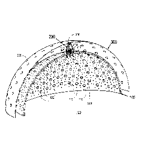
[0013] Figure 1. Illustrates a perspective view of light therapy device
for the
scalp.
[0014] Figure 2. Illustrates a cross-section, perspective view of light
therapy
device for the scalp with flexible PCB.
[0015] Figure 3. Illustrates a close-up, cross-section, perspective view
of
flexible PCB, illumination assembly and dome alignment.
[0016] Figure 4. Illustrates a perspective view of flattened flexible PCB
and
dome prior to assembly.
[0017] Figure 5. Illustrates a close-up, cut-away, perspective view of
flexible
PCB, illumination assembly and dome alignment.
[0018] Figure 6. Illustrates a perspective, cutaway view of alternative
embodiment of light therapy device for the scalp.
[0019] Figure 7. Illustrates a close-up, cross-section, perspective view
of the
illumination assembly and dome alignment.
[0020] Figure 8. Illustrates a close-up, cut-away, perspective view of
rigid PCB,
ferrule, and alignment structure.
DETAILED DESCRIPTION OF THE INVENTION
[0021] For a better understanding of the present invention, reference
will be made
to the following Description of the Embodiments, which is to be read in
association
with the accompanying drawings, which are incorporated in and constitute a
part of
this specification, show certain aspects of the subject matter disclosed
herein and,
together with the description, help explain some of the principles associated
with the
disclosed implementations.
[0022]
The term "plurality", as used herein, is defined as two or as more than two.
The
term "another", as used herein, is defined as at least a second or more. The
terms
"including" and/or "having", as used herein, are defined as comprising. (i.e.,
open
Date Recue/Date Received 2020-06-22
language). The term "coupled", as used herein, is defined as connected,
although not
necessarily directly, and not necessarily mechanically.
[0023] Reference throughout this document to "some embodiments", "one
embodiment", "certain embodiments", and "an embodiment" or similar terms means
that a particular feature, structure, or characteristic described in
connection with the
=
embodiment is included in at least one embodiment of the present invention.
Thus, the
appearances of such phrases or in various places throughout this specification
are not
necessarily all referring to the same embodiment. Furthermore, the particular
features,
structures, or characteristics may be combined in any suitable manner in one
or more
embodiments without limitation.
[0024] The term "or" as used herein is to be interpreted as an
inclusive or
meaning any one or any combination. Therefore, "A, B or C" means any of the
following: "A; B; C; A and B; A and C; B and C; A, B and C". An exception to
this
definition will occur only when a combination of elements, functions, steps or
acts are
in some way inherently mutually exclusive.
[0025] Figure 1 presents a perspective view of an exemplary light
therapy device
for the scalp. The exemplary light therapy device discussed below is intended
for
illustrative purposes only. Variations of the light therapy device 100 are
within the
scope of this disclosure for other applications of light therapy to a body
organ. A body
organ can comprise
skin or tissue that is part of such a
body structure.
[0026] A dome 102 is configured with a plurality of ferrule openings
110 and a
plurality of holes 120 for a plurality of optical fiber or illumination
assemblies 200,
only one of which is shown for clarity, extending between an interior surface
104 to
an exterior surface 106 of the dome 102. As noted herein, the illumination
source can
comprise an optical fiber having a source of illumination at the end of the
fiber closest
to the tissue. Alternatively, or in combination, the devices can include one
or more
projection elements, where each projection element comprises a distal portion
having
an illumination source configured to direct the electromagnetic energy to the
body
organ.
6
Date Recue/Date Received 2020-08-31
[0027] The ferrule openings 110 are configured with a taper from the
interior
surface 104 to the exterior surface 106 adapted to operably connect a tapered
end 222
of a base 220 therein as shown in Figure 3. The plurality of holes 120 are
located
adjacent to a ferrule opening 110 and configured in a dimension to be spaced
apart
such that the light from a plurality of optical illumination assemblies 200
disposed in
the ferrule opening 110 and holes 120 provide sufficient light therapy
coverage over a
treatment area and/or surface of the body organ in a substantially uniform
manner.
The optical fibers assemblies or illumination assemblies are independently
biased so
that the tips 216 of the optical fibers/projection elements 210 of each
assembly can
better conform to the shape of the head and/or be operably in contact with the
body
organ. This independent biasing allows for an improved device with multiple
illumination sources that achieve a perpendicular approach to the target
tissue. The
independent biasing allows each assembly to conform to a contoured surface of
the
body organ without significantly affecting adjacent assemblies, which
increases the
ability of the adjacent assemblies to irradiate the tissue in a normal
direction.
Lamberts cosine law predicts that an illumination beam that is perpendicular
to its
target can achieve a higher rate of irradiance. In contrast, a beam that is
offset from a
perpendicular approach will have a greater degree of reflection off of the
target area.
Which means that a curved or contoured surface like the scalp (or any other
contoured
body organ) has a greater chance of being irradiated if the sources of
illumination
irradiate the scalp at a perpendicular angle. The independent biasing of the
plurality of
illumination assemblies can decrease the overall reflectance of light from a
curved
surface and increase the effectiveness or uniformity of the LLLT applied.
[0028] The optical fibers/projection elements 210 are associated and
optically
coupled to a light source 310 situated above the dome 102. Advantageously,
light
therapy device 100 may dissipate any heat generated by a plurality of the
light sources
310 to be located away from the body organ, i.e. the skin of a patient's scalp
such as,
for example, the heat is dissipated through vents 410, 510 as shown in Figures
5 and
6. Optionally, a cooling source 402 can be fluidly coupled to the device to
cool any
component within the device and/or tissue.
[0029] Referring to Figures 1-4, the light therapy device 100 may be
formed
having the light sources 310 disposed on a flexible printed circuit board
(flex PCB)
7
Date Recue/Date Received 2020-08-31
CA 03072319 2020-02-06
WO 2019/035895
PCT/US2018/000162
300. The flex PCB 300 is configured to be located adjacent the dome 102 and
wrapped at a predetermined offset distance and/or gap 202 around the dome 102,
as
shown in Figure 2. The predetermined offset distance and/or gap 202 is
configured by
the dimension of the fiber/illumination assembly 200. The flex PCB 300 may be
formed of suitable materials and/ or construction that allows bending in a
direction
parallel to, and flexing in a direction normal to, the surface of the dome
102. The flex
PCB 300 is configured with an electronic circuit to energize the light sources
310.
[0030] Figure 3 illustrates the link from light source to skin achieved by
the light
therapy device 100. The optical fibers/projection elements 210 are coupled to
the light
source 310 by means of an optical fiber/illumination assembly 200. The optical
fiber/illumination assembly 200 is attached at one end to the dome 102 and at
the
other end adjacent to the light sources 310 operably connected by the
electrical circuit
in the flex PCB 300. For example, one or more slots 320 may be formed in the
flex
PCB 300 configured to engage one or more locking posts 236 of the ferrule 230
as
shown in Figures 1-3. In this manner, the optical fiber/illumination assembly
200 may
be located adjacent to the light sources 310 thereby optically connecting so
as to
transmit light along the optical fibers/projection elements 210, 212 to the
body organ.
[0031] According to an embodiment of the light therapy device 100, as is
illustrated in Figures 1- 3. For convenience, the variations are discussed as
having an
optical fiber. However, a projection element with an illumination source can
be used
in place of or in addition to the optical fiber assembly or fiber itself.
[0032] In Figures 1-3, the optical fiber assembly 200 comprises a base 220,
a
ferrule 230, a spring 240, a fiber collar 250, and one or more optical fibers
210. The
base 220 may be configured with a base tube 224 along its longitudinal length,
a
tapered end 222 to be received in the dome 102, and a projecting proximal end
223
with a guide shaft 225 adapted to receive an inner tube 232 of the ferrule 230
therein.
The ferrule 230 also may be configured with ferrule tube 234 along its
longitudinal
length for passing an axial optical fiber 212 there-through, whereby this
centrally
located, axial optical fiber 212 is allowed to move within the openings 224,
234 (e.g.
to travel up and down) by means of aligning the pair of concentric base and
ferrule
tubes 224, 234 formed in the base 220 and ferrule 230. The other optical
fibers 210
may be connected at one end in one or more fiber holes 238 formed in a base
portion
8
CA 03072319 2020-02-06
WO 2019/035895
PCT/US2018/000162
232 of the ferrule 230 and, at points along the optical fibers 210, by a fiber
collar 250,
whereby these optical fibers 210 are fixed to the ferrule 230 in the base
portion 232,
are aligned with and pass through the plurality of fiber openings 220 in the
dome 102
so as to slide freely there-through. Consequently, the free ends 216 of the
optical
fibers 210 and axial optical fiber 212 are configured to operatively connect
to the
body organ. Additionally, the base 220 and ferrule 230 are connected to each
other by
means of a biasing element 240 (e.g. a spring) that functions to provide a
force to
fiber optical assembly 200 so as to gently push the free ends 216 of each
optical fibers
210, 212 into contact with the scalp. Accordingly, the light therapy device
100
advantageously provides light in direct contact with the body organ so as to
guide
light for light therapy directly from the light source 310 to the scalp or
skin.
[0033] As shown in Figure 3, the ferrule 230 is assembled to the base 224
by
means of a spring 240 configured to be received in recesses 270 formed in the
base
and ferrule, whereby the spring 240 may snap-fit into the recesses 270 for
ease of
assembly. Once assembled, the spring 240 is configured to provide sufficient
retention force on the recesses 270 such that the spring 240 remains attached
and the
biasing force allows the inner tube 232 of ferrule 230 to move in the guide
shaft 225
of the base 220 during extension, for example, when connecting directly to the
contour of the body organ that forces free ends 216 of the optical fibers 210,
212
away from the body organ. The base 224 comprises a tapered end 222, which is
press
fit into a tapered hole 110 extending between the interior and exterior
surfaces 104,
106 formed in the dome 102. The spring 240 locks the proximal end 223 of the
base
220 above the dome 102 thereby preventing the base 220 from disengaging or
otherwise falling out of the tapered hole 110. The inner tube 232 is
configured to slide
into the guide shaft 225 of the base 220 thereby providing passive alignment
of the
axial optical fiber 212 relative to the tapered hole 110 in the dome 102.
Similarly, the
fiber collar 250 is configured to secure and/or align the optical fibers 210
in a precise
array such that the optical fibers 210 may be inserted insert into, and move
freely
within, the fiber holes 120 formed in the dome 102.
[0034] As is illustrated in Figure 3, the ferrule 230 is aligned to the
light source
310 by means of slots 320 in the flex PCB 300. The slots 336 engage locking
posts
236 disposed on a top end of a body portion 234 of the ferrule 230. The posts
236 are
9
CA 03072319 2020-02-06
WO 2019/035895
PCT/US2018/000162
configured to engage the slots 336 positioned in a circular array such that,
once
engaged, the posts 236 may rotate within the slots 336 while keeping the
ferrule 230
centered with respect to the light source 310. The fiber assembly 200 is
adjustable as
the posts 236 and slots 336 also are configured to provide for rotational
alignment of
the circular array of optical fibers 210 to the fiber holes 120 formed in the
dome 102,
as shown in Figure 3.
[0035] Referring to Figures 1-3, the body portion 232 of the ferrule 230
comprises
multiple fiber holes 238 which align the optical fibers 210 to the light
source 310. In
this embodiment, seven fibers are aligned to a light emitting diode (LED) 310:
one
axial optical fiber 212 along a lengthwise, longitudinal and/or vertical axis
of the
ferrule 230 and six optical fibers 210 arranged in a circular array 214 at an
angle to
the vertical axis sufficient to effectuate fiber-coupled light. At the
proximal end each
of the seven fibers, specifically, the optical fibers 210 and the axial
optical fiber 212
are configured to capture a significant fraction of the optical power emitted
by the
light source 310. Furthermore, the positioning of the optical fibers 210 in
the base
portion 214 and the axial optical fiber 212 in the base tube 224 and ferrule
tube 234 is
such that the fiber-coupled light is substantially evenly distributed amongst
the seven
fibers. The axial optical fiber 212 is secured within a hole formed by the
base tube
224 and the ferule tube 234 in the center of the ferrule 230, thereby
remaining parallel
to the axis of motion. Each optical fiber 210 in the circular fiber array 214
is bent by
means of a fiber collar 250 such that the free distal end 216 is substantially
parallel to
the axial optical fiber 212. In this way, all seven fibers 210, 212 may travel
freely in a
direction parallel to the axis of the ferrule 230 that is arranged
substantially normal to
the surface of the dome 102.
[0036] As shown in Figure 1, the light therapy device 100 consists of
matching a
circular array 214 of optical fibers 120 to any hole array 228 of the fiber
holes 120
surrounding the tapered hole 110 in the dome 102. The circular fiber array 214
penetrates the matching hole array 228 such that all seven fibers 210, 212
travel
simultaneously. As shown in Figures 2 and 3, the light therapy device 100 may
be
configured to limit fiber travel in a dimension in extent toward the distal
end 216 by
the mating of the concentric base 228 and ferrule tubes 234 formed in the base
220
and ferrule 230 (maximum fiber length below the dome 102), and at the proximal
end
CA 03072319 2020-02-06
WO 2019/035895
PCT/US2018/000162
223 by the interior surface 104 of the dome 102 (fiber tips 216 flush with the
dome
102). The tips 216 of the optical fibers 120 and axial optical fiber 212 may
be shaped
for patient comfort. The shape of the fiber tips 216 can also be configured to
spread
the light laterally to an area larger than the fiber diameter.
[0037] For example, as shown in Figure 2, when a patient's head is placed
within
the dome 102 adjacent the inner surface 104 the scalp engages the extending
fibers
120 and axial optical fiber 212 and pushes these radially outward, with each
fiber
array 214 flexing individually in a particular hole array 228 depending on the
particular shape of the scalp. In this way, the fiber arrays 214 of the light
therapy
device 100 can conformally and simultaneously contact the entirety of the
scalp. The
optical fibers 120 and the axial optical fiber 212 are configured flexible to
allow
bending to accommodate a shift of position of the scalp with respect to the
interior
surface 104 of the dome 102, for example, bending slightly off-axis when moved
on
the scalp. Consequently, the light therapy device 100 may be integrated into a
portable, wearable helmet as shown in Figure 5.
[0038] Figure 4 displays one section of the flex PCB 300 in position above
the
dome 102 and prior to assembly. In an exemplary embodiment, the flex PCB 300
comprises a thin, flexible material that is cut in lines of relief to bend
and/or to
conform to the shape of the dome 102.
[0039] The dome 102 is affixed to a base 400, as illustrated in Figure 5.
The base
contains a rigid PCB (not shown), which contains the control electronics. The
flexible
PCB containing the light sources is connected to the rigid PCB. The base can
also
contain one or more batteries (also not shown) which are capable of powering
the
device for the duration of the treatment. An outer shell 500, which may itself
be either
flexible or rigid, protects the flex PCB 300 and other elements of the light
therapy
device 100 from physical and other interference. The heat from the light
sources and
associated control electronics are dissipated by means of vents in the base
410 and
outer shell 510, which draw cool air in from the bottom and allow heated air
to escape
from the top.
[0040] Referring to Figures 6 through 8, another embodiment of a light
therapy
device 100 provides direct contact between the body organ and the optical
fiber 120
using an alternative design. The light therapy device 100 comprises a ferrule
235, an
11
array of light sources 310 and associated control electronics (not shown)
mounted on
a rigid PCB 330 that may be secured to the base 400 at the back of the dome
102. The
dome 102 similarly contains tapered holes 110 formed by the opening extending
between an interior surface 104 and an exterior surface 106. The tapered holes
110 are
configured to receive a plurality of optical fiber assemblies 200, only one of
which is
shown in Figure 6. Axial optical fibers 212 disposed in the ferrule 235 are
configured
so that the tips 216 of the axial optical fibers 212 conform to the shape of
the head
and/or body organ. The axial optical fibers 212 are coupled to the light
sources 310 at
the back of the dome 102 by means of an adapter 630. The adapter 630 may be
configured to fasten and secure to a rigid printed circuit board (PCB) 330 by
interlocking tabs of a leaf spring latch 620 in a notch formed in the adapter
630 as
shown in Figure 8. The rigid PCB 330 is configured with the electrical circuit
for the
light sources 310 and control electronics. A source of electrical power may be
configured into the rigid PCB 330 assembly such as, for example, batteries
(not
shown). The source of electrical power is configured capable of powering the
light
therapy device 100 for the duration of the treatment.
[0041] As shown in Figures 6 and 7. the dome 102 is affixed to a base
400 and an
outer shell 500. The heat from the light sources 310 and associated driver
electronics
are mitigated by means of one or more vents 410 and 510 in the helmet. These
one or
more vents 410 in the helmet base, as shown in Figure 5 are configured to draw
cool
air in, while vents 510 in the helmet top allow heated air to escape. The
helmet may
be formed with an outer shell configured in the helmet base and helmet top
portions.
The outer shell may be formed flexible andfor rigid, as well as formed to
protect the
flex PCB 300 and other elements of the light therapy device 100 from physical
and
other interference.
[0042] As is illustrated in Figure 7, the light therapy device 100
comprises an
optical fiber assembly 200 attached using tapered holes 212 in the dome 102.
The
fiber assembly comprises a base 220, a ferrule 235, a flange guide 237, a
spring 240
and one or more optical axial optical fibers 212. The ferrule 235 comprises a
bent tube
238 formed in the flange guide 237 that directs the axial optical fiber 212
toward the
rear of the dome 102 for connecting to the rigid PCB 330 using the adapter
600. The
movement or action of the axial optical fiber 212can be achieved by means of
inner
12
Date Recue/Date Received 2020-08-31
CA 03072319 2020-02-06
WO 2019/035895
PCT/US2018/000162
232 and outer 220 tubes formed by the base 222 and ferrule 235 which slide
concentrically with respect to each other. The inner 232 and outer 220 tubes
are
biased by means of a biasing element 240, e.g. a spring. The light therapy
device 100
may be configured with a biasing force selected so that an aggregate spring
force of
all optical fiber assemblies 200 allow all axial optical fibers 212 to make
contact with
the scalp simultaneously.
[0043] As shown in Figures 6-8, the ferrule 235 is attached to the dome 102
by
means of a tapered base 222 and is configured to operably connect to the
tapered
holes 110. The tapered base 222 comprises slight protrusions 226 configured to
slidably, snap fit to a recesses 112 in the tapered holes 110, thereby
allowing rotation,
and preventing the tapered base 222 from dislodging and/or otherwise falling
out. The
ferrule 230 is secured to the tapered base 222 by means of a spring 240
configured to
slidably, snap fit into a recess 270a configured in the tapered base 222 and a
recess
270b formed in the ferrule 235. Once assembled, the biasing element 240 (e.g.
spring)
is configured to have sufficient retention force on the recesses 270a, 270b
such that it
remains attached at each end during extension.
[0044] As shown in Figure 8, the light therapy device 100 comprises one or
more
arrays of light sources 310 and their control electronics (not shown) mounted
on a
rigid PCB 330, which is secured to the base 400 at the back of the dome 102. A
multi-
fiber ferrule (MFF) 600 can be configured to align the light sources 310 to
each axial
optical fiber 212 disposed in one or more fiber openings or an array of fiber
holes 610
by means of an adapter 630 secured to the rigid PCB 330. The adapter 630 may
be
configured to align the one or more fiber openings 610 over arrays light
sources 310
(e.g. rows of individual light sources 310) and fiber openings 610. The
adapter 630
may connect to align the ends of each of the axial optical fibers 212 disposed
in fiber
openings 610 adjacent to the light source arrays 310 by means of precision
screw
holes 340 in the PCB. The MFF 600 comprises the array of fiber holes 610
populated
by receiving a plurality of axial optical fibers 212 herein. The MFF 600 is
secured to
the adapter 630 by leaf spring latches 620. Advantageously, the latches 620
may act
as leaf springs to center the MFF 600 within the adapter 630, thereby
providing fine
alignment of the fiber openings 610 adjacent to the light sources 310.
13
CA 03072319 2020-02-06
WO 2019/035895
PCT/US2018/000162
[0045] The devices and methods described herein can optimize cell
proliferation
(i.e. growth of hair) with low doses over longer periods of time. It is
believed that
LLLT/PBM creates a dose dependent effect so each dose builds on the previous
treatment (and the Arndt-Schulz Law means that too much dose has suppressive
effects). Therefore, the assemblies disclosed herein allow for a uniform
treatment
applied in a manner that avoids the suppressive effects of over-treatment. In
one
example, it was found that 14-20 minutes applied every few days was sufficient
to
penetrate to the depth of the hair follicle within the skin.
[0046] The previous description of the disclosed embodiments is provided to
enable any person skilled in the art to make or use the present invention.
Various
modifications to these embodiments will be readily apparent to those skilled
in the art,
and the generic principles defined herein can be applied to other embodiments
without
departing from the spirit or scope of the invention. For example, a wide
variety of
materials may be chosen for the various components of the embodiments. It is
therefore desired that the present embodiments be considered in all respects
as
illustrative and not restrictive, reference being made to the appended claims
as well as
the foregoing descriptions to indicate the scope of the invention.
14