Note: Descriptions are shown in the official language in which they were submitted.
CA 03072772 2020-02-11
WO 2019/046801
PCT/US2018/049237
MOLECULAR BACTERIOTHERAPY TO CONTROL SKIN ENZYMATIC ACTIVITY
STATEMENT AS TO FEDERALLY SPONSORED RESEARCH
[0001] This invention was made with Government support under
Grant Nos. AI117673, AR067547, AR062496, and AR064781 awarded by
the National Institutes of Health. The Government has certain
rights in the invention.
CROSS REFERENCE TO RELATED APPLICATIONS
[0002] This application claims priority under 35 U.S.C. 119
from Provisional Application Serial No. 62/553,025, filed August
31, 2017, the disclosures of which are incorporated herein by
reference.
TECHNICAL FIELD
[0003] The disclosure relates to composition and methods to
treat dermatological diseases and disorders and to composition that
modulate skin barrier permeability.
MICROORGANISM DEPOSIT
[0004] Exemplary microorganisms of the disclosure
(Staphylococcus epidermidis All, Staphylococcus hominis C5,
Staphylococcus hominis A9 and Staphylococcus warneri G2) were
deposited on August 28, 2018 with the American Type Culture
Collection, 10801 University Boulevard, Manassas, Va. 20110-2209,
as ATCC Number ________ (strain designation S. epidermidis All
81618, deposited August 28, 2018), ATCC Number _________ (strain
designation S. hominis C5 81618, deposited August 28, 2018), ATCC
Number _______ (strain designation S. hominis A9 81618, deposited
August 28, 2018) and as ATCC Number ________ (strain designation S.
warneri G2 81618, deposited August 28, 2018) under the Budapest
Treaty. This deposit will be maintained at an authorized depository
and replaced in the event of mutation, non-viability or destruction
for a period of at least five years after the most recent request
for release of a sample was received by the depository, for a
period of at least thirty years after the date of the deposit, or
during the enforceable life of the related patent, whichever period
is longest. All restrictions on the availability to the public of
these cell lines will be irrevocably removed upon the issuance of a
patent from the application.
1
CA 03072772 2020-02-11
WO 2019/046801
PCT/US2018/049237
BACKGROUND
[0005] The epidermis is the first line of immune defense and
protects and regulates interactions between microbes and the host
organism. Control of this interaction is important because bacteria
not only reside on the surface where they influence superficial
keratinocytes, but also penetrate below the stratum corneum and
into the dermis where some bacterial species have been shown to
influence immune function. For example, Staphylococcus epidermidis
(S. epidermidis) interacts with epidermal keratinocytes to prevent
toll-like receptor 3-mediated inflammation, recruits mast cells and
T cells, and increases tight junctions and antimicrobial peptide
production. In contrast to the common skin commensal bacteria, S.
epidermidis, Staphylococcus aureus (S. aureus) is often pathogenic
and has a negative influence on skin function. This is especially
evident in skin diseases such as atopic dermatitis (AD) where S.
aureus promotes this disease.
[0006] The microbiome inhabiting the skin of subjects with AD
has been shown to have a decrease in overall microbial diversity
and an increase in S. aureus abundance. Increased S. aureus
colonization has been linked to increased disease severity for
patients with AD. Mechanistically, it is unclear how S. aureus
worsens disease. Several products from S. aureus have been shown to
damage the barrier and/or trigger inflammation. These products
include a-toxin, superantigens, toxic shock syndrome toxin 1,
enterotoxins, protein A, Panton-Valentine leukocidin, exfoliative
toxins, and V8 serine Protease. Because of the potential
pathogenic effects of these molecules, understanding the response
of the skin to S. aureus colonization in the absence of clear
clinical signs of infection is critical to understanding the
pathogenesis of AD and for developing future therapies.
SUMMARY
[0007] The disclosure provides a purified polypeptide
comprising a sequence that is at least 98% identical to SEQ ID
NO:4, 11, 12, 13, 14, 15, 16, or 17 and which inhibits (i) protease
production and/or activity of keratinocytes, (ii) inhibits IL-6
production and/or activity of keratinocytes, (iii) inhibits
production of phenol soluble modulin alpha 3 from Staphylococcus
2
CA 03072772 2020-02-11
WO 2019/046801
PCT/US2018/049237
aureus (S. aureus) and/or (iv) inhibits agr production and/or
activity by S. aureus. In one embodiment, the polypeptide is at
least 98% identical to SEQ ID NO:2. In another embodiment the
polypeptide comprises SEQ ID NO:4, 11, 12, 13, 14, 15, 16, or 17.
In yet another embodiment, the polypeptide consists of SEQ ID NO:4,
11, 12, 13, 14, 15, 16, or 17. In another or further embodiment of
any of the foregoing, the polypeptide comprises one or more D-amino
acids. In yet another or further embodiment, the polypeptide
comprises a compound of Formula I, IA or IB (see below).
[0008] The disclosure also provides a topical formulation
comprising a polypeptide of the disclosure or a compound for
Formula I, IA or IB.
[0009] The disclosure also provides an isolated polynucleotide
encoding a polypeptide of the disclosure. In one embodiment, the
polynucleotide comprises a sequence that hybridizes under stringent
conditions to a polynucleotide consisting of SEQ ID NO:1 or 3 and
encodes a polypeptide comprising SEQ ID NO:4. In another
embodiment, the polynucleotide comprises SEQ ID NO:1 or 3.
[0010] The disclosure also provides vectors comprising a
polynucleotide of the disclosure. The vector can be any suitable
vector for expression in a cell or microbial host.
[0011] The disclosure also provides a recombinant microorganism
comprising a vector or polynucleotide of the disclosure. In some
embodiments, the microorganism does not naturally express a
polypeptide of the disclosure by through recombinant engineering is
engineered to expression a polynucleotide of the disclosure. In
still another embodiment, the microorganism is attenuated in that
it has been rendered non-pathogenic or has reduced pathogenicity
compared to a wild-type organism of the same species. In still
another embodiment, the recombinant microorganism is an
microorganism normally found (e.g. commensal) to the skin of the
mammal (e.g., a human).
[0012] The disclosure also provide a probiotic composition
comprising a recombinant microorganism of the disclosure.
[0013] The disclosure also provides a probiotic composition
comprising a microorganism that expresses a polypeptide of the
disclosure (e.g., SEQ ID NO:4, 11, 12, 13, 14, 15, 16, and/or 17).
3
CA 03072772 2020-02-11
WO 2019/046801
PCT/US2018/049237
In one embodiment, the microorganism is S. hominis, S. epidermidis,
S. warneri or any combination thereof. In a further embodiment, the
microorganism is S. hominis C5, S. hominis A9, S. epidermidis All
and/or S. warneri G2. In yet another or further embodiment, the
composition comprises a microorganism selected from the group of
microorganisms having ATCC Number ______________________________ (strain
designation S.
epidermidis All 81618, deposited August 28, 2018), ATCC Number
_______ (strain designation S. hominis C5 81618, deposited August
28, 2018), ATCC Number _________________________________________ (strain
designation S. hominis A9
81618, deposited August 28, 2018), ATCC Number _______________ (strain
designation S. warneri G2 81618, deposited August 28, 2018) and any
combination of the foregoing strains. In another embodiment, the
probiotic composition of the disclosure is non-natural (e.g., is
does not include the full spectrum of microorganism found on the
skin, or includes amounts of microorganisms per unit volume that
are not found on the skin, or the microorgnaisms have been
genetically modified, or the composition contains components or
compounds that are not normally found on the skin).
[0014] The disclosure
also provides a method of treating a
dermatological disorder comprising administering an effective
amount of a coagulase negative Staphylococcus sp. (CoNS), or an
effective amount of a fermentation extract of CoNS sufficient to
inhibit protease activity on the skin, wherein the CoNS produces
polypeptide comprising a sequence that is at least 98% identical to
SEQ ID NO:4, 11, 12, 13, 14, 15, 16, of 17 and which inhibits
protease production. In one embodiment, the dermatological
disorders is selected from the group consisting of Netherton
syndrome, atopic dermatitis, contact dermatitis, eczema, psoriasis,
acne, epidermal hyperkeratosis, acanthosis, epidermal inflammation,
dermal inflammation and pruritus. In another embodiment, the
administering is by topical application. In still another or
further embodiment, the CoNS is selected from the group consisting
of is Staphylococcus epidermidis, Staphylococcus capitis,
Staphylococcus caprae, Staphylococcus saccharolyticus,
Staphylococcus warneri, Staphylococcus pasteuri, Staphylococcus
haemolyticus, Staphylococcus devriesei, Staphylococcus Hominis,
Staphylococcus jettensis, Staphylococcus petrasii, and
4
CA 03072772 2020-02-11
WO 2019/046801
PCT/US2018/049237
Staphylococcus lugdunensis. In still another or further embodiment
of any of the foregoing, the fermentation extract of the CoNS
comprises a polypeptide sequence of SEQ ID NO:4 and/or a compound
of Formula I, IA, or IB. In another embodiment, the CoNS is
selected from the group consisting of S. epidermidis All, S.
hominis C4, S. hominis C5, S. hominis A9, S. warneri G2 and any
combination thereof.
[0015] The disclosure also provides a method of treating a skin
disease or disorder, comprising measuring the protease activity of
a culture from skin of a subject or of skin from the subject;
comparing the protease activity to a normal control; administering
a commensal skin bacterial composition and/or fermentation extract
from a coagulase negative Staphylococci, wherein the commensal skin
bacteria composition or fermentation extract comprises a
polypeptide that is at least 98% identical to SEQ ID NO:4, 11, 12,
13, 14, 15, 16, or 17, and/or comprises a compound of Formula I, IA
or IB, wherein the composition is formulated in a cream, ointment
or pharmaceutical composition that maintain the commensal skin
bacteria's ability to grow and replicate. In one embodiment, the
coagulase negative Staphylococci is selected from the group
consisting of is Staphylococcus epidermidis, Staphylococcus
capitis, Staphylococcus caprae, Staphylococcus saccharolyticus,
Staphylococcus warneri, Staphylococcus pasteuri, Staphylococcus
haemolyticus, Staphylococcus devriesei, Staphylococcus Hominis,
Staphylococcus jettensis, Staphylococcus petrasii, and
Staphylococcus lugdunensis.
[0016] The disclosure also provides a method of treating a skin
disease or disorder comprising administering a purified polypeptide
of the disclosure or a probiotic composition comprising a bacteria
that produces a polypeptide that is at least 98% identical to SEQ
ID NO:4, 11, 12, 13, 14, 15, 16, or 17 and that inhibits kallikrein
production or activity.
[0017] The disclosure also provides a method of treating a skin
disease or disorder comprising administering composition that
inhibits phenol soluble modulin expression, wherein the composition
comprises a purified polypeptide of the disclosure or a compound of
Formula I, IA, or IB. In one embodiment, the administering is
CA 03072772 2020-02-11
WO 2019/046801
PCT/US2018/049237
topical. In another embodiment, the composition is a fermentation
extract of a coagulase negative Staphylococci.
[0018] The disclosure also provides a topical probiotic
composition comprising a probiotic commensal skin bacteria selected
from the group consisting of S. epidermidis All, S. hominis C4, S.
hominis C5, S. hominis A9, S. warneri G2 and any combination
thereof. In one embodiment, the composition is formulated as a
lotion, shake lotion, cream, ointment, gel, foam, powder, solid,
paste or tincture.
[0019] The disclosure also provides a drug composition
comprising a drug and an S. aureus fermentation extract or S.
aureus-probiotic comprising a soluble phenol modulin alpha 3. The
disclosure also provides for the use of the composition for
delivering a drug through the skin of a subject.
[0020] The disclosure provides commensal/good bacteria and/or
their products to prevent increased protease activity in the skin.
This is important in many disease states including atopic
dermatitis, Netherton syndrome and other skin conditions that
suffer from abnormally high protease activity and barrier
breakdown.
[0021] This disclosure also provides factor and compositions to
induce protease activity and therefore help with proteolytic
remodeling of the skin in treatment of disorders related to wound
repair, aging, sun damage, pigment abnormalities and scarring.
[0022] The disclosure provides a method of treating a
dermatological disorder comprising administering an effective
amount of a coagulase negative Staphylococcus sp. (CoNS), or an
effective amount of a fermentation extract of CoNS sufficient to
inhibit protease activity on the skin. In one embodiment, the
dermatological disorders is selected from the group consisting of
Netherton syndrome, atopic dermatitis, contact dermatitis, eczema,
psoriasis, acne, epidermal hyperkeratosis, acanthosis, epidermal
inflammation, dermal inflammation and pruritus. In another
embodiment, the administering is by topical application. In
another embodiment, the CoNS is selected from the group consisting
of is Staphylococcus epidermidis, Staphylococcus capitis,
Staphylococcus caprae, Staphylococcus saccharolyticus,
6
CA 03072772 2020-02-11
WO 2019/046801
PCT/US2018/049237
Staphylococcus warneri, Staphylococcus pasteuri, Staphylococcus
haemolyticus, Staphylococcus devriesei, Staphylococcus hominis,
Staphylococcus jettensis, Staphylococcus petrasii, and
Staphylococcus lugdunensis. In a specific embodiment, the CoNS is
S. epidermidis.
[0023] The disclosure also provides a method of treating a skin
disease or disorder, comprising measuring the protease activity of
a culture from skin of a subject or of skin from the subject;
comparing the protease activity to a normal control; administering
a commensal skin bacterial composition and/or fermentation extract
from a coagulase negative Staphylococci, wherein the commensal skin
bacterial composition comprises at least one commensal bacteria
that reduces serine protease activity of the culture or skin,
wherein the at least one commensal bacteria is formulated in a
cream, ointment or pharmaceutical composition that maintain the
commensal skin bacteria's ability to grow and replicate. In one
embodiment, the coagulase negative Staphylococci is selected from
the group consisting of is Staphylococcus epidermidis,
Staphylococcus capitis, Staphylococcus caprae, Staphylococcus
saccharolyticus, Staphylococcus warneri, Staphylococcus pasteuri,
Staphylococcus haemolyticus, Staphylococcus devriesei,
Staphylococcus hominis, Staphylococcus jettensis, Staphylococcus
petrasii, and Staphylococcus lugdunensis.
[0024] The disclosure also provides a method of treating a skin
disease or disorder comprising administering an agent that inhibits
kallikrein expression. The disclosure also provides a method of
treating a skin disease or disorder comprising administering an
agent that inhibits phenol soluble modulin expression. In one
embodiment of either of the foregoing, the administering is
topical. In another embodiment, the agent is a fermentation
extract of a coagulase negative Staphylococci. In another
embodiment, the coagulase negative Staphylococci is selected from
the group consisting of is Staphylococcus epidermidis,
Staphylococcus capitis, Staphylococcus caprae, Staphylococcus
saccharolyticus, Staphylococcus warneri, Staphylococcus pasteuri,
Staphylococcus haemolyticus, Staphylococcus devriesei,
7
CA 03072772 2020-02-11
WO 2019/046801
PCT/US2018/049237
Staphylococcus hominis, Staphylococcus jettensis, Staphylococcus
petrasii, and Staphylococcus lugdunensis.
[0025] The disclosure also provides a topical composition
comprising a plurality of skin bacteria. In on embodiment, the
probiotic commensal skin bacteria is a coagulase negative
Staphylococcus species. In another distinct embodiment, the
probiotic commensal skin bacteria comprises Staphylococcus aureus.
In one embodiment of either of the foregoing embodiments, the
bacterial is formulated in cream, lotion, tincture, gel, or other
topical formulary wherein the bacteria remains viable.
[0026] The disclosure also provides a topical probiotic
composition comprising a probiotic commensal skin bacteria
fermentation extract, the probiotic commensal skin bacterial
fermentation extract obtained from a coagulase negative
staphylococcus (CoNS) species. In one embodiment, the CoNS is
selected from the group consisting of is Staphylococcus
epidermidis, Staphylococcus capitis, Staphylococcus caprae,
Staphylococcus saccharolyticus, Staphylococcus warneri,
Staphylococcus pasteuri, Staphylococcus haemolyticus,
Staphylococcus devriesei, Staphylococcus hominis, Staphylococcus
jettensis, Staphylococcus petrasii, and Staphylococcus lugdunensis.
[0027] In any of the embodiments described a topical probiotic
composition of is formulated as a lotion, shake lotion, cream,
ointment, gel, foam, powder, solid, paste or tincture.
[0028] The disclosure provides a drug composition comprising a
drug and an S. aureus fermentation extract or S. aureus-biotic
composition.
[0029] The disclosure provides a method for drug delivery
through the skin comprising contacting the skin with a composition
comprising a drug and an S. aureus fermentation extract or S.
aureus-biotic composition. In one embodiment, the drug is a topical
drug to be absorbed or adsorbed through the skin.
[0030] The disclosure also provides a method of delivering a
topical drug, the method comprising contacting the skin of a
subject with a composition comprising an S. aureus or a
fermentation extract of S. aureus for a time and under a dose and
8
CA 03072772 2020-02-11
WO 2019/046801
PCT/US2018/049237
conditions to increase permeability of the skin and then contacting
the skin with the drug to be delivered.
[0031] The disclosure provides a composition comprising a
fermentation extract from S. aureus or a lotion, shake lotion,
cream, ointment, gel, foam, powder, solid, paste or tincture
containing viable S. aureus.
[0032] The details of one or more embodiments of the invention
are set forth in the accompanying drawings and the description
below. Other features, objects, and advantages of the invention
will be apparent from the description and drawings, and from the
claims.
DESCRIPTION OF DRAWINGS
[0033] Figure 1A-D shows (A-C) NHEKs were treated for 24 hours
with S. aureus (SA; Newman, USA300, 113, SANGER252) and S.
epidermidis (ATCC12228, ATCC1457) sterile filtered supernatants and
the NHEK conditioned medium was analyzed with specific trypsin-
like, elastase-like, and MMP protease substrates. (D) S. aureus
(Newman) secreted proteases were analyzed for their influence on
trypsin activity. Data represent mean SEM (n = 4) and are
representative of at least three independent experiments. One-way
ANOVAs (aec) and two-way ANOVAs (d) were used and significance was
indicated by *P < 0.05, ***P < 0.001, ****P < 0.0001. ANOVA,
analysis of variance; MMP, matrix metalloproteinase; NHEK, normal
human epidermal keratinocyte.
[0034] Figure 2A-C shows (A) Total protease activity (5 pg ml
BODIPY FL casein) was measured in the NHEK conditioned medium after
S. aureus (SA, Newman) supernatant treatment for 0-48 hours, (B)
whereas the serine protease inhibitor aprotinin (800 pg ml) was
applied to the 24- hour posttreatment conditioned medium. (C) S.
aureus (USA300 LAC) WT and protease-null strains were compared for
effects on NHEK conditioned medium trypsin activity (Boc-Val-Pro-
Arg-AMC, 200 mM). Both two-way ANOVAs (A,B) and one-way ANOVAs (C)
were used and significance was indicated by *P < 0.05, **P < 0.01,
***P < 0.001, ****P < 0.0001. ANOVA, analysis of variance; NHEK,
normal human epidermal keratinocyte; WT, wild type.
[0035] Figure 3A-F shows S. aureus increases KLK expression in
human keratinocytes. (A) Relative abundance of KLK mRNA expression
9
CA 03072772 2020-02-11
WO 2019/046801
PCT/US2018/049237
in NHEKs after 24-hour S. aureus (SA, Newman) supernatant treatment
was analyzed by qPCR. (B-E) KLK5, 6, 13, and 14 were analyzed for
fold changes in mRNA expression in NHEKs treated with S. aureus
supernatant for 0-48 hours. All mRNA expression levels were
normalized with the housekeeping gene, glyceraldehyde-3-phosphate
dehydrogenase (GAPDH). (F) NHEK conditioned medium and cell lysates
were analyzed for changes in protein expression of KLK5, 6, 13, and
14 by immunoblotting after a 24-hour treatment with SA (Newman)
supernatant using both published and predicted molecular weights.
The housekeeping gene, a-tubulin, was used as a loading control for
cell lysates. Data represent mean SEM (n = 3) and are
representative of at least three independent experiments. Two-way
ANOVAs (bee) were used and significance was indicated by **P <
0.01, ***P < 0.001, ****P < 0.0001. ANOVA, analysis of variance;
KLK, kallikrein; NHEK, normal human epidermal keratinocyte; qPCR,
quantitative real-time PCR; SEM, standard error of the mean.
[0036] Figure 4A-D shows multiple KLKs are responsible for S.
aureus-induced serine protease activity in human keratinocytes.
NHEKs were treated with KLK6, 13, or 14 siRNA (15 nM) before CaCl2
differentiation and the addition of S. aureus (Newman) supernatant.
siRNA scrambled (-) controls 1 and 2 were used at 15 nM and 45 nM,
respectively. (A) Conditioned medium was analyzed for changes in
trypsin activity (Boc-Val-Pro-Arg-AMC, 200 pM). (B-D) Transcript
levels of KLK6, KLK13, and KLK14 were assessed by qPCR and
normalized to the housekeeping gene, GAPDH, to confirm siRNA
knockdown efficiency. Data represent mean SEM (n = 4) and are
representative of at least three independent experiments. One-way
ANOVA (a) was used and significance indicated by *P < 0.05, **P
<0.01, ***P < 0.001. ANOVA, analysis of variance; GAPDH,
glyceraldehyde-3-phosphate dehydrogenase; KLK, kallikrein; NHEK,
normal human epidermal keratinocyte; qPCR, quantitative real-time
PCR; siRNA, small interfering RNA; SEM, standard error of the mean.
[0037] Figure 5A-C shows multiple KLKs regulate S. aureus-
induced DSG-1 and FLG cleavage in human keratinocytes. NHEKs
treated with S. aureus (Newman) supernatant for 24 hours were
assessed for changes to (A) desmoglein-1 (DSG-1) and (B)
profilaggrin (Pro-FLG) cleavage after siRNA knockdown of KLK6, 13,
CA 03072772 2020-02-11
WO 2019/046801
PCT/US2018/049237
and 14 (15 nM) by immunoblotting. The housekeeping gene, a-tubulin,
was used as a loading control. DSG-1 (full length) and Pro-FLG are
indicated by black arrows. (C) Densitometry analysis of both DSG-1
(full length) and Pro-FLG represented by the average number of
pixels normalized to a-tubulin (n = 1). Immunoblots are
representative of at least three independent experiments. KLK,
kallikrein; NHEK, normal human epidermal keratinocyte; siRNA, small
interfering RNA.
[0038] Figure 6 depicts a method of preparing fermentation
extracts and assays for activity.
[0039] Figure 7 shows S. aureus phenol-soluble modulins (PSMs)
under control of agr quorum sensing system are responsible for
increased keratinocyte serine protease activity.
[0040] Figure 8 shows S. aureus PSMs increase mouse serine
protease activity and skin barrier damage.
[0041] Figure 9 shows S. aureus isolates from atopic
dermatitis (AD lesional skin can induce serine protease activity in
keratinocytes in an agr-type dependent manner.
[0042] Figure 10 shows that coagulase-negative Staphylococci
(CoNS) strain ATCC14490 (S. epidermidis) can produce auto-inducing
peptide (AIP) to turn off S. aureus agr activity.
[0043] Figure 11 shows the effect of S. aureus and commensal
bacteria on serine protease activity in atopic dermatitis.
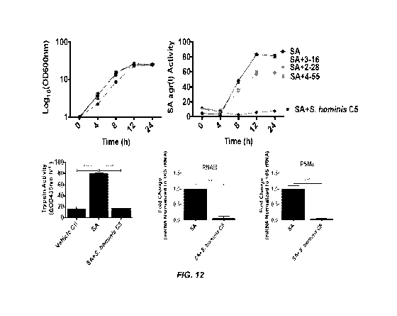
[0044] Figure 12 shows the effect of S. hominis C5 on S. aureus
agr activity.
[0045] Figure 13 shows the effect of various CoNS strains on S.
aureus agr activity.
[0046] Figure 14A-J shows that S. aureus PSMa leads to
disruption of epithelial barrier homeostasis. Human keratinocytes
(NHEKs) were stimulated with S. aureus (SA) sterile-filtered
supernatant from wild type (WT), PSMa (APSMa) or psmp (APSMp)
knockout strains for 24h and (A) trypsin activity and (B) KLK6 mRNA
compared to the housekeeping gene GAPDH were analyzed (n=4). (C)
PSM synthetic peptides were added to NHEKs for up to 24h to analyze
changes in trypsin activity. (D,E) Transcript analysis by RNA-Seq
of genes that changed 2 fold after PSMa3 treatment was assessed
followed by gene ontology (GO) analysis. 8 week old male C57BL/6
11
CA 03072772 2020-02-11
WO 2019/046801
PCT/US2018/049237
mice (n=6) were treated for 72h with SA WT, SA APSMa, or a SA 10
secreted protease knockout strain (Aproteases) (1e7 CFU). (F,G)
Murine skin representative pictures (dashed lines indicate
treatment area) and changes to epidermal thickness after treatment
(scale=200pm). (H-K) Changes in murine back skin with WT or mutant
SA strains in transepidermal water loss (TEWL) and SA CFU/cm2 were
assessed as well. All error bars are represented at standard error
of the mean (SEM) and One-way ANOVAs were used to determine
statistical significance indicated by: p<0.05 *, p<0.01 **, p<0.001
*** , p<0.0001
[0047] Figure 15A-G shows Staphylococcus epidermidis agr type I
auto-inducing peptide characterization and deficiency in AD skin.
(A,B) S. epidermidis agr types I-III supernatant inhibition of S.
aureus (SA) USA300 LAC agr type I activity after 24h (n=4) and
representation of known structure of S. epidermidis agr type I
autoinducing peptide (AIP). (C) Staphylococcus epidermidis (S. epi)
agr type I strain RP62A wild-type (WT) or autoinducing peptide
knockout (AAIP) effect on SA agr activity after 24h. (D) SA
sterile-filtered supernatant growth with or without S. epi WT or
AAIP supernatant was applied to NHEKs for an additionally 24h
followed by measurement of NHEK trypsin activity (n=4). (E)
Consensus of S. epidermidis agr types I-III genomes found on AD
skin. (F,G) Ratio of S. epidermidis agr type I to SA relative
abundance on flare regions of 8 individual AD subjects from 'least
severe' to 'most severe' AD score based upon objective SCORAD and
overall combined data of all subjects based upon AD severity. All
error bars are represented at standard error of the mean (SEM) and
One-way ANOVAs (A,C,D) and a (nonparametric) unpaired Mann-Whitney
test (F) were used to determine statistical significance indicated
by: p<0.05 *, p<0.01 **, p<0.001 ***, p<0.0001
[0048] Figure 16A-F shows multiple clinically isolated
Coagulase-negative Staphylococci inhibit S. aureus agr activity.
(A) Sterile-filtered supernatants of clinically isolated Coagulase-
negative Staphylococci (CoNS) were added to S. aureus (SA) USA300
LAC agr type I P3-YFP reporter strain for 24h followed by analysis
for SA agr activity (n=3). (B,C) S. hominis C5 strain genome was
further sequenced and analyzed at the agrD gene for the auto-
12
CA 03072772 2020-02-11
WO 2019/046801
PCT/US2018/049237
inducing peptide (AIP) sequence. Biochemical analysis of S. hominis
C5 supernatant tested the ability of a <3kDa size exclusion
centrifugation filtration, 80% ammonium sulfate precipitate, and
pH11 1h treated supernatant to effect SA agr activity as well. (D-
F) SA grown in presence of S. hominis C5 supernatant for 24h was
sterile filtered and added to human keratinocytes (NHEKs) for 24h
followed by analysis of trypsin activity, KLK6 mRNA expression
compared to the housekeeping gene GAPDH, and IL-6 protein levels.
All error bars are represented at standard error of the mean (SEM)
and One-way ANOVAs were used to determine statistical significance
indicated by: p<0.05 *, p<0.01 **, p<0.001 ***, p<0.0001
[0049] Figure 17A-H shows AD clinical CoNS isolate inhibits SA
induced murine skin barrier damage. S. aureus (SA) USA300 LAC agr
type I pAmi P3-Lux reporter strain (1e7 CFU) with or without live
S. hominis C5 (1e8 CFU) was applied to 8 week female C57BL/6 mice
for 48h (n=5). (A,B) SA agr activity was assessed on murine back
skin by changes in luminescence. (C) Representative images of
murine skin after 48h SA treatment (dashed boxes indicate treatment
area). (D-H) SA CFU/cm2 was determined and murine skin barrier
damage and inflammation was assessed by analyzing changes in 116
mRNA expression, transepidermal water loss (TEWL), trypsin
activity, and Klk6 mRNA expression normalized to the housekeeping
gene Gapdh. All error bars are represented at standard error of the
mean (SEM) and One-way ANOVAs were used to determine statistical
significance indicated by: p<0.05 *, p<0.01 **, p<0.001 ***,
p<0.0001
[0050] Figure 18A-H shows that S. aureus PSMa changes essential
barrier genes and cytokine expression in human keratinocytes. (A-D)
Human keratinocytes treated with synthetic PSMa3 were assessed for
changes in trypsin activity and KLK6 transcript expression
normalized to the housekeeping gene GAPDH in both a dose and time
dependent manner. (E) GO-term analysis of genes down-regulated 2
fold from the control in human keratinocytes treated with PSMa3 for
24h. (F-H) Changes in human keratinocyte cytokine protein
expression of IL-6, TNF-a, or IL-la treated with SA WT, SA Apsma,
or SA Apsmp supernatant for 24h. All error bars are represented at
standard error of the mean (SEM) and One-way ANOVAs were used to
13
CA 03072772 2020-02-11
WO 2019/046801
PCT/US2018/049237
determine statistical significance indicated by: p<0.05 *, p<0.01
** , p<0.001 ***, p<0.0001
[0051] Figure 19A-H shows that S. aureus PSMa and proteases are
responsible for barrier damage and induction of inflammation on
murine skin. S. aureus (SA) (1e7 CFU) wild type (WT), PSMa knockout
(Apsma), and protease null (Aproteases) strains were applied to
male murine back skin for 72h (n=6) and changes in (A,E) trypsin
activity, (B,F) Klk6, (C,G) 116, and (D,H) IL17a/f mRNA expression
normalized to the housekeeping gene Gapdh were measured. All error
bars are represented at standard error of the mean (SEM) and One-
way ANOVAs were used to determine statistical significance
indicated by: p<0.05 *, p<0.01 **, p<0.001 ***, p<0.0001
[0052] Figure 20A-C shows that CoNS strains do not effect SA
growth. Coagulase-negative Staphylococci (CoNS) supernatant affect
on SA agr type I P3-YFP reporter strain growth as assessed by
OD600nm (n=3-4) including (A) CoNS clinical isolates, (B) S.
epidermidis (S. epi) agr type I-III, and (C) S. epidermidis (S.
epi) wild type (WT) or auto-inducing peptide knockout (AAIP)
supernatant added to SA agr type I reporter strain for 24h. All
error bars are represented at standard error of the mean (SEM).
[0053] Figure 21A-B shows that S. hominis C5 inhibits SA agr
type I-III but not type IV. S. hominis C5 supernatant added to SA
agr types I-IV P3-YFP reporter strains for 24h (n=3). (A) SA
reporter strain agr type I-IV activity and (B) measurement of
growth by OD600nm when cultured in presence of S. hominis C5
supernatant. All error bars are represented at standard error of
the mean (SEM) and One-way ANOVAs were used to determine
statistical significance indicated by: p<0.05 *, p<0.01 **, p<0.001
*** , p<0.0001
[0054] Figure 22A-F shows S. hominis C5 supernatant inhibits SA
induced skin barrier damage. S. aureus (SA) (1e7 CFU) with or
without 10x concentrated <3kDa S. hominis C5 supernatant was
applied to female murine back skin for 48h (n=3). (A-B)
Representative images of murine back (dashed lines indicate
treatment area) and SA CFU/cm2 recovered from murine skin after SA
treatment. (C-F) SA induced skin barrier damage markers including
116, transepidermal water loss (TEWL), trypsin activity, and Klk6
14
CA 03072772 2020-02-11
WO 2019/046801
PCT/US2018/049237
mRNA expression compared to the housekeeping gene Gapdh. All error
bars are represented at standard error of the mean (SEM) and One-
way ANOVAs were used to determine statistical significance
indicated by: p<0.05 *, p<0.01 **, p<0.001 ***, p<0.0001
DETAILED DESCRIPTION
[0055] As used herein and in the appended claims, the singular
forms "a," "an," and "the" include plural referents unless the
context clearly dictates otherwise. Thus, for example, reference
to "an agent" includes a plurality of such agents and reference to
"the microorganism" includes reference to one or more
microorganisms and equivalents thereof known to those skilled in
the art, and so forth.
[0056] Also, the use of "or" means "and/or" unless stated
otherwise. Similarly, "comprise," "comprises," "comprising"
"include," "includes," and "including" are interchangeable and not
intended to be limiting.
[0057] It is to be further understood that where descriptions of
various embodiments use the term "comprising," those skilled in the
art would understand that in some specific instances, an embodiment
can be alternatively described using language "consisting
essentially of" or "consisting of."
[0058] Unless defined otherwise, all technical and scientific
terms used herein have the same meaning as commonly understood to
one of ordinary skill in the art to which this disclosure belongs.
Any methods and reagents similar or equivalent to those described
herein can be used in the practice of the disclosed methods and
compositions.
[0059] Atopic dermatitis (AD) is among the most common immune
disorders, and causes a serious burden to patient quality of life
and finances as well as posing a serious risk of comorbidities.
Defects in skin barrier function are an important characteristic of
AD. Eczematous skin lesions of patients with AD have increased
levels of Th2 cytokines such as IL4 and IL13. Th2 cytokines promote
decreased function of the skin barrier by inhibiting expression of
filaggrin. These cytokines also suppress expression of human
antimicrobial peptides such as cathelicidin and b-defensin-2, a
defect in AD that may lead to dysbiosis of the skin bacterial
CA 03072772 2020-02-11
WO 2019/046801
PCT/US2018/049237
community and enhanced colonization by S. aureus. Therapy targeting
IL4 receptor alpha result in significant improvement in disease.
The strong association between Th2 cytokine activity, barrier
function, antimicrobial activity, and disease outcome supports
efforts to define a causal link between these essential epidermal
functions.
[0060] The skin barrier of patients with AD may be compromised
by increased proteolytic activity as they have been found to
display increased kallikrein (KLK) expression. KLKs are a family of
15 serine proteases of which several are found predominately in the
upper granular and stratum corneal layers of the epidermis. In
Netherton syndrome, increased serine protease activity is observed
due to decreased activity of the serine protease inhibitor Kazal-
type 5. The resulting increase in enzymatic activity leads to
increased desquamation, altered antimicrobial peptide and filaggrin
(FLG) processing, and protease-activated receptor 2 activation and
inflammation. Increased protease activity may also play an
important role in the communication of the microbiome with the skin
immune system, and has recently been shown to directly influence
epidermal cytokine production and inflammation by enhancing
penetration of bacteria through the epidermis.
[0061] Dysbiosis of the skin's microbiome and the colonization
of the skin by Staphylococcus aureus is associated with the
exacerbations of atopic dermatitis (AD). The present disclosure
demonstrates S. aureus has the ability to induce expression of
specific KLKs from keratinocytes and increase overall proteolytic
activity in the skin. This illustrates a system by which bacteria
on the skin communicate with the host and suggests a previously
unknown but likely important mechanism for how S. aureus
colonization can increase disease severity in patients with AD.
[0062] S. aureus can secrete multiple proteases onto the skin
that alter skin barrier integrity. The serine protease V8 and
serine-like protease exfoliative toxins have been shown to cleave
corneodesmosome adhesion proteins including DSG-1 leading to
increased desquamation. Aureolysin, an MMP, is known to cleave and
inactivate LL-37, an important antimicrobial peptide on the skin.
However, these direct proteolytic actions of S. aureus products
16
CA 03072772 2020-02-11
WO 2019/046801
PCT/US2018/049237
require high levels of the enzyme and bacteria, and are more
consistent with events that occur during infection with this
organism.
[0063] Increased digestion of barrier proteins was observed
after keratinocytes were activated by S. aureus. FLG is known to be
cleaved from the larger Pro-FLG (400 kDa) into a monomeric form (37
kDa) that plays an important role in forming the physical barrier
of the stratum corneum with keratin. It has been shown that
accelerated Pro-FLG cleavage could be linked to increased
desquamation of the skin (Hewett et al., 2005). Interestingly,
increased cleavage of Pro-FLG was observed in human keratinocytes
treated with S. aureus supernatant. Pro-FLG cleavage was partially
blocked when KLK6 or KLK13 was silenced, indicating that S. aureus
may decrease skin barrier integrity in a KLK-dependent manner
through cleavage of Pro-FLG.
[0064] DSG-1 is an important corneodesmosome adhesion protein
that when cleaved leads to increased desquamation. Full-length DSG-
1 (160 kDa) in keratinocytes is readily cleaved by KLK activity
stimulated by S. aureus. It has been reported that KLK5, 6, 7, and
14 can cleave DSG-1, whereas KLK13 could not. This showed that
upregulated KLK6 and KLK14 can lead to enhanced cleavage of full-
length DSG-1 while providing contrary evidence to the notion that
KLK13 is not involved in DSG-1 cleavage. Thus, S. aureus can cause
KLKs to alter FLG cleavage, but also increase DSG-1 cleavage as
another way to decrease the epidermal skin barrier integrity.
Specific siRNA knockdown suggested that the increased expression of
KLKs was responsible, at least in part, for the increased serine
protease activity stimulated by S. aureus. Figure 2C demonstrates
that secreted proteases from S. aureus contribute to the induction
of increased trypsin activity in keratinocytes. Because bacteria
including S. aureus can penetrate the skin surface and elicit
strong dermal immune responses (Nakatsuji et al., 2013, 2016; Zhang
et al., 2015), it is possible that these bacteria may also
influence protease activity of dermal cells. These observations
also relate to Rosacea or Netherton syndrome.
[0065] The disclosure demonstrates that soluble factor(s)
produced by S. aureus have a potent and previously unsuspected
17
CA 03072772 2020-02-11
WO 2019/046801
PCT/US2018/049237
capacity to alter endogenous protease activity produced by the
keratinocyte. This occurred at a dilution of S. aureus products
from which the activity of the bacterial proteases was
undetectable. Thus, S. aureus can promote the epidermis to increase
expression of endogenous proteolytic activity, thus drastically
altering the balance of total epidermal proteolytic activity.
[0066] Different strains of S. aureus (Newman, USA300, 113, and
SANGER252) and S. epidermidis (ATCC12228 and ATCC1457) had
different effects on human keratinocyte protease activity. S.
aureus strains including Newman and USA300 increased trypsin
activity, whereas other strains of S. aureus and S. epidermidis
increased elastase or MMP activity. Thus, bacteria could alter
epidermal protease activity depending on both the species and
strain of bacteria. It is possible that other bacterial species and
strains of S. aureus could further uniquely influence the enzymatic
balance of human skin. Interestingly, preliminary data have found
that purified toll-like receptor ligands do not induce trypsin
activity or KLK expression in keratinocytes.
[0067] Protease activity is highly upregulated in multiple skin
diseases leading to a damaged skin barrier. This is associated with
a worsened disease state in almost all cases. The disclosure
demonstrates, in one aspect, that commensal microbes and their
bacterial products are useful to prevent increased protease
activity in the skin. In particular, the disclosure demonstrates
that coagulase negative Staphylococci can prevent Staphylococcus
aureus induced serine protease activity in the skin by inhibiting
the agr quorum sensing system. Staphylococcus aureus, a pathogenic
bacteria strain can induce serine protease activity in the skin.
Increased protease activity disrupts the skin barrier and leads to
worsened disease states including Netherton syndrome and atopic
dermatitis. The disclosure demonstrates that this increased serine
protease activity can be prevented through use of commensal, or
good, skin bacteria and factors derived therefrom.
[0068] The disclosure presents an unexpected response of
keratinocytes to S. aureus. Because of the increased DSG-1 and FLG
cleavage, S. aureus produces one or more factors that decrease the
integrity of the skin barrier in a KLK-dependent manner.
18
CA 03072772 2020-02-11
WO 2019/046801
PCT/US2018/049237
[0069] The disclosure demonstrates that S. aureus not only
secretes proteases but also can specifically activate keratinocytes
to increase expression of endogenous proteases. The disclosure
demonstrates that phenol-soluble modulin alpha (PSMa) is secreted
by S. aureus and triggers auto-digestion of the epidermis. For
example, three members of the KLK family appear to play a role in
this increased enzymatic activity.
[0070] The disclosure also identifies commensal bacteria, genes
and polypeptides that inhibit the accessory gene regulator (agr)
quorum sensing system of S. aureus and turns off PSMa thereby
inhibiting protease activity. Thus, the disclosure provides
targets for modulating atopic dermatitis as well as agents and
probiotic preparations to modulate atopic dermatitis and protease
activity on the skin.
[0071] The disclosure demonstrates that coagulase-negative
Staphylococci (CoNS) species that normally reside on skin such as
S. epidermidis and S. hominis protect against this biological
activity of S. aureus by producing auto-inducing peptides (AIP)
that inhibit the accessory gene regulatory (agr) quorum sensing
system of S. aureus and turn off PSMa secretion.
[0072] Virtually all S. aureus toxins are under the control of
the virulence accessory gene regulator (agr). The agr system
triggers changes in gene expression at a particular cell density by
a process called quorum sensing. In addition to toxins, agr is
known to upregulate a wide variety of virulence determinants, such
as exoenzymes (proteases, lipases, nucleases), and downregulate
expression of surface binding proteins. This adaptation is believed
to control production of certain virulence determinants of an
infection, when they are needed (e.g., binding proteins at the
beginning, when cell density is low and adhesion to host tissue is
important, and toxins and degradative exoenzymes when the infection
is established and nutrients need to be acquired from host tissues.
[0073] Multiple clinical isolates of different CoNS species
inhibited protease activation and prevented epithelial damage both
in vitro and in vivo without changing the abundance of S. aureus
(e.g., inhibited the biological activity of protease/agr activity,
without changing S. aureus density). Moreover, the disclosure shows
19
CA 03072772 2020-02-11
WO 2019/046801
PCT/US2018/049237
that patients with active AD showed a decrease in relative
abundance of these beneficial microbes (e.g., CoNS) compared to S.
aureus, thus overcoming inhibition of quorum sensing and enabling
barrier disruption by S. aureus. Taken together, the disclosure
shows how members of the normal human skin microbiome maintain
immune homeostasis by contributing as a community to the control of
S. aureus toxin production.
[0074] The disclosure has also identified polynucleotide
sequences, polypeptide sequences and fragments thereof that provide
for products that inhibit agr quorum sensing activity. These
polynucleotide and polypeptide can be used to provide therapeutics
and recombinant non-pathogenic or attenuated skin bacteria for use
in topical formulations to treat S. aureus infections and/or atopic
dermatitis.
[0075] For example, the disclosure provides for auto-inducing
peptides (AIPs) that downregulate agr activity. Polynucleotides
encoding the AIPs are also provided herein.
[0076] The disclosure provides a link between increased S.
aureus colonization and increased serine protease activity in AD
skin and provides new targets and therapies including, but not
limited to, fermentation extracts to either up regulate protease
activity (e.g., fermentation extracts from S. aureus) or
fermentation extracts from commensal bacteria that down regulate
protease activity in the skin (e.g., containing one or more AIPs of
the disclosure). Moreover, the disclosure provides for (i) topical
formulations comprising such extracts or purified AIPs peptides,
(ii) topical formulations comprising commensal probiotic bacteria
(e.g., non-pathogenic or attenuated bacterial that have been
transformed with an AIP coding sequence, or purified commensal
bacterial preparations in a topical formulation). Further
therapeutic targets can be antibodies to KLKs, and/or DSG-1 and/or
FLG therapy (e.g., increased expression or delivery of these
factors to AD subjects).
[0077] In one embodiment, an AIP polypeptide of the disclosure
has the consensus sequence of X1X2X3X4CX5X6X7X8 (SEQ ID NO:10),
wherein X1 is S, K, V, G or T; X2 is Y, Q, A, or I; X3 is N, S, T,
or D; X4 is V, P, M, or T; X5 is G, S, A, N, or T; X6 is G, N, T,
CA 03072772 2020-02-11
WO 2019/046801 PCT/US2018/049237
or L; X7 is Y or F; and X8 is F, L, or Y, wherein amino acids 5-9
of SEQ ID NO:10 form a thiolactone ring. Exemplary peptide
sequence that fall within the consensus sequence of SEQ ID NO:10
include SYNVCGGYF (SEQ ID NO:4), KYNPCSNYL (SEQ ID NO:11),
SYSPCATYF (SEQ ID NO:12), SQTVCSGYF (SEQ ID NO:13), GANPCALYY (SEQ
ID NO:14), TINTCGGYF (SEQ ID NO:15), VQDMCNGYF (SEQ ID NO:16), and
GYSPCTNFF (SEQ ID NO:17). In a further embodiment, the polypeptide
generates a structure of Formula I or IA. In another embodiment,
the polypeptide can comprise a combination of D- or L-amino acids.
In any of the foregoing embodiments, the polypeptide inhibits S.
aureus protease activity, agr activity or keratinocyte protease
activity.
[0078] The disclosure provides a compound of Formula I
R7
HNCH-
7../
-"CH 0 NH
1
C-0 0=C
o NH
1-12C
Xi X2 __________________
11
0
Formula I
wherein X1 is from 1-6 amino acids; X2 is an amino acid selected
from valine (V), proline (P), methionine (M) and threonine (T);
wherein R5 is selected from the group consisting of / , OH ,
4311, NH2
,CH3
0 , and OH; wherein R6 is selected from the
µ1,NH2
H
217,
group consisting of , 0 OH, and ;
wherein
711.1
OH, R7 is selected from the group consisting of and
21
CA 03072772 2020-02-11
WO 2019/046801 PCT/US2018/049237
x
; and wherein R8 is selected from the group consisting of
x
OH .
r , and
[0079] In one embodiment, the disclosure provides a compound of
Formula IA:
R7
/
HN--------CH
,1õ,_
`CH 0 NH
/ \
\
C=0 0=C
1
S
CH¨R6
/
i /
\ 9\ NH
0 0 0 .\
C
H H 11 H \
--C-4--
X __ N __ CH ¨C __ N __ CH ¨C __ NH _____________________ CH C X2 N CH ¨C N-
.
1
1
1.2" 1 H \
RS
IR' IR3 0
Formula IA
wherein X1 is from 1-6 amino acids; X2 is an amino acid selected
from valine (V), proline (P), methionine (M) and threonine (T);
wherein Rl is selected from the group consisting of
HO
NH2 ............._
\/------.7---/ H
)11, , and \ ; wherein R2 is selected
0
717, /_.....j--NH2
OH
from the group consisting of , \ ,
,CH3
7i, 4;tz-,-------\
, and ; wherein R3 is selected from the group
,31,c,NH2
''!Ii:NNOH
consisting of 0 , )1µ. OH, 0 , and OH;
wherein R5 is selected from the group consisting of , LIIC (311, \
'' OH ,
22
CA 03072772 2020-02-11
WO 2019/046801
PCT/US2018/049237
NH
N...-.---.. 2
H3
XC
'lli:N
r 0 , and OH; wherein R6 is selected from the
(311,NH2
H
iµt,
group consisting of , 0 , ill.. OH, and ;
wherein
0H, R7 is selected from the group consisting of and
...,
; and wherein R8 is selected from the group consisting of
..-.
OH
r , and .
[0080] The disclosure provides a purified polypeptide (e.g., an
AIP peptide) comprising a sequence that is at least 98% identical
to SEQ ID NO:4 and which inhibits (i) protease production and/or
protease activity of keratinocytes, (ii) inhibits IL-6 production
and/or activity of keratinocytes, (iii) inhibits production of
phenol soluble modulin alpha 3 from Staphylococcus aureus (S.
aureus) and/or (iv) inhibits agr production and/or activity by S.
aureus. In another embodiment, the disclosure provides for a
compound of Formula IB:
23
CA 03072772 2020-02-11
WO 2019/046801 PCT/US2018/049237
OH
H2a
-CH 0 'NH
C--0 0--C
/,CH¨H
o NH
%//
0 0 0 0 H2C
H H \
X __ N ____ CH C __ N __ CH C _________________ NH CH C --N1-1¨CH¨C--N
CH C
H
c.2 CH2 CH¨CH3
OH C __ 0 CH3
r4H2
OH
Formula IB
[0081] In still a further embodiment, the disclosure provides a
purified polypeptide comprising or consisting of SEQ ID NO:4, 11,
12, 13, 14, 15, 16, or 17. In a further embodiment, the polypeptide
forms a structure of formula I, IA or IB.
[0082] In one embodiment, an AIP peptide of the disclosure can
comprise one or more D-amino acids.
[0083] The disclosure provides a topical formulation comprising
an AIP peptide having a consensus sequence of SEQ ID NO:10 or a
peptide of SEQ ID NO:4, 11, 12, 13, 14, 15, 16, or 17 or compound
of Formula I, IA or IB.
[0084] "Substantially identical" means that an amino acid
sequence is largely, but not entirely, the same, but retains a
functional activity of the sequence to which it is related. The
percent of identity to polypeptide sequence or polynucleotides
sequences share is based upon the alignment of the sequence. It is
common in the art to use various programs to perform alignment and
to determine identity. In general two polypeptides or domains are
"substantially identical" if their sequences are at least 85%, 90%,
95%, 98% or 99% identical, or if there are conservative variations
24
CA 03072772 2020-02-11
WO 2019/046801
PCT/US2018/049237
in the sequence. A computer program, such as the BLAST program
(Altschul et al., 1990) can be used to compare sequence identity.
[0085] The disclosure also provides a polynucleotide (i.e., an
"AIP polynucleotide") encoding an AIP polypeptide of the
disclosure. For example, the disclosure provides a polynucleotide
encoding SEQ ID NO:2 or 4. In one embodiment, the polynucleotide
hybridizes under stringent conditions to a polynucleotide
consisting of SEQ ID NO:3 and encodes a polypeptide of SEQ ID NO:4.
"Stringency" of hybridization reactions is readily determinable by
one of ordinary skill in the art, and generally is an empirical
calculation dependent upon probe length, washing temperature, and
salt concentration. In general, longer probes require higher
temperatures for proper annealing, while shorter probes need lower
temperatures. Hybridization generally depends on the ability of
denatured DNA to reanneal when complementary strands are present in
an environment below their melting temperature. The higher the
degree of desired homology between the probe and hybridizable
sequence, the higher the relative temperature which can be used. As
a result, it follows that higher relative temperatures would tend
to make the reaction conditions more stringent, while lower
temperatures less so. For additional details and explanation of
stringency of hybridization reactions, see Ausubel et al., Current
Protocols in Molecular Biology, Wiley Interscience Publishers,
(1995). "Stringent conditions" or "high stringency conditions", as
defined herein, typically: (1) employ low ionic strength and high
temperature for washing, for example 0.015 M sodium chloride/0.0015
M sodium citrate/0.1% sodium dodecyl sulfate at 50 C; (2) employ
during hybridization a denaturing agent, such as formamide, for
example, 50% (v/v) formamide with 0.1% bovine serum albumin/0.1%
Fico11/0.1% polyvinylpyrrolidone/50 mM sodium phosphate buffer at
pH 6.5 with 750 mM sodium chloride, 75 mM sodium citrate at 42 C;
or (3) employ 50% formamide, 5xSSC (0.75 M NaCl, 0.075 M sodium
citrate), 50 mM sodium phosphate (pH 6.8), 0.1% sodium
pyrophosphate, 5xDenhardt's solution, sonicated salmon sperm DNA
(50 pg/ml), 0.1% SDS, and 10% dextran sulfate at 42 C, with washes
at 42 C in 0.2xSSC (sodium chloride/sodium citrate) and 50%
formamide at 55 C, followed by a high-stringency wash consisting of
CA 03072772 2020-02-11
WO 2019/046801
PCT/US2018/049237
0.1xSSC containing EDTA at 55 C. Polynucleotide sequences encoding
SEQ ID NO:11, 12, 13, 14, 15, 16, and 17 can be deduced using codon
charts.
[0086] An AIP polynucleotide can be cloned into various vectors
for use in the disclosure. For example, an AIP polynucleotide can
be cloned into an expression vector or plasmid for use in
transformation and/or expression in a recombinant host cell.
Vectors for use in bacterial transformations are known. The four
major types of vectors are plasmids, viral vectors, cosmids, and
artificial chromosomes. Common to all engineered vectors are an
origin of replication, a multicloning site, and a selectable
marker. Any of these are suitable for use herein. An AIP
polyucleotide can be inserted into a clone, vector, shuttle,
plasmid, BAC, or can also be integrated into the bacterial genome.
If a plasmid is used, the copy number of the plasmid can be between
5-500 copy numbers per cell. Exemplary plasmids and expression
vectors include but are not limited to: p252, p256, p353-2 (Leer et
al. 1992), p8014-2, pA1, pACYC, pAJ01, pAl-derived (Vujcic &
Topisirovic 1993), pall, pAM-beta-1,2,3,5,8 (simon and chopin
1988), pAR1411, pBG10, pBK, pBM02, pBR322, pBR328, pBS-slpGFP,
pC194 (McKenzie et al. 1986, 1987; Horinouchi & Weisblum 1982b),
PC194/PUB110, pC30il, pC30il (Skaugen 1989), pCD034-1, pCD034-2,
pCD256, pC12000, pC1305, pC1528, pCIS3, pCL2.1, pCT1138, pD125,
pE194, pE194/PLS1, pEGFP-C1, pEH, pF8801, pFG2, pFK-series, pGK-
series, pGK12, pGK13, pIA, pIAV1,5,6,7,9, pIL.CatT, pIL252/3,
pIL253, pIL7, pISA (low for E. coli), pJW563, pKRV3, pLAB1000
(Josson et al. 1990), pLB4 (Bates & Gilbert 1989, pLBS, pLE16,
pLEB124, pLEB590, pLEB591, pLEB600, pLEB604, pLEP24Mcop, pLJ1
(Takiguchi et al. 1989), pLKS, pLTK2, pWCFS101 and pMD5057 (Bates &
Gilbert, 1989; Skaugen, 1989; Leer et al., 1992; Vujcic &
Topisirovic, 1993; Eguchi et al., 2000; Kaneko et al., 2000;
Danielsen, 2002; Daming et al., 2003; de las Rivas et al., 2004;
van Kranenburg et al., 2005), pLP1/18/30, pLP18, pLP317, pLP317cop,
pLP3537, pLP3537xyl, pLP402, pLP825, pLP825 and pLPE323, pLP82H,
pLPC37, pLPE23M, pLPE323, pLPE350, pLPI (Bouia et al. 1989), pLS1,
pLS1 and pE194 (Lacks et al. 1986; Horinouchi & Weisblum 1982a),
p1u1631, pLUL631 from L. reuteri carrying an erythromycin-
26
CA 03072772 2020-02-11
WO 2019/046801
PCT/US2018/049237
resistance gene, pM3, pM4, pMD5057, pMG36e, pND324, pNZ-series,
pPSC series, pSH71 (de vos, 1987), pSIP-series, pSK11L, pSL2, PSN2,
pSN2 (Khan & Novick 1982), pT181 (Koepsel et al. 1987), (Khan &
Novick 1983), pT181, pC194 and pE194 are not functional in B.
subtilis (Gruss et al. 1987), pT181, pE194/pLS1, pC194/pUB110 and
pSN2 (Khan, 2005), pTL, pTRK family, pTRT family, pTUAT35, pUBII0
and pC194 (McKenzie et al. 1986, 1987; Horinouchi & Weisblum
1982b), pUCL22, pULP8/9, pVS40, pWC1, pWCFS101, pWV02, pWV04,
pWV05, RepA, system BetL.
[0087] In one embodiment, the disclosure provides a topical
composition comprising an AIP polypeptide or peptide of the
disclosure. For example, in one embodiment, the topical
composition comprises a purified polypeptide (e.g., an AIP peptide)
comprising a consensus sequence of SEQ ID NO:10, or a sequence that
is at least 98% identical to any of SEQ ID NO:4, 11, 12, 13, 14,
15, 16, or 17 and which inhibits (i) protease production and/or
protease activity of keratinocytes, (ii) inhibits IL-6 production
and/or activity of keratinocytes, (iii) inhibits production of
phenol soluble modulin alpha 3 from Staphylococcus aureus (S.
aureus) and/or (iv) inhibits agr production and/or activity by S.
aureus. In another embodiment, the topical composition comprises a
compound of Formula I, IA, or IB (as defined above).
[0088] In another embodiment, the topical composition can
comprise a non-pathogenic microorganism (including attenuated
microorganism that have been engineered to reduce or eliminate
pathogenic activity), wherein the microorganism has been engineered
to expression an AIP polypeptide. The microorganism can be
engineered to contain a vector and/or AIP polynucleotide. In one
embodiment, the microorganism produces a compound of Formula I, IA
and/or IB.
[0089] In one embodiment, the compositions and methods herein
use non-pathogenic bacteria that have been engineered to produce a
compound of Formula I, IA and/or IB, by transforming the bacteria
with an AIP polynucleotide of the disclosure. In one embodiment,
the bacteria in the population are non-pathogenic and non-invasive
microorganisms, and can be in certain embodiments a gram-positive
food grade bacterial strain. In another embodiment, the populations
27
CA 03072772 2020-02-11
WO 2019/046801
PCT/US2018/049237
of transformed bacteria are prepared from a bacterium that occurs
naturally in the skin microbiome.
[0090] In certain embodiments, bacteria forming the population
of bacteria in the composition, and that are transformed to express
a compound of Formula I, IA, and/or IB, can be a collection of the
same bacteria or a mixture of different bacteria, at different
phylogenetic levels. Bacteria resident on the skin of healthy
humans include bacterial species typically resident on the face of
humans, such as Actinobacteria, including bacterial in the genus
corynebacterium and in the genus propionibacterium. In other
embodiments, bacteria resident on the skin of healthy human
subjects include bacterial species typically resident on skin other
than the face, including for example bacteria in the genus
bacteroidetes and proteobacteria. Other bacteria in the skin
microbiome include those listed herein below.
[0091] In one embodiment, the bacteria are from the genus
Propionibacterium, including but not limited to, Propionibacterium
acidifaciens, Propionibacterium acidipropionici, Propionibacterium
acidipropionici strain 4900, Propionibacterium acnes,
Propionibacterium australiense, Propionibacterium avidum,
Propionibacterium cyclohexanicum, Propionibacterium freudenreichii
subsp. Freudenreichii, P. freudenreichii ssp. freudenreichii strain
20271, Propionibacterium freudenreichii subsp. Shermanii, P.
freudenreichii ssp. shermanii strain 4902, P. freudenreichii ssp.
shermanii strain 4902, Propionibacterium granulosum,
Propionibacterium innocuum, P. jensenii strain 20278,
Propionibacterium lymphophilum, Propionibacterium microaerophilum,
Propionibacterium propionicum, Propionibacterium thoenii, and P.
thoenii strain 20277. In one embodiment, the bacteria is not
Propionibacterium acnes. In one embodiment, the bacteria are from
the genus Corynebacterium, including but not limited to, C.
accolens, C. afermentan, C. amycolatum, C. argentoratense, C.
aquaticum, C. auris, C. bovis, C. diphtheria, C. equi (now
Rhodococcus equi), C. flavescens, C. glucuronolyticum, C.
glutamicum, C. granulosum, C. haemolyticum, C. halofytica, C.
jeikeium (group JK), C. macginleyi, C. matruchotii, C.
minutissimum, C. parvum (Propionibacterium acnes), C. propinquum,
28
CA 03072772 2020-02-11
WO 2019/046801
PCT/US2018/049237
C. pseudodiphtheriticum (C. hofmannii), C. pseudotuberculosis, (C.
ovis), C. pyogenes, C. urealyticum (group D2), C. renale, C. spec,
C. striatum, C. tenuis, C. ulcerans, C. urealyticum, and C.
xerosis. Bacterial with lipophilic and nonlipophilic groups are
contemplated, and the nonlipophilic bacteria may include
fermentative corynebacteria and nonfermentative corynebacteria. In
one embodiment, the bacteria is not C. diphtheria, C. amicolatum,
C. striatum, C. jeikeium, C. urealyticum, C. xerosis, C.
pseudotuberculosis, C. tenuis, C. striatum, or C. minutissimum, as
these may be pathogenic. In one embodiment, the bacteria are from
the suborder Micrococcineae, including but not limited to the GRAS
bacteria species Arthrobacter arilaitensis, Arthrobacter bergerei,
Arthrobacter globiformis, Arthrobacter nicotianae, Kocuria
rhizophila, Kocuria varians, Micrococcus luteus, Micrococcus lylae,
Microbacterium gubbeenense, Brevibacterium aurantiacum,
Brevibacterium casei, Brevibacterium linens, Brachybacterium
alimentarium, and Brachybacterium tyrofermentans. In another
embodiment, the bacteria are from the genus Staphylococcus,
including but not limited to, Staphylococcus agnetis, S. arlettae,
S. auricularis, S. capitis, S. caprae, S. carnosus, Staphylococcus
caseolyticus, S. chromogenes, S. cohnii, S. condiment, S. delphini,
S. devriesei, S. equorum, S. felis, S. fleurettii, S. gallinarum,
S. haemolyticus, S. hominis, S. hyicus, S. intermedius, S. kloosii,
S. leei, S. lentus, S. lugdunensis, S. lutrae, S. massiliensis, S.
microti, S. muscae, S. nepalensis, S. pasteuri, S. pettenkoferi, S.
piscifermentans, S. pseudintermedius, S. pseudolugdunensis, S.
pulvereri, S. rostra, S. saccharolyticus, S. saprophyticus, S.
schleiferi, S. sciuri, S. simiae, S. simulans, S. stepanovicii, S.
succinus, S. vitulinus, S. warneri, and S. xylosus. In one
embodiment, the bacteria is not S. aureus or S. epidermidis. In
another embodiment, the bacteria are from the genus Streptococcus,
including but not limited to, Streptococcus acidominimus,
Streptococcus adjacens, Streptococcus agalactiae, Streptococcus
alactolyticus, Streptococcus anginosus, Streptococcus australis,
Streptococcus bovis, Streptococcus caballi, Streptococcus canis,
Streptococcus caprinus, Streptococcus castoreus, Streptococcus
cecorum, Streptococcus constellatus, Streptococcus constellatus
29
CA 03072772 2020-02-11
WO 2019/046801
PCT/US2018/049237
subsp. Constellatus, Streptococcus constellatus subsp. Pharyngis,
Streptococcus cremoris, Streptococcus criceti, Streptococcus
cristatus, Streptococcus danieliae, Streptococcus defectives,
Streptococcus dentapri, Streptococcus dentirousetti, Streptococcus
didelphis, Streptococcus difficilis, Streptococcus durans,
Streptococcus dysgalactiae, Streptococcus dysgalactiae subsp.
Dysgalactiae, Streptococcus dysgalactiae subsp. Equisimilis,
Streptococcus entericus, Streptococcus equi, Streptococcus equi
subsp. Equi, Streptococcus equi subsp. Ruminatorum, Streptococcus
equi subsp. Zooepidemicus, Streptococcus equines, Streptococcus
faecalis, Streptococcus faecium, Streptococcus ferus, Streptococcus
gallinaceus, Streptococcus gallolyticus, Streptococcus gallolyticus
subsp. Gallolyticus, Streptococcus gallolyticus subsp. Macedonicus,
Streptococcus gallolyticus subsp. Pasteurianus, Streptococcus
garvieae, Streptococcus gordonii, Streptococcus halichoeri,
Streptococcus hansenii, Streptococcus henryi, Streptococcus
hyointestinalis, Streptococcus hyovaginalis, Streptococcus
ictaluri, Streptococcus infantarius, Streptococcus infantarius
subsp. Coli, Streptococcus infantarius subsp. Infantarius,
Streptococcus infantis, Streptococcus iniae, Streptococcus
intermedius, Streptococcus intestinalis, Streptococcus lactarius,
Streptococcus lactis, Streptococcus lactis subsp. Cremoris,
Streptococcus lactis subsp. Diacetilactis, Streptococcus lactis
subsp. Lactis, Streptococcus lutetiensis, Streptococcus macacae,
Streptococcus macedonicus, Streptococcus marimanunalium,
Streptococcus massiliensis, Streptococcus merionis, Streptococcus
minor, Streptococcus mitis, Streptococcus morbillorum,
Streptococcus mutans, Streptococcus oligofermentans, Streptococcus
oralis, Streptococcus orisratti, Streptococcus ovis, Streptococcus
parasanguinis, Streptococcus parauberis, Streptococcus parvulus,
Streptococcus pasteurianus, Streptococcus peroris, Streptococcus
phocae, Streptococcus plantarum, Streptococcus pleomorphus,
Streptococcus pluranimalium, Streptococcus plurextorum,
Streptococcus pneumonia, Streptococcus porci, Streptococcus
porcinus, Streptococcus porcorum, Streptococcus pseudopneumoniae,
Streptococcus pseudoporcinus, Streptococcus pyogenes, Streptococcus
raffinolactis, Streptococcus ratti, Streptococcus rupicaprae,
CA 03072772 2020-02-11
WO 2019/046801
PCT/US2018/049237
Streptococcus saccharolyticus, Streptococcus salivarius,
Streptococcus salivarius subsp. Salivarius, Streptococcus
salivarius subsp. Thermophilus, Streptococcus sanguinis,
Streptococcus shiloi, Streptococcus sinensis, Streptococcus
sobrinus, Streptococcus suis, Streptococcus thermophilus,
Streptococcus thoraltensis, Streptococcus tigurinus, Streptococcus
troglodytae, Streptococcus troglodytidis, Streptococcus uberis,
Streptococcus urinalis, Streptococcus vestibularis, and
Streptococcus waius. In another embodiment, the bacteria are from
the genus Lactobacillus, including but not limited to, Lactococcus
garvieae, Lactococcus lactis, Lactococcus lactis subsp. cremoris,
Lactococcus lactis subsp. hordniae, Lactococcus lactis, Lactococcus
lactis subsp. Lactis, Lactococcus piscium, Lactococcus plantarum,
Lactococcus raffinolactis, Lactobacillus acetotolerans,
Lactobacillus acidophilus, Lactobacillus agilis, Lactobacillus
algidus, Lactobacillus alimentarius, Lactobacillus amylolyticus,
Lactobacillus amylophilus, Lactobacillus amylovorus, Lactobacillus
animalis, Lactobacillus aviarius, Lactobacillus aviarius subsp.
araffinosus, Lactobacillus aviarius subsp. aviarius, Lactobacillus
bavaricus, Lactobacillus bifermentans, Lactobacillus brevis,
Lactobacillus buchneri, Lactobacillus bulgaricus, Lactobacillus
carnis, Lactobacillus casei, Lactobacillus casei subsp. alactosus,
Lactobacillus casei subsp. casei, Lactobacillus casei subsp.
pseudoplantarum, Lactobacillus casei subsp. rhamnosus,
Lactobacillus casei subsp. tolerans, Lactobacillus catenaformis,
Lactobacillus cellobiosus, Lactobacillus collinoides, Lactobacillus
confusus, Lactobacillus coryniformis, Lactobacillus coryniformis
subsp. coryniformis, Lactobacillus coryniformis subsp. torquens,
Lactobacillus crispatus, Lactobacillus curvatus, Lactobacillus
curvatus subsp. curvatus, Lactobacillus curvatus subsp. melibiosus,
Lactobacillus delbrueckii, Lactobacillus delbrueckii subsp.
bulgaricus, Lactobacillus delbrueckii subsp. delbrueckii,
Lactobacillus delbrueckii subsp. lactis, Lactobacillus divergens,
Lactobacillus farciminis, Lactobacillus fermentum, Lactobacillus
formicalis, Lactobacillus fructivorans, Lactobacillus fructosus,
Lactobacillus gallinarum, Lactobacillus gasseri, Lactobacillus
graminis, Lactobacillus halotolerans, Lactobacillus hamsteri,
31
CA 03072772 2020-02-11
WO 2019/046801
PCT/US2018/049237
Lactobacillus helveticus, Lactobacillus heterohiochii,
Lactobacillus hilgardii, Lactobacillus homohiochii, Lactobacillus
iners, Lactobacillus intestinalis, Lactobacillus jensenii,
Lactobacillus johnsonii, Lactobacillus kandleri, Lactobacillus
kefiri, Lactobacillus kefuranofaciens, Lactobacillus kefirgranum,
Lactobacillus kunkeei, Lactobacillus lactis, Lactobacillus
leichmannii, Lactobacillus lindneri, Lactobacillus malefermentans,
Lactobacillus mali, Lactobacillus maltaromicus, Lactobacillus
manihotivorans, Lactobacillus minor, Lactobacillus minutus,
Lactobacillus mucosae, Lactobacillus murinus, Lactobacillus
nagelii, Lactobacillus oris, Lactobacillus panis, Lactobacillus
parabuchneri, Lactobacillus paracasei, Lactobacillus paracasei
subsp. paracasei, Lactobacillus paracasei subsp. tolerans,
Lactobacillus parakefiri, Lactobacillus paralimentarius,
Lactobacillus paraplantarum, Lactobacillus pentosus, Lactobacillus
perolens, Lactobacillus piscicola, Lactobacillus plantarum,
Lactobacillus pontis, Lactobacillus reuteri, Lactobacillus
rhamnosus, Lactobacillus rhamnosus strain 5/E5a, Lactobacillus
rimae, Lactobacillus rogosae, Lactobacillus ruminis, Lactobacillus
sakei, Lactobacillus sakei subsp. camosus, Lactobacillus sakei
subsp. sakei, Lactobacillus salivarius, Lactobacillus salivarius
subsp. salicinius, Lactobacillus salivarius subsp. salivarius,
Lactobacillus sanfranciscensis, Lactobacillus sharpeae,
Lactobacillus suebicus, Lactobacillus trichodes, Lactobacillus uli,
Lactobacillus vaccinostercus, Lactobacillus vaginalis,
Lactobacillus viridescens, Lactobacillus vitulinus, Lactobacillus
xylosus, Lactobacillus yamanashiensis, Lactobacillus yamanashiensis
subsp. mali, Lactobacillus yamanashiensis subsp. Yamanashiensis and
Lactobacillus zeae. In another embodiment, the bacteria are from
the genus Lactococcus, including but not limited to, Lactococcus
Schleifer, Lactococcus chungangensis, Lactococcus fujiensis,
Lactococcus garvieae, Lactococcus lactis, Lactococcus lactis subsp.
Cremoris, Lactococcus lactis subsp. Hordniae, Lactococcus lactis
subsp. Lactis, Lactococcus lactis subsp. Tructae, Lactococcus
piscium, Lactococcus plantarum, and Lactococcus raffinolacti.
[0092] In yet another embodiment, the disclosure provides a
probiotic composition for topical delivery comprising a CoNS
32
CA 03072772 2020-02-11
WO 2019/046801
PCT/US2018/049237
commensal skin bacteria of the disclosure. In one embodiment, the
CoNS bacteria comprises a bacterial that produces an AIP
polypeptide and/or a compound of Formula I. In a further
embodiment, the topical composition contains only a single species
of microorganisms that produce an AIP polypeptide or compound of
Formula I. In still another embodiment, the commensal skin
bacteria of the disclosure comprise a microorganism selected from
the group consisting of S. epidermidis All, S. hominis A9, S.
hominis C4, S. hominis C5, and S. warneri G2. In still another
embodiment, a topical probiotic composition of the disclosure can
comprise or consist of a commensal skin bacteria selected from the
group consisting of S. epidermidis All, S. hominis A9, S. hominis
C4, S. hominis C5, S. warneri G2, and any combination thereof.
[0093] A commensal bacterial of the disclosure can be isolated
from human skin and identified using methods described herein. For
example, the disclosure provides a method of obtaining, identifying
and culturing a commensal bacteria described herein by swabbing
human skin surface using, e.g., a foam tip swab. The swabs were
placed in tryptic soy broth. The broth is diluted onto mannitol
salt agar plates (MSA) supplemented with 3% egg yolk. Pink
colonies without halo representing coagulase-negative Staphylococci
(CoNS) strains are collected and grown in tryptic soy broth (TSB)
prior to addition of sterile-filteed supernatant at 25% by volume
to a S. aureus agr type I YFP reporter strain grown in fresh TSB
(for measurement of S. aureus agr activity inhibition after a 24 h
incubation). Agr activity of S. aureus reporter strain is measured
using a fluorometer. Strains with strong inhibition of S. aureus
agr activity are further characterized by gDNA isolation and
sequencing. gDNA is isolated using any number of commercially
available kits (e.g., DNeasy UltraClean Microbial Kit, Qiagen).
The gDNA can be sequenced using various sequence platforms (e.g.,
MiSeq; Illumin Inc., San Diego, CA) for two cycles, which can
generated 2x 250 bp paired-end reads. Adapters are removed using
cutadapt (see, e.g., world-wide-web at
cutadapt.readthedocs.io/en/stable/). Low-quality sequences can be
removed using Trim Galore (see, e.g., world-wide-web at
bioinformatics.babraham.ac.uk/projects/trimgalore/) with default
33
CA 03072772 2020-02-11
WO 2019/046801
PCT/US2018/049237
parameters. Sequences mapping to the human genome are removed from
the quality-trimmed dataset using the Bowtie2 program (ver.
2.28)(1) with parameters (-D 20 -R 3 -N 1 -L 20 - very-sensitive-
local) and the human reference genome hg19. The filtered reads are
de novo assembled using SPAdes (version 3.8.0) with k-mer length
ranging from 33-127. The genome is annotated with rapid annotation
of microbial genomes using subsystems technology (RASY) with
default parameters. Amino acid sequences from annotated CDS
(coding DNA sequences) are aligned to bacterial agr proteins
obtained from Uniprot database. Agr genes from the assembled
genome are identified following three criteria: (i) sequence
identity >60%, (ii) e-value <e100; and (iii) the agr locus
organization, an operon of four genes, agrBDCA. Microorganism with
a sequence that is at least 60%, 70%, 80%, 90%, 95%, 98%, 99% or
100% identical to the sequence of SEQ ID NO:1 or 3 are useful in
the methods and compositions of the disclosure.
[0094] As used herein, the term "probiotic composition" or
"topical probiotic composition" or "probiotic skin composition"
includes a composition, which include a probiotic commensal skin
bacteria, a probiotic commensal skin bacteria fermentation extract,
an attenuated or engineered microorganism that expresses an AIP
polypeptide and an agent that (i) inhibits protease activity or
(ii) promotes protease activity, and a pharmaceutical carrier that
maintain the viability of the commensal skin bacteria.
[0095] As used herein, the term "Topical" can include
administration to the skin externally, as well as shallow injection
(e.g., intradermally and intralesionally) such that a topical
probiotic composition comes in direct contact with skin.
[0096] As used herein, the term "Fermentation Extract" means a
product of fermenting a probiotic commensal skin bacteria in a
culture and under appropriate fermentation conditions. For example,
culturing S. aureus can produce PSMa3 useful for increasing skin
barrier permeability. An extract from S. aureus contains PSMa3
that can be applied to the skin to improve permeability, induce
skin remodeling or to promote skin barrier permeability for drug
delivery. Similarly, a fermentation extract of a CoNS bacteria
that produces an AIP of the disclosure can be cultured and the
34
CA 03072772 2020-02-11
WO 2019/046801
PCT/US2018/049237
extract from such culture used to inhibit S. aureus associated
pathology (e.g., protease activity, dermatitis etc.).
[0097] As used herein, the term "Probiotic Commensal Skin
Bacteria" includes a microorganism of the skin microbiome. The
probiotic commensal skin bacteria can include a composition of
bacterial that promotes protease activity (a "Protease promoting
probiotic commensal skin bacteria"). Protease promoting probiotic
commensal skin bacteria is typically a bacteria of the skin that
produces phenol soluble module alpha 3 (PSMa3). A protease
promoting probiotic commensal skin bacterial composition (or
fermentation extract thereof) are useful, e.g., for promoting skin
remodeling, wound repair, aging, sun damage, pigment abnormalities
and scarring. In one embodiment, a protease promoting probiotic
commensal skin bacteria comprises one or more bacteria the have
serine protease activity and/or induce serine protease activity of
the skin. For example, a protease promoting probiotic commensal
skin bacteria can include an S. aureus strain that produces phenol
soluble module alpha 3 (PSMa3).
[0098] In another embodiment, the probiotic commensal skin
bacteria can include a composition of bacteria that inhibits
protease activity (a "Protease inhibiting probiotic commensal skin
bacteria"). A protease inhibiting probiotic commensal skin
bacterial composition are useful for treating disease such as
rosacea, atopic dermatitis and Netherton syndrome. In one
embodiment, a protease inhibiting probiotic commensal skin bacteria
comprises one or more bacteria that inhibit serine protease
activity of other bacteria of the skin and/or inhibit serine
protease activity of the skin. For example, a protease inhibiting
probiotic commensal skin bacteria can include a coagulase negative
Staphylococci sp. In one embodiment, the coagulase negative strain
is selected from the group consisting of is Staphylococcus
epidermidis, Staphylococcus capitis, Staphylococcus caprae,
Staphylococcus saccharolyticus, Staphylococcus warneri,
Staphylococcus pasteuri, Staphylococcus haemolyticus,
Staphylococcus devriesei, Staphylococcus hominis, Staphylococcus
jettensis, Staphylococcus petrasii, and Staphylococcus lugdunensis.
In one embodiment, the protease inhibiting commensal skin bacteria
CA 03072772 2020-02-11
WO 2019/046801
PCT/US2018/049237
is selected from the group consisting an S. epidermidis strain, S.
hominis strain, S. warneri strain and any combination thereof. In
specific embodiments, the S. epidermidis strain is S. epidermidis
14990 and/or S. epidermidis All. In another embodiment, the S.
hominis strain is S. hominis C4, S. hominis C5, and/or S. hominis
A9. In still another specific embodiment, the S. warneri strain is
S. warneri G2. In one embodiment, the CoNS bacteria comprises a
bacteria that produces an AIP polypeptide and/or a compound of
Formula I. In a further embodiment, the topical composition
contains only a single species of microorganisms that produce an
AIP polypeptide or compound of Formula I. In still another
embodiment, the commensal skin bacteria of the disclosure comprise
a microorganism selected from the group consisting of S.
epidermidis All, S. hominis A9, S. hominis C4, S. hominis C5, and
S. warneri G2. In still another embodiment, the a topical
probiotic composition of the disclosure can comprise or consist of
a commensal skin bacteria selected from the group consisting of S.
epidermidis All, S. hominis A9, S. hominis C4, S. hominis C5, S.
warneri G2, and any combination of the foregoing.
[0099] The term "contacting" refers to exposing the skin to a
topical probiotic composition such that the probiotic skin
composition can modulate protease activity (e.g., serine protease
activity) on the skin.
[00100] The terms "inhibiting" or "inhibiting effective amount"
refers to the amount of probiotic skin composition consisting of
one or more probiotic microorganism and/or fermented medium or
extract and/or fermentation by-products and/or synthetic molecules
that is sufficient to cause, for example, inhibition of protease
activity (e.g., serine protease activity) on the skin or in a skin
culture. The term "inhibiting" also includes preventing or
ameliorating a sign or symptoms of a disorder (e.g., a rash, sore,
and the like).
[00101] The term "therapeutically effective amount" as used herein
for treatment of a subject afflicted with a disease or disorder
means an amount of a probiotic skin composition or extract thereof
sufficient to ameliorate a sign or symptom of the disease or
disorder. For example, a therapeutically effective amount can be
36
CA 03072772 2020-02-11
WO 2019/046801
PCT/US2018/049237
measured as the amount sufficient to decrease a subject's symptoms
of dermatitis or rash by measuring the frequency of severity of
skin sores. Typically, the subject is treated with an amount to
reduce a symptom of a disease or disorder by at least 50%, 90% or
100%. Generally, the optimal dosage will depend upon the disorder
and factors such as the weight of the subject, the type of
bacteria, the sex of the subject, and degree of symptoms.
Nonetheless, suitable dosages can readily be determined by one
skilled in the art.
[00102] The term "purified" and "substantially purified" as used
herein refers to cultures, or co-cultures of microorganisms or of
biological agent (e.g. fermentation media and extracts,
fractionated fermentation media, fermentation by-products, an AIP
peptide, polypeptide, gene, polynucleotide, compound of formula I
etc.) that is substantially free of other cells or components found
in the natural environment with which an in vivo-produced agent
would naturally be associated. In some embodiments, a co-culture
probiotic can comprise a plurality of commensal skin bacteria.
[00103] The disclosure provides whole cell preparations
comprising a substantially homogeneous preparation of
S.epidermidis, S. hominis and/or S.warneri. Such a preparation can
be used in the preparation of compositions for the treatment of
inflammation and microbial infections. Whole cell preparation can
comprise S.epidermidis, S. hominis and/or S.warneri or may comprise
non-pathogenic (e.g., attenuated microbe) vector as described
below. The disclosure also provides fractions derived from such
whole cells comprising agents the reduce protease activity in the
skin resulting from S. aureus activity.
[00104] The ability of a first bacterial composition to inhibit the
protease activity of a second bacterial composition can be
determined by contacting measuring the protease activity of the
second bacterial composition before and after contacting the second
composition with the first composition. Contacting of an organism
with a topical probiotic composition of the disclosure can occur in
vitro, for example, by adding the topical probiotic composition to
a bacterial culture to test for protease inhibitory activity of the
bacteria. Alternatively, contacting can occur in vivo, for example
37
CA 03072772 2020-02-11
WO 2019/046801
PCT/US2018/049237
by contacting the topical probiotic composition with a subject
afflicted with a skin disease or disorder.
[00105] A probiotic commensal skin bacterial preparation can be
prepared in any number of ways. Any of a variety of methods known
in the art can be used to administer a topical probiotic
compositions to a subject. For example, a probiotic skin
composition or extract or synthetic preparation of the disclosure
may be formulated for topical administration (e.g., as a lotion,
cream, spray, gel, or ointment). Such topical formulations are
useful in treating or inhibiting microbial, fungal, viral presence
or infections or inflammation on the skin. Examples of
formulations include topical lotions, creams, soaps, wipes, and the
like.
[00106] In yet another embodiment, a topical probiotic
composition is provided that comprises a plurality of probiotic
commensal skin bacteria. When used for the treatment of dermatitis
or other skin diseases or disorders associated with increased
protease (e.g., serine protease) activity, the composition
comprises one or more bacteria that inhibit protease activity on
the skin. In such instances, the probiotic commensal skin bacteria
is a coagulase negative Staphylococcus sp. In one embodiment, the
probiotic commensal skin bacterial is selected from the group
consisting of S. epidermidis strain, S. hominis strain, S. warneri
strain and any combination thereof. Where increased protease
activity is desired (e.g., for the treatment of wounds, skin
remodeling, etc.), the probiotic commensal bacterial composition
contains bacteria that have increase protease activity or that
stimulate skin protease activity (e.g., serine protease activity).
In this embodiment, an exemplary commensal bacterial composition
will comprise an S. aureus bacteria or a virulence-attenuated S.
aureus that produces PSMa3.
[00107] In another embodiment, the topical probiotic composition
comprises a probiotic commensal skin bacteria fermentation extract
that promotes protease activity on the skin. In various aspects,
the bacteria from which the extract is produced comprises
Staphylococcus aureus.
38
CA 03072772 2020-02-11
WO 2019/046801
PCT/US2018/049237
[00108] In yet another embodiment, a topical probiotic
composition is provided consisting essentially of S. aureus
fermentation extract alone or in combination with an S. aureus. In
accordance with a further aspect, the topical probiotic composition
above can be formulated as a lotion, shake lotion, cream, ointment,
gel, foam, powder, solid, paste or tincture.
[00109] In another embodiment, the topical probiotic composition
comprises a probiotic commensal skin bacteria fermentation extract.
In various aspects, the bacteria from which the extract is produced
comprise a coagulase negative Staphylococcus species. In one
embodiment, the Staphylococcus species is selected from the group
consisting of S. epidermidis strain, S. hominis strain, S. warneri
strain and any combination thereof that produce an AIP that
inhibits agr quorum sensing system and/or protease production in
the skin or microbiome of the skin. In one embodiment, the AIP
comprises a consensus sequence of SEQ ID NO:10 or a sequence that
is at least 98% identical to SEQ ID NO:4, 11, 12, 13, 14, 15, 16,
or 17 having agr quorum modulating activity and/or a compound of
Formula I, IA or IB.
[00110] In yet another embodiment, a topical probiotic
composition is provided consisting essentially of a coagulase
negative Staphylococcus sp. fermentation extract or an S.
epidermidis fermentation extract alone or in combination with a
coagulase negative Staphylococcus sp. or an S. epidermidis. In
another embodiment, the composition comprises one or more of the
deposited microorganism strains described herein (e.g., S.
epidermidis All, S. hominis A9, S. hominis C5 and/or S. warneri
G2).
[00111] In accordance with a further embodiment, the topical
probiotic composition above can be formulated as a lotion, shake
lotion, cream, ointment, gel, foam, powder, solid, paste or
tincture.
[00112] In another embodiment, a fermentation extract is
provided which can be obtained by fermenting a bacteria selected
from the group consisting of S. epidermidis strain, S. hominis
strain, S. warneri strain and any combination thereof under
fermentation conditions. In various aspects, such fermentation
39
CA 03072772 2020-02-11
WO 2019/046801
PCT/US2018/049237
extracts can be used for inhibiting serine protease activity on the
skin. In another embodiment, the fermentation extract is obtained
from any one or more of the deposited microorganism strains
described herein (e.g., S. epidermidis All, S. hominis A9, S.
hominis C5 and/or S. warneri G2). In accordance with a further
embodiment, the fermentation extract can be formulated as a lotion,
shake lotion, cream, ointment, gel, foam, powder, solid, paste or
tincture.
[00113] In another embodiment, a bandage or dressing is provided
comprising the topical probiotic compositions described above, a
probiotic commensal skin bacteria fermentation extract described
above, a probiotic commensal skin bacteria described above, and any
combination thereof. In various aspects, a bandage or dressing is
provided the major constituents of which includes a matrix and a
probiotic commensal skin bacteria that inhibits protease activity
on the skin. In various aspects, a bandage or dressing is provided
the major constituents of which includes a matrix and a probiotic
commensal skin bacteria fermentation extract that inhibits protease
activity on the skin.
[00114] In another embodiment, a bandage or dressing is provided
comprising the topical probiotic compositions described above, a
probiotic commensal skin bacteria fermentation extract described
above, a probiotic commensal skin bacteria described above, and any
combination thereof. In various aspects, a bandage or dressing is
provided the major constituents of which includes a matrix and a
probiotic commensal skin bacteria that promotes protease activity
on the skin. In various aspects, a bandage or dressing is provided
the major constituents of which includes a matrix and a probiotic
commensal skin bacteria fermentation extract that promotes protease
activity on the skin.
[00115] The disclosure also provides a method for treating a
disease or disorder of the skin associated with protease (e.g.,
serine protease activity). Example of such disease or disorder
include Netherton syndrome, atopic dermatitis, contact dermatitis,
eczema, psoriasis, acne, epidermal hyperkeratosis, acanthosis,
epidermal inflammation, dermal inflammation and pruritus. In one
embodiment, the presence of the disease or disorder is first
CA 03072772 2020-02-11
WO 2019/046801
PCT/US2018/049237
determined by measuring protease activity of a sample (e.g., of the
skin or a culture of bacteria from the skin) from a subject
suspected of having the disease or disorder. If the sample, shows
higher than normal protease activity (e.g., serine protease
activity) then the subject is treated with a protease inhibitory
commensal bacterial preparation by contacting the skin of the
subject with the preparation. In another embodiment, a culture
from the subject having high protease activity and comprising
bacteria is contacted with a preparation in vitro to determine the
susceptibility of the culture to the preparation and its effect on
protease inhibition.
[00116] A protease inhibitory commensal bacteria preparation or
fermentation extract can be combined with one or more known serine
protease inhibitors. There are a number of commercially and
clinically relevant serine protease inhibitors that can be used in
the methods and compositions of the disclosure. For example,
serine protease inhibitors such as those disclosed in, for example,
U.S. Pat. No. 5,786,328, U.S. Pat. No. 5,770,568, or U.S. Pat. No.
5,464,820, the disclosures of which are incorporated herein by
reference. Exemplary serine protease inhibitory agents include
antibodies that bind to and inhibit a serine protease polypeptide
or functional fragment thereof, enzymes that degrade a serine
protease polypeptide to inactive peptides, substrate analogs, and
the like. A serine protease expression inhibitor includes, for
example, antisense molecules, ribozymes and small molecule agents
(e.g., vitamin D antagonists) that reduce the transcription or
translation of a serine protease polynucleotide (e.g., DNA or RNA).
One embodiment of the disclosure is directed to substrate analogs
of tissue kallikrein. These substrate analogs comprise a peptide
with an amino acid sequence corresponding to positions 388 to 390
of tissue kallikrein. Peptides may be made synthetically,
genetically by recombinant engineering techniques, such as by
cloning and expressing of a nucleic acid sequence, or purified from
natural sources such as a bacterial, fungal or cellular extracts.
The structure, chemical, physicochemical, nomenclature and
analytical aspects of amino acids are described in Chemistry of the
Amino Acids (J. P. Greenstein and M. Winitz editors, John Wiley &
41
CA 03072772 2020-02-11
WO 2019/046801
PCT/US2018/049237
Sons, New York, N.Y., 1961, reprinted 1984), which is hereby
specifically incorporated by reference. The peptides are comprised
of modified and/or unmodified amino acids which include the
naturally occurring amino acids, the non-naturally occurring (non-
coding) amino acids, synthetically made amino acids, and
combinations thereof. The naturally occurring amino acids include
glycine (Gly), the amino acids with alkyl side chains such as
alanine (Ala), valine (Val), leucine (Leu), isoleucine (Ile), and
proline (Pro), the aromatic amino acids phenylalanine (Phe),
tyrosine (Tyr) and tryptophan (Trp), the amino acid alcohols serine
(Ser) and threonine (Thr), the acidic amino acids aspartic acid
(Asp) and glutamic acid (Glu), the amides of Asp and Glu,
asparagine (Asn) and glutamine (Gin), the sulfur-containing amino
acids cysteine (Cys) and methionine (Met), and the basic amino
acids histidine (His), lysine (Lys), and arginine (Arg). The non-
naturally occurring amino acids include, for example, ornithine
(Orn), norleucine (Nle), citralline (Cit), homo-citralline (hCit),
desmosine (Des), and isodesmosine (Ide). Modified amino acids
include derivatives and analogs of naturally and non-naturally
occurring, and synthetically produced amino acids. Such amino acid
forms have been chemically modified such as, for example, by
halogenation of one or more active sites with chlorine (Cl)
bromine (Br), fluorine (F), or iodine (I), alkylation with a carbon
containing group such as a methyl (Me), ethyl (Et), butyl (Bu),
amino (NH2 or NH3), amidino (Am), acetomidomethyl (Acm), or phenyl
(Ph) group, or by the addition of a phosphorous (P), nitrogen (N),
oxygen (0) or sulfur (S) containing group. Modifications may also
be made by, for example, hydration, oxidation, hydrogenation,
esterification, or cyclization of another amino acid or peptide, or
of a precursor chemical. Examples include the amino acid
hydroxamates and decarboxylases, the dansyl amino acids, the
polyamino acids, and amino acid derivatives. Specific examples
include gamma amino butyric acid (GABA), hydroxyproline (Hyp),
aminoadipic acid (Aad) which may be modified at the 2 or 3
position, o-aminobutyric acid (Aab or Abu), selenocysteine
(SeCys2), tert-butylglycine (Bug or tert-BuGly), the N-carbamyl
amino acids, the amino acid methyl esters, amino-propionic acid (or
42
CA 03072772 2020-02-11
WO 2019/046801
PCT/US2018/049237
p-alanine; 13-Ala), adamentylglycine (Adg), aminocaproic acid
(Acp), N-ethylasparagine (Et-Asn), allo-hydroxylysine (aHyl), allo-
isoleucine (aIle), phenylglycine (Phg), pyridylalanine (Pal),
thienylalanine (Thi), a-A-aminobutyric acid (Kbu), a-p-
diaminopropionic acid (Kpr), 1- or 2-naptithylalanine (1Nal or
2Nal), orthofluorophenylalanine (Phe(o-F)), N-methylglycine
(MeGly), N-methyl-isoleucine (Melle), N-methyl-valine (MeVal), 2-
amino-heptanoic acid (Ahe), 2- or 3-amino-isobutyric acid (Aib), 2-
amino-pimellic acid (Dbu), 2-2'-diaminopimellic acid (Dpm), 2,3-
diaminopropionic acid (Dpr), and N-ethylglycine (EtGly). Chemically
produced non-coded amino acids include, for example, phenylglycine
(Ph-Gly), cyclohexylalanine (Cha), cyclohexylglycine (Chg), and 4-
amino phenylalanine (Phe(4NH2) or Aph). Modified amino acids may
also be chemical structures which are not amino acids at all, but
are actually classified as another chemical form such as an alkyl
amine, a saccharide, a nucleic acid, a lipid, a fatty acid or
another acid. Any of the modified or unmodified amino acids which
comprise the peptide may be in the D- or L-conformations or
comprise one, two or more tautomeric or resonance forms.
[00117] A pharmaceutical composition comprising a probiotic skin
composition disclosed herein comprising a commensal bacteria (e.g.,
S. epidermidis All, S. hominis A9, S. hominis C4, S. hominis C5
and/or S. warneri G2), an engineered form thereof (e.g., attenuated
or genetically modified), or an attenuated microorganism comprising
an AIP peptide coding sequence may be formulated in any dosage form
that is suitable for topical administration for local or systemic
effect, including emulsions, solutions, suspensions, creams, gels,
hydrogels, ointments, dusting powders, dressings, elixirs, lotions,
suspensions, tinctures, pastes, foams, films, aerosols,
irrigations, sprays, suppositories, bandages, dermal patches. The
topical formulation comprising a probiotic disclosed herein may
also comprise liposomes, micelles, microspheres, nanosystems, and
mixtures thereof.
[00118] In one embodiment, a bandage or dressing is provided
comprising a probiotic skin composition disclosed herein comprising
a commensal bacteria (e.g., S. epidermidis All, S. hominis A9, S.
hominis C4, S. hominis C5 and/or S. warneri G2), an engineered form
43
CA 03072772 2020-02-11
WO 2019/046801
PCT/US2018/049237
thereof (e.g., attenuated or genetically modified), or an
attenuated microorganism comprising an AIP peptide coding sequence
described herein. In various aspects, a bandage or dressing is
provided the major constituents of which includes a matrix and a
probiotic skin composition comprising a commensal bacteria (e.g.,
S. epidermidis All, S. hominis A9, S. hominis C4, S. hominis C5
and/or S. warneri G2), an engineered form thereof (e.g., attenuated
or genetically modified), or an attenuated microorganism comprising
an AIP peptide coding sequence described above. In various
embodiments, a bandage or dressing is provided the major
constituents of which includes a matrix and a probiotic commensal
skin bacteria or extract. In various aspects, a bandage or dressing
is provided the major constituents of which includes a matrix and a
probiotic commensal skin bacteria fermentation extract. In various
aspects, a bandage or dressing is provided the major constituents
of which includes a matrix and glycerol. In one embodiment, the
bandage or dressing is applied to site of skin damage or injury.
In another embodiment, the bandage or dressing is applied to a site
of infection.
[00119] A "pharmaceutically acceptable carrier" is intended to
include solvents, dispersion media, coatings, antibacterial and
antifungal agents (as needed so long as they are not detrimental to
the probiotic commensal bacteria), isotonic and absorption delaying
agents, and the like. The use of such media and agents for
pharmaceutically active substances is well known in the art.
Except insofar as any conventional media or agent is incompatible
with the pharmaceutical composition, use thereof in the therapeutic
compositions and methods of treatment is contemplated.
Supplementary active compounds can also be incorporated into the
compositions.
[00120] Pharmaceutically acceptable carriers and excipients
suitable for use in the topical formulations disclosed herein
include, but are not limited to, aqueous vehicles, water-miscible
vehicles, non-aqueous vehicles, stabilizers, solubility enhancers,
isotonic agents, buffering agents, antioxidants, local anesthetics,
suspending and dispersing agents, wetting or emulsifying agents,
complexing agents, sequestering or chelating agents, penetration
44
CA 03072772 2020-02-11
WO 2019/046801
PCT/US2018/049237
enhancers, cryopretectants, lyoprotectants, thickening agents, and
inert gases.
[00121] A pharmaceutical composition comprising a probiotic may
be formulated in the forms of ointments, creams, sprays and gels.
Suitable ointment vehicles include oleaginous or hydrocarbon
vehicles, including such as lard, benzoinated lard, olive oil,
cottonseed oil, and other oils, white petrolatum; emulsifiable or
absorption vehicles, such as hydrophilic petrolatum, hydroxystearin
sulfate, glycerol and anhydrous lanolin; water-removable vehicles,
such as hydrophilic ointment; water-soluble ointment vehicles,
including polyethylene glycols of varying molecular weight;
emulsion vehicles, either water-in-oil (W/0) emulsions or oil-in-
water (0/W) emulsions, including cetyl alcohol, glyceryl
monostearate, lanolin, and stearic acid (see, Remington: The
Science and Practice of Pharmacy). These vehicles are emollient but
generally require addition of antioxidants and preservatives.
[00122] Suitable cream base can be oil-in-water or water-in-oil.
Cream vehicles may be water-washable, and contain an oil phase, an
emulsifier, and an aqueous phase. The oil phase is also called the
"internal" phase, which is generally comprised of petrolatum and a
fatty alcohol such as cetyl or stearyl alcohol. The aqueous phase
usually, although not necessarily, exceeds the oil phase in volume,
and generally contains a humectant. The emulsifier in a cream
formulation may be a nonionic, anionic, cationic, or amphoteric
surfactant.
[00123] Gels are semisolid, suspension-type systems. Single-
phase gels contain material substantially uniformly throughout the
liquid carrier. Suitable gelling agents include crosslinked acrylic
acid polymers, such as carbomers, carboxypolyalkylenes, CarbopolRTM;
hydrophilic polymers, such as polyethylene oxides, polyoxyethylene-
polyoxypropylene copolymers, and polyvinylalcohol; cellulosic
polymers, such as hydroxypropyl cellulose, hydroxyethyl cellulose,
hydroxypropyl methylcellulose, hydroxypropyl methylcellulose
phthalate, and methylcellulose; gums, such as tragacanth and
xanthan gum; sodium alginate; and gelatin. In order to prepare a
uniform gel, dispersing agents such as alcohol or glycerin can be
CA 03072772 2020-02-11
WO 2019/046801
PCT/US2018/049237
added, or the gelling agent can be dispersed by trituration,
mechanical mixing, and/or stirring.
[00124] In another embodiment, a pharmaceutical composition
comprising a compound of Formula I and/or a commensal probiotic
disclosed herein, a derivative or analog thereof, can be formulated
either alone or in combination with one or more additional
therapeutic agents, including, but not limited to,
chemotherapeutics, antibiotics (so long as they don't destroy the
probiotic benefits), antifungal-agents, anti-pruritics, analgesics,
protease inhibitors and/or antiviral agents.
[00125] Topical administration, as used herein, include
(intra)dermal, conjunctival, intracorneal, intraocular, ophthalmic,
auricular, transdermal, nasal, vaginal, uretheral, respiratory, and
rectal administration. Such topical formulations are useful in
treating or inhibiting cancers of the eye, skin, and mucous
membranes (e.g., mouth, vagina, rectum). Examples of formulations
in the market place include topical lotions, creams, soaps, wipes,
and the like.
[00126] Solutions or suspensions for use in a pressurized
container, pump, spray, atomizer, or nebulizer may be formulated to
contain ethanol, aqueous ethanol, or a suitable alternative agent
for dispersing, solubilizing, or extending release of the active
ingredient disclosed herein, a propellant as solvent; and/or a
surfactant, such as sorbitan trioleate, oleic acid, or an
oligolactic acid.
[00127] Materials useful in forming an erodible matrix include,
but are not limited to, chitin, chitosan, dextran, and pullulan;
gum agar, gum arabic, gum karaya, locust bean gum, gum tragacanth,
carrageenans, gum ghatti, guar gum, xanthan gum, and scleroglucan;
starches, such as dextrin and maltodextrin; hydrophilic colloids,
such as pectin; phosphatides, such as lecithin; alginates;
propylene glycol alginate; gelatin; collagen; and cellulosics, such
as ethyl cellulose (EC), methylethyl cellulose (MEC), carboxymethyl
cellulose (CMC), CMEC, hydroxyethyl cellulose (HEC), hydroxypropyl
cellulose (HPC), cellulose acetate (CA), cellulose propionate (CP),
cellulose butyrate (CB), cellulose acetate butyrate (CAB), CAP,
CAT, hydroxypropyl methyl cellulose (HPMC), HPMCP, HPMCAS,
46
CA 03072772 2020-02-11
WO 2019/046801
PCT/US2018/049237
hydroxypropyl methyl cellulose acetate trimellitate (HPMCAT), and
ethylhydroxy ethylcellulose (EHEC); polyvinyl pyrrolidone;
polyvinyl alcohol; polyvinyl acetate; glycerol fatty acid esters;
polyacrylamide; polyacrylic acid; copolymers of ethacrylic acid or
methacrylic acid (EUDRAGIT , Rohm America, Inc., Piscataway, N.J.);
poly(2-hydroxyethyl-methacrylate); polylactides; copolymers of L-
glutamic acid and ethyl-L-glutamate; degradable lactic acid-
glycolic acid copolymers; poly-D-(-)-3-hydroxybutyric acid; and
other acrylic acid derivatives, such as homopolymers and copolymers
of butylmethacrylate, methylmethacrylate, ethylmethacrylate,
ethylacrylate, (2-dimethylaminoethyl)methacrylate, and
(trimethylaminoethyl)methacrylate chloride.
[00128] In yet a further embodiment, a composition (e.g., a
probiotic composition or a composition comprising a peptide or
compound of Formula I) provided herein can be combined with one or
more steroidal drugs known in the art, including, but not limited
to, aldosterone, beclometasone, betamethasone, deoxycorticosterone
acetate, fludrocortisone acetate, hydrocortisone (cortisol),
prednisolone, prednisone, methylprenisolone, dexamethasone, and
triamcinolone.
[00129] In yet a further embodiment, a composition (e.g., a
probiotic composition or a composition comprising a peptide or
compound of Formula I) provided herein can be combined with one or
more anti-fungal agents, including, but not limited to, amorolfine,
amphotericin B, anidulafungin, bifonazole, butenafine,
butoconazole, caspofungin, ciclopirox, clotrimazole, econazole,
fenticonazole, filipin, fluconazole, isoconazole, itraconazole,
ketoconazole, micafungin, miconazole, naftifine, natamycin,
nystatin, oxyconazole, ravuconazole, posaconazole, rimocidin,
sertaconazole, sulconazole, terbinafine, terconazole, tioconazole,
and voriconazole.
[00130] For use in the therapeutic applications described
herein, kits and articles of manufacture are also described herein.
Such kits can comprise a carrier, package, or container that is
compartmentalized to receive one or more containers such as vials,
tubes, and the like, each of the container(s) comprising one of the
separate elements to be used in a method described herein. Suitable
47
CA 03072772 2020-02-11
WO 2019/046801
PCT/US2018/049237
containers include, for example, bottles, vials, syringes, and test
tubes. The containers can be formed from a variety of materials
such as glass or plastic.
[00131] For example, the container(s) can comprise one or more
compositions (e.g., a probiotic composition or a composition
comprising a peptide or compound of Formula I) provided herein,
optionally in combination with another agent as disclosed herein.
Such kits optionally comprise a composition disclosed herein with
an identifying description or label or instructions relating to its
use in the methods described herein.
[00132] The following EXAMPLES are provided to further
illustrate but not limit the invention.
EXAMPLES
Example 1
[00133] Culture of primary human keratinocytes. Neonatal NHEKs
(ThermoFisher Scientific, Waltham, MA) were cultured in EpiLife
medium (ThermoFisher Scientific) supplemented with lx EpiLife
defined growth supplement (ThermoFisher Scientific), 60 pM CaCl2,
and lx antibiotic-antimycotic (PSA; 100 U/ml penicillin, 100 U/ml
streptomycin, 250 ng/ml amphotericin B; ThermoFisher Scientific) at
37 C, 5% CO2. For experiments, NHEKs were grown to 70% confluency
followed by differentiation in high calcium EpiLife medium (2 mM
CaCl2) for 48 hours before treatment with bacteria sterile filtered
supernatant. Use of these human-derived commercial cell products
does not require informed consent. For bacterial supernatant
treatments, differentiated NHEKs were treated with sterile filtered
bacterial supernatant at 5% by volume to EpiLife medium. NHEKs were
only used for experiments between passages 3 and 5.
[00134] Bacterial culture. All bacteria were cultured in 3%
tryptic soy broth (TSB; Sigma, St. Louis, MO) at 37 C with shaking
at 300 r.p.m. S. aureus strains Newman, USA300, 113, SANGER252 and
S. epidermidis strains ATCC12228 and ATCC1457 were grown for 24
hours to stationary phase followed by centrifugation (4,000 r.p.m.,
room temperature [RT], 10 minutes) and sterile filtration (0.22 pm)
of supernatants before addition to NHEKs. Briefly, the protease-
null strain was cultured for 24 hours in 3% TSB containing 25 pg/ml
lincomycin and 5 pg/ml erythromycin followed by subculture in 3%
48
CA 03072772 2020-02-11
WO 2019/046801
PCT/US2018/049237
TSB only for an additional 24 hours. For murine live S. aureus
colonization assays, 2 x 10⁶ colony-forming units of bacteria were
applied to 8-mm TSB agar disks and allowed to dry for 30 minutes at
RT before addition to murine dorsal skin.
[00135] Murine bacteria disk model. Female C57BJ/6L mice (8
weeks old) were used for a murine model of bacterial skin
colonization. Briefly, to remove dorsal skin hair, mice were shaved
and nair was applied for 2e3 minutes followed by removal of hair
with alcohol wipes. After 24 hours of recovery, 3 x 8 mm TSB agar
disks were applied to murine dorsal skin with TSB only (vehicle
control) or 2e6 colony-forming units of S. aureus (USA300) per disk
for 12 hours. Tegaderm was applied on top of agar disks to hold in
place. Mice were euthanized followed by the collection of 8-mm
whole skin punch biopsies for analysis.
[00136] In situ zymography. Murine skin sections (10 pm
thickness) were rinsed lx with 1% tween-20 in water for 5 minutes.
The sections were treated with 2 pg ml of BODIPY FL casein total
protease activity substrate (Thermo-Fisher Scientific) for 4 hours
at 37 C in a humidified chamber for the measurement of total
protease activity. The serine protease inhibitor AEBSF (50 mM;
Sigma) was applied to sections 30 minutes before the addition of
the BODIPY FL casein as well. Slides were rinsed lx in phosphate
buffered saline followed by application of ProLong gold antifade
mounting medium without DAPI (ThermoFisher Scientific) and a cover
slide. Fluorescent signal was measured using an Olympus BX51
(Tokyo, Japan) fluorescent microscope.
[00137] Protease activity assays. The NHEK conditioned medium
was added at 50 ml to 96-well black bottom plates (Corning,
Corning, NY) followed by the addition of 150 ml of 5 pg ml BODIPY
FL casein substrate, 2 pg ml of elastin (elastase-like substrate;
ThermoFisher Scientific), or 4 pg ml gelatin (MMP substrate;
ThermoFisher Scientific) according to the manufacturer's
instructions. Additionally, 200 pM of the peptide Boc-Val-Pro-Arg-
AMC (trypsin-like substrate; BACHEM, Bubendorf, Switzerland) was
added to the NHEK conditioned medium at 150 pl in lx digestion
buffer (ThermoFisher Scientific). Relative fluorescent intensity
was analyzed with a SpectraMAX Gemini EM fluorometer (ThermoFisher
49
CA 03072772 2020-02-11
WO 2019/046801
PCT/US2018/049237
Scientific) at RTwith readings every 2 hours for 24 hours. BODIPY
FL casein plates were read at ex: 485nmand em: 530 nm. Elastin-like
and MMP substrate plates were read at ex: 485 nm and em: 515 nm.
Trypsin-like substrate plates were read at ex: 354 nm and em: 435
nm.
[00138] Quantitative real-time PCR. RNA was isolated from NHEKs
using Purelink RNA isolation columns (ThermoFisher Scientific)
according to the manufacturer's instructions. RNA was quantified
using a Nanodrop spectrophotometer (ThermoFisher Scientific), and
500 ng of RNAwas reverse-transcribed using the iScript cDNA
synthesis kit (Bio-Rad, Irvine, CA). Quantitative real-time PCR
reactions were ran on a CFX96 real-time detection system (Bio-Rad)
using gene-specific primers and TaqMan probes (ThermoFisher
Scientific).
[00139] Immunoblotting. For cell lysis, cold lx radio-
immunoprecipitation assay (RIPA) buffer (Sigma) containing lx
protease inhibitor cocktail (Cell Signaling Technology, Danvers,
MA) was applied to NHEKs followed by scraping. Cell lysates were
incubated for 30 minutes on ice and centrifuged (13,000 r.p.m., 15
minutes, 4 C) to remove cell debris. Samples were prepared by
determining protein concentration with bicinchoninic acid (BCA)
assays (Pierce, Rockford, IL) followed by the addition of 40 mg of
sample to 4x Laemmli sample buffer (Bio-Rad) containing 1% b-
mercaptoethanol and heating for 7 minutes at 95 C. Samples were
ran on 4-20% tris-glycine precast TGX gels (Bio-Rad), transferred
to 0.22- pm polyvinylidene difluoride (PVDF) membranes (Bio-Rad)
using a trans-blot turbo transfer system (Bio-Rad), blocked for 1
hour at RT in lx odyssey blocking solution containing 0.1% tween-20
(LI-COR, Lincoln, NE), and stained overnight at 4 C with primary
antibodies. Odyssey (LI-COR) fluorescent secondary antibodies were
applied to membranes for 1 hour at RT on an orbital shaker after 3x
PBST (phosphate buffered saline with 0.1% tween-20) washes.
dditional 3x PBST washes were applied before analysis on an
infrared imager (LI-COR). The primary antibodies KLK5 (H-55), KLK6
(H-60), DSG-1 (H-290), FLG (H-300), and a-tubulin (TU-02) from
Santa Cruz Biotechnologies (Santa Cruz, CA) were used at 1:100
CA 03072772 2020-02-11
WO 2019/046801
PCT/US2018/049237
dilutions. KLK13 (ab28569) and KLK14 (ab128957) antibodies from
Abcam (Cambridge, UK) were used at 1:1,000 dilutions.
[00140] KLK gene silencing. NHEKs were treated for 24 hours with
15 nM or 45 nM of specific KLK silencer select siRNA or a siRNA
scrambled (-) control (ThermoFisher Scientific) using RNAiMAX
(ThermoFisher Scientific) and OptiMEM medium (ThermoFisher
Scientific). NHEKs were differentiated in high calcium medium (2 mM
CaCl2) for 48 hours followed by a 24-hour treatment with sterile
filtered S. aureus (Newman) supernatant before the analysis of NHEK
lysates and the conditioned medium.
[00141] Statistical analysis. Both one-way analyses of variance
and two-way analyses of variance were used for statistical analysis
with a P value < 0.05 being significant. GraphPad prism version 6.0
(GraphPad, La Jolla, CA) was used for statistical analysis of
results.
[00142] Staphylococci affect the protease activity of human
keratinocytes. To evaluate if different strains of bacteria found
on human skin can induce protease activity of keratinocytes,
primary cultures of normal human epidermal keratinocytes (NHEKs)
were treated with sterile filtered culture supernatant from four
different laboratory isolates of S. aureus including two
methicillin-resistant S. aureus strains (USA300 and SANGER252) and
two methicillin-sensitive S. aureus strains. Two S. epidermidis
isolates (ATCC12228 and ATCC1457) were also tested. Twenty-four
hours after exposure to the sterile bacterial culture supernatants,
the keratinocyte culture mediawas analyzed for protease activity
with substrates selective for trypsin-like, elastase-like, or
matrix metalloproteinase (MMP) activity. The NHEK conditioned
medium contained significantly more trypsin activity after
treatment with S. aureus strains Newman and USA300 (Figure la).
Both MMP and elastase activity were increased by S. epidermidis
strain ATCC12228, whereas the S. aureus strains USA300 and SANGER
252 and the S. epidermidis strain ATCC1457 increased elastase
activity to a lesser extent in the NHEK conditioned medium (Figure
lb and c). To confirm that the increased protease activity observed
in the NHEK conditioned medium was derived from NHEKs and not
produced by the bacteria themselves, trypsin activity was analyzed
51
CA 03072772 2020-02-11
WO 2019/046801
PCT/US2018/049237
after the addition of S. aureus (Newman) supernatant to culture
wells with and without the presence of NHEKs. No enzymatic activity
was detected in the absence of NHEKs when the same concentration of
diluted supernatant from S. aureus was added to the NHEK media
alone (Figure 1d).
[00143] S. aureus increases epidermal serine protease activity.
Because of the large increase in trypsin activity induced by
certain S. aureus strains (Newman and USA300), and the potential
role this activity could have in diseases mediated by S. aureus,
experiments were focused on this organism to better understand how
the bacteria induce protease activity in NHEKs. To evaluate the
kinetics of the protease response to S. aureus, keratinocytes were
treated for 0, 8, 24, and 48 hours with sterile filtered culture
supernatant from S. aureus (Newman) and then the NHEK conditioned
medium was collected for protease analysis. Measurement of total
protease activity in the conditioned medium of NHEKs showed a time-
dependent increase in total proteolytic activity after exposure to
S. aureus supernatant (Figure 2a). The addition of the serine
protease inhibitor aprotinin confirmed that this activity was due
to serine proteases (Figure 2b), and this was consistent with the
observation of an increase in trypsin-like activity shown in Figure
la. A comparison of S. aureus USA300 LAC wild-type and a protease-
null strain demonstrated that both the wildtype and protease-null
strains increased trypsin activity in the NHEK conditioned medium,
but the protease-null strain had significantly decreased capacity
to induce trypsin activity compared with that of the wild-type
strain (Figure 2c). Together, these data confirm that S. aureus can
increase endogenous NHEK serine protease activity and that S.
aureus proteases and other S. aureus products contribute to the
ability of this bacterium to activate keratinocytes.
[00144] To further validate the action of S. aureus on epidermal
protease activity, live S. aureus (USA300) was applied to the back
skin of mice. Skin at the site of application was then biopsied and
sectioned for analysis of total proteolytic activity by in situ
zymography in the presence or absence of the serine protease
inhibitor 4-benzenesulfonyl fluoride (AEBSF). Total epidermal
protease activity was qualitatively increased in the epidermis
52
CA 03072772 2020-02-11
WO 2019/046801
PCT/US2018/049237
after treatment with S. aureus compared with skin treated with agar
disks alone, and the increased activity detected by increased
fluorescence was largely eliminated by inhibition of serine
protease activity with AEBSF. Background autofluorescence at hair
follicles was observed in all sections including the no substrate
control. These observations further demonstrate that the presence
of S. aureus can increase protease activity in the epidermis.
[00145] S. aureus increases KLK expression in keratinocytes.
KLKs are an abundant serine protease family in the epidermis that
have trypsin-like or chymotrypsin-like activity. To determine if S.
aureus could change the expression of KLK mRNA in keratinocytes,
NHEKs were treated for 24 hours with S. aureus (Newman) supernatant
and expression of KLK1-15 was measured by quantitative real-time
PCR. KLK5 had the highest relative mRNA abundance, whereas KLK6,
13, and 14 consistently displayed the largest fold increase after
exposure to S. aureus (Figure 3a-e). All other KLKs analyzed showed
subtle increases in mRNA expression after exposure to S. aureus
except KLK1 that showed decreased expression. mRNA for KLK2, 3, and
15 were not detected.
[00146] Both cell lysates and NHEK conditioned medium were then
analyzed for changes in KLK protein expression after S. aureus
(Newman) supernatant treatment. Immunoblotting for KLK6 and 14
displayed increased expression of these KLK proteins after S.
aureus supernatant treatment in both the cell lysate and the
conditioned medium, whereas KLK13 was only increased in the
conditioned medium. KLK5 had no change in expression after S.
aureus supernatant treatment (Figure 3f).
[00147] KLK6, 13, and 14 contribute to increased keratinocyte
serine protease activity. Because KLK6, 13, and 14 showed the
largest increase in expression in NHEKs after S. aureus exposure,
experiments were performed to examine if these KLKs were
responsible for the observed increased serine protease activity.
Small interfering RNA (siRNA) was used to selectively silence their
expression. siRNA for KLK6 and KLK13 significantly decreased S.
aureusinduced trypsin activity, whereas KLK14 decreased trypsin
activity to a lesser extent. A triple knockdown of KLK6, 13, and 14
also showed a significant decrease in trypsin activity from the
53
CA 03072772 2020-02-11
WO 2019/046801
PCT/US2018/049237
control siRNA although an additive effect was not observed (Figure
4a). Interestingly, triple knockdown of KLK6, 13, and 14 led to
decreased knockdown efficiency of KLK13 and KLK14, which may
account for the lack of an additive effect for trypsin activity
(Figure 4b-d).
[00148] S. aureus promotes degradation of desmoglein-1 and FLG
by induction of KLKs. Desmoglein-1 (DSG-1) and FLG are both
important for regulating the epidermal skin barrier integrity.
Immunoblotting showed that exposure of NHEKs to S. aureus (Newman)
supernatant promoted the cleavage of full length DSG-1 (160 kDa),
and that DSG-1 cleavage was blocked by siRNA silencing of KLK6, 13,
or 14 (Figure 5a). S. aureus-mediated cleavage of profilaggrin
(Pro-FLG) in NHEKs, indicated by the >250 kDa band on the
immunoblot, was also partially blocked by siRNA silencing of KLK6
and KLK13 (Figure 5b). Densitometry analysis further illustrates
the ability of KLK6, 13, and 14 knockdowns to prevent either DSG-1
or Pro-FLG cleavage (Figure 5c). Overall, these observations
demonstrate that the capacity of S. aureus to increase keratinocyte
proteolytic activity by induction of KLK6, 13 and 14 can lead to
digestion of molecules essential for maintaining a normal epidermal
barrier.
Example 2
[00149] Bacterial Preparation. All bacteria used in this study
are listed in Table A. All Staphylococci strains (S. aureus, S.
epidermidis, S. hominis, S. warneri, S. capitis, and S.
lugdunensis) were grown to stationary phase in 3% tryptic soy broth
(TSB) for 24h at 250RPM in a 37 C incubator at either 4mL or 400pL
volumes depending on the assay. Specific strains were grown with
antibiotic selection where indicated in Table 51 at the following
concentrations: 5pg/mL Erm, 25pg/mL Lcm, and 10pg/mL Cm. For
treatment of bacterial supernatant on human keratinocytes or murine
skin, 24h cultured bacteria was pelleted (15 min, 4,000RPM, RT)
followed by filtered-sterilization of the supernatant (0.22pm). For
murine and human keratinocyte experiments with S. hominis C5 and S.
epidermidis RP62A strains, bacteria sterile-filtered supernatant
was filtered with a 3kDa size exclusion column (Amicon Ultra-15
centrifugual filter, Millipore) to collect the <3kDa fraction and
54
CA 03072772 2020-02-11
WO 2019/046801
PCT/US2018/049237
further concentrated 10x using a lyophilizer and a re-suspended in
molecular grade H20 prior to treatment. S. hominis C5 supernatant
was further biochemically tested with several techniques. Ammonium
sulfate precipitation (80%) for 1h at room temperature followed by
centrifugation (30 min, 4,000RPM, RT) and re-supsension of the
precipitate (pellet) in H20 was used for isolating small peptides.
Furthermore, S. hominis C5 supernatant was raised to pH11 with 2M
NaOH for 1h followed by using 2M HC1 to return the supernatant pH
to approximately the starting pH of 6.5 using pH 1-14 strips prior
to addition to the S. aureus agr reporter strain.
[00150] Table A
CA 03072772 2020-02-11
WO 2019/046801
PCT/US2018/049237
Bacterial and Plasmids
'Bacteria
Strain Name
S. eipitierntiths RP62A WT lagr type
S. epider Mi15 .RP62A AAIP (;147)
epidermichis ATCC1457 (agr type IU
S epidemidis 8247 .0_4 type III)
S. eipiderntidis A9
epidermidis Al I
S. ourens USA300 LAC WT
aureas= USA300 Ap.stric).
ourens USA300 Apsinfi
S. aureu.s' USA300 LAC WT (AF11263)
auren..i; USA300 LAC Aprotease
(AI-11919), affix. Lcifill=
S aurens USA300 LAC agr type I pAmi
P3-Lux Cm'
S (11P`eliS USA300 LAC agr type I P$-YFP
(A11.1677)õ CniR
.5, IlrellS 502a agr te II P3- YE?
(AH430), CmR
atfriliS .M.W2 agi. type III P3- YFP
(AH1747), CiniR
S. aureus MN TG agr type IV P5-YFP
(A1119721, CP
S. hominis C4
hoinim's- C5
horniFiis A9
warnol G25
.S. capitis HS
S. hIgdwietzsis El
DC I OB-CC:10
P1asmids
Strain Name
p.MAD (AmpR in E coh,'EnnR
Stapkviococci)
pMAD::
[00151] Normal Human Keratinocyte Culture. Normal neonatal
human epidermal keratinocytes (NHEKs; Thermo Fisher Scientific)
were cultured in Epilife medium containing 60 pM CaC12 (Thermo
Fisher Scientific) supplemented with lx Epilife Defined Growth
Supplement (EDGS; Thermo Fisher Scientific) and lx antibiotic-
antimycotic (PSA; 100 U/mL penicillin, 100 U/mL streptomycin,
250ng/mL amphotericin B; Thermo Fisher Scientific) at 37 C, 5% 002.
NHEKs were only used for experiments between passages 3-5. For
experiments, NHEKS were grown to 70% confluency followed by
56
CA 03072772 2020-02-11
WO 2019/046801
PCT/US2018/049237
differentiation in high calcium EpiLife medium (2mM CaCl2) for 48h
to simulate the upper layers of the epidermis. For bacterial
supernatant treatments, differentiated NHEKs were treated with
sterile-filtered bacterial supernatant at 5% by volume to Epilife
medium for 24h. Similarly for synthetic PSM treatments, 5-50pg-mL
of peptide were added to the NHEKs for 24h in DMSO.
[00152] S. aureus epicutaneous Mouse Model. Sex and age matched
male or female C57BL/6 (Jackson) mice at 8 weeks age were used for
all experiments (n=3-6) as specified in the figure legends. All
animal experiments were approved by the Institutional Animal Care
and Use Committee. Mouse hair was removed by shaving and
application of Nair for 2-3 min followed by immediate removal with
alcohol wipes. The skin barrier was allowed to recover from hair
removal for 48h prior to application of bacteria. S. aureus (1e7
CFU) in 3%TSB was applied to murine skin for 48-72h at a 100pL
volume on a 1.5cm2 piece of sterile guaze. Tegaderm was applied on
top of guaze to hold in place for duration of the treatment. For S.
aureus agr inhibition experiments, live S. hominis C5 (10:1) or 10x
concentrated <3kDa sterile-filtered commensal bacterial supernatant
(1:1) was combined with S. aureus in 3%TSB immediately before
application on guaze.
[00153] Synthetic Phenol-soluble modulin Preparation. All
synthetic phenol-soluble modluins (PSM) were produced by LifeTein
(Hillsborough, NJ). Peptides were produced at 95% purity with N-
terminal formylation (f). PSM sequences were as follows:
PSMod:f-MGIIAGIIKVIKSLIEQFTGK (SEQ ID NO:5),
PSMa2: f-MGIIAGIIKFIKGLIEKFTGK (SEQ ID NO:6),
PSMa3: f-MEFVAKLFKFFKDLLGKFLGNN (SEQ ID NO:7),
PSMa4: f-MAIVGTIIKIIKAIIDIFAK (SEQ ID NO:8),
PSM[32: f-MTGLAEAIANTVQAAQQHDSVKLGTSIVDIVANGVGLLGKLFGF (SEQ ID
NO:9).
Peptides were re-suspended in DMSO and concentrated by speedvac
into 500mg powdered stocks stored at -80 C prior to reconstitution
in DMSO for experiments.
[00154] RNA isolation and quantitative real-time PCR. All RNA
was isolated using the Purelink RNA isolation kit according to
manufacturer's instructions (Thermo Fisher Scientific). For NHEKs,
350pL RNA lysis buffer (with 1% p-mercaptoethanol) was added
57
CA 03072772 2020-02-11
WO 2019/046801
PCT/US2018/049237
directly to cells. For mouse tissue, 0.5cm2 full thickness skin was
bead beat (2x 30 sec, 2.0mm zirconia bead) in 750pL of RNA lysis
buffer with 5 minutes on ice in between. Tissue was than
centrifuged (10min, 13,000 RPM, 4 C), followed by adding 350pL of
clear lysate to 70% Et0H and column based isolation of RNA. For S.
aureus RNA isolation, 1x10⁹ CFU bacteria was incubated with a 2:1
ratio of RNAprotect (Qiagen) for 10min prior to centrifugation (10
min, 13,000 RPM, RT), re-suspension in 750pL of RNA lysis buffer,
and beading beating (2x 1 min 6.5 speed) using lysis matrix B tubes
and a Fastprep-24 (MP Biomedicals). Samples were than centrifuged
again and 350pL of clear lysate was added to 70% Et0H as above.
After RNA isolation, samples were quantified with a Nanodrop
(ThermoFisher Scientific), and 500ng of RNA was reverse-transcribed
using the iScript cDNA synthesis kit (Bio-Rad). qPCR reactions were
ran on a CFX96 Real-Time Detection System (Bio-Rad). For mammalian
cells, gene-specific primers and TaqMan probes (Thermo Fisher
Scientific) were used with GAPDH as a housekeeping gene.
[00155] Generation of RP62A Competent Cells and Transformation.
Electro-competent RP62A cells were prepared. Briefly, an overnight
culture of S. epidermidis RP62A was diluted to an OD600nm of 0.5 in
pre-warmed Brain Heart Infusion (BHI) broth, incubated for an
additional 30 min at 37 C with shaking, transferred to centrifuge
tubes and then chilled on ice for 10 min. Cells were harvested by
centrifugation (10 min, 4000RPM, 4 C), washed serially with 1
volume, 1/10 volume and then 1/25 volume of cold autoclaved water
followed by re-pelleting at 4 C after each wash. After the final
wash, cells were re-suspended in 1/200 volume of cold 10% sterile
glycerol and aliquoted at 50pL into tubes for storage at -80 C.
Transformation of S. epidermidis RP62A was carried out. Briefly,
frozen competent cells were thawed on ice for 5 min and then at RT
for 5 min. Thawed cells were briefly centrifuged (1 min, 5000g, RT)
and the pellet was resuspended in 50pL of 10% glycerol supplemented
with 500mM sucrose. After addition of DNA, cells were transferred
to a 1 mm cuvette and pulsed on a Micropulser (Bio-Rad) at 2.1kV
with a time constant of 1.1 msec. Immediately after
electroporation, cells were re-suspended in 1mL of BHI broth
58
CA 03072772 2020-02-11
WO 2019/046801
PCT/US2018/049237
supplemented with 500mM sucrose, shaken for 1hr at 30 C and then
plated on BHI agar with 10 pg/mL chloramphenicol (Cm) at 30 C.
[00156] Allelic Replacement of S. epidermidis RP62A AIP. The
allelic replacement plasmid pMAD (50) was used to selectively
generate an in-frame deletion of the AIP coding sequence of agrD in
S. epidermidis RP62A. Briefly, approximately 1000bp fragments
upstream and downstream of the AIP sequence of RP62A were amplified
by PCR and joined by gene splicing by overlap extension or
'S0Eing'. The sewn fragments and pMAD vector were digested with
BamHI and SalI, ligated together by T4 DNA ligase (New England
Biolabs) and subsequently used to chemically transform the S.
epidermidis clonal complex 10 plasmid artificial modification E.
coli strain, DC10B-CC10. Transformants were plated on LB with
100pg/mL Amp and 30pg/mL Cm at 37 C. Correct transformants were
validated by restriction digest and sequencing. The verified
construct was annotated as pMAD:: AAIP. Electro-competent RP62A was
then transformed with -5pg of pMAD:: AAIP derived from DC10B-CC10
and then plated on BHI agar with 10 pg/mL Cm and 50pL of 40 mg/mL
5-bromo-4-chloro-3-indolyl- p-D-galactopyranoside (X-Gal) at 30 C.
A single blue colony was selected and grown in BHI with 10 pg/mL Cm
overnight at 30 C. The overnight culture was then diluted 1:100
(for final volume of 100mL) into fresh, pre-warmed, BHI without
antibiotics and incubated for 24 hrs at 43 C. The dilution and
growth at 43 C was repeated an additional time to promote the
single crossover event by selecting for light blue colonies grown
on BHI agar supplemented with 10 pg/mL Cm and 50uL of 40 mg/mL X-
Gal at 43 C. A light blue colony was selected and incubated in BHI
without antibiotics overnight at 30 C to promote the double
crossover event. Dilutions of this overnight were plated on BHI
agar supplemented with 50pL of 40mg/mL X-Gal and incubated
overnight at 37 C. White colonies were selected and patched on BHI
agar supplemented with either 10 pg/mL Cm or 50pL of 40 mg/mL X-
Gal. Colonies that failed to grow in the presence of Cm and
remained white in the presence of X-Gal were selected and screened
for deletion of the AIP coding sequence by sequencing. The verified
mutant strain was annotated as S. epidermidis RP62A AAIP.
59
CA 03072772 2020-02-11
WO 2019/046801
PCT/US2018/049237
[00157] RNA sequencing. RNA was submitted to the University of
California, San Diego (UCSD) genomic core facility for library
preparation and sequencing. TruSeq mRNA Library Prep Kit (Illumina)
was used for library prep followed by high-throughput sequencing on
a HiSeq 2500 sequencer (Illumina). Data was analyzed using Partek
Flow and Partek Genomics Suite software and gene ontology analysis
was performed using the PANTHER classification system
(http[://]pantherdb.org).
[00158] Histology. Full-thickness murine skin (0.5cm2) were
collected, fixed in paraformaldehyde (4%), and washed in PBS prior
to overnight incubations with 30% and 10% sucrose prior to freezing
tissue in OCT mounting medium with dry ice. Cryostat cut sections
(10 mm) were mounted onto Superfrost Plus glass slides (Fisher
Scientific) and stained with hematoxylin and eosin (H&E). Sections
were incubated for 5min intervals in Et0H gradient of 75%-100%
prior to xylene incubation and mounting with paramount and glass
slide. Pictures were taken on an Olympus BX51 (Tokyo, Japan)
fluorescent microscope at a 200x magnification.
[00159] Cytokine Level Determination. Conditioned medium from
NHEKs (25pL) were used to quantify protein concentration of various
cytokines. Magnetic bead-based milliplex assay kits (Millipore) for
3 human cytokines (IL-6, IL-8, TNFa) were used according to the
manufacture's instructions on a Magpix 200 (Luminex) system. Human
IL-1a and IL-36a were quantified by ELISA (R&D Systems).
[00160] Quantification of Bacterial CFU. S. aureus colony-
forming units (CFU) was quantified via plating out serial dilutions
(10pL) of 10' to 10 on Baird-parker agar (BD) plates containing
3% egg yolk emulsion with tellurite for 24h in a 37 C incubator
followed by counting the CFU. Bacterial CFU for all Staphylococci
strains was also approximated using a spectrophotometer and
measuring the OD600nm of cells diluted 1:20 in PBS as well.
[00161] Transepidermal Water Loss Measurements. To determine
damage to the epidermal skin barrier, transepidermal water loss
(TEWL) of murine skin treated for 48-72h with S. aureus was
measured using a TEWAMETER TM300 (C & K).
[00162] Trypsin Activity Analysis. NHEK conditioned medium was
added at 50pL to black 96 well black bottom plates (Corning)
CA 03072772 2020-02-11
WO 2019/046801
PCT/US2018/049237
followed by addition of 150pL of the peptide Boc-Val-Pro-Arg-AMC
(trypsin-like substrate; BACHEM) at a final concentration of 200pM
in lx digestion buffer (10mM Tris-HC1 pH 7.8) and incubated at 37 C
for 24h. Relative fluorescent intensity (ex:354nm, em:435nm) was
analyzed with a SpectraMAX Gemini EM fluorometer (Thermo Fisher
Scientific). For murine skin trypsin activity analysis, 0.5cm2
full-thickness skin was bead-beat (2.0mm zirconia beads, 2x 30sec
with 5min after each) in 1mL of 1M acetic acid followed by an
overnight rotation at 4 C. Samples were centrifuged (10 min, 13,000
RPM, 4 C), added to a new microcentrifuge tube followed by protein
concentration using a speedvac to remove all remaining acetic acid.
Proteins were re-suspended in molecular grade water (500pL) and
rotated overnight at 4 C followed by another centrifugation. Clear
protein lysates were added to a new tube, and BCA (Bio-rad)
analysis used to determine protein concentration. Finally, 10pg of
total protein was added to a 96 well plate followed by analysis
with the trypsin substrate as above.
[00163] S. aureus agr activity. Either the S. aureus USA300 LAC
agr type I P3-YFP (AH1677) or the S. aureus USA300 LAC agr type I
pAmi P3-Lux (AH2759) reporter strains were used to detect S. aureus
agr activity. For in vitro experiments, 1e6 CFU of S. aureus USA300
LAC agr type I P3-YFP was added to 300pL of 3%TSB along with 100pL
of sterile-filtered commensal supernatant (25% by volume), and
shaken (250RPM) 24h at 37 C. Bacteria was than diluted 1:20 in PBS
(200pL final) and YFP (ex:495nm, em:530nm) was detected using the
fluorometer as above and bacterial density was determined using an
OD600nm readout on a spectrophotometer. For murine experiments, S.
aureus USA300 LAC agr type I pAmi P3-Lux activity was determined
using an IVIS machine and assessing luminescent intensity after a
2min exposure by measuring emitted photons (p/sec/cm2/sr) using the
LiveImaging software (PerkinElmer).
[00164] Genome sequencing and assembling. The S. hominis C5
genomic DNA was isolated using the DNeasy UltraClean Microbial Kit
(Qiagen). The libraries were sequenced using the MiSeq platform
(Illumina Inc., San Diego, CA) for two cycles, generating 2x250bp
paired-end reads. Adapters were removed using cutadapt (version
1.9.1) (http[://]cutadapt .readthedocs.io/en/stable/). Low-quality
61
CA 03072772 2020-02-11
WO 2019/046801
PCT/US2018/049237
sequences (quality score <30) were removed using the Trim Galore
(version 1.9.1) (https[://][www.]bioinformatics.babraham.ac.uk/
projects/trim galore/) with default parameters. Sequences mapping
to the human genome were removed from the quality-trimmed dataset
using the Bowtie2 (version 2.2.8) (51) with the following
parameters (-D 20 -R 3 -N 1 -L 20 --very-sensitive-local) and the
human reference genome hg19 (UCSC Genome Browser). The filtered
reads were de novo assembled using SPAdes (version 3.8.0) (52) with
k-mer length ranging from 33 to 127. The genome was annotated with
rapid annotation of microbial genomes using subsystems technology
(RAST) with default parameters. Amino acid sequences from annotated
CDS (coding DNA sequence) were aligned to bacterial agr proteins
obtained from Uniprot database (downloaded in October 2017). Agr
genes from the assembled genome were identified following three
criteria: i) sequence identity > 60%; ii) e-value < e100; and iii)
the agr locus organization, an operon of four genes, agrBDCA.
[00165] Microbiome data and genome comparative analysis.
Publicly available shotgun metagenomic data for atopic dermatitis
skin was analyzed. Relative abundance of S. aureus and S.
epidermidis strains were obtained directly from the published
supplementary material ([www.]sciencetranslationalmedicine.org/cgi/
content/ful1/9/397/eaa14651/DC1). The agrD characterization
analysis was restricted to eight patients (AD01, AD02, AD03, AD04,
AD05, AD08, AD09, and AD11) with information at 7 distinct body
sites on flared AD skin and differences in AD severity based upon
objective SCORAD. The 61 S. epidermidis strains evaluated were
classified as agr type I, II, or III through amino acid sequence
comparison with known agr type I-III sequences within the agrD gene
region.
[00166] Quantification and Statistical Analysis. The non-
parametric Mann-Whitney test was used for statistical significance
analysis of AD patient metagenomic data. Either One-way ANOVA or
Two-way ANOVA for statistical analysis as indicated in the various
figure descriptions. All statistical analysis was performed using
GraphPad Prism Version 6.0 (GraphPad, La Jolla, CA). All data is
presented as mean standard error of the mean (SEM) and a P-value
0.05 considered significant.
62
CA 03072772 2020-02-11
W02019/046801
PCT/US2018/049237
[00167] PSMa and proteases produced by S. aureus induce
epidermal barrier damage. A primary function of human skin is to
establish a physical barrier against the external environment.
Specific toxins produced by S. aureus such as the phenol soluble
modulins (PSM) can promote epithelial inflammation and have been
proposed to be key to driving disease in AD(19-22). Therefore, to
understand how S. aureus on the skin surface could influence
inflammatory activity across the epidermal barrier normal human
epidermal keratinocytes (NHEK) were assessed for their capacity to
expresses proteolytic activity when exposed to a S. aureus USA300
LAC strain that has a targeted deletion in either the PSMa or PSMb
operons. PSMa production was required for induction of trypsin-like
serine protease activity and increased mRNA levels of kallikrein 6
(KLK6) (Fig 14A-B), The PSMa and psmp operons in S. aureus contain
distinct peptides including PSMa1-4 and PSMp1-2. Thus synthetic
PSMa1-4 and PSMp2 peptides were tested on NHEKs and found that
(Fig. 14C) all PSMa peptides could stimulate trypsin activity while
PSMp2 could not. PSMa3, the strongest PSMa trypsin activity
inducers in NHEKs, was selected to further show it could stimulate
trypsin activity and KLK6 mRNA expression in NHEKs in both a dose
and time dependent manner (Fig. 18). Furthermore, transcriptional
profiling by RNA-Seq of NHEKs exposed to PSMa3 showed this toxin
had a broad effect on expression of genes related to the skin
barrier including multiple proteases (KLKs, MMPs), components of
the physical barrier (filaggrin, desmoglein-1, loricrin,
involucrin, keratins), antimicrobial peptides, and cytokines (Fig
14D-E, Fig 18).
[00168] To validate the role of the PSMa operon on the epidermal
barrier in vivo, mice were colonized for 72h on the skin surface
with equal numbers of the S. aureus USA300 LAC wild type or the
PSMa mutant strain. Wild type S. aureus induced erythema, scaling,
and epidermal thickening while no change in bacterial abundance was
observed in the absence of PSMa (Fig 14F). Despite increased
epidermal thickness, an increase in transepidermal water loss
(TEWL), a well-established method to assess skin barrier damage,
was observed after exposure to wild-type S. aureus but not when
PSMa was absent (Fig 14G). However, skin barrier disruption of a
63
CA 03072772 2020-02-11
WO 2019/046801
PCT/US2018/049237
fully differentiated epidermis in vivo was also dependent on S.
aureus protease expression. Using a S. aureus USA300 LAC mutant
strain that lacks 10 major secreted proteases including aureolysin,
V8, staphopain A/B, and Sp1A-F, visible evidence of injury and
increased TEWL was diminished in a S. aureus protease deficient
manner despite fully intact PSMa expression (Fig 14F,H). Coincident
with the gross and histologic changes observed to be associated
with expression of either PSMa or bacterial proteases, an increase
in keratinocyte trypsin activity, Klk6 transcript expression and
cytokines 116, 1117a, and 1117f was measured only in mice exposed
to wild-type S. aureus but not in PSMa or protease deficient
strains (Fig 19). Furthermore, despite changes in the skin barrier
and inflammatory milieu of the skin, S. aureus abundance was
unchanged on the skin surface under these conditions (Fig 14I-J).
Taken together, these data suggest that production of PSMa and
protease activity from S. aureus results in damage to the epidermal
barrier and that this barrier damage is required for S. aureus to
promote inflammation.
[00169] S. epidermidis auto-inducing peptide inhibits S. aureus
agr activity. Interestingly, both the S. aureus PSMa peptides and
secreted proteases are under regulation of the agr quorum sensing
system. S. aureus clinical isolates furthermore have been found to
have four distinct agr types with agr type I being the most
prominent in AD subjects. Although S. aureus skin colonization
increases in AD, other bacterial species such as coagulase-negative
Staphylococci (CoNS) strains including the abundant human skin
commensal organism S. epidermidis are also present making it
essential to understand how these bacteria communicate. S.
epidermidis agr type I lab isolates have been shown to produce an
autoinducing peptide (AIP) that to inhibits the S. aureus agr type
I-III systems but not type IV through an agr crosstalk mechanism,
while little is known of the other S. epidermidis agr types II and
III on their influence on S. aureus agr activity. Conditioned
culture supernatants from S. epidermidis strains with agr types I,
II, or III were added to a S. aureus USA300 LAC agr type I reporter
strain to explore if S. epidermidis agr activity could influence
the S. aureus agr system. This experiment confirmed that S.
64
CA 03072772 2020-02-11
WO 2019/046801
PCT/US2018/049237
epidermidis agr I was the only potent inhibitor of S. aureus agr
activity while S. epidermidis agr type II and III had little effect
(Fig 15A). Targeted deletion of the S. epidermidis agr type I AIP
within the agrD gene region abolished the capacity of S.
epidermidis to inhibit S. aureus agr activity (Fig 15B-C). Since S.
aureus PSMa induced NHEK trypsin activity is a component to
epidermal barrier damage, S. epidermidis agr type I wild type or
AIP knockout strain were tested to determine if they could effect
this result. It was observed that S. aureus induced NHEK trypsin
activity was inhibited when S. aureus was cultured in the presence
of wild type S. epidermidis agr type I supernatant but not by S.
epidermidis lacking this AIP (Fig 15D). Overall, these experiments
established that S. aureus capacity to induce NHEK barrier damage
can be influenced by S. epidermidis agr type I AIP expression.
[00170] Deficiency in S. epidermidis agr type I relative
abundance on AD skin. Having established the potential for a
laboratory strain of S. epidermidis to influence the effects of S.
aureus on the function of human keratinocytes, experiments were
performed to determine the abundance of these bacteria in a
clinical setting. Metagenomic data available from the skin
microbiome of 8 subjects with AD of different severity (based upon
the objective SCORAD) collected from 7 body sites were analyzed for
S. epidermidis relative abundance based upon agr type. Sequence
alignments identified S. epidermidis genomes based on agr types
IIII on AD patients and determined the most frequent S. epidermidis
agr type on AD skin is that of agr type I (Fig 15E). Comparison of
S. epidermidis agr I to S. aureus showed that S. epidermidis agr
type I became relatively less abundant in AD subjects with
increased disease severity (Fig 15F-G). These observations
confirmed the presence of S. epidermidis agr type I in the AD skin
microbiome and suggest the potential for association with clinical
disease.
[00171] Diverse Staphylococci species and strains inhibit S.
aureus agr activity. To further establish the physiological
significance of quorum sensing interactions between S. aureus and
other members of the skin microbiome, different AD clinical
isolates of CoNS were tested for the capacity of their culture
CA 03072772 2020-02-11
WO 2019/046801
PCT/US2018/049237
supernatant to inhibit S. aureus USA300 LAC agr type I quorum
sensing activity. Diverse species including S. epidermidis, S.
hominis, S warneri, and S capitis showed potent inhibitory activity
against agr activity of S. aureus (Fig 16A). Similar to the lab
isolates of S. epidermidis, the CoNS strains inhibited S. aureus
agr activity without inhibiting the growth rate (Fig 316S).
Furthermore, a genomic sequence analysis of the agrD AIP coding
region of S. hominis strain C5 revealed a novel AIP sequence in the
AIP coding region similar to the sequence of S. epidermidis agr
type I coding region and with a predicted octomer AIP sequence for
S. hominis C5 (Fig 16B; SEQ ID NO:4). Biochemical techniques of the
active S. hominis C5 supernatant showed that inhibition of S.
aureus agr activity was dependent on a <3kDa (small size), pH 11
sensitive (thiolactone ring) factor that could be precipitated with
80% ammonium sulfate (peptide) (Fig 16C).
[00172] Next, S. aureus was cultured in the presence of S.
hominis C5 sterile-filtered supernatant and the subsequent culture
supernatant was applied to NHEKs as in Figure 14. Similar to S.
epidermidis agr type I, S. hominis C5 inhibited S. aureus induced
trypsin activity, KLK6 transcript production, and IL-6 protein
expression in NHEKs (Fig 16D-F). Furthermore, S. hominis C5 could
inhibit multiple S. aureus agr systems aside from most common
clinical isolate of agr type I including agr types II and III but
not IV (Fig. 17S). This finding coincides with what has been
observed with the S. epidermidis agr type I system. Overall, these
observations suggest clinical isolates of CoNS species in addition
to S. epidermidis may use quorum sensing to suppress S. aureus
damage to keratinocytes.
[00173] A clinical CoNS isolate inhibits S. aureus agr activity
its ability to promote AD. To establish the physiological relevance
of quorum sensing interactions between CoNS and S. aureus in vivo,
S. aureus agr activity was assessed by IVIS using a S. aureus
USA300 LAC agr type I P3-Lux promoter (luminescence) strain. S.
aureus on back skin showed abundant agr activity but when in the
presence of live S. hominis C5, S. aureus agr activity was
inhibited (Fig. 17A-B). Furthermore, S. hominis C5 also protected
against S. aureus induced skin erythema and scaling (Fig. 17C)
66
CA 03072772 2020-02-11
WO 2019/046801
PCT/US2018/049237
without altering S. aureus abundance (Fig. 17D). This phenotype was
associated with improved evidence for inflammation, barrier
disruption, and epidermal protease activity and Klk6 expression
(Fig. 17E-H). Furthermore, when S. aureus was applied to murine
back skin in the presence of a <3kDa concentrated S. hominis C5
supernatant, similar reductions in barrier damage and inflammation
were observed without changes to S. aureus abundance. (Fig. 22).
These data show the skin CoNS microbial community likely contains
novel AIPs that promote epithelial barrier homeostasis by
interspecies quorum sensing activity.
[00174] A number of embodiments of the disclosure have been
described. Nevertheless, it will be understood that various
modifications may be made without departing from the spirit and
scope of the disclosure. Accordingly, other embodiments are within
the scope of the following claims.
67