Note: Descriptions are shown in the official language in which they were submitted.
CA 03073462 2020-02-20
WO 2019/037828
PCT/D1(2018/050207
1
A METHOD OF IMMOBILIZING A NUCLEIC ACID
PROBE TO A SOLID SUPPORT
TECHNICAL FIELD
The present invention generally relates to immobilizing of nucleic acid probes
.. to solid substrates, such as a microfluidic cartridge. Such nucleic acid
probes
may advantageously be applied for capturing target components and/or for
hybridization assay purposes.
BACKGROUND ART
Assay devices for use in the investigation and/or detection of biomolecules
are very important tools. Different types of devices carrying immobilized
probes for hybridization assay have been developed and marketed in recent
years.
Several attempt for improving the assay devices and for immobilizing desired
probes have been suggested. Some prior art methods comprises synthesizing
.. a sequence of nucleotides directly onto a support structure. Other and more
effective method comprises first providing a nucleic acid probe e.g. by
isolating it from a natural source or by synthesizing.
A.B. Steel et al. DOT: http://dx.doi.org/10.1016/50006-3495(00)76351-X;
"Immobilization of Nucleic Acids at Solid Surfaces: Effect of Oligonucleotide
.. Length on Layer Assembly". Biophysical Journal Vol. 79, August 2000 p.975-
981 discloses a study of the effect of DNA length and the presence of an
anchoring group on the assembly of pre-synthesized oligonucleotides at a
gold surface. The study shows that thiol anchoring group strongly enhances
oligonucleotide immobilization, but that the enhancement is reduced for
longer strand lengths. For strands longer than 24 bases, the surface coverage
begins to decrease notably with probe length.
CA 03073462 2020-02-20
WO 2019/037828
PCT/D1(2018/050207
2
Anke Pierik et al. DOT: 10.1021/ac902561w; "Immobilization of
Oligonucleotides with Homo-oligomer Tails onto Amine-Functional ized Solid
Substrates and the Effects on Hybridization". Anal. Chem., 2010, 82 (4), pp
1191-1199 discloses a study of photochemical (254 nm UV) DNA
immobilization onto amine-functionalized substrates. It was concluded that
short homo-oligomer sequences (tails) of uracils, thymines, and to a limited
extent, guanines attached to a hybridization sequence improve
immobilization. It was proposed that a possible mechanism explaining the
grafting of these nucleotides to amine-functionalized substrates.
A similar immobilizing method is disclosed in EP2334810. The invention
disclosed therein focuses on using longer wavelengths for immobilizing nucleic
acids, namely 300-500 nm and it is considered that using such long
wavelength light the risk of causing damages to nucleic acid molecules is
reduced. The disclosed method comprises the steps of:
(a) providing a nucleic acid with a stretch of nucleotides of only one base
type, wherein the stretch of nucleotides of only one base type is located at
least at the 3' or 5' terminus of the nucleic acid; and
(b) immobilizing the nucleic acid on a solid support by crosslinking by light,
wherein the crosslinking by light is performed at a wavelength of about 300-
500 nm, preferably at a wavelength of 365 nm, wherein the stretch of
nucleotides of only one base type has a length from about 7 to about 100
nucleotides, and the crosslinking is performed using an amount of energy
ranging from about 0.5 Joule/cm2 to about 10 Joule/cm2.
DISCLOSURE OF THE INVENTION
The objective of the invention is to provide an alternative method of
immobilizing of nucleic acid probes to solid substrates, which is simple and
effective.
In an embodiment it is an objective to provide a method of immobilizing of
nucleic acid probes to solid substrates wherein the substrate does not require
CA 03073462 2020-02-20
WO 2019/037828
PCT/D1(2018/050207
3
pretreatment, such as pretreatment comprising amine-Functional ization or
thiol functionalization of the solid support and preferably where the
substrate
is of thermoplastic and injected moldable material which does not require any
surface functionalization after being produced by injection molding.
In an embodiment, it is an objective to provide a method of immobilizing of
nucleic acid probes to solid substrates wherein the substrate does not require
washing after the immobilization by UV light. Thereby the final device support
carrying the immobilized probes can be produced at reduced cost.
The objective of the invention is to provide a probe comprising a terminus
anchor chain portion for immobilizing the nucleic acid probe to a substrate
with a high effectivity and wherein the substrate advantageously does not
require pretreatment.
These objectives has been accomplished by the invention or embodiments
thereof as defined in the claims and described herein below.
The invention provides a new and effective method of immobilizing a nucleic
acid probe to a solid support. The inventors of the invention has found a
novel anchoring change for immobilizing a nucleic acid probe to a solid
support where the immobilization efficiency is surprisingly high and the risk
of
undesired damage to the nucleic acid probe is very low.
The method of immobilizing a nucleic acid probe to a solid support, the
method comprises
= providing the nucleic acid probe to comprise a terminus anchor chain
portion, and a capture portion
= applying the nucleic acid probe onto a surface of the solid support, and
= anchoring the anchor chain portion of the nucleic acid probe to the
solid support by subjecting it to UV light.
The terminus anchor chain portion of the nucleic acid probe comprises a
sequence of N nucleotides composed of stretches of nucleotides of base type
CA 03073462 2020-02-20
WO 2019/037828
PCT/D1(2018/050207
4
X with intermediate nucleotide(s) of base type Cytosine (C) ) and optionally
one nucleotide of base type Guanine (G) or a sequence with at least 90 %
similarity thereto. The stretches of nucleotides of base type X independently
of each other comprises from 1 to 5 nucleotides. N is at least 18 and each
base type X independently of each other designate base type Thymine (T) or
base type Uracil (U). In an embodiment, N is at least 20.
In an embodiment, the terminus anchor chain portion has at most 1 or is free
of nucleotides of base type G.
Percent similarity is determined by counting the number n of nucleotides in
the sequence of N nucleotides which differs from the composition of stretches
from 1 to 5 nucleotides of base type X with intermediate nucleotide(s) of base
type C and calculating the similarity percent 100*(N-n)/N.
The terms nucleotide(s) of a specific base type, such as of respective base
type Cytosine (C), base type Thymine (T) or base type Uracil (U) are herein
.. used to include nucleotides comprising the specific base type as well as
chemical derivative thereof known to the person skilled in the art which is
capable of interacting with a complementary base, including functionally
equivalent derivatives or modifications thereof. The term "functionally
equivalent" relates to the capability of the base to establish a non-covalent
connection with a complementary base, which is chemically similar to the
non-covalent connection of the nucleotide or base it is derived from. Such
functionally equivalent or modified bases may still be able to perform a
hybridization binding with a complementary base.
The terms "terminus anchor chain portion", "polytail" or merely "anchor
change" are herein used interchangeable.
The term "target component" means any component which may be captured
by and/or be synthesized at the capture portion e.g. by hybridization, primer
extension or other reactions.
CA 03073462 2020-02-20
WO 2019/037828
PCT/D1(2018/050207
The terms "distal" and "proximal" should be interpreted in relation to the
orientation of the optical transmitter device or any other device used in
connection with minimally invasive surgery.
The term "about" is generally used to include what is within measurement
5 uncertainties. When used in ranges the term "about" should herein be
taken
to mean that what is within measurement uncertainties is included in the
range.
It should be emphasized that the term "comprises/comprising" when used
herein is to be interpreted as an open term, i.e. it should be taken to
specify
the presence of specifically stated feature(s), such as element(s), unit(s),
integer(s), step(s) component(s) and combination(s) thereof, but does not
preclude the presence or addition of one or more other stated features.
Unless otherwise specified or clear from the context, the term "substantially"
means that ordinary measurement uncertainties, or product variances and
tolerances, whichever are larger, are comprised.
The term "essentially" should herein be taken to mean that variations which
are practically irrelevant for the purpose in question are included.
Throughout the description or claims, the singular encompasses the plural
unless otherwise specified or required by the context.
The phrase "an embodiment" should be interpreted to include examples of the
invention comprising the feature(s) of the mentioned embodiment.
All features of the invention and embodiments of the invention as described
herein, including ranges and preferred ranges, may be combined in various
ways within the scope of the invention, unless there are specific reasons not
to combine such features.
By providing the novel nucleic acid probe the inventors of the present
invention has made a large and valuable contribution to the art of
immobilizing
CA 03073462 2020-02-20
WO 2019/037828
PCT/D1(2018/050207
6
nucleic acids to solid substrates. The method provided by the inventors has
several very valuable advantages, which will be explained further below.
In an embodiment, the terminus anchor chain portion of the nucleic acid
probe comprises said sequence of N nucleotides composed of stretches of
nucleotides of base type X with intermediate nucleotide(s) of base type
Cytosine (C). The combination of base type X and base type C has shown to
be very advantageous for obtaining a high immobilization efficiency. Thus in
an embodiment at least about 90 %, such as at least about 95 %, such as
each of the N nucleotides are independently of each other of base type X or
of base type C.
The stretches of nucleotides of base type X comprise at least one nucleotide
of base type X for each stretch. The stretches of nucleotides of base type X
may have equal or different length. In an embodiment, some, such as every
second or every third of the stretches of nucleotides of base type X have a
first length and some other, such as every second or every third stretches of
nucleotides of base type X have a second longer length.
It has been found that where the nucleic acid probe comprises one or more
stretches of nucleotide of base type X comprising 2-5 nucleotides the risk of
detachment of probes, which give false negative, may be highly reduced.
Advantageously the sequence of N nucleotides comprises less than 10 %,
preferably less than 5% of nucleotides with purine nucleobases, such as 1 or
zero nucleotides with purine nucleobases.
Nucleotides with purine nucleobases are Adenine (A) and Guanine (G). It has
been found that nucleotides with purine nucleobases generally reduced the
immobilization efficiency of the nucleic acid probe. It is believed that this
may
be because the high immobilization efficiency of the nucleic acid probe is
caused by formation of covalent bonds by reactions at the C=C double bonds
of pyrimidine. Thus, the nucleotides with purine nucleobases will not be
bonded to the solid support by this reaction.
CA 03073462 2020-02-20
WO 2019/037828
PCT/D1(2018/050207
7
Advantageously the sequence of N nucleotides comprises exclusively
nucleotides with pyrimidine nucleobases.
In an embodiment, the terminus anchor chain portion of the nucleic acid
probe comprises a sequence of at least N nucleotides composed of stretches
of from 2 to 5 nucleotides of base type X with intermediate nucleotide(s) of
base type C.
The number N of nucleotide of the terminus anchor chain portion should
advantageously not be too low because this may result in a too weak bonding
between the terminus anchor chain portion and the solid support. However, it
is also desired that the total number of nucleotide of the nucleic acid probe
is
not too high, since this may result in that the number of immobilized nucleic
acid probes per area unit may be low and/or in that the nucleic acid probes
may partly block for each other thereby resulting in a relatively weak
immobilizing. Advantageously N is at least 26, such as at least 30, such as at
least 34, such as at least 38, such as at least 40.
Generally, it is believed that increasing the number N of nucleotide of the
terminus anchor chain to above 60 does not result in further increasing
immobilization efficiency. In an embodiment, the number N of nucleotide of
the terminus anchor chain is less than 50.
Advantageously the stretches of nucleotides of base type X independently of
each other are separated by from 1-4 nucleotide(s) of base type C.
In an embodiment, the stretches of nucleotides of base type X are of equal
length, preferably a length of 2 nucleotides of base type X, a length of 3
nucleotides of base type X or a length of 4 nucleotides of base type X.
It has been found that where the terminus anchor chain portion comprises a
repetitive sub-sequence of nucleotide of base type X and nucleotide of base
type C a very high immobilization efficiency may be obtained and the risk of
detachment of immobilized nucleic acid probe is very low even when
CA 03073462 2020-02-20
WO 2019/037828
PCT/D1(2018/050207
8
subjected to temperature shifts such as those provided in thermocycling
processes such as the thermocycling applied in PCR (polymerase chain
reaction) for amplification of DNA segments.
In a highly suitable embodiment, the sequence of N nucleotides comprises
repeating sub-sequences of nucleotides of base types according to the
formula
(-(X)y-(C)2-)m,
wherein Y is an integer from 1 to 5, Z is an integer from 1 to 5, 11Z and M is
an integer from 4 to 20.
Advantageously Y is an integer from 2 to 5, Z is an integer from 1 to 4, Y>Z
and M is an integer from 4 to 20.
In an embodiment Z=1. In an embodiment Z=2. In an embodiment Y=2 and
1\110, such as 1\112, such as Ivi 14. In an embodiment Y=3 and Ivi 6,
such as Ivi 8, such as Ivi 10. In an embodiment Y=4 and Ivi 4, such as Ivi
6, such as 1\/1 8.
In another highly suitable embodiment, the sequence of N nucleotides
comprises repeating sub-sequences of nucleotides of base types according to
the formula
(-(X)y2-(C)2-(X)y2 -)rvi,
wherein Y2 is an integer from 1 to 4, Z is an integer from 1 to 4, and M is an
integer from 4 to 20.
Preferably, Y2 is an integer from 2 to 3, Z is an integer from 1 to 3. In an
embodiment Y2Z, preferably Y2>Z. In an embodiment Y2=2 and 1\110, such
as 1\112, such as Ivi 14. In an embodiment Y2=3 and Ivi 4, such as 1\16,
.. such as Ivi 8.
CA 03073462 2020-02-20
WO 2019/037828
PCT/D1(2018/050207
9
It has been found that embodiments where the number of base type X is
larger than the number of base type C are preferred for obtaining a very high
and stable immobilization efficiency.
In an embodiment, the number of Y or Y2 is larger than the number of Z,
preferably the number of Y or Y2 is at least twice the number of Z.
In an embodiment, the stretches of nucleotides of base type X are stretches
of nucleotides of base type T.
In an embodiment, the stretches of nucleotides of base type X are stretches
of nucleotides of base type U.
Since the bonding of the terminus anchor chain portion to the solid support is
believed to be a bonding caused by formation of covalent linkages by
reactions localized on the C=C double bonds of the pyrimidine it is believed
that nucleotides of base type U will have a bonding efficiency corresponding
to the bonding efficiency of nucleotides of base type T
In an embodiment the stretches of nucleotides of base type X comprises both
nucleotides of base type T and nucleotides of base type U, such as alternating
nucleotides of base type T and nucleotides of base type U.
The sequence of N nucleotides of the terminus anchor may be located at
either end of the nucleic acid probe. In an embodiment, the sequence of N
nucleotides of the terminus anchor is located at the 5f-end. In an
embodiment, the sequence of N nucleotides of the terminus anchor is located
at the 3f-end.
In an embodiment the nucleic acid probe has a terminus anchor at both its 5f-
end or at its 3f-end, wherein the sequence of N nucleotides at respective the
5f-end and the 3f-end may be equal or different from each other.
CA 03073462 2020-02-20
WO 2019/037828
PCT/D1(2018/050207
The capture portion of the nucleic acid probe may comprise DNA, RNA, PNA,
CNA, HNA, LNA or ANA; an oligonucleotide thereof, a fraction thereof; or any
combination thereof.
In an embodiment the capture portion comprises 2'0-methyl RNA, which is a
5 commonly used analogous of RNA, where a methyl group is added to the 2'
hydroxyl of the ribose moiety of the nucleoside thereby forming a methoxy
group.
The DNA may be in the form of, e.g. A-DNA, B-DNA or Z-DNA. The RNA may
be in the form of, e.g. p-RNA, i.e. pyranosysl-RNA or structurally modified
10 forms like hairpin RNA or a stem-loop RNA.
The term "PNA" means a peptide nucleic acid, which is an artificially
synthesized polymer similar to DNA or RNA which is used in biological
research and medical treatments, but which is not known to occur naturally.
The PNA backbone may be composed of repeating N-(2-aminoethyl)-glycine
units linked by peptide bonds.
The term "HNA" means a hexitol nucleic acid, i.e. a DNA analogues which is
built up from standard nucleobases and a phosphorylated 1,5-anhydrohexitol
backbone.
The term "LNA" means a locked nucleic acid. Typically, a locked nucleic acid
is
a modified and thus inaccessible RNA nucleotide. The ribose moiety of an LNA
nucleotide may for example be modified with an extra bridge connecting the
2' and 4' carbons.
The term "ANA" means an arabinoic nucleic acid or derivatives thereof.
The term "CNA" means an aminocyclohexylethane acid nucleic acid.
Furthermore, the term relates to a cyclopentane nucleic acid, i.e. a nucleic
acid molecule comprising for example 2'-deoxycarbaguanosine.
CA 03073462 2020-02-20
WO 2019/037828
PCT/D1(2018/050207
11
In an embodiment, the capture portion of the nucleic acid probe may
comprise a combination of any one of DNA, RNA, PNA, CNA, HNA, LNA and
ANA or fractions thereof.
In an embodiment the capture portion of the nucleic acid probe the nucleic
acid molecules as defined herein may be in the form of short oligonucleotides,
long oligonucleotides or polynucleotides.
In an embodiment, the capture portion of the nucleic acid probe is single-
stranded. In an embodiment, the capture portion of the nucleic acid probe is
double-stranded.
In an embodiment, the nucleic acid probe is obtained from a natural source or
is fully or partly synthesized.
Generally it is desired that at least the terminus anchor chain portion of the
nucleic acid probe is at least partly synthesized, preferably fully
synthesized.
In an embodiment, the entire nucleic acid probe is single-stranded.
In an embodiment the nucleic acid probe is double-stranded in at least a part
of its length, such as in a part of its capture portion.
The capture portion may in principle be any kind of moiety capable of
capturing a target component.
In an embodiment, the capture portion comprises a primer, such as a primer
adapted for primer extension. Primer extension is a technique used for
example for mapping the 5' ends of RNA. Primer extension can for example
be used to determine the start site of transcription.
In an embodiment, the capture portion comprises a hybridization chain
portion comprising a sequence of nucleotides, such as a sequence of
nucleotides adapted to hybridize to a complementary region of a target
nucleic acid probe and/or adapted for performing a Polymerase Chain
Reaction (PCR) assay. When the PCR is performed from a primer/probe
CA 03073462 2020-02-20
WO 2019/037828
PCT/D1(2018/050207
12
immobilized to a solid support it may also be referred to a solid-phase PCR or
SP-PCR.
In an embodiment, the capture portion is adapted for annealing
complementary target DNA with application such as microarray hybridization,
PCR, LAMP, WGA (whole-genome amplification), HDA, Solid phase PCR.
Loop-mediated isothermal amplification (LAMP) uses 4-6 primers recognizing
6-8 distinct regions of target DNA. A strand-displacing DNA polymerase
initiates synthesis and 2 of the primers form loop structures to facilitate
subsequent rounds of amplification. LAMP is rapid, sensitive, and
amplification
.. is so extensive that the magnesium pyrophosphate produced during the
reaction can be seen by eye, making LAMP well-suited for field diagnostics.
Strand displacement amplification (SDA) relies on a strand-displacing DNA
polymerase, typically Bst DNA Polymerase, Large Fragment or Klenow
Fragment (3'-5' exo¨), to initiate at nicks created by a strand-limited
restriction endonuclease or nicking enzyme at a site contained in a primer.
The nicking site is regenerated with each polymerase displacement step,
resulting in exponential amplification. SDA is typically used in clinical
diagnostics.
Helicase-dependent amplification (HDA) employs the double-stranded DNA
unwinding activity of a helicase to separate strands, enabling primer
annealing and extension by a strand-displacing DNA polymerase. Like PCR,
this system requires only two primers. HDA has been employed in several
diagnostic devices and FDA-approved tests.
Nicking enzyme amplification reaction (NEAR) employs a strand-displacing
DNA polymerase initiating at a nick created by a nicking enzyme, rapidly
producing many short nucleic acids from the target sequence. This process is
extremely rapid and sensitive, enabling detection of small target amounts in
minutes. NEAR is commonly used for pathogen detection in clinical and
biosafety applications.
CA 03073462 2020-02-20
WO 2019/037828
PCT/D1(2018/050207
13
Real-time polymerase chain reaction (Real-Time PCR), also known as
quantitative polymerase chain reaction (qPCR), is a laboratory technique of
molecular biology based on the polymerase chain reaction (PCR). It monitors
the amplification of a targeted DNA molecule during the PCR, i.e. in real-
time,
and not at its end, as in conventional PCR.
Advantageously the capture portion comprises a chain of nucleotides up to
about 200 nucleotides and preferably shorter. In an embodiment, the capture
portion comprises a chain of nucleotides having from about 4 to about 100
nucleotides, such as from about 10 to about 50 nucleotides, such as from
about 20 to about 30 nucleotides.
The capture portion may be directly linked to the terminus anchor chain
portion.
In an embodiment the capture portion is linked to the terminus anchor chain
portion via a spacer, such as an abasic spacer, such as a repetitive number of
spacers.
Examples of spacers includes a Spacer C3, a PC (photo-cleavable) spacer, a
Hexanediol spacer, a Spacer 9, a Spacer 18, a 1',2'-Dideoxyribose (dSpacer),
and nucleotides (A, T, G, C) spacers.
A Spacer C3 is a three-carbon spacer that is used to incorporate a short
spacer arm into an oligonucleotide. Spacer C3 can be incorporated in
consecutive additions if a longer spacer is required. Spacer 9 is a
triethylene
glycol chain that is 9 atoms long (6 carbons + 3 oxygens), and is used to
incorporate a spacer arm into an oligonucleotide. Spacer 9 can be
incorporated in consecutive additions whenever a longer spacer is required.
Spacer 18 is a hexaethylene glycol chain that is 18 atoms long (12 carbons +
6 oxygens), and is used to incorporate a long spacer arm into an
oligonucleotide. Spacer 18 can be incorporated in consecutive additions
whenever a longer spacer is required.
CA 03073462 2020-02-20
WO 2019/037828
PCT/D1(2018/050207
14
These and other suitable spacers may e.g. be purchased from Gene Link, Inc.
NY, USA or Bio-Synthesis Inc. TX USA.
In an embodiment, the terminus anchor chain portion is located at the 5'-end
or at the 3'-end of the nucleic acid probe and the capture portion H is
located
at the other one of the 5'-end and the 3'-end.
In an embodiment, the nucleic acid probe comprises a terminus anchor chain
portion at both of the 5'-end and the 3'-end and the capture portion is
located between the terminus anchor chain portions optionally with in-
between spacer(s).
For certain applications, it is desired that the nucleic acid probe comprises
a
marker. In other application or in the same application the target component
carries a marker. Where both carry a marker, it may be desired that the
markers are different. The marker(s) may in principle be any kind of marker.
In an embodiment the nucleic acid probe comprises a marker, such as a
radioactive marker or a fluorescent marker, such as a cyanine dye e.g. Cy3
(1,1'-bis(3-hydroxypropyI)-3,3,3',3'-tetramethylindocarbocyanine) or Cy5(1,1'-
bis(3-hydroxypropy1)-3,3,3',3'-tetramethylindodicarbocyanine).
Cyanine dyes are important chemical modifications of oligonucleotides
exhibiting intensive and stable fluorescence at visible light wavelengths.
Cyanine dyes have sharp absorption bands, high extinction coefficients,
excellent resistance to photobleaching and make DNA and other oligomers
highly fluorescent, so that even single molecules can be observe
The nucleic acid probe may be deposited onto the surface of the solid support
by any method such as spotting.
The terms "spotting" and "printing" are herein used interchangeable.
Advantageously the spotting comprises spotting of the nucleic acid probe in a
solvent onto the surface of the solid substrate.
CA 03073462 2020-02-20
WO 2019/037828
PCT/D1(2018/050207
The spotting may e.g. be performed using a spotting robot and/or an inkjet
printer which for example uses the same technology as computer printers to
expel nanoliter to picoliter volume droplets of probe solution, instead of
ink,
onto the surface of the solid support. Alternatively, these probes can be
5 applied with a pin directly onto a specific location on the surface of
the solid
support.
Advantageously the nucleic acid probe is deposited onto the solid support in a
solvent. The optimal concentration of the nucleic acid probe in the solvent
depends largely on the length of the nucleic acid probe. However, it has also
10 been found that when using nucleic acid probes having the preferred
terminus anchor chain portions as described above the concentration of the
nucleic acid probe may be increased.
In an embodiment, the nucleic acid probe is spotted in the solvent in a
concentration of up to 100 pM, such as in a concentration of from about 1 pM
15 to about 80 pM, such as from about 3 pM to about 70 pM, such as from
about
5 pM t about 60 pM. In an embodiment, the nucleic acid probe is spotted in
the solvent in a concentration of up to about 800 ng/pL, such as from about 1
ng/pL to about 500 ng/pL.
The individual spots may e.g. have a volume of from about 0.1 nL to about 1
nL, such as from about 0.05 nL to about 1 nL, such as from about 0.1 nL to
about 0.8 nL, such as from about 0.3 nL to about 0.6 nL.
Examples of suitable solvents includes SSC (saline sodium citrate), DMSO
(dimethyl sulfoxide), NaHPO4 (Sodium phosphate dibasic), SDS (Sodium
dodecyl sulfate) and NaOH (Sodium hydroxide). A further example includes
Triton X-100 in combination with SSC.
After being spotted onto the solid support the nucleic acid probe is dried,
e.g.
by allowing it to dry. Thereafter the solid support comprising the nucleic
acid
probe is subjected to the UV treatment.
CA 03073462 2020-02-20
WO 2019/037828
PCT/D1(2018/050207
16
In practice, the solid support may be of any kind of materials or combination
thereof. Advantageously the solid support is a polymer support or a glass
support, preferably the support comprises polystyrene (PS), cyclic olefin
copolymer (COC), polycarbonate (PC), Poly-methyl methacrylate (PMMA) or a
mixture comprising one or more of the before mentioned polymers.
The solid support may be a layered support.
In a preferred embodiment, the solid support comprises or is of polystyrene
(PS). Preferably, at least the surface of the solid support to which the
nucleic
acid probe is spotted is a PS surface. Generally, it is desired that the
substrate
is non-foamed, and has a generally low friction and smooth surface.
The solid support may advantageously be an injection molded solid support.
The injection molded solid support may optionally be subjected to post-
molding surface modification with oxygen rich plasma to introduce polar
groups at the surface of the solid support. This may in particular be an
advantage where the surface is adapted to a hydrophilic character.
Due to the immobilization efficiency of the nucleic acid it may not be
required
to make any pre-treatment or add any functional group to the solid support.
Thus in an embodiment the solid support surface is essentially free of one or
more of amine groups, methylene groups, thiol groups, epoxy groups, diazo
groups or amide groups, preferably the support surface is essentially free of
all of amine groups, methylene groups, thiol groups, epoxy groups, diazo
groups or amide groups.
The solid support may advantageously be or form part of a cartridge, an
[LISA assay plate, a cuvette, a microplate or any combinations thereof.
Such assay devices are generally known and in principle, any of these may
form the solid support.
CA 03073462 2020-02-20
WO 2019/037828
PCT/D1(2018/050207
17
In an embodiment, the solid support is or form part of a cartridge comprising
a channel with a channel surface defining the channel, wherein the surface of
the solid substrate forms at least a part of the channel surface.
In an embodiment, the channel comprises a reaction section and the method
comprises immobilizing the nucleic acid probe to a surface within the reaction
section of the channel. The reaction may be a length section of the channel.
In an embodiment, reaction section comprises at least one optical element.
The optical element may advantageously be constructed to redirect and
preferably collimate light emitted from a fluorescent marker (fluorophore) of
or connected to the immobilized nucleic acid probe.
In an embodiment, the optical element comprises a lens structure and/or a
supercritical angle fluorescence structure (SAF structure), the SAF structure
preferably has a top surface and the method comprises immobilizing the
nucleic acid probe to the top surface.
The optical element advantageously has a conical, frustum shape as
described further below.
Advantageously the solid substrate is or form part of the test device
described
further below.
The solid support may advantageously be a microfluidic cartridge such as the
microfluidic cartridge disclosed in W017133741, which is hereby incorporated
by reference. In an embodiment, the solid support is provided by the SAF
structure(s) as disclosed in W017133741 and the nucleic acid probe is
immobilized to the top surface(s) of the SAF structure(s).
It has been found that the nucleic acid probe may be immobilized onto the
solid support using a relative low dose of UV light, thereby ensuring that the
risk of damaging the capture portion of the nucleic acid probe is relatively
low
or even avoided.
CA 03073462 2020-02-20
WO 2019/037828
PCT/D1(2018/050207
18
In an embodiment, the anchor chain portion of the nucleic acid is anchored to
the solid support by subjecting it to UV light comprising wavelength in the
range of from about 250 nm to 500 nm, preferably comprising wavelength of
at least one of about 254 nm, about 265 nm and/or about 365 nm.
In an embodiment the anchor chain portion of the nucleic acid is anchored to
the solid support by subjecting it to UV light using a very low amount of
energy e.g. from, about 0.2 Joule/cm2 to about 1 Joule/cm2, such as about
0.3 Joule/cm2 or more.
In an embodiment the anchor chain portion of the nucleic acid is anchored to
the solid support by subjecting it to UV light using an amount of energy from
about 0.4 Joule/cm2 to about 15 Joule/cm2, such as from about 1 Joule/cm2
to about 10 Joule/cm2, such as from about 1.5 Joule/cm2 to about 6
Joule/cm2, such as from about 1.6 Joule/cm2 to about 3 Joule/cm2, such as
from about 1.7 Joule/cm2 to about 2 Joule/cm2.
In an embodiment the nucleic acid probe is immobilized by exposing the solid
support carrying the spotted and dried nucleic acid probe to an UV
illumination for at least 30 sec, such as for about 1 to about 8 minutes, such
as from about 2 to about 6 minutes. The UV illumination may e.g. be provided
by a UV emitter, such as a 3-12 W UV emitter, such as a 5-10 W UV emitter.
Only a small amount of the emitted UV light is reaching and affecting the
nucleic acid probe. Thus, when calculating the amount of energy the solid
support is subjected to per area unit the distance between the UV emitter and
the solid support as well as the divergence of the beam emitted must be
taken into consideration.
The invention also comprises a solid support comprising an immobilized
nucleic acid probe obtained by the method disclosed above.
It is believed that Ultraviolet light induces the formation of covalent
linkages
by reactions localized on the C=C double bonds. The pyrimidine dimers are
CA 03073462 2020-02-20
WO 2019/037828
PCT/D1(2018/050207
19
molecular lesions formed from lesions formed from thymine or cytosine bases
in DNA via photochemical reactions. So theoretically, the damage of the DNA
molecule itself actually create the bonding between probe and PS.
As explained above the terminus anchor chain portion of the nucleic acid
probe results in an increased immobilization efficiency to the solid support
such as a PS solid support. It is hypothesized that this is because UV light
induces the formation of covalent linkages by reactions localized on the C=C
double bonds. As shown in the examples below 42TTCCTT7 polytail increased
at least 18 folds immobilized efficiency as compare to 20T1 C1 polytail. The
.. naming of the polytails is as follows. The first number indicated the total
length of the terminus anchor chain portion (polytail), the letters indicates
the
nucleotide types and the lifted number indicates the number of times the
mentioned sequence of nucleotides is repeated.
The structure and bonding at the surface of the solid support may for
example be examined using Surface Analysis by X-Ray Photoelectron
Spectroscopy e.g. as described in" SURFACE CHARACTERIZATION OF
POLYMERS BY XPS AND SIMS TECHNIQUES" by Janez Kova, Materials
and technology 45 (2011) 3, 191-197.
The invention also comprises a nucleic acid probe as disclosed above.
.. The novel nucleic acid probe is capable of being immobilized to a solid
support with an increased immobilization efficiency. The nucleic acid probe
comprises a terminus anchor chain portion, and a capture portion, wherein
the terminus anchor chain portion of the nucleic acid probe comprises a
sequence of N nucleotides composed of stretches of nucleotides of base type
X with intermediate nucleotide(s) of base type Cytosine (C) or a sequence
with at least 90 % similarity thereto, wherein the stretches of nucleotides of
base type X independently of each other comprises from 2 to 5 nucleotides,
wherein N is at least 18.
Preferred nucleic acid probes are as the nucleic acid probes described above.
CA 03073462 2020-02-20
WO 2019/037828
PCT/D1(2018/050207
A particularly preferred nucleic acid probe is a nucleic acid probe where the
sequence of N nucleotides comprises repeating sub-sequences of nucleotides
of base types according to the formula
(-(X)y-(C)z-)m,
5 wherein Y, Z and M are as described above.
Another particularly preferred nucleic acid probe is a nucleic acid probe
where
the sequence of N nucleotides comprises repeating sub-sequences of
nucleotides of base types according to the formula
10 (-(X)y2-(C)z-(X)y2 -)m,
wherein Y2, Z and M are as described above.
The invention also relates to a test device, which is suitable for use in the
above described method.
15 The test device comprises a solid support which may be the solid support
described above. The solid support comprises at least one supercritical angle
fluorescence structure (SAF structure). The SAF structure has a conical,
frustum shape with a frustum angle a, a top surface, a top diameter D and a
height h.
20 The optimal frustum angle will normally be equal to the angle at which
the
fluorophore emits most of its light. This angle depends on two refractive
indices of respective the SAF structure and the medium surrounding and in
contact with the SAF structure i.e. air or the sample fluid, such as water or
an
aqueous fluid, which normally has a refractive index identical to water. It
has
been found that 60 degrees is best for the water/PS interface, whereas about
50 degrees is better for the air/PS interface.
CA 03073462 2020-02-20
WO 2019/037828
PCT/D1(2018/050207
21
The frustum angle a may for example be from about 400 to about 70 , such
as from about 550 to about 65 , such as about 60 .
Generally it is desired that the frustum angle a is from about 30 to about 70
,
such as from about 35 to about 65, such as from about 40 to about 60 , such
as about 40 or about 60 . In an embodiment where the SAF structure(s) is
of polystyrene and it is adapted for use with air surrounding and forming an
air/polystyrene interface at the surface of the SAF structure the frustum
angle
a is advantageously from about 35 to about 55 . In an embodiment where
the SAF structure(s) is of polystyrene and it is adapted for use with water
(e.g. an aqueous sample fluid) surrounding and forming an air/polystyrene
interface at the surface of the SAF structure the frustum angle a is
advantageously from about 55 to about 65 .
In use the nucleic acid probe may be spotted onto the top surface of the SAF
structure and dried e.g. as described elsewhere herein.
Advantageously the height h of the SAF structure is at least about 0.2 mm,
such as from about 0.25 mm to about 0.5 mm, such as from about 0.3 mm to
about 0.35 mm. It has been found that the height may be important in order
to obtain an optimal signal.
Advantageously the top diameter is from about 0.05 mm to about 0.5 mm,
.. such as from about 0.1 mm to about 0.3 mm. Generally, it is desired that
the
one or preferably more SAF structures are relatively small, because this
allows
more SAF structures on the same test device. Thereby several tests may be
performed using one test device. However, where the top diameter is very
small some of the nucleic acid probe may be spotted at the edge of the top
surface or even beside the top surface. Hence a SAF structure with a very
small top diameter e.g. where D is less than about 0.2, may have a low
robustness for spotting.
It has been found that by ensuring that at least one SAF structure is
relatively
high compared to its top diameter the obtained read out signal has a very
CA 03073462 2020-02-20
WO 2019/037828
PCT/D1(2018/050207
22
good intensity. Advantageously the SAF structure has a top diameter to height
aspect ratio D/h, which is about 1.1 or less, such as about 1.05 or less, such
as about 1 or less.
The solid support may preferably be a polymer support or a glass support.
Preferably the support comprises polystyrene (PS), cyclic olefin copolymer
(COC), polycarbonate (PC), Poly-methyl methacrylate (PMMA) or a mixture or
a combination comprising one or more of the before mentioned polymers.
The support material may advantageously be transparent at least for the
signal wavelength(s), which is expected to read out or use for excitation.
For example, Cy3 fluoresces greenish yellow (-550 nm excitation, ¨570 nm
emission), while Cy5 is fluorescent in the red region (-650 excitation, 670 nm
emission).
In an embodiment, the support material is transparent for one or more
wavelengths in and outside the visible range.
In an embodiment, the SAF structure is a PS SAF structure with an aspect
ratio D/h, which is about 1.1 or less.
In an embodiment, the top surface of at least one SAF structure has a top
surface recess. It has been found that such top surface recess may improve
the spotting robustness of the SAF structure and ensure that the spotted
nucleic acid probe is located as centrally of the SAF structure as desired.
The
risk of losing signal may thus be reduced.
The top surface recess is advantageously round. However, it may have other
shapes such as oval or angular.
Advantageously the top surface recess has a center axis, which is parallel
with
the center axis of the SAF structure. The recess center axis is advantageously
at most offset about 0.2 mm, such as at most offset about 0.1 mm from the
CA 03073462 2020-02-20
WO 2019/037828
PCT/D1(2018/050207
23
center axis of the SAF structure. Preferably, the center axis of the surface
recess is coincident with the center axis of the SAF structure.
Advantageously the recess has a substantially flat recess floor. The recess
may for example have a diameter d, which about 10 % of the top diameter D
or more, such as from about 15 % to about 80 %, such as from about 20 %
to about 50 of the top diameter D. A recess diameter d from about 0.01 to
about 0.2, such as from about 0.25 to about 0.1 is generally desired.
Advantageously the edge surrounding the recess at the top surface has a
width of at least 0.01 mm. In practice, it may be expensive to produce the
SAF structure with a top surface recess and a surrounding edge with a width
below 0.005. On the other hand a very large edge width, such as 0.1 or larger
or even 0.2 or larger, may result in a very small recess diameter d, which for
some applications may be undesired.
It has been found that the recess advantageously should not be too high
since this may reduce the read out signal. It is desired that the recess
height
h1 preferably should be less than 25 % of the SAF height h, such as less than
% of the SAF height h.
In an embodiment, the recess height h1 is about 0.05 mm or less, such as
about 0.02 or less.
20 The edge of the recess may be sharp or rounded. In an embodiment the
recess has rounded recess edge, preferably the recess edge is rounded with a
with a rounding radius R, which is about 0.1 mm or less, such as between
0.01 and 0.8 mm.
In a preferred embodiment, the recess has a conical, frustum shape with a
top surface formed by the surface formed by the floor. Hence, the recess
conical, frustum shape is turned upside down relative to the SAF conical,
frustum shape.
CA 03073462 2020-02-20
WO 2019/037828
PCT/D1(2018/050207
24
Preferably, the recess conical, frustum shape has a recess frustum angle 0,
which is from about 400 to about 70 , such as from about 550 to about 65 ,
such as about 60 . It has been found that the signal may be increased where
the recess frustum angle 0 is close to the frustum angle a, such as up to 5
degrees in difference, preferably up to 2 degrees in difference. Preferably,
the
recess frustum angle 0 is substantially identical to the frustum angle a.
Preferably, the test device is or form part of a cartridge comprising a
channel
with a channel surface defining the channel, wherein the solid substrate
comprising the at least one SAF structure forms at least a part of the channel
surface.
The cartridge may advantageously be as the microfluidic cartridge described
in WO 2017/133741, with the difference that the SAF structure(s) is/are as
described herein.
Brief description of the drawings
The above and/or additional objects, features and advantages of the present
invention will be further elucidated by the following illustrative and non-
limiting description of embodiments and examples of the present invention,
with reference to the appended drawings.
Figure 1 is diagram showing a number of marked polytails tested in a first
example a schematic top view of a microfluidic cartridge according to an
embodiment of the invention.
Figure 2 is diagram showing the immobilization percent's of the respective
marked polytails in the first example.
Figure 3 is a process diagram applied in a further example.
Figure 4 is diagram showing the immobilization percent's of a number of
marked polytails tested in the further example.
Figure 5 are images of the spots of the polytails in the further example.
CA 03073462 2020-02-20
WO 2019/037828
PCT/D1(2018/050207
Figure 6 shows two nucleic acid probes of embodiments of the invention and
one nucleic acid probe having a comparative polytail (terminus anchor chain
portion)
Figure 7 shows images of different concentration of the nucleic acid probes of
5 figures 6a and 6b where the nucleic acid probes are marked.
Figure 8 is an image of a control probe and two different capture portions ¨ a
Flic gene that targeting salmonella and a Brfz gene that targeting Bordetella
bacteria.
Figure 9a show a first process scheme for performing SP-PCR.
10 Figure 9b show a second process scheme for performing SP-PCR.
Figure 10a are images of a solid support with spotted nucleic acid probes
subject to SP-PCR with and without washing.
Figure 10b is a plot of the average signal minus background of the images of
figure 10a.
15 Figure 11 is a diagram showing the immobilization percent's of a number
of
marked polytails and nucleic acid probes where the polytails/nucleic acid
probes are immobilized using different UV exposure time.
Figure 12 show the immobilization percent as a function of UV exposure time
for a marked polytail.
20 Figures 13a-13e are images of a number of immobilized polytails and
nucleic
acid probes obtained at different UV exposure time before and after wash
wherein the UV emitter used was an 8 W UV emitter.
Figures 14a-14c are images of a number of immobilized polytails and nucleic
acid probes obtained at different UV exposure time before and after wash
25 wherein the UV emitter used was a 16 W UV emitter.
CA 03073462 2020-02-20
WO 2019/037828
PCT/D1(2018/050207
26
Figure 15a is diagram showing the signal minus background for a number of
marked nucleic acid probes having different polytails.
Figure 15b are images of the immobilized nucleic acid probes of figure 15a.
Figure 16a is diagram showing the signal minus background for a marked
nucleic acid probes immobilized to the solid support using different time of
UV
exposure and thereby UV dosage, where the immobilized nucleic acid probe
has been subjected to SP-PCR.
Figure 16b are images of the immobilized nucleic acid probes of figure 16a.
Figure 17 is a cross-sectional view of a SAF structure comprising immobilized
nucleic acid probes.
Figure 18 is a perspective view of a section of a reaction channel of a
cartridge, where the reaction section comprises SAP structures with
immobilized nucleic acid probes, which have been subjected to SP-CPR.
Figure 19 is a perspective view of a SAF structure illustrated with a dotted
top
part to show the frustum angle a.
Figures 19a-19d illustrate a standard SAF structure with a frustum angle of 60
degrees.
Figures 20a-20e show a SAF structure with a top surface recess.
Figures 21a-21d show another SAF structure with a top surface recess.
Figure 22 shows seven different SAF structures used in example 11.
Figure 23 shows the signal intensity result of example 11.
Figure 24 shows the coefficient of variation result of example 11.
The figures are schematic and simplified for clarity. Throughout, the same
reference numerals are used for identical or corresponding parts.
CA 03073462 2020-02-20
WO 2019/037828
PCT/D1(2018/050207
27
Further scope of applicability of the present invention will become apparent
from the description given hereinafter. However, it should be understood that
the description and specific examples, while indicating preferred embodiments
of the invention, are given by way of illustration only, since various changes
and modifications within the spirit and scope of the invention will become
apparent to those skilled in the art from this description and examples.
A simple UV cross-linking process scheme for attaching TC-tagged DNA
oligonucleotides on various substrates was used. The process scheme used
corresponds to the process scheme described in Sun Y, Perch-Nielsen I, Dufva
M, et al. "Direct immobilization of DNA probes on non-modified plastics by UV
irradiation and integration in microfluidic devices for rapid bioassay". Anal
Bioanal Chem. 2012;402(2):741-748. doi:10.1007/s00216-011-5459-4.
The technique has been showed to have not only high versatility but also high
thermal stability comparable to other. In this study, this method was used to
immobilize different marked polytails and marked nucleic acid probes to a PS
solid support. The markers used in the below examples were fluorescence
dyes. "Quasar 570" and "Cy3" were used as fluorescence dyes.
A number of different marked polytails and nucleic acid probes were used in
the experiments including the following listed in table 1.
Table 1: Different polytail labelled with fluorescence dye for washing and
thermocycling experiments.
# Polytail and 5'-3'
optional
capture
portion
1 20T10C1 TTTTTTTTTTCCCCCCCCCC/3'cy3
2 30T15C15 TTTTTTTTTTTTTTTCCCCCCCCCCCCCCC/3'cy3
CA 03073462 2020-02-20
WO 2019/037828
PCT/D1(2018/050207
28
3 40T20C2 TTTTTTTTTTTTTTTTTTTTCCCCCCCCCCCCCCCCCCCC/3'cy
3
4 60T30C3 TTTTTTTTTTTTTTTTTTTTTTTTTTTTTTCCCCCCCCCCCCCC
CCCCCCCCCCCCCCCC/3'cy3
40TC2 TCTCTCTCTCTCTCTCTCTCTCTCTCTCTCTCTCTCTCTC/3'cy
3
6 20T2 TTTTTTTTTTTTTTTTTTTT/3'cy3
7 201'100 hilA TTTTTTTTTTCCCCCCCCCCCGGTTTAATCGTCCGGTCGTAG
TGGTGTCTCCGCCAGCGCCGCAACCTACGACTCATACA/3'cy
3
8 201'100 fliC TTTTTTTTTTCCCCCCCCCCACTTACGCTGCAAGTAAAGCCG
AAGGTCACAACTTTAAAGCACAGCCTGATCTGGCGGAA/3'c
313
9 20C2 CCCCCCCCCCCCCCCCCCCC/3'cy3
20A2 AAAAAAAAAAAAAAAAAAAA-3'-Cy3
11 20G2 GGGGGGGGGGGGGGGGGGGG-3'-Cy3
12 40CT2 CTCTCTCTCTCTCTCTCTCTCTCTCTCTCTCTCTCTCTCT-
quasar 570
13 4OTTCC TTCCTTCCTTCCTTCCTTCCTTCCTTCCTTCCTTCCTTCC-
quasar 570
14 42TTTCCC7 TTTCCCTTTCCCTTTCCCTTTCCCTTTCCCTTTCCCTTTCCC-
quasar 570
4OTTTTCCC TTTTCCCCTTTTCCCCTTTTCCCCTTTTCCCCTTTTCCCC-
CC5 quasar 570
16 42TTC14 TTCTTCTTCTTCTTCTTCTTCTTCTTCTTCTTCTTCTTCTTC-
quasar 570
17 4OTTTC TTTCTTTCTTTCTTTCTTTCTTTCTTTCTTTCTTTCTTTC-quasar
570
18 4OTTTTC TTTTCTTTTCTTTTCTTTTCTTTTCTTTTCTTTTCTTTTC-quasar
570
19 39TCC13 TCCTCCTCCTCCTCCTCCTCCTCCTCCTCCTCCTCCTCC-
quasar 570
40TCCC1 TCCCTCCCTCCCTCCCTCCCTCCCTCCCTCCCTCCCTCCC-
quasar 570
CA 03073462 2020-02-20
WO 2019/037828
PCT/D1(2018/050207
29
21 39TCT13 TCTTCTTCTTCTTCTTCTTCTTCTTCTTCTTCTTCTTCT-quasar
570
22 42TTCCTT7 TTCCTTTTCCTTTTCCTTTTCCTTTTCCTTTTCCTTTTCCTT-
quasar 570
23 40TA2 TATATATATATATATATATATATATATATATATATATATA-
quasar 570
24 40TG2 TGTGTGTGTGTGTGTGTGTGTGTGTGTGTGTGTGTGTGTG-
quasar 570
25 40AG2 AGAGAGAGAGAGAGAGAGAGAGAGAGAGAGAGAGAGAG
AG-quasar 570
26 40GC2 GCGCGCGCGCGCGCGCGCGCGCGCGCGCGCGCGCGCGCGC
-quasar 570
27 40AC2 ACACACACACACACACACACACACACACACACACACACAC
-quasar 570
28 39TCG13 TCGTCGTCGTCGTCGTCGTCGTCGTCGTCGTCGTCGTCG-
quasar 570
29 40TTCG1 TTCGTTCGTTCGTTCGTTCGTTCGTTCGTTCGTTCGTTCG-
quasar 570
30 40TAGC1 TAGCTAGCTAGCTAGCTAGCTAGCTAGCTAGCTAGCTAGC-
quasar 570
The nucleic acid probes or nucleic acid probes comprising polytails of numbers
5, 12, 13, 14, 15, 16, 17, 18, 19, 20, 21, 22 and 29 are examples of the
inventions. The remaining nucleic acid probes or nucleic acid probes
comprising polytails are comparative examples.
Example 1
In this example, the nucleic acid probes or nucleic acid probes comprising
polytails of numbers 6, 9, 10, 1, 2, 3, 4, 5, 7, 8 (in the order as shown in
figure 1).
CA 03073462 2020-02-20
WO 2019/037828
PCT/D1(2018/050207
The polytails/nucleic acid probes were diluted in 5 x saline sodium citrate
(SSC)
buffer (Promega, WI, USA) with 0.04% Triton X-100 (Sigma-Aldrich, USA). The
polytails/nucleic acid probes solutions were spotted onto a cleaned PS slides
using a non-contact sciFLEXARRAYER S11 spotting machine (Scienion,
5 Germany). Each polytails/nucleic acid probes solution was spotted in four
consecutive spots. After drying, the slides were exposed to UV irradiation at
254 nm with energy of 1.8 Joule/cm2 in an Ultraviolet Crosslinkers (UVP,
Fisher
Scientific, Denmark) to immobilize the polytails/nucleic acid probes onto
surface
of the substrate.
10 Thereafter the solid support (PS slide) was washed for 5 minutes using
milliQ
water obtained from Millipore Corporation. The MilliQ water was 'ultrapure'
water of "Type 1", as defined by various authorities (e.g. ISO 3696),
After the UV exposure the immobilization efficiency (immobilization percent)
was measured and determined as follows
15 The immobilization efficiency was calculated as below equation:
Signal obtained after washing
X 100% = Immobilization efficiency of washing.
Signal obtained after UV crosslink
The results are shown in figure 2. It can be seen that the polytail 40TC2
(the
number 5 polytail as listed above) has a much higher immobilization efficiency
than the comparative polytails.
20 Example 2
This example was conducted following the process diagram shown in figure 3.
Different lengths and configurations of TC polytails/nucleic acid probes with
different polytails were used. The marked polytails/nucleic acid probe used
was
as follows (mentioned in the order from left to right as shown in figure 4)
25 Numbers 12, 5, 13, 14, 15, 16, 17, 18, 22, 19, 20, 21, 23, 24, 25, 26,
27, 28,
29, 30.
CA 03073462 2020-02-20
WO 2019/037828
PCT/D1(2018/050207
31
The polytails/nucleic acid probes were immobilized using the same procedure
as described in example 1. Thereafter the solid support was washed.
The signals of different polytails/probes were obtained by microscope after
spotted and UV crosslink. Next, the slides were washed with 0.1 x saline
sodium
citrate (SSC) buffer for 5 minutes and another 5 minutes in MilliQ water to
remove un-attached probe and fluorescence signal.
The solid support was imaged and the immobilization efficiency was calculated
as below equation:
Signal obtained after washing
X 100% = Immobilization efficiency of washing.
Signal obtained after UV crosslink
The immobilization efficiency after wash for each polytails/nucleic acid probe
is
shown as the first columns on figure 4.
Example 3
The immobilized and washed polytails/nucleic acid probes were thereafter
subjected to treatment conditions corresponding to harsh SP-PCR thermocycler
treatment conditions.
The immobilized polytails/probes were subjected different temperature by the
PCR program of 94 C for 2 minutes follow by 30 cycles of 94 C for 10 seconds,
60 C for 20 seconds, 72 C for 20 seconds, then another 15 PCR cycles of 94 C
for 10 seconds, 65 C for 20 seconds, 72 C for 20 seconds. The polytail were
tested in a flat-bed PCR thermocycler (Proflex, Thermo fisher) and
fluorescence
signal were obtained.
The immobilized efficiency after PCR thermocycler treatment was calculated as
follows:
Signal obtained after thermo cycler
X 100% = Immobilization efficiency of thermo
Signal obtained after UV crosslink
cycler (2).
CA 03073462 2020-02-20
WO 2019/037828
PCT/D1(2018/050207
32
The immobilization efficiency after PCR thermocycler treatment for each
polytails/nucleic acid probe is shown as the second columns on figure 4.
It can be seen that the immobilization efficiency both after washing and in
particular after the PCR thermocycler treatment is much higher for the nucleic
acid probes of the present invention. In particular, the above mentioned
preferred nucleic acid probes show an extraordinary high immobilization
efficiency.
The images acquired of the PS slides solid support in examples 2 and 3 are
shown in figure 5. Clearly, the nucleic acid probes with polytails having more
base type T have an exceptional high immobilization efficiency.
Example 4
3 different nucleic acid probes were synthesized comprising a) a first nucleic
acid probe according to an embodiment of the invention had a polytail of the
nucleotide sequence 40TTTTC8 and a capture portion targeting a hilA gene, b)
a second nucleic acid probe according to an embodiment of the invention had
a polytail of the nucleotide sequence 42TTCCTT7 and a capture portion
targeting the hilA gene and c) a comparative nucleic acid probe with a
polytail
of the nucleotide sequence 20T1 C1 and a capture portion with hilA gene for
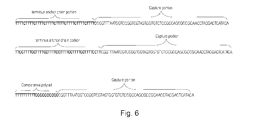
detecting Salmonella spp. The nucleic acid probes are shown in figure 6.
The nucleic acid probes were spotted to a solid support (PS substrate) in
different concentrations ranging from 1 pM to 60 pM.
A 25 pL of SP-PCR reaction mixture was prepared. The SP-PCR mixture consists
of 1 x Phusion Human Specimen PCR Buffer (Thermo Fisher Scientific), 400
nM of hiA forward and 1600 nM hi/A reverse primers, and 0.05 U/pL Phusion
Hot Start II High-Fidelity DNA polymerase (Thermo Fisher Scientific). A Gene
Frame (Thermo Fisher Scientific) was used to create a 25 pL reaction chamber
surrounding the solid support primer array. The PCR master mix was loaded by
pipette into the gene frame and sealed with a cover slip. The PS slide was
CA 03073462 2020-02-20
WO 2019/037828
PCT/D1(2018/050207
33
spotted with the nucleic acid probes. The SP-PCR was conducted in a flat-bed
PCR thermocycler, where a piece of 1 cm thick polystyrene insulation foam was
used to separate the slides from the lid of the PCR thermocycler. The SP-PCR
conditions were: 94 C 2 minutes follow by 30 cycles of 94 C for 10 seconds,
60 C for 20 seconds, 72 C for 20 seconds, then another 15 PCR cycles of 94 C
for 10 seconds, 65 C for 20 seconds, 72 C for 20 seconds. A higher annealing
temperature was used in the later 15 PCR cycles to enhance the SP-PCR. After
the SP- PCR, the chamber was washed with 0.1 x SSC and 0.1% of Sodium
dodecyl sulphate (SDS) (Promega, WI, USA) for 5 minutes then rinsed with
deionized water and dried at room temperature. The slide was ready for
scanning.
After the SP-PCR, the slides were scanned using a microscope (ZEISS Axiovert
200, Germany). Microarray image was analysed using Image] software
(Molecular devices). A circle was drawn and adjusted to the size of the spot
and the mean light intensity value was determined as signal. Another circle
was
drawn nearby was used as the background. The signal in this study was defined
as the signal of the 4 spots on the array, subtracting the mean background.
Figure 7 shows the resulting immobilization efficiency at different nucleic
acid
probe concentrations after the PS-PCR. It can easily be seen that the nucleic
acid probes of the invention has a much higher immobilization efficiency than
the comparative nucleic acid probe.
Figure 8 is an image of a control probe and two different capture portions ¨ a
Flic gene that targeting salmonella and a Brtz gene that targeting Bordetella
bacteria. As control probe the polytail 42TTCCTT7 was used
As shown in figure 7, the polytail 42TTCCTT7 that targeting Salmonella spp.
showed about the same round shape after SP PCR than before, which means
that the 42TTCCTT7 polytail help the entire probe to be immobilized on the
surface with a very high bonding efficiency.
CA 03073462 2020-02-20
WO 2019/037828
PCT/D1(2018/050207
34
The first process scheme for performing SP-PCR shown in figure 9a is a
standard process scheme
The second process scheme for performing SP-PCR shown in figure 9b is a
novel SP-PCR process, which has been made available due to the present
invention. Thanks to the high immobilization efficiency provided by the
nucleic
acid probes and the method of the invention the SP-PCR may now be
performed without washing after the UV crosslinking (immobilization) and/or
without washing after the PS-PCR procedure. .
Figure 10a are images of a solid support with spotted nucleic acid probes
subject to SP-PCR with and without washing.
Example 5
In example 5 the two nucleic acid probes of example 5 which represent
embodiments of the invention namely the nucleic acid probe a) a first nucleic
acid probe according to an embodiment of the invention had a polytail of the
nucleotide sequence 40TTTTC8 and a capture portion targeting a hilA gene
and b) a second nucleic acid probe according to an embodiment of the
invention had a polytail of the nucleotide sequence 42TTCCTT7 and a capture
portion targeting the hilA gene were used.
The spotting and the SP-PCR procedure was performed following the
procedure of example 4 using a nucleic acid probe concentration of 60 pM.
The result is shown in figure 10a prior to washing and after washing. Figure
10b shows the average of the signal minus background with washing and
without washing and it can be seen that there is a relatively low amount a
false positive in the non-washed samples.
Example 6
CA 03073462 2020-02-20
WO 2019/037828
PCT/D1(2018/050207
A number of different marked polytail/nucleic acid probes were subjected to
different UV exposure time and different UV dose for immobilization. The
polytail/nucleic acid probes used were as shown in figure 1.
The polytail/nucleic acid probes were spotted to the solid support as
5 described in example 1 but with different UV exposure.
Four spots of each polytails/nucleic acid probe were subjected to an UV
exposure from an 8 W UV emitter for 3 minutes. Four spots of each
polytails/nucleic acid probe were subjected to an UV exposure from a 16 W
UV emitter for 8 minutes.
10 The result is shown in figure 11 where the left plot for each
polytail/nucleic
acid probe is the 8 W UV emitter for 3 minutes treatment and the right left
plot for each polytail/nucleic acid probe is the 16 W UV emitter for 8 minutes
treatment. It appears that the 8 W UV emitter for 3 minutes treatment is
better than the 16 W UV emitter for 8 minutes treatment.
15 Example 7
A marked polytail with the sequence 40 TIC (also called 40TC20) was used in
this test. Samples of the polytail were spotted to the solid support as
described in example 1 but with different UV exposure.
For some samples the 8 W UV emitter was used and for other the 16 W UV
20 emitter was used. The exposure time was varied as shown in figure 12
where
the immobilization efficiency after wash is plotted as a function of the
exposure time for each of the two emitters.
It can be seen that the lower watt (8 Watt UV emitter) is better than the
higher watt emitter. Further, the 8 W emitter has an immobilization optimum
25 around 3 minutes which means that the nucleic acid probe can be
immobilized a rather low UV dosage, which is highly advantageous since the
risk of damaging the capture portion thereby may be reduced or even
avoided.
CA 03073462 2020-02-20
WO 2019/037828
PCT/D1(2018/050207
36
to an embodiment of the invention had a polytail of the nucleotide sequence
40TTTTC8 and a capture portion targeting a hilA gene was used.
3 different nucleic acid probes was synthesized and c) a comparative nucleic
acid probe with a polytail of the nucleotide sequence 20T1 C1 and a capture
.. portion with hilA gene for detecting Salmonella spp. The nucleic acid
probes
are shown in figure 6.
In figures 13a-13e the images of the immobilized polytails obtained at the
different UV exposure time before and after wash using the 8W UV emitter
are shown.
In figures 14a-14c the images of the immobilized polytails obtained at the
different UV exposure time before and after wash using the 16 W UV emitter
are shown.
Example 8
Nucleic acid probes with different length of polytails were tested. The
nucleic
acid probes comprised the Brtz gene that targets Bordetella bacteria.
The nucleic acid probes were spotted, immobilized and washed according to
the process described in example 1. Figure 15a show the signal minus
background for the various nucleic acid probes. It can be seen that the
nucleic
acid probes with very short polytails are difficult to immobilize and that
nucleic
acid probes of embodiments of the invention with polytails of 18 nucleotides
or
more show an effective immobilization. Figure 15b are images of the
immobilized nucleic acid probes.
Example 10
Samples of a nucleic acid probe of an embodiment of the invention having the
polytail 42TTCCTT7 and the capture portion that targeting Bordetella
bronchiseptica bacteria were tested.
CA 03073462 2020-02-20
WO 2019/037828
PCT/D1(2018/050207
37
The nucleic acid probe samples were spotted, immobilized and washed
according to the process described in example 1 but using a different UV
exposure time. After the immobilization the samples were subjected to PS-
PCR treatment as described in example 2.
After the PS-PCR treatment the signal minus background signal for each
sample were determined. Figure 16a show the results and it can be seen that
an effect immobilization of the nucleic acid probes if embodiments of the
invention may be obtained using very low UV dosage.
Figure 16b are images of the immobilized nucleic acid probes.
The SAF structure 1 corresponds to the SAF structures disclosed in
W017133741 and further details may be found in this document. The SAF
structure is mounted to a bottom 2 of a reaction section of a channel of a
microfluidic cartridge. The nucleic acid probes 3 marked with fluorophores
and of an embodiment of the invention are mounted to a top surface of the
SAF structure 1.
The SAF structure 1 has a conical frustum shape with the top surface The SAF
structure 1 has a protruding height, a top surface diameter, and a bottom
diameter. The excited fluorophores emit light anisotropically into the SAF
structure - which has a higher refractive index than the sample, the air or
the
liquid in the reaction section - with an angle above a supercritical angle
(ec).
The emitted light is collimated and can be read out by a reader as a circle of
light.
Figure 18 is a perspective view of a section of a reaction channel with the
edges 12 of a cartridge, where the reaction section comprises SAP structures
11 with immobilized nucleic acid probes, which have been subjected to SP-
CPR.
CA 03073462 2020-02-20
WO 2019/037828
PCT/D1(2018/050207
38
Figure 19 shows a SAF structure illustrated with a dotted top part to show the
frustum angle a. The SAF structure 21 has a bottom periphery 24 where it in
mounted to or integrated with the remaining part of the solid support. At its
bottom periphery, the SAF structure has a bottom diameter db. The SAF
structure has a top surface 23 with a diameter D. From the bottom to the top
surface, the SAF structure has the height h. The illustrated top part is an
imaginary top, shown to illustrate the frustum angle.
Figures 19a-19d illustrate a standard SAF structure 31, comprising top surface
33. The SAF structure is integrated with a remaining part of the solid support
32. Only some of the remaining part of the solid support is shown. As
explained above the solid support may form a cartridge with a channel or it
may be a part thereof.
Figure 19a is a perspective view of the SAF structure 31.
Figure 19b is a top view of the SAF structure 31.
Figure 19c is a side view of the SAF structure 31.
Figure 19d is a cross sectional view of the SAF structure 31 seen in the
section A-A of figure 19c.
The SAF structure has a height h and a top diameter D. D and h may
individually of each other be as disclosed elsewhere herein. The SAF
structure 31 is illustrated with a frustum angle of 60 degrees. It should be
understood that the SAF structure may have another frustum angle as
disclosed elsewhere herein.
It can be seen that the top surface is flat.
In an embodiment, the SAF structure 31 has the following dimensions:
D=0.2 mm; h =0.25 mm and the SAF frustum angle a is 60 degrees.
CA 03073462 2020-02-20
WO 2019/037828
PCT/D1(2018/050207
39
Figures 20a-20d illustrate a preferred SAF structure 41, comprising top
surface 43 with a recess 44. The SAF structure is integrated with a remaining
part of the solid support 42.
Figure 20a is a perspective view of the SAF structure 41.
Figure 20b is a top view of the SAF structure 41.
Figure 20c is a side view of the SAF structure 41.
Figure 20d is a cross sectional view of the SAF structure 41 seen in the
section A-A of figure 20c.
Figure 20e show a part of the figure 20a, where the recess edge width W is
marked. The recess edge width W is advantageously at least about 0.008,
such as at least about 0.01, such as at least about 0.015.
The SAF structure has a height h and a top diameter D. D and h may,
individually of each other, be as disclosed elsewhere herein. The SAF
structure height h is determine from the top surface 43 without the recess 44.
The recess has a height h1, which may be as disclosed elsewhere herein.
The recess is substantially round and is located such that its center axis is
coincident with the center axis of the SAF. The recess has a height h1, which
may be as disclosed elsewhere herein.
As it can be seen, the recess floor is substantially flat. The recess diameter
d
is determined at the floor of the recess and may be as disclosed elsewhere
herein.
The recess has a conical, frustum shape with a top surface formed by the
floor.
In the shown embodiment, the frustum angle 0 and the frustum angle a are
both 60 degrees. It should be understood that the recess frustum angle 0 and
CA 03073462 2020-02-20
WO 2019/037828
PCT/D1(2018/050207
the SAF frustum angle a may have other value(s) as disclosed elsewhere
herein.
The recess increases the spotting robustness and increase the read out signal
intensity.
5 In an embodiment, the SAF structure 41 has the following dimensions:
D=0.2 mm, h =0.25 mm, d =0.05 mm, h1 = 0.01 mm, the SAF frustum angle
a is 60 degrees and the recess frustum angle 0 is 60 degrees.
In another embodiment, the SAF structure 41 has the following dimensions:
D=0.2 mm, h =0.3 mm, d =0.05 mm, h1 = 0.01 mm, the SAF frustum angle
10 a is 60 degrees and the recess frustum angle 0 is 60 degrees.
Figures 21a-21d illustrate another preferred SAF structure 51, comprising top
surface 53 with a recess 54. The SAF structure is integrated with a remaining
part of the solid support 52.
Figure 21a is a perspective view of the SAF structure 51.
15 Figure 21b is a top view of the SAF structure 51.
Figure 21c is a side view of the SAF structure 51.
Figure 21d is a cross sectional view of the SAF structure 51 seen in the
section A-A of figure 21c.
The SAF structure has a height h and a top diameter D. D and h may,
20 individually of each other, be as disclosed elsewhere herein. The SAF
structure height h is determine from the top surface 53 without the recess 54.
The recess 54 has a height h1, which may be as disclosed elsewhere herein.
CA 03073462 2020-02-20
WO 2019/037828
PCT/D1(2018/050207
41
The recess is substantially round and is located such that its center axis is
coincident with the center axis of the SAF. The recess has a height h1, which
may be as disclosed elsewhere herein.
As it can be seen, the recess floor is substantially flat. The recess diameter
d
is determined at the floor of the recess and may be as disclosed elsewhere
herein.
The recess has rounded recess edge a rounding radius R, which may be as
disclosed elsewhere herein.
In an embodiment, the SAF structure 51 has the following dimensions:
D=0.2 mm; h =0.25 mm, d =0.08 mm, h1 = 0.01 mm, the recess edge is
rounded with a radius R=0.05 mm and the SAF frustum angle a is 60
degrees.
In another embodiment, the SAF structure 51 has the following dimensions:
D=0.2 mm; h =0.3 mm, d =0.08 mm, h1 = 0.01 mm, the recess edge is
rounded with a radius R=0.05 mm and the SAF frustum angle a is 60
degrees.
EXAMPLE 11
Seven cartridges were produced from polystyrene. Each cartridge had a
microfluidic channel with a reaction section and eight identical SAF
structures
protruding from the wall in the reaction section.
The SAF structures of the first cartridge were shaped as the SAF structure no.
1 in figure 22; the structures of the second cartridge were shaped as the SAF
structure no.2 in figure 22 and so on.
SAF structure no. 3 was of the type shown in figures 19a-19d.
CA 03073462 2020-02-20
WO 2019/037828
PCT/D1(2018/050207
42
SAF structure no. 5 was of the type shown in figures 21a-21d and SAF
structure no. 6 was of the type shown in figures 20a-20e.
An equal amount of Cy3-labelled oligo was spotted onto the top surface of
each of the respective SAF structures and allowed to dry.
In the first test round, the reaction chambers was maintained filled with air.
The Cy3-labels were subjected to light at the excitation wavelength (-550 nm)
and the signal intensities of the SAF structure emission signals were
detected.
For each cartridge, the average SAF structure air/PS interface intensity
signal
was determined.
In the second test round, the reaction chambers of the respective cartridges
were filled with water. The Cy3-labels were subjected to light at the
excitation
wavelength (-550 nm) and the signal intensities of the SAF structure emission
signals were detected.
For each cartridge, the average SAF structure water/PS interface intensity
signal was determined.
The results are shown in figure 23.
The coefficient of variation (Coy) was determined for the SAF structures of
the respective cartridges.
It can be seen that the SAF structures nos. 1 and 2 had relatively high light
intensities for the air/PS interface signals. Both for the air/PS interface
signal
intensities and the water/PS interface signals, the Coy were however,
relatively high.
The SAF structures no. 6 had the highest light intensity for the water/PS
interface signals and the Coy. Both for the air/PS interface signal
intensities
.. and the water/PS interface signals, the Coy were however, relatively high.
CA 03073462 2020-02-20
WO 2019/037828
PCT/D1(2018/050207
43
The Coy for sample 6 was however relatively high. It is believed that the
reason for this relatively high Coy is that the some of the SAF structures at
their bottom periphery where they are integrated with the remaining solid
support, had small surface corrugations and/or protrusions, which may result
in loss of signal. Hence, it is expected that by decreasing the top diameter
to
height aspect ratio D/h, this possibly loss of light signal may be mitigated.
It
is believed that by increasing the height e.g. to about 0.3 mm, the signal may
be increased and the Coy may be decreased.
The SAF structures no. 5 had both a highest light intensity and a low Coy for
the water/PS interface signals.
It is believed that the rounded edges of the recess ensure a very effective
and
robust spotting, which add to the low Coy.
The SAF structures no. 6 had the highest light intensity for the water/PS
interface signals and the Coy. Both for the air/PS interface signal
intensities
and the water/PS interface signals, the Coy were however, relatively high.