Note: Descriptions are shown in the official language in which they were submitted.
CA 03073737 2020-02-21
WO 2019/046802 PCT/US2018/049240
SPHINCTEROTOME DEVICE AND METHODS AND USES THEREOF
TECHNICAL FIELD
[0001] The present description relates in general to medical devices,
and more
particularly to, for example, without limitation, sphincterotome devices,
methods and uses
thereof.
BACKGROUND OF THE DISCLOSURE
[0002] An estimated 1,230,000 endoscopic retrograde
cholangiopancreatography
("ERCP") procedures were performed in the 28 member countries of the European
Union and
the United States in 2016. As part of an ERCP procedure, cannulation must
first be achieved in
order to gain access to the desired duct(s); however, this can sometimes be
challenging. One
approach is to use a sphincterotome device (also called a papillotome),
inserted through a
working channel of a duodenoscope. A sphincterotome is a catheter that
contains an
electrosurgical cutting wire at the distal end, which is used to perform
sphincterotomies (i.e.,
cutting of sphincter muscles in order to gain duct access to perform follow-up
procedures).
[0003] The description provided in the background section should not be
assumed to
be prior art merely because it is mentioned in or associated with the
background section. The
background section may include information that describes one or more aspects
of the subject
technology.
BRIEF DESCRIPTION OF THE DRAWINGS
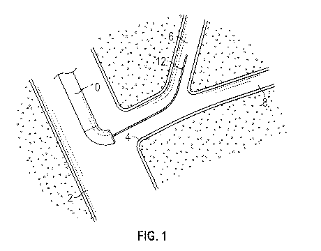
[0004] FIG. 1 illustrates a diagram of a stage of a biliary cannulation
procedure using
a sphincterotome device.
[0005] FIG. 2 illustrates a diagram of another stage of the biliary
cannulation
procedure using a sphincterotome device.
[0006] FIG. 3 illustrates a diagram of another stage of the biliary
cannulation
procedure using a sphincterotome device.
[0007] FIG. 4 illustrates a diagram of another stage of the biliary
cannulation
procedure using a sphincterotome device.
[0008] FIG. 5 illustrates a plan view of an example of a sphincterotome
device.
- 1 -
CA 03073737 2020-02-21
WO 2019/046802 PCT/US2018/049240
[0009] FIG. 6 illustrates a perspective view of a distal end portion of
a
sphincterotome device.
[0010] FIG. 7 illustrates a perspective view of a distal end portion of
a
sphincterotome device.
[0011] FIG. 8 illustrates a sectional view of the sphincterotome device
of FIG. 7
taken along line A-A.
[0012] FIG. 9 illustrates a sectional view of an alternative
sphincterotome device.
[0013] FIG. 10 illustrates a diagram of a stage of a biliary
cannulation procedure
using a sphincterotome device.
[0014] FIG. 11 illustrates a diagram of another stage of the biliary
cannulation
procedure using a sphincterotome device.
[0015] FIG. 12 illustrates a diagram of another stage of the biliary
cannulation
procedure using a sphincterotome device.
[0016] In one or more implementations, not all of the depicted
components in each
figure may be required, and one or more implementations may include additional
components
not shown in a figure. Variations in the arrangement and type of the
components may be made
without departing from the scope of the subject disclosure. Additional
components, different
components, or fewer components may be utilized within the scope of the
subject disclosure.
DETAILED DESCRIPTION
[0017] The detailed description set forth below is intended as a
description of various
implementations and is not intended to represent the only implementations in
which the subject
technology may be practiced. As those skilled in the art would realize, the
described
implementations may be modified in various different ways, all without
departing from the scope
of the present disclosure. Accordingly, the drawings and description are to be
regarded as
illustrative in nature and not restrictive.
[0018] An exemplary embodiment of a sphincterotome device of the
present
disclosure can be used to achieve appropriate positioning within the biliary
duct during
cannulation. The common biliary duct splits into left and right hepatic ducts,
and an exemplary
sphincterotome device of the present disclosure may help with accessing these
ducts more easily.
In some cannulation procedures, high obstruction of the biliary tree may
require the placement of
two guidewires. In order to place the two wires, a sphincterotome device might
need to be taken
- 2 -
CA 03073737 2020-02-21
WO 2019/046802 PCT/US2018/049240
out of one duct after the first wire is placed, which then allows a second
wire to be placed. An
example embodiment of the present disclosure allows both wires to be placed
without the need
for removal of the sphincterotome, thus shortening the procedure.
[0019] In some embodiments of the present disclosure, an exemplary
sphincterotome
device makes the "double-wire" technique easier to perform, as it would allow
manipulation of
the biliary duct while at the same time manipulating the second wire to direct
it into the bile duct.
When a guidewire or the sphinctertome are inserted into the pancreatic duct,
it may facilitate the
second wire entry into the bile duct but blocking the pancreatic duct or
stretching the biliary
duct, thus making it easier to access the biliary duct with the second wire.
In some instances, it is
more difficult to pass a wire through the biliary duct when the catheter is
pushed into it as it may
fold the duct leading to blocking the passage of the wires.
[0020] One or more embodiments of the present disclosure may include
various
advantages during a "double-wire" or "multiple wire" technique. For example,
some
embodiments may allow for a shorter procedure because there is no need to
remove the
sphincterotome device and reintroduce it again over the first guidewire; the
second guidewire
may be inserted immediately and is positioned in such a way that it may enter
the biliary duct.
Additionally, one or more embodiments of the present disclosure may reduce the
risk of post-
ERCP pancreatitis because there would be a lesser need for the often-difficult
manipulation of
two guidewires to achieve correct angulation and avoid the possibility of
leaving a wire in the
pancreatic duct for a longer time.
[0021] Accordingly, embodiments of the present disclosure may achieve
advantages
such as shorter and easier endoscopic retrograde cholangiopancreatographies,
for example,
involving patients with challenging ductal anatomies. Additionally,
embodiments of the present
disclosure may lead to higher successful cannulation rates; a reduced risk of
post-ERCP
pancreatitis; increased safety by reducing the need for more invasive
procedures; and the ability
to conserve time, devices, and resources when performing further intrahepatic
procedures after
biliary cannulation.
[0022] Referring now to FIGS. 1-4, an example of a biliary cannulation
procedure is
illustrated along with the use of a sphincterotome device. In some procedures,
a guidewire 12
can be inserted through the lumen of a sphincterotome 10, which extends
through the duodenum
2 to the location of a sphincter 4 (i.e., sphincter of Oddi) at the major
duodenal papilla. The
- 3 -
CA 03073737 2020-02-21
WO 2019/046802 PCT/US2018/049240
guidewire 12 is then manipulated to achieve biliary duct cannulation into the
biliary duct 6, as
shown in FIG. 1.
[0023] In some procedures, it can also be challenging to reach the
biliary duct 6, in
which case the guidewire 12 may be placed in the pancreatic duct 8 instead, as
shown in FIG. 2.
In this scenario, a "double-wire" technique can be used so that more
aggressive and risky
techniques can be avoided. As shown in FIG. 3, in an exemplary double-wire
technique, the first
guidewire 12 is left in the pancreatic duct 8, the sphincterotome is
withdrawn. As shown in FIG.
4, a second guidewire 14 is inserted into the sphincterotome 10. The
sphincterotome 10 is
inserted next to the first guidewire 12 in order to enter the biliary duct 6
more easily, as shown in
FIG. 4. In some instances, contrast dye may be injected through the
sphincterotome 10 to
confirm such placement.
[0024] To reduce operation complexity and duration, a sphincterotome
device can be
provided with features that facilitate entry into both the pancreatic duct and
the biliary duct. An
example of such a device is shown in FIGS. 5-9. While these figures illustrate
different features,
it will be understood that the features can be implemented together or
alternatively.
[0025] As shown in FIG. 5, a sphincterotome 100 can include a proximal
portion 102
for operation by a user and a distal portion 106 for insertion into a patient.
At the proximal
portion 102, various features can be provided for interaction by a user to
operate the distal
portion 106. For example, a handle 110 and a slider 112 can be provided for
actuating a cutting
wire 116 at the distal portion 106. The cutting wire 116 can be retracted
and/or extended based
on movement of the slider 112 relative to the handle 110. By moving the
cutting wire 116, the
distal portion 106 can be deflected relative to a middle portion 104 of the
shaft 160 of the
sphincterotome 100, as discussed further herein. The cutting wire 116 may
comprise a conductor.
For example, the cutting wire 116 may be formed from a conductive metal that
can be electrified
during cutting (e.g., for heating), if desired.
[0026] The proximal portion can further include one or more infusion
ports. A
proximal fluid entry port 120 at the proximal portion 102 is provided in fluid
communication
with a proximal fluid exit port at the distal portion 106. Fluid can be
provided at the proximal
fluid entry port 120 for infusion at the distal portion 106 by way of a lumen
extending there
between, as discussed further herein. A distal fluid entry port 130 at the
proximal portion 102 is
provided in fluid communication with a distal fluid exit port at the distal
portion 106. Fluid can
- 4 -
CA 03073737 2020-02-21
WO 2019/046802 PCT/US2018/049240
be provided at the distal fluid entry port 130 for infusion at the distal
portion 106 by way of a
lumen extending there between, as discussed further herein.
[0027] The proximal portion can further include one or more ports for
controlling
guidewires. A proximal guidewire entry port 140 at the proximal portion 102 is
provided with a
proximal guidewire 142 extending therein and toward the distal portion 106. A
distal guidewire
entry port 150 at the proximal portion 102 is provided with a distal guidewire
152 extending
therein and toward the distal portion 106. The proximal guidewire 142 and the
distal guidewire
152 can exit the distal portion 106 at different locations and/or angles to
provide access and entry
into different areas of the body.
[0028] FIGS. 6 and 7 illustrate embodiments and various components of
the
sphincterotome 100. A shaft can be rotatable to change the position of the
distal end of the
sphincterotome. A shaft can include multiple lumens. In the depicted example,
five lumens are
provided. At the distal portion, each lumen can terminate to provide access
for fluid or other
devices. Lumens may be used to convey guidewires, which correspond to the
proximal and
distal guidewire lumens. Two more lumens may be used for contrast injection,
which correspond
to proximal and distal injection ports. In some embodiments, the proximal
guidewire and
injection lumens/ports may be fused into one. The sphincterotome device of the
present
disclosure also may contain one lumen for a cutting wire. The distal tip of an
exemplary
sphincterotome shaft may be 5 millimeters long and pre-curved in some
embodiments. The
diameter of an exemplary sphincterotome shaft may be approximately 7 Fr, but
could also be
smaller. In some embodiments, the sphincterotome shaft of the present
disclosure may be
compatible with standard duodenoscopes that include a 4.2 millimeter working
channel, although
this description is not limiting to that effect.
[0029] As shown in FIG. 6, a distal guidewire exit port 156 can be used
with the
distal guidewire 152. The distal guidewire exit port 156 can be on the
distalmost tip of the shaft
160. The distal guidewire exit port 156 can be used to direct the distal
guidewire 152 into the
pancreatic or biliary duct. In this way, pancreatic duct entry stretches the
bile duct, thus
facilitating second guidewire insertion through the proximal guidewire exit
port 146 into the
biliary duct without need to withdraw the shaft 160. If biliary cannulation is
achieved at first
attempt using the distal guidewire 152, the shaft 160 can be left in place for
appropriate
positioning and further intrahepatic manipulation.
- 5 -
CA 03073737 2020-02-21
WO 2019/046802 PCT/US2018/049240
[0030] As further shown in FIGS. 6 and 7, a proximal guidewire exit
port 146 can be
used with the proximal guidewire 142. The proximal guidewire exit port 146 can
be on a cutting
wire side of the shaft 160, e.g., approximately 0.2 to 0.25 inches
(approximately 0.5 to 1.0
centimeters) away from the distalmost tip of the shaft 160. In some
embodiments, the proximal
guidewire 142 is inserted into the proximal guidewire exit port 146 if a first
attempted biliary
cannulation using the distal guidewire 152 is unsuccessful. The proximal
guidewire exit port 146
is oriented at an oblique angle 148 with respect to a central axis 162 of the
shaft 160. Such an
angle can be 20-80 , 40-80 , 50-70 , or about 60 . The angle can be selected
to align with the
bile duct anatomy of a patient to facilitate biliary cannulation. The proximal
guidewire exit port
146 can also be used for contrast injection to confirm placement of the
proximal guidewire exit
port 146.
[0031] As further shown in FIGS. 6 and 7, a distal fluid exit port 136
can be located
on the distalmost tip of the shaft 160. The distal fluid exit port 136 can be
used for contrast
injection, which facilitates placement of the distal guidewire 152. The distal
fluid exit port 136
can optionally be aligned with the central axis 162 of the shaft 160.
[0032] As further shown in FIGS. 6 and 7, a proximal fluid exit port
126 can be
located proximal to the distalmost end of the shaft 160. The proximal fluid
exit port 126 can be
used for contrast injection, which facilitates placement of the proximal
guidewire 142. The
proximal fluid exit port 126 can be on a cutting wire side of the shaft 160,
e.g., approximately
0.2 to 0.25 inches (approximately 0.5 to 1.0 centimeters) away from the
distalmost tip of the
shaft 160. The proximal fluid exit port 126 is oriented at an oblique angle
128 with respect to a
central axis 162 of the shaft 160. Such an angle can be 20-80 , 40-80 , 50-70
, or about 60 .
The angle 128 can be selected to align with the bile duct anatomy of a patient
to facilitate biliary
cannulation. The angle 128 can be the same as or different from the angle 148.
[0033] As further shown in FIGS. 6 and 7, one or more cutting wires
such as a cutting
wire 116 can be placed within a cutting wire lumen and attached near a distal
end of the shaft
160. An exposed portion of the cutting wire 116 can be approximately 0.1 inch
or 20 to 30
millimeters long and positioned on a radial side of the shaft. The cutting
wire 116 can be pulled
to bend the distal portion 106 of the shaft 160, and the flexibility allows
the sphincterotome
device to accommodate a wider range of individual patient anatomies. The
cutting wire 116 can
extend outside the shaft 160 to connect to an end (FIG. 6) distal to the
proximal guidewire exit
- 6 -
CA 03073737 2020-02-21
WO 2019/046802 PCT/US2018/049240
port 146 and/or the proximal fluid exit port 126 or to a portion of the shaft
160 that is proximal to
the proximal guidewire exit port 146 and/or the proximal fluid exit port 126
(FIG. 7). It will be
recognized that other placements of the cutting wire 116 can be selected to
providing bending as
desired.
[0034] FIG. 8 illustrates an example of a design of a sphincterotome
device. In some
embodiments, and illustrated in FIG. 8, the cutting wire 116 is positioned at
about 12 o'clock
relative to a transverse cross-section of the shaft 160 and within a cutting
wire lumen 114. The
proximal guidewire lumen 144 is at about 11 o'clock relative to a transverse
cross-section of the
shaft 160 (e.g., about 30 from or within 45 from the cutting wire 116). The
circumferential
offset of the proximal guidewire lumen 144 and the cutting wire lumen 114
allows the cutting
wire 116 and the proximal guidewire 142 to exit at different circumferential
locations at the
distal portion of the shaft 160. The sphincterotome 100 of FIG. 8 can also
contain a distal
guidewire lumen 154 and/or two contrast injection lumens: a proximal fluid
lumen 124 and a
distal fluid lumen 134. The proximal fluid lumen 124 fluidly connects the
proximal fluid entry
port 120 with the proximal fluid exit port 126. The distal fluid lumen 134
fluidly connects the
distal fluid entry port 130 with the distal fluid exit port 136. Thus, in the
exemplary embodiment
of FIG. 8, the sphincterotome device can include five lumens: two for contrast
injection, two for
guidewires, and one for the cutting wire.
[0035] In some embodiments, for 0.025-inch guidewires, the lumens for
the proximal
and distal guidewires may be approximately the same size (i.e., in diameter).
Use of a 0.025-inch
wire for the first guidewire, in the event the first guidewire enters the
pancreatic duct, is less
likely to cause pancreatitis than would a larger wire. Moreover, such smaller
guidewires can be
advanced further and become more stable and are almost as stable as 0.035-inch
wires. In some
embodiments, the external diameter of the sphincterotome device should not be
larger than 7 Fr,
and will depend on the exact engineering/design of the sphincterotome device.
Thus, having
0.025 inch wires may help decrease the total outer diameter of the
sphincterotome device. Other
diameters of wire may also be used to achieve a similar result, and this
description is not meant
to be limiting with respect to other feasible wire sizes.
[0036] In some embodiments, the proximal exit ports on the side of the
shaft are
placed 0.5 to1.0 centimeters away from the tip of the sphincterotome device.
Positioning of the
proximal exit ports at other distances with respect to the tip of the
sphincterotome device may
- 7 -
CA 03073737 2020-02-21
WO 2019/046802 PCT/US2018/049240
provide similar results. Regardless, in some embodiments, the guidewire can
exit the
sphincterotome device at an angle relative to the sphincterotome device at
600. In some patients,
the biliary tract is angled at 30-40 with respect to the pancreatic duct, and
with appropriate
movement of an exemplary sphincterotome device of the present disclosure,
cannulation of a
greater number of patients can be achieved with an angle of 50-70 (e.g.,
approximately 60').
Other angles, greater than or less than 60 , can be used to achieve a similar
result.
[0037] FIG. 9 illustrates another example of a design of a
sphincterotome device.
While some features are the same as the design illustrated in FIG. 8, it will
be understood that a
smaller number of fluid lumens can be included. For example, the
sphincterotome 100 of FIG. 8
can include only one contrast injection lumen, such as the distal fluid lumen
134. Alternatively,
the sole lumen can be the proximal fluid lumen 124. Thus, in the exemplary
embodiment of
FIG. 9, the sphincterotome device has four lumens in total: one for contrast
injection, two for
guidewires (one of which can also be used for contrast injection) and one for
the cutting wire.
[0038] Although the examples of FIGS. 6-9 show lumens 114, 144, and 154
as each
accommodating a single wire (e.g., a cutting wire, a first guidewire, and a
second guidewire
respectively), it should be appreciated that any or all of lumens 114, 144,
154 can accommodate
more than one wire at the same time or at different times (e.g., by providing
guidewires or
cutting wires that are smaller in cross-sectional diameter than those shown in
the noted figures or
by providing lumens 114, 144, and/or 154 that are larger in cross-sectional
diameter than those
shown).
[0039] Moreover, it should be appreciated that any or all of lumens
114, 144, 154,
124, and/or 134 can be provided with a wire (e.g., a cutting wire or a
guidewire) therein or a wire
and/or one or more other suitable medical devices can be provided separately
from, and later
inserted into and/or removed from or through the lumen. For example, in some
scenarios, the
sphincterotome 100 can be used to deliver two or more wires to a cavity (e.g.,
a fluid collection,
or an abscess) to secure access to place multiple devices such as stents or
dilators, or to obtain
one or more samples such as fluid samples or tissue samples.
[0040] Referring now to FIGS. 10-12, an example of a biliary
cannulation procedure
is illustrated along with the use of a sphincterotome device.
[0041] As shown in FIG. 10, a shaft 160 of a sphincterotome can be
provided through
a duodenum 2 to the location of a sphincter 4 (i.e., sphincter of Oddi) at the
major duodenal
- 8 -
CA 03073737 2020-02-21
WO 2019/046802 PCT/US2018/049240
papilla. Access can be facilitated by use of a duodenoscope (not shown) in
concert with the
sphincterotome. The distal guidewire 152 and/or the shaft 160 is advanced to
achieve
cannulation into either the biliary duct 6 or the pancreatic duct 8. In some
cases, cannulation
with the distal guidewire 152 is achieved into the pancreatic duct 8, rather
than the biliary duct 6.
Injection of contrast can be performed to verify the position of the distal
guidewire 152.
[0042] As shown in FIG. 11, the distal guidewire 152 can be maintained
within the
pancreatic duct 8, and the shaft 160 can be advanced over the distal guidewire
152 to align with
the biliary duct 6. In particular, proximal exit ports of the shaft 160 can be
positioned axially and
on a radial side of the shaft 160 to align with the biliary duct 6.
[0043] As shown in FIG. 12, the proximal guidewire 142 can be extended
out of a
guidewire exit port to achieve cannulation into the biliary duct 6. Based on
the position and
orientation of the guidewire exit port, the extension of the proximal
guidewire 142 should be
aligned with the position and orientation of a pathway into the biliary duct
6. Injection of
contrast can be performed to verify the position of the proximal guidewire
142.
[0044] Additional procedures can be performed after or during
cannulation. For
example, the cutting wire 116 can be actuated to bend a distal portion of the
shaft 160 as desired.
The shaft 160 and/or the cutting wire 116 can maneuver within or otherwise act
on nearby
anatomy. By further example, the sphincterotome can cut sphincter muscles of
the biliary duct
with the cutting wire 116. An endoscopic retrograde cholangiopancreatography
can be
performed on the patient.
- 9 -
CA 03073737 2020-02-21
WO 2019/046802 PCT/US2018/049240
[0045] By further example, the shaft 160 can be withdrawn while
maintaining the
proximal guidewire 142 within the biliary duct 6. Additionally or
alternatively, the shaft 160 can
be withdrawn while maintaining the distal guidewire 152 within the pancreatic
duct 8. Other
tools and/or devices can be advanced, operated, and/or withdrawn over or along
the proximal
guidewire 142 and/or the distal guidewire 152. By way of additional examples,
additional wires
can be used in conjunction with the sphincterotome for other procedures such
as dilation or stent
procedures or obtaining a tissue sample.
[0046] It is understood that the specific order or hierarchy of steps,
operations, or
processes disclosed is an illustration of exemplary approaches. Unless
explicitly stated
otherwise, it is understood that the specific order or hierarchy of steps,
operations, or processes
may be performed in different order. Some of the steps, operations, or
processes may be
performed simultaneously. The accompanying method claims, if any, present
elements of the
various steps, operations or processes in a sample order, and are not meant to
be limited to the
specific order or hierarchy presented. These may be performed in serial,
linearly, in parallel or
in different order.
[0047] A reference to an element in the singular is not intended to
mean one and only
one unless specifically so stated, but rather one or more. For example, "a"
module may refer to
one or more modules. An element proceeded by "a," "an," "the," or "said" does
not, without
further constraints, preclude the existence of additional same elements.
[0048] Headings and subheadings, if any, are used for convenience only
and do not
limit the invention. The word exemplary is used to mean serving as an example
or illustration.
To the extent that the term include, have, or the like is used, such term is
intended to be inclusive
in a manner similar to the term comprise as comprise is interpreted when
employed as a
transitional word in a claim. Relational terms such as first and second and
the like may be used
to distinguish one entity or action from another without necessarily requiring
or implying any
actual such relationship or order between such entities or actions.
[0049] Phrases such as an aspect, the aspect, another aspect, some
aspects, one or
more aspects, an implementation, the implementation, another implementation,
some
implementations, one or more implementations, an embodiment, the embodiment,
another
embodiment, some embodiments, one or more embodiments, a configuration, the
configuration,
another configuration, some configurations, one or more configurations, the
subject technology,
- 10 -
CA 03073737 2020-02-21
WO 2019/046802 PCT/US2018/049240
the disclosure, the present disclosure, other variations thereof and alike are
for convenience and
do not imply that a disclosure relating to such phrase(s) is essential to the
subject technology or
that such disclosure applies to all configurations of the subject technology.
A disclosure relating
to such phrase(s) may apply to all configurations, or one or more
configurations. A disclosure
relating to such phrase(s) may provide one or more examples. A phrase such as
an aspect or
some aspects may refer to one or more aspects and vice versa, and this applies
similarly to other
foregoing phrases.
[0050] A phrase "at least one of' preceding a series of items, with the
terms "and" or
"or" to separate any of the items, modifies the list as a whole, rather than
each member of the
list. The phrase "at least one of' does not require selection of at least one
item; rather, the phrase
allows a meaning that includes at least one of any one of the items, and/or at
least one of any
combination of the items, and/or at least one of each of the items. By way of
example, each of
the phrases "at least one of A, B, and C" or "at least one of A, B, or C"
refers to only A, only B,
or only C; any combination of A, B, and C; and/or at least one of each of A,
B, and C.
[0051] In one aspect, a term coupled or the like may refer to being
directly coupled.
In another aspect, a term coupled or the like may refer to being indirectly
coupled.
[0052] Terms such as top, bottom, front, rear, side, horizontal,
vertical, distal,
proximal, and the like refer to an arbitrary frame of reference, rather than
to the ordinary
gravitational frame of reference. Thus, such a term may extend upwardly,
downwardly,
diagonally, or horizontally in a gravitational frame of reference.
[0053] The disclosure is provided to enable any person skilled in the
art to practice
the various aspects described herein. In some instances, well-known structures
and components
are shown in block diagram form in order to avoid obscuring the concepts of
the subject
technology. The disclosure provides various examples of the subject
technology, and the subject
technology is not limited to these examples. Various modifications to these
aspects will be
readily apparent to those skilled in the art, and the principles described
herein may be applied to
other aspects.
[0054] All structural and functional equivalents to the elements of the
various aspects
described throughout the disclosure that are known or later come to be known
to those of
ordinary skill in the art are expressly incorporated herein by reference and
are intended to be
encompassed by the claims. Moreover, nothing disclosed herein is intended to
be dedicated to
-11-
CA 03073737 2020-02-21
WO 2019/046802 PCT/US2018/049240
the public regardless of whether such disclosure is explicitly recited in the
claims. No claim
element is to be construed under the provisions of 35 U.S.C. 112, sixth
paragraph, unless the
element is expressly recited using the phrase "means for" or, in the case of a
method claim, the
element is recited using the phrase "step for".
[0055] The title, background, brief description of the drawings,
abstract, and
drawings are hereby incorporated into the disclosure and are provided as
illustrative examples of
the disclosure, not as restrictive descriptions. They are submitted with the
understanding that
they will not be used to limit the scope or meaning of the claims. In
addition, in the detailed
description, it can be seen that the description provides illustrative
examples and the various
features are grouped together in various implementations for the purpose of
streamlining the
disclosure. The method of disclosure is not to be interpreted as reflecting an
intention that the
claimed subject matter requires more features than are expressly recited in
each claim. Rather,
as the claims reflect, inventive subject matter lies in less than all features
of a single disclosed
configuration or operation. The claims are hereby incorporated into the
detailed description,
with each claim standing on its own as a separately claimed subject matter.
[0056] The claims are not intended to be limited to the aspects
described herein, but
are to be accorded the full scope consistent with the language of the claims
and to encompass all
legal equivalents. Notwithstanding, none of the claims are intended to embrace
subject matter
that fails to satisfy the requirements of the applicable patent law, nor
should they be interpreted
in such a way.
- 12-