Note: Descriptions are shown in the official language in which they were submitted.
CA 03074130 2020-02-26
WO 2019/057099 PCT/CN2018/106618
NOVEL ANTI-CD3EPSILON ANTIBODIES
FIELD OF THE INVENTION
[0001] The present disclosure generally relates to novel anti-human CD3epsilon
antibodies.
BACKGROUND
[0002] The CD3 (cluster of differentiation 3) T-cell co-receptor is a protein
complex and is
composed of four distinct chains, a CD3gamma chain, a CD3delta chain, and two
CD3epsilon
chains. These chains associate with a molecule known as the T-cell receptor
(TCR) and the
zeta-chain to generate activation signal in T lymphocytes. The TCR, zeta-
chain, and CD3
molecules together comprise the TCR complex, in which TCR as a subunit
recognizes and
binds to antigen, and CD3 as a subunit transfers and conveys the antigen-
stimulation to
signaling pathway, and ultimately regulates T-cell activity. The CD3 protein
is virtually present
in all T cells.
[0003] The CD3 together with TCR forms a CD3-TCR complex, which plays pivotal
role in
modulating T cell vast functions in both innate and adoptive immune response,
as well as
cellular and humoral immune functions. These include eliminating pathogenic
organisms and
controlling tumor growth by broad range of cytotoxic effects.
[0004] Mouse monoclonal antibodies specific for human CD3, such as OKT3 (Kung
et al.
(1979) Science 206: 347-9), were the first generation CD3 antibodies for
treatment. Although
OKT3 has strong immunosuppressive potency, its clinical use was hampered by
serious side
effects linked to its immunogenic and mitogenic potentials (Chatenoud (2003)
Nature Reviews
3:123-132). OKT3 induced an anti-globulin response, promoting its own rapid
clearance and
neutralization (Chatenoud et al. (1982) Eur. J. Immunol. 137:830-8). In
addition, OKT3
induced T-cell proliferation and cytokine production in vitro, and led to a
large scale release of
cytokine in vivo (Hirsch et al. (1989) J. Immunol 142: 737-43, 1989). The
cytokine release
(also referred to as "cytokine storm") in turn led to a "flu-like" syndrome,
characterized by
fever, chills, headaches, nausea, vomiting, diarrhea, respiratory distress,
septic meningitis and
hypotension (Chatenoud, 2003). Such serious side effects limited the more
widespread use of
OKT3 in transplantation as well as the extension of its use to other clinical
fields such as
autoimmunity (Id.).
[0005] There is a significant need for novel anti-CD3 antibodies.
1
CA 03074130 2020-02-26
WO 2019/057099 PCT/CN2018/106618
BRIEF SUMMARY OF THE INVENTION
[0004] Throughout the present disclosure, the articles "a," "an," and "the"
are used herein to
refer to one or to more than one (i.e., to at least one) of the grammatical
object of the article.
By way of example, "an antibody" means one antibody or more than one antibody.
[0005] The present disclosure provides novel monoclonal anti-CD3epsilon
antibodies,
amino acid and nucleotide sequences thereof, and uses thereof.
[0006] In one aspect, the present disclosure provides isolated antibodies or
antigen-binding
fragments thereof, comprising 1, 2, or 3 heavy chain CDR sequences selected
from the group
consisting of: SEQ
NOs: 1, 3, 5, 7, 9, 11, 13, 15, 17, 19, 21, 23, 25, 27, 29, 31, 33, 35, 37,
39, 41, 43, 45, and 47, and/or 1, 2, or 3 kappa light chain CDR sequences
selected from the
group consisting of: SEQ ID NOs: 2, 4, 6, 8, 10, 12, 14, 16, 18, 20, 22, 24,
26, 28, 30, 32, 34,
36, 38, 40, 42, 44, 46 and 48.
[0007] In
certain embodiments, the antibodies or antigen-binding fragments thereof
comprise 1, 2, or 3 heavy chain CDR sequences having at least 80% (e.g. at
least 85%, 88%,
90%, 91%, 92%, 93%, 94%, 95%, 96%, 97%, 98%, 99%) sequence identity to the
sequence of
SEQ
NOs: 1,3, 5, 7, 9, 11, 13, 15, 17, 19, 21, 23, 25, 27, 29, 31, 33, 35, 37, 39,
41, 43, 45,
or 47. In certain embodiments, the antibodies or antigen-binding fragments
thereof comprise
1, 2, or 3 light chain CDR sequences having at least 80% (e.g. at least 85%,
88%, 90%, 91%,
92%, 93%, 94%, 95%, 96%, 97%, 98%, 99%) sequence identity to the sequence of
SEQ ID
NOs: 2, 4, 6, 8, 10, 12, 14, 16, 18, 20, 22, 24, 26, 28, 30, 32, 34, 36, 38,
40, 42, 44, 46 or 48.
[0008] In certain embodiments, the antibodies or antigen-binding fragments
thereof comprise
a heavy chain variable region selected from the group consisting of:
a) a heavy chain variable region comprising 1, 2, or 3 CDR sequences
selected from SEQ
ID NO: 1, SEQ ID NO: 3, and SEQ ID NO: 5;
b) a heavy chain variable region comprising 1, 2, or 3 CDR sequences
selected from SEQ
ID NO: 7, SEQ ID NO: 9, and SEQ ID NO: 11;
c) a heavy chain variable region comprising 1, 2, or 3 CDR sequences
selected from SEQ
ID NO: 13, SEQ ID NO: 15, and SEQ ID NO: 17;
d) a heavy chain variable region comprising 1, 2, or 3 CDR sequences
selected from SEQ
ID NO: 19, SEQ ID NO: 21, and SEQ ID NO: 23;
e) a heavy chain variable region comprising 1, 2, or 3 CDR sequences
selected from SEQ
ID NO: 25, SEQ ID NO: 27, and SEQ ID NO: 29;
2
CA 03074130 2020-02-26
WO 2019/057099 PCT/CN2018/106618
f) a heavy chain variable region comprising 1, 2, or 3 CDR sequences
selected from SEQ
ID NO: 31, SEQ NO: 33, and SEQ NO: 35;
g) a heavy chain variable region comprising 1, 2, or 3 CDR sequences
selected from SEQ
ID NO: 37, SEQ ID NO: 39, and SEQ ID NO: 41; and
h) a heavy chain variable region comprising 1, 2, or 3 CDR sequences
selected from SEQ
ID NO: 43, SEQ ID NO: 45, and SEQ ID NO: 47.
[0009] In certain embodiments, the antibodies or antigen-binding fragments
thereof comprise
a kappa light chain variable region selected from the group consisting of:
a) a kappa light chain variable region comprising 1, 2, or 3 CDR sequences
selected from
SEQ ID NO: 2, SEQ ID NO: 4, and SEQ ID NO: 6;
b) a kappa light chain variable region comprising 1, 2, or 3 CDR sequences
selected from
SEQ ID NO: 8, SEQ ID NO: 10, and SEQ ID NO: 12;
c) a kappa light chain variable region comprising 1, 2, or 3 CDR sequences
selected from
SEQ ID NO: 14, SEQ NO: 16 and/or SEQ ID NO: 18;
d) a kappa light chain variable region comprising 1, 2, or 3 CDR sequences
selected from
SEQ ID NO: 20, SEQ ID NO: 22, and SEQ ID NO: 24;
e) a kappa light chain variable region comprising 1, 2, or 3 CDR sequences
selected from
SEQ ID NO: 26, SEQ ID NO: 28, and SEQ ID NO: 30;
f) a kappa light chain variable region comprising 1, 2, or 3 CDR sequences
selected from
SEQ ID NO: 32, SEQ ID NO: 34, and SEQ ID NO: 36;
g) a kappa light chain variable region comprising 1, 2, or 3 CDR sequences
selected from
SEQ ID NO: 38, SEQ ID NO: 40, and SEQ ID NO: 42; and
h) a kappa light chain variable region comprising 1, 2, or 3 CDR sequences
selected from
SEQ ID NO: 44, SEQ ID NO: 46, and SEQ ID NO: 48.
[00010] In certain embodiments, the antibodies or antigen-binding fragments
thereof
comprise a heavy chain CDR3 sequence selected from the group consisting of SEQ
ID NOs:
5,11, 17, 23, 29, 35, 41, and 47.
[00011] In certain embodiments, the antibodies or antigen-binding fragments
thereof
comprise:
a) a heavy chain CDR1 sequence selected from SEQ ID NO: 1, SEQ ID NO: 7,
SEQ ID
NO: 13, SEQ ID NO: 19, SEQ ID NO: 25, SEQ ID NO: 31, SEQ ID NO: 37, and SEQ
ID NO: 43;
b) a heavy chain CDR2 sequence selected from SEQ ID NO: 3, SEQ ID NO: 9, SEQ
ID
3
CA 03074130 2020-02-26
WO 2019/057099 PCT/CN2018/106618
NO: 15, SEQ ID NO: 21, SEQ ID NO: 27, SEQ ID NO: 33, SEQ ID NO: 39, and SEQ
ID NO: 45; and
c) a heavy chain CDR3 sequence selected from SEQ ID NO: 5, SEQ ID NO: 11, SEQ
lD NO: 17, SEQ ID NO: 23, SEQ ID NO: 29, SEQ ID NO: 35, SEQ ID NO: 41, and
SEQ ID NO: 47.
[00012] In certain embodiments, the antibodies or antigen-binding fragments
thereof
comprise:
a) a light chain CDR1 sequence selected from SEQ ID NO: 2, SEQ ID NO: 8, SEQ
ID
NO: 14, SEQ ID NO: 20, SEQ ID NO: 26, SEQ ID NO: 32, SEQ ID NO: 38, and SEQ
ID NO: 44;
b) a light chain CDR2 sequence selected from SEQ ID NO: 4, SEQ ID NO: 10, SEQ
ID
NO: 16, SEQ ID NO: 22, SEQ ID NO: 28, SEQ ID NO: 34, SEQ ID NO: 40, and SEQ
ID NO: 46; and
c) a light chain CDR3 sequence selected from SEQ ID NO: 6, SEQ ID NO: 12,
SEQ ID
NO: 18, SEQ ID NO: 24, SEQ ID NO: 30, SEQ ID NO: 36, SEQ ID NO: 42, and SEQ
ID NO: 48.
[00013] In certain embodiments, the antibodies or antigen-binding fragments
thereof
comprises:
a) a heavy chain CDR1 sequence selected from SEQ ID NO: 1, SEQ ID NO: 7,
SEQ ID
NO: 13, SEQ ID NO: 19, SEQ ID NO: 25, SEQ ID NO: 31, SEQ ID NO: 37, and SEQ
ID NO: 43;
b) a heavy chain CDR2 sequence selected from SEQ ID NO: 3, SEQ ID NO: 9, SEQ
ID
NO: 15, SEQ ID NO: 21, SEQ ID NO: 27, SEQ ID NO: 33, SEQ ID NO: 39, and SEQ
ID NO: 45;
c) a heavy chain CDR3 sequence selected from SEQ ID NO: 5, SEQ ID NO: 11, SEQ
lD NO: 17, SEQ ID NO: 23, SEQ ID NO: 29, SEQ ID NO: 35, SEQ ID NO: 41, and
SEQ ID NO: 47;
d) a light chain CDR1 sequence selected from SEQ ID NO: 2, SEQ ID NO: 8, SEQ
ID
NO: 14, SEQ ID NO: 20, SEQ ID NO: 26, SEQ ID NO: 32, SEQ ID NO: 38, and SEQ
ID NO: 44;
e) a light chain CDR2 sequence selected from SEQ ID NO: 4, SEQ ID NO: 10,
SEQ ID
NO: 16, SEQ ID NO: 22, SEQ ID NO: 28, SEQ ID NO: 34, SEQ ID NO: 40, and SEQ
ID NO: 46; and
4
CA 03074130 2020-02-26
WO 2019/057099 PCT/CN2018/106618
f) a light chain CDR3 sequence selected from SEQ ID NO: 6, SEQ ID NO: 12,
SEQ ID
NO: 18, SEQ ID NO: 24, SEQ ID NO: 30, SEQ ID NO: 36, SEQ ID NO: 42, and SEQ
ID NO: 48.
[00014] In certain embodiments, the antibodies or antigen-binding fragments
thereof
comprises:
a) a heavy chain variable region comprising 1, 2, or 3 CDR sequences
selected from SEQ
ID NO: 1, SEQ ID NO: 3, and SEQ ID NO: 5; and a kappa light chain variable
region
comprising 1, 2, or 3 CDR sequences selected from SEQ ID NO: 2, SEQ ID NO: 4,
and SEQ ID NO: 6;
b) a heavy chain variable region comprising 1, 2, or 3 CDR sequences
selected from SEQ
ID NO: 7, SEQ ID NO: 9, and SEQ ID NO: 11; and a kappa light chain variable
region
comprising 1, 2, or 3 CDR sequences selected from SEQ ID NO: 8, SEQ ID NO: 10,
and SEQ NO: 12;
c) a heavy chain variable region comprising 1, 2, or 3 CDR sequences
selected from SEQ
ID NO: 13, SEQ ID NO: 15, and SEQ ID NO: 17; and a kappa light chain variable
region comprising 1, 2, or 3 CDR sequences selected from SEQ ID NO: 14, SEQ ID
NO: 16, and SEQ NO: 18;
d) a heavy chain variable region comprising 1, 2, or 3 CDR sequences
selected from SEQ
ID NO: 19, SEQ ID NO: 21, and SEQ ID NO: 23; and a kappa light chain variable
region comprising 1, 2, or 3 CDR sequences selected from SEQ ID NO: 20, SEQ ID
NO: 22, and SEQ ID NO: 24;
e) a heavy chain variable region comprising 1, 2, or 3 CDR sequences
selected from SEQ
ID NO: 25, SEQ ID NO: 27, and SEQ ID NO: 29; and a kappa light chain variable
region comprising 1, 2, or 3 CDR sequences selected from SEQ ID NO: 26, SEQ ID
NO: 28, and SEQ ID NO: 30;
f) a heavy chain variable region comprising 1, 2, or 3 CDR sequences
selected from SEQ
ID NO: 31, SEQ ID NO: 33, and SEQ ID NO: 35; and a kappa light chain variable
region comprising 1, 2, or 3 CDR sequences selected from SEQ ID NO: 32, SEQ ID
NO: 34, and SEQ ID NO: 36;
g) a heavy chain variable region comprising 1, 2, or 3 CDR sequences
selected from SEQ
ID NO: 37, SEQ ID NO: 39, and SEQ ID NO: 41; and a kappa light chain variable
region comprising 1, 2, or 3 CDR sequences selected from SEQ ID NO: 38, SEQ ID
NO: 40, and SEQ ID NO: 42; or
CA 03074130 2020-02-26
WO 2019/057099 PCT/CN2018/106618
h) a heavy chain variable region comprising 1, 2, or 3 CDR sequences
selected from SEQ
ID NO: 43, SEQ ID NO: 45, and SEQ ID NO: 47; and a kappa light chain variable
region comprising 1, 2, or 3 CDR sequences selected from SEQ ID NO: 44, SEQ ID
NO: 46, and SEQ ID NO: 48.
[00015] In certain embodiments, the antibodies or antigen-binding fragments
thereof further
comprises 1, 2, 3 or 4 heavy chain framework region (FR) sequences selected
from the group
consisting of: SEQ ID NO: 57, 59, 61, 63, 73, 75, 77 and 79, and/or 1, 2, 3,
or 4 light chain
framework region (FR) sequences selected from SEQ ID NO: 58, 60, 62, 64, 74,
76, 78 and 80.
[00016] In certain embodiments, the antibodies or antigen-binding fragments
thereof further
comprises heavy chain FR1 sequence selected from SEQ ID NO: 57 and 73; heavy
chain FR2
sequence selected from SEQ ID NO: 59 and 75; heavy chain FR3 sequence selected
from SEQ
ID NO: 61 and 77; and heavy chain FR4 sequence selected from SEQ ID NO: 63 and
79.
[00017] In certain embodiments, the antibodies or antigen-binding fragments
thereof further
comprises light chain FR1 sequence selected from SEQ ID NO: 58 and 74; light
chain FR2
sequence selected from SEQ ID NO: 60 and 76; light chain FR3 sequence selected
from SEQ
ID NO: 62 and 78; and light chain FR4 sequence selected from SEQ ID NO: 64 and
80.
[00018] In certain embodiments, the antibodies or antigen-binding fragments
thereof
comprises a heavy chain variable region selected from the group consisting of:
SEQ ID NO:
81, SEQ ID NO: 85, SEQ ID NO: 89, SEQ ID NO: 93, SEQ ID NO: 97, SEQ ID NO:
101,
SEQ ID NO: 105, SEQ ID NO: 109, SEQ ID NO: 113, SEQ ID NO: 117 and a
homologous
sequence thereof having at least 80% (e.g. at least 85%, 88%, 90%, 91%, 92%,
93%, 94%,
95%, 96%, 97%, 98% or 99%) sequence identity.
[00019] In certain embodiments, the antibodies or antigen-binding fragments
thereof
comprises a light chain variable region selected from the group consisting of:
SEQ ID NO: 83,
SEQ ID NO: 87, SEQ ID NO: 91, SEQ ID NO: 95, SEQ ID NO: 99, SEQ ID NO: 103,
SEQ
ID NO: 107, SEQ ID NO: 111, SEQ ID NO: 115, SEQ ID NO: 119 and a homologous
sequence
thereof having at least 80% (e.g. at least 85%, 88%, 90%, 91%, 92%, 93%, 94%,
95%, 96%,
97%, 98% or 99%) sequence identity.
[00020] In some embodiments, the antibodies or antigen-binding fragments
thereof comprises
all or a portion of the heavy chain variable region sequence selected from the
group consisting
of: SEQ ID NO: 81, 85, 89, 93, 97, 101, 105, 109, 113, and 117; and/or, all or
a portion of the
light chain variable region sequence selected from the group consisting of:
SEQ ID NO: 83,
6
CA 03074130 2020-02-26
WO 2019/057099 PCT/CN2018/106618
87, 91, 95, 99, 103, 107, 111, 115, and 119. In one embodiment, the antibodies
or antigen-
binding fragments thereof is a single domain antibody which consists of all or
a portion of the
heavy chain variable region selected from the group consisting of: SEQ ID NO:
81, 85, 89, 93,
97, 101, 105, 109, 113, and 117.
[00021] In certain embodiments, the antibodies or antigen-binding fragments
thereof
comprises:
a) a heavy chain variable region comprising SEQ ID NO: 81 and a kappa light
chain
variable region comprising SEQ ID NO: 83;
b) a heavy chain variable region comprising SEQ ID NO: 85 and a kappa light
chain
variable region comprising SEQ ID NO: 87;
c) a heavy chain variable region comprising SEQ ID NO: 89 and a kappa light
chain
variable region comprising SEQ ID NO: 91;
d) a heavy chain variable region comprising SEQ ID NO: 93 and a kappa light
chain
variable region comprising SEQ ID NO: 95;
e) a heavy chain variable region comprising SEQ ID NO: 97 and a kappa light
chain
variable region comprising SEQ ID NO: 99;
f) a heavy chain variable region comprising SEQ ID NO: 101 and a kappa light
chain
variable region comprising SEQ ID NO: 103;
g) a heavy chain variable region comprising SEQ ID NO: 105 and a kappa light
chain
variable region comprising SEQ ID NO: 107;
h) a heavy chain variable region comprising SEQ ID NO: 109 and a kappa light
chain
variable region comprising SEQ ID NO: 111;
i) a heavy chain variable region comprising SEQ ID NO: 113 and a kappa light
chain
variable region comprising SEQ ID NO: 115; or
j) a heavy chain variable region comprising SEQ ID NO: 117 and a kappa light
chain
variable region comprising SEQ ID NO: 119.
[00022] In certain embodiments, the antibody or antigen-binding fragment
thereof comprises
one or more amino acid residue substitutions yet retains specific binding
affinity to CD3epsilon.
[00023] In certain embodiments, the substitution is in one or more CDR
sequences, or in one
or more FR sequences, or in one or both variable region sequences, or in Fc
region. In some
embodiments, at least one (or all) of the substitution(s) in the CDR
sequences, FR sequences,
variable region sequences or Fc region comprises a conservative substitution.
7
CA 03074130 2020-02-26
WO 2019/057099 PCT/CN2018/106618
[00024] In certain embodiments, the antibody or antigen-binding fragment
thereof comprises
no more than 10, 9, 8, 7, 6, 5, 4, 3, 2, or 1 amino acid residue substitutions
in one or more CDR
sequences selected from SEQ ID NO: 1-48. In certain embodiments, the antibody
or antigen-
binding fragment thereof comprises no more than 10, 9, 8, 7, 6, 5, 4, 3, 2, or
1 amino acid
residue substitutions in one or more FR sequences selected from SEQ ID NO: 57-
80. In certain
embodiments, the antibody or antigen-binding fragment thereof comprises no
more than 10, 9,
8, 7, 6, 5, 4, 3, 2, or 1 substitutions in total in CDR sequences and/or FR
sequences of a heavy
chain variable region sequences selected from SEQ ID NO: 81, 85, 89, 93, 97,
101, 105, 109,
113, and 117. In certain embodiments, the antibody or antigen-binding fragment
thereof
comprises no more than 10, 9, 8, 7, 6, 5, 4, 3, 2, or 1 amino acid residue
substitutions in all the
FRs of a light chain variable region sequences selected from SEQ ID NO: 83,
87, 91, 95, 99,
103, 107, 111, 115, and 119.
[00025] In certain embodiments, the substitution confers one or more desirable
properties
selected from: a) improving binding affinity to CD3epsilon, b) introducing or
removing a
glycosylation site, c) introducing a free cysteine residue, d) enhancing or
reducing ADCC or
CDC, e) increasing serum half-life; and f) increasing FcRn binding.
[00026] In certain embodiments, the antibody or antigen-binding fragment
thereof further
comprises an immunoglobulin constant region. In certain embodiments, the
antibody or
antigen-binding fragment thereof comprises a constant region of IgG. In
certain embodiment,
the antibody or antigen-binding fragment thereof comprises a constant region
of human IgGl.
[00027] In certain embodiments, the antibodies or antigen-binding fragments
thereof is a
murine antibody or a humanized antibody.
[00028] In certain embodiments, the antibodies or antigen-binding fragments
thereof are a
camelized single domain antibody, a diabody, a scFv, an scFv dimer, a BsFv, a
dsFv, a (dsFv)2,
a dsFv-dsFv', an Fv fragment, a Fab, a Fab', a F(ab')2, a bispecific antibody,
a ds diabody, a
nanobody, a domain antibody, or a bivalent domain antibody.
[00029] In certain embodiments, the antibodies or antigen-binding fragments
thereof is
bispecific. In certain embodiments, the antibody or an antigen-binding
fragment thereof has a
first antigenic specificity for CD3epsilon, and a second antigenic
specificity. In certain
embodiments, the second antigenicity is for a second antigen different from
CD3epsilon,
wherein presence of the second antigen in proximity to a CD3epsilon-expressing
T cells is
desirable for the second antigen to be recognized by immune system. In certain
embodiments,
8
CA 03074130 2020-02-26
WO 2019/057099 PCT/CN2018/106618
the first antigenic specificity is for CD3epsilon, and the second antigenic
specificity is for a
tumor associated antigen.
[00030] In certain embodiments, the antibodies or antigen-binding fragments
thereof is linked
to one or more conjugates. In certain embodiments, the conjugate can be a
chemotherapeutic
agent, a toxin, a radioactive isotope, a lanthanide, a luminescent label, a
fluorescent label, or
an enzyme-substrate label.
[00031] In certain embodiments, the antibody or an antigen-binding fragment
thereof is
capable of specifically binding to CD3epsilon. In certain embodiments, the
CD3epsilon are
derived from mouse, rat, monkey or human. In certain embodiments, the
CD3epsilon is a
recombinant CD3epsilon or a CD3epsilon expressed on a cell surface.
[00032] In certain embodiments, the antibodies or antigen-binding fragments
thereof is
capable of specifically binding to human CD3epsilon expressed on a cell
surface at a KD value
of no more than 5x10⁻⁹M, no more than 4x10⁻⁹M, no more than 3x10⁻⁹M, no more
than 2x10-
9M, no more than 10-9M, no more than 5x10⁻¹ M, no more than 4x10⁻¹ M, no more
than 3x10-
A-,
NI no more than 2x10⁻¹ M, no more than 10-1 M, no more than 5x10-11M, no more
than 4x10
NI-
A-,
no more than 3x10-" M, no more than 2x10-" M, or no more than 10-11 M as
measured
by flow cytometry assay. In certain embodiments, the antibodies or antigen-
binding fragments
thereof is capable of specifically binding to human CD3epsilon expressed on
surface of cells
at an EC50 of no more than 0.50 nM, or no more than 1.10 nM by flow cytometry
assay.
[00033] In certain embodiments, the antibodies or antigen-binding fragments
thereof is
capable of specifically binding to recombinant Cynomolgus monkey CD3epsilon
with an EC50
of no more than 0.001 nM, no more than 0.005 nM, no more than 0.01 nM, no more
than 0.02
nM, no more than 0.03 nM, no more than 0.04 nM, or no more than 0.05 nM as
measured by
ELISA.
[00034] In certain embodiments, the antibodies or antigen-binding fragments
thereof is
capable of specifically binding to recombinant human CD3epsilon at an EC50 of
no more than
0.01 nM, no more than 0.02 nM, no more than 0.03 nM, no more than 0.04 nM, no
more than
0.05 nM, no more than 0.06 nM, no more than 0.07 nM or no more than 0.08 nM as
measured
by ELISA.
[00035] In certain embodiments, the antibodies or antigen-binding fragments
thereof is
capable of specifically binding to human CD3epsilon expressed on a CD3-
expressing cell
surface at an EC50 of no more than 0.5 nM, no more than 0.6 nM, no more than
0.7 nM, no
9
CA 03074130 2020-02-26
WO 2019/057099 PCT/CN2018/106618
more than 0.8 nM, no more than 0.9 nM, no more than 1 nM, no more than 2 nM,
no more
than 3 nM, no more than 4 nM, no more than 5 nM, no more than 6 nM, no more
than 7 nM,
no more than 8 nM, no more than 9 nM or no more than 10 nM as measured by flow
cytometry
assay.
[00036] In certain embodiments, the antibodies or antigen-binding fragments
thereof is a
humanized antibody, which is capable of specifically binding to human
CD3epsilon expressed
on a CD4-expressing cell surface at an ECso of no more than 0.50 nM, or no
more than 1.10
nM as measured by flow cytometry assay.
[00037] In one aspect, the present disclosure provides an antibody or an
antigen-binding
fragment thereof, which competes for the same epitope with the antibody or
antigen-binding
fragment thereof provided herein.
[00038] In one aspect, the present disclosure further provides a
pharmaceutical composition
comprising the antibody or antigen-binding fragment thereof provided herein
and a
pharmaceutically acceptable carrier. In certain embodiments, the
pharmaceutical composition
further comprises a second agent which is capable of enhancing a therapeutic
effect of the
antibody or antigen-binding fragment thereof and/or is capable of reducing a
side effect of the
antibody or antigen-binding fragment thereof.
[00039] In one aspect, the present disclosure further provides an isolated
polynucleotide
encoding the antibody or an antigen-binding fragment thereof provided herein.
In certain
embodiments, the isolated polynucleotide comprises a nucleotide sequence
selecting from a
group consisting of SEQ ID NO: 82, 84, 86, 88, 90, 92, 94, 96, 98, 100, 102,
104, 106, 108,
110, 112, 114, 116, 118 and 120.
[00040] In one aspect, the present disclosure further provides a vector
comprising said
isolated polynucleotide.
[00041] In one aspect, the present disclosure further provides a host cell
comprising said
vector.
[00042] In one aspect, the present disclosure further provides a method of
expressing the
antibody or antigen-binding fragment thereof provided herein, comprising
culturing said host
cell under the condition at which said polynucleotide is expressed.
[00043] In one aspect, the present disclosure further provides a method of
treating a disease
or condition in a subject that would benefit from modulation of CD3epsilon
activity,
CA 03074130 2020-02-26
WO 2019/057099 PCT/CN2018/106618
comprising administering to the subject a therapeutically effective amount of
the antibody or
antigen-binding fragment thereof provided herein or the pharmaceutical
composition provided
herein. In certain embodiments, said subject is human. In certain embodiments,
said disease
or condition is a CD3 related disease or condition. In certain embodiments,
said disease or
condition is cancer, autoimmune disease, inflammatory disease, or infectious
disease.
[00044] In one aspect, the present disclosure further provides a method of
activating
CD3epsilon-expressing T cells in vivo or in vitro, comprising contacting the
CD3 epsilon -
expressing T cells with the antibody or antigen-binding fragment thereof
provided herein.
[00045] In one aspect, the present disclosure further provides a method of
modulating CD3
activity in a CD3 epsilon-expressing cell, comprising exposing the CD3 epsilon-
expressing cell
to the antibody or antigen-binding fragment thereof provided herein.
[00046] In one aspect, the present disclosure further provides a method of
promoting in vivo
or in vitro processing of a second antigen by CD3epsilon-expressing T cells,
comprising
contacting the CD3 epsilon-expressing T cells with a bispecific antibody or
antigen-binding
fragment thereof provided herein, wherein the bispecific antibody or antigen-
binding fragment
is capable of specifically binding to both the CD3 epsilon-expressing T cells
and a second
antigen thereby bringing both in close proximity.
[00047] In one aspect, the present disclosure further provides a method of
detecting presence
or amount of CD3 epsilon in a sample, comprising contacting the sample with
the antibody or
antigen-binding fragment thereof provided herein, and determining the presence
or the amount
of CD3 epsilon in the sample.
[00048] In one aspect, the present disclosure further provides a method of
diagnosing a CD3
related disease or condition in a subject, comprising: a) obtaining a sample
from the subject; b)
contacting the sample with the antibodies or antigen-binding fragments thereof
provided herein;
c) determining presence or amount of CD3 epsilon in the sample; and d)
correlating the presence
or the amount of CD3 epsilon to a disease or condition in the subject.
[00049] In one aspect, the present disclosure further provides use of the
antibody or antigen-
binding fragment thereof provided herein in the manufacture of a medicament
for treating a
CD3 related disease or condition in a subject.
[00050] In one aspect, the present disclosure further provides use of the
antibody or antigen-
binding fragment thereof provided herein in the manufacture of a diagnostic
reagent for
diagnosing a CD3 related disease or condition.
11
CA 03074130 2020-02-26
WO 2019/057099 PCT/CN2018/106618
[00051] In one aspect, the present disclosure further provides a kit
comprising the antibody
or antigen-binding fragment thereof provided herein, useful in detecting
CD3epsilon. In
certain embodiments, the kit comprises antibodies or antigen-binding fragment
thereof useful
in detecting recombinant CD3epsilon, CD3epsilon expressed on cell surface, or
CD3epsilon-
expresing cells.
BRIEF DESCFRIPTION OF FIGURES
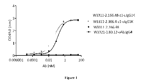
[00052] Figure 1 shows binding of the monoclonal antibodies, WBP3311 2.166.48-
uIgG1K,
WBP3311 2.306.4 - uIgG1K, and WBP3311 2.166.48, to recombinant Cynomolgus
Monkey
CD3epsilon protein as measured by ELISA assay.
[00053] Figure 2 shows binding of the monoclonal antibodies, WBP3311 2.166.48-
uIgG1K,
WBP3311 2.306.4 - uIgG1K, WBP3311 2.166.48, and WBP3311 2.306.4, to human CD4
T
cells as measured by flow cytometry assay.
[00054] Figure 3 shows binding affinity of eight mouse antibodies (W3311-
2.166.48, W3311-
2.306.4, W3311-2.383.47, W3311-2.400.5, W3311-2.482.5, W3311-2.488.33, W3311-
2.615.8,
and W3311-2.844.8) to human CD3 expressing cells (Jurkat cells) as measured by
flow
cytometry assay.
[00055] Figure 4A shows binding affinity of humanized antibody, WBP3311
2.166.48-z1-
uIgG1K to human CD3 expressing cells (Jurkat cells) as measured by
flowcytometry assay.
[00056] Figure 4B shows the result of binding affinity of humanized antibody,
WBP3311 2.306.4-z1-uIgG1K to human CD3 expressing cells (Jurkat cells) as
measured by
flow cytometry assay.
[00057] Figure 4C shows the result of binding affinity of the positive
control, OKT3 to human
CD3 expressing cells (Jurkat cells) as measured by flow cytometry assay.
[00058] Figure 5 shows cell internalization rate of eight mouse antibodies
(W3311-2.166.48,
W3311-2.306.4, W3311-2.383.47, W3311-2.400.5, W3311-2.482.5, W3311-2.488.33,
W3311-2.615.8, and W3311-2.844.8) to human CD3 expressing cells (Jurkat cells)
as
measured by flow cytometry assay.
[00059] Figure 6 shows the result of human T cell activation by eight mouse
antibodies
(W3311-2.166.48, W3311-2.306.4, W3311-2.383.47, W3311-2.400.5, W3311-2.482.5,
W3311-2.488.33, W3311-2.615.8, and W3311-2.844.8) as measured by intracellular
cytokine
TNFalpha and IFNgamma staining.
12
CA 03074130 2020-02-26
WO 2019/057099 PCT/CN2018/106618
[00060] Figure 7 shows the result of human T cell activation by two humanized
antibodies
(WBP3311 2.166.48-z1-uIgG1K and WBP3311 2.306.4-z1-uIgG1K) as measured by
intracellular cytokine TNFalpha and IFNgamma staining.
[00061] Figure 8 shows the result of epitope binning of seven mouse antibodies
(W3311-
2.166.48, W3311-2.306.4, W3311-2.400.5, W3311-2.482.5, W3311-2.488.33, W3311-
2.615.8,
and W3311-2.844.8) against the clone WBP3311 2.383.47.
DETAILED DESCRIPTION OF THE INVENTION
[00062] The following description of the disclosure is merely intended to
illustrate various
embodiments of the disclosure. As such, the specific modifications discussed
are not to be
construed as limitations on the scope of the disclosure. It will be apparent
to one skilled in the
art that various equivalents, changes, and modifications may be made without
departing from
the scope of the disclosure, and it is understood that such equivalent
embodiments are to be
included herein. All references cited herein, including publications, patents
and patent
applications are incorporated herein by reference in their entirety.
[00063] Definitions
[00064] The term "antibody" as used herein includes any immunoglobulin,
monoclonal
antibody, polyclonal antibody, multivalent antibody, bivalent antibody,
monovalent antibody,
multispecific antibody, or bispecific antibody that binds to a specific
antigen. A native intact
antibody comprises two heavy (H) chains and two light (L) chains. Mammalian
heavy chains
are classified as alpha, delta, epsilon, gamma, and mu, each heavy chain
consists of a variable
region (VH) and a first, second, and third constant region (Cm, CH2, CH3,
respectively);
mammalian light chains are classified as X, or lc, while each light chain
consists of a variable
region (VL for X, light chain or VK for lc light chain, respectively) and a
constant region(CL for
X, light chain or CK for lc light chain, respectively). The antibody has a "Y"
shape, with the
stem of the Y consisting of the second and third constant regions of two heavy
chains bound
together via disulfide bonding. Each arm of the Y includes the variable region
and first constant
region of a single heavy chain bound to the variable and constant regions of a
single light chain.
The variable regions of the light and heavy chains are responsible for antigen
binding. The
variable regions in both chains generally contain three highly variable loops
called the
complementarity determining regions (CDRs) (light chain CDRs including LCDR1,
LCDR2,
and LCDR3, heavy chain CDRs including HCDR1, HCDR2, HCDR3). CDR boundaries for
the antibodies and antigen-binding fragments disclosed herein may be defined
or identified by
13
CA 03074130 2020-02-26
WO 2019/057099 PCT/CN2018/106618
the conventions of Kabat, IMGT, Chothia, or Al-Lazikani (Al-Lazikani, B.,
Chothia, C., Lesk,
A. M., J. Mol. Biol., 273(4), 927 (1997); Chothia, C. et at., J Mol Biol. Dec
5;186(3):651-63
(1985); Chothia, C. and Lesk, A.M., J.Mol.Biol., 196,901 (1987); Chothia, C.
et at., Nature.
Dec 21-28;342(6252):877-83 (1989) ; Kabat E.A. et al., National Institutes of
Health, Bethesda,
Md. (1991)). The three CDRs are interposed between flanking stretches known as
framework
regions (FRs), which are more highly conserved than the CDRs and form a
scaffold to support
the hypervariable loops. The constant regions of the heavy and light chains
are not involved
in antigen-binding, but exhibit various effector functions. Antibodies are
assigned to classes
based on the amino acid sequence of the constant region of their heavy chain.
The five major
classes or isotypes of antibodies are IgA, IgD, IgE, IgG, and IgM, which are
characterized by
the presence of alpha, delta, epsilon, gamma, and mu heavy chains,
respectively. Several of
the major antibody classes are divided into subclasses such as IgG1 (gammal
heavy chain),
IgG2 (gamma2 heavy chain), IgG3 (gamma3 heavy chain), IgG4 (gamma4 heavy
chain), IgAl
(alphal heavy chain), or IgA2 (alpha2 heavy chain).
[00065] The term "bivalent" as used herein refers to an antibody or an antigen-
binding
fragment having two antigen-binding sites; the term "monovalent" refers to an
antibody or an
antigen-binding fragment having only one single antigen-binding site; and the
term
"multivalent" refers to an antibody or an antigen-binding fragment having
multiple antigen-
binding sites. In some embodiments, the antibody or antigen-binding fragment
thereof is
bivalent.
[00066] As used herein, a "bispecific" antibody refers to an artificial
antibody which has
fragments derived from two different monoclonal antibodies and is capable of
binding to two
different epitopes. The two epitopes may present on the same antigen, or they
may present on
two different antigens.
[00067] The term "antigen-binding fragment" as used herein refers to an
antibody fragment
formed from a portion of an antibody comprising 1, 2 or 3 CDRs, or any other
antibody
fragment that binds to an antigen but does not comprise an intact native
antibody structure.
Examples of antigen-binding fragment include, without limitation, a diabody, a
Fab, a Fab', a
F(ab')2, an Fv fragment, a disulfide stabilized Fv fragment (dsFv), a (dsFv)2,
a bispecific dsFy
(dsFv-dsFv'), a disulfide stabilized diabody (ds diabody), a single-chain
antibody molecule
(scFv), an scFv dimer (bivalent diabody), a bispecific antibody, a
multispecific antibody, a
camelized single domain antibody, a nanobody, a domain antibody, and a
bivalent domain
14
CA 03074130 2020-02-26
WO 2019/057099 PCT/CN2018/106618
antibody. An antigen-binding fragment is capable of binding to the same
antigen to which the
parent antibody binds.
[00068] "Fab" with regard to an antibody refers to that portion of the
antibody consisting of
a single light chain (both variable and constant regions) bound to the
variable region and first
constant region of a single heavy chain by a disulfide bond.
[00069] "Fab' refers to a Fab fragment that includes a portion of the hinge
region.
[00070] "F(ab')2"refers to a dimer of Fab'. "Fv" with regard to an antibody
refers to the
smallest fragment of the antibody to bear the complete antigen-binding site.
An Fv fragment
consists of the variable region of a single light chain bound to the variable
region of a single
heavy chain.
[00071] A "dsFv" refers to a disulfide-stabilized Fv fragment that the linkage
between the
variable region of a single light chain and the variable region of a single
heavy chain is a
disulfide bond. In some embodiments, a "(dsFv)2" or "(dsFv-dsFv)" comprises
three peptide
chains: two VH moieties linked by a peptide linker (e.g., a long flexible
linker) and bound to
two VL moieties, respectively, via disulfide bridges. In some embodiments,
dsFv-dsFv' is
bispecific in which each disulfide paired heavy and light chain has a
different antigen
specificity.
[00072] "Single-chain Fv antibody" or "scFv" refers to an engineered antibody
consisting of
a light chain variable region and a heavy chain variable region connected to
one another directly
or via a peptide linker sequence (Huston JS et al. Proc Natl Acad Sci USA,
85:5879(1988)).
[00073] "Fc" with regard to an antibody refers to that portion of the antibody
consisting of
the second and third constant regions of a first heavy chain bound to the
second and third
constant regions of a second heavy chain via disulfide bonding. The Fc portion
of the antibody
is responsible for various effector functions such as antibody-dependent cell-
mediated
cytotoxicity (ADCC), and complement dependent cytotoxicity (CDC), but does not
function in
antigen binding.
[00074] "Single-chain Fv-Fc antibody" or "scFv-Fc" refers to an engineered
antibody
consisting of a scFv connected to the Fc region of an antibody.
[00075] "Camelized single domain antibody," "heavy chain antibody," or "HCAb"
refers to
an antibody that contains two VH domains and no light chains (Riechmann L. and
Muyldermans
S., J Immunol Methods. Dec 10;231(1-2):25-38 (1999); Muyldermans S., J
Biotechnol.
CA 03074130 2020-02-26
WO 2019/057099 PCT/CN2018/106618
Jun;74(4):277-302 (2001); W094/04678; W094/25591; U.S. Patent No. 6,005,079).
Heavy
chain antibodies were originally derived from Camelidae (camels, dromedaries,
and llamas).
Although devoid of light chains, camelized antibodies have an authentic
antigen-binding
repertoire (Hamers-Casterman C. et at., Nature. Jun 3;363(6428):446-8 (1993);
Nguyen VK.
et at. "Heavy-chain antibodies in Camelidae; a case of evolutionary
innovation,"
Immunogenetics. Apr;54(1):39-47 (2002); Nguyen VK. et a/.Immunology.
May;109(1):93-
101 (2003)). The variable domain of a heavy chain antibody (VHH domain)
represents the
smallest known antigen-binding unit generated by adaptive immune responses
(Koch-Nolte F.
et al., FASEB J. Nov;21(13):3490-8. Epub 2007 Jun 15 (2007) ).
[00076] A "nanobody" refers to an antibody fragment that consists of a VHH
domain from a
heavy chain antibody and two constant domains, CH2 and CH3.
[00077] "Diabodies" or "dAbs" include small antibody fragments with two
antigen-binding
sites, wherein the fragments comprise a VH domain connected to a VL domain in
the same
polypeptide chain (VH-VL or VL-VH) (see, e.g., Holliger P. et at., Proc Natl
Acad Sci U S A.
Jul 15;90(14):6444-8 (1993); EP404097; W093/11161). By using a linker that is
too short to
allow pairing between the two domains on the same chain, the domains are
forced to pair with
the complementary domains of another chain, thereby creating two antigen-
binding sites. The
antigen¨binding sites may target the same or different antigens (or epitopes).
In certain
embodiments, a "bispecific ds diabody" is a diabody target two different
antigens (or
epitopes),In certain embodiments, an "scFv dimer" is a bivalent diabody or
bivalent ScFv
(BsFv) comprising VH-VL (linked by a peptide linker) dimerized with another VH-
VL moiety
such that VH's of one moiety coordinate with the VL's of the other moiety and
form two binding
sites which can target the same antigens (or eptipoes) or different antigens
(or eptipoes). In
other embodiments, an "scFv dimer" is a bispecific diabody comprising Vm-VL2
(linked by a
peptide linker) associated with VL1-VH2 (also linked by a peptide linker) such
that Vm and VLi
coordinate and VH2 and VL2 coordinate and each coordinated pair has a
different antigen
specificity.
[00078] A "domain antibody" refers to an antibody fragment containing only the
variable
region of a heavy chain or the variable region of a light chain. In certain
instances, two or more
VH domains are covalently joined with a peptide linker to create a bivalent or
multivalent
domain antibody. The two VH domains of a bivalent domain antibody may target
the same or
different antigens.
16
CA 03074130 2020-02-26
WO 2019/057099 PCT/CN2018/106618
[00079] The term "chimeric" as used herein, means an antibody or antigen-
binding fragment,
having a portion of heavy and/or light chain derived from one species, and the
rest of the heavy
and/or light chain derived from a different species. In an illustrative
example, a chimeric
antibody may comprise a constant region derived from human and a variable
region from a
non-human animal, such as from mouse. In some embodiments, the non-human
animal is a
mammal, for example, a mouse, a rat, a rabbit, a goat, a sheep, a guinea pig,
or a hamster.
[00080] The term "humanized" as used herein means that the antibody or antigen-
binding
fragment comprises CDRs derived from non-human animals, FR regions derived
from human,
and when applicable, the constant regions derived from human.
[00081] "CD3" as used herein, refers to the Cluster of Differentiation 3
protein derived from
any vertebrate source, including mammals such as primates (e.g. humans,
monkeys) and
rodents (e.g., mice and rats). In mammals, the CD3 molecule is a multi-protein
complex of six
chains, including: a CD3gamma chain, a CD3delta chain, two CD3epsilon chains,
and a
homodimer of CD3zeta chains, wherein the CD3zeta chain is the intracellular
tail of CD3
molecule, and the CD3gamma, CD3delta and CD3epsilon chains all contain
extracellular
domain (ECD) expressed on surface of T cells. Exemplary sequence of human CD3
includes
human CD3epsilon protein (NCBI Ref Seq No. NP 000724), human CD3 delta protein
(NCBI
Ref Seq No. NP 000723), and human CD3gamma protein (NCBI Ref Seq No. NP
000064).
Exemplary sequence of non-human CD3 includes Macaca fascicularis (monkey)
CD3epsilon
protein (NCBI Ref Seq No. NP 001270544), Macaca fascicularis (monkey) CD
3delta protein
(NCBI Ref Seq No. NP 001274617), Macaca fascicularis (monkey) CD3gamma protein
(NCBI Ref Seq No. NP 001270839); mouse CD3epsi1on protein (NCBI Ref Seq No.
NP 031674), mouse CD3delta protein (NCBI Ref Seq No. NP 038515), mouse
CD3gamma
protein (NCBI Ref Seq No. AAA37400); Rattus norvegicus (Rat) CD3epsilon
protein (NCBI
Ref Seq No. NP 001101610), Rattus norvegicus (Rat) CD3delta protein (NCBI Ref
Seq No.
NP 037301), Rattus norvegicus (Rat) CD3gamma protein (NCBI Ref Seq No.
NP 001071114). In certain embodiments, CD3 used herein can also be recombinant
CD3, for
example, including recombinant CD3 epsilon protein, recombinant CD3delta
protein, and
recombinant CD3gamma protein, which may optionally be expressed as a
recombinant CD3
complex. The recombinant CD3 complex may be expressed on a cell surface, or
alternatively
may be expressed as a soluble form which is not associated on a cell surface.
[00082] The term "CD3epsilon" as used herein is intended to encompass any form
of
CD3epsilon, for example, 1) native unprocessed CD3epsilon molecule, "full-
length"
17
CA 03074130 2020-02-26
WO 2019/057099 PCT/CN2018/106618
CD3epsilon chain or naturally occurring variants of CD3epsilon, including, for
example, splice
variants or allelic variants; 2) any form of CD3 epsilon that results from
processing in the cell;
or 3) full length, a fragment (e.g., a truncated form, an
extracellular/transmembrane domain)
or a modified form (e.g. a mutated form, a glycosylated/PEGylated, a His-
tag/immunofluorescence fused form) of CD3 epsilon subunit generated through
recombinant
method.
[00083] The term "anti-CD3epsilon antibody" refers to an antibody that is
capable of specific
binding CD3epsilon (e.g. human or monkey CD3epsilon).
[00084] The term "specific binding" or "specifically binds" as used herein
refers to a non-
random binding reaction between two molecules, such as for example between an
antibody and
an antigen. In certain embodiments, the antibodies or antigen-binding
fragments provided
herein specifically bind to human and/or CD3epsilon with a binding affinity
(KD) of <10-6 M
(e.g., <5x10⁻⁷ M, <2x10⁻⁷ M, <10-7 M, <5x10⁻⁸ M, <2x10⁻⁸ M, <10-8 M, <5x10⁻⁹
M, <4x10⁻⁹M,
<3x10⁻⁹M,<2x10⁻⁹ M, or <10-9 M). KD used herein refers to the ratio of the
dissociation rate to
the association rate (koff/kon), which may be determined by using any
conventional method
known in the art, including but are not limited to surface plasmon resonance
method,
microscale thermophoresis method, El:PLC-MS method and flow cytometry (such as
FACS)
method. In certain embodiments, the KD value can be appropriately determined
by using flow
cytometry.
[00085] The ability to "block binding" or "compete for the same epitope" as
used herein refers
to the ability of an antibody or antigen-binding fragment to inhibit the
binding interaction
between two molecules (e.g. human CD3epsilon and an anti-CD3epsilon antibody)
to any
detectable degree. In certain embodiments, an antibody or antigen-binding
fragment that
blocks binding between two molecules inhibits the binding interaction between
the two
molecules by at least 85%, or at least 90%. In certain embodiments, this
inhibition may be
greater than 85%, or greater than 90%.
[00086] The term "epitope" as used herein refers to the specific group of
atoms or amino acids
on an antigen to which an antibody binds. Two antibodies may bind the same or
a closely
related epitope within an antigen if they exhibit competitive binding for the
antigen. For
example, if an antibody or antigen-binding fragment blocks binding of a
reference antibody to
the antigen (e.g., recombinant human/monkey CD3 epsilon or CD3epsilon
expressed on surface
of cells in the present disclosure) by at least 85%, or at least 90%, then the
antibody or antigen-
18
CA 03074130 2020-02-26
WO 2019/057099 PCT/CN2018/106618
binding fragment may be considered to bind the same/closely related epitope as
the reference
antibody.
[00087] Those skilled in the art will recognize that it is possible to
determine, without undue
experimentation, if a human monoclonal antibody binds to the same epitope as
the antibody of
present disclosure (e.g., mouse monoclonal
antibodies WBP3311 2.166.48,
WBP33 I I 2.306.4, WBP33 I I 2.383.47, WBP33 I I 2.400.5,
WBP33 I I 2.482.5,
WBP331 2.488.33, WBP3311 2.615.8, WBP3311 2.844.8, and humanized antibodies
WBP3311 2.166.48-z1 and WBP3311 2.306.4-z1) by ascertaining whether the former
prevents the latter from binding to a CD3epsilon antigen polypeptide. If the
test antibody
competes with the antibody of present disclosure, as shown by a decrease in
binding by the
antibody of present disclosure to the CD3epsilon antigen polypeptide, then the
two antibodies
bind to the same, or a closely related, epitope. Or if the binding of a test
antibody to the
CD3epsilon antigen polypeptide was inhibited by the antibody of present
disclosure, then the
two antibodies bind to the same, or a closely related, epitope.
[00088] The various symbols used in the antibody names as provided herein are
of different
representation: "mIgG2" refers to an antibody with mouse constant region of
IgG2 isotype;
"uIgGl" refers an antibody with human constant region of IgG1 isotype; "K" or
"L" refers to
an antibody using the kappa or lambda light chain.
[00089] A "conservative substitution" with reference to amino acid sequence
refers to
replacing an amino acid residue with a different amino acid residue having a
side chain with
similar physiochemical properties. For example, conservative substitutions can
be made
among amino acid residues with hydrophobic side chains (e.g. Met, Ala, Val,
Leu, and Ile),
among residues with neutral hydrophilic side chains (e.g. Cys, Ser, Thr, Asn
and Gln), among
residues with acidic side chains (e.g. Asp, Glu), among amino acids with basic
side chains (e.g.
His, Lys, and Arg), or among residues with aromatic side chains (e.g. Trp,
Tyr, and Phe). As
known in the art, conservative substitution usually does not cause significant
change in the
protein conformational structure, and therefore could retain the biological
activity of a protein.
[00090] The term "homologue" and "homologous" as used herein are
interchangeable and
refer to nucleic acid sequences (or its complementary strand) or amino acid
sequences that
have sequence identity of at least 80% (e.g., at least 85%, 88%, 90%, 91%,
92%, 93%, 94%,
95%, 96%, 97%, 98%, 99%) to another sequences when optimally aligned.
19
CA 03074130 2020-02-26
WO 2019/057099 PCT/CN2018/106618
[00091] "Percent (%) sequence identity" with respect to amino acid sequence
(or nucleic acid
sequence) is defined as the percentage of amino acid (or nucleic acid)
residues in a candidate
sequence that are identical to the amino acid (or nucleic acid) residues in a
reference sequence,
after aligning the sequences and, if necessary, introducing gaps, to achieve
the maximum
number of identical amino acids (or nucleic acids). Conservative substitution
of the amino acid
residues may or may not be considered as identical residues. Alignment for
purposes of
determining percent amino acid (or nucleic acid) sequence identity can be
achieved, for
example, using publicly available tools such as BLASTN, BLASTp (available on
the website
of U.S. National Center for Biotechnology Information (NCBI), see also,
Altschul S.F. et al, J.
Mol. Biol., 215:403-410 (1990); Stephen F. et al, Nucleic Acids Res., 25:3389-
3402 (1997)),
ClustalW2 (available on the website of European Bioinformatics Institute, see
also, Higgins
D.G. et al, Methods in Enzymology, 266:383-402 (1996); Larkin M.A. et al,
Bioinformatics
(Oxford, England), 23(21): 2947-8 (2007)), and ALIGN or Megalign (DNASTAR)
software.
Those skilled in the art may use the default parameters provided by the tool,
or may customize
the parameters as appropriate for the alignment, such as for example, by
selecting a suitable
algorithm.
[00092] "Effector functions" as used herein refer to biological activities
attributable to the
binding of Fc region of an antibody to its effectors such as Cl complex and Fc
receptor.
Exemplary effector functions include: complement dependent cytotoxicity (CDC)
induced by
interaction of antibodies and Clq on the Cl complex; antibody-dependent cell-
mediated
cytotoxicity (ADCC) induced by binding of Fc region of an antibody to Fc
receptor on an
effector cell; and phagocytosis.
[00093] "Treating" or "treatment" of a condition as used herein includes
preventing or
alleviating a condition, slowing the onset or rate of development of a
condition, reducing the
risk of developing a condition, preventing or delaying the development of
symptoms associated
with a condition, reducing or ending symptoms associated with a condition,
generating a
complete or partial regression of a condition, curing a condition, or some
combination thereof.
[00094] An "isolated" substance has been altered by the hand of man from the
natural state.
If an "isolated" composition or substance occurs in nature, it has been
changed or removed
from its original environment, or both. For example, a polynucleotide or a
polypeptide
naturally present in a living animal is not "isolated," but the same
polynucleotide or polypeptide
is "isolated" if it has been sufficiently separated from the coexisting
materials of its natural
state so as to exist in a substantially pure state. An "isolated nucleic acid
sequence" refers to
CA 03074130 2020-02-26
WO 2019/057099 PCT/CN2018/106618
the sequence of an isolated nucleic acid molecule. In certain embodiments, an
"isolated
antibody or antigen-binding fragment thereof' refers to the antibody or
antigen-binding
fragments having a purity of at least 60%, 70%, 75%, 80%, 81%, 82%, 83%, 84%,
85%, 86%,
87%, 88%, 89%, 90%, 91%, 92%, 93%, 94%, 95%, 96%, 97%, 98%, 99% as determined
by
electrophoretic methods (such as SDS-PAGE, isoelectric focusing, capillary
electrophoresis),
or chromatographic methods (such as ion exchange chromatography or reverse
phase HPLC).
[00095] The term "vector" as used herein refers to a vehicle into which a
polynucleotide
encoding a protein may be operably inserted so as to bring about the
expression of that protein.
A vector may be used to transform, transduce, or transfect a host cell so as
to bring about
expression of the genetic element it carries within the host cell. Examples of
vectors include
plasmids, phagemids, cosmids, artificial chromosomes such as yeast artificial
chromosome
(YAC), bacterial artificial chromosome (BAC), or P1-derived artificial
chromosome (PAC),
bacteriophages such as lambda phage or M13 phage, and animal viruses.
Categories of animal
viruses used as vectors include retrovirus (including lentivirus), adenovirus,
adeno-associated
virus, herpesvirus (e.g., herpes simplex virus), poxvirus, baculovirus,
papillomavirus, and
papovavirus (e.g., SV40). A vector may contain a variety of elements for
controlling
expression, including promoter sequences, transcription initiation sequences,
enhancer
sequences, selectable elements, and reporter genes. In addition, the vector
may contain an
origin of replication. A vector may also include materials to aid in its entry
into the cell,
including but not limited to a viral particle, a liposome, or a protein
coating. A vector can be
an expression vector or a cloning vector. The present disclosure provides
vectors (e.g.,
expression vectors) containing the nucleic acid sequence provided herein
encoding the
antibody or antigen-binding fragment thereof, at least one promoter (e.g.,
SV40, CMV, EF-1a)
operably linked to the nucleic acid sequence, and at least one selection
marker. Examples of
vectors include, but are not limited to, retrovirus (including lentivirus),
adenovirus, adeno-
associated virus, herpesvirus (e.g., herpes simplex virus), poxvirus,
baculovirus,
papillomavirus, papovavirus (e.g., SV40), lambda phage, and M13 phage, plasmid
pcDNA3.3,
pMD18-T, pOptivec, pCMV, pEGFP, plRES, pQD-Hyg-GSeu, pALTER, pBAD, pcDNA,
pCal, pL, pET, pGEMEX, pGEX, pCI, pEGFT, pSV2, pFUSE, pVITRO, pVIVO, pMAL,
pMONO, pSELECT, pUNO, pDUO, Psg5L, pBABE, pWPXL, pBI, p15TV-L, pPro18, pTD,
pRS10, pLexA, pACT2.2, pCMV-SCRIPT®, pCDM8, pCDNA1.1/amp, pcDNA3.1,
pRc/RSV, PCR 2.1, pEF-1, pFB, pSG5, pXT1, pCDEF3, pSVSPORT, pEF-Bos etc.
21
CA 03074130 2020-02-26
WO 2019/057099 PCT/CN2018/106618
[00096] The phrase "host cell" as used herein refers to a cell into which an
exogenous
polynucleotide and/or a vector has been introduced.
[00097] A "CD3 related disease or condition" as used herein refers to any
disease or condition
caused by, exacerbated by, or otherwise linked to increased or decreased
expression or
activities of CD3. In some embodiments, the CD3 related condition is immune-
related disorder,
such as, for example, cancer, autoimmune disease, inflammatory disease or
infectious disease.
[00098] "Cancer" as used herein refers to any medical condition characterized
by malignant
cell growth or neoplasm, abnormal proliferation, infiltration or metastasis,
and includes both
solid tumors and non-solid cancers (hematologic malignancies) such as
leukemia. As used
herein "solid tumor" refers to a solid mass of neoplastic and/or malignant
cells.
[00099] The term "pharmaceutically acceptable" indicates that the designated
carrier, vehicle,
diluent, excipient(s), and/or salt is generally chemically and/or physically
compatible with the
other ingredients comprising the formulation, and physiologically compatible
with the recipient
thereof.
[000100] Anti-CD3epsilon antibody
[000101] The present disclosure provides anti-CD3epsilon antibodies and
antigen-binding
fragments thereof comprising one or more (e.g. 1, 2, 3, 4, 5, or 6) CDR
sequences of an anti-
CD3epsilon antibody WBP3311 2.166.48, WBP3311 2.306.4, WBP3311 2.383.47,
WBP3311 2.400.5, WBP3311 2.482.5, WBP331 2.488.33, WBP3311 2.615.8, or
WBP3311 2.844.8. Throughout the present disclosure, the term "WBP3311" with
respect to
the antibody names is used interchangeably with "W3311". For example, antibody
WBP3311 2.166.48 is also referred to as W33112.166.48 and such names refer to
the same
antibody.
[000102] "WBP3311 2.166.48" as used herein refers to a mouse monoclonal
antibody having
a heavy chain variable region of SEQ ID NO: 81, and a kappa light chain
variable region of
SEQ ID NO: 83.
[000103] "WBP3311 2.306.4" as used herein refers to a mouse monoclonal
antibody having
a heavy chain variable region of SEQ ID NO: 85, and a kappa light chain
variable region of
SEQ ID NO: 87.
22
CA 03074130 2020-02-26
WO 2019/057099 PCT/CN2018/106618
[000104] "WBP3311 2.383.47" as used herein refers to a mouse monoclonal
antibody having
a heavy chain variable region of SEQ ID NO: 89, and a kappa light chain
variable region of
SEQ ID NO: 91.
[000105] "WBP3311 2.400.5" as used herein refers to a mouse monoclonal
antibody having a
heavy chain variable region of SEQ ID NO: 93, and a kappa light chain variable
region of SEQ
ID NO: 95.
[000106] "WBP3311 2.482.5" as used herein refers to a mouse monoclonal
antibody having a
heavy chain variable region of SEQ ID NO: 97, and a kappa light chain variable
region of SEQ
ID NO: 99.
[000107] "WBP331 2.488.33" as used herein refers to a mouse monoclonal
antibody having a
heavy chain variable region of SEQ ID NO: 101, and a kappa light chain
variable region of
SEQ ID NO: 103.
[000108] "WBP3311 2.615.8" as used herein refers to a mouse monoclonal
antibody having a
heavy chain variable region of SEQ ID NO: 105, and a kappa light chain
variable region of
SEQ NO: 107.
[000109] "WBP3311 2.844.8" as used herein refers to a mouse monoclonal
antibody having a
heavy chain variable region of SEQ ID NO: 109, and a kappa light chain
variable region of
SEQ NO: 111.
[000110] Table 1 shows the CDR sequences of these 8 anti-CD3epsilon
antibodies. The heavy
chain and light chain variable region sequences are also provided below.
[000111] Table 1.
Antibody ID: CDR1 CDR2 CDR3
SEQ ID NO: 1 SEQ ID NO: 3 SEQ ID NO: 5
WBP3311 2.166.48 VH WIFPGNDNIKYSE
GYSFTTYYIH DSVSIYYFDY
KFKG
SEQ ID NO: 2 SEQ ID NO: 4 SEQ ID NO: 6
WBP3311 2.166.48 VK KSSQSLLNSRTRKN
WASTRKS TQSFILRT
YLA
SEQ ID NO: 7 SEQ ID NO: 9 SEQ ID NO: 11
VH WISPGNVNTKYN
WBP3311 2.306.4 GFAFTDYYIH DGYSLYYFDY
ENFKG
SEQ ID NO: 8 SEQ ID NO: 10 SEQ ID NO: 12
VK KS S Q SLLNSRTRKN
WBP3311 2.306.4 WASTRQS TQSHTLRT
YLA
SEQ ID NO: 13 SEQ ID NO: 15 SEQ ID NO: 17
VH WISPENGNTKYNE
WBP3311 2.383.47 GFTFTNYYIH DGYSLYYFDY
NFQD
SEQ ID NO: 14 SEQ ID NO: 16 SEQ ID NO: 18
23
CA 03074130 2020-02-26
WO 2019/057099 PCT/CN2018/106618
WBP3311 2.383.47 VK KSSQSLLNSRTRKN
WASIRVS TQSHTLRT
YLA
SEQ ID NO: 19 SEQ ID NO: 21 SEQ ID NO: 23
VH WIFPESDNTKYNE
WBP3311 2.400.5 GYSFTNYYLH DSVGNYFFDF
KLKG
SEQ ID NO: 20 SEQ ID NO: 22 SEQ ID NO: 24
VK KSSQSLVNNRTRKN
WBP3311 2.400.5 WASTRES AQSFILRT
YLA
SEQ ID NO: 25 SEQ ID NO: 27 SEQ ID NO: 29
VH WIFPGSDNIKYNE
WBP3311 2.482.5 GYTFTTYYIH DSVSRYYFDY
NFKD
SEQ ID NO: 26 SEQ ID NO: 28 SEQ ID NO: 30
VK KSSQSLVNDRTRKN
WBP3311 2.482.5 WASTRES AQSFILRT
YLA
SEQ ID NO: 31 SEQ ID NO: 33 SEQ ID NO: 35
VH WIFPGTVNTKYNE
WBP331 2.488.33 GFSFTNYYIH DSVGIYYFDF
KFKG
SEQ ID NO: 32 SEQ ID NO: 34 SEQ ID NO: 36
VK KSSQSLLNNRTRKN
WBP331 2.488.33 WASTRES TQSFILRT
YLA
SEQ ID NO: 37 SEQ ID NO: 39 SEQ ID NO: 41
VH WIFPGSDNIKYNE
WBP3311 2.615.8 GYSFTDFYTH DSVSVYYFDY
KFKG
SEQ ID NO: 38 SEQ ID NO: 40 SEQ ID NO: 42
VK KSSQSLLNIRTRKNY
WBP3311 2.615.8 WASTRDS TQSFILRT
LA
SEQ ID NO: 43 SEQ ID NO: 45 SEQ ID NO: 47
VH WISPGNVNTKYN
WBP3311 2.844.8 GFAFTDYYIH DGYSLYYFDY
ENFKG
SEQ ID NO: 44 SEQ ID NO: 46 SEQ ID NO: 48
VK KSSQSLLNSRTRKN
WBP3311 2.844.8 WASTRES TQSHTLRT
YLA
[000112] Heavy or kappa light chain variable region sequences of WBP3311
2.166.48,
WBP3311 2.306.4, WBP3311 2.383.47,
WBP3311 2.400.5, WBP3311 2.482.5,
WBP331 2.488.33, WBP3311 2.615.8, and WBP3311 2.844.8, and humanized
WBP3311 2.166.48 and WBP3311 2.306.4 antibodies are provided below.
[000113] WBP3311_2.166.48-VH
Amino acid sequence (SEQ ID NO: 81):
QVQLQQ S GPELVKPGASVKIACKAS GYSF T TYYLHWVKQRPGQGLEWIGWIFPGND
NlKYSEKFKGKATLTADTSSSTAYMQLSSLTSEDSAVYFCAIDSVSIYYFDYWGQGTT
LTVSS
Nucleic acid sequence (SEQ ID NO: 82):
CAGGTCCAGCTGCAGCAGTCTGGACCTGAGCTGGTGAAACCTGGGGCTTCAGTG
AAGATTGCCTGCAAGGCTTCTGGCTACAGCTTCACAACCTACTATATACACTGGG
TGAAGCAGAGGCCTGGACAGGGACTTGAGTGGATTGGATGGATTTTTCCTGGAA
ATGATAATATTAAGTACAGTGAGAAGTTCAAGGGCAAGGCCACACTGACGGCAG
24
CA 03074130 2020-02-26
WO 2019/057099 PCT/CN2018/106618
ACACTTCCTCCAGTACAGCCTACATGCAGCTCAGCAGCCTGACATCTGAGGACTC
TGCTGTCTATTTCTGTGCTATAGACTCCGTTAGTATCTACTACTTTGACTATTGGG
GCCAAGGCACCACTCTCACAGTCTCCTCA
[000114] WBP3311_2.166.48-VK
Amino acid sequence (SEQ ID NO: 83):
DIVMSQSPSSLAVSAGEKVTMSCKSSQSLLNSRTRKNYLAWYQQKPGQSPKWYW
ASTRKSGVPDRFTGSGSGTDFTLTINSVQAEDLAVYYCTQSF1LRTFGGGTKLEIK
Nucleic acid sequence (SEQ ID NO: 84):
GACATTGTGATGTCACAGTCTCCATCCTCCCTGGCTGTGTCAGCAGGAGAGAAGG
TCACTATGAGCTGCAAATCCAGTCAGAGTCTGCTCAACAGTAGAACCCGAAAGA
ACTACTTGGCTTGGTACCAGCAGAAACCAGGGCAGTCTCCTAAACTGCTGATCTA
CTGGGCATCCACTAGGAAATCTGGGGTCCCTGATCGCTTCACAGGCAGTGGATCT
GGGACAGATTTCACTCTCACCATCAACAGTGTGCAGGCTGAAGACCTGGCAGTTT
ATTACTGCACGCAATCTTTTATTCTTCGGACGTTCGGTGGAGGCACCAAGCTGGA
AATCAAA
[000115] WBP3311_2.306.4-VH
Amino acid sequence (SEQ ID NO: 85):
QVQLQQSGPELVKPGASVRISCKASGFAFTDYYIEIWVKQRPGQGLEWIGWISPGNVN
TKYNENFKGRATLTADLSSSTAYMQLSSLTSEDSAVYFCARDGYSLYYFDYWGQGT
TLTVSS
Nucleic acid sequence (SEQ ID NO: 86):
CAGGTCCAGCTGCAGCAGTCTGGACCTGAATTGGTGAAGCCTGGGGCTTCCGTGA
GGATATCCTGCAAGGCTTCTGGCTTCGCCTTCACAGACTACTATATACACTGGGT
GAAGCAGAGGCCTGGACAGGGTCTTGAGTGGATTGGATGGATTTCTCCTGGAAA
TGTTAATACTAAATACAATGAAAACTTCAAGGGCAGGGCCACACTGACTGCAGA
CCTATCCTCCAGCACAGCCTACATGCAGCTCAGCAGCCTGACCTCTGAGGACTCT
GCGGTCTATTTCTGTGCAAGAGATGGATATTCCCTGTATTACTTTGACTACTGGG
GCCAAGGCACCACTCTCACAGTCTCCTCA
[000116] WBP3311_2.306.4-VK
Amino acid sequence (SEQ ID NO: 87):
DIVMSQSPSSLTVSAGEKVTMSCKSSQSLLNSRTRKNYLAWYQQKPGQSPKWYWA
STRQSGVPDRFTGSGSGTAFTLTISGVQAEDLAVYFCTQSHTLRTFGGGTKLEIK
Nucleic acid sequence (SEQ ID NO: 88):
CA 03074130 2020-02-26
WO 2019/057099 PCT/CN2018/106618
GACATTGTGATGTCACAGTCTCCATCCTCCCTGACTGTGTCAGCAGGAGAGAAGG
TCACTATGAGCTGCAAATCCAGTCAGAGTCTGCTCAACAGTAGAACCCGAAAGA
ACTACTTGGCTTGGTACCAGCAGAAGCCAGGGCAGTCTCCTAAACTACTAATCTA
CTGGGCATCCACTAGGCAATCTGGGGTCCCTGATCGCTTCACAGGCAGTGGATCT
GGGACAGCTTTCACTCTCACCATCAGCGGTGTGCAGGCTGAAGACCTGGCAGTTT
ATTTCTGCACGCAATCTCATACTCTTCGGACGTTCGGTGGAGGCACCAAGCTGGA
AATCAAA
[000117] WBP3311_2.383.47-VH
Amino acid sequence (SEQ ID NO: 89):
QVQLQQSGPELVKPGASVRISCKTSGFTFTNYYIEIWVIQRPGQGLEWIGWISPENGNT
KYNENFQDKATLTADISSSTAYMEILSSLTSEDSAVYFCARDGYSLYYFDYWGQGTT
LTVSS
Nucleic acid sequence (SEQ ID NO: 90):
CAGGTCCAGCTGCAGCAGTCTGGACCTGAATTGGTGAAGCCTGGGGCTTCAGTG
AGGATATCCTGCAAGACTTCTGGCTTCACCTTCACAAACTACTATATACACTGGG
TGATACAGAGGCCTGGACAGGGACTTGAGTGGATTGGTTGGATTTCTCCTGAAAA
TGGTAATACTAAATACAATGAAAACTTCCAGGACAAGGCCACACTGACTGCAGA
CATATCGTCCAGCACAGCCTACATGCACCTCAGCAGCCTGACCTCTGAGGACTCT
GCGGTCTATTTCTGTGCAAGAGATGGGTATTCCCTTTACTACTTTGACTACTGGGG
CCAAGGCACCACTCTCACAGTCTCCTCA
[000118] WBP3311_2.383.47-VK
Amino acid sequence (SEQ ID NO: 91):
DIVMSQSPSSLTVSAGEKVTMSCKSSQSLLNSRTRKNYLAWYQQKPGQSPKWYWA
S1RVSGVPDRFTGSGSGTTFTLTISGVQAEDLAVYYCTQSHTLRTFGGGTKLEIK
Nucleic acid sequence (SEQ ID NO: 92):
GACATTGTGATGTCACAGTCTCCATCCTCCCTGACTGTGTCAGCAGGAGAGAAGG
TCACTATGAGCTGCAAATCCAGTCAGAGTCTGCTCAACAGTAGAACCCGAAAGA
ACTACTTGGCTTGGTACCAGCAGAAACCAGGGCAGTCTCCTAAGCTACTGATCTA
CTGGGCATCCATTAGGGTATCTGGGGTCCCTGATCGCTTCACAGGCAGTGGATCT
GGGACAACTTTCACTCTCACCATCAGCGGTGTGCAGGCTGAAGACCTGGCAGTTT
ATTATTGCACGCAATCTCATACTCTTCGGACGTTCGGTGGAGGCACCAAGCTGGA
AATCAAA
[000119] WBP3311_2.400.5-VH
Amino acid sequence (SEQ ID NO: 93):
26
CA 03074130 2020-02-26
WO 2019/057099 PCT/CN2018/106618
QVQLQQSGPELVNPGASVKISCKASGYSFTNYYLHWVKQRPGQGLEWIGWIFPESD
NTKYNEKLKGKATLTADTSSDTAYMEILSSLTFEDSAVYFCARDSVGNYFFDFWGQG
TTLTVSS
Nucleic acid sequence (SEQ ID NO: 94):
CAGGTCCAGCTGCAGCAGTCTGGACCTGAGCTGGTGAATCCTGGGGCTTCAGTGA
AGATATCCTGCAAGGCTTCTGGCTACAGTTTCACAAACTACTATTTACACTGGGT
GAAACAGAGGCCTGGACAGGGACTTGAGTGGATTGGATGGATTTTTCCTGAAAG
TGATAATACCAAGTACAATGAGAAATTGAAGGGCAAGGCCACACTGACGGCAGA
CACATCCTCCGATACAGCCTACATGCACCTCAGCAGCCTGACATTTGAGGACTCT
GCAGTCTATTTCTGTGCAAGAGACTCCGTTGGAAACTACTTCTTTGACTTCTGGG
GCCAAGGCACCACTCTCACAGTCTCCTCA
[000120] WBP3311_2.400.5-VK
Amino acid sequence (SEQ ID NO: 95):
DIVMSQSPSSLAVSAGEKVTMRCKSSQSLVNNRTRKNYLAWYQQKPGQPPKWYW
ASTRESGVPDRFTGSGSGTDFTLTISSVQAEDLAVYYCAQSFILRTFGGGTKLEIK
Nucleic acid sequence (SEQ ID NO: 96):
GACATTGTGATGTCACAGTCTCCATCCTCCCTGGCTGTGTCAGCGGGAGAGAAGG
TCACTATGAGGTGCAAATCCAGTCAGAGTCTGGTCAACAATAGAACCCGAAAGA
ACTACTTGGCATGGTACCAGCAGAAACCAGGGCAGCCTCCTAAACTATTGATCTA
CTGGGCATCCACTAGGGAATCTGGGGTCCCTGATCGCTTCACAGGCAGTGGATCT
GGGACAGATTTCACTCTCACCATCAGCAGTGTGCAGGCTGAAGACCTGGCAGTTT
ATTACTGCGCGCAATCTTTTATTCTTCGGACGTTCGGTGGAGGCACCAAACTGGA
AATCAAA
[000121] WBP3311_2.482.5-VH
Amino acid sequence (SEQ ID NO: 97):
QVQLQQSGPELVKPGSSVKISCKPSGYTFTTYYLEIWVKQRPGQGLEWIGWIFPGSDNI
KYNENFKDKATLTADTSSSTAYMQLSSLTSEDSAVYFCARDSVSRYYFDYWGQGTI
LTVSS
Nucleic acid sequence (SEQ ID NO: 98):
CAGGTTCAGCTGCAGCAGTCTGGACCTGAGCTGGTGAAACCTGGGTCTTCAGTGA
AGATATCCTGCAAACCTTCTGGCTACACCTTCACAACTTACTATATACATTGGGT
GAAGCAGAGGCCTGGACAGGGACTTGAGTGGATTGGATGGATTTTTCCTGGAAG
TGATAATATTAAATACAATGAGAATTTCAAGGACAAGGCCACACTGACGGCAGA
CACATCCTCCAGCACAGCCTACATGCAGCTCAGCAGCCTGACATCTGAAGACTCT
GCAGTCTATTTCTGTGCAAGAGACTCCGTCAGTAGGTACTACTTTGACTACTGGG
GCCAAGGCACCATTCTCACAGTTTCTTCA
27
CA 03074130 2020-02-26
WO 2019/057099 PCT/CN2018/106618
[000122] WBP3311_2.482.5-VK
Amino acid sequence (SEQ ID NO: 99):
DIVMSQSPSSLAVSAGEKVTMSCKSSQSLVNDRTRKNYLAWYQQKPGLSPKWYW
ASTRESGVPDRFTGSGSGTDFTLTISSVQAEDLAVYYCAQSFILRTFGGGTKLEIK
Nucleic acid sequence (SEQ ID NO: 100):
GACATTGTGATGTCACAGTCTCCATCCTCCCTGGCTGTGTCAGCAGGAGAGAAGG
TCACTATGAGCTGCAAATCCAGTCAGAGTCTGGTCAATGATAGAACCCGAAAAA
ACTACTTGGCTTGGTACCAGCAGAAACCAGGGCTGTCTCCTAAACTGCTGATCTA
CTGGGCTTCCACTAGGGAATCTGGGGTCCCTGATCGCTTCACAGGCAGTGGATCT
GGGACAGATTTCACTCTCACCATCAGCAGTGTGCAGGCTGAAGACCTGGCTGTTT
ATTACTGCGCGCAATCTTTTATTCTTCGGACGTTCGGTGGAGGCACCAAGCTGGA
AATCAAA
[000123] WBP331_2.488.33-VH
Amino acid sequence (SEQ ID NO: 101):
QVQLQQSGPELVKPGTSVKISCKASGFSFTNYYIEIWVKQRPGQGPEWIGWIFPGTVN
TKYNEKFKGKATLTADTSSNTAFMQLSSLTSADSAVYFCARDSVGIYYFDFWGLGTT
LTVSS
Nucleic acid sequence (SEQ ID NO: 102):
CAGGTCCAGCTGCAACAGTCTGGACCTGAACTGGTGAAACCTGGGACTTCAGTG
AAGATATCCTGCAAGGCTTCTGGCTTCAGCTTCACAAACTACTATATACACTGGG
TGAAGCAGAGGCCTGGACAGGGACCTGAGTGGATTGGATGGATTTTTCCTGGAA
CTGTTAATACTAAGTACAATGAGAAGTTCAAGGGTAAGGCCACACTGACGGCAG
ACACATCCTCCAATACAGCCTTCATGCAGCTCAGCAGCCTGACTTCTGCGGACTC
TGCAGTCTATTTCTGTGCAAGAGACTCCGTTGGTATCTACTACTTTGACTTCTGGG
GCCTAGGCACCACTCTCACAGTCTCCTCA
[000124] WBP331_2.488.33-VK
Amino acid sequence (SEQ ID NO: 103):
DIVMSQSPSSLAVSAGEKVTVSCKSSQSLLNNRTRKNYLAWYQQKPGQSPKWYW
ASTRESGVPDRFTGSGSGTDFTLTISSVQAEDLAVYYCTQSF1LRTFGGGTKLEIK
Nucleic acid sequence (SEQ ID NO: 104):
GACATTGTGATGTCACAGTCTCCATCCTCCCTGGCTGTGTCAGCAGGAGAGAAGG
TCACTGTGAGTTGCAAATCCAGTCAGAGTCTGCTCAACAATAGAACCCGAAAAA
ACTACTTGGCTTGGTACCAGCAGAAACCAGGGCAGTCTCCTAAACTACTAATCTA
28
CA 03074130 2020-02-26
WO 2019/057099 PCT/CN2018/106618
CTGGGCATCCACTAGGGAATCTGGGGTCCCTGATCGCTTCACAGGCAGTGGATCT
GGTACAGATTTCACTCTCACCATCAGCAGTGTGCAGGCTGAAGACCTGGCAGTTT
ATTACTGCACGCAATCTTTTATTCTTCGGACGTTCGGTGGAGGCACCAAGCTGGA
GATCAAA
[000125] WBP3311_2.615.8-VH
Amino acid sequence (SEQ ID NO: 105):
QVQLQQSGPELVKPGTSMKISCKASGYSFTDFYTHWVRQRPGQGLEWIGWIFPGSDN
MYNEKFKGKATLTADTSSSTAYMQLSSLTSEDSAVYFCARDSVSVYYFDYWGQGT
TLTVSS
Nucleic acid sequence (SEQ ID NO: 106):
CAGGTCCAGCTGCAGCAGTCTGGACCTGAGCTGGTAAAACCTGGGACTTCAATG
AAAATATCCTGCAAGGCTTCTGGCTACAGTTTCACAGACTTCTATACACACTGGG
TGAGGCAGAGGCCTGGACAGGGACTTGAGTGGATTGGATGGATTTTTCCTGGAA
GTGATAATATTAAATACAATGAGAAGTTCAAGGGCAAGGCCACACTGACGGCAG
ACACATCCTCCAGCACAGCCTACATGCAGCTCAGCAGCCTGACATCTGAGGACTC
TGCAGTCTATTTCTGTGCAAGAGACTCCGTTAGTGTCTACTACTTTGACTATTGGG
GCCAAGGCACCACTCTCACAGTCTCCTCA
[000126] WBP3311_2.615.8-VK
Amino acid sequence (SEQ ID NO: 107):
DIVMSQSPSSLAVTAGEKVTMSCKSSQSLLNIRTRKNYLAWYQQKPGQSPKWYWA
STRDSGVPDRFTGSGSGTDFTLTISSVQAEDLAVYYCTQSFILRTFGGGTKLEIK
Nucleic acid sequence (SEQ ID NO: 108):
GACATCGTGATGTCACAGTCTCCATCCTCCCTGGCTGTGACAGCAGGAGAGAAG
GTCACTATGAGCTGCAAATCCAGTCAGAGTCTGCTCAACATTAGAACCCGAAAG
AACTACTTGGCTTGGTACCAACAGAAACCAGGGCAGTCTCCTAAACTGCTGATCT
ACTGGGCATCCACTAGGGACTCTGGGGTCCCTGATCGCTTCACAGGCAGTGGATC
TGGGACAGATTTCACTCTCACCATCAGCAGTGTGCAGGCTGAAGACCTGGCAGTT
TATTACTGCACGCAATCTTTTATTCTTCGGACGTTCGGTGGAGGCACCAAGCTGG
AAATCAAA
[000127] WBP3311_2.844.8-VH
Amino acid sequence (SEQ ID NO: 109):
QVQLQQSGPELVKPGASVRISCKASGFAFTDYYIEIWVKQRPGQGLEWIGWISPGNVN
TKYNENFKGRATLTADLSSSTAYMQLSSLTSEDSAVYFCARDGYSLYYFDYWGQGT
TLTVSS
29
CA 03074130 2020-02-26
WO 2019/057099 PCT/CN2018/106618
Nucleic acid sequence (SEQ ID NO: 110):
CAGGTCCAGCTGCAGCAGTCTGGACCTGAATTGGTGAAGCCTGGGGCTTCCGTGA
GGATATCCTGCAAGGCTTCTGGCTTCGCCTTCACAGACTACTATATACACTGGGT
GAAGCAGAGGCCTGGACAGGGTCTTGAGTGGATTGGATGGATTTCTCCTGGAAA
T GT TAATAC TAAATAC AATGAAAAC T T CAAGGGC AGGGCC ACAC TGAC T GC AGA
CCTATCCTCCAGCACAGCCTACATGCAGCTCAGCAGCCTGACCTCTGAGGACTCT
GCGGTCTATTTCTGTGCAAGAGATGGATATTCCCTGTATTACTTTGACTACTGGG
GCCAAGGCACCACTCTCACAGTCTCCTCA
[000128] WBP3311_2.844.8-VK
Amino acid sequence (SEQ ID NO: 111):
DIVMSQ SP S SLTVSAGEKVTMSCKS SQSLLNSRTRKNYLAWYQQKPGQSPKLLIYWA
S TRES GVPDRF TGS GS GTAF TLT IS GVQ AEDLAVYF C T Q SHTLRTF GGGTKLE1K
Nucleic acid sequence (SEQ ID NO: 112):
GACATTGTGATGTCACAGTCTCCATCCTCCCTGACTGTGTCAGCAGGAGAGAAGG
TCACTATGAGCTGCAAATCCAGTCAGAGTCTGCTCAACAGTAGAACCCGAAAGA
ACTACTTGGCTTGGTACCAGCAGAAACCAGGGCAGTCTCCTAAGCTACTAATCTA
CTGGGCATCCACTAGGGAATCTGGGGTCCCTGATCGCTTCACAGGCAGTGGATCT
GGGACAGCTTTCACTCTCACCATCAGCGGTGTGCAGGCTGAAGACCTGGCAGTTT
ATTTCTGCACGCAATCTCATACTCTTCGGACGTTCGGTGGAGGCACCAAGCTGGA
AATCAAA
[000129] CDRs are known to be responsible for antigen binding, however, it has
been found
that not all of the 6 CDRs are indispensable or unchangeable. In other words,
it is possible to
replace or change or modify one or more CDRs in anti-CD3epsilon antibody
WBP3311 2.166.48, WBP3311 2.306.4, WBP3311 2.383.47,
WBP3311 2.400.5,
WBP3311 2.482.5, WBP331 2.488.33, WBP3311 2.615.8, or WBP3311 2.844.8, yet
substantially retain the specific binding affinity to CD3epsilon.
[000130] In certain embodiments, the anti-CD3epsilon antibodies and the
antigen-binding
fragments provided herein comprise a heavy chain CDR3 sequence of one of the
anti-
CD3epsilon antibodies WBP3311 2.166.48, WBP3311 2.306.4, WBP3311 2.383.47,
WBP3311 2.400.5, WBP3311 2.482.5, WBP331 2.488.33, WBP3311 2.615.8, and
WBP3311 2.844.8. In certain embodiments, the anti-CD3epsilon antibodies and
the antigen-
binding fragments provided herein comprise a heavy chain CDR3 sequence
selected from the
group consisting of SEQ ID NOs: 5, 11, 17, 23, 29, 35, 41, and 47. Heavy chain
CDR3 regions
are located at the center of the antigen-binding site, and therefore are
believed to make the most
contact with antigen and provide the most free energy to the affinity of
antibody to antigen. It
CA 03074130 2020-02-26
WO 2019/057099 PCT/CN2018/106618
is also believed that the heavy chain CDR3 is by far the most diverse CDR of
the antigen-
binding site in terms of length, amino acid composition and conformation by
multiple
diversification mechanisms (Tonegawa S. Nature. 302:575-81). The diversity in
the heavy
chain CDR3 is sufficient to produce most antibody specificities (Xu JL, Davis
MM. Immunity.
13:37-45) as well as desirable antigen-binding affinity (Schier R, etc. J Mol
Biol. 263:551-67).
[000131] In certain embodiments, the antibodies and antigen-binding fragments
thereof
provided herein comprise suitable framework region (FR) sequences, as long as
the antibodies
and antigen-binding fragments thereof can specifically bind to CD3epsilon. The
CDR
sequences provided in Table 1 are obtained from mouse antibodies, but they can
be grafted to
any suitable FR sequences of any suitable species such as mouse, human, rat,
rabbit, among
others, using suitable methods known in the art such as recombinant
techniques.
[000132] In certain embodiments, the antibodies and antigen-binding fragments
thereof
provided herein are humanized. A humanized antibody or antigen-binding
fragment is
desirable in its reduced immunogenicity in human. A humanized antibody is
chimeric in its
variable regions, as non-human CDR sequences are grafted to human or
substantially human
FR sequences. Humanization of an antibody or antigen-binding fragment can be
essentially
performed by substituting the non-human (such as murine) CDR genes for the
corresponding
human CDR genes in a human immunoglobulin gene (see, for example, Jones et al.
(1986)
Nature 321:522-525; Riechmann et al. (1988) Nature 332:323-327; Verhoeyen et
al. (1988)
Science 239:1534-1536).
[000133] Suitable human heavy chain and light chain variable domains can be
selected to
achieve this purpose using methods known in the art. In an illustrative
example, "best-fit"
approach can be used, where a non-human (e.g. rodent) antibody variable domain
sequence is
screened or BLASTed against a database of known human variable domain
sequences, and the
human sequence closest to the non-human query sequence is identified and used
as the human
scaffold for grafting the non-human CDR sequences (see, for example, Sims et
al, (1993) J.
Immunol. 151:2296; Chothia et al. (1987) J. Mot. Biol. 196:901).
Alternatively, a framework
derived from the consensus sequence of all human antibodies may be used for
the grafting of
the non-human CDRs (see, for example, Carter et al. (1992) Proc. Natl. Acad.
Sci. USA,
89:4285; Presta et al. (1993) J. Immunol.,151:2623).
[000134] In certain embodiments, the humanized antibodies or antigen-binding
fragments
provided herein are composed of substantially all human sequences except for
the CDR
31
CA 03074130 2020-02-26
WO 2019/057099 PCT/CN2018/106618
sequences which are non-human. In some embodiments, the variable region FRs,
and constant
regions if present, are entirely or substantially from human immunoglobulin
sequences. The
human FR sequences and human constant region sequences may be derived
different human
immunoglobulin genes, for example, FR sequences derived from one human
antibody and
constant region from another human antibody. In some embodiments, the
humanized antibody
or antigen-binding fragment comprise human FR1-4 and human JH and R.
[000135] In certain embodiments, the humanized antibodies and antigen-binding
fragment
thereof provided herein comprise one or more FR sequences of WBP3311 2.166.48-
z1 or
WBP3311 2.306.4-z1. Table 2 below shows the FR sequences of WBP3311 2.166.48-
z1 or
WBP3311 2.306.4-z1. The native mouse FR sequences are also listed in Table 2.
The heavy
chain and light chain variable region sequences are also provided below.
[000136] "WBP3311 2.166.48-z1" as used herein refers to a humanized antibody
based on
WBP3311 2.166.48 that comprises a heavy chain variable region of SEQ ID NO:
113, and a
kappa light chain variable region of SEQ ID NO: 115. WBP3311 2.166.48-z1 has
comparable
affinity to the antigen as compared with its parent antibody WBP3311 2.166.48.
[000137] "WBP3311 2.306.4-z1" as used herein refers to a humanized antibody
based on
WBP3311 2.306.4 that comprises a heavy chain variable region of SEQ ID NO:
117, and a
kappa light chain variable region of SEQ ID NO: 119. WBP3311 2.306.4-z1 has
comparable
affinity to the antigen as compared with its parent antibody WBP3311 2.306.4.
[000138] Table 2.
FR! FR2 FR3 FR4
SEQ ID NO: 49 SEQ ID NO: 51 SEQ ID NO: 53 SEQ ID NO: 55
WBP3311 QVQLQQSGPEL KATLTADTSSSTA
2.166.48- VKPGASVKIAC WVKQRPGQG WGQGTTLTVYMQLS
SLTSEDS
LEWIG SS
VII KAS AVYFCAI
SEQ ID NO: 57 SEQ ID NO: 59 SEQ ID NO: 61 SEQ ID NO: 63
WBP3311
QVQLVQSGAE RVTITADKSTST
2.166.48- WVRQAPGQG WGQGTLVTV
VKKPGSSVKVS AYMELS SLRSED
zl-VH LEWMG SS
CKAS TAVYYCAI
SEQ ID NO: 50 SEQ ID NO: 52 SEQ ID NO: 54 SEQ ID NO: 56
WBP3311
DIVMSQ SP S SL GVPDRFTGSGSG
2.166.48-
AV S AGEKVTM WYQQKPGQS TDF TLTIN S VQ A F GGGTKLEIK
VK PKLLIY
SC EDLAVYYC
SEQ ID NO: 58 SEQ ID NO: 60 SEQ ID NO: 62 SEQ ID NO: 64
WBP3311 DIVMTQSPDSL GVPDRF SGS GS G
WYQQKPGQP
2.166.48- AVSLGERATIN TDFTLTISSLQAE FGGGTKVE11(
PKLLIY
zl-VK C DVAVYYC
32
CA 03074130 2020-02-26
WO 2019/057099
PCT/CN2018/106618
SEQ ID NO: 65 SEQ ID NO: 67 SEQ ID NO: 69 SEQ ID NO: 71
WBP3311 QVQLQQ SGPEL RATVTADLSSST
2.306.4- VKPGASVRLSC WMKQRPGQG WGQGTTLTVAYMQLS
SLT SED
LEWIG SS
VII KAS SAVYFCAR
SEQ ID NO: 73 SEQ ID NO: 75 SEQ ID NO: 77 SEQ ID NO: 79
WBP3311
QVQLVQ SGAE RVTITADKST ST
2.306.4- WVRQAPGQG WGQGTLVTV
VKKPGSSVKV AYMELS SLRSED
zl-VH LEWMG SS
SCKAS TAVYYCAR
SEQ ID NO: 66 SEQ ID NO: 68 SEQ ID NO: 70 SEQ ID NO: 72
WBP3311
DIVMSQ SP S SLT GVPDRFTGSGSG
2.306.4- WYQQKPGQ S
VSAGEKVTMS TAFTLTISGVQAE FGGGTKLEIK
VK PKLLIY
DLAVYFC
SEQ ID NO: 74 SEQ ID NO: 76 SEQ ID NO: 78 SEQ ID NO: 80
WBP3311 DIVMTQ SPD SL GVPDRF SGS GS G
2.306.4- AVSLGERATIN WYQQKPGQPTDFTLTISSLQAE FGGGTKVEIK
PKLLIY
zl-VK C DVAVYYC
[000139] WBP3311_2.166.48-z1-VH
Amino acid sequence (SEQ ID NO: 113):
QVQLVQ S GAEVKKP GS S VKVS CKA S GY SF T TYYIF1WVRQAP GQ GLEWMGWIFP GN
DN1KYSEKFKGRVTITADKSTSTAYMELS SLRSEDTAVYYCAID S V S IYYFDYWGQ G
TLVTVS S
Nucleic acid sequence (SEQ ID NO: 114):
CAGGTGCAACTCGTGCAGTCTGGAGCTGAAGTGAAGAAGCCTGGGTCTTCAGTC
AAGGTCAGTTGCAAGGCCAGTGGGTATTCCTTCACTACCTACTACATCCACTGGG
TGCGGCAGGCACCAGGACAGGGGCTTGAGTGGATGGGCTGGATCTTTCCCGGCA
ACGATAATATTAAGTACAGCGAGAAGTTCAAAGGGAGGGTCACCATTACCGCCG
ACAAATCCACTTCCACAGCCTACATGGAGTTGAGCAGCCTGAGATCCGAGGATA
CAGCCGTGTACTACTGTGCCATTGACAGCGTGTCCATCTACTACTTTGACTACTG
GGGCCAGGGCACACTGGTCACAGTGAGCAGC
[000140] WBP3311_2.166.48-z1-VK
Amino acid sequence (SEQ ID NO: 115):
DIVMTQ SPD SLAVSLGERATINCKS SQ SLLNSRTRKNYLAWYQQKPGQPPKLLIYWA
STRKSGVPDRFSGSGSGTDFTLTISSLQAEDVAVYYCTQSFILRTFGGGTKVE1K
Nucleic acid sequence (SEQ ID NO: 116):
GACATCGTCATGACCCAGTCCCCAGACTCTTTGGCAGTGTCTCTCGGGGAAAGAG
CTACCATCAACTGCAAGAGCAGCCAGTCCCTTCTGAACAGCAGGACCAGGAAGA
ATTACCTCGCCTGGTACCAACAGAAGCCCGGACAGCCTCCTAAGCTCCTGATCTA
CTGGGCCTCAACCCGGAAGAGTGGAGTGCCCGATCGCTTTAGCGGGAGCGGCTC
33
CA 03074130 2020-02-26
WO 2019/057099 PCT/CN2018/106618
CGGGACAGATTTCACACTGACAATTTCCTCCCTGCAGGCCGAGGACGTCGCCGTG
TATTACTGTACTCAGAGCTTCATTCTGCGGACATTTGGCGGCGGGACTAAAGTGG
AGATTAAG
[000141] WBP3311_2.306.4-z1-VH
Amino acid sequence (SEQ ID NO: 117):
QVQLVQ S GAEVKKP GS S VKV S CKA S GFAF TDYYLEIWVRQAP GQ GLEWMGWISP GN
VNTKYNENFKGRVTITADKST STAYMELS SLRSEDTAVYYCARDGYSLYYFDYWGQ
GTLVTVS S
Nucleic acid sequence (SEQ ID NO: 118):
CAGGTGCAGCTTGTGCAGTCTGGGGCAGAAGTGAAGAAGCCTGGGTCTAGTGTC
AAGGTGTCATGCAAGGCTAGCGGGTTCGCCTTTACTGACTACTACATCCACTGGG
TGCGGCAGGCTCCCGGACAAGGGTTGGAGTGGATGGGATGGATCTCCCCAGGCA
ATGTCAACACAAAGTACAACGAGAACTTCAAAGGCCGCGTCACCATTACCGCCG
ACAAGAGCACCTCCACAGCCTACATGGAGCTGTCCAGCCTCAGAAGCGAGGACA
C T GC C GTC TAC TAC TGT GC CAGGGAT GGGTAC TC C C T GTAT TAC TT TGAT TAC TGG
GGCCAGGGCACACTGGTGACAGTGAGCTCC
[000142] WBP3311_2.306.4-z1-VK
Amino acid sequence (SEQ ID NO: 119):
DIVMTQ SPDSLAVSLGERATINCKS SQ SLLNSRTRKNYLAWYQQKPGQPPKLLIYWA
STRQ SGVPDRF SGS GS GTDF TLTIS SLQAEDVAVYYC T Q SHTLRTF GGGTKVElK
Nucleic acid sequence (SEQ ID NO: 120):
GATATCGTGATGACCCAGAGCCCAGACTCCCTTGCTGTCTCCCTCGGCGAAAGAG
CAACCATCAACTGCAAGAGCTCCCAAAGCCTGCTGAACTCCAGGACCAGGAAGA
ATTACCTGGCCTGGTATCAGCAGAAGCCCGGCCAGCCTCCTAAGCTGCTCATCTA
CTGGGCCTCCACCCGGCAGTCTGGGGTGCCCGATCGGTTTAGTGGATCTGGGAGC
GGGACAGACTTCACATTGACAATTAGCTCACTGCAGGCCGAGGACGTGGCCGTC
TACTACTGTACTCAGAGCCACACTCTCCGCACATTCGGCGGAGGGACTAAAGTGG
AGATTAAG
[000143] The two exemplary humanized anti-CD3epsilon antibodies WBP3311
2.166.48-z1
or WBP3311 2.306.4-z1 both retained the specific binding affinity to CD3-
expressing cell (e.g.
CD4 T cell), and are at least comparable to, or even better than, the parent
mouse antibodies in
that aspect. The two exemplary humanized antibodies both retained their
functional interaction
with CD3-expressing cell, in that both can activate human T cells and trigger
cytokine release
of TNFalpha and IFNgamma.
34
CA 03074130 2020-02-26
WO 2019/057099 PCT/CN2018/106618
[000144] In some embodiments, the FR regions derived from human may comprise
the same
amino acid sequence as the human immunoglobulin from which it is derived. In
some
embodiments, one or more amino acid residues of the human FR are substituted
with the
corresponding residues from the parent non-human antibody. This may be
desirable in certain
embodiments to make the humanized antibody or its fragment closely approximate
the non-
human parent antibody structure. In certain embodiments, the humanized
antibody or antigen-
binding fragment provided herein comprises no more than 10, 9, 8, 7, 6, 5, 4,
3, 2, or 1 amino
acid residue substitutions in each of the human FR sequences, or no more than
10, 9, 8, 7, 6, 5,
4, 3, 2, or 1 amino acid residue substitutions in all the FRs of a heavy or a
light chain variable
domain. In some embodiments, such change in amino acid residue could be
present in heavy
chain FR regions only, in light chain FR regions only, or in both chains.
[000145] In certain embodiments, the antibodies and antigen-binding fragments
thereof
provided herein comprise a heavy chain variable domain sequence selected from
the group
consisting of SEQ ID NO: 81, SEQ ID NO: 85, SEQ ID NO: 89, SEQ ID NO: 93, SEQ
ID NO:
97, SEQ NO: 101, SEQ NO: 105, SEQ NO: 109, SEQ NO: 113, SEQ NO: 117.
In certain embodiments, the antibodies and antigen-binding fragments thereof
provided herein
comprise a light chain variable domain sequence selected from the group
consisting of SEQ ID
NO: 83, SEQ ID NO: 87, SEQ ID NO: 91, SEQ ID NO: 95, SEQ ID NO: 99, SEQ ID NO:
103,
SEQ ID NO: 107, SEQ ID NO: 111, SEQ ID NO: 115, SEQ ID NO: 119.
[000146] In some embodiments, the anti-CD3epsilon antibodies and the antigen-
binding
fragments provided herein comprise all or a portion of the heavy chain
variable domain and/or
all or a portion of the light chain variable domain. In one embodiment, the
anti-CD3epsilon
antibodies and the antigen-binding fragments provided herein is a single
domain antibody
which consists of all or a portion of the heavy chain variable domain provided
herein. More
information of such a single domain antibody is available in the art (see,
e.g., U.S. Pat. No.
6,248,516).
[000147] In certain embodiments, the anti-CD3epsilon antibodies and the
fragments thereof
provided herein further comprise an immunoglobulin constant region. In some
embodiments,
an immunoglobulin constant region comprises a heavy chain and/or a light chain
constant
region. The heavy chain constant region comprises CH1, hinge, and/or CH2-CH3
regions. In
certain embodiments, the heavy chain constant region comprises an Fc region.
In certain
embodiments, the light chain constant region comprises GK.
CA 03074130 2020-02-26
WO 2019/057099 PCT/CN2018/106618
[000148] In some embodiments, the anti-CD3epsilon antibodies and antigen-
binding
fragments thereof have a constant region of IgG1 or IgG2a isotype, which has
reduced or
depleted effector function such as ADCC or CDC, which can be evaluated using
various assays
such as Fc receptor binding assay, Clq binding assay, and cell lysis assay.
[000149] Binding affinity of the antibody and antigen-binding fragment
provided herein can
be represented by KD value, which represents the ratio of dissociation rate to
association rate
(koffikon) when the binding between the antigen and antigen-binding molecule
reaches
equilibrium. The antigen-binding affinity (e.g. KD) can be appropriately
determined using
suitable methods known in the art, including, for example, flow cytometry
assay. In some
embodiments, binding of the antibody to the antigen at different
concentrations can be
determined by flow cytometry, the determined mean fluorescence intensity (MFI)
can be firstly
plotted against antibody concentration, KD value can then be calculated by
fitting the
dependence of specific binding fluorescence intensity (Y) and the
concentration of antibodies
(X) into the one site saturation equation: Y=B.*X/(KD + X) using Prism version
5 (GraphPad
Software, San Diego, CA), wherein B. refers to the maximum specific binding of
the tested
antibody to the antigen.
10001501 In certain embodiments, the anti-CD3epsilon antibodies and antigen-
binding
fragments thereof provided herein are capable of specifically binding to human
CD3epsilon
expressed on a cell surface, or a recombinant human CD3epsilon. CD3epsilon is
a receptor
expressed on cell. A recombinant CD3epsilon is soluble CD3epsilon which is
recombinantly
expressed and is not associated with a cell membrane. A recombinant CD3epsilon
can be
prepared by various recombinant technologies. In one example, the CD3 epsilon
DNA
sequence encoding the extracellular domain of human CD3 epsilon (NP 000724.1)
(Metl-
Asp126) can be fused with a polyhistidine tag at the C-terminus in an
expression vector, and
then transfected and expressed in 293E cells and purified by Ni-Affinity
chromatography.
[000151] In some embodiments, the anti-CD3epsilon antibodies and antigen-
binding
fragments thereof provided herein are capable of specifically binding to human
CD3epsilon
expressed on surface of cells with a binding affinity (KD) of no more than
5x10⁻⁹M, no more
than 4x10⁻⁹M, no more than 3x10⁻⁹M, no more than 2x10⁻⁹M, no more than 10-9M,
no more
than 5x10⁻¹ M, no more than 4x10⁻¹ M, no more than 3x10⁻¹ M, no more than 2x10-
1 M, no
more than 10-1 M, no more than 5x10-11 M, or no more than 4x10-11 M, no more
than 3x10-11
M, or no more than 2x10-" M, or no more than 10-11 M as measured by flow
cytometry assay.
36
CA 03074130 2020-02-26
WO 2019/057099 PCT/CN2018/106618
[000152] In certain embodiments, the anti-CD3epsilon antibodies and antigen-
binding
fragments thereof provided herein cross-react with Cynomolgus monkey
CD3epsilon, for
example, Cynomolgus monkey CD3epsilon expressed on a cell surface, or a
soluble
recombinant Cynomolgus monkey CD3epsilon.
[000153] Binding of the antibodies to recombinant CD3epsilon or CD3epsilon
expressed on
surface of cells can also be represented by "half maximal effective
concentration" (EC50) value,
which refers to the concentration of an antibody where 50% of its maximal
effect (e.g., binding
or inhibition etc.) is observed. The EC50 value can be measured by methods
known in the art,
for example, sandwich assay such as ELISA, Western Blot, flow cytometry assay,
and other
binding assay. In certain embodiments, the antibodies and the fragments
thereof provided
herein specifically bind to recombinant human CD3epsilon at an EC50 (i.e. 50%
binding
concentration) of no more than 0.01 nM, no more than 0.02 nM, no more than
0.03 nM, no
more than 0.04 nM, no more than 0.05 nM, no more than 0.06 nM, no more than
0.07 nM or
no more than 0.08 nM by ELISA. In certain embodiments, the antibodies and the
fragments
thereof provided herein specifically bind to human CD3epsilon expressed on
surface of cells
at an EC50 of no more than 0.5 nM, no more than 0.6 nM, no more than 0.7 nM,
no more than
0.8 nM, no more than 0.9 nM, no more than 1 nM, no more than 2 nM, no more
than 3 nM,
no more than 4 nM, no more than 5 nM, no more than 6 nM, no more than 7 nM, no
more
than 8 nM, no more than 9 nM or no more than 10 nM by flow cytometry assay.
[000154] In certain embodiments, the antibodies and antigen-binding fragments
thereof bind
to Cynomolgus monkey CD3epsilon with a binding affinity similar to that of
human
CD3epsilon. For example, binding of the exemplary antibodies WBP3311 2.166.48,
WBP3311 2.306.4, WBP3311 2.383.47, WBP3311 2.400.5,
WBP3311 2.482.5,
WBP3311 2.488.33, WBP3311 2.615.8, WBP3311 2.844.8 to Cynomolgus monkey
CD3epsilon is at a similar affinity or EC50 value to that of human CD3epsilon.
[000155] In certain embodiments, the antibodies and the fragments thereof
provided herein
specifically bind to recombinant Cynomolgus monkey CD3epsilon with an EC50 of
no more
than 0.001 nM, no more than 0.005 nM, no more than 0.01 nM, no more than 0.02
nM, no more
than 0.03 nM, no more than 0.04 nM, or no more than 0.05 nM by ELISA.
[000156] In certain embodiments, the antibodies and the fragments thereof
provided herein
have a specific binding affinity to human CD3epsilon which is sufficient to
provide for
diagnostic and/or therapeutic use. A number of therapeutic strategies modulate
T cell
37
CA 03074130 2020-02-26
WO 2019/057099 PCT/CN2018/106618
immunity by targeting TCR signaling, particularly by anti-human CD3 monoclonal
antibodies
that are clinically used.
[000157] The antibodies or antigen-binding fragments thereof provided herein
can be a
monoclonal antibody, polyclonal antibody, humanized antibody, chimeric
antibody,
recombinant antibody, bispecific antibody, labeled antibody, bivalent
antibody, or anti-
idiotypic antibody. A recombinant antibody is an antibody prepared in vitro
using recombinant
methods rather than in animals.
[000158] Antibody Variants
[000159] The present disclosure also encompasses various variants of the
antibodies and
antigen-binding fragments thereof provided herein. In certain embodiments, the
present
disclosure encompasses various types of variants of an exemplary antibody
provided herein,
i.e., WBP3311 2.166.48, WBP3311 2.306.4, WBP3311 2.383.47, WBP3311 2.400.5,
WBP3311 2.482.5, WBP331 2.488.33, WBP3311 2.615.8, and WBP3311 2.844.8.
[000160] In certain embodiments, the antibody variants comprise one or more
modifications
or substitutions in one or more CDR sequences as provided in Table 1, one or
more FR
sequences provided in Table 2, the heavy or light chain variable region
sequences provided
herein, and/or the constant region (e.g. Fc region). Such variants retain
specific binding affinity
to CD3epsilon of their parent antibodies, but have one or more desirable
properties conferred
by the modification(s) or substitution(s). For example, the antibody variants
may have
improved antigen-binding affinity, improved glycosylation pattern, reduced
risk of
glycosylation, reduced deamination, reduced or depleted effector function(s),
improved FcRn
receptor binding, increased pharmacokinetic half-life, pH sensitivity, and/or
compatibility to
conjugation (e.g. one or more introduced cysteine residues).
[000161] The parent antibody sequence may be screened to identify suitable or
preferred
residues to be modified or substituted, using methods known in the art, for
example "alanine
scanning mutagenesis" (see, for example, Cunningham and Wells (1989) Science,
244:1081-
1085). Briefly, target residues (e.g., charged residues such as Arg, Asp, His,
Lys, and Glu) can
be identified and replaced by a neutral or negatively charged amino acid
(e.g., alanine or
polyalanine), and the modified antibodies are produced and screened for the
interested property.
If substitution at a particular amino acid location demonstrates an interested
functional change,
then the position can be identified as a potential residue for modification or
substitution. The
38
CA 03074130 2020-02-26
WO 2019/057099 PCT/CN2018/106618
potential residues may be further assessed by substituting with a different
type of residue (e.g.
cysteine residue, positively charged residue, etc.).
[000162] Affinity variant
[000163] Affinity variant may contain modifications or substitutions in one or
more CDR
sequences as provided in Table 1, one or more FR sequences provided in Table
2, or the heavy
or light chain variable region sequences provided herein. The affinity
variants retain specific
binding affinity to CD3epsilon of the parent antibody, or even have improved
CD3epsilon
specific binding affinity over the parent antibody. In certain embodiments, at
least one (or all)
of the substitution(s) in the CDR sequences, FR sequences, or variable region
sequences
comprises a conservative substitution.
[000164] A skilled artisan will understand that in the CDR sequences and FR
sequences
provided in Table 1 and Table 2, one or more amino acid residues may be
substituted yet the
resulting antibody or antigen-binding fragment still retain the binding
affinity to CD3epsilon,
or even have an improved binding affinity. Various methods known in the art
can be used to
achieve this purpose. For example, a library of antibody variants (such as Fab
or scFv variants)
can be generated and expressed with phage display technology, and then
screened for the
binding affinity to human CD3epsilon. For another example, computer software
can be used
to virtually simulate the binding of the antibodies to human CD3epsilon, and
identify the amino
acid residues on the antibodies which form the binding interface. Such
residues may be either
avoided in the substitution so as to prevent reduction in binding affinity, or
targeted for
substitution to provide for a stronger binding.
[000165] In certain embodiments, the humanized antibody or antigen-binding
fragment
provided herein comprises one or more amino acid residue substitutions in one
or more CDR
sequences, and/or one or more FR sequences. In certain embodiments, an
affinity variant
comprises no more than 10, 9, 8, 7, 6, 5, 4, 3, 2, or 1 substitutions in the
CDR sequences and/or
FR sequences in total.
[000166] In certain embodiments, the anti-CD3epsilon antibodies and antigen-
binding
fragments thereof comprise 1, 2, or 3 CDR sequences having at least 80% (e.g.
at least 85%,
88%, 90%, 91%, 92%, 93%, 94%, 95%, 96%, 97%, 98%, 99%) sequence identity to
that (or
those) listed in Table 1, and in the meantime retain the binding affinity to
CD3epsilon at a level
similar to or even higher than its parent antibody.
39
CA 03074130 2020-02-26
WO 2019/057099 PCT/CN2018/106618
10001671 In certain embodiments, the anti-CD3epsilon antibodies and antigen-
binding
fragments thereof comprise one or more FR sequences having at least 80% (e.g.
at least 85%,
88%, 90%, 91%, 92%, 93%, 94%, 95%, 96%, 97%, 98%, 99%) sequence identity to
that (or
those) listed in Table 2, and in the meantime retain the binding affinity to
CD3epsilon at a level
similar to or even higher than its parent antibody.
10001681 In certain embodiments, the anti-CD3epsilon antibodies and antigen-
binding
fragments thereof comprise one or more variable region sequences having at
least 80% (e.g. at
least 85%, 88%, 90%, 91%, 92%, 93%, 94%, 95%, 96%, 97%, 98%, 99%) sequence
identity
to that (or those) listed in SEQ NO: 81, SEQ ID NO: 85, SEQ NO: 89, SEQ NO:
93,
SEQ ID NO: 97, SEQ ID NO: 101, SEQ ID NO: 105, SEQ ID NO: 109, SEQ ID NO: 113,
SEQ NO: 117, SEQ ID NO: 83, SEQ NO: 87, SEQ NO: 91, SEQ NO: 95, SEQ
ID NO: 99, SEQ ID NO: 103, SEQ ID NO: 107, SEQ ID NO: 111, SEQ ID NO: 115, SEQ
ID
NO: 119, and in the meantime retain the binding affinity to CD3epsilon at a
level similar to or
even higher than its parent antibody. In some embodiments, a total of 1 to 10
amino acids have
been substituted, inserted, or deleted in a sequence selected from SEQ ID NO:
81, SEQ ID NO:
85, SEQ ID NO: 89, SEQ ID NO: 93, SEQ ID NO: 97, SEQ ID NO: 101, SEQ ID NO:
105,
SEQ ID NO: 109, SEQ ID NO: 113, SEQ ID NO: 117, SEQ ID NO: 83, SEQ ID NO: 87,
SEQ
ID NO: 91, SEQ ID NO: 95, SEQ ID NO: 99, SEQ ID NO: 103, SEQ ID NO: 107, SEQ
ID
NO: 111, SEQ ID NO: 115, and SEQ ID NO: 119. In some embodiments, the
substitutions,
insertions, or deletions occur in regions outside the CDRs (i.e., in the FRs).
[000169] Glycosylation variant
[000170] The anti-CD3epsilon antibodies and antigen-binding fragments provided
herein also
encompass a glycosylation variant, which can be obtained to either increase or
decrease the
extent of glycosylation of the antibody or antigen binding fragment.
[000171] The antibody or antigen binding fragment thereof may comprise one or
more amino
acid residues with a side chain to which a carbohydrate moiety (e.g. an
oligosaccharide
structure) can be attached. Glycosylation of antibodies is typically either N-
linked or 0-linked.
N-linked refers to the attachment of the carbohydrate moiety to the side chain
of an asparagine
residue, for example, an asparagine residue in a tripeptide sequence such as
asparagine-X-
serine and asparagine-X-threonine, where X is any amino acid except proline. 0-
linked
glycosylation refers to the attachment of one of the sugars N-
aceylgalactosamine, galactose, or
xylose to a hydroxyamino acid, most commonly to serine or threonine. Removal
of a native
CA 03074130 2020-02-26
WO 2019/057099 PCT/CN2018/106618
glycosylation site can be conveniently accomplished, for example, by altering
the amino acid
sequence such that one of the above-described tripeptide sequences (for N-
linked glycosylation
sites) or serine or threonine residues (for 0-linked glycosylation sites)
present in the sequence
is substituted. A new glycosylation site can be created in a similar way by
introducing such a
tripeptide sequence or serine or threonine residue.
[000172] Cysteine-engineered variant
[000173] The anti-CD3epsilon antibodies and antigen-binding fragments provided
herein also
encompass a cysteine-engineered variant, which comprises one or more
introduced free
cysteine amino acid residues.
[000174] A free cysteine residue is one which is not part of a disulfide
bridge. A cysteine-
engineered variant is useful for conjugation with for example, a cytotoxic
and/or imaging
compound, a label, or a radioisoptype among others, at the site of the
engineered cysteine,
through for example a maleimide or haloacetyl. Methods for engineering
antibodies or antigen-
binding fragments to introduce free cysteine residues are known in the art,
see, for example,
W02006/034488.
[000175] Fc Variant
[000176] The anti-CD3epsilon antibodies and antigen-binding fragments provided
herein also
encompass an Fc variant, which comprises one or more amino acid residue
modifications or
substitutions at its Fc region and/or hinge region.
[000177] In certain embodiments, the anti-CD3epsilon antibodies or antigen-
binding
fragments comprise one or more amino acid substitution(s) that improves pH-
dependent
binding to neonatal Fc receptor (FcRn). Such a variant can have an extended
pharmacokinetic
half-life, as it binds to FcRn at acidic pH which allows it to escape from
degradation in the
lysosome and then be translocated and released out of the cell. Methods of
engineering an
antibody and antigen-binding fragment thereof to improve binding affinity with
FcRn are well-
known in the art, see, for example, Vaughn, D. et al, Structure, 6(1): 63-73,
1998; Kontermann,
R. et al, Antibody Engineering, Volume 1, Chapter 27: Engineering of the Fc
region for
improved PK, published by Springer, 2010; Yeung, Y. et al, Cancer Research,
70: 3269-3277
(2010); and Hinton, P. et al, J. Immunology, 176:346-356 (2006).
[000178] In certain embodiments, the anti-CD3epsilon antibodies or antigen-
binding
fragments comprise one or more amino acid substitution(s) that alters the
antibody-dependent
cellular cytotoxicity (ADCC). Certain amino acid residues at CH2 domain of the
Fc region
41
CA 03074130 2020-02-26
WO 2019/057099 PCT/CN2018/106618
can be substituted to provide for enhanced ADCC activity. Alternatively or
additionally,
carbohydrate structures on the antibody can be changed to enhance ADCC
activity. Methods
of altering ADCC activity by antibody engineering have been described in the
art, see for
example, Shields RL. et al., J Biol Chem. 2001. 276(9): 6591-604; Idusogie EE.
et al., J
Immunol. 2000.164(8):4178-84; Steurer W. et al., J Immunol. 1995, 155(3): 1165-
74; Idusogie
EE. et al., J Immunol. 2001, 166(4): 2571-5; Lazar GA. et al., PNAS, 2006,
103(11): 4005-
4010; Ryan MC. et al., Mol. Cancer Ther., 2007, 6: 3009-3018; Richards JO,. et
al., Mol Cancer
Ther. 2008, 7(8): 2517-27; Shields R. L. et al, J. Biol. Chem, 2002, 277:
26733-26740;
Shinkawa T. et al, J. Biol. Chem, 2003, 278: 3466-3473.
10001791 In certain embodiments, the anti-CD3epsilon antibodies or antigen-
binding
fragments comprise one or more amino acid substitution(s) that alters
Complement Dependent
Cytotoxicity (CDC), for example, by improving or diminishing Clq binding
and/or CDC (see,
for example, W099/51642; Duncan & Winter Nature 322:738-40 (1988); U.S. Pat.
No.
5,648,260; U.S. Pat. No. 5,624,821); and W094/29351 concerning other examples
of Fc region
variants.
[000180] In certain embodiments, the anti-CD3epsilon antibodies or antigen-
binding
fragments comprise one or more amino acid substitution(s) in the interface of
the Fc region to
facilitate and/or promote heterodimerization. These modifications comprise
introduction of a
protuberance into a first Fc polypeptide and a cavity into a second Fc
polypeptide, wherein the
protuberance can be positioned in the cavity so as to promote interaction of
the first and second
Fc polypeptides to form a heterodimer or a complex. Methods of generating
antibodies with
these modifications are known in the art, e.g., as described in U.S. Pat. No.
5,731,168.
[000181] Antigen-binding fragments
[000182] Provided herein are also anti-CD3epsilon antigen-binding fragments.
Various types
of antigen-binding fragments are known in the art and can be developed based
on the anti-
CD3epsilon antibodies provided herein, including for example, the exemplary
antibodies
whose CDR and FR sequences are shown in Tables 1 and 2, and their different
variants (such
as affinity variants, glycosylation variants, Fc variants, cysteine-engineered
variants and so on).
[000183] In certain embodiments, an anti-CD3epsilon antigen-binding fragment
provided
herein is a camelized single domain antibody, a diabody, a single chain Fv
fragment (scFv), an
scFv dimer, a BsFv, a dsFv, a (dsFv)2, a dsFv-dsFv', an Fv fragment, a Fab, a
Fab', a F(a1302, a
42
CA 03074130 2020-02-26
WO 2019/057099 PCT/CN2018/106618
bispecific antibody, a ds diabody, a nanobody, a domain antibody, a single
domain antibody,
or a bivalent domain antibody.
[000184] Various techniques can be used for the production of such antigen-
binding fragments.
Illustrative methods include, enzymatic digestion of intact antibodies (see,
e.g., Morimoto et
al., Journal of Biochemical and Biophysical Methods 24:107-117 (1992); and
Brennan et al.,
Science, 229:81 (1985)), recombinant expression by host cells such as E. Coli
(e.g. for Fab, Fv
and ScFv antibody fragments), screening from a phage display library as
discussed above (e.g.
for ScFv), and chemical coupling of two Fab'-SH fragments to form F(ab')2
fragments (Carter
et al., Bio/Technology 10:163-167 (1992)). Other techniques for the production
of antibody
fragments will be apparent to a skilled practitioner.
[000185] In certain embodiments, the antigen-binding fragment is a scFv.
Generation of scFv
is described in, for example, WO 93/16185; U.S. Pat. Nos. 5,571,894; and
5,587,458. scFv may
be fused to an effector protein at either the amino or the carboxy terminus to
provide for a
fusion protein (see, for example, Antibody Engineering, ed. Borrebaeck).
[000186] Bispecific Antibodies, Multivalent Antibodies
[000187] In certain embodiments, the antibodies and antigen-binding fragments
thereof
provided herein are bivalent, tetravalent, hexavalent, or multivalent. In
certain embodiments,
the antibodies and antigen-binding fragments thereof provided herein are
monospecific, or
bispecific.
[000188] The term "valent" as used herein refers to the presence of a
specified number of
antigen binding sites in a given molecule. As such, the terms "bivalent",
"tetravalent", and
"hexavalent" denote the presence of two binding site, four binding sites, and
six binding sites,
respectively, in an antigen-binding molecule. A bivalent molecule can be
monospecific if the
two binding sites are both for specific binding of the same antigen or the
same epitope.
Similarly, a trivalent molecule can be bispecific, for example, when two
binding sites are
monospecific for a first antigen (or epitope) and the third binding site is
specific for a second
antigen (or epitope).
[000189] In certain embodiments, the antibodies and antigen-binding fragments
thereof
provided herein can be monospecific but bivalent, trivalent, or tetravalent,
with at least two
binding sites specific for the same antigen or epitope. This, in certain
embodiments, provides
for stronger binding to the antigen or the epitope than a monovalent
counterpart. In certain
embodiments, in a bivalent antigen-binding moiety, the first valent of binding
site and the
43
CA 03074130 2020-02-26
WO 2019/057099 PCT/CN2018/106618
second valent of binding site are structurally identical (i.e. having the same
sequences), or
structurally different (i.e. having different sequences albeit with the same
specificity).
[000190] In certain embodiments, the antibodies and antigen-binding fragments
thereof
provided herein are bispecific. In some embodiments, the bispecific antibodies
and antigen-
binding fragments thereof provided herein has a first specificity for
CD3epsilon, and a second
specificity. In some embodiments, the second specificity is for CD3epsilon but
to different
epitopes. In some embodiments, the second specificity is for a second antigen
different from
CD3epsilon and whose presence in proximity to CD3epsilon-expressing T cells is
desirable for
the second antigen to be recognized by immune system. For example, bringing
CD3epsilon-
expressing T cells in close proximity to a tumor antigen or a pathogen antigen
and hence
promoting recognition or elimination of such an antigen by the immune system.
[000191] In certain embodiments, the second specificity is for a tumor
associated antigen or an
epitope thereof. The term "tumor associated antigen" refers to an antigen that
is or can be
presented on a tumor cell surface and that is located on or within tumor
cells. In some
embodiments, the tumor associated antigens can be presented only by tumor
cells and not by
normal, i.e. non-tumor cells. In some other embodiments, the tumor associated
antigens can be
exclusively expressed on tumor cells or may represent a tumor specific
mutation compared to
non-tumor cells. In some other embodiments, the tumor associated antigens can
be found in
both tumor cells and non-tumor cells, but is overexpressed on tumor cells when
compared to
non-tumor cells or are accessible for antibody binding in tumor cells due to
the less compact
structure of the tumor tissue compared to non-tumor tissue. In some
embodiments, the tumor
associated antigen is located on the vasculature of a tumor.
[000192] Illustrative examples of a tumor associated antigen are CD10, CD19,
CD20, CD21,
CD22, CD25, CD30, CD33, CD34, CD37, CD44v6, CD45, CD133, Fms-like tyrosine
kinase
3 (FLT-3, CD135), chondroitin sulfate proteoglycan 4 (CSPG4, melanoma-
associated
chondroitin sulfate proteoglycan), Epidermal growth factor receptor (EGFR),
Her2, Her3,
IGFR, 1L3R, fibroblast activating protein (FAP), CDCP1, Derlinl, Tenascin,
frizzled 1-10, the
vascular antigens VEGFR2 (KDR/FLK1), VEGFR3 (FLT4, CD309), PDGFR-alpha
(CD140a),
PDGFR-beta (CD140b), Endoglin, CLEC14, Tem1-8, and Tie2. Further examples may
include
A33, CAMPATH-1 (CDw52), Carcinoembryonic antigen (CEA), Carboanhydrase IX
(MN/CA IX), de2-7, EGFR, EGFRvIII, EpCAM, Ep-CAM, Folate-binding protein,
G250,
Fms-like tyrosine kinase 3 (FLT-3, CD135), c-Kit (CD117), CSF1R (CD115), HLA-
DR, IGFR,
IL-2 receptor, IL3R, MCSP (Melanoma-associated cell surface chondroitin
sulphate
44
CA 03074130 2020-02-26
WO 2019/057099 PCT/CN2018/106618
proteoglycane), Muc-1, Prostate-specific membrane antigen (PSMA), Prostate
stem cell
antigen (PSCA), Prostate specific antigen (PSA), and TAG-72.
[000193] The bispecific antibodies and antigen-binding fragments provided
herein can be
made with any suitable methods known in the art. In a conventional approach,
two
immunoglobulin heavy chain-light chain pairs having different antigenic
specificities can be
co-expressed in a host cell to produce bispecific antibodies in a recombinant
way (see, for
example, Milstein and Cuello, Nature, 305: 537 (1983)), followed by
purification by affinity
chromatography.
[000194] Recombinant approach may also be used, where sequences encoding the
antibody
heavy chain variable domains for the two specificities are respectively fused
to
immunoglobulin constant domain sequences, followed by insertion to an
expression vector
which is co-transfected with an expression vector for the light chain
sequences to a suitable
host cell for recombinant expression of the bispecific antibody (see, for
example, WO 94/04690;
Suresh et al., Methods in Enzymology, 121:210 (1986)). Similarly, scFv dimers
can also be
recombinantly constructed and expressed from a host cell (see, e.g. Gruber et
al., J. Immunol.,
152:5368 (1994).)
[000195] In another method, leucine zipper peptides from the Fos and Jun
proteins can be
linked to the Fab' portions of two different antibodies by gene fusion. The
linked antibodies are
reduced at the hinge region to four half antibodies (i.e. monomers) and then
re-oxidized to form
heterodimers (Kostelny et al., J. Immunol., 148(5):1547-1553 (1992)).
[000196] The two antigen-binding domains may also be conjugated or cross-
linked to form a
bispecific antibody or antigen-binding fragment. For example, one antibody can
be coupled to
biotin while the other antibody to avidin, and the strong association between
biotin and avidin
would complex the two antibodies together to form a bispecific antibody (see,
for example,
U.S. Pat. No. 4,676,980; WO 91/00360, WO 92/00373, and EP 03089). For another
example,
the two antibodies or antigen-binding fragments can be cross-linked by
conventional methods
known in the art, for example, as disclosed in U.S. Pat. No. 4,676,980.
[000197] Bispecific antigen-binding fragments may be generated from a
bispecific antibody,
for example, by proteolytic cleavage, or by chemical linking. For example, an
antigen-binding
fragment (e.g. Fab') of an antibody may be prepared and converted to Fab'-
thiol derivative and
then mixed and reacted with another converted Fab' derivative having a
different antigenic
CA 03074130 2020-02-26
WO 2019/057099 PCT/CN2018/106618
specificity to form a bispecific antigen-binding fragment (see, for example,
Brennan et al.,
Science, 229: 81 (1985)).
[000198] In certain embodiments, the bispecific antibody or antigen-binding
fragments may be
engineered at the interface so that a knob-into-hole association can be formed
to promote
heterodimerization of the two different antigen-binding sites. "Knob-into-
hole" as used herein,
refers to an interaction between two polypeptides (such as CH3 domain), where
one
polypeptide has a protuberance (i.e. "knob") due to presence of an amino acid
residue having
a bulky side chain (e.g. tyrosine or tryptophan), and the other polypeptide
has a cavity (i.e.
"hole") where a small side chain amino acid residue resides (e.g. alanine or
threonine), and the
protuberance is positionable in the cavity so as to promote interaction of the
two polypeptides
to form a heterodimer or a complex. Methods of generating polypeptides with
knobs-into-holes
are known in the art, e.g., as described in U.S. Pat. No. 5,731,168.
[000199] Conjugates
[000200] In some embodiments, the anti-CD3epsilon antibodies and antigen-
binding
fragments thereof further comprise a conjugate. The conjugate can be linked to
the antibodies
and antigen-binding fragments thereof A conjugate is a non-proteinaceous
moiety that can be
attached to the antibody or antigen-binding fragment thereof. It is
contemplated that a variety
of conjugates may be linked to the antibodies or antigen-binding fragments
provided herein
(see, for example, "Conjugate Vaccines", Contributions to Microbiology and
Immunology, J.
M. Cruse and R. E. Lewis, Jr. (eds.), Carger Press, New York, (1989)). These
conjugates may
be linked to the antibodies or antigen-binding fragments by covalent binding,
affinity binding,
intercalation, coordinate binding, complexation, association, blending, or
addition, among
other methods.
[000201] In certain embodiments, the antibodies and antigen-binding fragments
disclosed
herein may be engineered to contain specific sites outside the epitope binding
portion that may
be utilized for binding to one or more conjugates. For example, such a site
may include one or
more reactive amino acid residues, such as for example cysteine or histidine
residues, to
facilitate covalent linkage to a conjugate.
[000202] In certain embodiments, the antibodies may be linked to a conjugate
indirectly, or
through another conjugate. For example, the antibody or antigen-binding
fragments may be
conjugated to biotin, then indirectly conjugated to a second conjugate that is
conjugated to
avidin. The conjugate can be a toxin (e.g., a chemotherapeutic agent), a
detectable label (e.g.,
46
CA 03074130 2020-02-26
WO 2019/057099 PCT/CN2018/106618
a radioactive isotope, a lanthanide, a luminescent label, a fluorescent label,
or an enzyme-
substrate label).
[000203] A "toxin" can be any agent that is detrimental to cells or that can
damage or kill cells.
Examples of toxin include, without limitation, taxol, cytochalasin B,
gramicidin D, ethidium
bromide, emetine, mitomycin, etoposide, tenoposide, vincristine, vinblastine,
colchicin,
doxorubicin, daunorubicin, dihydroxy anthracin dione, mitoxantrone,
mithramycin,
actinomycin D, 1-dehydrotestosterone, glucocorticoids, procaine, tetracaine,
lidocaine,
propranolol, puromycin and analogs thereof, antimetabolites (e.g.,
methotrexate, 6-
mercaptopurine, 6-thioguanine, cytarabine, 5-fluorouracil decarbazine),
alkylating agents (e.g.,
mechlorethamine, thioepa chlorambucil, melphalan, carmustine (BSNU) and
lomustine
(CCNU), cyclothosphamide, busulfan, dibromomannitol, streptozotocin, mitomycin
C, and
cis-dichlorodiamine platinum (II) (DDP) cisplatin), anthracyclines (e.g.,
daunorubicin
(formerly daunomycin) and doxorubicin), antibiotics (e.g., dactinomycin
(formerly
actinomycin), bleomycin, mithramycin, and anthramycin (AMC)), and anti-mitotic
agents (e.g.,
vincristine and vinblastine).
[000204] Examples of detectable label may include a fluorescent labels (e.g.
fluorescein,
rhodamine, dansyl, phycoerythrin, or Texas Red), enzyme-substrate labels (e.g.
horseradish
peroxidase, alkaline phosphatase, luceriferases, glucoamylase, lysozyme,
saccharide oxidases
or 13-D-galactosidase), radioisotopes (e.g. 1231, 1241, 1251, 1311, 35s, 3H,
1111n, 1121n, 14C, 64cti, 67cti,
86y, 88y, 90y, 177Lu, 211At, 186Re, 188Re, 153sm,
bci and 32P, other lanthanides, luminescent
labels), chromophoric moiety, digoxigenin, biotin/avidin, a DNA molecule or
gold for
detection.
[000205] In certain embodiments, the conjugate can be a pharmacokinetic
modifying moiety
which helps increase half-life of the antibody. Illustrative example include
water-soluble
polymers, such as PEG, carboxymethylcellulose, dextran, polyvinyl alcohol,
polyvinyl
pyrrolidone, copolymers of ethylene glycol/propylene glycol, and the like. The
polymer may
be of any molecular weight, and may be branched or unbranched. The number of
polymers
attached to the antibody may vary, and if more than one polymer are attached,
they can be the
same or different molecules.
[000206] In certain embodiments, the conjugate can be a purification moiety
such as a
magnetic bead.
47
CA 03074130 2020-02-26
WO 2019/057099 PCT/CN2018/106618
[000207] In certain embodiments, the antibodies and antigen-binding fragments
thereof
provided herein is used for a base for a conjugate.
[000208] Polynucleotides and Recombinant Methods
[000209] The present disclosure provides isolated polynucleotides that encode
the anti-
CD3epsilon antibodies and antigen-binding fragments thereof. In certain
embodiments, the
isolated polynucleotides comprise one or more nucleotide sequences as shown in
SEQ IN NO:
82, 84, 86, 88, 90, 92, 94, 96, 98, 100, 102, 104, 106, 108, 110, 112, 114,
116, 118, and/or 120,
which encodes the variable region of the exemplary antibodies provided herein.
DNA encoding
the monoclonal antibody is readily isolated and sequenced using conventional
procedures (e.g.,
by using oligonucleotide probes that are capable of binding specifically to
genes encoding the
heavy and light chains of the antibody). The encoding DNA may also be obtained
by synthetic
methods.
[000210] The isolated polynucleotide that encodes the anti-CD3epsilon
antibodies and antigen-
binding fragments thereof (e.g. including the sequences in as shown in SEQ IN
NO: 82, 84, 86,
88, 90, 92, 94, 96, 98, 100, 102, 104, 106, 108, 110, 112, 114, 116, 118,
and/or 120) can be
inserted into a vector for further cloning (amplification of the DNA) or for
expression, using
recombinant techniques known in the art. Many vectors are available. The
vector components
generally include, but are not limited to, one or more of the following: a
signal sequence, an
origin of replication, one or more marker genes, an enhancer element, a
promoter (e.g. 5V40,
CMV, EF-1a), and a transcription termination sequence.
[000211] In some embodiments, the vector system includes mammalian, bacterial,
yeast
systems, etc, and comprises plasmids such as, but not limited to, pALTER,
pBAD, pcDNA,
pCal, pL, pET, pGEMEX, pGEX, pCI, pCMV, pEGFP, pEGFT, pSV2, pFUSE,
pVITRO,pVIVO, pMAL, pMD18-T, pMONO, pSELECT, pUNO, pDUO, Psg5L, pBABE,
pWPXL, pBI, p15TV-L, pPro18, pTD, pRS420, pLexA, pACT2.2 etc, and other
laboratorial
and commercially available vectors. Suitable vectors may include, plasmid, or
viral vectors
(e.g., replication defective retroviruses, adenoviruses and adeno-associated
viruses).
[000212] Vectors comprising the polynucleotide sequence encoding the antibody
or antigen-
binding fragment can be introduced to a host cell for cloning or gene
expression. Suitable host
cells for cloning or expressing the DNA in the vectors herein are the
prokaryote, yeast, or
higher eukaryote cells described above. Suitable prokaryotes for this purpose
include
eubacteria, such as Gram-negative or Gram-positive organisms, for example,
48
CA 03074130 2020-02-26
WO 2019/057099 PCT/CN2018/106618
Enterobacteriaceae such as Escherichia, e.g., E. coli, Enterobacter, Erwinia,
Klebsiella, Proteus,
Salmonella, e.g., Salmonella typhimurium, Serratia, e.g., Serratia marcescans,
and Shigella, as
well as Bacilli such as B. subtilis and B. licheniformis, Pseudomonas such as
P. aeruginosa,
and Streptomyces.
[000213] In addition to prokaryotes, eukaryotic microbes such as filamentous
fungi or yeast
are suitable cloning or expression hosts for anti-CD3epsilon antibody-encoding
vectors.
Saccharomyces cerevisiae, or common baker's yeast, is the most commonly used
among lower
eukaryotic host microorganisms. However, a number of other genera, species,
and strains are
commonly available and useful herein, such as Schizosaccharomyces pombe;
Kluyveromyces
hosts such as, e.g., K. lactis, K. fragilis (ATCC 12,424), K. bulgaricus (ATCC
16,045), K.
wickeramii (ATCC 24,178), K. waltii (ATCC 56,500), K. drosophilarum (ATCC
36,906), K.
thermotolerans, and K. marxianus; yarrowia (EP 402,226); Pichia pastoris (EP
183,070);
Candida; Trichoderma reesia (EP 244,234); Neurospora crassa; Schwanniomyces
such as
Schwanniomyces occidentalis; and filamentous fungi such as, e.g., Neurospora,
Penicillium,
Tolypocladium, and Aspergillus hosts such as A. nidulans and A. niger.
[000214] Suitable host cells for the expression of glycosylated antibodies or
antigen-fragment
provided here are derived from multicellular organisms. Examples of
invertebrate cells include
plant and insect cells. Numerous baculoviral strains and variants and
corresponding permissive
insect host cells from hosts such as Spodoptera frugiperda (caterpillar),
Aedes aegypti
(mosquito), Aedes albopictus (mosquito), Drosophila melanogaster (fruiffly),
and Bombyx
mori have been identified. A variety of viral strains for transfection are
publicly available, e.g.,
the L-1 variant of Autographa californica NPV and the Bm-5 strain of Bombyx
mori NPV, and
such viruses may be used as the virus herein according to the present
invention, particularly for
transfection of Spodoptera frugiperda cells. Plant cell cultures of cotton,
corn, potato, soybean,
petunia, tomato, and tobacco can also be utilized as hosts.
[000215] However, interest has been greatest in vertebrate cells, and
propagation of vertebrate
cells in culture (tissue culture) has become a routine procedure. Examples of
useful mammalian
host cell lines are monkey kidney CV1 line transformed by SV40 (COS-7, ATCC
CRL 1651);
human embryonic kidney line (293 or 293 cells subcloned for growth in
suspension culture,
Graham et al., J. Gen Virol. 36:59 (1977)); baby hamster kidney cells (BHK,
ATCC CCL 10);
Chinese hamster ovary cells/-DHFR (CHO, Urlaub et al., Proc. Natl. Acad. Sci.
USA 77:4216
(1980)); mouse sertoli cells (TM4, Mather, Biol. Reprod. 23:243-251 (1980));
monkey kidney
cells (CV1 ATCC CCL 70); African green monkey kidney cells (VERO-76, ATCC CRL-
1587);
49
CA 03074130 2020-02-26
WO 2019/057099 PCT/CN2018/106618
human cervical carcinoma cells (HELA, ATCC CCL 2); canine kidney cells (MDCK,
ATCC
CCL 34); buffalo rat liver cells (BRL 3A, ATCC CRL 1442); human lung cells
(W138, ATCC
CCL 75); human liver cells (Hep G2, HB 8065); mouse mammary tumor (MMT 060562,
ATCC CCL51); TM cells (Mather et al., Annals N.Y. Acad. Sci. 383:44-68
(1982)); MRC 5
cells; FS4 cells; and a human hepatoma line (Hep G2). In some preferable
embodiments, the
host cell is 293F cell.
[000216] Host cells are transformed with the above-described expression or
cloning vectors
for anti-CD3epsilon antibody production and cultured in conventional nutrient
media modified
as appropriate for inducing promoters, selecting transformants, or amplifying
the genes
encoding the desired sequences. In another embodiment, the antibody may be
produced by
homologous recombination known in the art.
[000217] The host cells used to produce the antibodies or antigen-binding
fragments provided
herein may be cultured in a variety of media. Commercially available media
such as Ham's F10
(Sigma), Minimal Essential Medium (MEM), (Sigma), RPMI-1640 (Sigma), and
Dulbecco's
Modified Eagle's Medium (DMEM), Sigma) are suitable for culturing the host
cells. In addition,
any of the media described in Ham et al., Meth. Enz. 58:44 (1979), Barnes et
al., Anal. Biochem.
102:255 (1980), U.S. Pat. No. 4,767,704; 4,657,866; 4,927,762; 4,560,655; or
5,122,469; WO
90/03430; WO 87/00195; or U.S. Pat. Re. 30,985 may be used as culture media
for the host
cells. Any of these media may be supplemented as necessary with hormones
and/or other
growth factors (such as insulin, transferrin, or epidermal growth factor),
salts (such as sodium
chloride, calcium, magnesium, and phosphate), buffers (such as HEPES),
nucleotides (such as
adenosine and thymidine), antibiotics (such as GENTAMYCINTm drug), trace
elements
(defined as inorganic compounds usually present at final concentrations in the
micromolar
range), and glucose or an equivalent energy source. Any other necessary
supplements may also
be included at appropriate concentrations that would be known to those skilled
in the art. The
culture conditions, such as temperature, pH, and the like, are those
previously used with the
host cell selected for expression, and will be apparent to the ordinarily
skilled artisan.
[000218] When using recombinant techniques, the antibody can be produced
intracellularly, in
the periplasmic space, or directly secreted into the medium. If the antibody
is produced
intracellularly, as a first step, the particulate debris, either host cells or
lysed fragments, is
removed, for example, by centrifugation or ultrafiltration. Carter et al.,
Bio/Technology
10:163-167 (1992) describe a procedure for isolating antibodies which are
secreted to the
periplasmic space of E. coli. Briefly, cell paste is thawed in the presence of
sodium acetate (pH
CA 03074130 2020-02-26
WO 2019/057099 PCT/CN2018/106618
3.5), EDTA, and phenylmethylsulfonylfluoride (PMSF) over about 30 min. Cell
debris can be
removed by centrifugation. Where the antibody is secreted into the medium,
supernatants from
such expression systems are generally first concentrated using a commercially
available protein
concentration filter, for example, an Amicon or Millipore Pellicon
ultrafiltration unit. A
protease inhibitor such as PMSF may be included in any of the foregoing steps
to inhibit
proteolysis and antibiotics may be included to prevent the growth of
adventitious contaminants.
[000219] The anti-CD3epsilon antibodies and antigen-binding fragments thereof
prepared
from the cells can be purified using, for example, hydroxylapatite
chromatography, gel
electrophoresis, dialysis, DEAE-cellulose ion exchange chromatography,
ammonium sulfate
precipitation, salting out, and affinity chromatography, with affinity
chromatography being the
preferred purification technique.
[000220] In certain embodiments, Protein A immobilized on a solid phase is
used for
immunoaffinity purification of the antibody and antigen-binding fragment
thereof. The
suitability of protein A as an affinity ligand depends on the species and
isotype of any
immunoglobulin Fc domain that is present in the antibody. Protein A can be
used to purify
antibodies that are based on human gammal, gamma2, or gamma4 heavy chains
(Lindmark et
al., J. Immunol. Meth. 62:1-13 (1983)). Protein G is recommended for all mouse
isotypes and
for human gamma3 (Guss et al., EMBO J. 5:1567 1575 (1986)). The matrix to
which the
affinity ligand is attached is most often agarose, but other matrices are
available. Mechanically
stable matrices such as controlled pore glass or poly(styrenedivinyl)benzene
allow for faster
flow rates and shorter processing times than can be achieved with agarose.
Where the antibody
comprises a CH3 domain, the Bakerbond ABX.TM. resin (J. T. Baker,
Phillipsburg, N.J.) is
useful for purification. Other techniques for protein purification such as
fractionation on an
ion-exchange column, ethanol precipitation, Reverse Phase HPLC, chromatography
on silica,
chromatography on heparin SEPHAROSETM chromatography on an anion or cation
exchange
resin (such as a polyaspartic acid column), chromatofocusing, SDS-PAGE, and
ammonium
sulfate precipitation are also available depending on the antibody to be
recovered.
[000221] Following any preliminary purification step(s), the mixture
comprising the antibody
of interest and contaminants may be subjected to low pH hydrophobic
interaction
chromatography using an elution buffer at a pH between about 2.5-4.5,
preferably performed
at low salt concentrations (e.g., from about 0-0.25M salt).
[000222] Pharmaceutical Composition
51
CA 03074130 2020-02-26
WO 2019/057099 PCT/CN2018/106618
[000223] The present disclosure further provides pharmaceutical compositions
comprising the
anti-CD3epsilon antibodies or antigen-binding fragments thereof and one or
more
pharmaceutically acceptable carriers.
[000224] Pharmaceutical acceptable carriers for use in the pharmaceutical
compositions
disclosed herein may include, for example, pharmaceutically acceptable liquid,
gel, or solid
carriers, aqueous vehicles, nonaqueous vehicles, antimicrobial agents,
isotonic agents, buffers,
antioxidants, anesthetics, suspending/dispending agents, sequestering or
chelating agents,
diluents, adjuvants, excipients, or non-toxic auxiliary substances, other
components known in
the art, or various combinations thereof.
[000225] Suitable components may include, for example, antioxidants, fillers,
binders,
disintegrants, buffers, preservatives, lubricants, flavorings, thickeners,
coloring agents,
emulsifiers or stabilizers such as sugars and cyclodextrins. Suitable
antioxidants may include,
for example, methionine, ascorbic acid, EDTA, sodium thiosulfate, platinum,
catalase, citric
acid, cysteine, thioglycerol, thioglycolic acid, thiosorbitol, butylated
hydroxanisol, butylated
hydroxytoluene, and/or propyl gallate. As disclosed herein, inclusion of one
or more
antioxidants such as methionine in a composition comprising an antibody or
antigen-binding
fragment and conjugates as provided herein decreases oxidation of the antibody
or antigen-
binding fragment. This reduction in oxidation prevents or reduces loss of
binding affinity,
thereby improving antibody stability and maximizing shelf-life. Therefore, in
certain
embodiments compositions are provided that comprise one or more antibodies or
antigen-
binding fragments as disclosed herein and one or more antioxidants such as
methionine.
Further provided are methods for preventing oxidation of, extending the shelf-
life of, and/or
improving the efficacy of an antibody or antigen-binding fragment as provided
herein by
mixing the antibody or antigen-binding fragment with one or more antioxidants
such as
methionine.
[000226] To further illustrate, pharmaceutical acceptable carriers may
include, for example,
aqueous vehicles such as sodium chloride injection, Ringer's injection,
isotonic dextrose
injection, sterile water injection, or dextrose and lactated Ringer's
injection, nonaqueous
vehicles such as fixed oils of vegetable origin, cottonseed oil, corn oil,
sesame oil, or peanut
oil, antimicrobial agents at bacteriostatic or fungistatic concentrations,
isotonic agents such as
sodium chloride or dextrose, buffers such as phosphate or citrate buffers,
antioxidants such as
sodium bisulfate, local anesthetics such as procaine hydrochloride, suspending
and dispersing
agents such as sodium carboxymethylcelluose, hydroxypropyl methylcellulose, or
52
CA 03074130 2020-02-26
WO 2019/057099 PCT/CN2018/106618
polyvinylpyrrolidone, emulsifying agents such as Polysorbate 80 (TWEEN-80),
sequestering
or chelating agents such as EDTA (ethylenediaminetetraacetic acid) or EGTA
(ethylene glycol
tetraacetic acid), ethyl alcohol, polyethylene glycol, propylene glycol,
sodium hydroxide,
hydrochloric acid, citric acid, or lactic acid. Antimicrobial agents utilized
as carriers may be
added to pharmaceutical compositions in multiple-dose containers that include
phenols or
cresols, mercurials, benzyl alcohol, chlorobutanol, methyl and propyl p-
hydroxybenzoic acid
esters, thimerosal, benzalkonium chloride and benzethonium chloride. Suitable
excipients may
include, for example, water, saline, dextrose, glycerol, or ethanol. Suitable
non-toxic auxiliary
substances may include, for example, wetting or emulsifying agents, pH
buffering agents,
stabilizers, solubility enhancers, or agents such as sodium acetate, sorbitan
monolaurate,
triethanolamine oleate, or cyclodextrin.
[000227] The pharmaceutical compositions can be a liquid solution, suspension,
emulsion, pill,
capsule, tablet, sustained release formulation, or powder. Oral formulations
can include
standard carriers such as pharmaceutical grades of mannitol, lactose, starch,
magnesium
stearate, polyvinyl pyrollidone, sodium saccharine, cellulose, magnesium
carbonate, etc.
[000228] In certain embodiments, the pharmaceutical compositions are
formulated into an
injectable composition. The injectable pharmaceutical compositions may be
prepared in any
conventional form, such as for example liquid solution, suspension, emulsion,
or solid forms
suitable for generating liquid solution, suspension, or emulsion. Preparations
for injection may
include sterile and/or non-pyretic solutions ready for injection, sterile dry
soluble products,
such as lyophilized powders, ready to be combined with a solvent just prior to
use, including
hypodermic tablets, sterile suspensions ready for injection, sterile dry
insoluble products ready
to be combined with a vehicle just prior to use, and sterile and/or non-
pyretic emulsions. The
solutions may be either aqueous or nonaqueous.
[000229] In certain embodiments, unit-dose parenteral preparations are
packaged in an
ampoule, a vial or a syringe with a needle. All preparations for parenteral
administration should
be sterile and not pyretic, as is known and practiced in the art.
[000230] In certain embodiments, a sterile, lyophilized powder is prepared by
dissolving an
antibody or antigen-binding fragment as disclosed herein in a suitable
solvent. The solvent
may contain an excipient which improves the stability or other pharmacological
components
of the powder or reconstituted solution, prepared from the powder. Excipients
that may be used
include, but are not limited to, water, dextrose, sorbital, fructose, corn
syrup, xylitol, glycerin,
53
CA 03074130 2020-02-26
WO 2019/057099 PCT/CN2018/106618
glucose, sucrose or other suitable agent. The solvent may contain a buffer,
such as citrate,
sodium or potassium phosphate or other such buffer known to those of skill in
the art at, in one
embodiment, about neutral pH. Subsequent sterile filtration of the solution
followed by
lyophilization under standard conditions known to those of skill in the art
provides a desirable
formulation. In one embodiment, the resulting solution will be apportioned
into vials for
lyophilization. Each vial can contain a single dosage or multiple dosages of
the anti-
CD3epsilon antibody or antigen-binding fragment thereof or composition
thereof. Overfilling
vials with a small amount above that needed for a dose or set of doses (e.g.,
about 10%) is
acceptable so as to facilitate accurate sample withdrawal and accurate dosing.
The lyophilized
powder can be stored under appropriate conditions, such as at about 4 C to
room temperature.
[000231] Reconstitution of a lyophilized powder with water for injection
provides a
formulation for use in parenteral administration. In one embodiment, for
reconstitution the
sterile and/or non-pyretic water or other liquid suitable carrier is added to
lyophilized powder.
The precise amount depends upon the selected therapy being given, and can be
empirically
determined.
[000232] Methods of Use
[000233] The present disclosure also provides therapeutic methods comprising:
administering
a therapeutically effective amount of the antibody or antigen-binding fragment
as provided
herein to a subject in need thereof, thereby treating or preventing a CD3-
related condition or a
disorder. In some embodiment, the CD3 -related condition or a disorder is
cancer, autoimmune
disease, inflammatory disease, or infectious disease.
[000234] Examples of cancer include but are not limited to, non-small cell
lung cancer
(squamous/nonsquamous), small cell lung cancer, renal cell cancer, colorectal
cancer, colon
cancer, ovarian cancer, breast cancer (including basal breast carcinoma,
ductal carcinoma and
lobular breast carcinoma), pancreatic cancer, gastric carcinoma, bladder
cancer, esophageal
cancer, mesothelioma, melanoma, head and neck cancer, thyroid cancer, sarcoma,
prostate
cancer, glioblastoma, cervical cancer, thymic carcinoma, melanoma, myelomas,
mycoses
fungoids, merkel cell cancer, hepatocellular carcinoma (HCC), fibrosarcoma,
myxosarcoma,
liposarcoma, chondrosarcoma, osteogenic sarcoma, and other sarcomas,
synovioma,
mesothelioma, Ewing's tumor, leiomyosarcoma, rhabdomyosarcoma, lymphoid
malignancy,
basal cell carcinoma, adenocarcinoma, sweat gland carcinoma, medullary thyroid
carcinoma,
papillary thyroid carcinoma, pheochromocytomas sebaceous gland carcinoma,
papillary
54
CA 03074130 2020-02-26
WO 2019/057099 PCT/CN2018/106618
carcinoma, papillary adenocarcinomas, medullary carcinoma, bronchogenic
carcinoma,
hepatoma, bile duct carcinoma, choriocarcinoma, Wilms' tumor, cervical cancer,
testicular
tumor, seminoma, classical Hodgkin lymphoma (CUL), primary mediastinal large B-
cell
lymphoma, T-cell/histiocyte-rich B-cell lymphoma, acute lymphocytic leukemia,
acute
myelocytic leukemia, acute myelogenous leukemia, chronic myelocytic
(granulocytic)
leukemia, chronic myelogenous leukemia, chronic lymphocytic leukemia,
polycythemia vera,
mast cell derived tumors, EBV-positive and -negative PTLD, and diffuse large B-
cell
lymphoma (DLBCL), plasmablastic lymphoma, extranodal NK/T-cell lymphoma,
nasopharyngeal carcinoma, HEV8-associated primary effusion lymphoma, non-
Hodgkin's
lymphoma, multiple myeloma, Waldenstrom's macroglobulinemia, heavy chain
disease,
myelodysplastic syndrome, hairy cell leukemia and myelodysplasia, primary CNS
lymphoma,
spinal axis tumor, brain stem glioma, astrocytoma, medulloblastoma,
craniopharyogioma,
ependymoma, pinealoma, hemangioblastoma, acoustic neuroma, oligodendroglioma,
menangioma, melanoma, neuroblastoma and retinoblastoma.
[000235] Autoimmune diseases include, but are not limited to, Acquired
Immunodeficiency
Syndrome (AIDS, which is a viral disease with an autoimmune component),
alopecia areata,
ankylosing spondylitis, antiphospholipid syndrome, autoimmune Addison's
disease,
autoimmune hemolytic anemia, autoimmune hepatitis, autoimmune inner ear
disease (AIED),
autoimmune lymphoproliferative syndrome (ALPS), autoimmune thrombocytopenic
purpura
(ATP), Behcet's disease, cardiomyopathy, celiac sprue-dermatitis hepetiformis;
chronic fatigue
immune dysfunction syndrome (CFIDS), chronic inflammatory demyelinating
polyneuropathy
(CIPD), cicatricial pemphigold, cold agglutinin disease, crest syndrome,
Crohn's disease,
Degos' disease, dermatomyositis-juvenile, discoid lupus, essential mixed
cryoglobulinemia,
fibromyalgia-fibromyositis, Graves' disease, Guillain-Barre syndrome,
Hashimoto's thyroiditis,
idiopathic pulmonary fibrosis, idiopathic thrombocytopenia purpura (ITP), IgA
nephropathy,
insulin-dependent diabetes mellitus, juvenile chronic arthritis (Still's
disease), juvenile
rheumatoid arthritis, Meniere's disease, mixed connective tissue disease,
multiple sclerosis,
myasthenia gravis, pemacious anemia, polyarteritis nodosa, polychondritis,
polyglandular
syndromes, polymyalgia rheumatica, polymyositis and dermatomyositis, primary
agammaglobulinemia, primary biliary cirrhosis, psoriasis, psoriatic arthritis,
Raynaud's
phenomena, Reiter's syndrome, rheumatic fever, rheumatoid arthritis,
sarcoidosis, scleroderma
(progressive systemic sclerosis (PSS), also known as systemic sclerosis (SS)),
Sjogren's
syndrome, stiff-man syndrome, systemic lupus erythematosus, Takayasu
arteritis, temporal
CA 03074130 2020-02-26
WO 2019/057099 PCT/CN2018/106618
arteritis/giant cell arteritis, ulcerative colitis, uveitis, vitiligo and
Wegener's granulomatosis.
Inflammatory disorders, include, for example, chronic and acute inflammatory
disorders.
Examples of inflammatory disorders include Alzheimer's disease, asthma, atopic
allergy,
allergy, atherosclerosis, bronchial asthma, eczema, glomerulonephritis, graft
vs. host disease,
hemolytic anemias, osteoarthritis, sepsis, stroke, transplantation of tissue
and organs, vasculitis,
diabetic retinopathy and ventilator induced lung injury. In some embodiments,
the CD3
associated conditions are inflammatory diseases such as systemic lupus
erythematosus (SLE),
intestinal mucosal inflammation, wasting disease associated with colitis,
multiple sclerosis,
viral infections, rheumatoid arthritis, osteoarthritis, Cohn's disease, and
inflammatory bowel
disease, psoriasis, systemic scleroderma, autoimmune diabetes and the like.
[000236] Infectious disease include, but are not limited to, fungus infection,
parasite/protozoan
infection or chronic viral infection, for example, malaria, coccidioiodmycosis
immitis,
hi stoplasmosi s, onychomycosis, aspergilosis, blastomycosis, candidiasis
albicans,
paracoccidioiomycosis, microsporidiosis, Acanthamoeba keratitis, Amoebiasis,
Ascariasis,
Babesiosis, Balantidiasis, Baylisascariasis, Chagas disease, Clonorchiasis,
Cochliomyia,
Cryptosporidiosis, Diphyllobothriasis, Dracunculiasis, Echinococcosi s,
Elephantiasis,
Enterobiasis, Fascioliasis, Fasci olopsiasis, Filariasis,
Giardiasis, Gnathostomiasis,
Hymenolepiasis, Isosporiasis, Katayama fever, Leishmaniasis, Lyme disease,
Metagonimiasis,
Myiasis, Onchocerciasis, Pediculosis, Scabies, Schistosomiasis, Sleeping
sickness,
Strongyloidiasis, Taeniasis, Toxocariasis, Toxoplasmosi s, Trichinosis,
Trichuriasis,
Trypanosomiasis, helminth infection, infection of hepatitis B (HBV), hepatitis
C (HCV),
herpes virus, Epstein-Barr virus, HIV, cytomegalovirus, herpes simplex virus
type I, herpes
simplex virus type II, human papilloma virus, adenovirus, human
immunodeficiency virus I,
human immunodeficiency virus II, Kaposi West sarcoma associated herpes virus
epidemics,
thin ring virus (Torquetenovirus), human T lymphotrophic viruse I, human T
lymphotrophic
viruse II, varicella zoster, JC virus or BK virus.
[000237] In another aspect, methods are provided to treat a condition in a
subject that would
benefit from upregulation of immune response, comprising administering a
therapeutically
effective amount of the antibody or antigen-binding fragment as provided
herein to a subject
in need thereof
[000238] The therapeutically effective amount of an antibody or antigen-
binding fragment as
provided herein will depend on various factors known in the art, such as for
example body
weight, age, past medical history, present medications, state of health of the
subject and
56
CA 03074130 2020-02-26
WO 2019/057099 PCT/CN2018/106618
potential for cross-reaction, allergies, sensitivities and adverse side-
effects, as well as the
administration route and extent of disease development. Dosages may be
proportionally
reduced or increased by one of ordinary skill in the art (e.g., physician or
veterinarian) as
indicated by these and other circumstances or requirements.
[000239] In certain embodiments, an antibody or antigen-binding fragment as
provided herein
may be administered at a therapeutically effective dosage of about 0.01 mg/kg
to about 100
mg/kg (e.g., about 0.01 mg/kg, about 0.5 mg/kg, about 1 mg/kg, about 2 mg/kg,
about 3 mg/kg,
about 5 mg/kg, about 10 mg/kg, about 15 mg/kg, about 20 mg/kg, about 25 mg/kg,
about 30
mg/kg, about 35 mg/kg, about 40 mg/kg, about 45 mg/kg, about 50 mg/kg, about
55 mg/kg,
about 60 mg/kg, about 65 mg/kg, about 70 mg/kg, about 75 mg/kg, about 80
mg/kg, about 85
mg/kg, about 90 mg/kg, about 95 mg/kg, or about 100 mg/kg). In certain of
these embodiments,
the antibody or antigen-binding fragment is administered at a dosage of about
50 mg/kg or less,
and in certain of these embodiments the dosage is 10 mg/kg or less, 5 mg/kg or
less, 3 mg/kg
or less, 1 mg/kg or less, 0.5 mg/kg or less, or 0.1 mg/kg or less. In certain
embodiments, the
administration dosage may change over the course of treatment. For example, in
certain
embodiments the initial administration dosage may be higher than subsequent
administration
dosages. In certain embodiments, the administration dosage may vary over the
course of
treatment depending on the reaction of the subject.
[000240] Dosage regimens may be adjusted to provide the optimum desired
response (e.g., a
therapeutic response). For example, a single dose may be administered, or
several divided
doses may be administered over time.
[000241] The antibodies and antigen-binding fragments disclosed herein may be
administered
by any route known in the art, such as for example parenteral (e.g.,
subcutaneous,
intraperitoneal, intravenous, including intravenous infusion, intramuscular,
or intradermal
injection) or non-parenteral (e.g., oral, intranasal, intraocular, sublingual,
rectal, or topical)
routes.
[000242] In some embodiments, the antibodies or antigen-binding fragments
disclosed herein
may be administered alone or in combination with one or more additional
therapeutic means
or agents. For example, the antibodies or antigen-binding fragments disclosed
herein may be
administered in combination with another therapeutic agent, for example, an
chemotherapeutic
agent or an anti-cancer drug.
57
CA 03074130 2020-02-26
WO 2019/057099 PCT/CN2018/106618
[000243] In certain of these embodiments, an antibody or antigen-binding
fragment as
disclosed herein that is administered in combination with one or more
additional therapeutic
agents may be administered simultaneously with the one or more additional
therapeutic agents,
and in certain of these embodiments the antibody or antigen-binding fragment
and the
additional therapeutic agent(s) may be administered as part of the same
pharmaceutical
composition. However, an antibody or antigen-binding fragment administered "in
combination"
with another therapeutic agent does not have to be administered simultaneously
with or in the
same composition as the agent. An antibody or antigen-binding fragment
administered prior
to or after another agent is considered to be administered "in combination"
with that agent as
the phrase is used herein, even if the antibody or antigen-binding fragment
and second agent
are administered via different routes. Where possible, additional therapeutic
agents
administered in combination with the antibodies or antigen-binding fragments
disclosed herein
are administered according to the schedule listed in the product information
sheet of the
additional therapeutic agent, or according to the Physicians' Desk Reference
2003 (Physicians'
Desk Reference, 57th Ed; Medical Economics Company; ISBN: 1563634457; 57th
edition
(November 2002)) or protocols well known in the art.
[000244] The present disclosure further provides methods of using the anti-
CD3epsilon
antibodies or antigen-binding fragments thereof In some embodiments, the
present disclosure
provides methods of activating CD3 epsilon-expressing T cells in vivo or in
vitro, comprising:
contacting the CD3 epsilon-expressing T cells with the antibody or antigen-
binding fragment
thereof provided herein. In some embodiments, the present disclosure provides
methods of
modulating CD3 activity in a CD3 epsilon-expressing cell, comprising exposing
the
CD3 epsilon-expressing cell to the antibody or antigen-binding fragment
thereof provided
herein.
[000245] In some embodiments, the present disclosure provides methods of
promoting in vivo
or in vitro processing of a second antigen by CD3 epsilon-expressing T cell,
comprising
contacting the CD3epsilon-expressing T cells with the bispecific antibody or
antigen-binding
fragment thereof provided herein, wherein the bi specific antibody or antigen-
binding fragment
is capable of specifically binding to both the CD3epsilon-expressing T cells
and a second
antigen thereby bringing both in close proximity.
[000246] In some embodiments, the present disclosure provides methods of
detecting
presence or amount of CD3epsilon in a sample, comprising contacting the sample
with the
58
CA 03074130 2020-02-26
WO 2019/057099 PCT/CN2018/106618
antibody or antigen-binding fragment thereof, and determining the presence or
the amount of
CD3 epsilon in the sample.
[000247] In some embodiments, the present disclosure provides methods of
diagnosing a CD3
related disease or condition in a subject, comprising: a) obtaining a sample
from the subject; b)
contacting the sample obtained from the subject with the antibody or antigen-
binding fragment
thereof provided herein; c) determining presence or amount of CD3 epsilon in
the sample; and
d) correlating the existence of the CD3 epsilon to the CD3 related disease or
condition in the
subj ect.
[000248] In some embodiments, the present disclosure also provides use of the
antibody or
antigen-binding fragment thereof provided herein in the manufacture of a
medicament for
treating a CD3 related disease or condition in a subject, in the manufacture
of a diagnostic
reagent for diagnosing a CD3 related disease or condition.
[000249] The following examples are provided to better illustrate the claimed
invention and
are not to be interpreted as limiting the scope of the invention. All specific
compositions,
materials, and methods described below, in whole or in part, fall within the
scope of the present
invention. These specific compositions, materials, and methods are not
intended to limit the
invention, but merely to illustrate specific embodiments falling within the
scope of the
invention. One skilled in the art may develop equivalent compositions,
materials, and methods
without the exercise of inventive capacity and without departing from the
scope of the invention.
It will be understood that many variations can be made in the procedures
herein described while
still remaining within the bounds of the present invention. It is the
intention of the inventors
that such variations are included within the scope of the invention.
EXAMPLE 1: Generation of Hybridoma Antibody
[000250] 1.1 Animal immunization:
[000251] Recombinant extracellular domains (ECD) proteins of human CD3,
including human
CD3 epsilon (CD3 epsilon) with His Tag (cat. No.: 10977-H08H; Sino Biological
Inc. Beijing,
China), human CD3 Gamma (CD3gamma) (cat. No.: ab140563; Abcam Shanghai China)
and
human CD3 delta (CD3delta) with His Tag (cat. No.: 10977-H08H; Sino Biological
Inc.
Beijing, China) were used as immunogens for animal immunization. Balb/c mice
were
purchased from Shanghai SLAC laboratory animal Co, Ltd. and were housed in an
IACUC
approved animal facility. Mice were immunized with the ECD protein mixture of
CD3 epsilon,
CD3gamma and CD3delta or immunized with 7x10⁶ freshly isolated human T-cells
for each
59
CA 03074130 2020-02-26
WO 2019/057099 PCT/CN2018/106618
mouse, mixed with the Titermax adjuvant by subcutaneous and footpad injections
every other
week.
[000252] Blood were collected from mice before and after immunization and
serum titers
against target proteins were monitored by ELISA.
[000253] 1.2 Hybridoma generation
[000254] The mouse with the highest serum titer was chosen for cell fusion.
The B cells from
mouse spleen and lymphanodes were fused with SP2/0 myeloma cells by electro-
fusion
according to general electro-fusion procedures. After cell fusion, the cells
were plated in 96-
well plates with DMEM medium supplemented with 20% FBS and 1% HAT selective
reagents.
[000255] Antibodies presented in the supernatant of hybridoma medium were
screened using
CD3-expressing Jurkat cells by Flow cytometer assay (FACS), and counter-
screened using
CD3-negative MOLT-4 T cells by FACS. The hybridoma cells with binding activity
to Jurkat
cells, but not cross binding to MOLT-4 cells were collected as positive
hybridoma cell lines,
and then proceed to subcloning stage using semi-solid medium approach.
[000256] Single colonies were picked into 96-well plates with DMEM medium
supplemented
with 10% FBS for 2-3 days, and re-screened against target of CD3 using CD3-
expressing
Jurkat cells by Flow cytometer assay (FACS), and counter-screened using CD3-
negative
MOLT-4 T cells by FACS assay.
[000257] 1.3 Hybridoma sequencing
RNA was extracted from hybridoma cells and cDNA was amplified by using 5'-RACE
kit,
followed by PCR amplification using 3'-degenerated primers. PCR products were
then cloned
into pMD18-T vector, transformed, amplified and sequenced.
[000258] Eight mouse monoclonal antibodies were generated, CDR sequences of
which are
shown in Table 1 above.
EXAMPLE 2: Generation of Humanized Antibody
[000259] 2.1 IgG conversion and humanization
[000260] Generation of Chimeric antibody from murine-derived mAbs: WBP3311
2.166.48
and WBP3311 2.306.4 and WBP3312 3.179.16 VH and VL genes were re-amplified
with
cloning primers containing appropriate restriction sites and cloned into WuXi
Biologics'
proprietary expression vector to create corresponding clones of chimeric
antibodies with
constant region of human IgGl.
CA 03074130 2020-02-26
WO 2019/057099 PCT/CN2018/106618
[000261] Humanization and synthesis of humanized V-genes: "Best Fit" approach
was used
to humanize WBP3311 2.166.48 and WBP3311 2.306.4 light and heavy chains. For
light
chains amino acid sequences of corresponding V-genes were blasted against in-
house human
germline V-gene database. The sequence of humanized VL-gene was derived by
replacing
human CDR sequences in the top hit with mouse CDR sequences using Kabat CDR
definition.
Frameworks were defined using extended CDR where Kabat CDR1 was extended by 5
amino
acids at N-terminus. Multiple humanized sequences were created for each heavy
chain and
light chain by blasting mouse frameworks against human germline immunoglobulin
database.
Different FR combinations were created and analyzed for binding affinity, top
three hits were
used to derive sequences of humanized VH-genes. Humanized genes were back-
translated,
codon-optimized for mammalian expression, and synthesized by GeneArt Costum
Gene
Synthesis (Life Technologies). Synthetic genes were re-cloned into IgG
expression vector,
expressed and purified. FRs of the two humanized antibodies and their parental
mouse
antibodies were shown in Table 2 above.
[000262] Transient expression and purification of chimeric and humanized
antibodies: The
chimeric and humanized antibodies described above were constructed into WuXi
Biologics'
proprietary expression vector and expressed from 293F cells. The culture
supernatant
containing corresponding antibodies were harvested and purified using Protein
A
chromatography.
EXAMPLE 3: In vitro Characterization
[000263] 3.1 Antibody binding by ELISA and FACS
[000264] Antigens, antibodies and cells used in the ELISA and FACS described
below are
listed in table 3.
[000265] Table 3. Antigens, antibodies and cells used in ELISA and FACS
Material Company Cat.log No.
Human CD3 epsilon (CD3 epsilon)
Sino Biological Inc. 10977-H08H
protein (His Tag)
Human CD3 delta (CD3delta) protein
Sino Biological Inc. 10981-H08H
(His Tag)
Human CD3 Garmma (CD3 gamma)
Abcam ab140563
protein
61
CA 03074130 2020-02-26
WO 2019/057099 PCT/CN2018/106618
Cynomolgus Monkey CD3epsilon
ACRO CDE-05226
protein
PharmaLegacy Laboratories
Cyno PBMCs
(Shanghai) Co.
10977-MMO3
Mouse anti human CD3epsilon
Sino Biological Inc. (Clone NO.:
monoclonal antibody
1A7E5G5)
Mouse anti-human CD3delta monoclonal
Sino Biological Inc. 10981-MMO8
Antibody
Mouse anti-human CD3gamma SANTA CRUZ
sc-55563
monoclonal Antibody BIOTECHNOLOGY, INC
Benchmark antibody OKT3 Abcam ab86883
Jurkat cells ATCC T1B-152
ECACC-
HUT78 cells ATCC
880401901
MOLT-4 cells ATCC CRL-1582
Semi-solid medium Stemcells 03814
[000266] Binding of antibodies to protein by ELISA and EC50: Human CD3epsilon,
CD3delta
and CD3gamma proteins were pre-coated in 96-well plates, respectively. The
binding affinity
of eight mAbs at various concentrations to these three different CD3 protein
extracellular
domain was detected by corresponding EIRP labeled 2nd antibodies. The binding
EC50
(concentration of the test antibody when reaching half maximum binding) were
analyzed by
using GraphPad Prism software equation: Nonlinear regression (curve fit)¨log
(agonist) vs.
response¨Variable slope.
[000267] Specific binding of antibodies to human CD3epsilon protein by ELISA:
Eight
mouse mAbs showed highly specific binding activity to human CD3epsilon without
binding to
CD3gamma and CD3delta and data were shown in Table 5.
[000268] Table 5. Specific binding against human CD3epsilon sub-unit protein
mAbs ELISA (A450)
62
CA 03074130 2020-02-26
WO 2019/057099 PCT/CN2018/106618
Human CD3epsi1on Human CD3gamma Human CD3de1ta
(PC: 1.6; NC: 0.05) (PC: 1.21; NC: 0.05)
(PC: 1.9; NC: 0.05)
W3311-2.166.48 1.64 0.05 0.05
W3311-2.306.4 1.46 0.06 0.05
W3311-2.383.47 1.61 0.05 0.05
W3311-2.400.5 1.57 0.05 0.05
W3311-2.482.5 1.33 0.06 0.05
W3311-2.488.33 1.70 0.06 0.05
W3311-2.615.8 1.55 0.05 0.05
W3311-2.844.8 1.54 0.07 0.06
[000269] Cellular binding of antibodies by FACS and EC50: Various
concentrations of testing
mAbs were added to Jurkat cells, and then the binding activity of mAbs onto
the surface of
cells was detected by 2nd antibody-FITC. OKT3 was used as the positive
control. The stained
cells were analyzed by using a BD Biosciences FACSCanto II instrument and Flow
Jo Version
software. The binding EC50 were calculated by using GraphPad Prism software
equation:
Nonlinear regression (curve fit)-log (agonist) vs. response-Variable slope.
[000270] Specific binding to human CD3 receptor on T cell surface: Eight mAbs
showed
highly specific binding activity to human CD3 expressing cells (Jurkat cells),
without binding
to CD3-negative cells (MOTL-4 cells and 293F cells) in Table 6.
[000271] Table 6. Specific binding to human CD3 receptor cross multiple cell
lines by FACS
MFI (FACS )
MFI (FACS ) (FACS )
mAbs (JurkatB10:
(MOLT-4: 22.7-27.3) (293F:22.6-24.6)
3145-4045)
W3311-2.166.48 2139 42.5 24.9
W3311-2.306.4 3365 53.5 29.1
W3311-2.383.47 2132 44.5 29.8
W3311-2.400.5 2741 54.5 25.1
W3311-2.482.5 2390 54.8 25.0
W3311-2.488.33 2266 58.4 30.2
63
CA 03074130 2020-02-26
WO 2019/057099 PCT/CN2018/106618
W3311-2.615.8 2215 57.2 25.7
W3311-2.844.8 984 42.5 22.7
[000272] 3.2 Cross species target protein binding of antibodies by ELISA and
FACS
[000273] Binding affinities of the test antibodies to CD3 from different
species were analyzed.
Homology of the human, monkey, rat and mouse CD3 reference sequences are shown
below.
[000274] Table 4. Human, monkey, rat and mouse CD3 domain sequences homology
Species CD3epsilon ( /0) CD3de1ta (%) CD3gamma (%)
100 100 100
Human
(NP 000724) (NP 000723) (NP 000064)
Macaca fascicularis 83 94 81
(monkey) (NP 001270544) (NP 001274617) (NP 001270839)
Macaca mulatta 84 94 82
(monkey) (XP 001097204) (NP 001097302) (NP 001253854)
Mouse 59 (NP 031674) 64 (NP 038515) 70 (AAA37400)
57 71
Rattus norvegicus (Rat) 69 (NP 037301)
(NP 001101610) (NP 001071114)
[000275] Cross-binding of antibodies to Cynomolgus Monkey CD3epsilon and mouse
CD3
epsilon by ELISA: Various concentrations of testing antibodies, positive and
negative controls
were added to the 96-well plates that were pre-coated with Cynomolgus Monkey
CD3epsilon
protein and mouse CD3epsilon. The binding of the antibodies to the Cynomolgus
Monkey
CD3epsilon protein and mouse CD3epsilon protein was detected by corresponding
HRP
labeled 2nd antibodies (BETHYL, A90-231P). The EC50 were calculated by using
GraphPad
Prism software.
[000276] Data showed that all eight mAbs showed potent cross-binding activity
to
Cynomolgus Monkey CD3epsilon, but no binding to mouse CD3epsilon. The positive
control
OKT3 showed neither cross-binding activity to Cynomolgus Monkey CD3epsilon or
mouse
CD3epsilon Table 7.
[000277] Table 7. Cross-binding against Cynomolgus Monkey CD3epsilon and mouse
CD3epsilon
64
CA 03074130 2020-02-26
WO 2019/057099 PCT/CN2018/106618
ELISA A450
mAbs Human Cyno-Monkey Mouse
CD3epsilon CD3epsi1on CD3epsi1on
Negative Control 0.046 0.046 0.179
OKT3 0.048 0.052 0.24
W3311-2.166.48 2.761 2.808 0.446
W3311-2.306.4 2.909 2.822 0.711
W3311-2.383.47 2.786 2.756 0.666
W3311-2.400.5 2.828 2.794 0.709
W3311-2.482.5 2.953 2.874 0.61
W3311-2.488.33 2.914 2.831 0.607
W3311-2.615.8 2.824 2.713 0.471
W3311-2.844.8 2.923 2.75 0.6
[000278] Cross-binding of antibodies to Cynomolgus Monkey CD3epsilon by FACS:
Cyno
PBMCs were isolated from healthy Cyno whole blood by using Ficoll-Paque PLUS
gradient
centrifugation and 100g centrifugation steps to remove thrombocytes. PBMC were
cultured in
complete RPMI-1640 medium until ready to use. Various concentrations of test
antibodies
were added to Cyno PBMCs, and then the binding activity of the antibodies onto
the surface
of the cells were detected by 2nd antibody-FITC (Jackson, 115-095-008). The
stained cells were
analyzed by using a BD Biosciences FACSCanto II and FlowJo Version software.
The binding
EC50 were calculated by using GraphPad Prism software equation: Nonlinear
regression (curve
fit).
[000279] Epitope Binning of antibodies by FACS: Various concentrations of test
antibodies
were mixed with certain amount of biotinylated antibody of W3311-2.383.47,
respectively.
The mixture was then added to CD3-expressing cells in 96-well plates and
incubated at 4 C
for 1 hour. The binding of the target antibody onto the cells expressing CD3
was detected using
PE-conjugated anti-biotin Ab. Samples were tested by flow cytometry and data
were analyzed
by FlowJo.
[000280] Binding against human CD3epsilon and Cynomolgus Monkey CD3epsilon
protein
and EC50 calculation by ELISA: Eight mAbs showed strong binding activity to
human
CA 03074130 2020-02-26
WO 2019/057099
PCT/CN2018/106618
CD3epsilon and cross binding to Cynomolgus Monkey CD3epsilon protein and
showed
comparable EC50 in low nM range between human CD3epsilon and Cynomolgus Monkey
CD3epsilon in Table 8.
[000281] Table 8. Eight mAbs EC50 for human CD3epsilon and Cynomolgus Monkey
CD3epsilon
Human CD3epsilon
Cynomolgus Monkey CD3epsi1on
mAbs
EC50 (nM) EC50 (nM)
W3311-2.166.48 0.049 0.033
W3311-2.306.4 0.012 0.011
W3311-2.383.47 0.074 0.028
W3311-2.400.5 0.053 0.024
W3311-2.482.5 0.069 0.028
W3311-2.488.33 0.026 0.019
W3311-2.615.8 0.033 0.025
W3311-2.844.8 0.011 0.009
WBP331-BMK1
0.186 NA
(UCHT1)
Isotype control NA NA
[000282] 4.3 Detection of cross-species binding of humanized antibodies
[000283] Cynomolgus Monkey CD3epsilon protein binding and EC50 value of two
humanized mAbs: All data indicated both humanized mAbs retained their high
binding
activity to Cynomolgus Monkey CD3epsilon protein (see Figure 1) with EC50
value of 0.043
nM for both humanized antibodies in Table 9.
[000284] Table 9. Two humanized mAbs EC50 for Cynomolgus Monkey CD3epsilon
mAbs EC50 (nM)
W3311-2.166.48-z1-u1gG1K 0.043
W3311-2.306.4-z1-u1gG1K 0.043
W3311-2.166.48 0.125
Isotype control N/A
66
CA 03074130 2020-02-26
WO 2019/057099 PCT/CN2018/106618
[000285] Affinity of antibodies by FACS: 5x10⁴ Jurkat cells/well were seeded
in 96-well
plates, followed by the addition of purified testing lead antibodies at
various concentrations as
the 1st antibody for 1 hour at 4 C. The 2nd antibody of Goat Anti-Mouse IgG Fc-
FITC was
added for 30 min at 4 C, and then stained cells by FITC were detected by FACS.
KD value was
calculated according to the method described above.
[000286] Two humanized mAbs were tested the binding activity on human CD4 T
cells: Data
indicated that both humanized antibodies retained high binding activity to CD4
T cells (see
Figure 2) with low EC50 values of 1.01 and 0.46 nM for WBP3311-2.166.48-z1-
uIgGlk and
WBP3311-2.306.4-z1-uIgGlk, respectively, which were 0.5-1 fold lower than that
of
respective parental antibodies in Table 10.
[000287] Table 10. hCD4 T cell binding of two humanized mAbs by FACS and EC50
mAbs EC50 (nM)
W3311-2.166.48-z1-uIgG1K 1.01
W3311-2.166.48-mIgG2aK 3.514
W3311-2.306.4-z1-uIgG1K 0.253
W3311 -2.306.4-mIgG2bK 0.461
OKT3 0.202
Isotype control N/A
[000288] 4.5 Affinity and EC50 of antibodies by FACS
[000289] Eight mAbs were tested for their EC50 by FACS and were tested
affinity to human
CD3 expressing cells (Jurkat cells). The EC50 of the 8 mAbs ranged from 0.57
nM to 6.91 nM
(see Table 11), and the affinity ranged from 1.3x 10⁻⁹ to 9.3x 10-mM in Table
12.
[000290] Table 11. EC50 measurement on Jurkat cells by FACS
mAbs EC 50 (nM)
W3311-2.166.48 6.91
W3311-2.306.4 0.57
W3311-2.383.47 0.91
W3311-2.400.5 2.5
W3311-2.482.5 3.71
67
CA 03074130 2020-02-26
WO 2019/057099 PCT/CN2018/106618
W3311-2.488.33 2.97
W3311-2.615.8 4.65
W3311-2.844.8 0.77
OKT3 0.2
[000291] Table 12. Eight mAbs KD value on Jurkat cells by FACS
WBP3311.
W3311- W3311- W3311- W3311- W3311- W3311- W3311-
W3311-
Sample OKT3-
2.166.48 2.306.4 2.383.47 2.400.5 2.482.5 2.488.33
2.615.8 2.844.8
Average
Best fit-
1.26E-10 1.17E-10 1.12E-10 1.13E-10 1.30E-10 1.30E-10
1.20E-10 1.19E-10 9.41E-11
Bmax
Best fit-
1.67E-09 1.91E-10 4.03E-10 9.34E-10 2.29E-09 1.32E-09 2.23E-09 2.52E-10 1.29E-
10
KD
Std. Error-
1.66E-12 1.19E-12 1.62E-12 1.76E-12 2.35E-12 2.51E-12
2.18E-12 1.00E-12 1.95E-12
Bmax
Std. Error-
5.62E-11 8.32E-12 2.15E-11 4.38E-11 9.63E-11 6.98E-11
9.47E-11 8.64E-12 1.27E-11
KD
[000292] 4.6 Affinity and Kinetic KD measurement of two humanized mAb by FACS
[000293] Two humanized mAbs were tested for Affinity measurement on Jurkat
cells by FACS.
The data indicated that both humanized mAbs retained their high affinity
activity on Jurkat
cells with the KD values similar to their respective parental mAbs in Figure 4
and Table 13.
[000294] Table 13. Kinetic KD measurement of two humanized mAbs on Jurkat
cells by FACS
WBP3311-2.166.48-z1- WBP3311-2.306.4-z1-
Sample OKT3
uIgGlk uIgGlk
Best fit-Bmax 7.32E-12 6.83E-12 6.06E-12
Best fit-KD 2.01E-10 5.30E-11 2.77E-11
Std. Error-
3.82E-13 3.49E-13 2.19E-13
Bmax
Std. Error-KD 4.38E-11 1.14E-11 5.13E-12
[000295] 3.3 Cell based functional assays
[000296] Cell internalization of antibodies by FACS: Jurkat cells were seeded
at 1 x 10⁵/well
in 96-well plates. Testing antibodies (1 [tg/mL) were added to the cells, and
incubated for 1
hour at 4 C. After incubation, unbound antibodies were washed away, and the
cells were then
incubated for 3 hours at 4 C or 37 C with 5% CO2. After incubation, the
presence of antibodies
68
CA 03074130 2020-02-26
WO 2019/057099 PCT/CN2018/106618
on the cell surface was detected by 2nd antibody (Abcam, ab98742). The stained
cells were
analyzed by using a BD Biosciences FACSCanto II and FlowJo Version software.
The
internalization rate was calculated as below: [(MFIo hour - MFI3hours) / MFIo
hour] * 100%.
[000297] 4.7 Cell internalization rate measurement
[000298] Internalization rate was tested using Jurkat cells by FACS. The data
indicated all 8
mAbs showed high internalization rate ranging from 86-94% antibody
internalized after 3
hours study period, whereas the OKT3 showed about 72% internalization rate in
Figure 5.
[000299] T cell activation of antibodies with Intracellular cytokine staining
assay:
Intracellular cytokine staining is a flow cytometry-based assay that can
detect cytokine
production by immune cells in combination with cell surface markers. Human
PBMCs were
isolated from healthy donor. Briefly, Ficoll-Paque PLUS gradient
centrifugation was used with
subsequent 100g centrifugation steps to remove thrombocytes. PBMC were
cultured in
complete RPMI-1640 medium. PBMC were resuspended in cell culture medium
supplemented
with Golgi Stop and dispended at 2x10⁵/well in 96-well plates. Various
concentrations of test
antibodies, positive and negative controls were added and then incubated with
the cells for 4.5
hours at 37 C. After incubation, the cells were washed two times with 1% BSA,
and then
stained for their surface makers by using anti-human CD4-FITC antibody (BD,
550628), and
anti-human CD8-PE antibody (BD, 560959), followed by fixation and
permeabilization using
the Human Regulatory T Cell Staining Kit (eBioscience, 88-8999). Post
fixation/permeabilization, the cells were detected for cytokine production by
using anti-human
TNF-APC antibody (BD, 554514) and anti-human IFN-PerCP-Cy5.5 antibody
(eBioscience,
45-7319-42). The stained cells were analyzed by using BD Biosciences FACSCanto
II and
FlowJo Version software.
[000300] T cell activation: T cell activation was evaluated using Human PBMCs
by
Intracellular cytokine staining method and the cytokines of TNFalpha and
INFgamma were
monitored. The data indicated that among 8 testing mAbs 2.306.4 and 2.844.8
did not show
significant T cell activation event as no significant cytokines of TNFalpha
and INFgamma
release was monitored, whereas all other 6 test mAbs showed T cell activation
to the extent
similar to that of OKT3, except for clone 2.383.47 which showed a weaker T
cell activation as
compared to OKT3, data shown in Figure 6.
[000301] Human T cell activation of Two Humanized mAbs: Two humanized mAbs
were
tested the T cell activation activity on Human PBMCs by Intracellular cytokine
staining method
69
CA 03074130 2020-02-26
WO 2019/057099 PCT/CN2018/106618
and the cytokines of TNFalpha and IFNgamma were monitored. The data indicated
that both
humanized antibodies showed similar extent T cell activation to OKT3 positive
control on
CD8+ T cells, whereas both humanized mAbs showed lower extent activation on
CD4+ T cells
compared to OKT3. It is also noticed that the parental mouse antibody of clone
2.306.4
repeatedly displayed no T cells activation. Data were shown in Figure 7.
[000302] 3.4 Epitope binning of antibodies by FACS
[000303] Epitope Binning: Seven of 8 mAbs are binned against clone 2.383.47
and the result
showed that all 8 mAbs sharing the same epitope bin in Figure 8.