Note: Descriptions are shown in the official language in which they were submitted.
CA 03077353 2020-03-27
WO 2019/071206 PCT/US2018/054723
METHODS OF DETECTING TRANSTHYRETIN
CROSS REFERENCE TO RELATED APPLICATIONS
[0001] This application claims the benefit under 35 USC 119(e) of U.S.
Provisional Application
No. 62/569,438, filed October 6, 2017, of U.S. Provisional Application No.
62/579,817, filed
October 31, 2017, and of U.S. Provisional Application No.62/647,582, filed
March 23, 2018,
each of which is incorporated by reference in its entirety for all purposes.
REFERENCE TO A SEQUENCE LISTING
[0002] The Sequence Listing written in file 5167135EQL5T.txt is 61.8
kilobytes, was created on
September 20, 2018, and is hereby incorporated by reference.
BACKGROUND
[0003] Several diseases are thought to be caused by the abnormal folding and
aggregation of
disease-specific proteins. These proteins can accumulate into pathologically
diagnostic
accumulations, known as amyloids, which are visualized by certain histologic
stains. Amyloids
are thought to elicit inflammatory responses and have multiple negative
consequences for the
involved tissues. In addition, smaller aggregates of abnormally folded protein
may exist and
exert cytotoxic effects.
[0004] Transthyretin (TTR) is one of the many proteins that are known to
misfold and aggregate
(e.g., undergo amyloidogenesis). Transthyretin-mediated amyloidosis (ATTR)
encompasses two
forms of disease: familial disease arising from misfolding of a mutated or
variant TTR, and a
sporadic, non-genetic disease caused by misfolding and aggregation of wild-
type TTR. The
process of TTR amyloidogenesis can cause pathology in the nervous system
and/or heart, as well
as in other tissues.
1
CA 03077353 2020-03-27
WO 2019/071206 PCT/US2018/054723
SUMMARY OF THE CLAIMED INVENTION
[0005] In one aspect, the invention provides a method of detecting misfolded
transthyretin
(TTR) in a biological sample, the method comprising: (a) contacting a
biological sample with a
capture antibody that specifically binds to an epitope within residues 89-97
of TTR and a
reporter antibody that specifically binds to a different epitope of TTR;
wherein if misfolded TTR
is present in the sample, the capture antibody and reporter antibody bind to
the misfolded TTR
forming a sandwich complex; and (b) detecting the reporter antibody that forms
a sandwich
complex in step (a), if any, to indicate presence or absence of the misfolded
TTR.
[0006] In some such methods, the biological sample is from a hereditary ATTR
patient. In some
such methods, the biological sample is from a hereditary ATTR patient carrying
a mutation
selected from the group consisting of V30M, Y114C, G47R, S50I, T49S, F33V,
A45T, E89K,
E89Q, and V1221. In some such methods, the mutation is selected from the group
consisting of
V30M, Y114C, and S50I. In some such methods, the mutation is V30M. In some
such methods,
the mutation is Y114C. In some such methods, the mutation is S50I. In some
such methods, the
mutation is E89K. In some such methods, the mutation is E89Q.
[0007] In some such methods, the capture antibody is 9D5. In some such
methods, the capture
antibody is 9D5 and the reporter antibody is a polyclonal anti-TTR antibody.
In some such
methods, the reporter antibody is 18C5, 8C3, 7G7, AD7F6, RT24, NI-301.35G11,
MFD101,
MDF102, MFD103, MFD105, MFD107, MFD108, MFD109, MFD111, MFD114, or a chimeric
version or humanized version thereof, or a polyclonal anti-TTR antibody.
[0008] In some such methods, the capture antibody is 9D5 and the reporter
antibody is 18C5, or
a chimeric version or humanized version thereof. In some such methods, the
capture antibody is
9D5 and the reporter antibody is 8C3, or a chimeric version or humanized
version thereof In
some such methods, the capture antibody is 9D5 and the reporter antibody is
7G7, or a chimeric
version or humanized version thereof.
[0009] In some such methods, the capture antibody is 9D5 and the reporter
antibody is AD7F6,
or a chimeric version or humanized version thereof. In some such methods, the
capture antibody
is 9D5 and the reporter antibody is RT24, or a chimeric version or humanized
version thereof. In
2
CA 03077353 2020-03-27
WO 2019/071206 PCT/US2018/054723
some such methods, the capture antibody is 9D5 and the reporter antibody is NI-
301.35G11, or a
chimeric version or humanized version thereof.
[0010] In some such methods, the capture antibody is 9D5 and the reporter
antibody is MFD101,
or a chimeric version or humanized version thereof. In some such methods, the
capture antibody
is 9D5 and the reporter antibody is MDF102, or a chimeric version or humanized
version
thereof. In some such methods, the capture antibody is 9D5 and the reporter
antibody is
MFD103, In some such methods, the capture antibody is 9D5 and the reporter
antibody is
MFD105, or a chimeric version or humanized version thereof In some such
methods, the
capture antibody is 9D5 and the reporter antibody is MFD107, or a chimeric
version or
humanized version thereof. In some such methods, the capture antibody is 9D5
and the reporter
antibody is MFD108, or a chimeric version or humanized version thereof In some
such
methods, the capture antibody is 9D5 and the reporter antibody is MFD109, or a
chimeric
version or humanized version thereof. In some such methods, the capture
antibody is 9D5 and
the reporter antibody is MFD111, or a chimeric version or humanized version
thereof. In some
such methods, the capture antibody is 9D5 and the reporter antibody is MFD114,
or a chimeric
version or humanized version thereof.
[0011] In some such methods, the reporter antibody is an antibody that binds
within residues
101-109, 118-122, 115-124, 53-63, 54-61, 36-49, 49-61, 109-121, 30-66, 70-127,
80-127, 90-
127, 100-127, 110-127, or 115-127 of TTR.
[0012] Some such methods detect misfolded TTR including an E89K substitution.
Some such
methods detect misfolded TTR including an E89Q substitution.
[0013] In some such methods, the biological sample is a first aliquot of a
collected sample and
the method further comprises repeating step (a) and (b) on a second aliquot of
the collected
sample further comprising a test antibody that competes with the capture
antibody for binding to
TTR, wherein reduced reporter antibody forming the sandwich complex on
repeating the steps
provides an indication of the test antibody's ability to bind to misfolded
TTR.
3
CA 03077353 2020-03-27
WO 2019/071206 PCT/US2018/054723
[0014] In some such methods, the test antibody is 14G8 or a chimeric or
humanized form
thereof, the capture antibody is 9D5, and the reporter antibody is a
polyclonal anti-TTR
antibody. In some such methods, the test antibody is a humanized form of 14G8.
[0015] In another aspect, the invention provides a method of determining in
vivo target
engagement of a test antibody administered to a subject, which target is an
epitope within
residues 89-97 of misfolded transthyretin (mis-TTR) by detecting the mis-TTR
remaining in a
biological sample from the subject treated with the test antibody, the method
comprising: (a)
contacting the biological sample with a capture antibody that specifically
binds to an epitope
within residues 89-97 and a reporter antibody that specifically binds to a
different epitope within
TTR from that of the capture antibody; wherein if misfolded TTR is present in
the sample, the
capture antibody and reporter antibody bind to the misfolded TTR forming a
sandwich complex;
and (b) detecting the reporter antibody forming a sandwich complex in step
(a), if any, to
indicate presence or absence of the misfolded TTR.
[0016] In some such methods, the biological sample is from a hereditary ATTR
patient. In some
such methods, the biological sample is from a hereditary ATTR patient carrying
a mutation
selected from the group consisting of V3OM, Y114C, G47R, S50I, T49S, F33V,
A45T, E89K,
E89Q, and V1221. In some such methods, the mutation is selected from the group
consisting of
V3OM, Y114C, and S50I. In some such methods, the mutation is V3OM. In some
such methods,
the mutation is Y114C. In some such methods, the mutation is S50I. In some
such methods, the
mutation is E89K. In some such methods, the mutation is E89Q.
[0017] In some such methods, a reduction of detection of misfolded TTR in the
treated subject
relative to the detection of misfolded TTR in a biological sample from an
untreated subject
correlates with positive target engagement and treating the subject with the
same or a lesser
amount of the test antibody. In some such methods, the treated subject and the
untreated subject
are the same individual.
[0018] In some such methods, the capture antibody is 9D5 and the reporter
antibody is 9D5,
wherein the capture antibody and the reporter antibody have different labels.
In some such
methods, the capture antibody is 9D5 and the reporter antibody is 14G8. In
some such methods,
the capture antibody is 9D5 and the reporter antibody is 5A1. In some such
methods, the capture
4
CA 03077353 2020-03-27
WO 2019/071206 PCT/US2018/054723
antibody is 9D5 and the reporter antibody is 6C1. In some such methods, the
reporter antibody
competes for binding with the capture antibody for binding multimeric
misfolded transthyretin.
[0019] In some embodiments, the reporter antibody has an
electrochemiluminescent label and is
detected by electrochemiluminescence. In some methods of the invention, the
capture antibody
has a biotin label.
[0020] In an embodiment, the methods are performed qualitatively. In an
embodiment, the
methods are performed quantitatively to indicate an absolute or relative
amount of the misfolded
TTR.
[0021] In some such methods, the capture antibody is bound to a solid phase
before the
contacting step. In some such methods, the capture antibody is attached to the
solid phase via a
linker. In some such methods, the solid phase comprises at least one
electrode.
[0022] Some such methods further comprise comparing a signal from the reporter
antibody with
a signal from the reporter antibody in a control sample containing a known
amount of misfolded
TTR to determine the amount of misfolded TTR in the sample.
[0023] Some such methods further comprise comparing a signal from the reporter
antibody from
a calibration curve of signal versus amount of misfolded TTR to determine the
amount of
misfolded TTR in the sample. In some such methods, a signal from the reporter
antibody is
proportional to the amount of misfolded TTR in the sample.
[0024] In some such methods, the sample is a sample from a human. In some such
methods, the
sample is a body fluid. In some such methods, the sample is plasma of a human.
[0025] In an embodiment, the presence of misfolded TTR is used to diagnose a
subject from
whom the sample was obtained with a transthyretin-mediated amyloidosis. In an
embodiment,
the transthyretin-mediated amyloidosis is a familial transthyretin amyloidosis
or a sporadic wild-
type transthyretin amyloidosis. In an embodiment, the subject has been
identified as at risk of a
familial transthyretin amyloidosis based on genetic testing.
[0026] In an embodiment, the subject does not show symptoms of a familial
transthyretin
amyloidosis. In an embodiment, the subject does not have polyneuropathy or
cardiomyopathy.
CA 03077353 2020-03-27
WO 2019/071206 PCT/US2018/054723
[0027] In an embodiment, the familial transthyretin amyloidosis is familial
amyloid
cardiomyopathy (FAC), familial amyloid polyneuropathy (FAP), or central
nervous system
selective amyloidosis (CNSA).
[0028] In an embodiment, the biological sample is from a patient receiving
treatment for a
transthyretin-mediated amyloidosis.
[0029] In another aspect, the invention provides a method of adjusting
treatment of a subject
with a transthyretin-mediated amyloidosis, comprising comparing the amount of
misfolded TTR
detected in the subject previously treated for transthyretin mediated
amyloidosis with the amount
of misfolded TTR detected in the subject prior to treatment or during an
earlier stage of
treatment; and (a) increasing the dose or frequency of administration of the
prior treatment if the
amount of misfolded TTR is the same or greater than that previously detected;
or (b) decreasing
the dose or frequency of administration of the prior treatment or
discontinuing the prior treatment
if the amount of misfolded TTR is less than that previously detected. In an
embodiment, the
misfolded TTR is detected by a method comprising contacting a biological
sample with a capture
antibody that specifically binds to an epitope within residues 89-97 of TTR
and a reporter
antibody that specifically binds to a different epitope of TTR; wherein if
misfolded TTR is
present in the sample, the capture antibody and reporter antibody bind to the
misfolded TTR
forming a sandwich complex; and detecting the reporter antibody that binds to
the misfolded
TTR in step (a), if any, to indicate presence or absence of the misfolded TTR.
[0030] In some such methods, the prior treatment is by intravenous
administration of an anti-
TTR antibody and if the amount of misfolded TTR is less than that previously
detected, the prior
treatment is discontinued and replaced with treatment with a TTR stabilizer,
antisense
oligonucleotide based therapy, RNA interference therapy or combination therapy
with
doxycycline and tauroursodeoxycholic acid. In some such methods, the subject
no longer
receives treatment with the anti-TTR antibody. In some such methods, the TTR
tetramer
stabilizer is tafamidis or diflunisal. In some such methods, the TTR tetramer
stabilizer is
diflunisal. In some such methods, the antisense oligonucleotide based
therapeutic is inotersen.
In some such methods, the RNAi based therapeutic is patisiran or revusiran.
6
CA 03077353 2020-03-27
WO 2019/071206 PCT/US2018/054723
[0031] In some such methods, the anti-TTR antibody is 14G8. In some such
methods, the
subject no longer receives treatment with the anti-TTR antibody. In some such
methods, the
TTR tetramer stabilizer is tafamidis or diflunisal. In some such methods, the
TTR tetramer
stabilizer is diflunisal. In some such methods, the antisense oligonucleotide
based therapeutic is
inotersen. In some such methods,the RNAi based therapeutic is patisiran or
revusiran.
[0032] In another aspect, the invention provides a method of adjusting
treatment of a subject
with a transthyretin-mediated amyloidosis, comprising (a) comparing the amount
of reporter
antibody binding in a sample from a subject before initiating treatment for
transthyretin mediated
amyloidosis with the amount of reporter antibody binding detected in the
subject after initiating
treatment; and (b) increasing the dose or frequency of administration of the
treatment if the
amount of reporter antibody binding is the same or greater than that
previously detected; or (c)
decreasing the dose or frequency of administration of the treatment or
discontinuing the
treatment if the amount of reporter antibody binding is less than that
previously detected. In an
embodiment, the reporter antibody binding is detected by a method comprising
contacting a
biological sample with a capture antibody that specifically binds to an
epitope within residues
89-97 of TTR and a reporter antibody that specifically binds to a different
epitope of TTR;
wherein if misfolded TTR is present in the sample, the capture antibody and
reporter antibody
bind to the misfolded TTR forming a sandwich complex; and detecting the
reporter antibody that
binds to the misfolded TTR in step (a), if any, to indicate presence or
absence of the misfolded
TTR.
[0033] In some methods, a transthyretin-mediated amyloidosis is distinguished
from amyloid
light-chain (AL) amyloidosis. In some methods, the sample is from a transgenic
mouse with a
transgene expressing human TTR.
[0034] In another aspect, the invention provides a method of determining a
ratio of the level of
total multimeric misfolded transthyretin (TTR) to the level of total misfolded
TTR in a biological
sample; the method comprising:
(a) dividing a biological sample into two portions;
(b) detecting total misfolded TTR in a first portion of the biological sample
by
7
CA 03077353 2020-03-27
WO 2019/071206 PCT/US2018/054723
(i) contacting the first portion of the biological sample with a capture
antibody that specifically binds to an epitope within residues 89-97 of TTR
and a
reporter antibody that specifically binds to a different epitope of TTR;
wherein if
misfolded TTR is present in the sample, the capture antibody and reporter
antibody bind to the misfolded TTR forming a sandwich complex; and
(ii) detecting the reporter antibody forming a sandwich complex in step
(b)(i), if any, to indicate presence or absence of the misfolded TTR;
(c) detecting total multimeric misfolded TTR in a second portion of the
biological
sample by
(i) contacting the second portion of the biological sample with a capture
antibody that specifically binds to an epitope within residues 89-97and a
reporter
antibody that specifically binds to an epitope within residues 89-97 of TTR;
wherein if multimeric misfolded TTR is present in the sample, the capture
antibody and reporter antibody bind to the multimeric misfolded TTR forming a
sandwich complex; and
(ii) detecting the reporter antibody forming a sandwich complex in step
(c)(i), if any, to indicate presence or absence of the multimeric misfolded
TTR.
(d) calculating a ratio of the level of total multimeric misfolded TTR of (c)
to the
level of total misfolded TTR of (b).
[0035] In one aspect, the invention provides a method of detecting misfolded
transthyretin
(TTR) in a biological sample, the method comprising contacting a biological
sample with a
capture antibody that specifically binds to an epitope within residues 101-109
of TTR and a
reporter antibody that specifically binds to a different epitope of TTR;
wherein if misfolded TTR
is present in the sample, the capture antibody and reporter antibody bind to
the misfolded TTR
forming a sandwich complex; and detecting the reporter antibody that binds to
the misfolded
TTR in step (a), if any, to indicate presence or absence of the misfolded TTR.
8
CA 03077353 2020-03-27
WO 2019/071206 PCT/US2018/054723
[0036] In some such methods, the capture antibody is 18C5. In some such
methods, the capture
antibody is 18C5 and the reporter antibody is a polyclonal anti-TTR antibody.
In some such
methods, the reporter antibody is 9D5, 14G8, 5A1, 6C1, 8C3, 7G7, AD7F6, RT24,
NI-
301.35G11, MFD101, MDF102, MFD103, MFD105, MFD107, MFD108, MFD109, MFD111,
MFD114, or a chimeric version or humanized version thereof
[0037] In some such methods, the capture antibody is 18C5 and the reporter
antibody is 9D5, or
a chimeric version or humanized version thereof. In some such methods, the
capture antibody is
18C5 and the reporter antibody is 14G8, or a chimeric version or humanized
version thereof In
some such methods, the capture antibody is 18C5 and the reporter antibody is
5A1, or a chimeric
version or humanized version thereof. In some such methods, the capture
antibody is 18C5 and
the reporter antibody is 6C1, or a chimeric version or humanized version
thereof In some such
methods, the capture antibody is 18C5 and the reporter antibody is 8C3, or a
chimeric version or
humanized version thereof. In some such methods, the capture antibody is 18C5
and the reporter
antibody is 7G7, or a chimeric version or humanized version thereof.
[0038] In some such methods, the capture antibody is 18C5 and the reporter
antibody is AD7F6,
or a chimeric version or humanized version thereof. In some such methods, the
capture antibody
is 18C5 and the reporter antibody is RT24, or a chimeric version or humanized
version thereof
In some such methods, the capture antibody is 18C5 and the reporter antibody
is NI-301.35G11,
or a chimeric version or humanized version thereof.
[0039] In some such methods, the capture antibody is 18C5 and the reporter
antibody is
MFD101, or a chimeric version or humanized version thereof In some such
methods, the
capture antibody is 18C5 and the reporter antibody is MDF102, or a chimeric
version or
humanized version thereof. In some such methods, the capture antibody is 18C5
and the reporter
antibody is MFD103, or a chimeric version or humanized version thereof In some
such
methods, the capture antibody is 18C5 and the reporter antibody is MFD105, or
a chimeric
version or humanized version thereof. In some such methods, the capture
antibody is 18C5 and
the reporter antibody is MFD107, or a chimeric version or humanized version
thereof. In some
such methods, the capture antibody is 18C5 and the reporter antibody is
MFD108, or a chimeric
version or humanized version thereof. In some such methods, the capture
antibody is 18C5 and
9
CA 03077353 2020-03-27
WO 2019/071206 PCT/US2018/054723
the reporter antibody is MFD109, or a chimeric version or humanized version
thereof. In some
such methods, the capture antibody is 18C5 and the reporter antibody is
MFD111, or a chimeric
version or humanized version thereof. In some such methods, the capture
antibody is 18C5 and
the reporter antibody is MFD114, or a chimeric version or humanized version
thereof.
[0040] In some such methods, the reporter antibody is an antibody that binds
within residues 89-
97, 118-122, 115-124, 53-63, 54-61, 36-49, 49-61, 109-121, 30-66, 70-127, 80-
127, 90-127, 100-
127, 110-127, or 115-127 of TTR.
[0041] In another aspect, the invention provides a method of detecting
multimeric misfolded
transthyretin (TTR) in a biological sample, the method comprising contacting a
biological
sample with a capture antibody that specifically binds to an epitope within
residues 101-109 and
a reporter antibody that specifically binds to an epitope within residues 101-
109 of TTR; wherein
if multimeric misfolded TTR is present in the sample, the capture antibody and
reporter antibody
bind to the multimeric misfolded TTR forming a sandwich complex; and detecting
the reporter
antibody that binds to the multimeric misfolded TTR in step (a), if any, to
indicate presence or
absence of the multimeric misfolded TTR.
[0042] In some such methods, the capture antibody is 18C5 and the reporter
antibody is 18C5,
wherein the capture antibody and the reporter antibody have different labels.
In some such
methods, the reporter antibody competes for binding with the capture antibody
for binding
multimeric misfolded transthyretin.
[0043] In some embodiments, the reporter antibody has an
electrochemiluminescent label and is
detected by electrochemiluminescence. In some methods of the invention, the
capture antibody
has a biotin label.
[0044] In an embodiment, the methods are performed qualitatively. In an
embodiment, the
methods are performed quantitatively to indicate an absolute or relative
amount of the misfolded
TTR.
[0045] In some such methods, the capture antibody is bound to a solid phase
before the
contacting step. In some such methods, the capture antibody is attached to the
solid phase via a
linker. In some such methods, the solid phase comprises at least one
electrode.
CA 03077353 2020-03-27
WO 2019/071206 PCT/US2018/054723
[0046] Some such methods further comprise comparing a signal from the reporter
antibody with
a signal from the reporter antibody in a control sample containing a known
amount of misfolded
TTR to determine the amount of misfolded TTR in the sample.
[0047] Some such methods further comprise comparing a signal from the reporter
antibody from
a calibration curve of signal versus amount of misfolded TTR to determine the
amount of
misfolded TTR in the sample. In some such methods, a signal from the reporter
antibody is
proportional to the amount of misfolded TTR in the sample.
[0048] In some such methods, the sample is from a human. In some such methods,
the sample is
a body fluid. In some such methods, the sample is plasma of a human.
[0049] In an embodiment, the presence of misfolded TTR is used to diagnose a
subject from
whom the sample was obtained with a transthyretin-mediated amyloidosis. In an
embodiment,
the transthyretin-mediated amyloidosis is a familial transthyretin amyloidosis
or a sporadic wild-
type transthyretin amyloidosis. In an embodiment, the subject has been
identified as at risk of a
familial transthyretin amyloidosis based on genetic testing.
[0050] In an embodiment, the subject does not show symptoms of a familial
transthyretin
amyloidosis. In an embodiment, the subject does not have polyneuropathy or
cardiomyopathy.
[0051] In an embodiment, the familial transthyretin amyloidosis is familial
amyloid
cardiomyopathy (FAC), familial amyloid polyneuropathy (FAP), or central
nervous system
selective amyloidosis (CNSA).
[0052] In an embodiment, the biological sample is from a patient receiving
treatment for a
transthyretin-mediated amyloidosis.
[0053] In another aspect, the invention provides a method of adjusting
treatment of a subject
with a transthyretin-mediated amyloidosis, comprising comparing the amount of
misfolded TTR
in a sample from a subject previously treated for transthyretin mediated
amyloidosis with the
amount of misfolded TTR detected in the subject prior to treatment or during
an earlier stage of
treatment; and increasing the dose or frequency of administration of the prior
treatment if the
amount of misfolded TTR is the same or greater than that previously detected;
or decreasing the
11
CA 03077353 2020-03-27
WO 2019/071206 PCT/US2018/054723
dose or frequency of administration of the prior treatment or discontinuing
the prior treatment if
the amount of misfolded TTR is less than that previously detected. In an
embodiment, the
misfolded TTR is detected by a method comprising contacting a biological
sample with a capture
antibody that specifically binds to an epitope within residues 101-109 of TTR
and a reporter
antibody that specifically binds to a different epitope of TTR; wherein if
misfolded TTR is
present in the sample, the capture antibody and reporter antibody bind to the
misfolded TTR
forming a sandwich complex; and detecting the reporter antibody that binds to
the misfolded
TTR in step (a), if any, to indicate presence or absence of the misfolded TTR.
[0054] In some such methods, the prior treatment is by intravenous
administration of an anti-
TTR antibody and if the amount of misfolded TTR is less than that previously
detected, the prior
treatment is discontinued and replaced with treatment with a TTR stabilizer,
antisense
oligonucleotide based therapy, RNA interference therapy or combination therapy
with
doxycycline and tauroursodeoxycholic acid. In some such methods, the subject
no longer
receives treatment with the anti-TTR antibody. In some such methods, the TTR
tetramer
stabilizer is tafamidis or diflunisal. In some such methods, the TTR tetramer
stabilizer is
diflunisal. In some such methods, the antisense oligonucleotide based
therapeutic is inotersen.
In some such methods, the RNAi based therapeutic is patisiran or revusiran.
[0055] In some such methods, the anti-TTR antibody is 14G8. In some such
methods, the
subject no longer receives treatment with the anti-TTR antibody. In some such
methods, the
TTR tetramer stabilizer is tafamidis or diflunisal. In some such methods, the
TTR tetramer
stabilizer is diflunisal. In some such methods, the antisense oligonucleotide
based therapeutic is
inotersen. In some such methods, the RNAi based therapeutic is patisiran or
revusiran.
[0056] In another aspect, the invention provides a method of adjusting
treatment of a subject
with a transthyretin-mediated amyloidosis, comprising (a) comparing the amount
of reporter
antibody binding in a sample from a subject before initiating treatment for
transthyretin mediated
amyloidosis with the amount of reporter antibody binding detected in the
subject after initiating
treatment; and (b)increasing the dose or frequency of administration of the
treatment if the
amount of reporter antibody binding is the same or greater than that
previously detected; or (c)
decreasing the dose or frequency of administration of the treatment or
discontinuing the
12
CA 03077353 2020-03-27
WO 2019/071206 PCT/US2018/054723
treatment if the amount of reporter antibody binding is less than that
previously detected. In an
embodiment, the reporter antibody binding is detected by a method comprising
contacting a
biological sample with a capture antibody that specifically binds to an
epitope within residues
101-109 of TTR and a reporter antibody that specifically binds to a different
epitope of TTR;
wherein if misfolded TTR is present in the sample, the capture antibody and
reporter antibody
bind to the misfolded TTR forming a sandwich complex; and detecting the
reporter antibody that
binds to the misfolded TTR in step (a), if any, to indicate presence or
absence of the misfolded
TTR.
[0057] In some methods, a transthyretin-mediated amyloidosis is distinguished
from amyloid
light-chain (AL) amyloidosis. In some methods, the sample is from a transgenic
mouse with a
transgene expressing human TTR.
[0058] In another aspect, the invention provides a method of determining a
ratio of the level of
total multimeric misfolded transthyretin (TTR) to the level of total misfolded
TTR in a biological
sample; the method comprising:
(a) dividing a biological sample into two portions;
(b) detecting total misfolded TTR in a first portion of the biological sample
by
(i) contacting the first portion of the biological sample with a capture
antibody that specifically binds to an epitope within residues 101-109 of TTR
and
a reporter antibody that specifically binds to a different epitope of TTR;
wherein
if misfolded TTR is present in the sample, the capture antibody and reporter
antibody bind to the misfolded TTR forming a sandwich complex; and
(ii) detecting the reporter antibody bound to the misfolded TTR in step (i),
if any, to indicate presence or absence of the misfolded TTR;
(c) detecting total multimeric misfolded TTR in a second portion of the
biological
sample by
(i) contacting the second portion of the biological sample with a capture
antibody that specifically binds to an epitope within residues 101-109 and a
13
CA 03077353 2020-03-27
WO 2019/071206 PCT/US2018/054723
reporter antibody that specifically binds to an epitope within residues 101-
109 of
TTR; wherein if multimeric misfolded TTR is present in the sample, the capture
antibody and reporter antibody bind to the multimeric misfolded TTR forming a
sandwich complex; and
(ii) detecting the reporter antibody bound to the multimeric misfolded
TTR in step (a), if any, to indicate presence or absence of the multimeric
misfolded TTR.
(d) calculating a ratio of the level of total multimeric misfolded TTR of (c)
to the
level of total misfolded TTR of (b).
[0059] In one aspect, the invention provides a method of detecting misfolded
transthyretin
(TTR) in a biological sample, the method comprising contacting a biological
sample with a
capture antibody that specifically binds to an epitope within residues 101-109
of TTR and a
reporter antibody that specifically binds to a different epitope of TTR;
wherein if misfolded TTR
is present in the sample, the capture antibody and reporter antibody bind to
the misfolded TTR
forming a sandwich complex; and detecting the reporter antibody that binds to
the misfolded
TTR in step (a), if any, to indicate presence or absence of the misfolded TTR.
[0060] In some such methods, the biological sample is from a hereditary ATTR
patient. In some
such methods, the biological sample is from a hereditary ATTR patient carrying
a mutation
selected from the group consisting of V30M, Y114C, and S50I. In some such
methods, the
mutation is V30M. In some such methods, the mutation is Y114C. In some such
methods, the
mutation is S50I.
[0061] In some such methods, the capture antibody is 18C5. In some such
methods, the capture
antibody is 18C5 and the reporter antibody is a polyclonal anti-TTR antibody.
In some such
methods, the reporter antibody is 9D5, 14G8, 5A1, 6C1, 8C3, 7G7, AD7F6, RT24,
NI-
301.35G11, MFD101, MDF102, MFD103, MFD105, MFD107, MFD108, MFD109, MFD111,
MFD114, or a chimeric version or humanized version thereof
[0062] In some such methods, the capture antibody is 18C5 and the reporter
antibody is 9D5, or
a chimeric version or humanized version thereof. In some such methods, the
capture antibody is
18C5 and the reporter antibody is 14G8, or a chimeric version or humanized
version thereof In
14
CA 03077353 2020-03-27
WO 2019/071206 PCT/US2018/054723
some such methods, the capture antibody is 18C5 and the reporter antibody is
5A1, or a chimeric
version or humanized version thereof. In some such methods, the capture
antibody is 18C5 and
the reporter antibody is 6C1, or a chimeric version or humanized version
thereof In some such
methods, the capture antibody is 18C5 and the reporter antibody is 8C3, or a
chimeric version or
humanized version thereof. In some such methods, the capture antibody is 18C5
and the reporter
antibody is 7G7, or a chimeric version or humanized version thereof.
[0063] In some such methods, the capture antibody is 18C5 and the reporter
antibody is AD7F6,
or a chimeric version or humanized version thereof. In some such methods, the
capture antibody
is 18C5 and the reporter antibody is RT24, or a chimeric version or humanized
version thereof
In some such methods, the capture antibody is 18C5 and the reporter antibody
is NI-301.35G11,
or a chimeric version or humanized version thereof.
[0064] In some such methods, the capture antibody is 18C5 and the reporter
antibody is
MFD101, or a chimeric version or humanized version thereof In some such
methods, the
capture antibody is 18C5 and the reporter antibody is MDF102, or a chimeric
version or
humanized version thereof. In some such methods, the capture antibody is 18C5
and the reporter
antibody is MFD103, or a chimeric version or humanized version thereof In some
such
methods, the capture antibody is 18C5 and the reporter antibody is MFD105, or
a chimeric
version or humanized version thereof. In some such methods, the capture
antibody is 18C5 and
the reporter antibody is MFD107, or a chimeric version or humanized version
thereof. In some
such methods, the capture antibody is 18C5 and the reporter antibody is
MFD108, or a chimeric
version or humanized version thereof. In some such methods, the capture
antibody is 18C5 and
the reporter antibody is MFD109, or a chimeric version or humanized version
thereof. In some
such methods, the capture antibody is 18C5 and the reporter antibody is
MFD111, or a chimeric
version or humanized version thereof. In some such methods, the capture
antibody is 18C5 and
the reporter antibody is MFD114, or a chimeric version or humanized version
thereof.
[0065] In some such methods, the reporter antibody is an antibody that binds
within residues 89-
97, 118-122, 115-124, 53-63, 54-61, 36-49, 49-61, 109-121, 30-66, 70-127, 80-
127, 90-127, 100-
127, 110-127, or 115-127 of TTR.
CA 03077353 2020-03-27
WO 2019/071206 PCT/US2018/054723
[0066] In one aspect, the invention provides a method of determining in vivo
target engagement
of a test antibody administered to a subject, which target is an epitope
within residues 101-109 of
misfolded transthyretin (mis-TTR) by detecting the mis-TTR remaining in a
biological sample
from the subject treated with the test antibody, the method comprising: (a)
contacting the
biological sample with a capture antibody that specifically binds to an
epitope within residues
101-109 and a reporter antibody that specifically binds to a different epitope
within TTR from
that of the capture antibody; wherein if misfolded TTR is present in the
sample, the capture
antibody and reporter antibody bind to the misfolded TTR forming a sandwich
complex; and (b)
detecting the reporter antibody forming a sandwich complex in step (a), if
any, to indicate
presence or absence of the misfolded TTR.
[0067] In some such methods, the biological sample is from a hereditary ATTR
patient. In some
such methods, the biological sample is from a hereditary ATTR patient carrying
a mutation
selected from the group consisting of V30M, Y114C, and S50I. In some such
methods, the
mutation is V30M. In some such methods, the mutation is Y114C. In some such
methods, the
mutation is S50I.
[0068] In some such methods, the capture antibody is 18C5, wherein the capture
antibody and
the reporter antibody have different labels. In some such methods, the
reporter antibody
competes for binding with the capture antibody for binding multimeric
misfolded transthyretin.
[0069] In some embodiments, the reporter antibody has an
electrochemiluminescent label and is
detected by electrochemiluminescence. In some methods of the invention, the
capture antibody
has a biotin label.
[0070] In an embodiment, the methods are performed qualitatively. In an
embodiment, the
methods are performed quantitatively to indicate an absolute or relative
amount of the misfolded
TTR.
[0071] In some such methods, the capture antibody is bound to a solid phase
before the
contacting step. In some such methods, the capture antibody is attached to the
solid phase via a
linker. In some such methods, the solid phase comprises at least one
electrode.
16
CA 03077353 2020-03-27
WO 2019/071206 PCT/US2018/054723
[0072] Some such methods further comprise comparing a signal from the reporter
antibody with
a signal from the reporter antibody in a control sample containing a known
amount of misfolded
TTR to determine the amount of misfolded TTR in the sample.
[0073] Some such methods further comprise comparing a signal from the reporter
antibody from
a calibration curve of signal versus amount of misfolded TTR to determine the
amount of
misfolded TTR in the sample. In some such methods, a signal from the reporter
antibody is
proportional to the amount of misfolded TTR in the sample.
[0074] In some such methods, the sample is from a human. In some such methods,
the sample is
a body fluid. In some such methods, the sample is plasma of a human.
[0075] In an embodiment, the presence of misfolded TTR is used to diagnose a
subject from
whom the sample was obtained with a transthyretin-mediated amyloidosis. In an
embodiment,
the transthyretin-mediated amyloidosis is a familial transthyretin amyloidosis
or a sporadic wild-
type transthyretin amyloidosis. In an embodiment, the subject has been
identified as at risk of a
familial transthyretin amyloidosis based on genetic testing.
[0076] In an embodiment, the subject does not show symptoms of a familial
transthyretin
amyloidosis. In an embodiment, the subject does not have polyneuropathy or
cardiomyopathy.
[0077] In an embodiment, the familial transthyretin amyloidosis is familial
amyloid
cardiomyopathy (FAC), familial amyloid polyneuropathy (FAP), or central
nervous system
selective amyloidosis (CNSA).
[0078] In an embodiment, the biological sample is from a patient receiving
treatment for a
transthyretin-mediated amyloidosis.
[0079] In another aspect, the invention provides a method of adjusting
treatment of a subject
with a transthyretin-mediated amyloidosis, comprising comparing the amount of
misfolded TTR
in a sample from a subject previously treated for transthyretin mediated
amyloidosis with the
amount of misfolded TTR detected in the subject prior to treatment or during
an earlier stage of
treatment; and increasing the dose or frequency of administration of the prior
treatment if the
amount of misfolded TTR is the same or greater than that previously detected;
or decreasing the
17
CA 03077353 2020-03-27
WO 2019/071206 PCT/US2018/054723
dose or frequency of administration of the prior treatment or discontinuing
the prior treatment if
the amount of misfolded TTR is less than that previously detected. In an
embodiment, the
misfolded TTR is detected by a method comprising contacting a biological
sample with a capture
antibody that specifically binds to an epitope within residues 101-109 of TTR
and a reporter
antibody that specifically binds to a different epitope of TTR; wherein if
misfolded TTR is
present in the sample, the capture antibody and reporter antibody bind to the
misfolded TTR
forming a sandwich complex; and detecting the reporter antibody that binds to
the misfolded
TTR in step (a), if any, to indicate presence or absence of the misfolded TTR.
[0080] In some such methods, the prior treatment is by intravenous
administration of an anti-
TTR antibody and if the amount of misfolded TTR is less than that previously
detected, the prior
treatment is discontinued and replaced with treatment with a TTR stabilizer,
antisense
oligonucleotide based therapy, RNA interference therapy or combination therapy
with
doxycycline and tauroursodeoxycholic acid. In some such methods, the subject
no longer
receives treatment with the anti-TTR antibody. In some such methods, the TTR
tetramer
stabilizer is tafamidis or diflunisal. In some such methods, the TTR tetramer
stabilizer is
diflunisal. In some such methods, the antisense oligonucleotide based
therapeutic is inotersen.
In some such methods, the RNAi based therapeutic is patisiran or revusiran.
[0081] In some such methods, the anti-TTR antibody is 14G8. In some such
methods, the
subject no longer receives treatment with the anti-TTR antibody. In some such
methods, the
TTR tetramer stabilizer is tafamidis or diflunisal. In some such methods, the
TTR tetramer
stabilizer is diflunisal. In some such methods, the antisense oligonucleotide
based therapeutic is
inotersen. In some such methods,the RNAi based therapeutic is patisiran or
revusiran.
[0082] In another aspect, the invention provides a method of adjusting
treatment of a subject
with a transthyretin-mediated amyloidosis, comprising comparing the amount of
reporter
antibody binding detected by the method of claim 46 in a sample from a subject
before initiating
treatment for transthyretin mediated amyloidosis with the amount of reporter
antibody binding
detected in the subject after initiating treatment; and increasing the dose or
frequency of
administration of the treatment if the amount of reporter antibody binding is
the same or greater
than that previously detected; or decreasing the dose or frequency of
administration of the
18
CA 03077353 2020-03-27
WO 2019/071206 PCT/US2018/054723
treatment or discontinuing the treatment if the amount of reporter antibody
binding is less than
that previously detected. In an embodiment, the misfolded TTR is detected by a
method
comprising contacting a biological sample with a capture antibody that
specifically binds to an
epitope within residues 101-109 of TTR and a reporter antibody that
specifically binds to a
different epitope of TTR; wherein if misfolded TTR is present in the sample,
the capture
antibody and reporter antibody bind to the misfolded TTR forming a sandwich
complex; and
detecting the reporter antibody that binds to the misfolded TTR in step (a),
if any, to indicate
presence or absence of the misfolded TTR.
[0083] In some methods, a transthyretin-mediated amyloidosis is distinguished
from amyloid
light-chain (AL) amyloidosis. In some methods, the sample is from a transgenic
mouse with a
transgene expressing human TTR.
[0084] In another aspect, the invention provides a method of determining a
ratio of the level of
total multimeric misfolded transthyretin (TTR) to the level of total misfolded
TTR in a biological
sample; the method comprising:
(a) dividing a biological sample into two portions;
(b) detecting total misfolded TTR in a first portion of the biological sample
by
(i) contacting the first portion of the biological sample with a capture
antibody that specifically binds to an epitope within residues 101-109 of TTR
and
a reporter antibody that specifically binds to a different epitope of TTR;
wherein
if misfolded TTR is present in the sample, the capture antibody and reporter
antibody bind to the misfolded TTR forming a sandwich complex; and
(ii) detecting the reporter antibody bound to the misfolded TTR in step (i),
if any, to indicate presence or absence of the misfolded TTR;
(d) detecting total multimeric misfolded TTR in a second portion of the
biological
sample by
(i) contacting the second portion of the biological sample with a capture
antibody that specifically binds to an epitope within residues 101-109 and a
19
CA 03077353 2020-03-27
WO 2019/071206 PCT/US2018/054723
reporter antibody that specifically binds to an epitope within residues 101-
109 of
TTR; wherein if multimeric misfolded TTR is present in the sample, the capture
antibody and reporter antibody bind to the multimeric misfolded TTR forming a
sandwich complex; and
(ii) detecting the reporter antibody bound to the multimeric misfolded
TTR in step (a), if any, to indicate presence or absence of the multimeric
misfolded TTR.
(e) calculating a ratio of the level of total multimeric misfolded TTR of (c)
to the
level of total misfolded TTR of (b).
BRIEF DESCRIPTION OF THE DRAWINGS
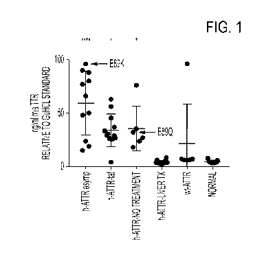
[0085] FIG. 1 depicts the results of a sandwich immunoassay employing the Meso
Scale
Discovery (MSD) electrochemiluminescence platform showing that 9D5 detects
elevated
misfolded TTR in plasma samples from transthyretin-mediated amyloidosis (ATTR)
patients that
had not undergone a liver transplant, in an assay using a polyclonal anti-TTR
reporter antibody.
[0086] FIG. 2 depicts results of an ex vivo target engagement assay, using a
9D5 capture
antibody and a polyclonal anti-TTR reporter antibody, showing that m14G8
reduces levels of
free misfolded TTR when spiked into patient plasma.
[0087] FIG. 3 depicts the results of a sandwich immunoassay employing the Meso
Scale
Discovery (MSD) electrochemiluminescence platform showing that 9D5 detects
elevated
misfolded TTR in plasma samples from transthyretin-mediated amyloidosis (ATTR)
patients that
had not undergone a liver transplant, in an assay using an 18C5 reporter
antibody.
[0088] FIG. 4 depicts results of a Western blot experiment showing that 18C5
has strong
reactivity toward denatured TTR monomer, minor reactivity toward denatured
dimer, and very
weak reactivity toward native TTR species.
[0089] FIG. 5 depicts results of a Western blot experiment showing that a
commercial TTR
antibody could not distinguish between native versus denatured TTR and showed
very strong
reactivity toward monomeric as well as dimeric native and denatured TTR
CA 03077353 2020-03-27
WO 2019/071206 PCT/US2018/054723
[0090] FIG. 6 depicts the results of a sandwich immunoassay employing the Meso
Scale
Discovery (MSD) electrochemiluminescence platform showing that 18C5 detects
elevated
misfolded TTR in plasma samples from transthyretin-mediated amyloidosis (ATTR)
patients that
had not undergone a liver transplant.
[0091] FIG. 7 depicts the results of a sandwich immunoassay employing the Meso
Scale
Discovery (MSD) electrochemiluminescence platform showing that 18C5 detects
elevated
multimeric misfolded TTR in plasma samples from transthyretin-mediated
amyloidosis (ATTR)
patients that had not undergone a liver transplant.
[0092] FIG. 8 depicts the results of a Western blot experiment showing that
9D5 detects elevated
levels of mis-TTR (misfolded TTR monomers and oligomers) in plasma from
hereditary ATTR
patients.
[0093] FIG. 9 depicts a diagram of a pharmacodynamic assay to measure binding
of a mis-TTR
mAb (m14G8) to plasma mis-TTR (target engagement).
[0094] FIG. 10 depicts the results of a sandwich immunoassay employing the
Meso Scale
Discovery (MSD) electrochemiluminescence platform showing that 9D5 detects
elevated
misfolded TTR in plasma samples from transthyretin-mediated amyloidosis (ATTR)
patients that
had not undergone a liver transplant, in an assay using a polyclonal anti-TTR
reporter antibody.
[0095] FIG. 11 depicts an alignment of heavy chain variable regions of the
mouse 18C5
antibody (SEQ ID NO: 81), human germline sequence IGHV3-48*01 (SEQ ID NO:84),
human
acceptor 5VZY-VH huFrwk (Crenefab-VH) (SEQ ID NO:83), and humanized versions
of the
18C5 antibody (hu18C5 VH-vl and hu18C5 VH-v2, SEQ ID NOs: 84 and 85,
respectively).
CDRs defined according to Kabat/Chothia Composite are bolded in the mouse 18C5
heavy chain
variable region sequence.
[0096] FIG. 12 depicts an alignment of light chain variable regions of the
mouse 18C5 antibody
(SEQ ID NO:87), human germline sequence IGKV2-30*02 (SEQ ID NO:90), human
acceptor
5VZY-VL huFrwk (Crenefab-VL) (SEQ ID NO:89), and humanized versions of the
18C5
antibody (hu18C5-VL-v1 and hu18C5-VL-v2, SEQ ID NOs: 91 and 92, respectively).
CDRs
21
CA 03077353 2020-03-27
WO 2019/071206 PCT/US2018/054723
defined according to Kabat/Chothia Composite are bolded in the mouse 18C5
light chain
variable region sequence.
BRIEF DESCRIPTION OF THE SEQUENCES
[0097] SEQ ID NO:1 sets forth the amino acid sequence of a heavy chain
variable region of the
mouse 18C5 antibody with signal peptide.
[0098] SEQ ID NO:2 sets forth a nucleic acid sequence encoding a heavy chain
variable region
of the mouse 18C5 antibody with signal peptide.
[0099] SEQ ID NO:3 sets forth the amino acid sequence of a light chain
variable region of the
mouse 18C5 antibody with signal peptide.
[0100] SEQ ID NO:4 sets forth a nucleic acid sequence encoding a light chain
variable region of
the mouse 18C5 antibody with signal peptide.
[0101] SEQ ID NO:5 sets forth the amino acid sequence of a Kabat/Chothia
Composite CDR-H1
of the mouse 18C5 antibody.
[0102] SEQ ID NO:6 sets forth a nucleic acid sequence encoding a Kabat/Chothia
Composite
composite CDR-H1 of the mouse 18C5 antibody.
[0103] SEQ ID NO:7 sets forth the amino acid sequence of a Kabat/Chothia
Composite CDR-H2
of the mouse 18C5 antibody.
[0104] SEQ ID NO:8 sets forth a nucleic acid sequence encoding a Kabat/Chothia
Composite
CDR-H2 of the mouse 18C5 antibody.
[0105] SEQ ID NO:9 sets forth the amino acid sequence of a Kabat/Chothia
Composite CDR-H3
of the mouse 18C5 antibody.
[0106] SEQ ID NO:10 sets forth a nucleic acid sequence encoding a
Kabat/Chothia Composite
CDR-H3 of the mouse 18C5 antibody.
22
CA 03077353 2020-03-27
WO 2019/071206 PCT/US2018/054723
[0107] SEQ ID NO:11 sets forth the amino acid sequence of a Kabat/Chothia
Composite CDR-
Li of the mouse 18C5 antibody.
[0108] SEQ ID NO:12 sets forth a nucleic acid sequence encoding a
Kabat/Chothia Composite
CDR-L1 of the mouse 18C5 antibody.
[0109] SEQ ID NO:13 sets forth the amino acid sequence of a Kabat/Chothia
Composite CDR-
L2 of the mouse 18C5 antibody.
[0110] SEQ ID NO:14 sets forth a nucleic acid sequence encoding a
Kabat/Chothia Composite
CDR-L2 of the mouse 18C5 antibody.
[0111] SEQ ID NO:15 sets forth the amino acid sequence of a Kabat/Chothia
Composite CDR-
L3 of the mouse 18C5 antibody.
[0112] SEQ ID NO:16 sets forth a nucleic acid sequence encoding the a
Kabat/Chothia
Composite CDR-L3 of the mouse 18C5 antibody.
[0113] SEQ ID NO:17 sets forth the amino acid sequence of a chimeric 18C5
heavy chain
constant region (human IgG1).
[0114] SEQ ID NO:18 sets forth a nucleic acid sequence encoding the amino acid
sequence of a
chimeric 18C5 heavy chain constant region (human IgG1).
[0115] SEQ ID NO:19 sets forth the amino acid sequence of a chimeric 18C5
light chain
constant region (human kappa).
[0116] SEQ ID NO:20 sets forth a nucleic acid sequence encoding the amino acid
sequence of a
chimeric 18C5 light chain constant region (human kappa).
[0117] SEQ ID NO:21 sets forth the amino acid sequence of an exemplary IgG1
heavy chain
constant region.
[0118] SEQ ID NO:22 sets forth the amino acid sequence of an exemplary IgG1
Glm3 heavy
chain constant region.
23
CA 03077353 2020-03-27
WO 2019/071206
PCT/US2018/054723
[0119] SEQ ID NO:23 sets forth the amino acid sequence of an exemplary IgG1
G1m3 heavy
chain constant region.
[0120] SEQ ID NO:24 sets forth the amino acid sequence of an exemplary light
chain constant
region with N-terminal Arginine.
[0121] SEQ ID NO:25 sets forth the amino acid sequence of an exemplary light
chain constant
region without N-terminal Arginine.
[0122] SEQ ID NO:26 sets forth the amino acid sequence of human transthyretin
set forth in
accession number P02766.1 (UniProt).
[0123] SEQ ID NO:27 sets forth the amino acid sequence of human transthyretin
set forth in
accession number AAB35639.1 (GenBank).
[0124] SEQ ID NO:28 sets forth the amino acid sequence of human transthyretin
set forth in
accession number AAB35640.1 (GenBank).
[0125] SEQ ID NO:29 sets forth the amino acid sequence of human transthyretin
set forth in
accession number and ABI63351.1 (GenBank).
[0126] SEQ ID NO:30 sets forth the amino acid sequence of residues 101-109 of
human
transthyretin.
[0127] SEQ ID NO:31 sets forth the amino acid sequence of residues 87-127 of
human
transthyretin.
[0128] SEQ ID NO:32 sets forth a nucleic acid sequence encoding an exemplary
IgG1 G1m3
heavy chain constant region.
[0129] SEQ ID NO:33 sets forth a nucleic acid sequence encoding an exemplary
light chain
constant region with C-terminal Arginine.
[0130] SEQ ID NO:34 sets forth a nucleic acid sequence encoding an exemplary
light chain
constant region without C-terminal Arginine.
24
CA 03077353 2020-03-27
WO 2019/071206 PCT/US2018/054723
[0131] SEQ ID NO:35 sets forth the amino acid sequence of a heavy chain
constant region
signal peptide.
[0132] SEQ ID NO:36 sets forth a nucleic acid sequence encoding a heavy chain
constant region
signal peptide.
[0133] SEQ ID NO:37 sets forth the amino acid sequence of a light chain
constant region signal
peptide.
[0134] SEQ ID NO:38 sets forth a nucleic acid sequence encoding a light chain
constant region
signal peptide.
[0135] SEQ ID NO: 39 sets forth the amino acid sequence of a Kabat CDR-H1 of
antibody
14G8.
[0136] SEQ ID NO: 40 sets forth the amino acid sequence of a Kabat CDR-H2 of
antibody
14G8.
[0137] SEQ ID NO: 41 sets forth the amino acid sequence of a Kabat CDR-H3 of
antibody
14G8.
[0138] SEQ ID NO: 42 sets forth the amino acid sequence of a Kabat CDR-L1 of
antibody
14G8.
[0139] SEQ ID NO: 43 sets forth the amino acid sequence of a Kabat CDR-L2 of
antibody
14G8.
[0140] SEQ ID NO: 44 sets forth the amino acid sequence of a Kabat CDR-L3 of
antibody
14G8.
[0141] SEQ ID NO: 45 sets forth the amino acid sequence of an epitope of
antibody 5A1.
[0142] SEQ ID NO: 46 sets forth the amino acid sequence of a Kabat CDR-H1 of
antibody 5A1.
[0143] SEQ ID NO: 47 sets forth the amino acid sequence of a Kabat CDR-H2 of
antibody 5A1.
[0144] SEQ ID NO: 48 sets forth the amino acid sequence of a Kabat CDR-H3 of
antibody 5A1.
CA 03077353 2020-03-27
WO 2019/071206 PCT/US2018/054723
[0145] SEQ ID NO: 49 sets forth the amino acid sequence of a Kabat CDR-L1 of
antibody 5A1.
[0146] SEQ ID NO: 50 sets forth the amino acid sequence of a Kabat CDR-L2 of
antibody 5A1.
[0147] SEQ ID NO: 51 sets forth the amino acid sequence of a Kabat CDR-L3 of
antibody 5A1.
[0148] SEQ ID NO: 52 sets forth the amino acid sequence of a Kabat CDR-H1 of
antibody 6C1.
[0149] SEQ ID NO: 53 sets forth the amino acid sequence of a Kabat CDR-H2 of
antibody 6C1.
[0150] SEQ ID NO: 54 sets forth the amino acid sequence of a Kabat CDR-H3 of
antibody 6C1.
[0151] SEQ ID NO: 55 sets forth the amino acid sequence of a Kabat CDR-L1 of
antibody 6C1.
[0152] SEQ ID NO: 56 sets forth the amino acid sequence of a Kabat CDR-L2 of
antibody 6C1.
[0153] SEQ ID NO: 57 sets forth the amino acid sequence of a Kabat CDR-L3 of
antibody 6C1.
[0154] SEQ ID NO: 58 sets forth the amino acid sequence of a VH region of
antibody AD7F6.
[0155] SEQ ID NO: 59 sets forth the amino acid sequence of a VL region of
antibody AD7F6.
[0156] SEQ ID NO: 60 sets forth the amino acid sequence of a CDR-H1 of
antibody RT24.
[0157] SEQ ID NO: 61 sets forth the amino acid sequence of a CDR-H2 of
antibody RT24.
[0158] SEQ ID NO: 62 sets forth the amino acid sequence of a CDR-H3 of
antibody RT24.
[0159] SEQ ID NO:63 sets forth the amino acid sequence of a CDR-L1 of antibody
RT24.
[0160] SEQ ID NO: 64 sets forth the amino acid sequence of a CDR-L2 of
antibody RT24.
[0161] SEQ ID NO: 65 sets forth the amino acid sequence of a CDR-L3 of
antibody RT24.
[0162] SEQ ID NO: 66 sets forth the amino acid sequence of a CDR-H1 of
antibody NI-
301.35G11.
[0163] SEQ ID NO:67 sets forth the amino acid sequence of a CDR-H2 of antibody
NI-
301.35G11.
26
CA 03077353 2020-03-27
WO 2019/071206 PCT/US2018/054723
[0164] SEQ ID NO: 68 sets forth the amino acid sequence of a CDR-H3 of
antibody NI-
301.35G11.
[0165] SEQ ID NO: 69sets forth the amino acid sequence of a CDR-L1 of antibody
NI-
301.35G11.
[0166] SEQ ID NO: 70 sets forth the amino acid sequence of a CDR-L2 of
antibody NI-
301.35G11.
[0167] SEQ ID NO: 71 sets forth the amino acid sequence of a CDR-L3 of
antibody NI-
301.35G11.
[0168] SEQ ID NO: 72 sets forth the amino acid sequence of an epitope of
antibodies MFD101,
MDF102, MFD103, MFD105.
[0169] SEQ ID NO: 73 sets forth the amino acid sequence of an epitope of
antibodies MFD107,
MFD108, MFD109, MFD111.
[0170] SEQ ID NO: 74 sets forth the amino acid sequence of an epitope of
antibody MFD114.
[0171] SEQ ID NO: 75 sets forth the amino acid sequence of a Kabat CDR-H1 of
antibody 9D5.
[0172] SEQ ID NO: 76 sets forth the amino acid sequence of a Kabat CDR-H2 of
antibody 9D5.
[0173] SEQ ID NO: 77 sets forth the amino acid sequence of a Kabat CDR-H3 of
antibody 9D5.
[0174] SEQ ID NO: 78 sets forth the amino acid sequence of a Kabat CDR-L1 of
antibody 9D5.
[0175] SEQ ID NO: 79 sets forth the amino acid sequence of a Kabat CDR-L2 of
antibody 9D5.
[0176] SEQ ID NO: 80 sets forth the amino acid sequence of a Kabat CDR-L3 of
antibody 9D5.
[0177] SEQ ID NO:81 sets forth the amino acid sequence of a mature heavy chain
variable
region of the mouse 18C5 antibody.
[0178] SEQ ID NO: 82 sets forth the amino acid sequence of a heavy chain
variable region of
the murine anti-pyroglutamate-Abeta antibody Fab c#17, GenBank Acc. No.
1212215935.
27
CA 03077353 2020-03-27
WO 2019/071206 PCT/US2018/054723
[0179] SEQ ID NO: 83 sets forth the amino acid sequence of a heavy chain
variable region of
humanized Crenezumab Fab (CreneFab) PDB: 5VZY, GenBank Acc. No. 1229749875.
[0180] SEQ ID NO: 84 sets forth the amino acid sequence of a heavy chain
variable region of
the human germline sequence IGHV3-48*01, GenBank Acc. No. 1FN550289.1.
[0181] SEQ ID NO: 85 sets forth the amino acid sequence of a heavy chain
variable region of
the humanized 18C5 antibody hu18C5-VH 1.
[0182] SEQ ID NO: 86 sets forth the amino acid sequence of a heavy chain
variable region of
the humanized 18C5 antibody hu18C5-VH 2.
[0183] SEQ ID NO:87 sets forth the amino acid sequence of a mature light chain
variable region
of the mouse 18C5 antibody.
[0184] SEQ ID NO: 88 sets forth the amino acid sequence of a light chain
variable region of the
murine anti-pyroglutamate-Abeta antibody Fab c#17, GenBank Acc. No.
1212215934.
[0185] SEQ ID NO: 89 sets forth the amino acid sequence of a light chain
variable region of
humanized Crenezumab Fab (CreneFab) PDB: 5VZY, GenBank Acc. No. 1229749876.
[0186] SEQ ID NO: 90 sets forth the amino acid sequence of a light chain
variable region of the
human germline sequence IGKV2-30*2, GenBank Acc. No. CAA77315.
[0187] SEQ ID NO: 91 sets forth the amino acid sequence of a light chain
variable region of the
humanized 18C5 antibody hu18C5-VL 1.
[0188] SEQ ID NO: 92 sets forth the amino acid sequence of a light chain
variable region of the
humanized 18C5 antibody hu18C5-VL 2.
[0189] SEQ ID NO: 93 sets forth the amino acid sequence of Kabat CDR-H1 of the
mouse 18C5
antibody.
[0190] SEQ ID NO: 94 sets forth the amino acid sequence of Chothia CDR-H1 of
the mouse
18C5 antibody.
28
CA 03077353 2020-03-27
WO 2019/071206 PCT/US2018/054723
[0191] SEQ ID NO: 95 sets forth the amino acid sequence of Contact CDR-H1 of
the mouse
18C5 antibody.
[0192] SEQ ID NO: 96 sets forth the amino acid sequence of Chothia CDR-H2 of
the mouse
18C5 antibody.
[0193] SEQ ID NO: 97 sets forth the amino acid sequence of AbM CDR-H2 of the
mouse 18C5
antibody.
[0194] SEQ ID NO: 98 sets forth the amino acid sequence of Contact CDR-H2 of
the mouse
18C5 antibody.
[0195] SEQ ID NO: 99 sets forth the amino acid sequence of Contact CDR-H3 of
the mouse
18C5 antibody.
[0196] SEQ ID NO: 100 sets forth the amino acid sequence of Contact CDR-L1 of
the mouse
18C5 antibody.
[0197] SEQ ID NO: 101 sets forth the amino acid sequence of Contact CDR-L2 of
the mouse
18C5 antibody.
[0198] SEQ ID NO: 102 sets forth the amino acid sequence of Contact CDR-L3 of
the mouse
18C5 antibody.
[0199] SEQ ID NO: 103 sets forth the amino acid sequence of a heavy chain
variable region of
the mouse 9D5 antibody.
[0200] SEQ ID NO: 104 sets forth the amino acid sequence of a light chain
variable region of
the mouse 9D5 antibody.
DEFINITIONS
[0201] Monoclonal antibodies or other biological entities are typically
provided in isolated form.
This means that an antibody or other biologically entity is typically at least
50% w/w pure of
interfering proteins and other contaminants arising from its production or
purification but does
not exclude the possibility that the monoclonal antibody is combined with an
excess of
pharmaceutically acceptable carrier(s) or other vehicle intended to facilitate
its use. Sometimes
monoclonal antibodies are at least 60%, 70%, 80%, 90%, 95% or 99% w/w pure of
interfering
29
CA 03077353 2020-03-27
WO 2019/071206 PCT/US2018/054723
proteins and contaminants from production or purification. Often an isolated
monoclonal
antibody or other biological entity is the predominant macromolecular species
remaining after its
purification.
[0202] Specific binding of an antibody to its target antigen means an affinity
of at least 106, 107
,
108, 109, or 1010 M1. Specific binding is detectably higher in magnitude and
distinguishable
from non-specific binding occurring to at least one unrelated target. Specific
binding can be the
result of formation of bonds between particular functional groups or
particular spatial fit (e.g.,
lock and key type) whereas nonspecific binding is usually the result of van
der Waals forces.
Specific binding does not however necessarily imply that an antibody binds one
and only one
target.
[0203] The basic antibody structural unit is a tetramer of subunits. Each
tetramer includes two
identical pairs of polypeptide chains, each pair having one "light" (about 25
kDa) and one
"heavy" chain (about 50-70 kDa). The amino-terminal portion of each chain
includes a variable
region of about 100 to 110 or more amino acids primarily responsible for
antigen recognition.
This variable region is initially expressed linked to a cleavable signal
peptide. The variable
region without the signal peptide is sometimes referred to as a mature
variable region. Thus, for
example, a light chain mature variable region means a light chain variable
region without the
light chain signal peptide. The carboxy-terminal portion of each chain defines
a constant region
primarily responsible for effector function.
[0204] Light chains are classified as either kappa or lambda. Heavy chains are
classified as
gamma, mu, alpha, delta, or epsilon, and define the antibody's isotype as IgG,
IgM, IgA, IgD and
IgE, respectively. Within light and heavy chains, the variable and constant
regions are joined by
a "J" region of about 12 or more amino acids, with the heavy chain also
including a "D" region
of about 10 or more amino acids. See generally, Fundamental Immunology, Paul,
W., ed., 2nd
ed. Raven Press, N.Y., 1989, Ch. 7 (incorporated by reference in its entirety
for all purposes).
[0205] An immunoglobulin light or heavy chain variable region (also referred
to herein as a
"light chain variable domain" ("VL domain") or "heavy chain variable domain"
("VH domain"),
respectively) consists of a "framework" region interrupted by three
"complementarity
determining regions" or "CDRs." The framework regions serve to align the CDRs
for specific
CA 03077353 2020-03-27
WO 2019/071206 PCT/US2018/054723
binding to an epitope of an antigen. The CDRs include the amino acid residues
of an antibody
that are primarily responsible for antigen binding. From amino-terminus to
carboxyl-terminus,
both VL and VH domains comprise the following framework (FR) and CDR regions:
FR1,
CDR1, FR2, CDR2, FR3, CDR3, and FR4. CDRs 1, 2, and 3 of a VL domain are also
referred
to herein, respectively, as CDR-L1, CDR-L2, and CDR-L3; CDRs 1, 2, and 3 of a
VH domain
are also referred to herein, respectively, as CDR-H1, CDR-H2, and CDR-H3.
[0206] The assignment of amino acids to each VL and VH domain is in accordance
with any
conventional definition of CDRs. Conventional definitions include, the Kabat
definition (Kabat,
Sequences of Proteins of Immunological Interest (National Institutes of
Health, Bethesda, MD,
1987 and 1991), The Chothia definition (Chothia & Lesk, I Mol. Biol. 196:901-
917, 1987;
Chothia et at., Nature 342:878-883, 1989); a composite of Chothia Kabat CDR in
which CDR-
H1 is a composite of Chothia and Kabat CDRs; the AbM definition used by Oxford
Molecular's
antibody modelling software; and, the contact definition of Martin et al
(bioinfo.org.uk/abs) (see
Table 1). Kabat provides a widely used numbering convention (Kabat numbering)
in which
corresponding residues between different heavy chains or between different
light chains are
assigned the same number. When an antibody is said to comprise CDRs by a
certain definition
of CDRs (e.g., Kabat) that definition specifies the minimum number of CDR
residues present in
the antibody (i.e., the Kabat CDRs). It does not exclude that other residues
falling within another
conventional CDR definition but outside the specified definition are also
present. For example,
an antibody comprising CDRs defined by Kabat includes among other
possibilities, an antibody
in which the CDRs contain Kabat CDR residues and no other CDR residues, and an
antibody in
which CDR H1 is a composite Chothia-Kabat CDR H1 and other CDRs contain Kabat
CDR
residues and no additional CDR residues based on other definitions.
31
CA 03077353 2020-03-27
WO 2019/071206
PCT/US2018/054723
Table 1
Conventional Definitions of CDRs Using Kabat Numbering
Composite of
Loop Kabat Chothia Chothia AbM
Contact
Kabat
Li L24--L34 L24--L34 L24--L34 L24--L34 L30--L36
L2 L50--L56 L50--L56 L50--L56 L50--L56 L46--L55
L3 L89--L97 L89--L97 L89--L97 L89--L97 L89--L96
H1 H31--H35B H26--H32..H34* H26--H35B* H26--H35B H30--H35B
H2 H50--H65 H52--H56 H50--H65 H50--H58 H47--H58
H3 H95--H102 H95--H102 H95--H102 H95--H102 H93--H101
*CDR-H1 by Chothia can end at H32, H33, or H34 (depending on the length of
the loop). This is because the Kabat numbering scheme places insertions of
extra
residues at 35A and 35B, whereas Chothia numbering places them at 31A and
31B. If neither H35A nor H35B (Kabat numbering) is present, the Chothia CDR-
H1 loop ends at H32. If only H35A is present, it ends at H33. If both H35A and
H35B are present, it ends at H34.
[0207] The term "antibody" includes intact antibodies and binding fragments
thereof. Typically,
fragments compete with the intact antibody from which they were derived for
specific binding to
the target including separate heavy chains, light chains Fab, Fab', F(ab')2,
F(ab)c, Dabs,
nanobodies, and Fv. Fragments can be produced by recombinant DNA techniques,
or by
enzymatic or chemical separation of intact immunoglobulins. The term
"antibody" also includes
a bispecific antibody and/or a humanized antibody. A bispecific or
bifunctional antibody is an
artificial hybrid antibody having two different heavy/light chain pairs and
two different binding
sites (see, e.g., Songsivilai and Lachmann, Cl/n. Exp. Immunol., 79:315-321
(1990); Kostelny et
at., I Immunol., 148:1547-53 (1992)). In some bispecific antibodies, the two
different
heavy/light chain pairs include a humanized 9D5 heavy chain/light chain pair
and a heavy
chain/light chain pair specific for a different epitope on transthyretin than
that bound by 9D5. In
some bispecific antibodies, the two different heavy/light chain pairs include
a humanized 18C5
32
CA 03077353 2020-03-27
WO 2019/071206 PCT/US2018/054723
heavy chain/light chain pair and a heavy chain/light chain pair specific for a
different epitope on
transthyretin than that bound by 18C5.
[0208] In some bispecific antibodies, one heavy chain/light chain pair is a
humanized 9D5
antibody or a humanized 18C5 antibody as further disclosed below and the other
heavy
chain/light chain pair is from an antibody that binds to a receptor expressed
on the blood brain
barrier, such as an insulin receptor, an insulin-like growth factor (IGF)
receptor, a leptin receptor,
or a lipoprotein receptor, or a transferrin receptor (Friden et at., Proc.
Natl. Acad. Sci. USA
88:4771-4775, 1991; Friden et at., Science 259:373-377, 1993). Such a
bispecific antibody can
be transferred cross the blood brain barrier by receptor-mediated
transcytosis. Brain uptake of
the bispecific antibody can be further enhanced by engineering the bispecific
antibody to reduce
its affinity to the blood brain barrier receptor. Reduced affinity for the
receptor resulted in a
broader distribution in the brain (see, e.g., Atwal et al., Sci. Trans. Med.
3, 84ra43, 2011; Yu et
at., Sci. Trans. Med. 3, 84ra44, 2011).
[0209] Exemplary bispecific antibodies can also be: (1) a dual-variable-domain
antibody (DVD-
Ig), where each light chain and heavy chain contains two variable domains in
tandem through a
short peptide linkage (Wu et at., Generation and Characterization of a Dual
Variable Domain
Immunoglobulin (DVD-IgTM) Molecule, In: Antibody Engineering, Springer Berlin
Heidelberg
(2010)); (2) a Tandab, which is a fusion of two single chain diabodies
resulting in a tetravalent
bispecific antibody that has two binding sites for each of the target
antigens; (3) a flexibody,
which is a combination of scFvs with a diabody resulting in a multivalent
molecule; (4) a so-
called "dock and lock" molecule, based on the "dimerization and docking
domain" in Protein
Kinase A, which, when applied to Fabs, can yield a trivalent bispecific
binding protein consisting
of two identical Fab fragments linked to a different Fab fragment; or (5) a so-
called Scorpion
molecule, comprising, e.g., two scFvs fused to both termini of a human Fc-
region. Examples of
platforms useful for preparing bispecific antibodies include BiTE (Micromet),
DART
(MacroGenics), Fcab and Mab2 (F-star), Fc-engineered IgG1 (Xencor) or DuoBody
(based on
Fab arm exchange, Genmab).
[0210] The term "epitope" refers to a site on an antigen to which an antibody
binds. An epitope
can be formed from contiguous amino acids or noncontiguous amino acids
juxtaposed by tertiary
33
CA 03077353 2020-03-27
WO 2019/071206 PCT/US2018/054723
folding of one or more proteins. Epitopes formed from contiguous amino acids
(also known as
linear epitopes) are typically retained on exposure to denaturing solvents
whereas epitopes
formed by tertiary folding (also known as conformational epitopes) are
typically lost on
treatment with denaturing solvents. An epitope typically includes at least 3,
and more usually, at
least 5 or 8-10 amino acids in a unique spatial conformation. Methods of
determining spatial
conformation of epitopes include, for example, x-ray crystallography and 2-
dimensional nuclear
magnetic resonance. See, e.g., Epitope Mapping Protocols, in Methods in
Molecular Biology,
Vol. 66, Glenn E. Morris, Ed. (1996). The epitope can be linear, such as an
epitope of, for
example, 2-5, 3-5, 3-9, or 5-9 contiguous amino acids from SEQ ID NO:26,
including for
example, two or more contiguous amino acids within residues 89-97 of the
mature region of SEQ
ID NO:26. The epitope can also be a conformational epitope including, for
example, two or
more non-contiguous segments of amino acids within residues 89-97 of the
mature region of
SEQ ID NO:26. If an antibody is said to bind to an epitope within amino acid
residues 89-97 of
transthyretin (TTR) (the mature region of SEQ ID NO:26), for example, what is
meant is that the
epitope is within the recited range of amino acids including those defining
the outer-limits of the
range. It does not necessarily mean that every amino acid within the range
constitutes part of the
epitope. Thus, for example, an epitope within amino acid residues 89-97 of TTR
may consist of
amino acids 89-97, 89-96, 90-97, 89-95, 90-96, 91-97, 89-94, 90-95, 91-96, 92-
97, 89-93, 90-94,
91-95, 92-96, 93-97, 89-92, 90-93, 91-94, 92-95, 93-96, 94-97, 89-91, 90-92,
91-93, 92-94, 93-
95, 94-96, 95-97 of SEQ ID NO:26, among other linear segments of SEQ ID NO:45,
or in the
case of conformational epitopes, non-contiguous segments of amino acids of SEQ
ID NO:45.
Epitopes can include E89K and E89Q substitutions, E being the wildtype
residue).
The epitope can be linear, such as an epitope of, for example, 2-5, 3-5, 3-9,
or 5-9 contiguous
amino acids from SEQ ID NO:26, including for example, two or more contiguous
amino acids
within residues 101-109 of the mature region of SEQ ID NO:26. The epitope can
also be a
conformational epitope including, for example, two or more non-contiguous
segments of amino
acids within residues 101-109 of the mature region of SEQ ID NO:26. If an
antibody is said to
bind to an epitope within amino acid residues 101-109 of transthyretin (TTR)
(the mature region
of SEQ ID NO:26), for example, what is meant is that the epitope is within the
recited range of
amino acids including those defining the outer-limits of the range. It does
not necessarily mean
that every amino acid within the range constitutes part of the epitope. Thus,
for example, an
34
CA 03077353 2020-03-27
WO 2019/071206 PCT/US2018/054723
epitope within amino acid residues 101-109 of TTR may consist of amino acids
101-109, 101-
108, 102-109, 101-107, 102-108, 103-109, 101-106, 102-107, 103-108, 104-109,
101-105, 102-
106, 103-107, 104-108, 105-109, 101-104, 102-105, 103-106, 104-107, 105-108,
106-109, 101-
103, 102-104, 103-105, 104-106, 105-107, 106-108, 107-109, 101-102, 102-103,
103-104, 104-
105, 105-106, 106-107, 107-108, or 108-109 of SEQ ID NO:26, among other linear
segments of
SEQ ID NO:30, or in the case of conformational epitopes, non-contiguous
segments of amino
acids of SEQ ID NO:30.
[0211] Antibodies that recognize the same or overlapping epitopes can be
identified in a simple
immunoassay showing the ability of one antibody to compete with the binding of
another
antibody to a target antigen. The epitope of an antibody can also be defined
by X-ray
crystallography of the antibody bound to its antigen to identify contact
residues. Alternatively,
two antibodies have the same epitope if all amino acid mutations in the
antigen that reduce or
eliminate binding of one antibody reduce or eliminate binding of the other.
Two antibodies have
overlapping epitopes if some amino acid mutations that reduce or eliminate
binding of one
antibody reduce or eliminate binding of the other.
[0212] Competition between antibodies is determined by an assay in which an
antibody under
test inhibits specific binding of a reference antibody to a common antigen
(see, e.g., Junghans et
at., Cancer Res. 50:1495, 1990). A test antibody competes with a reference
antibody if an
excess of a test antibody (e.g., at least 2x, 5x, 10x, 20x or 100x) inhibits
binding of the reference
antibody by at least 50% as measured in a competitive binding assay. Some test
antibodies
inhibit binding of the references antibody by at least 75%, 90% or 99%.
Antibodies identified by
competition assay (competing antibodies) include antibodies binding to the
same epitope as the
reference antibody and antibodies binding to an adjacent epitope sufficiently
proximal to the
epitope bound by the reference antibody for steric hindrance to occur.
[0213] The term "native" with respect to the structure transthyretin (TTR)
refers to the normal
folded structure of TTR in its properly functioning state (i.e., a TTR
tetramer). As TTR is a
tetramer in its natively folded form, non-native forms of TTR include, for
example, misfolded
TTR tetramers, TTR monomers, aggregated forms of TTR, and fibril forms of TTR.
Non-native
CA 03077353 2020-03-27
WO 2019/071206 PCT/US2018/054723
forms of TTR can include molecules comprising wild-type TTR amino acid
sequences or
mutations.
[0214] The term "misfolded" with respect to TTR refers to the secondary and
tertiary structure
of a TTR polypeptide monomer or multimer, and indicates that the polypeptide
has adopted a
conformation that is not normal for that protein in its properly functioning
state. Although TTR
misfolding can be caused by mutations in the protein (e.g., deletion,
substitution, or addition),
wild-type TTR proteins can also be misfolded in diseases, exposing specific
epitopes.
[0215] The term "pharmaceutically acceptable" means that the carrier, diluent,
excipient, or
auxiliary is compatible with the other ingredients of the formulation and not
substantially
deleterious to the recipient thereof.
[0216] The term "patient" includes human and other mammalian subjects that
receive either
prophylactic or therapeutic treatment.
[0217] An individual is at increased risk of a disease if the subject has at
least one known risk-
factor (e.g., genetic, biochemical, family history, and situational exposure)
placing individuals
with that risk factor at a statistically significant greater risk of
developing the disease than
individuals without the risk factor.
[0218] The term "biological sample" refers to a sample of biological material
within or
obtainable from a biological source, for example a human or mammalian subject.
Such samples
can be organs, organelles, tissues, sections of tissues, bodily fluids,
peripheral blood, blood
plasma, blood serum, cells, molecules such as proteins and peptides, and any
parts or
combinations derived therefrom. The term biological sample can also encompass
any material
derived by processing the sample. Derived material can include cells or their
progeny.
Processing of the biological sample may involve one or more of filtration,
distillation, extraction,
concentration, fixation, inactivation of interfering components, and the like.
[0219] The term "control sample" refers to a biological sample not known or
suspected to
include monomeric, misfolded, aggregated, or fibril forms of transthyretin
(TTR), such as in
TTR amyloid deposits. Control samples can be obtained from individuals not
afflicted with a
TTR amyloidosis or a specifically chosen type of TTR amyloidosis.
Alternatively, control
36
CA 03077353 2020-03-27
WO 2019/071206 PCT/US2018/054723
samples can be obtained from patients afflicted with TTR amyloidosis or a
specifically chosen
type of TTR amyloidosis. Such samples can be obtained at the same time as a
biological sample
thought to comprise the TTR amyloidosis or on a different occasion. A
biological sample and a
control sample can both be obtained from the same tissue (e.g., a tissue
section containing both
TTR amyloid deposits and surrounding normal tissue). Preferably, control
samples consist
essentially or entirely of tissue free of TTR amyloid deposits and can be used
in comparison to a
biological sample thought to comprise TTR amyloid deposits. Preferably, the
tissue in the
control sample is the same type as the tissue in the biological sample (e.g.,
cardiomyocytes in the
heart).
[0220] The term "disease" refers to any abnormal condition that impairs
physiological function.
The term is used broadly to encompass any disorder, illness, abnormality,
pathology, sickness,
condition, or syndrome in which physiological function is impaired,
irrespective of the nature of
the etiology.
[0221] The term "symptom" refers to a subjective evidence of a disease, such
as altered gait, as
perceivable by a subject. A "sign" refers to objective evidence of a disease
as observable by a
physician.
[0222] For purposes of classifying amino acids substitutions as conservative
or nonconservative,
amino acids are grouped as follows: Group I (hydrophobic side chains): met,
ala, val, leu, ile;
Group II (neutral hydrophilic side chains): asn, gln, cys, ser, thr; Group III
(acidic side chains):
asp, glu; Group IV (basic side chains): his, lys, arg; Group V (residues
influencing chain
orientation): gly, pro; and Group VI (aromatic side chains): trp, tyr, phe.
Conservative
substitutions involve substitutions between amino acids in the same class. Non-
conservative
substitutions constitute exchanging a member of one of these classes for a
member of another.
[0223] Percentage sequence identities are determined with antibody sequences
maximally
aligned by the Kabat numbering convention. After alignment, if a subject
antibody region (e.g.,
the entire mature variable region of a heavy or light chain) is being compared
with the same
region of a reference antibody, the percentage sequence identity between the
subject and
reference antibody regions is the number of positions occupied by the same
amino acid in both
37
CA 03077353 2020-03-27
WO 2019/071206 PCT/US2018/054723
the subject and reference antibody region divided by the total number of
aligned positions of the
two regions, with gaps not counted, multiplied by 100 to convert to
percentage.
[0224] Compositions or methods "comprising" or "including" one or more recited
elements may
include other elements not specifically recited. For example, a composition
that "comprises" or
"includes" an antibody may contain the antibody alone or in combination with
other ingredients.
[0225] Designation of a range of values includes all integers within or
defining the range, and all
subranges defined by integers within the range.
[0226] Unless otherwise apparent from the context, the term "about"
encompasses values within
a standard margin of error of measurement (e.g., SEM) of a stated value.
[0227] Statistical significance means p0.05.
[0228] Antibodies of the invention can be administered concomitant with
another treatment for
the same indication as the antibody, meaning that the other treatment is
administered at least
once during the period in which the antibody is administered, such period
beginning one month
before the first dosing and ending one month after the last dosing of the
antibody. The other
treatment can be administered at recurring intervals during this period, which
may or may not be
the same as the intervals at which the antibody is administered. The other
treatment may be a
symptomatic treatment.
[0229] A treatment is symptomatic if it only affects one or more symptoms of a
disease, not its
cause, i.e., its etiology.
[0230] The singular forms of the articles "a," "an," and "the" include plural
references unless the
context clearly dictates otherwise. For example, the term "a compound" or "at
least one
compound" can include a plurality of compounds, including mixtures thereof.
38
CA 03077353 2020-03-27
WO 2019/071206 PCT/US2018/054723
DETAILED DESCRIPTION
I. General
[0231] The invention provides antibodies that specifically bind to residues 89-
97 or to residues
101-109 of transthyretin (TTR). Some antibodies bind any or all of monomeric,
misfolded,
aggregated, or fibril forms of TTR preferentially relative to native
tetrameric form of TTR.
Some antibodies bind either or both of monomeric or misfolded forms of TTR
preferentially
relative to native tetrameric form of TTR. The antibodies can be used for
treating or effecting
prophylaxis of diseases or disorders associated with TTR accumulation or
accumulation of TTR
deposits (e.g., TTR amyloidosis). The antibodies can also be used for
diagnosing TTR
amyloidosis and inhibiting or reducing aggregation of TTR. The antibodies can
also be used to
demonstrate pharmacodynamics effects of a transthyretin-mediated amyloidosis
therapy, among
other applications. Preferential binding means an association constant at
least five times higher
for any or all of monomeric, misfolded, aggregated, or fibril forms of TTR
than for native
tetrameric form of TTR. Optionally, the association constant is at least ten
times higher for any
or all of monomeric, misfolded, aggregated, or fibril forms of TTR than for
native tetrameric
form of TTR. Optionally, the antibody, such as 9D5 or 18C5, for example, lacks
specific
binding to native tetrameric form of TTR.
Target Molecules
[0232] Transthyretin (TTR) is a 127-amino acid, 55 kDa serum and cerebrospinal
fluid transport
protein primarily synthesized by the liver. It has also been referred to as
prealbumin, thyroxine
binding prealbumin, ATTR, and TBPA. In its native state, TTR exists as a
tetramer. In
homozygotes, the tetramers comprise identical 127-amino-acid beta-sheet-rich
subunits. In
heterozygotes, the TTR tetramers are made up of variant and/or wild-type
subunits, typically
combined in a statistical fashion.
[0233] The established function of TTR in the blood is to transport ho/o-
retinol binding protein.
Although TTR is the major carrier of thyroxine (T4) in the blood of rodents,
utilizing binding
sites that are orthogonal to those used for ho/o-retinol binding protein, the
T4 binding sites are
effectively unoccupied in humans.
39
CA 03077353 2020-03-27
WO 2019/071206 PCT/US2018/054723
[0234] TTR is one of at least thirty different human proteins whose
extracellular misfolding
and/or misassembly (amyloidogenesis) into a spectrum of aggregate structures
is thought to
cause degenerative diseases referred to as amyloid diseases. TTR undergoes
conformational
changes in order to become amyloidogenic. Dissociation of the TTR tetramer and
partial
unfolding exposes stretches of largely uncharged hydrophobic residues in an
extended
conformation that efficiently misassemble into largely unstructured spherical
aggregates that
ultimately undergo conformation conversion into cross-beta sheet amyloid
structures.
[0235] Unless otherwise apparent from context, reference to transthyretin
(TTR) or its fragments
or domains includes the natural human amino acid sequences including isoforms,
mutants (e.g.,
E89K and E89Q), and allelic variants thereof. Exemplary TTR polypeptide
sequences are
designated by Accession Numbers P02766.1 (UniProt) (SEQ ID NO:26), AAB35639.1
(GenBank) (SEQ ID NO:27), AAB35640.1 (GenBank) (SEQ ID NO:28), and ABI63351.1
(GenBank) (SEQ ID NO:29). Residues are numbered according to Swiss Prot
P02766.1, with
the first amino acid of the mature protein (i.e., not including the 20 amino
acid signal sequence)
designated residue 1. In any other TTR protein, residues are numbered
according to the
corresponding residues in P02766.1 on maximum alignment.
III. Transthyretin Amyloidosis
[0236] Transthyretin (TTR) amyloidosis is a systemic disorder characterized by
pathogenic,
misfolded TTR and the extracellular deposition of amyloid fibrils composed of
TTR. TTR
amyloidosis is generally caused by destabilization of the native TTR tetramer
form (due to
environmental or genetic conditions), leading to dissociation, misfolding, and
aggregation of
TTR into amyloid fibrils that accumulate in various organs and tissues,
causing progressive
dysfunction. See, e.g., Almeida and Saraiva, FEBS Letters 586:2891-2896
(2012); Ando et al.,
Orphanet Journal of Rare Diseases 8:31(2013).
[0237] In humans, both wild-type TTR tetramers and mixed tetramers comprised
of mutant and
wild-type subunits can dissociate, misfold, and aggregate, with the process of
amyloidogenesis
leading to the degeneration of affected tissue. Thus, TTR amyloidoses
encompass diseases
caused by pathogenic misfolded TTR resulting from mutations in TTR or
resulting from non-
mutated, misfolded TTR.
CA 03077353 2020-03-27
WO 2019/071206 PCT/US2018/054723
[0238] For example, wild-type ATTR amyloidosis (also called senile systemic
amyloidosis or
SSA) and senile cardiac amyloidosis (SCA) are age-related types of amyloidosis
that result from
the deposition of wild-type TTR amyloid outside and within the cardiomyocytes
of the heart.
TTR amyloidosis is also the most common form of hereditary (familial)
amyloidosis, which is
caused by mutations that destabilize the TTR protein. The TTR amyloidoses
associated with
point mutations in the TTR gene include familial amyloid polyneuropathy (FAP),
familial
amyloid cardiomyopathy (FAC), and the rare central nervous system selective
amyloidosis
(CNSA). Patients with hereditary (familial) TTR amyloidosis are almost always
heterozygotes,
meaning that the TTR tetramers are composed of mutant and/or wild-type TTR
subunits,
generally statistically distributed. Hereditary (familial) versions of TTR
amyloidosis are
generally autosomal dominant and are typically earlier onset than the sporadic
diseases (SSA and
SCA).
[0239] There are over 100 mutations in the gene encoding TTR that have been
implicated in the
autosomal dominant disorders FAP and FAC. See, e.g., US 2014/0056904; Saraiva,
Hum.
Mutat. 17(6):493-503 (2001); Damas and Saraiva, I Struct. Biol. 130:290-299;
Dwulet and
Benson, Biochem. Biophys. Res. Commun. 114:657-662 (1983). These amyloid-
causing
mutations are distributed throughout the entire molecule of TTR. Generally,
the more
destabilizing the mutant subunits are to the TTR tetramer structure, the
earlier the onset of
amyloid disease. The pathogenic potential of a TTR variant is generally
determined by a
combination of its instability and its cellular secretion efficiency. The
initial pathology caused
by some TTR variants comes from their selective destruction of cardiac tissue,
whereas that from
other TTR variants comes from compromising the peripheral and autonomic
nervous system.
The tissue damage caused by TTR amyloidogenesis appear to stem largely from
the toxicity of
small, diffusible TTR aggregates, although accumulation of extracellular
amyloid may contribute
and almost certainly compromises organ structure in the late stages of the TTR
amyloidosis.
Exemplary TTR mutations include V30M, Y114C, G47R, S50I, E61L, T495, F33V,
A45T,
E89K, E89Q, and V1221.
[0240] TTR amyloidosis presents in many different forms, with considerable
phenotypic
variation across individuals and geographic locations. For example, TTR
amyloidosis can
41
CA 03077353 2020-03-27
WO 2019/071206 PCT/US2018/054723
present as a progressive, axonal sensory autonomic and motor neuropathy. TTR
amyloidosis can
also present as an infiltrative cardiomyopathy.
[0241] The age at onset of disease-related symptoms varies between the second
and ninth
decades of life, with great variations across different populations. The
multisystem involvement
of TTR amyloidosis is a clue to its diagnosis. For example, TTR amyloidosis
diagnosis is
considered when one or several of the following are present: (1) family
history of neuropathic
disease, especially associated with heart failure; (2) neuropathic pain or
progressive sensory
disturbances of unknown etiology; (3) carpal tunnel syndrome without obvious
cause,
particularly if it is bilateral and requires surgical release; (4)
gastrointestinal motility
disturbances or autonomic nerve dysfunction of unknown etiology (e.g.,
erectile dysfunction,
orthostatic hypotension, neurogenic bladder); (5) cardiac disease
characterized by thickened
ventricular walls in the absence of hypertension; (6) advanced atrio-
ventricular block of
unknown origin, particularly when accompanied by a thickened heart; and (6)
vitreous body
inclusions of the cotton-wool type. See Ando et al., Orphanet Journal of Rare
Diseases 8:31
(2013). Other symptoms can include, for example, polyneuropathy, sensory loss,
pain, weakness
in lower limbs, dyshidrosis, diarrhea, constipation, weight loss, and urinary
incontinence/retention.
[0242] Diagnosis of TTR amyloidosis typically relies on target organ biopsies,
followed by
histological staining of the excised tissue with the amyloid-specific dye,
Congo red. If a positive
test for amyloid is observed, immunohistochemical staining and mass
spectroscopic
identification ofTTR is subsequently performed to ensure that the precursor
protein responsible
for amyloid formation is indeed TTR. Antibodies disclosed herein are useful in
distinguishing
TTR amyloidosis from a non-TTR amyloidosis e.g. amyloid light-chain (AL)
amyloidosis, also
known as primary systemic amyloidosis For familial forms of the diseases,
demonstration of a
mutation in the gene encoding TTR is then needed before diagnosis can be made.
This can be
accomplished, for example, through isoelectric focusing electrophoresis,
polymerase chain
reaction, or laser dissection/liquid chromatography-tandem mass spectrometry.
See, e.g., US
2014/0056904; Ruberg and Berk, Circulation 126:1286-1300 (2012); Ando et al.,
Orphanet
Journal of Rare Diseases 8:31(2013).
42
CA 03077353 2020-03-27
WO 2019/071206 PCT/US2018/054723
IV. Antibodies
A. Binding Specificity and Functional Properties
[0243] The invention provides monoclonal antibodies binding to transthyretin
(TTR) protein,
more specifically, to epitopes within amino acid residues 89-97 (SEQ ID NO:45)
or to epitopes
within amino acid residues 101-109 (SEQ ID NO:30) of TTR. Such epitopes are
buried in the
native TTR tetramer and exposed in monomeric, misfolded, aggregated, or fibril
forms of TTR.
[0244] Such antibodies include 9D5, 18C5, and their chimeric, veneered and
humanized forms.
9D5 specifically binds within amino acid residues 89-97 (SEQ ID NO:45) of TTR.
18C5
specifically binds within amino acid residues 101-109 (SEQ ID NO: 30) of TTR.
These
antibodies are further characterized by their ability to bind to monomeric,
misfolded, aggregated,
or fibril forms of TTR but not to native tetrameric form of TTR. Ability to
bind to specific
proteins or fragments thereof may be demonstrated using exemplary assay
formats provided in
the examples. Unless otherwise apparent from the context, reference to 9D5 or
18C5 should be
understood as referring to any of the mouse, chimeric, veneered or humanized
forms. A
hybridoma cell line that produces monoclonal antibody 9D5 was deposited with
the Patent
Depository of the American Type Culture Collection (ATCC), Manassas, Virginia,
20110-2209
on April 4, 2017 and assigned Patent Deposit No. PTA-124078. A hybridoma cell
line that
produces monoclonal antibody 18C5 was deposited with the Patent Depository of
the American
Type Culture Collection (ATCC), Manassas, Virginia, 20110-2209 on October
31,
2017 _________ and assigned Patent Deposit No. PTA-124570 _____
[0245] Some antibodies bind to the same or overlapping epitope as an antibody
designated 9D5.
The sequences of the heavy and light chain mature variable regions of 9D5 are
designated SEQ
ID NOs: 81 and 82, respectively. Other antibodies having such a binding
specificity can be
produced by immunizing mice with TTR, or a portion thereof including the
desired epitope (e.g.,
SEQ ID NO:45), and screening resulting antibodies for binding to monomeric TTR
or a peptide
comprising SEQ ID NO:45, optionally in competition with an antibody having the
variable
regions of mouse 18C5 (IgGl,kappa). Fragments of TTR including the desired
epitope can be
linked to a carrier that helps elicit an antibody response to the fragment
and/or be combined with
an adjuvant that helps elicit such a response. Such antibodies can be screened
for differential
43
CA 03077353 2020-03-27
WO 2019/071206 PCT/US2018/054723
binding to wild-type, monomeric versions of TTR or a fragment thereof (e.g.,
SEQ ID NO:26)
compared with mutants of specified residues.
Some antibodies bind to the same or overlapping epitope as an antibody
designated 18C5. The
sequences of the heavy and light chain mature variable regions of 18C5 are
designated SEQ ID
NOs: 1 and 3, respectively. Other antibodies having such a binding specificity
can be produced
by immunizing mice with TTR, or a portion thereof including the desired
epitope (e.g., SEQ ID
NO:30), and screening resulting antibodies for binding to monomeric TTR or a
peptide
comprising SEQ ID NO:30, optionally in competition with an antibody having the
variable
regions of mouse 18C5 (IgGl, kappa). Fragments of TTR including the desired
epitope can be
linked to a carrier that helps elicit an antibody response to the fragment
and/or be combined with
an adjuvant that helps elicit such a response. Such antibodies can be screened
for differential
binding to wild-type, monomeric versions of TTR or a fragment thereof (e.g.,
SEQ ID NO:26)
compared with mutants of specified residues.
Screening against such mutants more precisely defines the binding specificity
to allow
identification of antibodies whose binding is inhibited by mutagenesis of
particular residues and
which are likely to share the functional properties of other exemplified
antibodies. The
mutations can be systematic replacement substitution with alanine (or serine
or glycine if an
alanine is present already) one residue at a time, or more broadly spaced
intervals, throughout the
target or throughout a section thereof in which an epitope is known to reside.
If the same set of
mutations significantly reduces the binding of two antibodies, the two
antibodies bind the same
epitope.
[0246] Antibodies having the binding specificity of a selected murine antibody
(e.g., 9D5 or
18C5) can also be produced using a variant of the phage display method. See
Winter, WO
92/20791. This method is particularly suitable for producing human antibodies.
In this method,
either the heavy or light chain variable region of the selected murine
antibody is used as a
starting material. If, for example, a light chain variable region is selected
as the starting material,
a phage library is constructed in which members display the same light chain
variable region
(i.e., the murine starting material) and a different heavy chain variable
region. The heavy chain
variable regions can for example be obtained from a library of rearranged
human heavy chain
44
CA 03077353 2020-03-27
WO 2019/071206 PCT/US2018/054723
variable regions. A phage showing strong specific binding (e.g., at least 108
and preferably at
least 109M-1) for monomeric TTR or a fragment thereof (e.g., amino acid
residues 89-97 or
amino acid residues 101-109) is selected. The heavy chain variable region from
this phage then
serves as a starting material for constructing a further phage library. In
this library, each phage
displays the same heavy chain variable region (i.e., the region identified
from the first display
library) and a different light chain variable region. The light chain variable
regions can be
obtained for example from a library of rearranged human variable light chain
regions. Again,
phage showing strong specific binding for monomeric TTR or a fragment thereof
(e.g., amino
acid residues 89-97 or amino acid residues 101-109) are selected. The
resulting antibodies
usually have the same or similar epitope specificity as the murine starting
material.
[0247] Other antibodies can be obtained by mutagenesis of cDNA encoding the
heavy and light
chains of an exemplary antibody, such as 9D5 or 18C5. Monoclonal antibodies
that are at least
70%, 80%, 90%, 95%, 96%, 97%, 98%, or 99% identical to 9D5 or 18C5 in amino
acid
sequence of the mature heavy and/or light chain variable regions and maintain
its functional
properties, and/or which differ from the respective antibody by a small number
of functionally
inconsequential amino acid substitutions (e.g., conservative substitutions),
deletions, or
insertions are also included in the invention. Monoclonal antibodies having at
least one or all six
CDR(s) as defined by conventional definition, but preferably Kabat, that are
90%, 95%, 99% or
100% identical to corresponding CDRs of 9D5 or 18C5 are also included.
[0248] The invention also provides antibodies having some or all (e.g., 3, 4,
5, and 6) CDRs
entirely or substantially from 9D5 or 18C5. Such antibodies can include a
heavy chain variable
region that has at least two, and usually all three, CDRs entirely or
substantially from the heavy
chain variable region of 9D5 or 18C5 and/or a light chain variable region
having at least two, and
usually all three, CDRs entirely or substantially from the light chain
variable region of 9D5 or
18C5. The antibodies can include both heavy and light chains. A CDR is
substantially from a
corresponding 9D5 or 18C5 CDR when it contains no more than 4, 3, 2, or 1
substitutions,
insertions, or deletions, except that CDR-H2 (when defined by Kabat) can have
no more than 6,
5, 4, 3, 2, or 1 substitutions, insertions, or deletions. Such antibodies can
have at least 70%,
80%, 90%, 95%, 96%, 97%, 98%, or 99% identity to 9D5 or 18C5 in the amino acid
sequence of
the mature heavy and/or light chain variable regions and maintain their
functional properties,
CA 03077353 2020-03-27
WO 2019/071206 PCT/US2018/054723
and/or differ from 9D5 or 18C5 by a small number of functionally
inconsequential amino acid
substitutions (e.g., conservative substitutions), deletions, or insertions.
[0249] Kabat CDRs (CDR-H1, CDR-H2, CDR-H3) of the heavy chain of 9D5 are
designated
SEQ ID NOs: 75, 76, and 77, respectively, and Kabat CDRs (CDR-L1, CDR-L2, CDR-
L3) of
the light chain of 9D5 are designated SEQ ID NOs: 78, 79, and 80,
respectively.
[0250] Kabat/Chothia Composite CDRs (CDR-H1, CDR-H2, CDR-H3) of the heavy
chain of
18C5 are designated SEQ ID NOs: 5, 7, and 9, respectively, and Kabat/Chothia
Composite
CDRs (CDR-L1, CDR-L2, CDR-L3) of the light chain of 18C5 are designated SEQ ID
NOs: 11,
13, and 15, respectively.
[0251] Table 2 indicates the 18C5 CDRs as defined by Kabat, Chothia, Composite
of Chothia
and Kabat (also referred to herein as "Kabat/Chothia Composite"), AbM, and
Contact.
46
CA 03077353 2020-03-27
WO 2019/071206
PCT/US2018/054723
Table 2
18C5 CDRs as defined by Kabat, Chothia, Composite of Chothia and Kabat, AbM,
and
Contact, Using Kabat Numbering
Composite of
Loop Kabat Chothia Chothia AbM
Contact
& Kabat
L24--L34 L30--L36
L24--L34 L24--L34 L24--L34
Li SEQ ID SEQ ID NO:
SEQ ID NO:11 SEQ ID NO:11 SEQ ID NO:11
NO:11 100
L50--L56 L50--L56 L50--L56 L46--L55
L50--L56
L2 SEQ ID SEQ ID NO: SEQ ID NO: SEQ ID NO:
SEQ ID NO: 13
NO:13 13 13 101
L89--L97 L89--L97 L89--L97 L89--L96
L89--L97
L3 SEQ ID NO: SEQ ID NO: SEQ ID NO: SEQ ID NO:
SEQ ID NO:15
15 15 15 102
H31--H35B H30--H35B
H26--H32 H26--H35B H26--H35B
H1 SEQ ID NO: SEQ ID NO:
SEQ ID NO: 94 SEQ ID NO:5 SEQ ID NO:5
93 95
H50--H65 H50--H58 H47--H58
H52--H56 H50--H65
H2 SEQ ID NO: SEQ ID NO: SEQ ID NO:
SEQ ID NO: 96 SEQ ID NO: 7
7 97 98
H95--H102 H93--H101
H95--H102 H95--H102 H95--H102
H3 SEQ ID SEQ ID NO:
SEQ ID NO:9 SEQ ID NO: 9 SEQ ID NO: 9
NO:9 99
47
CA 03077353 2020-03-27
WO 2019/071206 PCT/US2018/054723
[0252] Some antibodies identified by such assays can bind to monomeric,
misfolded, aggregated,
or fibril forms of TTR but not to native tetrameric form of TTR, as described
in the examples or
otherwise. Likewise, some antibodies are immunoreactive on TTR-mediated
amyloidosis tissue
but not on healthy tissue.
[0253] Some antibodies can inhibit or reduce aggregation of TTR, inhibit or
reduce TTR fibril
formation, reduce or clear TTR deposits or aggregated TTR, or stabilize non-
toxic conformations
of TTR in an animal model or clinical trial. Some antibodies can treat, effect
prophylaxis of, or
delay the onset of a TTR amyloidosis as shown in an animal model or clinical
trial. Exemplary
animal models for testing activity against a TTR amyloidosis include those
described in Kohno
et at., Am. I Path. 150(4):1497-1508 (1997); Teng et at., Laboratory
Investigations 81:385-396
(2001); Wakasugi et al., Proc. Japan Acad. 63B:344-347 (1987); Shimada et at.,
Mot. Biol. Med.
6:333-343 (1989); Nagata et at., I Biochem. 117:169-175 (1995); Sousa et at.,
Am. I Path.
161:1935-1948 (2002); and Santos et at., Neurobiology of Aging 31:280-289
(2010).
[0254] Anti-TTR antibodies including chimeric and humanized versions thereof,
are useful in
combination therapies, in bispecific antibodies, in methods of diagnosis
and/or treatment of TTR
associated disorders, and in methods of detecting TTR. Such anti-TTR
antibodies, may include
antibodies as in Table 3 below.
Table 3
Anti-TTR antibodies.
Name Epitope on TTR as VHNL or CDRs as Reference
reported reported
9D5 EHAEVVFTA (89-97) Kabat CDRs: WO 2016/120810 Al
(SEQ ID NO:45) CDR-H1 SEQ ID ATCC Deposit
NO:75 No.PTA-124078
CDR-H2 SEQ ID Date: April 4, 2017
NO:76
CDR-H3 SEQ ID
NO:77
CDR-L1 SEQ ID NO:78
CDR-L2 SEQ ID NO:79
CDR-L3 SEQ ID NO:80
14G8 EHAEVVFTA (89-97) Kabat CDRs WO 2016/120810 Al
(SEQ ID NO:45) ATCC Deposit No.
48
CA 03077353 2020-03-27
WO 2019/071206
PCT/US2018/054723
Name Epitope on TTR as VHNL or CDRs as Reference
reported reported
CDR-H1 SEQ ID PTA-124079
NO:39 Date: April 4, 2017
CDR-H2 SEQ ID
NO:40
CDR-H3 SEQ ID
NO:41
CDR-L1 SEQ ID NO:42
CDR-L2 SEQ ID NO:43
CDR-L3 SEQ ID NO:44
5A1 EHAEVVFTA (89-97) Kabat CDRs WO 2016/120811
(SEQ ID NO:45) CDR-H1 SEQ ID ATCC Deposit No.
NO:46 PTA-124080
CDR-H2 SEQ ID Date: April 4, 2017
NO:47
CDR-H3 SEQ ID
NO:48
CDR-L1 SEQ ID NO 49
CDR-L2 SEQ ID NO
:50
CDR-L3 SEQ ID NO:
51
6C1 EHAEVVFTA (89-97) Kabat CDRs WO 2016/120809
(SEQ ID NO:45) CDR-H1 SEQ ID ATCC Deposit No.
NO:52 PTA-124077
CDR-H2 SEQ ID Date: April 4, 2017
NO:53
CDR-H3 SEQ ID NO:
54
CDR-L1 SEQ ID NO:
CDR-L2 SEQ ID NO:56
CDR-L3 SEQ ID NO:
57
18C5 GPRRYTIAA (101-109) Kabat/Chothia US Provisional Appl.
(SEQ ID NO:30) Composite CDRs: 62/596,438, filed
CDR-H1 SEQ ID NO:5 October 6, 2017
CDR-H2 SEQ ID NO: 7
CDR-H3 SEQ ID NO: 9
CDR-L1 SEQ ID NO:
11
CDR-L2 SEQ ID NO:
13
49
CA 03077353 2020-03-27
WO 2019/071206 PCT/US2018/054723
Name Epitope on TTR as VHNL or CDRs as Reference
reported reported
CDR-L3 SEQ ID NO:
AD7F6 VH SEQ ID NO: 58 WO 2010/030203 Al
VL SEQ ID NO: 59
RT24 118-122, 115-124 CDR-H1 SEQ ID W02015/115331
NO:60
CDR-H2 SEQ ID
NO:61
CDR-H3 SEQ ID NO:
62
CDR-L1 SEQ ID NO:
63
CDR-L2 SEQ ID NO:
64
CDR-L3 SEQ ID NO:
NI- 53-63, 54-61 CDR-H1 SEQ ID NO: US 2016/0355576 Al (
301.35G11 66
CDR-H2 SEQ ID NO:
67
CDR-H3 SEQ ID
NO:68
CDR-L1 SEQ ID NO:
69
CDR-L2 SEQ ID NO:
CDR-L3 SEQ ID NO:71
MFD101, ADDTWEPFASGKT US 2016/0039916 Al
MDF102, (residues 36-49) (SEQ ID
MFD103, NO:72)
MFD105
MFD107, TSESGELHGLTTE US 2016/0039916 Al
MFD108, (residues 49-61) (SEQ ID
MFD109, NO:73)
MFD111
MFD114 ALLSPYSYSTTAV US 2016/0039916 Al
(residues 109-121) (SEQ
ID NO:74)
30-66 US 2016/0039916 Al
70-127 US 2016/0039916 Al
80-127 US 2016/0039916 Al
90-127 US 2016/0039916 Al
CA 03077353 2020-03-27
WO 2019/071206 PCT/US2018/054723
Name Epitope on TTR as VHNL or CDRs as Reference
reported reported
100-127 US 2016/0039916 Al
110-127 US 2016/0039916 Al
115-127 US 2016/0039916 Al
B. Non-Human Antibodies
[0255] The production of other non-human antibodies, e.g., murine, guinea pig,
primate, rabbit
or rat, against monomeric TTR or a fragment thereof (e.g., amino acid residues
89-97 or amino
acid residues 101-109) can be accomplished by, for example, immunizing the
animal with TTR
or a fragment thereof See Harlow & Lane, Antibodies, A Laboratory Manual (CSHP
NY, 1988)
(incorporated by reference for all purposes). Such an immunogen can be
obtained from a natural
source, by peptide synthesis, or by recombinant expression. Optionally, the
immunogen can be
administered fused or otherwise complexed with a carrier protein. Optionally,
the immunogen
can be administered with an adjuvant. Several types of adjuvant can be used as
described below.
Complete Freund's adjuvant followed by incomplete adjuvant is preferred for
immunization of
laboratory animals. Rabbits or guinea pigs are typically used for making
polyclonal antibodies.
Mice are typically used for making monoclonal antibodies. Antibodies are
screened for specific
binding to monomeric TTR or an epitope within TTR (e.g., an epitope comprising
one or more
of amino acid residues 89-97 or of amino acid residues 101-109). Such
screening can be
accomplished by determining binding of an antibody to a collection of
monomeric TTR variants,
such as TTR variants containing amino acid residues 89-97 amino acid residues
101-109 or
mutations within these residues, and determining which TTR variants bind to
the antibody.
Binding can be assessed, for example, by Western blot, FACS or ELISA.
C. Humanized Antibodies
[0256] A humanized antibody is a genetically engineered antibody in which CDRs
from a non-
human "donor" antibody are grafted into human "acceptor" antibody sequences
(see, e.g.,
Queen, US 5,530,101 and 5,585,089; Winter, US 5,225,539; Carter, US 6,407,213;
Adair, US
5,859,205; and Foote, US 6,881,557). The acceptor antibody sequences can be,
for example, a
mature human antibody sequence, a composite of such sequences, a consensus
sequence of
51
CA 03077353 2020-03-27
WO 2019/071206 PCT/US2018/054723
human antibody sequences, or a germline region sequence. Thus, a humanized
antibody is an
antibody having at least three, four, five or all CDRs entirely or
substantially from a donor
antibody and variable region framework sequences and constant regions, if
present, entirely or
substantially from human antibody sequences. Similarly a humanized heavy chain
has at least
one, two and usually all three CDRs entirely or substantially from a donor
antibody heavy chain,
and a heavy chain variable region framework sequence and heavy chain constant
region, if
present, substantially from human heavy chain variable region framework and
constant region
sequences. Similarly a humanized light chain has at least one, two and usually
all three CDRs
entirely or substantially from a donor antibody light chain, and a light chain
variable region
framework sequence and light chain constant region, if present, substantially
from human light
chain variable region framework and constant region sequences. Other than
nanobodies and
dAbs, a humanized antibody comprises a humanized heavy chain and a humanized
light chain.
A CDR in a humanized antibody is substantially from a corresponding CDR in a
non-human
antibody when at least 85%, 90%, 95% or 100% of corresponding residues (as
defined by any
conventional definition but preferably defined by Kabat) are identical between
the respective
CDRs. The variable region framework sequences of an antibody chain or the
constant region of
an antibody chain are substantially from a human variable region framework
sequence or human
constant region respectively when at least 85%, 90%, 95% or 100% of
corresponding residues
defined by any conventional definition but preferably defined by Kabat are
identical. To be
classified as humanized under the 2014 World Health Organization (WHO)
International non-
proprietary names (INN) definition of humanized antibodies, an antibody must
have at least 85%
identity in the mature variable regions to human germline antibody sequences
(i.e., prior to
somatic hypermutation). Mixed antibodies are antibodies for which one antibody
chain (e.g.,
heavy chain) meets the threshold but the other chain (e.g., light chain) does
not meet the
threshold. An antibody is classified as chimeric if neither chain meets the
threshold, even though
the variable framework regions for both chains were substantially human with
some murine
backmutations. See, Jones et al. (2016) The INNs and outs of antibody
nonproprietary names,
mAbs 8:1, 1-9, DOT: 10.1080/19420862.2015.1114320. See also "WHO-INN:
International
nonproprietary names (INN) for biological and biotechnological substances (a
review)"
(Internet) 2014. Available from: http://www.
who.int/medicines/services/inn/BioRev2014.pdf),
incorporated herein by reference. For the avoidance of doubt, the term
"humanized" as used
52
CA 03077353 2020-03-27
WO 2019/071206 PCT/US2018/054723
herein is not intended to be limited to the 2014 WHO INN definition of
humanized antibodies.
Some of the humanized antibodies provided herein have at least 85% sequence
identity to human
germline sequences in either or both mature variable regions and some of the
humanized
antibodies provided herein have less than 85% sequence identity to human
germline sequences in
either or both mature variable regions. Some of the mature heavy chain
variable regions of the
humanized antibodies provided herein have from about 60% to 100% sequence
identity to
human germ line sequences, such as, for example, in the range of about 60% to
69%, 70% to
79%, 80% to 84%, or 85% to 89%. Some of the mature heavy chain variable
regionsheavy
chains fall below the 2014 WHO INN definition and have, for example, about
64%, 65%, 66%,
67%, 68%, 69%, 70%, 71%, 72%, 73%, 74%, 75%, 76%, 77%, 78%, 79%, 80%, 81%, or
82%,
83%, or 84% sequence identity to human germ line sequences, while other mature
heavy chain
variable regions meet the 2014 WHO INN definition and have about 85%, 86%,
87%, 88%, 89%
or greater sequence identity to human germ line sequences. Some of the mature
light chain
variable regions of the humanized antibodies provided herein have from about
60% to 100%
sequence identity to human germ line sequences, such as, for example, in the
range of about 80%
to 84% or 85% to 89%. Some of the mature light chain variable regions fall
below the 2014
WHO INN definition and have, for example, about 81%, 82%, 83% or 84% sequence
identity to
human germ line sequences, while other mature light chain variable regions
meet the 2014 WHO
INN definition and have about 85%, 86%, 87%, 88%, 89% or greater sequence
identity to human
germ line sequences. Some humanized antibodies provided herein that are
"chimeric" under the
2014 WHO INN definition have mature heavy chain variable regions with less
than 85% identity
to human germ line sequences paired with mature light chain variable regions
having less than
85% identity to human germ line sequences. Some humanized antibodies provided
herein are
"mixed" under the 2014 WHO INN definition, for example, having a mature heavy
chain
variable region with at least 85% sequence identity to human germ line
sequences paired with a
mature light chain variable region having less than 85% sequence identity to
human germ line
sequences, or vice versa. Some humanized antibodies provided herein meet the
2014 WHO INN
definition of "humanized" and have a mature heavy chain variable region with
at least 85%
sequence identity to human germ line sequences paired with a a mature light
chain variable
region having at least 85% sequence identity to human germ line sequences.
Exemplary 18C5
antibodies that meet the 2014 WHO INN definition of "humanized" include
antibodies having a
53
CA 03077353 2020-03-27
WO 2019/071206 PCT/US2018/054723
mature heavy chain variable region with an amino acid sequence of SEQ ID NO:85
or SEQ ID
NO: 86 paired with a mature light chain variable region having an amino acid
sequence of SEQ
ID NO:91 or SEQ ID NO:92.
[0257] Although humanized antibodies often incorporate all six CDRs
(preferably as defined by
Kabat) from a mouse antibody, they can also be made with less than all CDRs
(e.g., at least 3, 4,
or 5 CDRs) from a mouse antibody (e.g., Pascalis et al., I Immunol. 169:3076,
2002; Vaj dos et
at., I of Mot. Biol., 320: 415-428, 2002; Iwahashi et al .,Mol. Immunol.
36:1079-1091, 1999;
Tamura et at, I Immunol., 164:1432-1441, 2000).
[0258] In some antibodies only part of the CDRs, namely the subset of CDR
residues required
for binding, termed the SDRs, are needed to retain binding in a humanized
antibody. CDR
residues not contacting antigen and not in the SDRs can be identified based on
previous studies
(for example residues H60-H65 in CDR H2 are often not required), from regions
of Kabat CDRs
lying outside Chothia hypervariable loops (Chothia, I Mot. Biol. 196:901,
1987), by molecular
modeling and/or empirically, or as described in Gonzales et at., Mot. Immunol.
41: 863, 2004. In
such humanized antibodies at positions in which one or more donor CDR residues
is absent or in
which an entire donor CDR is omitted, the amino acid occupying the position
can be an amino
acid occupying the corresponding position (by Kabat numbering) in the acceptor
antibody
sequence. The number of such substitutions of acceptor for donor amino acids
in the CDRs to
include reflects a balance of competing considerations. Such substitutions are
potentially
advantageous in decreasing the number of mouse amino acids in a humanized
antibody and
consequently decreasing potential immunogenicity. However, substitutions can
also cause
changes of affinity, and significant reductions in affinity are preferably
avoided. Positions for
substitution within CDRs and amino acids to substitute can also be selected
empirically.
[0259] The human acceptor antibody sequences can optionally be selected from
among the many
known human antibody sequences to provide a high degree of sequence identity
(e.g., 65-85%
identity) between a human acceptor sequence variable region frameworks and
corresponding
variable region frameworks of a donor antibody chain.
54
CA 03077353 2020-03-27
WO 2019/071206 PCT/US2018/054723
[0260] An example of an acceptor sequence for the 18C5 heavy chain is the
humanized
Crenezumab Fab (CreneFab) VH, with PDB accession code 5VZY (SEQ ID NO:83). An
example of an acceptor sequence for the 18C5 light chain is the humanized
Crenezumab Fab
(CreneFab) VL, with PDB accession code 5VZY (SEQ ID NO:89). Another example of
an
acceptor sequence for the 18C5 light chain is the human germline gene IGKV2-
30*02 (SEQ ID
NO:90).
[0261] If more than one human acceptor antibody sequence is selected for a
chain (either light
or heavy), a composite or hybrid of those acceptors can be used for that
chain, and the amino
acids used at different can be taken from any of the human acceptor antibody
sequences used.
[0262] Certain amino acids from the human variable region framework residues
can be selected
for substitution based on their possible influence on CDR conformation and/or
binding to
antigen. Investigation of such possible influences is by modeling, examination
of the
characteristics of the amino acids at particular locations, or empirical
observation of the effects
of substitution or mutagenesis of particular amino acids.
[0263] For example, when an amino acid differs between a murine variable
region framework
residue and a selected human variable region framework residue, the human
framework amino
acid can be substituted by the equivalent framework amino acid from the mouse
antibody when
it is reasonably expected that the amino acid:
(1) noncovalently binds antigen directly;
(2) is adjacent to a CDR region or within a CDR as defined by Chothia but
not Kabat;
(3) otherwise interacts with a CDR region (e.g., is within about 6 A of a
CDR
region), (e.g., identified by modeling the light or heavy chain on the solved
structure of a
homologous known immunoglobulin chain); or
(4) is a residue participating in the VL-VH interface.
[0264] The invention provides humanized forms of the murine 18C5 antibody
including 2
exemplified humanized heavy chain mature variable regions (hu18C5-VH vi (SEQ
ID NO:85),
and hu18C5-VH v2 (SEQ ID NO:86)), and 2 exemplified humanized light chain
mature variable
regions (hu18C5-VL vi (SEQ ID NO:91) and hu18C5-VL v2 (SEQ ID NO:92)).
CA 03077353 2020-03-27
WO 2019/071206 PCT/US2018/054723
[0265] In an embodiment, humanized sequences are generated using a two-stage
PCR protocol
that allows introduction of multiple mutations, deletions, and insertions
using QuikChange site-
directed mutagenesis [Wang, W. and Malcolm, B.A. (1999) BioTechniques 26:680-
682)].
[0266] Framework residues from classes (1) through (3) as defined by Queen, US
5,530,101, are
sometimes alternately referred to as canonical and vernier residues. Framework
residues that
help define the conformation of a CDR loop are sometimes referred to as
canonical residues
(Chothia & Lesk, I Mol. Biol. 196:901-917 (1987); Thornton & Martin, I Mol.
Biol. 263:800-
815 (1996)). Framework residues that support antigen-binding loop
conformations and play a
role in fine-tuning the fit of an antibody to antigen are sometimes referred
to as vernier residues
(Foote & Winter, I Mol. Biol 224:487-499 (1992)).
[0267] Other framework residues that are candidates for substitution are
residues creating a
potential glycosylation site. Still other candidates for substitution are
acceptor human
framework amino acids that are unusual for a human immunoglobulin at that
position. These
amino acids can be substituted with amino acids from the equivalent position
of the mouse donor
antibody or from the equivalent positions of more typical human
immunoglobulins.
[0268] Other framework residues that are candidates for substitution are N-
terminal glutamic
acid residues (E) that may be replaced with glutamine (Q).
[0269] Exemplary humanized antibodies are humanized forms of the mouse 18C5,
designated
Hul8C5.
[0270] The mouse antibody 18C5 comprises mature heavy and light chain variable
regions
having amino acid sequences comprising SEQ ID NO: 81 and SEQ ID NO:87,
respectively. The
invention provides 2 exemplified humanized mature heavy chain variable
regions: hu18C5-
VH vl and hu18C5-VH v2. The invention further provides 2 exemplified human
mature light
chain variable regions: hu18C5-VL vi and hu18C5-VL v2. Alignments of the
murine 18C5
and various humanized antibodies are shown for the light chain variable
regions (Table 8 and
Figure 12), and heavy chain variable regions (Table 9 and Figure 11).
56
CA 03077353 2020-03-27
WO 2019/071206 PCT/US2018/054723
[0271] For reasons such as possible influence on CDR conformation and/or
binding to antigen,
mediating interaction between heavy and light chains, interaction with the
constant region, being
a site for desired or undesired post-translational modification, being an
unusual residue for its
position in a human variable region sequence and therefore potentially
immunogenic, getting
aggregation potential, and other reasons, the following 8 variable region
framework positions of
18C5 were considered as candidates for substitutions in the 2 exemplified
human mature light
chain variable regions and the 2 exemplified human mature heavy chain variable
regions, as
further specified in Example 7: L2 (I2V), L45 (Q45R), H37 (V37A), H45 (L45Q),
H47 (L47W),
H48 (V48I), H49 (A49G), and H94 (S94R).
[0272] Here, as elsewhere, the first-mentioned residue is the residue of a
humanized antibody
formed by grafting Kabat CDRs or a composite Chothia Kabat CDR in the case of
CDR-H1 into
a human acceptor framework, and the second-mentioned residue is a residue
being considered
for replacing such residue. Thus, within variable region frameworks, the first
mentioned residue
is human, and within CDRs, the first mentioned residue is mouse.
[0273] Exemplified antibodies include any permutations or combinations of the
exemplified
mature heavy and light chain variable regions of 18C5 e.g., hu18C5VH v1/
hu18C5VL vl,
hu18C5VH v1/ hul8C5VL v2, hul8C5VH v2/ hul8C5VL vi, or hu18C5VH v2/
hul8C5VL v2.
[0274] The invention provides variants of the 18C5 humanized antibody in which
the
humanized mature heavy chain variable region shows at least 90%, 95%, 96%,
97%, 98%, or
99% identity to any one of hu18C5-VH vi, and hu18C5-VH v2. (SEQ ID NOs: 85-86)
and the
humanized mature light chain variable region shows at least 90%, 95%, 96%,
97%, 98%, or 99%
identity to any one of hu18C5-VL vi, and hu18C5-VL v2, (SEQ ID NOs: 91-92). In
some such
antibodies at least 1, 2, 3, 4, 5, 6, 7, or all 8, of the backmutations or
other mutations found in
SEQ ID NO:86 and SEQ ID NO:92 are retained.
[0275] In some humanized 18C5 antibodies, at least one of the following
positions is occupied
by the amino acid as specified: H37 is occupied by V or A, H45 is occupied by
L or Q, H47 is
57
CA 03077353 2020-03-27
WO 2019/071206 PCT/US2018/054723
occupied by L or W, H48 is occupied by L or I, H49 is occupied by A or G, and
H94 is occupied
by S or R.
[0276] In some humanized 18C5 antibodies, positions H37, H45, H47, H48, H49,
and H94 in
the VH region are occupied by A, Q, W, I, G, and R, respectively, as in hu18C5-
VH v2.
[0277] In some humanized 18C5 antibodies, at least one of the following
positions is occupied
by the amino acid as specified: L2 is occupied by I or V and L45 is occupied
by Q or R.
[0278] In some humanized 18C5 antibodies, positions L2 and L45 in the VL
region are
occupied by V and R, respectively, as in hu18C5-VL v2.
[0279] In some humanized 18C5 antibodies, the variable heavy chain has > 85%
identity to
human sequence. In some humanized 18C5 antibodies, the variable light chain
has > 85%
identity to human sequence. In some humanized 18C5 antibodies, each of the
variable heavy
chain and variable light chain has > 85% identity to human germline sequence.
[0280] The CDR regions of such humanized antibodies can be identical or
substantially identical
to the CDR regions of the 9D5 or 18C5 mouse donor antibody. The CDR regions
can be defined
by any conventional definition, such as those in Table 1, but are preferably
as defined by Kabat
or Kabat+Chothia composite.
[0281] Variable regions framework positions are in accordance with Kabat
numbering unless
otherwise stated. Other such variants typically differ from the sequences of
the exemplified
Hu18C5 heavy and light chains by a small number (e.g., typically no more than
1, 2, 3, 5, 10, or
15) of replacements, deletions or insertions.
[0282] A possibility for additional variation in humanized 9D5 or 18C5
variants is additional
backmutations in the variable region frameworks. Many of the framework
residues not in
contact with the CDRs in the humanized mAb can accommodate substitutions of
amino acids
from the corresponding positions of the donor mouse mAb or other mouse or
human antibodies,
and even many potential CDR-contact residues are also amenable to
substitution. Even amino
acids within the CDRs may be altered, for example, with residues found at the
corresponding
position of the human acceptor sequence used to supply variable region
frameworks. In addition,
58
CA 03077353 2020-03-27
WO 2019/071206 PCT/US2018/054723
alternate human acceptor sequences can be used, for example, for the heavy
and/or light chain.
If different acceptor sequences are used, one or more of the backmutations
recommended above
may not be performed because the corresponding donor and acceptor residues are
already the
same without backmutations.
[0283] Some replacements or backmutations in Hu9D5 or Hu18C5variants (whether
or not
conservative) have no substantial effect on the binding affinity or potency of
the humanized
mAb, that is, its ability to bind to monomeric TTR (e.g., the potency in some
or all of the assays
described in the present examples of the variant humanized 9D5 or 18C5
antibody is essentially
the same, i.e., within experimental error, as that of murine 9D5 or murine
18C5). Exemplary
humanized 9D5 variants are described in WO 2016/120810.
D. Chimeric and Veneered Antibodies
[0284] The invention further provides chimeric and veneered forms of non-human
antibodies,
particularly the 9D5 or 18C5 antibodies of the examples.
[0285] A chimeric antibody is an antibody in which the mature variable regions
of light and
heavy chains of a non-human antibody (e.g., a mouse) are combined with human
light and heavy
chain constant regions. Such antibodies substantially or entirely retain the
binding specificity of
the mouse antibody, and are about two-thirds human sequence. In an embodiment,
a chimeric
18C5 antibody has a mature heavy chain variable region amino acid sequence of
SEQ ID NO:81,
a mature light chain variable region amino acid sequence of SEQ ID NO:87, a
human heavy
chain constant region amino acid sequence of SEQ ID NO: 17, and a human light
chain constant
region amino acid sequence of SEQ ID NO: 19. An exemplary chimeric 9D5
antibody is
described in WO 2016/120810,
[0286] A veneered antibody is a type of humanized antibody that retains some
and usually all of
the CDRs and some of the non-human variable region framework residues of a non-
human
antibody but replaces other variable region framework residues that may
contribute to B- or T-
cell epitopes, for example exposed residues (Padlan, Mol. Immunol. 28:489,
1991) with residues
from the corresponding positions of a human antibody sequence. The result is
an antibody in
which the CDRs are entirely or substantially from a non-human antibody and the
variable region
59
CA 03077353 2020-03-27
WO 2019/071206 PCT/US2018/054723
frameworks of the non-human antibody are made more human-like by the
substitutions.
Veneered forms of the 9D5 or 18C5 antibody are included in the invention.
E. Human Antibodies
[0287] Human antibodies against monomeric TTR or a fragment thereof (e.g.,
amino acid
residues 89-97 (SEQ ID NO:45) of TTR, or amino acid residues 101-109 (SEQ ID
NO:30) of
TTR) are provided by a variety of techniques described below. Some human
antibodies are
selected by competitive binding experiments, by the phage display method of
Winter, above, or
otherwise, to have the same epitope specificity as a particular mouse
antibody, such as one of the
mouse monoclonal antibodies described in the examples. Human antibodies can
also be
screened for particular epitope specificity by using only a fragment of TTR,
such as a TTR
variant containing only amino acid residues 89-97 or amino acid residues 101-
109 of TTR, as the
target antigen, and/or by screening antibodies against a collection of TTR
variants, such as TTR
variants containing various mutations within amino acid residues 89-97 or
amino acid residues
101-109 of TTR.
[0288] Methods for producing human antibodies include the trioma method of
Oestberg et at.,
Hybridoma 2:361-367 (1983); Oestberg, U.S. Patent No. 4,634,664; and Engleman
et at., US
Patent 4,634,666, use of transgenic mice including human immunoglobulin genes
(see, e.g.,
Lonberg et al., W093/12227 (1993); US 5,877,397; US 5,874,299; US 5,814,318;
US 5,789,650;
US 5,770,429; US 5,661,016; US 5,633,425; US 5,625,126; US 5,569,825; US
5,545,806;
Neuberger, Nat. Biotechnol. 14:826 (1996); and Kucherlapati, WO 91/10741
(1991)) and phage
display methods (see, e.g., Dower et al., WO 91/17271; McCafferty et al., WO
92/01047; US
5,877,218; US 5,871,907; US 5,858,657; US 5,837,242; US 5,733,743; and US
5,565,332).
F. Selection of Constant Region
[0289] The heavy and light chain variable regions of chimeric, veneered or
humanized
antibodies can be linked to at least a portion of a human constant region. The
choice of constant
region depends, in part, whether antibody-dependent cell-mediated
cytotoxicity, antibody
dependent cellular phagocytosis and/or complement dependent cytotoxicity are
desired. For
example, human isotypes IgG1 and IgG3 have complement-dependent cytotoxicity
and human
CA 03077353 2020-03-27
WO 2019/071206 PCT/US2018/054723
isotypes IgG2 and IgG4 do not. Human IgG1 and IgG3 also induce stronger cell
mediated
effector functions than human IgG2 and IgG4. Light chain constant regions can
be lambda or
kappa. Numbering conventions for constant regions include EU numbering
(Edelman, G.M. et
al., Proc. Natl. Acad. USA, 63, 78-85 (1969)), Kabat numbering (Kabat,
Sequences of Proteins
of Immunological Interest (National Institutes of Health, Bethesda, MD, 1991,
IMGT unique
numbering (Lefranc M.-P. et al., IMGT unique numbering for immunoglobulin and
T cell
receptor constant domains and Ig superfamily C-like domains, Dev. Comp.
Immunol., 29, 185-
203 (2005), and IMGT exon numbering (Lefranc, supra).
[0290] One or several amino acids at the amino or carboxy terminus of the
light and/or heavy
chain, such as the C-terminal lysine of the heavy chain, may be missing or
derivatized in a
proportion or all of the molecules. Substitutions can be made in the constant
regions to reduce or
increase effector function such as complement-mediated cytotoxicity or ADCC
(see, e.g., Winter
et at., US Patent No. 5,624,821; Tso et at., US Patent No. 5,834,597; and
Lazar et at., Proc. Natl.
Acad. Sci. USA 103:4005, 2006), or to prolong half-life in humans (see, e.g.,
Hinton et al.,
Biol. Chem. 279:6213, 2004). Exemplary substitutions include a Gln at position
250 and/or a
Leu at position 428 (EU numbering is used in this paragraph for the constant
region) for
increasing the half-life of an antibody. Substitution at any or all of
positions 234, 235, 236
and/or 237 reduce affinity for Fcy receptors, particularly FcyRI receptor
(see, e.g., US
6,624,821). An alanine substitution at positions 234, 235, and 237 of human
IgG1 can be used
for reducing effector functions. Some antibodies have alanine substitution at
positions 234, 235
and 237 of human IgG1 for reducing effector functions. Optionally, positions
234, 236 and/or
237 in human IgG2 are substituted with alanine and position 235 with glutamine
(see, e.g., US
5,624,821). In some antibodies, a mutation at one or more of positions 241,
264, 265, 270, 296,
297, 322, 329, and 331 by EU numbering of human IgG1 is used. In some
antibodies, a
mutation at one or more of positions 318, 320, and 322 by EU numbering of
human IgG1 is
used. In some antibodies, positions 234 and/or 235 are substituted with
alanine and/or position
329 is substituted with glycine. In some antibodies, positions 234 and 235 are
substituted with
alanine, such as in SEQ ID NO:23. In some antibodies, the isotype is human
IgG2 or IgG4.
[0291] An exemplary human light chain kappa constant region has the amino acid
sequence of
SEQ ID NO:24. The N-terminal arginine of SEQ ID NO:24 can be omitted, in which
case light
61
CA 03077353 2020-03-27
WO 2019/071206 PCT/US2018/054723
chain kappa constant region has the amino acid sequence of SEQ ID NO:25. An
exemplary
human IgG1 heavy chain constant region has the amino acid sequence of SEQ ID
NO:21 (with
or without the C-terminal lysine). Antibodies can be expressed as tetramers
containing two light
and two heavy chains, as separate heavy chains, light chains, as Fab, Fab',
F(ab')2, and Fv, or as
single chain antibodies in which heavy and light chain mature variable domains
are linked
through a spacer.
[0292] Human constant regions show allotypic variation and isoallotypic
variation between
different individuals, that is, the constant regions can differ in different
individuals at one or
more polymorphic positions. Isoallotypes differ from allotypes in that sera
recognizing an
isoallotype bind to a non-polymorphic region of a one or more other isotypes.
Thus, for
example, another heavy chain constant region is of IgG1 G1m3 allotype and has
the amino acid
sequence of SEQ ID NO:22. Another heavy chain constant region of the IgG1 G1m3
allotype
has the amino acid sequence of SEQ ID NO:23 (with or without the C-terminal
lysine).
Reference to a human constant region includes a constant region with any
natural allotype or any
permutation of residues occupying positions in natural allotypes.
G. Expression of Recombinant Antibodies
[0293] A number of methods are known for producing chimeric and humanized
antibodies using
an antibody-expressing cell line (e.g., hybridoma). For example, the
immunoglobulin variable
regions of antibodies can be cloned and sequenced using well known methods. In
one method,
the heavy chain variable VH region is cloned by RT-PCR using mRNA prepared
from
hybridoma cells. Consensus primers are employed to the VH region leader
peptide
encompassing the translation initiation codon as the 5' primer and a g2b
constant regions specific
3' primer. Exemplary primers are described in U.S. patent publication US
2005/0009150 by
Schenk et at. (hereinafter "Schenk"). The sequences from multiple,
independently derived
clones can be compared to ensure no changes are introduced during
amplification. The sequence
of the VH region can also be determined or confirmed by sequencing a VH
fragment obtained by
5' RACE RT-PCR methodology and the 3' g2b specific primer.
[0294] The light chain variable VL region can be cloned in an analogous
manner. In one
approach, a consensus primer set is designed for amplification of VL regions
using a 5' primer
62
CA 03077353 2020-03-27
WO 2019/071206 PCT/US2018/054723
designed to hybridize to the VL region encompassing the translation initiation
codon and a 3'
primer specific for the Ck region downstream of the V-J joining region. In a
second approach,
5'RACE RT-PCR methodology is employed to clone a VL encoding cDNA. Exemplary
primers
are described in Schenk, supra. The cloned sequences are then combined with
sequences
encoding human (or other non-human species) constant regions. Exemplary
sequences encoding
human constant regions include SEQ ID NO:32, which encodes a human IgG1
constant region,
and SEQ ID NOs:33 and 34, which encode a human kappa light chain constant
region.
[0295] In one approach, the heavy and light chain variable regions are re-
engineered to encode
splice donor sequences downstream of the respective VDJ or VJ junctions and
are cloned into a
mammalian expression vector, such as pCMV- hyl for the heavy chain and pCMV-
Mcl for the
light chain. These vectors encode human yl and Ck constant regions as exonic
fragments
downstream of the inserted variable region cassette. Following sequence
verification, the heavy
chain and light chain expression vectors can be co-transfected into CHO cells
to produce
chimeric antibodies. Conditioned media is collected 48 hours post-transfection
and assayed by
western blot analysis for antibody production or ELISA for antigen binding.
The chimeric
antibodies are humanized as described above.
[0296] Chimeric, veneered, humanized, and human antibodies are typically
produced by
recombinant expression. Recombinant polynucleotide constructs typically
include an expression
control sequence operably linked to the coding sequences of antibody chains,
including naturally
associated or heterologous expression control elements, such as a promoter.
The expression
control sequences can be promoter systems in vectors capable of transforming
or transfecting
eukaryotic or prokaryotic host cells. Once the vector has been incorporated
into the appropriate
host, the host is maintained under conditions suitable for high level
expression of the nucleotide
sequences and the collection and purification of the crossreacting antibodies.
[0297] These expression vectors are typically replicable in the host organisms
either as episomes
or as an integral part of the host chromosomal DNA. Commonly, expression
vectors contain
selection markers, e.g., ampicillin resistance or hygromycin resistance, to
permit detection of
those cells transformed with the desired DNA sequences.
63
CA 03077353 2020-03-27
WO 2019/071206 PCT/US2018/054723
[0298] E. colt is one prokaryotic host useful for expressing antibodies,
particularly antibody
fragments. Microbes, such as yeast, are also useful for expression.
Saccharomyces is a yeast
host with suitable vectors having expression control sequences, an origin of
replication,
termination sequences, and the like as desired. Typical promoters include 3-
phosphoglycerate
kinase and other glycolytic enzymes. Inducible yeast promoters include, among
others,
promoters from alcohol dehydrogenase, isocytochrome C, and enzymes responsible
for maltose
and galactose utilization.
[0299] Mammalian cells can be used for expressing nucleotide segments encoding
immunoglobulins or fragments thereof See Winnacker, From Genes to Clones, (VCH
Publishers, NY, 1987). A number of suitable host cell lines capable of
secreting intact
heterologous proteins have been developed, and include CHO cell lines, various
COS cell lines,
HeLa cells, HEK293 cells, L cells, and non-antibody-producing myelomas
including Sp2/0 and
NSO. The cells can be nonhuman. Expression vectors for these cells can include
expression
control sequences, such as an origin of replication, a promoter, an enhancer
(Queen et al.,
Immunol. Rev. 89:49 (1986)), and necessary processing information sites, such
as ribosome
binding sites, RNA splice sites, polyadenylation sites, and transcriptional
terminator sequences.
Expression control sequences can include promoters derived from endogenous
genes,
cytomegalovirus, SV40, adenovirus, bovine papillomavirus, and the like. See Co
et al.,
Immunol. 148:1149 (1992).
[0300] Alternatively, antibody coding sequences can be incorporated in
transgenes for
introduction into the genome of a transgenic animal and subsequent expression
in the milk of the
transgenic animal (see, e.g.,U U.S. Pat. No. 5,741,957; U.S. Pat. No.
5,304,489; and U.S. Pat. No.
5,849,992). Suitable transgenes include coding sequences for light and/or
heavy chains operably
linked with a promoter and enhancer from a mammary gland specific gene, such
as casein or
beta lactoglobulin.
[0301] The vectors containing the DNA segments of interest can be transferred
into the host cell
by methods depending on the type of cellular host. For example, calcium
chloride transfection is
commonly utilized for prokaryotic cells, whereas calcium phosphate treatment,
electroporation,
lipofection, biolistics, or viral-based transfection can be used for other
cellular hosts. Other
64
CA 03077353 2020-03-27
WO 2019/071206 PCT/US2018/054723
methods used to transform mammalian cells include the use of polybrene,
protoplast fusion,
liposomes, electroporation, and microinjection. For production of transgenic
animals, transgenes
can be microinjected into fertilized oocytes or can be incorporated into the
genome of embryonic
stem cells, and the nuclei of such cells transferred into enucleated oocytes.
[0302] Having introduced vector(s) encoding antibody heavy and light chains
into cell culture,
cell pools can be screened for growth productivity and product quality in
serum-free media.
Top-producing cell pools can then be subjected of FACS-based single-cell
cloning to generate
monoclonal lines. Specific productivities above 50 pg or 100 pg per cell per
day, which
correspond to product titers of greater than 7.5 g/L culture, can be used.
Antibodies produced by
single cell clones can also be tested for turbidity, filtration properties,
PAGE, IEF, UV scan, HP-
SEC, carbohydrate-oligosaccharide mapping, mass spectrometry, and binding
assay, such as
ELISA or Biacore. A selected clone can then be banked in multiple vials and
stored frozen for
subsequent use.
[0303] Once expressed, antibodies can be purified according to standard
procedures of the art,
including protein A capture, HPLC purification, column chromatography, gel
electrophoresis
and the like (see generally, Scopes, Protein Purification (Springer-Verlag,
NY, 1982)).
[0304] Methodology for commercial production of antibodies can be employed,
including codon
optimization, selection of promoters, selection of transcription elements,
selection of terminators,
serum-free single cell cloning, cell banking, use of selection markers for
amplification of copy
number, CHO terminator, or improvement of protein titers (see, e.g., US
5,786,464; US
6,114,148; US 6,063,598; US 7,569,339; W02004/050884; W02008/012142;
W02008/012142;
W02005/019442; W02008/107388; W02009/027471; and US 5,888,809).
H. Antibody Screening Assays
[0305] Antibodies can be subject to several screens including binding assays,
functional screens,
screens in animal models of diseases associated with TTR deposits, and
clinical trials. Binding
assays test for specific binding and, optionally, affinity and epitope
specificity to monomeric
TTR or a fragment thereof For example, binding assays can screen for
antibodies that bind to
amino acid residues 89-97 (SEQ ID NO:45) or amino acid residues 101-109 (SEQ
ID NO:30) of
CA 03077353 2020-03-27
WO 2019/071206 PCT/US2018/054723
TTR, which which are epitopes that are buried in the native TTR tetramer and
exposed in
monomeric, misfolded, aggregated, or fibril forms of TTR. Antibodies can also
be screened for
the ability to bind pre-fibrillar, non-native conformations of TTR and TTR
amyloid fibrils but
not native TTR conformations. For example, antibodies can be screened for the
ability to bind to
monomeric forms of TTR created by dissociation or disaggregation of native
tetrameric TTR,
and can be counter-screened against native tetrameric TTR, as described in the
examples or
otherwise. Likewise, antibodies can also be screened for their
immunoreactivity on TTR-
mediated amyloidosis tissue but not on healthy tissue. Such screens are
sometimes performed in
competition with an exemplary antibody, such as an antibody having the
variable regions of 9D5
or of 18C5 or IgG1 kappa isotype. Optionally, either the antibody or TTR
target is immobilized
in such assay.
[0306] Functional assays can be performed in cellular models including cells
naturally
expressing TTR or transfected with DNA encoding TTR or a fragment thereof
Suitable cells
include cells derived from cardiac tissue or other tissues affected by TTR
amyloidogenesis.
Cells can be screened for reduced levels of monomeric, misfolded, aggregated,
or fibril forms of
TTR (e.g., by Western blotting or immunoprecipitation of cell extracts or
supernatants) or
reduced toxicity attributable to monomeric, misfolded, aggregated, or fibril
forms of TTR. For
example, antibodies can be tested for the ability to inhibit or reduce
aggregation of TTR, inhibit
or reduce TTR fibril formation, reduce TTR deposits, clear aggregated TTR, or
stabilize non-
toxic conformations of TTR.
[0307] Other functional assays can be performed in solution, such as testing
whether an antibody
is capable of disrupting or reducing TTR fibril formation when monomeric TTR
or misfolded
TTR intermediates in solution are contacted with the antibody. The extent of
fibril formation can
be probed by turbidity measurements, for example, at 400 nm on a UV-visible
spectrometer
equipped with a temperature control unit. Thioflavin-T can also be used to
assess the extent of
amyloid fibril formation. For example, a five-fold molar excess of Thioflavin-
T can be added to
TTR samples and left at room temperature for 30 minutes before measurements
are taken.
Thioflavin-T fluorescence can be monitored using a spectrofluorimeter. See US
2014/0056904.
66
CA 03077353 2020-03-27
WO 2019/071206 PCT/US2018/054723
[0308] Animal model screens test the ability of the antibody to
therapeutically or
prophylactically treat signs or symptoms in an animal model simulating a human
disease
associated with accumulation of TTR or TTR deposits. Such diseases include
types of TTR
amyloidosis, such as wild-type ATTR amyloidosis (also called senile systemic
amyloidosis
SSA), senile cardiac amyloidosis (SCA), familial amyloid polyneuropathy (FAP),
familial
amyloid cardiomyopathy (FAC), and central nervous system selective amyloidosis
(CNSA).
Suitable signs or symptoms that can be monitored include the presence and
extent of amyloid
deposits in various tissues, such as the gastrointestinal tract or heart. The
extent of reduction of
amyloid deposits can be determined by comparison with an appropriate control,
such the level of
TTR amyloid deposits in control animals that have received a control antibody
(e.g., an isotype
matched control antibody), a placebo, or no treatment at all. An exemplary
animal model for
testing activity against a TTR amyloidosis is a mouse model carrying a null
mutation at the
endogenous mouse Ttr locus and the human mutant TTR gene comprising a V3OM
mutation that
is associated with familial amyloidotic polyneuropathy. See, e.g., Kohno et
at., Am. I Path.
150(4):1497-1508 (1997); Cardoso and Saraiva, FASEB 20(2):234-239 (2006).
Similar models
also exist, including other models for familial versions of TTR amyloidosis
and models for
sporadic versions of TTR amyloidosis. See, e.g., Teng et al., Lab. Invest.
81(3): 385-396 (2001);
Ito and Maeda, Mouse Models of Transthyretin Amyloidosis, in Recent Advances
in
Transthyretin Evolution, Structure, and Biological Functions, pp. 261-280
(2009) (Springer
Berlin Heidelberg). Transgenic animals can include a human TTR transgene, such
as a TTR
transgene with a mutation associated with TTR amyloidosis or a wild-type TTR
transgene. To
facilitate testing in animal models, chimeric antibodies having a constant
region appropriate for
the animal model can be used (e.g., mouse-rat chimeras could be used for
testing antibodies in
rats). It can be concluded that a humanized version of an antibody will be
effective if the
corresponding mouse antibody or chimeric antibody is effective in an
appropriate animal model
and the humanized antibody has similar binding affinity (e.g., within
experimental error, such as
by a factor of 1.5,2, or 3).
[0309] Clinical trials test for safety and efficacy in a human having a
disease associated with
TTR amyloidosis.
67
CA 03077353 2020-03-27
WO 2019/071206 PCT/US2018/054723
I. Nucleic Acids
[0310] The invention further provides nucleic acids encoding any of the heavy
and light chains
described above (e.g., SEQ ID NOs: 2, 4, 18, and 20). Optionally, such nucleic
acids further
encode a signal peptide and can be expressed with the signal peptide linked to
the constant
region (e.g., signal peptides having amino acid sequences of SEQ ID NOs:35
(heavy chain) and
37 (light chain) that can be encoded by SEQ ID NOs:36 (heavy chain) and 38
(light chain),
respectively. Coding sequences of nucleic acids can be operably linked with
regulatory
sequences to ensure expression of the coding sequences, such as a promoter,
enhancer, ribosome
binding site, transcription termination signal, and the like. The nucleic
acids encoding heavy and
light chains can occur in isolated form or can be cloned into one or more
vectors. The nucleic
acids can be synthesized by, for example, solid state synthesis or PCR of
overlapping
oligonucleotides. Nucleic acids encoding heavy and light chains can be joined
as one contiguous
nucleic acid, e.g., within an expression vector, or can be separate, e.g.,
each cloned into its own
expression vector.
.1. Conjugated Antibodies
[0311] Conjugated antibodies that specifically bind to antigens exposed in
pathogenic forms of
TTR but not in native tetrameric form of TTR, such as amino acid residues 89-
97 (SEQ ID
NO:45) or amino acid residues 101-109 (SEQ ID NO:30) of TTR, are useful in
detecting the
presence of monomeric, misfolded, aggregated, or fibril forms of TTR;
monitoring and
evaluating the efficacy of therapeutic agents being used to treat patients
diagnosed with a TTR
amyloidosis; inhibiting or reducing aggregation of TTR; inhibiting or reducing
TTR fibril
formation; reducing or clearing TTR deposits; stabilizing non-toxic
conformations of TTR; or
treating or effecting prophylaxis of a TTR amyloidosis in a patient. For
example, such
antibodies can be conjugated with other therapeutic moieties, other proteins,
other antibodies,
and/or detectable labels. See WO 03/057838; US 8,455,622.
[0312] Conjugated therapeutic moieties can be any agent that can be used to
treat, combat,
ameliorate, prevent, or improve an unwanted condition or disease in a patient,
such as a TTR
amyloidosis. Therapeutic moieties can include, for example, immunomodulators
or any
biologically active agents that facilitate or enhance the activity of the
antibody. An
68
CA 03077353 2020-03-27
WO 2019/071206 PCT/US2018/054723
immunomodulator can be any agent that stimulates or inhibits the development
or maintenance
of an immunologic response. If such therapeutic moieties are coupled to an
antibody specific for
monomeric, misfolded, aggregated, or fibril forms of TTR, such as the
antibodies described
herein, the coupled therapeutic moieties will have a specific affinity for non-
native, pathogenic
forms of TTR over native tetrameric form of TTR. Consequently, administration
of the
conjugated antibodies directly targets tissues comprising pathogenic forms of
TTR with minimal
damage to surrounding normal, healthy tissue. This can be particularly useful
for therapeutic
moieties that are too toxic to be administered on their own. In addition,
smaller quantities of the
therapeutic moieties can be used.
[0313] Examples of suitable therapeutic moieties include drugs that reduce
levels of TTR,
stabilize the native tetrameric structure of TTR, inhibit aggregation of TTR,
disrupt TTR fibril or
amyloid formation, or counteract cellular toxicity. See, e.g., Almeida and
Saraiva, FEBS Letters
586:2891-2896 (2012); Saraiva, FEBS Letters 498:201-203 (2001); Ando et al.,
Orphanet
Journal of Rare Diseases 8:31 (2013); Ruberg and Berk, Circulation 126:1286-
1300 (2012);
Johnson et al., J. Mol. Biol. 421(2-3):185-203 (2012, Ueda and Ando,
Translational
Neurodegeneration 3:19 (2014), and Hawkins et al. Annals of Medicine 47:625-
638 (2015)). For
example, antibodies can be conjugated to tafamidis, diflunisal, AG10, ALN-
TTR01,
ALNTTR02, antisense oligonucleotides such as IONIS TTRRx (inotersen), siRNAs
such as
patisiran or revusiran, doxycycline (doxy), tauroursodeoxycholic acid (TUDCA),
Doxy-
TUDCA, cyclodextrin (CyD), 4'-iodo-4'-deoxydoxorubicin (IDOX),
epigallocatechin gallate
(EGCG), curcumin, resveratrol (3,5,4'-trihydroxystilbene), or antibodies to
serum amyloid P
component (SAP). Other representative therapeutic moieties include other
agents known to be
useful for treatment, management, or amelioration of a TTR amyloidosis or
symptoms of a TTR
amyloidosis. See, e.g., Ando et al., Orphanet Journal of Rare Diseases
8:31(2013) for common
clinical symptoms of TTR amyloidosis and typical agents used to treat those
symptoms.
[0314] Antibodies can also be coupled with other proteins. For example,
antibodies can be
coupled with Fynomers. Fynomers are small binding proteins (e.g., 7 kDa)
derived from the
human Fyn 5H3 domain. They can be stable and soluble, and they can lack
cysteine residues
and disulfide bonds. Fynomers can be engineered to bind to target molecules
with the same
affinity and specificity as antibodies. They are suitable for creating multi-
specific fusion
69
CA 03077353 2020-03-27
WO 2019/071206
PCT/US2018/054723
proteins based on antibodies. For example, Fynomers can be fused to N-terminal
and/or C-
terminal ends of antibodies to create bi- and tri-specific FynomAbs with
different architectures.
Fynomers can be selected using Fynomer libraries through screening
technologies using FACS,
Biacore, and cell-based assays that allow efficient selection of Fynomers with
optimal properties.
Examples of Fynomers are disclosed in Grabulovski et al., I Biol. Chem.
282:3196-3204 (2007);
Bertschinger et at., Protein Eng. Des. Set. 20:57-68 (2007); Schlatter et at.,
MAbs. 4:497-508
(2011); Banner et at., Acta. Crystallogr. D. Biol. Crystallogr. 69(Pt6):1124-
1137 (2013); and
Brack et at., Mot. Cancer Ther. 13:2030-2039 (2014).
[0315] The antibodies disclosed herein can also be coupled or conjugated to
one or more other
antibodies (e.g., to form antibody heteroconjugates). Such other antibodies
can bind to different
epitopes within TTR or a portion thereof or can bind to a different target
antigen. Such anti-TTR
antibodies binding to TTR epitopes different from that of 9D5 or 18C5, may
include antibodies
as in Table 3.
[0316] Antibodies can also be coupled with a detectable label. Such antibodies
can be used, for
example, for diagnosing a TTR amyloidosis, for monitoring progression of a TTR
amyloidosis,
and/or for assessing efficacy of treatment. Such antibodies are particularly
useful for performing
such determinations in subjects having or being susceptible to a TTR
amyloidosis, or in
appropriate biological samples obtained from such subjects. Representative
detectable labels
that may be coupled or linked to an antibody disclosed herein include various
enzymes, such as
horseradish peroxidase, alkaline phosphatase, beta-galactosidase, or
acetylcholinesterase;
prosthetic groups, such streptavidin, avidin or biotin; fluorescent materials,
such as
umbelliferone, DyLight fluors, fluorescein, fluorescein isothiocyanate,
rhodamine,
dichlorotriazinylamine fluorescein, dansyl chloride or phycoerythrin;
luminescent materials, such
as luminol; bioluminescent materials, such as luciferase, luciferin, and
aequorin; radioactive
materials, such as yttrium90 (90Y), radiosilver-111, radiosilver-199,
Bismuth213, iodine (1311, 1251,
1231, 121-.-1),\,
carbon (14C), sulfur (5S), tritium (3H), indium ("In, H3In,
technetium
(99Tc), thallium (201Ti), gallium ("Ga, 67Ga), palladium (1 3Pd), molybdenum
(99Mo), xenon
(133Xe), fluorine (18F), 1535m, 177Lb, 159Gd, 149pm, 140La, 175yb, 166H0, 90y,
475c, 186Re, 188Re,
142pr, 105-
1(11 97Ru, 68Ge, 57co, 65zn, 855r, 32p, 153Gd, 169yb, 51cr, 54-,
mn755e, "35n, and 117Tin;
positron emitting metals using various positron emission tomographies;
nonradioactive
CA 03077353 2020-03-27
WO 2019/071206 PCT/US2018/054723
paramagnetic metal ions; and molecules that are radiolabelled or conjugated to
specific
radioisotopes. Representative detectable labels that may be coupled or linked
to an antibody
disclosed herein include electrochemiluminescent labels, for example MSD GOLD
SULF0-
TAG NETS-Ester (SULFO-TAG) (Meso Scale Diagnostics, Rockville, MD).
[0317] Linkage of radioisotopes to antibodies may be performed with
conventional bifunction
chelates. For radiosilver-111 and radiosilver-199 linkage, sulfur-based
linkers may be used. See
Hazra et at., Cell Biophys. 24-25:1-7 (1994). Linkage of silver radioisotopes
may involve
reducing the immunoglobulin with ascorbic acid. For radioisotopes such as
111In and 90Y,
ibritumomab tiuxetan can be used and will react with such isotopes to form
111In-ibritumomab
tiuxetan and 90Y-ibritumomab tiuxetan, respectively. See Witzig, Cancer
Chemother.
Pharmacol., 48 Suppl 1:S91-S95 (2001).
[0318] Therapeutic moieties, other proteins, other antibodies, and/or
detectable labels may be
coupled or conjugated, directly or indirectly through an intermediate (e.g., a
linker), to a murine,
chimeric, veneered, or humanized 9D5 or 18C5 antibody using techniques known
in the art. See
e.g., Arnon et at., "Monoclonal Antibodies For Immunotargeting Of Drugs In
Cancer Therapy,"
in Monoclonal Antibodies And Cancer Therapy, Reisfeld et at. (eds.), pp. 243-
56 (Alan R. Liss,
Inc. 1985); Hellstrom et at., "Antibodies For Drug Delivery," in Controlled
Drug Delivery (2nd
Ed.), Robinson et at. (eds.), pp. 623-53 (Marcel Dekker, Inc. 1987); Thorpe,
"Antibody Carriers
Of Cytotoxic Agents In Cancer Therapy: A Review," in Monoclonal Antibodies 84:
Biological
And Clinical Applications, Pinchera et al. (eds.), pp. 475-506 (1985);
"Analysis, Results, And
Future Prospective Of The Therapeutic Use Of Radiolabeled Antibody In Cancer
Therapy," in
Monoclonal Antibodies For Cancer Detection And Therapy, Baldwin et at. (eds.),
pp. 303-16
(Academic Press 1985); and Thorpe et at., Immunol. Rev., 62:119-58 (1982).
Suitable linkers
include, for example, cleavable and non-cleavable linkers. Different linkers
that release the
coupled therapeutic moieties, proteins, antibodies, and/or detectable labels
under acidic or
reducing conditions, on exposure to specific proteases, or under other defined
conditions can be
employed.
71
CA 03077353 2020-03-27
WO 2019/071206 PCT/US2018/054723
V. Therapeutic Applications
[0319] The above antibodies can be used for treating or effecting prophylaxis
of a disease in a
patient having or at risk for the disease mediated at least in part by
transthyretin (TTR), and
particularly by monomeric, misfolded, aggregated, or fibril forms of TTR.
Although an
understanding of mechanism is not required for practice, it is believed that
any or all of the
following mechanisms may contribute to treatment of TTR amyloidosis using the
above
antibodies: antibody-mediated inhibition of TTR aggregation and fibril
formation, antibody-
mediated stabilization of non-toxic conformations of TTR (e.g., tetrameric
forms), or antibody-
mediated clearance of aggregated TTR, oligomeric TTR, or monomeric TTR.
Antibody-drug
conjugates can have additional mechanisms of action determined by the
conjugated moiety.
[0320] Antibodies are administered in an effective regime meaning a dosage,
route of
administration and frequency of administration that delays the onset, reduces
the severity,
inhibits further deterioration, and/or ameliorates at least one sign or
symptom of a disorder being
treated. If a patient is already suffering from a disorder, the regime can be
referred to as a
therapeutically effective regime. If the patient is at elevated risk of the
disorder relative to the
general population but is not yet experiencing symptoms, the regime can be
referred to as a
prophylactically effective regime. In some instances, therapeutic or
prophylactic efficacy can be
observed in an individual patient relative to historical controls or past
experience in the same
patient. In other instances, therapeutic or prophylactic efficacy can be
demonstrated in a
preclinical or clinical trial in a population of treated patients relative to
a control population of
untreated patients.
[0321] The frequency of administration depends on the half-life of the
antibody in the
circulation, the condition of the patient and the route of administration
among other factors. The
frequency can be daily, weekly, monthly, quarterly, or at irregular intervals
in response to
changes in the patient's condition or progression of the disorder being
treated. An exemplary
frequency for intravenous administration is between weekly and quarterly over
a continuous
cause of treatment, although more or less frequent dosing is also possible.
For subcutaneous
administration, an exemplary dosing frequency is daily to monthly, although
more or less
frequent dosing is also possible.
72
CA 03077353 2020-03-27
WO 2019/071206 PCT/US2018/054723
[0322] The number of dosages administered depends on whether the disorder is
acute or chronic
and the response of the disorder to the treatment. For acute disorders or
acute exacerbations of a
chronic disorder, between 1 and 10 doses are often sufficient. Sometimes a
single bolus dose,
optionally in divided form, is sufficient for an acute disorder or acute
exacerbation of a chronic
disorder. Treatment can be repeated for recurrence of an acute disorder or
acute exacerbation.
For chronic disorders, an antibody can be administered at regular intervals,
e.g., weekly,
fortnightly, monthly, quarterly, every six months for at least 1, 5 or 10
years, or the life of the
patient.
VI. Pharmaceutical Compositions and Methods of Use
[0323] Provided herein are several methods of diagnosing, monitoring, treating
or effecting
prophylaxis of diseases or conditions mediated at least in part by
transthyretin (TTR), and
particularly by monomeric, misfolded, aggregated, or fibril forms of TTR
(e.g., TTR
amyloidosis). Examples of such diseases include familial TTR amyloidoses, such
as familial
amyloid cardiomyopathy (FAC), familial amyloid polyneuropathy (FAP), or
central nervous
system selective amyloidosis (CNSA), and sporadic TTR amyloidoses, such as
senile systemic
amyloidosis (SSA) or senile cardiac amyloidosis (SCA). Antibodies described
above can be
incorporated into a pharmaceutical composition for use in such methods. In
general, an antibody
or pharmaceutical composition containing an antibody is administered to a
subject in need
thereof. Patients amenable to treatment include individuals at risk of TTR
amyloidosis but not
showing symptoms, as well as patients presently showing symptoms. Some
patients can be
treated during the prodromal stage of TTR amyloidosis.
[0324] The pharmaceutical compositions can be administered prophylactically to
individuals
who have a known genetic risk of TTR amyloidosis. Such individuals include
those having
relatives who have experienced such a disease, and those whose risk is
determined by analysis of
genetic or biochemical markers (e.g., mutations in TTR associated with TTR
amyloidosis),
including using the diagnostic methods provided herein. For example, there are
over 100
mutations in the gene encoding TTR that have been implicated in TTR
amyloidosis. See, e.g.,
US 2014/0056904; Saraiva, Hum. Mutat. 17(6):493-503 (2001); Damas and Saraiva,
I Struct.
Biol. 130:290-299; Dwulet and Benson, Biochem. Biophys. Res. Commun. 114:657-
662 (1983).
73
CA 03077353 2020-03-27
WO 2019/071206 PCT/US2018/054723
[0325] Individuals suffering from TTR amyloidosis can sometimes be recognized
from the
clinical manifestations of TTR amyloidosis, including one or more of the
following: (1) family
history of neuropathic disease, especially associated with heart failure; (2)
neuropathic pain or
progressive sensory disturbances of unknown etiology; (3) carpal tunnel
syndrome without
obvious cause, particularly if it is bilateral and requires surgical release;
(4) gastrointestinal
motility disturbances or autonomic nerve dysfunction of unknown etiology
(e.g., erectile
dysfunction, orthostatic hypotension, neurogenic bladder); (5) cardiac disease
characterized by
thickened ventricular walls in the absence of hypertension; (6) advanced atrio-
ventricular block
of unknown origin, particularly when accompanied by a thickened heart; and (6)
vitreous body
inclusions of the cotton-wool type. See Ando et at., Orphanet Journal of Rare
Diseases 8:31
(2013). Definitive diagnosis of TTR amyloidosis, however, typically relies on
target organ
biopsies, followed by histological staining of the excised tissue with the
amyloid-specific dye,
Congo red. If a positive test for amyloid is observed, immunohistochemical
staining and mass
spectroscopic identification of TTR are subsequently performed to ensure that
the precursor
protein responsible for amyloid formation is indeed TTR. For familial forms of
the diseases,
demonstration of a mutation in the gene encoding TTR is then needed before a
definitive
diagnosis can be made.
[0326] The identification of the subject can occur in a clinical setting, or
elsewhere, such as in
the subject's home, for example, through the subject's own use of a self-
testing kit. For example,
the subject can be identified based on various symptoms such as peripheral
neuropathy (sensory
and motor), autonomic neuropathy, gastrointestinal impairment, cardiomyopathy,
nephropathy,
or ocular deposition. See Ando et at., Orphanet Journal of Rare Diseases
8:31(2013). The
subject can also be identified by increased levels of non-native forms of TTR
in plasma samples
from the subject compared to control samples, as disclosed in the examples.
[0327] As warranted by family history, genetic testing, or medical screening
for TTR
amyloidosis, treatment can begin at any age (e.g., 20, 30, 40, 50, 60, or 70
years of age).
Treatment typically entails multiple dosages over a period of time and can be
monitored by
assaying antibody or activated T-cell or B-cell responses to a therapeutic
agent (e.g., a
truncated form of TTR comprising amino acid residues 89-97 or comprising amino
acid
residues 101-109) over time. If the response falls, a booster dosage is
indicated.
74
CA 03077353 2020-03-27
WO 2019/071206 PCT/US2018/054723
[0328] In prophylactic applications, an antibody or a pharmaceutical
composition of the same is
administered to a subject susceptible to, or otherwise at risk of a disease
(e.g., TTR amyloidosis)
in a regime (dose, frequency and route of administration) effective to reduce
the risk, lessen the
severity, or delay the onset of at least one sign or symptom of the disease.
In therapeutic
applications, an antibody or immunogen to induce an antibody is administered
to a subject
suspected of, or already suffering from a disease (e.g., TTR amyloidosis) in a
regime (dose,
frequency and route of administration) effective to ameliorate or at least
inhibit further
deterioration of at least one sign or symptom of the disease.
[0329] A regime is considered therapeutically or prophylactically effective if
an individual
treated subject achieves an outcome more favorable than the mean outcome in a
control
population of comparable subjects not treated by methods disclosed herein, or
if a more
favorable outcome is demonstrated for a regime in treated subjects versus
control subjects in a
controlled clinical trial (e.g., a phase II, phase
or phase III trial) or an animal model at the p
<0.05 or 0.01 or even 0.001 level.
[0330] An effective regime of an antibody can be used for, e.g., inhibiting or
reducing
aggregation of TTR in a subject having or at risk of a condition associated
with TTR
accumulation; inhibiting or reducing TTR fibril formation in a subject having
or at risk of a
condition associated with TTR accumulation; reducing or clearing TTR deposits
or aggregated
TTR in a subject having or at risk of a condition associated with TTR
accumulation; stabilizing
non-toxic conformations of TTR in a subject having or at risk of a condition
associated with TTR
accumulation; inhibiting toxic effects of TTR aggregates, fibrils or deposits
in a subject having
or at risk of a condition associated with TTR accumulation; diagnosing the
presence or absence
of TTR amyloid accumulation in a tissue suspected of comprising the amyloid
accumulation;
determining a level of TTR deposits in a subject by detecting the presence of
bound antibody in
the subject following administration of the antibody; detecting the presence
of monomeric,
misfolded, aggregated, or fibril forms of TTR in a subject; monitoring and
evaluating the
efficacy of therapeutic agents being used to treat patients diagnosed with a
TTR amyloidosis;
inducing an immune response comprising antibodies to TTR in a subject;
delaying the onset of a
condition associated with TTR amyloid accumulation in a subject; or treating
or effecting
prophylaxis of a TTR amyloidosis in a patient.
CA 03077353 2020-03-27
WO 2019/071206 PCT/US2018/054723
[0331] Effective doses vary depending on many different factors, such as means
of
administration, target site, physiological state of the subject, whether the
subject is human or an
animal, other medications administered, and whether treatment is prophylactic
or therapeutic.
[0332] An exemplary dose range for antibodies can be from about 0.1-20, or 0.5-
5 mg/kg body
weight (e.g., 0.5, 1, 2, 3, 4 or 5 mg/kg) or 10-1500 mg as a fixed dosage. The
dosage depends on
the condition of the patient and response to prior treatment, if any, whether
the treatment is
prophylactic or therapeutic and whether the disorder is acute or chronic,
among other factors.
[0333] Antibody can be administered in such doses daily, on alternative days,
weekly,
fortnightly, monthly, quarterly, or according to any other schedule determined
by empirical
analysis. An exemplary treatment entails administration in multiple doses over
a prolonged
period, for example, of at least six months. Additional exemplary treatment
regimes entail
administration once per every two weeks or once a month or once every 3 to 6
months.
[0334] Antibodies can be administered via a peripheral route. Routes of
administration include
topical, intravenous, oral, subcutaneous, intraarterial, intracranial,
intrathecal, intraperitoneal,
intranasal or intramuscular. Routes for administration of antibodies can be
intravenous or
subcutaneous. Intravenous administration can be, for example, by infusion over
a period such as
30-90 min. This type of injection is most typically performed in the arm or
leg muscles. In
some methods, agents are injected directly into a particular tissue where
deposits have
accumulated, for example intracranial injection.
[0335] Pharmaceutical compositions for parenteral administration can be
sterile and substantially
isotonic (250-350 mOsm/kg water) and manufactured under GMP conditions.
Pharmaceutical
compositions can be provided in unit dose form (i.e., the dose for a single
administration).
Pharmaceutical compositions can be formulated using one or more
physiologically acceptable
carriers, diluents, excipients or auxiliaries. The formulation depends on the
route of
administration chosen. For injection, antibodies can be formulated in aqueous
solutions, e.g., in
physiologically compatible buffers such as Hank's solution, Ringer's solution,
or physiological
saline or acetate buffer (to reduce discomfort at the site of injection). The
solution can contain
formulatory agents such as suspending, stabilizing and/or dispersing agents.
Alternatively
76
CA 03077353 2020-03-27
WO 2019/071206 PCT/US2018/054723
antibodies can be in lyophilized form for constitution with a suitable
vehicle, e.g., sterile
pyrogen-free water, before use.
[0336] The regimes can be administered in combination with, concomitantly
with, or
sequentially with another agent effective in treatment or prophylaxis of the
disease being treated.
Such agents can include siRNA to inhibit expression of TTR or Vyndaqel, a
stabilizer of TTR in
tetramer formation. Such agents can include TTR tetramer stabilizers such as
tafamidis or
diflunisal (see, .e.g., W02011116123, US Patent No. 9,150,489), gene therapies
to suppress TTR
expression such as antisense oligonucleotides such as IONIS-TTRRx (inotersen)
(see, e.g., U.S.
Patent Nos. 8,101,743, 8,697,860, 9,061,044, and 9,399,774; Japanese Patent
No. JP5896175) or
siRNAs such as patisiran or revusiran (see, e.g., W02016033326), amyloid
degrader compounds
such as doxycycline (doxy), tauroursodeoxycholic acid (TUDCA), Doxy-TUDCA,
cyclodextrin
(CyD), 4'-iodo-4'-deoxydoxorubicin (IDOX), or antibodies to serum amyloid P
component
(SAP).
[0337] Another agent effective in treatment or prophylaxis of the disease
being treated may be
administered to a subject who has previously been treated with an antibody
disclosed herein.
The subject treated with another agent effective in treatment or prophylaxis
of the disease being
treated may no longer be receiving treatment with an antibody disclosed
herein.
[0338] Treatment with antibodies disclosed herein can be combined with other
treatments
effective against the disorder being treated. Combination treatments can be
formulated together
or administered separately. Some examples of treatments useful for combination
therapies
include a second anti-TTR antibody that binds an epitope different from that
of 9D5 or 18C5, for
example an antibody as disclosed in Table 3.
[0339] After treatment, the subject's condition can be evaluated to determine
the progress or
efficacy of such treatment. Such methods preferably test for changes in TTR
amyloid levels or
levels of non-native forms of TTR. For example, TTR amyloid levels may be
evaluated to
determine improvement relative to the subject's TTR amyloid levels under
comparable
circumstances prior to treatment. The subject's TTR amyloid levels can also be
compared with
control populations under comparable circumstances. The control populations
can be similarly
afflicted, untreated subjects or normal untreated subjects (among other
control subjects).
77
CA 03077353 2020-03-27
WO 2019/071206 PCT/US2018/054723
Improvement relative to similarly afflicted, untreated subjects or levels
approaching or reaching
the levels in untreated normal subjects indicates a positive response to
treatment.
[0340] TTR amyloid levels can be measured by a number of methods, including
imaging
techniques. Examples of suitable imaging techniques include PET scanning with
radiolabeled
TTR of fragments thereof, TTR antibodies or fragments thereof, Congo-red-based
amyloid
imaging agents, such as, e.g., PIB (US 2011/0008255), amyloid-imaging peptide
p31
(Biodistribution of amyloid-imaging peptide, p31, correlates with amyloid
quantitation based on
Congo red tissue staining, Wall et at., Abstract No. 1573, 2011 ISNM Annual
Meeting), and
other PET labels. Levels of non-native forms of TTR can be measured, for
example, by
performing SDS-PAGE/Western blot or Meso Scale Discovery plate assays with the
antibodies
disclosed herein on plasma samples or biopsy samples from a subject and
comparing to control
samples, as described in the examples.
A. Diagnostics and Monitoring Methods
[0341] Also provided are methods of detecting an immune response against TTR
in a patient
suffering from or susceptible to diseases associated with TTR deposition or
pathogenic forms of
TTR (e.g., monomeric, misfolded, aggregated, or fibril forms of TTR). The
methods can be used
to monitor a course of therapeutic and prophylactic treatment with the agents
provided herein.
The antibody profile following passive immunization typically shows an
immediate peak in
antibody concentration followed by an exponential decay. Without a further
dose, the decay
approaches pretreatment levels within a period of days to months depending on
the half-life of
the antibody administered. For example, the half-life of some human antibodies
is of the order
of 20 days.
[0342] In some methods, a baseline measurement of antibody to TTR in the
subject is made
before administration, a second measurement is made soon thereafter to
determine the peak
antibody level, and one or more further measurements are made at intervals to
monitor decay of
antibody levels. When the level of antibody has declined to baseline or a
predetermined
percentage of the peak less baseline (e.g., 50%, 25% or 10%), administration
of a further dose of
antibody is administered. In some methods, peak or subsequent measured levels
less background
are compared with reference levels previously determined to constitute a
beneficial prophylactic
78
CA 03077353 2020-03-27
WO 2019/071206 PCT/US2018/054723
or therapeutic treatment regime in other subjects. If the measured antibody
level is significantly
less than a reference level (e.g., less than the mean minus one or,
preferably, two standard
deviations of the reference value in a population of subjects benefiting from
treatment)
administration of an additional dose of antibody is indicated.
[0343] Also provided are methods of detecting monomeric, misfolded,
aggregated, or fibril
forms of TTR in a subject, for example, by measuring TTR amyloid or pathogenic
forms of TTR
(e.g., monomeric, misfolded, aggregated, or fibril forms of TTR) in a sample
from a subject or
by in vivo imaging of TTR in a subject. Such methods are useful to diagnose or
confirm
diagnosis of diseases associated with such pathogenic forms of TTR (e.g., TTR
amyloidosis), or
susceptibility thereto. The methods can also be used on asymptomatic subjects.
The presence of
monomeric, misfolded, aggregated, or fibril forms of TTR indicates
susceptibility to future
symptomatic disease. The methods are also useful for monitoring disease
progression and/or
response to treatment in subjects who have been previously diagnosed with a
TTR amyloidosis.
[0344] Biological samples obtained from a subject having, suspected of having,
or at risk of
having a TTR amyloidosis can be contacted with the antibodies disclosed herein
to assess the
presence of monomeric, misfolded, aggregated, or fibril forms of TTR. For
example, levels of
monomeric, misfolded, aggregated, or fibril forms of TTR in such subjects may
be compared to
those present in healthy subjects. Alternatively, levels of TTR amyloid or
pathogenic forms of
TTR (e.g., monomeric, misfolded, aggregated, or fibril forms of TTR) in such
subjects receiving
treatment for the disease may be compared to those of subjects who have not
been treated for a
TTR amyloidosis. Some such tests involve a biopsy of tissue obtained from such
subjects.
ELISA assays can also be used, for example, for assessing levels of monomeric,
misfolded,
aggregated, or fibril forms of TTR in fluid samples. Some such ELISA assays
involve anti-TTR
antibodies that preferentially bind monomeric, misfolded, aggregated, or
fibril forms of TTR
relative to native tetrameric form of TTR.
[0345] Some such tests are sandwich immunoassays. Some such immunoassays
employ the
Meso Scale Discovery (MSD) electrochemiluminescence platform (Meso Scale
Diagnostics,
Rockville, MD.) Some such immunoassays use electrochemiluminescent labels on
reporter
antibodies, e.g., MSD Assays (Meso Scale Diagnostics, Rockville, MD.) For
example, the
79
CA 03077353 2020-03-27
WO 2019/071206 PCT/US2018/054723
reporter antibody can be labeled with a SULFO-TAG label ((Meso Scale
Diagnostics, Rockville,
MD). Plates useful in electrochemiluminescent assays may incorporate
electrodes (e.g., MSD
plates (Meso Scale Diagnostics, Rockville, MD). Plates useful in
electrochemiluminescent
assays may incorporate electrodes in the bottom of each well (e.g., MSD
plates, (Meso Scale
Diagnostics, Rockville, MD). Some assays employ a labeled capture antibody.
For example, the
labeled capture antibody can be 9D5 or 18C5 or a humanized, chimeric, or
veneered variant
thereof. Some assays employ a labeled reporter antibody. For example, the
labeled reporter
antibody can be 9D5 or 18C5 or a humanized, chimeric, or veneered variant
thereof The labeled
reporter antibody can also be an antibody of Table 3, or a humanized,
chimeric, or veneered
variant thereof. The labeled reporter antibody can be an antibody that binds
TTR with no
conformational specificity. In an embodiment, the antibody that binds TTR with
no
conformational specificity can be 8C3 or 7G7 or a humanized, chimeric, or
veneered variant
thereof (See, e.g., WO 2016/120811). In an embodiment, the antibody that binds
TTR with no
conformational specificity can be a polyclonal antibody. In an embodiment, the
polyclonal
antibody is a polyclonal rabbit antihuman prealburniri (Cat. No. A000202-2,
Dako, Agilent
Technologies, Inc, Santa Clara, CA). In an embodiment, the polyclonal rabbit
anti-TTR
antibody is Sigma, Catalog No. HPA002550 (Sigma-Aldrich, St. Louis, MO),
[0346] Some assays detect all misfolded TTR in a sample (i.e., all misfolded
forms of TTR
including monomers and multimers). Other assays specifically detect monomeric
misfolded
TTR or multimeric misfolded TTR. Other assays detect all forms of TTR
(misfolded forms and
native tetrameric form). Some such assays employ a capture antibody that
specifically binds to
an epitope within residues 89-97 of TTR or an epitope within residues 101-109
of TTR and a
reporter antibody that specifically binds to a different epitope of TTR;
wherein if misfolded TTR
is present in the sample, the capture antibody and reporter antibody bind to
the misfolded TTR
forming a sandwich complex; and wherein detection of the reporter antibody
that binds to the
misfolded TTR, if any, indicates presence or absence of all the misfolded
forms of TTR present
in the sample. Such reporter antibodies can include 18C5, 9D5, 14G8, 5A1, 6C1,
AD7F6, RT24,
NI-301.35G11, MFD101, MDF102, MFD103, MFD105, MFD107, MFD108, MFD109,
MFD111, MFD114, or a chimeric version or humanized version thereof Such
reporter
antibodies can include an antibody which binds within residues 89-97, 101-109,
118-122, 115-
124, 53-63, 54-61, 36-49, 49-61, 109-121, 30-66, 70-127, 80-127, 90-127, 100-
127, 110-127, or
CA 03077353 2020-03-27
WO 2019/071206 PCT/US2018/054723
115-127 of TTR. Such reporter antibodies can include 8C3 or 7G7 (see, e.g., WO
2016/120811).
Such reporter antibodies can include a polycional rabbit anti-human
prealburnin (Cat. No.
A000202-2, Dako, Agilent Technologies, Inc, Santa Clara, CA) or a polyclonal
rabbit anti-TTR
antibody (Sigma, Catalog No. HPA002550, Sigma-Aldrich, St. Louis, MO). Some
such assays
detect misfolded forms of TTR in a biological sample from hereditary TTR
amyloidosis patients
carrying a mutation at position 89 within the 9D5 epitope of TTR. Exemplary
mutations are
E89K TTR and E89Q TTR. Some such assays employ a 9D5 capture antibody and a
polyclonal
anti-TTR reporter antibody or an 18C5 reporter antibody.
[0347] Some assays detect multimeric forms of misfolded TTR in a sample. Such
assays can be
configured to detect multimeric misfolded TTR preferentially or exclusively
over monomeric
misfolded TTR. Some such assays employ a capture antibody that specifically
binds to an
epitope within residues 89-97 or 101-109 of TTR and a reporter antibody that
specifically binds
to an epitope within residues 89-97 or 101-109 of TTR. Such a combination of
capture and
reporter antibodies can bind preferentially or exclusively to multimeric TTR
over monomeric
because the multiple copies of TTR provide multiple epitopes for antibody
binding. Detection of
reporter antibody binding to multimeric misfolded TTR, if any, indicates
presence or absence of
the multimeric misfolded TTR. In some such assays, the reporter antibody
competes for binding
TTR with the capture antibody and/or the reporter and capture antibody bind to
the same or
overlapping epitope of TTR. In some such assays the capture antibody binds a
first misfolded
TTR molecule in the multimeric misfolded TTR, and the reporter antibody binds
a second
misfolded TTR molecule in the multimeric misfolded TTR. Competition for
binding between
the capture and the reporter antibodies precludes or at least reduces
(depending on whether
competition is the result of overlapping epitopes or steric hindrance)
simultaneous binding and
detection of monomeric misfolded TTR. In some such assays, detection of the
reporter antibody
binding that binds to the second misfolded TTR molecule in the multimeric TTR
indicates
presence or absence of multimeric misfolded TTR.
[0348] The antibodies disclosed herein can be used in a method of determining
a ratio of the
level of total multimeric misfolded transthyretin (TTR) to the level of total
misfolded TTR in a
biological sample. A first portion of a biological sample can be assayed for
all misfolded TTR in
a sample (i.e., all misfolded forms of TTR including monomers and multimers)
in a first assay
81
CA 03077353 2020-03-27
WO 2019/071206 PCT/US2018/054723
wherein monomeric misfolded and multimeric misfolded TTR are detected. The
first assay can
employ a capture antibody that specifically binds to an epitope within
residues 89-97 or 101-109
of TTR and a reporter antibody that specifically binds to a different epitope
of TTR. If
misfolded TTR is present in the sample, the capture antibody and reporter
antibody bind to the
misfolded TTR forming a sandwich complex. Detection of the reporter antibody
that binds to
the misfolded TTR, if any, indicates presence or absence of the misfolded TTR
in the sample. A
second portion of a biological sample can be assayed for multimeric forms of
misfolded TTR a
biological sample in a second assay that detects multimeric misfolded TTR
preferentially over
monomeric misfolded TTR. The second assay can employ a capture antibody that
specifically
binds to an epitope within residues 89-97 or 101-109 of TTR and a reporter
antibody that
specifically binds to an epitope within residues 89-97 or 101-109 of TTR. If
multimeric
misfolded TTR is present in the sample, the capture antibody and reporter
antibody bind to the
multimeric misfolded TTR forming a sandwich complex. The capture and the
reporter antibody
can bind simultaneously preferentially or exclusively to the multimeric
misfolded TTR, if any, to
indicate presence or absence of the multimeric misfolded TTR. In some such
assays, the reporter
antibody competes for binding TTR with the capture antibody or binds to the
same or
overlapping epitope as the capture antibody. In some such assays the capture
antibody binds a
first misfolded TTR molecule in the multimeric misfolded TTR, and the reporter
antibody binds
a second misfolded TTR molecule in the multimeric misfolded TTR. Competition
for binding
between the capture and the reporter antibodies precludes or at least reduces
(depending on
whether competition is the result of overlapping epitopes or steric hindrance)
simultaneous
binding and detection of monomeric misfolded TTR. In some such assays,
detection of the
reporter antibody binding that binds to the second misfolded TTR molecule in
the multimeric
TTR indicates presence or absence of multimeric misfolded TTR. In some assays,
a ratio of
multimeric misfolded TTR to all misfolded TTR is calculated.
[0349] The antibodies disclosed herein can also be used in a method of
determining a ratio of the
level of all misfolded TTR to total TTR (misfolded forms and native tetrameric
form) in a
biological sample. A first portion of a biological sample can be assayed for
all misfolded TTR in
a sample (i.e., all misfolded forms of TTR including monomers and multimers),
in a first assay
wherein monomeric misfolded and multimeric misfolded TTR are detected. The
first assay can
employ a capture antibody that specifically binds to an epitope within
residues 89-97 or 101-109
82
CA 03077353 2020-03-27
WO 2019/071206 PCT/US2018/054723
of TTR and a reporter antibody that specifically binds to a different epitope
of TTR. If
misfolded TTR is present in the sample, the capture antibody and reporter
antibody bind to the
misfolded TTR forming a sandwich complex. Detection of the reporter antibody
binding to the
misfolded TTR, if any, indicates presence or absence of the misfolded TTR in
the sample. A
second portion of a biological sample can be assayed for total TTR (misfolded
forms and native
tetrameric form) in a second assay wherein total TTR is detected. The second
assay can employ
a capture antibody that binds TTR with no conformational specificity and a
reporter antibody
that binds TTR with no conformational specificity. If TTR is present in the
sample, the capture
antibody and reporter antibody bind to the TTR forming a sandwich complex.
Detection of the
reporter antibody binding to the TTR, if any, indicates presence or absence of
TTR present in the
sample. A ratio of all misfolded TTR to total TTR (misfolded forms and native
tetrameric form)
can be calculated.
[0350] The in vivo imaging methods can work by administering a reagent, such
as antibody that
binds to monomeric, misfolded, aggregated, or fibril forms of TTR in the
subject, and then
detecting the reagent after it has bound. Such antibodies typically bind to an
epitope within
residues 89-97 or 101-109 of TTR. If desired, the clearing response can be
avoided by using
antibody fragments lacking a full length constant region, such as Fabs. In
some methods, the
same antibody can serve as both a treatment and diagnostic reagent.
[0351] Diagnostic reagents can be administered by intravenous injection into
the body of the
subject, or via other routes deemed reasonable. The dose of reagent should be
within the same
ranges as for treatment methods. Typically, the reagent is labeled, although
in some methods,
the primary reagent with affinity for monomeric, misfolded, aggregated, or
fibril forms of TTR is
unlabeled and a secondary labeling agent is used to bind to the primary
reagent. The choice of
label depends on the means of detection. For example, a fluorescent label is
suitable for optical
detection. Use of paramagnetic labels is suitable for tomographic detection
without surgical
intervention. Radioactive labels can also be detected using PET or SPECT.
[0352] Diagnosis is performed by comparing the number, size, and/or intensity
of labeled loci to
corresponding base line values. The base line values can represent the mean
levels in a
population of undiseased individuals. Base line values can also represent
previous levels
83
CA 03077353 2020-03-27
WO 2019/071206 PCT/US2018/054723
determined in the same subject. For example, base line values can be
determined in a subject
before beginning treatment, and measured values thereafter compared with the
base line values.
A decrease in values relative to base line generally signals a positive
response to treatment.
[0353] Monitoring of changes in amount of misfolded TTR, multimeric misfolded
TTR,
transthyretin deposits, anti-TTR antibody binding or the like allows
adjustment of a treatment
regime in response to the treatment. Then a determination can be made whether
to adjust
treatment and if desired treatment can be adjusted in response to the
monitoring. A significant
change means that comparison of the value of a parameter after treatment
relative to basement
provides some evidence that treatment has or has not resulted in a beneficial
effect. In some
instances, a change of values of a parameter in a patient itself provides
evidence that treatment
has or has not resulted in a beneficial effect. In other instances, the change
of values, if any, in a
patient, is compared with the change of values, if any, in a representative
control population of
patients not undergoing treatment. A difference in response in a particular
patient from the
normal response in the control patient (e.g., mean plus variance of a standard
deviation) can also
provide evidence that a treatment regime is or is not achieving a beneficial
effect in a patient.
Changes in the above TTR parameters can also be combined with other change(s)
in signs or
symptoms such as side effects in determining whether and how to adjust
treatment.
[0354] In some patients, monitoring indicates that the amount of misfolded
TTR, multimeric
misfolded TTR, transthyretin deposits, or anti-TTR antibody binding is the
same or greater than
previously detected. In such patients, if there are no unacceptable side
effects, the treatment
regime can be continued as is or even increased in frequency of administration
and/or dose if not
already at the maximum recommended dose.
[0355] In some patients, monitoring indicates a detectable decline in amount
of misfolded TTR,
misfolded multimeric TTR, transthyretin deposits, anti-TTR antibody binding or
the like but that
amount of misfolded TTR, multimeric misfolded TTR, transthyretin deposits, or
anti-TTR
antibody binding remains above normal. In such patients, if there are no
unacceptable side
effects, the treatment regime can be continued as is or even increased in
frequency of
administration and/or dose if not already at the maximum recommended dose.
Alternatively, in
some such patients, the treatment regime can be discontinued and replaced with
treatment with
84
CA 03077353 2020-03-27
WO 2019/071206 PCT/US2018/054723
other agents, such as a TTR tetramer stabilizer, an antisense oligonucleotide
based therapeutic,
an RNA interference (RNAi) based therapeutic or doxycycline plus
tauroursodeoxycholic acid.
[0356] If the monitoring indicates an amount of misfolded TTR, multimeric
misfolded TTR,
transthyretin deposits, anti-TTR antibody binding or the like in a patient has
already been
reduced to at or near a normal level of amount of misfolded TTR, multimeric
misfolded TTR,
transthyretin deposits, or amount of anti-TTR antibody binding, the treatment
regime can be
adjusted from one of induction (i.e., that reduces the level of amount of
misfolded TTR,
multimeric misfolded TTR, transthyretin deposits, or anti-TTR antibody
binding) to one of
maintenance (i.e., that maintains amount of misfolded TTR, multimeric
misfolded TTR,
transthyretin deposits, or anti-TTR antibody binding at an approximately
constant level). Such a
regime can be effected by reducing the dose and or frequency of administering
the treatment.
Alternatively, in some such patients, the treatment regime can be discontinued
and replaced with
treatment with other agents, such as a TTR tetramer stabilizer, an antisense
oligonucleotide
based therapeutic, an RNA interference (RNAi) based therapeutic or doxycycline
plus
tauroursodeoxycholic acid.
[0357] In other patients, monitoring can indicate that the treatment regime is
having some
beneficial effect but a suboptimal effect. An optimal effect can be defined as
a percentage
reduction in amount of misfolded TTR, multimeric misfolded TTR, transthyretin
deposits, or
anti-TTR antibody binding within the top half or quartile of the change in
amount of misfolded
TTR, multimeric misfolded TTR, transthyretin deposits, or amount of anti-TTR
antibody
binding) experienced by a representative sample of patients undergoing the
treatment regime at a
given time point after commencing therapy. A patient experiencing a smaller
decline or a patient
whose amount of misfolded TTR, multimeric misfolded TTR, transthyretin
deposits, or anti-TTR
antibody binding remains constant or even increases but to a lesser extent
than expected in the
absence the treatment regime (e.g., as inferred from a control group of
patients not administered
the treatment regime) can be classified as experiencing a positive but
suboptimal response. Such
patients can optionally be subject to an adjustment of regime in which the
dose and or frequency
of administration of an agent is increased. Alternatively, or additionally if
upward adjustment
does not result in an improved response, in some such patients, the treatment
regime can be
discontinued and replaced with treatment with other agents, such as a TTR
tetramer stabilizer, an
CA 03077353 2020-03-27
WO 2019/071206 PCT/US2018/054723
antisense oligonucleotide based therapeutic, an RNA interference (RNAi) based
therapeutic or
doxycycline plus tauroursodeoxycholic acid.
[0358] In some patients, amount of misfolded TTR, multimeric misfolded TTR,
transthyretin
deposits, or anti-TTR antibody binding may increase in similar or greater
fashion to misfolded
TTR, multimeric misfolded TTR, transthyretin deposits, or anti-TTR antibody
binding in patients
not receiving treatment. If such increases persist over a period of time,
treatment can if desired
be discontinued in favor of treatment with one or more other agents.
[0359] Diagnostic methods with antibodies disclosed herein can be performed in
combination
with a second anti-TTR antibody that binds an epitope different from that of
9D5 or 18C5, for
example an antibody as disclosed in Table 3.
[0360] The assays disclosed herein can also be used to assess target
engagement
(pharmacodynamics effects) of unbound (free) misfolded-TTR in a biological
sample from a
subject by an antibody being used or tested for use in treatment. Such an
antibody is referred to
in the present assay as being a test antibody because it is being tested for
its target engagement.
In the present assay, the biological sample can be an aliquot of a larger
sample, referred to as a
collected sample, such that the assay can be run in a parallel manner of
multiple aliquots of the
collected sample. The test antibody competes with the capture antibody (or
alternatively the
reporter antibody) for binding to TTR. The test antibody can bind to the same
epitope as the
capture antibody (or the reporter antibody). The sandwich assay described
above can be run in
parallel for first and second aliquots of a collected sample, which are
usually of the same
volume. One aliquot is supplemented with the test antibody and both aliquots
are supplemented
with capture and reporter antibodies. In the aliquot without the test
antibody, detection of the
reporter antibody as part of a sandwich provides an indication of presence and
amount of
misfolded TTR in a sample. In the aliquot with the test antibody, detection of
a reduced amount
of reporter antibody as part of a sandwich relative to the aliquot without the
test antibody
provides an indication the test antibody is binding to misfolded TTR and
thereby competing with
either the capture or reporter antibody and reducing formation of a sandwich
between the capture
antibody, misfolded TTR and the reporter antibody. Such an assay can be
performed on
additional aliquots containing increasing amounts of the test antibody (as
well as capture
86
CA 03077353 2020-03-27
WO 2019/071206 PCT/US2018/054723
antibody) further to characterize binding of the test antibody to misfolded
TTR. The sample can
be from a subject with a TTR amyloidosis. The sample can be from a subject
with a hereditary
TTR amyloidosis. The subject with a hereditary TTR amyloidosis may carry a
mutation selected
from the group consisting of V30M, Y114C, G47R, S50I, E61L, T49S, F33V, A45T,
E89K,
E89Q, and V1221. In some such assays, the biological sample is a plasma
sample. In some such
assays, the assay is performed with a 9D5 or 18C5 capture antibody and a
polyclonal anti-TTR
reporter antibody. Such an assay can be used to inform on target engagement in
clinical trials of
an antibody intended for therapeutic use as disclosed herein. In some such
assays, the test
antibody is 14G8, and the assay is performed with a 9D5 or 18C5 capture
antibody and a
polyclonal anti-TTR reporter antibody.
[0361] The assays disclosed herein can be used to measure pharmocodynamic
effects of
therapies targeting misfolded forms of TTR. In some such assays, target
engagement of unbound
(free) mis-TTR in a biological sample is measured after ex-vivo treatment
(spiking) of a
biological sample with a test antibody. The assays disclosed herein can also
be used to measure
efficacy of a test antibody in a patient. Biological samples are collected
from a patient before
and after treatment with a test antibody. In some such assays, target
engagement of unbound
(free) mis-TTR in a biological sample is measured before and after in vivo
treatment of a patient
with a test antibody. In some assays the target of the test antibody is an
epitope within residues
89-97 of TTR. In some assays the target of the test antibody is an epitope
within residues 101-
109 of TTR.
[0362] The present methods also allow distinction of a transthyretin-mediated
amyloidosis from
a non-TTR amyloidosis, e.g. amyloid light-chain (AL) amyloidosis, also known
as primary
systemic amyloidosis.
IX. Kits
[0363] The invention further provides kits (e.g., containers) comprising the
9D5 or 18C5
antibodies disclosed herein and related materials, such as instructions for
use (e.g., package
insert). The instructions for use may contain, for example, instructions for
administration of the
antibodies and optionally one or more additional agents. The containers of
antibodies may be
unit doses, bulk packages (e.g., multi-dose packages), or sub-unit doses.
87
CA 03077353 2020-03-27
WO 2019/071206 PCT/US2018/054723
[0364] Package insert refers to instructions customarily included in
commercial packages of
therapeutic products that contain information about the indications, usage,
dosage,
administration, contraindications and/or warnings concerning the use of such
therapeutic
products
[0365] Kits can also include a second container comprising a pharmaceutically-
acceptable
buffer, such as bacteriostatic water for injection (BWFI), phosphate-buffered
saline, Ringer's
solution and dextrose solution. It can also include other materials desirable
from a commercial
and user standpoint, including other buffers, diluents, filters, needles, and
syringes.
X. Other Applications
[0366] The antibodies can be used for detecting monomeric, misfolded,
aggregated, or fibril
forms of transthyretin (TTR), or fragments thereof, in the context of clinical
diagnosis or
treatment or in research. For example, the antibodies can be used to detect
the presence of
monomeric, misfolded, aggregated, or fibril forms of TTR in a biological
sample as an indication
that the biological sample comprises TTR amyloid deposits. Binding of the
antibodies to the
biological sample can be compared to binding of the antibodies to a control
sample. The control
sample and the biological sample can comprise cells of the same tissue origin.
Control samples
and biological samples can be obtained from the same individual or different
individuals and on
the same occasion or on different occasions. If desired, multiple biological
samples and multiple
control samples are evaluated on multiple occasions to protect against random
variation
independent of the differences between the samples. A direct comparison can
then be made
between the biological sample(s) and the control sample(s) to determine
whether antibody
binding (i.e., the presence of monomeric, misfolded, aggregated, or fibril
forms of TTR) to the
biological sample(s) is increased, decreased, or the same relative to antibody
binding to the
control sample(s). Increased binding of the antibody to the biological
sample(s) relative to the
control sample(s) indicates the presence of monomeric, misfolded, aggregated,
or fibril forms of
TTR in the biological sample(s). In some instances, the increased antibody
binding is
statistically significant. Optionally, antibody binding to the biological
sample is at least 1.5-fold,
2-fold, 3-fold, 4-fold, 5-fold, 10-fold, 20-fold, or 100-fold higher than
antibody binding to the
control sample.
88
CA 03077353 2020-03-27
WO 2019/071206 PCT/US2018/054723
[0367] In addition, the antibodies can be used to detect the presence of
monomeric, misfolded,
aggregated, or fibril forms of TTR in a biological sample to monitor and
evaluate the efficacy of
a therapeutic agent being used to treat a patient diagnosed with a TTR
amyloidosis. A biological
sample from a patient diagnosed with a TTR amyloidosis is evaluated to
establish a baseline for
the binding of the antibodies to the sample (i.e., a baseline for the presence
of the monomeric,
misfolded, aggregated, or fibril forms of TTR in the sample) before commencing
therapy with
the therapeutic agent. In some instances, multiple biological samples from the
patient are
evaluated on multiple occasions to establish both a baseline and measure of
random variation
independent of treatment. A therapeutic agent is then administered in a
regime. The regime may
include multiple administrations of the agent over a period of time.
Optionally, binding of the
antibodies (i.e., presence of monomeric, misfolded, aggregated, or fibril
forms of TTR) is
evaluated on multiple occasions in multiple biological samples from the
patient, both to establish
a measure of random variation and to show a trend in response to
immunotherapy. The various
assessments of antibody binding to the biological samples are then compared.
If only two
assessments are made, a direct comparison can be made between the two
assessments to
determine whether antibody binding (i.e., presence of monomeric, misfolded,
aggregated, or
fibril forms of TTR) has increased, decreased, or remained the same between
the two
assessments. If more than two measurements are made, the measurements can be
analyzed as a
time course starting before treatment with the therapeutic agent and
proceeding through the
course of therapy. In patients for whom antibody binding to biological samples
has decreased
(i.e., the presence of monomeric, misfolded, aggregated, or fibril forms of
TTR), it can be
concluded that the therapeutic agent was effective in treating the TTR
amyloidosis in the patient.
The decrease in antibody binding can be statistically significant. Optionally,
binding decreases
by at least 1%, 2%, 3%, 4%, 5%, 10%, 15%, 20%, 30%, 40%, 50%, 60%, 70%, 80%,
90%, or
100%. Assessment of antibody binding can be made in conjunction with assessing
other signs
and symptoms of TTR amyloidosis.
[0368] The antibodies can also be used as research reagents for laboratory
research in detecting
monomeric, misfolded, aggregated, or fibril forms of TTR, or fragments thereof
In such uses,
antibodies can be labeled with fluorescent molecules, spin-labeled molecules,
enzymes, or
radioisotopes, and can be provided in the form of kit with all the necessary
reagents to perform
the detection assay. The antibodies can also be used to purify monomeric,
misfolded,
89
CA 03077353 2020-03-27
WO 2019/071206 PCT/US2018/054723
aggregated, or fibril forms of TTR, or binding partners of monomeric,
misfolded, aggregated, or
fibril forms of TTR, e.g., by affinity chromatography.
[0369] The antibodies can also be used for inhibiting or reducing aggregation
of TTR, inhibiting
or reducing TTR fibril formation, reducing or clearing TTR deposits or TTR
aggregates, or
stabilizing non-toxic conformations of TTR in a biological sample. The
biological sample can
comprise, for example, blood, serum, plasma, or tissue (e.g., tissue from the
heart, peripheral
nervous system, autonomic nervous system, kidneys, eyes, abdominal fat, or
gastrointestinal
tract). In some instances, TTR aggregation, TTR fibril formation, or TTR
deposits are inhibited
or reduced by at least 10%, 20%, 25%, 30%, 40%, 50%, or 75%, (e.g., 10%-75% or
30%-70%).
Assays for detecting fibril formation are described elsewhere herein. See also
US
2014/0056904.
[0370] All patent filings, websites, other publications, accession numbers and
the like cited
above or below are incorporated by reference in their entirety for all
purposes to the same extent
as if each individual item were specifically and individually indicated to be
so incorporated by
reference. If different versions of a sequence are associated with an
accession number at
different times, the version associated with the accession number at the
effective filing date of
this application is meant. The effective filing date means the earlier of the
actual filing date or
filing date of a priority application referring to the accession number if
applicable. Likewise if
different versions of a publication, website or the like are published at
different times, the
version most recently published at the effective filing date of the
application is meant unless
otherwise indicated. Any feature, step, element, embodiment, or aspect of the
invention can be
used in combination with any other unless specifically indicated otherwise.
Although the present
invention has been described in some detail by way of illustration and example
for purposes of
clarity and understanding, it will be apparent that certain changes and
modifications may be
practiced within the scope of the appended claims.
EXAMPLES
Example 1. Materials and reagent preparation
CA 03077353 2020-03-27
WO 2019/071206
PCT/US2018/054723
Table 4
Materials and assay reagents
Identity Description Lot/Batch/Cat No.
Supplier
Mouse 14G8 2.95 mg/mL in 1xPBS PB-0334
Prothena
Goat anti-mouse IgG IRDye 800CW conjugated, Cat. 925-32210 LI-
COR
reconstituted with 0.5 mL
H20
h14G8 49.4 mg/mL in formulation PNA PRX004 MSR01 Rentschler
buffer (25mM Histidine, DS02
230mM Trehalose, 0.02%
Polysorbate 20, pH 6.5)
Neutravidin 10 mg vial reconstituted in Cat. 31000 ThermoFisher
water then diluted to 1
Scientific
mg/mL in PBS
Guanidine mis-TTR 1 mg/mL wild-type TTR in PB-0216
Prothena
Standard 6M Guanidine HC1
lx PBS
9D5 anti-misfolded- 1 mg/mL P5021017D
Prothena
TTR-biotin mAb
Rabbit anti-Hu TTR- 1 mg/mL PS022317A
Prothena
SULFO-TAG
polyclonal Ab (Dako)
Human PreAlbumin Measures Total human TTR Cat. AB108895 Abcam
(TTR) ELISA kit
SULFO-TAG-mouse- PB-0386, Conjugate
Prothena
anti-misfolded TTR, lot#013020175T-JML
Clone 18C5
Cat = catalog;; Hu = human; mAb = monoclonal antibody; MSD = Meso Scale
Discovery; TTR
= transthyretin; mis-TTR = misfolded TTR; ST SULFO-TAG; PBS Phosphate Buffered
Saline
pH 7.4.
[0371] Reagent Preparation
[0372] Biotinylation of 9D5: 1 mg/mL 9D5 in PBS was biotinylated following
manufacturer's
instructions with the exception of a 10M excess of NHS-biotin (Thermo-Fisher
Cat. 21329) to
antibody which was used instead of a 20M excess.
[0373] SULFO-TAG of Rabbit anti-TTR pAb: Antibody (Cat. No. A000202-2, Dako,
Agilent
Technologies, Inc, Santa Clara, CA) was diluted to 1 mg/mL in PBS then
desalted using a Zeba
spin column (ThermoFisher Scientific). 50 p.L of cold H20 was added to SULFO-
TAG to
91
CA 03077353 2020-03-27
WO 2019/071206 PCT/US2018/054723
reconstitute and then the material was vortexed. 2.2 [IL of reconstituted
SULFO-TAG was added
to 50 1.iL of diluted antibody, vortexed, incubated 2 hours at RT, and stored
in the dark at 4C.
[0374] Preparation of the Guanidine HC1 misfolded-TTR Standard: Denatured
human TTR was
prepared by incubating 1 mg/mL purified recombinant human wild-type TTR in
1xPBS
containing 6M guanidine hydrochloride for 24 hours at 4 C, then aliquoted and
frozen at -70 C.
Example 2. ECL Method for Determination of Free Misfolded-TTR in Plasma
[0375] Free (unbound) misfolded-TTR, including monomer, was detected in human
plasma
using a quantitative ECL (Meso Scale Discovery; MSD) assay research method. A
96-well
MSD plate was coated with 30 hL per well of 4 figlinL neutravidin in PBS,
incubated overnight
at 4 C, and washed three times in TBS containing 0.05% Tween 20, Plates had
non-specific
binding sites blocked with 3% MSD Blocker A (MSD#: R93BA-4) in PBS for 1 hour
at room
temperature with shaking, then washed three times. 30 tL of 1 hg/mL
biotinylated 9D5 in
Assay Buffer (1% MSD Blocker A/PBS+0.05% Tween-20) was added to each well and
incubated 1 hour at room temperature with shaking. The standard curve was
prepared from a
fresh aliquot of TTR in 6M guanidine HC1 diluted into Diluent 2 (MSD#: R51BB-
3). Standard
Curve consisted of seven 4-fold dilutions starting at 200 ng/mL ending at 49
pg/mL. Patient or
control plasma was diluted 1:5 in MSD Diluent 2. Standard or diluted plasma
(30 lit) were
added to the plates in duplicate, incubated at room temperature with shaking
for 2 hours then
washed. 30 1.iL of the polyclonal anti-TTR SULFO-TAG label ((Cat. No. A000202-
2, Dako,
Agilent Technologies, Inc, Santa Clara, CA) at 11,1g/mL in assay buffer was
added to each well,
incubated 1 hour with shaking then washed. 150 IlL/well of lx MSD Read Buffer
T (MSD#:
R92TC-1) was added to each well and read within 10 min on a MSD Sector Imager
S600. The
top and the bottom standard were anchor points and the curve had a
quantitative range in neat
plasma of 1 to 500 ng/mL equivalents based on the guanidine-HC1 TTR standard.
Example 3. Bioanalysis of Free misfolded-TTR in TTR Amyloidosis Patient PLASMA
[0376] Individual and mean free misfolded-TTR levels (ng-eq/mL) in TTR
Amyloidosis patient
and normal plasma combined from two assay occasions are shown in Figure 1.
[0377] Results are shown in Figure 1. The first column (labeled h-ATTR
asympto") depicts
results for asymptomatic patients with hereditary TTR amyloidosis. The second
column (labeled
92
CA 03077353 2020-03-27
WO 2019/071206 PCT/US2018/054723
h-ATTR-taf') depicts results for patients with hereditary TTR amyloidosis
treated with
tafamidis. The third column (labeled h-ATTR-no treatment") depicts results for
untreated
patients with hereditary TTR amyloidosis. The fourth column (labeled "h-ATTR
liver TX")
depicts results for patients with hereditary TTR amyloidosis with a liver
transplant. The fifth
column (labeled "WT-ATTR") depicts results for patients with wild-type ATTR
amyloidosis.
The sixth column (labeled "Normal") depicts results for healthy volunteers.
Points are individual
data and bars are mean and SD. * 0.01-0.05, ** 0.0005-0.0099, ***0.0001-0.005,
and
****<0.0001 compared to Normal control (non-parametric Kruskal-Wallis test
with Dunn's
multiplicity adjustment).
[0378] Abbreviations used in Figure 1:
asympt = asymptomatic; ATTR = amyloid transthyretin; eq = equivalents; GuHCL =
guanidine
hydrochloride; h = hereditary; mis = misfolded; std = standard; Tx =
transplant; wt = wild type
[0379] Among hereditary TTR amyloidosis patients, there were significantly
higher mean levels
of misfolded-TTR in untreated patients, including showing asymptomatic
carriers show
misfolded-TTR before disease symptoms, and tafamidis-treated patients still
show circulating
misfolded-TTR, however in liver transplant patients levels were similar to
normal controls with
little or no detectable misfolded-TTR. Mean misfolded-TTR concentrations in
the hereditary
TTR amyloidosis patient samples with elevated levels ranged from 32 to 59 ng-
eq/mL compared
to 4 ng-eq/mL in normal controls. Hereditary TTR amyloidosis liver transplant
patients produce
normally folded TTR resulting in misfolded-TTR levels that were similar to
normal, healthy
subjects. The wild-type ATTR amyloidosis patient samples, with the exception
of a single
outlier, also had little or no detectable misfolded-TTR similar to normal
plasma, suggesting that
misfolding of TTR may occur locally within the affected tissue and misfolded-
TTR does not
readily circulate in this wild-type ATTR amyloidosis patient subset.
[0380] Among the noted outliers in the tafamidis-treated (1 low misfolded-
TTR), and wild-type
ATTR amyloidosis (1 high misfolded-TTR) patient samples, it is not clear if
divergent values
represent normal patient variation or were due to procedural error (i.e.
inadvertent mis-labeling
of samples). As a result, all reported values were included in data
evaluations.
93
CA 03077353 2020-03-27
WO 2019/071206 PCT/US2018/054723
[0381] Additionally, the assay is able to detect misfolded-TTR in plasma from
hereditary TTR
amyloidosis patients carrying mutations at position 89 within the 9D5 epitope
of TTR. Two
mutations (E89K and E89Q) that fall within the 9D5 epitope (amino acid
residues 89-97) did not
affect 9D5 binding in the assay. While alanine mapping of the epitope showed
this residue to be
less important for antibody binding, it was unknown if substitutions that were
not conserved for
example acidic to basic amino acid (E89K) or charged to non- charged (E89Q)
would impact
antibody binding. The arrows in Figure 1 indicate these substitutions and the
suitability of the
assay for mutations at this amino acid. Table 5 shows all samples tested and
the results in the
9D5-Dako polyclonal assay.
Table 5
samples tested and the results in the 9D5-Dako assay
misfolded-
Pro Collaborator TTR
TTR
ID ID Age Sex Mutation Treatment (ng-eq/mL)
1 8120326 37 M V3OM Liver TX 4
2 16091078 67 F V3OM Untreated 24
4 3098983 44 M V3OM Liver TX 4
98084122 44 F Y114C Tafamidis 26
6 1051830 45 F V3OM Liver TX 3
7 12004496 44 M V3OM Liver TX 3
8 16032804 74 M V3OM Untreated 27
13097486 39 M V3OM Tafamidis 26
11 11017266 30 F V3OM Liver TX 3
12 14094510 72 M V3OM Tafamidis 27
13 14053483 66 M V3OM Tafamidis 29
14 96038112 53 F V3OM Liver TX 2
11019596 48 M V3OM Tafamidis 33
16 8099635 44 F V3OM Liver TX 4
17 8110529 41 F V3OM Tafamidis 45
18 16015447 52 M G47R Liver TX 4
19 95023663 54 F V3OM Liver TX 3
4080271 38 M Y114C Tafamidis 29
21 16012192 34 M S501 Asymptom 19
22 84041739 54 F V3OM Liver TX 3
23 98063581 38 F S501 Asymptom 24
24 16123223 24 M V3OM Untreated 35
15085485 35 F V3OM Tafamidis 30
26 5069417 37 F V3OM Liver TX 6
27 8095365 48 F V3OM Liver TX 4
94
CA 03077353 2020-03-27
WO 2019/071206
PCT/US2018/054723
misfolded-
Pro Collaborator TTR
TTR
ID ID Age Sex Mutation Treatment (ng-eq/mL)
28 8099602 70 F V3OM Tafamidis 37
29 16050297 27 F G47R Asymptom 48
30 16013388 68 F V3OM Tafamidis 63
31 94045408 63 F V3OM Liver TX 5
32 9105775 48 M V3OM Liver TX 8
33 8127510 42 F V3OM Asymptom 66
34 16129566 62 M E61L Tafamidis 4
35 9007376 34 F V3OM Asymptom 88
36 98029314 41 F V3OM Asymptom 77
37 15030924 26 M V3OM Asymptom 80
38 15030913 27 F V3OM Asymptom 90
39 17032391 39 M T49S Asymptom 15
40 12120567 64 M V3OM Tafamidis 56
42 13063964 41 F Y114C
44 17057097 62 M V3OM
45 17090197 72 M V3OM
46 14044203 27 F Y114C Asymptom
47 15048606 38 M F33V
SSA-1 15090038 81 M wt-ATTR Untreated 6
SSA-2 85017908 84 F wt-ATTR Untreated 6
SSA-3 16023646 78 M wt-ATTR Untreated 6
Ni n/a 38 M Normal n/a 7
N2 n/a 22 F Normal n/a 3
N3 n/a 24 F Normal n/a 4
N4 n/a 42 M Normal n/a 4
NS n/a 29 F Normal n/a 3
N6 n/a 34 F Normal n/a 3
N7 n/a 51 M Normal n/a 3
N8 n/a 30 M Normal n/a 4
N9 n/a 28 M Normal n/a 6
1 03PA17002004001 n/a M A45T Asymptom 50
2 557722 n/a M E89K Asymptom 96
3 913320 n/a F E89Q Untreated 32
4 03PA17000804 n/a F V1221 Untreated 18
03PA17002702 n/a M V3OM Untreated 76
6 03PA17001104 n/a M wt-ATTR Untreated 96
7 03PA17001904 n/a M wt-ATTR Untreated 7
8 03PA17000704 n/a M wt-ATTR Untreated 7
Asymptom = asymptomatic; ATTR = amyloid transthyretin; eq = equivalents; mis =
misfolded;
n/a = not applicable; wt = wild type
CA 03077353 2020-03-27
WO 2019/071206 PCT/US2018/054723
Example 4. Ex Vivo target engagement assay in selected TTR amyloidosis patient
samples
with Elevated misfolded-TTR Levels
[0382] To measure the ability of a potential mis-TTR therapeutic antibody
(ml4G8, mouse
14G8) to bind to plasma mis-TTR (target engagement), ATTR plasma (V30M) with
high levels
of mis-TTR were treated ex-vivo with increasing amount of ml4G8. Binding of
ml4G8 to
plasma mis-TTR (target) should decrease the amount of remaining unbound (free)
mis-TTR
since binding sites become occupied with ml4G8, resulting in a concentration-
dependent
decrease in the MSD signal and evidence of target engagement by this mis-TTR
mAb. Figure 9
depicts a diagram of such a target engagement assay.
[0383] Plasma samples from 12 patients with elevated levels of misfolded-TTR
as measured in
the free misfolded-TTR assay were selected for analysis in a target engagement
experiment to
show ml4G8 (mouse 14G8) reduces levels of free misfolded-TTR when spiked into
patient
plasma. Since 9D5 (MSD plate ligand) and ml4G8 bind to the same epitope and
compete for
free misfolded-TTR, the assay effectively quantitates free misfolded-TTR
unbound to ml4G8
(i.e. bound to 9D5), resulting in a reduction in ECL signal detection when
competed off by
ml4G8 bound to misfolded-TTR. Neat TTR amyloidosis patient plasma was spiked
with a
range of ml4G8concentrations (0, 12, 40, 120, 400 or 1200 [tg/mL) and
incubated for 30
minutes at room temperature. Samples were diluted 1:5 and the assay and
standard curves were
run as described in Example 2.
[0384] Figure 2 shows the antibody competition curve for exemplary TTR
amyloidosis patient
plasma samples with elevated misfolded-TTR levels indicating free misfolded-
TTR (%) levels in
the presence of increasing concentrations of ml4G8.
[0385] Abbreviations used in Figure 2:
EC50 = effective concentration 50% (free/unbound to ml4G8); mis = misfolded
[0386] Mean (point) and SD (bar) of 12 individual h-ATTR plasma samples (each
spiked sample
value normalized to its pre-spike ECL signal at 0 nM ml4G8 in plasma, i.e.
100% unbound
misfolded-TTR). Line represents asymmetric sigmoidal nonlinear curve fit of
data.
96
CA 03077353 2020-03-27
WO 2019/071206 PCT/US2018/054723
[0387] High background was compensated for by subtracting background signal at
maximal
competition. Since the bioassay only detects misfolded-TTR not bound to m14G8
(free
misfolded-TTR), binding of misfolded-TTR by m14G8 results in decreased ECL
signal with
increased m14G8 in plasma. Accordingly, a m14G8 concentration-dependent
reduction in free
misfolded-TTR levels (%) was observed confirming m14G8 was able to target
(bind) misfolded-
TTR (including monomer) in h-ATTR patient plasma at an estimated EC50 of 343
nM (-50
pg/mL). This assay is used to inform on target engagement in 14G8 trials.
Example 5: ECL Method for Determination of Free Misfolded-TTR in Plasma
(9D5/18C5)
[0388] Method: Free (unbound) misfolded-TTR, including monomer, was detected
in human
plasma using a quantitative ECL (Meso Scale Discovery; MSD) assay research
method. A 96-
well MSD plate was coated with 30 !IL per well of 4 pg/mL neutravidin in PBS,
incubated
overnight at 4 C, and washed three times in TBS containing 0.05% Tween 20.
Plates had non-
specific binding sites blocked with 3% MSD Blocker A in PBS for 1 hour at room
temperature
with shaking, then washed three times. 30 tL of 1 pg/mL 9D5-biotin in Assay
Buffer (1% MSD
Blocker A/PBS+0.05% Tween-20) was added to each well and incubated 1 hour at
room
temperature with shaking. The standard curve was prepared from a fresh aliquot
of TTR in 6M
guanidine HC1 diluted into Assay Buffer. Standard Curve consisted of seven 5-
fold dilutions
starting at 1000 ng/mL ending at 64 pg/mL. Patient or control plasma was
diluted 1:5 in Assay
Buffer. Standard or diluted plasma (30 ilL) were added to the plates in
duplicate, incubated at
room temperature with shaking for 1 hour then washed. 30 !IL of the SULFO-TAG
labeled
18C5 at 1 pg/mL in assay buffer was added to each well, incubated 1 hour with
shaking then
washed. 150 ilt/well of IX MSD Read Buffer T was added to each well and read
within 10
minutes on a MSD Sector Imager S600. The top and the bottom standard were
anchor points and
the curve had a quantitative range in neat plasma of 1.6 to 1000 ng/mL
equivalents based on the
guanidine-HC1 TTR standard.
[0389] Results are shown in Figure 3. The first column (labeled "h-ATTR
asympto") depicts
results for asymptomatic patients with hereditary TTR amyloidosis. The second
column (labeled
"h-ATTR-taf') depicts results for patients with hereditary TTR amyloidosis
treated with
tafamidis. The third column (labeled "h-ATTR-no treatment") depicts results
for untreated
97
CA 03077353 2020-03-27
WO 2019/071206 PCT/US2018/054723
patients with hereditary TTR amyloidosis. The fourth column (labeled "h-ATTR
liver TX")
depicts results for patients with hereditary TTR amyloidosis with a liver
transplant. The fifth
column (labeled "Normal") depicts results for normal patients.
[0390] Points are individual data and bars are mean and SD. * 0.01-0.05, **
0.0005-0.0099,
***0.0001-0.005, ****<0.0001, compared to Normal control (non-parametric
Kruskal-Wallis
test with Dunn's multiplicity adjustment).
[0391] Abbreviations used in Figure 3:
asym = asymptomatic; ATTR = amyloid transthyretin; eq = equivalents; GuHC1 =
guanidine
hydrochloride; h = hereditary; mis = misfolded; std = standard; Tx =
transplant; wt = wild type
Example 6. Preparation of the immunogen and Immunization or mice for 18C5
[0392] Both BALB/c and C57BL/6 female mice were injected with transthyretin
engineered
with a mutation at F87M and L110M referred to as transthyretin-double mutant
(TTR-DM).
Intraperitoneal injection of 50 pg/ mouse for three injections followed by 25
pg/mouse for 3
injections and followed by 10 pg/mouse for 4 injections of TTR-DM emulsified
in RIBI
adjuvant were injected weekly using 5 BALB/c, additionally 5 C57BL/6 mice were
injected for 3
injections at 50 pg/mouse, 3 injections at 25 pg/mouse and 4 injections at 10
pg/mouse
emulsified in RIBI adjuvant. Mice were titered against the TTR-DM, Native
tetrameric TTR,
and his-MCAM. Mice with the highest titers to TTR-DM and lowest titers to
Native tetrameric
TTR and his-MCAM (BALB/c #1 and 5 and C57BL/6 #4 and 5) were fused by a
modification of
Kohler and Milstein. Resultant hybridomas were screened against TTR-DM, native
tetrameric
TTR and his-MCAM. Hybridomas showing specificity to TTR-DM were cloned and
further
characterized. 18C5 was identified.
Example 7. Characterization of 18C5 by BIAcore
[0393] Analysis was performed using a Biacore T200 to compare the binding
affinity of murine
antibodies to recombinant human TTR denatured in 6M Guanidine Hydrochloride
(Gu-hTTR),
Cynomolgus TTR denatured in 6 M Guanidine Hydrochloride (Gu-cTTR) and native
tetrameric
human TTR. Anti-Mouse antibody was immobilized on sensor chip CM3 (GE
Healthcare Life
Sciences) via amine coupling, and murine antibodies (ligand) were captured to
a level to ensure a
98
CA 03077353 2020-03-27
WO 2019/071206 PCT/US2018/054723
maximum binding of analyte of 50 RU (approximately 250RU of ligand binding).
Various
concentrations of Gu-TTR (ranging from 0.4 nM to 100 nM) were passed over the
captured
ligand at 50 IlL/min in running buffer (HBS + 0.05% P-20, 1 mg/mL BSA, 50mM Gu-
HC1) for
300s association time and 900s dissociation time. Regeneration of the chip
surface was
accomplished by 2 short injections of 10mM Glycine-HC1 at pH 1.7.
Concentrations of Gu-
cTTR used ranged from 1-1000 nM, while concentrations of Human TTR tetramer
ranged from
nM to 10,000 nM. Data was blank subtracted to both a sensor not containing
ligand and 0 nM
analyte concentration. Analysis was performed using a global 1:1 fit with
Biacore Evaluation
software (v3.0) with bulk refractive index set to zero RU. Binding data are
shown in Table 6.
Table 6
Binding data of 18C5 toward human misfolded TTR and Cynomolgus misfolded TTR,
and
human TTR tetramer.
TTR ka 1/Ms kd /S KD nM
Human misfolded 1.1X10⁵ 6.2X10' 5.9 nM
TTR
Cynomolgus 1.0X10⁵ 9.2X10' Minimal binding at
misfolded TTR highest
concentration,
hard to estimate KD
Human TTR 2.2X10³ 2.2X10' Minimal binding at
tetramer highest
concentration,
hard to estimate KD
Example 8. Characterization of chimeric 18C5 by BIAcore
[0394] Analysis was performed using a Biacore T200 to compare the binding
affinity of murine
and chimeric antibodies to recombinant human TTR denatured in 6M Guanidine
Hydrochloride
(Gu-hTTR). Anti-Human antibody was immobilized on flow cells 1 and 2, Anti-
Mouse antibody
was immobilized on flow cells 3 and 4 on sensor chip CM3 (GE Healthcare Life
Sciences) via
amine coupling, chimeric and murine 18C5 antibodies (ligand) were captured to
a level to ensure
a maximum binding of analyte of 50 RU (approximately 250RU of ligand binding).
Various
99
CA 03077353 2020-03-27
WO 2019/071206 PCT/US2018/054723
concentrations of Gu-TTR (ranging from 0.4 nM to 100 nM) were passed over the
captured
ligand at 50 IlL/min in running buffer (HBS + 0.05% P-20, 1 mg/mL BSA, 50mM Gu-
HCl) for
300s association time and 900s dissociation time. Regeneration of the chip
surface was
accomplished by 2 short injections of 3M Magnesium Chloride or 2 short
injections of 10mM
Glycine-HC1 at pH 1.7. Data was blank subtracted to both a sensor not
containing ligand and 0
nM analyte concentration. Analysis was performed using a global 1:1 fit with
Biacore
Evaluation software (v3.0) with bulk refractive index set to zero RU. Binding
data are shown in
Table 7.
[0395] A mature heavy chain variable region amino acid sequence of a chimeric
18C5 antibody
is provided as SEQ ID NO:81, a mature light chain variable region amino acid
sequence of a
chimeric 18C5 antibody is provided as SEQ ID NO:87, a human heavy chain
constant region
amino acid sequence of SEQ ID NO: 17, and a human light chain constant region
amino acid
sequence of SEQ ID NO: 19.
[0396] Table 7. Binding data for murine and chimeric antibodies to recombinant
human
TTR denatured in 6M Guanidine Hydrochloride (Gu-hTTR).
TTR ka 1/Ms kd /S KD nM
Chimeric 18C5 1.3X10⁵ 1.6X10' 1.8 nM
Murine 18C5 9.9X10⁴ 2.1X10' 2.2 nM
Example 9. Epitope Mapping of 18C5
[0397] Initial mapping showed the epitope for 18C5 lies within 87-127 of SEQ
ID NO: 26.
Further mapping showed the epitope to lie between 101-109 of SEQ ID NO:26. The
much lower
affinity to misfolded Cynomolgus TTR suggest that amino acid 104 is important
as this is the
only amino acid substitution between human and Cynomolgus TTR.
100
CA 03077353 2020-03-27
WO 2019/071206 PCT/US2018/054723
Example 10. Sequence of 18C5
[0398] 18C5 hybridoma clone LA89 18C5. Al .A1 frozen cell pellet was used for
mRNA
extraction and purification. mRNA isolation & purification was performed using
Oligotex direct
mRNA mini kit (Qiagen, cat# 72022) protocol. Briefly, 9x10⁶ hybridoma clone
cells were lysed
and homogenized in the presence of highly denaturing guanidine-isothiocyanate
buffer, which
inactivates RNases. Oligotex suspension was added to the lysis mix,
hybridization was allowed
to take place between the oligo dT30 of the oligotex particles and the poly-A
tail of the mRNA.
Contaminations were then washed and poly-A+ RNA was eluted. mRNA was reverse
transcribed to cDNA using Marathon cDNA amplification kit (Clontech, cat#
634913). An
adaptor was ligated to the 5' terminus of the cDNA. 5' RACE method was used to
amplify V
regions. 5' V region consensus primers and constant region specific anchor
primers were used
for PCR. Amplified V regions were cloned into pTOPO cloning vector and
transformed into
Top10 E. coli. 15-20 individual colonies were grown up and purified plasmid
was sequenced. A
clone sequence was considered a genuine V region sequence if it met following
criteria
No stop codon between Met and C region
The sequence contains key features of antibody V regions
The sequence contains definable CDRs.
Minimum three independent clones with matching ORF
[0399] Variable region sequences determined were cloned into expression
vectors and
transfected into CHOs cells. Purified chimeric antibody was characterized for
binding using
murine 18C5 as control antibody.
[0400] The mature heavy chain variable amino acid sequence of 18C5 is provided
as SEQ ID
NO:81, and the mature light chain variable amino acid sequence of 18C5 is
provided as SEQ ID
NO:87. The Kabat/Chothia Composite heavy chain CDR-H1, CDR-H2, and CDR-H3
amino
acid sequences are provided as SEQ ID NOs 5, 7, and 9, respectively. The
Kabat/Chothia
Composite light chain CDR-L1, CDR-L2, and CDR-L3 amino acid sequences are
provided as
SEQ ID NOs 11, 13, and 15, respectively.
Example 11. Specificity of 18C5 Demonstrated by Western Blot Analysis
101
CA 03077353 2020-03-27
WO 2019/071206 PCT/US2018/054723
[0401] To demonstrate the conformational specificity of 18C5 toward non-native
(denatured)
TTR, a Western blot analysis was performed as follows:
Procedure:
[0402] SDS-polyacrylamide gels
[0403] Native TTR: A 1.0, 0.5, and 0.1 j_tg sample of recombinant human TTR in
LDS sample
buffer (Life Technologies) was run on a 10% NuPAGE bis-tris gel (MES buffer;
90 V, 105 min)
and transferred to nitrocellulose membranes for Western blot analysis.
[0404] Denatured TTR: Recombinant human TTR was prepared as described above
for native
TTR except the LDS sample buffer contained reducing agent and the samples were
denatured by
boiling prior to SDS-PAGE.
[0405] Western blot analysis
[0406] SDS-PAGE gels were blotted onto nitrocellulose membranes (iBlot, Life
Technologies),
treated with blocking buffer (LI-COR, Lincoln, NE) and incubated in 1.0- g /mL
primary
antibody, washed with lx TBS and placed in 1:20,000 dilution of IRDye 800CW-
conjugated
goat-anti-mouse or anti-rabbit secondary antibody (LI-COR, Lincoln, NE), then
imaged on an
Odyssey CLx infrared imager (LI-COR, Lincoln, NE).
[0407] Results and Conclusions:
[0408] Western blot of native vs denatured recombinant TTR (Figure 4) showed
that 18C5 had
very weak reactivity toward native TTR species (Lanes 1-3), but very strong
reactivity toward
denatured TTR monomer (-15kDa) with minor reactivity toward denatured dimer (-
30kDa)
(Lanes 5-7). Lane 4 shows molecular weight markers.
[0409] In contrast, a commercial TTR antibody (Sigma, Catalog No. HPA002550)
that is not
conformationally specific did not distinguish between native versus denatured
TTR and showed
very strong reactivity toward monomeric as well as dimeric native and
denatured TTR (Figure
5). Lanes 1-3 show results for Native TTR; Lane 4 shows molecular weight
markers, and Lanes
5-7 show results for Denatured TTR.
102
CA 03077353 2020-03-27
WO 2019/071206 PCT/US2018/054723
Example 12 18C5 use in a potential diagnostic for TTR amyloidosis
[0410] A sandwich immunoassay employing the Meso Scale Discovery (MSD)
electrochemiluminescence platform was used to detect total misfolded TTR
(Total misTTR) in
patient plasma samples. Samples were from normal patients and patients with
transthyretrin-
mediated amyloidosis (ATTR). Patients with transthyretrin-mediated amyloidosis
(ATTR) had
the following TTR mutations and treatments: ATTR (V3OM familial amyloid
polyneuropathy
(FAP)) with Liver Transplant, ATTR (V3OM familial amyloid polyneuropathy
(FAP)) No
Treatment, ATTR (V3OM familial amyloid polyneuropathy (FAP)) with Tafamidis,
ATTR
(Y114C familial amyloid polyneuropathy (FAP)) with Tafamidis, and ATTR (S50I)
No
Treatment.
[0411] Briefly, 96-well MSD standard plate was first coated with NeutrAvidin
Protein (Thermo
Fisher Scientific, Waltham, MA), followed by blocking with 3% Blocker A in PBS
(MSD,
Rockville, MD). Biotinylated 18C5 was then added to each well at 0.5 [tg/mL to
capture
misfolded TTR. Plasma samples were diluted 5-fold in 1% Blocker A/PBS before
transferred to
the MSD plate, along with guanidine denatured TTR as reference standard. After
1 -hour
incubation, misfolded TTR bound to the wells was detected with SULFO-TAGTm-
labeled
Polyclonal Rabbit Anti-Human TTR (Agilent Technologies, Santa Clara, CA).
Chemiluminescence signals proportional to the amount of misfolded TTR were
generated by
addition of 1X MSD Read Buffer and captured on a MESO SECTOR S 600 plate
reader.
[0412] Results are shown in Figure 6. The first column (labeled "ATTR")
depicts results for
patients with transthyretrin-mediated amyloidosis (ATTR) with either no
treatment or with
tafamidis. The second column (labeled "ATTR (Liver TX)" depicts results for
patients with
transthyretrin-mediated amyloidosis (ATTR) with a liver transplant. The third
column (labeled
"Normal") depicts results for normal patients. 18C5 detected elevated
misfolded TTR in all but
one of the transthyretrin-mediated amyloidosis (ATTR) patients that had not
undergone a liver
transplant. Transthyretrin-mediated amyloidosis (ATTR) patients undergoing a
liver transplant
and normal patients showed low levels of misfolded TTR with one exception in
the Liver
transplant group. It should be noted that the levels are ng/ml equivalents
based off a guanidine
103
CA 03077353 2020-03-27
WO 2019/071206 PCT/US2018/054723
denatured TTR standard curve. The two patients with results that were
anomalous were
collected on the same day and is possible the samples were switched when
aliquoted.
Example 13. 18C5-18C5 for determination of multimeric misfolded TTR
[0413] A sandwich immunoassay employing the Meso Scale Discovery (MSD)
electrochemiluminescence platform was used to detect total multimeric
misfolded TTR (Total
Multimeric misTTR) in patient plasma samples. Samples were from normal
patients and patients
with transthyretrin-mediated amyloidosis (ATTR). Patients with transthyretrin-
mediated
amyloidosis (ATTR) had the following TTR mutations and treatments: ATTR (V3OM
familial
amyloid polyneuropathy (FAP)) with Liver Transplant, ATTR (V3OM familial
amyloid
polyneuropathy (FAP)) No Treatment, ATTR (V3OM familial amyloid polyneuropathy
(FAP))
with Tafamidis, ATTR (Y114C familial amyloid polyneuropathy (FAP)) with
Tafamidis, and
ATTR (S501) No Treatment.
[0414] MSD plates were coated overnight with 41.tg/mL NeutrAvidin. Plates were
blocked for 1
hour with 3% MSD Blocker A, then washed 3x with Tris Buffered Saline with 0.1%
Tween-20
(TBS-T). l[tg/mL biotinylated 18C5 was added for 1 hour, followed by 3x wash
with TBS-T.
Samples were diluted 5-fold in sample diluent (PBS + 0.1% BSA + 0.05% Triton X-
100) and
then were added to the plate in duplicate. A standard curve of TTR-DM was used
ranging in
concentration from 64pg/mL to l[tg/mL, with a O[tg/mL blank. Plates were
washed 3x with
TBS-T, followed by an addition of l[tg/mL 18C5 labeled with MSD Sulfo-tag, in
sample diluent,
and incubated for 1 hour. Finally, plates were washed 3x with TBS-T, and 150uL
of MSD Read
Buffer T was added, and plates were read on the MESO SECTOR S 600 plate
reader. Samples
were analyzed using MSD Discovery Workbench 4Ø
[0415] Results are shown in Figure 7. The first column (labeled "ATTR")
depicts results for
patients with transthyretrin-mediated amyloidosis (ATTR) with either no
treatment or with
tafamidis. The second column (labeled "ATTR (Liver TX)" depicts results for
patients with
transthyretrin-mediated amyloidosis (ATTR) with a liver transplant. The third
column (labeled
"Normal") depicts results for normal patients. While not as many of the
samples were positive
for multimeric TTR, all positives were in the TTR patient samples, there was a
statistical
difference using a Kruskal Wallis test with Dunn correction of P= 0.04 with
patient samples to
104
CA 03077353 2020-03-27
WO 2019/071206 PCT/US2018/054723
patients with liver transplant and P=0.004 between patients and normal
controls. There was no
significance between normal controls and patients treated with liver
transplant.
Example 14. 9D5 detects elevated levels of mis-TTR (misfolded TTR monomers and
oligomers) in plasma from hereditary ATTR patients
[0416] Collection of ATTR and normal plasma
Blood from ATTR patients with a positive diagnosis of hereditary (h-ATTR) and
wildtype
ATTR (WT-ATTR) were collected in 7m1 Vacutainer K2-EDTA tubes and centrifuged
to
remove cellular debris. The plasma fractions were aliquoted into 1.5m1LoBind
Eppendorf tubes
and immediately frozen. Plasma samples from healthy normal volunteers were
collected in the
same fashion. All samples were used immediately after thawing and not
refrozen. Samples are
as described in Table 5.
[0417] Analysis of ATTR plasma by Western blot
Plasma samples were diluted 10-fold into lithium dodecyl sulfate sample buffer
(Life
Technologies), loaded (12 iaL) on a 10% NuPAGE bis-tris gel, electrophoresed
at 90V for
105min., transferred to a nitrocellulose membrane and probed with 1.0 iag/mL
mis-TTR 9D5
followed by 1:20,000 dilution of IRDye 800CW-conjugated goat-anti-mouse
secondary antibody
(LICOR). The blot was then rinsed and imaged on an Odyssey CLx infrared imager
(LI-COR).
Results are shown in Figure 8.
[0418] Legend of Figure 8:
Primary mAb: 1iag/m1 9D5; M, molecular weight markers (BioRad); solid black
star, non-liver
transplanted TTR-V3OM patients; hatched star, (Patients 5, 20) TTR-T114C; open
star, (Patients
21, 23) TTR-550I; no star, liver transplantation; 1N-5N, normal (correspond to
Pro IDs N1-N5
in Table 5).
[0419] The conformation-specific mis-TTR antibody 9D5 detects elevated levels
of mis-TTR
species (monomers and oligomers) in plasma derived from hereditary ATTR
patients. In
contrast, plasma from hereditary ATTR patients that had undergone liver
transplantation therapy
had barely detectable levels of mis-TTR similar to those in plasma from normal
individuals.
105
CA 03077353 2020-03-27
WO 2019/071206 PCT/US2018/054723
Example 15. Elevated levels of mis-TTR in plasma from hereditary ATTR Patients
[0420] Collection of ATTR and normal plasma
[0421] Blood from ATTR patients with a positive diagnosis of hereditary (h-
ATTR) and
wildtype ATTR (WT-ATTR) were collected in 7m1 Vacutainer K2-EDTA tubes and
centrifuged
to remove cellular debris. The plasma fractions were aliquoted into
1.5m1LoBind Eppendorf
tubes and immediately frozen. Plasma samples from healthy normal volunteers
were collected in
the same fashion. All samples were used immediately after thawing and not
refrozen. Samples
are as described in Table 5.
[0422] Total mis-TTR in human plasma was detected using a Meso Scale Discovery
(MSD)
qualitative electrochemiluminescence assay. MSD plates were coated overnight
with 4 pg/mL
NeutrAvidin followed by 111g/mL biotinylated mis-TTR 9D5. Plasma samples were
diluted 5-
fold in sample diluent (PBS + 0.1% BSA + 0.05% Tween-20) prior to adding to
the plate. Plates
were washed with TBS-T, followed by the addition of 111g/mL anti-TTR
polyclonal antibody
(Dako) labeled with MSD Sulfo-tag. Plates were washed with TB S-T and read on
an MSD
Sector S 600 after addition of 150 L/well of diluted MSD Read Buffer T.
Samples were
analyzed using MSD Discovery Workbench 4Ø
[0423] Results are shown in Figure 10.
[0424] Legend of Figure 10:
h-ATTR, hereditary ATTR; Liver TX, liver transplant. Limits of quantification
(BLQ) of 1.0
ng/ml; Bars represent group means and SD; p values, Mann-Whitney test.
[0425] mis-TTR levels in plasma from hereditary ATTR patients are
significantly elevated
relative to normal controls. Elevated levels of mis-TTR were also observed in
asymptomatic
individuals positively genotyped for TTR mutations. Normal, low (<10 ng/ml to
BLQ) levels of
mis-TTR were observed in plasma from hereditary ATTR patients that had
undergone liver
transplantation treatment.
Example 7. Design of Humanized 18C5 Antibodies
106
CA 03077353 2020-03-27
WO 2019/071206 PCT/US2018/054723
[0426] The starting point or donor antibody for humanization was the mouse
antibody 18C5.
The heavy chain variable amino acid sequence of mature m18C5 is provided as
SEQ ID NO: 81.
The light chain variable amino acid sequence of mature m18C5 is provided as
SEQ ID NO:87.
The heavy chain Kabat/Chothia Composite CDR1, CDR2, and CDR3 amino acid
sequences are
provided as SEQ ID NOs:5, 7, and 9, respectively. The light chain Kabat CDR1,
CDR2, and
CDR3 amino acid sequences are provided as SEQ ID NOs:11, 13, and 15
respectively. Kabat
numbering is used throughout.
[0427] Alignment of the variable region sequences of 18C5 with the consensus
sequences of
antibody variable regions from Kabat, et at. indicates that the heavy chain
variable region (VH)
of 18C5 belongs to mouse VH subgroup 3b, which corresponds to human VH
subgroup 3. The
kappa light chain variable region (VL) of 18C5 belongs to mouse Vk subgroup 2,
which
corresponds to human Vk subgroup 2. [Kabat E.A., et al, (1991) Sequences of
Proteins of
Immunological Interest, Fifth Edition. NIH Publication No. 91-32421.
[0428] The residues at the interface between the Vk and Vh domains are the
ones commonly
found, except that Ala is at position 37 in heavy chain, whereas typically Val
or Ile is at this
position. Leu at position 95 in Vh is typically Asp, Gly or Ser. These
positions are candidates
for back mutations. For the Vk chain Glu 34 is typically Ala, His, Asn or Ser,
and these
positions are candidates for back mutations.
[0429] The CDRs of 18C5 VH and VL were identified using Martin's sequence-
based CDR-
identification rules. The CDRs were then assigned to the Chothia canonical
classes using the
summary of key residues presented in Table 3.5 of Martin (Martin ACR. (2010).
In: Kontermann
R and Dithel S (eds). Antibody Engineering. Heidelberg, Germany: Springer
International
Publishing AG.):
CDR-H1 consists of 10 amino acids and is similar to Chothia canonical class 1.
CDR-H2 consists of 17 amino acids and is similar to Chothia canonical class 3.
CDR-H3 consists of 4 amino acids; there are no classes for CDR-H3.
CDR-L1 consists of 16 amino acids and is similar to Chothia canonical class 4.
CDR-L2 consists of 7 amino acids and is of Chothia canonical class 1.
CDR-L3 consists of 9 amino acids and is similar to Chothia canonical class 1.
107
CA 03077353 2020-03-27
WO 2019/071206 PCT/US2018/054723
[0430] A search was made over the protein sequences in the PDB database
[Deshpande et al,
2005, Nucleic Acids Research 33:D233-D237] to find structures, which would
provide a rough
structural model of 18C5. The crystal structure of murine anti-pyroglutamate-
Abeta antibody
Fab c#17 (PDB code 5MYK) [Piechotta, A. et al. ((2017) J. Biol. Chem. 292:
12713-12724)
was used for both Vh and Vk structure since it had good resolution (1.6A ) and
overall sequence
similarity to 18C5 Vh and Vk, retaining the same canonical structures for the
loops. Bioluminate
software was used to model a rough structure of 18C5. This software is
licensed from
Schrodinger Inc.
[0431] The frameworks of 18C5 VH and VL share a high degree of sequence
similarity with the
corresponding regions of humanized Crenezumab Fab (CreneFab) PDB: 5VZY,
designed by
Ultsch M, et al. ((2016) Sci Rep.6:39374) The variable domains of 18C5 and
CreneFab also
share identical lengths for the CDR-H1, H2, Li, L2, and L3 loops. The
framework regions of
CreneFab VH (5VZY-VH) and VL (5VZY-VL) were chosen as the acceptor sequences
for the
CDRs of 18C5. Bioluminate software was used to model the structure. This
software is licensed
from Schrodinger Inc.
[0432] Heavy and light chain variant sequences resulting from antibody
humanization process
were further aligned to human germ line sequences using IMGT Domain GapAlign
tool to assess
the humanness of the heavy and light chain as outlined by WHO INN committee
guidelines.
(WHO-INN: International nonproprietary names (INN) for biological and
biotechnological
substances (a review) (Internet) 2014. Available from: http://www.
who.int/medicines/services/inn/BioRev2014.pdf) Residues were changed to align
with
corresponding human germ line sequence, where possible, to enhance humanness.
For
humanized VL v2 variant, a Q45R mutation was introduced to render the sequence
more similar
to human germline gene IGKV2-30*02 (GenBank acc. No. CAA77315; SEQ ID NO:90).
The
amino acid sequences consisting of CreneFab frameworks and 18C5 CDRs are
designated
hul8C5-VH vl and hul8C5-VL vl.
108
CA 03077353 2020-03-27
WO 2019/071206 PCT/US2018/054723
[0433] Additional versions of hu18C5-VH and hu18C5-VL were designed to enable
assessment
of various framework residues for their contributions to antigen binding and
immunogenicity.
The positions considered for mutation include those that:
-define the canonical CDR conformations (summarized in Martin 2010),
-are within the Vernier zone (Foote J and Winter G. (1992). J Mot Blot.
224(2):487-99),
-localize to the VH/VL domain interface (summarized in Leger OW and Saldanha
J.
(2000) Preparation of recombinant antibodies from immune rodent spleens and
the design
of their humanization by CDR grafting. In: Shepherd P and Dean C (eds).
Monoclonal
Antibodies: a Practical Approach. Oxford, UK: Oxford University Press),
-are susceptible to post-translational modifications, such as glycosylation or
pyroglutamination,
-are occupied by residues that are predicted to clash with CDRs, according to
the model
of 18C5 CDRs grafted onto Crenezumab Fab frameworks, or
-are occupied by residues that are rare among sequenced human antibodies,
where either
the parental mouse 18C5 residue or some other residue is much more prevalent.
[0434] 2 humanized heavy chain variable region variants and 2 humanized light
chain variable
region variants were constructed containing different permutations of
substitutions: hu18C5-
VH vl and hu18C5-VH v2, (SEQ ID NOs: 85-86, respectively) and hu18C5-VL vi and
hu18C5-VL v2 v6 (SEQ ID NOs: 91-92, respectively). (Tables 8 and 9). The
exemplary
humanized Vk and Vh designs, with backmutations and other mutations based on
selected
human frameworks, are shown in Tables 8 and 9, respectively. The bolded areas
in Tables 8 and
9 indicate the CDRs as defined by Kabat/Chothia Composite. A "¨" indicates no
amino acid at
the indicated position. SEQ ID NOs: 86 and 92 contain backmutations and other
mutations as
shown in Table 10. The amino acids at positions in hul8C5-VH vi and hul8C5-VH
v2 are
listed in Table 11. The amino acids at positions in hul8C5-VL vi and hul8C5-VL
v2 are listed
in Table 12. The percentage humanness for humanized VH chains hul8C5-VH vl and
hul8C5-
109
CA 03077353 2020-03-27
WO 2019/071206 PCT/US2018/054723
VH v2 (SEQ ID NOs: 85-86, respectively) with respect to the most similar human
germline
gene IGHV3-48*01, and for humanized VL chains hu18C5-VL vl and hu18C5-VL v2
(SEQ ID
NOs:91-92, respectively) with respect to the most similar human germline gene
IGKV2-30*02,
is shown in Table 13.
110
Linear residue #
un .6. Lk.) (N.) oo C Ji 4Lk.) (N.) oo
C Ji 4Lk.) cee
t=J Kabat
residue #
un .6. (k.) (N.) oo (k.) (N.) oo (k.) (N.)
=
FR or CDR
(11
CA CA ci) ci) ,o ci) ci) H,0 H 4
r
Murine 18C5 VL (SEQ ID NO:87)
cra
Acceptor 5VZY-VL huFrwk
CA
ci) ci) H <ci) ci) H 4 < (CreneFab) Acc. #
5VZY (SEQ ID
N089)
,0
JD cJDptnc41-1c/).-1-dtric)l-dH<I-drci)rl-dci) ,C) H 4 <
hu18C5-VL vl (SEQ NO:91)
0
0
CA CA tc=-)ci)l¨lci)=-,-Lttric)I-LtH<I-Ltrci)r,-Ltv),OH c
hu18C5-VL v2 (SEQ NO:92)
CA 03077353 2020-03-27
WO 2019/071206
PCT/US2018/054723
27 27 CDR-L1 Q Q Q Q
28 27A CDR-L1 S S S S
29 27B CDR-L1 I L I I
30 27C CDR-L1 V V V V
31 27D CDR-L1 D Y D D
32 27E CDR-L1 S S S S
33 27F CDR-L1
34 28 CDR-L1 N N N N
35 29 CDR-L1 G G G G
36 30 CDR-L1 N D N N
37 31 CDR-L1 T T T T
38 32 CDR-L1 Y Y Y Y
39 33 CDR-L1 L L L L
40 34 CDR-L1 E H E E
41 35 Fr2 W W W W
42 36 Fr2 Y Y Y Y
43 37 Fr2 L L L L
44 38 Fr2 Q Q Q Q
45 39 Fr2 K K K K
46 40 Fr2 P P P P
47 41 Fr2 G G G G
48 42 Fr2 Q Q Q Q
49 43 Fr2 S S S S
50 44 Fr2 P P P P
51 45 Fr2 K Q Q R
52 46 Fr2 L L L L
53 47 Fr2 L L L L
54 48 Fr2 I I I I
55 49 Fr2 Y Y Y Y
56 50 CDR-L2 K K K K
57 51 CDR-L2 V V V V
58 52 CDR-L2 S S S S
59 53 CDR-L2 N N N N
60 54 CDR-L2 R R R R
61 55 CDR-L2 F F F F
62 56 CDR-L2 S S S S
63 57 Fr3 G G G G
64 58 Fr3 V V V V
65 59 Fr3 P P P P
112
CA 03077353 2020-03-27
WO 2019/071206
PCT/US2018/054723
66 60 Fr3 D D D D
67 61 Fr3 R R R R
68 62 Fr3 F F F F
69 63 Fr3 S S S S
70 64 Fr3 G G G G
71 65 Fr3 S S S S
72 66 Fr3 G G G G
73 67 Fr3 S S S S
74 68 Fr3 G G G G
75 69 Fr3 T T T T
76 70 Fr3 D D D D
77 71 Fr3 F F F F
78 72 Fr3 T T T T
79 73 Fr3 L L L L
80 74 Fr3 K K K K
81 75 Fr3 I I I I
82 76 Fr3 S S S S
83 77 Fr3 R R R R
84 78 Fr3 V V V V
85 79 Fr3 E E E E
86 80 Fr3 A A A A
87 81 Fr3 E E E E
88 82 Fr3 D D D D
89 83 Fr3 L V V V
90 84 Fr3 G G G G
91 85 Fr3 I V V V
92 86 Fr3 Y Y Y Y
93 87 Fr3 Y Y Y Y
94 88 Fr3 C C C C
95 89 CDR-L3 F S F F
96 90 CDR-L3 Q Q Q Q
97 91 CDR-L3 G S G G
98 92 CDR-L3 S T S S
99 93 CDR-L3 H H H H
100 94 CDR-L3 V V V V
101 95 CDR-L3 P P P P
102 95A CDR-L3 _
103 95B CDR-L3 _
104 95C CDR-L3 _
113
CA 03077353 2020-03-27
WO 2019/071206
PCT/US2018/054723
105 95D CDR-L3 _
106 95E CDR-L3 _
107 95F CDR-L3 _
108 96 CDR-L3 L W L L
109 97 CDR-L3 T T T T
110 98 Fr4 F F F F
111 99 Fr4 G G G G
112 100 Fr4 A Q Q Q
113 101 Fr4 G G G G
114 102 Fr4 T T T T
115 103 Fr4 K K K K
116 104 Fr4 L V V V
117 105 Fr4 E E E E
118 106 Fr4 L I I I
119 106A Fr4
120 107 Fr4 K K K K
114
ks.õ)
Linear residue #
0
ks.õ)
Kabat residue #
(.,.) (N.) z, 00 (.,.) (N.) z, 00 (.,.) (N.)
e
c
:1 it :1 it :1 it :1 it :1 it :1 it :1 it :1 it :1 it :1 it :1 it :1 it :1 it
:1 it :1 it :1 it :1 it :1 it :1 it :1 it :1 it :1 it :1 it :1 it :1 it
e
FR or CDR
(11
Ci) <nci)r rci)c)c)'-Ltc<rc)c)c)v)trirr tri
Murine 18C5 VH (SEQ ID NO: 81)
e
cra
Acceptor 5VZY-VH huFrwk
ci) ,-('-)ci)rP:Jrci)c)c)'-c<rc)c)c)ci)r< <
(CreneFab) Acc. # 5VZY (SEQ ID CA
NO:83)
,0
ci) Prci)c)c)'-Ltc<rc)c)c)v)tri<rc<tri
hu18C5-VH vl (SEQ ID NO:85)
ci) Prci)c)c)'-Ltc<rc)c)c)v)tri<rc<tri
hu18C5-VH v2 (SEQ NO:86)
CA 03077353 2020-03-27
WO 2019/071206
PCT/US2018/054723
27 27 CDR-H1 F F F F
28 28 CDR-H1 D T D D
29 29 CDR-H1 F F F F
30 30 CDR-H1 S S S S
31 31 CDR-H1 R S R R
32 32 CDR-H1 F Y F F
33 33 CDR-H1 W G W W
34 34 CDR-H1 M M M M
35 35 CDR-H1 S S S S
36 35A CDR-H1 _
37 35B CDR-H1 _
38 36 Fr2 W W W W
39 37 Fr2 A V V A
40 38 Fr2 R R R R
41 39 Fr2 Q Q Q Q
42 40 Fr2 A A A A
43 41 Fr2 P P P P
44 42 Fr2 G G G G
45 43 Fr2 R K K K
46 44 Fr2 G G G G
47 45 Fr2 Q L L Q
48 46 Fr2 E E E E
49 47 Fr2 W L L W
50 48 Fr2 I V V I
51 49 Fr2 G A A G
52 50 CDR-I12 E S E E
53 51 CDR-I12 I I I I
54 52 CDR-I12 N N N N
55 52A CDR-I12 P S P P
56 52B CDR-I12 _
57 52C CDR-I12 _
58 53 CDR-I12 G N G G
59 54 CDR-I12 S G S S
60 55 CDR-I12 S G S S
61 56 CDR-I12 T S T T
62 57 CDR-I12 I T I I
63 58 CDR-I12 N Y N N
64 59 CDR-I12 Y Y Y Y
65 60 CDR-I12 T P T T
116
CA 03077353 2020-03-27
WO 2019/071206
PCT/US2018/054723
66 61 CDR-I12 P D P P
67 62 CDR-I12 S S S S
68 63 CDR-I12 L V L L
69 64 CDR-I12 K K K K
70 65 CDR-I12 D G D D
71 66 Fr3 K R R R
72 67 Fr3 F F F F
73 68 Fr3 I T T T
74 69 Fr3 I I I I
75 70 Fr3 S S S S
76 71 Fr3 R R R R
77 72 Fr3 D D D D
78 73 Fr3 N N N N
79 74 Fr3 A A A A
80 75 Fr3 K K K K
81 76 Fr3 N N N N
82 77 Fr3 T S S S
83 78 Fr3 L L L L
84 79 Fr3 F Y Y Y
85 80 Fr3 L L L L
86 81 Fr3 Q Q Q Q
87 82 Fr3 M M M M
8882A Fr3 S N N N
8982B Fr3 K S S S
9082C Fr3 V L L L
91 83 Fr3 R R R R
92 84 Fr3 S A A A
93 85 Fr3 E E E E
94 86 Fr3 D D D D
95 87 Fr3 S T T T
96 88 Fr3 A A A A
97 89 Fr3 L V V V
98 90 Fr3 Y Y Y Y
99 91 Fr3 Y Y Y Y
100 92 Fr3 C C C C
101 93 Fr3 A A A A
102 94 Fr3 R S S R
103 95 CDR-I13 L G L L
104 96 CDR-I13 G G G
117
CA 03077353 2020-03-27
WO 2019/071206
PCT/US2018/054723
105 97 CDR-I13 Y Y Y
106 98 CDR-I13 G G G
107 99 CDR-I13 N N N
108 100 CDR-I13 Y Y Y
109 100A CDR-I13 G G G
110 100B CDR-I13 W .. W W
111 100C CDR-I13 A A A
112 100D CDR-I13 L L L
113 100E CDR-I13 _
114 100F CDR-I13 _
115 100G CDR-I13 _
116 10011 CDR-I13 _
117 100! CDR-I13 _
118 100J CDR-I13 _
119 100K CDR-I13 _
120 101 CDR-I13 D D D D
121 102 CDR-I13 Y Y Y Y
122 103 Fr4 W W W W
123 104 Fr4 G G G G
124 105 Fr4 Q Q Q Q
125 106 Fr4 G G G G
126 107 Fr4 T T T T
127 108 Fr4 S T T T
128 109 Fr4 V V V V
129 110 Fr4 T T T T
130 111 Fr4 V V V V
131 112 Fr4 S S S S
132 113 Fr4 S S S S
118
CA 03077353 2020-03-27
WO 2019/071206 PCT/US2018/054723
Table 10
Vii, VL Backmutations and Other Mutations for Humanized 18C5
or VL Variant V11 or VL Exon Acceptor
Changes from Acceptor
Sequence Framework Residues
(based on Kabat/Chothia
Composite CDRs)
hu18C5-VH vl (SEQ ID Acceptor 5VZY-VH huFrwk None
NO:85) (CreneFab) Acc. # 5VZY (SEQ ID
NO:83)
hu18C5-VH v2 (SEQ ID Acceptor 5VZY-VH huFrwk
H37, H45, H47, H48, H49,
NO:86) (CreneFab) Acc. # 5VZY (SEQ ID H94
NO:83)
hu18C5-VL vl (SEQ ID Acceptor 5VZY-VL huFrwk none
NO:91) (CreneFab) Acc. # 5VZY (SEQ ID
NO: 89)
hu18C5-VL v2 (SEQ ID Acceptor 5VZY-VL huFrwk L2, L45
NO:92) (CreneFab) Acc. # 5VZY (SEQ ID
NO: 89)
119
CA 03077353 2020-03-27
WO 2019/071206 PCT/US2018/054723
Table 11
Kabat Numbering of Framework Residues (based on Kabat/Chothia Composite CDRs)
for
Backmutations and Other Mutations in Heavy Chains of Humanized 18C5 Antibodies
00 0(2 0.F)0
P4 0, 0 0
&Dz z
2 2 2
- P.4
oc
r:4 N C
ctoZI I
,-C
1C3
TEL ct 00
o <L)
oo oo
=
C-)
H37 V A V A
H45 L Q L Q
H47 L W L W
H48 V I V I
H49 A G A G
H94 S R S R
120
CA 03077353 2020-03-27
WO 2019/071206 PCT/US2018/054723
Table 12
Kabat Numbering of Framework Residues (based on Kabat/Chothia Composite CDRs)
for
Backmutations and Other Mutations in Light Chains of Humanized 18C5 Antibodies
N ----i
, ----i
00
W 0 0 0
Z Z
. 1 N
0' 0'
-cs
' (7'
> W W
co ci) ci)
a) I-1 It
cl
ct 8 z
,c 1 h
sz,, ct00. .
ku 4-, c.v3 c.v3 0 u k c)
0 00 00
,
c..)
L2 I V I V
L45 Q K Q R
121
CA 03077353 2020-03-27
WO 2019/071206 PCT/US2018/054723
Table 13
Percentage Humanness of Heavy and Light Chains of Humanized 18C5 Antibodies
or Vi. Variant % Humanness
hu18C5-VH vl (SEQ ID NO:85) 96.2%
hu18C5-VH v2 (SEQ ID NO:86) 93.8%
hu18C5-VL vl (SEQ ID NO:911 88.6%
hu18C5-VL v2 (SEQ ID NO:921 92.4%
[0435] Positions at which Chothia class canonical, vernier, or
interface/packing residues differ
between mouse and human acceptor sequences are candidates for substitution.
Examples of
Chothia class canonical residues include Kabat residue H48 in Tables 8 and 9.
Examples of
interface/packing (VH+VL) residues include Kabat residues H35, H37, H39, H45,
H47, H91,
H93, H95, H103, L34, L36, L38, L44, L46, L87, L89, L91, L96, and L98 in Tables
8 and 9.
[0436] The rationales for selection of the positions indicated in Table 8 in
the light chain
variable region as candidates for substitution are as follows.
I2V is a backmutation of a Chothia canonical residue.
Q45R is a mutation to IGKV2-30*02 germline residue. Q is rare in human at this
position. R is
frequent at this position.
[0437] hu18C5-VL vl: CDR-L1, L2, and L3 loops of 18C5-VL grafted onto the
framework of
CreneFab (5VZY-VL).
[0438] hu18C5-VL v2: reverts all framework substitutions at positions that are
key for defining
the Chothia canonical classes, are part of the Vernier zone, or locate to the
VH/VL domain
interface; hu18C5-VL v2 incorporates backmutations I2V and human acceptor to
germline
mutation Q45R, to enable assessment of these positions' contributions to
antigen-binding affinity
and immunogenicity.
[0439] Light chain variable regions:
122
CA 03077353 2020-03-27
WO 2019/071206 PCT/US2018/054723
[0440] hu18C5-VL 1 (SEQ ID NO: 91)
DIVMTQSPLSLPVTPGEPASISCRSSQSIVDSNGNTYLEWYLQKPGQSPQLLIYKVSNRFS
GVPDRF SGSGSGTDFTLKISRVEAEDVGVYYCFQGSHVPLTFGQGTKVEIK
[0441] hu18C5-VL 2 (SEQ ID NO: 92)
DVVMTQSPLSLPVTPGEPASISCRSSQSIVDSNGNTYLEWYLQKPGQSPRLLIYKVSNRFS
GVPDRF SGSGSGTDFTLKISRVEAEDVGVYYCFQGSHVPLTFGQGTKVEIK
[0442] The rationales for selection of the positions indicated in Table 9 in
the heavy chain
variable region as candidates for substitution are as follows.
V37A: is a backmutation in the vernier zone. Val shows repulsive interaction
with Trp47.
L45Q: is a backmutation of a core interface residue per Chothia. Leu at this
position has
repulsive Van der Waals interactions.
L47W: is a backmutation of an interface residue. In murine 18C5 Trp is at 47
position and Trp
makes hydrogen bond with Ser 35 thereby stabilizing intra-chain beta-sheets.
Leu does not
establish any interface with any residue and may destabilize conformation.
V48I: is a backmutation of a CDR interacting Vernier zone residue to preserve
this interaction.
A49G: is a backmutation of a Vernier zone residue.
594R: is a backmutation of a Vernier zone residue, to preserve CDR
interaction.
[0443] hu18C5-VH vi: CDR-H1, H2, and H3 loops of 18C5-VH grafted onto the
framework of
CreneFab VH (5VZY-VH).
[0444] Hu18C5-VH v2: reverts all framework substitutions at positions that are
key for defining
the Chothia canonical classes, are part of the Vernier zone, or localize to
the VH/VL domain
interface. 18C5-VH v2 incorporates backmutations V37A, L45Q, L47W, V48I, A49G
and
594R, to enable assessment of these positions' contributions to antigen-
binding affinity and
immunogenicity.
123
CA 03077353 2020-03-27
WO 2019/071206 PCT/US2018/054723
[0445] Heavy chain variable regions:
[0446] hu18C5-VH 1 (SEQ ID NO: 85)
EVQLVESGGGLVQPGGSLRLSCAASGFDFSRFWMSWVRQAPGKGLELVAEINPGSSTIN
YTPSLKDRFTISRDNAKNSLYLQMNSLRAEDTAVYYCASLGYGNYGWALDYWGQGTT
VT VS S
[0447] hu18C5-VH 2 (SEQ ID NO: 86)
EVQLVESGGGLVQPGGSLRLSCAASGFDFSRFWMSWARQAPGKGQEWIGEINPGSSTIN
YTPSLKDRFTISRDNAKNSLYLQMNSLRAEDTAVYYCARLGYGNYGWALDYWGQGTT
VT VS S
124
CA 03077353 2020-03-27
WO 2019/071206 PCT/US2018/054723
Sequence Listing:
SEQ ID NO:1
18C5 VH amino acid sequence with signal peptide
MDFGLIFFIVALLKGVQCEVKLLESGGGLVQPGGSLNL SCVASGFDF SRFWMSWARQAP
GRGQEWIGEINP GS STINYTPSLKDKFIISRDNAKNTLFLQMSKVRSEDSALYYCARLGY
GNYGWALDYWGQ GT SVTVS S
SEQ ID NO:2
nucleotide sequence encoding mouse 18C5 VH with signal peptide
ATGGATTTTGGGCTGATTTTTTTCATTGTTGCCCTTTTAAAAGGGGTCCAGTGTGAGGT
AAAGCTTCTCGAGTCTGGAGGTGGCCTGGTGCAGCCTGGAGGATCCCTGAATCTCTC
CTGTGTAGCCTCAGGATTCGATTTTAGTAGATTCTGGATGAGTTGGGCTCGGCAGGCT
CCAGGGAGAGGACAGGAATGGATTGGAGAGATTAATCCAGGAAGCAGTACGATAAA
CTATACGCCATCTCTGAAGGATAAATTCATCATCTCCAGAGACAACGCCAAAAATACG
CTGTTCCTGCAAATGAGCAAAGTGAGATCTGAGGACTCAGCCCTTTATTACTGTGCAA
GACTGGGGTATGGTAACTACGGATGGGCTCTGGACTACTGGGGTCAAGGAACCTCAG
TCACCGTCTCCTCA
SEQ ID NO:3
18C5 VL amino acid sequence with signal peptide
MKLPVRLLVLMFWIPASRSDVLMTQTPLSLPVSLGDQASISCRSSQSIVDSNGNTYLEWY
LQKPGQSPKLLIYKVSNRF SGVPDRF S GS GS GTDF TLKI SRVEAEDL GIYYCF Q GSHVPLT
FGAGTKLELK
SEQ ID NO:4
nucleotide sequence encoding mouse 18C5 VL with signal peptide
ATGAAGTTGCCTGTTAGGCTGTTGGTGCTGATGTTCTGGATTCCTGCTTCCAGAAGTG
ATGTTTTGATGACCCAAACTCCACTCTCCCTGCCTGTCAGTCTTGGAGATCAAGCCTC
CATCTCTTGCAGATCTAGTCAGAGCATTGTAGATAGTAATGGAAACACCTATTTAGAAT
GGTACCTGCAGAAACCAGGCCAGTCTCCAAAGCTCCTGATCTACAAAGTTTCCAACC
GATTTTCTGGGGTCCCAGACAGGTTCAGTGGCAGTGGATCAGGGACAGATTTCACAC
125
CA 03077353 2020-03-27
WO 2019/071206 PCT/US2018/054723
TCAAGATCAGCAGAGTGGAGGCTGAGGATCTGGGAATTTATTACTGCTTTCAAGGTTC
ACATGTTCCGCTCACGTTCGGTGCTGGGACCAAGTTGGAGCTGAAA
SEQ ID NO: 5
amino acid sequence of an 18C5 CDR-H1
GFDFSRFWMS
SEQ ID NO: 6
nucleic acid sequence encoding an 18C5 CDR-H1
GGATTCGATTTTAGTAGATTCTGGATGAGT
SEQ ID NO: 7
amino acid sequence of an 18C5 CDR-H2
EINPGSSTINYTPSLKD
SEQ ID NO: 8
nucleic acid sequence encoding an 18C5 CDR-H2
GAGATTAATCCAGGAAGCAGTACGATAAACTATACGCCATCTCTGAAGGAT
SEQ ID NO: 9
amino acid sequence of an 18C5 CDR-H3
LGYGNYGWALDY
SEQ ID NO: 10
nucleic acid sequence encoding an 18C5 CDR-H3
CTGGGGTATGGTAACTACGGATGGGCTCTGGACTAC
SEQ ID NO: 11
amino acid sequence of an 18C5 CDR-L1
RSSQSIVDSNGNTYLE
126
CA 03077353 2020-03-27
WO 2019/071206 PCT/US2018/054723
SEQ ID NO: 12
nucleic acid sequence encoding an 18C5 CDR-L1
AGATCTAGTCAGAGCATTGTAGATAGTAATGGAAACACCTATTTAGAA
SEQ ID NO: 13
amino acid sequence of an 18C5 CDR-L2
KVSNRFS
SEQ ID NO: 14
nucleic acid sequence encoding an 18C5 CDR-L2
AAAGTTTCCAACCGATTTTCT
SEQ ID NO: 15
amino acid sequence of an 18C5 CDR-L3
FQGSHVPLT
SEQ ID NO: 16
nucleic acid sequence encoding an 18C5 CDR-L3
TTTCAAGGTTCACATGTTCCGCTCACG
SEQ ID NO: 17
amino acid sequence of a chimeric 18C5 heavy chain constant region (human
IgG1)
ASTKGPSVFPLAPSSKSTSGGTAALGCLVKDYFPEPVTVSWNSGALTSGVHTFPAVLQSS
GLYSLSSVVTVPSSSLGTQTYICNVNHKPSNTKVDKRVEPKSCDKTHTCPPCPAPELLGG
PSVFLFPPKPKDTLMISRTPEVTCVVVDVSHEDPEVKFNWYVDGVEVHNAKTKPREEQY
NSTYRVVSVLTVLHQDWLNGKEYKCKVSNKALPAPIEKTISKAKGQPREPQVYTLPPSR
EEMTKNQVSLTCLVKGFYPSDIAVEWESNGQPENNYKTTPPVLDSDGSFFLYSKLTVDKS
RWQQGNVFSCSVMHEALHNHYTQKSLSLSPGK
SEQ ID NO: 18
127
CA 03077353 2020-03-27
WO 2019/071206 PCT/US2018/054723
nucleic acid encoding a chimeric 18C5 heavy chain constant region (human IgG1)
GCCTCCACCAAGGGCCCATCGGTCTTCCCCCTGGCACCCTCCTCCAAGAGCACCTCT
GGGGGCACAGCGGCCCTGGGCTGCCTGGTCAAGGACTACTTCCCCGAACCGGTGAC
GGTGTCGTGGAACTCAGGCGCCCTGACCAGCGGCGTGCACACCTTCCCGGCTGTCCT
ACAGTCCTCAGGACTCTACTCCCTCAGCAGCGTGGTGACCGTGCCCTCCAGCAGCTT
GGGCACCCAGACCTACATCTGCAACGTGAATCACAAGCCCAGCAACACCAAGGTGG
ACAAGAGAGTTGAGCCCAAATCTTGTGACAAAACTCACACATGCCCACCGTGCCCAG
CACCTGAACTCCTGGGGGGACCGTCAGTCTTCCTCTTCCCCCCAAAACCCAAGGACA
CCCTCATGATCTCCCGGACCCCTGAGGTCACATGCGTGGTGGTGGACGTGAGCCACG
AAGACCCTGAGGTCAAGTTCAACTGGTACGTGGACGGCGTGGAGGTGCATAATGCCA
AGACAAAGCCGCGGGAGGAGCAGTACAACAGCACGTACCGTGTGGTCAGCGTCCTC
ACCGTCCTGCACCAGGACTGGCTGAATGGCAAGGAGTACAAGTGCAAGGTCTCCAA
CAAAGCCCTCCCAGCCCCCATCGAGAAAACCATCTCCAAAGCCAAAGGGCAGCCCC
GAGAACCACAGGTGTACACGCTGCCCCCATCCCGGGAGGAGATGACCAAGAACCAG
GTCAGCCTGACCTGCCTGGTCAAAGGCTTCTATCCCAGCGACATCGCCGTGGAGTGG
GAGAGCAATGGGCAGCCGGAGAACAACTACAAGACCACGCCTCCCGTGCTGGACTC
CGACGGCTCCTTCTTCCTCTATAGCAAGCTCACCGTGGACAAGAGCAGGTGGCAGCA
GGGGAACGTCTTCTCATGCTCCGTGATGCATGAGGCTCTGCACAACCACTACACGCA
GAAGAGCCTCTCCCTGTCCCCGGGTAAATGA
SEQ ID NO: 19
amino acid sequence of a chimeric 18C5 light chain constant region (human
kappa)
RTVAAP SVFIFPP SDEQLKSGTASVVCLLNNFYPREAKVQWKVDNALQSGNSQESVTEQ
D SKD S TY SL SSTLTL SKADYEKHKVYACEVTHQGLS SPVTKSFNRGEC
SEQ ID NO: 20
nucleic acid sequence encoding an amino acid sequence of a chimeric 18C5 light
chain
constant region (human kappa)
CGGGGAACTGTGGCTGCACCATCTGTCTTCATCTTCCCGCCATCTGATGAGCAGTTGA
AATCTGGAACTGCCTCTGTTGTGTGCCTGCTGAATAACTTCTATCCCAGAGAGGCCAA
128
CA 03077353 2020-03-27
WO 2019/071206 PCT/US2018/054723
AGTACAGTGGAAGGTGGATAACGCCCTCCAATCGGGTAACTCCCAGGAGAGTGTCAC
AGAGCAGGACAGCAAGGACAGCACCTACAGCCTCAGCAGCACCCTGACGCTGAGCA
AAGCAGACTACGAGAAACACAAAGTCTACGCCTGCGAAGTCACCCATCAGGGCCTG
AGCTCGCCCGTCACAAAGAGCTTCAACAGGGGAGAGTGTTAG
SEQ ID NO:21
amino acid sequence of an exemplary IgG1 heavy chain constant region
ASTKGPSVFPLAPSSKSTSGGTAALGCLVKDYFPEPVTVSWNSGALTSGVHTFPAVLQSS
GLYSLSSVVTVPSSSLGTQTYICNVNHKPSNTKVDKRVEPKSCDKTHTCPPCPAPELLGG
PSVFLFPPKPKDTLMISRTPEVTCVVVDVSHEDPEVKFNWYVDGVEVHNVKTKPREEQY
NSTYRVVSVLTVLHQDWLNGKEYKCKVSNKALPAPIEKTISKAKGQPREPQVYTLPPSR
EEMTKNQVSLTCLVKGFYPSDIAVEWESNGQPENNYKTTPPVLDSDGSFFLYSKLTVDKS
RWQQGNVFSCSVMHEALHNHYTQKSLSLSPGK
SEQ ID NO:22
amino acid sequence of an exemplary IgG1 G1m3 heavy chain constant region
ASTKGPSVFPLAPSSKSTSGGTAALGCLVKDYFPEPVTVSWNSGALTSGVHTFPAVLQSS
GLYSLSSVVTVPSSSLGTQTYICNVNHKPSNTKVDKRVEPKSCDKTHTCPPCPAPELLGG
PSVFLFPPKPKDTLMISRTPEVTCVVVDVSHEDPEVKFNWYVDGVEVHNAKTKPREEQY
NSTYRVVSVLTVLHQDWLNGKEYKCKVSNKALPAPIEKTISKAKGQPREPQVYTLPPSR
EEMTKNQVSLTCLVKGFYPSDIAVEWESNGQPENNYKTTPPVLDSDGSFFLYSKLTVDKS
RWQQGNVFSCSVMHEALHNHYTQKSLSLSPGK
SEQ ID NO:23
amino acid sequence of an exemplary IgG1 G1m3 heavy chain constant region
ASTKGPSVFPLAPSSKSTSGGTAALGCLVKDYFPEPVTVSWNSGALTSGVHTFPAVLQSS
GLYSLSSVVTVPSSSLGTQTYICNVNHKPSNTKVDKRVEPKSCDKTHTCPPCPAPEAAGG
PSVFLFPPKPKDTLMISRTPEVTCVVVDVSHEDPEVKFNWYVDGVEVHNAKTKPREEQY
NSTYRVVSVLTVLHQDWLNGKEYKCKVSNKALPAPIEKTISKAKGQPREPQVYTLPPSR
EEMTKNQVSLTCLVKGFYPSDIAVEWESNGQPENNYKTTPPVLDSDGSFFLYSKLTVDKS
RWQQGNVFSCSVMHEALHNHYTQKSLSLSPGK
SEQ ID NO:24
129
CA 03077353 2020-03-27
WO 2019/071206 PCT/US2018/054723
amino acid sequence of an exemplary light chain constant region with N-
terminal
Arginine
RTVAAPSVFIFPPSDEQLKSGTASVVCLLNNFYPREAKVQWKVDNALQSGNSQESVTEQ
DSKDSTYSLSSTLTLSKADYEKHKVYACEVTHQGLSSPVTKSFNRGEC
SEQ ID NO:25
amino acid sequence of an exemplary light chain constant region without N-
terminal
Arginine
TVAAPSVFIFPPSDEQLKSGTASVVCLLNNFYPREAKVQWKVDNALQSGNSQESVTEQD
SKDSTYSLSSTLTLSKADYEKHKVYACEVTHQGLSSPVTKSFNRGEC
SEQ ID NO:26
amino acid sequence of human transthyretin set forth in accession number
P02766.1
(UniProt)
MASHRLLLLCLAGLVFVSEAGPTGTGESKCPLMVKVLDAVRGSPAINVAVHVFRKAADD
TWEPFASGKTSESGELHGLTTEEEFVEGIYKVEIDTKSYWKALGISPFHEHAEVVFTANDS
GPRRYTIAALLSPYSYSTTAVVTNPKE
SEQ ID NO:27
amino acid sequence of human transthyretin set forth in accession number
AAB35639.1
(GenBank)
GPTGTGESKCPLMVKVLDAVRGSPAINVAVHVFRKAADDTWEPFASGKTSESGELHGLT
TEEQFVEGIYKVEIDTKSYWKALGISPFHEHAEVVFTANDSGPRRYTIAALLSPYSYSTTA
VVTNPKE
SEQ ID NO:28
amino acid sequence of human transthyretin set forth in accession number
AAB35640.1
(GenBank)
GPTGTGESKCPLMVKVLDAVRGSPAINVAVHVFRKAADDTWEPFASGKTSESGELHGLT
TEEQFVEGIYKVEIDTKSYWKALGISPFHEHAEVVFTANDSGPRRYTIAALLSPYSYSTTA
VVTNPKE
SEQ ID NO:29
amino acid sequence of human transthyretin set forth in accession number and
ABI63351.1 (GenBank)
130
CA 03077353 2020-03-27
WO 2019/071206 PCT/US2018/054723
MASHRLLLLCLAGLVFVSEAGPTGTGESKCPLMVKVLDAVRGSPAINVAVHVFRKAADD
TWEPFA S GKT SE S GELHGLTTEEEFVEGIYKVEID TK S YWKAL GI SPFHEHAEVVF TAND S
GPRRYSYSTTAVVTNPKE
SEQ ID NO:30
amino acid sequence of residues 101-109 of human transthyretin
GPRRYTIAA
SEQ ID NO:31
amino acid sequence of residues 87-127 of human transthyretin
FHEHAEVVF TAND SGPRRYTIAALL SPY SY S T TAVVTNPKE
SEQ ID NO:32
nucleic acid sequence encoding an exemplary IgG1 G1m3 heavy chain constant
region
GCCTCCACCAAGGGTCCATCGGTCTTCCCCCTGGCACCCTCCTCCAAGAGCACCTCT
GGGGGCACAGCGGCCCTGGGCTGCCTGGTCAAGGACTACTTCCCCGAACCGGTGAC
GGTGTCGTGGAACTCAGGCGCCCTGACCAGCGGCGTGCACACCTTCCCGGCTGTCCT
ACAGTCCTCAGGACTCTACTCCCTCAGCAGCGTGGTGACCGTGCCCTCCAGCAGCTT
GGGCACCCAGACCTACATCTGCAACGTGAATCACAAGCCCAGCAACACCAAGGTGG
ACAAGAGAGTTGAGCCCAAATCTTGTGACAAAACTCACACATGCCCACCGTGCCCAG
CACCTGAACTCCTGGGGGGACCGTCAGTCTTCCTCTTCCCCCCAAAACCCAAGGACA
CCCTCATGATCTCCCGGACCCCTGAGGTCACATGCGTGGTGGTGGACGTGAGCCACG
AAGACCCTGAGGTCAAGTTCAACTGGTACGTGGACGGCGTGGAGGTGCATAATGCCA
AGACAAAGCCGCGGGAGGAGCAGTACAACAGCACGTACCGTGTGGTCAGCGTCCTC
ACCGTCCTGCACCAGGACTGGCTGAATGGCAAGGAGTACAAGTGCAAGGTCTCCAA
CAAAGCCCTCCCAGCCCCCATCGAGAAAACCATCTCCAAAGCCAAAGGGCAGCCCC
GAGAACCACAGGTGTACACCCTGCCCCCATCCCGGGAGGAGATGACCAAGAACCAG
GTCAGCCTGACCTGCCTGGTCAAAGGCTTCTATCCCAGCGACATCGCCGTGGAGTGG
GAGAGCAATGGGCAGCCGGAGAACAACTACAAGACCACGCCTCCCGTGCTGGACTC
CGACGGCTCCTTCTTCCTCTATAGCAAGCTCACCGTGGACAAGAGCAGGTGGCAGCA
GGGGAACGTCTTCTCATGCTCCGTGATGCATGAGGCTCTGCACAACCACTACACGCA
GAAGAGCCTCTCCCTGTCCCCGGGTAAA
SEQ ID NO:33
131
CA 03077353 2020-03-27
WO 2019/071206 PCT/US2018/054723
nucleic acid sequence encoding an exemplary light chain constant region with N-
terminal
Arginine
CGAACTGTGGCTGCACCATCTGTCTTCATCTTCCCGCCATCTGATGAGCAGTTGAAAT
CTGGAACTGCCTCTGTTGTGTGCCTGCTGAATAACTTCTATCCCAGAGAGGCCAAAGT
ACAGTGGAAGGTGGATAACGCCCTCCAATCGGGTAACTCCCAGGAGAGTGTCACAG
AGCAGGACAGCAAGGACAGCACCTACAGCCTCAGCAGCACCCTGACGCTGAGCAAA
GCAGACTACGAGAAACACAAAGTCTACGCCTGCGAAGTCACCCATCAGGGCCTGAG
CTCGCCCGTCACAAAGAGCTTCAACAGGGGAGAGTGT
SEQ ID NO:34
nucleic acid sequence encoding an exemplary light chain constant region
without N-
terminal Arginine
ACTGTGGCTGCACCATCTGTCTTCATCTTCCCGCCATCTGATGAGCAGTTGAAATCTG
GAACTGCCTCTGTTGTGTGCCTGCTGAATAACTTCTATCCCAGAGAGGCCAAAGTACA
GTGGAAGGTGGATAACGCCCTCCAATCGGGTAACTCCCAGGAGAGTGTCACAGAGC
AGGACAGCAAGGACAGCACCTACAGCCTCAGCAGCACCCTGACGCTGAGCAAAGCA
GACTACGAGAAACACAAAGTCTACGCCTGCGAAGTCACCCATCAGGGCCTGAGCTC
GCCCGTCACAAAGAGCTTCAACAGGGGAGAGTGT
SEQ ID NO:35
amino acid sequence of a heavy chain constant region signal peptide
MNFGLSLIFLVLVLKGVQC
SEQ ID NO:36
nucleic acid sequence encoding a heavy chain constant region signal peptide
ATGAACTTTGGGCTCAGCTTGATTTTCCTTGTCCTTGTTTTAAAAGGTGTCCAGTGT
SEQ ID NO:37
amino acid sequence of a light chain constant region signal peptide
MESHTQVFVFVFLWLSGVDG
SEQ ID NO:38
nucleic acid sequence encoding a light chain constant region signal peptide
ATGGAGTCACATACTCAGGTCTTTGTATTCGTGTTTCTCTGGTTGTCTGGTGTTGACGG
A
132
CA 03077353 2020-03-27
WO 2019/071206
PCT/US2018/054723
SEQ ID NO: 39
amino acid sequence of a Kabat CDR-H1 of 14G8
SYTMS
SEQ ID NO: 40
amino acid sequence of a Kabat CDR-H2 of 14G8
EINNSGDTTYYPDTVKG
SEQ ID NO: 41
amino acid sequence of a Kabat CDR-H3 of 14G8
HYYYGGGYGGWFFDV
SEQ ID NO: 42
amino acid sequence of a Kabat CDR-L1 of 14G8
RSNKSLLHSNGNTYLY
SEQ ID NO: 43
amino acid sequence of a Kabat CDR-L2 of 14G8
RVSNLAS
SEQ ID NO: 44
amino acid sequence of a Kabat CDR-L3 of 14G8
MQHLEYPLT
SEQ ID NO: 45
epitope of 5A1
EHAEVVF TA
SEQ ID NO: 46
amino acid sequence of a Kabat CDR-H1 of 5A1
NYAMS
133
CA 03077353 2020-03-27
WO 2019/071206
PCT/US2018/054723
SEQ ID NO: 47
amino acid sequence of a Kabat CDR-H2 of 5A1
SISSGGSTYYPDSVKG
SEQ ID NO: 48
amino acid sequence of a Kabat CDR-H3 of 5A1
YYYGQYFDF
SEQ ID NO: 49
amino acid sequence of a Kabat CDR-L1 of 5A1
KASQDVSTTVA
SEQ ID NO: 50
amino acid sequence of a Kabat CDR-L2 of 5A1
SASYRCT
SEQ ID NO: 51
amino acid sequence of a Kabat CDR-L3 of 5A1
QQHYSTPLT
SEQ ID NO: 52
amino acid sequence of a Kabat CDR-H1 of 6C1
NYYMS
SEQ ID NO: 53
amino acid sequence of a Kabat CDR-H2 of 6C1
YISIDGNNIYHPDSVKG
SEQ ID NO: 54
amino acid sequence of a Kabat CDR-H3 of 6C1
134
CA 03077353 2020-03-27
WO 2019/071206 PCT/US2018/054723
DSDYGYFDV
SEQ ID NO: 55
amino acid sequence of a Kabat CDR-L1 of 6C1
RSSQSIVHSNGNTYLE
SEQ ID NO: 56
amino acid sequence of a Kabat CDR-L2 of 6C1
KVSKRFS
SEQ ID NO: 57
amino acid sequence of a Kabat CDR-L3of 6C1
FQGSHVPLT
SEQ ID NO: 58
amino acid sequence of VH region of AD7F6
EVQLVESGGDLVKPGGSLKLSCAASGFTF SNYGMSWIRQTPDKRLEWVATISSSGTYTYY
TESVKGRFTVSRDNAKNTLSLQMSNLKSDDTAMYYCTRQAYGREYFDVWGTGTTVTV
SSAKTTPPSVYPLAPGCGDTTGSSVTLGCLVKGYFPESVTVTWNSGSLSSSVHTFPALLQS
GLYTMSSSVTVPSSTWPSQTVTCSVAHPASSTTVDKKLEPSGPISTINPCPPCKECHKCPAP
NLEGGPSVFIFPPNIKDVLMISLTPKVTCVVVDVSEDDPDVRISWFVNNVEVHTAQTQTH
REDYNSTIRVVSALPIQHQDWMSGKEFKCKVNNKDLPSPIERTISKIKGLVRAPQVYILPP
PAEQLSRKDVSLTCLVVGFNPGDISVEWTSNGHTEENYKDTAPVLDSDGSYFIYSKLDIK
TSTVGENRFLLMQRETRGSEKLLPEEDHLPSPGK
SEQ ID NO: 59
amino acid sequence of VL region of AD7F6
DIVMSQSPSSLAVSAGEKVTMSCKSSQSLFDSRTRKNYLAWYQQKPGQSPKLLIYWASN
RESGVPDRFIGSGSGTDFTLTISSVQAEDLAVYFCKQSNYLRTFGGGTRVEIKRADAAPTV
SIFPPSSEQLTSGGASVVCFLNNFYPKDINVKWKIDGSERQNGVLNSWTDQDSKDSTYS
MSSTLTLTKDEYERHNSYTCEATHKTSTSPIVKSFNRNEC
135
CA 03077353 2020-03-27
WO 2019/071206
PCT/US2018/054723
SEQ ID NO: 60
amino acid sequence of CDR-H1 of RT24
RYWIT
SEQ ID NO: 61
amino acid sequence of CDR-H2 of RT24
DIYPGSGRTNYNEKFKN
SEQ ID NO: 62
amino acid sequence of CDR-H3 of RT24
YYGSTYFYV
SEQ ID NO:63
amino acid sequence of CDR-L1 of RT24
RS SKSLLYKDGKTYLN
SEQ ID NO: 64
amino acid sequence of CDR-L2 of RT24
LMSTRAS
SEQ ID NO: 65
amino acid sequence of CDR-L3 of RT24
QQLVEYPRT
SEQ ID NO: 66
amino acid sequence of CDR-H1 of NI-301.35G11
SYAMS
SEQ ID NO:67
amino acid sequence of CDR-H2 of NI-301.35G11
136
CA 03077353 2020-03-27
WO 2019/071206
PCT/US2018/054723
SISGSGDTTKYTDSVKG
SEQ ID NO: 68
amino acid sequence of CDR-H3 of NI-301.35G11
D GS GRIDPFAL
SEQ ID NO: 69
amino acid sequence of CDR-L1 of NI-301.35G11
RS SRSLVYSDGNIYLN
SEQ ID NO: 70
amino acid sequence of CDR-L2 of NI-301.35G11
KVSNRDSG
SEQ ID NO: 71
amino acid sequence of CDR-L3 of NI-301.35G11
MQGTHWPRT
SEQ ID NO: 72
epitope of MFD101, MDF102, MFD103, MFD105,
ADDTWEPFASGKT
SEQ ID NO: 73
epitope of MFD107, MFD108, MFD109, MFD111
TSESGELHGLTTE
SEQ ID NO: 74
epitope of MFD114
ALLSPYSYSTTAV
SEQ ID NO: 75
amino acid sequence of a Kabat CDR-H1 of antibody 9D5
137
CA 03077353 2020-03-27
WO 2019/071206 PCT/US2018/054723
SYTMS
SEQ ID NO: 76
amino acid sequence of a Kabat CDR-H2 of antibody 9D5
EISNSGDTTYYPDTVKG
SEQ ID NO: 77
amino acid sequence of a Kabat CDR-H3 of antibody 9D5
HYYYGGGYGGWFFDV
SEQ ID NO: 78
amino acid sequence of a Kabat CDR-L1 of antibody 9D5
RS SKSLLHSNGNTYLY
SEQ ID NO: 79
amino acid sequence of a Kabat CDR-L2 of antibody 9D5
RVSNLAS
SEQ ID NO: 80
amino acid sequence of a Kabat CDR-L3 of antibody 9D5
MQHLEYPLT
SEQ ID NO:81
amino acid sequence of a mature heavy chain variable region of the mouse 18C5
antibody
EVKLLESGGGLVQPGGSLNLSCVASGFDF SRFWM SWARQAPGRGQEWIGEINP GS S TIN
YTP SLKDKF II SRDNAKNTLFL QM SKVRSED S ALYYCARLGYGNYGWALDYWGQ GT SV
TVSS
SEQ ID NO: 82
amino acid sequence of a heavy chain variable region of the murine anti-
pyroglutamate-
Abeta antibody Fab c#17
138
CA 03077353 2020-03-27
WO 2019/071206 PCT/US2018/054723
EVKLVESGGGLVQPGGSRKLSCAASGFTF SDYGMAWVRQAPGKGPEWVAFISNLAYSI
YYADTVTGRFTISRENAKNTLYLEMSSLRSEDTAMYYCARYDYDNILDYVMDYWGQG
TSVTVSS
SEQ ID NO: 83
amino acid sequence of a heavy chain variable region of humanized Crenezumab
Fab
(CreneFab) PDB: 5VZY
EVQLVESGGGLVQPGGSLRLSCAASGFTF SSYGMSWVRQAPGKGLELVASINSNGGSTY
YPDSVKGRFTISRDNAKNSLYLQMNSLRAEDTAVYYCASGDYWGQGTTVTVSS
SEQ ID NO: 84
amino acid sequence of a heavy chain variable region of the human germline
sequence
IGHV3-48*01
EVQLVESGGGLVQPGGSLRLSCAASGFTFSSYSMNWVRQAPGKGLEWVSYISSSSSTIY
YADSVKGRFTISRDNAKNSLYLQMNSLRAEDTAVYYCARYFDYWGQGTLVTVSS
SEQ ID NO: 85
amino acid sequence of a heavy chain variable region of the humanized 18C5
antibody
hu18C5-VH 1
EVQLVESGGGLVQPGGSLRLSCAASGFDFSRFWMSWVRQAPGKGLELVAEINPGSSTIN
YTPSLKDRFTISRDNAKNSLYLQMNSLRAEDTAVYYCASLGYGNYGWALDYWGQGTT
VTVSS
SEQ ID NO: 86
amino acid sequence of a heavy chain variable region of the humanized 18C5
antibody
hu18C5-VH 2
EVQLVESGGGLVQPGGSLRLSCAASGFDFSRFWMSWARQAPGKGQEWIGEINPGSSTIN
YTPSLKDRFTISRDNAKNSLYLQMNSLRAEDTAVYYCARLGYGNYGWALDYWGQGTT
VTVSS
139
CA 03077353 2020-03-27
WO 2019/071206 PCT/US2018/054723
SEQ ID NO:87
amino acid sequence of a mature light chain variable region of the mouse 18C5
antibody
DVLMTQTPLSLPVSLGDQASISCRSSQSIVDSNGNTYLEWYLQKPGQSPKLLIYKVSNRF
SGVPDRF SGSGSGTDFTLKISRVEAEDLGIYYCFQGSHVPLTFGAGTKLELK
SEQ ID NO: 88
amino acid sequence of a light chain variable region of the murine anti-
pyroglutamate-
Abeta antibody Fab c#17
DVVMTQTPLSLPVSLGDQASISCRSSQSLVHSDGNTYLHWYLQKPGQSPKLLIYKVSNR
FSGVPDRFSGSGSGTDFTLKISRVEAEDLGVYFCSQSTHVPPTFGGGTKLEIK
SEQ ID NO: 89
amino acid sequence of a light chain variable region of humanized Crenezumab
Fab
(CreneFab) PDB: 5VZY
DIVMTQSPLSLPVTPGEPASISCRSSQSLVYSNGDTYLHWYLQKPGQSPQLLIYKVSNRFS
GVPDRF SGSGSGTDFTLKISRVEAEDVGVYYCSQSTHVPWTFGQGTKVEIK
SEQ ID NO: 90
amino acid sequence of a light chain variable region of the human germline
sequence
IGKV2-30*2
DVVMTQSPLSLPVTLGQPASISCRSSQSLVHSDGNTYLNWFQQRPGQSPRRLIYKVSNRD
SGVPDRF SGSGSGTDFTLKISRVEAEDVGVYYCMQGTHWPWTFGQGTKVEIK
SEQ ID NO: 91
amino acid sequence of a light chain variable region of the humanized 18C5
antibody
hu18C5-VL 1
DIVMTQSPLSLPVTPGEPASISCRSSQSIVDSNGNTYLEWYLQKPGQSPQLLIYKVSNRFS
GVPDRF SGSGSGTDFTLKISRVEAEDVGVYYCFQGSHVPLTFGQGTKVEIK
SEQ ID NO: 92
140
CA 03077353 2020-03-27
WO 2019/071206 PCT/US2018/054723
amino acid sequence of a light chain variable region of the humanized 18C5
antibody
hu18C5-VL 2
DVVMTQSPLSLPVTPGEPASISCRSSQSIVDSNGNTYLEWYLQKPGQSPRLLIYKVSNRFS
GVPDRF SGSGSGTDFTLKISRVEAEDVGVYYCFQGSHVPLTFGQGTKVEIK
SEQ ID NO: 93
amino acid sequence of Kabat CDR-H1 of the mouse 18C5 antibody
RFWMS
SEQ ID NO: 94
amino acid sequence of Chothia CDR-H1 of the mouse 18C5 antibody
GFDFSRF
SEQ ID NO: 95
amino acid sequence of Contact CDR-H1 of the mouse 18C5 antibody
SRFWMS
SEQ ID NO: 96
amino acid sequence of Chothia CDR-H2 of the mouse 18C5 antibody
NPGSST
SEQ ID NO: 97
amino acid sequence of AbM CDR-H2 of the mouse 18C5 antibody
EINPGSSTIN
SEQ ID NO: 98
amino acid sequence of Contact CDR-H2 of the mouse 18C5 antibody
WIGEINPGS S TIN
SEQ ID NO: 99
141
CA 03077353 2020-03-27
WO 2019/071206 PCT/US2018/054723
amino acid sequence of Contact CDR-H3 of the mouse 18C5 antibody
ARLGYGNYGWALD
SEQ ID NO: 100
amino acid sequence of Contact CDR-L1 of the mouse 18C5 antibody
NTYLEWY
SEQ ID NO: 101
amino acid sequence of Contact CDR-L2 of the mouse 18C5 antibody
LLIYKVSNRF
SEQ ID NO: 102
amino acid sequence of Contact CDR-L3 of the mouse 18C5 antibody
FQGSHVPL
SEQ ID NO: 103
9D5 mouse VH amino acid sequence
EVKLVESGGGLVQPGGSLKL SCAASGFTF S SYTMSWVRQTPEKRLELVAEISNSGDTTY
YPDTVKGRFTFSRDNAKNTLYLQMSSLKSEDTAMYYCARHYYYGGGYGGWFFDVWG
TGTTVTVSS
SEQ ID NO: 104
9D5 mouse VL amino acid sequence
DIVMTQAAPSVPVTPGESVSISCRS SKSLLHSNGNTYLYWFLQRPGQSPQLLIYRVSNLA
SGVPDRF SGSGSGTAFTLRISRVEAEDVGVYYCMQHLEYPLTFGAGTKLELK
142