Note: Descriptions are shown in the official language in which they were submitted.
CA 03078458 2020-04-03
WO 2019/070834
PCT/US2018/054137
DNA MONOCLONAL ANTIBODIES TARGETING CTLA-4 FOR THE
TREATMENT AND PREVENTION OF CANCER
CROSS REFERENCE TO RELATED APPLICATIONS
The This application claims priority to U.S. Provisional Application No.
62/569,470, filed October 6, 2017 which is hereby incorporated by reference
herein in its
entirety.
TECHNICAL FIELD
The present invention relates to a composition comprising a recombinant
nucleic
acid sequence for generating one or more synthetic antibodies, including
antibodies
targeting one or more immune checkpoint molecules (e.g., CTLA-4 and functional
fragments thereof), in vivo, and a method of preventing and/or treating cancer
and other
conditions in a subject by administering said composition.
BACKGROUND
CTLA-4 is an important player in the CD8 T cell exhaustion that takes place in
chronic immune conditions such as chronic viral infection and cancer in both
experimental models and humans. These known features and function of CTLA-4
make it
an appealing target for immune modulation in vaccine and therapeutic settings.
Conventional antibody therapies targeting CTLA-4 are very expensive to
manufacture,
and the elevated cost of these therapies places a significant financial burden
on the
patient.
Thus, there is a need in the art for improved, cost-effective compositions and
methods that target immune checkpoint molecules, such as CTLA-4, for the
treatment of
cancer and other conditions.
BRIEF DESCRIPTION OF THE DRAWINGS
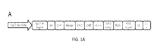
Figure 1, comprising Figure 1A through Figure 1B, depicts schematic diagrams
of
the anti-mouse CTLA-4 DMAb design. Figure 1A depicts a diagram of the
orientation of
CA 03078458 2020-04-03
WO 2019/070834
PCT/US2018/054137
the antibody regions. Figure 1B depicts a diagram of modifications that were
made to the
original CTLA-4 DMAb.
Figure 2, comprising Figure 2A through Figure 2D, depicts exemplary
experimental results demonstrating the expression and binding of mouse anti-
mouse
.. CTLA-4 DMAbs. Figure 2A depicts exemplary data demonstrating the secreted
mouse
IgG levels for the indicated DMAb from transfected HEK293T cells. Figure 2B
depicts
exemplary data demonstrating a western blot analysis of mouse IgG from lysates
(left)
and supernatants (right). Red bands indicate the ladder, green bands indicate
mouse IgG.
Figure 2C depicts exemplary data demonstrating binding of purified 9D9 or
supernatants
.. from transfected cells to mouse CTLA-4 protein. ICso is indicated in the
figure legend.
Individual curves from biological replicates are shown. Figure 2D depicts
exemplary data
demonstrating the serum concentration of anti-CTLA-4 mouse IgG from C57B1/6
mice
injected with 100pg of the indicated DMAb. Error bars indicate mean SD for in
vitro
studies, and mean SEM for in vivo studies. Figure 2A and 2C, n= at least 2
biological
.. replicates. Figure 2D, n=5 mice per group.
Figure 3, comprising Figure 3A through Figure 3D, depicts exemplary
experimental results demonstrating the anti-tumor activity of anti-CTLA-4 DMAb
in
SalN and CT26 tumor models. Figure 3A depicts the tumor study outline for DMAb
delivery using prophylactic SalN tumor model in A/J mice (top), and serum
levels of
.. anti-CTLA-4 mouse IgG from these mice (bottom). 400pg DMAb was delivered by
IM-
EP 4 days prior to implantation of tumor cells. Figure 3B depicts exemplary
data
demonstrating the tumor volume measurements and survival analysis of the mice
described in 3A. Figure 3C depicts the tumor study outline for DMAb delivery
using
therapeutic CT26 tumor model in Balb/c mice (top), and serum levels of anti-
CTLA-4
mouse IgG from these mice (bottom). 400pg DMAb was delivered by IM-EP 3 days
after
implantation of CT26 tumor cells. Figure 3D depicts exemplary data
demonstrating the
tumor volume measurements and survival analysis of the mice described in 3C.
Error bars
indicate mean SEM. N=10 mice per group. Shown is a representative of two
independent experiments.
Figure 4, comprising Figure 4A through Figure 4D, depicts exemplary
experimental results demonstrating the efficacy of recombinant 9D9 antibody in
SalN
tumor model. Figure 4A depicts the tumor study outline for antibody treatment.
Figure 4B
depicts serum levels of anti-CTLA-4 mouse IgG from these mice. Figure 4C
depicts
2
CA 03078458 2020-04-03
WO 2019/070834
PCT/US2018/054137
exemplary data demonstrating the tumor volume measurements of the mice
described in
4B. Figure 4C depicts a survival analysis of the mice described in 4B.
Figure 5 depicts exemplary experimental results demonstrating mouse anti-mouse
CTLA-4 DMAb induces immune memory and protection from tumor re-challenge.
Figure 6, comprising Figure 6A through Figure 6C, depicts exemplary
experimental results demonstrating the efficacy of mouse anti-mouse CTLA-4
DMAb
when delivered at an earlier time point. Figure 6A depicts the tumor study
outline for
dMAB delivery. Figure 6B depicts exemplary data demonstrating the tumor volume
measurements of the mice following early administration of the anti-mouse CTLA-
4
DMAb. Figure 6C depicts a survival analysis of the mice following early
administration
of the anti-mouse CTLA-4 DMAb.
Figure 7, comprising Figure 7A through Figure 7D, depicts exemplary
experimental results demonstrating that anti-mouse CTLA-4 DMAb induces T cell
infiltration into tumors. Figure 7A depicts the tumor study outline for dMAB
delivery.
Figure 7B depicts immunofluorescent staining of tumors for T-cell
infiltration. Figure 7C
depicts a quantification of the numbers of CD8+ and CD3+ T cells per HPF.
Figure 7D
depicts a quantification of the types of TILs present.
Figure 8, comprising Figure 8A through Figure 8D, depicts exemplary
experimental results demonstrating the expression and binding of human anti-
human
CTLA-4 DMAbs. Figure 8A depicts exemplary data demonstrating the secreted
human
IgG levels for the indicated DMAb from transfected HEK293T cells. Figure 8B
depicts
an exemplary western blot of human IgG from lysates (left) and supernatants
(right).
Figure 8C depicts exemplary data demonstrating the serum concentration of
human IgG
over time in Balb/c mice injected with 400pg of the indicated DMAb by IM-EP.
Figure
8D depicts exemplary data demonstrating the binding of ipi-DMAb and treme-DMAb
purified from mouse serum to human CTLA-4 protein by ELISA. Curves from
individual
mice are shown. For in vitro experiments, error bars indicate mean SD. For in
vivo
experiments, error bars indicate mean SEM. Figure 8A, n= 2 biological
replicates.
Figure 8C, n=5 mice per group. Figure 8D, n=3 mice per group.
Figure 9 depicts exemplary experimental results demonstrating the efficiency
of
CD4 and CD8 depletion antibodies.
Figure 10, comprising Figure 10A through Figure 10D, depicts exemplary
experimental results demonstrating the functionality of human anti-human CTLA-
4
3
CA 03078458 2020-04-03
WO 2019/070834
PCT/US2018/054137
DMAbs. Figure 10A depicts exemplary data demonstrating the flow cytometric
staining
of CD3+CD8-CD25+ human PBMCs for CTLA-4 with the indicated antibodies, with or
without PMA/ionomycin stimulation. Figure 10B depicts the quantification of
the
staining depicted in Figure 10A, for 3 individual donors. Figure 10C depicts
an
illustration of CTLA-4 blockade bioassay. Figure 10D depicts results from
bioassay
described in Figure 10C. The Relative Luciferase Units (RLU) are graphed
relative to the
RLU from no antibody control wells. Ipi-DMAb and treme-DMAb were purified from
mouse serum. Error bars indicate SD. For Figure 10D, curves indicate 4-
parameter
nonlinear fit.
Figure 11 depicts results from example experiments, demonstrating delivery of
anti-human CTLA-4 using DNA (in viro expression and binding).
Figure 12 depicts results from example experiments, demonstrating synergy of
mTERT DNA vaccine + aCTLA-4 recombinant antibody.
Figure 13 depicts results from example experiments, demonstrating synergy of
mTERT DNA vaccine + aCTLA-4 DMAb.
DETAILED DESCRIPTION
The present invention relates to compositions comprising a recombinant nucleic
acid sequence encoding an antibody, a fragment thereof, a variant thereof, or
a
combination thereof The composition can be administered to a subject in need
thereof to
facilitate in vivo expression and formation of a synthetic antibody.
In particular, the heavy chain and light chain polypeptides expressed from the
recombinant nucleic acid sequences can assemble into the synthetic antibody.
The heavy
chain polypeptide and the light chain polypeptide can interact with one
another such that
assembly results in the synthetic antibody being capable of binding the
antigen, being
more immunogenic as compared to an antibody not assembled as described herein,
and
being capable of eliciting or inducing an immune response against the antigen.
Additionally, these synthetic antibodies are generated more rapidly in the
subject
than antibodies that are produced in response to antigen induced immune
response. The
synthetic antibodies are able to effectively bind and neutralize a range of
antigens. The
synthetic antibodies are also able to effectively protect against and/or
promote survival of
disease.
4
CA 03078458 2020-04-03
WO 2019/070834
PCT/US2018/054137
In one aspect, the present invention relates to a composition that can be used
to
increase or enhance an immune response, i.e., create a more effective immune
response,
by administering a checkpoint inhibitor, such as an engineered or synthetic
antibody
directed to CTLA-4 (e.g., engineered MAb in the form of synthetic DNA
plasmids;
"DMAb").
With respect to engineered MAb in the form of synthetic DNA plasmids, the
present invention relates to compositions comprising a recombinant nucleic
acid sequence
encoding an antibody, a fragment thereof, a variant thereof, or a combination
thereof The
composition can be administered to a subject in need thereof to facilitate in
vivo
expression and formation of a synthetic antibody. In one embodiment, the
nucleotide
sequence comprises one or more nucleotide sequences described herein. In one
embodiment, the nucleotide sequence comprises sequence encoding the
polypeptide
sequence of SEQ ID NOs: 1, 2, 3, 4, 5, 6, or a variant thereof or a fragment
thereof In
one embodiment, the nucleotide sequence comprises an RNA sequence transcribed
from a
DNA sequence described herein. For example, in one embodiment, the nucleotide
sequence comprises an RNA sequence transcribed by a DNA sequence encoding the
polypeptide sequence of SEQ ID NOs: 1, 2, 3, 4, 5, 6, or a variant thereof or
a fragment
thereof
In one embodiment, the nucleotide sequence encodes an amino acid sequence
having at least about 80%, at least about 85%, at least about 90%, or at least
about 95%
identity over the entire length of the amino acid sequence to an amino acid
sequence
selected from the group SEQ ID NOs: 1, 2, 3, 4, 5, 6. In one embodiment, the
nucleotide
sequence encodes a fragment of an amino acid sequence having at least about
80%, at
least about 85%, at least about 90%, or at least about 95% identity over the
entire length
of the amino acid sequence to an amino acid sequence selected from the group
SEQ ID
NOs: 1, 2, 3, 4, 5, 6.
In one embodiment, the nucleotide sequence has at least about 80%, at least
about
85%, at least about 90%, or at least about 95% identity over the entire length
of the
nucleotide sequence to one or more nucleotide sequences encoding one or more
of SEQ
ID NOs: 1, 2, 3, 4, 5, 6. In one embodiment, the nucleotide sequence is a
fragment of a
nucleotide sequence that has at least about 80%, at least about 85%, at least
about 90%, or
at least about 95% identity over the entire length of the nucleotide sequence
to one or
more nucleotide sequences encoding one or more of SEQ ID NOs: 1, 2, 3, 4, 5,
6.
5
CA 03078458 2020-04-03
WO 2019/070834
PCT/US2018/054137
In one embodiment, nucleotide sequence has at least about 80% identity over
the
entire length of at least one nucleotide sequence selected from the group of
SEQ ID NOs:
7, 8, 9, 10, 11, and 12.
In some instances, the antibodies of the invention can be administered in
combination with a desired composition comprising an antigen, such as TERT, to
produce
a synergistic effect; whereas, in other instances, the antibodies can be
administered
separately from the composition comprising the antigen. In some instances the
antibodies
of the invention comprise a DNA sequence that encodes such antibody, which
includes at
least the variable regions of the immunoglobulin.
The composition of the present invention can increase the immune response to
the
antigen of the vaccine in the subject by increasing the CD8+ T cell response,
as compared
to the vaccine not including checkpoint inhibitors. This increased CD8+ T cell
response
has cytolytic activity and secretes the cytokine interferon-gamma (IFN-y).
The compositions provided herein can also include a pharmaceutically
acceptable
excipient.
Aspects of the invention also include methods for increasing an immune
response
in a subject in need thereof by administering any of the compositions provided
herein to
the subject. The methods of increasing an immune response can also include an
electroporating step.
1. Definitions
Unless otherwise defined, all technical and scientific terms used herein have
the
same meaning as commonly understood by one of ordinary skill in the art. In
case of
conflict, the present document, including definitions, will control. Exemplary
methods
and materials are described herein, although methods and materials similar or
equivalent
to those described herein can be used in practice or testing of the present
invention. All
publications, patent applications, patents and other references mentioned
herein are
incorporated by reference in their entirety. The materials, methods, and
examples
disclosed herein are illustrative only and not intended to be limiting.
The terms "comprise(s)," "include(s)," "having," "has," "can," "contain(s),"
and
variants thereof, as used herein, are intended to be open-ended transitional
phrases, terms,
or words that do not preclude the possibility of additional acts or
structures. The singular
forms "a," "and" and "the" include plural references unless the context
clearly dictates
6
CA 03078458 2020-04-03
WO 2019/070834
PCT/US2018/054137
otherwise. The present disclosure also contemplates other embodiments
"comprising,"
"consisting of" and "consisting essentially of," the embodiments or elements
presented
herein, whether explicitly set forth or not.
"Antibody" may mean an antibody of classes IgG, IgM, IgA, IgD or IgE, or
fragments, fragments or derivatives thereof, including Fab, F(ab')2, Fd, and
single chain
antibodies, and derivatives thereof The antibody may be an antibody isolated
from the
serum sample of mammal, a polyclonal antibody, affinity purified antibody, or
mixtures
thereof which exhibits sufficient binding specificity to a desired epitope or
a sequence
derived therefrom.
"Antigen" refers to proteins that have the ability to generate an immune
response
in a host. An antigen may be recognized and bound by an antibody. An antigen
may
originate from within the body or from the external environment.
"CDRs" are defined as the complementarity determining region amino acid
sequences of an antibody which are the hypervariable regions of immunoglobulin
heavy
and light chains. See, e.g., Kabat et al., Sequences of Proteins of
Immunological Interest,
4th Ed., U.S. Department of Health and Human Services, National Institutes of
Health
(1987). There are three heavy chain and three light chain CDRs (or CDR
regions) in the
variable portion of an immunoglobulin. Thus, "CDRs" as used herein refers to
all three
heavy chain CDRs, or all three light chain CDRs (or both all heavy and all
light chain
CDRs, if appropriate). The structure and protein folding of the antibody may
mean that
other residues are considered part of the antigen binding region and would be
understood
to be so by a skilled person. See for example Chothia et al., (1989)
Conformations of
immunoglobulin hypervariable regions; Nature 342, p 877-883.
"Antibody fragment" or "fragment of an antibody" as used interchangeably
herein
refers to a portion of an intact antibody comprising the antigen-binding site
or variable
region. The portion does not include the constant heavy chain domains (i.e.
CH2, CH3, or
CH4, depending on the antibody isotype) of the Fc region of the intact
antibody.
Examples of antibody fragments include, but are not limited to, Fab fragments,
Fab'
fragments, Fab'-SH fragments, F(ab')2 fragments, Fd fragments, Fv fragments,
diabodies,
single-chain Fv (scFv) molecules, single-chain polypeptides containing only
one light
chain variable domain, single-chain polypeptides containing the three CDRs of
the light-
chain variable domain, single-chain polypeptides containing only one heavy
chain
7
CA 03078458 2020-04-03
WO 2019/070834
PCT/US2018/054137
variable region, and single-chain polypeptides containing the three CDRs of
the heavy
chain variable region.
"Adjuvant" as used herein means any molecule added to the vaccine described
herein to enhance the immunogenicity of the antigen.
"Checkpoint inhibitor" as used herein means inhibitors or molecules that block
immune checkpoints as commonly understood in the field of cancer
immunotherapy.
More commonly the checkpoint inhibitors are antibodies that block these immune
checkpoints.
"Coding sequence" or "encoding nucleic acid" as used herein may refer to the
nucleic acid (RNA or DNA molecule) that comprise a nucleotide sequence which
encodes
an antibody as set forth herein. The coding sequence may also comprise a DNA
sequence
which encodes an RNA sequence. The coding sequence may further include
initiation and
termination signals operably linked to regulatory elements including a
promoter and
polyadenylation signal capable of directing expression in the cells of an
individual or
.. mammal to whom the nucleic acid is administered. The coding sequence may
further
include sequences that encode signal peptides.
"Complement" or "complementary" as used herein may mean a nucleic acid may
have Watson-Crick (e.g., A-T/U and C-G) or Hoogsteen base pairing between
nucleotides
or nucleotide analogs of nucleic acid molecules.
"Constant current" as used herein to define a current that is received or
experienced by a tissue, or cells defining said tissue, over the duration of
an electrical
pulse delivered to same tissue. The electrical pulse is delivered from the
electroporation
devices described herein. This current remains at a constant amperage in said
tissue over
the life of an electrical pulse because the electroporation device provided
herein has a
feedback element, preferably having instantaneous feedback. The feedback
element can
measure the resistance of the tissue (or cells) throughout the duration of the
pulse and
cause the electroporation device to alter its electrical energy output (e.g.,
increase
voltage) so current in same tissue remains constant throughout the electrical
pulse (on the
order of microseconds), and from pulse to pulse. In some embodiments, the
feedback
element comprises a controller.
"Current feedback" or "feedback" as used herein may be used interchangeably
and may mean the active response of the provided electroporation devices,
which
comprises measuring the current in tissue between electrodes and altering the
energy
8
CA 03078458 2020-04-03
WO 2019/070834
PCT/US2018/054137
output delivered by the EP device accordingly in order to maintain the current
at a
constant level. This constant level is preset by a user prior to initiation of
a pulse
sequence or electrical treatment. The feedback may be accomplished by the
electroporation component, e.g., controller, of the electroporation device, as
the electrical
circuit therein is able to continuously monitor the current in tissue between
electrodes and
compare that monitored current (or current within tissue) to a preset current
and
continuously make energy-output adjustments to maintain the monitored current
at preset
levels. The feedback loop may be instantaneous as it is an analog closed-loop
feedback.
"Decentralized current" as used herein may mean the pattern of electrical
currents
delivered from the various needle electrode arrays of the electroporation
devices
described herein, wherein the patterns minimize, or preferably eliminate, the
occurrence
of electroporation related heat stress on any area of tissue being
electroporated.
"Electroporation," "electro-permeabilization," or "electro-kinetic
enhancement"
("EP") as used interchangeably herein may refer to the use of a transmembrane
electric
field pulse to induce microscopic pathways (pores) in a bio-membrane; their
presence
allows biomolecules such as plasmids, oligonucleotides, siRNA, drugs, ions,
and water to
pass from one side of the cellular membrane to the other.
"Endogenous antibody" as used herein may refer to an antibody that is
generated
in a subject that is administered an effective dose of an antigen for
induction of a humoral
immune response.
"Feedback mechanism" as used herein may refer to a process performed by either
software or hardware (or firmware), which process receives and compares the
impedance
of the desired tissue (before, during, and/or after the delivery of pulse of
energy) with a
present value, preferably current, and adjusts the pulse of energy delivered
to achieve the
preset value. A feedback mechanism may be performed by an analog closed loop
circuit.
"Fragment" may mean a polypeptide fragment of an antibody that is function,
i.e.,
can bind to desired target and have the same intended effect as a full length
antibody. A
fragment of an antibody may be 100% identical to the full length except
missing at least
one amino acid from the N and/or C terminal, in each case with or without
signal peptides
and/or a methionine at position 1. Fragments may comprise 20% or more, 25% or
more,
30% or more, 35% or more, 40% or more, 45% or more, 50% or more, 55% or more,
60% or more, 65% or more, 70% or more, 75% or more, 80% or more, 85% or more,
90% or more, 91% or more, 92% or more, 93% or more, 94% or more, 95% or more,
9
CA 03078458 2020-04-03
WO 2019/070834
PCT/US2018/054137
96% or more, 97% or more, 98% or more, 99% or more percent of the length of
the
particular full length antibody, excluding any heterologous signal peptide
added. The
fragment may comprise a fragment of a polypeptide that is 95% or more, 96% or
more,
97% or more, 98% or more or 99% or more identical to the antibody and
additionally
comprise an N terminal methionine or heterologous signal peptide which is not
included
when calculating percent identity. Fragments may further comprise an N
terminal
methionine and/or a signal peptide such as an immunoglobulin signal peptide,
for
example an IgE or IgG signal peptide. The N terminal methionine and/or signal
peptide
may be linked to a fragment of an antibody.
A fragment of a nucleic acid sequence that encodes an antibody may be 100%
identical to the full length except missing at least one nucleotide from the
5' and/or 3' end,
in each case with or without sequences encoding signal peptides and/or a
methionine at
position 1. Fragments may comprise 20% or more, 25% or more, 30% or more, 35%
or
more, 40% or more, 45% or more, 50% or more, 55% or more, 60% or more, 65% or
more, 70% or more, 75% or more, 80% or more, 85% or more, 90% or more, 91% or
more, 92% or more, 93% or more, 94% or more, 95% or more, 96% or more, 97% or
more, 98% or more, 99% or more percent of the length of the particular full
length coding
sequence, excluding any heterologous signal peptide added. The fragment may
comprise
a fragment that encode a polypeptide that is 95% or more, 96% or more, 97% or
more,
98% or more or 99% or more identical to the antibody and additionally
optionally
comprise sequence encoding an N terminal methionine or heterologous signal
peptide
which is not included when calculating percent identity. Fragments may further
comprise
coding sequences for an N terminal methionine and/or a signal peptide such as
an
immunoglobulin signal peptide, for example an IgE or IgG signal peptide. The
coding
sequence encoding the N terminal methionine and/or signal peptide may be
linked to a
fragment of coding sequence.
"Genetic construct" as used herein refers to the DNA or RNA molecules that
comprise a nucleotide sequence which encodes a protein, such as an antibody.
The
genetic construct may also refer to a DNA molecule which transcribes an RNA.
The
coding sequence includes initiation and termination signals operably linked to
regulatory
elements including a promoter and polyadenylation signal capable of directing
expression
in the cells of the individual to whom the nucleic acid molecule is
administered. As used
herein, the term "expressible form" refers to gene constructs that contain the
necessary
CA 03078458 2020-04-03
WO 2019/070834
PCT/US2018/054137
regulatory elements operable linked to a coding sequence that encodes a
protein such that
when present in the cell of the individual, the coding sequence will be
expressed.
"Identical" or "identity" as used herein in the context of two or more nucleic
acids
or polypeptide sequences, may mean that the sequences have a specified
percentage of
residues that are the same over a specified region. The percentage may be
calculated by
optimally aligning the two sequences, comparing the two sequences over the
specified
region, determining the number of positions at which the identical residue
occurs in both
sequences to yield the number of matched positions, dividing the number of
matched
positions by the total number of positions in the specified region, and
multiplying the
result by 100 to yield the percentage of sequence identity. In cases where the
two
sequences are of different lengths or the alignment produces one or more
staggered ends
and the specified region of comparison includes only a single sequence, the
residues of
single sequence are included in the denominator but not the numerator of the
calculation.
When comparing DNA and RNA, thymine (T) and uracil (U) may be considered
equivalent. Identity may be performed manually or by using a computer sequence
algorithm such as BLAST or BLAST 2Ø
"Impedance" as used herein may be used when discussing the feedback
mechanism and can be converted to a current value according to Ohm's law, thus
enabling
comparisons with the preset current.
"Immune response" as used herein may mean the activation of a host's immune
system, e.g., that of a mammal, in response to the introduction of one or more
nucleic
acids and/or peptides. The immune response can be in the form of a cellular or
humoral
response, or both.
"Nucleic acid" or "oligonucleotide" or "polynucleotide" as used herein may
mean
at least two nucleotides covalently linked together. The depiction of a single
strand also
defines the sequence of the complementary strand. Thus, a nucleic acid also
encompasses
the complementary strand of a depicted single strand. Many variants of a
nucleic acid
may be used for the same purpose as a given nucleic acid. Thus, a nucleic acid
also
encompasses substantially identical nucleic acids and complements thereof A
single
strand provides a probe that may hybridize to a target sequence under
stringent
hybridization conditions. Thus, a nucleic acid also encompasses a probe that
hybridizes
under stringent hybridization conditions.
11
CA 03078458 2020-04-03
WO 2019/070834
PCT/US2018/054137
Nucleic acids may be single stranded or double stranded, or may contain
portions
of both double stranded and single stranded sequence. The nucleic acid may be
DNA,
both genomic and cDNA, RNA, or a hybrid, where the nucleic acid may contain
combinations of deoxyribo- and ribo-nucleotides, and combinations of bases
including
.. uracil, adenine, thymine, cytosine, guanine, inosine, xanthine
hypoxanthine, isocytosine
and isoguanine. Nucleic acids may be obtained by chemical synthesis methods or
by
recombinant methods.
"Operably linked" as used herein may mean that expression of a gene is under
the
control of a promoter with which it is spatially connected. A promoter may be
positioned
.. 5' (upstream) or 3' (downstream) of a gene under its control. The distance
between the
promoter and a gene may be approximately the same as the distance between that
promoter and the gene it controls in the gene from which the promoter is
derived. As is
known in the art, variation in this distance may be accommodated without loss
of
promoter function.
A "peptide," "protein," or "polypeptide" as used herein can mean a linked
sequence of amino acids and can be natural, synthetic, or a modification or
combination
of natural and synthetic.
"Promoter" as used herein may mean a synthetic or naturally-derived molecule
which is capable of conferring, activating or enhancing expression of a
nucleic acid in a
cell. A promoter may comprise one or more specific transcriptional regulatory
sequences
to further enhance expression and/or to alter the spatial expression and/or
temporal
expression of same. A promoter may also comprise distal enhancer or repressor
elements,
which can be located as much as several thousand base pairs from the start
site of
transcription. A promoter may be derived from sources including viral,
bacterial, fungal,
plants, insects, and animals. A promoter may regulate the expression of a gene
component
constitutively or differentially with respect to cell, the tissue or organ in
which expression
occurs or, with respect to the developmental stage at which expression occurs,
or in
response to external stimuli such as physiological stresses, pathogens, metal
ions, or
inducing agents. Representative examples of promoters include the
bacteriophage T7
promoter, bacteriophage T3 promoter, SP6 promoter, lac operator-promoter, tac
promoter, SV40 late promoter, SV40 early promoter, RSV-LTR promoter, CMV IE
promoter, SV40 early promoter or SV 40 late promoter and the CMV IE promoter.
12
CA 03078458 2020-04-03
WO 2019/070834
PCT/US2018/054137
"Signal peptide" and "leader sequence" are used interchangeably herein and
refer
to an amino acid sequence that can be linked at the amino terminus of a
protein set forth
herein. Signal peptides/leader sequences typically direct localization of a
protein. Signal
peptides/leader sequences used herein may facilitate secretion of the protein
from the cell
in which it is produced. Signal peptides/leader sequences are often cleaved
from the
remainder of the protein, often referred to as the mature protein, upon
secretion from the
cell. Signal peptides/leader sequences are linked at the N terminus of the
protein.
"Stringent hybridization conditions" as used herein may mean conditions under
which a first nucleic acid sequence (e.g., probe) will hybridize to a second
nucleic acid
sequence (e.g., target), such as in a complex mixture of nucleic acids.
Stringent conditions
are sequence dependent and will be different in different circumstances.
Stringent
conditions may be selected to be about 5-10 C lower than the thermal melting
point (Tm)
for the specific sequence at a defined ionic strength pH. The Tm may be the
temperature
(under defined ionic strength, pH, and nucleic concentration) at which 50% of
the probes
complementary to the target hybridize to the target sequence at equilibrium
(as the target
sequences are present in excess, at Tm, 50% of the probes are occupied at
equilibrium).
Stringent conditions may be those in which the salt concentration is less than
about 1.0 M
sodium ion, such as about 0.01-1.0 M sodium ion concentration (or other salts)
at pH 7.0
to 8.3 and the temperature is at least about 30 C for short probes (e.g.,
about 10-50
nucleotides) and at least about 60 C for long probes (e.g., greater than about
50
nucleotides). Stringent conditions may also be achieved with the addition of
destabilizing
agents such as formamide. For selective or specific hybridization, a positive
signal may
be at least 2 to 10 times background hybridization. Exemplary stringent
hybridization
conditions include the following: 50% formamide, 5x SSC, and 1% SDS,
incubating at
42 C, or, 5x SSC, 1% SDS, incubating at 65 C, with wash in 0.2x SSC, and 0.1%
SDS at
65 C.
"Subject" and "patient" as used herein interchangeably refers to any
vertebrate,
including, but not limited to, a mammal (e.g., cow, pig, camel, llama, horse,
goat, rabbit,
sheep, hamsters, guinea pig, cat, dog, rat, and mouse, a non-human primate
(for example,
a monkey, such as a cynomolgous or rhesus monkey, chimpanzee, etc) and a
human). In
some embodiments, the subject may be a human or a non-human. The subject or
patient
may be undergoing other forms of treatment.
13
CA 03078458 2020-04-03
WO 2019/070834
PCT/US2018/054137
"Substantially complementary" as used herein may mean that a first sequence is
at
least 600o, 65%, 700o, 75%, 800o, 810o, 82%, 83%, 84%, 85%, 86%, 87%, 88%,
89%,
900o, 910o, 920o, 930o, 940o, 950o, 960o, 970o, 980o or 990o identical to the
complement
of a second sequence over a region of 8, 9, 10, 11, 12, 13, 14, 15, 16, 17,
18, 19, 20, 21,
22, 23, 24, 25, 30, 35, 40, 45, 50, 55, 60, 65, 70, 75, 80, 85, 90, 95, 100 or
more
nucleotides or amino acids, or that the two sequences hybridize under
stringent
hybridization conditions.
"Substantially identical" as used herein may mean that a first and second
sequence
are at least 600o, 650o, 700o, 750o, 800o, 810o, 820o, 830o, 840o, 850o, 860o,
870o, 880o,
890o, 900o, 910o, 920o, 930o, 940o, 950o, 960o, 970o, 980o, or 990o identical
over a region
of 1, 2, 3, 4, 5, 6, 7, 8, 9, 10, 11, 12, 13, 14, 15, 16, 17, 18, 19, 20, 21,
22, 23, 24, 25, 30,
35, 40, 45, 50, 55, 60, 65, 70, 75, 80, 85, 90, 95, 100, 200, 300, 400, 500,
600, 700, 800,
900, 1000, 1100 or more nucleotides or amino acids, or with respect to nucleic
acids, if
the first sequence is substantially complementary to the complement of the
second
sequence.
"Synthetic antibody" as used herein refers to an antibody that is encoded by
the
recombinant nucleic acid sequence described herein and is generated in a
subject.
"Treatment" or "treating," as used herein can mean protecting of a subject
from a
disease through means of preventing, suppressing, repressing, or completely
eliminating
the disease. Preventing the disease involves administering a vaccine of the
present
invention to a subject prior to onset of the disease. Suppressing the disease
involves
administering a vaccine of the present invention to a subject after induction
of the disease
but before its clinical appearance. Repressing the disease involves
administering a
vaccine of the present invention to a subject after clinical appearance of the
disease.
"Variant" used herein with respect to a nucleic acid may mean (i) a portion or
fragment of a referenced nucleotide sequence; (ii) the complement of a
referenced
nucleotide sequence or portion thereof; (iii) a nucleic acid that is
substantially identical to
a referenced nucleic acid or the complement thereof; or (iv) a nucleic acid
that hybridizes
under stringent conditions to the referenced nucleic acid, complement thereof,
or a
sequences substantially identical thereto.
"Variant" with respect to a peptide or polypeptide, may indicate that the
peptide or
polypeptide differs in amino acid sequence by the insertion, deletion, or
conservative
substitution of amino acids, but retains at least one biological activity.
Variant may also
14
CA 03078458 2020-04-03
WO 2019/070834
PCT/US2018/054137
mean a protein with an amino acid sequence that is substantially identical to
a referenced
protein with an amino acid sequence that retains at least one biological
activity. A
conservative substitution of an amino acid, i.e., replacing an amino acid with
a different
amino acid of similar properties (e.g., hydrophilicity, degree and
distribution of charged
regions) is recognized in the art as typically involving a minor change. These
minor
changes can be identified, in part, by considering the hydropathic index of
amino acids, as
understood in the art. Kyte et al., J. Mol. Biol. 157:105-132 (1982). The
hydropathic
index of an amino acid is based on a consideration of its hydrophobicity and
charge. It is
known in the art that amino acids of similar hydropathic indexes can be
substituted and
still retain protein function. In one aspect, amino acids having hydropathic
indexes of 2
are substituted. The hydrophilicity of amino acids can also be used to reveal
substitutions
that would result in proteins retaining biological function. A consideration
of the
hydrophilicity of amino acids in the context of a peptide permits calculation
of the
greatest local average hydrophilicity of that peptide, a useful measure that
has been
.. reported to correlate well with antigenicity and immunogenicity. U.S.
Patent No.
4,554,101, incorporated fully herein by reference. Substitution of amino acids
having
similar hydrophilicity values can result in peptides retaining biological
activity, for
example immunogenicity, as is understood in the art. Substitutions may be
performed
with amino acids having hydrophilicity values within 2 of each other. Both
the
hydrophobicity index and the hydrophilicity value of amino acids are
influenced by the
particular side chain of that amino acid. Consistent with that observation,
amino acid
substitutions that are compatible with biological function are understood to
depend on the
relative similarity of the amino acids, and particularly the side chains of
those amino
acids, as revealed by the hydrophobicity, hydrophilicity, charge, size, and
other
.. properties.
A variant may be a nucleic acid sequence that is substantially identical over
the
full length of the full gene sequence or a fragment thereof The nucleic acid
sequence may
be 80%, 81%, 82%, 83%, 84%, 85%, 86%, 87%, 88%, 89%, 90%, 91%, 92%, 93%, 94%,
95%, 96%, 97%, 98%, 99%, or 100% identical over the full length of the gene
sequence
.. or a fragment thereof A variant may be an amino acid sequence that is
substantially
identical over the full length of the amino acid sequence or fragment thereof
The amino
acid sequence may be 80%, 81%, 82%, 83%, 84%, 85%, 86%, 87%, 88%, 89%, 90%,
CA 03078458 2020-04-03
WO 2019/070834
PCT/US2018/054137
91%, 92%, 93%, 94%, 95%, 96%, 97%, 98%, 99%, or 100% identical over the full
length
of the amino acid sequence or a fragment thereof
"Vector" as used herein may mean a nucleic acid sequence containing an origin
of
replication. A vector may be a plasmid, bacteriophage, bacterial artificial
chromosome or
yeast artificial chromosome. A vector may be a DNA or RNA vector. A vector may
be
either a self-replicating extrachromosomal vector or a vector which integrates
into a host
genome.
For the recitation of numeric ranges herein, each intervening number there
between with the same degree of precision is explicitly contemplated. For
example, for
the range of 6-9, the numbers 7 and 8 are contemplated in addition to 6 and 9,
and for the
range 6.0-7.0, the number 6.0, 6.1, 6.2, 6.3, 6.4, 6.5, 6.6, 6.7, 6.8, 6.9,
and 7.0 are
explicitly contemplated. This applies regardless of the breadth of the range.
2. Compositions
The invention also includes novel sequences for use for producing antibodies.
In
one embodiment, the antibodies of the invention can be produced in mammalian
cells or
for delivery in DNA or RNA vectors including bacterial, yeast, as well as
viral vectors.
The present invention relates to a composition comprising a recombinant
nucleic
acid sequence encoding an antibody, a fragment thereof, a variant thereof, or
a
combination thereof The composition, when administered to a subject in need
thereof,
can result in the generation of a synthetic antibody in the subject. The
synthetic antibody
can bind a target molecule (i.e., an antigen, such as CTLA-4) present in the
subject. Such
binding can neutralize the antigen, block recognition of the antigen by
another molecule,
for example, a protein or nucleic acid, and elicit or induce an immune
response to the
.. antigen.
In one embodiment, the composition comprises a nucleotide sequence encoding a
synthetic antibody. In one embodiment, the composition comprises a nucleic
acid
molecule comprising a first nucleotide sequence encoding a first synthetic
antibody and a
second nucleotide sequence encoding a second synthetic antibody. In one
embodiment,
the nucleic acid molecule comprises a nucleotide sequence encoding a cleavage
domain.
In one embodiment, the nucleic acid molecule comprises a nucleotide sequence
encoding one or more anti-CTLA-4 antibodies.
16
CA 03078458 2020-04-03
WO 2019/070834
PCT/US2018/054137
In one embodiment, the nucleotide sequence encoding an anti-CTLA-4 antibody
comprises one or more codon optimized nucleic acid sequences encoding one or
more
amino acid sequences as set forth in SEQ ID NOs: 1,2, 3,4, 5, 6, or a fragment
of one or
more amino acid sequences as set forth in SEQ ID NOs: 1, 2, 3, 4, 5, 6.
In one embodiment, the nucleotide sequence has at least about 80% identity
over
the entire length of at least one nucleotide sequence selected from the group
of SEQ ID
NOs: 7, 8, 9, 10, 11, and 12.
In one embodiment, the nucleotide sequence encoding an anti-CTLA-4 antibody
comprises one or more RNA sequences transcribed from one or more DNA sequences
encoding an amino acid sequence at least 90% homologous to one or more of SEQ
ID
NOs: 1, 2, 3, 4, 5, 6, or a fragment of an amino acid sequence at least 90%
homologous to
one or more of SEQ ID NOs: 1, 2, 3, 4, 5, 6. In one embodiment, the nucleotide
sequence
encoding an anti-CTLA-4 antibody comprises one or more RNA sequences
transcribed
from one or more DNA sequences encoding an amino acid sequence as set forth in
SEQ
ID NOs: 1, 2, 3, 4, 5, 6, or a fragment of an amino acid sequence as set forth
in SEQ ID
NOs: 1, 2, 3, 4, 5, 6.
In one embodiment, the nucleotide sequence encoding an anti-CTLA-4 antibody
comprises one or more codon optimized nucleic acid sequences at least 90%
homologous
to one or more nucleic acid sequences encoding one or more of SEQ ID NOs: 1,
2, 3, 4, 5,
6, or a fragment of a nucleic acid sequence at least 90% homologous to one or
more
nucleic acid sequences encoding one or more of SEQ ID NOs: 1, 2, 3, 4, 5, 6.
The composition of the invention can treat, prevent, and/or protect against
any
disease, disorder, or condition associated with CTLA-4 activity. In certain
embodiments,
the composition can treat, prevent, and/or protect against cancer.
In one embodiment, the composition of the invention is provided in combination
with at least one other agent, such as an antigen. In one embodiment, a
combination can
be a single formulation or can be separate formulations and administered in
sequence
(either antigen first and then anti-CTLA-4 antibody, or anti-CTLA-4 antibody
first and
then antigen). The composition can increase antigen presentation and the
overall immune
response to the antigen in a subject. The combination of antigen and anti-CTLA-
4
antibody induces the immune system more efficiently than a composition
comprising the
antigen alone. This more efficient immune response provides increased efficacy
in the
treatment and/or prevention of a disease, such as cancer.
17
CA 03078458 2020-04-03
WO 2019/070834
PCT/US2018/054137
The composition of the invention may comprise a checkpoint inhibitor. The
checkpoint inhibitor may be one or more anti-CTLA-4 antibodies. The antigen
may be
one or more of hTERT, mTERT, PSA, PSMA, STEAP, PSCA, and PAP, WT1,
tyrosinase, NYES01, PRAME, and MAGE. The checkpoint inhibitor(s) and the
antigen(s)
of the composition can be administered together or separately to the subject
in need
thereof, in nucleic acid or polypeptide forms. In some instances, the
checkpoint
inhibitor(s) can be administered separately from the antigen(s) of the
composition.
The composition can result in the generation of the synthetic antibody in the
subject within at least about 1 hour, 2 hours, 3 hours, 4 hours, 5 hours, 6
hours, 7 hours, 8
hours, 9 hours, 10 hours, 11 hours, 12 hours, 13 hours, 14 hours, 15 hours, 16
hours, 17
hours, 18 hours, 19 hours, 20 hours, 21 hours, 22 hours, 23 hours, 24 hours,
36 hours, 48
hours, 60 hours, 72 hours, 84 hours, or 96 hours. The composition can be
administered
before or after administration of the antigen(s) to the subject. In some
embodiments, the
checkpoint inhibitor(s) can be administered at least 1 day, 2 days, 3 days, 4
days, 5 days,
6 days, 7 days, 8 days, 9 days, 10 days, 11 days, 12 days, 13 days, 14 days,
15 days, 16
days, 17 days, 18 days, 19 days, 20 days, 21 days, 22 days, 23 days, 24 days,
25 days, 26
days, 27 days, 28 days, 29 days, 30 days, 60 days, or 90 days before or after
administration of the antigen(s) to the subject.
In still other embodiments, the checkpoint inhibitor(s) can be administered at
least
1 week, 2 weeks, 3 weeks, 4 weeks, 5 weeks, 6 weeks, 7 weeks, 8 weeks, 9
weeks, 10
weeks, 11 weeks, 12 weeks, 13 weeks, 14 weeks, or 15 weeks before or after
administration of the antigen(s) to the subject. In other embodiments, the
checkpoint
inhibitor(s) can be administered about 12 hours to about 15 weeks, about 12
hours to
about 10 weeks, about 12 hours to about 5 weeks, about 12 hours to about 1
week, about
12 hours to about 60 hours, about 12 hours to about 48 hours, about 24 hours
to about 15
weeks, about 60 hours to about 15 weeks, about 96 hours to about 15 weeks,
about 1 day
to about 15 weeks, about 5 days to about 15 weeks, about 10 days to about 15
weeks,
about 15 days to about 15 weeks, about 20 days to about 15 weeks, about 25
days to
about 15 weeks, about 30 days to about 15 weeks, about 1 week to about 15
weeks, about
5 weeks to about 15 weeks, or about 10 weeks to about 15 weeks before or after
administration of the antigen(s) to the subject.
The composition, when administered to the subject in need thereof, can result
in
the generation of the synthetic antibody in the subject more quickly than the
generation of
18
CA 03078458 2020-04-03
WO 2019/070834
PCT/US2018/054137
an endogenous antibody in a subject who is administered an antigen to induce a
humoral
immune response. The composition can result in the generation of the synthetic
antibody
at least about 1 day, 2 days, 3 days, 4 days, 5 days, 6 days, 7 days, 8 days,
9 days, or 10
days before the generation of the endogenous antibody in the subject who was
administered an antigen to induce a humoral immune response.
The composition of the present invention can have features required of
effective
compositions such as being safe so that the composition does not cause illness
or death;
being protective against illness; and providing ease of administration, few
side effects,
biological stability, and low cost per dose. The composition may accomplish
some or all
of these features by combining the antigen(s) with the checkpoint
inhibitor(s), such as an
anti-CTLA-4 antibody as discussed herein.
a. Checkpoint inhibitors
Checkpoint inhibitors can be any antagonist to the various immune checkpoints,
and may be antibodies that block immune checkpoints. The antibodies can be a
protein
including a Fab, monoclonal or polyclonal. The antibodies can also be a DNA
expression
construct that encodes for and can express functional antibodies. The vaccine,
in addition
to one or more antigens, can further comprise a CTLA-4 antibody. The antibody
can be a
synthetic antibody comprised of DNA sequence encoding at least the variable
regions of
an immunoglobulin. Such antibody can be generated by identifying or screening
for the
antibody described herein, which is reactive to or binds the antigen described
herein. The
method of identifying or screening for the antibody can use the antigen in
methodologies
known to those skilled in art to identify or screen for the antibody. Such
methodologies
can include, but are not limited to, selection of the antibody from a library
(e.g., phage
display) and immunization of an animal followed by isolation and/or
purification of the
antibody. See for example methods available in Rajan, S., and Sidhu, S.,
Methods in
Enzymology, vol 502, Chapter One "Simplified Synthetic Antibody Libraries
(2012),
which is incorporated herein in its entirety.
Any antibodies of the invention can also be combined with one or more other
checkpoint inhibitor antibodies, including antibodies against one or more of
PD-1, PD-
L1, LAG-3, GITR, CD40, 0X40, TIM-3, 4-1BB, and others. The checkpoint
inhibitors
can be a known product such as, for example, ipilimumab, tremelimumab,
nivolumab,
pembrolizumab, pidilizumab, BMS-936559 (See ClinicalTrials.gov Identifier
19
CA 03078458 2020-04-03
WO 2019/070834
PCT/US2018/054137
NCT02028403), MPDL3280A (Roche, see ClinicalTrials.gov Identifier
NCT02008227),
MDX1105-01 (Bristol Myers Squibb, see ClinicalTrials.gov Identifier
NCT00729664),
MEDI4736 (MedImmune, See ClinicalTrials.gov Identifier NCT01693562), and MK-
3475 (Merck, see ClinicalTrials.gov Identifier NCT02129556).
b. Recombinant Nucleic Acid Sequence
As described above, the composition can comprise a recombinant nucleic acid
sequence. The recombinant nucleic acid sequence can encode the antibody, a
fragment
thereof, a variant thereof, or a combination thereof The antibody is described
in more
detail elsewhere herein.
The recombinant nucleic acid sequence can be a heterologous nucleic acid
sequence. The recombinant nucleic acid sequence can include one or more
heterologous
nucleic acid sequences.
The recombinant nucleic acid sequence can be an optimized nucleic acid
sequence. Such optimization can increase or alter the immunogenicity of the
antibody.
Optimization can also improve transcription and/or translation. Optimization
can include
one or more of the following: low GC content leader sequence to increase
transcription;
mRNA stability and codon optimization; addition of a kozak sequence (e.g., GCC
ACC)
for increased translation; addition of an immunoglobulin (Ig) leader sequence
encoding a
signal peptide; addition of an internal IRES sequence and eliminating to the
extent
possible cis-acting sequence motifs (i.e., internal TATA boxes).
c. Recombinant Nucleic Acid Sequence Construct
The recombinant nucleic acid sequence can include one or more recombinant
nucleic acid sequence constructs. The recombinant nucleic acid sequence
construct can
include one or more components, which are described in more detail herein.
The recombinant nucleic acid sequence construct can include a heterologous
nucleic acid sequence that encodes a heavy chain polypeptide, a fragment
thereof, a
variant thereof, or a combination thereof The recombinant nucleic acid
sequence
construct can include a heterologous nucleic acid sequence that encodes a
light chain
polypeptide, a fragment thereof, a variant thereof, or a combination thereof
The
recombinant nucleic acid sequence construct can also include a heterologous
nucleic acid
CA 03078458 2020-04-03
WO 2019/070834
PCT/US2018/054137
sequence that encodes a protease or peptidase cleavage site. The recombinant
nucleic acid
sequence construct can also include a heterologous nucleic acid sequence that
encodes an
internal ribosome entry site (IRES). An IRES may be either a viral IRES or a
eukaryotic
IRES. The recombinant nucleic acid sequence construct can include one or more
leader
sequences, in which each leader sequence encodes a signal peptide. The
recombinant
nucleic acid sequence construct can include one or more promoters, one or more
introns,
one or more transcription termination regions, one or more initiation codons,
one or more
termination or stop codons, and/or one or more polyadenylation signals. The
recombinant
nucleic acid sequence construct can also include one or more linker or tag
sequences. The
tag sequence can encode a hemagglutinin (HA) tag.
(1) Heavy Chain Polypeptide
The recombinant nucleic acid sequence construct can include the heterologous
nucleic acid encoding the heavy chain polypeptide, a fragment thereof, a
variant thereof,
or a combination thereof The heavy chain polypeptide can include a variable
heavy chain
(VH) region and/or at least one constant heavy chain (CH) region. The at least
one
constant heavy chain region can include a constant heavy chain region 1 (CH1),
a
constant heavy chain region 2 (CH2), and a constant heavy chain region 3
(CH3), and/or
a hinge region.
In some embodiments, the heavy chain polypeptide can include a VH region and a
CH1 region. In other embodiments, the heavy chain polypeptide can include a VH
region,
a CH1 region, a hinge region, a CH2 region, and a CH3 region.
The heavy chain polypeptide can include a complementarity determining region
("CDR") set. The CDR set can contain three hypervariable regions of the VH
region.
Proceeding from N-terminus of the heavy chain polypeptide, these CDRs are
denoted
"CDR1," "CDR2," and "CDR3," respectively. CDR1, CDR2, and CDR3 of the heavy
chain polypeptide can contribute to binding or recognition of the antigen.
(2) Light Chain Polypeptide
The recombinant nucleic acid sequence construct can include the heterologous
nucleic acid sequence encoding the light chain polypeptide, a fragment
thereof, a variant
thereof, or a combination thereof The light chain polypeptide can include a
variable light
chain (VL) region and/or a constant light chain (CL) region.
21
CA 03078458 2020-04-03
WO 2019/070834
PCT/US2018/054137
The light chain polypeptide can include a complementarity determining region
("CDR") set. The CDR set can contain three hypervariable regions of the VL
region.
Proceeding from N-terminus of the light chain polypeptide, these CDRs are
denoted
"CDR1," "CDR2," and "CDR3," respectively. CDR1, CDR2, and CDR3 of the light
chain polypeptide can contribute to binding or recognition of the antigen.
(3) Protease Cleavage Site
The recombinant nucleic acid sequence construct can include heterologous
nucleic
acid sequence encoding a protease cleavage site. The protease cleavage site
can be
recognized by a protease or peptidase. The protease can be an endopeptidase or
endoprotease, for example, but not limited to, furin, elastase, HtrA, calpain,
trypsin,
chymotrypsin, trypsin, and pepsin. The protease can be furin. In other
embodiments, the
protease can be a serine protease, a threonine protease, cysteine protease,
aspartate
protease, metalloprotease, glutamic acid protease, or any protease that
cleaves an internal
.. peptide bond (i.e., does not cleave the N-terminal or C-terminal peptide
bond).
The protease cleavage site can include one or more amino acid sequences that
promote or increase the efficiency of cleavage. The one or more amino acid
sequences
can promote or increase the efficiency of forming or generating discrete
polypeptides.
The one or more amino acids sequences can include a 2A peptide sequence.
(4) Linker Sequence
The recombinant nucleic acid sequence construct can include one or more linker
sequences. The linker sequence can spatially separate or link the one or more
components
described herein. In other embodiments, the linker sequence can encode an
amino acid
sequence that spatially separates or links two or more polypeptides.
(5) Promoter
The recombinant nucleic acid sequence construct can include one or more
promoters. The one or more promoters may be any promoter that is capable of
driving
gene expression and regulating gene expression. Such a promoter is a cis-
acting sequence
element required for transcription via a DNA dependent RNA polymerase.
Selection of
the promoter used to direct gene expression depends on the particular
application. The
promoter may be positioned about the same distance from the transcription
start in the
recombinant nucleic acid sequence construct as it is from the transcription
start site in its
22
CA 03078458 2020-04-03
WO 2019/070834
PCT/US2018/054137
natural setting. However, variation in this distance may be accommodated
without loss of
promoter function.
The promoter may be operably linked to the heterologous nucleic acid sequence
encoding the heavy chain polypeptide and/or light chain polypeptide. The
promoter may
be a promoter shown effective for expression in eukaryotic cells. The promoter
operably
linked to the coding sequence may be a CMV promoter, a promoter from simian
virus 40
(SV40), such as SV40 early promoter and SV40 later promoter, a mouse mammary
tumor
virus (MMTV) promoter, a human immunodeficiency virus (HIV) promoter such as
the
bovine immunodeficiency virus (BIV) long terminal repeat (LTR) promoter, a
Moloney
virus promoter, an avian leukosis virus (ALV) promoter, a cytomegalovirus
(CMV)
promoter such as the CMV immediate early promoter, Epstein Barr virus (EBV)
promoter, or a Rous sarcoma virus (RSV) promoter. The promoter may also be a
promoter from a human gene such as human actin, human myosin, human
hemoglobin,
human muscle creatine, human polyhedrin, or human metalothionein.
The promoter can be a constitutive promoter or an inducible promoter, which
initiates transcription only when the host cell is exposed to some particular
external
stimulus. In the case of a multicellular organism, the promoter can also be
specific to a
particular tissue or organ or stage of development. The promoter may also be a
tissue
specific promoter, such as a muscle or skin specific promoter, natural or
synthetic.
Examples of such promoters are described in US patent application publication
no.
US20040175727, the contents of which are incorporated herein in its entirety.
The promoter can be associated with an enhancer. The enhancer can be located
upstream of the coding sequence. The enhancer may be human actin, human
myosin,
human hemoglobin, human muscle creatine or a viral enhancer such as one from
CMV,
FMDV, RSV or EBV. Polynucleotide function enhances are described in U.S.
Patent
Nos. 5,593,972, 5,962,428, and W094/016737, the contents of each are fully
incorporated
by reference.
(6) Intron
The recombinant nucleic acid sequence construct can include one or more
introns.
Each intron can include functional splice donor and acceptor sites. The intron
can include
an enhancer of splicing. The intron can include one or more signals required
for efficient
splicing.
23
CA 03078458 2020-04-03
WO 2019/070834
PCT/US2018/054137
(7) Transcription Termination Region
The recombinant nucleic acid sequence construct can include one or more
transcription termination regions. The transcription termination region can be
downstream
of the coding sequence to provide for efficient termination. The transcription
termination
region can be obtained from the same gene as the promoter described herein or
can be
obtained from one or more different genes.
(8) Initiation Codon
The recombinant nucleic acid sequence construct can include one or more
initiation codons. The initiation codon can be located upstream of the coding
sequence.
The initiation codon can be in frame with the coding sequence. The initiation
codon can
be associated with one or more signals required for efficient translation
initiation, for
example, but not limited to, a ribosome binding site.
(9) Termination Codon
The recombinant nucleic acid sequence construct can include one or more
termination or stop codons. The termination codon can be downstream of the
coding
sequence. The termination codon can be in frame with the coding sequence. The
termination codon can be associated with one or more signals required for
efficient
translation termination.
(10) Polyadenylation Signal
The recombinant nucleic acid sequence construct can include one or more
polyadenylation signals. The polyadenylation signal can include one or more
signals
required for efficient polyadenylation of the transcript. The polyadenylation
signal can be
positioned downstream of the coding sequence. The polyadenylation signal may
be a
SV40 polyadenylation signal, LTR polyadenylation signal, bovine growth hormone
(bGH) polyadenylation signal, human growth hormone (hGH) polyadenylation
signal, or
human P-globin polyadenylation signal. The SV40 polyadenylation signal may be
a
polyadenylation signal from a pCEP4 plasmid (Invitrogen, San Diego, CA).
24
CA 03078458 2020-04-03
WO 2019/070834
PCT/US2018/054137
(11) Leader Sequence
The recombinant nucleic acid sequence construct can include one or more leader
sequences. The leader sequence can encode a signal peptide. The signal peptide
can be an
immunoglobulin (Ig) signal peptide, for example, but not limited to, an IgG
signal peptide
and a IgE signal peptide.
d. Arrangement of the Recombinant Nucleic Acid Sequence Construct
As described above, the recombinant nucleic acid sequence can include one or
more recombinant nucleic acid sequence constructs, in which each recombinant
nucleic
acid sequence construct can include one or more components. The one or more
components are described in detail above. The one or more components, when
included
in the recombinant nucleic acid sequence construct, can be arranged in any
order relative
to one another. In some embodiments, the one or more components can be
arranged in the
recombinant nucleic acid sequence construct as described herein.
(1) Arrangement 1
In one arrangement, a first recombinant nucleic acid sequence construct can
include the heterologous nucleic acid sequence encoding the heavy chain
polypeptide and
a second recombinant nucleic acid sequence construct can include the
heterologous
nucleic acid sequence encoding the light chain polypeptide.
The first recombinant nucleic acid sequence construct can be placed in a
vector.
The second recombinant nucleic acid sequence construct can be placed in a
second or
separate vector. Placement of the recombinant nucleic acid sequence construct
into the
vector is described in more detail herein.
The first recombinant nucleic acid sequence construct can also include the
promoter, intron, transcription termination region, initiation codon,
termination codon,
and/or polyadenylation signal. The first recombinant nucleic acid sequence
construct can
further include the leader sequence, in which the leader sequence is located
upstream (or
5') of the heterologous nucleic acid sequence encoding the heavy chain
polypeptide.
Accordingly, the signal peptide encoded by the leader sequence can be linked
by a
peptide bond to the heavy chain polypeptide.
The second recombinant nucleic acid sequence construct can also include the
promoter, initiation codon, termination codon, and polyadenylation signal. The
second
recombinant nucleic acid sequence construct can further include the leader
sequence, in
CA 03078458 2020-04-03
WO 2019/070834
PCT/US2018/054137
which the leader sequence is located upstream (or 5') of the heterologous
nucleic acid
sequence encoding the light chain polypeptide. Accordingly, the signal peptide
encoded
by the leader sequence can be linked by a peptide bond to the light chain
polypeptide.
Accordingly, one example of arrangement 1 can include the first vector (and
thus
first recombinant nucleic acid sequence construct) encoding the heavy chain
polypeptide
that includes VH and CH1, and the second vector (and thus second recombinant
nucleic
acid sequence construct) encoding the light chain polypeptide that includes VL
and CL. A
second example of arrangement 1 can include the first vector (and thus first
recombinant
nucleic acid sequence construct) encoding the heavy chain polypeptide that
includes VH,
CH1, hinge region, CH2, and CH3, and the second vector (and thus second
recombinant
nucleic acid sequence construct) encoding the light chain polypeptide that
includes VL
and CL.
(2) Arrangement 2
In a second arrangement, the recombinant nucleic acid sequence construct can
include the heterologous nucleic acid sequence encoding the heavy chain
polypeptide and
the heterologous nucleic acid sequence encoding the light chain polypeptide.
The
heterologous nucleic acid sequence encoding the heavy chain polypeptide can be
positioned upstream (or 5') of the heterologous nucleic acid sequence encoding
the light
chain polypeptide. Alternatively, the heterologous nucleic acid sequence
encoding the
light chain polypeptide can be positioned upstream (or 5') of the heterologous
nucleic
acid sequence encoding the heavy chain polypeptide.
The recombinant nucleic acid sequence construct can be placed in the vector as
described in more detail herein.
The recombinant nucleic acid sequence construct can include the heterologous
nucleic acid sequence encoding the protease cleavage site and/or the linker
sequence. If
included in the recombinant nucleic acid sequence construct, the heterologous
nucleic
acid sequence encoding the protease cleavage site can be positioned between
the
heterologous nucleic acid sequence encoding the heavy chain polypeptide and
the
heterologous nucleic acid sequence encoding the light chain polypeptide.
Accordingly,
the protease cleavage site allows for separation of the heavy chain
polypeptide and the
light chain polypeptide into distinct polypeptides upon expression. In other
embodiments,
if the linker sequence is included in the recombinant nucleic acid sequence
construct, then
26
CA 03078458 2020-04-03
WO 2019/070834
PCT/US2018/054137
the linker sequence can be positioned between the heterologous nucleic acid
sequence
encoding the heavy chain polypeptide and the heterologous nucleic acid
sequence
encoding the light chain polypeptide.
The recombinant nucleic acid sequence construct can also include the promoter,
intron, transcription termination region, initiation codon, termination codon,
and/or
polyadenylation signal. The recombinant nucleic acid sequence construct can
include one
or more promoters. The recombinant nucleic acid sequence construct can include
two
promoters such that one promoter can be associated with the heterologous
nucleic acid
sequence encoding the heavy chain polypeptide and the second promoter can be
associated with the heterologous nucleic acid sequence encoding the light
chain
polypeptide. In still other embodiments, the recombinant nucleic acid sequence
construct
can include one promoter that is associated with the heterologous nucleic acid
sequence
encoding the heavy chain polypeptide and the heterologous nucleic acid
sequence
encoding the light chain polypeptide.
The recombinant nucleic acid sequence construct can further include two leader
sequences, in which a first leader sequence is located upstream (or 5') of the
heterologous
nucleic acid sequence encoding the heavy chain polypeptide and a second leader
sequence is located upstream (or 5') of the heterologous nucleic acid sequence
encoding
the light chain polypeptide. Accordingly, a first signal peptide encoded by
the first leader
sequence can be linked by a peptide bond to the heavy chain polypeptide and a
second
signal peptide encoded by the second leader sequence can be linked by a
peptide bond to
the light chain polypeptide.
Accordingly, one example of arrangement 2 can include the vector (and thus
recombinant nucleic acid sequence construct) encoding the heavy chain
polypeptide that
includes VH and CH1, and the light chain polypeptide that includes VL and CL,
in which
the linker sequence is positioned between the heterologous nucleic acid
sequence
encoding the heavy chain polypeptide and the heterologous nucleic acid
sequence
encoding the light chain polypeptide.
A second example of arrangement of 2 can include the vector (and thus
recombinant nucleic acid sequence construct) encoding the heavy chain
polypeptide that
includes VH and CH1, and the light chain polypeptide that includes VL and CL,
in which
the heterologous nucleic acid sequence encoding the protease cleavage site is
positioned
27
CA 03078458 2020-04-03
WO 2019/070834
PCT/US2018/054137
between the heterologous nucleic acid sequence encoding the heavy chain
polypeptide
and the heterologous nucleic acid sequence encoding the light chain
polypeptide.
A third example of arrangement 2 can include the vector (and thus recombinant
nucleic acid sequence construct) encoding the heavy chain polypeptide that
includes VH,
CH1, hinge region, CH2, and CH3, and the light chain polypeptide that includes
VL and
CL, in which the linker sequence is positioned between the heterologous
nucleic acid
sequence encoding the heavy chain polypeptide and the heterologous nucleic
acid
sequence encoding the light chain polypeptide.
A fourth example of arrangement of 2 can include the vector (and thus
recombinant nucleic acid sequence construct) encoding the heavy chain
polypeptide that
includes VH, CH1, hinge region, CH2, and CH3, and the light chain polypeptide
that
includes VL and CL, in which the heterologous nucleic acid sequence encoding
the
protease cleavage site is positioned between the heterologous nucleic acid
sequence
encoding the heavy chain polypeptide and the heterologous nucleic acid
sequence
encoding the light chain polypeptide.
e. Expression from the Recombinant Nucleic Acid Sequence Construct
As described above, the recombinant nucleic acid sequence construct can
include,
amongst the one or more components, the heterologous nucleic acid sequence
encoding
the heavy chain polypeptide and/or the heterologous nucleic acid sequence
encoding the
light chain polypeptide. Accordingly, the recombinant nucleic acid sequence
construct
can facilitate expression of the heavy chain polypeptide and/or the light
chain
polypeptide.
When arrangement 1 as described above is utilized, the first recombinant
nucleic
acid sequence construct can facilitate the expression of the heavy chain
polypeptide and
the second recombinant nucleic acid sequence construct can facilitate
expression of the
light chain polypeptide. When arrangement 2 as described above is utilized,
the
recombinant nucleic acid sequence construct can facilitate the expression of
the heavy
chain polypeptide and the light chain polypeptide.
Upon expression, for example, but not limited to, in a cell, organism, or
mammal,
the heavy chain polypeptide and the light chain polypeptide can assemble into
the
synthetic antibody. In particular, the heavy chain polypeptide and the light
chain
polypeptide can interact with one another such that assembly results in the
synthetic
28
CA 03078458 2020-04-03
WO 2019/070834
PCT/US2018/054137
antibody being capable of binding the antigen. In other embodiments, the heavy
chain
polypeptide and the light chain polypeptide can interact with one another such
that
assembly results in the synthetic antibody being more immunogenic as compared
to an
antibody not assembled as described herein. In still other embodiments, the
heavy chain
polypeptide and the light chain polypeptide can interact with one another such
that
assembly results in the synthetic antibody being capable of eliciting or
inducing an
immune response against the antigen.
f. Vectors
The recombinant nucleic acid sequence construct described above can be
placed in one or more vectors. The one or more vectors can contain an origin
of
replication. The one or more vectors can be a plasmid, bacteriophage,
bacterial artificial
chromosome or yeast artificial chromosome. The one or more vectors can be
either a self-
replication extra chromosomal vector, or a vector which integrates into a host
genome.
Vectors include, but are not limited to, plasmids, expression vectors,
recombinant viruses, any form of recombinant "naked DNA" vector, and the like.
A
"vector" comprises a nucleic acid which can infect, transfect, transiently or
permanently
transduce a cell. It will be recognized that a vector can be a naked nucleic
acid, or a
nucleic acid complexed with protein or lipid. The vector optionally comprises
viral or
bacterial nucleic acids and/or proteins, and/or membranes (e.g., a cell
membrane, a viral
lipid envelope, etc.). Vectors include, but are not limited to replicons
(e.g., RNA
replicons, bacteriophages) to which fragments of DNA may be attached and
become
replicated. Vectors thus include, but are not limited to RNA, autonomous self-
replicating
circular or linear DNA or RNA (e.g., plasmids, viruses, and the like, see,
e.g., U.S. Pat.
No. 5,217,879), and include both the expression and non-expression plasmids.
In some
embodiments, the vector includes linear DNA, enzymatic DNA or synthetic DNA.
Where
a recombinant microorganism or cell culture is described as hosting an
"expression
vector" this includes both extra-chromosomal circular and linear DNA and DNA
that has
been incorporated into the host chromosome(s). Where a vector is being
maintained by a
host cell, the vector may either be stably replicated by the cells during
mitosis as an
autonomous structure, or is incorporated within the host's genome.
The one or more vectors can be a heterologous expression construct, which
is generally a plasmid that is used to introduce a specific gene into a target
cell. Once the
29
CA 03078458 2020-04-03
WO 2019/070834
PCT/US2018/054137
expression vector is inside the cell, the heavy chain polypeptide and/or light
chain
polypeptide that are encoded by the recombinant nucleic acid sequence
construct is
produced by the cellular-transcription and translation machinery ribosomal
complexes.
The one or more vectors can express large amounts of stable messenger RNA, and
therefore proteins.
(1) Expression Vector
The one or more vectors can be a circular plasmid or a linear nucleic acid.
The circular plasmid and linear nucleic acid are capable of directing
expression of a
particular nucleotide sequence in an appropriate subject cell. The one or more
vectors
comprising the recombinant nucleic acid sequence construct may be chimeric,
meaning
that at least one of its components is heterologous with respect to at least
one of its other
components.
(2) Plasmid
The one or more vectors can be a plasmid. The plasmid may be useful for
transfecting cells with the recombinant nucleic acid sequence construct. The
plasmid may
be useful for introducing the recombinant nucleic acid sequence construct into
the
subject. The plasmid may also comprise a regulatory sequence, which may be
well suited
for gene expression in a cell into which the plasmid is administered.
The plasmid may also comprise a mammalian origin of replication in order
to maintain the plasmid extrachromosomally and produce multiple copies of the
plasmid
in a cell. The plasmid may be pVAX, pCEP4 or pREP4 from Invitrogen (San Diego,
CA),
which may comprise the Epstein Barr virus origin of replication and nuclear
antigen
EBNA-1 coding region, which may produce high copy episomal replication without
integration. The backbone of the plasmid may be pAV0242. The plasmid may be a
replication defective adenovirus type 5 (Ad5) plasmid.
The plasmid may be pSE420 (Invitrogen, San Diego, Calif), which may
be used for protein production in Escherichia coil (E.coli). The plasmid may
also be p
YES2 (Invitrogen, San Diego, Calif), which may be used for protein production
in
Saccharomyces cerevisiae strains of yeast. The plasmid may also be of the
MAXBACTM
complete baculovirus expression system (Invitrogen, San Diego, Calif), which
may be
used for protein production in insect cells. The plasmid may also be pcDNAI or
pcDNA3
CA 03078458 2020-04-03
WO 2019/070834
PCT/US2018/054137
(Invitrogen, San Diego, Calif.), which may be used for protein production in
mammalian
cells such as Chinese hamster ovary (CHO) cells.
(3) RNA
In one embodiment, the nucleic acid is an RNA molecule. In one embodiment, the
RNA molecule is transcribed from a DNA sequence described herein. For example,
in
some embodiments, the RNA molecule is encoded by a DNA sequence at least 90%
homologous to a DNA sequence encoding one of SEQ ID NOs: 1, 2, 3, 4, 5, 6, or
a
variant thereof or a fragment thereof Accordingly, in one embodiment, the
invention
provides an RNA molecule encoding one or more of the MAbs or DMAbs. The RNA
may be plus-stranded. Accordingly, in some embodiments, the RNA molecule can
be
translated by cells without needing any intervening replication steps such as
reverse
transcription. A RNA molecule useful with the invention may have a 5' cap
(e.g. a 7-
methylguanosine). This cap can enhance in vivo translation of the RNA. The 5'
nucleotide of a RNA molecule useful with the invention may have a 5'
triphosphate
group. In a capped RNA this may be linked to a 7-methylguanosine via a 5'-to-
5' bridge.
A RNA molecule may have a 3' poly-A tail. It may also include a poly-A
polymerase
recognition sequence (e.g. AAUAAA) near its 3' end. A RNA molecule useful with
the
invention may be single-stranded. A RNA molecule useful with the invention may
comprise synthetic RNA. In some embodiments, the RNA molecule is a naked RNA
molecule. In one embodiment, the RNA molecule is comprised within a vector.
In one embodiment, the RNA has 5' and 3' UTRs. In one embodiment, the 5' UTR
is between zero and 3000 nucleotides in length. The length of 5' and 3' UTR
sequences to
be added to the coding region can be altered by different methods, including,
but not
limited to, designing primers for PCR that anneal to different regions of the
UTRs. Using
this approach, one of ordinary skill in the art can modify the 5' and 3' UTR
lengths
required to achieve optimal translation efficiency following transfection of
the transcribed
RNA.
The 5' and 3' UTRs can be the naturally occurring, endogenous 5' and 3' UTRs
for
the gene of interest. Alternatively, UTR sequences that are not endogenous to
the gene of
interest can be added by incorporating the UTR sequences into the forward and
reverse
primers or by any other modifications of the template. The use of UTR
sequences that are
not endogenous to the gene of interest can be useful for modifying the
stability and/or
31
CA 03078458 2020-04-03
WO 2019/070834
PCT/US2018/054137
translation efficiency of the RNA. For example, it is known that AU-rich
elements in 3'
UTR sequences can decrease the stability of RNA. Therefore, 3' UTRs can be
selected or
designed to increase the stability of the transcribed RNA based on properties
of UTRs
that are well known in the art.
In one embodiment, the 5' UTR can contain the Kozak sequence of the
endogenous gene. Alternatively, when a 5' UTR that is not endogenous to the
gene of
interest is being added by PCR as described above, a consensus Kozak sequence
can be
redesigned by adding the 5' UTR sequence. Kozak sequences can increase the
efficiency
of translation of some RNA transcripts, but does not appear to be required for
all RNAs to
enable efficient translation. The requirement for Kozak sequences for many
RNAs is
known in the art. In other embodiments, the 5' UTR can be derived from an RNA
virus
whose RNA genome is stable in cells. In other embodiments, various nucleotide
analogues can be used in the 3' or 5' UTR to impede exonuclease degradation of
the RNA.
In one embodiment, the RNA has both a cap on the 5' end and a 3' poly(A) tail
which determine ribosome binding, initiation of translation and stability of
RNA in the
cell.
In one embodiment, the RNA is a nucleoside-modified RNA. Nucleoside-
modified RNA have particular advantages over non-modified RNA, including for
example, increased stability, low or absent innate immunogenicity, and
enhanced
translation.
(4) Circular and Linear Vector
The one or more vectors may be one or more circular plasmids, which may
transform a target cell by integration into the cellular genome or exist
extrachromosomally (e.g., autonomous replicating plasmid with an origin of
replication).
The vector can be pVAX, pcDNA3.0, or provax, or any other expression vector
capable
of expressing the heavy chain polypeptide and/or light chain polypeptide
encoded by the
recombinant nucleic acid sequence construct.
Also provided herein is a linear nucleic acid, or linear expression cassette
("LEC"), that is capable of being efficiently delivered to a subject via
electroporation and
expressing the heavy chain polypeptide and/or light chain polypeptide encoded
by the
recombinant nucleic acid sequence construct. The LEC may be any linear DNA
devoid of
any phosphate backbone. The LEC may not contain any antibiotic resistance
genes and/or
32
CA 03078458 2020-04-03
WO 2019/070834
PCT/US2018/054137
a phosphate backbone. The LEC may not contain other nucleic acid sequences
unrelated
to the desired gene expression.
The LEC may be derived from any plasmid capable of being linearized. The
plasmid may be capable of expressing the heavy chain polypeptide and/or light
chain
polypeptide encoded by the recombinant nucleic acid sequence construct. The
plasmid
can be pNP (Puerto Rico/34) or pM2 (New Caledonia/99). The plasmid may be
WLV009,
pVAX, pcDNA3.0, or provax, or any other expression vector capable of
expressing the
heavy chain polypeptide and/or light chain polypeptide encoded by the
recombinant
nucleic acid sequence construct.
The LEC can be perM2. The LEC can be perNP. perNP and perMR can be
derived from pNP (Puerto Rico/34) and pM2 (New Caledonia/99), respectively.
(5) Viral Vectors
In one embodiment, viral vectors are provided herein which are capable of
delivering a nucleic acid of the invention to a cell. The expression vector
may be provided
to a cell in the form of a viral vector. Viral vector technology is well known
in the art and
is described, for example, in Sambrook et al. (2001), and in Ausubel et al.
(1997), and in
other virology and molecular biology manuals. Viruses, which are useful as
vectors
include, but are not limited to, retroviruses, adenoviruses, adeno-associated
viruses,
herpes viruses, and lentiviruses. In general, a suitable vector comprises an
origin of
replication functional in at least one organism, a promoter sequence,
convenient
restriction endonuclease sites, and one or more selectable markers. (See,
e.g., WO
01/96584; WO 01/29058; and U.S. Pat. No. 6,326,193). Viral vectors, and
especially
retroviral vectors, have become the most widely used method for inserting
genes into
.. mammalian, e.g., human cells. Other viral vectors can be derived from
lentivirus,
poxviruses, herpes simplex virus I, adenoviruses and adeno-associated viruses,
and the
like. See, for example, U.S. Pat. Nos. 5,350,674 and 5,585,362.
(6) Method of Preparing the Vector
Provided herein is a method for preparing the one or more vectors in which the
recombinant nucleic acid sequence construct has been placed. After the final
subcloning
step, the vector can be used to inoculate a cell culture in a large scale
fermentation tank,
using known methods in the art.
33
CA 03078458 2020-04-03
WO 2019/070834
PCT/US2018/054137
In other embodiments, after the final subcloning step, the vector can be used
with
one or more electroporation (EP) devices. The EP devices are described herein
in more
herein.
The one or more vectors can be formulated or manufactured using a combination
of known devices and techniques, and may be manufactured using a plasmid
manufacturing technique that is described in U.S. provisional application U.S.
Serial No.
60/939,792, which was filed on May 23, 2007. In some examples, the DNA
plasmids
described herein can be formulated at concentrations greater than or equal to
10 mg/mL.
The manufacturing techniques also include or incorporate various devices and
protocols
that are commonly known to those of ordinary skill in the art, in addition to
those
described in U.S. Serial No. 60/939792, including those described in US Patent
No.
7,238,522, which issued on July 3, 2007. The above-referenced application and
patent,
US Serial No. 60/939,792 and US Patent No. 7,238,522, respectively, are hereby
incorporated in their entirety.
3. Antibody
As described herein, the recombinant nucleic acid sequence can encode the
antibody, a fragment thereof, a variant thereof, or a combination thereof The
antibody
can bind or react with the antigen, which is described in more detail herein.
The antibody may comprise a heavy chain and a light chain complementarily
determining region ("CDR") set, respectively interposed between a heavy chain
and a
light chain framework ("FR") set which provide support to the CDRs and define
the
spatial relationship of the CDRs relative to each other. The CDR set may
contain three
hypervariable regions of a heavy or light chain V region. Proceeding from the
N-terminus
of a heavy or light chain, these regions are denoted as "CDR1," "CDR2," and
"CDR3,"
respectively. An antigen-binding site, therefore, may include six CDRs,
comprising the
CDR set from each of a heavy and a light chain V region.
The antibody can treat, prevent, and/or protect against disease, such as
cancer, in
the subject administered a composition of the invention. The antibody, by
binding the
.. antigen, can treat, prevent, and/or protect against disease in the subject
administered the
composition. The antibody can promote survival of the disease in the subject
administered the composition. In one embodiment, the antibody can provide
increased
survival of the disease in the subject over the expected survival of a subject
having the
34
CA 03078458 2020-04-03
WO 2019/070834
PCT/US2018/054137
disease who has not been administered the antibody. In various embodiments,
the
antibody can provide at least about a 1%, 2%, 3%, 4%, 5%, 6%, 7%, 8%, 9%, 10%,
15%,
20%, 25%, 30%, 35%, 40%, 45%, 50%, 55%, 60%, 65%, 70%, 75%, 80%, 85%, 90%,
95%, or a 100% increase in survival of the disease in subjects administered
the
composition over the expected survival in the absence of the composition. In
one
embodiment, the antibody can provide increased protection against the disease
in the
subject over the expected protection of a subject who has not been
administered the
antibody. In various embodiments, the antibody can protect against disease in
at least
about 1%, 2%, 3%, 4%, 5%, 6%, 7%, 8%, 9%, 10%, 15%, 20%, 25%, 30%, 35%, 40%,
45%, 50%, 55%, 60%, 65%, 70%, 75%, 80%, 85%, 90%, 95%, or 100% of subjects
administered the composition over the expected protection in the absence of
the
composition.
The proteolytic enzyme papain preferentially cleaves IgG molecules to yield
several fragments, two of which (the F(ab) fragments) each comprise a covalent
heterodimer that includes an intact antigen-binding site. The enzyme pepsin is
able to
cleave IgG molecules to provide several fragments, including the F(ab')2
fragment, which
comprises both antigen-binding sites. Accordingly, the antibody can be the Fab
or F(ab')2
The Fab can include the heavy chain polypeptide and the light chain
polypeptide. The
heavy chain polypeptide of the Fab can include the VH region and the CH1
region. The
light chain of the Fab can include the VL region and CL region.
The antibody can be an immunoglobulin (Ig). The Ig can be, for example, IgA,
IgM, IgD, IgE, and IgG. The immunoglobulin can include the heavy chain
polypeptide
and the light chain polypeptide. The heavy chain polypeptide of the
immunoglobulin can
include a VH region, a CH1 region, a hinge region, a CH2 region, and a CH3
region. The
light chain polypeptide of the immunoglobulin can include a VL region and CL
region.
The antibody can be a polyclonal or monoclonal antibody. The antibody can be a
chimeric antibody, a single chain antibody, an affinity matured antibody, a
human
antibody, a humanized antibody, or a fully human antibody. The humanized
antibody can
be an antibody from a non-human species that binds the desired antigen having
one or
more complementarity determining regions (CDRs) from the non-human species and
framework regions from a human immunoglobulin molecule.
The antibody can be a bispecific antibody as described herein in more detail.
The
antibody can be a bifunctional antibody as also described herein in more
detail.
CA 03078458 2020-04-03
WO 2019/070834
PCT/US2018/054137
As described above, the antibody can be generated in the subject upon
administration of the composition to the subject. The antibody may have a half-
life within
the subject. In some embodiments, the antibody may be modified to extend or
shorten its
half-life within the subject. Such modifications are described herein in more
detail.
The antibody can be defucosylated as described in more detail herein.
The antibody may be modified to reduce or prevent antibody-dependent
enhancement (ADE) of disease associated with the antigen as described in more
detail
herein.
a. Bispecific Antibody
The recombinant nucleic acid sequence can encode a bispecific antibody, a
fragment thereof, a variant thereof, or a combination thereof The bispecific
antibody can
bind or react with two antigens, for example, two of the antigens described
herein in more
detail. The bispecific antibody can be comprised of fragments of two of the
antibodies
described herein, thereby allowing the bispecific antibody to bind or react
with two
desired target molecules, which may include the antigen, which is described
herein in
more detail, a ligand, including a ligand for a receptor, a receptor,
including a ligand-
binding site on the receptor, a ligand-receptor complex, and a marker,
including a cancer
marker.
b. Bifunctional Antibody
The recombinant nucleic acid sequence can encode a bifunctional antibody, a
fragment thereof, a variant thereof, or a combination thereof The bifunctional
antibody
can bind or react with the antigen described herein. The bifunctional antibody
can also be
modified to impart an additional functionality to the antibody beyond
recognition of and
binding to the antigen. Such a modification can include, but is not limited
to, coupling to
factor H or a fragment thereof Factor H is a soluble regulator of complement
activation
and thus, may contribute to an immune response via complement-mediated lysis
(CML).
c. Extension of Antibody Half-Life
As described above, the antibody may be modified to extend or shorten the half-
life of the antibody in the subject. The modification may extend or shorten
the half-life of
the antibody in the serum of the subject.
36
CA 03078458 2020-04-03
WO 2019/070834
PCT/US2018/054137
The modification may be present in a constant region of the antibody. The
modification may be one or more amino acid substitutions in a constant region
of the
antibody that extend the half-life of the antibody as compared to a half-life
of an antibody
not containing the one or more amino acid substitutions. The modification may
be one or
more amino acid substitutions in the CH2 domain of the antibody that extend
the half-life
of the antibody as compared to a half-life of an antibody not containing the
one or more
amino acid substitutions.
In some embodiments, the one or more amino acid substitutions in the constant
region may include replacing a methionine residue in the constant region with
a tyrosine
residue, a serine residue in the constant region with a threonine residue, a
threonine
residue in the constant region with a glutamate residue, or any combination
thereof,
thereby extending the half-life of the antibody.
In other embodiments, the one or more amino acid substitutions in the constant
region may include replacing a methionine residue in the CH2 domain with a
tyrosine
residue, a serine residue in the CH2 domain with a threonine residue, a
threonine residue
in the CH2 domain with a glutamate residue, or any combination thereof,
thereby
extending the half-life of the antibody.
d. Defucosylation
The recombinant nucleic acid sequence can encode an antibody that is not
fucosylated (i.e., a defucosylated antibody or a non-fucosylated antibody), a
fragment
thereof, a variant thereof, or a combination thereof Fucosylation includes the
addition of
the sugar fucose to a molecule, for example, the attachment of fucose to N-
glycans, 0-
glycans and glycolipids. Accordingly, in a defucosylated antibody, fucose is
not attached
to the carbohydrate chains of the constant region. In turn, this lack of
fucosylation may
improve FcyRIIIa binding and antibody directed cellular cytotoxic (ADCC)
activity by
the antibody as compared to the fucosylated antibody. Therefore, in some
embodiments,
the non-fucosylated antibody may exhibit increased ADCC activity as compared
to the
fucosylated antibody.
The antibody may be modified so as to prevent or inhibit fucosylation of the
antibody. In some embodiments, such a modified antibody may exhibit increased
ADCC
activity as compared to the unmodified antibody. The modification may be in
the heavy
chain, light chain, or a combination thereof The modification may be one or
more amino
37
CA 03078458 2020-04-03
WO 2019/070834
PCT/US2018/054137
acid substitutions in the heavy chain, one or more amino acid substitutions in
the light
chain, or a combination thereof
e. Reduced ADE Response
The antibody may be modified to reduce or prevent antibody-dependent
enhancement (ADE) of disease associated with the antigen, but still neutralize
the
antigen.
In some embodiments, the antibody may be modified to include one or more
amino acid substitutions that reduce or prevent binding of the antibody to
FcyRla. The
one or more amino acid substitutions may be in the constant region of the
antibody. The
one or more amino acid substitutions may include replacing a leucine residue
with an
alanine residue in the constant region of the antibody, i.e., also known
herein as LA, LA
mutation or LA substitution. The one or more amino acid substitutions may
include
replacing two leucine residues, each with an alanine residue, in the constant
region of the
antibody and also known herein as LALA, LALA mutation, or LALA substitution.
The
presence of the LALA substitutions may prevent or block the antibody from
binding to
FcyRla, and thus, the modified antibody does not enhance or cause ADE of
disease
associated with the antigen, but still neutralizes the antigen.
4. Monoclonal Antibodies
In one embodiment, the invention provides anti-CTLA-4 antibodies. The
antibodies may be intact monoclonal antibodies, and immunologically active
fragments
(e.g., a Fab or (Fab)2 fragment), a monoclonal antibody heavy chain, or a
monoclonal
antibody light chain.
The antibody may comprise a heavy chain and a light chain complementarity
determining region ("CDR") set, respectively interposed between a heavy chain
and a
light chain framework ("FR") set which provide support to the CDRs and define
the
spatial relationship of the CDRs relative to each other. The CDR set may
contain three
hypervariable regions of a heavy or light chain V region. Proceeding from the
N-terminus
of a heavy or light chain, these regions are denoted as "CDR1," "CDR2," and
"CDR3,"
respectively. An antigen-binding site, therefore, may include six CDRs,
comprising the
CDR set from each of a heavy and a light chain V region.
38
CA 03078458 2020-04-03
WO 2019/070834
PCT/US2018/054137
The antibody can be an immunoglobulin (Ig). The Ig can be, for example, IgA,
IgM, IgD, IgE, and IgG. The immunoglobulin can include the heavy chain
polypeptide
and the light chain polypeptide. The heavy chain polypeptide of the
immunoglobulin can
include a VH region, a CH1 region, a hinge region, a CH2 region, and a CH3
region. The
light chain polypeptide of the immunoglobulin can include a VL region and CL
region.
5. Method of Generating the Synthetic Antibody
The present invention also relates a method of generating the synthetic
antibody.
The method can include administering the composition to the subject in need
thereof by
using the method of delivery described in more detail herein. Accordingly, the
synthetic
antibody is generated in the subject or in vivo upon administration of the
composition to
the subject.
The method can also include introducing the composition into one or more
cells,
and therefore, the synthetic antibody can be generated or produced in the one
or more
cells. The method can further include introducing the composition into one or
more
tissues, for example, but not limited to, skin and muscle, and therefore, the
synthetic
antibody can be generated or produced in the one or more tissues.
6. Cancer antigen
The compositions and methods of the invention can be used in combination with
an antigen, or fragment or variant thereof
Markers are known proteins that are present or upregulated vis-à-vis certain
cancer cells. By methodology of generating antigens that represent such
markers in a way
to break tolerance to self, a cancer vaccine can be generated. Such cancer
vaccines can
include the checkpoint inhibitor(s) to enhance the immune response.
Aspects of the present invention include compositions for enhancing an immune
response against an antigen in a subject in need thereof, comprising synthetic
antibody in
combination with a synthetic antigen capable of generating an immune response
in the
subject, or a biologically functional fragment or variant thereof In some
embodiments,
the antigen comprises mTERT. In some embodiments, the antigen comprises hTERT.
The synthetic antigen can be an isolated DNA that encodes for the antigen. In
one
embodiment, the antigen is a tumor associated surface antigen. Illustrative
examples of a
tumor associated surface antigen are CD10, CD19, CD20, CD22, CD33, Fms-like
tyrosine kinase 3 (FLT-3, CD135), chondroitin sulfate proteoglycan 4 (CSPG4,
39
CA 03078458 2020-04-03
WO 2019/070834
PCT/US2018/054137
melanoma-associated chondroitin sulfate proteoglycan), Epidermal growth factor
receptor
(EGFR), Her2neu, Her3, IGFR, CD133, IL3R, fibroblast activating protein (FAP),
CDCP1, Derlinl, Tenascin, frizzled 1-10, the vascular antigens VEGFR2
(KDR/FLK1),
VEGFR3 (FLT4, CD309), PDGFR-.alpha. (CD140a), PDGFR-.beta. (CD140b) Endoglin,
.. CLEC14, Tem1-8, and Tie2. Further examples may include A33, CAMPATH-1
(CDw52), Carcinoembryonic antigen (CEA), Carboanhydrase IX (MN/CA IX), CD21,
CD25, CD30, CD34, CD37, CD44v6, CD45, CD133, de2-7 EGFR, EGFRvIII, EpCAM,
Ep-CAM, Folate-binding protein, G250, Fms-like tyrosine kinase 3 (FLT-3,
CD135), c-
Kit (CD117), CSF1R (CD115), HLA-DR, IGFR, IL-2 receptor, IL3R, MCSP
(Melanoma-associated cell surface chondroitin sulphate proteoglycane), Muc-1,
Prostate-
specific membrane antigen (PSMA), Prostate stem cell antigen (PSCA), Prostate
specific
antigen (PSA), and TAG-72. Examples of antigens expressed on the extracellular
matrix
of tumors are tenascin and the fibroblast activating protein (FAP).
In one embodiment, the synthetic antigen can be selected from the group
including: hTERT, PSA, PSMA, STEAP, PSCA, and PAP, WT1, tyrosinase, NYES01,
PRAME, and MAGE. The following are some exemplary cancer antigens:
a. hTERT
hTERT is a human telomerase reverse transcriptase that synthesizes a TTAGGG
tag on the end of telomeres to prevent cell death due to chromosomal
shortening.
Hyperproliferative cells with abnormally high expression of hTERT may be
targeted by
immunotherapy. Recent studies demonstrate that hTERT expression in dendritic
cells
transfected with hTERT genes can induce CD8+ cytotoxic T cells and elicit CD4+
T cells
in an antigen-specific fashion.
hTERT can be administered in vectors described herein, and combined with
checkpoint inhibitors in various vaccination schedules, including that in the
Example,
herein.
b. prostate antigens
The following are antigens capable of eliciting an immune response in a mammal
against a prostate antigen. The consensus antigen can comprise epitopes that
make them
particularly effective as immunogens against prostate cancer cells can be
induced. The
CA 03078458 2020-04-03
WO 2019/070834
PCT/US2018/054137
consensus prostate antigen can comprise the full length translation product, a
variant
thereof, a fragment thereof or a combination thereof
The prostate antigens can include one or more of the following: PSA antigen,
PSMA antigen, STEAP antigen, PSCA antigen, Prostatic acid phosphatase (PAP)
antigen,
and other known prostate cancer markers. Proteins may comprise sequences
homologous
to the prostate antigens, fragments of the prostate antigens and proteins with
sequences
homologous to fragments of the prostate antigens.
The prostate antigens can be administered in vectors described herein, and
combined with checkpoint inhibitors in various vaccination schedules,
including that in
the Example, herein.
c. WT1
The antigen can be Wilm's tumor suppressor gene 1 (WT1), a fragment thereof, a
variant thereof, or a combination thereof WT1 is a transcription factor
containing at the
N-terminus, a proline/glutamine-rich DNA-binding domain and at the C-terminus,
four
zinc finger motifs. WT1 plays a role in the normal development of the
urogenital system
and interacts with numerous factors, for example, p53, a known tumor
suppressor and the
serine protease HtrA2, which cleaves WT1 at multiple sites after treatment
with a
cytotoxic drug.
Mutation of WT1 can lead to tumor or cancer formation, for example, Wilm's
tumor or tumors expressing WT1. Wilm's tumor often forms in one or both
kidneys
before metastasizing to other tissues, for example, but not limited to, liver
tissue, urinary
tract system tissue, lymph tissue, and lung tissue. Accordingly, Wilm's tumor
can be
considered a metastatic tumor. Wilm's tumor usually occurs in younger children
(e.g.,
less than 5 years old) and in both sporadic and hereditary forms. Accordingly,
the vaccine
can be used for treating subjects suffering from Wilm's tumor. The vaccine can
also be
used for treating subjects with cancers or tumors that express WT1 for
preventing
development of such tumors in subjects. The WT1 antigen can differ from the
native,
"normal" WT1 gene, and thus, provide therapy or prophylaxis against an WT1
antigen-
expressing tumor. Proteins may comprise sequences homologous to the WT1
antigens,
fragments of the WT1 antigens and proteins with sequences homologous to
fragments of
the WT1 antigens.
41
CA 03078458 2020-04-03
WO 2019/070834
PCT/US2018/054137
The WT1 antigens can be administered in vectors described herein, and combined
with checkpoint inhibitors in various vaccination schedules, including that in
the
Example, herein.
d. Tyrosinase antigen
The antigen tyrosinase (Tyr) antigen is an important target for immune
mediated
clearance by inducing (1) humoral immunity via B cell responses to generate
antibodies
that block monocyte chemoattractant protein-1 (MCP-1) production, thereby
retarding
myeloid derived suppressor cells (MDSCs) and suppressing tumor growth; (2)
increase
cytotoxic T lymphocyte such as CD8+ (CTL) to attack and kill tumor cells; (3)
increase T
helper cell responses; (4) and increase inflammatory responses via IFN-y and
TFN-a or
all of the aforementioned.
Tyrosinase is a copper-containing enzyme that can be found in plant and animal
tissues. Tyrosinase catalyzes the production of melanin and other pigments by
the
oxidation of phenols such as tyrosine. In melanoma, tyrosinase can become
unregulated,
resulting in increased melanin synthesis. Tyrosinase is also a target of
cytotoxic T cell
recognition in subjects suffering from melanoma. Accordingly, tyrosinase can
be an
antigen associated with melanoma.
The antigen can comprise protein epitopes that make them particularly
effective as
immunogens against which anti-Tyr immune responses can be induced. The Tyr
antigen
can comprise the full-length translation product, a variant thereof, a
fragment thereof or a
combination thereof
The Tyr antigen can comprise a consensus protein. The Tyr antigen induces
antigen-specific T-cell and high titer antibody responses both systemically
against all
cancer and tumor related cells. As such, a protective immune response is
provided against
tumor formation by vaccines comprising the Tyr consensus antigen. Accordingly,
any
user can design a vaccine of the present invention to include a Tyr antigen to
provide
broad immunity against tumor formation, metastasis of tumors, and tumor
growth.
Proteins may comprise sequences homologous to the Tyr antigens, fragments of
the Tyr
antigens and proteins with sequences homologous to fragments of the Tyr
antigens.
The Tyr antigens can be administered in vectors described herein, and combined
with checkpoint inhibitors in various vaccination schedules, including that in
the
Example, herein.
42
CA 03078458 2020-04-03
WO 2019/070834
PCT/US2018/054137
e. NYES01
NY-ESO-1 is a cancer-testis antigen expressed in various cancers where it can
induce both cellular and humoral immunity. Gene expression studies have shown
.. upregulation of the gene for NY-ESO-1, CTAG1B, in myxoid and round cell
liposarcomas. Proteins may comprise sequences homologous to the NYES01
antigens,
fragments of the NYES01 antigens and proteins with sequences homologous to
fragments
of the NYES01 antigens.
The NYES01 antigens can be administered in vectors described herein, and
combined with checkpoint inhibitors in various vaccination schedules,
including that in
the Example, herein.
f. PRAME
Melanoma antigen preferentially expressed in tumors (PRAME antigen) is a
protein that in humans is encoded by the PRAME gene. This gene encodes an
antigen that
is predominantly expressed in human melanomas and that is recognized by
cytolytic T
lymphocytes. It is not expressed in normal tissues, except testis. The gene is
also
expressed in acute leukemias. Five alternatively spliced transcript variants
encoding the
same protein have been observed for this gene. Proteins may comprise sequences
homologous to the PRAME antigens, fragments of the PRAME antigens and proteins
with sequences homologous to fragments of the PRAME antigens.
The PRAME antigens can be administered in vectors described herein, and
combined with checkpoint inhibitors in various vaccination schedules,
including that in
the Example, herein.
g. MAGE
MAGE stands for Melanoma-associated Antigen, and in particular melanoma
associated antigen 4 (MAGEA4). MAGE-A4 is expressed in male germ cells and
tumor
cells of various histological types such as gastrointestinal, esophageal and
pulmonary
.. carcinomas. MAGE-A4 binds the oncoprotein, Gankyrin. This MAGE-A4 specific
binding is mediated by its C-terminus. Studies have shown that exogenous MAGE-
A4
can partly inhibit the adhesion-independent growth of Gankyrin-overexpressing
cells in
vitro and suppress the formation of migrated tumors from these cells in nude
mice. This
inhibition is dependent upon binding between MAGE-A4 and Gankyrin, suggesting
that
43
CA 03078458 2020-04-03
WO 2019/070834
PCT/US2018/054137
interactions between Gankyrin and MAGE-A4 inhibit Gankyrin-mediated
carcinogenesis.
It is likely that MAGE expression in tumor tissue is not a cause, but a result
of tumor
genesis, and MAGE genes take part in the immune process by targeting early
tumor cells
for destruction.
Melanoma-associated antigen 4 protein (MAGEA4) can be involved in embryonic
development and tumor transformation and/or progression. MAGEA4 is normally
expressed in testes and placenta. MAGEA4, however, can be expressed in many
different
types of tumors, for example, melanoma, head and neck squamous cell carcinoma,
lung
carcinoma, and breast carcinoma. Accordingly, MAGEA4 can be antigen associated
with
a variety of tumors.
The MAGEA4 antigen can induce antigen-specific T cell and/or high titer
antibody responses, thereby inducing or eliciting an immune response that is
directed to
or reactive against the cancer or tumor expressing the antigen. In some
embodiments, the
induced or elicited immune response can be a cellular, humoral, or both
cellular and
humoral immune responses. In some embodiments, the induced or elicited
cellular
immune response can include induction or secretion of interferon-gamma (IFN-y)
and/or
tumor necrosis factor alpha (TNF-a). In other embodiments, the induced or
elicited
immune response can reduce or inhibit one or more immune suppression factors
that
promote growth of the tumor or cancer expressing the antigen, for example, but
not
limited to, factors that down regulate MHC presentation, factors that up
regulate antigen-
specific regulatory T cells (Tregs), PD-L1, FasL, cytokines such as IL-10 and
TFG-0,
tumor associated macrophages, tumor associated fibroblasts.
The MAGEA4 antigen can comprise protein epitopes that make them particularly
effective as immunogens against which anti-MAGEA4 immune responses can be
induced. The MAGEA4 antigen can comprise the full length translation product,
a variant
thereof, a fragment thereof or a combination thereof The MAGEA4 antigen can
comprise
a consensus protein.
The nucleic acid sequence encoding the consensus MAGEA4 antigen can be
optimized with regards to codon usage and corresponding RNA transcripts. The
nucleic
acid encoding the consensus MAGEA4 antigen can be codon and RNA optimized for
expression. In some embodiments, the nucleic acid sequence encoding the
consensus
MAGEA4 antigen can include a Kozak sequence (e.g., GCC ACC) to increase the
efficiency of translation. The nucleic acid encoding the consensus MAGEA4
antigen can
44
CA 03078458 2020-04-03
WO 2019/070834
PCT/US2018/054137
include multiple stop codons (e.g., TGA TGA) to increase the efficiency of
translation
termination.
The MAGE antigens can be administered in vectors described herein, and
combined with checkpoint inhibitors in various vaccination schedules,
including that in
the Example, herein.
h. Tumor Antigen
In the context of the present invention, "tumor antigen" or
"hyperproliferative
disorder antigen" or "antigen associated with a hyperproliferative disorder,"
refers to
antigens that are common to specific hyperproliferative disorders such as
cancer. The
antigens discussed herein are merely included by way of example. The list is
not intended
to be exclusive and further examples will be readily apparent to those of
skill in the art.
Tumor antigens are proteins that are produced by tumor cells that elicit an
immune response, particularly T-cell mediated immune responses. The selection
of the
antigen binding moiety of the invention will depend on the particular type of
cancer to be
treated. Tumor antigens are well known in the art and include, for example, a
glioma-
associated antigen, carcinoembryonic antigen (CEA), 13-human chorionic
gonadotropin,
alphafetoprotein (AFP), lectin-reactive AFP, thyroglobulin, RAGE-1, MN-CA IX,
human
telomerase reverse transcriptase, RU1, RU2 (AS), intestinal carboxyl esterase,
mut hsp70-
2, M-CSF, prostase, prostate-specific antigen (PSA), PAP, NY-ESO-1, LAGE-la,
p53,
prostein, PSMA, Her2/neu, survivin and telomerase, prostate-carcinoma tumor
antigen-1
(PCTA-1), MAGE, ELF2M, neutrophil elastase, ephrinB2, CD22, insulin growth
factor
(IGF)-I, IGF-II, IGF-I receptor and mesothelin.
In one embodiment, the tumor antigen comprises one or more antigenic cancer
epitopes associated with a malignant tumor. Malignant tumors express a number
of
proteins that can serve as target antigens for an immune attack. These
molecules include
but are not limited to tissue-specific antigens such as MART-1, tyrosinase and
GP 100 in
melanoma and prostatic acid phosphatase (PAP) and prostate-specific antigen
(PSA) in
prostate cancer. Other target molecules belong to the group of transformation-
related
molecules such as the oncogene HER-2/Neu/ErbB-2. Yet another group of target
antigens
are onco-fetal antigens such as carcinoembryonic antigen (CEA). In B-cell
lymphoma the
tumor-specific idiotype immunoglobulin constitutes a truly tumor-specific
immunoglobulin antigen that is unique to the individual tumor. B-cell
differentiation
CA 03078458 2020-04-03
WO 2019/070834
PCT/US2018/054137
antigens such as CD19, CD20 and CD37 are other candidates for target antigens
in B-cell
lymphoma. Some of these antigens (CEA, HER-2, CD19, CD20, idiotype) have been
used as targets for passive immunotherapy with monoclonal antibodies with
limited
success.
The type of tumor antigen referred to in the invention may also be a tumor-
specific antigen (TSA) or a tumor-associated antigen (TAA). A TSA is unique to
tumor
cells and does not occur on other cells in the body. A TAA associated antigen
is not
unique to a tumor cell and instead is also expressed on a normal cell under
conditions that
fail to induce a state of immunologic tolerance to the antigen. The expression
of the
antigen on the tumor may occur under conditions that enable the immune system
to
respond to the antigen. TAAs may be antigens that are expressed on normal
cells during
fetal development when the immune system is immature and unable to respond or
they
may be antigens that are normally present at extremely low levels on normal
cells but
which are expressed at much higher levels on tumor cells.
Non-limiting examples of TSA or TAA antigens include the following:
Differentiation antigens such as MART-1/MelanA (MART-I), gp100 (Pmel 17),
tyrosinase, TRP-1, TRP-2 and tumor-specific multilineage antigens such as MAGE-
1,
MAGE-3, BAGE, GAGE-1, GAGE-2, p15; overexpressed embryonic antigens such as
CEA; overexpressed oncogenes and mutated tumor-suppressor genes such as p53,
Ras,
HER-2/neu; unique tumor antigens resulting from chromosomal translocations;
such as
BCR-ABL, E2A-PRL, H4-RET, IGH-IGK, MYL-RAR; and viral antigens, such as the
Epstein Barr virus antigens EBVA and the human papillomavirus (HPV) antigens
E6 and
E7. Other large, protein-based antigens include TSP-180, MAGE-4, MAGE-5, MAGE-
6,
RAGE, NY-ESO, p185erbB2, p180erbB-3, c-met, nm-23H1, PSA, TAG-72, CA 19-9,
CA 72-4, CAM 17.1, NuMa, K-ras, beta-Catenin, CDK4, Mum-1, p 15, p 16, 43-9F,
5T4,
791Tgp72, alpha-fetoprotein, beta-HCG, BCA225, BTAA, CA 125, CA 15-3\CA
27.29\BCAA, CA 195, CA 242, CA-50, CAM43, CD68\Pl, CO-029, FGF-5, G250,
Ga733\EpCAM, HTgp-175, M344, MA-50, MG7-Ag, MOV18, NB/70K, NY-CO-1,
RCAS1, SDCCAG16, TA-90\Mac-2 binding protein\cyclophilin C-associated protein,
TAAL6, TAG72, TLP, and TPS.
46
CA 03078458 2020-04-03
WO 2019/070834
PCT/US2018/054137
7. Excipients and Other Components of the Composition
The composition may further comprise a pharmaceutically acceptable excipient.
The pharmaceutically acceptable excipient can be functional molecules such as
vehicles,
adjuvants, carriers, or diluents. The pharmaceutically acceptable excipient
can be a
transfection facilitating agent, which can include surface active agents, such
as immune-
stimulating complexes (ISCOMS), Freunds incomplete adjuvant, LPS analog
including
monophosphoryl lipid A, muramyl peptides, quinone analogs, vesicles such as
squalene
and squalene, hyaluronic acid, lipids, liposomes, calcium ions, viral
proteins, polyanions,
polycations, or nanoparticles, or other known transfection facilitating
agents.
The transfection facilitating agent is a polyanion, polycation, including poly-
L-
glutamate (LGS), or lipid. The transfection facilitating agent is poly-L-
glutamate, and the
poly-L-glutamate may be present in the composition at a concentration less
than 6 mg/ml.
The transfection facilitating agent may also include surface active agents
such as
immune-stimulating complexes (ISCOMS), Freunds incomplete adjuvant, LPS analog
including monophosphoryl lipid A, muramyl peptides, quinone analogs and
vesicles such
as squalene and squalene, and hyaluronic acid may also be used administered in
conjunction with the composition. The composition may also include a
transfection
facilitating agent such as lipids, liposomes, including lecithin liposomes or
other
liposomes known in the art, as a DNA-liposome mixture (see for example
W09324640),
calcium ions, viral proteins, polyanions, polycations, or nanoparticles, or
other known
transfection facilitating agents. The transfection facilitating agent is a
polyanion,
polycation, including poly-L-glutamate (LGS), or lipid. Concentration of the
transfection
agent in the composition is less than 4 mg/ml, less than 2 mg/ml, less than 1
mg/ml, less
than 0.750 mg/ml, less than 0.500 mg/ml, less than 0.250 mg/ml, less than
0.100 mg/ml,
less than 0.050 mg/ml, or less than 0.010 mg/ml.
The pharmaceutically acceptable excipient can be an adjuvant in addition to
the
checkpoint inhibitor antibodies of the invention. The additional adjuvant can
be other
genes that are expressed in an alternative plasmid or are delivered as
proteins in
combination with the plasmid above in the composition. The adjuvant may be
selected
from the group consisting of: a-interferon(IFN- a), 13-interferon (IFN-13), y-
interferon,
platelet derived growth factor (PDGF), TNFa, TNF13, GM-CSF, epidermal growth
factor
(EGF), cutaneous T cell-attracting chemokine (CTACK), epithelial thymus-
expressed
chemokine (TECK), mucosae-associated epithelial chemokine (MEC), IL-12, IL-15,
47
CA 03078458 2020-04-03
WO 2019/070834
PCT/US2018/054137
MHC, CD80, CD86 including IL-15 having the signal sequence deleted and
optionally
including the signal peptide from IgE. The adjuvant can be IL-12, IL-15, IL-
28, CTACK,
TECK, platelet derived growth factor (PDGF), TNFa, TNFP, GM-CSF, epidermal
growth factor (EGF), IL-1, IL-2, IL-4, IL-5, PD-1, IL-10, IL-12, IL-18, or a
combination
thereof
Other genes that can be useful as adjuvants in addition to the antibodies of
the
invention include those encoding: MCP-1, MIP-la, MIP-1p, IL-8, RANTES, L-
selectin,
P-selectin, E-selectin, CD34, GlyCAM-1, MadCAM-1, LFA-1, VLA-1, Mac-1,
p150.95,
PECAM, ICAM-1, ICAM-2, ICAM-3, CD2, LFA-3, M-CSF, G-CSF, IL-4, mutant forms
of IL-18, CD40, CD4OL, vascular growth factor, fibroblast growth factor, IL-7,
IL-22,
nerve growth factor, vascular endothelial growth factor, Fas, TNF receptor,
Flt, Apo-1,
p55, WSL-1, DR3, TRAMP, Apo-3, AIR, LARD, NGRF, DR4, DRS, KILLER, TRAIL-
R2, TRICK2, DR6, Caspase ICE, Fos, c-jun, Sp-1, Ap-1, Ap-2, p38, p65Rel,
MyD88,
IRAK, TRAF6, IkB, Inactive NIK, SAP K, SAP-1, JNK, interferon response genes,
NFkB, Bax, TRAIL, TRAILrec, TRAILrecDRC5, TRAIL-R3, TRAIL-R4, RANK,
RANK LIGAND, 0x40, 0x40 LIGAND, NKG2D, MICA, MICB, NKG2A, NKG2B,
NKG2C, NKG2E, NKG2F, TAP1, TAP2 and functional fragments thereof
The composition may further comprise a genetic facilitator agent as described
in
U.S. Serial No. 021,579 filed April 1, 1994, which is fully incorporated by
reference.
The composition may comprise DNA at quantities of from about 1 nanogram to
100 milligrams; about 1 microgram to about 10 milligrams; or preferably about
0.1
microgram to about 10 milligrams; or more preferably about 1 milligram to
about 2
milligram. In some preferred embodiments, composition according to the present
invention comprises about 5 nanogram to about 1000 micrograms of DNA. In some
preferred embodiments, composition can contain about 10 nanograms to about 800
micrograms of DNA. In some preferred embodiments, the composition can contain
about
0.1 to about 500 micrograms of DNA. In some preferred embodiments, the
composition
can contain about 1 to about 350 micrograms of DNA. In some preferred
embodiments,
the composition can contain about 25 to about 250 micrograms, from about 100
to about
200 microgram, from about 1 nanogram to 100 milligrams; from about 1 microgram
to
about 10 milligrams; from about 0.1 microgram to about 10 milligrams; from
about 1
milligram to about 2 milligram, from about 5 nanogram to about 1000
micrograms, from
about 10 nanograms to about 800 micrograms, from about 0.1 to about 500
micrograms,
48
CA 03078458 2020-04-03
WO 2019/070834
PCT/US2018/054137
from about 1 to about 350 micrograms, from about 25 to about 250 micrograms,
from
about 100 to about 200 microgram of DNA.
The composition can be formulated according to the mode of administration to
be
used. An injectable pharmaceutical composition can be sterile, pyrogen free
and
.. particulate free. An isotonic formulation or solution can be used.
Additives for isotonicity
can include sodium chloride, dextrose, mannitol, sorbitol, and lactose. The
composition
can comprise a vasoconstriction agent. The isotonic solutions can include
phosphate
buffered saline. The composition can further comprise stabilizers including
gelatin and
albumin. The stabilizers can allow the formulation to be stable at room or
ambient
temperature for extended periods of time, including LGS or polycations or
polyanions.
8. Method of Vaccination
The present invention is also directed to a method of increasing an immune
response in a subject. Increasing the immune response can be used to treat
and/or prevent
disease in the subject. The method can include administering the herein
disclosed vaccine
to the subject. The subject administered the vaccine can have an increased or
boosted
immune response as compared to a subject administered the antigen alone. In
some
embodiments, the immune response can be increased by about 0.5-fold to about
15-fold,
about 0.5-fold to about 10-fold, or about 0.5-fold to about 8-fold.
Alternatively, the
immune response in the subject administered the vaccine can be increased by at
least
about 0.5-fold, at least about 1.0-fold, at least about 1.5-fold, at least
about 2.0-fold, at
least about 2.5-fold, at least about 3.0-fold, at least about 3.5-fold, at
least about 4.0-fold,
at least about 4.5-fold, at least about 5.0-fold, at least about 5.5-fold, at
least about 6.0-
fold, at least about 6.5-fold, at least about 7.0-fold, at least about 7.5-
fold, at least about
8.0-fold, at least about 8.5-fold, at least about 9.0-fold, at least about 9.5-
fold, at least
about 10.0-fold, at least about 10.5-fold, at least about 11.0-fold, at least
about 11.5-fold,
at least about 12.0-fold, at least about 12.5-fold, at least about 13.0-fold,
at least about
13.5-fold, at least about 14.0-fold, at least about 14.5-fold, or at least
about 15.0-fold.
In still other alternative embodiments, the immune response in the subject
administered the vaccine can be increased about 50% to about 1500%, about 50%
to
about 1000%, or about 50% to about 800%. In other embodiments, the immune
response
in the subject administered the vaccine can be increased by at least about
50%, at least
about 100%, at least about 150%, at least about 200%, at least about 250%, at
least about
49
CA 03078458 2020-04-03
WO 2019/070834
PCT/US2018/054137
300%, at least about 350%, at least about 400%, at least about 450%, at least
about 500%,
at least about 550%, at least about 600%, at least about 650%, at least about
700%, at
least about 750%, at least about 800%, at least about 850%, at least about
900%, at least
about 950%, at least about 1000%, at least about 1050%, at least about 1100%,
at least
about 1150%, at least about 1200%, at least about 1250%, at least about 1300%,
at least
about 1350%, at least about 1450%, or at least about 1500%.
The vaccine dose can be between 1 pg to 10 mg active component/kg body
weight/time, and can be 20 pg to 10 mg component/kg body weight/time. The
vaccine
can be administered every 1, 2, 3, 4, 5, 6, 7, 8, 9, 10, 11, 12, 13, 14, 15,
16, 17, 18, 19, 20,
21, 22, 23, 24, 25, 26, 27, 28, 29, 30, or 31 days. The number of vaccine
doses for
effective treatment can be 1, 2, 3, 4, 5, 6, 7, 8, 9, or 10.
9. Method of Delivery of the Composition
The present invention also relates to a method of delivering the composition
to the
subject in need thereof The method of delivery can include, administering the
composition to the subject. Administration can include, but is not limited to,
DNA
injection with and without in vivo electroporation, liposome mediated
delivery, and
nanoparticle facilitated delivery.
The mammal receiving delivery of the composition may be human, primate, non-
human primate, cow, cattle, sheep, goat, antelope, bison, water buffalo,
bison, bovids,
deer, hedgehogs, elephants, llama, alpaca, mice, rats, and chicken.
The composition may be administered by different routes including orally,
parenterally, sublingually, transdermally, rectally, transmucosally,
topically, via
inhalation, via buccal administration, intrapleurally, intravenous,
intraarterial,
intraperitoneal, subcutaneous, intramuscular, intranasal, intranasal,
intrathecal, and
intraarticular or combinations thereof For veterinary use, the composition may
be
administered as a suitably acceptable formulation in accordance with normal
veterinary
practice. The veterinarian can readily determine the dosing regimen and route
of
administration that is most appropriate for a particular animal. The
composition may be
administered by traditional syringes, needleless injection devices,
"microprojectile
bombardment gone guns", or other physical methods such as electroporation
("EP"),
"hydrodynamic method", or ultrasound.
CA 03078458 2020-04-03
WO 2019/070834
PCT/US2018/054137
a. Electroporation
Administration of the composition via electroporation may be accomplished
using
electroporation devices that can be configured to deliver to a desired tissue
of a mammal,
a pulse of energy effective to cause reversible pores to form in cell
membranes, and
preferable the pulse of energy is a constant current similar to a preset
current input by a
user. The electroporation device may comprise an electroporation component and
an
electrode assembly or handle assembly. The electroporation component may
include and
incorporate one or more of the various elements of the electroporation
devices, including:
controller, current waveform generator, impedance tester, waveform logger,
input
element, status reporting element, communication port, memory component, power
source, and power switch. The electroporation may be accomplished using an in
vivo
electroporation device, for example CELLECTRA EP system (Inovio
Pharmaceuticals,
Plymouth Meeting, PA) or Elgen electroporator (Inovio Pharmaceuticals,
Plymouth
Meeting, PA) to facilitate transfection of cells by the plasmid.
The electroporation component may function as one element of the
electroporation devices, and the other elements are separate elements (or
components) in
communication with the electroporation component. The electroporation
component may
function as more than one element of the electroporation devices, which may be
in
communication with still other elements of the electroporation devices
separate from the
electroporation component. The elements of the electroporation devices
existing as parts
of one electromechanical or mechanical device may not limited as the elements
can
function as one device or as separate elements in communication with one
another. The
electroporation component may be capable of delivering the pulse of energy
that produces
the constant current in the desired tissue, and includes a feedback mechanism.
The
electrode assembly may include an electrode array having a plurality of
electrodes in a
spatial arrangement, wherein the electrode assembly receives the pulse of
energy from the
electroporation component and delivers same to the desired tissue through the
electrodes.
At least one of the plurality of electrodes is neutral during delivery of the
pulse of energy
and measures impedance in the desired tissue and communicates the impedance to
the
electroporation component. The feedback mechanism may receive the measured
impedance and can adjust the pulse of energy delivered by the electroporation
component
to maintain the constant current.
51
CA 03078458 2020-04-03
WO 2019/070834
PCT/US2018/054137
A plurality of electrodes may deliver the pulse of energy in a decentralized
pattern. The plurality of electrodes may deliver the pulse of energy in the
decentralized
pattern through the control of the electrodes under a programmed sequence, and
the
programmed sequence is input by a user to the electroporation component. The
programmed sequence may comprise a plurality of pulses delivered in sequence,
wherein
each pulse of the plurality of pulses is delivered by at least two active
electrodes with one
neutral electrode that measures impedance, and wherein a subsequent pulse of
the
plurality of pulses is delivered by a different one of at least two active
electrodes with one
neutral electrode that measures impedance.
The feedback mechanism may be performed by either hardware or software. The
feedback mechanism may be performed by an analog closed-loop circuit. The
feedback
occurs every 50 ps, 20 ps, 10 ps or 1 ps, but is preferably a real-time
feedback or
instantaneous (i.e., substantially instantaneous as determined by available
techniques for
determining response time). The neutral electrode may measure the impedance in
the
desired tissue and communicates the impedance to the feedback mechanism, and
the
feedback mechanism responds to the impedance and adjusts the pulse of energy
to
maintain the constant current at a value similar to the preset current. The
feedback
mechanism may maintain the constant current continuously and instantaneously
during
the delivery of the pulse of energy.
Examples of electroporation devices and electroporation methods that may
facilitate delivery of the composition of the present invention, include those
described in
U.S. Patent No. 7,245,963 by Draghia-Akli, et al., U.S. Patent Pub.
2005/0052630
submitted by Smith, et al., the contents of which are hereby incorporated by
reference in
their entirety. Other electroporation devices and electroporation methods that
may be used
for facilitating delivery of the composition include those provided in co-
pending and co-
owned U.S. Patent Application, Serial No. 11/874072, filed October 17, 2007,
which
claims the benefit under 35 USC 119(e) to U.S. Provisional Applications Ser.
Nos.
60/852,149, filed October 17, 2006, and 60/978,982, filed October 10, 2007,
all of which
are hereby incorporated in their entirety.
U.S. Patent No. 7,245,963 by Draghia-Akli, et al. describes modular electrode
systems and their use for facilitating the introduction of a biomolecule into
cells of a
selected tissue in a body or plant. The modular electrode systems may comprise
a
plurality of needle electrodes; a hypodermic needle; an electrical connector
that provides
52
CA 03078458 2020-04-03
WO 2019/070834
PCT/US2018/054137
a conductive link from a programmable constant-current pulse controller to the
plurality
of needle electrodes; and a power source. An operator can grasp the plurality
of needle
electrodes that are mounted on a support structure and firmly insert them into
the selected
tissue in a body or plant. The biomolecules are then delivered via the
hypodermic needle
into the selected tissue. The programmable constant-current pulse controller
is activated
and constant-current electrical pulse is applied to the plurality of needle
electrodes. The
applied constant-current electrical pulse facilitates the introduction of the
biomolecule
into the cell between the plurality of electrodes. The entire content of U.S.
Patent No.
7,245,963 is hereby incorporated by reference.
U.S. Patent Pub. 2005/0052630 submitted by Smith, et al. describes an
electroporation device which may be used to effectively facilitate the
introduction of a
biomolecule into cells of a selected tissue in a body or plant. The
electroporation device
comprises an electro-kinetic device ("EKD device") whose operation is
specified by
software or firmware. The EKD device produces a series of programmable
constant-
current pulse patterns between electrodes in an array based on user control
and input of
the pulse parameters, and allows the storage and acquisition of current
waveform data.
The electroporation device also comprises a replaceable electrode disk having
an array of
needle electrodes, a central injection channel for an injection needle, and a
removable
guide disk. The entire content of U.S. Patent Pub. 2005/0052630 is hereby
incorporated
by reference.
The electrode arrays and methods described in U.S. Patent No. 7,245,963 and
U.S.
Patent Pub. 2005/0052630 may be adapted for deep penetration into not only
tissues such
as muscle, but also other tissues or organs. Because of the configuration of
the electrode
array, the injection needle (to deliver the biomolecule of choice) is also
inserted
completely into the target organ, and the injection is administered
perpendicular to the
target issue, in the area that is pre-delineated by the electrodes The
electrodes described in
U.S. Patent No. 7,245,963 and U.S. Patent Pub. 2005/005263 are preferably 20
mm long
and 21 gauge.
Additionally, contemplated in some embodiments, that incorporate
electroporation
devices and uses thereof, there are electroporation devices that are those
described in the
following patents: US Patent 5,273,525 issued December 28, 1993, US Patents
6,110,161
issued August 29, 2000, 6,261,281 issued July 17, 2001, and 6,958,060 issued
October
25, 2005, and US patent 6,939,862 issued September 6, 2005. Furthermore,
patents
53
CA 03078458 2020-04-03
WO 2019/070834
PCT/US2018/054137
covering subject matter provided in US patent 6,697,669 issued February 24,
2004, which
concerns delivery of DNA using any of a variety of devices, and US patent
7,328,064
issued February 5, 2008, drawn to method of injecting DNA are contemplated
herein. The
above-patents are incorporated by reference in their entirety.
10. Cancer Therapy
The invention provides methods of treating or preventing cancer, or of
treating
and preventing growth or metastasis of tumors. Related aspects of the
invention provide
methods of preventing, aiding in the prevention, and/or reducing metastasis of
hyperplastic or tumor cells in an individual.
One aspect of the invention provides a method of inhibiting metastasis in an
individual in need thereof, the method comprising administering to the
individual an
effective amount of a composition of the invention. The invention further
provides a
method of inhibiting metastasis in an individual in need thereof, the method
comprising
administering to the individual an effective metastasis-inhibiting amount of
any one of the
compositions described herein.
In some embodiments of treating or preventing cancer, or of treating and
preventing metastasis of tumors in an individual in need thereof, a second
agent is
administered to the individual, such as an antineoplastic agent. In some
embodiments, the
second agent comprises a second metastasis-inhibiting agent, such as a
plasminogen
antagonist, or an adenosine deaminase antagonist. In other embodiments, the
second
agent is an angiogenesis inhibiting agent.
The compositions of the invention can be used to prevent, abate, minimize,
control, and/or lessen cancer in humans and animals. The compositions of the
invention
can also be used to slow the rate of primary tumor growth. The compositions of
the
invention when administered to a subject in need of treatment can be used to
stop the
spread of cancer cells. As such, the compositions of the invention can be
administered as
part of a combination therapy with one or more drugs or other pharmaceutical
agents.
When used as part of the combination therapy, the decrease in metastasis and
reduction in
primary tumor growth afforded by the compositions of the invention allows for
a more
effective and efficient use of any pharmaceutical or drug therapy being used
to treat the
patient. In addition, control of metastasis by the compositions of the
invention affords the
subject a greater ability to concentrate the disease in one location.
54
CA 03078458 2020-04-03
WO 2019/070834
PCT/US2018/054137
In one embodiment, the invention provides methods for preventing metastasis of
malignant tumors or other cancerous cells as well as to reduce the rate of
tumor growth.
The methods comprise administering an effective amount of one or more of the
compositions of the invention to a subject diagnosed with a malignant tumor or
cancerous
cells or to a subject having a tumor or cancerous cells.
The following are non-limiting examples of cancers that can be treated by the
methods and compositions of the invention: Acute Lymphoblastic; Acute Myeloid
Leukemia; Adrenocortical Carcinoma; Adrenocortical Carcinoma, Childhood;
Appendix
Cancer; Basal Cell Carcinoma; Bile Duct Cancer, Extrahepatic; Bladder Cancer;
Bone
Cancer; Osteosarcoma and Malignant Fibrous Histiocytoma; Brain Stem Glioma,
Childhood; Brain Tumor, Adult; Brain Tumor, Brain Stem Glioma, Childhood;
Brain
Tumor, Central Nervous System Atypical Teratoid/Rhabdoid Tumor, Childhood;
Central
Nervous System Embryonal Tumors; Cerebellar Astrocytoma; Cerebral
Astrocytotna/Malignant Glioma; Craniopharyngioma; Ependymoblastoma;
Ependymoma; Medulloblastoma; Medulloepithelioma; Pineal Parenchymal Tumors of
intermediate Differentiation; Supratentorial Primitive Neuroectodermal Tumors
and
Pineoblastoma; Visual Pathway and Hypothalamic Glioma; Brain and Spinal Cord
Tumors; Breast Cancer; Bronchial Tumors; Burkitt Lymphoma; Carcinoid Tumor;
Carcinoid Tumor, Gastrointestinal; Central Nervous System Atypical
Teratoid/Rhabdoid
Tumor; Central Nervous System Embryonal Tumors; Central Nervous System
Lymphoma; Cerebellar Astrocytoma Cerebral Astrocytoma/Malignant Glioma,
Childhood; Cervical Cancer; Chordoma, Childhood; Chronic Lymphocytic Leukemia;
Chronic Myelogenous Leukemia; Chronic Myeloproliferative Disorders; Colon
Cancer;
Colorectal Cancer; Craniopharyngioma; Cutaneous T-Cell Lymphoma; Esophageal
Cancer; Ewing Family of Tumors; Extragonadal Germ Cell Tumor; Extrahepatic
Bile
Duct Cancer; Eye Cancer, intraocular Melanoma; Eye Cancer, Retinoblastoma;
Gallbladder Cancer; Gastric (Stomach) Cancer; Gastrointestinal Carcinoid
Tumor;
Gastrointestinal Stromal Tumor (GIST); Germ Cell Tumor, Extracranial; Germ
Cell
Tumor, Extragonadal; Germ Cell Tumor, Ovarian; Gestational Trophoblastic
Tumor;
Glioma; Glioma, Childhood Brain Stem; Glioma, Childhood Cerebral Astrocytoma;
Glioma, Childhood Visual Pathway and Hypothalamic; Hairy Cell Leukemia; Head
and
Neck Cancer; Hepatocellular (Liver) Cancer; Histiocytosis, Langerhans Cell;
Hodgkin
Lymphoma; Hypopharyngeal Cancer; Hypothalamic and Visual Pathway Glioma;
CA 03078458 2020-04-03
WO 2019/070834
PCT/US2018/054137
intraocular Melanoma; Islet Cell Tumors; Kidney (Renal Cell) Cancer;
Langerhans Cell
Histiocytosis; Laryngeal Cancer; Leukemia, Acute Lymphoblastic; Leukemia,
Acute
Myeloid; Leukemia, Chronic Lymphocytic; Leukemia, Chronic Myelogenous;
Leukemia,
Hairy Cell; Lip and Oral Cavity Cancer; Liver Cancer; Lung Cancer, Non-Small
Cell;
Lung Cancer, Small Cell; Lymphoma, AIDS-Related; Lymphoma, Burkitt; Lymphoma,
Cutaneous T-Cell; Lymphoma, Hodgkin; Lymphoma, Non-Hodgkin; Lymphoma,
Primary Central Nervous System; Macroglobulinemia, Waldenstrom; Malignant
Fibrous
Histiocvtoma of Bone and Osteosarcoma; Medulloblastoma; Melanoma; Melanoma,
intraocular (Eye); Merkel Cell Carcinoma; Mesothelioma; Metastatic Squamous
Neck
Cancer with Occult Primary; Mouth Cancer; Multiple Endocrine Neoplasia
Syndrome,
(Childhood); Multiple Myeloma/Plasma Cell Neoplasm; Mycosis; Fungoides;
Myelodysplastic Syndromes; Myelodysplastic/Myeloproliferative Diseases;
Myelogenous
Leukemia, Chronic; Myeloid Leukemia, Adult Acute; Myeloid Leukemia, Childhood
Acute; Myeloma, Multiple; Myeloproliferative Disorders, Chronic; Nasal Cavity
and
Paranasal Sinus Cancer; Nasopharyngeal Cancer; Neuroblastoma; Non-Small Cell
Lung
Cancer; Oral Cancer; Oral Cavity Cancer; Oropharyngeal Cancer; Osteosarcoma
and
Malignant Fibrous Histiocytoma of Bone; Ovarian Cancer; Ovarian Epithelial
Cancer;
Ovarian Germ Cell Tumor; Ovarian Low Malignant Potential Tumor; Pancreatic
Cancer;
Pancreatic Cancer, Islet Cell Tumors; Papillomatosis; Parathyroid Cancer;
Penile Cancer;
Pharyngeal Cancer; Pheochromocytoma; Pineal Parenchymal Tumors of Intermediate
Differentiation; Pineoblastoma and Supratentorial Primitive Neuroectodermal
Tumors;
Pituitary Tumor; Plasma Celt Neoplasm/Multiple Myeloma; Pleuropulmonary
Blastoma;
Primary Central Nervous System Lymphoma; Prostate Cancer; Rectal Cancer; Renal
Cell
(Kidney) Cancer; Renal Pelvis and Ureter, Transitional Cell Cancer;
Respiratory Tract
Carcinoma Involving the NUT Gene on Chromosome 15; Retinoblastoma;
Rhabdomyosarcoma; Salivary Gland Cancer; Sarcoma, Ewing Family of Tumors;
Sarcoma, Kaposi; Sarcoma, Soft Tissue; Sarcoma, Uterine; Sezary Syndrome; Skin
Cancer (Nonmelanoma); Skin Cancer (Melanoma); Skin Carcinoma, Merkel Cell;
Small
Cell Lung Cancer; Small Intestine Cancer; Soft Tissue Sarcoma; Squamous Cell
Carcinoma, Squamous Neck Cancer with Occult Primary, Metastatic; Stomach
(Gastric)
Cancer; Supratentorial Primitive Neuroectodermal Tumors; T-Cell Lymphoma,
Cutaneous; Testicular Cancer; Throat Cancer; Thymoma and Thymic Carcinoma;
Thyroid Cancer; Transitional Cell Cancer of the Renal Pelvis and Ureter;
Trophoblastic
56
CA 03078458 2020-04-03
WO 2019/070834
PCT/US2018/054137
Tumor, Gestational; Urethral Cancer; Uterine Cancer, Endometrial; Uterine
Sarcoma;
Vaginal Cancer; Vulvar Cancer; Waldenstrom Macroglobulinemia; and Wilms Tumor.
In one embodiment, the invention provides a method to treat cancer metastasis
comprising treating the subject prior to, concurrently with, or subsequently
to the
treatment with a composition of the invention, with a complementary therapy
for the
cancer, such as surgery, chemotherapy, chemotherapeutic agent, radiation
therapy, or
hormonal therapy or a combination thereof
Chemotherapeutic agents include cytotoxic agents (e.g., 5-fluorouracil,
cisplatin,
carboplatin, methotrexate, daunorubicin, doxorubicin, vincristine,
vinblastine, oxorubicin,
carmustine (BCNU), lomustine (CCNU), cytarabine USP, cyclophosphamide,
estramucine phosphate sodium, altretamine, hydroxyurea, ifosfamide,
procarbazine,
mitomycin, busulfan, cyclophosphamide, mitoxantrone, carboplatin, cisplatin,
interferon
alfa-2a recombinant, paclitaxel, teniposide, and streptozoci), cytotoxic
alkylating agents
(e.g., busulfan, chlorambucil, cyclophosphamide, melphalan, or ethylesulfonic
acid),
alkylating agents (e.g., asaley, AZQ, BCNU, busulfan, bisulphan,
carboxyphthalatoplatinum, CBDCA, CCNU, CHIP, chlorambucil, chlorozotocin, cis-
platinum, clomesone, cyanomorpholinodoxorubicin, cyclodisone,
cyclophosphamide,
dianhydrogalactitol, fluorodopan, hepsulfam, hycanthone, iphosphamide,
melphalan,
methyl CCNU, mitomycin C, mitozolamide, nitrogen mustard, PCNU, piperazine,
piperazinedione, pipobroman, porfiromycin, spirohydantoin mustard,
streptozotocin,
teroxirone, tetraplatin, thiotepa, triethylenemelamine, uracil nitrogen
mustard, and Yoshi-
864), antimitotic agents (e.g., allocolchicine, Halichondrin M, colchicine,
colchicine
derivatives, dolastatin 10, maytansine, rhizoxin, paclitaxel derivatives,
paclitaxel,
thiocolchicine, trityl cysteine, vinblastine sulfate, and vincristine
sulfate), plant alkaloids
(e.g., actinomycin D, bleomycin, L-asparaginase, idarubicin, vinblastine
sulfate,
vincristine sulfate, mitramycin, mitomycin, daunorubicin, VP-16-213, VM-26,
navelbine
and taxotere), biologicals (e.g., alpha interferon, BCG, G-CSF, GM-CSF, and
interleukin-
2), topoisomerase I inhibitors (e.g., camptothecin, camptothecin derivatives,
and
morpholinodoxorubicin), topoisomerase II inhibitors (e.g., mitoxantron,
amonafide, m-
AMSA, anthrapyrazole derivatives, pyrazoloacridine, bisantrene HCL,
daunorubicin,
deoxydoxorubicin, menogaril, N,N-dibenzyl daunomycin, oxanthrazole,
rubidazone, VM-
26 and VP-16), and synthetics (e.g., hydroxyurea, procarbazine, o,p'-DDD,
dacarbazine,
57
CA 03078458 2020-04-03
WO 2019/070834
PCT/US2018/054137
CCNU, BCNU, cis-diamminedichloroplatimun, mitoxantrone, CBDCA, levamisole,
hexamethylmelamine, all-trans retinoic acid, gliadel and porfimer sodium).
Antiproliferative agents are compounds that decrease the proliferation of
cells.
Antiproliferative agents include alkylating agents, antimetabolites, enzymes,
biological
response modifiers, miscellaneous agents, hormones and antagonists, androgen
inhibitors
(e.g., flutamide and leuprolide acetate), antiestrogens (e.g., tamoxifen
citrate and analogs
thereof, toremifene, droloxifene and roloxifene), Additional examples of
specific
antiproliferative agents include, but are not limited to levamisole, gallium
nitrate,
granisetron, sargramostim strontium-89 chloride, filgrastim, pilocarpine,
dexrazoxane,
and ondansetron.
The compounds of the invention can be administered alone or in combination
with
other anti-tumor agents, including cytotoxic/antineoplastic agents and anti-
angiogenic
agents. Cytotoxic/anti-neoplastic agents are defined as agents which attack
and kill cancer
cells. Some cytotoxic/anti-neoplastic agents are alkylating agents, which
alkylate the
genetic material in tumor cells, e.g., cis-platin, cyclophosphamide, nitrogen
mustard,
trimethylene thiophosphoramide, carmustine, busulfan, chlorambucil, belustine,
uracil
mustard, chlomaphazin, and dacabazine. Other cytotoxic/anti-neoplastic agents
are
antimetabolites for tumor cells, e.g., cytosine arabinoside, fluorouracil,
methotrexate,
mercaptopuirine, azathioprime, and procarbazine. Other cytotoxic/anti-
neoplastic agents
are antibiotics, e.g., doxorubicin, bleomycin, dactinomycin, daunorubicin,
mithramycin,
mitomycin, mytomycin C, and daunomycin. There are numerous liposomal
formulations
commercially available for these compounds. Still other cytotoxic/anti-
neoplastic agents
are mitotic inhibitors (vinca alkaloids). These include vincristine,
vinblastine and
etoposide. Miscellaneous cytotoxic/anti-neoplastic agents include taxol and
its
derivatives, L-asparaginase, anti-tumor antibodies, dacarbazine, azacytidine,
amsacrine,
melphalan, VM-26, ifosfamide, mitoxantrone, and vindesine.
Anti-angiogenic agents are well known to those of skill in the art. Suitable
anti-
angiogenic agents for use in the methods and compositions of the invention
include anti-
VEGF antibodies, including humanized and chimeric antibodies, anti-VEGF
aptamers
and antisense oligonucleotides. Other known inhibitors of angiogenesis include
angiostatin, endostatin, interferons, interleukin 1 (including alpha and beta)
interleukin
12, retinoic acid, and tissue inhibitors of metalloproteinase-1 and -2. (TIMP-
1 and -2).
58
CA 03078458 2020-04-03
WO 2019/070834
PCT/US2018/054137
Small molecules, including topoisomerases such as razoxane, a topoisomerase II
inhibitor
with anti-angiogenic activity, can also be used.
Other anti-cancer agents that can be used in combination with the compositions
of
the invention include, but are not limited to: acivicin; aclarubicin;
acodazole
hydrochloride; acronine; adozelesin; aldesleukin; altretamine; ambomycin;
ametantrone
acetate; aminoglutethimide; amsacrine; anastrozole; anthramycin; asparaginase;
asperlin;
azacitidine; azetepa; azotomycin; batimastat; benzodepa; bicalutamide;
bisantrene
hydrochloride; bisnafide dimesylate; bizelesin; bleomycin sulfate; brequinar
sodium;
bropirimine; busulfan; cactinomycin; calusterone; caracemide; carbetimer;
carboplatin;
carmustine; carubicin hydrochloride; carzelesin; cedefingol; chlorambucil;
cirolemycin;
cisplatin; cladribine; crisnatol mesylate; cyclophosphamide; cytarabine;
dacarbazine;
dactinomycin; daunorubicin hydrochloride; decitabine; dexormaplatin;
dezaguanine;
dezaguanine mesylate; diaziquone; docetaxel; doxorubicin; doxorubicin
hydrochloride;
droloxifene; droloxifene citrate; dromostanolone propionate; duazomycin;
edatrexate;
eflornithine hydrochloride; elsamitrucin; enloplatin; enpromate; epipropidine;
epirubicin
hydrochloride; erbulozole; esorubicin hydrochloride; estramustine;
estramustine
phosphate sodium; etanidazole; etoposide; etoposide phosphate; etoprine;
fadrozole
hydrochloride; fazarabine; fenretinide; floxuridine; fludarabine phosphate;
fluorouracil;
fluorocitabine; fosquidone; fostriecin sodium; gemcitabine; gemcitabine
hydrochloride;
.. hydroxyurea; idarubicin hydrochloride; ifosfamide; ilmofosine; interleukin
II (including
recombinant interleukin II, or rIL2), interferon alfa-2a; interferon alfa-2b;
interferon alfa-
n1; interferon alfa-n3; interferon beta-I a; interferon gamma-I b; iproplatin;
irinotecan
hydrochloride; lanreotide acetate; letrozole; leuprolide acetate; liarozole
hydrochloride;
lometrexol sodium; lomustine; losoxantrone hydrochloride; masoprocol;
maytansine;
.. mechlorethamine hydrochloride; megestrol acetate; melengestrol acetate;
melphalan;
menogaril; mercaptopurine; methotrexate; methotrexate sodium; metoprine;
meturedepa;
mitindomide; mitocarcin; mitocromin; mitogillin; mitomalcin; mitomycin;
mitosper;
mitotane; mitoxantrone hydrochloride; mycophenolic acid; nocodazole;
nogalamycin;
ormaplatin; oxisuran; paclitaxel; pegaspargase; peliomycin; pentamustine;
peplomycin
.. sulfate; perfosfamide; pipobroman; piposulfan; piroxantrone hydrochloride;
plicamycin;
plomestane; porfimer sodium; porfiromycin; prednimustine; procarbazine
hydrochloride;
puromycin; puromycin hydrochloride; pyrazofurin; riboprine; rogletimide;
safingol;
safingol hydrochloride; semustine; simtrazene; sparfosate sodium; sparsomycin;
59
CA 03078458 2020-04-03
WO 2019/070834
PCT/US2018/054137
spirogermanium hydrochloride; spiromustine; spiroplatin; streptonigrin;
streptozocin;
sulofenur; talisomycin; tecogalan sodium; tegafur; teloxantrone hydrochloride;
temoporfin; teniposide; teroxirone; testolactone; thiamiprine; thioguanine;
thiotepa;
tiazofurin; tirapazamine; toremifene citrate; trestolone acetate; triciribine
phosphate;
trimetrexate; trimetrexate glucuronate; triptorelin; tubulozole hydrochloride;
uracil
mustard; uredepa; vapreotide; verteporfin; vinblastine sulfate; vincristine
sulfate;
vindesine; vindesine sulfate; vinepidine sulfate; vinglycinate sulfate;
vinleurosine sulfate;
vinorelbine tartrate; vinrosidine sulfate; vinzolidine sulfate; vorozole;
zeniplatin;
zinostatin; zorubicin hydrochloride. Other anti-cancer drugs include, but are
not limited
to: 20-epi-1,25 dihydroxyvitamin D3; 5-ethynyluracil; abiraterone;
aclarubicin;
acylfulvene; adecypenol; adozelesin; aldesleukin; ALL-TK antagonists;
altretamine;
ambamustine; amidox; amifostine; aminolevulinic acid; amrubicin; amsacrine;
anagrelide; anastrozole; andrographolide; angiogenesis inhibitors; antagonist
D;
antagonist G; antarelix; anti-dorsalizing morphogenetic protein-1;
antiandrogen, prostatic
carcinoma; antiestrogen; antineoplaston; antisense oligonucleotides;
aphidicolin
glycinate; apoptosis gene modulators; apoptosis regulators; apurinic acid; ara-
CDP-DL-
PTBA; arginine deaminase; asulacrine; atamestane; atrimustine; axinastatin 1;
axinastatin
2; axinastatin 3; azasetron; azatoxin; azatyrosine; baccatin III derivatives;
balanol;
batimastat; BCR/ABL antagonists; benzochlorins; benzoylstaurosporine; beta
lactam
derivatives; beta-alethine; betaclamycin B; betulinic acid; bFGF inhibitor;
bicalutamide;
bisantrene; bisaziridinylspermine; bisnafide; bistratene A; bizelesin;
breflate; bropirimine;
budotitane; buthionine sulfoximine; calcipotriol; calphostin C; camptothecin
derivatives;
canarypox IL-2; capecitabine; carboxamide-amino-triazole;
carboxyamidotriazole;
CaRest M3; CARN 700; cartilage derived inhibitor; carzelesin; casein kinase
inhibitors
(ICOS); castanospermine; cecropin B; cetrorelix; chlorins; chloroquinoxaline
sulfonamide; cicaprost; cis-porphyrin; cladribine; clomifene analogues;
clotrimazole;
collismycin A; collismycin B; combretastatin A4; combretastatin analogue;
conagenin;
crambescidin 816; crisnatol; cryptophycin 8; cryptophycin A derivatives;
curacin A;
cyclopentanthraquinones; cycloplatam; cypemycin; cytarabine ocfosfate;
cytolytic factor;
cytostatin; dacliximab; decitabine; dehydrodidemnin B; deslorelin;
dexamethasone;
dexifosfamide; dexrazoxane; dexverapamil; diaziquone; didemnin B; didox;
diethylnorspermine; dihydro-5-azacytidine; dihydrotaxol, 9-; dioxamycin;
diphenyl
spiromustine; docetaxel; docosanol; dolasetron; doxifluridine; droloxifene;
dronabinol;
CA 03078458 2020-04-03
WO 2019/070834
PCT/US2018/054137
duocarmycin SA; ebselen; ecomustine; edelfosine; edrecolomab; eflornithine;
elemene;
emitefur; epirubicin; epristeride; estramustine analogue; estrogen agonists;
estrogen
antagonists; etanidazole; etoposide phosphate; exemestane; fadrozole;
fazarabine;
fenretinide; filgrastim; finasteride; flavopiridol; flezelastine; fluasterone;
fludarabine;
fluorodaunorunicin hydrochloride; forfenimex; formestane; fostriecin;
fotemustine;
gadolinium texaphyrin; gallium nitrate; galocitabine; ganirelix; gelatinase
inhibitors;
gemcitabine; glutathione inhibitors; hepsulfam; heregulin; hexamethylene
bisacetamide;
hypericin; ibandronic acid; idarubicin; idoxifene; idramantone; ilmofosine;
ilomastat;
imidazoacridones; imiquimod; immunostimulant peptides; insulin-like growth
factor-1
receptor inhibitor; interferon agonists; interferons; interleukins;
iobenguane;
iododoxorubicin; ipomeanol, 4-; iroplact; irsogladine; isobengazole;
isohomohalicondrin
B; itasetron; jasplakinolide; kahalalide F; lamellarin-N triacetate;
lanreotide; leinamycin;
lenograstim; lentinan sulfate; leptolstatin; letrozole; leukemia inhibiting
factor; leukocyte
alpha interferon; leuprolide+estrogen+progesterone; leuprorelin; levamisole;
liarozole;
.. linear polyamine analogue; lipophilic disaccharide peptide; lipophilic
platinum
compounds; lissoclinamide 7; lobaplatin; lombricine; lometrexol; lonidamine;
losoxantrone; lovastatin; loxoribine; lurtotecan; lutetium texaphyrin;
lysofylline; lytic
peptides; maitansine; mannostatin A; marimastat; masoprocol; maspin;
matrilysin
inhibitors; matrix metalloproteinase inhibitors; menogaril; merbarone;
meterelin;
.. methioninase; metoclopramide; MIF inhibitor; mifepristone; miltefosine;
mirimostim;
mismatched double stranded RNA; mitoguazone; mitolactol; mitomycin analogues;
mitonafide; mitotoxin fibroblast growth factor-saporin; mitoxantrone;
mofarotene;
molgramostim; monoclonal antibody, human chorionic gonadotrophin;
monophosphoryl
lipid A+myobacterium cell wall sk; mopidamol; multiple drug resistance gene
inhibitor;
multiple tumor suppressor 1-based therapy; mustard anticancer agent;
mycaperoxide B;
mycobacterial cell wall extract; myriaporone; N-acetyldinaline; N-substituted
benzamides; nafarelin; nagrestip; naloxone+pentazocine; napavin; naphterpin;
nartograstim; nedaplatin; nemorubicin; neridronic acid; neutral endopeptidase;
nilutamide; nisamycin; nitric oxide modulators; nitroxide antioxidant;
nitrullyn; 06-
.. benzylguanine; octreotide; okicenone; oligonucleotides; onapristone;
ondansetron;
ondansetron; oracin; oral cytokine inducer; ormaplatin; osaterone;
oxaliplatin;
oxaunomycin; paclitaxel; paclitaxel analogues; paclitaxel derivatives;
palauamine;
palmitoylrhizoxin; pamidronic acid; panaxytriol; panomifene; parabactin;
pazelliptine;
61
CA 03078458 2020-04-03
WO 2019/070834
PCT/US2018/054137
pegaspargase; peldesine; pentosan polysulfate sodium; pentostatin; pentrozole;
perflubron; perfosfamide; penny' alcohol; phenazinomycin; phenylacetate;
phosphatase
inhibitors; picibanil; pilocarpine hydrochloride; pirarubicin; piritrexim;
placetin A;
placetin B; plasminogen activator inhibitor; platinum complex; platinum
compounds;
platinum-triamine complex; porfimer sodium; porfiromycin; prednisone; propyl
bis-
acridone; prostaglandin J2; proteasome inhibitors; protein A-based immune
modulator;
protein kinase C inhibitor; protein kinase C inhibitors, microalgal; protein
tyrosine
phosphatase inhibitors; purine nucleoside phosphorylase inhibitors; purpurins;
pyrazoloacridine; pyridoxylated hemoglobin polyoxyethylene conjugate; raf
antagonists;
raltitrexed; ramosetron; ras farnesyl protein transferase inhibitors; ras
inhibitors; ras-GAP
inhibitor; retelliptine demethylated; rhenium Re 186 etidronate; rhizoxin;
ribozymes; RhI
retinamide; rogletimide; rohitukine; romurtide; roquinimex; rubiginone Bl;
ruboxyl;
safingol; saintopin; SarCNU; sarcophytol A; sargramostim; Sdi 1 mimetics;
semustine;
senescence derived inhibitor 1; sense oligonucleotides; signal transduction
inhibitors;
signal transduction modulators; single chain antigen binding protein;
sizofuran;
sobuzoxane; sodium borocaptate; sodium phenylacetate; solverol; somatomedin
binding
protein; sonermin; sparfosic acid; spicamycin D; spiromustine; splenopentin;
spongistatin
1; squalamine; stem cell inhibitor; stem-cell division inhibitors; stipiamide;
stromelysin
inhibitors; sulfinosine; superactive vasoactive intestinal peptide antagonist;
suradista;
suramin; swainsonine; synthetic glycosaminoglycans; tallimustine; tamoxifen
methiodide;
tauromustine; tazarotene; tecogalan sodium; tegafur; tellurapyrylium;
telomerase
inhibitors; temoporfin; temozolomide; teniposide; tetrachlorodecaoxide;
tetrazomine;
thaliblastine; thiocoraline; thrombopoietin; thrombopoietin mimetic;
thymalfasin;
thymopoietin receptor agonist; thymotrinan; thyroid stimulating hormone; tin
ethyl
etiopurpurin; tirapazamine; titanocene bichloride; topsentin; toremifene;
totipotent stem
cell factor; translation inhibitors; tretinoin; triacetyluridine; triciribine;
trimetrexate;
triptorelin; tropisetron; turosteride; tyrosine kinase inhibitors;
tyrphostins; UBC
inhibitors; ubenimex; urogenital sinus-derived growth inhibitory factor;
urokinase
receptor antagonists; vapreotide; variolin B; vector system, erythrocyte gene
therapy;
velaresol; veramine; verdins; verteporfin; vinorelbine; vinxaltine; vitaxin;
vorozole;
zanoterone; zeniplatin; zilascorb; and zinostatin stimalamer. In one
embodiment, the anti-
cancer drug is 5-fluorouracil, taxol, or leucovorin.
62
CA 03078458 2020-04-03
WO 2019/070834
PCT/US2018/054137
The present invention is further illustrated in the following Examples. It
should be
understood that these Examples, while indicating exemplary embodiments of the
invention, are given by way of illustration only. From the above discussion
and these
Examples, one skilled in the art can ascertain the essential characteristics
of this
invention, and without departing from the spirit and scope thereof, can make
various
changes and modifications of the invention to adapt it to various usages and
conditions.
Thus, various modifications of the invention in addition to those shown and
described
herein will be apparent to those skilled in the art from the foregoing
description. Such
modifications are also intended to fall within the scope of the appended
claims.
11. Examples
Example 1
Targeting immune suppression in tumors with immune checkpoint blockade using
DNA
Monoclonal Antibodies (DMAb)
Here a novel platform for the administration of immune checkpoint blockade
antibodies through the use of DNA plasmids encoding IgG is described. The
CELLECTRA electroporation approach described here has been widely used in
clinical
DNA vaccine trials, has a favorable safety and tolerability profile, and would
be more
rapid and cost efficient for mAb delivery compared to intravenous injection,
which may
broaden the applications that can be used for checkpoint antibodies (Trimble
et al., 2015,
Lancet, 386(10008):2078-2088; Tebas et al., 2017, N Engl J Med. EPub ahead of
print).
In these pre-clinical studies, engineered DMAbs were efficient at driving in
vivo
expression of anti-CTLA-4 mAbs, and exhibited properties of IgG encoded CTLA-4
mAb. The DMAbs were capable of inducing potent anti-tumor immunity and CD8 T
cell
infiltration while decreasing Treg infiltration. These results suggest that
this technology
could be used for novel therapeutic approaches that are currently limited for
biologic
mAbs, such as maintenance therapies.
Both DNA plasmid and viral delivery approaches have been used in pre-clinical
models to deliver therapeutic mAbs for cancer therapy (Jiang et al., 2006,
Clin Cancer
Res, 12(20 Pt 1):6179-6185; Watanabe et al., 2010, Gene Ther, 17(8):1042-1051;
Shi et
al., 2006, Cancer Res, 66:11946-53). However, these approaches thus far have
focused
on antibodies targeting cancer surface antigens or angiogenic factors. While
viral vectors
63
CA 03078458 2020-04-03
WO 2019/070834
PCT/US2018/054137
can drive high expression, their use is limited to seronegative individuals,
they can
genetically mark patients, and they are difficult to re-administer due to
seroconversion
(Hollevoet and Declerck, 2017, J Transl Med. BioMed Central, 15:131). Here, it
is
reported that the DMAb approach for immune checkpoint delivery can result in
significant and prolonged in vivo expression from as little as a single dose.
Immune checkpoint blockade combination therapies are showing synergy in the
clinic for certain indications (Ribas and Wolchok, 2018, American Association
for the
Advancement of Science, 359:1350-1355). While combination therapy between
ipilimumab and nivolumab is highly effective in melanoma patients, it also
results in even
more toxicity compared to monotherapy (Wolchok et al., 2017, N Engl J Med.
Massachusetts Medical Society, 377:1345-1356). Unfortunately, the full scope
of this
toxicity was difficult to predict using pre-clinical mouse or non-human
primate models
(Keler et al., 2003, J Immunol. American Association of Immunologists;
171:6251-6259;
Selby et al., 2016, PLoS One, 2016;11:e0161779). Due to this toxicity concern,
next
generation versions of ipilimumab that can be selectively activated within
tumors are
currently being developed and tested in clinical trials (Arce Vargas et al.,
2018, Cancer
Cell, 33(4):649-663; Korman et al., 2017, Cancer Res, 77:5Y09-01). Additional
designs
are being developed to enhance the effector function induced by these
antibodies,
including Fc mutations that enhance binding to the human FcyRIIIa as well as
non-
fucosylated versions with enhanced antibody-dependent cell-mediated
cytotoxicity
activity (Arce Vargas et al., 2018, Cancer Cell, 33(4):649-663; Lazar et al.,
Proc Natl
Acad Sci U S A, 103(11):4005-4010). These important antibody improvements may
provide expanded uses for CTLA-4 targeted antibodies in the future (e.g.,
combination
therapy with anti-PD1 DMAbs or with vaccines.)
The materials and methods used for the experiments are now described
Cell Culture and Transfection
HEK293T cells, CT26 and SalN tumor cells were obtained from ATCC, which
performs thorough testing and authentication of their cell lines using
morphology,
karyotyping and PCR based approaches. They were maintained in Dulbecco's
Modified
Eagle Medium (DMEM) supplemented with 10% fetal bovine serum (FBS). They were
both routinely tested for Mycoplasma contamination, and maintained at low
passage (<20
64
CA 03078458 2020-04-03
WO 2019/070834
PCT/US2018/054137
passages) in cell culture. Only SalN or CT26 cells at lower than passage 5
were
implanted into mice. HEK293T cells were transfected with GeneJammer
transfection
reagent according to the manufacturer's recommendations (Agilent). Cells and
conditioned media were harvested 48 hours after transfection using RIPA lysis
buffer
(Cell Signaling Technology) containing EDTA-free protease inhibitor (Roche)
for
analysis by western blot.
DNA plasmid construction
The amino acid sequences for 9D9, ipilimumab and tremelimumab were obtained
from published patents or available DrugBank sequences (U59868961B2 for 9D9).
The
nucleotide sequence for the mouse IgG2b (9D9) was codon optimized for mouse to
enhance mammalian expression, and the nucleotide sequences for the human IgG1
(ipilimumab) and IgG2 (tremelimumab) were optimized for both mouse and human
codon biases. All sequences were also RNA optimized and included a Kozak
sequence.
Plasmids were cloned into the modified pVaxl plasmid with a human
cytomegalovirus
promoter and bovine growth hormone polyA sequence (GenScript). Both heavy and
light
chains were encoded in the same plasmid, separated by a furin cleavage site
(RGRKRRS;
SEQ ID NO:17) and a P2A peptide to ensure cleavage. Additional sequence
modifications for 9D9 were made based on sequence alignment to the mouse
germline
IGHV1-19*01 sequence, and are indicated in Figure 1 and Table 1.
Table 1: Sequences for DMAbs used in these experiments.
SEQ ID NO: Type Description
1 Amino Acid 9D9 DMAb original
2 Amino Acid 9D9 DMAb mod #2
3 Amino Acid 9D9 DMAb mod #3
4 Amino Acid 9D9 DMAb mod #4
5 Amino Acid Tremelimumab DMAb
6 Amino Acid Ipilimumab DMAb
7 Nucletoide 9D9 DMAb original
8 Nucletoide 9D9 DMAb mod #2
9 Nucletoide 9D9 DMAb mod #3
10 Nucletoide 9D9 DMAb mod #4
CA 03078458 2020-04-03
WO 2019/070834
PCT/US2018/054137
11 Nucletoide Tremelimumab DMAb
12 Nucletoide Ipilimumab DMAb
DMAb injection and mouse tumor studies
C57B1/6, Balb/c and A/J mice were purchased from Jackson laboratories. DNA
plasmids were formulated with 12 Units of hyaluronidase enzyme (Sigma-Aldrich)
in
304 total injection volume. Formulated DNA plasmid was injected at one site
(100[tg)
in the tibialis anterior (TA) muscle, or at 4 sites (100[tg per site) in both
TA muscles and
quadriceps muscles. Following plasmid injection, the muscles were pulsed with
two 0.1
Amp electric constant current square-wave pulses using the CELLECTRA -3P
device
(Inovio Pharmaceuticals). For tumor challenge studies, A/J or Balb/c mice were
implanted subcutaneously with 10 million SalN tumor cells or 500,000 CT26
tumor cells,
respectively, in PBS on the right flank. As human antibodies are immunogenic
in immune
competent mice, their expression was studied in Balb/c mice that were depleted
of CD4+
and CD8+ T cells transiently at the time of DMAb injection (using a 200[tg
injection of
clone GK1.5 and clone YTS 169.4, BioXCell). For tumor studies, mice were
euthanized
when tumors reached 1.5cm in diameter. All mice still alive at the end of
study cleared
their tumors completely.
Human peripheral blood mononuclear cell (PBMC) isolation
Human blood was obtained from consenting adult healthy volunteers through the
Wistar Phlebotomy core under Institutional Review Board (IRB) approved
protocol
#21801304. Written informed consent was obtained from all patients, and
studies were
conducted in accordance with recognized ethical guidelines. Whole blood was
collected
in heparinized tubes and subsequently layered on top of an equal volume of
histopaque
1083 (Sigma-Aldrich).
CTLA-4 blockade luciferase assay
T cell activation after CTLA-4 blockade was assessed using the CTLA-4
Blockade Bioassay (Promega), according to manufacturer's instructions.
Ipilimumab and
tremelimumab DMAb was purified from individual mice for this assay (n=3 mice
for
each DMAb), using the Nab Protein A/G Spin Kit (ThermoFisher), and was
concentrated
66
CA 03078458 2020-04-03
WO 2019/070834
PCT/US2018/054137
using Amicon Ultra Centrifugal Filters (Millipore Sigma). Luciferase activity
was
measured using the 5ynergy2 plate reader (Biotek).
Western Blot
Western blot analysis was performed using NuPAGE reagents (ThermoFisher
Scientific) and PVDF membranes (Millipore). Odyssey blocking buffer was used
for
blocking and antibody incubation. Detection antibodies (IRDye800RD goat anti-
mouse
and IRDye800RD goat anti-human) were diluted at 1:10,000 dilution in Odyssey
blocking buffer containing 0.1% Tween-20 and 0.01% SDS. Membranes were imaged
using the LiCor Odyssey CLx. The Odyssey One-Color Protein molecular weight
marker
was used as a ladder in the 680RD channel (red).
ELISA Assay
For quantification of human IgG antibodies in culture or in mouse serum, 96-
well
Nunc MaxiSorp plates were coated with lOug/mL of goat anti-human IgG Fc
fragment
(Bethyl) overnight at 4 C. Plates were blocked with 10% fetal calf serum (FCS)
in PBS
for 1 hour at room temperature. Both primary and secondary antibodies were
incubated
for 1 hour at room temperature. Standard curves consisting of a known
concentration of
human IgG (Bethyl) were used for quantitation as a primary antibody on each
ELISA
plate. HRP-conjugated goat anti-human kappa light chain (Bethyl) was used at a
1:20,000
dilution for secondary antibody incubation. Plates were washed four times with
PBS-T
(0.2% Tween-20 in PBS) between antibody incubations. Plates were developed
using
SigmaFastOPD (Sigma-Aldrich) development for 10 minutes at room temperature.
Development was stopped after 10 minutes using 1M H2504. Absorbance (OD 450nm)
was measured using a 5ynergy2 plate reader at 0D450 (Biotek).
Mouse IgG was quantified in cell culture using the same basic procedure, with
the
following antibodies: lOug/mL of goat anti-mouse IgG Fc fragment for coat
protein
(Bethyl), purified mouse IgG (Bethyl) for standard curve, and HRP conjugated
goat anti-
mouse light chain antibody (Millipore) at a 1:20,000 dilution.
Anti-CTLA-4 mouse IgG was quantified in cell culture or mouse serum with a
binding ELISA using the same basic procedure with the following reagents:
lug/mL of
mouse CTLA-4 protein for coat protein (MyBioSource), recombinant 9D9
(BioXCell) for
standard curve, and HRP conjugated goat anti-mouse light chain antibody
(Millipore) at
67
CA 03078458 2020-04-03
WO 2019/070834
PCT/US2018/054137
1:5,000 dilution. For this binding ELISA, plates were developed for 20
minutes.
Immunofluorescence staining
For immunofluorescence staining, SalN tumors were harvested and frozen in
OCT. (Tissue-Tek) on dry ice. Frozen tissue was stored at -80 C. Tissue was
sectioned
onto PermaFrost slides. Frozen tissue was fixed with 4% paraformaldehyde (in
PBS) for
minutes at room temperature, washed with PBS and permeabilized with 0.5%
Triton
X-100 for 15 minutes at room temperature. Tissue was blocked for 1 hour at
room
temperature in 2.5% BSA and 5% horse serum in PBS. Slides were incubated in
10 Avidin/Biotin Blocking Kit buffers (Vector Labs) prior to primary
antibody incubation.
The following primary antibodies were used: CD8a-biotin (Biolegend, clone 53-
6.7,
1:2000) and CD3E-biotin (Biolegend, clone 145-2C11, 1:2000). Primary antibody
was
incubated overnight at 4 C in 2.5% BSA and 5% horse serum in PBS in a
humidified
chamber. The TSA-Biotin kit (Perkin Elmer) was used for signal amplification,
followed
15 by secondary antibody incubation in 1% horse serum in PBS for 30 minutes
at room
temperature (Streptavidin AF488, 1:500). Slides were mounted with Prolong Gold
Antifade, and imaged using a Zeiss LSM Confocal microscope at the University
of
Pennsylvania Cell and Developmental Biology Microscopy Core. Numbers of CD3
and
CD8 cells were counted using Fiji/ImageJ software.
Human peripheral blood mononuclear cell (PBMC) stimulation
Cells were spun, and PBMCs were collected from the buffy coat for stimulation
with Cell Stimulation Cocktail containing a mixture of phorbol 12-myristate 13-
acetate
(PMA) and ionomycin (eBioscience). Cells were stimulated in RPMI 1640 media
containing 10% FBS, 1% Penicillin/Streptomycin, 0.5mM sodium pyruvate, 50 M fl-
mercaptoethanol, 1% glutamax/glutamine and 0.1U/mL IL-2 (Peprotech).
Mouse TIL isolation
Mouse SalN tumors were minced using a scalpel, and incubated in a tumor
dissociation enzyme mix consisting of: 170mg/L Collagenase I, II and IV
(ThermoFisher), 12.5mg/L DNAse I (Roche), 25mg/L Elastase (Worthington) in 50%
RPMI + 10% FBS and 50% Hyclone L-15 Leibowitz medium (ThermoFisher). Tumors
were incubated in this mixture with end-over-end mixing for 1 hour at 37 C,
and then
68
CA 03078458 2020-04-03
WO 2019/070834
PCT/US2018/054137
filtered twice through a 40 m filter prior to plating for staining.
Staining of human PBMCs and mouse TILs for flow cytometry
The following antibodies were used for human T cell staining: CD4 BV510
(OKT4, 1:200, biolegend), CD8 ApcCy7 (SK1, 1:200, biolegend), CD25 APC (BC96,
1:200, biolegend), CD3 BV650 (5P34-2, 1:200, biolegend), CD152 PE (1:100, BD
Biosciences) and anti-human PE (1:100, biolegend). The following antibodies
were used
for mouse TIL staining: CD45 FITC (30-F11, 1:200, biolegend), FoxP3 APC (FJK-
16s,
1:100, ebioscience), CD44 AF700 (IM7, 1:200, biolegend), CD8 APC-Cy7 (53-6.7,
1:200, biolegend), CD3 PE-Cy5 (145-2C11, 1:100, BD Pharmingen), CD25 PE-Cy7
(PC61.5, 1:100, ebioscience), CD69 BV605 (H1.2F3, 1:200, biolegend), and PD-1
BV711 (29F.1Al2, 1:100, biolegend). First, cells were washed and incubated
with
LIVE/DEAD violet (ThermoFisher), and subsequently incubated with surface
antibodies
in 1% FBS in PBS for 30 minutes at room temperature. Cells were then fixed and
permeabilized (BD Biosciences) for 15 minutes at 4 C. Cells were then
incubated with
CD3 antibody (human samples) or FoxP3 antibody (mouse samples) in
fixation/permeabilization wash buffer for 1 hour at 4 C. Samples were run on
an LSR18
flow cytometer (BD Biosciences), and data was analyzed using FlowJo software
(TreeStar).
The results of the experiments are now described
Design, expression and binding of mouse anti-mouse CTLA-4 DMAbs
The mouse anti-mouse CTLA-4 9D9 clone was used to encode in the optimized
DNA expression system, based on its previously described anti-tumor activity
(Selby et
al., Cancer Immunol Res. 2013;1:32-42; Arce Vargas et al., 2018, Cancer Cell,
33(4):649-663). The design for this DMAb plasmid was built off prior DMAb work
in the
infectious disease space, and is described in detail in the methods section
(Elliot et al.,
2017, NPJ Vaccines, 2:18; Patel et al., 2017, Nat Commun, 8:637).
Transfected HEK293T cells were able to produce and secrete 9D9 DMAb
antibody in vitro, detected by ELISA and western blot (Figure 2A,B). However,
expression of this DMAb was low (-660ng/mL) compared to other previously
examined
DMAbs (Elliot et al., 2017, NPJ Vaccines, 2:18; Patel et al., 2017, Nat
Commun, 8:637).
69
CA 03078458 2020-04-03
WO 2019/070834
PCT/US2018/054137
Therefore several modifications were engineered into the DMAb to improve
expression,
including modification of the beginning and end of the heavy chain sequence
Figure
1A,B. While modification of the end sequence alone (mod #2) only slightly
improved
antibody production in vitro, modification of the beginning sequence or both
sequences
significantly improved antibody production, with nearly a 10-fold improvement
in
antibody secretion to the media for mod #4 (Figure 2B). These framework
modifications
did not alter the binding to mouse CTLA-4 protein by ELISA, with similar IC50
values
compared to recombinant 9D9 (range 36.105-44.25ng/mL) (Figure 2C).
Next, expression of these DMAbs was tested in C57B1/6 mice through delivery by
IM-EP (100 g) (Figure 2D). Similar to the in vitro results, the original 9D9
DMAb
produced antibody in the serum at relatively low levels (-1.2ug/mL of serum)
(Figure
2D). All three modified DMAbs expressed at higher levels, with the mod #4
producing
levels of ¨7.9ug/mL, over 6-fold higher than the original DMAb sequence
(Figure 2D).
These important framework modifications therefore greatly improved both in
vitro and in
vivo expression of this DMAb without altering binding to mouse CTLA-4 protein.
Anti-tumor activity of anti-mouse CTLA-4 DMAb in multiple tumor models
Next, the highest expressing 9D9 DMAb (9D9 DMAb mod #4) was studied in
mouse tumor challenge models. The SalN fibrosarcoma model was utilized first,
which is
one of the first models used to demonstrate anti-tumor immunity from CTLA-4
blockade
(Leach et al., 1996, Science, 271:1734-1736). Anti-tumor activity of the 9D9
DMAb was
compared to that of the recombinant 9D9 antibody (Figure 3A). Because DMAbs
take a
few days to be secreted from the muscle tissue, DMAb delivery was started 4
days earlier
than recombinant 9D9. One injection of DNA (400 g) was compared to three
injections
of recombinant 9D9 antibody, delivered three days apart (lOug per injection).
Similar
kinetics of expression were observed (Figure 3A, Figure 4A,B), indicating
prolonged
duration of expression of the DMAb. Upon challenge with SalN tumor cells, both
the
9D9 DMAb and the recombinant 9D9 were effective at inducing tumor clearance
compared to control groups (Figure 3B, Figure 4C). Tumors grew in all mice
initially
upon implantation; however, upon DMAb delivery, 8/10 mice cleared their tumors
(Figure 3B). Upon recombinant 9D9 delivery, 9/10 mice completely cleared their
tumors
(Figure 4D). Due to the immunogenic nature of this tumor, 3/10 mice in the
mouse IgG
control group also cleared their tumors spontaneously (Figure 4D). To test for
CA 03078458 2020-04-03
WO 2019/070834
PCT/US2018/054137
immunologic memory after DMAb exposure, the mice that cleared their tumors
were re-
challenged 6 months after the initial treatment (Figure 5). 100% of the mice
that were
previously treated with either recombinant 9D9 antibody or 9D9 DMAb cleared
the re-
implanted tumors (Figure 6). It is also demonstrated that earlier DMAb
administration (7
days prior to tumor implantation) was also effective at inducing tumor
clearance in 6/10
mice (Figure 6A-6C). In summary, anti-CTLA4 DMAbs exhibit prolonged serum
antibody levels exhibiting an injection sparing effect with similar anti-tumor
activity
compared to recombinant mAb.
Next, the impact of 9D9 DMAb on the tumor microenvironment prior to tumor
clearance at day 10 (Figure 7A) was tested. At this early time point, tumors
from both
groups were similar sizes. The 9D9 DMAb induced higher levels of global
lymphocyte
infiltration (CD3+ cells) as well as specifically CD8+ T cell infiltration,
compared to
isotype control mice, indicating potent immune stimulatory capacity driven by
the DMAb
(Figure 7B,C). In addition, the CD8+ T cells infiltrating the 9D9 DMAb-treated
tumors
expressed higher levels of activation markers, including CD44, CD69 and PD1
(Figure
7D). Importantly, tumors treated with the 9D9 DMAb had a significantly lower
proportion of regulatory T cells (CD4+/CD25+/FoxP3+) (Figure 7E).
Next, the efficacy of this DMAb was tested in a therapeutic setting in the
CT26
tumor model. For this model, DMAb administration was begun 3 days after tumor
implantation (Figure 3C). The 9D9 DMAb exhibited high expression in this mouse
strain
(Figure 3C), and was effective at controlling tumor growth in this therapeutic
setting,
inducing tumor clearance in 8/10 mice (Figure 3D). These results support the
versatility
of this DMAb platform across multiple mouse strains and tumor models.
Expression and binding of human anti-human CTLA-4 DMAbs
Next, both in vitro and in vivo production of clinically relevant ipilimumab
and
tremelimumab DMAbs (ipi-DMAb and treme-DMAb) was tested (Figure 8). Both of
these DMAbs were expressed and secreted at very high levels into the media of
transfected cells in vitro (-14.3 g/mL for ipi-DMAb and ¨5.8ug/mL for treme-
DMAb,
Figure 8A). In addition, both heavy and light chains were clearly visible in
both lysate
and media by western blot (Figure 8B).
Dosing of 400ug of formulated DNA in the tibialis anterior and quadriceps
muscles of Balb/c mice demonstrated robust expression of both DMAbs, with
potent peak
71
CA 03078458 2020-04-03
WO 2019/070834
PCT/US2018/054137
expression levels of ¨85 g/mL for ipi-DMAb and ¨58 g/mL for treme-DMAb
(Figure
8C). These studies were done in mice depleted of CD4 and CD8 T cells to
eliminate the
anti-human immune response (Figure 9). Both DMAbs produced mAb for prolonged
periods of over one year (Figure 8C). Importantly, the DMAb harbored in the
serum of
the treated animals bound robustly to human CTLA-4 by ELISA (Figure 8D).
Functionality of human anti-human CTLA-4 DMAbs
Functionality of the ipi-DMAb and treme-DMAbs was assessed using in vitro
human T cell assays (Figure 10). Peripheral blood mononuclear cells (PBMCs)
were
isolated from three healthy donors, and stimulated with PMA/ionomycin to
induce
CTLA-4 surface expression on regulatory T cells (Figure 10A) (Jago et al.,
2004, Clin
Exp Immunol, 136:463-471). Because CD4 surface expression is down-regulated
upon
stimulation with PMA/ionomycin, regulatory T cells (Tregs) were classified as
CD3+,
CD8- and CD25+ PBMCs. Similar to the positive control anti-human CTLA-4
antibody,
in vivo produced ipi-DMAb and treme-DMAb efficiently stained stimulated Tregs,
but
not unstimulated Tregs (Figure 10A,B).
A functional T cell activation assay was utilized to test the ability of the
DMAbs
to induce T cell activation in vitro. For this assay, aAPC/Raji cells were
coincubated with
Jurkat cells that were transduced with a construct expressing luciferase off
of the IL-2
promoter (Figure 10C). Upon efficient blockade of the CTLA-4/CD80/CD86
interaction,
these Jurkat cells can be efficiently activated and express luciferase (Figure
10C). It was
found that ipi-DMAb, treme-DMAb and the positive control aCTLA-4 antibody
induced
luciferase expression in a dose-dependent manner (Figure 10D). As expected,
the
negative control antibody (9D9) did not induce luciferase expression (Figure
10D).
Interestingly, the treme-DMAb induced luciferase expression at lower
concentrations
compared to the ipi-DMAb, potentially indicating more potent blocking function
(Figure
10D). Together, these results demonstrate that anti-CTLA-4 antibodies produced
by DNA
plasmids in vivo are functional. The functionality of these in vivo expressed
antibodies
was confirmed as well (Figure 11).
EXAMPLE 2
Synergy of mTERT DNA vaccine with anti-CTLA-4 checkpoint inhibitor
72
CA 03078458 2020-04-03
WO 2019/070834
PCT/US2018/054137
The TC-1 mouse tumor model was used to investigate potential synergy between
an mTERT DNA vaccine and an anti-CTLA-4 recombinant antibody. As depicted in
Figure 12, tumor volume was reduced in cohorts that received mTERT DNA vaccine
in
combination with recombinant anti-CTLA-4 antibody (clone 9D9), compared to
Naïve
mice or mice that received mTERT DNA vaccine alone.
In another experiment, the TC-1 mouse tumor model was used to investigate
potential synergy between an mTERT DNA vaccine and an anti-CTLA-4 DMAb. As
depicted in Figure 13, tumor volume was reduced in cohorts that received mTERT
DNA
vaccine in combination with 9D9, compared to all other groups (pVax + control
DMAb,
mTERT + control DMAb, pVax + 9D9 DMAb).
The disclosures of each and every patent, patent application, and publication
cited
herein are hereby incorporated herein by reference in their entirety.
While the invention has been disclosed with reference to specific embodiments,
it
is apparent that other embodiments and variations of this invention may be
devised by
others skilled in the art without departing from the true spirit and scope of
the invention.
The appended claims are intended to be construed to include all such
embodiments and
equivalent variations.
73