Note: Descriptions are shown in the official language in which they were submitted.
CA 03083260 2020-05-21
WO 2019/104328
PCT/US2018/062625
DESCRIPTION
MINIMALLY DIVASIVE COLLECTION PROBE AND METHODS FOR THE USE THEREOF
[00011 This application claims the benefit of United States Provisional Patent
Application Nos. 62/591,179, filed November 27, 2017 and 62/640,385, filed
March 8, 2018,
both of which are incorporated herein by reference in their entirety.
[0002] This invention was made with government support under Grant No. ROO
CA190783 awarded by the National Institutes of Health. The government has
certain rights in
the invention.
BACKGROUND OF THE INVENTION
1. Field of the Invention
[0003] The present invention relates generally to the field of medicine,
molecular
biology and biochemistry. More particularly, it concerns methods and devices
for assessment
of tissue samples using mass spectrometry.
2. Description of Related Art
[0004] Clinical diagnosis is commonly performed through the evaluation of
tissue
samples pre-operatively, intra-operative, and post-operatively, at several
other stages of the
patient's treatment process. Tissue evaluation is very critical in the
diagnosis and management
of cancer patients. Intra-operative pathologic assessment of excised tissues,
for example, is
routinely performed for diagnosis and surgical margin evaluation in a variety
of cancer
surgeries. The resected tissue specimens are sent to a nearby room, often
called the "frozen
room", for tissue preparation, staining, and evaluation. The tissue specimen
is frozen,
sectioned, stained, and interrogated using light microscopy by an expert
pathologist who
carefully evaluates if the surgical margins contain cancer cells (positive
margin) or not
(negative margin). While intraoperative frozen section analysis has been
performed in clinical
practice for decades, it presents many challenges. Freezing artifacts occur
during tissue
processing and interfere with tissue structure and cell morphology, thus
complicating
pathologic interpretation, causing the analysis to be unreliable and
subjective. Moreover,
certain tumor cells are very difficult to recognize due to their atypical
pattern of growth and
shape. Molecular approaches could provide highly accurate and potentially real-
time
assessments of tissue samples. Coupling the molecular approaches with
minimally invasive
- 1 -
SUBSTITUTE SHEET (RULE 26)
CA 03083260 2020-05-21
WO 2019/104328
PCT/US2018/062625
surgical techniques, or non-invasive techniques could provide a highly
accurate, yet low
trauma, way to assess and diagnose tissue and surgical samples. However, to
date, adequate
devices or methodologies have not been developed that provide effective
molecular assessment
of tissue samples.
SUMMARY OF THE INVENTION
[0005] In a first embodiment there is provided a method for obtaining a mass
spectrometry profile comprising using a probe to apply a fixed or discrete
volume of a solvent
to an assay site (e.g., a tissue site); using the probe to collect the applied
solvent to obtain a
liquid sample; and subjecting the liquid sample to mass spectrometry analysis.
In further
embodiment a method is provided for assessing tissue samples comprising
obtaining a plurality
of liquid samples from a plurality of tissue sites in a subject and subjecting
the plurality of
liquid samples to mass spectrometry.
[0006] Still a further embodiment provides an apparatus for obtaining or
producing
samples (e.g., from tissues) for mass spectrometry analysis, the apparatus
comprising: a
chamber comprising a solvent; a gas supply (e.g., a pressurized gas supply); a
mass
spectrometer; a probe comprising a reservoir, a first conduit, a second
conduit and a third
conduit, wherein: the reservoir is in fluid communication with the first
conduit, the second
conduit and the third conduit; the first (solvent) conduit is in fluid
communication with the
chamber; the second (gas) conduit is in fluid communication with a gas supply;
and the third
(collection) conduit is in fluid communication with the mass spectrometer. In
some aspects,
the gas supply can be a pressurized gas supply. In some aspects, the probe is,
or is comprised
in, the cannula of a surgical instrument. In further aspects, the surgical
instrument may be a
laparoscope, trocar needle, biopsy guide, or multiple-lumen catheter. In
certain aspects, the
surgical instrument manually operated. In other aspects, the surgical
instrument is robotic.
[0007] In yet still further aspects, the probe comprises a distal probe end
and the distal
probe end comprises a shutter that can be closed to prevent fluid
communication outside of the
probe. In some aspects, the shutter is a balloon that can be inflated to
prevent fluid
communication outside of the probe. In certain aspects, the balloon can be
inflated with a gas
or a liquid. In specific aspects, the shutter is a door that can be closed to
prevent fluid
communication outside of the probe. In other aspects, the shutter is
configured such that is can
be opened and closed multiple times. The shutter may be controlled manually or
robotically.
- 2 -
CA 03083260 2020-05-21
WO 2019/104328
PCT/US2018/062625
In several aspects, the first, second or third conduit is more than 1 meter in
length. In additional
aspects, the first conduit is in fluid communication with the third conduit;
and the second
conduit is in fluid communication with the third conduit. In further specific
aspects, the first
conduit is disposed within the third conduit. In other aspects, the second
conduit is disposed
within the third conduit.
[0008] In certain specific aspects, the first conduit and the second conduit
are disposed
within the third conduit. In further aspects, the first conduit comprises a
first distal end; the
second conduit comprises a second distal end; the third conduit comprises a
third distal end;
and the first distal end and the second distal end are located within the
third conduit. In some
aspects, the third distal end is located within the probe. In another aspect,
the first distal end is
located a first distance from the distal probe end; the second distal end is
located a second
distance from the distal probe end; the third distal end is located a third
distance from the distal
probe end; the first distance is greater than the third distance; and the
second distance is greater
than the third distance. In an additional aspect, the first distal end and the
second distal end
terminate proximal to a sample collection region of the third conduit. In
certain aspects, the
sample collection region is located between the first and second distal ends
and the third distal
end. In further specific aspects, the sample collection region is in fluid
communication with
the mass spectrometer via the third conduit. In some additional aspects, the
apparatus further
comprises a control system configured to control; a solvent flow from the
chamber through the
first conduit to the first distal end; a gas flow from the gas supply through
the second conduit
to the second distal end; and a sample flow through the third conduit to the
mass spectrometer.
[0009] In yet still further aspects, the apparatus may additionally comprise a
fourth
conduit, wherein the first conduit, the second conduit and the third conduit
are each in fluid
communication with the fourth conduit. In some aspects, the apparatus may
further comprise
a first valve configured to control flow between the first conduit and the
fourth conduit; and a
second valve configured to control flow between the second conduit and the
fourth conduit. In
an additional aspect, the apparatus may further comprise a third first valve
configured to control
flow between the third conduit and the fourth conduit. In still additional
aspects, the gas supply
provides air, nitrogen or carbon dioxide to the probe. In certain aspects, the
gas supply is a
.. pressurized gas supply that provides a gas to the probe at a pressure
between 0.1 psig and 5.0
psig. In other aspects, the pressurized gas supply provides a gas to the probe
at a pressure
between 0.5 psig and 2.5 psig. In specific aspects, the pressurized gas supply
provides a gas to
- 3 -
CA 03083260 2020-05-21
WO 2019/104328
PCT/US2018/062625
the probe at a pressure less than 100 psig. In some aspects, the gas for use
in an apparatus of
the embodiments may be provided by a pressurized gas supply. In further
aspects, the gas can
be pumped into an apparatus. Likewise, in some aspects, the gas can be pulled
through an
apparatus by use of a vacuum. In some aspects, the vacuum is provided by the
mass
spectrometer inlet. In further aspects, an additional vacuum system is
employed. In certain
aspects wherein the apparatus is used for a laparoscopic procedure, the gas
supply is preferably
a pressurized gas supply.
[0010] In some aspects, the solvent comprises water. In more specific aspects,
the
solvent comprises sterile water. In several aspects, the solvent comprises
ethanol. In certain
specific aspects, the solvent comprises an aqueous mixture including from 1 to
25% ethanol.
[0011] In still further aspects, the probe comprises a tracking device or dye
to track a
location of the probe. In additional aspects, the apparatus may further
comprise a control
system configured to control: a solvent flow from the chamber through the
first conduit; a gas
flow from the gas supply through the second conduit; and a sample flow through
the third
conduit to the mass spectrometer. In some aspects, the control system is
configured to: control
the solvent flow at a flow rate between 200 and 5000 microliters per minute
for a period of
time between 1 and 3 seconds; control the gas flow at a flow rate between 0.1
and 15 psig for
a period of time between 5 and 50 seconds; and/or control the sample flow for
a period of time
between 5 and 50 seconds. In certain aspects, the control system comprises
programing that
initiates solvent flow.
[0012] In additional aspects, the mass spectrometer is in electronic
communication with
a computer that can provide sample analysis. In some aspects, the computer
provides a visual
or auditory read-out of the sample analysis. In further aspects, the apparatus
may additionally
comprise a waste container in fluid communication with the third conduit. In
certain aspects,
the apparatus may further comprise a valve configured to diverge a fluid from
the third conduit
to the waste container. In other aspects, the apparatus may further comprise a
pump configured
to remove contents of the waste container. In still further aspects, the
apparatus may comprise
a pump in fluid communication with the third conduit. In some aspects, the
pump is configured
to increase the velocity of the contents within the third conduit. In several
aspects, the
apparatus may further comprise a heating element coupled to the third conduit.
In a specific
aspect, the heating element is a heating wire.
- 4 -
CA 03083260 2020-05-21
WO 2019/104328
PCT/US2018/062625
[0013] In yet still further aspects, the apparatus may comprise an ionization
device in
fluid communication with the third conduit. In certain aspects, the ionization
device is an
electrospray ionization (ESI) device. In other aspects, the ionization device
is an atmospheric
pressure chemical ionization (APCI) device. In some aspects, the ionization
device is to form
a spray proximal to an inlet for mass spectrometer. In several aspects, the
third conduit is not
directly coupled to the mass spectrometer. In specific aspects, the apparatus
may further
comprise a venturi device in fluid communication with the third conduit. In
certain aspects,
the apparatus does not include device for application of ultrasonic or
vibrational energy.
[0014] In a further embodiment there is provided a method for assessing tissue
samples
.. from a subject comprising (a) applying a fixed or discrete volume of a
solvent to a tissue site
in the subject through the cannula of a surgical instrument; (b) collecting
the applied solvent
to obtain a liquid sample; and (c) subjecting the sample to mass spectrometry
analysis. In
some aspects, the fixed or discrete volume of a solvent is not applied as a
spray. In other aspects,
the fixed or discrete volume of a solvent is applied as a droplet. In certain
aspects, the surgical
instrument is a laparoscope, trocar needle, or biopsy guide. The surgical
instrument may be
manually operated or robotic.
[0015] In further aspects, the cannulas comprised in a probe having a distal
probe end
and the distal probe end comprises a shutter that can be closed to prevent
fluid from passing
out of the cannula of the probe. In some aspects, the shutter is a balloon
that can be inflated to
prevent fluid communication outside of the probe. In specific aspects, the
balloon can be
inflated with a gas. In certain aspects, the shutter is a door than can be
closed to prevent fluid
communication outside of the probe. For example, the shutter can be an iris
diaphragm, a
mechanical closure, gate, or tapenade. In some aspects, the shutter can be
manually controlled
or may be automated. For example, in some aspects, the shutter may be on a
timer that activates
the shutter after solvent has been in contact with the tissue site for a
predetermined time period
(e.g., at least about 1, 2, or 3 seconds). In still further aspects, the fixed
or discrete volume of
a solvent is applied at using a pressure of less than 100 psig. In other
aspects, the fixed or
discrete volume of a solvent is applied at using a pressure of less than 10
psig. In some aspects,
the fixed or discrete volume of a solvent is applied using a mechanical pump
to move the
solvent through a solvent conduit. In certain aspects, collecting the applied
solvent comprises
applying a negative pressure to pull the sample into a collection conduit
and/or applying a gas
pressure to push the sample into a collection conduit. In other aspects,
collecting the applied
- 5 -
CA 03083260 2020-05-21
WO 2019/104328
PCT/US2018/062625
solvent comprises applying a negative pressure to pull the sample into a
collection conduit and
applying a positive pressure to push the sample into a collection conduit. In
certain specific
aspects, the solvent is applied through a solvent conduit that is separate
from the collection
conduit. In further aspects, the gas pressure is applied through a gas conduit
that is separate
from the solvent conduit and the collection conduit. In still other aspects,
applying a gas
pressure to push the sample into a collection conduit comprises applying a
pressure of less than
100 psig.
[0016] In yet still further aspects, the method produces no detectable
physical damage
to the tissue. In some aspects, the method does not involve application of
ultrasonic or
vibrational energy to the tissue. In certain aspects, the solvent may be
sterile. In specific
aspects, the solvent may be a pharmaceutically acceptable formulation, and
further an aqueous
solution, and still further sterile water. In further specific aspects, the
solvent consists
essentially of water. In other aspects, the solvent comprises from about 1 to
20% of an alcohol.
In some aspects, the alcohol comprises ethanol. In still additional aspects,
the discrete volume
of solvent is between about 0.1 and 100 4. In certain aspects, the discrete
volume of solvent
is between about 1 and 50 4. In further aspects, collecting the applied
solvent is between 0.1
and 30 seconds after the applying step. In another aspect, collecting the
applied solvent is
between 1 and 10 seconds after the applying step. In some aspects, the tissue
site in an internal
tissue site that is being surgically assessed.
[0017] In still further aspects, the method additionally comprises collecting
a plurality
liquid samples from a plurality of tissue sites. In certain aspects, the
liquid samples are
collected with a probe. In specific aspects, the probe is washed between
collection of the
different samples. In some aspects, the probe is disposable and is changed
between collection
of the different samples. In another aspect, the probe comprises a collection
tip and further
comprising ejecting the collection tip from the probe after the liquid samples
are collected. In
further aspects, the plurality of tissue sites comprises 2, 3, 4, 5, 6, 7, 8,
9 or 10 tissues sites. In
an additional aspect, the plurality of tissue sites surrounds a section of
tissue that has been
surgically resected. In some aspects, the resected tissue is a tumor. In other
aspects, the method
is further defined as an intraoperative or post operative method. In certain
aspects, the mass
spectrometry comprises ambient ionization MS. In certain specific aspects,
subjecting the
sample to mass spectrometry analysis comprises determining a profile
corresponding to the
tissue site. In a further aspect, the method comprises comparing the profile
to a reference
- 6 -
CA 03083260 2020-05-21
WO 2019/104328
PCT/US2018/062625
profile to identify tissue sites that include diseased tissue. Still a further
aspect comprises
resecting tissue sites that are identified to include diseased tissue. In
another aspect, the method
is performed using an apparatus in accordance with the embodiments and aspects
described
above.
[0018] In further aspects, the mass spectrometer is in communication with a
computer
that provides a sample analysis. In certain aspects, the results of each
sample analysis are
provided by a visual or auditory output from the computer. For example, the
results of each
sample analysis by the computer can be indicated by a differently colored
light that is
illuminated or by a different frequency of sound produced. In some aspects,
the mass
spectrometer is a mobile the mass spectrometer. In further aspects, the mass
spectrometer can
comprise an uninterruptable power supply (e.g., a battery power supply). In
still further
aspects, the mass spectrometer comprises an inlet that may be closed to keep
instrument
vacuum. In yet further aspects, the mass spectrometer is separated from the
probe by a mesh
filter (e.g., to block contamination).
[0019] In some aspects, the reservoir is configured to form a droplet of the
solvent. In
certain aspects, the pressurized gas supply provides a gas to the probe at a
pressure between
0.1 psig and 5.0 psig. In further aspects, the pressurized gas supply provides
a gas to the probe
at a pressure between 0.5 psig and 2.5 psig. In several aspects, the
pressurized gas supply
provides air to the probe. In other aspects, the pressurized gas supply
provides an inert gas
such as nitrogen or carbon dioxide to the probe. In some aspects, a gas supply
for use according
to the embodiments is at atmospheric pressure. For example, the conduit for
delivery of gas
may be supplied by the atmosphere around the apparatus.
[0020] In additional aspects, the apparatus further comprises a pump
configured to
transfer the solvent from the chamber to the first conduit. In further
aspects, the apparatus may
comprise a first valve configured to control a flow from the third conduit to
the mass
spectrometer. In some aspects, the third conduit is under a vacuum when the
first valve is in
the open position. In other aspects, the apparatus may comprise a second valve
configured to
control a flow of gas (e.g., pressurized gas) through the second conduit.
[0021] In certain aspects, the solvent may comprise water and/or ethanol. In
several
aspects, the probe is formed from poly dimethylsiloxane (P DM S) and/or
polytetrafluoroethylene (PTFE). In some aspects, the probe is disposable. In
particular aspects,
- 7 -
CA 03083260 2020-05-21
WO 2019/104328
PCT/US2018/062625
the probe may include a collection tip that is ejectable (e.g. capable of
being ejected from the
probe). In further aspects, the probe comprises a tracking device configured
to track a location
of the probe. In some aspects, the reservoir has a volume between 1 microliter
and 500
microliters, between about 1 microliter and 100 microliters or between about 2
microliters and
50 microliters. In additional aspects, the reservoir has a volume between 5.0
microliters and
20 microliters.
[0022] In still further aspects, the apparatus may additionally comprise a
control system
configured to control: a solvent flow (e.g., flow of a fixed or discrete
volume of solvent) from
the chamber through the first conduit to the reservoir; a gas flow from the
gas supply through
the second conduit to the reservoir; and a sample flow from the reservoir
through the third
conduit to the mass spectrometer. In some aspects, the control system is
configured to: control
the solvent flow at a flow rate between 100 and 5000 microliters per minute
(e.g., between 200
and 400 microliters per minute) for a period of time between 1 and 3 seconds;
control the gas
flow at a flow rate between 1 and 10 psig for a period of time between 10 and
15 seconds; and
control the sample flow for a period of time between 10 and 15 seconds. For
example, in some
aspects, the control system comprises a trigger or button to initiate solvent
flow. In further
aspects, the control system comprises a pedal (i.e., that can be operated by
foot action) to
initiate solvent flow. A skilled artisan will recognize that the lengths of
the first and/or second
conduit may be adjusted to fit the particular use of the system. In yet
further aspects, the control
system is configured to control: a solvent flow (e.g., flow rate for a fixed
period of time) from
the chamber through the first conduit to the reservoir. In further aspects, an
apparatus of the
embodiments does not include a device for producing ultrasonic or vibrational
energy (e.g., in
sufficient amounts to disrupt tissues).
[0023] A further embodiment provided a method for assessing tissue samples
from a
subject comprising applying a solvent to a tissue site on the subject,
collecting the applied
solvent to obtain a liquid sample, and subjecting the sample to mass
spectrometry analysis. In
certain aspects, the solvent may be sterile. In some aspects, the solvent is
pharmaceutically
acceptable formulation. In specific aspects, the solvent is an aqueous
solution. For example,
the solvent may be sterile water or consist essentially of water. In other
aspects, the solvent
may comprise from about 1% to 5%, 10%, 15%, 20%, 25% or 30% of an alcohol. In
some
aspects, the solvent comprises 0.1% to 20% of an alcohol, 1% to 10% of an
alcohol or 1% to
5% 1% to 10% of an alcohol (e.g., ethanol). In some cases, the alcohol may be
ethanol.
- 8 -
CA 03083260 2020-05-21
WO 2019/104328
PCT/US2018/062625
[0024] In some aspects, applying the solvent to the tissue comprises applying
a discrete
volume of solvent to the tissue site. In some aspect, the solvent is applied
in a single droplet.
In a further aspect, the solvent is applied in a discrete number of droplets
from 1 to 10. In some
embodiments, the solvent is applied to the sample from the reservoir via a
channel independent
of the gas. In further embodiments, the solvent is applied to the sample under
low pressure.
For example, in some aspects, the solvent is applied by a mechanical pump such
that solvent is
applied to the tissue site (e.g., moved into a reservoir where it is in
contact with the tissue site)
with minimal force thereby exerting minimal pressure (and producing minimal
damage) at a
tissue site. The low pressure may be less than 100 psig, less than 90 psig,
less than 80 psig,
less than 70 psig, less than 60 psig, less than 50 psig, or less than 25 psig.
In some
embodiments, the low pressure is from about 0.1 psig to about 100 psig, from
about 0.5 psig to
about 50 psig, from about 0.5 psig to about 25 psig, or from about 0.1 psig to
about 10 psig. In
particular aspects, the discrete volume of solvent is between about 0.1 and
100 4, or between
about 1 and 50 4. In further aspects, collecting the applied solvent is
between 0.1 and 30
seconds after the applying step. In a specific aspect, collecting the applied
solvent is between
1 and 10 seconds after the applying step (e.g., at least 1,2, 4, 5, 6, 7, 8 or
9 seconds). In further
aspects, a method of the embodiments does not involve application of
ultrasonic or vibrational
energy to a sample or tissue. In some aspects, the tissue site in an internal
tissue site that is
being surgically assessed.
[0025] In a further aspect, a method of the embodiments comprises applying a
fixed or
discrete volume of a solvent (e.g., using mechanical pump) to a tissue site
through a solvent
conduit. In some aspects, the fixed or discrete volume of a solvent is moved
through a solvent
conduit into a reservoir where it is in direct contact with a tissue site
(e.g., for 0.5-5.0 seconds).
In further aspects, collecting the applied solvent comprises applying a
negative pressure to pull
the sample into a collection conduit and/or applying a gas pressure to push
the sample into a
collection conduit. In some aspects, the solvent is applied through a solvent
conduit that is
separate from the collection conduit. In further aspects, wherein a gas
pressure is applied to
push the sample into the collection conduit the gas pressure is applied
through a gas conduit
that is separate from the solvent conduit and the collection conduit. In
certain aspects, wherein
a gas pressure is applied to push the sample into the collection conduit, the
applied gas pressure
of less than 100 psig. For example, the gas pressure is preferably less than
10 psig, such as 0.1
to 5 psig. In still further aspects, a method of the embodiments is defined as
producing no
detectable physical damage to the tissue being assessed.
- 9 -
CA 03083260 2020-05-21
WO 2019/104328
PCT/US2018/062625
[0026] In still further aspects, the method may additionally comprise
collecting a
plurality liquid samples from a plurality of tissue sites. In some cases, the
device (e.g., the
probe) used to collect the samples is washed between each sample collection.
In other aspects,
a device used to collect the samples includes a disposable collection tip
(probe) that can be
changed between each sample collection. In particular aspects, the collection
tip may be
ejectable (e.g. capable of being ejected from the device). In certain aspects,
the plurality of
tissue sites comprise 2, 3, 4, 5, 6, 7, 8, 9, 10 or more tissues sites in
vivo. In another aspect, the
plurality of tissue sites surround a section of tissue that has been
surgically resected (e.g., ex
vivo). In a specific aspect, the resected tissue is a tumor. In some aspects,
the method may be
defined as an intraoperative method.
[0027] A further embodiment provides a method of identifying a sampled tissue
site
and a method to communicate location of the site to the device (probe)
operator. Identification
of a sampled tissue site allows the operator to access the molecular
information recorded at
sampled tissue site at a time after sampling molecules collected from the
tissue. At least three
types of identification approaches are recognized. In the first approach, an
exogenous material
is attached to the sampled tissue site that identifies the sampled molecular
information. In a
second approach, the device (probe) is equipped with a tracking sensor/emitter
that allows
recording the location of the probe (device) and communication to an imaging
device when the
molecular information is sampled. In a third approach, the tissue region is
modified so that the
.. site may be easily identified after harvesting tissue molecules. In the
first approach, materials
that may be attached to the sampled tissue site include, for example, a
suture, a surgical clip, a
biocompatible polymer that adheres to the tissue, or an RFID chip that is
attached to a magnetic
bead that allows easy reading and removal. In the second approach type, the
probe may contain
an RF emitter that is part of a RF surgical tracking system, an ultrasound
emitter or reflector
that is part of an intra-operative US imaging system. In this second approach,
when the
operator initiates collection of tissue molecules, the tracking system records
location of the
probe in the associated imaging system (e.g., RF, US, CT, MRI) that may be in
communication
with the device. The operator may then identify any of the sampled tissue
sites at a later time
by referring to the recorded image(s) that can indicate the location of
sampled sites to the
operator. In the third approach, the tissue is modified. In this third
approach, a laser source in
communication with the probe may be used to ablate or coagulate a pattern into
the tissue that
identifies the sampled site. Any of these three approaches may be combined.
For example,
approach 1, 2 and 3 could be combined wherein an exogenous material is
attached to the tissue
- 10 -
CA 03083260 2020-05-21
WO 2019/104328
PCT/US2018/062625
site after harvesting tissue molecules and a laser patterns the exogenous
tissue while an RF
sensor records location of the harvest location and communicates to the
imaging device.
[0028] In yet still further aspects, the mass spectrometry comprises ambient
ionization
MS. As disclosed herein a probe in contact with a tissue site can be in fluid
communication
with the MS via a conduit. In some aspects, conduit between the probe and
tissue site is less
than about 10m, 8m, 6m or 4m from MS. In further aspects, the conduit is
between about 0.5,
1.0, 1.5, 2.0, 2.5, 3.0 and 4.0m in length. In several aspects, subjecting the
sample to mass
spectrometry analysis may comprise determining a profile corresponding to the
tissue site. In
another aspect, the method may additionally comprise comparing the profile to
a reference
profile to identify tissue sites that include diseased tissue. In other
aspects, the method also
comprises resecting tissue sites that are identified to include diseased
tissue. In some aspects,
the method is performed using an apparatus in accordance with any of the
embodiments and
aspects described above.
[0029] In a further embodiment, the invention provides an ex vivo method for
assessing
tissue samples comprising obtaining a plurality of liquid samples from a
plurality of tissue sites
in a subject, subjecting the plurality of liquid samples to mass spectrometry
to obtain a plurality
of profiles corresponding to the tissue sites, and comparing the plurality of
profiles to reference
profiles to identify tissue sites that include diseased tissue. In certain
aspects, the liquid
samples are comprised in a solvent. In further aspects, the diseased tissues
comprise cancer
cells.
[0030] In some aspects of the embodiments, the diseased tissue sites for
assessment by
methods and devices of the embodiments comprise (or are suspected of
comprising) cancer
cells. Cancer cells that may be assessed according to the embodiments include
but are not
limited to cells or tumor tissues from a thyroid, parathyroid, lymph node,
bladder, blood, bone,
bone marrow, brain, breast, colon, esophagus, gastrointestine, gum, head,
kidney, liver, lung,
nasopharynx, neck, ovary, pancreas, prostate, skin, stomach, testis, tongue,
or uterus (or tissues
surrounding such tumors). In some aspects, the cancer may be a neoplasm,
malignant;
carcinoma; carcinoma, undifferentiated; giant and spindle cell carcinoma;
small cell
carcinoma; papillary carcinoma; squamous cell carcinoma; lymphoepithelial
carcinoma; basal
cell carcinoma; pilomatrix carcinoma; transitional cell carcinoma; papillary
transitional cell
carcinoma; adenocarcinoma; gastrinoma, malignant; cholangiocarcinoma;
hepatocellular
carcinoma; combined hepatocellular carcinoma and cholangiocarcinoma;
trabecular
-11 -
CA 03083260 2020-05-21
WO 2019/104328
PCT/US2018/062625
adenocarcinoma; adenoid cystic carcinoma; adenocarcinoma in adenomatous polyp;
adenocarcinoma, familial polyposis coli; solid carcinoma; carcinoid tumor,
malignant;
branchiolo-alveolar adenocarcinoma; papillary adenocarcinoma; chromophobe
carcinoma;
acidophil carcinoma; oxyphilic adenocarcinoma; basophil carcinoma; clear cell
adenocarcinoma; granular cell carcinoma; follicular adenocarcinoma; papillary
and follicular
adenocarcinoma; nonencapsulating sclerosing carcinoma; adrenal cortical
carcinoma;
endometroid carcinoma; skin appendage carcinoma; apocrine adenocarcinoma;
sebaceous
adenocarcinoma; ceruminous adenocarcinoma; mucoepidermoid
carcinoma;
cystadenocarcinoma; papillary cystadenocarcinoma; papillary serous
cystadenocarcinoma;
mucinous cystadenocarcinoma; mucinous adenocarcinoma; signet ring cell
carcinoma;
infiltrating duct carcinoma; medullary carcinoma; lobular carcinoma;
inflammatory carcinoma;
paget's disease, mammary; acinar cell carcinoma; adenosquamous carcinoma;
adenocarcinoma
w/squamous metaplasia; thymoma, malignant; ovarian stromal tumor, malignant;
thecoma,
malignant; granulosa cell tumor, malignant; androblastoma, malignant; sertoli
cell carcinoma;
leydig cell tumor, malignant; lipid cell tumor, malignant; paraganglioma,
malignant; extra-
mammary paraganglioma, malignant; pheochromocytoma; glomangiosarcoma;
malignant
melanoma; amelanotic melanoma; superficial spreading melanoma; malig melanoma
in giant
pigmented nevus; epithelioid cell melanoma; blue nevus, malignant; sarcoma;
fibrosarcoma;
fibrous histiocytoma, malignant; myxosarcoma; liposarcoma; leiomy sarcoma;
rhabdomyosarcoma; embryonal rhabdomyosarcoma; alveolar rhabdomyosarcoma;
stromal
sarcoma; mixed tumor, malignant; mullerian mixed tumor; nephroblastoma;
hepatoblastoma;
carcinosarcoma; mesenchymoma, malignant; brenner tumor, malignant; phyllodes
tumor,
malignant; synovial sarcoma; mesothelioma, malignant; dysgerminoma; embryonal
carcinoma; teratoma, malignant; struma ovarii, malignant; choriocarcinoma;
mesonephroma,
malignant; hemangiosarcoma; hemangioendothelioma, malignant; kaposi's sarcoma;
hemangiopericytoma, malignant; lymphangiosarcoma; osteosarcoma; jthxtacortical
osteosarcoma; chondrosarcoma; chondroblastoma, malignant; mesenchymal
chondrosarcoma;
giant cell tumor of bone; ewing's sarcoma; odontogenic tumor, malignant;
ameloblastic
odontosarcoma; ameloblastoma, malignant; ameloblastic fibrosarcoma; pinealoma,
malignant;
chordoma; glioma, malignant; ependymoma; astrocytoma; protoplasmic
astrocytoma;
fibrillary astrocytoma; astroblastoma; glioblastoma; oligodendroglioma;
oligodendroblastoma;
primitive neuroectodermal; cerebellar sarcoma; ganglioneuroblastoma;
neuroblastoma;
retinoblastoma; olfactory neurogenic tumor; meningioma, malignant;
neurofibrosarcoma;
neurilemmoma, malignant; granular cell tumor, malignant; malignant lymphoma;
hodgkin's
- 12 -
CA 03083260 2020-05-21
WO 2019/104328
PCT/US2018/062625
disease; hodgkin's; or paragranuloma. In further aspects the cancer is a
thyroid cancer, brain
cancer (e.g., a glioma), a prostate cancer, a breast cancer (e.g., a triple
negative breast cancer),
a pancreatic cancer (e.g., a pancreatic ductal adenocarcinoma), acute myeloid
leukemia (AML),
melanoma, renal cell cancer or a cancer that has metastasized to a lymph node.
[0031] In still a further embodiment there is provided a method for
characterizing a
material comprising (a) applying a fixed or discrete volume of a solvent to
the material; (b)
collecting the applied solvent to obtain a liquid sample; and (c) subjecting
the sample to mass
spectrometry analysis to provide a mass spectrometry profile that
characterizes the material.
In some aspects, the material is commodity product and characterizing the
material comprises
identifying the material. For example, the commodity product can be food, such
as a meat,
fish, fungus, vegetable or fruit. Thus, in some aspects, characterizing the
material comprises
identifying the type of meat or fish the material is composed of In the case
where the material
is a meat the method can comprise identifying the meat as, for example, lamb,
deer, moose,
chicken, turkey, sheep, dog, cat, horse, pork, beef, buffalo or goat. In
further aspects, the
method can be used to identify a meat as meat from a grass fed or grain fed
animal. In the case
where the material is a fish the method can comprise identifying the fish as,
for example, tuna,
salmon, cod, trout, halibut or sea bass. In further aspects, the method can be
used to identify a
fish as from a farm-raised or wild caught fish. In still further aspects, the
fish can be a shell
fish. In still further aspects, methods of the embodiments may be used to
identify the region
of origin for the food product such a fish or meat. In certain aspects, a
method of characterizing
a material is carried-out using an apparatus as described herein. For example
the apparatus can
comprise apparatus comprising: a chamber comprising a solvent; a gas supply
(e.g. a
pressurized gas supply); a mass spectrometer; and a probe comprising a
reservoir, a first
conduit, a second conduit and a third conduit, wherein: the first conduit is
in fluid
communication with the chamber; the second conduit is in fluid communication
with gas
supply; and the third conduit is in fluid communication with the mass
spectrometer.
[0032] In a further embodiment there is provided a method for characterizing a
material
comprising (a) applying a fixed or discrete volume of a solvent to the
material; (b) collecting
the applied solvent to obtain a liquid sample; and (c) subjecting the sample
to mass
spectrometry analysis to provide a mass spectrometry profile that
characterizes the material.
In some aspects, characterizing the material comprises detecting and/or
quantifying the amount
of a compound in the material. For example, the compound may be small
molecule, such as
- 13 -
CA 03083260 2020-05-21
WO 2019/104328
PCT/US2018/062625
pharmaceutical, a drug (e.g., a pain killer), a pesticide (e.g., an
insecticide), a herbicide, an
antibiotic or a toxin. For example, in some aspects, the a drug can be
adderall, cocaine, codeine,
morphine, marijuana, amphetamine, methamphetamine, MDMA, heroin, ketamine,
lysergic
acid diethylamide or oxycodone. In further aspects, the compound can be a
pesticide or
herbicide such as dicamba, glyphosate, azoxystrobin or atrazine. In certain
aspects, a method
of characterizing a material is carried-out using an apparatus as described
herein. For example
the apparatus can comprise apparatus comprising: a chamber comprising a
solvent; a gas supply
(e.g. a pressurized gas supply); a mass spectrometer; and a probe comprising a
reservoir, a first
conduit, a second conduit and a third conduit, wherein: the first conduit is
in fluid
communication with the chamber; the second conduit is in fluid communication
with gas
supply; and the third conduit is in fluid communication with the mass
spectrometer.
[0033] As used herein, "sample" or "liquid samples" can refer to extracts from
tissues
or other biological specimens (e.g., extracts comprising proteins and
metabolites) obtained by
contacting tissue or biological specimen with a solvent according to the
embodiments. In some
aspects, a sample can be an extract from a non-biological specimen, such as
the surface on an
object (e.g., a forensic sample).
[0034] As used herein, "essentially free," in terms of a specified component,
is used
herein to mean that none of the specified components has been purposefully
formulated into a
composition and/or is present only as a contaminant or in trace amounts. The
total amount of
the specified component resulting from any unintended contamination of a
composition is
therefore well below 0.01%. Most preferred is a composition in which no amount
of the
specified component can be detected with standard analytical methods.
[0035] As used herein in the specification and claims, "a" or "an" may mean
one or
more. As used herein in the specification and claims, when used in conjunction
with the word
"comprising", the words "a" or "an" may mean one or more than one. As used
herein, in the
specification and claim, "another" or "a further" may mean at least a second
or more.
[0036] As used herein in the specification and claims, the terms "conduit" and
"tube"
are used interchangeably and refer to a structure that can be used to direct
flow of a gas or
liquid.
- 14 -
CA 03083260 2020-05-21
WO 2019/104328
PCT/US2018/062625
[0037] As used herein in the specification and claims, the term "about" is
used to
indicate that a value includes the inherent variation of error for the device,
the method being
employed to determine the value, or the variation that exists among the study
subjects.
[0038] Other objects, features and advantages of the present invention will
become
apparent from the following detailed description. It should be understood,
however, that the
detailed description and the specific examples, while indicating certain
embodiments of the
invention, are given by way of illustration only, since various changes and
modifications within
the spirit and scope of the invention will become apparent to those skilled in
the art from this
detailed description.
BRIEF DESCRIPTION OF THE DRAWINGS
[0039] The following drawings form part of the present specification and are
included
to further demonstrate certain aspects of the present invention. The invention
may be better
understood by reference to one or more of these drawings in combination with
the detailed
description of specific embodiments presented herein.
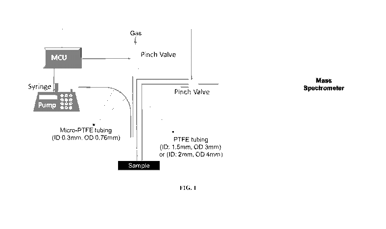
[0040] FIG. 1: Representative schematic of a mass spectroscopy probe for
minimally
invasive surgery.
[0041] FIG. 2: Multilumen tubing for use with the mass spectroscopy probe for
minimally invasive surgery.
[0042] FIG. 3: A cannula and trocar needle for housing and inserting the mass
spectrometry probe for minimally invasive surgery.
[0043] FIG. 4: Representative schematic of a mass spectrometry probe for
minimally
invasive surgery. This embodiment includes a shutter for occluding the probe.
[0044] FIG. 5: Mass spectra of mouse brains tissue section from the minimally
invasive
mass spectrometry probe using Q Exactive Orbitrap Mass Spectrometer. PTFE
tubing of 1.5
meters was used with an inner diameter of 2 mm and outer diameter of 4 mm.
[0045] FIG. 6: Mass spectra of mouse brains tissue section from the minimally
invasive
mass spectrometry probe using Q Exactive Orbitrap Mass Spectrometer. PTFE
tubing of 3.5
meters was used with an inner diameter of 2 mm and outer diameter of 4 mm.
- 15 -
CA 03083260 2020-05-21
WO 2019/104328
PCT/US2018/062625
[0046] FIG. 7: Mass spectra of mouse brains tissue section from the minimally
invasive
mass spectrometry probe using Q Exactive Orbitrap Mass Spectrometer. PTFE
tubing of 4.5
meters was used with an inner diameter of 2 mm and outer diameter of 4 mm.
[0047] FIG. 8: Mass spectra of mouse brain tissue section from the minimally
invasive
mass spectrometry probe using Q Exactive Orbitrap Mass Spectrometer.
[0048] FIG. 9: Representative schematice of a mass spectrometry probe for
minimally
invasive surgery. Depicted on the lower left is the multichannel probe tip.
[0049] FIG. 10: Simulated laparoscopic surgery shown from a laparoscopic
optical
camera on a simulated uterus. Shown on the right are forceps holding the
minimally invasive
mass spectrometry probe.
[0050] FIG. 11: Mass spectra generated from a 16 p.m mouse brain section using
4.5
meter long tubing compared to the water background.
[0051] FIG. 12: Mass spectra generated with the minimally invasive mass
spectrometry
probe using Q Exactive Orbitrap Mass Spectrometer and conduits of 1.5-4.0 mm
diameter.
[0052] FIG. 13: Depiction of the mechanics of a balloon shutter for use with
the
minimally invasive mass spectrometry probe.
[0053] FIG.14: Mass spectra of human lung tissue section from the minimally
invasive
mass spectrometry probe using Q Exactive Orbitrap Mass Spectrometer.
[0054] FIG. 15: Diagram of washing chamber for minimally invasive mass
spectrometry probe.
[0055] FIGS. 16A-16D: Schematic representation of the laparoscopic MasSpec Pen
system being used in a (a) manual laparoscopic MIS procedure and (b) robotic
assisted MIS.
(c) The pen tip was designed with a grasping fin to allow manipulation and
application of the
MasSpec Pen using forceps or other graspers. (d) The tip contacts tissue for
analysis and when
the system is triggered (t = 0 sec) by use of the foot pedal, the syringe pump
delivers a
controlled volume of water to the reservoir. The discrete water droplet
interacts with the tissue
to extract molecules. After 3 seconds, the vacuum and the gas conduits are
concomitantly
- 16 -
CA 03083260 2020-05-21
WO 2019/104328
PCT/US2018/062625
opened to transport the droplet from the MasSpec Pen to the mass spectrometer
through the
tubing system for molecular analysis.
[0056] FIGS. 17A-17C: Comparison between designs and performance of the
handheld
and laparoscopic MasSpec Pen. (a) The handheld MasSpec Pen contains a PDMS tip
and three
PTFE conduits, which provide incoming water (1) and gas (2) to the tip, and an
outgoing
conduit (3) for the water droplet to the mass spectrometer. The pen tip holds
a water droplet
within the reservoir, which contacts tissue for analysis. (b) The laparoscopic
MasSpec Pen
PDMS tip is grafted with two micro- PTFE tubes, one for the incoming water
(1), and another
for incoming gas (2). The proximal end of the pen tip was then connected to a
larger PTFE
conduit, which functions as the outgoing water conduit (3). Using this design,
the hollow space
in the distal end of pen tip functions as the water droplet reservoir (c)
Representative mass
spectra obtained with the handheld and laparoscopic MasSpec Pen of a mouse
brain tissue
section, both operated at 2.7 mm reservoir diameter and a 1.5 meter tubing
length.
[0057] FIGS. 18A-18C: Different tubing lengths between the laparoscopic
MasSpec
Pen (2.7 mm reservoir diameter) and the mass spectrometer were evaluated using
mouse brain
tissue sections. Similar molecular profiles were obtained at different
transfer times using (a)
1.5 m (3.8 seconds, n=10), (b) 3.0 m (5.8 seconds, n=10), and (c) 4.5 m (7.5
seconds, n=10).
[0058] FIGS. 19A-19B: An automated system was developed for automatic mass
spectrometry data acquisition, statistical analysis, and communication of
results. (a) A foot
pedal is used to trigger the analysis workflow through communication with an
Arduino
microcontroller, which then activates water droplet deposition by triggering
the syringe pump.
In the GUI, the user selects which type of tissue is being evaluated prior to
usage, so that the
software selects the proper statistical classifier, providing a predictive
diagnosis with the
associated cancer probability. (b) The laparoscopic MasSpec Pen platform was
tested using a
mannequin through an 8 mm cannula. Laparoscopic forceps were used to
manipulate the
MasSpec Pen, while a video camera was employed to transmit an image and/or
video of the
organs inside the abdomen and guide the operator during the procedure.
[0100] FIGS. 20A-20C: (a-c) Representative mass spectra obtained from mouse
brain
tissue sections with laparoscopic MasSpec Pen at reservoir diameters of 1.5,
2.7, and 4.0 mm
and a 4.5 meter tubing length.
- 17 -
CA 03083260 2020-05-21
WO 2019/104328
PCT/US2018/062625
[0059] FIG. 21: Representative mass spectra obtained with the laparoscopic
MasSpec
Pen (2.7 mm reservoir diameter and a 4.5 meter tubing length) of a human
normal and a
cancerous ovarian tissues.
[0060] FIG. 22: Representative mass spectra obtained with the MasSpec Pen from
samples of beef, lamb, chicken and pork using CAN:DMF 1:1 as the solvent.
Results show
that the spectra obtained using the MasSpec Pen were able to assess the source
of the meat with
a high level of accuracy.
[0061] FIG. 23: Representative mass spectra obtained with the MasSpec Pen from
samples of grass-fed versus grain-fed beef (using CAN:DMF 1:1 as the solvent).
Results show
that the spectra obtained using the MasSpec Pen were able determine whether
the meat was
sourced from a grass-fed or grain-fed animal with a high level of accuracy.
[0062] FIG. 24: Representative mass spectra obtained with the MasSpec Pen from
samples of beef, lamb, chicken and pork using water as the solvent. Results
show that the
spectra obtained using the MasSpec Pen were able to assess the source of the
meat with a high
level of accuracy even when using water as the only solvent.
[0063] FIG. 25: Representative mass spectra obtained with the MasSpec Pen from
samples of fish, including Atlantic salmon, sockeye salmon, steelhead trout,
cod loin or halibut
(using ACN:DMF (1:1) as a solvent). Results show that the spectra obtained
using the
MasSpec Pen were able to assess the source of the fish with a high level of
accuracy.
[0064] FIG. 26: Representative mass spectra obtained with the MasSpec Pen from
samples including amounts of illicit drugs, cocaine and amphetamine. Results
show that the
spectra obtained using the MasSpec Pen were able to detect the drugs with a
high degree of
sensitivity and quantify the amount of drug in the sample.
[0065] FIG. 27: Representative mass spectra obtained with the Mas Spec Pen
from
samples including amounts of oxycodone. Results show that the spectra obtained
using the
MasSpec Pen were able to detect and quantify the amount of oxycodone in the
sample.
[0066] FIG. 28: Representative mass spectra (and a comparative chromatograph)
obtained with the MasSpec Pen from samples including the pesticide
azoxystrobin. Results
- 18 -
CA 03083260 2020-05-21
WO 2019/104328
PCT/US2018/062625
show that the MasSpec Pen analysis was able to detect and quantify the amount
of pesticide in
the sample.
[0067] FIG. 29: Representative mass spectra (and a comparative chromatograph)
obtained with the MasSpec Pen from samples including the pesticide atrazine.
Results show
that the MasSpec Pen analysis was able to detect and quantify the amount of
pesticide in the
sample.
[0068] FIG. 30: Representative mass spectra obtained with the MasSpec Pen from
grapes. Results show that the MasSpec Pen analysis was able to produce a
spectrum that could
be used to characterize the sample.
- 19 -
CA 03083260 2020-05-21
WO 2019/104328
PCT/US2018/062625
DESCRIPTION OF ILLUSTRATIVE EMBODIMENTS
I. The Present Embodiments
[0069] In certain aspects, the instant application provides methods and
devices for
.. minimally invasive molecular assessment of samples, such as tissue samples.
In particular,
aspects the methods can be used to assess multiple tissue sites during an
operation (or biopsy)
of the tissue. This feature allows for accurate identification of diseased
tissues (e.g., tissue sites
retaining cancer cells) in "real-time" allowing surgeons to more accurately
address only the
diseased tissue relative to surrounding normal tissues. In particular aspects,
the methods
disclosed here can involve delivery of a fixed or discrete volume of solvent
to a tissue site,
followed by collection of a liquid sample from the site and analysis of the
liquid sample by
mass spectrometry. Importantly, rather than being applied in a high-pressure
spray, solvent is
applied as discrete droplets and at low pressure. These methods allow for
accurate collection
of samples from a distinct tissue site while avoiding damage to the tissue
being assessed. The
resulting mass spectrometry profile from collected samples allows for
differentiation of
diseased versus normal tissue sites. The method can be repeated at multiple
sites of interest to
very accurately map molecular changes (e.g., in a tissue). Importantly, the
profiles of samples
could be differentiated even without the use of an ionization source. Thus,
while methods of
the embodiments could be used in conjunction with an ionization source, the
use of such a
source is not required. These methodologies can allow assessment of plurality
of tissue sites
over a short range of time, thereby allowing for very accurate assessment of
the boundaries of
diseased versus normal tissues.
[0070] In some aspects, the methods detailed herein can be used to collect and
analyze
samples from a wide range of sources. For example, the methods can be used to
assess surgical,
forensic, agriculture, pharmaceutical, and/or oil/petroleum samples.
[0071] In some aspects, the materials (PDMS and PTFE) and solvent (e.g., water
only
solvents) used in the devices of the embodiments are biologically compatible,
such that they
can be used in surgery in for real-time analysis. Furthermore, because the
devices can be very
compact, it can be hand-held and used in used in minimally invasive surgical
procedures, or
.. non-surgical procedures.
[0072] In some aspects, the present invention provides devices of extended
length and
increased compactness for delivery of fixed or discrete volumes of solvents to
tissues for use
- 20 -
CA 03083260 2020-05-21
WO 2019/104328
PCT/US2018/062625
in minimally invasive surgeries. In some aspects, these methods can be
encapsulated in a
variety of form factors such as a conduit, ranging from 0.5 mm to 10.0 mm
inner diameter (e.g.,
with an inner diameter of between about 1.0 and 5.0; 1.0 and 10.0; 2.0 and
8.0; or 5.0 and 10.0
mm). In some aspects, the site of delivery of a fixed or discrete volume of
solvent, followed by
collection of a liquid sample may be inside the body, such as a surgical site.
In some aspects,
two smaller conduits may be inserted into a third, larger, conduit to create a
multi-lumen
catheter. For example, the multi-lumen catheter can have 2, 3, 4, 5, 6 or more
luminal spaces
with each having an internal diameter of, e.g., 0.05 to 5.0 mm; 0.1 to 5.0 mm;
0.25 to 3.0mm;
or 0.5 mm to 10.0 mm. The multi-lumen catheter may be attached to a mass
spectrometry
device for analysis of sample tissues inside the body during surgery, while
avoiding
unnecessary damage to surrounding tissues.
[0073] In some aspects, the device may be used through cannulas or catheters
in
minimally invasive surgical or endoscopy procedures, or may be used in non-
surgical
procedures through needle guides or biopsy guides. In some aspects, the
present invention can
be integrated into a robotic surgical system allowing several regions of the
human body cavity
to be quickly sampled and analyzed. In some aspects, the device be used to
analyze tissues
using a database of molecular signatures and machine learning algorithms,
allowing diagnosis
in real time for each sampled region. The present invention may be used in a
wide variety of
oncological and other surgical interventions, such as endometriosis, for which
real time
characterization and diagnosis of tissues are needed.
[0074] In some aspects, the present disclosure provides an attachment to the
probe, for
fine manipulation of the probe during minimally or non-invasive procedures.
For example, the
attachment to the probe may be a fin. In some aspects, such a fin may be
composed of the
same material as the probe. In some cases, the fin is made of PDMS. A fin can,
in some aspects,
be formed by an injection molding process or it may be 3D printed. In some
aspects, the present
invention may further comprise a device for grasping the probe, external to
the probe, in order
to manipulate the probe during laparoscopic procedures. The grasping device
may be used to
hold, rotate, or move the probe, or may grasp the fin attached to the probe,
in order to move or
rotate the probe.
[0075] In some aspects, the present invention maintains a reservoir using a
multi-lumen
catheter with recessed ports for depositing water and nitrogen gas during
laparoscopic surgical
procedures. A multi-lumen catheter may be formed, for example, using a multi-
lumen extrusion
- 21 -
CA 03083260 2020-05-21
WO 2019/104328
PCT/US2018/062625
as is well known in the art. These catheters may be utilized in any cannula.
The most commonly
used cannulas are of 5 mm and 10 mm diameters, and are typically used for
laparoscopic
surgeries.
[0076] In some aspects, the present disclosure provides tools, devices and
methods for
manipulation of the probe during endoscopy. For example, multi-lumen tubing
may be used
with an external vacuum source in order to attach the probe to the tissue
surface while
analyzing.
[0077] In some aspects, the present invention provides a shutter system that
occludes
the orifice of the minimally invasive surgical device. In some aspects, this
shutter system may
be a catheter balloon that is integrated within the device or added separately
to the device. The
shutter, or balloon, may close the probe tip, preventing unwanted biological
material from
entering the device, including the lumens and tubing, upon insertion of the
catheter into the
patient. The shutter or balloon may disallow endogenous biological fluids from
entering the
mass spectrometer after analysis has been initiated, thus preventing
contamination of the
results. Finally, closing of the shutter or balloon may prevent excess
nitrogen gas and water
from entering the body. Inclusion of lengthened probes for minimally invasive
surgeries and
occlusion technologies for the tips of the probes may mitigate the
unpredictable and often
tumultuous nature of internal organ movement and organ systems during surgery
which could
affect signal acquisition. Balloons technologies could also be used in other
region of the device
instead or in addition to the pinch valves to control solvent and gas motions
through the tubes.
[0078] In some aspects, the present invention may be used with robotic
manipulation.
In some aspects, the technologies of the present invention may integrate in
modern surgical
theaters through an accessory port, or via a robotic arm. These devices may be
integrated into
robotic systems such as the Intuitive Surgical da Vinci robotic surgical
system. A device of the
present invention may have its own dedicated arm in a robotic system, or be
handled by robotic
graspers by incorporating a "fin" onto the probe. Smaller and larger diameters
can also be used
to be coupled to any existing catheters, cannulas and also needle/biopsy
guides.
[0079] In some aspects, a tracking probe can be integrated with this device in
order to
display and record where the tissue sample has been analyzed to better assist
the surgeon in
localizing the sampling points both intraoperatively or otherwise. For
example, during
intraoperative ultrasound, an ultrasound emitter on the device may be utilized
to display the
- 22 -
CA 03083260 2020-05-21
WO 2019/104328
PCT/US2018/062625
probe when sampling. The probe may be integrated with a tracking device based
on radio
frequency technology, such as the Biosense Webster Cart system. In that case,
the probe may
display the device/sampling location on any of a variety of imaging
modalities, such as
intraoperative UltraSound (US)/Computed Tomogrpahy (CT)/Magnetic Resonance
Imaging
(MRI)/ Optical Coherence Tomography (OCT). Additionally, fluorescent imaging
and
molecular dyes may be used to track the analyzed areas and charted to provide
2-dimensional
or 3-dimensional spatial imaging. More simply, the probe tip may be coated
with a surgical
dye which is then stamped on the tissue to track the region analyzed. Yet
another tracking
approach is to integrate an RF emitter into the probe so that the spatial
location may be tracked.
[0080] In some aspects, the probe of the present invention may be used to
assist
surgeons and medical professionals during minimally invasive surgical
interventions by
providing comprehensive and definitive diagnostic molecular information in
vivo and in real
time, without necessarily causing damage or alteration to the patient's native
living tissues. The
handheld MasSpec Pen has demonstrated a capacity to do this during non-
laparoscopic/endoscopic surgical procedures (U.S. Patent Application No.
15/692,167
incorporated herein by reference, in its entirety). Similarly to the handheld
Mas Spec Pen, the
present invention is suitable for ex vivo analysis of tissues (fresh, frozen,
sections, biopsies) or
other clinical specimens that might be examined by a pathologist, and may be
used for chemical
analysis of any given sample for which direct analysis is desired in confined
and spatially
limited domains (animals, plants, explosives, drugs, etc). A variety of tissue
types may be
analyzed as well, including but not limited to, breast, kidney, lymph node,
thyroid, ovary,
pancreatic and brain tissues.
[0081] In some aspects, the probe of the present invention may be used in
conjunction
with surgical instruments for the treatment of a disease. A variety of
surgical instruments may
be used to excise or ablate cells or tissues, including, but not limited to,
laser ablation tools,
tools for cauterization or electrocauterization, or tools for the manual
dissection of tissue such
as a scalpel.
[0082] Thus, many regions of the human body cavity can be quickly sampled
during
surgery, and analyzed (e.g., by using a database of molecular signatures and
machine learning
algorithms). Therefore, the diagnostic results may be provided in real time
for each sampled
region. Exemplary devices for use in these methods are detailed below.
- 23 -
CA 03083260 2020-05-21
WO 2019/104328
PCT/US2018/062625
Exemplary Features of a Device of the Embodiments
A. Shutter systems
[0083] In some aspects a device of the embodiments further comprises a shutter
system
that can occlude the orifice, and creates a separation between the reservoir
and the tissue. For
example, the shutter system can activate after the droplet rests for 3 seconds
and before the
droplet is transported to the mass spectrometer. One reason for this is to
ensure no biological
material reach the mass spectrometer and cause damage to the instrument. The
shutter can be
an iris diaphragm, a mechanical closure, gate, or tapenade. An additional
design for the shutter
is a balloon mechanism, which seals the exterior of the device from the
tissue. The balloon can
be positions on the distal end of the conduit, e.g., perpendicular to the pen
or probe. When
activated, the balloon expands and fills up the reservoir towards the
direction of the tissue. This
accomplishes at least 3 things: first it gently lifts the pen tip off of the
tissue using the inflated
balloon, insuring that there is no damage to the tissue. This is to ensure
that the probe remains
nondestructive and biocompatible in case the analyzed tissue is determined to
be 'normal'.
Secondly, it seals the solvent droplet that is inside the reservoir and
prevents leakage or
absorbance of lipids after the sampling window. Thirdly, it creates a seal at
the end of the
conduit, which will allow for more effective transfer of the droplet to the
mass spectrometer.
B. Catheter systems
[0084] In some cases, where a probe is incorporated into a
laparoscopic/endoscopic
device a reservoir includes using a multi-lumen catheter, e.g., with recessed
ports for depositing
water and nitrogen gas. The reservoir also retains the water during the
extraction period. A
multi-lumen catheter can be formed for example using a multi-lumen extrusion
as is well
known in the art. It has been demonstrated that these catheters can be
utilized in any cannula,
most commonly 5mm and 1 Omm diameters, for laparoscopic surgeries. This
technology is
compatible with robotic manipulation such as the Intuitive Surgical da Vinci
robotic surgical
system. The Laparoscopic/Endoscopic probes will easily integrate in current
surgical theaters
through an accessory port or via a robotic arm. Smaller and larger diameters
can also be used
to be coupled to any existing catheters, cannulas and also needle/biopsy
guides.
C. Valve systems
[0085] In further aspects, a probe system of the embodiments can incorporate
additional valves. For example, micro-solenoid valves can be located at each
conduit, e.g., at
- 24 -
CA 03083260 2020-05-21
WO 2019/104328
PCT/US2018/062625
the distal end of the sampling probe. These will be individually controlled by
an arduino,
microcontroller, or signal. In some cases the value operation is automated. In
other cases it can
be manually controlled. In some aspects, valves are positioned in the inner
wall of the solvent
conduit sealing the conduits. Thus, by using such values, only two or even one
conduit can be
used in the sampling operation. For example, a delivering solvent conduit and
a return conduit
to transfer the droplet to the mass spectrometer. Additional micro-solenoids
could be
implanted to have more control. For example, three or four micro-solenoids can
be into the
probes of the embodiments.
D. Further surgical system features
[0086] In some aspects, medical devices require passage to areas of the body
that are
difficult to maintain manual control. One solution is to use endoscopic
catheters, but these are
often less precise when compared to handheld devices. Further control can be
attained using
robotic tools that can function nearly to the same extent, and sometimes
better than physicians
equipped with a traditional scalpel. A further feature of the
Laparoscopic/Endoscopic probes
of the embodiments is a 'fin' that can be grasped by forceps, robotic tools,
or laparoscopic
graspers. This will allow the probe to be used in a variety of modalities
without sacrificing
resolution or sensitivity. In some aspects, the fin itself is a gradual sloped
protrusion from the
exterior of the conduit running parallel to said conduit. It is textured to
provide extra traction
for the grasping mechanism.
[0087] In further aspects, a tracking probe can be integrated with this device
in order
to display and record where the tissue sample has been analyzed to better
assist the surgeon in
localizing the sampling points both intraoperatively or otherwise. For
intraoperative
ultrasound, an ultrasound emitter on the device may be utilized to display the
probe when
sampling. Alternatively, the probe can be integrated with a tracking device
based on radio
frequency technology, such as the e.g., Biosense Webster Cart system. With
this approach,
the probe displays the device/sampling location on any various imaging
modalities like
intraoperative UltraSound (US)/Computed Tomography (CT)/Magnetic Resonance
Imaging
(MR1)/ Optical Coherence Tomography (OCT).
[0088] In some further aspects, tissue sites that are assessed by a probe of
the
embodiments can be marked. For example, a dye that is up-taken by cancerous
cells and
normal cells, which will mark where the probe has been placed. In some
aspects, a chemical
dye can be delivered using an additional conduit in the catheter or by using a
multilumen
- 25 -
CA 03083260 2020-05-21
WO 2019/104328
PCT/US2018/062625
catheter. An alternative delivery of a tracking dye is to dissolve it in the
solvent that we use to
analyze the tissue. For instance, one advantage of using a dye within the
solvent is that it will
directly correlate with where the tissue sample was taken, instead of the
peripheral region. Of
course in this aspects, the chemical dye would be present in the mass spectra
and would have
to be distinguished from biomolecules in a sample. In some aspects, it may
useful to make the
dye visible (e.g., in white operating room light). In other aspects, the dye
may be a fluorescent
dye. In yet a further aspect, the pen tip can be coated with a surgical dye,
which is then stamped
on the tissue to track the region analyzed. Likewise, as discussed above, a
tracking approach
can be used to virtually map the tissues sites analyzed. For instance, a RF
emitter can be
integrated into a probe so that the spatial location may be tracked. Thus, in
some aspects, dyes
(or probe tracking) can be used to track analyzed areas of tissues. In some
aspects, tissues
analyzed can be charted to provide 2 dimensional and 3 dimensional spatial
imaging.
[0089] In further aspects, a probe system can include a filter. For example a
filter can
prevent biological tissue from going into the conduits. For example, a filter
mesh system can
be incorporated within the device to prevent smaller bodies of tissue, protein
aggregates, or
coagulated cell clusters from entering. This mesh could be placed at the
opening and have
contact with the tissue, or be positioned higher up within the probe, such
that no tissue contact
occurs. In some aspects such a filter mesh comprises average apature sizes of
less than about
1.0, 0.5, 0.25 or 0.1 mm. Since solid matter can damage a mass spectrometer,
such a filter
system can increase instrument lifespan with out negatively effecting signal
detected.
[0090] In still further aspects, an endoscopic/laparoscopic probe of the
embodiments is
integrated with a microcontroller, user interface, and/or associated hardware
that will operate
with appropriate software.
[0091] In some further cases, a light, such as a LED will be incorporated to
provide
visual feed back to the user, for example, to indicate that the probe is ready
for sampling, in the
process of doing so, or needs to be replaced/repaired. Acoustic feedback can
also be used, for
instance, to let the user know what step of the process the device is in
(e.g., since physical cues
may be unavailable laparoscopically). A user interface system can also be
integrated with the
device, such as in a foot pedal and buttons on the housing of the probe.
- 26 -
CA 03083260 2020-05-21
WO 2019/104328
PCT/US2018/062625
III. Assay Methodologies
[0092] In some aspects, the present disclosure provides methods of determining
the
presence of diseased tissue (e.g., tumor tissue) or detecting a molecular
signature of a biological
specimen by identifying specific patterns of a mass spectrometry profile.
Biological specimens
for analysis can be from animals, plants or any material (living or non-
living) that has been in
contact with biological molecules or organisms. A biological specimen can be
samples in vivo
(e.g. during surgery) or ex vivo.
[0093] A profile obtained by the methods of the embodiments can correspond to,
for
example, proteins, metabolites, or lipids from analyzed biological specimens
or tissue sites.
These patterns may be determined by measuring the presence of specific ions
using mass
spectrometry. Some non-limiting examples of ionizations methods that can be
coupled to this
device include chemical ionization, laser ionization, atmospheric-pressure
chemical ionization,
electron ionization, fast atom bombardment, electrospray ionization, thermal
ionization.
Additional ionization methods include inductively coupled plasma sources,
photoionization,
glow discharge, field desorption, thermospray, desorption/ionization on
silicon, direct analysis
in real time, secondary ion mass spectroscopy, spark ionization, and thermal
ionization.
[0094] In particular, the present methods may be applied or coupled to an
ambient
ionization source or method for obtaining the mass spectral data such as
extraction ambient
ionization source. Extraction ambient ionization sources are methods with, in
this case, liquid
extraction processes dynamically followed by ionization. Some non-limiting
examples of
extraction ambient ionization sources include air flow-assisted desorption
electrospray
ionization (AFADESI), direct analysis in real time (DART), desorption
electrospray ionization
(DESI), desorption ionization by charge exchange (DICE), electrode-assisted
desorption
electrospray ionization (EADESI), electrospray laser desorption ionization
(ELDI),
electrostatic spray ionization (ESTASI), Jet desorption electrospray
ionization (JeDI), laser
assisted desorption electrospray ionization (LADESI), laser desorption
electrospray ionization
(LDESI), matrix-assisted laser desorption electrospray ionization (MALDESI),
nanospray
desorption electrospray ionization (nano-DESI), or transmission mode
desorption electrospray
ionization (TM-DESI).
[0095] As with many mass spectrometry methods, ionization efficiency can be
optimized by modifying the collection or solvent conditions such as the
solvent components,
the pH, the gas flow rates, the applied voltage, and other aspects which
affect ionization of the
- 27 -
CA 03083260 2020-05-21
WO 2019/104328
PCT/US2018/062625
sample solution. In particular, the present methods contemplate the use of a
solvent or solution
which is compatible with human issue. Some non-limiting examples of solvent
which may be
used as the ionization solvent include water, ethanol, methanol, acetonitrile,
dimethylformamide, an acid, or a mixture thereof In some embodiments, the
method
contemplates a mixture of acetonitrile and dimethylformamide. The amounts of
acetonitrile
and dimethylformamide may be varied to enhance the extraction of the analytes
from the
sample as well as increase the ionization and volatility of the sample. In
some embodiments,
the composition contains from about 5:1 (v/v) dimethylformamide:acetonitrile
to about 1:5
(v/v) dimethylformamide: acetonitrile such as 1:1 (v/v) dimethylformamide:
acetonitrile.
However, in preferred embodiment the solvent for use according to the
embodiments is a
pharmaceutically acceptable solvent, such as sterile water or a buffered
aqueous solution.
IV. Examples
[0096] The following examples are included to demonstrate preferred
embodiments of
the invention. It should be appreciated by those of skill in the art that the
techniques disclosed
in the examples which follow represent techniques discovered by the inventor
to function well
in the practice of the invention, and thus can be considered to constitute
preferred modes for
its practice. However, those of skill in the art should, in light of the
present disclosure,
appreciate that many changes can be made in the specific embodiments which are
disclosed
and still obtain a like or similar result without departing from the spirit
and scope of the
invention.
Example 1 ¨ Minimally invasive probe for mass spectrometry design
[0097] The system developed consists of three main parts: 1) a syringe pump
that is
programmed to deliver a discrete solvent volume using a controlled flow rate;
2) tubing systems
integrated to two-way pinch valves for controlled solvent and gas transport;
3) a probe tip
which is used for direct sampling of biological tissues. The tubing systems
and probe tip are
also integrated into a minimally invasive surgical device such as a cannula or
catheter for use
in laparoscopic or endoscopic surgeries. Several iterations of the system were
explored and
optimized with the ultimate goal of minimizing tissue damage, maximizing
tissue-analyte
extraction, and maximizing solvent transmission to the mass spectrometer. FIG.
1 shows a
schematic figure of one example of a minimally invasive apparatus for
analyzing biological
tissue. The syringe pump feeds solvent and gas into the minimally invasive
probe via micro-
PTFE tubing. The probe maintains contact with the sample, retains solvent
during interaction
- 28 -
CA 03083260 2020-05-21
WO 2019/104328
PCT/US2018/062625
with the tissue. The tip was manufactured using 3D-printing and is made of
biologically
compatible polydimethylsiloxane (PDMS). The probe has three main ports: one
for the
incoming tubing system, a central port for gas delivery, and a third for the
outgoing tubing
system. All ports come in junction at a small reservoir where the droplet is
retained and exposed
to the tissue sample for a controlled amount of time, allowing for efficient
extraction of
molecules. The size of the reservoir determines the spatial resolution of the
device. A solvent
volume of 10 pL is exposed to the tissue sample. FIG. 2 shows the three
conduit tubes. The
three conduit tubes used are made of polytetrafluoroethylene (PTFE), which is
also biologically
compatible. The tube from the syringe pump is used to deliver solvent from
syringe pump to
the probe tip, while the other micro-PTFE tube is used to deliver an inert gas
(N2 or CO2) to
the probe tip. The gas serves three main purposes: 1) tissue drying prior to
analysis; 2) prevent
solvent gap due to the mass spectrometer's vacuum when the reservoir is closed
by contacting
the tissue specimen; 2) assist solvent transport from tissue to the mass
spectrometer through
the wider PTFE tubing. The larger PTFE tubing is directly connected to the
inlet of the mass
spectrometer so that the positive pressure of the mass spectrometer vacuum
system is used to
drive the droplet from the reservoir to the mass spectrometer inlet for
ionization. FIG. 9 shows
a schematic of the minimally invasive probe which includes a diagram of the
tip of the probe,
including the tree conduit tubes and the reservoir at the base (labelled 4).
FIG. 3 shows two of
the possible devices to house the minimally invasive probe. The cannula shown
has the gas and
solvent tubing entering the top, as well as the tubing to the mass
spectrometer. The probe is
shown emerging from the bottom of the cannula. The probe may also be
introduced into the
body cavity using a trocar needle. FIG. 10 depicts a simulated laparoscopic
uterine surgery,
and shows that the minimally invasive probe may be controlled by forceps. A
shutter system
that occludes the orifice of the minimally invasive probe may be employed as
shown in FIG.
4. One option for the shutter is to use a catheter balloon which may close the
probe tip, a
diagram of which is shown in FIG. 13, preventing unwanted biological material
from entering
the device, including the lumens and tubing, upon insertion of the catheter
into the patient. The
shutter may disallow endogenous biological fluids from entering the mass
spectrometer after
analysis has been initiated, thus preventing contamination of the results.
Closing of the shutter
can also prevent excess nitrogen gas and water from entering the body. The use
of a shutter in
the lengthened probes necessary for minimally invasive surgery may help
mitigate the
unpredictable and often tumultuous nature of internal organ movement and organ
systems
during surgery which could affect signal acquisition. The minimally invasive
mass
spectrometry probe may also include a vacuum tube separate from the sample
vacuum above.
- 29 -
CA 03083260 2020-05-21
WO 2019/104328
PCT/US2018/062625
The purpose of this second vacuum tube is to gently secure, or latch, the tip
of the probe onto
the tissue during analysis.
[0098] The time events involved in the device operation are automated and
precisely
controlled by software that communicates with an Arduino system and two two-
way pinch
valves. All pinch valves are closed until the process is initiated when, under
300 pL/min, a
pulse is sent to the pump to infuse the solvent for two seconds and stop,
generating a 10 pt
droplet filling in the minimally invasive probe reservoir. The gas and mass
spectrometer tubes
are closed at pinch valves, allowing the solvent in the reservoir to interact
with the tissue for
three seconds to extract the molecules. The pinch valves controlling the gas
and mass
spectrometer tubes are opened simultaneously, allowing the droplet to transfer
to the mass
spectrometer for ionization and molecular analysis. A pulse is sent to the
pump to infuse the
solvent for another 12 seconds and stop, to completely drive all the extracted
molecules into
the mass spectrometer. The gas and mass spectrometer tubes are left open for
another 20
seconds to allow all the solvent in the mass spectrometer tube to go into the
mass spectrometer.
The total analyzing time is 37 seconds.
[0099] The probe may be washed between analyses in a variety of methods.
Generally,
the tip of the probe is wiped with sterile water. An additional design that
can facilitate the
washing step is a retractable design that will wash the exterior of the probe
without having to
remove the device from the patient (FIG. 15). The design consists of a chamber
with valves
located at the openings to maintain a water and gas seal. A longer tube that
contains the probe
tip, water, and gas conduits will transect only the top valve when the tip is
located in the
washing chamber, but will pass through both valves when the tip is deployed
into the patient
environment. After the probe tip, tubing, or both have become contaminated
during the surgery
process, the probe will withdraw into the washing chamber. Water tubes can be
located inside
the washing chamber and point upwards providing a strong jet of cleaning
solvent. Two
positions of vacuum tubing will be located above the first and second valve to
remove dirtied
solvent. The vacuum tube placed above the first valve is an emergency tube in
case any water
breaks the first valve barrier. The entire system will fit smoothly inside of
a trocar, and the
deployable probe will be located inside of this system. The vacuums located
inside the probe
will also operate during this cleaning process, which will flush the tubing
until clean.
- 30 -
CA 03083260 2020-05-21
WO 2019/104328
PCT/US2018/062625
Example 2 ¨ Molecular Profiles and Analysis
[00100] The
system described herein operates by directly connecting the transfer
tube to the mass spectrometer inlet for transporting the analyte-containing
solvents to the mass
spectrometer for molecular analysis. This set up greatly simplifies
operational details and
precludes the use of ionization sources. After the probe interacts with the
tissue, the solvent is
then transported to the mass spectrometer and directly infused without the
need of an additional
ionization source. Since the system is fully automated so that each 10 [it
solvent droplet is
delivered separately to the inlet, the mass spectrometer operates without any
impact on its
performance. Rich molecular information is obtained in this manner, similar to
what is
observed from other solvent-extraction ambient ionization techniques such as
desorption
electrospray ionization. The ionization mechanism may be similar to inlet
ionization. For inlet
ionization methods, the ionization occurs in the inlet pressure drop region
between atmosphere
and vacuum. Because of the nature of minimally invasive surgical techniques,
the diameter of
tubing, and length of tubing is of critical importance. A variety of tube
lengths were tested for
the delivery of solvent to the mass spectrometer, as seen in FIGS. 5-8).
[00101]
FIGS. 5-8 show the total ion chromatograms obtained from mouse brain
sections during the total analysis period while using tubing lengths of 1.5
meters up to 4.5
meters. Rich molecular profiles were observed in all cases. At a tube length
of 4.5 meters the
molecular profile is easily established over the background signal of the
water (FIG. 11). FIG.
12 shows total ion chromatograms obtained using conduit sizes from 1.5 mm to
4.0 mm. Again,
rich molecular profiles were observed with each conduit size. To further
demonstrate the utility
of the minimally invasive probe for mass spectrometry, human lung tissue was
analyzed (FIG.
14), and generated a robust molecular profile.
[00102] The
molecular profiles generated by the minimally invasive mass
spectrometry probe can also be used for tissue typing. A series of tissue
samples were evaluated
with the minimally invasive mass spectrometry probe and were able to be
identified with an
overall accuracy of 98.55% (Table 1).
[00103] Table 1. Tissue typing results.
TRUE Thyroid Lymph Parathyroid Breast Lung Ovarian Pancreas
Thyroid 42 0 1 0 0 0 0
Lymph 0 26 0 0 0 0 0
Parathyroid 0 1 62 0 0 0 0
- 31 -
CA 03083260 2020-05-21
WO 2019/104328
PCT/US2018/062625
Breast 0 0 0 29 0 0 0
Lung 0 0 0 0 47 0 0
Ovarian 0 1 0 1 0 41 0
Pancreas 0 0 0 0 0 0 24
[00104] The
system was able to identify lymph, breast, and lung tissues with
100% accuracy, thyroid and parathyroid with between 97% and 99% accuracy,
ovarian with
95.35% accuracy, and pancreas tissue with 83.33% accuracy. These tissue typing
results were
generated from selected features of the mass spectrometry profiles shown in
Table 2.
[00105] Table 2. Selected features
for tissue typing.
Parathyroi
Pancrea
Thyroid Lymph d Breast Lung Ovarian s
2.26901
m/z -0.25915 -1.37199
0.833471 -0.44036 -0.34999 8 -0.68101
0.09713
125.01 0 0 0 0 0 7 0
130.06 0 0 0.008242 0 0 0 0
146.05 0 0 -0.00177 0 0 0 0
147.69 0 0 0.28651 0 0 0 0
0.02570
148.95 0 0 0 0 0 3 0
0.01411
183.96 0 0 0 0 0 8 0
191.02 0 0 0 0 0 -0.01003 0
194.99 0 0 0 0 0.00062 0 0
200.17 0 -0.00053 0 0 0 0 0
205.46 0 0 0 0 0 0 0.260268
218.1 0 0 -0.01171 0 0 0 0
239.17 0 -0.02264 0 0 0 0 0
241.92 0 0 0 0 0 0.00716 0
243.97 0 0 -0.01202 0 0 0 0
244.92 0 0 0.004177 0 0 0 0
0.06468
250.96 1 0 0 0 0 0 0
0.03052
251.96 0 0 0 0 0 1 0
252.85 0 0.00771 0 0 0 0 0
0.03283
255.9 0 0 0 0 0 6 0
0.00984
256.23 0 0 0 0 2 0 0
271 0 0 -0.0484 0 0 0 0
271.19 0 -0.00793 0 0 0 0 0
- 32 -
CA 03083260 2020-05-21
WO 2019/104328
PCT/US2018/062625
272.01 0 0 0.042591 0 0 0 0
273.08 0 0 0.015766 0 0 0 0
0.01443
276.8 8 0 0 0 0 0 0
0.01105
279.24 0 0 0 0 3 0 0
279.92 0 0 0 0 -0.03074 0 0
0.02669
287.01 0 0 0 0 0 3 0
287.98 0 0 0.131242 0 0 0 0
0.08193
291.01 0 3 0 0 0 0 0
294.82 0 0 -0.01746 0 0 0 0
296.09 0 0 -0.01414 0 0 0 0
296.94 0 0 0 0 0 0 0.105634
0.01676
306.07 0 0 0 0 5 0 0
0.00434
318.85 0 0 0 0 0 5 0
0.02568
323.91 0 0 0 0 0 9 0
326.06 0 0 0.031889 0 0 0 0
0.06413
332.27 0 0 0 0 0 8 0
341.27 0 0 0 0.01519 0 0 0
0.06630
344.97 0 0 0 0 0 6 0
354.16 0 0 0 0 0 -0.04315 0
0.01144
357.84 0 0 0 0 0 7 0
0.00037
362.24 0 0 -0.07847 0 0 9 0
407.23 0 0 0 0 0 -0.00779 0
0.08931
428.03 0 3 0 0 0 0 0
428.19 0 0 0 0 0 -0.00888 0
436.28 0 0 0 0 0 0 0.006734
437.29 0 0 0 0 0 0 0.077554
0.15155
444.08 3 0 0 0 0 0 0
453.28 0 0 0 0 0 -0.00176 0
455.8 0 0 0 0 0 0.01032 0
460.23 0 0 0 0 0 -0.04105 0
462.3 0 0 0 0 0 0 0.08139
463.98 0 0 0.024299 0 0 0 0
465.3 0 0 0 0 0 -0.03072 0
465.32 0 0 0 0 0 0 0.012348
476.21 0 0 -0.02093 0 0 0 0
- 33 -
CA 03083260 2020-05-21
WO 2019/104328
PCT/US2018/062625
0.03298
485.2 0 0 0 0 0 1 0
0.18170
519.32 0 0 0 6 0 0 0
524.3 0 0 0 0 0 0 0.0418
530.26 0 0 0.011799 0 0 0 0
0.04137
535.13 0 0 0 0 0 6 0
565.05 0 0 0.076194 0 0 0 0
0.06734
578.27 0 3 0 0 0 0 0
616.17 0 0 0 0 0 -0.03131 0
0.10099
637.33 2 0 0 0 0 0 0
655.51 0 0 0 0 0 0 0.088267
0.06425
688.51 0 0 0 3 0 0 0
0.04455
690.51 0 0 0 0 8 0 0
701.53 0 0 0 0 0 -0.00473 0
714.51 0 0 0 0 0 -0.02904 0
0.28605
715.54 0 0 0 9 0 0 0
717.53 0 0 0.032346 0 0 0 0
0.10747
718.54 0 0 0 0 1 0 0
0.16655
719.49 0 0 0 0 4 0 0
0.01948
721.5 0 0 0 0 2 0 0
724.99 0 0 0.012692 0 0 0 0
725.49 0 0 0.061029 0 0 0 0
726.5 0 0 0.022406 0 0 0 0
0.07785
729.37 0 7 0 0 0 0 0
741.53 0 0 0.04295 0 0 0 0
0.02362
743.57 0 0 0 7 0 0 0
747.52 0 0 0 0 0 -0.0096 0
0.14091
748.52 0 0 0 0 2 0 0
0.02291
752.56 0 0 0 0 3 0 0
758.4 0.0479 0 0 0 0 0 0
0.02512
761.4 3 0 0 0 0 0 0
764.52 0 0 0.003296 0 0 0 0
768.55 0 0 0.002224 0 0 0 0
- 34 -
CA 03083260 2020-05-21
WO 2019/104328
PCT/US2018/062625
769.5 0 0 0 0 0 -0.00431 0
0.00472
769.51 0 0 0 0 7 0 0
770.53 0 0 0 0 0.08501 0 0
771.52 0 0 0 0 0 -0.03674 0
775.55 0 0 0 0 0 -0.02396 0
0.12170
776.55 0 0 0 0 5 0 0
0.01200
793.56 0 3 0 0 0 0 0
0.03905
795.52 0 0 0 0 1 0 0
0.02479
796.52 0 0 0 0 2 0 0
809.52 0 0 0.063972 0 0 0 0
0.04729
811.53 0 4 0 0 0 0 0
0.08788
812.55 0 2 0 0 0 0 0
0.03164
813.55 0 7 0 0 0 0 0
0.06204
822.47 8 0 0 0 0 0 0
0.21582
823.48 2 0 0 0 0 0 0
833.52 0 -0.0026 0 0 0 0 0
835.54 0 -0.00055 0 0 0 0 0
0.31181
836.55 0 6 0 0 0 0 0
0.02082
838.56 0 3 0 0 0 0 0
860.54 0 0 0.004644 0 0 0 0
861.55 0 0 0 0 0 -0.02476 0
0.00146
991.29 0 7 0 0 0 0 0
0.06956
991.69 0 2 0 0 0 0 0
1305.9 0.05880
0 0 0 0 0 9 0
1448.9
7 0 0 0.002613 0 0 0 0
[00106]
Similarly to the differentiation of tissue types, the minimally invasive
mass spectrometry probe can be used to differentiate between normal and
cancerous tissues.
The system predicted normal tissues with greater than 89% accuracy, and cancer
tissues with
5 greater than 91% accuracy as seen in Table 3.
- 35 -
CA 03083260 2020-05-21
WO 2019/104328
PCT/US2018/062625
[00107] Table 3. Cancer tissue prediction results.
Predicted
Normal Cancer
Normal 247 28
Cancer 12 129
[00108] These tissues were predicted based on the selected
features shown in
Table 4.
[00109] Table 4. Selected features used for the prediction of cancer
tissues.
Cancer MaxIntensityNorm MinIntensityNorm MaxIntensity
MinIntensity
m/z
0.1200838 0.00000000 0 0.0 0
124.01
40.9603959 0.07309089 0 1910468.9 0
146.05
25.4909642 0.08721183 0 4249182.8 0
154.06
1.7548952 0.27268945 0 1028115.5 0
165.02 42.8950874 0.02168110 0 189918.4
0
174.04 114.2977347 0.01893967 0 703388.7
0
175.02
27.1843207 0.21139870 0 6752479.6 0
175.03
24.4596324 0.12211639 0 2175467.9 0
187.04
60.8607370 0.22046140 0 5731647.5 0
201.04 118.9618023 0.08163593 0 1262331.3
0
214.05
128.8755917 0.03867785 0 773249.9 0
215.03
31.7356346 0.09055005 0 3274645.3 0
221.01 138.9164198 0.01083151 0 568690.6
0
241.04
74.5285436 0.01626196 0 3302753.8 0
246.95
2.9550154 0.05776215 0 1111366.1 0
267.07 4.3468095 0.03979236 0 4039159.9
0
268.8
51.7317355 0.04524576 0 1488145.8 0
271
3.2679671 0.06004618 0 492594.7 0
- 36 -
CA 03083260 2020-05-21
WO 2019/104328
PCT/US2018/062625
283.27
55.5183712 0.14933261 0 2355024.1 0
296.94 7.5951233 0.22379688 0 2429905.2
0
313.16
3.7134875 0.17606736 0 7786035.2 0
328.06 71.2579706 0.04428292 0 957525.7
0
332.9 23.4374773 0.03396153 0 1214185.0
0
341.27
1.6023939 0.28304768 0 6590254.4 0
345.16
50.1650411 0.04368856 0 1696500.0 0
346.05 83.1031168 0.01781918 0 628038.1
0
353.16 23.4132756 0.06995437 0 2172310.7
0
377.09
9.9688497 0.10964367 0 1100627.9 0
559.47
2.7064435 0.05334380 0 49840406.6 0
572.48 83.3837979 0.01727858 0 1439590.6
0
585.49
20.0678254 0.09010271 0 86114572.9 0
615.17
115.9821356 0.03578691 0 2801168.1 0
722.51 66.9597188 0.04599428 0 13448313.9
0
742.54 89.8907769 0.04732031 0 8284542.5
0
744.55 64.7272067 0.02038387 0 905261.7
0
748.52
39.0756981 0.04027318 0 1870669.2 0
766.54
23.4133796 0.05266494 0 4504433.6 0
773.53
60.0910994 0.09127090 0 3784388.4 0
788.54 44.1054014 0.04591038 0 1660366.4
0
788.55
1.4204710 0.03125159 0 4032842.0 0
822.47
19.2490565 0.05601076 0 738427.0 0
823.48
66.6013083 0.02864992 0 320932.5 0
861.55
135.4621348 0.05829841 0 9614626.2 0
885.55 25.0244403 0.12046562 0 9712708.6
0
888.57 90.5815624 0.02339633 0 1145858.3 0
- 37 -
CA 03083260 2020-05-21
WO 2019/104328
PCT/US2018/062625
[00110] To
evaluate the system performance, consecutive analysis was
conducted on the same tissue section, and on different tissue sections to
demonstrate that the
system is highly reproducible within samples and across different samples.
Materials and Methods.
[00111] Mass
Spectrometer. Q Exactive Hybrid Quadrupole-Orbitrap mass
spectrometer (Thermo Scientific, San Jose, CA) was used. Full-scan was carried
out at the
range of m/z 500-1800, and the other mass spectrometric parameters were listed
as follows:
resolving power 140,000, micro scan 2, maximum injection time 300 ms,
capillary temperature
350 C and S-lens RF level 100.
[00112] Biological
Tissues. Wild-type mouse brains were purchased from
Bioreclamation IVT. 62 frozen human tissue specimens including breast,
thyroid, lymph node,
ovarian, and kidney were obtained from Cooperative Human Tissue Network and
Baylor
College Tissue Bank. Samples were stored in a -80 C freezer. Tissue slides
were sectioned at
16 p.m using a CryoStarTM NX50 cryostat. Frozen tissue specimen were thawed
under room
temperature before use.
[00113]
Statistical Analysis. IBM SPSS Statistics 22.0 (IBM Corporation,
Armonk, NY, USA) was used to perform principal component analysis (PCA) to
reveal
patterns in the data. The analysis was performed directly using the raw data.
The 10 peaks of
the top relative intensities in the m/z range of 700-900 were used for PCA.
Typically, the first
three components, which all encompassed more than 85% of the total variance,
are used in the
present results.
Example 3¨ System Automation for Handheld and Laparoscopic Use
[00114]
Because all the materials (PDMS and PTFE) and solvent (only water)
used in the minimally invasive probe design are biologically compatible, the
system has a high
potential to be used in laparoscopic and endoscopic surgeries for real-time
analysis. More than
that, due to the small dimension of the device, it can be integrated to a
robotic surgical system,
such as the Da Vinci surgical system, through an accessory port or one of its
robotic arms.
Several regions of the human body cavity can be quickly sampled during surgery
with or
without wash/flush steps in between each analysis, and analyzed by using a
database of
molecular signatures and machine learning algorithms. Therefore, the
diagnosing results may
be provided in real time for each sampled region. This system can be broadly
used in a wide
- 38 -
CA 03083260 2020-05-21
WO 2019/104328
PCT/US2018/062625
variety of oncological and other surgical interventions (such as
endometriosis) for which real-
time characterization and diagnosis of tissues are needed.
[00115]
Thus, a laparoscopic MasSpec Pen platform was developed, which may
be used in manual or robotically controlled MIS procedures (FIG. 16A and 16B).
The
laparoscopic MasSpec Pen platform was developed with emphasis on three main
design
features: 1) adherence to dimensions and material specifications necessary for
use in the
laparoscopic environment; 2) similar performance specification to the handheld
MasSpec Pen
to ensure compatibility with the previously generated statistical models; and
3) integration to
an automated software and graphical user interface for real time data analysis
and statistical
classification.
[00116] The
laparoscopic MasSpec Pen was engineered with the specifications
needed to function in MIS. Design modifications allowed introduction of the
MasSpec Pen
through the cannula of a laparoscopic trocar, or through the open ports of
robotic systems
(commonly of 5 mm, 8 mm, or 12 mm in diameter), while maintaining similar
operation to the
handheld MasSpec Pen. The handheld MasSpec Pen has a diameter of 10 mm, which
was
dictated by the diameter of the 3D printed polydimethylsiloxane (PDMS) pen
tip. The tip of
the handheld MasSpec Pen was designed with three conduits (incoming water,
incoming gas,
and outgoing water), which are in fluid communication with an open reservoir
that positions
the water droplet for contact with tissue surface (FIG. 17A). In the
laparoscopic MasSpec Pen,
two micro-polytetrafluoroethylene (PTFE) tubes (OD 0.794 mm, ID 0.339 mm), one
for the
incoming water, and another for incoming gas, were grafted into a 3D printed
PDMS tip (FIG.
17B). The proximal end of pen tip was connected to a larger PTFE conduit (OD
1.59 mm, ID
0.794 mm) to serve as the outgoing water conduit. In this design, the hollow
space in the distal
end of pen tip functions as the water droplet reservoir and can be customized
to varied
diameters depending on the intended use.
[00117] In
this study, the reservoir was designed with three diameters of 1.5 mm,
2.7 mm, and 4.0 mm to display a range of capabilities. Although reservoir
diameters below
1.5 mm could be manufactured via micromolding, these were not tested due to
limitations in
manufacturing capabilities. Further, current recommended cancer-free margins
for solid cancer
excision are often larger than 1.5 mm - such as 3 mm for basal cell carcinoma,
2 mm for breast
cancer, and 5 cm for gastric cancer.
- 39 -
CA 03083260 2020-05-21
WO 2019/104328
PCT/US2018/062625
[00118] To
manipulate the MasSpec Pen for contact with the organ of interest in
vivo, a grasping fin was incorporated on the pen tip to provide an anchor
point for a
laparoscopic tool, such as forceps or a robotic arm (FIG. 16C). As in any
laparoscopic
procedure, organ access is dictated by trocar placement. One benefit of the
laparoscopic
MasSpec Pen is flexibility and light weight of the polymer-based tubing
system. These features
allow the device to be easily manipulated through a trocar in the x, y, and z
directions given
the cannula's placement in reference to an organ of interest. A 3 mm in length
fin was
incorporated into the 3D printed molds unilaterally at the proximal end of the
pen tip to avoid
increasing the diameter of the device. The total diameter of the laparoscopic
MasSpec Pen
including the fin was 7.5 mm for the 1.5 and 2.7 mm reservoir diameter tips,
and 9.5 mm for
the 4.0 mm reservoir diameter tip; all compatible for use through common sized
trocars.
[00119]
Different tube lengths between the laparoscopic MasSpec Pen and the
mass spectrometer were investigated for use within the operating room
environment (FIG. 18).
Longer tubing lengths than those used in the handheld MasSpec Pen (1.5 m) were
tested
considering the additional length needed to insert the MasSpec Pen through the
laparoscopic
trocars (5-12 mm length), constraints of the operating room (OR) workspace,
and the need for
instrument placement outside of the surgical sterile field. As with the
handheld MasSpec Pen,
3 seconds of contact time between the water droplet within the pen tip and the
probed brain
tissue section was used for molecular extraction (FIG. 16D).
[00120] After the
sampling period, a 4-second water flush was used to facilitate
droplet transport from the pen tip, through the PTFE tube, to the mass
spectrometer. The PTFE
tube was directly connected to an extended, heated mass spectrometer transfer
tube (350 C)
via flexible silicon tubing, therefore eliminating the use of an external
ionization source.
Analyses were performed in the negative ion mode. FIGS. 18A-C show the mass
spectra
obtained from serial sections of mouse brain tissue analyzed with the 2.7 mm
laparoscopic
MasSpec Pen at 1.5 m, 3.0 m and 4.5 m tubing lengths. Similar patterns were
observed in the
recorded mass spectra, displaying high relative abundances of a variety of
negatively charged
ions, identified as lipid species typically observed from mouse brain tissue
sections using
MasSpec Pen and other ambient ionization techniques. For example, m/z 834.529
(identified
as [PS(40:6)-I-11-), m/z 885.550 (identified as [PI(38:4)-I-1]-), and m/z
790.539 (identified as
[PE(40:6)-I-1]-) were observed at high relative abundances in the mass spectra
obtained from
the grey matter of mouse brain.
- 40 -
CA 03083260 2020-05-21
WO 2019/104328
PCT/US2018/062625
[00121] An
average cosine similarity of 0.93 (n=12) was achieved for the mass
spectra obtained with various tube lengths, which demonstrates that the
molecular information
obtained is reproducible and independent of tube length (Table 5).
Additionally, transfer time
was measured for each length tested, yielding 3.8 s 0.5 s (n=10), 5.8 s
0.7 s (n=10), and 7.5
s 0.4 s (n=10), for tube lengths of 1.5 m, 3.0 m, and 4.5 meters,
respectively (Table 6).
Interestingly, tripling the tube length from 1.5 m to 4.5 m resulted in
doubling of the transfer
time, which indicates a non-linear velocity of droplet transport in the tubing
system. At a tube
length of 4.5 meters, different laparoscopic MasSpec Pen reservoir diameters
(1.5, 2.7, and 4.0
mm) were tested, yielding comparable performance with expected changes in the
mass spectral
profile due to sampling of different brain tissue regions (FIG. 20).
[00122] To
compare the performance of the laparoscopic MasSpec Pen with that
of the handheld system, serial tissue sections of mouse brain were analyzed
using the same
dimensions previously described for the handheld MasSpec Pen (2.7 mm pen tip
diameter and
1.5 m tubing length). Similar profiles were observed from mouse brain tissue
sections analyzed
with both designs across the full m/z range (m/z 120-1800, cosine similarly =
0.88, n=8) and
restricted m/z range (m/z 600-1800, cosine similarly = 0.92, n=8) (FIG. 17C).
These results
suggested that the laparoscopic MasSpec Pen produced comparable results to
those obtained
with the handheld MasSpec Pen, despite changes in reservoir design and tube
length. A 2.7
mm reservoir diameter and 4.5 m tubing length were chosen for the remaining
experiments
performed with the laparoscopic MasSpec Pen. Using these parameters, an
overall
measurement time of ¨20 seconds/spot was achieved, which includes the time
employed for
tissue sampling (3 seconds), droplet transport (7 seconds), and droplet
ionization and analysis
(-10 seconds).
[00123] To
facilitate clinical use of the laparoscopic MasSpec Pen, a software
with a graphical user interface (GUI) was developed for real time mass
spectrometry data
acquisition, statistical analysis, and display of results. As previously
reported for the handheld
MasSpec Pen, a foot pedal is used to trigger the analysis workflow. Here, the
system was
further refined so that the foot pedal also triggers the lab-built software
(FIG. 19A). Using this
approach, the user selects which type of tissue is being evaluated in the GUI
prior to tissue
analysis, so that the software properly selects the corresponding statistical
model for data
analysis. Then, upon activation by the pedal, a syringe pump is triggered so
that a discrete water
droplet is formed in the pen tip, where it interacts with the tissue for 3
seconds. Following the
- 41 -
CA 03083260 2020-05-21
WO 2019/104328
PCT/US2018/062625
tissue contact, the droplet enriched with extracted bio-molecular species is
transported to the
Orbitrap mass spectrometer through a PTFE tube for ionization and mass
analysis, as
previously explained. The mass spectra data is continuously recorded and read
by the software
program. The three mass spectra with the highest intensity for selected ions
are averaged and
pre-processed for statistical analysis. Prediction is then performed using the
statistical models
previously built by the Lasso method using data acquired from histologically
validated tissues
and the probability of the tissue being cancer is reported in the GUI.
[00124] To
test performance of the integrated system for tissue diagnosis, 12
human ovarian tissue samples including 7 normal tissues and 5 high grade
serous carcinoma
(HGSC) were analyzed following the workflow described above. A statistical
classifier
previously built for ovarian cancer diagnosis was incorporated in the software
and analysis was
triggered using foot pedal activation. For larger-sized tissue specimens,
several regions within
the same tissue sample were analyzed, yielding a total of 24 analyses. The
mass spectra
obtained presented characteristic lipid profiles similar to those we have
previously described
for normal and cancer ovarian tissues (FIG. 21). Using the software and
statistical classifier, a
cancer probability and associated predictive diagnosis were reported for each
region analyzed.
[00125]
Based on the cut-off values generated through the statistical classifier,
samples with a predictive probability greater than 0.51 were as "cancer",
while samples with
predictive probabilities lower than 0.51 were called "normal". As shown in
Table 7, 100%
sensitivity for cancer diagnosis was achieved, as all of the regions of the
five cancer samples
analyzed were classified as cancer. One of the seven normal tissue samples (ON
164b) was
misclassified as cancer in both regions analyzed, while one of the four
regions analyzed of
sample ON 135a was classified as cancer, yielding 80% selectivity. Overall, an
87.5%
agreement between the predictive and pathologic diagnoses was achieved.
[00126] Lastly, the
laparoscopic MasSpec Pen was tested using a laparoscopic
simulation mannequin. The laparoscopic MasSpec Pen was inserted into the
mannequin
through an 8 mm cannula. Inside the mannequin, laparoscopic forceps were used
to manipulate
the MasSpec Pen, while a video camera was employed to transmit an image and/or
video of
the organs inside the abdomen and guide placement of the laparoscopic MasSpec
pen during
the procedure (FIG. 19B). Normal and cancer human ovarian samples were placed
on top of a
mimic organ and accessed through the laparoscopic incisions inside the
mannequin, and then
analyzed using the laparoscopic equipment. Forceps were used to grasp the
engineered fin and
- 42 -
CA 03083260 2020-05-21
WO 2019/104328
PCT/US2018/062625
correctly guide the pen to the tissue surface for analysis. Using the foot
pedal, the analysis was
automatically activated, as previously described. A predictive diagnosis of
"normal" was
achieved for the normal ovarian tissue (probability of cancer = 0.37), while a
predictive
diagnosis of "cancer" was achieved for the ovarian cancer tissue (probability
of cancer = 0.68),
which agrees with the histopathologic diagnosis of the samples. Overall, the
laparoscopic
MasSpec Pen showed robust performance in the simulated mannequin suggesting
its
compatibility for use in MIS procedures.
[00127] Table 5. Cosine
similarity results between the mass spectra obtained
using the laparoscopic MasSpec Pen (2.7 mm reservoir diameter) and different
tubing lengths.
Tubing Length
1.5 meters (n=4) 3.0 meters (n=4)
3.0 meters (n=4) 0.91
4.5 meters (n=4) 0.92 0.97
[00128] Table 6. Droplet
transport time from pen tip to mass spectrometer
through various tubing lengths.
Droplet Transport Time
Average (n=10) Relative Standard Deviation
(RSD)
1.5 meters 3.8 12.4%
3.0 meters 5.8 11.5%
4.5 meters 7.5 5.2%
[00129] Table 7. Pathologic
diagnosis, software predictive diagnosis and cancer
probabilities for the human ovarian tissue samples analyzed using the
laparoscopic MasSpec
Pen (2.7 mm reservoir diameter and 4.5 m tubing length).
Pathologic Software
Tissue ID Cancer Probability
Diagnosis Predictive Diagnosis
Spot] Cancer 0.59
Normal Ovary ON 135a Spot 2 Normal 0.40
Spot 3 Normal 0.32
- 43 -
CA 03083260 2020-05-21
WO 2019/104328 PCT/US2018/062625
Spot 4 Normal 0.01
Spot] Normal 0.39
ON 263c
Spot 2 Normal 0.42
ON 294a Spot] Normal 0.04
Spot] Normal 0.11
ON 220a
Spot 2 Normal 0.38
Spot] Cancer 0.76
ON 164b
Spot 2 Cancer 0.62
Spot] Normal 0.00
ON 335c
Spot 2 Normal 0.00
Spot] Normal 0.38
ON 161a
Spot 2 Normal 0.36
Spot] Cancer 0.88
OT 403a
Spot 2 Cancer 0.98
Spot] Cancer 0.98
OT 054a
High Grade Spot 2 Cancer 0.72
Serous Ovarian Spot] Cancer 0.89
OT 337a
Cancer Spot 2 Cancer 1.00
Spot] Cancer 0.99
OT 405a
Spot 2 Cancer 0.99
OT 058a Spot] Cancer 0.93
[00130] In conclusion, the laparoscopic MasSpec Pen is an automated device
that
provides near real time diagnostic information for MIS. The unique design and
features of
laparoscopic MasSpec Pen described meet many of the requirements needed for
manual and
robotic MIS. When compared to the handheld version, similar molecular patterns
were
obtained from mouse brain tissue sections. Different pen tip diameters and
tube lengths were
also tested for various clinical needs. Finally, a customized lab-built
software was developed,
providing a fully automated workflow for tissue analysis and diagnostic
feedback. This
technology may be a complementary tool for MIS, which could expedite clinical
workflow and
improve surgical outcomes.
Example 4 ¨ Materials and Methods
[00131] Laparoscopic MasSpec Pen Design. Three different
laparoscopic/robotic MasSpec Pen tips were created from PDMS with diameters of
1.5 mm,
2.7 mm, and 4.0 mm. Grasping fins were built-in unilaterally in order to
minimize overall cross
- 44 -
CA 03083260 2020-05-21
WO 2019/104328
PCT/US2018/062625
section. Two micro-PTFE tubings (OD 0.794 mm, ID 0.339 mm) were grafted to the
interior
of the PDMS tip near the distal end. The micro-PTFE tubing was terminated 2
millimeters
above the reservoir in order to avoid tissue interaction. The PDMS mixing
solution from Dow
Corning (Midland, MI) was molded into negative prints created by a Stratasys
uPrint SE Plus
3D Printer (Eden Prairie, Minnesota, USA). PTFE tubing was purchased from
Sigma-Aldrich
(St. Louis, MI, USA), and silicone tubing was purchased from Saint Gobain
(Tygon #3550,
Malvern, PA, USA). This design allows the formation of a water droplet at the
distal end of the
PDMS tip that interacts with tissue to extract cellular lipids and small
metabolites via phase
diffusion. This entire system was inserted into a laparoscopic simulation
mannequin through a
10 mm HiCap 30107 H5 trocar by Karl Storz, (Tuttlingen, Germany). Images were
taken with
a wireless endoscope IP67 snake camera in the simulation mannequin.
[00132]
Tissue Samples. Human tissues were obtained from the Cooperative
Human Tissue Network (CHTN) (Charlottesville, VA) under approved Institutional
Review
Board protocol. Mouse brains were obtained from BioIVT (Westbury, NY, USA).
Tissue
.. samples were thawed to room temperature before analysis.
[00133]
Mass Spectrometry Analysis. Experiments were performed on a Q
ExactiveTM Hybrid Quadrupole-OrbitrapTM mass spectrometer (Thermo Fisher
Scientific, San
Jose, CA, USA). HPLC grade water was used for analysis. Full scan mode was
carried out at
the range of m/z 120 to 1800, at a resolving power of 140,000, capillary
temperature was set to
350 C, and an S-lens radio frequency level was set to 100.
[00134]
Statistical Analysis and Software Tools. The statistical analysis
procedure for generating the statistical model using Lasso was explained in a
previous work.
The lab-built desktop software designed to predict and display a diagnosis in
real time.
Following activation by the pedal through the Arduino microcontroller, data
from the mass
spectrometer is continuously read using MSFileReader (Thermo Fisher
Scientific) and the
MSFileReader-Python-bindings package open-sourced on GitHub. The three
consecutive mass
spectra of highest intensity for selected ions are averaged and pre-processed
for statistical
prediction. Then, using the previously fitted Lasso model, a prediction is
generated and
displayed back to the user via the GUI.
- 45 -
CA 03083260 2020-05-21
WO 2019/104328
PCT/US2018/062625
Example 5 ¨ Characterizing materials using the MasSpec Pen
[00135] The
MasSpec pen was also used to obtain samples from various
materials to further evaluate the use of the pen is characterizing materials
in the environment
such a foods and forensic samples. Results demonstrated a wide range of
further application
for the mass spectroscopy analyses of the embodiments. For example, studies
presented in
FIGs. 22-25 and 30 demonstrated effect spectra could be produced from a wide
range of food
products including meats, fish and fruits. These spectra could be used to
accurately
characterize the source material for the samples, such the type of meat or
fish that was being
assessed. For these studies, meat and fish (salmon, trout, atlantic cod, and
pollack) samples
were obtained from a local supermarket (Central Market, Austin, TX. Samples
were stored in
a fridge (4 C) until analysis at room temperature. Analysis was performed
using LTQ Orbitrap
XL mass spectrometer (Thermo Scientific) coupled to the MasSpec Pen. Analyzed
regions of
the samples were marked to prevent duplicated analysis. Molecular profiles of
each sample
were collected and used to create a classification model with the least
absolute shrinkage and
selection operator (Lasso). Lasso was used to identify predictive markers of
meat type and
build a classification models for identification of unknown samples.
[00136] The
MasSpec Pen was used to analyze meat samples as well as 5
samples of each type of fish. Initial experiments were performed to optimize
parameters of the
MasSpec Pen for the highest lipid extraction and transmission. In the negative
ion mode,
detection of various glycerophospholipid species (GP), such as
glycerophosphoinositols,
glycerophosphoserines, and glycerophosphoethanolamines was achieved. In
addition to GP,
small metabolites, sphingolipids (SP), such as ceramides, and free fatty acids
(FA), such as
arachidonic acid and oleic acid were also observed. Despite the mass spectra
complexity, the
profiles obtained demonstrated trends in ion abundances characteristic of each
meat sample.
Statistical analysis was then applied to identify predictive markers of each
meat type, as well
as to build and evaluate the performance of classification models for
prediction of meat type.
These analysis demonstrated a high degree of accuracy in correctly identifying
source meat
samples. In fact, the methods were sensitive enough to even discern between
grass-and grain-
raised meat products (FIG. 24). Accordingly, methods of the embodiments could
be used to
authenticate consumer products such as meats and fish, which would otherwise
be difficult to
accurately and quickly authenticate.
- 46 -
CA 03083260 2020-05-21
WO 2019/104328
PCT/US2018/062625
[00137] The
methods likewise can be used to detect and quantify the amounts of
compounds present in materials. For example, in the case of forensic samples,
the amount of
illicit drugs could be accurately determined using a sampling taken by the
MasSpec Pen (see,
FIGs. 26 and 27). Likewise, the methods have been shown to be effective in
detecting the
presence of pesticides in source materials (see, FIGs 28-29). Thus, methods of
the
embodiments can be effectively applied to characterize a wide range of
materials and the
determine and quantify the presence of compounds of interest in the materials.
* * *
[00138] All of the methods disclosed and claimed herein can be made and
executed
without undue experimentation in light of the present disclosure. While the
compositions and
methods of this invention have been described in terms of preferred
embodiments, it will be
apparent to those of skill in the art that variations may be applied to the
methods and in the
steps or in the sequence of steps of the method described herein without
departing from the
concept, spirit and scope of the invention. More specifically, it will be
apparent that certain
agents which are both chemically and physiologically related may be
substituted for the agents
described herein while the same or similar results would be achieved. All such
similar
substitutes and modifications apparent to those skilled in the art are deemed
to be within the
spirit, scope and concept of the invention as defined by the appended claims.
- 47 -