Note: Descriptions are shown in the official language in which they were submitted.
CA 03083898 2020-04-17
WO 2019/079-188
PCT/US2018/056336
PRENYLATION ASSAY
RELATED APPLICATIONS
1011 This application claims the benefit of provisional application USSN
62/573,522, filed
October 17, 2017 and of provisional application USSN 62/636,722, filed
February 28, 2018,
the contents of each of which are herein incorporated by reference in their
entirety.
INCORPORATION OF SEQUENCE LISTING
[02] The contents of the text file named "NIGH-005_001WO_SeqList.txt," which
was
created on October 16, 2018 and is 59 KB in size, are hereby incorporated by
reference in
their entirety.
FIELD OF THE DISCLOSURE
[03] The present invention relates to an assay for use in determining the
activity of Rab
escort protein 1 (REP1). More specifically, the invention relates to the use
of Rab6a in an
assay as a substrate for prenylation, in particular wherein the REP I has been
delivered to a
cell using a gene therapy vector.
BACKGROUND OF THE DISCLOSURE
[04] Choroideremia may be successfully treated by providing functional
copies of the
REP1 transgene to the affected cells of the eye. Specifically, it has been
shown that adeno-
associated virus (AAV) gene therapy vectors may be used to deliver a
nucleotide sequence
encoding functional REP1 to the eye to treat the disease. As gene therapy of
choroideremia is
becoming a clinical reality, there is a need for reliable and sensitive assays
to determine the
activity of exogenously delivered REP1, in particular to test new gene therapy
vectors and as
a quality control screen for clinical vector stocks. The disclosure provides a
reliable and
sensitive assay to determine the activity of exogenously delivered REP I.
SUMMARY OF THE INVENTION
[05] The present inventors have surprisingly found that, despite a
prevailing
understanding that Rab27a (also referred to as RAB27A) provides the most
suitable
- 1 -
CA 03083898 2020-04-17
WO 2019/079-188
PCT/US2018/056336
prenylation assay substrate, use of Rab6a (also referred to as RAB6A) as a
substrate in a
prenylation reaction provides a more sensitive method for determining the
activity of Rab
escort protein I (REP!). Not only does Rab6a provide for increased sensitivity
in an assay
detecting prenylation, it also provides for beneficial signal-to-noise ratios,
better dynamic
range of signal and better consistency.
[06] Moreover, the inventors have demonstrated that the increased
sensitivity of a Rab6a-
based assay may be harnessed to accurately and reliably determine the activity
of REP!-
encoding vectors, in particular AAV gene therapy vectors, such as those
suitable for use in
the clinic.
[07] The disclosure provides a method for determining an activity of Rab
escort protein I
(REP!) comprising the steps: (a) contacting a REP I protein with a Rab6a
protein, a Rab
geranylgeranyltransferase (Rab GGTase) and a lipid donor substrate to produce
a lipidated
Rab6a; and (b) detecting the lipidated Rab6a.
[08] In some embodiments of the methods of determining an activity of Rab
escort
protein! (REP!) of the disclosure, a sample comprises the REP! protein. In
some
embodiments, the sample comprising the REP1 protein is isolated or derived
from a cell and
wherein the cell is genetically engineered to express the REPI protein. In
some
embodiments, the sample comprising the REP! protein comprises a lysate of the
cell.
[09] In some embodiments of the methods of determining an activity of Rab
escort
protein 1 (REP I) of the disclosure, the REPI protein is expressed from a
viral vector
comprising a nucleotide sequence encoding the REP1 protein. In some
embodiments, the
viral vector is an adeno-associated viral (AAV) vector.
10101 In some embodiments of the methods of determining an activity of Rab
escort
protein 1 (REP]) of the disclosure, the Rab6a protein or the Rab GGTase is
substantially
pure. In some embodiments, the Rab6a protein and the Rab GGTase are
substantially pure. In
some embodiments, the Rab6a:Rab GGTase molar ratio is about 1:2-3. In some
embodiments, the Rab6a:Rab GGTase molar ratio is 1:2-3. In some embodiments,
the
Rab6a:Rab GGTase molar ratio is about 1:2.5. In some embodiments, the
Rab6a:Rab
GGTase molar ratio is 1:2.5.
10111 In some embodiments of the methods of determining an activity of Rab
escort
protein 1 (REP I) of the disclosure, the lipid donor substrate comprises
- 2 -
CA 03083898 2020-04-17
WO 2019/079-188
PCT/US2018/056336
geranylgeranylpyrophosphate (GGPP) or an analogue thereof. In some
embodiments, the
lipid donor substrate comprises biotin-geranylpyrophosphate (BGPP).
[012] In some embodiments of the methods of determining an activity of Rab
escort
protein 1 (REP1) of the disclosure, detecting the lipidated Rab6acomprises an
enzyme-linked
immunosorbent assay (ELISA), a Western blot analysis or an autoradiography.
[013] In some embodiments of the methods of determining an activity of Rab
escort
protein 1 (REP!) of the disclosure, the AAV vector comprising nucleotide
sequence
encoding the REP! protein is manufactured for use in the treatment of
choroideremia. In
some embodiments, the lipidated Rab6a is detected and the REP-1 protein or the
AAV vector
comprising nucleotide sequence encoding the REP1 protein is suitable for use
in the
treatment of choroideremia.
[0141 In some embodiments of the methods of determining an activity of Rab
escort
protein 1 (REP1) of the disclosure, detecting the lipidated Rab6a further
comprises
quantifying an amount of the lipidated Rab6a. In some embodiments, the amount
of lipidated
Rab6a is an absolute amount. In some embodiments, the amount of lipidated
Rab6a is a
relative amount. In some embodiments, the amount of lipidated Rab6a is
relative to a control
amount or to a reference level.
[015] The disclosure provides a use of a Rab6a protein for determining an
activity of a
Rab escort protein 1 (REP1) protein.
[016] In some embodiments of the use of a Rab6a protein for determining an
activity of a
REP1 protein of the disclosure, the REP1 protein is isolated or derived from a
cell and the
cell is genetically-engineered to express the REP I protein. In some
embodiments, a cell
comprises the REP1 protein and the cell is genetically engineered to express
the REP1
protein. In some embodiments, the REP! protein is isolated or derived from a
lysate of from
a cell and the cell is genetically engineered to express the REP1 protein. In
some
embodiments, a cell lysate comprises the REP1 protein, the cell lysate is
isolated or derived
from a cell, and the cell is genetically-engineered to express the REP1
protein.
[017] In some embodiments of the use of a Rab6a protein for determining an
activity of a
REP1 protein of the disclosure, the REP1 protein is expressed from a viral
vector comprising
a nucleotide sequence encoding the REP1 protein. In some embodiments, the
viral vector is
an adeno-associated viral (AAV) vector. In some embodiments, the AAV vector
comprising
the nucleotide sequence encoding the REP1 protein is manufactured for use in
the treatment
-3-
CA 03083898 2020-04-17
WO 2019/079-188
PCT/US2018/056336
of choroideremia. In some embodiments, the lipidated Rab6a is detected and the
REP-1
protein or the AAV vector comprising nucleotide sequence encoding the REP I
protein is
suitable for use in the treatment of choroideremia.
10181 In some embodiments of the use of a Rab6a protein for determining an
activity of a
REP1 protein of the disclosure, theRab6a protein is substantially pure.
[019] The disclosure provides a method for determining the activity of Rab
escort protein
1 (REP1) comprising the steps: (a) providing a sample comprising REP1; (b)
contacting the
sample of step (a) with Rab6a, Rab geranylgeranyltransferase (Rab GGTase) and
a lipid
donor substrate; and (c) detecting the lipidated Rab6a product.
[020] For example, the method of the invention may be for testing gene therapy
vectors
suitable for the delivery of REP! to a target cell or for quality control
analysis of vector
stocks (e.g. medicament stocks).
[021] Validation of gene therapy vectors is mandatory for the safe and
efficacious
implementation of gene therapy in the clinic. For analysis and quality control
steps,
comparison with control experiments or reference levels may provide a measure
of the
activity of the gene therapy vector or REP1 relative to a known or accepted
standard (e.g.
better or worse than a known or accepted standard). This may be used to
validate whether a
gene therapy vector stock meets specific targets and regulations.
[0221 In one embodiment, comparison is made to a sample of REP1 or REP1-
encoding
AAV vector that is defined as a primary reference standard. The method of the
invention may
be, for example, carried out in parallel on a test sample and the primary
reference standard
sample. Potency, biological activity and/or behavior of the test sample may
be, for example,
defined relative to the primary reference standard.
[023] Put another way, the method of the invention may, for example, be used
for quality
control analysis and validation of a gene therapy vector as efficacious (e.g.
for the treatment
of choroideremia), preferably an AAV vector particle comprising a REP1-
encoding
nucleotide sequence, preferably wherein an output activity or efficacy of the
vector
determined by the method of the invention above a threshold activity or within
a specified
target range (e.g. by comparison to a control experiment or reference level)
indicates the
vector is suitable for gene therapy purposes.
-4-
CA 03083898 2020-04-17
WO 2019/079-188
PCT/US2018/056336
[024] Accordingly, in another aspect, the method of the invention is for
quality control
analysis of a Rab escort protein 1 (REP1)-encoding gene therapy vector
(preferably an AAV
vector).
[025] In another aspect, the invention provides a method for quality control
analysis of a
Rab escort protein 1 (REP1)-encoding gene therapy vector (preferably an AAV
vector)
comprising the steps: (a) transducing a cell with the vector, culturing the
cell under
conditions suitable for the expression of the REP1 and lysing the cells to
provide a sample
comprising REP1; (b) contacting the sample of step (a) with Rab6a, Rab
geranylgeranyltransferase (Rab GGTase) and a lipid donor substrate; and (c)
detecting the
lipidated Rab6a product.
[026] Accordingly, the method may comprise carrying out a plurality of
experiments
comprising steps (a) to (c) in which parameters relating to the sample
comprising REP1 are
varied, while other parameters (e.g. parameters relating to the Rab6a, Rab
(IGTase and lipid
donor substrate) are kept constant. Such parameters may include, for example,
the amino acid
sequence of the relevant protein (e.g. REPO, the REP1-encoding nucleotide
sequence
comprised in a vector used to express the REP1 in a cell, the type of vector
used to deliver a
REP1-encoding nucleotide sequence to a cell (e.g. the type of viral vector,
such as the type of
adeno-associated viral (AAV) vector), the concentration of REP1 and/or the
multiplicity-of-
infection (MOD of a vector used to deliver a REP1-encoding nucleotide sequence
to a cell. In
a preferred embodiment, the method comprises carrying out a plurality of
experiments
comprising steps (a) to (c) at different MOIs of a vector used to deliver a
REP1-encoding
nucleotide sequence to a cell (e.g. to generate a dose-response curve).
[027] In one embodiment, the detection of the lipidated Rab6a product
comprises
quantifying the amount of the lipidated Rab6a product. In a preferred
embodiment, the
detection of the lipidated Rab6a product comprises quantifying the amount of
the lipidated
Rab6a product relative to a control or reference level. The quantification may
be, for
example, made relative to a sample of REP1 or REP1-encoding AAV vector that is
defined
as a primary reference standard. The method of the invention may be, for
example, carried
out in parallel on a test sample and the primary reference standard sample.
Potency,
biological activity and/or behavior of the test sample may be, for example,
defined relative to
the primary reference standard.
-5-
CA 03083898 2020-04-17
WO 2019/079-188
PCT/US2018/056336
[028] In one embodiment, the method comprises a further step of comparing the
amount of
lipidated Rab6a product (e.g. prenylated, such as geranylgeranylated or biotin-
geranylated,
Rab6a) with an amount determined from a control experiment, such as an
experiment using a
known or standard sample of REP1.
[0291 In another embodiment, the method comprises a further step of comparing
the
amount of lipidated Rab6a product (e.g. prenylated, such as geranylgeranylated
or biotin-
geranylated, Rab6a) with a reference level.
[030] In one embodiment, the sample comprising REP I is from a cell
genetically
engineered to express the REP1. Preferably, the sample comprising REP1 is a
lysate of a cell
genetically engineered to express the REP I. Preferably, a cell is transfected
or transduced
with a vector comprising a REP1-encoding nucleotide sequence to provide the
cell
genetically engineered to express the REP1. Preferably, the vector is a viral
vector.
[031] In one embodiment, the REP1 is expressed using a viral vector comprising
a REP 1-
encoding nucleotide sequence.
[032] In one embodiment, the viral vector is an adeno-associated viral (AAV)
vector.
Preferably the viral vector is in the form of a viral vector particle.
[033] The AAV vector may be of any serotype (e.g. comprise any AAV serotype
genome
and/or capsid protein). Preferably, the vector is capable of infecting or
transducing cells of
the eye.
[034] In one embodiment, the AAV vector comprises an AAV serotype 1, 2, 3, 4,
5, 6, 7,
8, 9, 10, or ii genome. In another embodiment, the AAV vector comprises an AAV
serotype
2, 4, 5 or 8 genome. Preferably, the AAV vector comprises an AAV serotype 2
genome.
[035] In one embodiment, the AAV vector particle comprises an AAV serotype 1,
2, 3, 4,
5, 6, 7, 8, 9, 10, or II capsid protein. In another embodiment, the AAV vector
particle
comprises an AAV serotype 2, 4, 5 or 8 capsid protein. The AAV serotype 8
capsid protein
may, for example, be an AAV8/Y733F mutant capsid protein. Preferably, the AAV
vector
particle comprises an AAV serotype 2 capsid protein.
[036] In one embodiment, the AAV vector particle comprises an AAV2 genome and
AAV2 capsid proteins (AAV2/2): an AAV2 genome and AAV5 capsid proteins
(AAV2/5);
or an AAV2 genome and AAV8 capsid proteins (AAV2/8). Preferably, the AAV
vector
particle comprises an AAV2 genome and AAV2 capsid proteins (AAV2/2).
-6-
CA 03083898 2020-04-17
WO 2019/079-188
PCT/US2018/056336
[037] The AAV vector particle may be a chimeric, shuffled or capsid-modified
derivative
of one or more naturally occurring AAVs. In particular, the AAV vector
particle may
comprise capsid protein sequences from different serotypes, clades, clones or
isolates of
AAV within the same vector (i.e. a pseudotyped vector). Thus, in one
embodiment the AAV
vector is in the form of a pseudotyped AAV vector particle.
[038] In one embodiment, the REP1 is human REP1.
[039] In one embodiment, the REP1 comprises an amino acid sequence that has at
least
70%, 80%, 85%, 90%, 91 %, 92%, 93%, 94%, 95%, 96%, 97%, 98%, 99% or 100%
identity
to SEQ ID NO: 5, preferably wherein the amino acid sequence substantially
retains the
natural function of the protein represented by SEQ ID NO: 5.
[0401 In one embodiment, the REP1-encoding nucleotide sequence comprises a
nucleotide
sequence that has at least 70%, 80%, 85%, 90%, 91 A, 92%, 93%, 94%, 95%, 96%,
97%,
98%, 99% or 100% identity to SEQ ID NO: 6 or 7, preferably wherein the protein
encoded
by the nucleotide sequence substantially retains the natural function of the
protein
represented by SEQ ID NO: 5.
[041] In one embodiment, the REP1-encoding nucleotide sequence comprises a
nucleotide
sequence that encodes an amino acid sequence that has at least 70%, 80%, 85%,
90%, 91 %,
92%, 93%, 94%, 95%, 96%, 97%, 98%, 99% or 100% identity to SEQ ID NO: 5,
preferably
wherein the amino acid sequence substantially retains the natural function of
the protein
represented by SEQ ID NO: 5.
[042] In one embodiment, the Rab6a and/or Rab GGTase are substantially pure.
[043] In one embodiment, the Rab6a comprises an amino acid sequence that has
at least
70%, 80%, 85%, 90%, 91 %, 92%, 93%, 94%, 95%, 96%, 97%, 98%, 99% or 100%
identity
to SEQ ID NO: 1, preferably wherein the amino acid sequence substantially
retains the
natural function of the protein represented by SEQ ID NO: 1.
[0441 In one embodiment, the Rab GGTase comprises an amino acid sequence that
has at
least 70%, 80%, 85%, 90%, 91 %, 92%, 93%, 94%, 95%, 96%, 97%, 98%, 99% or 100%
identity to SEQ ID NO: 3 or 8, preferably SEQ ID NO: 8, preferably wherein the
amino acid
sequence substantially retains the natural function of the protein represented
by SEQ ID NO:
8; and/or an amino acid sequence that has at least 70%, 80%, 85%, 90%, 91 %,
92%, 93%,
94%, 95%, 96%, 97%, 98%, 99% or 100% identity to SEQ ID NO: 4 or 9, preferably
SEQ ID
-7-
CA 03083898 2020-04-17
WO 2019/079-188
PCT/US2018/056336
NO: 9, preferably wherein the amino acid sequence substantially retains the
natural function
of the protein represented by SEQ ID NO: 9.
[045] In another embodiment, the Rab6a:Rab GGTase molar ratio is about 1:0.25-
3, 1:0.3-
2.9, 1:0.35-2.8, 1:0.4-2.7, 1:0.45-2.6 or 1:0.5-2.5, preferably about 1:0.5-
2.5.
[0461 In one embodiment, the Rab6a:Rab GGTase molar ratio is about 1:2-3,
1:2.1-2.9,
1:2.2-2.8, 1:2.3-2.7 or 1:2.4-2.6, preferably about 1:2.4-2.6. In one
embodiment, the
Rab6w.Rab GGTase molar ratio is about 1:2, 1:2.1 , 1:2.2, 1:2.3, 1:2.4, 1:2.5,
1:2.6, 1:2.7,
1:2.8, 1:2.9 or 1:3, preferably about 1:2.5.
[047] In another embodiment, the Rab6a:Rab GGTase molar ratio is about 1:0.25-
0.75,
1:0.3-0.7, 1:0.35-0.65, 1:0.4-0.6 or 1:0.45-0.55, preferably about 1:0.4-0.6.
In one
embodiment, the Rab6a:Rab GGTase molar ratio is about 1:0.25, 1:0.3, 1:0.35,
1:0.4, 1:0.45,
1:0.5, 1:0.55, 1:0.6, 1:0.65, 1:0.7 or 1:0.75, preferably about 1:0.5.
10481 In one embodiment, the lipid donor substrate is
geranylgeranylpyrophosphate
(GGPP) or an analogue thereof Preferably, the lipid donor substrate is
labelled with a
detectable marker. For example, the lipid donor substrate may be isotopically
labelled (e.g.
the lipid donor substrate may comprise 3H), or may comprise a fluorescent
group, epitope or
biotin moiety.
[049] In a preferred embodiment, the lipid donor substrate is biotin-
geranylpyrophosphate
(BGPP).
[050] In one embodiment, the lipidated Rab6a product is detected using an
enzyme-linked
irrununosorbent assay (ELISA). Western blot analysis or autoradiography. In a
preferred
embodiment, the lipidated Rab6a product is detected using an ELISA. The ELISA
may be,
for example, a sandwich ELISA.
[051] In a preferred embodiment, a biotin-labelled lipidated Rab6a product is
detected
using a detection reagent specific for biotin, for example streptavidin.
Preferably, the biotin-
labelled lipidated Rab6a product is detected using Western blot analysis using
a detection
reagent specific for biotin, for example streptavidin (e.g. a streptavidin-
horseradish
peroxidase conjugate). More preferably, the biotin-labelled lipidated Rab6a
product is
detected using an ELISA using a detection reagent specific for biotin, for
example
streptavidin.
[052] In one embodiment, the method is for determining the activity of a REP1-
encoding
gene therapy vector for use in the treatment of choroideremia.
-8-
CA 03083898 2020-04-17
WO 2019/079-188
PCT/US2018/056336
[053] In another aspect, the invention provides the use of Rab6a for
determining the
activity of Rab escort protein 1 (REP1).
[054] The method of determining the activity of REP I, the Rab6a, Rab GGTase,
lipid
donor substrate and the REP! may be as described herein.
[0551 In another aspect, the invention provides a method for determining the
efficacy of a
vector comprising a Rab escort protein 1 (REP 1) encoding nucleotide sequence,
wherein the
method comprises the steps: (a) providing a sample comprising REP I, wherein
the REP1 is
expressed using the vector comprising a REP1-encoding nucleotide sequence; (b)
contacting
the sample of step (a) with Rab6a, Rab geranylgeranyltransferase (Rab GGTase)
and a lipid
donor substrate; and (c) detecting the lipidated Rab6a product.
[056] In another aspect, the invention provides the use of Rab6a for
determining the
efficacy of a vector comprising a Rab escort protein 1 (REP1)-encoding
nucleotide sequence.
[057] Preferably, the method and use are for determining the efficacy of a
vector for use in
the treatment of choroideremia
[058] The vector, REP1, Rab6a, Rab GGTase, lipid donor substrate, lipidated
Rab6a
product, method of detection and other features of the method and use may be
as described
herein.
BRIEF DESCRIPTION OF THE DRAWINGS
[0591 The patent or application file contains at least one drawing executed in
color.
Copies of this patent or patent application publication with color drawing(s)
will be provided
by the Office upon request and payment of the necessary fee.
[0601 Figure IA-D is series of photographs depicting an in vitro prenylation
reaction using
Rab27a as a substrate followed by Western blot (WB) analysis of incorporated
biotinylated
lipid donor using untransduced cells and cells transduced with either AAV-GFP
or AAV-
REP1 (M01 = 10,000 gp/cell). The experiment involved 3 sets of lysates
prepared
independently. Prenylation reactions were set up using 10 g of lysate in a
total volume of
12.5 L. Positive controls were spiked with 2 LIM of fish REP1. Detection time
was 2 min.
(A) WB analysis of human REP1 levels in cell lysates (1:1000). Untransduced
cells (#4, #9
and #12) show endogenous levels of REP1. Cells transduced with AAV-GFP (#5,
#10 and #
3) show endogenous levels of REP1 similar to untransduced cells. Cells
transduced with
AAV-REP1 (#6, #1 1 and #14) show an increase of REP1 levels compared to
untransduced
-9-
CA 03083898 2020-04-17
WO 2019/079-188
PCT/US2018/056336
and AAV-GFP transduced cells. Positive controls (+ve) show endogenous REPI
levels. (B)
WB analysis of13-actin as loading control (1:15000). The levels of 13-actin
are similar in all
samples analyzed. (C) WB analysis of incorporated biotinylated lipid donor in
Rab27a
(1:10000). Untransduced cells and AAV-GFP transduced cells show no detectable
incorporation of biotin in Rab27a. Cells transduced with AAV-REP1 show some
level of
biotin incorporated into Rab27a (#6, #1 1 and #14). Positive controls show the
strongest band
of all as a result of fish REPI activity. (D) Semiquantification of the WB
analysis in (C)
using Image Studio Lite software.
[0611 Figure 2A-D is a series of photographs depicting and in vitro
prenylation reaction
using Rab27a as a substrate followed by Western blot (WB) analysis of
incorporated
biotinylated lipid donor using untransduced cells and cells transduced with
either AAV-GFP
or AAV-REPI (M01 = 10,000 gp/cell). The experiment involved 2 sets of lysates
prepared
independently. Prenylation reactions were set up using 30 Ltg of lysate in a
total volume of 22
L. Positive controls were spiked with 1 M of fish REP1. Detection time was 2
min. (A)
WB analysis of human REP1 levels in cell lysates (1:1000). Untransduced cells
(#9 and #I2)
show endogenous levels of REP1. Cells transduced with AAV-GFP (#10 and #13)
show
endogenous levels of REP1 similar to untransduced cells. Cells transduced with
AAV-REPI
(#1 1 and #14) show an increase of REP1 levels compared to untransduced and
AAV-GFP
transduced cells. Positive controls (+ve) show endogenous REP1 levels. (B) WB
analysis of
0-actin as loading control (1:15000). The levels of -actin are similar in all
samples analyzed.
(C) WB analysis of incorporated biotinylated lipid donor in Rab27a (1:10000).
Untransduced
cells and AAV-GFP transduced cells show no detectable incorporation of biotin
in Rab27a.
Cells transduced with AAV-REP1 show some level of biotin incorporated into
Rab27a (#11
and #14). Positive controls show the strongest band of all as a result of fish
REP1 activity.
(D) Semiquantification of the WB analysis in (C) using Image Studio Lite
software.
[0621 Figure 3A-D is a series of photographs depicting an in vitro prenylation
reaction
using Rab6a as a substrate followed by Western blot (WB) analysis of
incorporated
biotinylated lipid donor using untransduced cells and cells transduced with
either AAV-GFP
or AAV-REPI (MO! = 10,000 gp/cell). The experiment involved 2 sets of lysates
prepared
independently. Prenylation reactions were set up using 20 g of lysate in a
total volume of 20
iaL. Positive controls were spiked with 1 M of fish REPI. Detection time was
2 min. (A)
WB analysis of human REP1 levels in cell lysates (1:1000). Untransduced cells
(#9 and #12)
- 10-
CA 03083898 2020-04-17
WO 2019/079-188
PCT/US2018/056336
show endogenous levels of REP1. Cells transduced with AAV-GFP (#10 and #13)
show
endogenous levels of REP1 similar to untransduced cells. Cells transduced with
AAV-REP1
(#11 and #14) show an increase of REP1 levels compared to untransduced and AAV-
GFP
transduced cells. Positive controls (+ve) show endogenous REP1 levels. (B) WB
analysis of
0-actin as loading control (1:15000). The levels of -actin are similar in all
samples analyzed.
(C) WB analysis of incorporated biotinylated lipid donor in Rab6a (1:10000).
Untransduced
cells and AAV-GFP transduced cells show very low incorporation of biotin in
Rab6a. Cells
transduced with AAV-REP1 show biotin incorporated into Rab6a (#11 and #14).
Positive
controls show the strongest band of all as a result of fish REP1 activity. (D)
Semiquantification of the WB analysis in (C) using Image Studio Lite software.
10631 Figure 4A-D is a series of photographs depicting an in vitro prenylation
reaction
using Rab6a as a substrate followed by Western blot (WB) analysis of
incorporated
biotinylated lipid donor using untransduced cells and cells transduced with
AAV-REP1 (MOI
= 250, 1,000, 5,000, 10,000 and 20,000 gp/cell). Prenylation reactions were
set up using 20
g of lysate in a total volume of 15 L. The positive control was spiked with
0.5 M of fish
REP1. Detection time was 2 min. (A) WB analysis of human REP1 levels in cell
lysates
(1:2500). Untransduced cells show endogenous levels of REP1. Cells transduced
with AAV-
REP I show an increase of REP1 levels that directly correlates with the MOI
used. The
positive control (+ve) shows endogenous REP1 levels. (B) WB analysis of 0-
actin as loading
control (1:50,000). The levels of 13-actin are similar in all samples
analyzed. (C) WB analysis
of incorporated biotinylated lipid donor in Rab6a (1:10,000). Untransduced
cells show very
low of biotin incorporation in Rab6a. Cells transduced with AAV-REP1 show
increasingly
more biotin incorporated into Rab6a. The positive control shows the strongest
band of all as a
result of fish REP1 activity. (D) Semiquantification of the WB analysis in (C)
using Image
Studio Lite software.
10641 Figure 5A-D is a series of photographs depicting an in vitro prenylation
reaction
using Rab6a as a substrate followed by Western blot (WB) analysis of
incorporated
biotinylated lipid donor using untransduced cells and cells transduced with
AAV-REP1 (MOI
= 10,000 gp/cell). Prenylation reactions were set up using 20 jig of lysate in
a total volume of
15 L. The positive control was spiked with 0.5 M of fish REP1. Detection
time was 2 min
for REP 1/actin and 30 seconds for biotin. (A) WB analysis of human REP1
levels in cell
lysates (1:2500). Untransduced cells show endogenous level of REP1. Cells
transduced with
- 11 -
CA 03083898 2020-04-17
WO 2019/079-188
PCT/US2018/056336
AAV-REP I show an increase of REP1 levels. The positive control (+ve) shows
endogenous
REP1 levels. (B) WB analysis of -actin as loading control (1:50,000). The
levels of-actin
are similar in all samples analyzed. (C) WB analysis of incorporated
biotinylated lipid donor
in Rab6a (1:10,000). Untransduced cells show very low biotin incorporation in
Rab6a. Cells
transduced with AAV-REP1 show increased biotin incorporation into Rab6a. The
positive
control shows the strongest band of all as a result of fish REP1 activity. (D)
Semiquantification of WB analysis in (C) using Image Studio Lite software.
[0651 Figure 6A-D is a series of photographs depicting an in vitro prenylation
reaction,
followed by Western blot (WB) analysis of incorporated biotinylated lipid
donor in
untransduced ARPE-19 cells, and ARPE-19 cells transduced with AAV-REP1.
Prenylation
reactions were set up using 15 g of lysate in a total volume of 45 L.
Positive control was
spiked with 0.1 M of fish REP1. Detection time for REP1/actin: 2 min; for
biotin: 30
seconds. Experiments were carried out in parallel using the following cells:
(a) Untransduced
cells (#86 and #87); and (b) Cells + AAV-REP1 MOT 10,000 (#90 and #91) - R&D
grade
vector. (A) WB analysis of human REP1 levels in cell lysates (1:2,500).
Untransduced cells
show an endogenous level of REP1. Cells transduced with AAV-REP1 show an
increase of
REP1 levels. Positive control (+ve) shows endogenous REP1 levels. (B) WB
analysis of f3-
actin as loading control (1:50,000). The levels of -actin are similar in all
samples analyzed.
(C) WB analysis of incorporated biotinylated lipid donor in Rab6a (1:10,000).
Untransduced
cells show very low incorporation of biotin in Rab6a; cells transduced with
AAV-REP1 show
increased biotin incorporation into Rab6a. Positive control shows the
strongest band of all, as
a result of fish REP! activity. (D) Semiquantification of WB analysis in (C)
using Image
Studio Lite software.
10661 Figure 7A-D is a series of photographs depicting an in vitro prenylation
reaction,
followed by Western blot (WB) analysis of incorporated biotinylated lipid
donor in
untransduced HT1080 cells, and HT1080 cells transduced with AAV-
REP1.Prenylation
reactions were set up using 20 Lig of lysate in a total volume of 20 L.
Positive control was
spiked with 0.1 M of fish REP1. Detection time for REP 1/actin: 2 min; for
biotin: 30
seconds. Experiments were carried out in parallel using the following cells:
(a) Untransduced
cells (#56 and #57); (b) Cells + AAV-REP1 MOI 10,000 (#60 and #61) - R&D grade
vector;
and (c) Cells + AAV-REP1 MOI 10,000 (#64 and #65) - clinical grade vector. (A)
WB
analysis of human REP1 levels in cell lysates (1:2,500). Untransduced cells
show
- 12 -
CA 03083898 2020-04-17
WO 2019/079-188
PCT/US2018/056336
endogenous levels of REPI. Cells transduced with AAV-REP1 show an increase of
REPI
levels. Positive control (+ve) shows endogenous REP1 levels. (B) WB analysis
of -actin as
loading control (1:50,000). The levels of 0-actin are similar in all samples
analyzed. (C) WB
analysis of incorporated biotinylated lipid donor in Rab6a (1:10,000).
Untransduced cells
show baseline levels of biotin incorporation in Rab6a, cells transduced with
AAV-REP1
show increased biotin incorporation into Rab6a. (D) Semiquantification of WB
analysis in
(C) using Image Studio Lite software.
[067] Figure 8A-E is a series of photographs and graphs depicting an in vitro
prenylation
reaction, followed by Western blot (WB) analysis of incorporated biotinylated
lipid donor in
untransduced cells, and cells transduced with AAV-REP1 (MOT = 250, 1,000,
5,000, 10,000
and 20,000 gpicell) comparing Rab6a and Rab27a substrates. Prenylation
reactions were set
up using 20 pg of lysate in a total volume of 15 AL, and 2 different
substrates: Rab27a (left-
hand lanes and plots; in red) and Rab6a (right-hand lanes and plots; in blue).
Positive
controls, one for each substrate, were spiked with 0.1 AM of fish REP1.
Detection time: 2
min. (A) WB analysis of human REPI levels in cell lysates (1:2,500).
Untransduced cells
show endogenous level of REP1. Cells transduced with AAV-REP1 show an increase
of
REP! levels that directly correlates with the MO! used. Positive control (+ve)
shows
endogenous REPI levels. (B) WB analysis of-actin as loading control
(1:50,000). The
levels of (E-actin are similar in all samples analyzed. (C) WB analysis of
incorporated
biotinylated lipid donor in Rab27a and Rab6a. Untransduced cells show very low
incorporation of biotin into the Rab protein, which increases as the MO!
increases. Positive
controls show strong biotin incorporation, as a result of fish REPI activity.
(D)
Semiquantification of WB analysis in (C) using Image Studio Lite software.
Data was plotted
using Prism software as shown in the four plots in (E). (E) Band density
values from
biotinylated substrates across MOIs used (upper two plots) and ratio between
biotinylated
substrates and REP1 across MOls used (lower two plots).
[068] Figure 9A-D is a series of tables and photographs depicting an in vitro
prenylation
reaction, followed by Western blot (WB) analysis of incorporated biotinylated
lipid donor in
untransduced cells comparing different prenylation reaction conditions.
Prenylation reactions
were set up using 20 mg of lysate in a total volume of 15 AL, and 2 different
substrates:
Rab27a (in red) and Rab6a (in blue). Positive controls, one for each
substrate, were spiked
with recombinant human REP1. Detection time: 2 min. (A) WB analysis of
incorporated
- 13-
CA 03083898 2020-04-17
WO 2019/079-188
PCT/US2018/056336
biotinylated lipid donor in Rab27a and Rab6a. Level of biotin incorporation is
directly
proportional to the amount of total protein in the reaction. Positive controls
show strong
biotin incorporation, as a result of fish REP] activity. (B) WB analysis of f3-
actin as loading
control (1:50,000). The levels of [3-actin match the amount of total cell
lysates used in the
reaction, and are similar between samples. (C) WB analysis of human REP1
levels in cell
lysates (1:2,500). Untransduced cells show endogenous level of REP1. Positive
control (+ve)
shows higher density of REP!. (D) Semiquantification of WB analysis in (A)
using Image
Studio Lite software. Data was plotted using Prism software. Values
highlighted are for those
conditions where a higher difference between substrates was detected.
[069] Figure 10 is a graph depicting a comparison between Rab27a and Rab6a as
substrates for prenylation in AAV-REP1 transduced cells.
(0701 Figure 11A-B is a table, photograph, and graph showing that both Rab27a
and
Rab6a are subject to prenylation by endogenous REP1 from a 293 cell lysate. A)
Summary
table of experimental conditions (#) used in prenylation reactions in vitro
regarding the
amount of cell lysate (I: 2.5 pg; 5 pig; 10 mg; 20 mg), concentration of GGT-
II (II: 0.5 04; 1
M; 2 M) and concentration of Rab substrate (Rab27a or Rab6a) (III: 0.16 1.1M;
0.8 iiM; 4
LIM). Positive control (#17; +): cell lysate spiked with recombinant human
REP1. B) Protein
expression (human REP1 and (3-actin) and biotin incorporation detected in
prenylation
reaction products following SDS-PAGE and western blot analysis. The
densitometry analysis
of the biotinylated Rab substrate bands is depicted in a bar graph (conditions
1-16).
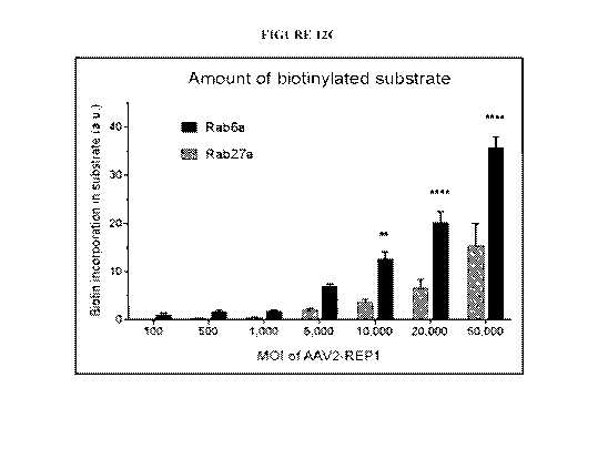
[071] Figure 12A-D is a series of photographs and graphs showing that Rab6a is
more fit
than Rab27a to assess the potency of human REP1 following AAV2 transduction of
293
cells. A) 293 cells were transduced with increasing MO! of AAV2-REP1 (100;
500; 1,000;
5,000; 10,000; 20,000 and 50,000). Protein expression (human REP1 and (3-
actin) and biotin
incorporation were detected in prenylation reaction products (20 jig)
following SDS-PAGE
and western blot analysis (representative image of 3 replicates). B) Nonlinear
regression plot
of normalised REP1 (corrected for the corresponding actin levels) per log
(MOI) of AAV2-
REP1. Data was analysed using a sigmoidal four-parameter fit (95% CT;
constrains: bottom
>0; hill slope =1). Symbols are mean of 6 replicates SEM. C) Plot of the
band density
values obtained for biotinylated Rab27a and Rab6a following transduction of
293 cells with
AAV2-REP1 (n:=3) and corrected for endogenous levels (MOI = 0). Rab6a showed
significantly higher values than Rab27a at MOI 10,000, 20,000 and 50,000 (two-
way
- 14-
CA 03083898 2020-04-17
WO 2019/079-188
PCT/US2018/056336
ANOVA with Bonferroni's multiple comparison test: **p.0042; ****p<0.0001). D)
Densitometry values of biotin incorporation per normalised REP1 were plotted
for both
Rab27a and Rabfia and analysed by linear regression (Rab27a, Y = 6.335*X -
0.6392: Rab6a,
Y = 12.6*X + 0.9576).
10721 Figure 13A-B is a series of photographs showing Rab6a validation as a
substrate for
in vitro prenylation by other cell lines. Protein expression (human REPI and (-
actin) and
biotin incorporation were detected in prenylation reaction products following
cell
transduction, SDS-PAGE and western blot analysis (two replicates in one
experiment). HT-
1080 cells (A) and ARPE-19 cells (B) were transduced with rAAV2/2-REP1 (M01
1,000;
10,000 and 30,000 gc/cell) and prenylation reactions prepared with 20 1.ig and
10 Lig of total
protein, respectively. Positive controls (+ rREP I) were prepared using
untransduced cell
lysate spiked with a recombinant fish REPI protein (25 nM for HT-1080; 11 nM
for ARPE-
19).
(0731 Figure 14A-D is a table (A), 6 photographs (B), 3 graphs (C) and a table
(D)
showing that both RAB27A and RAB6A are subject to prenylation by endogenous
REP1
from a 293 cell lysate. A) Summary table of experimental conditions (#1-#8)
used in
prenylation reactions in vitro regarding the amount of total cell lysate (2,5;
5; 10; 20 rig),
concentration of GGT-II (0,5: 1: 2 pM) and concentration of Rab substrate
(RAB27A or
RAB6A) (0.16; 0.8; 4 M). Positive control (+ve): cell lysate spiked with
recombinant fish
REPI (25 nM). B) Protein expression (human REPI and 0-actin) and biotin
incorporation
detected in prenylation reaction products following SDS-PAGE and western blot
analysis
(representative of 3 independent experiments). C) Plots for condition sets
assessing biotin
incorporation in both RAB27A and RAB6A when different amounts of total cell
lysate,
concentration of GGT-IT or concentration of Rab substrate were used (n=3). D)
Summary
table of statistical analysis performed in the data sets in C). Two-way ANOVA
tests were run
independently for each condition (total cell lysate, concentration of GGT-11
or concentration
of Rab substrate) with 'condition' and 'substrate' as factors. The p values
and the
significance of each test, as well the Bonferroni's multiple comparison test
for comparison of
RAB27A with RAB6A, are given in detail.
[0741 Figure 15A-D is 3 photographs (A) and 3 graphs (B-D) showing that RAB6A
is
more sensitive than RAB27A to assess the biological activity of human REP1
following
rAAV2/2 transduction of 293 cells. A) 293 cells were transduced with
increasing MOT of
- 15-
CA 03083898 2020-04-17
WO 2019/079-188
PCT/US2018/056336
rAAV2/2-REP1 (100: 300: 1,000; 3,000; 10,000; 30,000; 100,000 and 300,000).
Protein
expression (human REP1 and 0-actin) and biotin incorporation were detected in
prenylation
reaction products (20 lig) following SDS-PAGE and western blot analysis
(representative
image of 3 independent experiments). B) Nonlinear regression plot of
normalized REP1
(corrected for the corresponding actin levels) per rAAV2/2-REP1 (log gc/cell).
Data was
analyzed using a sigmoidal four-parameter fit (95% confidence interval;
R2=0.8625).
Symbols are mean of 6 replicates SEM. C) Nonlinear regression plots of
biotin
incorporation per MOI of rAAV2-REP I (log gc/cell). Data was analyzed using a
sigmoidal
four-parameter fit (95% confidence interval; R2=0.8873 for RAB6A; R2=0.8772
for
RAB27A). Symbols are mean of 3 replicates SEM. RAB6A showed statistically
significant
higher incorporation of biotin than RAB27A at MO! 10,000 (**, p=0.0097),
30,000
(***,p).0002) and 100,000 and 300,000 (****, p<0.0001) (two-way ANOVA with
Bonferroni's multiple comparison test). D) Linear regression plots of biotin
incorporation in
substrate, corrected for the untransduced control, against the normalized
overexpressed REP1
for RAB6A (R2=0.8959, Y=18.82*X+0.4803) and RAB27A (R2=0.533,
Y=6.569*X+0.9042).
[075] Figure 16A-B are a graph (A) and a table of rhREP1 calibration standards
(B)
showing an enzyme-linked immunosorbent assay (ELISA) to detect REP1. Plates
were
coated with Rabbit anti-CHM polyclonal antibody (Sigma HPA003231) at 2 mg/inL
and 100
pL per well. The block/wash was done with Superblock from Thermo Fisher
Scientific.
Calibration standards were with rhREP1 (NAC) at 0.5-100 ng/mL in prenylation
buffer
without clithiothreitol (DTT). Detection was with biotinylated mouse
monoclonal 2F1
(Merck) at 0.5 gglmL. Biotinylation was performed using a Miltenyi kit.
Samples of
transduced and non-transduced cell lysates were diluted 1:100 or 1:1000 with
lysis buffer
without DTT.
10761 Figure 17 is a table showing the results of a REP1 potency assay using
an ELISA to
detect REP1. Cells were transduced with the REP1 vector ENG1014A at a
multiplicity of
infection (MOD of 10,000, lysed and REP! was detected using ELISA. Non-trans =
non
transduced control, Trans = transduced cells. Samples were diluted 1:100.
[0771 Figure 18A-C are a graph (A) and a pair of tables (B and C) showing an
exemplary
rAAV2-REP1 potency assay REP1 ELISA. (A) Shows concentration (x-axis) versus
raw data
- 16-
CA 03083898 2020-04-17
WO 2019/079-188
PCT/US2018/056336
(optical density, y-axis). (B) is a table of rhREP1 calibration standards. (C)
is a table showing
a rhREP1 precision profile (n= 10).
[078] Figure 19A-B are a table (A) and diagram (B) showing prenylation
principles and
assays.
[0791 Figure 20 is a table showing assessments by in vitro prenylation assays
in gene
therapy.
[080] Figure 21A-C are a pair of plots (A, C), and a diagram (B) showing a Rab
hierarchy
according to prenylation rate.
[081] Figure 22 is a diagram depicting the detection of a pool of unprenylated
Rabs
(background) and co-staining with Rab27a in an unprenylated pool. WT cells are
depicted on
the left, CHM cells on right. In the wild type cells, unprenylated Rabs are
detected with
biotin. The signal is expected to be low. Detection of Rab27a in the
unprenylated pool is also
expected to be low. In CHM cells, the detection of unprenylated Rabs and
Rab27a in the
unprenylated pool are expected to generate high signal.
[082] Figure 23A-C are a photograph of a Western Blot (A) and a pair of graphs
(B, C)
showing the quantification of band intensity for unprenylated Rabs. In (A)
unprenylated Rabs
are in green, and Rab27A is in red. WT = wild type samples (n = 10), CHM =
choroideremia
samples (n = 12). In (B) the ration of unprenylated Rabs to actin in WT and
CHM samples
was compared using an unpaired t-test (p = 0.0362). In (C), the ratio of
unprenylated Rab27a
to actin in WT and CHM samples was compared using an unpaired t-test (p =
0.0044).
[083] Figure 24 is a table showing assessments by in vitro prenylation assays.
[084] Figure 25 is a series of 3 photographs of Western blots showing
prenylation activity
in rAAV2.REP1 in a test of a 12-well plate for a functional assay. Increasing
MOI of the
AAV2.REP1.ENG1014-A vector are used. Left box: cells were lysed in 40 Lit of
buffer.
Right box: cells were lysed in 50 tiL of buffer. From top to bottom are shown
hREP1 (83
KDa), Actin (42 KDa) and biotinylated Rab6a (24 KDa). Lanes in each box, from
left to
right, are 0 MOI, 300 MO!, 1,000 MO!, 3,000 MO!, 10,000 MO!, 30,000 MO! and 0
MO! +
fish REP1 protein. Protein sizes are indicated from top to bottom, at left, as
100, 75, 48, 35
and 25 KDa.
[0851 Figure 26A-C are three plots depicting prenylation activity in
rAAV2.REP1 in a test
of a 12-well plate for a functional assay. 504 cell lysate generated data
consistent with
previous findings. The test used 15 tig protein per reaction. (A) Normalized
REP1 (a.u.
- 17 -
CA 03083898 2020-04-17
WO 2019/079-188
PCT/US2018/056336
REPI/au. Actin) is shown on the y axis, MOT as log gc/cell rAAV2/2-REP1 on the
x-axis.
Open circles indicate REP1 from cells lysed in 40 p.L of buffer (R2= 0.9845),
black circles
indicate REP1 from cells lysed in 50 Lit of buffer (R2= 0.999). (B) Biotin
incorporation in
substrate (a.u.) is indicated on the y-axis, MO1 as log gc/cell rAAV2/2-REP I
on the x-axis.
Open circles indicate REP1 from cells lysed in 40 pi, of buffer (R2= 0.9997),
black circles
indicate REPI from cells lysed in 50 pi, of buffer (R2= 0.9992). (C) Biotin
incorporation in
substrate (a.u.) corrected for untransduced control is indicated on they-axis,
normalized
overexpressed REPI (a.u. REP1/a.u. actin) is depicted on the x-axis. x's
indicate Rab6a from
cells lysed in 404 of buffer (R2 = 0.8805, Y= 16.2*X-4.066), open circles
indicate Rab6a
from cells lysed in 501A of buffer (R2= 0.9957, Y=16.99*X-2.011). a.u. =
absorbance unit.
[0861 Figure 27 is a graph showing AAV titer as determined by PCR. On the X
axis are
samples at an initial titer of lx1012 Dnase resistant particles (DRP)/mL,
lx1011 DRP/mL and
lx1011 DRP/mL in balanced saline solution (BSS). On the Y axis, is shown titer
measured
after samples were treated as described to the right of the graph.
[087] Figure 28 is a series of 3 photographs of Western blots showing the
prenylation
activity of rAAV2.REP-1 in a compatibility study using AAV2.REP1.ENG1014-A
vector at
a high dose of lx1012 DRP/mL and an MOT of 10,000. From top to bottom are
shown:
hREP1 (83 KDa), Actin (42 KDa) and biotinylated Rab6a (24 KDa). Protein sizes
are
indicated at left, from top to bottom, as 180, 135, 100, 75, 63, 48, 35, 25,
20, 17 and 11 KDa.
Samples, from left to right, in triplicate, are: untransduced control, cells
transduced with
baseline vector, with vector held 6 hours at 4 C, with vector held 6 hours at
4 C and injected
after 180 minutes, with vector held 6 hours at 4 C and 180 minutes in a
syringe, and fish
REPI as a positive control (single sample).
[088] Figure 29 is a series of 3 photographs of Western blots showing the
prenylation
activity of rAAV2.REP-1 in a compatibility study using AAV2.REP1.ENG1014-A
vector at
a low dose of lx1011 DRP/mL and an MO! of 10,000. From top to bottom are
shown: hREP1
(83 KDa), Actin (42 KDa) and biotinylated Rab6a (24 KDa). Protein sizes are
indicated at
left, from top to bottom, as 180, 135, 100, 75, 63, 48, 35, 25, 20, 17 and 11
KDa. Samples,
from left to right, in triplicate, are: untransduced control, cells transduced
with baseline
vector, with vector held 6 hours at 4 C, with vector held 6 hours at 4 C and
injected after 180
minutes, with vector held 6 hours at 4 C and 180 minutes in a syringe, and
fish REPI as a
positive control (single sample).
- 18-
CA 03083898 2020-04-17
WO 2019/079-188
PCT/US2018/056336
[089] Figure 30A-B are a pair of plots showing semi quantification of Western
blots of
prenylation activity of rAAV2.REP-I in a compatibility study using
AAV2.REP1.ENG1014-
A vector. (A) Shows normalized REP!. Band density values (au.) are on the y-
axis and
AAV2-REP1 at a high dose of 1x10¹²DRP/mL and a low dose of lx10n DRP/mL are on
the
x-axis. (B) Shows normalized biotinylated Rab6a. Band density values (a.u.)
are on the y-
axis and AAV2-REP1 at a high dose of 1x10'2 DRP/mL and a low dose of lx1On
DRP/mL
are on the x-axis. In (A) and (B), bars for each dose, from left to right,
indicate untransduced
cells, cells transduced with baseline vector, with vector held 6 hours at 4 C,
+ 6 hours at 4 C
and injected after 180 minutes at 20 C, with vector 6 hours at 4 C and 180
minutes in a
syringe at 20 C.
DETAILED DESCRIPTION
[090] Choroideremia is a rare disease which leads to degeneration of the
choroid, retinal
pigment epithelium and photoreceptors of the eye. Afflicted males typically
exhibit
nightblindness during teenage years, progressive loss of peripheral vision
during the 20's and
30's and complete blindness in the 40's. 'Female carriers may maintain a good
vision
throughout life, but may have mild symptoms, most notably nightblindness, but
may
occasionally have a more severe phenotype.
[0911 Choroideremia is caused by mutations in the CHM gene, which encodes for
Rab
escort protein 1 (REPI). Rab escort protein 2 (REP2), which is 75% homologous
to REP I,
compensates for any REP I deficiency in most cells of the body. However, REP2
is unable to
compensate for REPI deficiency in the eye. This leads to insufficient Rab
escort protein
activity to maintain normal prenylation of target Rab GT'Pases and gives rise
to cellular
dysfunction and ultimately cell death.
[092] Choroideremia may be successfully treated by providing functional copies
of the
REPI transgene to the affected cells of the eye (MacLaren, R.E. et al. (2014)
Lancet 383: 1
129-37). Specifically, it has been shown that adeno-associated virus (AAV)
gene therapy
vectors may be used to deliver a nucleotide sequence encoding functional REP!
to the eye to
treat the disease. As gene therapy of choroideremia is becoming a clinical
reality, there is a
need for reliable and sensitive assays to determine the activity of
exogenously delivered
REP 1. in particular to test new gene therapy vectors and as a quality control
screen for
clinical vector stocks.
- 19-
CA 03083898 2020-04-17
WO 2019/079-188
PCT/US2018/056336
[093] Existing methods for assaying REP1 use Rab27a as a prenylation substrate
(Tolmachova, T. et al. (2012) J. Gene Med. 14: 158-168; Tolmachova, T. et al.
(2013) J. Mol.
Med. 91 : 825-837; Vasireddy, V. et al. (2013) PLoS ONE 8: e61396; and Black,
A. et al.
(2014) J. Gene Med. 16: 122-130). This has likely followed from numerous
implications of
Rab27a in the pathogenesis of choroideremia. For example, it has been shown
that Rab27a is
present unprenylated in choroideremia cells while other Rabs are properly
prenylated
(Seabra, M.C. et al. (1995) J. Biol. Chem. 270: 24420-24427). Furthermore,
Rab27a is
expressed at high levels in the retinal pigment epithelium and
choriocapillaries, the two sites
of earliest degeneration in choroideremia.
[094] However, assays relying on the prenylation of Rab27a give rise to very
weak
signals. As a result, the sensitivity of these assays is low and they may not
be suitable for
reliable screening of clinical gene therapy vectors. Accordingly, a
significant need exists for
more reliable and sensitive assays which can be used to determine REP1
activity and test
gene therapy vectors.
[095] Choroideremia (CHM) is a rare, X-linked recessive retinal dystrophy
caused by
mutations in the CHM gene, which encodes for Rab escort protein 1 (REP1).
Choroideremia
leads to degeneration of the retinal pigment epithelium (RPE) and the
photoreceptors of the
eye. CHM is ubiquitously expressed in human cells and encodes Rab escort
protein 1
(REP1). REP1 involved in the C-terminus posttranscriptional modification of
Rab GTPases,
the largest family within the Ras-like GTPase superfamily. This modification,
known as
prenylation, is catalyzed by the Rab geranylgeranyl transferase (RGGT or (iGT-
II) and
involves the covalent attachment of one or more C20 (geranylgeranyl)
isoprenoid groups to a
cysteine residue within a `prenylation motif'. REP1 assists by either
presenting the
unprenylated Rabs to the GUT-II and/or escorting the prenylated Rabs to their
destination
membrane where they play a role in vesicle trafficking.
10961 The choroideremia-like gene (CHML) encodes for Rab escort protein 2
(REP2).
REP2 shares 95% of its amino acid sequence with REP1, and studies have shown
that REP2
can compensate for REP1 deficiency in most cells of the body. However, REP2 is
unable to
fully compensate for REP1 deficiency in the eye. In choroideremia patients.
the prenylation
of Rab GTPases in the eye is affected, which causes cellular dysfunction and
ultimately cell
death.
- 20 -
CA 03083898 2020-04-17
WO 2019/079-188
PCT/US2018/056336
[097] REP! plays a role in intracellular trafficking through the prenylation
of Rab
GTPases, a reaction that can be reproduced in vitro. Adeno-associated virus
(AAV) gene
replacement therapy is a treatment for choroideremia. Choroideremia may be
treated by
providing functional copies of the CHM gene to the affected cells of the eye.
Specifically, a
recombinant adeno-associated virus (rAAV) vector encoding CHM can be delivered
subretinally. There is therefore a need for an assay to assess the biological
activity of the
vectors for the treatment of choroideremia. For example, there is a need for
reliable and
sensitive in vitro assays to determine the biological activity of rAAV2/2-
REP1. A prenylation
reaction can be reproduced in vitro to test for REP1 biological activity. One
substrate for a
prenylation assay following viral transduction is Rab27a. The Rab27a protein
was first
identified in the cytosolic fraction of CHM lymphoblasts in 1995. Another
substrate for a
prenylation assay in vitro is another Rab protein, RAB6A. The response of
these two Rab
proteins, Rab27A and RAB6A, to the incorporation of a biotinylated lipid donor
in a
prenylation reaction can be assayed in vitro and used to develop robust and
sensitive assays
for assessing the biological activity of AAV vectors for choroideremia.
[0981 Various preferred features and embodiments of the present invention will
now be
described by way of non-limiting examples.
[099] The practice of the present invention will employ, unless otherwise
indicated,
conventional techniques of chemistry, biochemistiy, molecular biology,
microbiology and
immunology, which are within the capabilities of a person of ordinary skill in
the art. Such
techniques are explained in the literature. See, for example, Sambrook, J.,
Fritsch, E.F. and
Maniatis, T. (1989) Molecular Cloning: A Laboratory Manual, 2nd Edition, Cold
Spring
Harbor Laboratoiy Press; Ausubel, F.M. et al. (1995 and periodic supplements)
Current
Protocols in Molecular Biology, Ch. 9, 13 and 16, John Wiley & Sons; Roe, B.,
Crabtree, J.
and Kahn, A. (1996) DNA Isolation and Sequencing: Essential Techniques, John
Wiley &
Sons; Polak, J.M. and McGee, J.O'D. ( 990) In Situ Hybridization: Principles
and Practice,
Oxford University Press; Gait, M.J. (1984) Oligonucleotide Synthesis: A
Practical Approach,
IRL Press; and Lilley, D.M. and Dahlberg, J.E. (1992) Methods in Enzymology:
DNA
Structures Part A: Synthesis and Physical Analysis of DNA, Academic Press.
Each of these
general texts is herein incorporated by reference.
&aviation
- 21 -
CA 03083898 2020-04-17
WO 2019/079-188
PCT/US2018/056336
101001 Previous methods for the detection of small GTPases in vitro used
radiolabelled-
prenyl donors. Radiolabelling can be replaced by either a fluorophore or a
biotin group. Both
approaches involve the use of a cultured cell lysate as REPl is ubiquitously
expressed in all
cells and tissues. Protein incorporation of biotin-containing isoprenoids
(biotin-labelled
geranyl pyrophosphate, B-GPP) can be used to detect prenylated proteins due to
their
superior sensitivity relatively to fluorescence-based methods.
[0101] Lipidation of proteins by the addition of isoprenoid moieties is a post-
translational
modification that affects up to 2% of the mammalian proteome. Such lipidation
enables
reversible association of the target proteins with cell membranes and can also
modulate
protein-protein interactions.
[0102] Preferably, the lipidation referred to herein is prenylation, such that
the lipid donor
substrate and lipidated Rab6a product are a prenyl donor substrate and
prenylated Rab6a
product, respectively.
[0103] Prenylation is a specific type of post-translational modification in
which a
geranylgeranyl or farnesyl moiety (or analogue of either) is attached to one
or two C-terminal
cysteine residues of a protein via a thioether linkage.
[0104] Preferably, the prenylation is the addition of a geranylgeranyl moiety
or an analogue
thereof (e.g. biotin-geranyl moiety) to a target protein (e.g. Rab6a).
[0105] A geranylgeranyl moiety attached to a protein (the protein is depicted
schematically
by the shaded circle) is:
[01061 A farnesyl moiety attached to a protein (the protein is depicted
schematically by the
shaded circle) is:
[0107] The term "analogue" is used herein in relation to the lipid (e.g.
geranylgeranyl or
farnesyl) moiety or lipid donor substrate to refer to a compound which has
been modified to
comprise a functional group suitable for a particular purpose, such as
detection. The analogue
is able to be added to a substrate protein by the prenylation machinery (i.e.
REP! and Rab
GGTase) in a manner substantially unhindered (for the purposes of the activity
assays of the
invention) by the modification.
- 22-
CA 03083898 2020-04-17
WO 2019/079-188
PCT/US2018/056336
[0108] Accordingly, analogues of the above moieties include those which have
been
artificially created for particular purposes (e.g. labelled moieties which are
suitable for
detection in an assay). In particular, Nguyen et al. (Nguyen, U.T. et al.
(2009) Nat. Chem.
Biol. 5: 227-235) developed the following biotin-geranyl moiety that can be
detected in in
vitro protein prenylation reactions (the biotin-geranyl moiety is shown
attached to a protein,
which is depicted schematically by the shaded circle):
II
NNI,,74"
k#\\ \'4A.
Rab6a
[0109] Rab6a (Ras-related protein Rab-6A) is a member of the mammalian Rab
GTPase
family, which is itself the largest of the Ras-like super-family of GTPases.
[0110] Rab GTPases (also known as Rab proteins) are peripheral membrane
proteins and
are involved in the regulation of membrane trafficking, including vesicle
formation, vesicle
movement along actin and tubulin networks, and membrane fusion. The main
function of
Rab6a is understood to be the regulation of protein transport from the Golgi
complex to the
endoplasmic reticulum.
[0111] Rab GTPases are typically anchored to a cell membrane via prenyl groups
(in
particular, geranylgeranyl groups) which are covalently bound to two C-
terminal cls,,steine
residues.
[0112] Rab GTPases exhibit two conformations: an inactive, GDP-bound form; and
an
active, GTP-bound form. Conversion from the GDP- to the GTP-bound forms is
catalyzed by
a GDP/GTP exchange factor (GU), which thereby activates the Rab GTPase.
Conversely,
GTP hydrolysis by Rab GTPases can be enhanced by a GTPase-activating protein
(GAP),
which thereby leads to Rab inactivation.
[0113] In one embodiment, the Rab6a is human Rab6a.
[0114] An example amino acid sequence of Rab6a is the sequence deposited under
NCBI
Accession No. NP 942599.1 (SEQ ID NO: 1).
[0115] An example amino acid sequence of Rab6a is:
MSTGGDFGNP LRKFKLVFLGEQSVGKT SL IT REMYDS FDNTYQATIGI DFLS KTMYLEDRTV
RLQLWDTAGQERFRSL I PS YIRDS TVAVVVYDITNVNS FQQTTKWIDDVRTERGSDVIIMLV
- 23 -
CA 03083898 2020-04-17
WO 2019/079-188
PCT/US2018/056336
GNKT DLADKRQVS I E EGERKAKELNVMFI ET SAKAGYNVKQL FRRVAAAL PGME STQDRS RE
DMIDIKLEKPQEQPVSEGGCSC (SEQ ID NO: 1).
[0116] An example nucleotide sequence encoding Rab6a is the sequence deposited
under
NCBI Accession No. NM_198896.1 (SEQ ID NO: 13).
[0117] An example nucleotide sequence encoding Rab6a is:
ATGTCCACGGGCGGAGACTTCGGGAATCCGCTGAGGAAATTCAAGCTGGTGTTCCTGGGGGA
G CAAAG C GT T GGAAAGACAT CT T T GAT CAC CAGAT T CAT GTAT GACAGT T T T
GACAACAC CT
AT CAG G CAACAAT T G G CAT T GACTT T T TAT CAAAAACTAT G TAC T T G GAGGAT C
GAACAG TA
CGATTGCAAT TAT GG GACACAG CAG G T CAAGAGC G GT T CAG GAG CT T GAT T C CTAG
CTACAT
TCGTGACTCCACTGTGGCAGTTGTTGTTTATGATATCACAAATGTTAACTCATTCCAGCAAA
CTACAAAGT G GAT T GAT GAT GT CAGAACAGAAAGAGGAAGT GAT G T TAT CAT CAT G CTAG TA
GGAAATAAAACAGATCTTGCTGACAAGAGGCAAGTGTCAATTGAGGAGGGAGAGAGGAAAGC
CAAAGAGCTGAATGTTATGTTTATTGAAACTAGTGCAAAAGCTGGATACAATGTAAAGCAGC
TCTTTCGACGTGTAGCAGCAGCTTTGCCGGGAATGGAAAGCACACAGGACAGAAGCAGAGAA
GATAT GAT T GACATAAAACT GGAAAAGCCTCAGGAGCAACCAGT CAGT GAAGGAGGCT GT T C
CTGCTAA (SEQ ID NO: 2).
[0118] A further example nucleotide sequence encoding Rab6a is:
gcacgcacgc acgcacgcca gcggccggcg gggccgcagg ctcgcgcccg ggctcgcccc 60
gcgccgctcc agaggctcgc gcactcagca ggttgggctg cggcggcggc ggcagctgtg 120
gaagctcagg cgctgcgcgt gagaggtccc agatacgtct gcggttccgg ctccgccacc 180
ctcagcttct cttccccagg tctgggagcc gagtgcggaa ggagggaacg gccctagctt 240
tgggaagcca gaggacaccc ctggctcctg ccgacaccgc cctccttccc ttcccagccg 300
cgggcctcgc tcggtgctag gctactctgc cgggaggcgg cggcggctgc cagtctgtgg 360
agagtcctgc tgccctccag ccgggctcct ccaccgggcc ttgcaggggc cgagagagct 420
cggtgcccgc ccttccgctc gcctttttcg tcagctggct ggagcagcat cggtccggga 480
ggtctctagg ctgaggcggc ggccgctcct ctagttccac aatgtccacg ggcggagact 540
tcgggaatcc gctgaggaaa ttcaagctgg tgttcctggg ggagcaaagc gttggaaaga 600
catctttgat caccagattc atgtatgaca gttttgacaa cacctatcag gcaacaattg 660
gcattgactt tttatcaaaa actatgtact tggaggatcg aacagtacga ttgcaattat 720
gggacacagc aggtcaagag cggttcagga gcttgattcc tagctacatt cgtgactcca 780
ctgtggcagt tgttgtttat gatatcacaa atgttaactc attccagcaa actacaaagt 840
ggattgatga tgtcagaaca gaaagaggaa gtgatgttat catcatgcta gtaggaaata 900
aaacagatct tgctgacaag aggcaagtgt caattgagga gggagagagg aaagccaaag 960
agctgaatgt tatgtttatt gaaactagtg caaaagctgg atacaatgta aagcagctct 1020
ttcgacgtgt agcagcagct ttgccgggaa tggaaagcac acaggacaga agcagagaag 1080
atatgattga cataaaactg gaaaagcctc aggagcaacc agtcagtgaa ggaggctgtt 1140
cctgctaatc tcccatgtca tcttcaacct tcttcagaag ctcactgctt tggccccctt 1200
actctttcat tgactgcagt gtgaatattg gcttgaacct tttcccttca gtaataacgt 1260
attgcaattc atcattgctg cctgtctcgt ggagatgatc tattagcttc acaagcacaa 1320
caaaagtcag tgtcttcatt atttatattt tacaaaaagc caaaatattt cagcatattc 1380
cagtgataac tttaaaaatt agatacattt tcttaacatt tttttctttt ttaatgttat 1440
gataatgtac ttcaaaatga tggaaatctc aacagtatga gtatggcttg gttaacgagc 1500
ggtatgttca cagcctactt tatctctcct tgcztttctc acctctcact tacccccatt 1560
ccctattacc ctattcttac ctagcctccc ccgacttcct caaaacaaac aagagatggc 1620
-24-
CA 03083898 2020-04-17
WO 2019/079-188
PCT/US2018/056336
aaagcagcag ttctaccaag cccattggaa ttatccttta attttacaga taccacttgc 1680
tgtaggctac ggaccaagat gtccaaaatt attcttgagc actgatataa attacggtct 1740
tctttgaggt caaaattcag ccatcatggt aggcagtgct tgaatgagaa aaggctcctg 1800
gtgcatcttc aaaatgagtc ctaaagaaca tactgagtac ttagaagtag aagaacataa 1860
gatgtatttc tgactaaaac aaatggctct ttcacatgtg ctttattaga ctctgggaga 1920
gaaaattaac caagtgcttc agaacaggtt tttagtattt aattcttcac ggtaagaaaa 1980
tgaagttcta atgaactgtt tctcccaagg ttttaaaatt gtcaagagtt attctgtttg 2040
tttaaaaaat aagaaacctc tttaagcaat agattttgct tgggttttct tttttaaaaa 2100
cataatactg tgcaggcaag gcactgtaaa agttttaatt ccttccagaa gaaccagtgg 2160
aagaatttaa atttggcgct acgatcaaaa ctactgaatt agtagaaata atgatgtcta 2220
aagcttacca acaaaagaac cctcagcaga ataacaaaaa ctttgctcag gacatttgag 2280
gtcaaattga agacggaaac cggaaaccgt tttcttgtaa gcccctagag gcagatcagg 2340
taaagcatac atagtagagg gaaaggagag aatggaaata aaactcaata ttatgcagat 2400
ttatgcctta ttttttagca ttttttaagg ttgggtcttt caggctggtt ttggtttgta 2460
ttagatctgt atagtttaat taactggtga tttagtttta tatttaagct acaattaatc 2520
ttttttcttt ggtgatattt atttctttgc cttttttttt tttaacaact ttcaatcttc 2580
agatgtttcg ttgaatctat ttagagcttc accatggcaa tatgtatttc ccttaaaaca 2640
ctgcaaacaa atatactagg agtgtgccct tttaatcttt actagttatt gtgagattgc 2700
tgtgtaagct aataaacaca tttgtaaata cattgtttgc aggacgaaaa cttctgagtt 2760
acagctcagg aaaagcctgc tgaatttatg ttgtaagcat tacttaacac agtataaaga 2820
tgaaaagaca acaaaaatat cttcatactt cctcatcccc tcattggaac aaaaccttaa 2880
actgggagaa ccttagtccc ctctctttcc tcttcctcct ccacttccca cttattgtca 2940
ccttgtaata ttcagagagc acttggatta tggatctgaa tagagaaatg cttacagata 3000
atcattagcc cacataccag taacttatac ttaaagatgg gatggagttg taaagtgctt 3060
ttataataca atataattgt taaaggcaag ggttgactct ttgttttatt ttgacatggc 3120
atgtcctgaa ataaatattg attcaatatg gcagatgggt catattcttt atttggaaga 3180
agttgtgact tctgacatgg gtgtgattgt cttcctacac tgttgcattt gattcttttt 3240
atgtattttt aagaaagtaa ccagttatac tgcttttaat attgattggt ctttttattt 3300
ggcttggagt tcttcaaagc attgaagtgt gttcatagtc caggtttttt ttttaataaa 3360
cacaattttg ctgccaaaaa tatataaata aaacacgaaa gaaaacaaaa aaaaaaaaa (SEQ ID
NO: 13).
[0119] An example amino acid sequence of Rab27a is:
1 MSDGDYDYLI KFLALGDSGV GKTSVLYQYT DGKFNSKFIT TVGIDFREKR VVYRASGPDG
61 ATGRGQRIHL QLWDTAGQER FRSLTTAFFR DAMGFLLLFD LTNEQSFLNV RNWISQLQMH
121 AYCENPDIVL CGNKSDLEW RVVKEEEAIA LAEKYGIPYF ETSAANGTNI SQAIEMLLDL
181 IMKRMERCVD KSWIPEGVVR SNGHASTDQL SEEKEKGACG C (SEQ ID NO: 17).
Rah geranylgeranyltransferase (Rab GGTase)
[01201 Rab geranylgeranyltransferase (Rab GGTase; also known as
geranylgeranyltransferase II) is a protein prenyltransferase which exclusively
prenylates the
GTPases of the Rab family.
[01211 Rab GGTase typically naturally catalyzes the transfer of two
geranylgeranyl groups
to cysteine residues at the C-terminus of Rab GTPases. Each geranylgeranyl
group is
conjugated to the Rab GTPase via a thioether linkage to a gsteine residue.
[01221 Rab GGTase has been shown to be capable of binding a range of
derivatized
phosphoisoprenoids and can catalyze their addition to Rab GTPase substrates
(e.g. Rab6a).
- 25 -
CA 03083898 2020-04-17
WO 2019/079-188
PCT/US2018/056336
For example, Nguyen et M. (Nguyen, U.T. et al. (2009) Nat. Chem. Biol. 5: 227-
235)
demonstrated the successful addition of a biotin-geranyl moiety to Rab
GTPases.
101231 Rab GGTase is a heterodimeric enzyme comprised of alpha and beta
subunits.
101241 In one embodiment, the Rab GGTase is human Rab GGTase. In a preferred
embodiment, the Rab GGTase is rat Rab GGTase.
[0125] Example amino acid sequences of Rab GGTase alpha subunits are the
sequences
deposited under NCBI Accession Nos. NP_004572.3 (SEQ ID NO: 10) and
NP_113842.1
(SEQ ID NO: 11).
[0126] Example amino acid sequences of Rab GGTase alpha subunits are:
MHGRLKVKTSEEQAEAKRLEREQKLKLYQSATQAVFQKRQAGEL DESVLELTSQILGANPDF
ATLWNCRREVLQQLETQKS PEELAALVKAELGFLESCLRVNPKS YGTWHHRCLLGRL PE PNW
TRELELCARFLEVDERNFHCWDYRRFVAT QAAVP PAEELAFT DS L ITRNFSNYS SWHYRSCL
LPQLHPQPDSGPQGRLPEDVLLKELELVQNAFFTDPNDQSAWFYHRWLLGRADPQDALRCLH
VS RDEACLTVS FS RPLLVGS RME I LLLMVDDS PLIVEWRT PDGRNRPS HVWLCDLPAASLND
QLPQHT FRVIWTAGDVQKECVLLKGRQEGWCRDSTTDEQL FRCELSVEKSTVLQSELESCKE
LQELEPENKWCLL I I LLMRALDPLLYEKETLQY FQTLKAVDPMRATYL DDLRSKFLLENSVL
KMEYAEVRVLHLAHKDLTVLCHLEQLLLVTHLDLSHNRLRTL PPALAALRCLEVLQAS DNAI
ESLDGVTNLPRLQELLLCNNRLQQPAVLQPLASCPRLVLLNLQGNPLCQAVGILEQLAELL P
SVSSVLT (SEQ ID NO: 3)
and:
MHGRLKVKTSEEQAEAKRLEREQKLKLYQSATQAVFQKRQAGELDESVLELTSQILGANPDF
ATLWNCRREVLQHLETEKS PEESAALVKAELGFLESCLRVN PKS Y GTHHRCWLL SRL PEPNW
ARELELCARFLEADERNFHCWDYRRFVAAQAAVAPAEELAFT DS L ITRNFSNY S SHYRSCLL
PQLHPQPDSGPQGRL PENVLLKELELVQNAFFTDPNDQSAWFYHRLLGRAEPHDVLCCVHVS
REEACL SVC FS RPLTVGS RMGTLLLMVDEAPLSVEWRT PDGRNRPSHVWLCDLPAASLNDQL
PQHT FRVIWT GS DS QKECVLLKDRPECWC RDSAT DEQLFRCELSVEKSTVLQSELESCKELQ
ELEPENWCLLT I I LLMRALD PLLYEKET LQYFST LKAVD PMRAAY LDDL RS KFLLENSVLKM
EYADVRVLHLAHKDLTVLCHLEQLLLVTHLDLSHNRLRALP PALAAL RC LEVLQAS DNALEN
VDGVANLPRLQELLLCNNRLQQSAAIQPLVSCPRLVLLNLQGNSLCQEEGIQERLAEMLPSV
SSILT (SEQ ID NO: 8)
and:
- 26 -
CA 03083898 2020-04-17
WO 2019/079488
PCT/US2018/056336
MHGRLKVKTSEEQAEAKRLEREQKLKLYQSATQAVFQKRQAGELDESVLELTSQILGANPDF
ATLWNCRREVLQQLETQKSPEELAArVKAELGFLESCLRVNPKSYGTWHHRCWLLGRLPEPN
WTRELELCARFLEVDERNFHCWDYRRFVATQAAVPPAEELAFTDSLITRNFSNYSSWHYRSC
LLPQLHPQPDSGPQGRLPEDVLLKELELVQNAFFTDPNDQSAWFYHRWLLGRADPQDALRCL
HVSRDEACLTVSFSRPLLVGSRMEILLLMVDDSPLIVEWRTPDGRNRPSHVWLCDLPAASLN
DQLPQHTFRVIWTAGDVQKECVILKGRQEGWCRDSTTDEQLFRCELSVEKSTVLQSELESCK
ELQELEPENKWCLLTIILLMRALDPLLYEKETLQYFQTLKAVDPMRATYLDDLRSKFLLENS
VLKMEYAEVRVLHLAHKDLTVLCHLEQLLLVTHLDLSHNRLRTLPPALAALRCLEVLQASDN
AIESLDGVTNLPRLQELLLCNNRLQQPAVLQPLASCPRLVLLNLQGNPLCQAVGILEQLAEL
LPSVSSVLT (SEQ ID NO: 10)
MHGRLKVKTSEEQAEAKRLEREQKLKLYQSATQAVFQKRQAGELDESVIELTSQILGANPDF
ATLWNCRREVLQHLETEKSPEESAALVKAELGFLESCLRVNPKSYGTWHHROWLLSRLPEPN
WARELELCARFLEADERNFHCWDYRRFVAAQAKVAPAEELAFTDSLITRNFSNYSSWHYRSC
LLPQLHPQPDSGPQGRLPENVILKELErVQNAFFTDPNDQSAWFYHRWLLGRAEPHUVLCCV
HVSREEACLSVCFSRPLTVGSRMGTLLLMVDEAPLSVEWRTPDGRNRPSHVWLCDLPAASLN
DQLPQHTFRVIWTGSDSQKECVILKDRPECWCRDSATDEQLFRCELgVEKSTVIQSELESCK
ELQELEPENKWCLLTIILLMRALDPLLYEKETLQYFSTLKAVDPMRAAYLDDLRSKFLLENS
VLKMEYADVRVIHLAHKDLTVICHLEQLLLVTHLDLSHNRLRALPPALAALRCLEVIQASDN
ALENVDGVANLPRLQELLLCNNRLQQSAAIQPrVSCPRLVILNLQGNSLCQEEGIQERLAEM
LPSVSSILT (SEQ ID NO: 11).
[0127] Example amino acid sequences of Rab GGTase beta subunits are the
sequences
deposited under NCBI Accession Nos. NP_004573.2 (SEQ ID NO: 4) and NP_619715.1
(SEQ ID NO: 12).
[0128] Example amino acid sequences of Rab GGTase beta subunits are:
MGTPQKUVIIKSDAPDTLLLEKHADYIASYGSKKDDYEYCMSEYLRMSGIYWGLTVMDLMGQ
LHRMNREEILAFIKSCQHECGGISASIGHDPHLLYTLSAVQILTLYDSINVIDVNKVVEYVK
GLQKEDGSFAGDIWGEIDTRFSFCAVATLALLGKLDAINVEKAIEFVLSCMNFDGGFGCRPG
SESHAGQIYCCTGFLAITSQLHQVNSDLLGWWLCERQLPSGGLNGRPEKLPDVCYSWWVIAS
LKIIGRLHWIDREKLRNFILACQDEETGGFADRPGDMVDPFHTLFGIAGLSLLGEEQIKPVN
FVFCMPEEVLORVNVQPELVS (SEQIDNO:4)
-27-
CA 03083898 2020-04-17
WO 2019/079488
PCT/US2018/056336
MGTQQKUVTIKSDAPDTLLLEKHADYIASYGSKKDDYEYCMSEYLRMSGVYWGLIVMDINGQ
LHRMNKEEILVFIKSCQHECGGVSASIGHDPHLLYTLSAVQILTLYDSIHVINVDKVVAYVQ
SLQEDGSFAGDIGEIDTRFSFCAVATLALLGKLDAINVEKAIEFVLSCMNFDGGFGCRPGSE
SHAGQIYCCTGFLAITSQLHQVNSDLLGWWLCERQLPSGGLNGRPEKLPDVCYSWWVLASLK
IIGRLHIDREKLRSFILACQDEETGGFADRPGDMVDPFHTLFGIAGLSLLGEEQIKPVSFVF
CMPEEVLQRVNVQPEINS (SEQII)N119)
and:
MGTQQKDVTIKSDAPDTLLLEKHADYIASYGSKKDDYEYCMSEYLRMSGVYWGLTVMDLMGQ
LHRMNKEEILVFIKSCQHECGGVSASIGHDPHLLYTLSAVQILTLYDSIHVINVDKVVAYVQ
SLQKEDGSFAGDIWGEIDTRFSFCAVATLALLGKLDAINVEKAIEFVLSCMNFDGGFGCRPG
SESHAGQIYCCTGFLAITSQLHQVNSDLLGWWLCERQLPSGGLNGRPEKLPDVCYSWWV1AS
LKIIGRLHWIDREKLRSFILACQDEETGGFADRPGDMVDPFHTLFGIAGLSLLGEEQIKPVS
PVFCMPEEVLQRVNVQPELVS (SEQ ID NO: 12).
Lipid donor substrate
[0129] To add a lipid moiety to a Rab GTPase, the Rab GGTase may use the lipid
moiety in
the form of a lipid (e.g. geranyigeranyl or biotin-geranyl) donor substrate as
a substrate.
These are typically pyrophosphate derivatives of the lipid moiety.
[0130] For example, geranylgeranylpyrophosphate (GGPP) or biotin-
geran:klpyrophosphate
(BGPP) may be used as lipid donor substrates by Rab GGTase to transfer a
geranyigeranyl or
biotin-geranyl moiety, respectively, to the substrate Rab GTPase.
[0131] Geranylgeranylpyrophosphate has the structure:
9 9.L
= 0 0
[0132] An example structure of biotin-geranylpyrophosphate is:
nulmi
9 9
µ)--S
or 0'
0
Rah escort protein 1 (REP 1)
-28-
CA 03083898 2020-04-17
WO 2019/079-188
PCT/US2018/056336
[0133] Rab escort proteins (REPs) perform the functions of presenting
unprenylated Rab
GTPases to Rab GGTases, and carrying prenylated Rab GTPases to their target
membranes.
[01341 Rob GTPases do not comprise a consensus sequence at the prenylation
site that may
be recognized by Rab GGTases. However, substrate recognition is effected
through REPs,
which bind Rab GTPases through a conserved region and then present the Rab
GGTase with
its substrate for prenylation.
[0135] Once prenylated, the lipid anchors render the Rab GTPases insoluble.
Accordingly,
REPs are required to bind and solubilize the geranyigeranyl groups and aid
delivery of the
Rab GTPase to the target cell membrane.
[0136] REP1 may also be known as Rab protein geranylgeranyltransferase
component A
Furthermore, the gene that encodes REP! may be known as the CHM gene
[0137] In one embodiment, the REP1 is human REP1.
10138] An example amino acid sequence of REP! is:
MADTL PS EFDVIVIGTGLPES I I AAACS RSGRRVLHVDS RS YYGGNWAS FS FSGLLSWLKEY
QENS D I VS DS PVWQDQI LENEEAIALSRKDT I QHVEVFCYASQDL HE DVEEAGALQKNHALV
T SANST EAADSAFL PTEDES LSTMS CEMLT EQT PS SDPENALEVNGAEVTGEKENHCDDKTC
VPST SAEDMS ENVP IAEDTT EQPKKNRIT YSQI I EGRRFNI DLVSKLLYSRGLL I DLL IKSN
VSRYAEF NIT RILAFREGRVEQVPCSRADVFNS KQLTMVEKRMLMKFLT FCMEYEKY PDEY
GYEEIT FYEYLKTQKLTPNLQYIVMHS IAMT S ETAS ST I DGLKATKNFLHCLGRYGNT P FL F
PLYGQGELPQCFCRMCAVFGGIYCLRHSVQCLVVDKESRKCKAI I DQFGQRI I S EHFLVEDS
YFPENMCSRVQYRQISRAVL IT DRSVLT DS DQQI S ILTVPAEEPGT FAVRVI ELCS STMT CM
KGTYLVHLTCT S S KTAREDLESVVQKL FVPYT EME I ENEQVEKPRI LWALY FNMRDS S DI S R
SCYNDL PSNVYVCSGPDCGLGNDNAVKQAETL FQE I CPNEDFC P P P PNPEDI I L DGDS LQPE
ASES SAI PEANS ET FKESTNLGNLE ES SE (SEQ ID NO: 5).
[0139] An example amino acid sequence of REP1 is:
MADTLPSEFDVIVIGTGLPES I IAAACSRSGRRVLHVDS RS YYGGNWAS FS FSGLLSWLKEY
QENSDIVS DS PVWQDQI LENEEAIAL S RKDKT I QHVEVFCYAS Q DLHE DVEEAGALQKNHAL
VT SANS TEAADSAFL PTEDESLSTMSCEMLT EQT PSSDPENALEVNGAEVTGEKENHCDDKT
CVPSTSAEDMSENVPIAEDTTEQPKKNRITYSQIIKEGRRFNIDLVSKLLYSRGLLIDLLIK
SNVSRYAEFKNITRILAFREGRVEQVPCSRADVFNSKQLTMVEKRMLMKFLTFCMEYEKYPD
EYKGYEEITFYEYLKTQKLTPNLQYIVMHSIAMTSETASSTIDGLKATKNFLHCLGRYGNTP
FLFPLYGQGELPQCFCRMCAVFGGIYCLRHSVQCLVVDKESRKCKAIIDQFGQRIISEHFLV
-29-
CA 03083898 2020-04-17
WO 2019/079-188
PCT/US2018/056336
EDS Y FPENMC S RVQY RQI SRAVL IT DRSVLKT DS DQQI S ILTVPAEEPGT FAVRVIELCS ST
MT CMKGT YLVHLTCT SSKTAREDLESVVQKLFVPYTEME I ENEQVEKPRI LWALY FNMRDS S
DI SRSCYNDL PSNVYVCSGPDCGLGNDNAVKQAETLFQE IC PNEDFC P PPPNPEDI I L DGDS
LQPEASESSAIPEANSETFKESTNLGNLEESSE ( SEQ ID NO: 1 4 ) .
101401 An example nucleotide sequence encoding REP1 is:
AT GGCGGATACTCTCCCTTCGGAGTTTGATGTGAT CGTAATAGGGACGGGTTTGCCTGAATC
CATCATTGCAGCTGCATGTT CAAGAAGTGGCCGGAGAGTTCTGCATGTTGATTCAAGAAGCT
ACTATGGAGGAAACT GGGCCAGTTTTAGCTTTTCAGGACTATTGTCCT GGCTAAAGGAATAC
CAGGAAAACAGTGACATTGTAAGTGACAGTCCAGTGTGGCAAGCCGATCCTTGAAAATGAAG
AGCCATTGCTCTTAGCAGGAAGGACAAAACATTCAACATGTGGAAGTATTTTGTTATGCCAG
TCAGGATTTGCAT GAAGATGTCGAAGAAGCTGGTGCACT GCAGAAAAAT CATGCTCTT GT GA
CATCTGCAAACTCCACAGAAGCTGCAGATTCTGCCTTCCTGCCTACGGAGGATGAGTCATTA
AGCACTAT GAGCT GT GAAAT GCT CACAGAACAAACT CCAAG CAGCGAT CCAGAGAAT G CG CT
AGAAGTAAATGGTGCTGAAGTGACAGGGGAAAAAGAAAACCATTGTGATGATAAAACTTGTG
TGCCAT CAAC T T CAGCAGAAGACAT GAGT GAAAAT GT GC CTATAG CAGAAGATACCACAGAG
CAACCAAAGAAAAACAGAAT TACT T ACT CACAAAT TAT TAAAGAAGG CAGGAGAT T TAATAT
TGATTTAGTATCAAAGCTGCTGTATTCTC GAGGATTACTAATTGATCTTCTAAT CAAATCTA
AT GTTAGTCGATATGCAGAGTTTAAAAATAT TACCAGGATTCTT GCATTTCGAGAAGGCGAG
TGGAACAGGTTCCGT GTTCCGGCGATGTCTTTAATAGCAAACAACTTACTATGGTAGAAAAG
CGAAT GCTAAT GAAATTT CT TACAT TTT GTAT GGAATAT GAGAAATAT CCT GAT GAATAT
AAAGGATATGAAGAGATCACATTTTTGAATTTTAAAGACTCAAAAATTAACCCCCAACCTCC
AATATAT T GT CAT GCAT T CAAT T G CAAT GACAT CAGAGACAGCCAGCAGCACCATAGAT G GT
CT CAAAGCTACCAAAAACTTTCTTCACTGTCTTGGGCGGTATGGCAACACTCCATTTTTGTT
TCCTTTATATGGCCAAGGAGAACTCCCCCAGTGTTTCTGCAGGATGTGTGCTGTGTTTGGTG
GAATTTATTGTCTTCGCCATTCAGTACAGTGCCTT GTAGTGGACAAAGAATCCAGAAAATGT
AAAGCAATTATAGATCAGTTTGGTCAGAGAATAATCTCTGAGCATTTCCTCGTGGAGGACAG
TTACTTTCCTGAGAACATGTGCTCACGTGTGCAATACAGGCAGATCTCCAGGGCAGTGCTGA
TTACAGAAGATCTGTCCTAAAAACAGATTCAGATCAACGATTTCCTTTTGACAGTGCCAGCA
GAGGAACCAGGAACTTTTGCTGTTCGGGTCATTGAGTTATGTTCTTCAACGATGACATGCAT
GAAAGGCACCTATTTGGTTCATTTGACTTGCACATCTTCTAAAACAGCAAGAGAAGATTTAG
AAT CAGT T GT GCAGAAAT T GT T T GT T C CATATACT GAAAT GGAGAT AGAAAAT GAACAAGT
A
GAAAAGCCAAGAATT CTGTGGGCTCTTTACTTCAATATGAGAGATTCGTCAGACATCAGCAG
-30-
CA 03083898 2020-04-17
WO 2019/079-188
PCT/US2018/056336
GAGCTGTTATAATGATTTACCATCCAACGTTTATGTCTGCTCTGGCCCAGATTGTGGTTTAG
GAAATGATAATGCAGTCAAACAGGCTGAAACACTTTTCCAGGAAATCTGCCCCAATGAAGAT
TTCTGTCCCCCTCCCCAAATCCTGAAGACATTATCCTTGATGGAGACAGTTTACAGCCAGAG
GCTTCAGAATCCAGTGCCATACCAGAGGCTAACTCGGAGACTTTCAAGGAAAGCACAAACCT
TGGAAACCTAGAGGAGTCCTCTGAAAA (SEQ ID NO: 6)
[0141] A further example nucleotide sequence encoding REP1 is:
GATATCGAATTCCTGCAGCCCGGCGGCACCATGGCGGATACTCTCCCTTCGGAGTTTGATGT
GATCGTAATAGGGACGGGTTTGCCTGAATCCATCATTGCAGCTGCATGTTCAAGAAGTGGCC
GGAGAGTTCTGCATGTTGATTCAAGAAGCTACTATGGAGGAAACTGGGCCAGTTTTAGCTTT
TCAGGACTATTGTCCTGGCTAAAGGAATACCAGGAAAACAGTGACATTGTAAGTGACAGTCC
AGTGTGGCAAGACCAGATCCTTGAAAATGAAGAAGCCATTGCTCTTAGCAGGAAGGACAAAA
CTATTCAACATGTGGAAGTATTTTGTTATGCCAGTCAGGATTTGCATGAAGATGTCGAAGAA
GCTGGTGCACTGCAGAAAAATCATGCTCTTGTGACATCTGCAAACTCCACAGAAGCTGCAGA
TTCTGCCTTCCTGCCTACGGAGGATGAGTCATTAAGCACTATGAGCTGTGAAATGCTCACAG
AACAAACTCCAAGCAGCGATCCAGAGAATGCGCTAGAAGTAAATGGTGCTGAAGTGACAGGG
GAAAAAGAAAACCATTGTGATGATAAAACTTGTGTGCCATCAACTTCAGCAGAAGACATGAG
TGAAAATGTGCCTATAGCAGAAGATACCACAGAGCAACCAAAGAAAAACGAATTACTTACTC
ACAAATATTAAGAAGGCAGGAGATTAATATTGATTTAGTATCAAAGCTGCTGTATTCTCGAG
GATTACTAATTGATCTTCTAATCAAATCTAATGTTAGTCGATATGCAGAGTTTAAAAATATT
ACCAGGATTCTTGCATTTCGAGAAGGACGAGTGGAACAGGTTCCGTGTTCCAGAGCAGATGT
CTTTAATAGCAAACAACTTACTATGGTAGAAAAGCGAATGCTAATGAAATTTCTTACATTTT
GTATGGAATATGAGAAATATCCTGATGAATATAAAGGATATGAAGAGATCACATTTTATGAA
TATTTAAAGACTCAAAAATTAACCCCCAACCTCCAATATATTGTCATGCATTCAATTGCAAT
GACATCAGAGACAGCCAGCAGCACCATAGATGGTCTCAAAGCTACCAAAAACTTTCTTCACT
GTCTTGGGCGGTATGGCAACACTCCATTTTTGTTTCCTTTATATGGCCAAGGAGAACTCCCC
CAGTGTTTCTGCAGGATGTGTGCTGTGTTTGGTGGAATTTATTGTCTTCGCCATTCAGTACA
GTGCCTTGTAGTGGACAAAGAATCCAGAAAATGTAAAGCAATTATAGATCAGTTTGGTCAGA
GAATAATCTCTGAGCATTTCCTCGTGGAGGACAGTTACTTTCCTGAGAACATGTGCTCACGT
GTGCAATACAGGCAGATCTCCAGGGCAGTGCTGATTACAGATAGATCTGTCCTAAAAACAGA
TTCAGATCAACAGATTTCCATTTTGACAGTGCCAGCAGAGGAACCAGGAACTTTTGCTGTTC
GGGTCATTGAGTTATGTTCTTCAACGATGACATGCTGAAAGGCACCTATTTGGTTCATTTGA
CTTGCACATCTTCTAAAACAGCAAGAGAAGATTTAGAATCAGTTGTGCAGAAATTGTTTGTT
-31-
CA 03083898 2020-04-17
WO 2019/079-188
PCT/US2018/056336
CCATATACTGAAATGGAGATAGAAAATGAACAAGTAGAAAAGCCAAGAATTCTGTGGGCTCT
TTACTTCAATATGAGAGATTCGTCAGACATCAGCAGGAGCTGTTATAATGATTTACCATCCA
ACGTTTATGTCTGCTCTGGCCCAGATTGTGGTTTAGGAAATGATAATGCAGTCAAACAGGCT
GAAACACTTTTCCAGGAAATCTGCCCCAATGAAGATTTCTGTCCCCCTCCACCAAATCCTGA
AGACATTATCCTTGATGGAGACAGTTTACAGCCAGAGGCTTCAGAATCCAGTGCCATACCAG
AGGCTAACTCGGAGACTTTCAGGAAAGCACAAACCTTGGAAACCTAGAGGAGTCCTCTGAA
AA (SEQ ID NO: 7)
[0142] A further example nucleotide sequence encoding REP1 is:
AT GGCGGATACT CT CCCTT CGGAGT TT GAT GT GAT CGT AATAGGGACGGGTTT GCCT GAATC
CATCATTGCAGCTGCATGTTCAAGAAGTGGCCGGAGAGTTCTGCATGTTGATTCAAGAAGCT
ACTATGGAGGAAACTGGGCCAGTTTTAGCTTTTCAGGACTATTGTCCTGGCTAAAGGAATAC
CAGGAAAACAGTGACATTGTAAGTGACAGTCCAGTGTGGCAAGACCAGATCCTTGAAAATGA
AGAAGCCATTGCTCTTAGCAGGAAGGACAAAACTATTCAACATGTGGAAGTATTTTGTTATG
CCAGT CAG GATTT GCAT GAAGAT GT CGAAGAAGCT GGT GCACT GCAGAAAAAT CAT GCT CTT
GTGACATCTGCAAACTCCACAGAAGCTGCAGATTCTGCCTTCCTGCCTACGGAGGATGAGTC
ATTAAGCACTATGAGCTGTGAAATGCTCACAGAACAAACTCCAAGCAGCGATCCAGAGAATG
CGCTAGAAGTAAATGGTGCTGAAGTGACAGGGGAAAAAGAAAACCATTGTGATGATAAAACT
TGTGTGCCATCAACTTCAGCAGAAGACATGAGTGAAAATGTGCCTATAGCAGAAGATACCAC
AGAGCAACCAAAGAAAAACAGAATTACTTACTCACAAATTATTAAAGAAGGCAGGAGATTTA
ATATTGATTTAGTATCAAAGCTGCTGTATTCTCGAGGATTACTAATTGATCTTCTAATCAAA
TCTAATGTTAGTCGATATGCAGAGTTTAAAAATATTACCAGGATTCTTGCATTTCGAGAAGG
ACGAGTGGAACAGGTTCCGTGTTCCAGAGCAGATGTCTTTAATAGCAAACAACTTACTATGG
TAGAAAAGCGAATGCTAATGAAATTTCTTACATTTTGTATGGAATATGAGAAATATCCTGAT
GAATATAAAGGATATGAAGAGATCACATTTTATGAATATTTAAAGACTCAAAAATTAACCCC
CAACCTCCAATATATTGTCATGCATTCAATTGCAATGACATCAGAGACAGCCAGCAGCACCA
TAGATGGTCTCAAAGCTACCAAAAACTTTCTTCACTGTCTTGGGCGGTATGGCAACACTCCA
TTTTTGTTTCCTTTATATGGCCAAGGAGAACTCCCCCAGTGTTTCTGCAGGATGTGTGCTGT
GT TT GGT GGAATTTATT GT CTT CGCCATT CAGTACAGT GCCTT GTAGT GGACAAAGAATCCA
GAAAATGTAAAGCAATTATAGATCAGTTTGGTCAGAGAATAATCTCTGAGCATTTCCTCGTG
GAGGACAGTTACTTTCCTGAGAACATGTGCTCACGTGTGCAATACAGGCAGATCTCCAGGGC
AGTGCT GATTACAGATAGAT CT GT CCTAAAAACAGATT CAGAT CAACAGATTT CCAT TTT GA
CAGTGCCAGCAGAGGAACCAGGAACTTTTGCTGTTCGGGTCATTGAGTTATGTTCTTCAACG
-32-
CA 03083898 2020-04-17
WO 2019/079-188
PCT/US2018/056336
ATGACATGCATGAAAGGCACCTATTTGGTTCATTTGACTTGCACATCTTCTAAAACAGCAAG
AGAAGATTTAGAATCAGTTGTGCAGAAATTGTTTGTTCCATATACTGAAATGGAGATAGAAA
ATGAACAAGTAGAAAAGCCAAGAATTCTGTGGGCTCTTTACTTCAATATGAGAGATTCGTCA
GACATCAGCAGGAGCTGTTATAATGATTTACCATCCAACGTTTATGTCTGCTCTGGCCCAGA
TTGTGGTTTAGGAAATGATAATGCAGTCAAACAGGCTGAAACACTTTTCCAGGAAATCTGCC
CCAATGAAGATTTCTGTCCCCCTCCACCAAATCCTGAAGACATTATCCTTGATGGAGACAGT
TTACAGCCAGAGGCTTCAGAATCCAGTGCCATACCAGAGGCTAACTCGGAGACTTTCAAGGA
AAGCACAAACCTTGGAAACCTAGAGGAGTCCTCTGAATAA (SEQ ID NO: 15).
[0143] A further example nucleotide sequence encoding REP! is:
GATATCGAATTCCTGCAGCCCGGCGGCACCATGGCGGATACTCTCCCTTCGGAGTTTGATGT
GATCGTAATAGGGACGGGTTTGCCTGAATCCATCATTGCAGCTGCATGTTCAAGAAGTGGCC
GGAGAGTTCTGCATGTTGATTCAAGAAGCTACTATGGAGGAAACTGGGCCAGTTTTAGCTTT
TCAGGACTATTGTCCTGGCTAAAGGAATACCAGGAAAACAGTGACATTGTAAGTGACAGTCC
AGTGTGGCAAGACCAGATCCTTGAAAATGAAGAAGCCATTGCTCTTAGCAGGAAGGACAAAA
CTATTCAACATGTGGAAGTATTTTGTTATGCCAGTCAGGATTTGCATGAAGATGTCGAAGAA
GCTGGTGCACTGCAGAAAAATCATGCTCTTGTGACATCTGCAAACTCCACAGAAGCTGCAGA
TTCTGCCTTCCTGCCTACGGAGGATGAGTCATTAAGCACTATGAGCTGTGAAATGCTCACAG
AACAAACTCCAAGCAGCGATCCAGAGAATGCGCTAGAAGTAAATGGTGCTGAAGTGACAGGG
GAAAAAGAAAACCATTGTGATGATAAAACTTGTGTGCCATCAACTTCAGCAGAAGACATGAG
TGAAAATGTGCCTATAGCAGAAGATACCACAGAGCAACCAAAGAAAAACAGAATTACTTACT
CACAAATTATTAAAGAAGGCAGGAGATTTAATATTGATTTAGTATCAAAGCTGCTGTATTCT
CGAGGATTACTAATTGATCTTCTAATCAAATCTAATGTTAGTCGATATGCAGAGTTTAAAAA
TATTACCAGGATTCTTGCATTTCGAGAAGGACGAGTGGAACAGGTTCCGTGTTCCAGAGCAG
ATGTCTTTAATAGCAAACAACTTACTATGGTAGAAAAGCGAATGCTAATGAAATTTCTTACA
TTTTGTATGGAATATGAGAAATATCCTGATGAATATAAAGGATATGAAGAGATCACATTTTA
TGAATATTTAAAGACTCAAAAATTAACCCCCAACCTCCAATATATTGTCATGCATTCAATTG
CAATGACATCAGAGACAGCCAGCAGCACCATAGATGGTCTCAAAGCTACCAAAAACTTTCTT
CACTGTCTTGGGCGGTATGGCAACACTCCATTTTTGTTTCCTTTATATGGCCAAGGAGAACT
CCCCCAGTGTTTCTGCAGGATGTGTGCTGTGTTTGGTGGAATTTATTGTCTTCGCCATTCAG
TACAGTGCCTTGTAGTGGACAAAGAATCCAGAAAATGTAAAGCAATTATAGATCAGTTTGGT
CAGAGAATAATCTCTGAGCATTTCCTCGTGGAGGACAGTTACTTTCCTGAGAACATGTGCTC
ACGTGTGCAATACAGGCAGATCTCCAGGGCAGTGCTGATTACAGATAGATCTGTCCTAAAAA
-33-
CA 03083898 2020-04-17
WO 2019/079488
PCT/US2018/056336
CAGATTCAGATCAACAGATTTCCATTTTGACAGTGCCAGCAGAGGAACCAGGAACTTTTGCT
GTTCGGGTCATTGAGTTATGTTCTTCAACGATGACATGCATGAAAGGCACCTATTTGGTTCA
TTTGACTTGCACATCTTCTAAAACAGCAAGAGAAGATTTAGAATCAGTTGTGCAGAAATTGT
TTGTTCCATATACTGAAATGGAGATAGAAAATGAACAAGTAGAAAAGCCAAGAATTCTGTGG
GCTCTTTACTTCAATATGAGAGATTCGTCAGACATCAGCAGGAGCTGTTATAATGATTTACC
ATCCAACGTTTATGTCTGCTCTGGCCCAGATTGTGGTTTAGGAAATGATAATGCAGTCAAAC
AGGCTGAAACACTTTTCCAGGAAATCTGCCCCAATGAAGATTTCTGTCCCCCTCCACCAAAT
CCTGAAGACATTATCCTTGATGGAGACAGTTTACAGCCAGAGGCTTCAGAATCCAGTGCCAT
ACCAGAGGCTAACTCGGAGACTTTCAAGGAAAGCACAAACCTTGGAAACCTAGAGGAGTCCT
CTGAATAA (SEQ ID NO: 1 6) .
[0144] Example variants of REP I are described further in WO 2012/114090
(incorporated
herein by reference).
Activity determination
[0145] In one aspect, the invention provides a method for determining the
activity of Rab
escort protein 1 (REP1) comprising the steps: (a) providing a sample
comprising REP! ;(b)
contacting the sample of step (a) with Rab6a, Rab geranylgeranyltransferase
(Rab GGTase)
and a lipid donor substrate; and (c) detecting the lipidated Rab6a product.
[0146] In another aspect, the invention provides the use of Rab6a for
determining the
activity of Rab escort protein 1 (REP1).
[0147] Assay sensitivity is an important factor to consider, because it
enables detection of
low levels of a target, which is particularly relevant when small quantities
of reagents are
present (e.g. as may be the case with gene therapy reagents). However, it is
also important to
maximize the dynamic range of an assay's signal, which may, for example, not
correlate with
reagents that provide low or high sensitivity.
[0148] The method and use of the invention are for testing the activity of REP
.1, rather than
testing other agents that are involved in the prenylation of a Rab GTPase, for
example, the
activity of Rab GGTases or lipid donor substrates, or the activity of Rab
GTPases as
prenylation substrates. For example, the method of the invention may be for
testing gene
therapy vectors suitable for the deliveiy of REP1 to a target cell or for
quality control
analysis of vector stocks (e.g. medicament stocks).
[0149] In one embodiment, the sample comprising REP1 is from a cell
genetically
engineered to express the REP1. Preferably, a cell is transfected or
transduced with a vector
-34-
CA 03083898 2020-04-17
WO 2019/079-188
PCT/US2018/056336
comprising a REP1-encoding nucleotide sequence to provide the cell genetically
engineered
to express the REP1. Preferably, the vector is a viral vector.
[0150] In one embodiment, the REP! is expressed using a viral vector
comprising a REP1-
encoding nucleotide sequence.
101511 The cell (which may be as a population of such cells) which is
genetically
engineered to express the REP1 may be any cell suitable for genetic
engineering and
expression of REP1, such as a cell from a cell line (e.g. HEK293). The cell
may be, for
example, a human or mouse cell. Preferably, the cell is a human cell. The cell
may, for
example, be a retinal cell, such as a retinal pigment epithelial or
photoreceptor cell. In one
embodiment, the cell is a HEK293 cell. In another embodiment, the cell is an
ARPE-19 cell.
In another embodiment, the cell is an HT1080 cell.
101521 Preferably, the Rab6a and/or Rab GGTase are from a standard source such
that they
provide for minimal or no variation in repeated experiments. Preferably, the
Rab6a and/or
Rab GGTase are substantially pure (i.e. comprise substantially no protein
contaminants that
interfere with the method or use of the invention).
[01531 Accordingly, the method or use may comprise carrying out a plurality of
experiments (e.g. comprising steps (a) to (c)) in which parameters relating to
the sample
comprising REP I are varied, while other parameters (e.g. parameters relating
to the Rab6a,
Rab GGTase and lipid donor substrate) are kept constant. Such parameters may
include, for
example, the amino acid sequence of the relevant protein (e.g. REP1), the REP1-
encoding
nucleotide sequence comprised in a vector used to express the REP1 in a cell,
the type of
vector used to deliver a REP 1-encoding nucleotide sequence to a cell (e.g.
the type of viral
vector, such as the type of adeno-associated viral (AAV) vector), the
concentration of REP1
and/or the multiplicity-of-infection (MOD of a vector used to deliver a REP1-
encoding
nucleotide sequence to a cell.
101541 The term "activity" is used herein to refer to the ability of REP1 to
facilitate the
lipidation of a Rab GTPase (e.g. Rab6a). Although the REP1 does not catalyze
the lipidation
itself, it is required for a Rab GGTase to catalyze the lipidation of its
substrate Rab GTPase.
Accordingly, the activity of the REP1 may be measured by determining the
amount of Rab
GTPase (i.e. Rab6a) which is lipidated under certain conditions.
-35-
CA 03083898 2020-04-17
WO 2019/079-188
PCT/US2018/056336
[0155] The term "efficacy" is used herein, in relation to efficacy of a vector
comprising a
REP1-encoding nucleotide sequence, to refer to the ability of the vector to
provide REP1
activity to a cell which is transfected or transduced by the vector.
[01561 The term "lipidated Rab6a product" as used herein refers to Rab6a to
which a lipid
moiety has been added. Preferably, the lipidated Rab6a product is a prenylated
Rab6a, such
as a geranylgeranylated Rab6a or a biotin-geranylated Rab6a.
[0157] Preferably, the step of detecting the lipidated Rab6a product provides
quantification
of the amount of lipidated Rab6a product.
101581 The detection of lipidated Rab6a may be carried out by any suitable
method, for
example an enzyme-linked immunosorbent assay (ELISA), a Western blot,
autoradiography
(e.g. utilizing an isotopically-labelled, such as tritiated, lipid donor
substrate),
chromatographic (e.g. HPLC or FPLC) and/or mass spectrometry-based method
(e.g.
LC/MS).
[0159] In one embodiment, the lipidated Rab6a product is detected using a
Western blot. In
a preferred embodiment, the lipidated Rab6a product is detected using an
ELISA.
[01601 By way of example, a prenylation reaction may be carried out according
to the
method of the invention using a biotin-geranylpyrophosphate lipid donor
substrate. The
product of the reaction may be subjected to Western blot analysis in which the
lipidated
Rab6a product (i.e. biotin-geranylated Rab6a) may be detected by direct
incubation with, for
example, streptavidin-horseradish peroxidase conjugate. Quantification of the
lipidated
Rab6a (i.e. biotin-geranylated Rab6a) may be achieved by densitometric
analysis of the
resulting Western blot, which may be carried out by any suitable means (e.g.
using Image
Studio Lite software (LI-COR)).
[0161] By way of further example, a prenylation reaction may be carried out
according to
the method of the invention using a biotin-geranylpyrophosphate lipid donor
substrate. The
product of the reaction may be subjected to an ELISA analysis in which the
Rab6a may be
immobilized on a plate directly or using an antibody that has been attached to
the plate (i.e. a
sandwich ELISA); and then the lipidated Rab6a product (i.e. biotin-geranylated
Rab6a) may
be detected by incubation with, for example, streptavidin-horseradish
peroxidase conjugate.
Quantification of the lipidated Rab6a (i.e. biotin-geranylated Rab6a) may be
achieved by any
suitable means (e.g. detection using a spectrophotometer, fluorometer or
luminometer).
-36-
CA 03083898 2020-04-17
WO 2019/079-188
PCT/US2018/056336
(01621 Further detection steps may be incorporated into the method of the
invention, as
required (e.g. for control purposes), such as the detection of the amount of
REP1 present in
the reaction or detection of the amount of (3-actin (e.g. as a loading
control).
[0163] In one embodiment, the method comprises a further step of comparing the
amount of
lipidated Rab6a product (e.g. prenylated, such as geranylgeranylated or biotin-
geranylated,
Rab6a) with an amount determined from a control experiment, such as an
experiment using a
known or standard sample of REP1.
(01641 In another embodiment, the method comprises a fiwther step of comparing
the
amount of lipidated Rab6a product (e.g. prenylated, such as geranylgeranylated
or biotin-
geranylated, Rab6a) with a reference level.
[0165] Comparison with such control experiments or reference levels may
provide a
measure of the activity of the REP1 relative to a known or accepted standard
(e.g. better or
worse than a known or accepted standard).
[0166] The method of the invention may, for example, be used for quality
control analysis
of a gene therapy vector for the treatment of choroideremia, preferably an AAV
vector
particle comprising a REP1-encoding nucleotide sequence, wherein an output
activity or
efficacy of the vector determined by the method of the invention above a
threshold activity or
within a specified target range (e.g. by comparison to a control experiment or
reference level)
indicates the vector is suitable for gene therapy purposes.
(0167] The conditions of the prenylation reaction (e.g. that occurring in step
(b) of the
method of the invention) are not particularly limited, providing that they do
not substantially
interfere with the prenylation of Rab6a.
(01681 The sample comprising REP1 may be formulated in any suitable form, for
example
the sample may be prepared in a prenylation buffer comprising about 50 mM
HEPES, 50 mM
NaCl, 2 mM MgCl2, 1 mM DTT and protease inhibitor cocktail (Roche) at about pH
7.5.
101691 The sample comprising REP1 may, for example, comprise about 1-100, 1-
75, 1-50,
1-40, 1-30, 1-20 or 1-10 pg of total protein. The sample comprising REP! may,
for example,
comprise about 10-100, 10-75, 10-50, 10-40, 10-30 or 10-20 pg of total
protein. Preferably,
the sample comprising REP1 comprises about 10-30 pg of total protein, for
example, about
10, 15, 20, 25 or 30 mg of total protein.
[01701 The Rab6a may, for example, be at a concentration of about 0.1-25, 0.1-
20, 0.1-15,
0.1-10 or 0.1-5 M, preferably about 0.1-5 pM. The Rab6a may, for example, be
at a low
-37-
CA 03083898 2020-04-17
WO 2019/079-188
PCT/US2018/056336
concentration of about 0.1-1 M. The Rab6a may, for example, be at a
concentration of about
0.1, 0.2, 0.3, 0.4, 0.5, 0.6, 0.7, 0.8, 0.9, 1, 2, 3, 4, 5, 6, 7, 8,9, 10, 11,
12, 13, 14, 15, 16, 17,
18, 19, 20, 21, 22, 23, 24 or 25 M, preferably about 4 iaM. In certain
embodiments, the
Rab6a may, for example, be at a concentration of about 0.16 M, 0.8 pM or 4
M.
[01711 The Rab GGTase may, for example, be at a concentration of about 0.1-25,
0.1-20,
0.1-15, 0.1-10, 0.1-5 or 0.1-2.5 pM, preferably about 0.1-2.5 tiM. The Rab
GGTase may, for
example, be at a concentration of about 0.1, 0.2, 0.3, 0.4, 0.5, 0.6, 0.7,
0.8, 0.9, 1, 2, 3, 4, 5, 6,
7, 8, 9, 10, 11 12, 13, 14, 15, 16, 17, 18, 19, 20, 21, 22, 23, 24 or 25 M,
preferably about 2
M. In certain embodiments, the Rab GGTase may, for example, be at a
concentration of
about 0.5 M, 1 M or 2 pM.
[0172] The lipid donor substrate (e.g. biotin-geranylpyrophosphate (BGPP))
may, for
example, be at a concentration of about 1-25, 1-20, 1-15, 1-10 or 1-5 pM,
preferably about 1-
M. The lipid donor substrate (e.g. biotin-geranylpyrophosphate (BGPP)) may,
for
example, be at a concentration of about 1, 2, 3,4, 5, 6, 7, 8, 9, 10, 11, 12,
13, 14, 15, 16, 17,
18, 19, 20, 21, 22, 23, 24 or 25 M, preferably about 4 M.
[0173] The prenylation reaction may be carried out in any suitable buffer, for
example the
reaction may be carried out in a prenylation buffer comprising about 50 mM
HEPES, 50 mM
NaCl, 2 mM MgC12, 1 mM DTI' and protease inhibitor cocktail (Roche) at about
pH 7.5.
[0174] Prenylation reactions may be carried out for any suitable length of
time at any
suitable temperature (e.g. about 37 C). For example, prenylation reactions may
be carried out
for about 1-10, 1-7.5, 1-5, 1-2.5 or 1-2 h, preferably about 1-2 h.
Prenylation reactions may,
for example, be carried out for about I, 2, 3, 4, 5, 6, 7, 8, 9 or 10 h,
preferably about 2 h.
Choroideremia
[0175] Choroideremia is a rare X-linked progressive degeneration of the
choroid, retinal
pigment epithelium and photoreceptors of the eye. The typical natural history
in afflicted
males is onset of nightblindness during teenage years, and then progressive
loss of peripheral
vision during the 20's and 30's leading to complete blindness in the 40's.
Female carriers have
mild symptoms, most notably nightblindness, but may occasionally have a more
severe
phenotype.
[0176] Choroideremia is caused by mutations in the CHM gene, which is located
on the X
chromosome 21q region. Rab escort protein 2 (REP2), which is 75% homologous to
REP1,
compensates for any REP1 deficiency in most cells of the body. However, for
reasons that
-38-
CA 03083898 2020-04-17
WO 2019/079-188
PCT/US2018/056336
are not yet clear, REP2 is unable to compensate for REP1 deficiency in the
eye. This leads to
insufficient Rab escort protein activity to maintain normal prenylation of
target Rab GTPases
and gives rise to cellular dysfunction and ultimately cell death, primarily
affecting the outer
retina and choroid.
[0177] Choroideremia may be successfully treated by providing functional
copies of the
REP1 transgene to the affected cells of the eye (MacLaren, R.E. et al. (2014)
Lancet 383: 1
129-37).
Vectors
[0178] A vector is a tool that allows or facilitates the transfer of an entity
from one
environment to another. In accordance with the invention, and by way of
example, some
vectors used in recombinant nucleic acid techniques allow entities, such as a
segment of
nucleic acid (e.g. a heterologous DNA segment, such as a heterologous cDNA
segment), to
be transferred into a target cell. The vector may serve the purpose of
maintaining the
heterologous nucleic acid (e.g. DNA or RNA) within the cell, facilitating the
replication of
the vector comprising a segment of nucleic acid or facilitating the expression
of the protein
encoded by a segment of nucleic acid.
[0179] Vectors may be non-viral or viral. Examples of vectors used in
recombinant nucleic
acid techniques include, but are not limited to, plasmids, chromosomes,
artificial
chromosomes and viruses. The vector may also be, for example, a naked nucleic
acid (e.g.
DNA or RNA). In its simplest form, the vector may itself be a nucleotide of
interest.
[0180] The vectors used in the invention may be, for example, plasmid or viral
vectors and
may include a promoter for the expression of a polynucleotide and optionally a
regulator of
the promoter.
Viral Vectors
[0181] In a preferred embodiment, the vector of the invention is a viral
vector. Preferably,
the viral vector is in the form of a viral vector particle.
[0182] The viral vector may be, for example, an adeno-associated viral (AAV),
retroviral,
lentiviral or adenoviral vector. Preferably, the viral vector is an AAV
vector.
[0183] The term "gene therapy vector" is used herein to refer to a vector
which is suitable
for use in gene therapy and includes, for example, viral (e.g. AAV) vectors
and vector
particles.
-39-
CA 03083898 2020-04-17
WO 2019/079-188
PCT/US2018/056336
[0184] In some embodiments, viral vectors and vector particles of the
disclosure may be
used in gene therapy. It is important that the viral vectors and vector
particles of the
disclosure maintain biocompatibility and stability following storage and
passage through
injection devices for AAV gene therapy. In some embodiments, the viral vectors
and vector
particles of the disclosure may be diluted in TMN 200 buffer to maintain
biocompatibility
and stability. TMN 200 buffer comprises 20 inM Tris (pH adjusted to 8.0), 1
inM MgCl2 and
200 mM NaCl.
[0185] The determination of the physical viral genome titer is part of the
characterization of
the vector and is a step to ensure potency and safety of viral vectors and
viral particles during
gene therapy. In some embodiments, a method to determine the AAV titer
comprises
quantitative PCR (qPCR). There are different variables that can influence the
results, such as
the conformation of the DNA used as standard or the enzymatic digestion during
the sample
preparation. For example, the viral vector or particle preparation whose titer
is to be
measured can be compared against a standard dilution curve generated using a
plasmid. In
some embodiments, the plasmid DNA used in the standard curve is in the
supercoiled
conformation. In some embodiments, the plasmid DNA used in the standard curve
is in the
linear conformation. Linearized plasmid can be prepared, for example by
digestion with
HindIII restriction enzyme, visualized by agarose gel electrophoresis and
purified using the
QIAquick Gel Extraction Kit (Qiagen) following manufacturer's instructions.
Other
restriction enzymes that cut within the plasmid used to generate the standard
curve may also
be appropriate. In some embodiments, the use of supercoiled plasmid as the
standard
significantly increased the titre of the AAV vector compared to the use of
linearized plasmid.
[01861 To extract the DNA from purified AAV vectors for quantification of AAV
genome
titer, two enzymatic methods can be used. In some embodiments, the AAV vector
may be
singly digested with DNase I. In some embodiments, the AAV vector may be and
double
digested with DNase I and an additional proteinase K treatment. QPCR can then
performed
with the CFX Connect Real-Time PCR Detection System (BioRad) using primers and
Taqman probe specific to the transgene sequence.
Variants, derivatives, analogues, homologues and fragntents
[01871 In addition to the specific proteins and nucleotides mentioned herein,
the invention
also encompasses the use of variants, derivatives, analogues, homologues and
fragments
thereof.
-40-
CA 03083898 2020-04-17
WO 2019/079-188
PCT/US2018/056336
[0188] In the context of the invention, a variant of any given sequence is a
sequence in
which the specific sequence of residues (whether amino acid or nucleic acid
residues) has
been modified in such a manner that the polypepfide or polynucleotide in
question
substantially retains its function. A variant sequence can be obtained by
addition, deletion,
substitution, modification, replacement and/or variation of at least one
residue present in the
naturally-occurring protein.
[0189] The term "derivative" as used herein, in relation to proteins or
polypeptides of the
invention includes any substitution of, variation of, modification of,
replacement of, deletion
of and/or addition of one (or more) amino acid residues from or to the
sequence providing
that the resultant protein or polypeptide substantially retains at least one
of its endogenous
functions.
[0190] The term "analogue" as used herein, in relation to polypeptides or
polynucleotides
includes any mimetic, that is, a chemical compound that possesses at least one
of the
endogenous functions of the polypeptides or polynucleofides which it mimics.
[0191] Typically, amino acid substitutions may be made, for example from I, 2
or 3 to 10
or 20 substitutions provided that the modified sequence substantially retains
the required
activity or ability. Amino acid substitutions may include the use of non-
naturally occurring
analogues.
[0192] Proteins used in the invention may also have deletions, insertions or
substitutions of
amino acid residues which produce a silent change and result in a functionally
equivalent
protein. Deliberate amino acid substitutions may be made on the basis of
similarity in
polarity, charge, solubility, hydrophobicity, hydrophilicity and/or the
amphipathic nature of
the residues as long as the endogenous function is retained. For example,
negatively charged
amino acids include aspartic acid and glutamic acid; positively charged amino
acids include
lysine and arginine; and amino acids with uncharged polar head groups having
similar
drophilicity values include asparagine, glutamine, serine, threonine and
tyrosine.
[0193] Conservative substitutions may be made, for example according to the
table below.
Amino acids in the same block in the second column and preferably in the same
line in the
third column may be substituted for each other:
ALIPHATIC Non-polar GAP
ILV
- 4 I -
CA 03083898 2020-04-17
WO 2019/079-188
PCT/US2018/056336
Polar - uncharged CSTM
NQ
Polar - charged DE
KRH
AROMATIC FWY
[0194] The term "homologue" as used herein means an entity having a certain
homology
with the wild type amino acid sequence and the wild type nucleotide sequence.
The term
"homology" can be equated with "identity".
[0195] A homologous sequence may include an amino acid sequence which may be
at least
50%, 55%, 60%, 65%, 70%, 75%, 80%, 85% or 90% identical, preferably at least
95 /i or
97% or 99% identical to the subject sequence. Typically, the homologues will
comprise the
same active sites etc. as the subject amino acid sequence. Although homology
can also be
considered in terms of similarity (i.e. amino acid residues having similar
chemical
properties/functions), in the context of the invention it is preferred to
express homology in
terms of sequence identity.
[0196] A homologous sequence may include a nucleotide sequence which may be at
least
50%, 55%, 60%, 65%, 70%, 75%, 80%, 85% or 90% identical, preferably at least
95 /i or
97% or 99% identical to the subject sequence. Although homology can also be
considered in
terms of similarity, in the context of the invention it is preferred to
express homology in
terms of sequence identity.
[0197] Preferably, reference to a sequence which has a percent identity to any
one of the
SEQ ID NOs detailed herein refers to a sequence which has the stated percent
identity over
the entire length of the SEQ ID NO referred to.
[0198] Homology comparisons can be conducted by eye or, more usually, with the
aid of
readily available sequence comparison programs. These commercially available
computer
programs can calculate percent homology or identity between two or more
sequences.
[0199] Percent homology may be calculated over contiguous sequences, i.e. one
sequence is
aligned with the other sequence and each amino acid in one sequence is
directly compared
with the corresponding amino acid in the other sequence, one residue at a
time. This is called
an "ungapped" alignment. Typically, such ungapped alignments are performed
only over a
relatively short number of residues.
-42-
CA 03083898 2020-04-17
WO 2019/079-188
PCT/US2018/056336
[0200] Although this is a very simple and consistent method, it fails to take
into
consideration that, for example, in an otherwise identical pair of sequences,
one insertion or
deletion in the nucleotide sequence may cause the following codons to be put
out of
alignment, thus potentially resulting in a large reduction in percent homology
when a global
alignment is performed. Consequently, most sequence comparison methods are
designed to
produce optimal alignments that take into consideration possible insertions
and deletions
without penalizing unduly the overall homology score. This is achieved by
inserting "gaps"
in the sequence alignment to try to maximize local homology.
[02011 However, these more complex methods assign "gap penalties" to each gap
that
occurs in the alignment so that, for the same number of identical amino acids,
a sequence
alignment with as few gaps as possible, reflecting higher relatedness between
the two
compared sequences, will achieve a higher score than one with many gaps.
"Affine gap
costs" are typically used that charge a relatively high cost for the existence
of a gap and a
smaller penalty for each subsequent residue in the gap. This is the most
commonly used gap
scoring system. High gap penalties will of course produce optimized alignments
with fewer
gaps. Most alignment programs allow the gap penalties to be modified. However,
it is
preferred to use the default values when using such software for sequence
comparisons. For
example when using the GCG Wisconsin Bestfit package the default gap penalty
for amino
acid sequences is -12 for a gap and -4 for each extension.
[0202] Calculation of maximum percent homology therefore firstly requires the
production
of an optimal alignment, taking into consideration gap penalties. A suitable
computer
program for carrying out such an alignment is the GCG Wisconsin Bestfit
package
(University of Wisconsin, USA; Deveretix et al. (1984) Nucleic Acids Res. 12:
387).
Examples of other software that can perform sequence comparisons include, but
are not
limited to, the BLAST package (see Ausubel et al. (1999) ibid - Ch. 18), FASTA
(Atschul et
al. (1990) J. Mol. Biol. 403-410) and the GENE WORKS suite of comparison
tools. Both
BLAST and FASTA are available for offline and online searching (see Ausubel et
al. (1999)
ibid, pages 7-58 to 7-60). However, for some applications, it is preferred to
use the GCG
Bestfit program. Another tool, called BLAST 2 Sequences is also available for
comparing
protein and nucleotide sequences (see FEMS Microbiol. Left. (1999) 174: 247-
50; and FEMS
Microbiol. Lett. (1999) 177: 187-8).
-43-
CA 03083898 2020-04-17
WO 2019/079-188
PCT/US2018/056336
[0203] Although the final percent homology can be measured in terms of
identity, the
alignment process itself is typically not based on an all-or-nothing pair
comparison. Instead, a
scaled similarity score matrix is generally used that assigns scores to each
pairwise
comparison based on chemical similarity or evolutionary distance. An example
of such a
matrix commonly used is the BLOSUM62 matrix - the default matrix for the BLAST
suite of
programs. GCG Wisconsin programs generally use either the public default
values or a
custom symbol comparison table if supplied (see the user manual for further
details). For
some applications, it is preferred to use the public default values for the
GCG package, or in
the case of other software, the default matrix, such as BLOSUM62.
[0204] Once the software has produced an optimal alignment, it is possible to
calculate
percent homology, preferably percent sequence identity. The software typically
does this as
part of the sequence comparison and generates a numerical result.
[0205] "Fragments" of full length polypeptides or polynucleotides of the
invention are also
variants and the term typically refers to a selected region of the polypeptide
or polynucleotide
that is of interest either functionally or, for example, in an assay.
"Fragment" thus refers to an
amino acid or nucleic acid sequence that is a portion of a full-length
polypeptide or
polynucleotide.
[0206] Such variants may be prepared using standard recombinant DNA techniques
such as
site-directed mutagenesis. Where insertions are to be made, synthetic DNA
encoding the
insertion together with 5' and 3' flanking regions corresponding to the
naturally-occurring
sequence either side of the insertion site may be made. The flanking regions
will contain
convenient restriction sites corresponding to sites in the naturally-occurring
sequence so that
the sequence may be cut with the appropriate enzyme(s) and the synthetic DNA
ligated into
the cut. The DNA is then expressed in accordance with the invention to make
the encoded
protein. These methods are only illustrative of the numerous standard
techniques known in
the art for manipulation of DNA sequences and other known techniques may also
be used.
Codon optimization
[0207] The polynucleotides used in the present invention may be codon-
optimized. Codon
optimization has previously been described in WO 1999/41397 and WO 2001/79518.
Different cells differ in their usage of particular codons. This codon bias
corresponds to a
bias in the relative abundance of particular tRNAs in the cell type. By
altering the codons in
the sequence so that they are tailored to match with the relative abundance of
corresponding
-44-
CA 03083898 2020-04-17
WO 2019/079-188
PCT/US2018/056336
tRNAs, it is possible to increase expression. By the same token, it is
possible to decrease
expression by deliberately choosing codons for which the corresponding tRNAs
are known to
be rare in the particular cell type. Thus, an additional degree of
translational control is
available.
EXAMPLES
Materials and methods (Examples 1-2)
Cell transduction and harvesting
[0208] Cultured HEK293 cells were treated with rAAV2/2-REP1 at a range of
multiplicities of infection (MO1, genome particles/cell). rAAV2/2-GFP was used
in parallel
as a control vector, and fluorescence was monitored for onset of transgene
expression.
[02091 Experiments on untransduced, and +AAV-GFP transduced and +AAV-REP1
transduced cells were conducted in parallel.
[0210] Cell lysates were prepared at day 5 post-transduction using the
following protocol:
cells were washed with PBS and incubated for 5 min with prenylation buffer, pH
7.5 (50 mM
HEPES, 50 mM NaCl, 2 mM MgCl?, 1 mM DTT and protease inhibitor cocktail
(Roche)) on
ice; cells were then scraped using a cell scraper into a 1.5 mL tube and
incubated on ice for
15 min; subsequently, cells were disrupted by pushing them 20 times through a
26-G syringe
needle attached to a 1 mL syringe.
[0211] Lysed cells were centrifuged for 5 min at 1500 x g at 4 C. The
supernatant was then
transferred to cellulose propionate tubes and centrifuged for 1 h at 100000 x
g at 4 C. The
supernatant from the second centrifugation step was used for the in vitro
prenylation
reactions (described below).
Total protein quantification
[02121 Total cell protein concentration was quantified using the Bradford
method according
to the manufacturer's instructions (Quick StartTM Bradford lx Dye Reagent,
BioRad, #500-
0205). Sample values were extrapolated from a standard curve.
In vitro prenylation reaction
102131 Prenylation reactions were set up using frozen cell lysate (10-30 g),
2 Li.M Rab
GGTase, 4 M Rab protein (Rab27a or Rab6a) and 5 AM biotin-
geranylpyrophosphate
(BGPP) as the lipid donor, in prenylation buffer. All reactions were
supplemented with fresh
GDP (guanosine diphosphate, 20 M) and DTT (1 mM).
-45-
CA 03083898 2020-04-17
WO 2019/079-188
PCT/US2018/056336
[0214] For positive control samples, fish REPI (see individual experiments for
the amount)
was added to the prenylation reaction containing lysate from untransduced
cells.
[0215] Reactions were incubated for 2 h at 37 C and then stopped by addition
of sample
buffer (Laenunli buffer, 2x concentrate, Sigma #S3401). This buffer contains
4% SDS, 20%
glycerol, 10% 2-mercaptoethanol, 0.004% bromphenol blue and 0.125 M Tris HCI,
pH
approx. 6.8.
[0216] Western blots (WB) were performed to detect human REP-1, 0-actin (as a
loading
control) and biotinylated Rab protein (Rab27a or Rab6a).
[02171 For detection of human REP1, a mouse monoclonal antibody from Millipore
was
used (clone 2F1, #MABN52). For detection of 0-actin, a mouse monoclonal
antibody from
Thermo Fisher Scientific was used (clone AC-15, #AM4302). Both detections were
followed
by a secondary antibody-labelling step (donkey anti-mouse HRP, Abcam,
#ab98799).
[0218] The incorporation of biotinylated lipid donor into the appropriate Rab
substrate was
detected by direct incubation with streptavidin-HRP (Thermo Fisher Scientific,
#43-4323).
[0219] All membranes were detected using ECL substrate and Odyssey FC
detection
system (Ll-COR). The intensities of the bands were quantitatively analyzed
using Image
Studio Lite software (LI-COR).
Examule 1 - Rab27a as a prenylation substrate
102201 To test the sensitivity of a prenylation assay using Rab27a as a
substrate,
experiments were carried out in parallel using the following cells: (a)
untransduced cells; (b)
cells transduced with AAV-GFP at a MOT of 10000; and (c) cells transduced with
AAV-
REP1 at a MO! of 10,000.
[02211 Prenylation reactions were set up using 10 Lig of lysate in a total
volume of 12.5 ML.
Positive controls were spiked with 2 M of fish REP1.
[0222] The results indicate that Rab27a is a substrate for the prenylation
assay to assess
REPI function following transduction of cells with AAV-REPI (Figure 1).
However, the
signal from the WB semiquantification is not very strong.
[0223] This study was repeated with an increased amount of total cell protein
to increase
the WB band intensity.
[02241 Prenylation reactions were set up using 30 Lig of lysate in a total
volume of 22 L.
Positive controls were spiked with 1 MM of fish REP1.
-46-
CA 03083898 2020-04-17
WO 2019/079-188
PCT/US2018/056336
[0225] The results confirm that Rab27a works as a substrate for the
prenylation assay to
assess REPI function following transduction of cells with AAV-REP1 (Figure 2).
Furthermore, the strength of the WB signal has increased compared to the data
obtained
using 10 g of lysate. However, the signal is still not very strong. Ideally,
a larger increase of
prenylated Rab protein when cells are transduced with AAV-REP1 would be
observed.
Example 2 - Rab6a as a prenvlation substrate
[0226] To test the sensitivity of a prenylation assay using Rab6a as a
substrate, experiments
were carried out in parallel using the following cells (same cell lysates used
in Example I):
(a) untransduced cells; (b) cells transduced with AAV-GFP at a MOI of 10,000;
and (c) cells
transduced with AAV-REP1 at a MOI of 10,000. Prenylation reactions were set up
using 20
mg of lysate in a total volume of 20 L. Positive controls were spiked with 1
M of fish
REP1.
[0227] The results indicate that Rab6a is an effective substrate for the
prenylation assay to
assess REP I function following transduction of cells with AAV-REP1 (Figure
3).
[0228] The strength of the WB signal has increased approximately 10-fold for
AAV-REP1
transduced cells, compared to the data shown in Figure 2, even though less
total protein was
used. Furthermore, the band intensity for the positive controls is
approximately 100-fold
greater compared to the data shown in Figure 2, confirming the increased
sensitivity of the
Rab6a-based assays.
[0229] The data also demonstrate the increased sensitivity enables the
detection of
differences at endogenous levels too, which makes the assay more accurate.
[0230] Following the successful demonstration of increased assay sensitivity
provided by
the use of Rab6a as the prenylation substrate, the assay was repeated using
different MOIs of
AAV-REP1 study whether Rab6a prenylation correlates with the amount of AAV-REP
I.
[0231] Experiments were carried out in parallel using the following cells: (a)
untransduced
cells; (b) cells transduced with AAV-REP1 at a MOI of 250; (c) cells
transduced with AAV-
REP1 at a MOI of 1000; (d) cells transduced with AAV-REP1 at a MOI of 5000;
(e) cells
transduced with AAV-REP1 at a MOI of 10,000; and (I) cells transduced with AAV-
REP1 at
a MOI of 20,000.
[0232] Prenylation reactions were set up using 20 g of lysate in a total
volume of 15 L.
The positive control was spiked with 0.5 M of fish REP I.
-47-
CA 03083898 2020-04-17
WO 2019/079-188
PCT/US2018/056336
[0233] The results confirm that Rab6a is an effective substrate for the
prenylation assay to
assess REP1 function following transduction of cells with AAV-REP I (Figure 4)
and
furthermore demonstrate that the incorporation of biotinylated lipid donor in
Rab6a correlates
with the amount of AAV-REP I used for cell transduction.
[0234] Following the validation of Rab6a as an effective assay substrate, we
then tested the
AAV-REP1 vector currently in use in our Phase 1 clinical trial (MacLaren, R.E.
et al. (2014)
Lancet 383: 1 129-37).
[02351 Experiments were carried out in parallel using the following cells: (a)
untransduced
cells (#29, #30 and #3I ); (b) cells transduced with AAV-REP1 at a MOI of
10,000 (#32, #33
and #34); and (c) cells transduced with GMP grade AAV-REP I at a MOT of 10,000
(#35, #36
and #37).
[0236] Prenylation reactions were set up using 20 pg of lysate in a total
volume of 15 pL.
The positive control was spiked with 0.5 p.M of fish REP!.
[02371 The results are in keeping with the previous experiments and confirm
that the
incorporation of biotinylated lipid donor in Rab6a correlates with the amount
of AAV-REP1
used for cell transduction (Figure 5).
Example 3- Rab6a as a substrate in the nrenvlation reaction usinE ARPE-19
cells
Cell transduction and harvesting
[0238] Cultured ARPE-19 cells were treated with rAAV2/2-REP1 at an MO! of
10,000
genome particles/cell. Cell lysates were prepared at day 13 post-transduction:
cells were
washed with PBS and incubated with prenylation buffer, pH 7.5 (50 mM HEPES, 50
mM
NaCl, 2 mM MgCl2, 1 mM DTT and protease inhibitor cocktail (Roche)) on ice.
Cells were
scraped using a cell scraper into a 1.5 mL tube, and incubated on ice for 15
min. Cells were
disrupted by pushing them 20 times through a 26-G syringe needle attached to a
1 mL
syringe. Cells were spun for 5 min at 1,500 x g, 4 C. The supernatant was then
transferred to
cellulose propionate tubes and centrifuged at 100,000 x g for 1 h at 4 C. The
supernatant was
used for the in vitro prenylation reaction.
Total protein quantification
[0239] Total cell protein was quantified using the Bradford method according
to the
manufacturer's instructions (Quick Start Tm Bradford lx Dye Reagent, BioRad,
#500-0205).
Sample values were extrapolated from a standard curve.
In vitro prenylation reaction
-48-
CA 03083898 2020-04-17
WO 2019/079-188
PCT/US2018/056336
[0240] The prenylation reactions were set up using frozen cell lysate (15 g),
2 tiM Rab
GGTase, 4 LiM of Rab protein (Rab6a) and 5 1.IM of biotin-geranylpyrophosphate
as lipid
donor, in prenylation buffer. All reactions were supplemented with fresh GDP
(20 LiM) and
D'TT (1 mM). In positive control samples, fish REP! (see experiments for
amount) was
added to the prenylation reaction containing untransduced cell lysate.
[0241] The reactions were incubated for 2 h at 37 C and then stopped by
addition of SDS-
PAGE sample buffer.
[0242] Western blotting (WB) was performed to detect human REP-I, 13-actin
(loading
control) and biotinylated Rab protein (Rab27a or Rab6a). For detection of
human REP I, a
mouse monoclonal antibody from Millipore was used (clone 2F1, #MABN52). For
detection
of-actin, a mouse monoclonal antibody from Thermo Fisher Scientific was used
(clone AC-
15, # AM4302). Both detections were followed by a secondary antibody-labelling
step
(donkey anti-mouse HRP, Abeam, flab98799). The incorporation of biotinylated
lipid donor
into the appropriate Rab substrate was detected by direct incubation with
streptavidin-HRP
(Thermo Fisher Scientific, #43-4323). All membranes were detected using ECL
substrate and
Odyssey FC detection system (Li-COR). The intensities of the bands were
quantitatively
analyzed using Image Studi Lite software (LT-COR).
Results and discussion
[0243] To test the prenylation assay using Rab6a as a substrate in ARPE-I9
cells (human
retinal pigment epithelium cells), experiments were carried out in parallel
using the following
cells: (a) Untransduced cells (#86 and #87); and (b) Cells + AAV-REPI MOT
10,000 (#90
and #91) - R&D grade vector. Prenylation reactions were set up using 15 jig of
lysate in a
total volume of 45 MI,. Positive control was spiked with 0.1 M of fish REPI.
[0244] The results indicate that Rab6a works as a substrate for the
prenylation assay to
assess REP1 function following transduction of ARPE-I9 cells with AAV-REP1
(Figure 6).
Example 4 Rab6a as a substrate in the prenvlation reaction using HT1080 cells
Cell transduction and harvesting
[0245] Cultured HT1080 cells were treated with rAAV2/2-REP1 at an MO! of
10,000
genome particles/cell. Cell lysates were prepared at day 5 post-transduction:
cells were
washed with PBS and incubated with prenylation buffer, pH 7.5 (50 mM HEPES, 50
mM
NaCI, 2 mM MgCl2. 1 mM DTT and protease inhibitor cocktail (Roche)) on ice.
Cells were
scraped using a cell scraper into a 1.5 mL tube, and incubated on ice for 15
min. Cells were
-49-
CA 03083898 2020-04-17
WO 2019/079-188
PCT/US2018/056336
disrupted by pushing them 20 times through a 26-G syringe needle attached to a
1 rnL
syringe. Cells were spun for 5 min at 1,500 x g, 4 C. The supernatant was then
transferred to
cellulose propionate tubes and centrifuged at 100,000 x g for 1 h at 4 C. The
supernatant was
used for the in vitro prenylation reaction.
Total protein quantification
[0246] Total cell protein was quantified using the Bradford method according
to the
manufacturer's instructions (Quick StartTM Bradford lx Dye Reagent, BioRad,
#500-0205).
Sample values were extrapolated from a standard curve.
/n vitro prenylation reaction
[0247] The prenylation reactions were set up using frozen cell lysate (20 g),
2 M Rab
GGTase, 4 M of Rab protein (Rab6a) and 5 pM of biotin-geranylpyrophosphate as
lipid
donor, in prenylation buffer. All reactions were supplemented with fresh GDP
(20 M) and
DTT (1 mM). In positive control samples, fish REP! (see experiments for
amount) was
added to the prenylation reaction containing untransduced cell lysate.
[0248] The reactions were incubated for 2 h at 37 C and then stopped by
addition of SDS-
PAGE sample buffer.
[0249] Western blotting (WB) was performed to detect human REP-1, 13-actin
(loading
control) and biotinylated Rab protein (Rab27a or Rab6a). For detection of
human REP I, a
mouse monoclonal antibody from Millipore was used (clone 2F1, #MABN52). For
detection
of [3-actin, a mouse monoclonal antibody from Thermo Fisher Scientific was
used (clone AC-
15, # AM4302). Both detections were followed by a secondary antibody-labelling
step
(donkey anti-mouse HRP, Abcam, #ab98799). The incorporation of biotinylated
lipid donor
into the appropriate Rab substrate was detected by direct incubation with
streptavidin-HRP
(Thermo Fisher Scientific, #43-4323). All membranes were detected using ECL
substrate and
Odyssey FC detection system (LI-COR). The intensities of the bands were
quantitatively
analyzed using Image Studi Lite software (LI-COR).
[0250] To test the prenylation assay using Rab6a as a substrate in HT1080
cells,
experiments were carried out in parallel using the following cells: (a)
Untransduced cells
(#56 and #57); (b) Cells + AAV-REP1 MO! 10,000 (#60 and #61 ) - R&D grade
vector; and
(c) Cells + AAV-REP1 MO! 10,000 (#64 and #65) - clinical grade vector.
[0251] Prenylation reactions were set up using 20 pg of lysate in a total
volume of 20 L.
Positive control was spiked with 0.1 M of fish REP1.
-50-
CA 03083898 2020-04-17
WO 2019/079-188
PCT/US2018/056336
[0252] The results indicate that Rab6a works as a substrate for the
prenylation assay to
assess REP1 function following transduction of HT1080 cells with AAV-REP1
(Figure 7).
Example 5- Comparison of Rab27a and Rab6a as substrates in_prenviation
reactions
102531 The same cell lysates were used as in the experiment shown in Figure 4:
(a)
Untransduced cells; (b) Cells + AAV-REP1 MOI 250; (c) Cells + AAV-REP1 MOI
1,000;
(d) Cells + AAV-REP1 MOI 5,000; (e) Cells + AAV-REP1 MOI 10,000; and (0 Cells
+
AAV-REP1 MOT 20,000.
[0254] Prenylation reactions were set up using 20 jig of lysate in a total
volume of 15 pt,
and 2 different substrates: Rab27a and Rab6a. Positive controls, one for each
substrate, were
spiked with 0.1 p.M of fish REP I . Samples were run in parallel on SDS-PAGE
and detected
simultaneously.
[0255] Both Rab27a and Rab6a work as a substrate for the prenylation assay to
assess
REP1 function following transduction of cells with AAV-REP1.
[0256] The incorporation of biotinylated lipid donor correlates with the
amount of AAV-
REP1 used for cell transduction for each of the substrates used (Figure 8).
[0257] The band density from biotinylated Rab6a is higher than for Rab27a,
which
indicates Rab6a is a more suitable substrate for a parallel line analysis for
determination of
relative potency and/or biological activity.
Example 6 - Comparison of Rab27a and Rab6a performance as substrates in
prenvlation reactions usina different conditions
[0258] Untransduced lysate of 293 cells was prepared for use in this
experiment using
Rab27a and Rab6a. The conditions tested are shown in the tables in Figure 9.
Samples were
run in parallel on SDS-PAGE and detected simultaneously.
[0259] Both Rab27a and Rab6a work as a substrate for the prenylation assay to
assess
endogenous REP1 function.
[0260] The incorporation of biotinylated lipid donor correlates with the
amount of total
protein in the reaction for each of the substrates used.
[02611 Comparing the conditions, the concentration of Rab substrate in the
reaction seems
to affect the signal the most.
[02621 There is a 2.5-fold increase in the biotinylated substrate when Rab6a
is used,
compared to Rab27a.
-51-
CA 03083898 2020-04-17
WO 2019/079-188
PCT/US2018/056336
Example 7 - Comparison of Rab27a and Rab6a as substrates in prenylation
reactions in
lysates transduced with AAV2-REP1
[0263] New lysates (in triplicate) were prepared using increasing MOIs of AAV2-
REP1
(R&D material): (a) Untransduced cells; (b) Cells + AAV-REP1 MOI 100; (c)
Cells + AAV-
REP1 MOI 500; (d) Cells + AAV-REP1 MOI 1 ,000; (e) Cells + AAV-REP1 MOI 5,000;
(1)
Cells + AAV-REP1 MOI 10,000; (g) Cells + AAV-REP1 MOI 20,000; and (h) Cells +
AAV-
REP1 MOI 50,000.
[0264] Prenylation reactions were prepared using 20 pg of total protein, 2 pM
of Rab
substrate (Rab27a or Rab6a) and 2 LtM of Rab GGTase, in a total volume of 10
AL. Positive
controls, one for each substrate, were spiked with 0.1 p.M of fish REP I.
[0265] Samples from each replicate were run in parallel on SDS-PAGE and
detected for
biotinylated substrate (1:10,000), 13-actin (1:50,000) as loading control and
human REP1
(1:2,500) using Image Studio Lite software. Data from semiquantification of
band density for
biotinylated substrate was plotted using Prism software (Figure 10).
[0266] The levels of 13-actin were similar in all samples analyzed.
Untransduced cells (and
positive control samples) showed endogenous level of REP1. Cells transduced
with AAV-
REP I showed an increase of REPI levels that directly correlates with the MOI
used. Positive
controls show stronger biotin incorporation, as a result of fish REP I
activity.
[0267] A two-way ANOVA analysis of all three replicates with substrate and MOI
as
factors found that both were highly significant (p<0.0001). Bonferroni's
multiple
comparisons test for the effect of the substrate at a given MOI showed a
significant pairwise
difference at MOI of 5,000 (p=0.0023) and all above (p<0.0001).
[0268] Both Rab27a and Rab6a work as a substrate for the prenylation assay to
assess
REP1 function following transduction of cells with AAV-REP1.
[0269] Semiquantification of band density for biotinylated substrate only
shows the values
for Rab6a to be significantly higher than those obtained for Rab27a.
Example 8¨ Prenylation of Rab6a as a biological activity for choroideremia
gene
therapy,
[0270] Protein incorporation of biotin-containing isoprenoids (biotin-labelled
geranyl
pyrophosphate, B-GPP) was used to detect prenylated proteins due to their
superior
sensitivity relatively to fluorescence-based methods. The first step in
establishing an assay of
this nature was to optimize the prenylation reaction conditions to detect
endogenous REP!
-52-
CA 03083898 2020-04-17
WO 2019/079-188
PCT/US2018/056336
activity (Figure 11). AN reactions were run in parallel using the same cell
lysate. Initially,
different amounts of total cell lysate from 293 cells (also known as HEI(293
or human
embryonic kidney 293) were tested (2.5 ttg, 5 rig, 10 jig and 20 jig) while
concentrations of
GGT-II (2 ttM) and Rab substrate (4 ttM, Rab27a or Rab6a) were kept fixed.
Lower
concentrations of GGT-II (1 and 0.5 ttM) and substrate (0.8 and 0.16 ttM) were
then tested
using 20 jig of total cell lysate (Figure 11A). Both substrates were
prenylated in vitro by
endogenous REP1 in a dose-dependent manner (top panel of Figure 11B,
conditions 1-4 and
9-12), suggesting both could be used to assess the biological activity of AAV2-
delivered
REP1. Moreover, when the amount of total protein was kept the same, both the
concentration
of GGT-II, as well as the concentration of the substrate affected the biotin
incorporation in a
dose-dependent manner (bottom panel in Figure 11B, conditions 5-8 and 13-16).
The signal
obtained with Rab6a was consistently higher than with Rab27a in all otherwise
matching
conditions tested as measured by the band density values (Figure 11C). This
difference could
be as high as 2.5-fold (0.8 versus 1.8 in conditions 4 and 12: 0.3 versus 0.75
in conditions 7
and 15).
[02711 Next, 293 cells were transduced with AAV2-REP1 at a range of
multiplicities of
infection (MOT, defined as number of genome copies/cell or gc/cell).
Prenylation reactions
were run with both substrates in parallel (Figure 12A) to test both Rab
proteins as substrates
in a scenario of CHM gene augmentation. Given the nonlinear relationship
between REP1
expression and MOI of AAV2-REP1, the model used for curve-fitting analysis was
the 4-
parameter logistic (4-PL) regression model (Figure 12B). The log (IC5o) to be
4.578 (IC5o =
37,887 MOI), i.e. the MO1 that gives a response half way between the basal
response and the
maximal response is ¨38,000. The amount of biotinylated substrate as measured
by the biotin
incorporation was plotted against MOI of AAV2-REP1 (Figure 12C). A two-way
ANOVA
with substrate and MOI as factors revealed both factors were significant (n=3,
p<0.0001). A
Bonferroni's multiple comparisons test for biotin incorporation relative to
untransduced cells
(MOI) for each substrate found it to be statistically significant in Rab27a at
MOI 20,000
(p=0.0329) and 50,000 (p<0.0001). For Rab6a the biotin incorporation over
untransduced
cells was found to be statistically significant at MOI 5,000 (p=0.0196) and
above (p<0.0001).
Finally, the relationship between incorporation of biotin in each substrate
against REP1 was
plotted in Figure 12D. Both parameters were corrected for endogenous levels
present in
untransduced cells. A linear regression analysis on both data sets showed that
incorporation
-53-
CA 03083898 2020-04-17
WO 2019/079-188
PCT/US2018/056336
of biotin on Rab6a per unit of normalized overexpressed REP1 is consistently
higher than for
Rab27a. The Rab6a data set also showed a better fit to the regression (R2 =
0.892 versus R2=
0.6313 for Rab27a). Altogether, the data show that Rab6a is more sensitive to
use as a
substrate to measure changes in prenylation activity.
[0272] To assess the use of Rab6a as a substrate in an in vitro prenylation
assay, other cell
lines were transduced in a similar manner. Prenylation reactions were prepared
using Rab6a
as a substrate, and the obtained results are depicted in Figure 3. Both HT-
1080 (human
fibrosarcoma) and ARPE-19 (human RPE) cell lines were transduced at MO1 1,000,
10,000
and 30,000 (Figure 13A and 13B, respectively). In both cases the level of
biotinylated
substrate is proportional to the amount of REP1, showing that our method can
be reproduced
in other cell lines.
[0273] Unless a cell or cell line is modified to become a REP1 deficient cell
or cell line, an
endogenous level of REP1 is present in the cell or cell line. Thus, an assay
which maximizes
the measured response provides a superior property by distinguishing between
the
endogenous and the vector transgene expressed protein.
[02741 This study demonstrates the use of a biotinylated lipid donor and a Rab
substrate to
measure the biological activity of AAV2-delivered REP! in vitro. The assay
described herein
provides a sensitive and reproducible in vitro test for assessing the
biological activity of AAV
gene therapy vectors.
[0275] Rab6a is at the exact opposite of Rab27a regarding the prenylation
rate: it is at the
top hierarchy of Rab proteins prenylation rate and will therefore provide a
more sensitive
readout of increased activity. Thus, the present disclosure compares Rab6a
with Rab27a for
use as a substrate in a biological activity assay. The data show that both
substrates could be
used to measure prenylation activity in untransduced cells. Both substrates
were tested to
determine how each substrate would behave in response to AAV2-delivered REP1.
The
relationship between REP1 expression and MOI is not linear but rather
logarithmic. The
sigmoidal-shaped curve implies there may be a limit for the amount of REP1
expressed from
an exogenously-delivered transgene that can be measured using this protocol,
which we have
not reached in this experiment. The linear regression analysis run on both
data sets shows that
Rab6a has a higher biotin incorporation (Figure 12D) within the range where
normalized
REP1 is linear (-1 to ¨2 log gcicell in Figure 12B). Therefore, Rab6a is the
substrate that
predicts more accurately how much biotin is incorporated per unit of
overexpressed REP1.
-54-
CA 03083898 2020-04-17
WO 2019/079-188
PCT/US2018/056336
[02761 The use of Rab6a was further validated in other cell lines. HT-1080
cells have been
used before to test a lentiviral construct delivering REP1 and to confirm REP1
expression
following the use of AAV2-REP I in a choroideremia gene therapy trial
(NCT01461213).
ARPE-19 cells were selected for their similarity to the target cell type of
choroideremia gene
therapy. Both cell lines responded as 293 cells regarding the incorporation of
biotin in Rab6a
following an in vitro prenylation protocol, confirming this assay is
reproducible and does not
appear to be cell type-specific.
[0277] Altogether, the data show that in vitro prenylation of Rab6a is a more
sensitive and
robust method to test REP1 transgene expressed activity following cell
transduction with
AAV2.
Examule 9- Comuarison of Rab27a and Rab6a as substrates in urenviation
reactions
[02781 Total cell lysate (20 AO, GOT-11 (2 M) and Rab substrate (4 M) were
used as
standard conditions in investigating differences in biotin incorporation in
RAB27A and
RAB6A using 293 cells. The first step in establishing this assay was to
optimize the
prenylation reaction conditions to detect endogenous REP1 activity (Figure
14). The
experimental conditions tested are depicted in Figure 14A, and include amount
of total cell
protein (2.5, 5, 10 and 20 pg), concentration of GOT-TT (0.5, 1 and 21.IM) and
concentration
of Rab substrate, either RAB27A or RAB6A (0.16, 0.8 and 4 [IM). Three separate
cell lysates
were used to run the three independent experiments. The reaction products were
subjected to
western blot analysis, of which one representative in shown in Figure 14B. The
positive
control (+ve) reaction was run with 2 LiM of GOT-TT and 4 tiM of RAB6A, and
spiked with
recombinant fish REP1 (25 nM). The band intensity for biotin incorporation in
the positive
control well (Figure 14B, right hand side) is proof that all substrates
involved in the reaction
were in appropriate conditions. In all three experiments it was observed that
both substrates
were prenylated in vitro by endogenous REP1 in a dose-dependent manner as
measured by
the biotin incorporation (Figure 14B and 14C). Both can be used to assess the
biological
activity of rAAV2/2-delivered REP1. As for statistical data analysis, three
independent two-
way ANOVA were run to compare the biotin incorporation between RAB27A and
RAB6A
for each of the conditions tested. The two-way ANOVA with 'condition' (total
cell lysate)
and 'substrate' as factors revealed both factors were significantly
contributing to the source
of variation in conditions #1-114 (Figure 14D; n=3; p=0.0102 and p:).0014,
respectively).
However, a Bonferroni's multiple comparison test for biotin incorporation
found RAB6A to
-55-
CA 03083898 2020-04-17
WO 2019/079-188
PCT/US2018/056336
have incorporated significantly more B-GPP than RAB27A when 20 lig of total
cell lysate
were used (Figure 14D; p=0.009). The same approach was used for analyzing the
impact of
both concentration of GOT-TT (conditions #4-#6) and concentration of Rab
substrate
(conditions #4, #7 and #8). The two-way ANOVA analysis with GGT-II
concentration as
'condition' revealed that only the 'substrate' contributes to the source of
variation in this case
(Figure 14D; n=3; p=0.0145). The Bonferroni's multiple comparison tests for
biotin
incorporation found no statistically significant differences between RAB27A
and RAB6A
when the concentration of GOT-I! varied (Figure 14D: ns). Regarding the Rab
substrate
concentrations (conditions #4, #7 and #8), a two-way ANOVA analysis showed
both
'condition' and 'substrate' to be contributing factors to the source of
variation (Figure 14D;
n=3; p=0.0382 and p=0.0044, respectively). A Bonferroni's multiple comparison
test for
biotin incorporation found RAB6A to have incorporated significantly more B-GPP
than
RAB27A when 4 1J,M of Rab substrate were used (p=0.0263). These data shows
that different
Rab substrates influenced the results obtained in all conditions tested.
Moreover, the
concentration of GGT-II in reaction is the least contributing factor for the
biotin
incorporation in the substrate, possibly because it was used in excess.
[0279] Both Rab proteins were tested as substrates in a scenario of CHM gene
augmentation. Three independent experiments were run where 293 cells were
transduced
with rAAV2/2-REP1 at a range of increasing multiplicities of infection (M01,
defmed as
number of genome copies/cell or gc/cell) (100; 300; 1,000; 3,000; 10,000;
30,000; 100,000;
300,000) (Figure 15). The prenylation reaction products were analyzed
simultaneously in
each experiment, using actin as a loading control; a representative western
blot is shown in
Figure 15A. Two positive control reactions (one for each Rab substrate) were
run in parallel
with recombinant fish REP! (25 nM) spiked in the untransduced cell lysate.
[0280] It was observed that the amount of REP1 detected by western blot
correlates to the
amount of viral particles added to the cells (Figure 15A, top panel): the band
density for
REP1 increases as the MO! increases. Normalized REP! band density (to
corresponding
actin band density) was plotted against the MO! (log scale) using a 4-
parameter logistic (4-
PL) regression model (Figure 15B). This model took into consideration the fact
that cells
were a biologically limited system in this experiment, where increasing MO1
will saturate the
system at some point and cease REP! production. The regression model was run
with no
constrains (R2.8625), and predicted the best-fit value for the top of the
curve to be 5.191
-56-
CA 03083898 2020-04-17
WO 2019/079-188
PCT/US2018/056336
arbitrary units (a.u.) of normalized REP!. The log(ICso) for this fit was
5.255 a.u.,
corresponding to a MO! of 179,735 gc/cell, which is within the range that was
tested.
[0281] Regarding biotin incorporation in the Rab substrate (Figure 15A, bottom
panel), it
was further observed that the incorporation of biotin in RAB6A was detected
over a wider
range than in RAB27A. Following the same rationale as for REP1 in Figure 15B,
the biotin
incorporation in both substrates (as measured by the band density) was plotted
against the
MOT of rAAV2/2-REP1 used for transduction (Figure 15C). The baseline value
obtained for
each Rab in the untransduced samples (average of three independent runs) was
represented
by the horizontal dotted line (RAB6A, 5.009 1.25 a.u.; RAB27A, 0.577 0.19
au.). A 4-
PL regression model was run for each Rab substrate, without any constrains,
and both R2 are
shown in Figure 15C (RAB27A, R2=0.8772; RAB6A, R2.8873). The best-fit
prediction for
the RAB6A top of the curve was 92.83 a.u., with a log(1C50) of 4.912,
corresponding to a
MO! of 81,694 gc/cell. For RAB27A, the top of curve was predicted to be 53.8
a.u., with a
log(TC50) of 5.514, corresponding to a MO! of 326,488 gc/cell. The differences
between the
log(IC5o) values for each Rab substrate were indicative of their sensitive in
this assay:
incorporation of biotin in RAB6A can be detected over a wider range than in
RAB27A,
which displays a lower slope and limit of detection. These findings were
reinforced by the
two-way ANOVA run in the same data set, with 'MO!' and 'substrate' as factors:
both were
found to be significant (n=3; p<0.0001). Moreover. Bonferroni's multiple
comparison tests
for biotin incorporation in substrate at each tested MOI revealed that such
incorporation was
significantly higher in RAB6A than RAB27A at the MOT of 10,000 (p=0.0097),
30,000
(p.0002) and 100,000 and 300,000 (p<0.0001). RAB6A was superior in
incorporating
biotin at a given MOI of rAAV2/2-REP1.
[0282] RAB6A was more sensitive to use as a substrate to measure the
biological activity of
rAAV2/2-REP1. Each value of the biotin incorporation in substrate was plotted,
corrected for
the corresponding untransduced sample, against the normalized overexpressed
REP1 (Figure
15D). The resultant linear regression analysis showed that incorporation of
biotin on RAB6A
per unit of REP I was higher than for RAB27A (Y=18.82*X+0.4803 versus
Y=6.569*X+0.9042, respectively). The RAB6A data set also showed a better fit
to the
regression (R2=0.8959 versus R2=0.533 for RAB27A).
[02831 The use of RAB6A as a substrate in an in vitro prenylation assay was
confirmed in
other cell lines. The cell lines HT-1080 (human fibrosarcoma) and ARI'E-19
(human RPE)
-57-
CA 03083898 2020-04-17
WO 2019/079-188
PCT/US2018/056336
were transduced with rAAV2/2-REP1 in a similar manner for a qualitative
analysis. In both
cases, the representative MOI of 1,000, 10,000 and 30,000 gc/cell were used to
transduce two
wells (replicates) in one single experiment. A positive control was run in
parallel with
recombinant fish REP! spiked in each untransduced cell lysate (25 nM for HT-
1080; 11 nM
for ARPE-19). The prenylation products were subjected to western blot analysis
and the
results are shown in Figure 13. We observed a correlation between the MOI used
for
transduction, the expression of REP1 and the incorporation of biotin in RAB6A,
as we did
for 293 cells (Figure 13A and 13B). However REP1 levels detected for ARPE-19
and
corresponding biotinylated-RAB6A were overall lower than for HT-1080 and 293
cells.
ARPE-19 cells are larger in size, which required a reduced number of cells
seeded in each
well, and volume restrictions to the total amount of cell lysate that could be
loaded in the gel.
[0284] The disclosure reports for the first time the use of a biotinylated
lipid donor and a
Rab substrate to measure the biological activity of rAAV2/2-delivered REP1 in
vitro. The
aim is to provide a reproducible and sensitive in vitro test for assessing the
biological activity
of rAAV gene therapy vectors for choroideremia.
[0285] Underprenylation of RAB27A is one of the molecular causes of
degeneration of
RPE cells in choroideremia, although other cellular perturbations may
contribute to the
choroideremia phenotype. For example, RAB27A is among a subset of Rab proteins
that are
under-prenylated in choroideremia lymphoblasts. RAB27A has a lower affinity
for REP2
than for REP1 than other Rab proteins, although RAB27A binds equally well to
REP1 and
REP2. RAB27A may accumulate unprenylated due to the fact that the RAB27A -REP1
complex has a higher affinity for GGT-II than RAB27A -REP2. Furthermore,
RAB27A has
both one of the slowest rates of GTP hydrolysis and one of the slowest
prenylation rates
among Rab proteins.
[0286] Biotinylated lipid donors are beneficial in biological assays. As
defined by the US
Food and Drug Administration (FDA), a biological assay is a "quantitative
assay that
measures the activity of the product related to its specific ability to effect
a given result".
Simple and sensitive methods of assessing prenylation in vitro are possible
using biotinylated
lipid donors. Unprenylated Rab protein levels have been detected using biotin-
labelled prenyl
donors in HeLa, lymphoblasts, fibroblasts and iPS-derived RPE cells.
[0287] The RAB6A substrate predicts more accurately how much biotin is
incorporated per
unit of overexpressed REP! than RAB27A. HEK293 cells were the cell line of
choice for this
-58-
CA 03083898 2020-04-17
WO 2019/079488
PCT/US2018/056336
study. HEK293 cells have characteristics for the development a potency test
for gene therapy
products according to the FDA recommendations. HEK293 cells commercially
available
from a certified cell line provider and as a master cell bank compliant with
current Good
Manufacturing Processes (cGMP). Moreover, due to REP! ubiquitous expression,
and in the
absence of a REP1-deficient stable cell line, there will always be an
endogenous level of
REP1 present. Therefore, an assay which maximizes the measured response is the
most
beneficial to distinguish between the endogenous and the vector transgene-
expressed REP1.
RAB27A was compared with RAB6A. RAB6A is at the exact opposite of RAB27A
regarding the prenylation rate: it is at the top hierarchy of Rab proteins
prenylation rate.
RAB6A provided a more sensitive readout of biological activity. RAB6A was
compared
with RAB27A for use as a substrate in a biological activity assay. Both
substrates could be
used to measure prenylation activity in 293 untransduced cells. The band
density obtained
with RAB6A was constantly higher than RAB27A. Both substrates were tested for
how they
would behave in response to rAAV2/2-delivered REP]. The expression of REP1 is
proportional to the amount of rAAV2/2-REP1 used to transduce the cells. The
statistical best-
fit for the relationship between REP1 expression and MO1 is not linear but
rather logarithmic,
due to the cells being a system where there is a limit to the amount of rAAV
that could
transduce it. The sigmoidal-shaped curve shows there is a limit for the amount
of REP!
expressed from an exogenously-delivered transgene that can be measured using
this protocol.
This limit has not been reached in the experiment of this disclosure. This is
also true for
biotin incorporation in the Rab substrate. RAB6A is a more efficient substrate
to use to
measure biotin incorporation, as its range is wider and steeper than RAB27A.
The linear
regression analysis run on both data sets shows that RAB6A has higher biotin
incorporation
within the range where normalized REP1 is linear.
[0288] The use of RAB6A was further assessed in other cell lines. ARPE-I9
cells were
selected for their similarity to the target cell type of choroideremia gene
therapy. Both cell
lines responded as 293 cells regarding the incorporation of biotin in RAB6A
following an in
vitro prenylation protocol. This assay is reproducible and does not appear to
be cell type-
specific.
[0289] Altogether, our data shows that in vitro prenylation of RAB6A is a
robust method to
test REP1 activity following cell transduction with rAAV2/2. RAB6A appears to
be more
sensitive to be used as a substrate in a potency assay for rAAV2/2-REP I as it
is capable of
-59-
CA 03083898 2020-04-17
WO 2019/079-188
PCT/US2018/056336
detecting minor differences between viral vector batches more accurately than
RAB27A as a
substrate. The disclosure provides valuable improvements to the development of
an in vitro
prenylation assay to assess the biological activity of AAV vectors in
choroideremia gene
therapy, including, for example, gene therapy in the context of clinical
trials.
Example 10¨ Assessment of Biocompatibility and Stability and Concentration
10290] The biocompatibility and stability of AAV drug products following
storage and
passage through injection devices for AAV gene therapy was assessed in a
setting that
mimicked the clinical scenario. Two doses and diluents of rAAV2.REP-1 were
tested.
Samples were collected and analyzed to determine if there were any losses of
vector, either
physical loss or loss of the biological function (e.g. REP-1 prenylation
activity).
102911 High dose vector at 1E+12 in TMN200 was diluted into TMN200 and
Balanced Salt
Solution (BSS) using a 10-fold dilution. The baseline sample and 3 independent
loaded
surgical devices (a 23G needle with a 41G Teflon tip) were kept at 4 C for 30
minutes,
followed by 90 and 180 minutes at room temperature. Samples collected at all
time points
from injected and 'syringe' samples, and qPCR was used to determine the
physical titer
(DRP/mL). The level of REP-1 protein expression and activity was determined by
WB and in
vitro prenylation using biotinylated lipid donors at baseline, 30 min at 4 C
and at 180
minutes at room temperature.
102921 Genomic titer analysis was run to ensure good precision between sample
replicates.
There were significant losses in the genomic titer of samples diluted with
BSS, compared to
baseline levels, for all time points tested (a 60-70% drop). Therefore, these
were excluded from
protein analysis. Samples diluted with TMN200 showed no significant difference
to baseline
for any of the time points. 4 C and 180 minute samples showed sustained REP-1
expression
compared to baseline. Similarly, the level of biotinylated Rab substrate did
not vary from
baseline.
102931 Use of TMN200 as a diluent ensured a physical titer of the AAV drug
product even
at a lower dilution, as well as level of expression and functionality of drug
product over a
period up to 3.75 hours.
[0294] The determination of the physical viral genome titer is part of the
characterization
of the vector and is a critical step to ensure viral particle potency and
safety for delivery
during gene therapy. The most prevalent method to determine the AAV titer is
quantitative
-60-
CA 03083898 2020-04-17
WO 2019/079-188
PCT/US2018/056336
PCR (qPCR). Different variables that can influence the results, such as the
conformation of
the DNA used as standard or the enzymatic digestion during the sample
preparation.
[0295] To analyses the influence of the DNA standard conformation, two
standard curves
were prepared using the supercoiled plasmid and the linearized form. The
linearized plasmid
was prepared by digestion with HindIII restriction enzyme, visualized by
agarose gel
electrophoresis and purified using the QIAquick Gel Extraction Kit (Qiagen)
following
manufacturer's instructions. Seven serial dilutions of each plasmid standard
(109 ¨ 103
copies of plasmid DNA) were used to generate the standard curves. To extract
the DNA from
purified AAV vectors, two enzymatic methods were used: single digestion with
DNase I and
double digestion with an additional proteinase K treatment. QPCR was performed
with the
CFX Connect Real-Time PCR Detection System (BioRad) using primers and Taqman
probe
specific to the transgene sequence.
[02961 The use of supercoiled plasmid as standard significantly increased the
titer of the
AAV vector, compared to the use of linearized plasmid (p <0.0001, Paired t-
test). Based on
the data generated, the absolute difference in the titer values is
approximately 4.6-times
higher using supercoiled standard compared to the linearized standard. No
significant
difference (p=0.075, Paired t- test) in the titer was found between samples
treated with
DNase I and the same samples with the additional Proteinase K treatment.
[02971 Standard DNA conformation influences absolute quantification by qPCR,
giving rise
to an overestimated AAV titer when the supercoiled plasmid is used. These
results highlight
the importance of using linearized plasmids to get reliable and accurate
titers of AAV vectors
not only for research purposes but also, to ensure the therapy safety and
potency in clinical
trials.
Materials and Methods.
[0298] AAV vector production: An AAV2 viral vector containing the CHM
transgene
under the control of a CAG promoter was produced following a standard protocol
(Zolotukhin, S. et at. (1999). Gene Ther. 6: 973-985) with some modifications.
Briefly,
HEK293 (293 human embiyonic kidney) cells were co-transfected with calcium
phosphate
and viral particles were purified from the cell lysates using iodixanol
discontinuous
centrifugation and heparin chromatography. The viral stock was prepared in
formulation
buffer (20 mM Tris pH 8.0, 1 mM MgC12, 200 mM NaC1, at pH 8 in water for
injections) at a
concentration of 4.95E+12 DRP/mL.
-61-
CA 03083898 2020-04-17
WO 2019/079-188
PCT/US2018/056336
[0299] Cell culture: HEK293 cells (human embryonic kidney, #85120602, Culture
Collections, Public Health England, Salisbury, UK) were cultured in MEM
culture medium.
HT1080 cells (human fibrosarcoma, #85111505, Culture Collections, Public
Health England,
Salisbury, UK) were cultured in DMEM. ARPE-19 cells (human RPE, #CRL-2302,
ATCC
via LGC Standards, Middlesex, UK) were cultured in DMEM:F12. MEM culture
medium
was supplemented with L-glutamine (2 mM). All three culture media were
supplemented
with penicillin (100 units/mL), streptomycin (100 ttg/mL), non-essential amino
acids (1 4)
and 10% fetal bovine serum. Cells were maintained at 37 C in a 5% CO2
environment. RPE-
J cells (rat retinal pigment epithelium, #CRL-2240, ATCC via LGC Standards,
Middlesex,
UK) were cultured in DMEM supplemented with L-glutamine (2 mM), penicillin
(100
units/mL), streptomycin (100 ttg/mL), non-essential amino acids (1%) and 4%
fetal bovine
serum. Cells were maintained at 34 C in a 5% CO2 environment.
[03001 Cell transduction and preparation of total cell lysates: For
transduction experiments,
all cells were seeded in 6-well plates on the day prior to transduction: 293,
9.5E+05
cells/well; HT1080, 4E+05 cells/well; ARPE-19, 2E+05 cells/well. Transduction
with
rAAV2/2 was performed at a range of multiplicities of infection (MOI, i.e.
genome
particles/cell), and media changed 3 days post-transduction (dpt) and every 2-
3 days
thereafter. Cell lysates were prepared at 5 dpt as follows: cells were washed
with PBS and
incubated with prenylation buffer (50 mM HEPES, 50 mM NaCl, 2 mM MgCl2, 1 inM
DTT,
pH 7.5) supplemented with protease inhibitors (cOmpleteTm Mini, Roche, Welwyn,
UK) on
ice. Cells were scraped into a 1.5 mL tube using and a cell scraper, incubated
on ice for 15
min and then disrupted by passing them 20 times through a 26-G needle attached
to a 1 rriL
syringe. Cells were centrifuged for 5 min at 1,500 RCF at 4 C, and the
supematant was
transferred to cellulose propionate tubes and centrifuged at 100,000 RCF for 1
h at 4 C. The
supernatant was kept as total cell lysate for prenylation reactions in vitro.
Total protein
content was determined using the Bradford method according to the
manufacturer's
instructions (Quick Start' Bradford lx Dye Reagent, Bio-Rad, Hertfordshire,
UK) and
samples values were extrapolated from a standard curve using a sigmoidal 4-
parameter
logistic regression.
[03011 In vitro prenylation assay: The prenylation reactions were set up using
total cell
lysate (up to 20 rig), recombinant rat Rab GGTase (2 LIM, Jena Biosciences,
Jena, Germany),
recombinant human Rab protein (Rab27A, Abnova Corporation, UK; Rab6A, Jena
-62-
CA 03083898 2020-04-17
WO 2019/079-188
PCT/US2018/056336
Biosciences, Jena, Germany) and biotin-labelled geranyl pyrophosphate (B-GPP,
5 pM, Jena
Biosciences, Jena, Germany) as lipid donor, in prenylation buffer. All
reactions were
supplemented with fresh guanosine 5'-diphosphate (GDP, 20 M, Merck Millipore,
Watford,
UK) and MT (I inM, Thermo-Fisher Scientific, Loughborough, UK). Positive
controls were
prepared using untransduced cell lysate spiked with a recombinant REP1 protein
(fish His-
REP1, Jena Biosciences, Jena, Germany, or human His-REP1, Nightstar
Therapeutics Ltd.,
UK). The reactions were incubated for 2 h at 37 C and then stopped by addition
of Laemmli
sample buffer.
[03021 Western blot analysis: Reaction products were subjected to SDS-PAGE on
10% pre-
cast polyacrylamide gel (CriterionTM, Bio-Rad, Hertfordshire, UK), transferred
to a PVDF
membrane (TransBlot Turbo, Bio-Rad, Hertfordshire, UK) and blocked with
blocking buffer
[PBS+0.1% Tween20 (PBST)+3% bovine serum albumin (BSA)] for 45 min. For
protein
expression, membranes were incubated separately for anti-ii-actin (AM4302,
Thermo-Fisher
Scientific, Loughborough, UK; 1:50,000) and anti-human REP1 (MABN52, Merck
Millipore, Watford, UK: 1:2,500) primary antibodies in blocking buffer for 1
hour under
agitation. Membranes were washed 3x7 min with PBST, incubated with HRP-
labelled
secondaiy antibody for 30 min in blocking buffer (1:10,000), washed again as
before, and
detected using Clarity ECL (Bio-Rad, Hertfordshire, UK) and an Odyssey Imaging
System
(LI-COR Biosciences, Cambridge, UK). The incorporation of biotinylated lipid
donor into
the appropriate Rab substrate was detected by direct incubation with
streptavidin-HRP
(Thermo-Fisher Scientific, Loughborough, UK) for 30 min. Densitometity data
analysis was
performed using the ImageStudio Lite software (LI-COR Biosciences, Cambridge,
UK).
[03031 Statistical analysis: REP1 expression levels were normalized to actin
as loading
control. The normalized REP1 was plotted against log-base-10 transformed MO!
of AAV2-
REP1 and fitted to a four-parameter logistic (4-PL) regression model with 95%
confidence
interval (Cl), hill slope = 1 and bottom>0 constrain (mean of 6 replicates
SEM). Biotin
incorporation for both substrates for each MOI was compared using a two-way
analysis of
variance (ANOVA) with substrate and MOI as factors (mean of 3 replicates
SEM). The
Bonferroni test was applied to correct for multiple comparisons (95% CI).
Biotin
incorporation in each substrate was plotted against the levels of normalized
REP1 (corrected
for untransduced control sample) and analyzed by linear regression (95% CI).
All statistical analysis was done using Prism 7 for Windows (San Diego, CA,
USA).
-63-
CA 03083898 2020-04-17
WO 2019/079-188
PCT/US2018/056336
[0304] For Figures 14 and 15, biotin incorporation in both RAB27A and RAB6A
using
different experimental conditions was compared using a two-way ANOVA with
'substrate'
and 'condition' as factors (mean of 3 replicates SEM). The Bonferroni test
was applied to
correct for multiple comparisons, with a 95% confidence interval (CI). The
normalized REP!
(corrected for corresponding actin levels) was plotted against log-base-10
transformed MOI
of rAAV2/2-REP1 (log gc/cell) and fitted to a four-parameter logistic (4-PL)
regression
model with 95% CT, no constrains (mean of 6 replicates SEM). Biotin
incorporation in both
substrates was plotted against the MOI of rAAV2/2-REP I (log gc/cell) and
fitted to a 4-PL
regression model with 95% Cl, no constrains (mean of 3 replicates SEM).
Biotin
incorporation per substrate for each MOI was compared using a two-way ANOVA
with
'substrate' and 'MO!' as factors. The Bonferroni test was applied to correct
for multiple
comparisons (95% Cl). Biotin incorporation in each substrate was plotted
against the levels
of normalized REP1 (corrected for untransduced control sample) and analyzed by
linear
regression (95% CI). All statistical analysis was done using Prism 7 for
Windows (San
Diego, CA, USA).
INCORPORATION BY REFERENCE
[0305] All publications mentioned in the above specification are herein
incorporated by
reference. Various modifications and variations of the described methods and
uses of the
present invention will be apparent to those skilled in the art without
departing from the scope
and spirit of the present invention. Although the present invention has been
described in
connection with specific preferred embodiments, it should be understood that
the invention as
claimed should not be unduly limited to such specific embodiments. Indeed,
various
modifications of the described modes for carrying out the invention, which are
obvious to
those skilled in biochemistry and biotechnology or related fields, are
intended to be within
the scope of the following claims.
OTHER EMBODIMENTS
[0306] While particular embodiments of the disclosure have been illustrated
and described,
various other changes and modifications can be made without departing from the
spirit and
scope of the disclosure. The scope of the appended claims includes all such
changes and
modifications that are within the scope of this disclosure.
-64-