Note: Descriptions are shown in the official language in which they were submitted.
CA 03084605 2020-05-27
WO 2019/108976 PCT/US2018/063381
METHOD FOR INACTIVATING ZIKA VIRUS AND RELATED METHODS
STATEMENT REGARDING FEDERALLY-SPONSORED RESEARCH
[00011 This invention was made with government support under Contract No.
HHS0100201600015C with the Department of Health and Human Services, Office of
the Assistant
Secretary for Preparedness and Response, Biomedical Advanced Research and
Development
Authority. This invention was created in the performance of a Cooperative
Research and
Development Agreement with the Centers for Disease Control and Prevention, an
Agency of the
Department of Health and Human Services. The Government of the United States
has certain rights
in the invention.
FIELD OF THE INVENTION
(0002) The present disclosure relates to methods for inactivating a Zika
virus which can be
used in vaccines and immunogenic compositions. The present disclosure also
relates to a method for
determining the completeness of inactivation of an arbovirus preparation and
to a method for
determining the residual formaldehyde content in a pharmaceutical composition
comprising an
inactivated virus.
BACKGROUND
100031 Zika virus, ajlavivirus classified with other mosquito-borne viruses
(e.g., yellow fever,
dengue, West Nile, and Japanese encephalitis viruses) within the Flaviviridae
family has spread
rapidly in a hemispheric-wide epidemic since the virus was introduced into
Brazil in 2013. The
virus has reached the Central and North Americas, including territories of the
United States,
consequently now threatening the continental US. Indeed, Zika virus strain
PRVABC59 was
isolated from serum from a person who had traveled to Puerto Rico in 2015. The
genome of this
strain has been sequenced at least three times (See Lanciotti et al. Emerg.
Infect. Dis. 2016
May;22(5):933-5 and GenBank Accession Number KU501215.1; GenBank Accession
Number
KX087101.3; and Yun et al. Genome Announc. 2016 Aug 18;4(4) and GenBank
Accession Number
ANK57897.1).
100041 Initially isolated in 1947 in Uganda, the virus was first linked to
human disease in 1952,
and has been recognized sporadically as a cause of mild, self-limited febrile
illness in Africa and
Southeast Asia (Weaver et al. (2016) Antiviral Res. 130:69-80; Faria et al.
(2016) Science.
352(6283):345-349). However, in 2007, an outbreak appeared in the North
Pacific island of Yap,
and then disseminated from island to island across the Pacific, leading to an
extensive outbreak in
2013-2014 in French Polynesia, spreading then to New Caledonia, the Cook
Islands, and ultimately,
to Easter Island. An Asian lineage virus was subsequently transferred to the
Western Hemisphere by
1
CA 03084605 2020-05-27
WO 2019/108976
PCT/US2018/063381
routes that remain undetermined (Faria etal. (2016) Science. 352(6283):345-
349). The virus may be
transmitted zoonotically by Aedes aegypti, A. albopictus, and possibly by A.
hensilli and A.
polynieseinsis (Weaver etal. (2016) Antiviral Res. 130:69-80). Additionally,
it is thought that other
vectors for transmitting the virus may exist, and the virus may be transmitted
by blood transfusion,
transplacentally, and/or through sexual transmission.
[0005] In late 2015, a significant increase in fetal abnormalities (e.g.,
microcephaly) and
Guillain-Barre syndrome (GBS) in areas of widespread Zika virus infection
raised alarm that Zika
virus might be much more virulent than originally thought, prompting the World
Health
Organization (WHO) to declare a Public Health Emergency of International
Concern (PHEIC)
(Heymann etal. (2016) Lancet 387(10020): 719-21). While Zika virus poses a
substantial public
health threat, no FDA-approved vaccine or treatment currently exists, and the
only preventative
measures for controlling Zika virus involve managing mosquito populations.
[0006] In recent efforts to characterize a recombinant Zika virus for the
development of a
potential vaccine, a non-human cell adapted Zika virus was identified that
harbors a mutation in the
viral Envelope protein at position 330 (Weger-Lucarelli etal. 2017. Journal of
Virology). The
authors of this study found that full-length infectious cDNA clones of Zika
virus strain PRVABC59
were genetically unstable when amplified during cloning, and opted to split
the viral genome to
address the observed instability, developing and applying a two plasmid
system. However, a two
plasmid system for the development of a Zika vaccine is less desirable.
BRIEF SUMMARY
[0007] Thus, there is a need to develop vaccines and immunogenic
compositions for treating
and/or preventing Zika virus infection that utilize a genetically stable Zika
virus. One option for the
development of a vaccine is to inactivate a whole virus and use this
inactivated whole virus for the
vaccination of subjects. However, during the development of an inactivated
viral vaccine, a key
safety assurance is to be certain that no infectious virus remains in the drug
product or drug
substance. Developing an effective inactivation process and sensitive
analytics to measure and
determine if infectious virions remain is a key safety aspect for the
development of a purified
inactivated virus derived from any wild-type virus, but certainly with a
pathogenic/encephalitic
virus that could cause fetal abnormalities. Further, formaldehyde which may be
used for inactivating
a virus is known to be genotoxic and carcinogenic so that it is important to
monitor residual levels
of formaldehyde in drug substances and drug products and regulatory
authorities require
manufacturers using formaldehyde as inactivating agent to determine the
residual formaldehyde
2
CA 03084605 2020-05-27
WO 2019/108976 PCT/US2018/063381
content in the drug product. Hence, there is a need for a sensitive method for
detecting residual
formaldehyde in a drug product or pharmaceutical composition containing an
inactivated virus such
as an inactivated Zika virus.
[0008] The present disclosure is based, at least in part, on the surprising
finding that Zika virus
can efficiently be inactivated with a low concentration of formaldehyde which
is applied to the virus
for a relatively short time at room temperature. Additionally, an assay was
developed which allows
to determine with a high sensitivity whether infectious virions are still
present after inactivation.
Finally, a method was developed which allows to detect low levels of residual
formaldehyde in the
final drug product or pharmaceutical composition.
[0009] Accordingly, certain aspects of the present disclosure relate to a
method for inactivating
a Zika virus preparation comprising:
(a) isolating the Zika virus preparation from one or more cells cultured in
vitro, wherein the cells are
used to produce the Zika virus preparation, wherein isolating the Zika virus
preparation comprises one
or more steps selected from:
(i) depth filtration,
(ii) buffer exchange and/or dilution;
(iii) ion exchange chromatography; and
(b) treating the Zika virus preparation with formaldehyde, wherein the
numerical result of the
multiplication of the formaldehyde concentration as measured in % (w/v) with
the period of
incubation with formaldehyde as measured in days is 0.025 to 0.5.
[0010] In some embodiments, the cells are non-human cells or Vero cells.
[0011] In some embodiments, the Zika virus preparation is obtained from an
inoculum
containing a heterogeneous population of Zika viruses.
[0012] In some embodiments, the Zika virus preparation is obtained from a
clinical isolate.
[0013] In some embodiments, the Zika virus preparation is obtained from a
Zika virus clonal
isolate. The Zika virus clonal isolate may be obtained by plaque purification.
Prior to plaque
purification a plurality of cells may be inoculated with an inoculum
containing a heterogenous
population of Zika viruses.
3
CA 03084605 2020-05-27
WO 2019/108976 PCT/US2018/063381
[0014] Some aspects of the present disclosure relate to a method for
inactivating a Zika virus
preparation comprising:
(a) obtaining a Zika virus preparation from a clinical isolate; and
(b) treating the Zika virus preparation with formaldehyde, wherein the
numerical result of the
multiplication of the formaldehyde concentration as measured in % (w/v) with
the period of
incubation with formaldehyde as measured in days is 0.025 to 0.5.
[0015] Some aspects of the present disclosure relate to a method for
inactivating a Zika virus
preparation comprising:
(a) obtaining a Zika virus preparation from an inoculum containing a
heterogeneous population of
Zika viruses; and
(b) treating the Zika virus preparation with formaldehyde, wherein the
numerical result of the
multiplication of the formaldehyde concentration as measured in % (w/v) with
the period of
incubation with formaldehyde as measured in days is 0.025 to 0.5.
[0016] In some embodiments, the Zika virus preparation is treated with
formaldehyde at a
concentration of 0.005 % (w/v) to 0.02 % (w/v).
[00171 In some embodiments, the Zika virus preparation is treated for eight
to twelve days or
for ten days.
[0018] In some embodiments, the Zika virus preparation is treated at a
temperature of 15 C to
30 C or of 22 C.
[0019] The method may further comprise a step (c) of determininv, the
completeness of
inactivation.
[0020] In some embodiments, step (c) comprises:
(i) inoculating cultured insect cells with a Zika virus preparation treated
according to step (b) and
incubating the insect cells for a first period of time, thereby producing an
insect cell supernatant;
(ii) inoculating cultured mammalian cells with the insect cell supernatant
produced in (i) and
incubating the mammalian cells for a second period of time; and
(iii) determining whether the Zika virus preparation contains a residual
replicating virus that
produces a cytopathic effect on the mammalian cells.
4
CA 03084605 2020-05-27
WO 2019/108976
PCT/US2018/063381
[0021] In some embodiments, the insect cells are selected from CCL-125
cells, Aag-2 cells,
RML-12 cells, C6/36 cells, C7-10 cells, AP-61 cells, A.t. GRIP-1 cells, A.t.
GRIP-2 cells, A.t.
GRIP-3 cells, UM-AVEI cells, Mos.55 cells, Sual B cells, 4a-3B cells, Mos.42
cells, MSQ43 cells,
LSB-AA695BB cells, NIID-CTR cells and TRA-171 cells, such as C6/36 cells.
[0022] In some embodiments, the first period of time is 3 to 7 days.
[0023] In some embodiments, the mammalian cells are selected from VERO
cells, LLC-MK2
cells, MDBK cells, MDCK cells, ATCC CCL34 MDCK (NBL2) cells, MDCK 33016
(deposit
number DSM ACC 2219 as described in W097/37001) cells, BIK21-F cells, HKCC
cells, and
Chinese hamster ovary cells (CHO cells), such as VERO cells.
[0024] In some embodiments, the second period of time is 3 to 14 days.
[0025] The method may further comprise a step (d) of neutralizing the
formaldehyde-treated
Zika virus preparation with sodium metabisulfite, such as neutralizing the
formaldehyde-treated
Zika virus preparation at least five, at least seven, at least nine, at least
11, or at least 14 days after
formaldehyde treatment.
[00261 The method may further comprise a step (e) of preparing a
pharmaceutical composition
comprising the inactivated Zika virus preparation.
[0027] In some embodiments, the Zika virus preparation is mixed with an
adjuvant. The
adjuvant may be selected from the group consisting of aluminum salts, toll-
like receptor (TLR)
agonists, monophosphoiy1 lipid A (MLA), synthetic lipid A, lipid A mimetics or
analogs, MLA
derivatives, cytokines, saponins, muramyl dipeptide (MDP) derivatives, CpG
oligos,
lipopolysaccharide (LPS) of gram-negative bacteria, polyphosphazenes,
emulsions, virosomes,
cochleates, poly(lactide-co-glycolides) (PLG) microparticles, poloxamer
particles, microparticles,
liposomes, Complete Freund's Adjuvant (CFA), and Incomplete Freund's Adjuvant
(IFA).
[0028] In some embodiments, the adjuvant is an aluminum salt, such as
aluminum phosphate,
aluminum hydroxide, potassium aluminum sulfate, or Alhydrogel 85.
[0029] In some embodiments, at least 75%, at least 85%, at least 90%, at
least 95%, at least
96%, at least 97%, at least 98%, at least 99%, or 100% of one or more antigens
in the Zika virus
preparation are adsorbed to the adjuvant.
CA 03084605 2020-05-27
WO 2019/108976 PCT/US2018/063381
[0030] In some embodiments, the Zika virus comprises a mutation at position
98 of SEQ ID
NO: 1 or at a position corresponding to position 98 of SEQ TD NO: 1, such as a
Trp98Gly mutation
in SEQ ID NO: 1.
[0031] In some embodiments, the Zika virus does not comprise a mutation in
the envelope
protein (E). In some embodiments, the sequence encoding the envelope protein
is the same as the
corresponding sequence in SEQ ID NO: 2.
[0032] Some aspects of the present disclosure also relate to a
pharmaceutical composition
comprising an inactivated Zika virus obtainable by any of the methods
described herein.
[0033] Some aspects of the present disclosure also relate to a
pharmaceutical composition
comprising an inactivated Zika virus and having a residual formaldehyde
content of less than 50
gg/ml. In some embodiments, the pharmaceutical composition is obtainable by
the method of any
one of claims 1 to 28.
[0034] Some aspects of the present disclosure also relate to a method for
determining the
completeness of inactivation of an arbovirus preparation, comprising the steps
of
(i) inoculating cultured insect cells with an arbovirus preparation which
was subjected to an
inactivation step and incubating the insect cells for a first period of time,
thereby producing an insect
cell supernatant;
(ii) inoculating cultured mammalian cells with the insect cell supernatant
produced in (i) and
incubating the mammalian cells for a second period of time: and
(iii) determining whether the arbovirus preparation contains a residual
replicating virus that
produces a cytopathic effect on the mammalian cells.
[0035] In some embodiments, the arbovirus is a flavivirus or an alphavirus.
In some
embodiments, the arbovirus is a Zika virus, a West Nile virus, a Yellow Fever
virus, a Japanese
Encephalitis virus, a tick borne-encephalitis virus, a dengue virus, a St.
Louis Encephalitis virus, a
Chikungunya virus, a O'nyong'nyong virus or a Mayarovirus.
[0036] In some embodiments, the arbovirus preparation was subjected to a
treatment with
detergent, formalin, hydrogen peroxide, beta-propiolactone (BPL), binary
ethylamine (BE!), acetyl
ethyleneimine, methylene blue, or psoralen.
6
CA 03084605 2020-05-27
WO 2019/108976
PCT/US2018/063381
100371 In some embodiments, the insect cells are selected from CCL-125
cells, Aag-2 cells,
RML-12 cells, C6/36 cells, C7-10 cells, AP-61 cells, A.t. GRIP-1 cells, A.t.
GRIP-2 cells, A.t.
GRIP-3 cells, UM-AVE1 cells, Mos.55 cells, Sual B cells, 4a-3B cells, Mos.42
cells, MSQ43 cells,
LSB-AA695BB cells, NIID-CTR cells and TRA-171 cells, such as C6/36 cells.
[0038] In some embodiments, the first period of time is 3 to 7 days.
[00391 In some embodiments, the mammalian cells are selected from VERO
cells, LLC-MK2
cells, MDBK cells, MDCK cells, ATCC CCL34 MDCK (NBL2) cells, MDCK 33016
(deposit
number DSM ACC 2219 as described in W097/37001) cells, BIK21-F cells, HKCC
cells, and
Chinese hamster ovary cells (CHO cells), such as VERO cells.
[0040] In some embodiments, the second period of time is 3 to 14 days.
[0041] In some embodiments, the method is capable of detecting less than
1.0 TCID50 of the
arbovirus.
[0042] Some aspects of the present disclosure also relate to a method for
determining the
residual formaldehyde content in a pharmaceutical composition comprising an
inactivated virus,
comprising the steps of:
(a) providing a composition comprising a virus which has been treated with
formaldehyde;
(b) mixing the composition of (a) with phosphoric acid and 2,4-
clinitrophenylhydrazine (DNPH),
thereby providing a mixture;
(c) incubating the mixture of (b) under suitable conditions; and
(d) analyzing the mixture for the presence of residual formaldehyde.
[0043] In some embodiments, the composition of (a) contains an adjuvant
which may be
aluminum hydroxide. In some embodiments, the composition of (a) contains 0.1
mg/ml to 1.0
mg/ml aluminum hydroxide as adjuvant.
[0044] In some embodiments, step (b) comprises mixing 50 parts of the
composition of (a)
with 1 part of 15 to 25% (v/v) phosphoric acid and 2.5 parts of 0.9 to 1.1
mg/ml DNPH.
[0045] In some embodiments, the the mixture of the composition of (a) with
phosphoric acid
and 2,4-dinitrophenylhydrazine (DNPH) is incubated at room temperature. In
some embodiments,
7
CA 03084605 2020-05-27
WO 2019/108976
PCT/US2018/063381
the mixture of the composition of (a) with phosphoric acid and 2,4-
dinitrophenylhydrazine (DNPH)
is incubated for 10 to 30 minutes.
[0046] In some embodiments, the the mixture of the composition of (a) with
phosphoric acid
and 2,4-clinitrophenylhydrazine (DNPH) is analyzed by HPLC which may be
reversed-phase HPLC.
In some embodiments, a mixture of water and acetonitrile (1:1, v/v) is used as
a mobile phase in
HPLC.
[0047] In some embodiments, the virus is an inactivated Zika virus. In some
embodiments, the
inactivated Zika virus has been treated with 0.01% (w/v) formaldehyde for 10
days at 22 C. In some
embodiments, the Zika virus comprises a mutation at position 98 of SEQ ID NO:
1 or at a position
corresponding to position 98 of SEQ ID NO: 1, such as a Trp98Gly mutation in
SEQ ID NO: 1.
BRIEF DESCRIPTION OF THE DRAWINGS
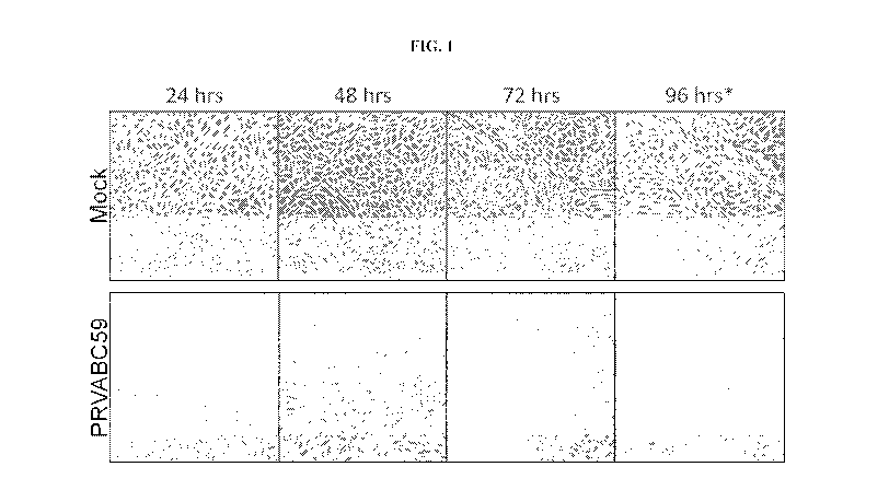
[00481 FIG. l shows bright field microscopy images of Vero cell monolayers
mock infected
(top) or infected with ZIKAV strain PRVABC59 (bottom).
[0049] FIG. 2 shows growth kinetics of ZIKAV PRVABC59 PI on Vero cell
monolayers, as
determined by TCID50.
[0050] FIG. 3 shows potency assay testing (TCID50) of Zika virus PRVABC59
P5 clones a-f.
[0051] FIG. 4 shows bright-field microscopy images depicting the cytopathic
effect (CPE) of
growth of Zika virus PRVABC59 P6 clones a-f on Vero cell monolayers.
[00521 FIG. 5 shows potency assay testing (TCID50) of Zika virus PRVABC59
P6 clones a-f
[0053] FIG. 6 shows an amino acid sequence alignment comparing the envelope
glycoprotein
sequence of Zika virus near residue 330 from Zika virus strains PRVABC59 P6e
(SEQ ID NO: 8)
and PRVABC59 (SEQ ID NO: 9) with several other flaviviruses (WNV (SEQ ID NO:
10); JEV
(SEQ ID NO: 11); SLEV (SEQ ID NO: 12); YFV (SEQ ID NO: 13); DENV 1 16007 (SEQ
ID NO:
14); DENY 2 16681 (SEQ ID NO: 15); DENY 3 16562 (SEQ IDNO: 16); and DENY 4
1036 (SEQ
ID NO: 17)).
8
CA 03084605 2020-05-27
WO 2019/108976 PCT/US2018/063381
[0054] FIG. 7 shows an amino acid sequence alignment comparing the NS1
protein sequence
of Zika virus near residue 98 from Zika virus strains PRVABC59 P6e (SEQ ID NO:
18) and
PRVABC59 (SEQ ID NO: 19) with several other flaviviruses (WNV (SEQ ID NO: 20);
JEV (SEQ
ID NO: 21); SLEV (SEQ ID NO: 22); YFV (SEQ ID NO: 23); DENV 1 16007 (SEQ ID
NO: 24);
DENV 2 16681 (SEQ ID NO: 25); DENV 3 16562 (SEQ IDNO: 26); and DENV 4 1036
(SEQ ID
NO: 27)).
100551 FIG. 8 shows the plaque phenotype of ZIKAV PRVABC59 P6 virus clones
a-f
compared to ZIKAV PRVABC59 PI virus.
[0056] FIG. 9 shows the mean plaque size of ZIKAV PRVABC59 P6 virus clones
compared
to ZIKAV PRVABC59 P1 virus.
[0057] FIG. 10 shows the growth kinetics of ZIKAV PRVABC59 P6 clones a-f in
Vero cells
under serum-free growth conditions.
100581 FIG. 11 shows a schematic of the steps taken to prepare PRVABC59 P6b
and P6e
formulated drug product for the immunization experiments.
[0059] FIG. 12A shows the schedule of dosing of CD-1 mice with vaccine
formulations
derived from the ZIKAV PRVABC59 P6b and P6e clones. PBS was used as placebo.
100601 FIG. 12B shows the serum ZIKAV neutralizing antibody titers of CD-1
mice
immunized as described in FIG. 12A using vaccine formulations derived from
ZIKAV PRVABC59
P6b and P6e clones. ZIKAV neutralizing antibody titers were determined by
Reporter Virus Particle
(RVP) neutralization assay. Solid lines represent the geometric mean of a
group. The limit of
detection (1.93 logio) is represented by a dashed line.
[0061] FIG. 13A shows the schedule of dosing of AG129 mice with vaccine
formulations
derived from the ZIKAV PRVABC59 P6b and P6e clones. PBS was used as a placebo.
[0062] FIG. 13B shows the serum ZIKAV neutralizing antibody titers of AG129
mice
immunized as described in FIG. 13A using vaccine formulations derived from
ZIKAV PRVABC59
P6b and P6e clones. Solid lines represent the geometric mean of a group. The
limit of detection
(1.30 logio) is represented by a dashed line. Animals with no detectable titer
(<1.30) were assigned a
titer of 0.5.
9
CA 03084605 2020-05-27
WO 2019/108976
PCT/US2018/063381
[0063] FIG. 14 shows the mean weight of AG129 test groups post-challenge,
represented as a
percentage of starting weight. Error bars represent standard deviation.
[0064] FIG. 15 shows the senun viremia of individual AG129 mice two days
post-challenge,
reported as PFU/mL. Solid lines represent the mean of a group. The limit of
detection (2.0 logio) is
represented by a dashed line.
[0065] FIG. 16 shows the survival analysis of AG129 test groups post-
challenge.
[0066] FIG. 17 shows the pre-challenge serum circulating ZIKAV neutralizing
antibody (Nab)
titers following passive transfer of pooled sera from vaccinated and
challenged AG129 mice.
[0067] FIG. 18 shows the mean body weight of passive transfer and control
mice challenged
with Zika virus.
[0068] FIG. 19 shows the serum viremia of individual AG129 mice three days
post-challenge,
reported as PFU/mL.
[0069] FIG. 20 shows the survival analysis of passive transfer and control
mice challenged
with Zika virus.
[0070] FIG. 21 shows the correlation between ZIKAV neutralizing antibody
titers and viremia
observed in passive transfer mice.
[0071] FIG. 22 shows the survival analysis of AG129 mice after infection
with Zika virus
preMVS stocks of P6a and P6e using a Kaplan Meier survival curve.
[0072] FIG. 23 shows the mean body weight as expressed in percentage of
starting weight at
time of invention after infection with Zika virus preMVS stocks of P6a and
P6e. The dashed line
represents 100% of starting weight for reference.
[0073] FIG. 24 shows the serum viremia of individual AG129 mice three days
post-infection
with Zika virus preMVS stocks of P6a and P6e, reported as PFU/mL. The dashed
line represents the
limit of detection of the assay.
CA 03084605 2020-05-27
WO 2019/108976
PCT/US2018/063381
[0074] FIG. 25 shows compiled kinetics of inactivation data. Data compares
infectious
potency (TCID50) to RNA copy, and completeness of inactivation (COI) for
samples from the four
toxicology lots. These data indicate that the sensitivity of the COI assay is
greater than TCID50.
[0075] FIG. 26 shows a comparison of C6/36 and Vero sensitivity in the
assay as
demonstrated with an input virus titer of 0.31 TC1D50.
[0076] FIG. 27 shows a logistic regression analysis of CPE vs. log TC1D50
using C6/36 cells
site that include 99% confidence intervals around a target value of 0.01
TCID50/well (-2 log
TCID50/well); the model predicts 0.85% of wells will be positive.
[0077] FIG. 28 shows chromatograms of PBS (a) and PBS solutions containing
0.049 pg/mL
(b), 0.098 pg/mL (c), 0.1961.tg/mL (d), 0.491 g/mL (e), 0.982 pg/mL (f), and
1.964 pg/mL (g)
formaldehyde.
DETAILED DESCRIPTION
General Techniques
[0078] The techniques and procedures described or referenced herein are
generally well
understood and commonly employed using conventional methodology by those
skilled in the art,
such as, for example, the widely utilized methodologies described in Sambrook
et al., Molecular
Cloning: A Laboratory Manual 3d edition (2001) Cold Spring Harbor Laboratory
Press, Cold
Spring Harbor, N.Y.; Current Protocols in Molecular Biology (RM. Ausubel, et
al. eds., (2003));
the series Methods in Enzymology (Academic Press, Inc.): PCR 2: A Practical
Approach (MI
MacPherson, B.D. Hames and G.R. Taylor eds. (1995)), Harlow and Lane, eds.
(1988) Antibodies,
A Laboratory Manual, and Animal Cell Culture (R.I. Freshney, ed. (1987));
Oligonucleotide
Synthesis (M.J. Gait, ed., 1984); Methods in Molecular Biology, Humana Press;
Cell Biology: A
Laboratory Notebook (J.E. Cellis, ed., 1998) Academic Press; Animal Cell
Culture (R.I. Freshney),
ed., 1987); Introduction to Cell and Tissue Culture (J.P. Mather and P.E.
Roberts, 1998) Plenum
Press; Cell and Tissue Culture: Laboratory Procedures (A. Doyle, J.B.
(Iriffiths, and D.G. Newell,
eds., 1993-8) J. Wiley and Sons; Handbook of Experimental Immunology (D.M.
Weir and C.C.
Blackwell, eds.); Gene Transfer Vectors for Mammalian Cells (J.M. Miller and
M.P. Cabs. eds.,
1987); PCR: The Polymerase Chain Reaction, (Mullis et al., eds., 1994);
Current Protocols in
Immunology (J.E. Coligan et al., eds., 1991); Short Protocols in Molecular
Biology (Wiley and
Sons, 1999); Immunobiology (C.A. Janeway and P. Travers, 1997); Antibodies (P.
Finch, 1997);
11
CA 03084605 2020-05-27
WO 2019/108976
PCT/US2018/063381
Antibodies: A Practical Approach (D. Catty., ed., 1RL Press, 1988-1989);
Monoclonal Antibodies:
A Practical Approach (P. Shepherd and C. Dean, eds., Oxford University Press,
2000); Using
Antibodies: A Laboratory Manual (E. Harlow and D. Lane (Cold Spring Harbor
Laboratory Press,
1999); and The Antibodies (M. Zanetti and J. D. Capra, eds., Harwood Academic
Publishers, 1995).
Zika virus
[0079] Certain aspects of the present disclosure relate to a purified
inactivated whole Zika
virus that may be useful in vaccines and/or immunogenic compositions.
[0080] Zika virus (ZIKV) is a mosquito-borne flavivirus first isolated from
a sentinel rhesus
monkey in the Zika Forest in Uganda in 1947. Since that time, isolations have
been made from
humans in both Africa and Asia, and more recently, the Americas. ZIKV is found
in two (possibly
three) lineages: an African lineage (possibly separate East and West African
lineages) and an Asian
lineage. Accordingly, examples of suitable Zika viruses of the present
disclosure include, without
limitation, viruses from the African and/or Asian lineages. In some
embodiments, the Zika virus is
an African lineage virus. In some embodiments, the Zika virus is an Asian
lineage virus.
Additionally, multiple strains within the African and Asian lineages of Zika
virus have been
previously identified. Any one or more suitable strains of Zika virus known in
the art may be used
in the present disclosure, including, for examples, strains Mr 766, ArD 41519,
IbH 30656, P6-740,
EC Yap, F5513025, ArD 7117, ArD 9957, ArD 30101, ArD 30156, ArD 30332, HD
78788, ArD
127707, ArD 127710, ArD 127984, ArD 127988, ArD 127994, ArD 128000, ArD
132912, 132915,
ArD 141170, ArD 142623, ArD 149917, ArD 149810, ArD 149938, ArD 157995, ArD
158084,
ArD 165522, ArD 165531, ArA 1465, ArA 27101, ArA 27290, ArA 27106, ArA 27096,
ArA
27407, ArA 27433, ArA 506/96, ArA 975-99, Ara 982-99, ArA 986-99, ArA 2718,
ArB 1362,
Nigeria68, Malaysia66, Kedougou84, Suriname, MR1429, PRVABC59, ECMN2007,
DakAr41524,
H/PF/2013, R103451, 103344, 8375, JMB-185, ZIKV/H, sapiens/Brazil/Natal/2015,
5PH2015,
ZIKV/Hu/Chiba/536/2016, and/or Cuba2017. In some embodiments, strain PRVABC59
is used in
the present disclosure.
[0081] In some embodiments, an example of a Zika virus genome sequence is set
forth below as
SEQ ID NO: 2:
gttgttgatc tgtgtgaatc agactgcgac agttcgagtt tgaagcgaaa gctagcaaca
61 gtatcaacag gttttatttt ggatttggaa aegagagttt ctggtcatga nnacccaaa
121 aaagaaatcc ggaggattcc ggattgtcaa tatgctaaaa cgcggagtag cccgtgtgag
181 cccattggg ggcttgaaga ggctgccagc cggacttctg ctgggtcatg ggcccatcag
12
CA 03084605 2020-05-27
WO 2019/108976
PCT/US2018/063381
241 gatggtcttg gcgattctag cctttttgag attcacggca atcaagccat cactgggtct
301 catcaataga tggggttcag tggggaaaaa agaggctatg gaaacaataa agaagttcaa
361 gaaagatctg gctgccatgc tgagaataat caatgctagg aaggagaaga agagacgagg
421 cgcagatact agtgtcggaa ttgttggcct cctgctgacc acagctatgg cagcggaggt
481 cactagacgt gggagtgcat actatatgta cttggacaga aacgatgctg gggaggccat
541 atcttttcca accacattgg ggatgaataa gtgttatata cagatcatgg atcttggaca
601 catgtgtgat gccaccatga gctatgaatg ccctatgctg gatgaggggg tggaaccaga
661. tgacgtcgat tgttggtgca acacgacgtc aacttgggtt gtgtacggaa cctgccatca
721 caaaaaaggt gaagcacgga gatctagaag agctgtgacg ctcccctccc attccaccag
781 gaagctgcaa acgcggtcgc aaacctggtt ggaatcaaga gaatacacaa agcacttgat
841 tagagtcgaa aattggatat tcaggaaccc tggcttcgcg ttagcagcag ctgccatcgc
901 ttggcttttg ggaagctcaa cgagccaaaa agtcatatac ttggtcatga tactgctgat
961 tgccccggca tacagcatca ggtgcatagg agtcagcaat agggactttg tggaaggtat
1021 gtcaggtggg acttgggttg atgagtat ggaacatgga ggttgtgtca ccgtaatggc
1081 acaggacaaa ccgactgtcg acatagagct ggttacaaca acagtcagca acatggcgga
1141 ggtaagatcc tactgctatg aggcatcaat atcagacatg gcttctgaca gccgctgccc
1201 aacacaaggt gaagcctacc ttgacaagca atcagacact cast tgtct gcannagaac
1261 gttagtggac agaggctggg gaaatggatg tggacttttt ggcaaaggga gcctggtgac
1321 atgcgctaag tttgcatgct ccaagaaaat gaccgggaag agcatccagc cagagaatct
1381. ggagtaccgg ataatgctgt cagttcatgg ctcccagcac agtgggatga tcgttaatga
1441 cacaggacat gaaactgatg agaatagagc gaaagttgag ataacgccca attcaccgag
1501 agccgaagcc accctggggg gttliggaag cctaggactt gattgtgaac cgaggacagg
1561 ccttgacttt tcagatttgt attacttgac tatgaataac aagcactggt tggttcacaa
1621 ggagtggttc cacgacattc cattaccttg gcacgctggg gcagacaccg gaactccaca
1681 ctggaacaac aapgaagcac tggtagagtt caaggacgca catgccaaaa ggcaaactgt
1741 cgtggttcta gggagtcaag aaggagcagt tcacacggcc cttgctggag ctctggaggc
1801 tgagatggat ggtgcaaagg gaaggctgtc ctctggccac ttgaaatgtc gcctgaaaat
1861 ggataaactt agattgaagg gcgtgtcata ctccttgtgt actgcagcgt tcacattcac
1921. caagatcccg gctgaaacac tgcacgggac agtcacagtg gaggtacagt acgcagggac
1981 agatggacct tgcaaggttc cagctcagat ggcggtggac atgcaaactc tgaccccagt
2041 tgggaggttg ataaccgcta accccgtaat cactgaaagc actgagaact ctaagatgat
2101 gctggaactt gatccaccat ttggggactc ttacattgtc ataggagtcg gggagaagaa
2161 gatcacccac cactggcaca ggagtggcag caccattgga aanecatttg aagccactgt
2221 gagaggtgcc aagagaatgg cagtcttag agacacagcc tgggactttg gatcagttgg
2281 aggcgctctc aactcattgg gcaagggcat ccatcaaatt tttggagcag ctttcaaatc
2341 attgtttgga ggaatgtcct ggttctcaca aattctcatt ggaacgttgc tgatgtggtt
2401. gggtctgaac acaaagaatg gatctatttc ccttatgtgc ttggccttag ggggagtgtt
13
CA 03084605 2020-05-27
WO 2019/108976
PCT/US2018/063381
2461 gatcttctta tccacagccg tactgctga tgtggggtgc tcggtggact tctcaaagaa
2521 ggagacgaga tgcggtacag gggtgttcgt ctataacgac gttgaagcct ggagggacag
2581 gtacaagtac catcctgact ccccccgtag attggcagca gcagtcaagc aagcctggga
2641 agatggtatc tgcgggatct cctctgtttc aagaatggaa aacatcatgt ggagatcagt
2701 agaaggggag ctcaacgcaa tcctggaaga gaatggagtt caactgacgg tcgttgtggg
2761 atctgtaaaa aaccccatgt ggagaggtcc acagagattg cccgtgcctg tgaacgagct
2821 gccccacggc tggaaggctt gggggaaatc gtatttcgtc agagcagcaa agacaaataa
2881 cagctttgtc gtggatggtg acacactgaa ggaatgccca ctcaaacata gagcatggaa
2941 cagctttctt gtggaggatc atgggttcgg ggtatttcac actagtgtct ggctcaaggt
3001 tagagaagat tattcattag agtgtgatcc agccgttatt ggaacagctg ttaagggaaa
3061 ggaggctgta cacagtgatc taggctactg gattgagagt gagaagaatg acacatggag
3121 gctgaagagg gcccatctga tcgagatgaa aacatgtgaa tggccaaagt cccacacatt
3181 gtggacagat ggaatagaag agagtgatct gatcataccc aagtctttag ctgggccact
3241 cagccatcac aataccagag agggctacag gacccaaatg aaagggccat ggcacagtga
3301 agagcttgaa attcggtttg aggaatgccc aggcactaag gtccacgtgg aggaaacatg
3361 tggaacaaga ggaccatctc tgagatcaac cactgcaagc ggaagggtga tcgaggaatg
3421 gtgctgcagg gagtgcacaa tgcccccact gtcgttccgg gctaaagatg gctgttggta
3481 tggaatggag ataaggccca ggaaagaacc agaaagcaac ttagtaaggt caatggtgac
3541 tgcaegatca actgatcaca tggaccactt ctcccttgga gtgcttgtga tcctgctcat
3601 ggtgcaggaa gggctgaaga agagaatgac cacalagatc atcataagca catcaatggc
3661 agtgctggta gctatgatcc tgggaggatt ttcaatgagt gacctggcta agcttgcaat
3721 tttgatgggt gccaccttcg cggaaatgaa cactggagga gatgtagctc atctggcgct
3781 gatagcggca ttcaaagtca gaccagcgtt gctggtatct ttcatcttca gagctaattg
3841 gacaccccgt gaaagcatgc tgctggcctt ggcctcgtgt cttttgcaaa ctgcgatctc
3901 cgccttggaa ggcgacctga tggttctcat caatggtttt gctttggcct ggttggcaat
3961 acgagcgatg gttgttccac gcactgataa catcaccttg gcaatcctgg ctgctctgac
4021 accactggcc cggggcacac tgcttgtggc gtggagagca ggccttgcta cttgcggggg
4081 gtttatgctc ctctctctga agegaaaagg cagtgtgaag aagaacttac catttgtcat
4141. ggccctggga ctaaccgctg tgaggctggt cgaccccatc aacgtggtgg gactgctgtt
4201 gctcacaagg agtgggaagc ggagctggcc ccctagcgaa gtactcacag ctgttggcct
4261 gatatgcgca ttggctggag ggttcgccaa ggcagatata gagatggctg ggcccatggc
4321 cgcggtcggt ctgctaattg tcagttacgt ggtctcagga aagagtgtgg acatgtacat
4381 tgaaagagca ggtgacatca catgggaaaa agatgcggaa gtcactggaa acagtccccg
4441 gctcgatgtg gcgctagatg agagtggtga tttctccctg gtggaggatg acggtccccc
4501 catgagagag atcatactca aggtggtcct gatgaccatc tgtggcatga acccaatagc
4561 catacccttt gcagctggag cgtggtacgt atacgtgaag actggaaaaa ggagtggtgc
4621. tctatgggat gtgcctgctc ccaaggaagt aaaaaagggg gagaccacag atggagtgta
14
CA 03084605 2020-05-27
WO 2019/108976
PCT/US2018/063381
4681 cagagtaatg actcgtagac tgctaggttc aacacaagtt ggagtgggag ttatgcaaga
4741 gggggtcttt cacactatgt ggcacgtcac aaaaggatcc gcgctgagaa gcggtgaagg
4801 gagacttgat ccatactggg gagatgtcaa gcaggatctg gtgtcatact gtggtccatg
4861 gaagctagat gccgcctggg atgggcacag cgaggtgcag ctcttggccg tgcccmcgg
4921 agagagagcg aggaacatcc agactctgcc cggaatattt aagacaaagg atggggacat
4981 tggagcggtt gcgctggatt acccagcagg aacttcagga tctccaatcc tagacaagtg
5041 tgggagagtg ataggacttt atggcaatgg ggtcgtgatc aaaaacggga gttatgttag
5101 tgccatcacc caagggagga gggaggaaga gactcctgtt gagtgcttcg agccctcgat
5161 gctgaagaag aagcagctaa ctgtcttaga cttgcatcct ggagctggga aaaccaggag
5221 agttcttcct gaaatagtcc gtgaagccat aaaaaca ga ctccgtactg tgatcttagc
5281 tccaaccagg gttgtcgctg ctgaaatgga ggaggccctt agagggcttc cagtgcgtta
5341 tatgacaaca gcagtcaatg tcacccactc tggaacagaa atcgtcgact taatgtgcca
5401 tgccaccttc acttcacgtc tactacagcc aatcagagtc cccaactata atctgtatat
5461 tatggatgag gcccacttca cagatccctc aagtatagca gcaagaggat acatttcaac
5521 aagggttgag atgggcgagg cggctgccat cttcatgacc gccacgccac caggaacccg
5581 tgacgcattt ccggactcca actcaccaat tatggacacc gaagtggaag tcccagagag
5641 agcctggagc tcaggctttg attgggtgac ggatcattct ggaaaaarag tttggtttgt
5701 tccaagcgtg aggaacggca atgagatcgc agcttgtctg acaaaggctg gaaaacgggt
5761 catacagctc agcagaaaga cattgagac agagttccae aaaacaaaac atcaagagtg
5821 ggactttgtc gtgacaactg acatttcaga gatgggcgcc aactttaaelg ctgaccgtgt
5881 catagattcc aggagatgcc taaagccggt catacttgat ggcgagagag tcattctggc
5941 tggacccatg cctgtcacac atgccagcgc tgcccagagg agggggcgca taggcaggaa
6001 tcccaacaaa cctggagatg agtatctgta tggaggtggg tgcgcagaga ctgacgaaga
6061 ccatgcacac tggcttgaag caagaatgct ccttgacaat atttacctcc aagatggcct
6121 catagcctcg ctctatcgac ctgaggccga caaagtagca gccattgagg gagagttcaa
6181 gcttaggacg gagcaaagga agacctttgt ggaactcatg aanagaggag atcttcctgt
6241 ttggctggcc tatcaggttg catctgccgg aataacctac acagatagaa gatggtgctt
6301 tgatggcacg accaacaaca ccataatgga agacagtgtg ccggcagagg tgtggaccag
6361 acacggagag aaongagtgc tcaaaccgag gtggatggac gccagagttt gttcagatca
6421 tgcggccctg aagtcattca aggagtttgc cgctgggaaa agaggagcgg cttttggagt
6481 gatggaagcc ctgggaacac tgccaggaca catgacagag agattccagg aagccattga
6541 caacctcgct gtgctcatgc gggcagagac tggaagcagg ccttacaaag ccgcggcggc
6601 ccaattgccg gagaccctag agaccataat gcttttgggg ttgctgggaa cagtctcgct
6661 gggaatcttc ttcgtcttga tgaggaacaa gggcataggg aagatgggct ttggaatggt
6721 gactcttggg gccagcgcat ggctcatgtg gctctcggaa attgagccag ccagaattgc
6781 atgtgtcctc attgttgtgt tcctattgct ggtggtgctc atacctgagc caeaaaagca
6841 aagatctccc caggacaacc aaatggcaat catcatcatg gtagcagtag gtcttctggg
CA 03084605 2020-05-27
WO 2019/108976
PCT/US2018/063381
6901 cttgattacc gccaatgaac tcggatggtt ggagagaaca aagagtgacc taagccatct
6961 aatgggaagg agagaggagg gggcaaccat aggattctca atggacattg acctgcggcc
7021 agcctcagct tgggccatct atgctgcctt gacaactttc attaccccag ccgtccaaca
7081 tgcagtgacc acctcataca acaactactc cttaatggcg atggccacgc aagctggagt
7141 gttgtttggc atgggcaaag ggatgccatt ctacgcatgg gactttggag tcccgctgct
7201 aatgataggt tgctactcac aattaacacc cctgacccta atagtggcca tcattttgct
7261 cgtggcgcac tacatgtact tgatcccagg gctgcaggca gcagctgcgc gtgctgccca
7321 gaagagaacg gcagctggca tcatgaagaa ccctgttgtg gatggaatag tggtgactga
7381 cattgacaca atgacaattg acccccaagt ggagsaaaag atgggacagg tgctactcat
7441 agcagtagcc gtctccagcg ccatactgtc gcggaccgcc tgggggtggg gggaggctgg
7501 ggctctgatc acagccgcaa cttccacttt gtgggaaggc tctccgaaca agtactggaa
7561 ctcctctaca gccacttcac tgtgtaacat ttttagggga agttacttgg ctggagcttc
7621 tctaatctac acagtaacaa gaaacgctgg cttggtcaag agacgtgggg gtggaacagg
7681 agagaccctg ggagagaaat ggaaggcccg cttgaaccag atgtcggccc tggagttcta
7741 ctcctacaaa aagtcaggca tcaccgaggt gtgcagagaa gaggcccgcc gcgccctcaa
7801 ggacggtgtg gcaacgggag gccatgctgt gtcccgagga agtgcaaagc tgagatggtt
7861 ggtggagcgg ggatacctgc agccctatgg aaaggtcatt gatcttggat gtggcagagg
7921 gggctggagt tactacgtcg ccaccatccg caaagttcaa gaagtgaaag gatacacaaa
7981 aggaggccct ggtcatgaag aacccgtgtt ggtgcaaagc tatgggtgga acatagtccg
8041 tataagagt ggggtggacg tctttcatat ggcggctgag ccgtgtgaca cgttgctgtg
8101 tgacataggt gagtcatcat ctagtcctga agtggaagaa gcacggacgc tcagagtcct
8161 ctccatggtg ggggattggc ttgaaaanag accaggagcc ttttgtataa aagtgttgtg
8221 cccatacacc agcactatga tggaaaccct ggagcgactg cagcgtaggt atgggggagg
8281 actggtcaga gtgccactct cccgcaactc tacacatgag atgtactggg tctctggagc
8341 gaaaagcaac accataaaaa gtgtgtccac cacgagccag ctcctcttgg ggcgcatgga
8401 cgggcctagg aggccagtga aatatgagga ggatgtgaat ctcggctctg gcacgcgggc
8461 tgtggtaagc tgcgctgaag ctcccaacat gaagatcatt ggtaaccgca ttgaaaggat
8521 ccgcagtgag cacgcggaaa cgtggttctt tgacgagaac cacccatata ggacatgggc
8581 ttaccatgga agctatgagg cccccacaca agggtcagcg tcctctctaa taaacggggt
8641 tgtcaggctc ctgtcaaaar, cctgggatgt ggtgactgga gtcacaggaa tagccatgac
8701 cgacaccaca ccgtatggtc agcaaagagt tttcaaggaa aaagtggaca ctagggtgcc
8761 agacccccaa gaaggcactc gtcaggttat gagcatggtc tcttcctggt tgtggaaaga
8821 gctaggcaaa cacaaacggc cacgagtctg caccaaalt,aa gagttcatca acaaggttcg
8881 tagcaatgca gcattagggg caatatttga agaggaaaaa gagtggaaga ctgcagtgga
8941 agctgtgaac gatccaaggt tctgggctct agtggacaag gaaagagagc accacctgag
9001 aggagagtgc cagagctgtg tgtacaacat gatgggaaaa agagaaaaga aacaagggga
9061 atttggaaag gccaagggca gccgcgccat ctggtatatg tggctagggg ctagatttct
16
CA 03084605 2020-05-27
WO 2019/108976
PCT/US2018/063381
9121 agagttcgaa gcccttggat tcttgaacga ggatcactgg atggggagag agaactcagg
9181 aggtggtgtt gaaeggctgg gattacaaae, actcggatat gtcctagaag agatgagtcg
9241. tataccagga ggaaggatgt atgcagatga cactgctggc tgggacaccc gcattagcag
9301 gtttgatctg gagaatgaag ctctaatcac caaccaaatg gagaaagggc acagggcctt
9361 ggcattggcc ataatcaagt acacatacca aaacaaagtg gtaaaggtcc ttagaccagc
9421 tgaaaaaggg aaaacagtta tggacattat ttcgagacaa gaccaaaggg ggagcggaca
9481 agttgtcact tacgctctta acacatttac caacctagtg gtgcaactca ttcggaatat
9541 ggaggctgag gaagttctag agatgcaaga cttgtggctg ctgcggaggt cagagaaagt
9601 gaccaactgg ttgcagagca acggatggga taggctcaaa cgaatggcag tcagtggaga
9661 tgattgcgtt gtgaagccaa ttgatgatag gtttgcacat gccctcaggt tatgaatga
9721 tatgggaaaa gttaggaagg acacacaaga gtggaaaccc tcaactggat gggacaactg
9781. ggaagaagtt ccgttttgct cccaccactt caacaagctc catctcaagg acgggaggtc
9841 cattgtggtt ccctgccgcc accaagatga actgattggc cgggcccgcg tctctccagg
9901 ggcgggatgg agcatccggg agactgcttg cctagcaaaa tcatatgcgc aaatgtggca
9961 gaccatat ttccacagaa gggacctccg actgatggcc aatgccattt gttcatctgt
10021. gccagttgac tgggttccaa ctgggagaac tacctggtca atccatggaa agggagaatg
10081 gatgaccact gaagacatgc ttgtggtgtg gaacagagtg tggattgagg agaacgacca
10141 catggaagac aagaccccag ttacgaaatg gacagacatt ccctatttgg gaaaaaggga
10201 agacttgtgg tgtggatctc tcataeggca cagaccgcgc accacctggg ctgagaacat
10261 tanaaacaca gtcaacatgg tgcgcaggat cataggtgat gaagaaaagt acatggacta
10321 cctatccacc caagttcgct acttgggtga agaagggtct acacctggag tgctgtaagc
10381 accaatctta atgttgtcag gcctgctagt cagccacagc ttggggaaag ctgtgcagcc
10441 tgtgaccccc ccaggagaag ctgggaaacc aagcctatag tcaggccgag aacgccatgg
10501 cacggaagaa gccatgctgc ctgtgagccc ctcagaggac actgagtcaa aaaaccccac
10561. gcgcttggag gcgcaggatg ggaanagaag gtggcgacct tccccaccct tcaatctggg
10621 gcctgaactg gagatcagct gtggatctcc agaagaggga ctagtggtta gagga
100821 In
some embodiments, the Zika virus may comprise the genome sequence of GenBank
Accession number KU501215.1. In some embodiments, the Zika virus is from
strain PRVABC59.
In some embodiments the genome sequence of GenBank Accession number KU501215.1
comprises
the sequence of SEQ ID NO: 2. In some embodiments, the Zika virus may comprise
a genomic
sequence that has at least 70%, at least 71%, at least 72%, at least 73%, at
least 74%,at least 75%, at
least 76%, at least 77%, at least 78%, at least 79%, at least 80%, at least
81%, at least 82%, at least
83%, at least 84%,at least 85%, at least 86%, at least 87%, at least 88%, at
least 89%, at least 90%,
at least 91%, at least 92%, at least 93%, at least 94%, at least 95%, at least
96%, at least 97%, at
least 98%, at least 99%, or 100% sequence identity with the sequence of SEQ ID
NO: 2.
17
CA 03084605 2020-05-27
WO 2019/108976
PCT/US2018/063381
[0083] In some embodiments, the Zika virus may comprise at least one
polypeptide encoded by
the sequence of SEQ ID NO: 2. In some embodiments, the Zika virus may comprise
at least one
polypeptide having an amino acid sequence that has at least 85%, at least 86%,
at least 87%, at least
88%, at least 89%, at least 90%, at least 910/0, at least 92%, at least 93%,
at least 94%,at least 95%,
at least 96%, at least 97%, at least 98%, at least 99%, or 100% sequence
identity with an amino acid
sequence encoded by the sequence of SEQ ID NO: 2.
[0084] Accordingly, in some embodiments, inactivated Zika viruses of the
present disclosure
may be used in any of the vaccines and/or immunogenic compositions disclosed
herein. For
example, inactivated Zika viruses of the present disclosure may be used to
provide one or more
antigens useful for treating or preventing Zika virus infection in a subject
in need thereof and/or for
inducing an immune response, such as a protective immune response, against
Zika virus in a subject
in need thereof.
[0085] The Zika virus used in the present disclosure may be obtained from
one or more cells in
cell culture (e.g., via plaque purification). Any suitable cells known in the
art for producing Zika
virus may be used, including, for example, insect cells (e.g., mosquito cells
such as CCL-125 cells,
Aag-2 cells, RML-12 cells, C6/36 cells, C7-10 cells, AP-61 cells, A.t. GRIP-1
cells, A.t. GR1P-2
cells, A.t. GRIP-3 cells, UM-AVE! cells, Mos.55 cells, SualB cells, 4a-3B
cells, Mos.42 cells,
M5Q43 cells, LSB-AA695BB cells, NTID-CTR cells, TRA-171, cells, and additional
cells or cell
lines from mosquito species such as Aedes aegvpti, Aedes albopictus. Aedes
pseudoscutellaris,
Aedes triseriatus, Aedes vexans, Anopheles gambiae, Anopheles stephensi,
Anopheles albimus,
Culex quinquefasciatus, Culex theileri. Culex tritaeniorhynchus, Culex
bitaeniorhynchus, and/or
.Toxorhynchites amboinensis), and mammalian cells (e.g., VERO cells (from
monkey kidneys),
LLC-MK2 cells (from monkey kidneys), MDBK cells, MDCK cells, ATCC CCL34 MDCK
(NBL2) cells, MDCK 33016 (deposit number DSM ACC 2219 as described in
W097/37001) cells,
BHK21-F cells, HKCC cells, or Chinese hamster ovaiy cells (CHO cells). In some
embodiments,
the Zika virus (e.g., a Zika virus clonal isolate) is produced from a non-
human cell. In some
embodiments, the Zika virus (e.g., a Zika virus clonal isolate) is produced
from an insect cell. In
some embodiments, the Zika virus (e.g., a Zika virus clonal isolate) is
produced from a mosquito
cell. In some embodiments, the Zika virus (e.g., a Zika virus clonal isolate)
is produced from a
mammalian cell. In some embodiments, the Zika virus (e.g., a Zika virus clonal
isolate) is produced
from a VERO cell.
[0086] Zika viruses possess a positive sense, single-stranded RNA genome
encoding both
structural and nonstructural polypeptides. The genome also contains non-coding
sequences at both
18
CA 03084605 2020-05-27
WO 2019/108976
PCT/US2018/063381
the 5'- and 3'- terminal regions that play a role in virus replication.
Structural polypeptides encoded
by these viruses include, without limitation, capsid (C), precursor membrane
(prM), and envelope
(E). Non-structural (NS) polypeptides encoded by these viruses include,
without limitation, NS1,
NS2A, NS2B, NS3, NS4A, NS4B, and NS5.
100871 In certain embodiments, the Zika virus includes a mutation in Zika
virus Non-structural
protein I (NS I). In some embodiments, the Zika virus contains a Trp98Gly
mutation at position 98
of SEQ ID NO: 1, or at a position corresponding to position 98 of SEQ ID NO:
1.
100881 In some embodiments, the mutation is within the NS1 polypeptide. The
amino acid
sequence of a wild-type, NS1 polypeptide from an exemplary Zika virus strain
is set forth as:
DVGCSVDFSKKE'TRCGTGVFVYNDVEAWRDRYKYHPDSPRRLAAAVKQAWEDGICGISS
VSRMEN1MWRSVEGELNAILEENGVQLTVVVGSVKNPMWRGPQRLPVPVNELPHGWKA
WGKSYFVRAAKTNN SFVVDGDTLKECPLKHRAWN SFLVEDHGFGVFHTSVWLKVREDYS
LECDPAVIGTAVKGKEAVHSDLGYWIESEKNDTWRLKRAHL1EMKTCEWPKSHTLWTDGI
EESDLTIPKSLAGPLSHHNTREGYRTQMKGPWHSEELEIRFEECPGTKVHVEETCGTRGPSL
RSTTASGRV1EEWCCRECTMPPLSFRAKDGCWYGMEIRPRKEPESNLVRSMVT (SEQ ID
NO: 1).
[0089] In some embodiments, the amino acid sequence of the NSI polypeptide
has at least
80%, at least 81%, at least 82%, at least 83%, at least 84%, at least 85%, at
least 86%, at least 87%,
at least 88%, at least 89%, at least 90%, at least 91%, at least 92%, at least
93%, at least 94%, at
least 95%, at least 96%, at least 97%, at least 98%, at least 99%, or 100%
sequence identity with the
sequence of SEQ ID NO: I. In some embodiments, the amino acid sequence of the
NS1 polypeptide
may be from the amino acid sequence encoded by the sequence of GenBank
Accession number
KU501215.1 (SEQ ID NO: 2). In some embodiments, the amino acid sequence of the
NS1
polypeptide may be amino acid positions 795 to 1145 of the amino acid sequence
encoded by the
sequence of GenBank Accession number KU501215.1. In some embodiments, the
amino acid
sequence of the NS1 polypeptide may be from Zika virus strain PRVABC59.
[0090] "Sequence Identity", "% sequence identity", "% identity", "%
identical" or "sequence
alignment" means a comparison of a first amino acid sequence to a second amino
acid sequence, or
a comparison of a first nucleic acid sequence to a second nucleic acid
sequence and is calculated as
a percentage based on the comparison. The result of this calculation can be
described as "percent
identical" or "percent ID."
19
CA 03084605 2020-05-27
WO 2019/108976
PCT/US2018/063381
[0091] Generally, a sequence alignment can be used to calculate the
sequence identity by one
of two different approaches. In the first approach, both mismatches at a
single position and gaps at a
single position are counted as non-identical positions in final sequence
identity calculation. In the
second approach, mismatches at a single position are counted as non-identical
positions in final
sequence identity calculation; however, gaps at a single position are not
counted (ignored) as non-
identical positions in final sequence identity calculation. In other words, in
the second approach
gaps are ignored in final sequence identity calculation. The difference
between these two
approaches, i.e. counting gaps as non-identical positions vs ignoring gaps, at
a single position can
lead to variability in the sequence identity value between two sequences.
[0092] In some embodiments, a sequence identity is determined by a program,
which produces
an alignment, and calculates identity counting both mismatches at a single
position and gaps at a
single position as non-identical positions in fmal sequence identity
calculation. For example
program Needle (EMBOS), which has implemented the algorithm of Needleman and
Wunsch
(Needleman and Wunsch, 1970, J. Mol. Biol. 48: 443-453), and which calculates
sequence identity
per default settings by first producing an alignment between a first sequence
and a second sequence,
then counting the number of identical positions over the length of the
alignment, then dividing the
number of identical residues by the length of an alignment, then multiplying
this munber by 100 to
generate the % sequence identity [% sequence identity = (# of Identical
residues / length of
alignment) x 100)].
[0093] A sequence identity can be calculated from a pairwise alignment
showing both
sequences over the full length, so showing the first sequence and the second
sequence in their full
length ("Global sequence identity"). For example, program Needle (EMBOSS)
produces such
alignments; % sequence identity = (# of identical residues / length of
alignment) x 100)].
[0094] A sequence identity can be calculated from a pairwise alignment
showing only a local
region of the first sequence or the second sequence ("Local Identity"). For
example, program Blast
(NCBI) produces such alignments; % sequence identity = (# of Identical
residues / length of
alignment) x 100)].
[0095] The sequence alignment is preferably generated by using the
algorithm of Needleman
and Wunsch (J. Mol. Biol. (1979) 48, p. 443-453). Preferably, the program
"NEEDLE" (The
European Molecular Biology Open Software Suite (EMBOSS)) is used with the
programs default
parameter (gap open=10.0, gap extend=0.5 and matrix=EBLOSUM62 for proteins and
matrix=EDNAFULL for nucleotides). Then, a sequence identity can be calculated
from the
CA 03084605 2020-05-27
WO 2019/108976
PCT/US2018/063381
alignment showing both sequences over the full length, so showing the first
sequence and the
second sequence in their full length ("Global sequence identity"). For
example: % sequence identity
= (# of identical residues / length of alignment) x 100)1
100961 In some embodiments, a mutation occurs at one or more amino acid
positions within the
NS1 polypeptide. In some embodiments, the mutation occurs at position 98 of
SEQ ID NO: 1, or at
a position corresponding to position 98 of SEQ ID NO: 1 when aligned to SEQ ID
NO: I using a
pairwise alignment algorithm. In some embodiments, the mutation at position 98
is a tryptophan to
glycine substitution.
100971 In some embodiments, the Zika virus comprises a mutation at position
98 of SEQ ID
NO: 1, or at a position corresponding to position 98 of SEQ ID NO: 1. A
position corresponding to
position 98 of SEQ ID NO: 1 can be determined by aligning the amino acid
sequence of an NS1
protein to SEQ ID NO: 1 using a pairwise alignment algorithm. Amino acid
residues in viruses
other than Zika virus which correspond to the tryptophan residue at position
98 of SEQ ID NO: I
are shown in Figure 7 of the present application where these residues are
boxed. In some
embodiments, the mutation at position 98 is a tryptophan to glycine
substitution. In some
embodiments, the mutation at position 98 is a tryptophan to glycine
substitution at position 98 of
SEQ ID NO: 1. In some embodiments, the mutation at position 98 is a tryptophan
to glycine
substitution at a position corresponding to position 98 of SEQ ID NO: 1 when
aligned to SEQ ID
NO: 1 using a pairwise alignment algorithm.
[0098] In some embodiments, the Zika virus contains a mutation within the
NS1 protein, and at
least one mutation within one or more of the C, prM, E, NS1, NS2A, NS2B, NS3,
NS4A, NS4B,
and NS5 viral proteins. In some embodiments, the Zika virus contains one or
more mutations within
the NS1 protein, and does not contain at least one mutation within one or more
of the C, prM, E.
NS I , NS2A, NS2B, NS3, NS4A, NS4B, and NS5 viral proteins. In some
embodiments, the Zika
virus contains a mutation within the NS I protein and does not contain at
least one mutation within
the envelope protein E. In some embodiments, whole, inactivated virus contains
at least one
mutation in Zika virus Non-structural protein 1 (NS1), and does not include a
mutation in Zika virus
envelope protein E (Env). In some embodiments, the Zika virus contains a
mutation at position 98
of SEQ ID NO: 1, or at a position corresponding to position 98 of SEQ ID NO: 1
and does not
contain any mutation within the envelope protein E. In some embodiments,
whole, inactivated Zika
virus contains a mutation at position 98 of SEQ ID NO: 1, or at a position
corresponding to position
98 of SEQ ID NO: 1 and does not include a mutation in Zika virus envelope
protein E (Env). In
some embodiments, whole, inactivated virus contains at least one mutation in
Zika virus Non-
21
CA 03084605 2020-05-27
WO 2019/108976
PCT/US2018/063381
structural protein 1 (NS1) and the sequence encoding the envelope protein is
the same as the
corresponding sequence in SEQ ID No. 2. In some embodiments, the Zika virus
contains a mutation
at position 98 of SEQ ID NO: 1, or at a position corresponding to position 98
of SEQ ID NO: 1 and
the sequence encoding the envelope protein is the same as the corresponding
sequence in SEQ ID
NO. 2. In some embodiments, whole, inactivated Zika virus contains a mutation
at position 98 of
SEQ ID NO: 1, or at a position corresponding to position 98 of SEQ ID NO: 1
and the sequence
encoding the envelope protein is the same as the corresponding sequence in SEQ
ID NO: 2. In some
embodiments, whole, inactivated Zika virus contains a try-ptophan to glycine
substitution at position
98 of SEQ ID NO: 1, or at a position corresponding to position 98 of SEQ ID
NO: 1 and the
sequence encoding the envelope protein is the same as the corresponding
sequence in SEQ ID NO:
2.
[0099] In some embodiments, the Zika virus contains at least one mutation
that enhances
genetic stability as compared to a Zika virus lacking the at least one
mutation. In some
embodiments, the Zika virus contains at least one mutation that enhances viral
replication as
compared to a Zika virus lacking the at least one mutation. In some
embodiments, the Zika virus
contains at least one mutation that reduces or otherwise inhibits the
occurrence of undesirable
mutations, such as within the envelope protein E (Env) of the Zika virus.
[00100] In the above embodiments of the present disclosure, an exemplary
pairwise alignment
algorithm is the Needleman-Wunsch global alignment algorithm, using default
parameters (e.g. with
Gap opening penalty=10.0, and with Gap extension penalty).5, using the
ERLOSUM62 scoring
matrix). This algorithm is conveniently implemented in the needle tool in the
EMBOSS package.
[00101] In some embodiments, the inactivated Zika virus may be used in
vaccines and
immunogenic compositions. For example, the inactivated Zika virus may be
useful for treating or
preventing Zika virus infection in a subject in need thereof and/or inducing
an immune response,
such as a protective immune response, against Zika virus in a subject in need
thereof.
Production of Vaccines and Immunogenic Compositions
[00102] Other aspects of the present disclosure relate to Zika virus vaccines
and immunogenic
compositions containing a purified inactivated whole virus, such as a Zika
virus with a mutation
which is a tryptophan to glycine substitution at position 98 of SEQ ID NO: 1
or at a position
corresponding to position 98 of SEQ ID NO: 1 as described herein. In some
embodiments, the
vaccine or immunogenic composition comprises a purified inactivated whole Zika
virus comprising
22
CA 03084605 2020-05-27
WO 2019/108976
PCT/US2018/063381
a Trp98Gly mutation at position 98 of SEQ ID NO: 1, or at a position
corresponding to position 98
of SEQ ID NO: 1, wherein the Zika virus is derived from strain PRVABC59. In
some embodiments,
the vaccine or immunogenic composition comprises a purified inactivated whole
Zika virus
comprising a Trp98Gly mutation at position 98 of SEQ ID NO: 1, or at a
position corresponding to
position 98 of SEQ ID NO: 1, wherein the Zika virus is derived from strain
PRVABC59 comprising
the genomic sequence according to SEQ ID NO: 2. In one embodiment, the
vaccines and
immunogenic compositions contain a plaque purified clonal Zika virus isolate.
Such vaccines and
immunogenic compositions may be useful, for example, for treating or
preventing Zika virus
infection in a subject in need thereof and/or inducing an immune response,
such as a protective
immune response, against Zika virus in a subject in need thereof.
1001031 Production of vaccines and/or immunogenic compositions of the present
disclosure
includes growth of Zika virus. Growth in cell culture is a method for
preparing vaccines and/or
immunogenic compositions of the present disclosure. Cells for viral growth may
be cultured in
suspension or in adherent conditions.
1001041 Cell lines suitable for growth of the at least one virus of the
present disclosure include,
but are not limited to: insect cells (e.g., mosquito cells as described
herein, VERO cells (from
monkey kidneys), horse, cow (e.g. MDBK cells), sheep, dog (e.g. MDCK cells
from dog kidneys,
ATCC CCL34 MDCK (NBL2) or MDCK 33016, deposit number DSM ACC 2219 as described
in
W097/37001), cat, and rodent (e.g. hamster cells such as BHK21-F, HKCC cells,
or Chinese
hamster ovary cells (CHO cells)), and may be obtained from a wide variety of
developmental
stages, including for example, adult, neonatal, fetal, and embryo. In certain
embodiments, the cells
are immortalized (e.g. PERC.6 cells, as described in WO 01/38362 and WO
02/40665, and as
deposited under ECACC deposit number 96022940). In preferred embodiments,
mammalian cells
are utilized, and may be selected from and/or derived from one or more of the
following non-
limiting cell types: fibroblast cells (e.g. dermal, lung), endothelial cells
(e.g. aortic, coronary,
pulmonary, vascular, dermal microvascular, umbilical), hepatocytes,
keratinocytes, immune cells
(e.g. T cell, B cell, macrophage, NK, dendritic), mammary cells (e.g.
epithelial), smooth muscle
cells (e.g. vascular, aortic, coronary, arterial, uterine, bronchial,
cervical, retinal pericytes),
melanocytes, neural cells (e.g. astrocytes), prostate cells (e.g epithelial,
smooth muscle), renal cells
(e.g. epithelial, mesangial, proximal tubule), skeletal cells (e.g.
chondrocyte, osteoclast, osteoblast),
muscle cells (e.g. myoblast, skeletal, smooth, bronchial), liver cells,
retinoblasts, and stromal cells.
WO 97/37000 and WO 97/37001 describe the production of animal cells and cell
lines that are
capable of growth in suspension and in serum free media and are useful in the
production and
replication of viruses. In one embodiment, the cells used for growing the at
least one virus are Vero
cells.
23
CA 03084605 2020-05-27
WO 2019/108976
PCT/US2018/063381
1001051 Culture conditions for the above cell types are known and described in
a variety of
publications. Alternatively culture medium, supplements, and conditions may be
purchased
commercially, such as for example, described in the catalog and additional
literature of Cambrex
Bioproducts (East Rutherford, N.J.).
1001061 In certain embodiments, the cells used in the methods described herein
are cultured in
serum free and/or protein fire media. A medium is referred to as a serum-free
medium in the
context of the present disclosure, if it does not contain any additives from
serum of human or animal
origin. Protein-free is understood to mean cultures in which multiplication of
the cells occurs with
exclusion of proteins, growth factors, other protein additives and non-serum
proteins, but can
optionally include proteins such as trypsin or other proteases that may be
necessary for viral growth.
The cells growing in such cultures naturally contain proteins themselves.
1001071 Known serum-free media include Iscove's medium, Ultra-CHO medium
(BioWhittaker)
or EX-CELL (JRH Bioscience). Ordinary serum-containing media include Eagle's
Basal Medium
(BME) or Minimum Essential Medium (MEM) (Eagle, Science, 130, 432 (1959)) or
Dulbecco's
Modified Eagle Medium (DMEM or EDM), which are ordinarily used with up to 10%
fetal calf
serum or similar additives. Optionally, Minimum Essential Medium (MEM) (Eagle,
Science, 130,
432 (1959)) or Dulbecco's Modified Eagle Medium (DMEM or EDM) may be used
without any
serum containing supplement. Protein-free media like PF-CHO (JHR Bioscience),
chemically-
defined media like ProCHO 4CDM (BioWhittaker) or SMIF 7 (Gibco/BRL Life
Technologies) and
mitogenic peptides like Primactone, Pepticase or HyPep.TM. (all from Quest
International) or
lactalbumin hydrolysate (Gibco and other manufacturers) are also adequately
known in the prior art.
The media additives based on plant hydrolysates have the special advantage
that contamination with
viruses, mycoplasma or unknown infectious agents can be excluded.
1001081 Cell culture conditions (temperature, cell density, pH value, etc.)
are variable over a
very wide range owing to the suitability of the cell line employed according
to the present
disclosure and can be adapted to the requirements of particular viral strains.
1001091 The method for propagating virus in cultured cells generally includes
the steps of
inoculating the cultured cells with the strain to be cultured, cultivating the
infected cells for a
desired time period for virus propagation, such as for example as determined
by virus titer or
antigen expression (e.g. between 24 and 168 hours after inoculation) and
collecting the propagated
virus. In some embodiments, the virus is collected via plaque purification.
The cultured cells are
24
CA 03084605 2020-05-27
WO 2019/108976
PCT/US2018/063381
inoculated with a virus (measured by PFU or TC1D50) to cell ratio of 1:500 to
1:1, preferably 1:100
to 1:5. The virus is added to a suspension of the cells or is applied to a
monolayer of the cells, and
the virus is absorbed on the cells for at least 10 minutes, at least 20
minutes, at least 30 minutes, at
least 40 minutes, at least 50 minutes, at least 60 minutes but usually less
than 300 minutes at 25 C to
40 C, preferably 28 C to 38 C. The infected cell culture (e.g monolayers) may
be removed either
by harvesting the supernatant (free of cells), freeze-thawing or by enzymatic
action to increase the
viral content of the harvested culture supernatants. The harvested fluids are
then either inactivated
or stored frozen. Cultured cells may be infected at a multiplicity of
infection ("MOT") of about
0.0001 to 10, preferably 0.002 to 5, more preferably to 0.001 to 2. Still more
preferably, the cells are
infected at an MOI of about 0.01. During infection the ratio of culture medium
to the area of the cell
culture vessel may be lower than during the culture of the cells. Keeping this
ratio low maximizes
the likelihood that the virus will infect the cells. The supernatant of the
infected cells may be
harvested from 30 to 60 hours post infection, or 3 to 10 days post infection.
In certain preferred
embodiments, the supernatant of the infected cells is harvested 3 to 7 days
post infection. More
preferably, the supernatant of the infected cells is harvested 3 to 5 days
post infection. In some
embodiments, proteases (e.g.. trypsin) may be added during cell culture to
allow viral release, and
the proteases may be added at any suitable stage during the culture.
Alternatively, in certain
embodiments, the supernatant of infected cell cultures may be harvested and
the virus may be
isolated or otherwise purified from the supernatant.
[00110] The viral inoculum and the viral culture are preferably free from
(i.e. will have been
tested for and given a negative result for contamination by) herpes simplex
virus, respiratory
syncytial virus, parainfluenza virus 3, SARS coronavirus, adenovirus,
rhinovirus, reoviruses,
polyomaviruses, birnaviruses, circoviruses, and/or parvoviruses (WO
2006/027698).
[00111] Where virus has been grown on a cell line then it is standard practice
to minimize the
amount of residual cell line DNA in the final vaccine, in order to minimize
any oncogenic activity
of the host cell DNA. Contaminating DNA can be removed during vaccine
preparation using
standard purification procedures e.g. chromatography, etc. Removal of residual
host cell DNA can
be enhanced by nuclease treatment e.g. by using a DNase. A convenient method
for reducing host
cell DNA contamination disclosed in references (Lundblad (2001) Biotechnology
and Applied
Biochemistry 34:195-197, Guidance for Industry: Bioanalytical Method
Validation. U.S.
Department of Health and Human Services Food and Drug Administration Center
for Drug
Evaluation and Research (CDER) Center for Veterinary Medicine (CVM). May
2001.) involves a
two-step treatment, first using a DNase (e.g. Benzonase), which may be used
during viral growth,
and then a cationic detergent (e.g. CTAB), which may be used during virion
disruption. Removal by
CA 03084605 2020-05-27
WO 2019/108976
PCT/US2018/063381
p-propiolactone treatment can also be used. In one embodiment, the
contaminating DNA is removed
by benzonase treatment of the culture supernatant.
Production of Antigens
1001121 The Zika virus may be produced and/or purified or otherwise isolated
by any suitable
method known in the art. In one embodiment, the antigen of the present
disclosure is a purified
inactivated whole Zika virus.
1001131 In some embodiments, inactivated viruses can be produced as described
in the above
section entitled "Production of Vaccines and Immunogenic Compositions."
1001141 In certain embodiments, the Zika virus of the present disclosure may
be produced by
culturing a non-human cell. Cell lines suitable for production of Zika virus
of the present disclosure
may include insect cells (e.g., any of the mosquito cells described herein).
Cell lines suitable for
production of Zika virus of the present disclosure may also be cells of
mammalian origin, and
include, but are not limited to: VERO cells (from monkey kidneys), horse, cow
(e.g. MDBK cells),
sheep, dog (e.g. MDCK cells from dog kidneys, ATCC CCL34 MDCK (NBL2) or MDCK
33016,
deposit number DSM ACC 2219 as described in WO 97/37001), cat, and rodent
(e.g. hamster cells
such as BHK21-F, HKCC cells, or Chinese hamster ovary cells (CHO cells)), and
may be obtained
from a wide variety of developmental stages, including for example, adult,
neonatal, fetal, and
embryo. In certain embodiments, the cells are immortalized (e.g. PERC.6 cells,
as described in WO
01/38362 and WO 02/40665, and as deposited under ECACC deposit number
96022940). In
preferred embodiments, mammalian cells are utilized, and may be selected from
and/or derived
from one or more of the following non-limiting cell types: fibroblast cells
(e.g. dermal, lung),
endothelial cells (e.g aortic, coronary, pulmonary, vascular, dermal
microvascular, umbilical),
hepatocytes, keratinocytes, immune cells (e.g. T cell, B cell, macrophage, NK,
dendritic), mammary
cells (e.g epithelial), smooth muscle cells (e.g. vascular, aortic, coronary,
arterial, uterine,
bronchial, cervical, retinal pericytes), melanocytes, neural cells (e.g.
astrocytes), prostate cells (e.g.
epithelial, smooth muscle), renal cells (e.g. epithelial, mesangial, proximal
tubule), skeletal cells
(e.g. chondrocyte, osteoclast, osteoblast), muscle cells (e.g myoblast,
skeletal, smooth, bronchial),
liver cells, retinoblasts, and stromal cells. WO 97/37000 and WO 97/37001
describe production of
animal cells and cell lines that are capable of growth in suspension and in
serum free media and are
useful in the production of viral antigens. In certain embodiments, the non-
human cell is cultured in
serum-free media. In certain embodiments, the Zika virus of the present
disclosure may be produced
by culturing Vero cells.
26
CA 03084605 2020-05-27
WO 2019/108976
PCT/US2018/063381
Virus Inactivation
1001151 Certain embodiments of the present disclosure relate to Zika virus
vaccines and/or
immunogenic compositions containing a purified inactivated Zika virus. The
term "inactivated Zika
virus" as used herein is intended to comprise a Zika virus which has been
treated with an
inactivating method such as treatment with an effective amount of formalin. In
particular, the
inactivated Zika virus is obtainable/obtained from a method wherein the Zika
virus is treated with
formaldehyde in an amount of about 0.01% w/v for 10 days at a temperature of
20 C to 24 C. The
inactivated Zika virus is no longer able to infect host cells which can be
infected with a Zika virus
which has not been inactivated. In one embodiment, the inactivated Zika virus
is no longer able to
infect VERO cells and to exert a cytopathic effect on the VERO cells.
1001161 The term "purified Zika virus" means that the Zika virus has been
subjected to a
purification process as described below. The purified Zika virus has a lower
content of host cell
proteins such as Vero cell proteins and host cell DNA such as Vero cell DNA
than a non-purified
Zika virus. The purity of the purified Zika virus can be determined by size
exclusion
chromatography. The main peak of the purified Zika virus in the size exclusion
chromatography
may be more than 85% of the total area under the curve in the size exclusion
chromatography, or
more than 90% of the total area under the curve in the size exclusion
chromatography, or more than
95% of the total area under the curve in the size exclusion chromatography.
Such results are
considered as "purified" Zika virus.
1001171 The term "purified inactivated whole Zika virus" thus refers to a Zika
virus
obtainable/obtained from a method wherein the purified Zika virus is treated
with formaldehyde in
an amount of 0.01% w/v for 10 days at a temperature of 20 C to 24 C and
provides a main peak of
at least 85% of the total area under the curve in the size exclusion
chromatography. In some
embodiments, the term "purified inactivated whole Zika virus" thus refers to a
Zika virus
obtainable/obtained from a method wherein the purified Zika virus is treated
with formaldehyde in
an amount of 0.01% w/v for 10 days at a temperature of 20 C to 24 C and
provides a main peak of
at least 90% of the total area under the curve in the size exclusion
chromatography. In some
embodiments, the term "purified inactivated whole Zika virus" thus refers to a
Zika virus
obtainable/obtained from a method wherein the purified Zika virus is treated
with formaldehyde in
an amount of 0.010/0 w/v for 10 days at a temperature of 20 C to 24 C and
provides a main peak of
at least 95% of the total area under the curve in the size exclusion
chromatography. In certain
27
CA 03084605 2020-05-27
WO 2019/108976 PCT/US2018/063381
embodiments the purified inactivated whole Zika virus is a clonal isolate
obtained/obtainable by
plaque purification.
1001181 Methods of inactivating or killing viruses to destroy their ability
to infect mammalian
cells, but do not destroy the secondary, tertiary or quaternary structure and
immunogenic epitopes of
the virus are known in the art. Such methods include both chemical and
physical means. Suitable
means for inactivating a virus include, without limitation, treatment with an
effective amount of one
or more agents selected from detergents, formalin (also referred to herein as
"formaldehyde"),
hydrogen peroxide, beta-propiolactone (BPL), binary ethylamine (BE!), acetyl
ethyleneimine, heat,
electromagnetic radiation, x-ray radiation, gamma radiation, ultraviolet
radiation (UV radiation),
UV-A radiation, UV-B radiation, UV-C radiation, methylene blue, psoralen,
carboxyfullerene
(C60), hydrogen peroxide and any combination of any thereof. As already
mentioned above, for the
purpose of the present application the terms "formalin" and "formaldehyde" are
used
interchangeably. When reference is made herein to a concentration of
formaldehyde, it refers to the
concentration of formaldehyde and not to the concentration of formalin.
Accordingly, a
"formaldehyde concentration of 0.01 % (w/v)" refers to 0.01 % (w/v)
formaldehyde, and no further
correction of this concentration for the formaldehyde concentration in the
formalin stock solution
(which typically contains 37% formaldehyde by mass) has to be made. For
example, such a
formaldehyde concentration in the virus preparation can be obtained by
diluting formalin to a
working solution having a formaldehyde content of 1.85% (w/v) which is then
further diluted to the
required concentration when it is mixed with the virus preparation such as the
Zika virus
preparation.
1001191 In certain embodiments of the present disclosure the at least one
virus is chemically
inactivated. Agents for chemical inactivation and methods of chemical
inactivation are well-known
in the art and described herein. In some embodiments, the at least one virus
is chemically
inactivated with one or more of BPL, hydrogen peroxide, formalin, or BE!. In
certain embodiments
where the at least one virus is chemically inactivated with BPL, the virus may
contain one or more
modifications. In some embodiments, the one or more modifications may include
a modified nucleic
acid. In some embodiments, the modified nucleic acid is an allcylated nucleic
acid. In other
embodiments, the one or more modifications may include a modified polypeptide.
In some
embodiments, the modified polypeptide contains a modified amino acid residue
including one or
more of a modified cysteine, methionine, histidine, aspartic acid, glutamic
acid, tyrosine, lysine,
serine, and threonine.
28
CA 03084605 2020-05-27
WO 2019/108976 PCT/US2018/063381
[00120] In certain embodiments where the at least one virus is chemically
inactivated with
formalin, the inactivated virus may contain one or more modifications. In some
embodiments, the
one or more modifications may include a modified poly-peptide. In some
embodiments, the one or
more modifications may include a cross-linked polypeptide. In some embodiments
where the at
least one virus is chemically inactivated with formalin, the vaccine or
immunogenic composition
further includes formalin. In certain embodiments where the at least one virus
is chemically
inactivated with BET, the virus may contain one or more modifications. In some
embodiments, the
one or more modifications may include a modified nucleic acid. In some
embodiments, the
modified nucleic acid is an alkylated nucleic acid.
[001.21.1 In some embodiments where the at least one virus is chemically
inactivated with
formalin, any residual unreacted formalin may be neutralized with sodium
metabisulfite, may be
dialyzed out, and/or may be buffer exchanged to remove the residual unreacted
formalin. In some
embodiments, the soditun metabisulfite is added in excess. In some
embodiments, the solutions may
be mixed using a mixer, such as an in-line static mixer, and subsequently
filtered or further purified
(e.g., using a cross flow filtrations system).
[00122] Certain embodiments of the present disclosure relate to a method for
inactivating a Zika
virus preparation. In some embodiments, the method comprises:
(a) isolating the Zika virus preparation from one or more cells cultured in
vitro, wherein the cells are
used to produce the Zika virus preparation, wherein isolating the Zika virus
preparation comprises one
or more steps selected from:
(i) depth filtration,
(ii) buffer exchange and/or dilution;
(iii) ion exchange chromatography; and
(b) treating the Zika virus preparation with formaldehyde, wherein the
numerical result of the
multiplication of the formaldehyde concentration as measured in A) (w/v) with
the period of
incubation with formaldehyde as measured in days is 0.025 to 0.5.
[00123] For example, when the formaldehyde concentration is 0.01 % (w/v) and
the period of
treatment with formaldehyde is 10 days, the numerical result of the
multiplication of the
formaldehyde concentration with the period of incubation with formaldehyde is
0.01 x 10 = 0.1.
[001241 In some embodiments, the numerical result of the multiplication of the
formaldehyde
concentration as measured in % (w/v) with the period of incubation with
formaldehyde as measured
in days is 0.05 to 0.25. In some embodiments, the numerical result of the
multiplication of the
29
CA 03084605 2020-05-27
WO 2019/108976
PCT/US2018/063381
formaldehyde concentration as measured in % (w/v) with the period of
incubation with
formaldehyde as measured in days is 0.075 to 0.15. In some embodiments, the
numerical result of
the multiplication of the formaldehyde concentration as measured in % (w/v)
with the period of
incubation with formaldehyde as measured in days is 0.1.
[00125] In some embodiments, the formaldehyde concentration is 0.005% (w/v) to
0.02% (w/v).
In some embodiments, the formaldehyde concentration is 0.0075% (w/v) to 0.015%
(w/v). In some
embodiments, the formaldehyde concentration is 0.01% (w/v).
[00126] In some embodiments, the numerical result of the multiplication of the
formaldehyde
concentration as measured in % (w/v) with the period of incubation with
formaldehyde as measured
in days is 0.025 to 0.5 and the formaldehyde concentration is 0.005% (w/v) to
0.02% (w/v). In some
embodiments, the numerical result of the multiplication of the formaldehyde
concentration as
measured in c1/0 (w/v) with the period of incubation with formaldehyde as
measured in days is 0.025
to 0.5 and the formaldehyde concentration is 0.0075% (w/v) to 0.015% (w/v). In
some
embodiments, the numerical result of the multiplication of the formaldehyde
concentration as
measured in % (w/v) with the period of incubation with formaldehyde as
measured in days is 0.025
to 0.5 and the formaldehyde concentration is 0.01% (w/v).
[00127] In some embodiments, the numerical result of the multiplication of the
formaldehyde
concentration as measured in % (w/v) with the period of incubation with
formaldehyde as measured
in days is 0.05 to 0.25 and the formaldehyde concentration is 0.005% (w/v) to
0.02% (w/v). In some
embodiments, the numerical result of the multiplication of the formaldehyde
concentration as
measured in % (w/v) with the period of incubation with formaldehyde as
measured in days is 0.05
to 0.25 and the formaldehyde concentration is 0.0075% (w/v) to 0.015% (w/v).
In some
embodiments, the numerical result of the multiplication of the formaldehyde
concentration as
measured in % (w/v) with the period of incubation with formaldehyde as
measured in days is 0.05
to 0.25 and the formaldehyde concentration is 0.01% (w/v).
[00128] In some embodiments, the numerical result of the multiplication of the
formaldehyde
concentration as measured in % (w/v) with the period of incubation with
formaldehyde as measured
in days is 0.075 to 0.15 and the formaldehyde concentration is 0.005% (w/v) to
0.02% (w/v). In
some embodiments, the numerical result of the multiplication of the
formaldehyde concentration as
measured in % (w/v) with the period of incubation with formaldehyde as
measured in days is 0.075
to 0.15 and the formaldehyde concentration is 0.0075% (w/v) to 0.015% (w/v).
In some
CA 03084605 2020-05-27
WO 2019/108976
PCT/US2018/063381
embodiments, the numerical result of the multiplication of the formaldehyde
concentration as
measured in % (w/v) with the period of incubation with formaldehyde as
measured in days is 0.075
to 0.15 and the formaldehyde concentration is 0.01% (w/v).
[00129] In some embodiments, the numerical result of the multiplication of the
formaldehyde
concentration as measured in % (w/v) with the period of incubation with
formaldehyde as measured
in days is 0.1 and the formaldehyde concentration is 0.005% (w/v) to 0.02%
(w/v). In some
embodiments, the numerical result of the multiplication of the formaldehyde
concentration as
measured in % (w/v) with the period of incubation with formaldehyde as
measured in days is 0.1
and the formaldehyde concentration is 0.0075% (w/v) to 0.015% (w/v). In some
embodiments, the
numerical result of the multiplication of the formaldehyde concentration as
measured in % (w/v)
with the period of incubation with formaldehyde as measured in days is 0.1 and
the formaldehyde
concentration is 0.01% (w/v).
[00130] In some embodiments, the numerical result of the multiplication of the
formaldehyde
concentration as measured in % (w/v) with the period of incubation with
formaldehyde as measured
in days is 0.025 to 0.5 and the period of incubation with formaldehyde is
eight to twelve days. In
some embodiments, the numerical result of the multiplication of the
formaldehyde concentration as
measured in % (w/v) with the period of incubation with formaldehyde as
measured in days is 0.025
to 0.5 and the period of incubation with formaldehyde is nine to eleven days.
In some embodiments,
the numerical result of the multiplication of the formaldehyde concentration
as measured in % (w/v)
with the period of incubation with formaldehyde as measured in days is 0.025
to 0.5 and the period
of incubation with formaldehyde is ten days.
[00131] In some embodiments, the numerical result of the multiplication of the
formaldehyde
concentration as measured in % (w/v) with the period of incubation with
formaldehyde is 0.05 to
0.25 and the period of incubation with formaldehyde is eight to twelve days.
In some embodiments,
the numerical result of the multiplication of the formaldehyde concentration
as measured in % (w/v)
with the period of incubation with formaldehyde as measured in days is 0.05 to
0.25 and the period
of incubation with formaldehyde is nine to eleven days. In some embodiments,
the numerical result
of the multiplication of the formaldehyde concentration as measured in c1/0
(w/v) with the period of
incubation with formaldehyde as measured in days is 0.05 to 0.25 and the
period of incubation with
formaldehyde is ten days.
31
CA 03084605 2020-05-27
WO 2019/108976
PCT/US2018/063381
[00132] In some embodiments, the numerical result of the multiplication of the
formaldehyde
concentration as measured in % (w/v) with the period of incubation with
formaldehyde as measured
in days is 0.075 to 0.15 and the period of incubation with formaldehyde is
eight to twelve days. In
some embodiments, the numerical result of the multiplication of the
formaldehyde concentration as
measured in % (w/v) with the period of incubation with formaldehyde as
measured in days is 0.075
to 0.15 and the period of incubation with formaldehyde is nine to eleven days.
In some
embodiments, the numerical result of the multiplication of the formaldehyde
concentration as
measured in % (w/v) with the period of incubation with formaldehyde as
measured in days is 0.075
to 0.15 and the period of incubation with formaldehyde is ten days.
[00133] In some embodiments, the numerical result of the multiplication of the
formaldehyde
concentration as measured in O/o (w/v) with the period of incubation with
formaldehyde as measured
in days is 0.1 and the period of incubation with formaldehyde is eight to
twelve days. In some
embodiments, the numerical result of the multiplication of the formaldehyde
concentration as
measured in % (w/v) with the period of incubation with formaldehyde as
measured in days is 0.1
and the period of incubation with formaldehyde is nine to eleven days. In some
embodiments, the
numerical result of the multiplication of the formaldehyde concentration as
measured in % (w/v)
with the period of incubation with formaldehyde as measured in days is 0.1 and
the period of
incubation with formaldehyde is ten days.
[00134] In some embodiments, the cells are non-human cells. Suitable non-human
mammalian
cells include, but are not limited to, VERO cells, LLC-MK2 cells, MDBK cells,
MDCK cells,
ATCC CCL34 MDCK (NBL2) cells, MDCK 33016 (deposit munber DSM ACC 2219 as
described
in W097/37001) cells, BIK21-F cells, HKCC cells, and Chinese hamster ovary
cells (CHO cells).
In some embodiments, the mammalian cells are Vero cells.
[00135] In certain embodiments of the method, the Zika virus preparation is
treated with
formalin at a temperature that ranges from about 2 C to about 42 C. For
example, the Zika virus
preparation may be treated with formalin at a temperature that ranges from
about 2 C to about 42 C,
about 2 C to about 8 C, about 15 C to about 37 C, about 17 C to about 27 C,
about 20 C to about
25 C, or at a temperature of about 2 C, about 4 C, about 8 C, about 10 C,
about 15 C, about 17 C,
about 18 C, about 19 C, about 20 C, about 21 C, about 22 C, about 23 C, about
24 C, about 25 C,
about 26 C, about 27 C, about 28 C, about 29 C, about 30 C, about 37 C, or
about 42 C. In some
embodiments, the Zika virus preparation is treated with formalin at a
temperature of 15 C to 30 C.
In some embodiments, the Zika virus preparation is treated with formalin at a
temperature of 18 C
32
CA 03084605 2020-05-27
WO 2019/108976
PCT/US2018/063381
to 25 C. In some embodiments, the Zika virus preparation is treated with
formalin at room
temperature. In some embodiments, the Zika virus preparation is treated with
formalin at a
temperature of 22 C.
[00136] In some embodiments, the Zika virus preparation is treated with
formalin for at least
about 1 day. For example, the Zika virus preparation may be treated with
formalin for at least about
1 day, at least about 2 days, at least about 3 days, at least about 4 days, at
least about 5 days, at least
about 6 days, at least about 7 days, at least about 8 days, at least about 9
days, at least about 10 days,
at least about 11 days, at least about 12 days, at least about 13 days, at
least about 14 days, at least
about 15 days, at least about 16 days, at least about 17 days, at least about
18 days, at least about 19
days, at least about 20 days, at least about 21 days, at least about 22 days,
at least about 23 days, at
least about 24 days, at least about 25 days, at least about 26 days, at least
about 27 days, at least
about 28 days, at least about 29 days, at least about 30 days, or more. In
some embodiments, the
Zika virus preparation is treated with formalin for at least about 9 days. In
some embodiments, the
Zika virus preparation is treated with formalin for at least about 11 days. In
some embodiments, the
Zika virus preparation is treated with formalin for at least about 14 days. In
some embodiments, the
Zika virus preparation is treated with formalin for at least about 20 days. In
some embodiments, the
Zika virus preparation is treated with formalin for at least about 30 days. In
some embodiments, the
Zika virus preparation is treated with formalin for eight to twelve days. In
some embodiments, the
Zika virus preparation is treated with formalin for nine to eleven days. In
some embodiments, the
Zika virus preparation is treated with formalin for ten days.
[00137] In the middle of the inactivation treatment period, the mixture of the
virus preparation
and the formalin may be filtered to remove aggregates. After filtration the
mixture of the virus
preparation and the formalin is transferred to a new vessel and further
treated with formalin until the
end of the inactivation treatment period. In some embodiments, the mixture of
the virus preparation
and the formalin is filtered after four to six days of formalin treatment, if
the overall formalin
treatment period is eight to twelve days. In some embodiments, the mixture of
the virus preparation
and the formalin is filtered after five to six days of formalin treatment, if
the overall formalin
treatment period is nine to eleven days. In some embodiments, the mixture of
the virus preparation
and the formalin is filtered after five days of formalin treatment, if the
overall formalin treatment
period is ten days. A suitable filter for this step is a 0.2 gm filter.
[00138] In some embodiments, the Zika virus preparation is treated with 0.005
to 0.02% (w/v)
formalin for eight to twelve days at a temperature of 15 C to 30 C. In some
embodiments, the Zika
33
CA 03084605 2020-05-27
WO 2019/108976
PCT/US2018/063381
virus preparation is treated with 0.005 to 0.02% (w/v) formalin for nine to
eleven days at a
temperature of 15 C to 30 C. In some embodiments, the Zika virus preparation
is treated with 0.005
to 0.02% (w/v) formalin for ten days at a temperature of 15 C to 30 C. In some
embodiments, the
Zika virus preparation is treated with 0.008 to 0.015% (w/v) formalin for
eight to twelve days at a
temperature of 15 C to 30 C. In some embodiments, the Zika virus preparation
is treated with 0.008
to 0.015% (w/v) formalin for nine to eleven days at a temperature of 15 C to
30 C. In some
embodiments, the Zika virus preparation is treated with 0.008 to 0.015% (w/v)
fonnalin for ten days
at a temperature of 15 C to 30 C. In some embodiments, the Zika virus
preparation is treated with
0.01 % (w/v) formalin for eight to twelve days at a temperature of 15 C to 30
C. In some
embodiments, the Zika virus preparation is treated with 0.01 % (w/v) formalin
for nine to eleven
days at a temperature of 15 C to 30 C. In some embodiments, the Zika virus
preparation is treated
with 0.01% (w/v) formalin for ten days at a temperature of 15 C to 30 C.
[00139] In some embodiments, the Zika virus preparation is treated with 0.005
to 0.02% (w/v)
formalin for eight to twelve days at a temperature of 18 C to 25 C. In some
embodiments, the Zika
virus preparation is treated with 0.005 to 0.02% (w/v) formalin for nine to
eleven days at a
temperature of 18 C to 25 C. In some embodiments, the Zika virus preparation
is treated with 0.005
to 0.02% (w/v) formalin for ten days at a temperature of 18 C to 25 C. In some
embodiments, the
Zika virus preparation is treated with 0.008 to 0.015% (w/v) formalin for
eight to twelve days at a
temperature of 18 C to 25 C. In some embodiments, the Zika virus preparation
is treated with 0.008
to 0.015% (w/v) formalin for nine to eleven days at a temperature of 18 C to
25 C. In some
embodiments, the Zika virus preparation is treated with 0.008 to 0.015% (w/v)
formalin for ten days
at a temperature of 18 C to 25 C. In some embodiments, the Zika virus
preparation is treated with
0.01 % (w/v) formalin for eight to twelve days at a temperature of 18 C to 25
C. In some
embodiments, the Zika virus preparation is treated with 0.01 % (w/v) formalin
for nine to eleven
days at a temperature of 18 C to 25 C. In some embodiments, the Zika virus
preparation is treated
with 0.01% (w/v) formalin for ten days at a temperature of 18 C to 25 C.
1001401 In some embodiments, the Zika virus preparation is treated with 0.005
to 0.02% (w/v)
formalin for eight to twelve days at a temperature of 22 C. In some
embodiments, the Zika virus
preparation is treated with 0.005 to 0.02% (w/v) formalin for nine to eleven
days at a temperature of
22 C. In some embodiments, the Zika virus preparation is treated with 0.005 to
0.02% (w/v)
formalin for ten days at a temperature of 22 C. In some embodiments, the Zika
virus preparation is
treated with 0.008 to 0.015% (w/v) formalin for eight to twelve days at a
temperature of 22 C. In
some embodiments, the Zika virus preparation is treated with 0.008 to 0.015%
(w/v) formalin for
nine to eleven days at a temperature of 22 C. In some embodiments, the Zika
virus preparation is
34
CA 03084605 2020-05-27
WO 2019/108976
PCT/US2018/063381
treated with 0.008 to 0.015% (w/v) formalin for ten days at a temperature of
22 C. In some
embodiments, the Zika virus preparation is treated with 0.01 % (w/v) fonnalin
for eight to twelve
days at a temperature of 22 C. In some embodiments, the Zika virus preparation
is treated with 0.01
% (w/v) formalin for nine to eleven days at a temperature of 22 C. In some
embodiments, the Zika
virus preparation is treated with 0.01% (w/v) formalin for ten days at a
temperature of 22 C.
[00141] An inactivated whole Zika virus preparation is considered to be
obtainable/obtained
from a method wherein the Zika virus is treated with formaldehyde in an amount
that ranges from
about 0.02% w/v for 14 days at a temperature of 22 C. In some embodiments, an
inactivated whole
Zika virus preparation is considered to be obtainable/obtained from a method
wherein the Zika virus
is treated with formaldehyde in an amount of about 0.01% w/v for 10 days at a
temperature of 22 C.
[00142] In some embodiments, the method further involves neutralizing
unreacted formalin with
an effective amount of sodium metabisulfite. In some embodiments, the
effective amount of sodium
metabisulfite ranges from about 0.01 mM to about 100 mM. For example, the
sodium metabisulfite
may be added at an effective concentration of from about 0.01 mM to about 100
mM, from about
0.1 mM to about 50 mM, from about 0.5 mM to about 20mM, or from about 1 mM to
about 10 mM,
or at a concentration of about 0.01mM, about 0.05mM, about 0.1mM, about
0.25mM, about 0.5mM,
about 0.75mM, about 1mM, about 2mM, about 3mM, about 4mM, about 5mM, about
6mM, about
7mM, about 8mM, about 9mM, about 10mM, about 20mM, about 30mM about 40mM,
about
50mM, about 75mM or about 100mM. In some embodiments, the formalin is
neutralized with about
2mM sodium metabisulfite.
[00143] In some embodiments, the the Zika virus preparation is treated with
hydrogen peroxide.
In some embodiments, the the Zika virus preparation is treated with hydrogen
peroxide at
concentrations ranging from 0.1. to 3%, or 0.1 to 1% at any temperature from
20 C to 30 C for 5 to
120 minutes. In some embodiments, the the Zika virus preparation is treated
with hydrogen
peroxide at a final concentration of 0.01% for 60 minutes or less.
[00144] In some embodiments, the method involves (a) isolating the Zika virus
preparation from
one or more cells cultured in vitro that are used to produce the virus
preparation; (b) purifying the
virus preparation by one or more purification steps; (c) treating the virus
preparation with an
effective amount of formalin; (d) neutralizing the virus preparation with an
effective amount of
sodium metabisulfite; and (e) preparing a pharmaceutical composition
comprising the inactivated
Zika virus. Any method of purifying a virus preparation known in the art may
be employed to
CA 03084605 2020-05-27
WO 2019/108976
PCT/US2018/063381
isolate the Zika virus, including, without limitation, using cross flow
filtration (CFF), multimodal
chromatography, size exclusion chromatography, cation exchange chromatography,
and/or anion
exchange chromatography. In some embodiments, the virus preparation is
isolated by cross flow
filtration (CFF). In some embodiments, the virus preparation is purified to a
high degree in an
amount that is about 70%, about 75%, about 80%, about 85%, about 90%, about
91%, about 92%,
about 93%, about 94%, about 95% about 96%, about 97%, about 98%, about 99%, or
more.
[00145] In some embodiments, the Zika virus may be selected from the group of
strains
consisting of strains Mr 766, ArD 41519, IbH 30656, P6-740, EC Yap, FSS13025,
ArD 7117, ArD
9957, ArD 30101, ArD 30156, ArD 30332, HD 78788, ArD 127707, ArD 127710, ArD
127984,
ArD 127988, ArD 127994, ArD 128000, ArD 132912, 132915, ArD 141170, ArD
142623, ArD
149917, ArD 149810, ArD 149938, ArD 157995, ArD 158084, ArD 165522, ArD
165531, ArA
1465, ArA 27101, ArA 27290, ArA 27106, ArA 27096, ArA 27407, ArA 27433, ArA
506/96, ArA
975-99, Ara 982-99, ArA 986-99, ArA 2718, ArB 1362, Nigeria68, Malaysia66,
Kedougou84,
Suriname, MR1429, PRVABC59, ECMN2007, DakAr41524, H/PF/2013, R103451, 103344,
8375,
JMB-185, ZTKV/H, sapiens/Brazil/Nata1/2015, SPH2015, ZIKV/Hu/Chiba/S36/2016,
Thailand
SV0127/14, Philippine COC C0740, Brazil Fortaleza 2015 and Cuba2017.
[00146] In certain embodiments, the Zika virus includes a mutation in Zika
virus Non-structural
protein 1 (NS1). In some embodiments, the Zika virus contains a Trp98Gly
mutation at position 98
of SEQ ID NO: 1, or at a position corresponding to position 98 of SEQ ID NO:
1. In some
embodiments, the vaccine or immunogenic composition comprises a purified
inactivated whole
Zika virus comprising a Trp98Gly mutation at position 98 of SEQ ID NO: 1, or
at a position
corresponding to position 98 of SEQ ID NO: 1, wherein the Zika virus is
derived from strain
PRVABC59. In some embodiments, the vaccine or immunogenic composition
comprises a purified
inactivated whole Zika virus comprising a Trp98Gly mutation at position 98 of
SEQ ID NO: 1, or at
a position corresponding to position 98 of SEQ ID NO: 1, wherein the Zika
virus is derived from
strain PRVABC59 comprising the genomic sequence according to SEQ ID NO: 2. In
some
embodiments, the vaccine or immunogenic composition comprises a purified
inactivated whole
Zika which differs from strain PRVABC59 in a Trp98Gly mutation at position 98
of SEQ TD NO: 1.
[00147] The vaccines and/or immunogenic compositions of the present disclosure
containing
one or more antigens from at least one inactivated Zika virus may be useful
for treating or
preventing Zika virus infection in a subject in need thereof and/or inducing
an immune response,
such as a protective immune response, against Zika virus in a subject in need
thereof.
36
CA 03084605 2020-05-27
WO 2019/108976 PCT/US2018/063381
[00148] The method for inactivating a Zika virus preparation may further
comprise a step (c)
of detennining the completeness of inactivation as described hereinbelow.
Determining completeness of inactivation
[00149] Other aspects of the present disclosure relate to methods for
determining the
completeness of inactivation of an arbovirus preparation by using the
sequential infection of two
different cell types. This method has a surprisingly low limit of detection
(LOD) compared to an
assay which only uses one cell type and also compared to other methods, such
as the TCID50
method. Further, this method avoids the use of animals to determine
infectivity of the inactivated
virus.
[00150] The method for detennining the completeness of inactivation of an
arbovirus
preparation comprises the following steps:
(i) inoculating cultured insect cells with an arbovirus preparation which
was subjected to an
inactivation step and incubating the insect cells for a first period of time,
thereby producing an insect
cell supernatant;
(ii) inoculating cultured mammalian cells with the insect cell supernatant
produced in (1) and
incubating the mammalian cells for a second period of time; and
(iii) determining whether the virus preparation contains a residual
replicating virus that produces a
cytopathic effect on the mammalian cells.
[00151] Arboviruses are viruses which are transmitted to humans by arthropods.
They include
viruses from the genera flavivirus, togavirus and bunyavirus. The arbovirus
preparation examined
by the method disclosed herein contains an arbovirus which is able to infect
mammalian cells, in
particular Vero cells, and to cause a cytopathic effect on these cells. In
some embodiments, the
arbovirus is selected from a Zika virus, a West Nile virus, a Yellow Fever
virus, a Japanese
Encephalitis virus, a dengue virus, a St. Louis Encephalitis virus, tick-borne
encephalitis virus, a
Chikungunya virus, a O'nyong'nyong virus or a Mayarovirus. In some
embodiments, the arbovirus
is a Zika virus.
[00152] In some embodiments, the Zika virus may be selected from the group of
strains
consisting of strains Mr 766, ArD 41519, IbH 30656, P6-740, EC Yap, FSS13025,
ArD 7117, ArD
9957, ArD 30101, ArD 30156, ArD 30332, HD 78788, ArD 127707, ArD 127710, ArD
127984,
37
CA 03084605 2020-05-27
WO 2019/108976
PCT/US2018/063381
ArD 127988, ArD 127994, ArD 128000, ArD 132912, 132915, ArD 141170, ArD
142623, ArD
149917, ArD 149810, ArD 149938, ArD 157995, ArD 158084, ArD 165522, ArD
165531, ArA
1465, ArA 27101, ArA 27290, ArA 27106, ArA 27096, ArA 27407, ArA 27433, ArA
506/96, ArA
975-99, Ara 982-99, ArA 986-99, ArA 2718, ArB 1362, Nigeria68, Malaysia66,
Kedougou84,
Suriname, MR1429, PRVABC59, ECMN2007, DakAr41524, H/PF/2013, R103451, 103344,
8375,
JMB-185, ZIKV/H, sapiens/Brazil/Nata1/2015, SPH2015, ZIKV/Hu/Chiba/S36/2016,
Thailand
SV0127/14, Philippine COC C0740, Brazil Fortaleza 2015 and Cuba2017.
[00153] In certain embodiments, the Zika virus includes a mutation in Zika
virus Non-structural
protein 1 (NS1). In some embodiments, the Zika virus contains a Trp98Gly
mutation at position 98
of SEQ ID NO: 1, or at a position corresponding to position 98 of SEQ ID NO:
1. In some
embodiments, the vaccine or immunogenic composition comprises a purified
inactivated whole
Zika virus comprising a Trp98Gly mutation at position 98 of SEQ ID NO: 1, or
at a position
corresponding to position 98 of SEQ ID NO: 1, wherein the Zika virus is
derived from strain
PRVABC59. In some embodiments, the vaccine or immunogenic composition
comprises a purified
inactivated whole Zika virus comprising a Trp98Gly mutation at position 98 of
SEQ ID NO: 1, or at
a position corresponding to position 98 of SEQ ID NO: 1, wherein the Zika
virus is derived from
strain PRVABC59 comprising the genomic sequence according to SEQ ID NO: 2. In
some
embodiments, the vaccine or immunogenic composition comprises a purified
inactivated whole
Zika which differs from strain PRVABC59 in a Trp98Gly mutation at position 98
of SEQ ID NO: 1.
[00154] The cultured insect cells are inoculated with the arbovirus
preparation by adding the
arbovirus preparation to the insect cell culture which contains insect cells
and growth medium. The
inoculated insect cells are then incubated for a first period of time with the
arbovirus preparation
under suitable conditions. In some embodiments, the first period of time is
three to seven days. In
some embodiments, the first period of time is five to seven days. In some
embodiments, the first
period of time is six days. Hence, in some embodiments the inoculated insect
cells are incubated
with the arbovirus preparation for three to seven days. In some embodiments,
the inoculated insect
cells are incubated with the arbovirus preparation for five to seven days. In
some embodiments, the
inoculated insect cells are incubated with the arbovirus preparation for six
days. During the
incubation, any live virus will be secreted into the insect cell supernatant.
1001551 The insect cells used may be any insect cells which can be infected by
the arbovirus to
be investigated and whose viability is not altered by virus infection. The
insect cells are selected
such that the virus does not have a cytopathic effect on the cells. Suitable
insect cells include, but
are not limited to, CCL-125 cells, Aag-2 cells, RML-12 cells, C6/36 cells, C7-
10 cells, AP-61 cells,
38
CA 03084605 2020-05-27
WO 2019/108976
PCT/US2018/063381
A.t. GRIP-1 cells, A.t. GRIP-2 cells, A.t. GRIP-3 cells, UM-AVE1 cells, Mos.55
cells, SualB cells,
4a-3B cells, Mos.42 cells, MSQ43 cells, LSB-AA695BB cells, NIID-CTR cells and
TRA-171 cells.
In some embodiments, the insect cells are C6/36 cells.
1001561 The insect cell supernatant produced by incubating the insect cells
with the arbovirus
preparation is then used to inoculate cultured mammalian cells. For
inoculation the insect cell
supernatant is transferred to the mammalian cells and incubated with the
mammalian cells for 60 to
120 minutes or for 80 to 100 minutes or for 90 minutes. After the inoculation
cell culture medium is
added and the mammalian cells are incubated with the insect cell supernatant
for a second period of
time under suitable conditions. In some embodiments, the second period of time
is three to 14 days.
In some embodiments, the second period of time is five to twelve days. In some
embodiments, the
second period of time is six to ten days. In some embodiments, the second
period of time is seven to
nine days. In some embodiments, the second period of time is eight days.
Hence, in some
embodiments the inoculated mammalian cells are incubated with the insect cell
supernatant for three
to 14 days. In some embodiments, the inoculated mammalian cells are incubated
with the insect cell
supernatant for five to twelve days. In some embodiments, the inoculated
mammalian cells are
incubated with the insect cell supernatant for seven to nine days. In some
embodiments, the
inoculated mammalian cells are incubated with the insect cell supernatant for
eight days. During the
incubation, any live virus will exert a cytopathic effect on the mammalian
cells. During the
incubation, any residual replicating virus will exert a cytopathic effect on
the mammalian cells such
as Vero cells.
1001571 The mammalian cells used may be any mammalian cells which can be
infected by the
arbovirus to be investigated and on which the virus exerts a cytopathic
effect. Suitable mammalian
cells include, but are not limited to, VERO cells, LLC-MK2 cells, MDBK cells,
MDCK cells,
ATCC CCL34 MDCK (NBL2) cells, MDCK 33016 (deposit munber DSM ACC 2219 as
described
in W097/37001) cells, BIIK21-F cells, HKCC cells, and Chinese hamster ovary
cells (CHO cells).
In some embodiments, the mammalian cells are Vero cells.
1001581 In some embodiments, the method for determining the completeness of
inactivation of
an arbovirus preparation comprises the following steps:
(i) inoculating C6/36 cells with an arbovirus preparation which was
subjected to an inactivation
step and incubating the C6/36 cells for a first period of time, thereby
producing a C6/36 cell
supernatant;
(ii) inoculating cultured mammalian cells with the C6/36 cell supernatant
produced in (i) and
incubating the mammalian cells for a second period of time; and
39
CA 03084605 2020-05-27
WO 2019/108976 PCT/US2018/063381
(iii) determining whether the virus preparation contains a residual
replicating virus that produces a
cytopathic effect on the mammalian cells.
[00159] In some embodiments, the method for determining the completeness of
inactivation of
an arbovirus preparation comprises the following steps:
(i) inoculating cultured insect cells with an arbovirus preparation which
was subjected to an
inactivation step and incubating the insect cells for a first period of time,
thereby producing an insect
cell supernatant;
(ii) inoculating Vero cells with the insect cell supernatant produced in
(i) and incubating the Vero
cells for a second period of time; and
(iii) determining whether the virus preparation contains a residual
replicating virus that produces a
cytopathic effect on the Vero cells.
[00160] In some embodiments, the method for determining the completeness of
inactivation of
an arbovirus preparation comprises the following steps:
(i) inoculating C6/36 cells with an arbovirus preparation which was
subjected to an inactivation
step and incubating the C6/36 cells for a first period of time, thereby
producing an C6/36 cell
supernatant;
(ii) inoculating Vero cells with the C6/36 cell supernatant produced in (i)
and incubating the Vero
cells for a second period of time; and
(iii) determining whether the virus preparation contains a residual
replicating virus that produces a
cytopathic effect on the Vero cells.
[00161] In some embodiments, the method for determining the completeness of
inactivation of a
Zika virus preparation comprises the following steps:
(i) inoculating C6/36 cells with an arbovirus preparation which was
subjected to an inactivation
step and incubating the C6/36 cells for a first period of time, thereby
producing an C6/36 cell
supernatant;
(ii) inoculating Vero cells with the C6/36 cell supernatant produced in (i)
and incubating the Vero
cells for a second period of time; and
(iii) determining whether the virus preparation contains a residual
replicating virus that produces a
cytopathic effect on the Vero cells.
[00162] In some embodiments, the method for determining the completeness of
inactivation of a
Zika virus preparation comprises the following steps:
CA 03084605 2020-05-27
WO 2019/108976 PCT/US2018/063381
(i) inoculating C6/36 cells with an arbovirus preparation which was
subjected to an inactivation
step and incubating the C6/36 cells for three to seven days, thereby producing
an C6/36 cell
supernatant;
(ii) inoculating Vero cells with the C6/36 cell supernatant produced in (i)
and incubating the Vero
cells for a second period of time; and
(iii) determining whether the virus preparation contains a residual
replicating virus that produces a
cytopathic effect on the Vero cells.
[00163] In some embodiments, the method for determining the completeness of
inactivation of a
Zika virus preparation comprises the following steps:
(i) inoculating C6/36 cells with an arbovirus preparation which was
subjected to an inactivation
step and incubating the C6/36 cells for a first period of time, thereby
producing an C6/36 cell
supernatant;
(ii) inoculating Vero cells with the C6/36 cell supernatant produced in (1)
and incubating the Vero
cells for three to 14 days; and
(iii) determining whether the virus preparation contains a residual
replicating virus that produces a
cytopathic effect on the Vero cells.
[00164] In some embodiments, the method for determining the completeness of
inactivation of a
Zika virus preparation comprises the following steps:
(i) inoculating C6/36 cells with an arbovirus preparation which was
subjected to an inactivation
step and incubating the C6/36 cells for three to seven days, thereby producing
an C6/36 cell
supernatant;
(ii) inoculating Vero cells with the C6/36 cell supernatant produced in (i)
and incubating the Vero
cells for three to 14 days; and
(iii) determining whether the virus preparation contains a residual
replicating virus that produces a
cytopathic effect on the Vero cells.
[00165] In some embodiments, the method for determining the completeness of
inactivation of a
Zika virus preparation comprises the following steps:
(i) inoculating C6/36 cells with an arbovirus preparation which was
subjected to an inactivation
step and incubating the C6/36 cells for six days, thereby producing an C6/36
cell supernatant;
(ii) inoculating Vero cells with the C6/36 cell supernatant produced in (i)
and incubating the
Verocells for eight days; and
(iii) determining whether the virus preparation contains a residual
replicating virus that produces a
cytopathic effect on the Vero cells.
41
CA 03084605 2020-05-27
WO 2019/108976 PCT/US2018/063381
1001661 At the end of the second period of time it is determined whether the
virus preparation
has a cytopathic effect on the mammalian cells. A cytopathic effect is any
change in the cell
structure caused by viral invasion, infection, and budding from the cells
during viral replication. In
the method of the present disclosure, the cytopathic effect is determined by a
change in the media
color from pink to orange or yellow, if the cells are cultured in a medium
containing phenol red, or
by a microscopic examination of the mammalian cells. If the microscopic
examination of the
mammalian cells shows that the cells round, begin to pull away from the tissue
culture vessel (plate,
well or flask), or clear from the tissue culture plate/flask, it is considered
that a cytopathic effect is
present. Other indicia of a cytopathic effect include the fusion of adjacent
cells to form syncytia and
the appearance of nuclear or cytoplasmic inclusion bodies.
1001671 As discussed above, the method disclosed herein has a very low limit
of detection. With
this method a virus content of less than 1.0 TCID50 can be detected. In some
embodiments, a virus
content of less than 0.8 TCID50 can be detected. In some embodiments, a virus
content of less than
0.5 TOD5o can be detected. In some embodiments, a virus content of less than
0.2 TCID50 can be
detected. In some embodiments, a virus content of less than 0.1 TCID50 can be
detected.
1001681 The above method for determining the completeness of inactivation can
be used in any
method of inactivating an arbovirus. In one embodiment, the method for
inactivating an arbovirus
preparation comprises:
(a) isolating the arbovirus preparation from one or more cells cultured in
vitro, wherein the cells are
used to produce the arbovirus preparation;
(b) treating the arbovirus preparation with 0.005% to 0.02% w/v of
formaldehyde;
(c) determining the completeness of inactivation by:
(i) inoculating cultured insect cells with a virus preparation treated
according to step (b)
and incubating the insect cells for a first period of time, thereby producing
a supernatant;
inoculating cultured mammalian cells with the supernatant produced in (i) and
incubating the mammalian cells for a second period of time; and
(iii) determining whether the arbovirus preparation contains a residual
replicating virus
that produces a cytopathic effect on the mammalian cells.
[001691 In one embodiment, the method for inactivating an arbovirus
preparation comprises:
(a) isolating the arbovirus preparation from one or more cells cultured in
vitro, wherein the cells are
used to produce the arbovirus preparation;
(b) treating the arbovirus preparation with 0.0075% to 0.015% w/v of
formaldehyde;
42
CA 03084605 2020-05-27
WO 2019/108976 PCT/US2018/063381
(c) determining the completeness of inactivation by:
(i) inoculating cultured insect cells with a virus preparation treated
according to step (b)
and incubating the insect cells for a first period of time, thereby producing
a supernatant;
(ii) inoculating cultured mammalian cells with the supernatant produced in
(i) and
incubating the mammalian cells for a second period of time; and
(iii) determining whether the arbovirus preparation contains a residual
replicating virus
that produces a cytopathic effect on the mammalian cells.
[00170] In one embodiment, the method for inactivating an arbovirus
preparation comprises:
(a) isolating the arbovirus preparation from one or more cells cultured in
vitro, wherein the cells are
used to produce the arbovirus preparation;
(b) treating the arbovirus preparation with 0.01% w/v of formaldehyde;
(c) determining the completeness of inactivation by:
(i) inoculating cultured insect cells with a virus preparation treated
according to step (b)
and incubating the insect cells for a first period of time, thereby producing
a supernatant;
(ii) inoculating cultured mammalian cells with the supernatant produced in
(i) and
incubating the mammalian cells for a second period of time; and
(iii) determining whether the arbovirus preparation contains a residual
replicating virus
that produces a cytopathic effect on the mammalian cells.
[00171] In one embodiment, the method for inactivating an arbovirus
preparation comprises:
(a) isolating the arbovirus preparation from one or more cells cultured in
vitro, wherein the cells are
used to produce the arbovirus preparation;
(b) treating the arbovirus preparation with 0.005% to 0.02% w/v of
formaldehyde for a period of eight
to twelve days;
(c) determining the completeness of inactivation by:
(i) inoculating cultured insect cells with a virus preparation treated
according to step (b)
and incubating the insect cells for a first period of time, thereby producing
a supernatant;
(ii) inoculating cultured mammalian cells with the supernatant produced in
(i) and
incubating the mammalian cells for a second period of time; and
(iii) determining whether the arbovirus preparation contains a residual
replicating virus
that produces a cytopathic effect on the mammalian cells.
[00172] In one embodiment, the method for inactivating an arbovirus
preparation comprises:
(a) isolating the arbovirus preparation from one or more cells cultured in
vitro, wherein the cells are
used to produce the arbovirus preparation;
43
CA 03084605 2020-05-27
WO 2019/108976 PCT/US2018/063381
(b) treating the arbovirus preparation with 0.0075% to 0.015% w/v of
formaldehyde for a period of
eight to twelve days;
(c) determining the completeness of inactivation by:
(i) inoculating cultured insect cells with a virus preparation treated
according to step (b)
and incubating the insect cells for a first period of time, thereby producing
a supernatant;
(ii) inoculating cultured mammalian cells with the supernatant produced in
(i) and
incubating the mammalian cells for a second period of time; and
(iii) determining whether the arbovirus preparation contains a residual
replicating virus
that produces a cytopathic effect on the mammalian cells.
1001731 In one embodiment, the method for inactivating an arbovirus
preparation comprises:
(a) isolating the arbovirus preparation from one or more cells cultured in
vitro, wherein the cells are
used to produce the arbovirus preparation;
(b) treating the arbovirus preparation with 0.01% w/v of formaldehyde for a
period of eight to twelve
days;
(c) determining the completeness of inactivation by:
(i) inoculating cultured insect cells with a virus preparation treated
according to step (b)
and incubating the insect cells for a first period of time, thereby producing
a supernatant;
(ii) inoculating cultured mammalian cells with the supernatant produced in
(i) and
incubating the mammalian cells for a second period of time; and
(iii) determining whether the arbovirus preparation contains a residual
replicating virus
that produces a cytopathic effect on the mammalian cells.
1001741 In one embodiment, the method for inactivating an arbovirus
preparation comprises:
(a) isolating the arbovirus preparation from one or more cells cultured in
vitro, wherein the cells are
used to produce the arbovirus preparation;
(b) treating the arbovirus preparation with 0.005% to 0.02% w/v of
formaldehyde for a period of nine
to eleven days;
(c) determining the completeness of inactivation by:
(i) inoculating cultured insect cells with a virus preparation treated
according to step (b)
and incubating the insect cells for a first period of time, thereby producing
a supernatant;
(ii) inoculating cultured mammalian cells with the supernatant produced in
(i) and
incubating the mammalian cells for a second period of time; and
(iii) determining whether the arbovirus preparation contains a residual
replicating virus
that produces a cytopathic effect on the mammalian cells.
44
CA 03084605 2020-05-27
WO 2019/108976 PCT/US2018/063381
[00175] In one embodiment, the method for inactivating an arbovirus
preparation comprises:
(a) isolating the arbovirus preparation from one or more cells cultured in
vitro, wherein the cells are
used to produce the arbovirus preparation;
(b) treating the arbovirus preparation with 0.0075% to 0.015% w/v of
formaldehyde for a period of
nine to eleven days;
(c) determining the completeness of inactivation by:
(i) inoculating cultured insect cells with a virus preparation treated
according to step (b)
and incubating the insect cells for a first period of time, thereby producing
a supernatant;
(ii) inoculating cultured mammalian cells with the supernatant produced in
(i) and
incubating the mammalian cells for a second period of time; and
(iii) determining whether the arbovirus preparation contains a residual
replicating virus
that produces a cytopathic effect on the mammalian cells.
[00176] In one embodiment, the method for inactivating an arbovirus
preparation comprises:
(a) isolating the arbovirus preparation from one or more cells cultured in
vitro, wherein the cells are
used to produce the arbovirus preparation;
(b) treating the arbovirus preparation with 0.01% w/v of formaldehyde for a
period of nine to eleven
days;
(c) determining the completeness of inactivation by:
(i) inoculating cultured insect cells with a virus preparation treated
according to step (b)
and incubating the insect cells for a first period of time, thereby producing
a supernatant;
(ii) inoculating cultured mammalian cells with the supernatant produced in
(i) and
incubating the mammalian cells for a second period of time; and
(iii) determining whether the arbovirus preparation contains a residual
replicating virus
that produces a cytopathic effect on the mammalian cells.
[00177] In one embodiment, the method for inactivating an arbovirus
preparation comprises:
(a) isolating the arbovirus preparation from one or more cells cultured in
vitro, wherein the cells are
used to produce the arbovirus preparation;
(b) treating the arbovirus preparation with 0.005% to 0.02% w/v of
formaldehyde for a period of ten
days;
(c) determining the completeness of inactivation by:
(i) inoculating cultured insect cells with a virus preparation treated
according to step (b)
and incubating the insect cells for a first period of time, thereby producing
a supernatant;
(ii) inoculating cultured mammalian cells with the supernatant produced in
(i) and
incubating the mammalian cells for a second period of time; and
CA 03084605 2020-05-27
WO 2019/108976 PCT/US2018/063381
(iii) determining whether the arbovirus preparation contains a residual
replicating virus
that produces a cytopathic effect on the mammalian cells.
1001781 In one embodiment, the method for inactivating an arbovirus
preparation comprises:
(a) isolating the arbovirus preparation from one or more cells cultured in
vitro, wherein the cells are
used to produce the arbovirus preparation;
(b) treating the arbovirus preparation with 0.0075% to 0.015% w/v of
fornialdehyde for a period of
ten days;
(c) determining the completeness of inactivation by:
(i) inoculating cultured insect cells with a virus preparation treated
according to step (b)
and incubating the insect cells for a first period of time, thereby producing
a supernatant;
(ii) inoculating cultured mammalian cells with the supernatant produced in
(i) and
incubating the mammalian cells for a second period of time; and
(iii) determining whether the arbovirus preparation contains a residual
replicating virus
that produces a cytopathic effect on the mammalian cells.
1001791 In one embodiment, the method for inactivating an arbovirus
preparation comprises:
(a) isolating the arbovirus preparation from one or more cells cultured in
vitro, wherein the cells are
used to produce the arbovirus preparation;
(b) treating the arbovirus preparation with 0.01% w/v of formaldehyde for a
period of ten days;
(c) determining the completeness of inactivation by:
(i) inoculating cultured insect cells with a virus preparation treated
according to step (b)
and incubating the insect cells for a first period of time, thereby producing
a supernatant;
(ii) inoculating cultured mammalian cells with the supernatant produced in
(i) and
incubating the mammalian cells for a second period of time; and
(iii) determining whether the arbovirus preparation contains a residual
replicating virus
that produces a cytopathic effect on the mammalian cells.
1001801 In some embodiments, the method for inactivating an arbovirus
preparation comprises:
(a) isolating the arbovirus preparation from one or more cells cultured in
vitro, wherein the cells are
used to produce the arbovirus preparation;
(b) treating the arbovirus preparation with 0.1 to 3% hydrogen peroxide at a
temperature of 20 C to
30 C for 5 to 120 minutes;
(c) determining the completeness of inactivation by:
(i) inoculating cultured insect cells with a virus preparation treated
according to step (b)
and incubating the insect cells for a first period of time, thereby producing
a supernatant;
46
CA 03084605 2020-05-27
WO 2019/108976 PCT/US2018/063381
(ii) inoculating cultured mammalian cells with the supernatant produced in
(i) and
incubating the mammalian cells for a second period of time; and
(iii) determining whether the arbovirus preparation contains a residual
replicating virus
that produces a cytopathic effect on the mammalian cells.
[00181] In some embodiments, the method for inactivating an arbovirus
preparation comprises:
(a) isolating the arbovirus preparation from one or more cells cultured in
vitro, wherein the cells are
used to produce the arbovirus preparation;
(b) treating the arbovirus preparation with 0.01% hydrogen peroxide at a
temperature of 20 C to 30 C
for 60 minutes;
(c) determining the completeness of inactivation by:
(i) inoculating cultured insect cells with a virus preparation treated
according to step (b)
and incubating the insect cells for a first period of time, thereby producing
a supernatant;
(ii) inoculating cultured mammalian cells with the supernatant produced in
(i) and
incubating the mammalian cells for a second period of time; and
(iii) determining whether the arbovirus preparation contains a residual
replicating virus
that produces a cytopathic effect on the mammalian cells.
[00182] The above method for determining the completeness of inactivation can
be used in any
method of inactivating a Zika virus. In one embodiment, the method for
inactivating a Zika virus
preparation comprises:
(a) isolating the Zika virus preparation from one or more cells cultured in
vitro, wherein the cells are
used to produce the Zika virus preparation;
(b) treating the Zika virus preparation with 0.005% to 0.02% w/v of
formaldehyde;
(c) determining the completeness of inactivation by:
(i) inoculating cultured insect cells with the Zika virus preparation
treated according to
step (b) and incubating the insect cells for a first period of time, thereby
producing a
supernatant;
(ii) inoculating cultured mammalian cells with the supernatant produced in
(i) and
incubating the mammalian cells for a second period of time; and
(iii) determining whether the Zika virus preparation contains a residual
replicating virus
that produces a cytopathic effect on the mammalian cells.
[00183] In one embodiment, the method for inactivating a Zika virus
preparation comprises:
(a) isolating the Zika virus preparation from one or more cells cultured in
vitro, wherein the cells are
used to produce the Zika virus preparation;
47
CA 03084605 2020-05-27
WO 2019/108976 PCT/US2018/063381
(b) treating the Zika virus preparation with 0.005% to 0.02% w/v of
formaldehyde;
(c) determining the completeness of inactivation by:
(i) inoculating cultured insect cells with a virus preparation treated
according to step (b)
and incubating the insect cells for a first period of time, thereby producing
a supernatant;
(ii) inoculating cultured mammalian cells with the supernatant produced in
(i) and
incubating the mammalian cells for a second period of time; and
(iii) determining whether the Zika virus preparation contains a residual
replicating virus
that produces a cytopathic effect on the mammalian cells.
[00184] In one embodiment, the method for inactivating a Zika virus
preparation comprises:
(a) isolating the Zika virus preparation from one or more cells cultured in
vitro, wherein the cells are
used to produce the Zika virus preparation;
(b) treating the Zika virus preparation with 0.0075% to 0.015% w/v of
formaldehyde;
(c) determining the completeness of inactivation by:
(i) inoculating cultured insect cells with a virus preparation treated
according to step (b)
and incubating the insect cells for a first period of time, thereby producing
a supernatant;
(ii) inoculating cultured mammalian cells with the supernatant produced in
(i) and
incubating the mammalian cells for a second period of time; and
(iii) determining whether the Zika virus preparation contains a residual
replicating virus
that produces a cytopathic effect on the mammalian cells.
[00185] In one embodiment, the method for inactivating a Zika virus
preparation comprises:
(a) isolating the Zika virus preparation from one or more cells cultured in
vitro, wherein the cells are
used to produce the Zika virus preparation;
(b) treating the Zika virus preparation with 0.01% w/v of formaldehyde;
(c) determining the completeness of inactivation by:
(i) inoculating cultured insect cells with a virus preparation treated
according to step (b)
and incubating the insect cells for a first period of time, thereby producing
a supernatant;
(ii) inoculating cultured mammalian cells with the supernatant produced in
(i) and
incubating the mammalian cells for a second period of time; and
(iii) determining whether the Zika virus preparation contains a residual
replicating virus
that produces a cytopathic effect on the mammalian cells.
[00186] In one embodiment, the method for inactivating a Zika virus
preparation comprises:
(a) isolating the Zika virus preparation from one or more cells cultured in
vitro, wherein the cells are
used to produce the Zika virus preparation;
48
CA 03084605 2020-05-27
WO 2019/108976 PCT/US2018/063381
(b) treating the Zika virus preparation with 0.005% to 0.02% w/v of
formaldehyde for a period of
eight to twelve days;
(c) determining the completeness of inactivation by:
(i) inoculating cultured insect cells with a virus preparation treated
according to step (b)
and incubating the insect cells for a first period of time, thereby producing
a supernatant;
(ii) inoculating cultured mammalian cells with the supernatant produced in
(i) and
incubating the mammalian cells for a second period of time; and
(iii) deterniining whether the Zika virus preparation contains a residual
replicating virus
that produces a cytopathic effect on the mammalian cells.
1001871 In one embodiment, the method for inactivating a Zika virus
preparation comprises:
(a) isolating the Zika virus preparation from one or more cells cultured in
vitro, wherein the cells are
used to produce the Zika virus preparation;
(b) treating the Zika virus preparation with 0.0075% to 0.015% w/v of
formaldehyde for a period of
eight to twelve days;
(c) determining the completeness of inactivation by:
(i) inoculating cultured insect cells with a virus preparation treated
according to step (b)
and incubating the insect cells for a first period of time, thereby producing
a supernatant;
(ii) inoculating cultured mammalian cells with the supernatant produced in
(i) and
incubating the mammalian cells for a second period of time; and
(iii) determining whether the Zika virus preparation contains a residual
replicating virus
that produces a cytopathic effect on the manunalian cells.
1001881 In one embodiment, the method for inactivating a Zika virus
preparation comprises:
(a) isolating the Zika virus preparation from one or more cells cultured in
vitro, wherein the cells are
used to produce the Zika virus preparation;
(b) treating the Zika virus preparation with 0.01% w/v of fonnaldehyde for a
period of eight to twelve
days;
(c) determining the completeness of inactivation by:
(i) inoculating cultured insect cells with a virus preparation treated
according to step (b)
and incubating the insect cells for a first period of time, thereby producing
a supernatant;
(ii) inoculating cultured mammalian cells with the supernatant produced in
(i) and
incubating the mammalian cells for a second period of time; and
(iii) determining whether the Zika virus preparation contains a residual
replicating virus
that produces a cytopathic effect on the mammalian cells.
49
CA 03084605 2020-05-27
WO 2019/108976 PCT/US2018/063381
[00189] In one embodiment, the method for inactivating a Zika virus
preparation comprises:
(a) isolating the Zika virus preparation from one or more cells cultured in
vitro, wherein the cells are
used to produce the Zika virus preparation;
(b) treating the Zika virus preparation with 0.005% to 0.02% w/v of
formaldehyde for a period of nine
to eleven days;
(c) determining the completeness of inactivation by:
(i) inoculating cultured insect cells with a virus preparation treated
according to step (b)
and incubating the insect cells for a first period of time, thereby producing
a supernatant;
(ii) inoculating cultured mammalian cells with the supernatant produced in
(i) and
incubating the mammalian cells for a second period of time; and
(iii) determining whether the Zika virus preparation contains a residual
replicating virus
that produces a cytopathic effect on the mammalian cells.
[00190] In one embodiment, the method for inactivating a Zika virus
preparation comprises:
(a) isolating the Zika virus preparation from one or more cells cultured in
vitro, wherein the cells are
used to produce the Zika virus preparation;
(b) treating the Zika virus preparation with 0.0075% to 0.015% w/v of
formaldehyde for a period of
nine to eleven days;
(c) determining the completeness of inactivation by:
(i) inoculating cultured insect cells with a virus preparation treated
according to step (b)
and incubating the insect cells for a first period of time, thereby producing
a supernatant;
(ii) inoculating cultured mammalian cells with the supernatant produced in
(i) and
incubating the mammalian cells for a second period of time; and
(iii) determining whether the Zika virus preparation contains a residual
replicating virus
that produces a cytopathic effect on the mammalian cells.
[00191] In one embodiment, the method for inactivating a Zika virus
preparation comprises:
(a) isolating the Zika virus preparation from one or more cells cultured in
vitro, wherein the cells are
used to produce the Zika virus preparation;
(b) treating the Zika virus preparation with 0.01% w/v of formaldehyde for a
period of nine to eleven
days;
(c) determining the completeness of inactivation by:
(i) inoculating cultured insect cells with a virus preparation treated
according to step (b)
and incubating the insect cells for a first period of time, thereby producing
a supernatant;
(ii) inoculating cultured mammalian cells with the supernatant produced in
(i) and
incubating the mammalian cells for a second period of time; and
CA 03084605 2020-05-27
WO 2019/108976 PCT/US2018/063381
(iii) determining whether the Zika virus preparation contains a residual
replicating virus
that produces a cytopathic effect on the mammalian cells.
1001921 In one embodiment, the method for inactivating a Zika virus
preparation comprises:
(a) isolating the Zika virus preparation from one or more cells cultured in
vitro, wherein the cells are
used to produce the Zika virus preparation;
(b) treating the Zika virus preparation with 0.005% to 0.02% w/v of
formaldehyde for a period of ten
days;
(c) determining the completeness of inactivation by:
(i) inoculating cultured insect cells with a virus preparation treated
according to step (b)
and incubating the insect cells for a first period of time, thereby producing
a supernatant;
inoculating cultured mammalian cells with the supernatant produced in (i) and
incubating the mammalian cells for a second period of time; and
(iii) determining whether the Zika virus preparation contains a residual
replicating virus
that produces a cytopathic effect on the mammalian cells.
1001931 In one embodiment, the method for inactivating a Zika virus
preparation comprises:
(a) isolating the Zika virus preparation from one or more cells cultured in
vitro, wherein the cells are
used to produce the Zika virus preparation;
(b) treating the Zika virus preparation with 0.0075% to 0.015% w/v of
formaldehyde for a period of
ten days;
(c) determining the completeness of inactivation by:
(i) inoculating cultured insect cells with a virus preparation treated
according to step (b)
and incubating the insect cells for a first period of time, thereby producing
a supernatant;
(ii) inoculating cultured mammalian cells with the supernatant produced
in (i) and
incubating the mammalian cells for a second period of time; and
(id) determining whether the Zika virus preparation contains a residual
replicating virus
that produces a cytopathic effect on the mammalian cells.
1001941 In one embodiment, the method for inactivating a Zika virus
preparation comprises:
(a) isolating the Zika virus preparation from one or more cells cultured in
vitro, wherein the cells are
used to produce the Zika virus preparation;
(b) treating the arbovirus preparation with 0.01% w/v of formaldehyde for a
period of ten days;
(c) determining the completeness of inactivation by:
(i) inoculating cultured insect cells with a virus preparation treated
according to step (b)
and incubating the insect cells for a first period of time, thereby producing
a supernatant;
51
CA 03084605 2020-05-27
WO 2019/108976 PCT/US2018/063381
(ii) inoculating cultured mammalian cells with the supernatant produced in
(i) and
incubating the mammalian cells for a second period of time; and
(iii) determining whether the Zika virus preparation contains a residual
replicating virus
that produces a cytopathic effect on the mammalian cells.
1001951 In some embodiments, the method of inactivating a Zika virus
preparation comprises:
(a) isolating the Zika virus preparation from one or more cells cultured in
vitro, wherein the cells are
used to produce the Zika virus preparation;
(b) treating the virus Zika preparation with 0.1 to 3% hydrogen peroxide at a
temperature of 20 C to
30 C for 5 to 120 minutes;
(c) determining the completeness of inactivation by:
(i) inoculating cultured insect cells with the Zika virus preparation
treated according to
step (b) and incubating the insect cells for a first period of time, thereby
producing a
supernatant,
(ii) inoculating cultured mammalian cells with the supernatant produced in
(i) and
incubating the mammalian cells for a second period of time; and
(iii) determining whether the Zika virus preparation contains a residual
replicating virus
that produces a c),Ttopathic effect on the mammalian cells.
1001961 In some embodiments, the method of inactivating a Zika virus
preparation comprises:
(a) isolating the Zika virus preparation from one or more cells cultured in
vitro, wherein the cells are
used to produce the Zika virus preparation;
(b) treating the Zika virus preparation with 0.01% hydrogen peroxide at a
temperature of 20 C to
30 C for 60 minutes;
(c) determining the completeness of inactivation by:
(i) inoculating insect cells with the Zika virus preparation treated
according to step (b)
and incubating the insect cells for a first period of time, thereby producing
a supernatant;
(ii) inoculating mammalian cells with the supernatant produced in (i) and
incubating the
mammalian cells for a second period of time; and
(iii) determining whether the Zika virus preparation contains a residual
replicating virus
that produces a c),Ttopathic effect on the mammalian cells.
[001971 In some embodiments, the method of inactivating a Zika virus
preparation comprises:
(a) isolating the Zika virus preparation from one or more cells cultured in
vitro, wherein the cells are
used to produce the Zika virus preparation;
52
CA 03084605 2020-05-27
WO 2019/108976
PCT/US2018/063381
(b) treating the Zika virus preparation with 0.05% formalin at a temperature
of 20 C to 30 C, such as
22 C, for seven days;
(c) determining the completeness of inactivation by:
(i) inoculating cultured insect cells with the Zika virus preparation
treated according to
step (b) and incubating the insect cells for a first period of time, thereby
producing a
supernatant;
(ii) inoculating cultured mammalian cells with the supernatant produced in
(1) and
incubating the mammalian cells for a second period of time; and
(iii) determining whether the virus preparation contains a residual
replicating virus that
produces a cytopathic effect on the mammalian cells.
1001981 In some embodiments, the cells are non-human cells. Suitable non-human
mammalian
cells include, but are not limited to, VERO cells, LLC-MK2 cells, MDBK cells,
MDCK cells,
ATCC CCL34 MDCK (NBL2) cells, MDCK 33016 (deposit munber DSM ACC 2219 as
described
in W097/37001) cells, BHK21-F cells, HKCC cells, and Chinese hamster ovary
cells (CHO cells).
In some embodiments, the mammalian cells are Vero cells.
Ph WM (went:will compositions
1001991 Other aspects of the present disclosure relate to pharmaceutical
compositions
comprising an inactivated Zika virus which is obtainable by the methods
disclosed herein. These
pharmaceutical compositions have a particularly low content of residual
formaldehyde. Without
wishing to be bound by any particular theory, it is postulated that the low
content of residual
formaldehyde lowers the risk of a subject of developing adverse effects.
1002001 The term "residual formaldehyde content" refers to the amount of
fornialdehyde which
is still present in the pharmaceutical composition after the Zika virus has
been inactivated and the
preparation has been neutralized and optionally subjected to one or more
further purification or
filtration steps. According to the US Pharmacopeia the upper limit for
residual formaldehyde in
vaccines comprising inactivated bacteria or viruses is 0.02% which is
equivalent to 100 g/ml
formaldehyde.
1002011 The residual formaldehyde content can be determined by any method
known to the
skilled person. One suitable method is described in EMEA, VICH Topic GL25,
Biologicals: Testing
of residual formaldehyde, 30 April 2002 and involves the use of
Methylbenzothiazolone hydrazone
hydrochloride (MBTH). Other methods include acetyl acetone titration, ferric
chloride titration and
the basic fuchsin test. A particularly suitable method is described herein.
53
CA 03084605 2020-05-27
WO 2019/108976
PCT/US2018/063381
[00202] The pharmaceutical composition is in a form which can be administered
to a subject
and typically contains one or more pharmaceutically acceptable excipients.
[00203] The content of residual formaldehyde in the pharmaceutical composition
is less than
50 pg/ml. In one embodiment, the residual formaldehyde content in the
pharmaceutical
composition is less than 45 g/m!, less than 40 pg/ml, less than 35 pg/ml,
less than 30 pg/ml,
less than 25 pg/ml, less than 20 pg/ml, less than 15 pg/ml or less than 10
pg/ml. In one
embodiment, the residual formaldehyde content in the pharmaceutical
composition is less than
9.5 pg/ml, less than 9 pg/ml, less than 8.5 pg/ml, less than 8 1.1 gl ml ,
less than 7.5 pg/ml, less
than 7 gen* less than 6.5 pg/ml, less than 6 pg/ml, less than 5.5 pg/ml, less
than 5 pg/ml, less
than 4.5 pg/ml, less than 4 pg/ml, less than 3.5 pg/ml, less than 3 pg/ml,
less than 2.5 pg/ml,
less than 2 pg/ml, less than 1.5 pg/ml, less than 1 pg/ml or less than 0.5
Wm!. In one
embodiment, the residual formaldehyde content in the pharmaceutical
composition is less than
0.5 pg/ml.
Methods for determining. residual forma/deb .de content
1002041 Other aspects of the present disclosure relate to a method for
determining the residual
formaldehyde content in a pharmaceutical composition comprising an inactivated
virus, comprising
the steps of:
(a) providing a composition comprising a virus which has been treated with
formaldehyde;
(b) mixing the composition of (a) with phosphoric acid and 2,4-
dinitrophenylhydrazine (DNPH),
thereby providing a mixture;
(c) incubating the mixture of (b) under suitable conditions; and
(d) analyzing the mixture for the presence of residual formaldehyde.
[00205] The use of DNPH as detection reagent offers the following advantages:
(1) high
sensitivity, (2) UV detection of the derivatized formaldehyde and (3) one-step
sample
preparation without heating. The present method is particularly suitable for
detecting residual
formaldehyde in vaccines containing an adjuvant such as aluminum hydroxide.
The method
was validated in terms of specificity, linearity, accuracy, repeatability,
robustness and stability
according to the International Conference on Harmonization (ICH) Q2
guidelines.
1002061 The composition comprising a virus which has been treated with
formaldehyde may
further comprise an adjuvant. In one embodiment, the adjuvant is aluminum
hydroxide. In one
embodiment, the composition comprising a virus which has been treated with
formaldehyde
comprises 0.1 mg/ml to 1.0 mg/ml aluminum hydroxide as adjuvant. In one
embodiment, the
54
CA 03084605 2020-05-27
WO 2019/108976
PCT/US2018/063381
composition comprising a virus which has been treated with formaldehyde
comprises 0.2 mg/ml to
0.9 mg/ml aluminum hydroxide as adjuvant. In one embodiment, the composition
comprising a
virus which has been treated with formaldehyde comprises 0.3 mg/ml to 0.8
mg/ml aluminum
hydroxide as adjuvant. In one embodiment, the composition comprising a virus
which has been
treated with formaldehyde comprises 0.3 nig/nil to 0.7 mg/ml aluminum
hydroxide as adjuvant. In
one embodiment, the composition comprising a virus which has been treated with
formaldehyde
comprises 0.3 mg/ml to 0.6 ing/m1 aluminum hydroxide as adjuvant. In one
embodiment, the
composition comprising a virus which has been treated with formaldehyde
comprises 0.3 mg/ml to
0.5 mg/ml aluminum hydroxide as adjuvant. In one embodiment, the composition
comprising a
virus which has been treated with formaldehyde comprises 0.4 mg/ml aluminum
hydroxide as
adjuvant.
[00207] In some embodiments 50 parts of the composition comprising a virus
which has been
treated with formaldehyde are mixed with 1 part of 15 to 25% (v/v) phosphoric
acid and 2.5 parts of
0.9 to 1.1 mg/ml DNPH. In some embodiments 50 parts of the composition
comprising a virus
which has been treated with formaldehyde are mixed with 1 part of 20% (v/v)
phosphoric acid and
2.5 parts of 1.0 mg/ml DNPH. In some embodiments 1 ml of the composition
comprising a virus
which has been treated with formaldehyde is mixed with 20 of 20% (v/v)
phosphoric acid and 50
tl of 1.0 mg/ml DNPH.
[00208] In some embodiments the mixture of the composition comprising a virus
which has
been treated with formaldehyde with phosphoric acid and DNPH is incubated at a
temperature of
18 C to 30 C. In some embodiments the mixture of the composition comprising a
virus which
has been treated with formaldehyde with phosphoric acid and DNPH is incubated
at a temperature
of 20 C to 25 C. In some embodiments the mixture of the composition comprising
a virus which
has been treated with formaldehyde with phosphoric acid and DNPH is incubated
at a temperature
of 22 C.
[00209] In some embodiments the mixture of the composition comprising a virus
which has
been treated with formaldehyde with phosphoric acid and DNPH is incubated for
10 to 30 minutes.
In some embodiments the mixture of the composition comprising a virus which
has been treated
with formaldehyde with phosphoric acid and DNPH is incubated for 15 to 25
minutes. In some
embodiments the mixture of the composition comprising a virus which has been
treated with
formaldehyde with phosphoric acid and DNPH is incubated for 20 minutes.
CA 03084605 2020-05-27
WO 2019/108976
PCT/US2018/063381
[00210] In some embodiments the mixture of the composition comprising a virus
which has
been treated with formaldehyde with phosphoric acid and DNPH is incubated at a
temperature of
18 C to 30 C for 10 to 30 minutes. In some embodiments the mixture of the
composition
comprising a virus which has been treated with formaldehyde with phosphoric
acid and DNPH is
incubated at a temperature of 18 C to 30 C for 15 to 25 minutes. In some
embodiments the
mixture of the composition comprising a virus which has been treated with
formaldehyde with
phosphoric acid and DNPH is incubated at a temperature of 18 C to 30 C for 20
minutes.
[00211] In some embodiments the mixture of the composition comprising a virus
which has
been treated with formaldehyde with phosphoric acid and DNPH is incubated at a
temperature of
20 C to 25 C for 10 to 30 minutes. In some embodiments the mixture of the
composition
comprising a virus which has been treated with formaldehyde with phosphoric
acid and DNPH is
incubated at a temperature of 20 C to 25 C for 15 to 25 minutes. In some
embodiments the
mixture of the composition comprising a virus which has been treated with
formaldehyde with
phosphoric acid and DNPH is incubated at a temperature of 20 C to 25 C for 20
minutes.
[00212] In some embodiments the mixture of the composition comprising a virus
which has
been treated with formaldehyde with phosphoric acid and DNPH is incubated at a
temperature of
22 C for 10 to 30 minutes. In some embodiments the mixture of the composition
comprising a
virus which has been treated with formaldehyde with phosphoric acid and DNPI-1
is incubated at a
temperature of 22 C for 15 to 25 minutes. In some embodiments the mixture of
the composition
comprising a virus which has been treated with formaldehyde with phosphoric
acid and DNPH is
incubated at a temperature of 22 C for 20 minutes.
[00213] In some embodiments the mixture of 50 parts of the composition
comprising a virus
which has been treated with formaldehyde with 1 part of 20% phosphoric acid
and 2.5 parts of 1.0
mg/ml DNPH is incubated at a temperature of 18 C to 30 C for 10 to 30 minutes.
In some
embodiments the mixture of 50 parts of the composition comprising a virus
which has been
treated with formaldehyde with 1 part of 20% phosphoric acid and 2.5 parts of
1.0 mg/ml DNPH is
incubated at a temperature of 18 C to 30 C for 15 to 25 minutes. In some
embodiments the
mixture of 50 parts of the composition comprising a virus which has been
treated with
formaldehyde with 1 part of 20% phosphoric acid and 2.5 parts of 1.0 mg/ml
DNPH is incubated at
a temperatum of 18 C to 30 C for 20 minutes.
[00214] In some embodiments the mixture of 50 parts of the composition
comprising a virus
which has been treated with formaldehyde with 1 part of 20% phosphoric acid
and 2.5 parts of 1.0
mg/ml DNPH is incubated at a temperature of 20 C to 25 C for 10 to 30 minutes.
In some
56
CA 03084605 2020-05-27
WO 2019/108976
PCT/US2018/063381
embodiments the mixture of 50 parts of the composition comprising a virus
which has been
treated with formaldehyde with 1 part of 20% phosphoric acid and 2.5 parts of
1.0 mg/ml DNPH is
incubated at a temperature of 20 C to 25 C for 15 to 25 minutes. In some
embodiments the
mixture of 50 parts of the composition comprising a virus which has been
treated with
formaldehyde with 1 part of 20% phosphoric acid and 2.5 parts of 1.0 mg/ml
DNPH is incubated at
a temperature of 20 C to 25 C for 20 minutes.
1002151 In some embodiments the mixture of 50 parts of the composition
comprising a virus
which has been treated with formaldehyde with 1 part of 20% phosphoric acid
and 2.5 parts of 1.0
mg/ml DNPH is incubated at a temperature of 22 C for 10 to 30 minutes. In some
embodiments
the mixture of 50 parts of the composition comprising a virus which has been
treated with
formaldehyde with 1 part of 20% phosphoric acid and 2.5 parts of 1.0 mg/ml
DNPH is incubated at
a temperature of 22 C for 15 to 25 minutes. In some embodiments the mixture of
50 parts of the
composition comprising a virus which has been treated with formaldehyde with 1
part of 20%
phosphoric acid and 2.5 parts of 1.0 mg/ml DNPH is incubated at a temperature
of 22 C for 20
minutes.
1002161 After incubation, the mixture of the composition comprising a virus
which has been
treated with formaldehyde with phosphoric acid and DNPH may be analyzed by any
suitable
method. In one embodiment, after incubation, the mixture of the composition
comprising a virus
which has been treated with formaldehyde with phosphoric acid and DNPH is
analyzed by HPLC.
In one embodiment, after incubation, the mixture of the composition comprising
a virus which
has been treated with formaldehyde with phosphoric acid and DNPH is analyzed
by reverse phase
HPLC. In one embodiment, the ligand of the reversed phase HPLC column is
selected from C18, n-
butal, n-octyl, phenyl and cyanopropyl. In one embodiment, the ligand of the
reversed phase HPLC
column is C18. In one embodiment, a mixture of water and acetonitrile (1:1,
v/v) is used as the
mobile phase in the reversed phase HPLC. In one embodiment, the detection
wavelength is 360 nm.
1002171 In one embodiment, the present disclosure provides a method for
determining the
residual formaldehyde content in a pharmaceutical composition comprising an
inactivated virus,
comprising the steps of:
(a) providing a composition comprising a virus which has been treated with
formaldehyde;
(b) mixing 50 parts of the composition of (a) with I part of 20% phosphoric
acid and 2.5 parts of!
mg/ml 2,4-dinitrophenylhydrazine (DNPH), thereby providing a mixture;
(c) incubating the mixture of (b) for 20 minutes at room temperature; and
57
CA 03084605 2020-05-27
WO 2019/108976 PCT/US2018/063381
(d) analyzing the mixture for the presence of residual formaldehyde by
reversed phase HPLC using a
C18 column and a mixture of water and acetonitrile (1:1, v/v) as the mobile
phase.
1002181 In one embodiment, the present disclosure provides a method for
determining the
residual formaldehyde content in a pharmaceutical composition comprising an
inactivated Zika
virus, comprising the steps of:
(a) providing a composition comprising a Zika virus which has been treated
with formaldehyde;
(b) mixing 50 parts of the composition of (a) with 1 part of 20% phosphoric
acid and 2.5 parts of!
mg/ml 2,4-dinitrophenylhydrazine (DNPH), thereby providing a mixture;
(c) incubating the mixture of (b) for 20 minutes at room temperature; and
(d) analyzing the mixture for the presence of residual formaldehyde by
reversed phase HPLC using a
C18 column and a mixture of water and acetonitrile (1:1, v/v) as the mobile
phase.
Adiuvants
1002191 Other aspects of the present disclosure relate to Zika virus vaccines
and/or
immunogenic compositions containing one or more antigens from at least one
Zika virus described
herein in combination with one or more adjuvants. In some embodiments, the
vaccines and/or
immunogenic compositions contain a purified inactivated whole Zika virus such
as a Zika virus
with a mutation which is a tiyptophan to glycine substitution at position 98
of SEQ ID NO: 1 or at a
position corresponding to position 98 of SEQ ID NO: 1 as described herein in
combination with one
or more adjuvants.. In some embodiments, the vaccine or immunogenic
composition comprises a
purified inactivated whole Zika virus comprising a Trp98G1y mutation at
position 98 of SEQ ID
NO: 1, or at a position corresponding to position 98 of SEQ ID NO: 1, wherein
the Zika virus is
derived from strain PRVABC59 in combination with one or more adjuvants. In
some embodiments,
the vaccine or immunogenic composition comprises a purified inactivated whole
Zika virus
comprising a Tip98Gly mutation at position 98 of SEQ NO: 1, or at a position
corresponding to
position 98 of SEQ ID NO: 1, wherein the Zika virus is derived from strain
PRVABC59 comprising
the genomic sequence according to SEQ ID NO: 2 in combination with one or more
adjuvants. In
one embodiment, the vaccines and immunogenic compositions contain a plaque
purified clonal Zika
virus isolate in combination with one or more adjuvants. Such adjuvanted
vaccines and/or
immunogenic compositions of the present disclosure may be useful for treating
or preventing Zika
virus infection in a subject in need thereof and/or inducing an immune
response, such as a protective
immune response, against Zika virus in a subject in need thereof.
58
CA 03084605 2020-05-27
WO 2019/108976
PCT/US2018/063381
[00220] Various methods of achieving an adjuvant effect for vaccines are known
and may be
used in conjunction with the Zika virus vaccines and/or immunogenic
compositions disclosed
herein. General principles and methods are detailed in "The Theory and
Practical Application of
Adjuvants", 1995, Duncan E. S. Stewart-Tull (ed.), John Wiley & Sons Ltd, ISBN
0-471-95170-6,
and also in "Vaccines: New Generation Immunological Adjuvants", 1995,
Gregoriaclis G et al.
(eds.), Plenum Press, New York, ISBN 0-306-45283-9.
[00221] Exemplary adjuvants may include, but are not limited to, aluminum
salts, calcium
phosphate, toll-like receptor (TLR) agonists, monophosphoiy1 lipid A (MLA),
MLA derivatives,
synthetic lipid A, lipid A mimetics or analogs, cytokines, saponins, muramyl
dipeptide (MDP)
derivatives, CpG oligos, lipopolysaccharide (LPS) of gram-negative bacteria,
polyphosphazenes,
emulsions (oil emulsions), chitosan, vitamin D, stearyl or octadecyl tyrosine,
virosomes, cochleates,
poly(lactide-co-glycolides) (PLG) microparticles, poloxamer particles,
microparticles, liposomes,
Complete Freund's Adjuvant (CFA), and Incomplete Freund's Adjuvant (WA). In
some
embodiments, the adjuvant is an aluminum salt.
[00222] In some embodiments, the adjuvant includes at least one of alum,
aluminum phosphate,
aluminum hydroxide, potassium aluminum sulfate, and Alhydrogel 85. In some
embodiments,
aluminum salt adjuvants of the present disclosure have been found to increase
adsorption of the
antigens of the Zika virus vaccines and/or immunogenic compositions of the
present disclosure.
Accordingly, in some embodiments, at least about 75%, at least about 80%, at
least about 85%, at
least about 90%, at least about 91%, at least about 92%, at least about 93%,
at least about 94%, at
least about 95%, at least about 96%, at least about 97%, at least about 98%,
at least about 99%, or
about 100% of the antigen is adsorbed to the aluminum salt adjuvant.
[00223] Certain embodiments of the present disclosure include a method for
preparing an
adjuvanted Zika virus vaccine or immunogenic composition, which involves (a)
mixing the vaccine
or immunogenic composition with an alumimun salt adjuvant, with the vaccine or
immunogenic
composition including one or more antigens from at least one Zika virus
described herein and (b)
incubating the mixture under suitable conditions for a period of time that
ranges from about l hour
to about 24 hours (e.g., about 16 hours to about 24 hours), with at least
about 75%, at least about
80%, at least about 85%, at least about 90%, at least about 91%, at least
about 92%, at least about
93%, at least about 94%, at least about 95%, at least about 96%, at least
about 97%, at least about
98%, at least about 99%, or about 100% of the antigen adsorbed to the aluminum
salt adjuvant. In
certain embodiments of the method, the at least one Zika virus is a Zika virus
comprising a non-
59
CA 03084605 2020-05-27
WO 2019/108976
PCT/US2018/063381
human cell adaptation mutation (e.g., a non-human cell adaptation mutation in
protein N S I such as
a Trp98Gly mutation). In some embodiments, the at least one Zika virus is a
purified inactivated
whole Zika virus comprising a Trp98Gly mutation at position 98 of SEQ TD NO:
1, or at a position
corresponding to position 98 of SEQ ID NO: 1, wherein the Zika virus is
derived from strain
PRVABC59. In some embodiments, the Zika virus is a purified inactivated whole
Zika virus
comprising a Trp98Gly mutation at position 98 of SEQ ID NO: 1, or at a
position corresponding to
position 98 of SEQ ID NO: 1, wherein the Zika virus is derived from strain
PRVABC59 comprising
the genomic sequence according to SEQ ID NO: 2.
Virus purification
1002241 Further aspects of the present disclosure relate to methods of
purifying Zika virus. In
some embodiments, the method includes inoculating a plurality of cells with an
inoculum
containing a population of Zika viruses, and obtaining from one or more of the
inoculated cells a
Zika virus clonal isolate by plaque purification. In some embodiments, the
cells are non-human cells
(e.g., insect cells, mammalian cells, etc.). In some embodiments, the cells
are insect cells (such as
any of the mosquito cells/cell lines described herein). In some embodiments,
the cells are
mammalian cells (such as any of the mammalian cells/cell lines described
herein). In some
embodiments, the mammalian cells are monkey cells. In some embodiments, the
mammalian cells
are Vero cells.
1002251 In some embodiments, the population of Zika virus is heterogeneous,
i.e. it comprises
two or more genotypes. The two or more genotypes differ from each other in at
least one nucleotide.
hi some embodiments, the population of Zika viruses comprises a Zika virus
clinical isolate (e.g.,
from strain PRVABC59) and/or one or more Zika viruses that have been
previously passaged in cell
culture. A clinical isolate of the Zika virus is obtained from a sample of a
patient who is infected
with Zika virus. In some embodiments, plaque purification (e.g., as described
herein) allows for the
substantial and/or complete separation of a (genetically homogenous) clonal
isolate from a
heterogeneous viral population. In some embodiments, the monkey cells are from
a VERO cell line
(e.g, VERO 10-87 cells). In some embodiments, the inoculum comprises human
serum. In some
embodiments, the inoculum comprises one or more adventitious agents (e.g., one
or more
contamination viruses). In some embodiments, plaque purification (e.g, as
described herein) allows
for the substantial and/or complete purification of a (genetically homogenous)
clonal isolate away
from one or more adventitious agents.
CA 03084605 2020-05-27
WO 2019/108976
PCT/US2018/063381
[00226] In some embodiments, the methods described for isolating and/or
purifying a Zika virus
clonal includes one or more (e.g., one or more, two or more, three or more,
four or more, five or
more, etc.) additional plaque purifications of the Zika virus clonal isolate.
In some embodiments,
the methods described for isolating and/or purifying a Zika virus clonal
isolate includes passaging
the Zika virus clonal isolate one or more (e.g., one or more, two or more,
three or more, four or
more, five or more, etc.) times in cell culture (e.g., in insect cells such as
a mosquito cell line and/or
in mammalian cells such as a VERO cell line).
[00227] Further aspects of the present disclosure relate to methods of
purifying Zika virus for
the preparation of a vaccine or immunogenic composition. In some embodiments,
the methods
include one or more (e.g., one or more, two or more, three or more, four or
more, five or more, or
six) steps of (in any order, including the following order): performing depth
filtration of a sample or
preparation containing a Zika virus; buffer exchanging and/or diluting a
sample containing a Zika
virus (e.g., by cross flow filtration (CFF)) to produce a retentate; binding a
sample comprising a
Zika virus to an ion exchange membrane (e.g., an anion exchange membrane, a
cation exchange
membrane) to produce a bound fraction, where the bound fraction comprises the
Zika virus, and
eluting the bound fraction from the ion exchange membrane; treating a sample
containing a Zika
virus with an effective amount of any of the chemical inactivators described
herein; neutralizing a
sample containing a chemically inactivated Zika virus with sodium
metabisulfite; and/or purifying a
neutralized sample comprising a chemically inactivated Zika virus (e.g., by
cross flow filtration
(CFF)). In some embodiments, the method includes the steps of (a) passing a
sample containing a
Zika virus through a first depth filter to produce a first eluate, where the
first eluate contains the
Zika virus; (b) buffer exchanging and/or diluting the first eluate by cross
flow filtration (CFF) to
produce a first retentate, where the first retentate contains the Zika virus:
(c) binding the first
retentate to an ion exchange membrane to produce a first bound fraction, where
the first bound
fraction contains the Zika virus, and eluting the first bound fraction from
the ion exchange
membrane to produce a second eluate, where the second eluate contains the Zika
virus; (d) passing
the second eluate through a second depth filter to produce a second retentate,
wherein the second
retentate contains the Zika virus; (e) treating the second retentate with an
effective amount of a
chemical inactivator; (f) neutralizing the treated second retentate with
sodium metabisulfite; and (g)
purifying the neutralized second retentate by cross flow filtration (CFF).
[00228] In the method of the present invention, the Zika virus preparation is
isolated from
one or more cells cultured in vitro wherein the cells are used to produce the
Zika virus
preparation by one or more steps selected from depth filtration, buffer
exchange and/or dilution
and ion exchange chromatography. In one embodiment, the Zika virus preparation
is isolated
61
CA 03084605 2020-05-27
WO 2019/108976
PCT/US2018/063381
from one or more cells cultured in vitro wherein the cells are used to produce
the Zika virus
preparation by the steps of depth filtration, buffer exchange and/or dilution
and ion exchange
chromatography. In one embodiment, the Zika virus preparation is isolated from
one or more
cells cultured in vitro wherein the cells are used to produce the Zika virus
preparation by the
steps of depth filtration, buffer exchange and/or dilution and ion exchange
chromatography,
wherein the steps are performed in the order of depth filtration, buffer
exchange and/or dilution
and ion exchange chromatography.
[00229] Depth filtration means that a porous filtration medium is used,
wherein particles are
retained within the medium and not only on the medium surface.
[00230] In one embodiment, the step of buffer exchange and/or dilution of the
sample
comprising the Zika virus preparation involves cross-flow filtration. In cross-
flow filtration
which is also called tangential flow filtration the feed flow travels
tangentially across the
surface of the filter and not into the filter.
1002311 In one embodiment, the step of ion exchange chromatography involves
anion
exchange chromatography. In one embodiment, anion exchange chromatography uses
an anion
exchange membrane comprising quaternary ammonium ligands. In some embodiments,
the
virus is eluted from the anion exchange membrane by step elution, e.g. using
250 mM NaCI,
500 mM NaC1 and 750 mM NaCl.
Formulations of Vaccines and/or immutmenic compositions
[00232] Further aspects of the present disclosure relate to formulations of
vaccines and/or
immunogenic compositions of the present disclosure containing one or more
antigens from a Zika
virus described herein. In some embodiments, the Zika virus is a purified
inactivated whole Zika
virus. In some embodiments, the purified inactivated whole Zika virus
comprises a mutation at
position 98 of SEQ ID NO: 1, or at a position corresponding to position 98 of
SEQ ID NO: 1. In
some embodiments, the purified inactivated whole Zika virus comprises a
Trp98Gly mutation at
position 98 of SEQ ID NO: 1, or at a position corresponding to position 98 of
SEQ ID NO: 1. In
some embodiments, the purified inactivated whole Zika virus comprises a
Trp98Gly mutation at
position 98 of SEQ ID NO: 1, or at a position corresponding to position 98 of
SEQ ID NO: 1,
wherein the Zika virus is derived from strain PRVABC59. In some embodiments,
the purified
62
CA 03084605 2020-05-27
WO 2019/108976
PCT/US2018/063381
inactivated whole Zika virus comprises a Trp98Gly mutation at position 98 of
SEQ ID NO: 1, or at
a position corresponding to position 98 of SEQ ID NO: 1, wherein the Zika
virus is derived from
strain PRVABC59 comprising the genomic sequence according to SEQ ID NO: 2.
[00233] Such vaccines and/or immunogenic compositions of the present
disclosure containing
one or more antigens from a Zika virus described herein may be useful for
treating or preventing
Zika virus infection in a subject in need thereof and/or inducing an immune
response, such as a
protective immune response, against Zika virus in a subject in need thereof.
[00234] Typically, vaccines and/or immunogenic compositions of the present
disclosure are
prepared as injectables either as liquid solutions or suspensions; solid forms
suitable for solution in,
or suspension in, liquid prior to injection may also be prepared. Such
preparations may also be
emulsified or produced as a dry powder. The active immunogenic ingredient is
often mixed with
excipients which are pharmaceutically acceptable and compatible with the
active ingredient.
Suitable excipients are, for example, water, saline, dextrose, sucrose,
glycerol, ethanol, or the like,
and combinations thereof In addition, if desired, the vaccine or immunogenic
composition may
contain auxiliary substances such as wetting or emulsifying agents, pH
buffering agents, or
adjuvants which enhance the effectiveness of the vaccine or immunogenic
composition.
[00235] Vaccines or immunogenic compositions may be conventionally
administered
parenterally, by injection, for example, either subcutaneously,
transcutaneously, intradermally,
subdermally or intramuscularly. Additional formulations which are suitable for
other modes of
administration include suppositories and, in some cases, oral, peroral,
intranasal, buccal, sublingual,
intraperitoneal, intravaginal, anal and intmcranial formulations. For
suppositories, traditional
binders and carriers may include, for example, polyalkalene glycols or
triglycerides; such
suppositories may be formed from mixtures containing the active ingredient in
the range of 0.5% to
10%, or even 1-2 /.In certain embodiments, a low melting wax, such as a
mixture of fatty acid
glycerides or cocoa butter is first melted and the Zika virus vaccine and/or
immunogenic
composition described herein is dispersed homogeneously, for example, by
stirring. The molten
homogeneous mixture is then poured into conveniently sized molds and allowed
to cool and to
solidify.
[00236] The vaccines and/or immunogenic compositions of the present disclosure
may be
administered in a manner compatible with the dosage formulation, and in such
amount as will be
therapeutically effective and immunogenic. The quantity to be administered
depends on the subject
63
CA 03084605 2020-05-27
WO 2019/108976
PCT/US2018/063381
to be treated, including, e.g., the capacity of the individual's immune system
to mount an immune
response, and the degree of protection desired. Suitable dosage ranges may
include, for example,
from about 0.1 ttg to about 100 jig of the purified inactivated whole Zika
virus.The amount of the
purified inactivated Zika virus can be determined by a Bradford assay
(Bradford et al. (1976) Anal.
Biochem. 72: 248-254) using defined amounts of recombinant Zika envelope
protein to establish the
standard curve.
1002371 Suitable regimens for initial administration and booster shots are
also variable but are
typified by an initial administration followed by subsequent inoculations or
other administrations.
1002381 The manner of application may be varied widely. Any of the
conventional methods for
administration of a vaccine or immunogenic composition are applicable. These
include oral
application on a solid physiologically acceptable base or in a physiologically
acceptable dispersion,
parenterally, by injection or the like. The dosage of the vaccine or
immunogenic composition will
depend on the route of administration and may vary according to the age of the
person to be
vaccinated and the formulation of the antigen. The vaccine or immunogenic
composition can have a
unit dosage volume of more than 0.5mL, of 0.5mL or of less than 0.5mL, as
described herein. For
instance, it can be administered at a volume of 0.25mL.
1002391 To control tonicity, it is preferred to include a physiological
salt, such as a sodium salt.
Sodium chloride (NaC1) is preferred, which may be present at between I and 20
mg/ml. Other salts
that may be present include potassium chloride, potassium dihydrogen
phosphate, disodium
phosphate dehydrate, magnesitun chloride, calcium chloride, etc.
1002401 Vaccines and/or immunogenic compositions of the present disclosure may
include one
or more buffers. Typical buffers include: a phosphate buffer; a Tris buffer; a
borate buffer; a
succinate buffer; a histidine buffer (particularly with an aluminum hydroxide
adjuvant); or a citrate
buffer. Buffers will typically be included in the 5-20mM range.
1002411 The pH of a vaccine or immunogenic composition will generally be
between 5.0 and
8.5 or 5.0 and 8.1, and more typically between 6.0 and 8.5 e.g. between 6.0
and 8.0, between 6.5
and 8.0, between 6.5 and 7.5, between 7.0 and 8.5, between 7.0 and 8.0, or
between 7.0 and 7.8. A
manufacturing process of the present disclosure may therefore include a step
of adjusting the pH of
the bulk vaccine prior to packaging.
64
CA 03084605 2020-05-27
WO 2019/108976
PCT/US2018/063381
[00242] The vaccine or immunogenic composition is preferably sterile. it is
preferably non
pyrogenic, e.g. containing <1 EU (endotoxin unit, a standard measure) per
dose, and preferably <0.1
EU per dose. It is preferably gluten free.
[00243] In certain embodiments, the vaccines and/or immunogenic compositions
of the present
disclosure may include a detergent in an effective concentration. In some
embodiments, an effective
amount of detergent may include without limitation, about 0.00005% v/v to
about 5% v/v or about
0.0001% v/v to about 1% v/v. In certain embodiments, an effective amount of
detergent is about
0.001% v/v, about 0.002% v/v, about 0.003% v/v, about 0.004% v/v, about 0.005%
v/v, about
0.006% v/v, about 0.007% v/v, about 0.008% v/v, about 0.009% v/v, or about
0.01% v/v. Without
wishing to be bound by theory, detergents help maintain the vaccines and/or
immunogenic
compositions of the present disclosure in solution and help to prevent the
vaccines and/or
immunogenic compositions from aggregating.
[00244] Suitable detergents include, for example, polyoxyethylene sorbitan
ester surfactant
(known as `Tweens'), octoxynol (such as octoxyno1-9 (Triton X 100) or t-
octylphenoxypolyethoxyethanol), cetyl trimethyl ammonium bromide ('CTAI3'),
and sodium
deoxycholate. The detergent may be present only at trace amounts. Other
residual components in
trace amounts could be antibiotics (e.g. neomycin, kanamycin, polymyxin B). In
some
embodiments, the detergent contains polysorbate. In some embodiments, the
effective concentration
of detergent includes ranges from about 0.00005% v/v to about 50/0 v/v.
[00245] The vaccines and/or immunogenic compositions are preferably stored at
between 2 C
and 8 C. They should ideally be kept out of direct light. The antigen and
emulsion will typically be
in admixture, although they may initially be presented in the form of a kit of
separate components
for extemporaneous admixing. Vaccines and/or immunogenic compositions will
generally be in
aqueous form when administered to a subject.
Methods 01 the Pre.Ncnt Disclosure
[00246] Further aspects of the present disclosure relate to methods for using
vaccines and/or
immunogenic compositions described herein containing a purified inactivated
whole Zika virus,
such as a Zika virus with a mutation which is a tryptophan to glycine
substitution at position 98 of
SEQ ID NO: 1 or at a position corresponding to position 98 of SEQ ID NO: 1 as
described herein)
to treat or prevent Zika virus in a subject in need thereof and/or to induce
an immune response to
CA 03084605 2020-05-27
WO 2019/108976
PCT/US2018/063381
Zika virus in a subject in need thereof. Further aspects of the present
disclosure relate to methods
for using vaccines and/or immunogenic compositions described herein containing
a purified
inactivated whole Zika virus with a mutation which is a tryptophan to glycine
substitution at
position 98 of SEQ ID NO: 1 or at a position corresponding to position 98 of
SEQ ID NO: 1 to treat
or prevent Zika virus in a subject in need thereof and/or to induce an immune
response to Zika virus
in a subject in need thereof. Further aspects of the present disclosure relate
to methods for using
vaccines and/or immunogenic compositions described herein containing a
purified inactivated
whole Zika virus with a mutation which is a tryptophan to glycine substitution
at position 98 of
SEQ ID NO:1 or at a position corresponding to position 98 of SEQ ID NO: 1,
wherein the Zika
virus is derived from strain PRVABC59, to treat or prevent Zika virus in a
subject in need thereof
and/or to induce an immune response to Zika virus in a subject in need
thereof. Further aspects of
the present disclosure relate to methods for using vaccines and/or or
immunogenic compositions
described herein containing a purified inactivated whole Zika virus with a
mutation which is a
tiyptophan to glycine substitution at position 98 of SEQ ID NO: 1 or at a
position corresponding to
position 98 of SEQ ID NO: 1, wherein the Zika virus is derived from strain
PRVABC59 comprising
the genomic sequence according to SEQ ID NO: 2 to treat or prevent Zika virus
in a subject in need
thereof and/or to induce an immune response to Zika virus in a subject in need
thereof.
1002471 In some embodiments, the present disclosure relates to methods for
treating or
preventing Zika virus infection in a subject in need thereof by administering
to the subject a purified
inactivated whole Zika virus, such as a Zika virus with a mutation which is a
tryptophan to glycine
substitution at position 98 of SEQ ID NO: 1 or at a position corresponding to
position 98 of SEQ ID
NO: 1 as described herein.
1002481 In some embodiments, the present disclosure relates to methods for
treating or
preventing Zika virus infection in a subject in need thereof by administering
to the subject a
therapeutically effective amount of a vaccine and/or immunogenic composition
of the present
disclosure containing a purified inactivated whole Zika virus with a mutation
which is a tryptophan
to glycine substitution at position 98 of SEQ ID NO: 1 or at a position
corresponding to position 98
of SEQ ID NO: 1. In some embodiments, the present disclosure relates to
methods for treating or
preventing Zika virus infection in a subject in need thereof by administering
to the subject a
therapeutically effective amount of a vaccine and/or immunogenic composition
of the present
disclosure containing a purified inactivated whole Zika virus with a mutation
which is a tryptophan
to glycine substitution at position 98 of SEQ ID NO: 1 or at a position
corresponding to position 98
of SEQ ID NO: 1, wherein the Zika virus is derived from strain PRVABC59. In
some embodiments,
the present disclosure relates to methods for treating or preventing Zika
virus infection in a subject
66
CA 03084605 2020-05-27
WO 2019/108976
PCT/US2018/063381
in need thereof by administering to the subject a therapeutically effective
amount of a vaccine
and/or immunogenic composition of the present disclosure containing a purified
inactivated whole
Zika virus with a mutation which is a tryptophan to glycine substitution at
position 98 of SEQ ID
NO: 1 or at a position corresponding to position 98 of SEQ ID NO: 1, wherein
the Zika virus is
derived from strain PRVABC59 comprising the genomic sequence according to SEQ
ID NO: 2.
1002491 In some embodiments, the present disclosure relates to methods for
inducing an
immune response to Zika virus in a subject in need thereof by administering to
the subject a
therapeutically effective amount of a vaccine and/or or immunogenic
composition of the present
disclosure containing a purified inactivated whole Zika virus, such as a Zika
virus with a mutation
which is a tryptophan to glycine substitution at position 98 of SEQ ID NO: 1
or at a position
corresponding to position 98 of SEQ ID NO: 1 as described herein). In some
embodiments, the
present disclosure relates to methods for inducing an immune response to Zika
virus in a subject in
need thereof by administering to the subject a therapeutically effective
amount of a vaccine and/or
or immunogenic composition of the present disclosure containing a purified
inactivated whole Zika
virus with a mutation which is a tryptophan to glycine substitution at
position 98 of SEQ ID NO: 1
or at a position corresponding to position 98 of SEQ ID NO: 1, wherein the
Zika virus is derived
from strain PRVABC59. In some embodiments, the present disclosure relates to
methods for
inducing an immune response to Zika virus in a subject in need thereof by
administering to the
subject a therapeutically effective amount of a vaccine and/or or immunogenic
composition of the
present disclosure containing a purified inactivated whole Zika virus with a
mutation which is a
tryptophan to glycine substitution at position 98 of SEQ ID NO: 1 or at a
position corresponding to
position 98 of SEQ ID NO: 1, wherein the Zika virus is derived from strain
PRVABC59 comprising
the genomic sequence according to SEQ ID NO: 2.
1002501 In some embodiments, the administering step induces a protective
immune response
against Zika virus in the subject. In some embodiments, the subject is a
human. In some
embodiments, the subject is pregnant or intends to become pregnant.
1002511 In some embodiments, the administering step includes one or more
administrations.
Administration can be by a single dose schedule or a multiple dose (prime-
boost) schedule. In a
multiple dose schedule the various doses may be given by the same or different
routes e.g a
parenteral prime and mucosal boost, a mucosal prime and parenteral boost, etc.
Typically they will
be given by the same route. Multiple doses will typically be administered at
least 1 week apart (e.g.
about 2 weeks, about 3 weeks, about 4 weeks, about 5 weeks, about 6 weeks,
about 7 weeks, about
67
CA 03084605 2020-05-27
WO 2019/108976
PCT/US2018/063381
8 weeks, about 9 weeks, about 10 weeks, about 11 weeks, about 12 weeks, about
16 weeks, etc.).
Giving two doses separated by from 25-30 days (e.g. 28 days) is particularly
useful.
1002521 The methods of the present disclosure include administration of a
therapeutically
effective amount or an immunogenic amount of the Zika virus vaccines and/or
immunogenic
compositions of the present disclosure. A therapeutically effective amount or
an immunogenic
amount may be an amount of the vaccines and/or immunogenic compositions of the
present
disclosure that will induce a protective immunological response in the
uninfected, infected or
unexposed subject to which it is administered. Such a response will generally
result in the
development in the subject of a secretory, cellular and/or antibody-mediated
immune response to
the vaccine. Usually, such a response includes, but is not limited to one or
more of the following
effects; the production of antibodies from any of the immunological classes,
such as
immunoglobulins A, D, E, G or M; the proliferation of B and T lymphocytes; the
provision of
activation, growth and differentiation signals to immunological cells;
expansion of helper T cell,
suppressor T cell, and/or cytotoxic T cell.
1002531 Preferably, the therapeutically effective amount or immunogenic amount
is sufficient to
bring about treatment or prevention of disease symptoms. The exact amount
necessary will vary
depending on the subject being treated; the age and general condition of the
subject to be treated;
the capacity of the subject's immune system to synthesize antibodies; the
degree of protection
desired; the severity of the condition being treated; the particular Zika
virus antigen selected and its
mode of administration, among other factors. An appropriate therapeutically
effective amount or
immunogenic amount can be readily determined by one of skill in the art. A
therapeutically
effective amount or immunogenic amount will fall in a relatively broad range
that can be
determined through routine trials.
1002541 The present disclosure will be more fully understood by reference to
the following
Examples. They should not, however, be construed as limiting any aspect or
scope of the present
disclosure in any way.
68
CA 03084605 2020-05-27
WO 2019/108976
PCT/US2018/063381
EXAMPLES
Example 1: Clonal Zika Virus Strain Generation
[00255] This example describes the production of Zika virus (ZIKAV) strains
with a known
research history.
Materials and Methods
Vero Cell Maintenance
[00256] One vial of WHO Vero 10-87 cells was rapidly thawed in a water bath
and directly
inoculated into 19mL pre-warmed DMEM (Dulbecco's modified minimal essential
medium)
containing penicillin-streptomycin, L-glutamine 40mM, and 10% FBS in a T-75cm2
flask at
36 C+/2 C, at 5% CO2. Cells were allowed to grow to confluency and subcultured
using TlyplE.
This flask was expanded to two T-185cm2 flasks, grown to confluency and
subcultured to 31 xT-
185cm2flasks and grown until the cells reached 100% confluency. Cells were
harvested by
try-psinization, centrifuged at 800 x g for 10 minutes, and resuspended in
DMEM containing 10%
FBS and 10% DMSO at a concentration of 1.9x10⁷ cells/mL. One vial of the Vero
cells was rapidly
thawed and resuscitated as described above into a T-75cm2 flask. These were
subcultured twice to
produce a cell bank in 13 x T-185cm2 flasks. After trypsinization, the cells
were centrifuged at 800
x g and resuspended in freezing media (DMEM containing 100/0 FBS, and 10%
DMSO) at a
concentration of 4.68x10⁵ cells/mL. This cell bank was aliquoted into
cryovials.
[00257] The Vero cells were grown and maintained in DMEM containing penicillin-
streptomycin, L-glutamine and 10% FBS (cDMEM-10%-FBS). TryplExpress was used
to maintain
and trypsinize cells. Two days before viral adsorption, 6-well plates were
seeded with 4-5 x 10⁵
cells/well in 3 mL of cDMEM-10%-FBS or 7 x 10⁵ cells in T-25cm2 flasks in 5 mL
cDMEM-10%-
FBS, or 1 x 10⁴ cells/well in 96-well plates in 0.1mL cDMEM-10%-FBS.
Incubators were
monitored daily to maintain indicated temperatures. The Vero cell lines were
stored in liquid
nitrogen.
69
CA 03084605 2020-05-27
WO 2019/108976
PCT/US2018/063381
Plaque Assay
1002581 Viral titers were determined by plaque titration in freshly confluent
monolayers of Vero
cells grown in 6-well plates. Frozen aliquots were thawed and ten-fold
dilution series of the aliquots
were made in cDMEM-0%-FBS in 96-well plates. The diluted viruses were
maintained on ice prior
to inoculation of the Vero cell monolayers. At the time of assay, the growth
medium was aspirated
from the 6-well plate, and 100 pL of each virus dilution was added to the
wells. Virus was adsorbed
for 60 mm at 36 C 2 C, at 5% CO2, with frequent (every 10 min) rocking of the
plates to prevent
drying of the cell sheets. Following viral adsorption, 4 mL of a first agarose
overlay (IX cDMEM-
2%-FBS + 0.8% agarose) maintained at 40-41 C was added to each well. The
agarose was allowed
to solidify for 30 min at room temperature, and the plates were then incubated
upside down for 4-6
days at 36 C+/2 C, at 5% CO2. Two mL of a second agarose overlay containing
160 pg/mL of
neutral red vital dye was added on day 4. Plaques were visualized on days 5
and 6.
Virus Quantification by TCID50 Assay
1002591 Viral titers were also determined by titration in freshly confluent
monolayers of Vero
cells grown in 96-well plates. Frozen aliquots were thawed and ten-fold
dilution series of the
aliquots were made in cDMEM-2%-FBS diluent in 96-well plates. The diluted
viruses were
maintained on ice prior to inoculation of the Vero cell monolayers. At the
time of assay, the growth
medium was aspirated from the 96-well plate, and 100 gL of each virus dilution
was added to the
wells. The plates were incubated for 5 days at 36 C+/2 C, at 5% CO,. The 50%
Tissue Culture
Infective Dose (TCID50) titer was calculated using the Reed/Muench calculator.
Test Articles
1002601 Zika virus strain PRVABC59 (one 0.5 mL vial on dry ice) was received
from the
Centers for Disease Control and Prevention (CDC) Zika virus identification was
confirmed through
RT-PCR. The strain tested negative for Alphavirus and mycoplasma contamination
by PCR. This
information is summarized in Table 1.
CA 03084605 2020-05-27
WO 2019/108976
PCT/US2018/063381
Table 1: PRVABC59 strain information
Isolation Patient
Strain Prep info Analyses PFU
Information information
a Sequencing by ion
torrent: gene
accession
#KU501215
Human = PFU by plaque
Passage:
serum; assay 6.7 log
PRVABC59 None Vero(2)C6/36(1)
travel to = Identity by RT- pfu/mL
(Asian) provided Prep: 29Jan2016
Puerto Rico PCR
Host: C6/36
in 2015 = (-) For alphaviruses
by PCR
= (-) for mycoplasma
by ATCC and
ABM PCR
Sequencing
1002611 A QIAampViral RNA Mini Spin kit was used to extract RNA from
stabilized virus
harvests of each isolate according to manufacturer protocols. Extracted RNA
from each isolate was
used to create and amplify 6 cDNA fragments encompassing the entire Zika viral
genome.
Amplified cDNA fragments were analyzed for size and purity on a 1% Agarose/TBE
gel and
subsequently gel purified using a Qiagen Quick Gel Extraction Kit. An ABI
3130XL Genetic
Analyzer sequencer was used to conduct automatic sequencing reactions.
Lasergene SeqMan
software was used to analyze sequencing data.
Results
1002621 A ZIKAV strain with a known research history, that was relevant to the
current ZIKAV
outbreak in the America's was sought. For this reason, ZIKAV strain PRVABC59
was chosen. To
generate a well-characterized virus adapted for growth in Vero cells, the
ZIKAV PRVABC59 was
first amplified in Vero cells (P1).
71
CA 03084605 2020-05-27
WO 2019/108976
PCT/US2018/063381
[00263] Flasks of Vero cells (T-175cm2), 100% confluent, were infected at an
MO1 of 0.01 in
4mL of cDMEM-0%-FBS. Virus was adsorbed to the monolayer for 60 minutes at 36
C 2 C, at 5%
CO2, then 20 mL of cDMEM-0%-FBS was applied for viral amplification at 36 C 2
C, at 5% CO2.
The cell layer was monitored daily for cytopathic effect (CPE) following
inoculation (FIG. I). The
supernatant was harvested after 96 hours by collecting the media and
clarifying by centrifugation
(600 x g, 4 C, 10 min). The harvest was stabilized by adding trehalose to a
final concentration of
18% w/v. The bulk was aliquoted into 0.5mL ciyovials and stored at -80 C.
[00264] The stabilized PI harvest was analyzed for the presence of infectious
virus on Vero cell
monolayers by a TCID50 assay. Growth kinetics were monitored by taking daily
aliquots beginning
on hour 0. Peak titer was reached by hour 72 (FIG. 2).
[00265] P1 material was plaque-purified by titrating the harvest from day 3 on
6-well
monolayers of Vero cells. Plaques were visualized on day 6, and 10 plaques to
be isolated were
identified by drawing a circle around a distinct and separate plaque on the
bottom of the plastic
plate. Plaques were picked by extracting the plug of agarose using a sterile
wide bore pipette while
scraping the bottom of the well and rinsing with cDMEM-10%-FBS. The agarose
plug was added to
0.5 mL of cDMEM-10%-FBS, vortexed, labeled as PRVABC59 P2a-j and placed in an
incubator
overnight at 36 C 2 C, at 5% CO2.
[00266] Three plaques (PRVABC59 P2a-c) were carried forward for additional
purification.
Each isolate was plated neat in duplicate onto a fresh 6-well monolayer of
Vero cells. This P2/P3
transition was plaque purified, and labeled PRVABC59 P3a-j.
[00267] Six plaques (PRVABC59 P3a-f) were carried forward for a final round of
purification.
Each isolate was plated neat in duplicate onto a fresh 6-well monolayer of
Vero cells. This P3/134
transition was plaque purified, and labeled PRVABC59 P4a-j.
[00268] Six plaques (PRVABC59 P4a-f) from the P4 plaque purification were
blind passaged
on monolayers of Vero cells in T-25 cm2 flasks. Each plaque pick was diluted
in 2 mL cDMEM-
0%-FBS - 1 mL was adsorbed for 1 hour at 36 C 2 C, at 5% CO2; the other 1 mL
was stabilized
with trehalose (18 /0 v/v final) and stored at <-60 C. Following virus
adsorption, cDMEM-0%-FBS
was added to each flask and allowed to grow at 36 C 2 C, at 5% CO2 for 4 days.
Virus supernatants
were harvested, clarified by centrifugation (600 x g, 4C, 10 min), stabilized
in 18% trehalose and
aliquoted and stored at <-60 C. This P5 seed was tested by TCID50 for Zika
virus potency (FIG. 3).
72
CA 03084605 2020-05-27
WO 2019/108976 PCT/US2018/063381
1002691 Confluent monolayers of T-175cm2 flasks of Vero cells were infected
with each of the
six clones of PRVABC59 (P5a-f) at an MO! of 0.01 in 4mL cDMEM-0%-FBS. The
virus was
allowed to adsorb for 60 minutes at 36 C+/2 C, at 5% CO,, after which 20 mL of
cDMEM-0%-FBS
was added to each flask and allowed to grow at 36 C+/2 C, at 5% CO2. Vero cell
monolayer health
and CPE was monitored daily. Virus was harvested on days 3 and 5 as indicated
(FIG. 4). The P6
strain harvests from days 3 and 5 were pooled, stabilized with 18% trehalose,
aliquoted and stored
<-60 C.
1002701 Each of the six clones of PRVABC59 (P6a-f) were tested for Zika virus
in vitro potency
(FIG. 5). The potency was determined by two different methods, TCID50 and
plaque titration. The
TCID50 was calculated by visual inspection of CPE (microscope) and by
measuring the difference
in absorbance (A560-A420) of the wells displaying CPE (yellow in color)
compared with red (no
CPE). The plates were read on a plate reader, and applied to the same
calculator as the
microscopically read-plates (absorbance). The values in TCID50 between the two
scoring
techniques are quite similar, while the values obtained by plaque titration
are lower.
1002711 A summary of the generation of the P6 virus and characterization is
shown in Table 2
below.
Table 2: Summary of virus passage and characterization for the generation of
clonal ZIKAV
strains
Passage Seed production/purification
Characterization
P rtis amplification in Vero T ( I D50 titer
Amplify P1 by plaque titration; , Plaque
P2 plaque purification
purification of PI
A _________________________________________________________
Pick and passage plaques from P2 plaque assay.
P.1 fi ca tl on
plaque purification of P2
Pick and passage plaques from P3 plaque assay:
plaque purification
plaque purification of P3
PS Amplify P4 plaques (a-f) in Vero cells TCID50 titer
TCID50 titer, plaque
P6 Amplify P5 (a-f) virus in Vero cells
phenotype, genotype, full
73
CA 03084605 2020-05-27
WO 2019/108976 PCT/US2018/063381
Passage Seed production/purification Characterization
genome sequencing, growth
kinetics
1002721 An isolated Zika virus clone that closely resembled the envelope
glycoprotein sequence
of the original isolate was sought, since the envelope protein of flaviviruses
is the dominant
immunogenic portion of the virus. PRVABC59 clones P6a, P6c, P6d and P6f
contained a G¨>T
mutation at nucleotide 990 in the envelope region (G990T), resulting in an
amino acid mutation of
Val¨>Leu at envelope residue 330, whereas the envelope gene of PRVABC59 clones
P6b and P6e
were identical relative to the reference strain (GenBank ref KU501215.1)
(Table 3 and FIG. 6).
Table 3: Sequencing of PRVABC59 P6 clones
Envelope sequencing (reference gene from PRVABC59; accession #KU501215)
St rain Nucleotide Amino Acid Mutation Comments
Env-990: Env-330:
PRVABC59 P6a Val/Leu Mutation in 3 of 4
reads.
G¨>T Va1330¨>Leu
Env-1404: Wild type relative
to
PRVABC59 P6b Wild type Wild type
T¨>G silent reference.
Env-990: Env-330:
PRVABC59 P6c Val/Leu Mutation in 3 of 4
reads.
G-->T Va1330¨>Leu
Env-990: Env-330:
PRVABC59 P6d Val/Leu Mutation in 2 of 2
reads.
G¨>T Va1330¨>Leu
Wild type relative to
PRVABC59 P6e Wild type Wild type Wild type
reference.
Mutation in 2 of 2 reads.
Env-990: Env-330:
PRVABC59 P6f Val/Leu 190 bp not sequenced
(aa
G¨>T Va1330-->Un
421 ¨484).
Full genome sequencing (reference gene from PRVABC59;accession #KU501215)
Strain Nucleotide Amino Acid Mutation Comments
Env-1404
Wild-type Silent Mutation in 2 of 2
reads
PRVABC59 P6b T-->G
NS1-292 NS1-98 Trp/Gly Mutation in 2 of 2
reads
74
CA 03084605 2020-05-27
WO 2019/108976 PCT/US2018/063381
Trp98--*G1y
NSI-292 NSI-98
PRVABC59 P6e TrpiGly
Mutation in 2 of 2 reads
T¨>G Trp98--Gly
I002731 The two clones lacking mutations in the Zika envelope sequence were
then subjected to
full genome sequencing. Sequencing results are summarized in Table 3 above.
Sequence analysis
revealed a T¨>G substitution at nucleotide 292 in the NS1 region for both
clones, resulting in a Tip
--->Gly mutation at NS1 residue 98. This mutation was also later con fimied
through deep
sequencing. The NS1 W98G mutation is located in the intertwined loop of the
wing domain of
ZIKAV NS1, which has been implicated in membrane association, interaction with
envelope protein
and potentially hexameric NS1 formation. While other tryptophan residues
(W115, W118), are
highly conserved across flaviviruses, W98 is not (FIG. 7). Interestingly,
however, 100%
conservation of the W98 residue is observed across 11 different ZIKAV strains,
including those
from the African and Asian lineages. The identified mutations in each strain
are summarized in
Table 4.
Table 4: SUITIMill'y of mutations identified in PRVABC59 P6 clones
Mutations identified in envelope
Clone Nucleotide Amino Acid
P6a G9901 V330L
P6b 11404G (silent)
P6c G9901 V330L
P6d G9901 V330L
P6e none none
P6f G9901 V330L
Additional mutations identified in genome
Clone Nucleotide Amino Acid
P6b NS1-T292G NS1-W98G
P6e NSI -T292G NS I -W986
Ref sequence: KU501215.1 (PRVABC59)
[00274] Phenotypic analysis of the ZIKAV PRVABC59 P6 stocks was conducted to
characterize the ZIKAV clones. As illustrated in FIG. 8 and quantified in FIG.
9, each clonal
isolate consisted of a relatively homogeneous population of large-sized
plaques as compared to the
CA 03084605 2020-05-27
WO 2019/108976
PCT/US2018/063381
PI virus which had a mixed population of large and small plaques. These data
suggest the
successful isolation of single ZIKAV clones.
1002751 Next, growth kinetics analyses in Vero cells of the ZIK.AV PRVABC59 P6
clones were
analyzed. Vero cells were infected with 0.01 TCID50/cell of each ZIKAV P6
clones in serum free
growth medium. Viral supernatant samples were taken daily and simultaneously
assayed for
infectious titer by TCID50 assay. For all P6 clones, peak titer occurred
between day 3 and 4 (-9.0
logto TCI)50/mL). There was no significant difference in growth kinetics of
the various P6 clones
(FIG. 10).
1002761 Taken together, the results indicate that a Zika virus seed was
successfully generated.
This seed selection required understanding of growth history, kinetics, yield,
genotype, and
phenotype of the virus. Importantly, clonal isolation of the Zika virus
strains allowed for the
successful purification of the virus away from contaminating agents (e.g.,
adventitious agents that
may be in the parental hiunan isolate). Interestingly, three sequential plaque
purifications succeeded
in quickly selecting Vero-cell adapted virus (strains P6a-f), where these
strains were able to
replicate well in serum-free Vero cell cultures, with strain P6a, c, d, and f
harboring a mutation in
the viral envelope protein, while strains p6b and p6e obtained a mutation in
the viral NS1 protein
(with no modification to the viral envelope). Additionally, the Vero-adapted
strains enabled
efficient and reproducible growth and manufacture of subsequent viral passages
propagated from
these strains. Without wishing to be bound by theory, the Env-V3301, mutation
observed in strains
P6a, c, d, and f may potentially be a result of in vitro adaptation, as a
mutation at Env 330 was also
observed upon passaging in Vero cells (Weger-Lucarelli etal. 2017. journal of
Virology). Because
the envelope protein is the dominant immunogenic epitope of Zika virus,
strains containing a Vero
adaptive mutation in Env may negatively impact vaccine immunogenicity. Without
wishing to be
bound by theory, the adaptation mutation in protein NS1 appears not only to
enhance viral
replication, but may also reduce or otherwise inhibit the occurrence of
undesirable mutations, such
as in the envelope protein E (Env) of the Zika virus. In addition, NS1 may be
known to bind to the
Envelope protein during the life cycle of the virus. This mutation (NS1 W98G)
may be implicated
in changing the ability of the NS1 to associate, and possibly co-purify, with
the virus during
downstream processing. NS1 is also known to be immunogenic, and could be
implicated in the
immune response to the vaccine.
76
CA 03084605 2020-05-27
WO 2019/108976 PCT/US2018/063381
Example 2: Preclinical immunogenicity and efficacy of a purified inactivated
Zika virus vaccine
(PIZV) derived from the P6b and P6e strains
1002771 The following example describes the preclinical immunogenicity and
efficacy in CD!
and AG129 mice of an inactivated Zika virus vaccine (PIZV) derived from the
P6b and P6e strains.
As described in Example 1, six clones were generated from the epidemically
relevant PRVABC59
strain, and two (P6b and P6e) were chosen for further preclinical
immunogenicity and efficacy
studies.
Materials and Methods
Purification, inactivation and formulation of a Zika virus vaccine
[00278] A lot of inactivated ZIKAV vaccine, suitable for use in preclinical
immunogenicity and
efficacy studies, was generated and characterized. Virus was amplified from
the P6b and P6e strains
by infecting flasks of confluent Vero cells at a MOI of 0.01. Virus was
adsorbed for 1 hour at 36 C
2 C /5% CO2. Following adsorption, 20 mL of cDMEM-0%-FBS was added to each
flask, and
incubated at 36 C 2 C /5% CO, for five days. Cell supernatants were
harvested on day 3 and 5
post-infection, and cell debris was clarified by centrifugation.
[00279] For each isolate, clarified supernatants were pooled, stabilized in
DMEM containing
18% trehalose and stored at <-60 C. Pooled, clarified virus supernatants were
thawed in a 37 C
water bath and treated with benzonase overnight at 4 C. Following benzonase
treatment, each
sample was applied to a Sartorius PP3 depth filter. Following depth
filtration, each sample was
applied to a Centricon Plus-70 tangential flow filtration (TFF) device.
Retentate was buffer
exchanged, diluted, and applied to a Sartorius SartobindQ 1EXNano. Each sample
was applied to a
second Sartorius SartobindQ TEXNano and eluted using a 3 step-elution process
with 250 mM, 500
mM, and 750 mM NaCI. Following MonoQ chromatography and dilution, each 250 mM
eluate was
applied to a Centricon Plus-70 cross flow filtration (CFF) device for buffer
exchange, diluted to 35
mL with PBS, and stored at 2-8 C.
[00280] For formalin inactivation, freshly prepared 1% formaldehyde was added
dropwise to
each purified sample with gentle swirling to obtain a final formaldehyde
concentration of 0.02%.
Samples were incubated at room temperature (-22 C) for 14 days with daily
inversion.
Formaldehyde was neutralized with sodium metabisulfite for 15' at room
temperature before being
applied to a Centricon Plus-70 tangential flow filtration (TFF) device. Buffer
exchange was
77
CA 03084605 2020-05-27
WO 2019/108976
PCT/US2018/063381
performed four times by the addition of 50 mL Drug Substance Buffer (10 mM
NaH2PO4 50 mM
NaC1, 6% sucrose, pH 7.4) . Each sample was then diluted to 15 mL with Drug
Substance Buffer,
sterilized using a 0.2m syringe filter, aliquoted into sterile stoppered glass
vials (0.5 mL per vial)
and frozen at <-60 C.
[00281] Virus inactivation was confirmed by TCID50 assay and double
infectivity assay.
Briefly drug substance sample was applied to C6/36 cells and allowed to
amplify for 6 days.
Supernatant from C6/36 cells was applied to Vero cells and CPE was monitored
for 8 days. For
drug product formulation, vials of PIZV drug substance were thawed, pooled
according to sample
type, and diluted to 1 p.g/mL or 10 g/mL in PBS with or without Alhydrogel
(Brenntag ; 0.5
mg/mL final, 0.050 mg/dose) and incubated overnight at 2-8 C with gentle
agitation. The resulting
drug product lots were then aliquoted into sterile stoppered glass vials and
stored at 2-8 C until use.
FIG. 11 provides a summary of the steps used to prepare drug product.
Mouse immunization and challenge
[00282] For the immunogenicity study, six-week old male and female Swiss-ICR
(CD-1) mice
were divided into 6 groups (n = 10/group). On Day 0, mice in groups 1-5 were
inoculated with 0.1
mL of vaccine by the intramuscular (i.m.) route (2 x 0.05 mL injections). Mice
in group 6 were
inoculated with PBS as a placebo control. Mice were boosted on day 28 and 56
using the same
dosage and vaccine type as day 0. Blood samples were collected on day -1 (pre-
immune), day 27
(prime), day 42 (boost 1) and day 70 (boost 2).
[00283] For the immunogenicity and efficacy study, four-week old male and
female AG129
mice were divided into 7 groups (n = 5/group). On Day 0, mice in groups 1-6
were inoculated with
0.1 mL of vaccine by the intramuscular (i.m.) route (2 x 0.05 mL injections).
Mice in group 7 were
inoculated with PBS as a placebo control. Mice were boosted on day 28 using
the same dosage and
vaccine type as on day 0. Blood samples were collected from the tail vein on
day -1 (pre-immune),
day 27 (prime) and day 55 (boost). At the time of euthanization, mice were
bled via cardiac
puncture under deep anesthesia with isofluorane (terminal). On day 56, mice
were intraperitoneally
challenged with 104 plaque forming units (PFU) of ZIKAV PRVABC59.
78
CA 03084605 2020-05-27
WO 2019/108976
PCT/US2018/063381
Serum transfer
[002841 Serum was collected from PIZV-vaccinated and challenged AG129 mice,
and were
frozen after pooling (groups 1, 2, 4, and 5 of Table 6). The serum pool was
thawed, and the test
articles were generated by three-fold dilutions of the serum pool in PBS. A
placebo was generated
using 3-fold dilutions of AG129 normal mouse serum in PBS.
1002851 The test articles were administered as 0.1 mL intraperitoneal
injections into AG129
mice (an equivalent volume of the placebo article was administered to control
mice). Animals were
then challenged intraperitoneally with 104 plaque forming units of Zika virus
strain PRVABC59 in
1004.
1002861 Allowable blood volume by weight was collected as whole blood by tail
bleeding from
ten mice on day -11 (pre-immunization). Whole blood was collected from each
mouse on day 1
(primary, circulating Nab) and day 4 (viremia) by tail bleeding. Terminal
bleeding after lethal
challenge was performed by heart puncture under deep anesthesia for larger
volume before
euthanization by cervical dislocation. Blood samples were collected in
microtainer SST serum
separation gel tubes and allowed to clot for at least 30 min before separation
of serum by
centrifugation (10,000 x g for 2 min) and frozen at -80 C.
Plaque reduction neutralization test
1002871 Neutralizing antibody titers were determined by a plaque reduction
neutralization test
(PRN'T) as described previously (See e.g, Osorio et al. Lancet Infect Dis.
2014 Sep;14(9):830-8).
Reporter virus particle (RVP) neutralization assay
1002881 Neutralizing antibody titers were analyzed by titration of serum
samples with a constant
amount of Zika RVPs in Vero cells grown in 96-well plates. RVPs contained the
prME proteins of
Zika (strain SPH2012) and a Dengue-based Renilla luciferase reporter. Briefly,
sera were heat
inactivated at 56 C for 30 min, diluted, and then incubated at 37 C with RVPs.
The serum/RVP
mixture was then mixed with Vero cells and incubated for 72 hours at 37 C 2
C/ 5% CO2 before
detection with luciferase substrate. Data was analyzed using JMP11 non-linear
4 parameter analysis,
normalized to a positive tracking control and effective dose 50% (EC50) was
reported.
79
CA 03084605 2020-05-27
WO 2019/108976
PCT/US2018/063381
1002891 Unless indicated to the contrary, all additional experimental
methods were carried out
as described in Example 1 above.
Results
1002901 To assess the immunogenicity of the PIZV candidates in 6 week old male
and female
CD-1 mice, groups of CD-1 mice (N=10/group) were immunized by the i.m. route
with either a 0.1
Ltg (+ alum), 1.0 Ltg (+ alum) dose of a vaccine derived from either ZIKAV
PRVABC69 P6b or P6e
virus strains. To assess the need for adjuvant, a group of animals was
vaccinated with 0.1 i.tg of
vaccine derived from P6e and lacking alum adjuvant. Vaccinations occurred on
days 0, 28, and 56,
with group 6 receiving PBS as a placebo control (FIG. 12A and Table 5).
Table 5: PIZV formulations and challenges in CD-1 mice
Group Strain Dose (11g) Alum (Mg)
1 P6b 0.1 0.50 10
2 P6b 1.0 0.50 10
3 P6e 0.1 0.50 10
4 P6e 1.0 0.50 10
P6e 0.1 10
6 Placebo (PBS) 10
1002911 Following vaccination, serum samples collected after primary (day 27),
secondary (day
40) and tertiary (day 70) immunizations were tested for ZIKAV-specific
neutralizing antibodies by
RVP neutralization assay (FIG. 12B). Twenty-seven days after receiving the
first dose, a slight
neutralizing antibody response was observed in mice vaccinated with PIZV
derived from either
clone containing alum, as compared to the PBS placebo control group.
Importantly, this response
increased significantly upon a second immunization (day 40), but was not
additionally enhanced
upon immunization with a third dose (day 70). No neutralizing antibody
response was observed in
mice vaccinated with non-adjuvanted vaccine (FIG. 12B).
1002921 To assess the immunogenicity and protective efficacy of the PIZV
candidates, groups of
4 week old AG129 mice (n=5/group) were immunized by the i.m. route with either
a 0.1 lig dose (+
CA 03084605 2020-05-27
WO 2019/108976
PCT/US2018/063381
alum), 1.0 jig dose (+ alum) or 0.1 jig dose (- alum) of a vaccine derived
from either the Z1KAV
PRVABC59 P6b or P6e stocks on days 1 and 28 (FIG. 13A and Table 6).
Table 6: PIZV formulations and challenges in AG129 mice
Group Sex Strain Dose (jig) Alum (jig)
1 F P6b 0.1 0.50 5
2 F P6b 1.0 0.50 5
3 F P6b 0.1 5
4 M P6e 0.1 0.50 5
M P6e 1.0 0.50 5
6 M P6e 0.1 5
7 M Placebo (PBS) 5
1002931 Following vaccination, vaccinated and control mice were
intraperitoneally challenged at
day 56 with 104 PFU of ZIKAV PRVABC59 (low passage). Serum samples collected
after primary
(D27) and secondary (D55) immunizations were tested for ZIKAV-specific
neutralizing antibody
response (FIG. 13B and Table 7). Only groups receiving the high dose of alum-
adjuvanted vaccine
(groups 2 and 5) elicited a neutralizing antibody response after a single
immunization, which
increased dramatically after boosting. In contrast, groups receiving either
the low or high dose of
alum-adjuvanted vaccine produced a high neutralizing antibody response after a
second dose. Upon
receiving two doses of vaccine, there was no statistical difference between
groups of mice receiving
alum-adjuvanted vaccine, regardless of the dosage or the derivation from the
P6 clone.
Table 7: ZIKAV-specific neutralizing antibody response
Serum neutralizing antibody titers
D27 (prime) D55 (boost)
Group Formulation GMT % sc GMT % sc
1 P6b 0.1 pg + <20 40 1280 100
alum
2 P6b 1.0 + 135 80 2229 100
alum
3 P6b 0.1 jig- <20 0 <20 0
alum
81
CA 03084605 2020-05-27
WO 2019/108976
PCT/US2018/063381
4 P6e + <20 20 640 100
alum
P6e + 30 100 905 100
alum
6 P6e 0.1 - <20 0 <20 20
alum
7 PBS <20 0 <20 0
1002941 All groups were also monitored for mortality, morbidity' and weight
loss for 21 days
post challenge. Viremia following challenge was detected and quantitated by
plaque titration. Mice
vaccinated with a low or high dose of PIZV candidates formulated with alum
(groups 1, 2, 4 and 5)
were fully protected from lethal ZIKAV challenge, as assessed by the plaque
reduction
neutralization test (PRNT) assay, as well as a comparable secondary
neutralization assay (Table 8).
No weight loss or clinical signs of illness were observed in vaccinated mice,
none had detectable
infectious viremia three days post challenge, and all mice vaccinated with
either low or high dose
antigen + alum adjuvant survived to 21 days post-challenge (FIGS. 14-16). In
contrast, challenge of
all naive mice resulted in high viremia on day 2 post challenge and
morbidity/mortality between day
and 18 post challenge (median survival = D13). Additionally, challenge of mice
vaccinated with
a non-alum-adjuvanted low dose vaccine derived from strain P6b resulted in
high viremia on day 2
post challenge and a median survival day similar to the placebo control group,
while mice
vaccinated with a non-alum-adjuvanted low dose derived from clone e remained
partially protected
with a median survival of 19 days. These results indicate immunization is more
effective with alum,
secondary immunization may be a requirement, and that low dose was as
effective as high dose.
Table 8: Serum neutralizing antibody titers
Serum neutralizing antibody titers ¨1
Terminal (post challenge)
Pool
PRNT50 Secondary assay
Alum (1,2,4,5) 10240 20480
No alum (3.6) 2560 2560
PBS (7) 1280 1280
1002951 Additionally, the presence of NS1 in the vaccine drug substance (DS)
produced from
whole inactivated P7b and P7e virus (one additional passage from the P6b and
P6e strains,
respectively) was tested. A sandwich ELISA was performed using plates pre-
coated with a
82
CA 03084605 2020-05-27
WO 2019/108976
PCT/US2018/063381
monoclonal antibody reactive to both Asian and African lineages of Zika virus
NS1, but non-cross-
reactive to Dengue NS1. Duplicate 2-, 4-, 8-, 16-, and 32-fold dilutions of DS
were prepared, and
were compared to a standard curve using recombinant purified NS I in duplicate
at a concentration
of 0-8 ng/mL. Duplicate dilutions of DS buffer alone were prepared as negative
controls. Bound
NS1 was detected with anti-NS1 HRP-conjugate, and absorbance (A450-A630) of
the wells with DS
buffer alone was subtracted from the absorbance measured in the wells
containing the matching DS
samples. Results of the sandwich ELISA are shown in Table 9 below.
Interestingly, NS I was
observed to co-purify with the vaccine drug substance preparations, suggesting
that viral NS I may
be an immunogenic component of the whole inactivated virus vaccine.
Table 9: NS1 ELISA
Strain in Sample Predicted Lower Upper Dilution Predicted
Std
vaccine OD log 95% 95% Factor concentration
Error
preparation ng/mL (ng/mL)
Pm 3.61 0.951 0.018 0.915 0.986 32
¨285
P7c 3.79 0.980 0.023 0.935 1.024 32
¨306
1002961 The threshold of neutralizing antibody (Nab) needed to confer
protection from wild-
type Zika virus challenge after passive transfer of antibodies was next
tested. (Tables 10A and B).
Table 10A: design of passive transfer study in AG129 mice
Group Test Article Serum dilution
Predicted Nab titer before IP
1 100 pt 1/3 6827 / 3.83
2 100 1/9 2276/3.36
3 100 pl 1/27 759 / 2.88
4 100 AL 1/81 253 / 2.40
100 1.11, 1/243 84 / 1.93
6 100 i.tL 1/729 28 / 1.45
7 !00L 1/2187 9/0.97
8 100 j.iL PBS
83
CA 03084605 2020-05-27
WO 2019/108976
PCT/US2018/063381
Table 10B: Timing of passive transfer study in AG129 mice
Description Study Day
Passive transfer Day 0
Prinialy Bleed (AM) Day I
Challenge (PM) Day I
Viremia Bleed Day 4
Terminal Bleed Day 29 for survivors
[00297] Pooled serum from vaccinated and challenged AG129 mice was serially
diluted 3-fold
in PBS and intraperitoneally injected into 7 groups (N=5/group) of 5-6 week
old AG129 mice. Pre-
immune AG129 mouse serum was used as placebo control (group 8). Following
passive transfer
(-46-19 hours later), whole blood was collected and serum was separated by
centrifugation from
each mouse prior to virus challenge for determination of circulating
neutralizing antibody titer
(FIG. 17). just prior to virus challenge, groups of mice (designated groups 1,
2, 3, 4, 5, 6, 7, 8) had
mean log10 neutralizing antibody titers of 2.69, 2.26, 1.72, 1.30, <1.30,
<1.30, <1.30, <1.30,
respectively.
[00298] Twenty four hours following passive transfer of ZIKV nAbs, mice were
intraperitoneally challenged with 104 pfu of ZIKV PRVABC59. Following
challenge, animals were
weighed daily and monitored 1-3 times a day for 28 days for signs of illness.
A clinical score was
given to each animal based on the symptoms (Table 11). Animals that were
moribund and/or
showed clear neurological signs (clinical score _?2) were humanely euthanized
and counted as non-
survivors.
Table 11: Description of clinical scores given while monitoring for morbidity
and mortality
Score Description
0 Normal appearance and behavior
1 Slightly ruffled fur and/or general loss of condition
2 Increases in above behavior/appearance, breathing changes,
twitching,
anti-social behavior
3 First signs of neuropathy ¨ Severely hunched posture, partial
paralysis
(immobility, unsteady gait, flaccid hind legs, severe twitching), or full
paralysis
4 Found dead without showing signs of score of 2 or 3 first
84
CA 03084605 2020-05-27
WO 2019/108976
PCT/US2018/063381
[00299] Signs of disease began appearing nine days after challenge in the
control group (group
8) and groups 5-7, with a corresponding loss in weight (FIG. 18). Whole blood
was collected and
serum was separated by centrifugation from each animal three days post
challenge. Serum samples
were analyzed for the presence of infectious ZIKV using a plaque titration
assay (FIG. 19). The
mean infectious titer (logio pfu/mL) for mice in groups 1-8 were: 1.66, 2.74,
4.70,4.92, 7.24, 7.54,
7.54 and 7.46, respectively. Importantly, mice in groups 1-4 with detectable
levels of ZIKV
neutralizing antibodies 1.30 logio) had statistically significant lower levels
(102.5- to 106.0- fold
lower titers) of viremia (p = 0.0001, 0.0003, 0.0007 and 0.0374) than control
mice. These results
suggested that detectable levels of ZIKV neutralizing antibodies 1.30 log10)
reduced viremia in a
dose-dependent manner.
[00300] The median survival day of mice in groups 1-8 were: not determined,
day 17, day 17,
day 13, day 11, day 11, day 11, and day 10, respectively (FIG. 20).
Importantly, the survival curves
for groups of mice with detectable ZIKV neutralizing antibody titers (groups 1-
4) were statistically
different compared to the control group (group 8) (p = 0.0019, 0.0019, 0.0019,
0.0153,
respectively). These results suggested that detectable levels (.1.30 logio) of
ZIKV neutralizing
antibodies delayed onset of disease in a dose-dependent manner.
[00301] Finally, the ZIKV neutralizing antibody titer of each animal was
graphed against its
corresponding viremia titer and linear regression analysis was performed. A
highly inversely
correlated relationship between ZIKV neutralizing antibody titers and viremia
levels at day 3 post-
challenge was observed (FIG. 21). A summary of the results from the passive
transfer studies is
shown in Table 12 below.
Table 12: Summary of passive transfer results
Viremia
Circulating ZIKV e/o Median
Serum (D3)
Group nAb survival survival
dilution log10
GMT (028) day
pfu/m L
1 1/3 2.69 0.i 7 6C) 0.62 20
2 1/9 2.26 0.13 2.73 0.68 0 1 7
=
127 1.72 0.16 4.69 0.77 0 17
4 1 /8 1 1.30 0.16 4.94 1.29 13
CA 03084605 2020-05-27
WO 2019/108976 PCT/US2018/063381
1/243 <1.30 7.25 0.10 0 II
6 1/729 7) 7.54 0.31 11
1/2187 7.52 0.39 11
1)ft= 7.47 0 77
[00302] While no groups of mice receiving ZIKAV neutralizing antibodies were
fully protected
from lethal ZIKAV challenge in this experiment, reduced viremia levels and
delayed onset of
disease in a dose-dependent manner among the groups of mice with detectable
levels of circulating
ZIKAV neutralizing antibody titers was demonstrated.
[00303] Taken together, preclinical data from both CD-1 and AG 129 mouse
studies indicate that
a PIZV derived from separate and well-characterized viral clones are
immunogenic and able to
provide protection against challenge with wild-type ZIKAV. Importantly, a low
and high vaccine
dose elicited a similar neutralizing antibody response after two doses, and
provided similar levels of
protection against lethal ZIKAV challenge. Interestingly, mice vaccinated with
an unadjuvanted
PIZV candidate also showed partial protection from ZIKAV challenge. Vaccine
antisera
significantly diminished viremia in passively immunized AG129 mice, and
prolonged survival
against lethal ZIKAV challenge. These results also demonstrate that the well-
characterized PIZV
candidates were highly efficacious against ZIKAV infection in the highly ZIKAV-
susceptible
AG129 mouse model.
[00304] Additionally, it was found that the sequence of a PRVABC59 (from
PRVABC59 P6e)
at passage 7 was genetically identical to that of passage 6. This was
surprising given that
flaviviruses are generally regarded as genetically labile. PRVABC59 P6e was
selected as the pre-
master virus seed due in part to its genetic stability over 7 passages.
Without wishing to be bound by
theory, it is believed that this enhanced genetic stability may be due to the
single amino acid
substitution (W98G) in the wing domain of NS1, as this was the only mutation
observed in the Vero
cell-adapted PRVABC59 P6 genome. Additionally, genetic stability and
homogeneity is
advantageous in that it reduces variability and increases reproducible
production of subsequent
strains that may be used for vaccine formulation.
86
CA 03084605 2020-05-27
WO 2019/108976
PCT/US2018/063381
Example 3: Preclinical assessment of the phenotype of the P6a and P6e strains
Materials and Methods
[003051 AG129 mice (lacking interferon a/13 and y receptors) are susceptible
to Z1KV infection
and disease, including severe pathologies in the brain. 14-week-old AG129 mice
were
intraperitoneally infected with 104 and 103 phi of the ZIKV passage 6 clones a
and e.
[00306] Mice were weighed and monitored daily (up to 28 days) for clinical
signs of illness
(weight loss, ruffled fur, hunched posture, lethargy, limb weakness,
partial/full paralysis).
Additionally, analysis of viremia was performed by plaque titration of senun
samples collected
three days post-challenge as described in Example 1.
Results
[00307] Infection with preMVS P6e resulted in 100% mortality (median survival
time = 12.5
days), while infection with preMVS P6a resulted in only 33% mortality (median
survival time =
undetermined) (Figure 22). In agreement with this, preMVS P6e infected mice
showed greater
weight loss as compared to PRVABC59 P6a infected mice (3). No statistical
difference was found
in mean group viremia levels between groups of mice infected with PRVABC59 P6a
or P6e (Figure
24). These data suggest that growth kinetics alone may not be a key
determinant (since both strains
produced similar viremia, and similar peak titers in vitro) and that a
characteristic of the Envelope
protein could be important for virulence (of a wildtype strain) and
immunogenicity (of an
inactivated candidate).
Example 4: Completeness of inactivation assay to determine effectiveness of
inactivation
[00308] A double-infectivity assay also called completeness of inactivation
(COI) assay was
developed to determine the effectiveness of formalin-inactivation (0.01%
formaldehyde) and
potential residual infectious viral activity of purified inactivated zika
virus (PTZV) bulk drug
substance (BDS).
[00309] Sample preparation: Four Purified Inactivated Zika Vaccine (PIZV) lots
(Tox lots 1-
4) of clone e as described above were manufactured by growth in Vero cells.
Supernatants from 4
daily harvests (totaling about 4000 mL) were purified by chromatography
followed by addition of
87
CA 03084605 2020-05-27
WO 2019/108976
PCT/US2018/063381
formaldehyde to a final concentration of 0.01%. w/v Inactivation was allowed
to proceed for 10
days at 22 C. In Process Control (IPC) samples were removed on a daily basis
from the bulk drug
substance (BDS) during inactivation for characterization and analytics. The
daily IPC samples were
neutralized with sodium metabisulfite and dialysed into DMEM (viral growth
media). The samples
contain the purified inactivated Zika virus. On the final day of inactivation,
the remaining volume of
BDS samples was not neutralized, but was processed with TFF to remove
formaldehyde and buffer
exchanged into PBS.
1003101 Completeness of inactivation assay (COI): The COI assay was used for
analysis of
the effectiveness of inactivation in the daily IPC samples to understand the
kinetics of inactivation,
and the final BDS. For maximum sensitivity, two cell lines, Vero and C6/36,
were initially utilized
in this assay to detect potential live virus in the IPC and DS samples. When
Zika virus infects Vero
cells in the presence of growth medium containing phenol red, the by-products
of cell death cause a
drop in pH. Consequently, the media color changes from red/pink to yellow,
indicative of this acidic
shift in the media pH. This phenomenon is caused by the apoptosis and
cytopathic effects (CPE),
which refers to the observed changes in the cell structure of host cells that
are caused by viral
invasion, infection, and budding from the cells during viral replication.
Ultimately, while both
C6/36 mosquito and Vero cells are a permissive cell line for infection, Zika
virus infection kills only
Vero cells in vitro. Therefore, Vero cells were used as the indicator cell
line for the assay. In
contrast, C6/36 cells which are derived from a natural host vector for Zika
virus do not exhibit a
CPE upon Zika infection and do not lyse. The media does not change color and
the viability of the
C6/36 cells is not altered.
1003111 The assay is thus split in two parts: The first part of the assay
allows for parallel
amplification of potentially live viral particles on 96-well plates of the two
susceptible cell lines for
six days. The second step of the assay involves the transfer of the
supernatant of the 96-well plates
(including potentially amplified particles) onto 6-well plates containing
monolayers of Vero cells,
and incubation for another 8 days to allow for viral infection and a
cytopathic effect to develop on
the Vero cells. Any CPE observed was confirmed using a light microscope.
1003121 Although described in detail with respect to the use of 96 well plates
in the first part of
the assay, i.e. the culture in C6/36 cells, and six well plates in the second
part of the assay, i.e. the
culture of Vero cells to observe a cytophatic effect, the assay can be easily
scaled up according to
the following table:
88
CA 03084605 2020-05-27
WO 2019/108976
PCT/US2018/063381
Assay part 1: BDS application (must fall within Assay
part 2:
recommended vol range)
transfer to
Vero (must
accommo-
date pooled
volume for
transfer)
plate or Sur- Recom- ml vol # vessels #
vessels pooled ml vol
flask face mended sample inocu- required required volum sam trans-
are volume per cm2 lum per for 15X for 15X e for
pie ferred
a range well (or scale-up; scale-up; transfe per
inocul
(cm (for per 2-fold 5-fold r (mt.)
cm2 um
2) growth) flask) dilution dilution
per
well
(or
flask)
96-well 0.3 100-200 0.3125 0.1
format 2 uL
12-well 3.8 0.076- 0.3125 1.188 6.48 16.21 11.88
format 1.14m
6-well 9.5 1.9- 0.3125 2.969 4.32 10.81 17.81 0.05 0.1
format 2.9m L. 26
T25 flask 25 5-7.5 mi. 0.3125 7.813 9.86 24.64 7.813
0.05 1.32
format 26
175 flask 75 15-22.5 0.3125 23.438 3.29 8.21
23.438 0.05 3.95
format ml 26
T150 150 30-45 mL 0.3125 46.875 1.64 4.11 46.88
0.05 7.89
flask 26
format
T175 175 35-52.5 0.3125 54.688 1.41 3.52 54.69 0.05 9.21
flask 26
format
89
CA 03084605 2020-05-27
WO 2019/108976
PCT/US2018/063381
T235 235 47-70.5 0.3125 73.438 1.05 2.62
73.44 0.05 12.36
flask 26
format
T300 300 30-40 0.3125 93.750 0.82 2.05
93.75 0.05 15.78
flask m L? 26
format
CF1 6/3 150-200 0.3125 198.750 0.39 0.05
33.45
6 26
CF2 127 300-400 0.3125 397.500 0.19 0.05
66.91
2 26
CF10 633 1500- 0.3125 19800.0 0.00 0.05
3332.
60 2000 00 26 74
It is apparent that during the scale up the volume of sample per cm2 of vessel
remains constant for
part 1 and the same viral infection conditions are kept in part 2.
[00313] COI assay control: The titer and back titration controls for this
assay were performed
using Vero indicator cells and scored in a TCID50 96-well format with wells
scored positive based
on the media color change from pink to yellow, as a surrogate for cell death,
or the presence of
CPE.
Virus titer control test: Two independent replicates of the control virus
(PRVABC59) of known titer
were subjected to a 10-fold dilution series in media containing 2% FBS, and
100 jiL of each dilution
was added to four wells of a 96-well plate containing Vero cells. Plates were
incubated for 5 days,
then wells containing CPE were recorded and virus titer was calculated using
the Reed-Meunch
calculator.
Virus back titration control test: The control virus of known titer was
serially diluted to 200 TC1D50.
Two independent replicates of the 200 TCID50 control virus were subjected to a
2-fold dilution series
in media containing 2% FBS, and 100 L of each dilution was added to four wells
of a 96-well plate
containing Vero cells. Cells were incubated for 5 days, then wells containing
CPE were recorded and
virus titer was calculated using the Reed-Meunch calculator.
[00314] Detailed COI protocol:
1. First part of the assay: Vero (1 .4E+' cells/mL) and Aedes cle,gypd
mosquito C6/36 (4E'
cells/mL) cells were seeded in 96-well plates two days prior to addition of
the samples. The Vero cells
were cultured in DMEM + 10% final FBS +2% L-glutamine + 1%
penicillin/streptomycin at 37 C.
CA 03084605 2020-05-27
WO 2019/108976 PCT/US2018/063381
C6/36 cells were cultured in DMEM + 10% FBS + 2% L-glutamine + 1%
Penicillin/streptomycin +
1% nonessential amino acids at 28 C.
2. Three independent replicates of the 200 TCTD50 control virus (prepared
in the virus back
titration control test) or the DS samples were diluted (5-fold and 10-fold
dilutions) into media
containing 2% FBS.
3. The cells in 96-well plates were inoculated with the samples. Prior to
the infection of the cell
monolayers in the 96-well plates, the sample was vortexed to disrupt any
possible aggregation. 100
LiL of each dilution was applied to each of 5 wells into two separate 96-well
plates containing Vero
and C6/36 cells, respectively.
4. Media alone was included in another well for each cell type as a
negative CPE control.
5. Plates were incubated for 6 days at the appropriate temperature for the
cell line.
6. Second part of the assay: To allow live virus to be further amplified
and visualized by CPE on
a permissive cell line, the entire volume of each 96-well supernatant from
both Vero and C6/36 cells
was transferred to individual wells of 6-well plates of Vero cells.
Inoculation proceeded for 90
minutes with rocking at 15 minutes intervals.
7. Medium containing 2% FBS was added to the wells and plates were
incubated for an
additional 8 days for subsequent detection of the amplified samples as a
function of CPE. The
inactivation was considered to be incomplete if any of the replicates of the
DS showed CPE at the end
of day 8.
7. The presence of live/replicating virions was visualized by the
forniation of plaques or CPE on
susceptible cell monolayers after transfer to the 6-well plate, and incubation
for 8 days to allow for
viral replication. The % CPE scoring in the 6-well plates at the end of the
assay was calculated as
follows:
- Each 6-well plate of Vero cells was examined for CPE by visualization of
colorimetric change,
followed by confirmation of CPE by inspection under an inverted light
microscope.
- Each 6-well plate represented one of the replicates of the DS dilutions
prepared in the 5 and 10-fold
dilutions described above (5 wells, plus one well containing media alone).
Therefore, % CPE for each replicate reflected the number of wells with CPE out
of 5 total wells per
sample (120 total wells are used per assay). Mean % CPE and standard deviation
were calculated
based on three replicates of each dilution.
1003151 Results: The daily samples were analyzed in each of the Tox lots #1-4
as shown in the
following tables.
91
CA 03084605 2020-05-27
WO 2019/108976
PCT/US2018/063381
Table A: Kinetics of Inactivation, Tox lot #1
Sample Transfer Mean STDV
%CPE
1:10 Day-1 Vero-to-Vero 1.00 0
1:10 Day 0 Vero-to-Vero 100 0
1:10 Day 1 Vero-to-Vero
1:10 Day 2 Vero-to-Vero 0 0
1:10 Day 3 Vero-to-Vero 0 0
1:10 Day 4 Vero-to-Vero 0 0
1:10 Day 7 Vero-to-Vero 0 0
1:10 Day 8 Vero-to-Vero 0 0
1:10 Day 9 Vero-to-Vero 0 0
1:10 Day 10 Vero-to-Vero 0
100TCID50/mL Vero-to-Vero 100 0
1:10 Day-1 C6/36-to-Vero 100 0
1:10 Day 0 C6/36-to-Vero 100 0
1:10 Day 1 C6/36-to-Vero 6.7 12
1:10 Day 2 C6/36-to-Vero 13.3 12
1:10 Day 3 C6/36-to-Vero 0 0
1:10 Day 4 C6/36-to-Vero 0
1:10 Day 7 C6/36-to-Vero 0 0
1:10 Day 8 C6/36-to-Vero 0 0
1:10 Day 9 C6/36-to-Vero 0 0
1:10 Day 10 C6/36-to-Vero 0 0
100TCID50/mL C6/36-to-Vero 100 0
Table B: Kinetics of Inactivation, Tox lot #2
Sample Transfer Mean STDV
%CPE
1:10 Day-1 Vero-to-Vero 100 0
1:10 Day 0 Vero-to-Vero 100
1:10 Day 1 Vero-to-Vero 100 0
1:10 Day 2 Vero-to-Vero 0 0
1:10 Day 3 Vero-to-Vero 0 0
1:10 Day 4 Vero-to-Vero 0 0
92
CA 03084605 2020-05-27
WO 2019/108976
PCT/US2018/063381
1:10 Day 7 Vero-to-Vero 0 0
1:10 Day 8 Vero-to-Vero 0 0
1:10 Day 9 Vero-to-Vero 0 0
1:10 Day 10 ' Vero-to-Vero 0 0
100TCID50/mL Vero-to-Vero 100 0
1:10 Day-1 C6/36-to-Vero 100 0
1:10 Day 0 C6/36-to-Vero 100 0
1:10 Day 1 C6/36-to-Vero 100 12
1:10 Day 2 C6/36-to-Vero 13.3 12
1:10 Day 3 C6/36-to-Vero 0 0
1:10 Day 4 . C6/36-to-Vero 0 0
1:10 Day 7 C6/36-to-Vero 0 0
1:10 Day 8 C6/36-to-Vero 0 0
1:10 Day 9 C6/36-to-Vero 0 0
1:10 Day 10 C6/36-to-Vero 0 0
100TCID50/mL C6/36-to-Vero 100 0
Table C: Kinetics of Inactivation, Tox lot #3
Sample Transfer Mean STDV
%CPE
1:10 Day-1 Vero-to-Vero 100 0 '
1:10 Day 0 Vero-to-Vero 100 0
1:10 Day 1 Vero-to-Vero 27 12
1:10 Day 2 Vero-to-Vero 0 0
' 1:10 Day 3 Vero-to-Vero 0 0
1:10 Day 4 ' Vero-to-Vero 0 0
1:10 Day 7 ' Vero-to-Vero 0 0
1:10 Day 8 Vero-to-Vero 0 0
1:10 Day 9 . Vero-to-Vero 0 0
1:10 Day 10 Vero-to-Vero 0 0
100TC1D50/mL Vero-to-Vero 100 0
1:10 Day-1 C6/36-to-Vero 100 0
1:10 Day 0 C6/36-to-Vero 100 0
1:10 Day 1 C6/36-to-Vero 87 12
1:10 Day 2 C6/36-to-Vero 27 12
93
CA 03084605 2020-05-27
WO 2019/108976
PCT/US2018/063381
1:10 Day 3 C6/36-to-Vero 0 0
1:10 Day 4 C6/36-to-Vero 0 0
1:10 Day 7 C6/36-to-Vero 0 0
1:10 Day 8 C6/36-to-Vero 0 0
1:10 Day 9 C6/3640-Vero 0 0
1:10 Day 10 C6/36-to-Vero 0 0
100TCIDSOMIL C6/36-to-Vero 100 0
Table D: Kinetics of Inactivation, Tax lot #4
Sample Transfer Mean STDV
%CPE
1:10 Day-1 Vero-to-Vero 100 0
' 1:10 Day 0 Vero-to-Vero 93 12
1:10 Day 1 Vero-to-Vero 0
1:10 Day 2 Vero-to-Vero 0 0
1:10 Day 3 Vero-to-Vero 0 0
1:10 Day 4 Vero-to-Vero 0 0
1:10 Day 7 Vero-to-Vero 0 0
1:10 Day 8 Vero-to-Vero 0 0
1:10 Day 9 Vero-to-Vero 0 0
1:10 Day 10 Vero-to-Vero 0 0
100TCID50/mL Vero-to-Vero 100 0
1:10 Day-1 C6/36-to-Vero 100 0
1:10 Day 0 C6/36-to-Vero 100 0
1:10 Day 1 C6/36-to-Vero 33 23
1:10 Day 2 C6/36-to-Vero 7 12
' 1:10 Day 3 C6/36-to-Vero 0 0
1:10 Day 4 C6/36-to-Vero 0 0
1:10 Day 7 C6/36-to-Vero 0 0
1:10 Day 8 C6/36-to-Vero 0 0
1:10 Day 9 C6/36-to-Vero 0 0
1:10 Day 10 C6/36-to-Vero 0 0
iourciD50/mL C6/36-to-Vero 100 0
[00316] Compiled kinetics of inactivation data: CO! data for samples from the
four
toxicology lots were compared to infectious potency (TCID50) determined as
described above and
94
CA 03084605 2020-05-27
WO 2019/108976
PCT/US2018/063381
to RNA copy. The RNA copy was determined by purifying nucleic acids from the
sample and
amplifying Zika RNA with serotype-specific primers using an RT-PCR kit. The
result shown in
Figure 25 shows that the sensitivity of the COI assay is significantly greater
than that of TCTD50.
[00317] Performance characteristics of the COI assay - Accuracy: The target
dilutions
(TCID50/well) and their respective proportions of CPE were used to determine
relative accuracy.
For the Vero cells, there was a statistically significant linear relationship
between the observed and
expected proportions of positive CPE. The slope of the line relating observed
and expected results is
0.92 with a 95% confidence interval (CI) of 0.83 to 1.01 that overlaps 1
indicate 100% accuracy.
For the C6/36 cells, there is a statistically significant linear relationship
between the observed and
expected proportions of positive CPE. The slope of the line relating observed
and expected results is
0.88 with a 95% confidence interval (CT) of 0.80 to 0.95 indicate that a
slight bias (5-20%) was seen
with this cell line. Both cell lines demonstrate satisfactory accuracy
(relative).
[00318] Performance characteristics of the COI assay ¨ Limit of Detection
(LoD): The
sensitivity of the assay was assessed for both the C6/36-to-Vero and Vero-to-
Vero plates. As
described above, the data was fitted using least squares regression of the
proportion of +ve CPE
observed per total wells plated with titer dilutions plated starting at 10.00
TC1D50/well down to a
lower titer of 0.08 TCID50. 'Furthermore, negative controls (0.00 TCID50/well)
were included for
each dilution within the plates. CPE scoring was performed for each dilution
across both the C6/36-
to-Vero and Vero-to-Vero plates. A clear relationship between the CPE and log
input virus titer was
seen. This displays the logistic (sigmoidal) relationship between the
proportion of CPE positive
wells relative to the log10 concentration of TCID50/well together with a lower
and upper 99%
confidence limit. At a -2 log10 concentration (= 0.01 TCID50/well), a model
based on and
accounting for all fixed and random sources variation in the qualification
data predicted 0.85%, or
0.01 when rounded up at 0.01 TC1D50/well, with a lower 99% confidence limit of
0.42%. Since the
lower 99% confidence limit does not include zero, there is a very small
quantifiable (< 1%) chance
the 0.85% CPE wells could have arisen from 0 TCID50/well (i.e., due to noise).
This establishes a
detection limit for the assay of at least 0.01 TCID50/well (i.e., the lowest
amount of live Zika
particles in the sample which can be detected). That is, when rounded up, 1 in
60 wells will be CPE
positive or given these parameters, the lowest theoretical proportion of the
CPE +ve that could be
detected in 60 wells would be 1.67%, or 0.0167.
[00319] The cell types (C363 and Vero) were compared for relative sensitivity,
with the C6/36
demonstrating that a lower dilution of virus titer could be detected compared
to Vero cells as shown
CA 03084605 2020-05-27
WO 2019/108976
PCT/US2018/063381
in Figure 26; at the same virus input level (0.31 TC1D50), the proportion of
CPE positive wells is
higher for C6/36 relative to Vero cells.
1003201 The lowest virus input value used during the qualification of this
assay was 0.02
TUD50 (-1.61 log TCID50). Using the fitted curve for C6/36 cells, this results
in 0.035 or 3.5% of
the wells scoring CPE positive (1 in 28 wells). If the curve is extrapolated
towards the lowest
practical level of 0.167 or 1.6%, then this equates to a virus input level of
0.015 TCID50 (-1.82 log
TCID50). However, the impact of transmitted assay variance needs to be
considered when
determining the lowest levels of infectious virus that can be detected as
reflected in the +ve CPE
results. This noise arises from generation of the working stock of input
virus. Comparison of the
target TCID50 and the back-titration calculation shows the TCID50 of the
working stock virus
exhibited a standard deviation (SD) of 85 TCID50/mL, derived from a mean of
213 when targeting
a stock TCID50/mL concentration of 200. The %CV calculates to ¨40% with a bias
of about +7%.
This noise was factored into the logistic regression model to generate
confidence intervals around
the targeted values for the virus dilutions. At a target value of 0.01
TCID50/well, a model based on
and accounting for all fixed and random sources of variation in the
qualification data across the two
sites predicts 0.86% of wells will be CPE positive (1 in 60 wells). Since the
lower 99% confidence
limit does not include zero, there is a very small quantifiable (< 1%) chance
the 0.85% CPE-positive
wells could have arisen from 0 TCID50/well due to noise (Figure 27). This
establishes a detection
limit for the assay: 0.01 TCID50/well is the lowest amount of live Zika
particles in the sample
which can be detected.
[00321] Performance characteristics of the COI assay ¨ Range: The range of the
assay was
0.01 TCID50/well to 4.5 TCID50/well and is defined as the range of input virus
that resulted in a
CPE +ve proportion scoring of more than 0% but less than 100%.
[00322] Conclusion: Analysis of the four Tox revealed that inactivation was
complete after
incubation in 0.01% formaldehyde for 10 days at room temperature. Inactivation
was achieved by
days 3-4 in all lots produced, as measured by the COI assay. The COI assay is
more sensitive than
TCTD50 potency or RNA measurements; the increased sensitivity has also been
observed by LoD.
96
CA 03084605 2020-05-27
WO 2019/108976
PCT/US2018/063381
Example 5: Determining residual fortnalin content in a pharmaceutical
composition
1.. Materials and methods
1.1 Materials
[00323] Formaldehyde standard solution (in methanol) (982 pg/mL), DNPH, HPLC-
grade
acetonitrile, and phosphoric acid were purchased from Wako Pure Chemicals Co.
(Tokyo, Japan).
Distilled water used for diluting phosphoric acid was obtained from Otsuka
Pharmaceutical
(Tokushima, Japan). Alhydrogel 2% (corresponding to 10 mg/mL alumintun) used
as altunimun
hydroxide gel was obtained from Brenntag (Frederikssund, Denmark). PBS was
prepared in-house,
and the Zika vaccine drug product containing aluminum hydroxide gel was
manufactured as
described below. The Zika virus was purified with various techniques after
harvest. After
inactivation with formaldehyde, the virus was concentrated, and the buffer was
exchanged with PBS
by filtration. The bulk drug substance was diluted with PBS and formulated
with aluminum
hydroxide gel (0.4 mg/mL alumintun) to form the final drug product.
1.2 HPLC conditions
[003241 A Waters HPLC alliance system equipped with a UV detector (Milford,
USA) and a
reverse-phase column (YMC-Pack ODS-A, 4.6 mm x 250 mm, 5 pm (Kyoto, Japan))
was used. A
mixture of water and acetonitrile (1:1, v/v) was used as the mobile phase, the
detection wavelength
was set at 360 nm, and the flow rate was 1.0 mL/min. The column temperature
and injection volume
were 25 C and 50 ML, respectively.
1.3 Sample preparation
[00325] The vaccine drug product (1.2 mL) was centrifuged at 15000 rpm for 10
min, and the
supematant (1 mL) was transferred into a 2-mL HPLC glass vial purchased from
Waters (Milford,
USA). Next, 20 ML of 20% (v/v) phosphoric acid and 50 L of 1.0 ing/mL DNPH
solution in
acetonitrile were added, and the mixture was stirred and left at room
temperature for 20 min before
injection.
97
CA 03084605 2020-05-27
WO 2019/108976 PCT/US2018/063381
1.4 Method validation
1003261 According to the ICH Q2 guidelines, the method was validated in temis
of specificity,
linearity, accuracy, repeatability, intermediate precision, robustness, and
stability of the sample. In
the accuracy study, the Zika vaccine drug product and aluminum hydroxide gel
solution were spiked
with a specific amount of formaldehyde, and the sample was mixed well by
vortex before following
the procedure described in Section 2.3.
2. Results and discussion
2.1 Linearity and specificity
1003271 Six standard solutions of formaldehyde (0.049, 0.098, 0.196, 0.491,
0.982, and 1.964
g/mL) were prepared by dilution with PBS. Next, 20% (v/v) phosphoric acid and
1 mg/mL DNPH
solution in acetonitrile were added to each solution, and the corresponding
chromatograms are
shown in Fig. 28. Clearly, the 10.4-min peak area showed linearity with the
regression equation: y =
1075730x + 11731 (where y is the area of the 10.4-min peak and x is the
concentration of
formaldehyde in tig/mL) (correlation coefficient: 0.9998), indicating that it
was due to HCHO-
DNPH (i.e., formaldehyde derivatized with DNPH). Moreover, the peak at 5.8 min
was attributed to
DNPH as it was detected in all samples added with DNPH. Hence, the HCHO-DNPH
peak area was
used for evaluation of linearity and accuracy after subtracting the background
peak area in PBS.
2.2 Accuracy and precision (repeatability)
1003281 The effect of aluminum hydroxide adjuvant was evaluated by recovery
studies, which
were carried out by spiking three samples of aluminum hydroxide (0.1, 0.4, and
1.0 mg/mL
aluminum) in PBS with 0.05 Lig/mL of formaldehyde in the absence of the
vaccine drug substance.
The average recoveries were 102% (n = 3), 100% (n = 3), and 100% (n = 3),
respectively, with low
relative standard deviation (RSD) values (Table 13). The RSD of the accuracy
data was calculated
to evaluate the repeatability, and was found to be 1.0%, indicating that
aluminum amounts up to 1.0
mg/mL did not interfere with the recovery of formaldehyde.
98
CA 03084605 2020-05-27
WO 2019/108976
PCT/US2018/063381
Table 13 Accuracy and repeatability evaluated using aluminum hydroxide samples
spiked with 0.05
jig/mL of formaldehyde
ir Aluminum hydroxide
Average (n 3) 1%.1
concentration
(RSI) Eq)
[mg/mL aluminum]
102
0.1
(0.2)
100
0.4
(0.8)
100
1.0
(0.3)
Repeatability [%j (n = 9) 1.0
[00329] The accuracy of the method was evaluated by recovery studies, which
were carried out
by spiking the Zika vaccine drug product containing aluminum hydroxide
adjuvant with three
concentrations of formaldehyde (0.05, 0.10, and 1.00 ug/mL), and the average
recovery results are
shown in Table 14. The RSD of the accuracy data was calculated to evaluate the
repeatability, and
was found to be 3.7%, indicating that Zika vaccine drug products formulated
with aluminum
hydroxide do not interfere with the recovery of formaldehyde between 0.05 and
1.00 pg/mL.
Table 14 Accuracy and repeatability evaluated using Zika vaccine drug products
containing
aluminum hydroxide spiked with formaldehyde
Spiked formaldehyde
- Average 3) 1%.]
concentration
(RSI)
102
0.05
(5.6)
97
0.10
(0.3)
98
1.00
(0.7)
Repeatability [ ,/0] (n = 9) 3.7
99
CA 03084605 2020-05-27
WO 2019/108976
PCT/US2018/063381
2.4 Robustness
1003301 The robustness of the method was evaluated to determine how
concentration of
formaldehyde in samples would be affected by variations in experimental
parameters during sample
preparation. Considering impact on the derivatization efficacy, concentration
of DNPH and
phosphoric acid were selected as the monitored parameters in this study. The
effect was examined
by varying the concentrations of DNPH and phosphoric acid by 0.1 mg/mL and
5%, respectively.
Formaldehyde was determined in two development drug product lots under each
condition, and the
results, shown in Table 15, suggest that variations in DNPH and phosphoric
acid concentrations had
no significant impact on the determination of formaldehyde.
Table 15 Robustness of the method
Coneentintion of fomia1deh.)-de
Coil4fittatiiiiitifEHIVititdfittatibit of
Condition 1g nil 1
TINPftfoliOiti.a opootoo$0000o**kfmiTE
LotB Lot C
1* 1.0 20 0.51 0.45
1..1 20 0.53 0.48
3 0.9 20 0.49 0.47
4 1.0 15 0.52 0.49
1.0 25 0.52 0.48
(*) Defined conditions of the method
100