Note: Descriptions are shown in the official language in which they were submitted.
CA 03085794 2020-06-15
METHOD AND DEVICE FOR LOCATING TARGET ON HUMAN BODY USING
SUPERFICIAL VENOUS CHARACTERISTICS
TECHNICAL FIELD
The present application relates to a method and device for locating a human
target using
superficial venous characteristics.
BACKGROUND
Medical operations often require the positioning of human targets. The vast
majority of human
targets are anatomical targets with well-defined anatomical structures that
can be clearly defined
in image examinations. However, acupoints of Chinese medicine do not have a
clear anatomical
structure, and they need to be positioned on the body surface according to
special methods. The
positioning of anatomical targets and the location of acupoints are of great
significance in
clinical medical operations.
The most common acupoint location is acupuncture at the acupuncture point of
Chinese
medicine. The proportional bone measurement is the most commonly used, and for
patients with
different body types, the finger of a doctor is used as the standard of the
proportional bone
measurement, resulting in a greatly reduced accuracy of positioning. Improper
locating will
cause unnecessary damage. The most common anatomical target is the puncture
and surgery of
tissue. The doctor needs to know what anatomical structures are around the
punctured tissue or
surgically incised tissue. However, on the operating table, only the
experience and fuzzy
memory at the moment reading the CT can be used, moreover, recall and
considering are needed.
When anatomical abnormalities are encountered, complications are easily
caused.
Clinical biopsy, minimally invasive surgery, internal drainage and many other
medical
treatments require human puncture operations, including: lumbar puncture,
abdominal puncture,
lung puncture, kidney puncture, liver puncture, etc., but how to accurately
puncture to the goal
has always been a problem.
Hemoglobin in human blood has strong ability to absorb infrared light, and the
surrounding
tissue has weak ability to absorb infrared light. So if the interference of
visible light is excluded,
in infrared imaging, optical contrast is generated in the venous blood vessel
and surrounding
tissue, so that the position of the subcutaneous venous blood vessels is
clearly displayed. The
exact infrared band can be further obtained by analyzing the spectrum of human
blood.
Therefore, using this specific-band near infrared to make a human surface
blood vessel display
1
CA 03085794 2020-06-15
instrument, the user can use the display device to scan and image the blood
vessels under their
surface skin, thereby accurately locating the blood vessel.
The morphology of the surficial veins does not change fundamentally with age,
and the position
on the body surface is relatively fixed. Therefore, the positional
relationship between the tissues
in the body and the surficial veins is also fixed, and the surficial veins are
suitable as an anchor
point for locating.
SUMMARY
The application provides a method and device for locating a human target using
superficial
venous characteristics, which can realize accurate location of the human
target, and the device
used has a simple structure and a low cost.
To solve the above technical problems, the present application adopts the
following solutions.
A method for locating a human target using superficial vein characteristics,
includes: (1)
acquiring a model displaying both of the human target and superficial veins;
(2) projecting the
model in the step (1) on a body surface where the superficial veins are
located, so that a ratio of
a projection on the body surface to actual dimensions of a human body is (0.9-
1.1) : 1; or
printing, by using a transparent material, the model in the step (1) according
to a size ratio,
(0.9-1.1) : 1, of the model to a human body so as to form a superficial vein
mold; and (3)
adjusting a position of the projection of the model in the step (2), observing
the superficial veins,
and making at least two of the superficial veins coincide with veins in the
projection of the
model, and determining that a position of the human target in the projection
of the model is a
projection of the human target on the actual body surface; or placing the
superficial vein mold
in the step (2) on a body surface of a user, adjusting a position of the
superficial vein mold,
observing the superficial veins, and making at least two of the superficial
veins coincide with
veins in the superficial vein mold, and determining that a position of the
human target on the
superficial vein mold is a projection of the human target on the actual body
surface.
Using the above method, the human body target of the patient is determined
accurately in the
first step, and then a model displaying both of the human target and
superficial veins is acquired,
it can be ensured that the position of the human target is highly consistent
with that of step (1).
In step (1), the model displaying both of the human target and the veins is
based on the body
surface where the human body target is located or the vertical projection of
the human target
and the veins is located. The established model improves the accuracy of human
target
2
CA 03085794 2020-06-15
re-1 oc ati on.
Preferably, in the step (2), the model in step (1) is projected on the body
surface where the
superficial veins are located such that the ratio of the projection on the
body surface to the
actual dimensions of the human body is (0.95-1.05) : (0.95-1.05); or, the
model in the step (1) is
printed according to the size ratio (0.95-1.05) : (0.95-1.05) by using the
transparent material to
obtain the superficial vein mold.
Preferably, in the step (2), the model in the above step (2) is printed in a
size ratio of 1 : 1 to the
human body by using a transparent material, and the printed model is placed on
the body
surface where the human target needs to be located; or the model of step (1)
is projected on the
body surface where the human target is located, the size ratio of the body
surface projection to
the actual human target is 1: 1.
The above (0.95-1.05) : (0.95-1.05) or 1 : 1 size ratio means that the size
ratio of the veins,
printed in or projected into the surficial vein model, to the veins of the
corresponding body of
the human body is (0.95-1.05) : (0.95) -1.05) or 1 : 1. That is, the veins in
the model are placed
or projected onto the corresponding body in an equal proportion, and the veins
in the model are
coincident with the veins of the body, and the position of the human target in
the model is the
corresponding target position of the human target.
When the acupoint is located, the model displaying both of the human target
and superficial
veins is obtained by the vein display device in step (1); the step (1) is
completed under a
standard condition, which is directly above the body surface with a distance
of 10-40 cm and an
illuminance of 300-1000 lumens. Preferably, the distance to the body surface
is 20-25 cm and
the illuminance is 500-800 lumens. The step (3) includes: displaying
superficial veins by a vein
display device, then adjusting a position of the model printed or projected in
step (2) or
adjusting the body to make at least two of the superficial veins coincide with
veins in the
projection of the model, and determining that a position of the human target
on the model or in
the model projected is a projection of the human target on the actual body
surface.
When the acupoint is located, as a preferred solution of the present
application, the step (1)
includes: finding the acupoint and marking a shootable sign on the body
surface, and acquiring,
by a vein display device, a picture displaying both of the acupoint and the
superficial veins as
the model.
3
CA 03085794 2020-06-15
When the acupoint is located, as another preferred solution of the present
application, the step
(1) includes: accurately finding the acupoint and marking a shootable sign on
the body surface,
scanning a body surface of a patient by a three-dimensional (3D) scanner to
establish a 3D
model, and acquiring a picture jointly displaying both of the acupoint and the
superficial veins
by a vein display device, and the picture is incorporated merging the picture
to obtain a dermal
3D model including the acupoint and the superficial veins.
When the acupoint is located, as another preferred solution of the present
application, the step
(1) includes: accurately finding the acupoint and marking a shootable sign on
the body surface,
scanning the body surface of a patient by a 3D scanner to establish a 3D
model, acquiring a
picture displaying both of the acupoint and the superficial veins by a vein
display device,
merging the picture into the 3D model to obtain a dermal 3D model including
the acupoint and
the superficial veins, and unfolding the dermal 3D model to form a two-
dimensional model.
When the acupoint is located, as another preferred solution of the present
application, the step
(1) includes: acquiring, by a vein display device, a picture of the
superficial veins of a patient
according to a posture of a standard acupoint map, where the picture includes
at least two edges
or at least two bone standard points of a body of the patient; expanding or
shrinking a body of a
target acupoint on the standard acupoint map to a same size as the body in the
picture of the
superficial veins; and performing a registration on the edges or the bone
standard points of the
body in the expanded or shrunk standard acupoint map with corresponding edges
or
corresponding bone standard points of the body in the picture of the
superficial veins, and then
merging and superimposing into one picture to obtain the model displaying both
of the acupoint
and the superficial veins.
When the acupoint is located, as another preferred solution of the present
application, the step
(1) includes: accurately finding the acupoint and marking a shootable sign on
the body surface,
and acquiring a picture displaying both of the acupoint and the superficial
veins by a vein
display device; in a case where the acupoint needs to be located again,
acquiring, by the vein
display device, a vein map of a target acupoint at a same angle through making
a patient take a
same posture at the time the picture is acquired; and performing a
registration on the picture
displaying both of the acupoint and the superficial veins and the vein map of
the target acupoint
to obtain the model displaying both of the acupoint and the superficial veins.
Step 1) of the above various schemes, in order to make the position of the
veins and acupoints
be displayed more obviously, the established models can be processed to deepen
the display of
veins and acupoints, and the model is preferably subjected to image
enhancement algorithm
processing. The image enhancement algorithm processing may be binarization
processing or the
4
CA 03085794 2020-06-15
like.
When an anatomical target is located, the step (1) of acquiring the model
displaying both of the
anatomical target and the superficial veins specifically includes: scanning,
by a computed
tomography (CT) device or a magnetic resonance imaging (MRI) device, the
anatomical target
and the superficial veins within 1 cm under a superficial layer, parallel to a
scanning layer, of
the human body, and establishing the model displaying both of the anatomical
target and the
superficial veins by an image processing software. The step (3) includes
displaying the veins of
the surface where human target is located by a vein display device, then
adjusting a position of
the model printed or projected in step (2), or adjusting the body, to make at
least two of the
superficial veins to coincide with veins in the projection of the model, and
determining that a
position of the human target on the model printed or projected is a
projection, on the actual
body surface, of the human target.
When an anatomical target is located, the method of establishing the model
displaying both of
the anatomical target and the superficial veins includes: (Al) acquiring a
tomographic image
including the superficial veins and the anatomical target of the human body;
(A2) processing, by
the image processing software, the tomographic image including the superficial
veins in the step
(Al), and extracting a superficial vein image; and (A3) normalizing the
superficial vein image
obtained in step (A2) and a tomographic image of the human target to establish
the model with
same dimensions and same coordinates.
When an anatomical target is located, as a preferred solution of the present
application,
normalizing in step (A3) means processing the superficial vein image obtained
in step (A2) and
the tomographic image of the anatomical target according to uniform dimensions
and uniform
coordinates. All images of a conventional tomography are in a same coordinate
system.
When the anatomical target is located, as another preferred solution of the
present application,
in step A3), a scale is added to the model. The scale is a scale that comes
with the tomography
device. It is convenient for checking whether the model and the actual ratio
are appropriate that
the scale is set on the model.
When the anatomical target is located, as another preferred solution of the
present application,
in the step A2), the superficial vein image is extracted by a matting method.
When the human target is located, the projecting in step (2) includes: (B1)
acquiring the model
5
CA 03085794 2020-06-15
displaying both of the human target and the superficial veins and storing the
model into a
gallery 1; (B2) in a case where the human target needs to be located again,
acquiring a
superficial vein map and storing the superficial vein map into a gallery 2;
and (B3) adjusting the
model in the gallery 1 by a computer data processing component, performing an
image
registration on the model with the superficial vein map in the gallery 2,
projecting, by a
projector, the model displaying both of the human target and superficial veins
in the gallery 1
subjected to the image registration on the body surface, so that the ratio of
the projection on the
body surface to the actual dimensions of the human body is (0.9-1.1) : 1, and
a position on the
body surface corresponding to the human target on the projected model is the
human target to be
located.
When the acupoint or the anatomical target is located, the computer data
processing component
in the step (B3) or the step (C3) is provided with image enhancement algorithm
software inside
the computer data processing component, where the image enhancement algorithm
software is
configured to analyze and extract characteristics of points, lines or faces in
the gallery 1 and the
gallery 2, perform a graphic transformation and a coordinate transformation on
the model in the
gallery 1, and perform the registration on the model in the gallery 1 and the
superficial vein map
in the gallery 2. Preferably, the graphic transformation includes one of: a
rigid transformation,
an affine transformation, a projection transformation, or a bending
transformation.
The advantage of the above method is that it is not necessary to adjust the
body, and the
acupoint standard is added to the captured image and then the captured image
is projected onto
the body surface. Even if the acupoint locating is required again, the
position of the body is
different from the original position, and the angle and size of the model in
the gallery 1 can be
adjusted by the computer data processing component and then the model is
projected, and the
body adjustment step is completed in the computer data processing component.
When the acupoint or the anatomical target is located, the superficial vein
mold used in the step
(2) includes the transparent material on which a superficial vein map is
provided. Preferably, a
target hole or a puncture channel is also provided. For convenience of use, a
model frame is
embedded on a periphery of the transparent material, where the model frame is
a tubular frame
or a semi-tubular frame. The model frame can be selected according to the
position where it is
located.
The body surface locating model described above is also the model printed in
step (2) of the
foregoing method. The vein map in the locating model is identical in size and
shape to the vein
6
CA 03085794 2020-06-15
image of the body where the portion to be positioned is located.
The vein display device includes an infrared light source, a computer data
processing
component, a control component and a liquid crystal display which are
connected in sequence.
Preferably, the vein display device includes an infrared light source, an
infrared filter, an
electronic camera, a computer data processing component, a control component
and a liquid
crystal display which are connected in sequence. When a projector is
installed, the projector is
connected to the computer data processing component; a spectroscope is
disposed in front of the
projector, a light outgoing path of the projector is perpendicular to a light
incoming path of the
electronic camera, the spectroscope is disposed at an intersection of the
light outgoing path of
the projector and the light incoming path of the electronic camera and is at
an angle of 45 with
each of the light outgoing path and the light incoming path; and the
spectroscope is a band pass
filter and is configured to selectively transmit visible light and reflect
near-infrared light.
Infrared filter protects the imaging system from visible light, making most of
the visible light
filtered out. The infrared filter may be placed at the bottom of the
electronic camera.
A device for locating a human target using superficial vein characteristics,
includes: a vein
display device, a brace and a human target matching device, where the brace
includes a
ring-shaped bracket and a plurality of legs connected to the ring-shaped
bracket; the vein
display device is disposed on the ring-shaped bracket; and the human target
matching device is
a superficial vein mold or a projector, where the superficial vein mold
includes a transparent
material on which a vein map and a human target hole are disposed; and in a
case where the
human target matching device is the projector, the projector is connected to
the vein display
device.
During use, when the superficial vein mold is included, the superficial vein
mold is placed on
the body surface where the human body target need to be located, and when at
least two veins in
the vein body displayed by the vein display device coincide with the veins in
the superficial
vein mold, the superficial position corresponding to the human target on the
mold is the
projection of the human target, a sign is marked on the body surface with a
pen through the
target hole on the superficial vein mold.
The ring-shaped bracket is provided with a support ring for supporting the
vein display device,
the support ring may clamp the vein display device in pairs, or may be fitted
with the vein
display device through a gear, and the vein display device can be moved up or
down on the ring
frame to adjust the distance between the vein display device and the body
surface.
7
CA 03085794 2020-06-15
At least three legs are provided, and each leg includes a slip ring and a
support rod connected to
the slip ring, where the slip ring is sleeved on the ring-shaped bracket.
Preferably, there are three
brackets, which not only saves cost and space, but also ensures the stability
of the device.
Preferably, the slip ring is horizontally slidable relative to the ring-shaped
bracket and cannot be
rotated up and down. As a solution, the cross-section of a tube of the ring-
shaped bracket is
non-circular, and the hole of the slip ring is also non-circular, so that the
slip ring may be
prevented from rotating around the ring-shaped bracket; or a locking knob that
limits the
rotation of the sliding ring on the ring-shaped bracket is provided on the
slip ring. Thus, the
cross-section of the ring-shaped bracket may be a perfect circle, and the hole
of the slip ring
may also have a perfect circular shape, and the locking knob can limit the
rotation of the slip
ring on the ring-shaped bracket.
In order to improve the flexibility of use of the device, the support rod
includes a first support
rod and a second support rod which are rotatably connected, where the first
support rod is
connected to the slip ring; the slip ring and the first support rod may be
fixedly connected or
may be movable connection.
As another solution of the present application, the support rod is arc-shaped,
an end of the
support rod is connected to the slip ring, and the other end of at least one
support rod is
provided with an arc-shaped snap ring, where a bottom of the arc-shaped snap
ring is a soft
structure. In this way, the snap ring can be stuck on the arm and the like
without causing
discomfort, which not only ensures the comfort of the human body, but also
ensures the stability
of the use of the device.
As a preferred solution of the present application, the first support rod is
rotatably connected to
the second support rod through the folding joint, and the first support rod
and the second
support rod can be folded, or can be opened at an angle and locked at an
angle, the movable
angle is 0-180 degrees; the device for locating a human target using
superficial vein
characteristics further includes a base, and the support rod is inserted into
the base. The first
support rod can also be inserted into the base after merging and can be
adjusted to meet the
needs of different heights or different positions by adjusting the angle of
the first support rod
and the angle of the second support rod.
The slip ring is sleeved on the ring-shaped bracket and can slide relative to
the ring-shaped
bracket. All the first support rods can be merged in a row, which is
convenient for holding by
8
CA 03085794 2020-06-15
hand or packaging, and can also be opened around, forming a tripod to be
stably supported
above the body surface.
In the present application, the terms, such as bottom, top, top and bottom,
refer to the relative
positions of the device for normal use.
The computer data processing component is used to process the image acquired
by the
electronic camera, and is processed by the built-in algorithm and image
enhancement algorithm
to send the image to the liquid crystal display. The specific method is
existing art. The control
component is used to control the operation of the vein display device, and the
operation of the
vein display device can be controlled by a button or by a remote controller,
and the existing art
may be referred to for the details.
Techniques not mentioned in the present application refer to the existing art.
DESCRIPTION OF THE DRAWINGS
FIG. 1 is a schematic diagram of a device for locating a human target using
superficial vein
characteristics according to Embodiment 1.
FIG. 2 is a schematic diagram of a device in a folded state for locating a
human target using
superficial vein characteristics according to Embodiment 1.
FIG. 3 is a schematic diagram of a device for locating a human target using
superficial vein
characteristics according to Embodiment 4.
FIG. 4 is a schematic diagram of a device for locating a human target using
superficial vein
characteristics according to Embodiment 5.
FIG. 5 is a schematic diagram of a device for vein display.
FIG. 6 is a schematic diagram according to Application embodiment 1.
FIG. 7 is a schematic diagram according to Application embodiment 4.
FIG. 8 is a structural schematic diagram of a two-dimensional acupoint
locating model (the
acupoint hole is omitted);
FIG. 9 is a structural schematic diagram of the 3D acupoint locating model
(the acupoint hole is
9
CA 03085794 2020-06-15
omitted);
FIG. 10 is a structural schematic diagram of FIG. 9 after flattening;
FIG. 11 is a flow chart of a projection method using superficial vein
characteristics according to
the present application.
FIG. 12A is a schematic diagram showing a tomographic scan image of surficial
veins on a right
side of umbilicus in the Application embodiment 8;
FIG. 12B is a schematic diagram of the image of FIG. 12A after being processed
in Application
embodiment 8;
FIG. 13A is a schematic diagram showing a tomographic scan image of surficial
veins on a right
side of umbilicus in the Application embodiment 8;
FIG. 13B is a schematic diagram of the image of FIG. 13A after being processed
in the
Application embodiment 8;
FIG. 14A is a schematic diagram showing a tomographic scan image of a cyst, in
an upper part
of a left kidney, as a target in Application embodiment 8;
FIG. 14B is a schematic diagram of the processing of FIG. 14A in Application
embodiment 8;
FIG. 15 is a schematic diagram showing a display model displaying both of the
superficial vein
and the scale in the Application embodiment 8;
FIG. 16 is a schematic diagram showing projection of an anatomical model on a
body surface
after locating in Application embodiment 8.
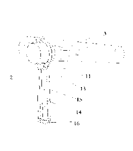
In the drawings: 1: brace; 2: vein display device; 3: an acupoint locating
model; 4: superficial
vein; 11: ring-shaped bracket; 12: support ring; 13: slip ring; 14: first
support rod; 15: second
support rod; 16: folding joint; 17: locking knob; 18: arc-shaped snap ring;
19: base; 20: infrared
filter; 21: electronic camera; 22: infrared light source; 23: computer data
processing component;
24: control component; 25: liquid crystal display; 26: projector; 32: target;
33: scale; 34: a
projector bracket.
CA 03085794 2020-06-15
DETAILED DESCRIPTION
For a better understanding of the present application, the content of the
present application will
be further described below in conjunction with embodiments, but is not only
limited to the
embodiments set forth below.
Embodiment 1
As shown in FIG. 1 and FIG. 2, a device for locating a human target using
superficial vein
characteristics includes a vein display device, a brace and a human target
locating model, where
the brace includes a ring-shaped bracket and legs connected to each other in
up-down direction,
and the vein display device is disposed on the ring-shaped bracket. A
superficial vein model
includes a transparent material on which a vein map and a target hole are
arranged.
The vein display device includes an infrared light source, an infrared filter,
an electronic camera,
a computer data processing component, a control component and a liquid crystal
display which
are connected to each other in sequence. During use, the liquid crystal
display is disposed at a
higher position and configured to display a vein of a body, and the infrared
light source is
disposed at a lower position and configured to irradiate the body.
The ring-shaped bracket is provided with a support ring for supporting the
vein display device,
where the support ring is fitted with the vein display device through a gear.
Three legs are
provided, and each of the three legs includes a slip ring and a support rod
connected to the slip
ring, where the slip ring is sleeved on the ring-shaped bracket.
A cross-section of a tube of the ring-shaped bracket is non-circular, and a
hole of the slip ring is
also non-circular, so that the slip ring may be prevented from rotating around
the ring-shaped
bracket.
The support rod includes a first support rod and a second support rod
rotatably connected to
each other, where the first support rod is connected to the slip ring and is
rotatably connected to
the second support rod through a folding joint.
During use, the vein display device is placed at an appropriate position
through the brace, has a
distance of 20 to 30 cm to a body surface, and has an illuminance of 500 to
800 lumens. Thus, it
is convenient for the vein display device to display the vein of the body at
the position of the
human target to be determined. A user or another person observes the liquid
crystal display of
11
CA 03085794 2020-06-15
the vein display device from which superficial veins of the user and the vein
map of the human
target locating model can be simultaneously observed. A position of a body of
the user and a
position of a superficial vein model are adjusted to make at least two of the
superficial veins of
the user coincide with at least two veins in the vein map of the human target
locating model. At
this time, a superficial position corresponding to a target point on the human
target locating
model is a position or a projection, on the body surface, of the human target
to be located.
Embodiment 2
The embodiment 2 is basically the same as the embodiment 1, and a difference
lies in that the
human target locating model is not included, and a projector is included. The
projector is
connected to the computer data processing component, and a spectroscope is
disposed in front
of the projector. A light outgoing path of the projector is perpendicular to a
light incoming path
of the electronic camera. The spectroscope is disposed at an intersection of
the light outgoing
path and the light incoming path and is at an angle of 45 with each of the
light outgoing path
and the light incoming path. The spectroscope is a band pass filter, and is
configured to
selectively transmit visible light and reflect near-infrared light.
The projector vertically projects an image including the vein map and an
acupoint position to
the body surface, and the user or another person observes the liquid crystal
display of the vein
display device from which the superficial veins of the user and the projected
vein map can be
simultaneously observed. A position of the body of the user and a position of
the projector are
adjusted to make at least two of the superficial veins of the user coincide
with at least two veins
in the projected vein map. At this time, the superficial position
corresponding to the target point
on the projected image is the position or the projection, on the body surface,
of the human target
to be located.
Embodiment 3
The embodiment 3 is basically the same as the embodiment 1, and a difference
lies in that the
slip ring is provided with a locking knob for preventing the slip ring from
rotating around the
ring-shaped bracket.
Embodiment 4
As shown in FIG. 3, the embodiment 4 is basically the same as the embodiment
3, and a
12
CA 03085794 2020-06-15
difference lies in that: the device for locating a human acupoint using
superficial vein
characteristics further includes a base, a bottom of the second support rod is
inserted into the
base.
Embodiment 5
As shown in FIG. 4, the embodiment 5 is basically the same as the embodiment
1, and a
difference lies in that: the support rod is arc-shaped, one end of the support
rod is connected to
the slip ring, and the other end of the support rod is provided with an arc-
shaped snap ring,
where a bottom of the arc-shaped snap ring is a soft structure.
Embodiment 6
The embodiment 6 is basically the same as the embodiment 1, and a difference
lies in that a
periphery of the transparent material is embedded with a model frame which is
a tubular or
semi-tubular frame.
Application embodiment 1
As shown in FIG. 6, in a case where an acupoint is located, a method for
locating human target
using superficial vein characteristics includes the steps below.
1) The acupoint is accurately found and a shootable sign is marked on the body
surface, and a
picture, as the model, displaying both of the acupoint and the superficial
veins is acquired by a
vein display device.
2) The model in step (1) is printed according to the ratio (1: 1) by using a
transparent material,
a hole is punched on the shootable sign of the superficial vein model, and the
superficial vein
model is placed on the body surface.
3) A vein of the body where acupoint is to be located is displayed with a vein
display device; a
position of the model printed in step 2 is adjusted, or a position of the body
is adjusted, to make
at least two of the body coincide with veins in the printed model; and it is
determined that a
superficial position corresponding to the acupoint on the printed model is the
position of the
to-be-located acupoint. A sign may be marked on the body surface with a pen
through the hole
at the position of acupoint of the printed model.
13
CA 03085794 2020-06-15
Application embodiment 2
In a case where an acupoint is located, a method for locating human target
using superficial vein
characteristics includes the steps below.
1) The acupoint is accurately found and a shootable sign is marked on the body
surface, the
body surface of a patient is scanned by a 3D scanner to establish a 3D model,
a picture
displaying both of the acupoint and the superficial veins is acquired by a
vein display device,
and the picture is merged into the 3D model to obtain a dermal 3D model
including the acupoint
and the veins.
2) The model in step (1) is printed according to the ratio (1 : 1), a hole is
punched on the
shootable sign of the superficial vein model, and the superficial vein model
is placed on the
body surface.
3) A vein of the body where acupoint is to be located is displayed with a vein
display device; a
position of the printed 3D model in step (2), model printed in step 2 is
adjusted, or a position of
the body is adjusted, to make at least two of the superficial veins of the
body coincide with
veins in the printed model, and it is determined that a superficial position
corresponding to the
acupoint on the printed model is the position of the to-be-located acupoint. A
sign may be
marked on the body surface with a pen through the hole at the position of
acupoint of the
printed model.
Application embodiment 3
In a case where an acupoint is located, a method for locating human target
using superficial vein
characteristics includes the steps below.
1) The acupoint is accurately found and a shootable sign is marked on the body
surface, the
body surface of a patient is scanned by a 3D scanner to establish a 3D model,
a picture
displaying both of the acupoint and the superficial veins is acquired by a
vein display device,
the picture is merged into the 3D model to obtain a dermal 3D model including
the acupoint and
the veins, and the dermal 3D model is unfolded to form a two-dimensional
model.
2) The two-dimensional model in step (1) is printed according to the ratio (1
: 1), a hole is
punched on the shootable sign of the superficial vein model, and the
superficial vein model is
placed on the body surface.
14
CA 03085794 2020-06-15
3) A vein of the body where acupoint is to be located is displayed with a vein
display device; a
position of the printed two-dimensional model in step (2), model printed in
step 2 is adjusted, or
a position of the body is adjusted, to make at least two of the superficial
veins of the body
coincide with veins in the printed model, and it is determined that a
superficial position
corresponding to the acupoint on the printed model is the position of the to-
be-located acupoint.
A sign may be marked on the body surface with a pen through the hole at the
position of
acupoint of the printed model.
Application embodiment 4
As shown in FIG. 7, in a case where an acupoint is located, a method for
locating human target
using superficial vein characteristics includes the steps below.
1) The acupoint is accurately found and a shootable sign is marked on the body
surface, and a
picture, as the model, displaying both of the acupoint and the superficial
veins is acquired by a
vein display device.
2) The model in step (1) is projected on a body surface where the acupoint is
located according
to the ratio (1 :1) to a human body.
3) A vein of the body where acupoint is to be located is displayed with a vein
display device; a
position of the projection of the model in step 2 is adjusted, or a position
of the body is adjusted,
to make at least two of the body coincide with veins in the projected model;
and it is determined
that a superficial position corresponding to the acupoint on the printed model
is the position of
the to-be-located acupoint. A sign may be marked on the body surface with a
pen.
Application embodiment 5
In a case where an acupoint is located, a method for locating human target
using superficial vein
characteristics includes the steps below.
1) The acupoint is accurately found and a shootable sign is marked on the body
surface, and a
picture displaying both of the acupoint and the superficial veins is acquired
by a vein display
device; in a case where the acupoint needs to be located again, a vein map of
a target acupoint is
acquired, by the vein display device at a same angle, through making a patient
take a same
posture at the time the picture is acquired; the vein map and the picture
displaying both of the
acupoint and the superficial veins are processed respectively by image
enhancement algorithm;
and then a registration is performed on the vein map and the picture
displaying both of the
CA 03085794 2020-06-15
acupoint and the superficial veins to obtain the model displaying both of the
acupoint and the
superficial veins.
2) The model in step (1) is projected on a body surface where the acupoint is
located according
to the ratio (1: 1) to a human body.
3) A vein of the body where acupoint is to be located is displayed with a vein
display device; the
registration is performed on the vein map and the model acquired in step (1),
finally making the
projection of veins in the model on the body surface is completely coincided
with the main
veins under skin; and it is determined that a superficial position
corresponding to the acupoint
on the printed model is the position of the to-be-located acupoint. A sign may
be marked on the
body surface with a pen.
Application embodiment 6
In a case where an acupoint is located, a method for locating human target
includes the
following steps.
1) A picture of the superficial veins of a patient is acquired by a vein
display device according to
a posture of a standard acupoint map, where the picture includes at least two
edges or at least
two bone standard points of a body of the patient; a body of a target acupoint
on the standard
acupoint map is expanded or shrunk to a same size as the body in the picture
of the superficial
veins; and a registration is performed on the edges or the bone standard
points of the body in the
expanded or shrunk standard acupoint map with corresponding edges or
corresponding bone
standard points of the body in the picture of the superficial veins, merging
and superimposing
into one picture, and then image enhancement algorithm processing is performed
on the picture,
so that the model displaying both of the acupoint and the superficial veins is
obtained.
2) The model in step (1) is printed according to the ratio (1: 1) by using a
transparent material,
a hole is punched on the shootable sign of the superficial vein model, and the
superficial vein
model is placed on the body surface.
3) A vein of the body where acupoint is to be located is displayed with a vein
display device; a
position of the model printed in step 2 is adjusted, or a position of the body
is adjusted, to make
at least two of the body coincide with veins in the printed model; and it is
determined that a
superficial position corresponding to the acupoint on the printed model is the
position of the
to-be-located acupoint. A sign may be marked on the body surface with a pen
through the hole
16
at the position of acupoint of the printed model.
Application embodiment 7
In a case where an acupoint is located, a method for locating human target
using superficial vein
characteristics includes the following steps.
1) The acupoint is accurately found and a shootable sign is marked on the body
surface, the body
surface is photographed with a vein display device under a standard condition,
a standard map is
obtained as reference images, the reference model displaying both of the human
target and
superficial veins is made, and the reference model is stored in reference
model gallery. The
standard condition is that the model is acquired directly above the body
surface with a distance
of 20 to 25 cm from the body surface and an illuminance of 500 to 800 lumens.
2) In practical use, when the acupoint needs to be re-located, the body
surface is taken with a vein
display device to obtain real-time images. by using a computer with X86
framework computer
system of the INTEL' corporation and with the image enhancement algorithm
software installed
inside, and running Windows 8 operating system, the point feature, line
feature or surface feature
of real-time image is analyzed and extracted, and the point feature, line
feature or surface feature
reference model is analyzed and extracted, and the register is performed on
them. A rigid
transformation, an affine transformation, projection transformation, or a
bending transformation
is performed on the reference model, and coordinate transformation is
performed on the reference
model, so as to make the reference model coincide with the real-time images,
and then the
transformed reference model is projected onto the body surface. The
superficial position
corresponding to the acupoint on the projected model is the position of the to-
be-located acupoint.
3) In actual use, the real-time image is refreshed 24 times per second, with
running step 2 for each
time, in this manner the acupoint location can be dynamically displayed.
According to this application embodiment, there is no need to adjust the body,
and the reference
model obtained under the standard condition is registered according to the
actual angle and actual
distance, and then the deformed reference model is projected onto the body
surface by the
shooting angle. Accurate positioning can be achieved even if the position of
the body is different
from the original position. High-frequency refreshing of the actual captured
image enables real-
time dynamic display. It is convenient for the user to use.
17
Date recue / Date received 2021-12-15
CA 03085794 2020-06-15
Application embodiment 8
In a case where an anatomical target is located, a method for locating human
target using
superficial vein characteristics includes steps described below.
In step 1, the anatomical target and the superficial veins within 1 cm under a
superficial layer,
parallel to a scanning layer, of the human body are scanned by a computed
tomography (CT)
device or a magnetic resonance imaging (MRI) device.
In step 2, a tomographic image including the superficial veins in step 1 is
processed by an image
processing software, and a superficial vein image is extracted by a matting
method.
In step 3, both the superficial vein image and the tomographic image of the
anatomical target in
step 2 are processed according to uniform dimensions and uniform coordinates
and a scale is
added to form a model which displays both of a human target and the
superficial veins.
In step 4, the model in step 3 is projected on a body surface where the
superficial veins are
located to make a ratio of a projection on the body surface to actual
dimensions of the human
body (0.9-1.1) : 1; or the model in step 1 is printed by using a transparent
material according to
the ratio (0.9-1.1) : 1 to obtain a superficial vein mold.
In step 5, a position of the projection of the model in step 4 is adjusted,
the superficial veins are
observed, and at least two of the superficial veins are made to coincide with
veins in the
projection of the model, and a position of the human target in the projection
of the model is a
projection of the human target on the actual body surface; or the superficial
vein mold in step 4
is placed on the body surface of a user and its position is adjusted, and the
superficial veins are
observed and at least two of the superficial veins are made to coincide with
veins in the
superficial vein mold, and a position of the human target on the superficial
vein mold is the
projection of the human target on the actual body surface.
In this embodiment, a renal cyst puncture is taken as an example. FIG. 11 is a
flowchart of a
projection method in the present application.
In step 1, a spatial relationship between a renal cyst and the body surface is
learned, the
tomographic image including the superficial veins is acquired by scanning the
human body with
a tomography device.
18
A conventional continuous tomography device includes CT and MRI, and in this
embodiment,
the CT is employed to examine the renal cyst. Generally, the CT is
concentrated around the target,
and coronal and sagittal scanning is generally not performed on the body
surface parallel to the
scanning layer. At present, the existing superficial vein display device can
display veins within 1
cm from the body surface, and veins with larger diameters are easier to be
observed. When the
human target is captured, an operator is required to specifically scan 1 to 3
layers on the body
surface parallel to the scanning layer to scan subcutaneous tissues including
the veins under the
body surface within 1 cm and then to scan the target according to the same
coordinates. If only
horizontal scanning is performed, because the superficial veins are traversed,
and the horizontal
scanning may be reconstructed and simulated to the coronal scanning and
sagittal scanning by
MimicsTM medical 17.0 software.
In step 2, FIG. 12A is a schematic diagram showing a CT tomographic scan image
of surficial
veins on a right side of umbilicus in the Application embodiment 8. Referring
to FIG. 12A, the
hole in the middle of the lower part is a sunken portion of a navel, and the
superficial veins which
are not apparent can be seen in superficial layers on a right side of the
navel. When the CT image
is processed using PhotoshopTM, an original image is taken as a background
layer, a new image
layer is copied on the background layer, a selection including only the
superficial veins is depicted
by a magnetic lasso function along an edge of the superficial veins on the new
image, and the
image except the selection is deleted. In this way, FIG. 12B which is a
schematic diagram
including only the superficial veins which is obtained through processing FIG.
12A in the
application embodiment 8.
In other embodiments, color level adjustment, local brightness adjustment,
local color adjustment
and drawing contour lines on the superficial veins may also be employed to
clearly distinguish
the superficial veins from other portions of the image.
FIG. 13A is a schematic diagram of a CT tomographic scan image of superficial
veins on a left
side of the umbilicus. FIG. 13B is obtained by processing the CT image with
the same method
described above. FIG. 14A is a schematic diagram of a CT tomographic scan
image of a cyst, in
an upper part of a left kidney, as the target. A hue inversion function of the
PhotoshopTM is
employed to inverse black and white to light the background. The CT image is
further processed
by the method of FIG. 12 to obtain FIG. 14B, where the FIG. 14 is a schematic
diagram with the
highlighted cyst target and is obtained through processing FIG. 14 in the
application embodiment
8.
19
Date recue / Date received 2021-12-15
In step 3, the processed superficial vein image and the tomographic scan image
of another
anatomical target are made into anatomical models with same dimensions and
same coordinates.
In the conventional continuous tomography, all images share a common
coordinate system and
are scaled at the same proportion. Multiple tomographic images obtained in
this embodiment are
not scaled in dimension and thus still match the original coordinate system.
In the conventional
continuous tomography, a length scale will be left on the image.
FIG. 12B, FIG. 13B and the length scale are pasted on a same blank background
image, and all
the three images abut against an upper left corner of the background. Even if
an alignment sign is
not marked in advance, accurate aligning may still be implemented. FIG. 15
which shows a
synthesized model of the superficial veins in the application embodiment 8 is
obtained. Other
tomographic images are also pasted on a blank background image, and all three
images abut
against an upper left corner of the background to generate a model with the
same coordinates and
dimensions as the model of the superficial veins.
In a non-limiting application embodiment, the alignment sign is marked with
the image
processing software in advance at positions with the same coordinates of the
images to check
whether the images are aligned.
In step 4, as shown in FIG. 16, the models in step 3 are projected on the body
surface of the human
body, making a ratio of projections on the body surface to the actual
dimensions of the human
target (0.95-1.05) : 1. The ratio of the projection on the body surface to the
actual dimensions of
the human target is calculated by comparing an identification length of the
scale with an actual
length. The ration is adjusted by adjusting a projection distance or an image
size.
In step 5, it may be seen on a vein display that the superficial veins are
also projected on the body
surface and do not certainly coincide with the actual superficial veins, and a
position of the
projector or a human body position needs to be adjusted to make the
superficial veins in the model
coincide with the actual superficial veins. At least two superficial veins in
the projection need to
coincide with the actual superficial veins, and a position on the body surface
corresponding to the
target on the projected model is a projection, on the body surface, of a
target to be located.
In this application embodiment, a position and an angle at which the model of
the superficial
Date recue / Date received 2021-12-15
CA 03085794 2020-06-15
veins is projected are adjusted in a computer, so that it may be seen that the
actual superficial
veins on the body surface are unchanged in a liquid crystal display while the
projected model of
the superficial is continuously aligned with the actual superficial veins. A
vertical distance from
the target to the projection on the body surface is denoted by d, where d is
equal to the number
of layers between the target and the body surface during the CT multiplied by
a layer thickness.
A size of d may be calculated with the scale after d is directly measured on a
CT image.
In a non-limiting application embodiment, a patient is guided to adjust the
human body position,
so that it may be seen that the projected model of the superficial veins is
unchanged in the liquid
crystal display and the actual superficial veins of the patient are
continuously aligned with the
projected model of the superficial veins until at least two superficial veins
coincide with the
veins in the projected model.
Application embodiment 9
This application embodiment is similar to the application embodiment 8, except
that this
application embodiment is used for layer-by-layer locating and projection
during a surgery.
During the surgery, a vein image projected by the model of the superficial
veins coincides with
the actual superficial veins and CT or MRI images at different layers and a
synthesized
three-dimensional (3D) image are projected in a surgery region by a projector
fixed on a
projector bracket to make the ratio of the projection on the body surface to
the actual
dimensions of the human body 1 : 1. During the surgery, different images at
different layers may
be projected according to the progress of the surgery to remind a surgeon of
tissues around the
target, improving an accuracy of the surgery and reducing vice-damages.
21