Note: Descriptions are shown in the official language in which they were submitted.
CA 03086089 2020-06-17
WO 2019/137946 PCT/EP2019/050418
1
Endoscopic non-contact measurement device
Technical field
[0001] The present invention is related to devices and methods
for non-
contact measurement, in particular internal cavity scoping devices and
methods, such
as, but not limited to, endoscopic devices and methods, which allow to perform
noncontact dimensional measurements on a surface of the internal cavity. The
non-
contact measurement is advantageously performed by triangulation to obtain a
measurement, e.g. of a distance between two points, such as a cavity diameter.
Background art
[0002] In the medical field, airway measurement is receiving increased
interest, notably prior to stent placement. Physicians need to measure the
bronchi
diameter in order to select the appropriate stent or to measure airway
stenosis. For these
measurements, physicians currently use either the CT-Scan, which irradiates
the patient
or a biopsy forceps used as a size reference, but which is not so accurate as
a
measurement tool. The diameter of an airway can vary within a relatively large
range:
more than 20 mm for the trachea down to almost 2 mm for the smallest bronchi.
[0003] From US 2012/0190923 an endoscope is known which measures
the topography of a surface. The endoscope contains a projection unit and an
imaging
unit. The projection unit and the imaging unit are arranged successively in
relation to an
axis of the endoscope. The configuration of the projection unit and the
imaging unit
arranged axially behind one another on the axis permits a significantly
smaller
endoscope configuration. The endoscope comprises a projection structure in the
form of
a slide having a radially symmetrical structure. The projection structure is
illuminated by
a waveguide to obtain a colour coded light pattern. One drawback of the above
device
is that its light pattern projector has a fairly complex structure with moving
parts (the
slider), which prevents the endoscope to be further miniaturized. Another
drawback of
the above device is that it requires a user handling a user interface when
simple
measurements, such as size or diameter are required. This can be cumbersome,
since
the operator of the device will generally have both hands involved in guiding
and/or
positioning the endoscope.
Summary of the invention
[0004] It is an object of the invention to provide an endoscopic
non-contact
measurement device which can accurately measure internal cavity diameters of a
large
CA 03086089 2020-06-17
WO 2019/137946 PCT/EP2019/050418
2
size range, and which would particularly be useful in human airways. It is an
object to
provide endoscopic non-contact measurement devices that can be miniaturized
for use
in instrument channels of endoscopes, such as channels having a diameter of
2.8 mm
or less, in particular instrument channels of bronchoscopes.
[0005] It is an object of the invention to provide an endoscopic non-
contact
measurement device which is of simpler structure and/or which is easier to
use. In
particular, it is an object to provide such devices which allow for taking
automatic
measurements in real time, preferably without any user intervention.
[0006] According to the invention, there is therefore provided a
device for
non-contact measurement as set out in the appended claims. Devices according
to the
present invention are advantageously configured to perform a measurement at a
target
site remotely, i.e. without need to contact measurement points at the target
site. The
device comprises a light source and a light pattern projector comprising a
diffractive
optical element optically coupled to the light source, an imaging system
configured for
imaging a target site illuminated by the light pattern projector, a support to
which the light
pattern projector and the imaging system are attached in fixed relative
positions, and a
processing unit configured to process data acquired by the imaging system.
[0007] The support has a longitudinal axis parallel to an
optical axis of the
light pattern projector, wherein the light pattern projector and the imaging
system are
arranged at spaced apart positions along the longitudinal axis. The light
pattern projector,
in particular, the diffractive optical element, and the imaging system are
advantageously
arranged the one behind the other. They are advantageously arranged coaxially.
The
light pattern projector is advantageously configured to project a pattern
having a
(substantially) rotational symmetry about the optical axis.
[0008] According to aspects of the present invention, the light source is
operable to emit a plurality of light beams of different colours, either
sequentially or
simultaneously. Each one of the plurality of light beams is a coherent beam.
The
diffractive optical element, which is optically coupled to each one of the
coherent beams,
is configured to diffract the plurality of light beams according to separate
or distinct
patterns. The separate patterns result from the diffractive optical element
diffracting the
plurality of light beams having a different colour according to different
angles due to the
different wavelengths of light.
[0009] According to an aspect, the processing unit is configured
to
determine a measurement based on at least two positions detected in data
acquired from
a single one of the separate patterns. The at least two positions are
automatically
recognised by the controller in the acquired data, e.g. through a suitable
image
CA 03086089 2020-06-17
WO 2019/137946 PCT/EP2019/050418
3
recognition algorithm. In other words, only one pattern of a single one of the
different
colours is used to perform a measurement at a time.
[0010] An advantage of the above aspects is that the indicated
device can
be used as pointer device for making automatic measurements. By way of
example, the
surgeon or operator points at least one of the separate patterns at the target
location,
where measurement is desired, e.g. on a bronchus in a human airway, on a
restriction
in a lumen, etc., and the device will automatically perform a desired
measurement at the
location that is pointed at with the light pattern, e.g. of the size of the
bronchus, or the
diameter of the restriction. The measurement can be performed automatically,
advantageously because it is based on only one of the separate patterns. The
measurement is therefore made with the operator only needing to correctly
point the
device, as can be verified e.g. on a visual display, without any further
intervention.
Devices according to the invention therefore allow for faster diagnosis and
imaging.
[0011] It will yet be easier for the processing unit and for the
operator, when
the projected pattern is of simple shape, and allows for correct visual
positioning, e.g. a
pattern having rotational symmetry, advantageously a single circle, or a
plurality of points
or line segments arranged on a (single) circle. The measurement can be a
diameter or
perimeter of the (circle) pattern, or an area (surface) enclosed by the circle
pattern. There
is hence a one to one registration between the measurement and the pattern,
which
makes it easy for the operator to correctly position or point the device on
the
measurement points.
[0012] Advantageously, the processing unit is configured to
process each
of the separate patterns individually. Still advantageously, the processing
unit is
configured to determine for each (or multiple ones) of the separate patterns a
corresponding measurement. Each of these measurements is advantageously based
on
at least two positions detected in data acquired from the respective one of
the separate
patterns only. While it is known that different wavelengths of light will be
diffracted under
different angles, in the present aspect this is exploited to create separate
projection
patterns. The separate patterns are advantageously spaced apart. The separate
patterns, each one relating to a different one of the plurality of light
beams,
advantageously have different sizes when projected on a same plane. The
inventors
have shown that measurements at a same target distance from the device and
made
based on patterns of different colour (i.e., different light wavelengths) will
have different
accuracy considering a same distance between the measurement device and the
target
site. It has been shown that measurements with patterns formed by a light beam
of larger
wavelength (e.g. red colour) have better accuracy than when a pattern formed
by a light
CA 03086089 2020-06-17
WO 2019/137946 PCT/EP2019/050418
4
beam of shorter wavelength (e.g. blue colour) is used. Larger wavelength beams
will
however be diffracted according to larger angles and therefore form patterns
of larger
size. Such larger patterns may not cover smaller features at the target site.
Therefore,
depending on the size of the feature that one wants to measure, devices
according to
present aspects allow for using patterns of different light beam wavelengths,
so that a
same relative accuracy is obtained for measurements of any size. As a result,
the
measurement accuracy is guaranteed for any lumen sizes or depths within the
measurement range.
[0013] Advantageously, the processing unit can be coupled to the
light
source for controlling which one of the plurality of light beams, or which one
of the
separate patterns, to emit. Advantageously, the pattern or light colour can be
automatically selected, e.g. by the processing unit. It will be convenient to
note that the
separate patterns indicated above are distinct patterns and not colour coded
patterns.
Therefore, colour coded patterns are advantageously not used in aspects of the
present
invention.
[0014] According to a second aspect of the present disclosure,
there is
provided a method of performing a non-contact measurement. The method
comprises
projecting a plurality of separate patterns on a target site in the internal
cavity. The
plurality of patterns originate from a plurality of light beams of different
colours. Each one
of the plurality of light beams is a coherent beam. The plurality of light
beams are
diffracted to separate ones of the plurality of patterns. The plurality of
patterns is imaged
and a measurement is determined based on at least two positions of a single
one of the
separate patterns, e.g. via triangulation. Advantageously, a measurement can
be
determined for each of the separate patterns, e.g. based on at least two
positions of each
of the patterns. The plurality of separate patterns are advantageously not
projected
simultaneously, and a user may select, e.g. through a user interface, which
one of the
plurality of separate patterns to project. Advantageously, the measurement is
determined
by automatic recognition of measurement points at the target site. Present
methods are
advantageously performed using devices according to the present invention.
Brief description of the figures
[0015] Aspects of the invention will now be described in more
detail with
reference to the appended drawings, wherein same reference numerals illustrate
same
features and wherein:
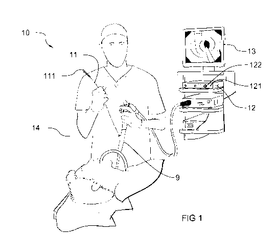
[0016] Figure 1 represents various components of a device
according to
aspects described herein;
CA 03086089 2020-06-17
WO 2019/137946 PCT/EP2019/050418
[0017] Figure 2 represents an optical assembly of the distal
part of a device
according to some aspects described herein;
[0018] Figure 3 represents a mathematical model of the optical
assembly
of Fig. 2;
5 [0019] Figure 4 represents a schematic setup of a device
according to
aspects described herein;
[0020] Figure 5 represents a graph showing the absolute
measurement
error in relation to the measurement depth for light patterns of different
wavelength (blue,
green and red), pixel error = 0.5 pixel, focal length = 165 pixels, baseline =
5 mm;
[0021] Figure 6 represents a graph showing the absolute measurement
error of the diameter in relation to the measured diameter for light patterns
of different
wavelength (blue, green and red), pixel error = 0.5 pixel, focal length = 165
pixels,
baseline = 5 mm;
[0022] Figure 7 represents a probe of a device according to a
further aspect
described herein;
[0023] Figure 8 represents a probe of a device according to a
yet further
aspect described herein;
[0024] Figure 9 represents a probe of a device to which an
external
instrument channel is attached, for guiding the probe there through according
to a further
aspect described herein;
[0025] Figure 10 represents a set of separate patterns projected
on a wall
obtained by emitting light beams of three different wavelengths through a same
diffractive optical element.
Description of embodiments
[0026] Referring to Fig. 1, non-contact measurement device 10 generally
comprises a probe 11 connected to a processing unit/controller 12 through a
cable 14. It
is alternatively possible to couple probe 11 wirelessly to controller 12. In
the instant
example for use in medical imaging, the probe 11 is configured to be inserted
in the
patient, either through the instrument channel of an endoscope 9 or through
any other
guiding endoscopic tools, such as a trocar. Probe 11 may alternatively be
inserted
directly in the patient (e.g. it may be formed as an endoscope) without
requiring additional
guiding tools. The controller 12 can be connected to a visual display 13 and
be configured
to display results on the display.
[0027] The probe 11 may be formed as a flexible and elongate
device,
extending between a proximal end 111 to which cable 14 is connected, and a
distal end
112 which forms the end which is inserted first in the patient and therefore
the end which
CA 03086089 2020-06-17
WO 2019/137946 PCT/EP2019/050418
6
is most remote from the operator. Referring to Fig. 2, at the distal end 112
of probe 11,
device 10 comprises a support to which an imaging system 16 and a light
pattern
projector 17 are attached in a fixed and advantageously spaced apart
relationship. The
support is advantageously arranged as a housing 15 which may be elongate and
advantageously cylindrical. The support may be made of a transparent plastic,
such as
PMMA, polycarbonate, or glass. It can be either moulded or machined.
Advantageously,
the probe is small enough to travel through instrument channels of endoscopes.
Specific
examples of instrument channel diameters are 2.8 mm, 2.1 mm and 2 mm.
[0028] The imaging system 16 is configured for capturing an
advantageously pixelized image of the light pattern projected by the light
pattern
projector 17. To this end, imaging system 16 can comprise an imaging sensor,
such as
a CCD or CMOS sensor, possibly coupled with an optical lens. The imaging
system is
connected to controller/processing unit 12 and possibly to a power supply
located at the
proximal end, e.g. within controller 12, through power and/or signal
transmission cables.
Alternatively, imaging system 16 can comprise an independent (carry-on) power
supply,
such as a battery. A suitable example of imaging sensor is the Omnivision
0V6946A,
which may allow the device to be used for general inspection, such as
inspection of
bronchi. The device 10 can comprise a light source which is advantageously
arranged
remotely, e.g. in controller 12. The light source can alternatively be
arranged within
housing 15. An optical fibre 171 advantageously connects the light source to
the light
pattern projector 17, which typically is arranged at the distal end 112,
within and
advantageously fixed to the housing 15. The light pattern projector comprises
a
diffractive optical element (DOE) 173, such as DE-R 220 provided by Holoeye
Photonics
(Germany). The optical system may further comprise one or more lenses 172 for
collimating the light beam carried by the optical fibre 171. A specific
example of a
collimating lens is a ball lens, such as the 2.0 mm diameter Fused Silica Ball
Lens from
Thorlabs (USA).
[0029] In the instant example, the imaging system 16 is arranged
in front
of the optical system of light pattern projector 17 in order to have a large
field of view,
and both are advantageously aligned on a longitudinal axis 151 of the housing
15. By so
doing, the imaging system 16 is located within the field of illumination 174
of the light
pattern projector 17. The imaging system 16 is however spaced apart from the
(optical
system of the) light pattern projector 17 along the longitudinal axis 151, and
therefore
the imaging system 16 only partially obstructs the field of illumination 174
of the light
pattern projector 17. It is alternatively possible to interchange the
positions of the imaging
CA 03086089 2020-06-17
WO 2019/137946 PCT/EP2019/050418
7
system 16 and the light pattern projector 17. In other words, the light
pattern projector 17
can be placed in front of the imaging system 16.
[0030] The optical axis 161 of the imaging system 16 is
advantageously
coincident with the optical axis 175 of the light pattern projector 17 and
either one or both
.. the optical axes 161, 175 can be coincident with the longitudinal axis 151.
Alternatively,
optical axis 161 can be offset from optical axis 175, e.g. optical axes 161
and 175 can
be substantially parallel and spaced apart. Optical axis 175 can e.g.
correspond to an
optical axis of the DOE 173.
[0031] Due to the placement of imaging system 16 in front of the
light
pattern projector 17, measurements can be performed of the target site which
is
illuminated by light pattern projector 17 and which is not shadowed by the
imaging
system 16. Imaging system 16 can be provided with a suitable lens adapted for
imaging
at least part of the field of illumination 174. Triangulation techniques can
be applied on
that part of the target site illuminated by the light pattern and imaged by
system 16 since
projector 17 and imaging system 16 are spaced apart.
[0032] One advantage of the disposition of Fig. 2, is that the
distance
between the light pattern projector 17, in particular the optical system and
more
particularly the DOE 173, and the imaging system 16 can advantageously be
selected
as desired without increasing the external diameter of the housing 15. As a
result, the
probe 11 can be made extremely compact to decrease the bulkiness of the
system, yet
providing desirable measurement accuracy. With such a configuration, probe 11
can be
made sufficiently small allowing for insertion through an instrument channel
or lumen of
an endoscope. The probe 11 is therefore advantageously able to travel along
with the
endoscope to reach the target site. In the alternative, it is possible to
provide probe 11
with a suitable connector, for external attachment to the endoscope. Also in
this case,
bulkiness is reduced.
[0033] The above arrangement of light pattern projector and
imaging
system (Fig. 2) advantageously uses a circular or at least rotationally
symmetrical
pattern, e.g. containing a unique point around the optical axis, such as a
circle, a square,
two parallel lines,.. .that is projected. In this case, deformation of the
pattern occurs
radially. To this end, the optical system and the DOE in particular
advantageously have
optical properties that have rotational symmetry all about the optical axis
175.
[0034] The projected pattern is preferably circular, or
advantageously has
rotational symmetry about the optical axis 175. The projected pattern may be
formed of
dots, e.g. arranged in a circle, or even a line, circle segment, arc, or any
other suitable
pattern which allows analysis by computer. Advantageously, the light pattern
is one
CA 03086089 2020-06-17
WO 2019/137946 PCT/EP2019/050418
8
which is not visually invasive and advantageously does not impede viewing of
the internal
cavity, e.g. by an endoscope monitor. The light pattern is advantageously of
simple
shape, e.g. a line, circle, or a plurality of dots or markers. The light
pattern, such as the
ones above, advantageously allows the user to position the pattern on the spot
at which
a measurement is desired. In particular, devices as described herein are
configured for
measuring distance, such as lengths (e.g., perimeter) or diameters. To this
end, the light
pattern should be positioned on the at least two points on target site between
which
distance is to be determined. Therefore, the light pattern is advantageously
such that it
easily allows for being positioned on two or possibly more points at the
target site
simultaneously. Patterns which depart from the circular geometry may be used
as well,
e.g. patterns with rotational symmetry over given angles, such as squares and
other
polygons.
[0035] As the light pattern is imaged by the imaging system 16,
its image
is acquired by the controller 12, which comprises processing means for
automatic
recognition of the light pattern, and for determining distance, advantageously
in real-
time. Generally, the more the light pattern will be of simple geometry, the
easier the
processing will be.
[0036] The light source coupled to the light pattern projector is
advantageously a monochromatic narrowband light source, typically a laser
diode. The
light source is advantageously operable to emit coherent light. One suitable
example of
such a light source is LP450-SF15 from Thorlabs, USA. The light source can be
arranged
in controller 12. The optical fibre 171 is then used to carry the light beam
emitted by the
light source to the light pattern projector 17 in housing 15. A suitable
example of optical
fibre is 5405-XP from Thorlabs, USA. Alternatively, the light source can be
provided in
housing 15.
[0037] Advantageously, the pattern assists the user in defining
the
measurement point(s) and/or enables automatic recognition of the measurement
point(s), such as e.g. two points between which a diameter or distance is
desired. It is
thus desirable that the light source is within the visible range, in order to
make the light
pattern easy to be seen by the physician which is guiding the probe 11 and/or
the
endoscope 9. An automatic recognition advantageously enables making automatic
measurements without any user handling on a user interface, such as a
graphical user
interface. Referring again to Fig. 1, a measurement can be shown on the visual
display
13 without the user having to select points with a pointing device. It will be
convenient to
note that the light pattern is dedicated for making distance measurements,
such as a
measurement between two points on the target site, for determining e.g. lumen
size,
CA 03086089 2020-06-17
WO 2019/137946 PCT/EP2019/050418
9
diameter, width, etc., or area measurements, and is not primarily intended for
surface
topography. Surface topography is advantageously not implemented in devices
according to the invention. The light pattern advantageously allows for making
real-time
distance measurements. Visualization of the measured distance advantageously
assists
the user simultaneously with imaging of the anatomical structure, e.g. as
performed by
the endoscope camera, and with making a correct diagnose with no risk of error
due to
bad 3D reconstruction. Another advantage is that the same arrangement can be
used to
project different pattern shapes simply by changing the DOE.
[0038] Referring to Fig. 4, in one aspect the light source is
operable to emit
a plurality of coherent light beams. The coherent light beams are of different
colours, e.g.
they form a set of primary colours such as though not limited to blue, green
and red (RGB
system, even though other systems may be used as well). To this end, the light
source
18 can comprise a plurality of coherent light sources 181, 182, 183, e.g.
different LEDs,
each one emitting a different colour (light wavelength). Each of the plurality
of light
sources is advantageously coupled to the same DOE 173. Each coherent light
source
181-183 may comprise suitable optics, such as a collimating lens 184 to
collimate the
light beam before it is fed to an optical fibre 185 which may carry the light
beam to the
DOE 173 at the distal end. Since the diffraction angle at the DOE 173 depends
on the
wavelength, one advantage of using coherent light beams is that each of the
coherent
light beams will be diffracted by a same DOE 173 with a different projection
(diffraction)
angle. By way of example, a DE-220 diffractive optical element (Holoeye,
Berlin), will
project a light beam of 450 nm at an angle of 190, a light beam of 532 nm at
23 and a
light beam of 650 nm at 28 . At a distance of 100 mm from the light pattern
projector, the
symmetrical pattern formed by the DE-220 DOE and obtained with the 450 nm
light beam
will have a size (diameter) of 70 mm, the pattern obtained with the 532 nm
light beam
will have a size of 85 mm and the pattern of the 650 nm light beam will have a
size of
110 mm.
[0039] Fig. 10 shows one example of a set 200 of separate
concentric
patterns as can be obtained in case of a circle as pattern. Innermost pattern
201 is a
pattern obtained by a light beam of a colour having smallest projection angle
(e.g. 450
nm in the above example). Outermost pattern 203 is obtained by a light beam of
a colour
having largest projection angle (e.g. 650 nm in the above example), and
intermediate
pattern 202 is e.g. obtained with the 532 nm light in the example above. It
can be seen
that the smaller pattern is advantageously completely contained in the larger
pattern.
Needless to say, other kinds of patterns are possible as well. Therefore, the
coherent
light beams will form spaced apart light patterns at the target site. These
light patterns
CA 03086089 2020-06-17
WO 2019/137946 PCT/EP2019/050418
may have a same shape, but will have different sizes, e.g. in case of circles,
each colour
light beam will project as a circle of different diameter at the target site
due to differing
projection (diffraction) angles. The circles are advantageously concentric. A
concentric
pattern generation has the advantage that it requires no additional space. The
smaller
5 the wavelength, the smaller the pattern. The use of several concentric
patterns of
different colours has the advantage that the pattern which best fits the lumen
size being
studied can be easily chosen for measurement. At the target site, a user may
choose
between different coherent light beams the one which best fits the cavity
feature that is
to be measured.
10 [0040] Therefore, measurements made with devices of aspects
described
herein advantageously rely on only one of the plurality of separate patterns,
i.e. they rely
on a pattern obtained with only one colour wavelength. At least two, possibly
more
positions at the target site may be determined based on data acquired from a
single one
of the plurality of patterns. The controller is advantageously configured to
determine a
measurement, such as a diameter, based on these positions only. The above may
be
repeated for each of the separate patterns, i.e. for each of the plurality of
light beams.
Each measurement made by present devices therefore is advantageously in one to
one
registration with one light beam colour. As a result, measurements are
obtained which
may relate to a same feature at the target site and which may have different
accuracy as
these measurements are obtained through the different patterns.
[0041] In one alternative, the light beams of different colours
are applied
consecutively to the probe 11. In another alternative, a beam combiner 186 is
coupled
to the plurality of coherent light sources 181-183 for combining the coherent
light beams
into a single waveguide, e.g. the optical fibre 171. This allows for
simultaneous projection
of light patterns corresponding to the different colour light beams.
[0042] Referring back to Fig. 1, a user interface 121 can be
provided,
coupled to controller 12. The user interface is advantageously configured to
allow the
operator to select which one of the different colour light beams to project.
By way of
example, the user interface 121 can comprise a control knob 122 to this end.
The specific
choice of pattern can depend on the actual size of the structure that is to be
measured
and/or on the desired accuracy.
[0043] Referring to Fig 3, an example is shown of measurement of
the wall
8 of an internal channel, such as an airway channel, e.g. the wall of a
bronchus. The light
pattern projector 17 has a projection angle a (for a given wavelength), the
imaging
system 16 has a Field-of-view (FOV), defined by its focal length fx (in pixel)
and the
pattern is detected on the wall 8 at the target location 81 with an angle [3.
The distance
CA 03086089 2020-06-17
WO 2019/137946 PCT/EP2019/050418
11
between the imaging system and the light pattern projector is d and the
measurement
depth (distance between imaging system and measurement point 81, along the
optical
axis 161) is z. The radius of the channel is rand r0 is the radius of the
light pattern FOV
at position of the imaging system 16. It can easily be shown that:
Px
tan 13 = ¨fx.
Depth (z) is given by:
= ig
d tan a ro
z ____________________________ .
tan ¨ tan a = tanig ¨ tan a
In the above, px is the pixel position of the detected point. The above
equations are given
for a given plane, and are valid for every plane around the longitudinal axis.
[0044] In the following it is shown that the measurement accuracy for a
given projection angle a depends on the size of the channel (radius r). Depth
error 6z is
given by:
= ____________
opx z2 = opx z2
6z _________________ .
fx d tan a fro
Figure 5 shows a plot of the error 6z in function of depth z. It represents
the
measurement accuracy of the device in relation to depth for three different
light
wavelengths and using a DOE DE-R 220 (Holoeye, Berlin).
[0045] For a diameter measurement, depth is less important. It
can be
shown that the error 6r on radius (r) is given by:
= OPx (r¨d tan a)2 = OPx (r¨r0)2
6r
fx d tan2a fro tan a .
The error 6r on the radius is plotted in Fig. 6 as error on diameter in
relation to the lumen
diameter. Both Fig. 5 and Fig. 6 show that sub-mm accuracy is achievable. In
order to
increase the accuracy, light of a larger wavelength can be used. However, this
results in
larger patterns which may not be visible in small lumens, since there may be
no overlap
between the imaging area (camera FOV) and the projected pattern at the target
site. In
small lumens (or for measuring small features), it is advantageous to use a
small pattern
to make the measurement. As shown in Fig. 6: for a given diameter, the
absolute error
decreases with increasing wavelength. For a given wavelength, the absolute
error
increases exponentially with diameter. From the plots it can be deduced that
larger
wavelengths will give a more accurate measurement for larger diameters.
Smaller
wavelengths are advantageously used for smaller diameters, in particular
because larger
wavelengths will be diffracted with larger angles and the resulting pattern
may be too
large and fall out of range of the boundary of the target structure. In the
particular
example of determining the size of a bronchus, a pattern formed with red
coloured light
CA 03086089 2020-06-17
WO 2019/137946 PCT/EP2019/050418
12
(larger wavelength) may be too large and therefore may be hidden from the
camera field
of view in the bronchus. In such case, either the operator, or the system
itself may switch
to a pattern of different colour, e.g. of blue light (smaller wavelength)
resulting in a smaller
pattern which may be visible by the camera. In case the bronchus size is
large, both the
red light and blue light patterns will be visible. However, measurements made
based on
the red light pattern will have better accuracy. Therefore, the device may be
set to use
the red light pattern in this case.
[0046] It is alternatively possible, if space requirements
permit, to position
the light pattern projector 17 and the imaging system 16 in a juxtaposed and
spaced
apart position, instead of aligning them on a same axis. In such case, it may
be
advantageous to project linear patterns, e.g. straight lines, or a plurality
of dots or
markers aligned along straight lines. The DOE 173 will diffract different
wavelengths,
such as red, green and blue light with different projection angles, and the
spacing
between parallel pattern lines will differ between different light
wavelengths. Such an
arrangement may be useful e.g. for measuring vocal folds.
[0047] It is known that an imaging sensor (CCD or CMOS) is
typically a
single chip with a planar matrix of light sensitive elements. The energy of
the light incident
to each element is converted into a signal charge which is output from the
sensor. This
charge, however, only represents the intensity of the light that was incident
on a
.. particular light sensitive element. It does not produce colour images. To
produce colour
images, in general, digital imaging systems employ a filtering scheme to look
at the
incoming light in a bundle of primary colours, typically three, such as red,
green and blue
(RGB). There are basically two possible ways of organising a digital imaging
system to
produce colour images. In a first possibility, each of the light sensitive
elements has
broadband spectral sensitivity. A cooperating filter disc passes a series of
colour filters,
e.g., red, green and blue filters, through the light beam in a repeating time
sequence.
The filter interpositions are synchronised to image scanning, with the filter
typically being
interposed during an entire field scan. Devices operating in this manner are
said to
produce a "field sequential" colour signal. The filter disc can be arranged
either at the
.. light projector side, e.g., before light is emitted to the target site, or
at the imaging system
side, e.g. prior to the light being incident on the light sensitive elements.
In an alternative
possibility, a mosaic of individual selectively transmissive filters is
superposed in one-to-
one registration with individual light sensitive elements. Neighbouring light
sensitive
elements have superposed a filter which selectively transmits a different
primary colour.
The signal charges acquired by these neighbouring light sensitive elements are
therefore
representative of different primary colours. The image is then digitally
reconstructed by
CA 03086089 2020-06-17
WO 2019/137946 PCT/EP2019/050418
13
interpolating the colour for each pixel of the image using the intensity of
the colours
detected at elements in a neighbourhood around the pixel location. Such
interpolating
algorithms are referred to as demosaicing algorithms. One known type of such
mosaic
filter is a Bayer filter and described in further detail in US 3971065 to
Bayer, 20 July
1976.
[0048] Regardless of the filtering scheme employed in a digital
imaging
system, the imaging sensor advantageously acquires signal charges relating to
the
different spectral regions of the light reflected by the target site
separately, such as
though not necessarily in different channels, e.g. a red channel, a green
channel and a
blue channel. The different channels are coupled for acquisition to the
processing unit
12 which can be implemented with a demosaicing algorithm to produce a desired
colour
image.
[0049] In one aspect of the invention, the coherent light beams
projected
by the light pattern projector 17 are individually acquired in different
signals, e.g. through
different channels of the imaging system 16. By way of example, coherent light
sources
181-183 are configured to emit light in wavelength regions corresponding to
those of the
different channels of the imaging sensor, e.g. the primary colours such as
red, green and
blue. The processing unit 12 is advantageously implemented to process the
colour
signals corresponding to each coherent light beam emitted by the light pattern
projector
17 separately from one another. By so doing, image segmentation for detecting
the
different light patterns is greatly simplified. Light pattern reconstruction
is consequently
more robust resulting in a more reliable measurement.
[0050] It will be convenient to note that other suitable colour
(spectral)
schemes can be applied, even employing more than three channels. Additionally,
applicable filtering schemes are not limited to visible light. By way of
example, the light
pattern projector 17 can be configured to emit light in the infrared spectral
region,
particularly in a near infrared spectral band. Advantageously, each of the
light patterns
reflected by the target site 8 is acquired as a separate (colour) signal, with
no or
insignificant interference from light emitted by the other light beams.
[0051] In this regard, it is not required that the spectral region of
a coherent
light beam be captured by only one channel of the imaging sensor. By way of
example,
a yellow pattern (588 nm) is captured simultaneously by the green and red
channel of a
RGB imaging sensor and is not present on the blue channel.
[0052] The controller 12 is advantageously configured to process
the data
acquired by the imaging system 16. As already indicated, the sensor of the
imaging
system 16 may comprise different colour channels, and data from each such
colour
CA 03086089 2020-06-17
WO 2019/137946 PCT/EP2019/050418
14
channel may be acquired separately by the controller 12. The controller 12 may
be
configured to process the data of each colour channel separately. Multiple
colour
channels may each acquire a light pattern relating to a different colour.
These light
patterns may be distinct, i.e. each pattern is created by a different light
wavelength of the
light pattern projector 17 flight source 18. Alternatively, or in addition,
some (but not all)
the colour channels may acquire data relating to a same light pattern, e.g.
where the light
pattern is projected in a light wavelength which is captured by two (or
possibly more)
colour channels of the imaging system sensor. The controller 12 may be
implemented
with a suitable algorithm for detecting the light patterns. By way of example,
a
background subtraction method, e.g. as available from the OpenCV library,
could be
used to easily detect patterns. In case of RGB colour channels, and a light
pattern with
a yellow light wavelength being used, the red and green channel can be merged
and
subtracted from the blue channel.
[0053] The controller 12 may be configured to determine a
(distance)
measurement between two points at the target site for at least one, and
advantageously
for each of the light patterns acquired, or alternatively, for each of the
colour channels
that it acquires. These different measurements advantageously relate to one
and the
same feature at the target site. On the basis of the distances or measurements
that have
been determined, the controller 12 may be configured to determine the distance
with
.. lowest measurement error. As indicated above, since the measurement error
for a given
distance is dependent on the wavelength of the light pattern, the controller
can easily
verify which of the wavelengths used will result in highest accuracy. This may
be
accomplished e.g. by storing a lookup table in readable memory included in the
controller
12. The lookup table may comprise for each wavelength, a measurement error
related
to a given distance measurement. In this case, controller 12 may be configured
to
compare entries of the lookup table for a given distance.
[0054] The controller 12 may be configured to determine three
dimensional
coordinates of points along the imaged pattern. In case of rotationally
symmetrical
patterns, such as a circle, with the coordinates of circle points, the
controller may be
configured to calculate one or more diameters, such as one or more of: an
average
diameter, a minimum or maximum diameter. Such measurement may be repeated for
each pattern, in particular each light wavelength, that is imaged. The
measurements,
such as diameters, may be visualized on the display 13.
[0055] The patterns are advantageously of a light wavelength in
the visible
range. One advantage is that the operator immediately receives feedback on
whether
the pattern is positioned on the desired target structure to be measured.
Rather than
CA 03086089 2020-06-17
WO 2019/137946 PCT/EP2019/050418
positioning measurement points with an indicator on a display 13, the operator
positions
the pattern directly on the target structure by positioning the probe 11.
[0056] It may be possible to project light patterns of different
wavelengths
successively. The controller may be configured to start with a first light
pattern, e.g.
5 corresponding to a longest or shortest light wavelength, and sequentially
changing the
light wavelength until a measurement having a desirable accuracy is obtained.
By way
of example, when the target structure is small, projecting a light pattern of
longer
wavelength (e.g. red colour) may result in a pattern that does not interfere
with the target
structure (because it is too large). In such case, the controller may be
configured to
10 change the projected light wavelength which alters the size (and colour)
of the projected
light pattern. This may be implemented either automatically, e.g. the
controller shifts
wavelength when no measurement can be detected, or manually, e.g. with a push
button
allowing the operator to change projected wavelength. In these cases a light
pattern
corresponding to one light wavelength may be projected at a time.
15 [0057] By way of example, a diameter measurement of a bronchus
is made
preferably using a circular pattern. The pattern is positioned to fit the
lumen and an image
of the pattern is recorded and fed to the controller. The pattern can be
automatically
reconstructed in 3D, and the diameter determined according to known
techniques. In
case the probe is not centred in the bronchus lumen, the 3D reconstruction of
the pattern
will be tilted with respect to the optical axis. This tilt can be detected by
the controller and
which may be configured to emit a warning signal to the user/operator or to
automatically
correct the measurement. Another possibility is to combine several of the
different
patterns. In this case, the 3D reconstruction of the different patterns
provides additional
information of the lumen and may provide all the required data to get a
reliable diameter
measurement. In case of non-circular anatomical shapes, several measurements
may
be obtained through the pattern (reconstruction), all of which may be
displayed on the
visual display, such as the surface, the maximum diameter, the minimum
diameter or
any geometrical data that could be obtained.
[0058] When the contours of the structure that is to be measured
is
irregular, e.g. due to malformation, geometric quantities such as smallest
circumscribed
circle, and largest enclosed circle can be determined by controller 12 based
on the
automatically recognised/reconstructed pattern.
[0059] Controller 12 is therefore advantageously equipped with
suitable
software and/or hardware allowing for automatically recognising the pattern
and/or
measurement points on the pattern. In addition, the controller 12 is
advantageously
equipped with suitable software allowing for automatically determining a
measurement
CA 03086089 2020-06-17
WO 2019/137946 PCT/EP2019/050418
16
from the automatically recognised pattern and/or measurement points. The
measurement is advantageously a value representative of a diameter, perimeter,
distance, or area. The pattern advantageously has a circular shape, e.g. a
full circle, or
line segments or points located on a circle. Such shape advantageously allows
easy
positioning of the pattern by the operator on the location where measurement
is desired,
and also allows easy automatic recognition and/or measurement calculation.
[0060] Advantageously, the controller is configured to perform
sequential
measurements, either with the same pattern, or with the separate patterns of
different
colours. Taking sequential measurements at e.g. regular time intervals with a
same
pattern may be useful for profiling of an internal lumen, or for averaging.
Since the
patterns of different colours have different size, taking sequential
measurements with
these patterns at a same target size may be useful for averaging, or for
measuring
different structures of the target size, e.g. at a constriction, the largest
pattern may
measure the size of the unobstructed aperture, while the smallest pattern may
measure
the size of the obstruction or of the obstructed aperture.
[0061] Advantageously, the light pattern projector 17 may
comprise
additional optical elements, in particular light refractors, in order to take
advantage of the
refraction to adjust the direction of the light pattern. Referring to Fig. 7,
a light refractor
176 may be arranged downstream of the DOE 173. The light beam is first
diffracted using
the DOE 173 and the beam is refracted in another advantageous direction. Both
combined give the projection angle. Using light refraction enables to have a
final smaller
projection angle and thus measure smaller elements. In Fig. 7, the shape is
optimised in
order to generate a telecentric pattern 177 (constant size along depth) in
order to
measure smaller elements.
[0062] In addition to any of the above, and referring to Fig. 8, the probe
may comprise a graduated indication 152 on an outer surface of the housing 15.
The
indication 152 may be arranged along the longitudinal axis 151 of housing 15
and enable
the endoscopist to determine how deep the probe has been inserted. This may
enable
to analyse e.g. how long a stenosis is. Measurement could be made using
another
endoscope (e.g. if the device passes through the instrument channel) or
directly outside
the patient as the user moves the probe in the patient.
[0063] The indication 152 can be an electronic encoder (e.g.,
optical
encoder), to measure automatically the travel distance of housing 15. The
encoder
readings can be made in registration with the measurements based on the light
pattern
(diameter, area, perimeter). This can be useful, e.g. for determining the
distance over
which a contraction in a lumen extends. To this end, the electronic encoder
can have an
CA 03086089 2020-06-17
WO 2019/137946 PCT/EP2019/050418
17
output coupled to controller 12 for reading travel distance in registration
with performing
the measurement(s).
[0064] Advantageously, the light pattern projector 17 is
configured to
generate a light pattern, e.g. one or more lines, possibly each in a different
colour due to
the use of coherent light beams of different colours, and one or more fiducial
elements
of light. One or more fiducial elements may be generated for each of the
different colours.
[0065] The fiducial element of light is configured to form a
fiducial marker
on the surface of target site. The fiducial marker is advantageously shaped
such that it
is discernible by the operator, e.g. on the visual display 13 coupled to the
controller 12.
.. The operator manipulating the scoping instrument 9 can thus position the
fiducial marker
on a location of interest on the target site 8 merely by appropriate
manipulation of the
scoping instrument 9.
[0066] In some cases, it may be useful to measure entire 3D
surfaces
instead of only determining a distance measurement, e.g. between two
anatomical
points. In this case, multiple light sources are advantageously switched on
successively.
This enables e.g. to scan along the lumen. Additionally, some of these light
sources can
be switched on together too. In this case, advantageously more than three
light sources
are used.
[0067] The device 10 can further comprise an illumination light
source. The
illumination light source may be a source of white light or at least broadband
light, which
may not be coherent. The illumination light source can be optically coupled to
the DOE
173 such that light emitted from the illumination light source passes through
the DOE.
Another possibility is to use a coherent light source that is not optically
coupled to the
DOE, such that its light is not diffracted using the DOE to provide a
homogeneous
illumination. Preferably, the illumination light source emits in a wavelength
band different
from the light wavelength of the light source 18 used for generating the
pattern(s). The
illumination light source and the pattern may be captured by different
channels of the
camera. By way of example, if the light source 18 is projecting a red pattern,
the
illumination light source can be blue and green. The illumination light source
may be
configured to emit in a plurality of selectable wavelengths.
[0068] A protecting sheath can be placed on the probe 11 in order
to isolate
the patient and any reusable part, such as the housing 15. This allows the
probe to be
easily reused with no contamination hazard. Additionally, the sheath can allow
for
preserving the optical properties of the probe, e.g. against scratching, as
well as
damages related to sterilisation.
CA 03086089 2020-06-17
WO 2019/137946 PCT/EP2019/050418
18
[0069] Referring to Fig. 9, in case the conventional endoscopic
system has
no or a too small instrument channel, an external instrument channel 91 can be
placed
on the endoscope, through which the probe 11 can be moved. This external
instrument
channel may be attached to the endoscope through a sheath or a plastic or
silicone ring
92.