Note: Descriptions are shown in the official language in which they were submitted.
CA 03087567 2020-07-02
WO 2019/136146 PCT/US2019/012179
ECHOGENIC CATHETER AND CATHETER SYSTEM
TECHNICAL FIELD
[001] The present application relates generally to medical instruments or
devices having
echogenic properties or features, in addition to or as an alternative to,
radiopaque properties or
features, to facilitate detection of the medical instrument or device during
medical procedures by
suitable imaging methods, such as ultrasound imaging and/or X-ray imaging
methods. More
particularly, the present application relates to catheters having echogenic
properties or features,
in addition to or as an alternative to, radiopaque properties or features, to
facilitate detection of
the catheter by ultrasound imaging and/or X-ray imaging methods to assist a
clinician with
insertion, placement, and maintenance of the catheter during intravascular
(IV) therapy, for
example.
BACKGROUND
[002] Peripheral IV catheter placement is the most common invasive hospital
procedure
and required by up to 90% of hospitalized patients. Clinical standards suggest
removing IV
catheters when clinically indicated; however, up to 50% of placed IV catheters
are removed
earlier than intended due to complications associated with the placement of
the IV catheter.
[003] Placing an IV catheter into a vein under the skin of a patient,
particularly, a
"difficult venous access" (DVA) patient, can be difficult. When a catheter is
inserted into a vein
of a DVA patient, ultrasound equipment is frequently used to help the
clinician see the patient
anatomy and then guide the IV catheter and needle into a proper position to
facilitate IV therapy.
However, the use of ultrasound imaging techniques requires a skilled clinician
and an expensive
ultrasound imaging device. Moreover, while ultrasound imaging may be useful to
detect
relatively dense materials, it may not detect less dense material, such as
conventional catheters.
The ability to detect the catheter using ultrasound imaging methods is
particularly important after
the needle is removed, e.g., to maintain the IV catheter properly placed
during the IV therapy
and/or retrieve dislodged, failed, or damaged catheters.
BRIEF SUMMARY OF SOME EXAMPLE EMBODIMENTS
[004] In one aspect, a medical device includes a catheter adapter and a
cannula extending
distally from the catheter adapter. The cannula forms a lumen having a length
between a first end
1
CA 03087567 2020-07-02
WO 2019/136146 PCT/US2019/012179
and an opposing second end of the cannula. The lumen extends parallel to a
longitudinal axis of
the cannula and along at least a portion of the length. The cannula includes
at least one echogenic
stripe extending along at least a portion of the length of the cannula.
[005] In another aspect, a catheter has a distal end and an opposing
proximal end. The
catheter includes a catheter adapter and a cannula extending distally from the
catheter adapter.
The cannula forms a lumen extending between the distal end and the proximal
end of the catheter
parallel to a longitudinal axis of the catheter. One or more stripes are
formed in the cannula. The
one or more stripes extend along at least a portion of a length of the
cannula. The one or more
stripes have echogenic properties or features.
[006] In yet another aspect, a method for forming a stripe in a cannula of
a catheter includes
extruding a thermoplastic polymer material through an array of dies aligned
such that a stripe
made of a first thermoplastic polymer material and having echogenic properties
or features is
formed between and bonded to a second thermoplastic polymer material forming
adjacent wall
portions of a wall of the cannula.
BRIEF DESCRIPTION OF THE DRAWINGS
[007] The detailed description is described with reference to non-limiting and
non-exhaustive
embodiments illustrated in the accompanying figures. The same reference
numerals in different
figures refer to similar or identical items.
[008] FIG. 1 is a perspective view of an example catheter, according to
various embodiments;
[009] FIG. 2 is a portion of the example catheter shown in FIG. 1, according
to various
embodiments;
[0010] FIG. 3 is an enlarged section of the example catheter portion shown in
FIG. 2, according
to various embodiments; and
[0011] FIG. 4 is a perspective view of an example catheter system, according
to various
embodiments.
DETAILED DESCRIPTION
[0012] Various embodiments are described below with reference to the
drawings in
which like elements generally are referred to by like numerals. The
relationship and functioning
of the various elements of the embodiments may better be understood by
reference to the
2
CA 03087567 2020-07-02
WO 2019/136146 PCT/US2019/012179
following detailed description. However, embodiments are not limited to those
illustrated in the
drawings. It should be understood that the drawings are not necessarily to
scale, and in certain
instances details may have been omitted that are not necessary for an
understanding of
embodiments disclosed herein, such as ¨ for example ¨conventional fabrication
and assembly.
[0013] The invention is defined by the claims, may be embodied in many
different forms,
and should not be construed as limited to the embodiments set forth herein;
rather, these
embodiments are provided so that this disclosure will be thorough and
complete, and will fully
convey enabling disclosure to those skilled in the art. As used in this
specification and the
claims, the singular forms "a," "an," and "the" include plural referents
unless the context clearly
dictates otherwise. Reference herein to any industry standards (e.g., ASTM,
ANSI, IEEE
standards) is defined as complying with the currently published standards as
of the original filing
date of this disclosure concerning the units, measurements, and testing
criteria communicated by
those standards unless expressly otherwise defined herein. The terms
"proximal" and "distal" are
used herein in the common usage sense where they refer respectively to a
handle/doctor-end of a
device or related object and a tool/patient-end of a device or related object.
The terms "about,"
"substantially," "generally," and other terms of degree, when used with
reference to any volume,
dimension, proportion, or other quantitative or qualitative value, are
intended to communicate a
definite and identifiable value within the standard parameters that would be
understood by one of
skill in the art (equivalent to a medical device engineer with experience in
this field), and should
be interpreted to include at least any legal equivalents, minor but
functionally-insignificant
variants, standard manufacturing tolerances, and including at least
mathematically significant
figures (although not required to be as broad as the largest range thereof).
[0014] In example embodiments described herein, example medical
instruments or
devices, such as catheters, have echogenic properties or features, in addition
to or as an
alternative to, radiopaque properties or features, to facilitate detection of
the medical instruments
or devices during medical procedures using suitable imaging methods, such as
ultrasound
imaging and/or X-ray imaging methods. For example, in certain embodiments,
example catheters
have a relatively increased echogenicity to facilitate a clinician with
detecting the catheter with
ultrasound imaging methods to assist the clinician with the insertion and/or
maintenance of the
catheter, for example.
3
CA 03087567 2020-07-02
WO 2019/136146 PCT/US2019/012179
[0015] In example embodiments, an increased radiopacity or radio density
increases the
relative inability of certain electromagnetic radiation, e.g., a radio wave or
an X-ray portion of
the electromagnetic spectrum, to pass through a particular material.
Radiopaque volumes of
material have a white appearance on radiographs, compared with a relatively
darker appearance
of radiolucent volumes. For example, on typical radiographs, bones look white
or light gray
(radiopaque), whereas muscle and skin look black or dark gray, being mostly
invisible
(radiolucent). A radiopacifier contained in a medical devices enhances the
visualization of the
medical device during implantation for temporary implantation devices, such as
catheters or
guidewires, or for monitoring the position of permanently implanted medical
devices, such as
stents, hip and knee implants, and screws. While metal implants typically have
sufficient
radiocontrast such that an additional radiopacifier is not necessary, polymer-
based devices may
require incorporation of materials with high electron density contrast
compared to the
surrounding tissue. Examples of suitable radiocontrast materials include
titanium oxide,
tungsten, barium sulfate, zinc oxide, iron oxide, platinum oxide, and
zirconium oxide.
[0016] Alternatively or in addition, an increased echogenicity increases
an ability of the
medical device to reflect an echo, e.g., return a signal during ultrasound
examination. For
example, when gas voids, cores, or bubbles are caught in an ultrasonic
frequency field, the gas
voids, cores, or bubbles may compress or oscillate to reflect a characteristic
echo to generate a
strong and unique sonogram in contrast-enhanced ultrasound. In certain
embodiments, the gas
voids, cores, or bubbles are composed of a suitable gas, such as air or heavy
gases, e.g.,
perfluorocarbon or nitrogen.
[0017] When a catheter is inserted into a vein of a "Difficult Venous
Access" (DVA)
patient, ultrasound equipment is frequently used to help the clinician see the
patient anatomy and
then guide the IV catheter needle and catheter into the proper position to
facilitate IV therapy.
The use of ultrasound is useful but sometimes difficult to learn and master.
For example, the
plane of an ultrasound beam is very thin ¨ several thousandths of an inch
thick or wide and it is
sometimes difficult for the clinician to see the catheter and associated
needle when using
ultrasound for placement of the catheter and needle.
[0018] Some conventional catheters or needles are echogenic (making the
catheter or
needle more visible by ultrasound imaging methods). In these conventional
catheters or needles,
material is added or a surface finish is changed or textured to better reflect
the ultrasound energy.
4
CA 03087567 2020-07-02
WO 2019/136146 PCT/US2019/012179
The increased texture reflects the ultrasound energy and appears on ultrasound
images. However,
an increase in material of the catheter or needle or texturing of a surface of
the catheter or needle
may undesirably promote thrombosis formation and/or blood clotting.
[0019] In example embodiments described herein, echogenic enhancing
features are
added to radiopaque stripes of the catheter tubing (e.g., gas bubbles,
chemically formed bubbles,
glass balloons, voids, irregularities, and/or relatively denser material such
as tungsten, glass
beads, or sand) while maintaining a smooth surface on the outer diameter (OD)
and the inner
diameter (ID) of the catheter tubing. In a particular embodiment, for example,
virtually
transparent tungsten particles having an average diameter less than 100
nanometers (nm) are
added to radiopaque stripes to provide an increased radiopaque response and
increased flashback
visibility. Improving the echogenic features of the stripe can be accomplished
in a variety of
methods as described herein. For example, in certain example embodiments,
chemical blowing
agents are added into the radiopaque material of the stripes as the material
is co-extruded with
the traditional catheter material to provide these benefits while maintaining
a very smooth finish
on surfaces that may contact bodily fluids, such as blood. In alternative
example embodiments, at
least a portion of an outer surface of the cannula and/or at least a portion
of an inner surface of
the cannula forming the lumen includes an intentionally regularly patterned
surface.
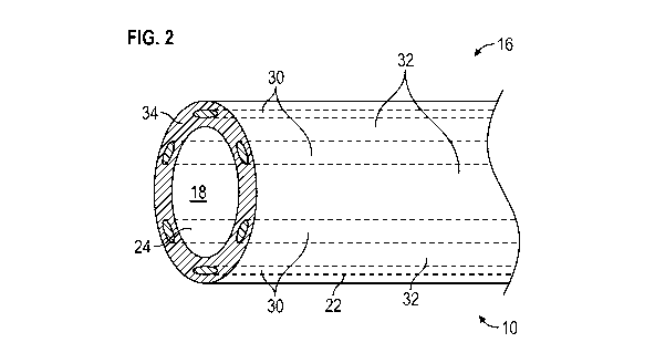
[0020] Referring now to the figures, and initially to FIGS. 1-3, an
example catheter 10
has a distal end 12 and an opposing proximal end 14. Catheter 10 includes a
catheter adapter 15
and a cannula 16 extending distally from catheter adapter 15. In example
embodiments, catheter
adapter 15 is configured to couple catheter 10 to a small bore Luer taper lock
fitting. Cannula 16
has a length extending from distal end 12 toward opposing proximal end 14 of
catheter 10 in
certain example embodiments. Catheter 10 forms or defines a lumen 18 extending
between distal
end 12 and proximal end 14 of catheter 10 along or parallel to a longitudinal
axis 20 of catheter
10. In example embodiments, cannula 16 includes a thin tube having an outer
diameter (OD) and
an inner diameter (ID) forming lumen 18 that extends through catheter 10 and,
in certain
embodiments, beyond a distal end of catheter adapter 15 in the proximal
direction. Referring
further to FIGS. 2 and 3, in example embodiment, an outer surface 22 of
cannula 16 (defining the
outer diameter of cannula 16) and an opposing inner surface 24 of cannula 16
forming lumen 18
(defining the inner diameter of cannula 16) comprise a smooth, anti-
thrombogenic surface to
CA 03087567 2020-07-02
WO 2019/136146 PCT/US2019/012179
prevent or limit accumulation of tissue and/or fluids on the surface, e.g.,
coagulation or clotting
of blood on outer surface 22 and inner surface 24.
[0021] In example embodiments, catheter adapter 15 is configured to
couple to a
cooperating small-bore fitting or connection, tubing, a hub, or another
suitable connection such
that lumen 18 provides a fluid flow path through catheter 10. In example
embodiments, lumen 18
has a suitable diameter or a suitable cross-sectional dimension to facilitate
fluid flow through
catheter 10. Additionally or alternatively, lumen 18 may accommodate a medical
device or
instrument, such as a needle or an obturator, for example, which is movably
positioned within
lumen 18.
[0022] As shown in FIGS. 2 and 3, at least a portion of catheter 10,
e.g., at least a portion
of cannula 16 at distal end 12 of catheter 10, includes one or more stripes 30
having echogenic
properties and/or features and/or radiopaque properties and/or features, e.g.,
a plurality of stripes
30 extending generally parallel to longitudinal axis 20 of catheter 10 along
at least a portion of a
length of catheter 10. In example embodiments, stripes 30 are formed in
cannula 16 using a
suitable method or technique to maintain the smooth outer surface 22 and inner
surface 24 of
cannula 16. For example, cannula 16 may be formed during a die extrusion
process by extruding
a suitable material, e.g., a biocompatible thermoplastic polymer material such
as a polyurethane
or fluoropolymer material, through an array of dies aligned such that each
stripe 30 is formed
between and bonded to extruded material forming adjacent transparent wall
portions 32 of a wall
34 of cannula 16. In this extrusion process, a first thermoplastic polymer
material is extruded
through one or more secondary dies, e.g., a plurality of secondary dies, such
as six secondary
dies, to form respective stripes 30 having echogenic properties and/or
features and/or radiopaque
properties and/or features and a second thermoplastic polymer material is
extruded through a
major die to form transparent wall portions 32. As a result of this extrusion
process, one or more
stripes 30 are formed encapsulated into wall 34 of cannula 16 while
maintaining the smooth
outer surface 22 and smooth inner surface 24 of cannula 16. Because only
stripes 30 include the
echogenic properties and/or features and/or the radiopaque properties and/or
features, the surface
finish of cannula 16 is preserved. In alternative example embodiments, at
least a portion of the
outer surface of cannula 16 and/or at least a portion of the inner surface of
cannula 16 forming
lumen 18 includes an intentionally regularly patterned surface. Further, the
clear or transparent
wall portions 32 provide a clinician with visibility through cannula 16 to
visualize blood flow
6
CA 03087567 2020-07-02
WO 2019/136146 PCT/US2019/012179
through lumen 18, i.e., blood flow occurring in the annular space between the
outer diameter of
the needle inserted in lumen 18 and an inner diameter of cannula 16, to
confirm proper
placement of the needle tip in a patient's vein, for example.
[0023]
As described above, in example embodiments, stripes 30 may include radiopaque
properties or features.
In example embodiments, stripes 30 include a biocompatible
thermoplastic polymer material filled with a material or substance opaque to x-
rays, thereby
rendering stripes 30 visible under fluoroscopy or x-ray imaging. These
fillers, or radiopacifiers,
e.g., dense metal powders, affect the energy attenuation of photons in an x-
ray beam as the x-ray
beam passes through stripe 30, reducing an intensity of the photons by
absorbing or deflecting
them. Because stripes 30 exhibit a higher attenuation coefficient than soft
tissue or bone, stripes
30 will appear lighter on a fluoroscope or x-ray film. This visibility may
provide the contrast
needed to accurately position or place catheter 10 in the desired vein. In
particular embodiments,
the image contrast and sharpness can be varied by a type and/or an amount of
radiopacifier in
stripes 30, and can be tailored to a specific application of catheter 10.
[0024]
For example, a higher loading of radiopaque material may be needed for a thin-
wall catheter cannula or tubing than for a catheter cannula or tubing with a
thicker wall. The
amount of additives may also be limited to prevent overloading, which may
result in a loss of the
material's mechanical properties. Suitable radiopacifiers for stripes 30
include, without
limitation, barium sulfate, bismuth compounds (bismuth trioxide, bismuth
subcarbonate, or
bismuth oxychloride), tungsten, titanium, and zirconium oxide, which include
metals that are
excellent absorbers of x-rays. One or more radiopaque materials, e.g., a blend
of barium sulfate
and a bismuth compound, may be incorporated into stripes 30.
[0025]
In addition to the radiopaque properties or features, or, in alternative
embodiments, as an alternative, stripes 30 include echogenic properties or
features. As shown,
for example, in FIG. 3, in an example embodiment, stripes 30 include a
plurality of voids 32
between outer surface 22 and inner surface 24 of cannula 16. In certain
example embodiments,
the plurality of voids 32 includes gas pockets or bubbles, e.g., air pockets
or bubbles, formed in a
thickness of each stripe 30 between outer surface 22 and inner surface 24 of
cannula 16. Voids
32 may be formed in stripes 30 by infusing gas, e.g., air, into the
thermoplastic polymer material
used to form stripes 30 during the extrusion process such as described above.
As gas is infused
7
CA 03087567 2020-07-02
WO 2019/136146 PCT/US2019/012179
directly into the thermoplastic polymer material during the extrusion process,
voids 32 are
formed in stripes 30 to enhance the echogenicity of stripes 30.
[0026] Voids 32 may be formed in stripes 30 using other suitable methods.
For example,
in an example embodiment, a chemical foaming agent is added to the
thermoplastic polymer
material. In this embodiment, the chemical foaming agent decomposes during the
extrusion
process to form a gas that creates gas bubbles forming voids 32.
Alternatively, various materials,
such as ceramic beads or particles (e.g., glass or carbon beads or particles),
metal beads or
particles, and/or expandable thermoplastic blowing agents and/or lightweight
fillers (e.g.,
Expancel microspheres), can be added to the thermoplastic polymer material to
create voids 32.
In certain embodiments, the void forming process may include a combination of
these methods
and/or other methods.
[0027] In an example alternative embodiment, also shown in FIG. 3,
stripes 30 include
one or more irregularities 36, e.g., a plurality of irregularities or
discontinuities 36, formed in one
or more stripes 30. The one or more irregularities or discontinuities 36 may
include, without
limitation, one or more bumps, grooves, valleys, peaks, ridges, and/or
undulations, to enhance
the echogenic reflective properties of the stripes. For example, a shape
and/or a contour of stripes
30 may be changed from a general elliptical cross-section to a cross-sectional
shape and/or a
contour having, for example, bumps, grooves, valleys, peaks, ridges,
undulations, and/or flower
pedals, optimizing the echogenicity-enhancing properties of stripes 30 for
ultrasound imaging
methods. In certain embodiments, a change in the cross-sectional shape of
stripes 30 and/or
forming irregularities or discontinuities 36 or non-planar aspects in an outer
surface of stripes 30
enhances the echogenic properties of stripes 30 potentially without having to
add any additives to
stripes 30. These changes to the cross-sectional shape of stripes 30 may be
influenced by using a
die having a corresponding profile or cross-sectional area rather than a
conventional die having a
circular or an elliptical cross-sectional area.
[0028] In alternative embodiments, stripes 30 may include a suitable,
relatively dense
material to create the echogenicity-enhancing properties of stripes 30. For
example, stripes 30
may include a relatively dense material 31, such as sand, silica, fine
particles, and/or glass beads.
This dense material may also enhance the radiopacity of stripes 30. A
combination of these
methods to create the echogenicity-enhancing properties of stripes 30 may be
used to offset a
potential decrease in radiopacity resulting from the formation of voids 32 in
stripes 30. Thus, the
8
CA 03087567 2020-07-02
WO 2019/136146 PCT/US2019/012179
methods described herein can be utilized to form stripes 30 optimized to
provide both
echogenicity and radiopacity properties or features. In alternative example
embodiments, at least
a portion of catheter 10, e.g., at least a portion of cannula 16, includes one
or more stripes 30
formed by or including a radiopaque and echogenic wire, e.g., a suitable metal
wire.
[0029] The echogenic features can be staggered, stepped, or placed for
better detection of
catheter 10. For example, the echogenic properties of catheter 10 or a system
including a needle,
catheter 10, and/or a flashback notch, for example, can be segmented with
echogenic features or
properties and non-echogenic features or properties to provide additional
information on needle
tip, depth, and/or location, for example.
[0030] Referring again to FIG. 1, at proximal end 14, catheter 10
includes an adapter,
such as a small-bore connector 40, for example. Small-bore connector 40 is
configured to
removably couple to any suitable medical device or component, for example, a
cooperating
small-bore fitting, a device, or a medical tubing. The medical device,
component, or tubing may
include a cooperating element, such as a cooperating small-bore connector, to
facilitate coupling
the medical device, component, or tubing, for example, to catheter 10. In the
example
embodiments, small-bore connector 40 includes a twist-lock mechanism to
removably couple
small-bore connector 40 to a cooperating small-bore connector having a
cooperating twist-lock
mechanism. Small-bore connector 40 can be easily disengaged from the
cooperating small-bore
connector by rotating small-bore connector 40 in an opposite direction with
respect to the
cooperating connector to disengage the threads. In alternative example
embodiments, small-bore
connector 40 may be a slip small-bore fitting that is pressed onto the
cooperating small-bore
connector.
[0031] Referring to FIG. 4, in further example embodiments, the echogenic
and, in some
embodiments, radiopaque, catheter 10, as described herein, may be combined
with other
complimentary medical devices or instruments, such as a needle 52, to
facilitate accessing veins
in DVA patients, for example. Below are a few example combinations of devices,
properties,
and/or features that may provide for an example catheter system 50 including
an example
catheter 10 as described herein:
a combination of the disclosed echogenic catheter and echogenic needle (or
tip)
technologies to provide an echogenic needle and an echogenic catheter, for
example, as described in U.S. Patent Application No. 12/930,580, U.S. Patent
9
CA 03087567 2020-07-02
WO 2019/136146
PCT/US2019/012179
Publication No. 2011/0172542, entitled "Ultrasound Guided Echogenic Catheter
and Related Methods" and U.S. Patent Application No. 15/297,731, U.S. Patent
Publication No. 2017/0112464, entitled "Echogenic Needle," each incorporated
by reference herein in its entirety;
a combination of the disclosed echogenic catheter and magnetic needle (and pre-
magnetized) technologies, as shown in FIG. 4, for example, as described in
U.S.
Patent Application No. 15/154,353, U.S. Patent Publication No. 2017/0325713,
entitled "Invasive Medical Device Cover with Magnet;" U.S. Patent Application
No. 15/154,362, U.S. Patent Publication No. 2017/0325714, entitled "Electro-
Magnetic Needle Catheter Insertion System;" U.S. Patent Application No.
15/170,518, U.S. Patent Publication No. 2017/0348510, entitled "Medical
Devices, Systems and Methods Utilizing Permanent Magnet and Magnetizable
Feature;" U.S. Patent Application No. 15/170,497, U.S. Patent Publication No.
2017/0347913, entitled "Invasive Medical Devices Including Magnetic Region
and Systems and Methods;" U.S. Patent Application No. 15/170,531, U.S. Patent
Publication No. 2017/0347914, entitled "Invasive Medical Devices Including
Magnetic Region and Systems and Methods;" and U.S. Patent Application No.
62/481,964, entitled "A System for Insertion Visualization of a Vascular
Access
Device with Multiple Magnetic Features on a Needle Assembly," each
incorporated by reference herein in its entirety;
a combination of the disclosed echogenic catheter with guidewire placement
technologies to provide an echogenic needle tip, magnetic needle tip and an
echogenic catheter in a device or a system, for example, as described in U.S.
Patent Application No. 15/604,244, U.S. Patent Publication No. 2017/0348511,
entitled "Medical Devices, Systems and Methods Utilizing Permanent Magnet
and Magnetizable Feature," incorporated by reference herein in its entirety;
a combination of the disclosed echogenic catheter with a reduced bevel length
or
shortened needle geometry for better first stick success, for example, as
described
in U.S. Patent Application No. 62/541,205 filed on August 4, 2017, entitled
"Introducer Needle for Catheter Placement," incorporated by reference herein
in
its entirety; and/or
CA 03087567 2020-07-02
WO 2019/136146 PCT/US2019/012179
a combination of the disclosed echogenic and radiogenic catheter with anti-
infective coatings, such as an anti-microbial, silver coating, or copper
coating, for
example, as described in U.S. Patent Application No. 14/326,036, U.S. Patent
Publication No. 2016/0008517, entitled "Antimicrobial Coating and Kink
Resistant Feature for Vascular Access Devices," incorporated by reference
herein
in its entirety.
[0032] Although the subject matter has been described in language
specific to structural
features and/or methodological acts, it is to be understood that the subject
matter defined in the
appended claims is not necessarily limited to the specific features or acts
described. Rather, the
specific features and acts are disclosed as illustrative forms of implementing
the claims. One
skilled in the art will realize that a virtually unlimited number of
variations to the above
descriptions are possible, and that the examples and the accompanying figures
are merely to
illustrate one or more examples of implementations. It will be understood by
those skilled in the
art that various other modifications can be made, and equivalents can be
substituted, without
departing from claimed subject matter. Additionally, many modifications can be
made to adapt a
particular situation to the teachings of claimed subject matter without
departing from the central
concept described herein. Therefore, it is intended that claimed subject
matter not be limited to
the particular embodiments disclosed, but that such claimed subject matter can
also include all
embodiments falling within the scope of the appended claims, and equivalents
thereof.
[0033] In the detailed description above, numerous specific details are
set forth to
provide a thorough understanding of claimed subject matter. However, it will
be understood by
those skilled in the art that claimed subject matter can be practiced without
these specific details.
In other instances, methods, devices, or systems that would be known by one of
ordinary skill
have not been described in detail so as not to obscure claimed subject matter.
[0034] Reference throughout this specification to "one embodiment" or "an
embodiment" can mean that a particular feature, structure, or characteristic
described in
connection with a particular embodiment can be included in at least one
embodiment of claimed
subject matter. Thus, appearances of the phrase "in one embodiment" or "an
embodiment" in
various places throughout this specification are not necessarily intended to
refer to the same
embodiment or to any one particular embodiment described. Furthermore, it is
to be understood
that particular features, structures, or characteristics described can be
combined in various ways
11
CA 03087567 2020-07-02
WO 2019/136146 PCT/US2019/012179
in one or more embodiments. In general, of course, these and other issues can
vary with the
particular context of usage. Therefore, the particular context of the
description or the usage of
these terms can provide helpful guidance regarding inferences to be drawn for
that context.
[0035] Various implementations have been specifically described. However,
many other
implementations are also possible.
[0036] Those of skill in the art will appreciate that embodiments not
expressly illustrated
herein may be practiced within the scope of the claims, including that
features described herein
for different embodiments may be combined with each other and/or with
currently-known or
future-developed technologies while remaining within the scope of the claims.
Although specific
terms are employed herein, they are used in a generic and descriptive sense
only and not for
purposes of limitation unless specifically defined by context, usage, or other
explicit designation.
It is therefore intended that the foregoing detailed description be regarded
as illustrative rather
than limiting. And, it should be understood that the following claims,
including all equivalents,
are intended to define the spirit and scope of this invention. Furthermore,
the advantages
described above are not necessarily the only advantages of the invention, and
it is not necessarily
expected that all of the described advantages will be achieved with every
embodiment. In the
event of any inconsistent disclosure or definition from the present
application conflicting with
any document incorporated by reference, the disclosure or definition herein
shall be deemed to
prevail.
12