Note: Descriptions are shown in the official language in which they were submitted.
CA 03087624 2020-07-03
WO 2019/134835 PCT/EP2018/085945
SELF-ASSEMBLING DIAGNOSTIC ARRAY PLATFORM
CROSS REFERENCE TO RELATED APPLICATIONS
[0001] This application claims the priority benefit of U.S. Provisional
Application Serial No.
62/614,313, filed January 5, 2018, which is hereby incorporated by reference
in its entirety.
SUBMISSION OF SEQUENCE LISTING ON ASCII TEXT FILE
[0002] The content of the following submission on ASCII text file is
incorporated herein by
reference in its entirety: a computer readable form (CRF) of the Sequence
Listing (file name:
7092520001405EQLI5T.txt, date recorded: December 14, 2018, size: 7 KB).
FIELD
[0003] The present disclosure relates to methods and kits for detecting
nucleic acids,
antigens, or both in a sample using a universal array platform and to an
apparatus for, and
methods of, amplification of nucleic acids comprising at least three
stationary temperature zones.
BACKGROUND
[0004] Microarray technology has been adapted for the detection of a wide
array of nucleic
acids, proteins, and other antigens, particularly in the field of nucleic acid
testing (NAT). The
existing micro-array format requires printing different capture reagents
(e.g., antibodies or
single-stranded oligonucleotides) against targets on an activated slide,
usually in duplicate or
triplicate, along with control spots. When testing a sample for the presence
of an antigen of
interest, the sample reacts with the array whereby the target is captured,
sample washed off, a
labeled antibody is used to detect captured targets, and
amplification/detection methods to
visualize the reaction, and recorded using a reader. This multi-step process
takes time and the
costs of printing many different capture spots increases dramatically when
testing various panels.
In addition, each array created must be manufactured and quality checked for
each target group
AND each target type (e.g., antibody, antigen, nucleic acid, etc.), thereby
requiring the
manufacture and inventory of multiple array types, which drive costs up.
1
CA 03087624 2020-07-03
WO 2019/134835 PCT/EP2018/085945
[0005] Microarrays are created by attaching capture ligands onto a solid
surface. With
increasing ability to spot smaller and smaller spots more accurately, these
microarrays can detect
a few targets to millions of targets depending upon the density, spot size,
etc. On a normal array,
each spot, or series of spots, contain a capture oligonucleotide complementary
to the target, and
antigen whereby an antibody in the sample binds the affixed target, or an
antibody is printed onto
the array (affixed) to bind targets in the sample, which are subsequently
labeled and detected,
although some arrays utilize non-labeled targets as well. The creation of
these arrays becomes
very cumbersome and expensive to produce since each spot is a different
material and potentially
hundreds, thousands, or even millions of different capture ligands are
required to produce a
single array.
[0006] The detection of infectious agents, biomarkers, toxins, and cells in
human clinical
samples is paramount for disease-free transfusions and transplants, as well as
a variety of
diagnostic purposes. However, as described supra, time and cost for
manufacturing different
microarray slides for different agents, biomarkers, polynucleotides, antigens,
etc. can be
prohibitive. There is a need for microarray platform technology that allows
for the robust and
accurate detection of a variety of nucleic acids or antigens in a sample while
reducing associated
manufacturing costs.
BRIEF SUMMARY
[0007] To meet these and other demands, described herein is a "universal
array" approach to
microarray platform technology. Such "microarrays" include any platform
capable of multiplex
detection including, without limitation, planar microarrays, strips, threads,
beads, and well
arrays. Using this approach, a single type of array can be printed, e.g., with
an array of
oligonucleotide spots, each conjugated to the solid support (e.g., via a
spacer reagent such as
bovine serum albumin, BSA). This universal array can be adapted for detection
of a wide variety
of nucleic acids in a sample by amplification using a primer with an adaptor
oligonucleotide
sequence that allows the resultant PCR product to hybridize with an
oligonucleotide sequence
spotted on the array. Each primer pair for a nucleic acid sequence of interest
can contain one of
these unique adaptor sequences, allowing each amplicon to hybridize to a
different spot on the
array. In this way, a common or "universal" microarray slide can be adapted to
detection of a
2
CA 03087624 2020-07-03
WO 2019/134835 PCT/EP2018/085945
multitude of different nucleic acids. In addition, the universal array
approach can also be
adapted to antigens such as polypeptides using specific antibodies conjugated
to an adaptor
oligonucleotide sequence that hybridizes to a corresponding oligonucleotide
sequence spotted on
the array. Thus, manufacturing costs are significantly minimized, as a single
micro array can be
produced and adapted for a variety of different nucleic acid or antigen
detection assays.
[0008] Accordingly, certain aspects of the present disclosure provide
methods for detecting a
nucleic acid in a sample. In some embodiments, the method includes: a)
amplifying at least a
portion of a nucleic acid from a sample using a primer pair under conditions
suitable for
amplification of an amplicon comprising the portion of the nucleic acid if
present in the sample,
wherein the primer pair comprises: 1) a first primer comprising a label and a
first oligonucleotide
sequence that hybridizes with a first strand of the portion of the nucleic
acid, and 2) a second
primer comprising a second oligonucleotide sequence that hybridizes with a
second strand of the
portion of the nucleic acid opposite the first strand and a third
oligonucleotide sequence; b) after
step (a), contacting the amplicon, if present, to a plurality of single-
stranded oligonucleotide
capture sequences each affixed to a solid support, and wherein the amplicon,
if present,
hybridizes with at least one of the single-stranded oligonucleotide capture
sequences on its solid
support via the third oligonucleotide sequence or the complement of the third
oligonucleotide
sequence; c) after step (a), applying a colloidal detection reagent to the
solid supports, wherein
the colloidal detection reagent comprises a first moiety that binds to the
label of the amplicon if
present and a second moiety that comprises a colloidal metal; d) after (c),
washing the solid
supports with a wash solution; and e) after steps (a)-(d), detecting the
colloidal detection
reagent, wherein detection of the colloidal detection reagent on a solid
support indicates the
presence of the hybridized amplicon, thereby detecting the nucleic acid in the
sample. In some
embodiments, the solid supports are arranged as a microarray, a multiplex bead
array, or a well
array. In some embodiments, the solid supports are nitrocellulose, silica,
plastic, or hydrogel. In
some embodiments, detecting the colloidal detection reagent in step (e)
comprises detection of
the colloidal metal. In some embodiments, detecting the colloidal detection
reagent in step (e)
comprises: 1) applying a developing reagent to the solid supports, wherein the
developing agent
is suitable for forming a precipitate in the presence of the colloidal metal;
and 2) detecting the
colloidal detection reagent by detecting the formation of the precipitate on a
solid support. In
some embodiments, the formation of the precipitate is detected by visual,
electronic, or magnetic
3
CA 03087624 2020-07-03
WO 2019/134835 PCT/EP2018/085945
detection. In some embodiments, the formation of the precipitate is detected
by a mechanical
reader. In some embodiments, the developing reagent comprises silver. In some
embodiments,
the conditions in step (a) are suitable for amplification by polymerase chain
reaction (PCR). In
some embodiments, the conditions in step (a) are suitable for amplification by
recombinase-
polymerase assay (RPA), nucleic acid sequenced-based chain assay (NASBA),
rolling circle
amplification, branched chain amplification, ligation amplification, or loop-
mediated isothermal
amplification. In some embodiments, the label comprises biotin and the third
oligonucleotide
sequence hybridizes with at least one of the single-stranded oligonucleotide
capture sequences.
In some embodiments, each single-stranded oligonucleotide capture sequence is
coupled to a
spacer reagent, and the spacer reagent is coupled to the corresponding solid
support. In some
embodiments, the spacer reagent comprises a serum albumin protein. In some
embodiments, the
spacer reagent comprises a dendrimer. In some embodiments, the method further
comprises
washing the solid supports with a wash solution after step (b). In some
embodiments, the first
primer is a forward primer that amplifies in the sense direction of the
nucleic acid, and the
second primer is a reverse primer that amplifies in the antisense direction of
the nucleic acid. In
some embodiments, the second primer is a forward primer that amplifies in the
sense direction of
the nucleic acid, and the first primer is a reverse primer that amplifies in
the antisense direction
of the nucleic acid. In some embodiments, the second primer comprises: the
second
oligonucleotide sequence, wherein the second oligonucleotide sequence allows
for primer
extension in the 5' to 3' direction; and the third oligonucleotide sequence,
wherein the third
oligonucleotide sequence is oriented in the opposite 5' to 3' direction
compared with the
direction of primer extension from the second oligonucleotide sequence. In
some embodiments,
the third oligonucleotide sequence comprises a modified nucleotide at the 3'
terminus that blocks
primer extension. In some embodiments, the second primer further comprises one
or more
linkers between the 5' end of the third oligonucleotide sequence and the 5'
end of the second
oligonucleotide sequence. In some embodiments, the portion of the nucleic acid
is amplified in
step (a) using an excess of the first primer relative to the second primer,
and wherein the
amplicon, if present, is a single-stranded nucleic acid that hybridizes with
at least one of the
single-stranded oligonucleotide capture sequences in step (b) via the
complement of the third
oligonucleotide sequence. In some embodiments, the portion of the nucleic acid
is amplified in
step (a) using a ratio of first primer to the second primer of between about
12.5:1 and about
4
CA 03087624 2020-07-03
WO 2019/134835 PCT/EP2018/085945
100:1. In some embodiments, the label of the first primer comprises biotin. In
some
embodiments, the first moiety of the colloidal detection reagent comprises
neutravidin,
streptavidin, or an antigen-binding domain that specifically binds biotin. In
some embodiments,
the first moiety of the colloidal detection reagent comprises neutravidin, and
wherein the second
moiety of the colloidal detection reagent comprises a colloidal gold ion. In
some embodiments,
the colloidal detection reagent is applied to the solid supports in step (c)
at a final dilution of
0.000010D to 200D. In some embodiments, the first moiety of the colloidal
detection reagent
comprises neutravidin, wherein the second moiety of the colloidal detection
reagent comprises a
colloidal gold ion, and wherein the colloidal detection reagent is applied to
the solid supports in
step (c) at a final dilution of 0.050D to 0.20D. In some embodiments, 1pL to
1000 L of
colloidal detection reagent is applied to the solid supports in step (c) per
iaL of amplicon. In
some embodiments, the method further comprises, prior to step (a), exposing
the sample to a
lysis buffer comprising greater than or equal to 0.1% and less than or equal
to 10% N,N-
dimethyl-N-dodecylglycine betaine (w/v). In some embodiments, the lysis buffer
comprises
greater than or equal to 0.5% and less than or equal to 4% N,N-dimethyl-N-
dodecylglycine
betaine (w/v). In some embodiments, the lysis buffer comprises greater than or
equal to 1% and
less than or equal to 2% N,N-dimethyl-N-dodecylglycine betaine (w/v). In some
embodiments,
the sample is exposed to the lysis buffer at a ratio between 1:50 sample:lysis
buffer and 50:1
sample:lysis. In some embodiments, the portion of the sample is exposed to the
lysis buffer at a
ratio of about 1:1 sample:lysis buffer. In some embodiments, the lysis buffer
further comprises
0.1X to 5X phosphate buffered saline (PBS) buffer or Tris EDTA (TE) buffer. In
some
embodiments, the lysis buffer further comprises lx PBS. In some embodiments,
the amplicon is
hybridized to the solid supports in step (b) in a hybridization buffer
comprising 0.1X to 10X
saline sodium citrate (SSC) buffer, 0.001% to 30% blocking agent, and 0.01% to
30% crowding
agent. In some embodiments, the blocking agent comprises bovine serum albumin
(BSA),
polyethylene glycol (PEG), casein, or polyvinyl alcohol (PVA). In some
embodiments, the
blocking agent comprises BSA, and the BSA is present in the hybridization
buffer at 1% to 3%.
In some embodiments, the crowding agent is Polyethylene Glycol Bisphenol A
Epichlorohydrin
Copolymer. In some embodiments, the Polyethylene Glycol Bisphenol A
Epichlorohydrin
Copolymer is present in the hybridization buffer at 1% to 3%. In some
embodiments, the
hybridization buffer comprises 2X to 5X SSC buffer. In some embodiments, the
method further
CA 03087624 2020-07-03
WO 2019/134835 PCT/EP2018/085945
comprises, prior to step (b), blocking the solid supports using a solution
comprising BSA. In
some embodiments, the solid supports are blocked for 1 hour at 37 C using 2%
BSA solution. In
some embodiments, the method further comprises washing the solid supports with
a wash
solution after blocking the solid supports. In some embodiments, the method
further comprises,
after step (b) and prior to step (c), washing the solid supports with a wash
buffer comprising
0.1X to 10X SSC buffer and 0.01% to 30% detergent. In some embodiments, the
detergent
comprises 0.05% to 5% N-lauroylsarcosine sodium salt. In some embodiments, the
wash buffer
comprises lx to 5X SSC buffer. In some embodiments, one or more of the lysis
buffer, wash
buffer, and hybridization buffer further comprises a control oligonucleotide
that hybridizes with
at least one of the single-stranded oligonucleotide capture sequences on its
solid support. In
some embodiments, the method further comprises, prior to step (a): (i)
contacting the sample
with an oligonucleotide coupled to a solid substrate, wherein the
oligonucleotide hybridizes with
the nucleic acid if present in the sample; (ii) washing the solid substrate
under conditions suitable
to remove non-specific interactions with the solid substrate but retain the
nucleic acid hybridized
with the oligonucleotide, if present in the sample; and (iii) eluting the
nucleic acid, if present in
the sample, from the oligonucleotide, wherein the eluted nucleic acid is
subjected to PCR
amplification in step (a). In some embodiments, the method further comprises,
prior to step (a):
(i) contacting the sample with an oligonucleotide, wherein the oligonucleotide
hybridizes with
the nucleic acid if present in the sample, (ii) simultaneous with or after
step (i), contacting the
sample with a solid substrate, wherein the solid substrate is coupled to a
first binding moiety,
wherein the oligonucleotide is coupled to a second binding moiety that binds
the first binding
moiety, and wherein the sample is contacted with the solid substrate under
conditions suitable for
the second binding moiety to bind the first binding moiety; (iii) washing the
solid substrate under
conditions suitable to remove non-specific interactions with the solid
substrate but retain the
oligonucleotide and the nucleic acid hybridized with the oligonucleotide, if
present in the
sample; and (iv) eluting the nucleic acid, if present in the sample, from the
oligonucleotide,
wherein the eluted nucleic acid is subjected to PCR amplification in step (a).
In some
embodiments, the oligonucleotide is coupled to the solid substrate via a
covalent interaction. In
some embodiments, the oligonucleotide is coupled to the solid substrate via an
avidin:biotin or
streptavidin:biotin interaction, or wherein the first binding moiety comprises
avidin, neutravidin,
streptavidin, or a derivative thereof and the second binding moiety comprises
biotin or a
6
CA 03087624 2020-07-03
WO 2019/134835 PCT/EP2018/085945
derivative thereof. In some embodiments, the solid substrate is positioned in
a pipet tip, and
wherein step (i) comprises pipetting the sample into the pipet tip. In some
embodiments, the
solid substrate comprises a matrix or plurality of beads. In some embodiments,
the nucleic acid
comprises DNA. In some embodiments, the nucleic acid comprises RNA. In some
embodiments, the method further comprises, prior to step (a), incubating at
least a portion of the
sample with a reverse transcriptase, primers, and deoxyribonucleotides under
conditions suitable
for generation of a cDNA synthesized from the nucleic acid, wherein the
portion of the nucleic
acid is amplified in step (a) using the cDNA. In some embodiments, the primers
used prior to
step (a) are random primers, poly-dT primers, or primers specific for the
portion of the nucleic
acid. In some embodiments, the portion of the sample is incubated with the
reverse transcriptase,
primers, and the deoxyribonucleotides in the presence of an RNase inhibitor.
In some
embodiments, the portion of the sample is incubated with the reverse
transcriptase, primers, and
the deoxyribonucleotides in the presence of betaine. In some embodiments, the
betaine is
present at a concentration of about 0.2M to about 1.5M. In some embodiments,
the nucleic acid
comprises a viral nucleic acid. In some embodiments, the viral nucleic acid is
from a virus
selected from the group consisting of HIV, HBV, HCV, West Nile, Zika, and
parvovirus. In
some embodiments, the nucleic acid comprises a bacterial, archaean, protozoan,
fungal, plant, or
animal nucleic acid.
[0009] Other aspects of the present disclosure relate to kits or articles
of manufacture for
detecting a nucleic acid in a sample. In some embodiments, the present
disclosure relates to a
kit, comprising: a plurality of primer pairs, wherein each primer pair of the
plurality comprises a
first primer coupled to a label, wherein the first primer hybridizes with a
first strand of a nucleic
acid, and a second primer comprising: 1) a first oligonucleotide sequence that
allows for primer
extension in the 5' to 3' direction and hybridizes with a second strand of the
nucleic acid
opposite the first strand; 2) a second oligonucleotide sequence, wherein the
second
oligonucleotide sequence is oriented in the opposite 5' to 3' direction
compared with the
direction of primer extension from the second oligonucleotide sequence; and 3)
one or more
linkers between the 5' end of the first oligonucleotide sequence and the 5'
end of the second
oligonucleotide sequence. In some embodiments, the second oligonucleotide
sequence
comprises a modified nucleotide at the 3' terminus that blocks primer
extension. In some
embodiments, the label coupled to the first primer comprises biotin. In some
embodiments, the
7
CA 03087624 2020-07-03
WO 2019/134835
PCT/EP2018/085945
kit further comprises a plurality of single-stranded oligonucleotide capture
sequences each
affixed to a solid support, and wherein at least one single-stranded
oligonucleotide sequence on
its solid support hybridizes with the second oligonucleotide sequence of a
second primer of a
primer pair of the plurality. In some embodiments, the present disclosure
relates to a kit,
comprising: a) a plurality of single-stranded oligonucleotide capture
sequences each affixed to a
solid support; and b) a plurality of primer pairs, wherein each primer pair of
the plurality
comprises: 1) a first primer comprising a label and a first oligonucleotide
sequence that
hybridizes with a first strand of the portion of the nucleic acid, and 2) a
second primer
comprising a second oligonucleotide sequence that hybridizes with a second
strand of the portion
of the nucleic acid opposite the first strand and a third oligonucleotide
sequence, wherein the
third oligonucleotide sequence of each primer pair of the plurality hybridizes
with a single-
stranded oligonucleotide capture sequence on its solid support. In some
embodiments, the
second oligonucleotide sequence of each primer pair of the plurality allows
for primer extension
in the 5' to 3' direction, wherein the third oligonucleotide sequence of each
primer pair of the
plurality is oriented in the opposite 5' to 3' direction compared with the
direction of primer
extension from the second oligonucleotide sequence, and wherein the second
primer of each
primer pair of the plurality further comprises one or more linkers between the
5' end of the third
oligonucleotide sequence and the 5' end of the second oligonucleotide
sequence. In some
embodiments, the third oligonucleotide sequence of each primer pair of the
plurality comprises a
modified nucleotide at the 3' terminus that blocks primer extension. In some
embodiments, each
of the single-stranded oligonucleotide capture sequences on its support is
coupled to a spacer
reagent, and the spacer reagent is coupled to the solid support. In some
embodiments, the spacer
reagent comprises a serum albumin protein. In some embodiments, the spacer
reagent comprises
a dendrimer.
[0010]
Certain other aspects of the present disclosure relate to methods for
amplifying and
detecting a nucleic acid in a sample. In some embodiments, the method
comprises: a)
incubating at least a portion of the sample with an amplification mixture
comprising
deoxyribonucleotides, a polymerase, and a primer pair, wherein the primer pair
comprises a first
primer comprising a label and a first oligonucleotide sequence that hybridizes
with a first strand
of a portion of the nucleic acid, and a second primer comprising a second
oligonucleotide
sequence that hybridizes with a second strand of the portion of the nucleic
acid opposite the first
8
CA 03087624 2020-07-03
WO 2019/134835 PCT/EP2018/085945
strand and a first capture moiety; b) passing the portion of the sample in
admixture with the
amplification mixture through first, second, and third stationary temperature
zones for a plurality
of cycles through continuous capillary tubing under conditions suitable for
amplification of an
amplicon comprising the portion of the nucleic acid, if present in the sample,
wherein each cycle
of the plurality comprises: 1) passing the portion of the sample in admixture
with the
amplification mixture through the first stationary temperature zone via the
continuous capillary
tubing at a first temperature and for a first duration suitable for denaturing
the strands of the
nucleic acid, if present in the sample, 2) after step (b)(1), passing the
portion of the sample in
admixture with the amplification mixture through the second stationary
temperature zone via the
continuous capillary tubing at a second temperature and for a second duration
suitable for
annealing the first and second primers to the respective strands of the
nucleic acid, if present in
the sample, and 3) after step (b)(2), passing the portion of the sample in
admixture with the
amplification mixture through the third stationary temperature zone via the
continuous capillary
tubing at a third temperature and for a third duration suitable for amplifying
the nucleic acid
target, if present in the sample, via the polymerase and primer pair; c) after
the plurality of
cycles, associating the amplicon, if present in the sample, with a first
capture moiety affixed to a
solid support; and d) detecting association of the amplicon, if present in the
sample, with the
solid support, wherein association of the amplicon with the one or more solid
supports indicates
the presence of the nucleic acid in the sample. In some embodiments, the first
capture moiety
comprises a third oligonucleotide sequence, and wherein the second capture
moiety comprises a
single-stranded oligonucleotide capture sequence that hybridizes with the
third oligonucleotide
sequence or the complement of the third oligonucleotide sequence in step (c).
In some
embodiments, detecting association of the amplicon, if present, with the solid
support comprises:
i) applying a colloidal detection reagent to the solid support, wherein the
colloidal detection
reagent comprises a first moiety that binds to the label of the amplicon if
present and a second
moiety that comprises a colloidal metal; and ii) detecting the colloidal
detection reagent. In
some embodiments, detecting the colloidal detection reagent in step (d)(ii)
comprises detection
of the colloidal metal. In some embodiments, detecting the colloidal detection
reagent in step
(d)(ii) comprises: a) applying a developing reagent to the solid support,
wherein the developing
agent is suitable for forming a precipitate in the presence of the colloidal
metal; and b) detecting
the colloidal detection reagent by detecting the formation of the precipitate
at the solid support.
9
CA 03087624 2020-07-03
WO 2019/134835 PCT/EP2018/085945
In some embodiments, the formation of the precipitate is detected by visual,
electronic, or
magnetic detection. In some embodiments, the formation of the precipitate is
detected by a
mechanical reader. In some embodiments, the developing reagent comprises
silver. In some
embodiments, the label comprises biotin or a derivative thereof, and wherein
the first moiety of
the colloidal detection reagent comprises neutravidin, streptavidin, or an
antigen-binding domain
that specifically binds biotin. In some embodiments, the first moiety of the
colloidal detection
reagent comprises neutravidin, and wherein the second moiety of the colloidal
detection reagent
comprises a colloidal gold ion. In some embodiments, the conditions in step
(b) are suitable for
amplification by polymerase chain reaction (PCR). In some embodiments, the
conditions in step
(b) are suitable for amplification by recombinase-polymerase assay (RPA),
nucleic acid
sequenced-based chain assay (NASBA), rolling circle amplification, branched
chain
amplification, ligation amplification, or loop-mediated isothermal
amplification. In some
embodiments, the portion of the sample in admixture with the PCR amplification
mixture is
passed through the continuous capillary tubing using a peristaltic pump, high
performance liquid
chromatography (HPLC) pump, precision syringe pump, or vacuum. In some
embodiments, the
method further copmrises, prior to step (b): passing the portion of the sample
in admixture with
the amplification mixture through a preheating zone at between about 20 C and
about 55 C via
the continuous capillary tubing. In some embodiments, the preheating zone is
between about
37 C and about 42 C. In some embodiments, the portion of the sample in
admixture with the
amplification mixture is passed through the preheating zone for up to 30
minutes. In some
embodiments, the portion of the sample in admixture with the amplification
mixture is passed
through the preheating zone for about 15 minutes. In some embodiments, the
method further
comprises, prior to step (b): passing the portion of the sample in admixture
with the amplification
mixture through an activation zone at between about 80 C and about 100 C via
the continuous
capillary tubing. In some embodiments, the activation zone is between about 90
C and about
95 C. In some embodiments, the portion of the sample in admixture with the
amplification
mixture is passed through the activation zone for up to 20 minutes. In some
embodiments, the
portion of the sample in admixture with the amplification mixture is passed
through the
activation zone for between about 5 minutes and about 10 minutes. In some
embodiments, the
method further comprises, after step (b) and prior to step (c): passing the
portion of the sample in
admixture with the amplification mixture through an extension zone at between
about 55 C and
CA 03087624 2020-07-03
WO 2019/134835 PCT/EP2018/085945
about 72 C via the continuous capillary tubing. In some embodiments, the
method further
comprises, after step (b) and prior to step (c): i) mixing at least a portion
of a second sample with
an amplification mixture comprising deoxyribonucleotides, a polymerase, and a
second primer
pair, wherein the second primer pair comprises a third primer comprising a
label and a fourth
oligonucleotide sequence that hybridizes with a first strand of a portion of a
second nucleic acid,
and a fourth primer comprising a fifth oligonucleotide sequence that
hybridizes with a second
strand of the portion of the second nucleic acid opposite the first strand and
a third capture
moiety; ii) passing the portion of the second sample in admixture with the
amplification mixture
through the first, second, and third stationary temperature zones for a second
plurality of cycles
through the continuous capillary tubing under conditions suitable for
amplification of the portion
of the second nucleic acid, if present in the sample, wherein each cycle of
the second plurality
comprises: 1) passing the portion of the second sample in admixture with the
amplification
mixture through the first stationary temperature zone via the continuous
capillary tubing at the
first temperature and for the first duration suitable for denaturing the
strands of the second
nucleic acid, if present in the second sample, 2) after step (ii)(1), passing
the portion of the
second sample in admixture with the amplification mixture through the second
stationary
temperature zone via the continuous capillary tubing at the second temperature
and for the
second duration suitable for annealing the third and fourth primers to the
respective strands of
the second nucleic acid, if present in the second sample, and 3) after step
(ii)(2), passing the
portion of the second sample in admixture with the amplification mixture
through the third
stationary temperature zone via the continuous capillary tubing at the third
temperature and for
the third duration suitable for amplifying the second nucleic acid, if present
in the second
sample, via the polymerase and second primer pair; wherein the second nucleic
acid, if present in
the second sample, is associated concurrently with the amplified first nucleic
acid target, if
present in the first sample, with a fourth capture moiety that associates with
the third capture
moiety, wherein the fourth capture moiety is coupled to a solid support; and
wherein the
association of the amplified second nucleic acid, if present in the second
sample, with the solid
support is detected concurrently with the hybridization of the amplified first
nucleic acid, if
present in the first sample, and wherein association of the amplified second
nucleic acid target
with the solid support indicates the presence of the second nucleic acid
target in the second
sample. In some embodiments, the first and the second samples are the same. In
some
11
CA 03087624 2020-07-03
WO 2019/134835 PCT/EP2018/085945
embodiments, the first and the second nucleic acids are different. In some
embodiments, the
method further comprises, after passing the portion of the first sample in
admixture with the
amplification mixture through the first, second, and third stationary
temperature zones for the
plurality of cycles, and prior to passing the portion of the second sample in
admixture with the
amplification mixture through the first, second, and third stationary
temperature zones for the
second plurality of cycles: passing a volume of air through the continuous
capillary tubing
sufficient to separate the portion of the first sample in admixture with the
amplification mixture
and the portion of the second sample in admixture with the amplification
mixture. In some
embodiments, the method further comprises, after passing the volume of air
through the
continuous capillary tubing, and prior to passing the portion of the second
sample in admixture
with the amplification mixture through the first, second, and third stationary
temperature zones
for the second plurality of cycles: passing a solution comprising sodium
hypochlorite at a
concentration of between about 0.1% and about 10% through the continuous
capillary tubing. In
some embodiments, the solution comprises sodium hypochlorite at a
concentration of about
1.6%. In some embodiments, the method further comprises, after passing the
bleach solution
through the continuous capillary tubing, and prior to passing the portion of
the second sample in
admixture with the amplification mixture through the first, second, and third
stationary
temperature zones for the second plurality of cycles: passing a solution
comprising thiosulfate at
a concentration of between about 5mM and about 500mM through the continuous
capillary
tubing. In some embodiments, the solution comprises thiosulfate at a
concentration of about
20mM. In some embodiments, the method further comprises, after passing the
thiosulfate
solution through the continuous capillary tubing, and prior to passing the
portion of the second
sample in admixture with the amplification mixture through the first, second,
and third stationary
temperature zones for the second plurality of cycles: passing water through
the continuous
capillary tubing. In some embodiments, the method further comprises, after
passing the water
through the continuous capillary tubing, and prior to passing the portion of
the second sample in
admixture with the PCR amplification mixture through the first, second, and
third stationary
temperature zones for the second plurality of cycles, passing a volume of air
through the
continuous capillary tubing sufficient to separate the water and the portion
of the second sample
in admixture with the PCR amplification mixture. In some embodiments, step (a)
comprises
inserting the portion of the sample into the continuous capillary tubing and
mixing the portion of
12
CA 03087624 2020-07-03
WO 2019/134835 PCT/EP2018/085945
the sample with the amplification mixture using a robotic arm or valve system.
In some
embodiments, the nucleic acid comprises DNA. In some embodiments, the nucleic
acid
comprises RNA. In some embodiments, the method further comprises, prior to
step (a):
incubating at least a portion of the sample with a reverse transcriptase,
primers, and
deoxyribonucleotides under conditions suitable for generation of a cDNA
synthesized from the
RNA, wherein the cDNA is mixed with the amplification mixture in step (a). In
some
embodiments, the primers used prior to step (a) are random primers, poly-dT
primers, or primers
specific for the portion of the RNA. In some embodiments, the portion of the
sample is
incubated with the reverse transcriptase, primers, and deoxyribonucleotides
while being passed
through a cDNA synthesis zone between about 37 C and about 42 C via the
continuous capillary
tubing for a time sufficient for generation of a cDNA synthesized from the
RNA. In some
embodiments, the method further comprises, after passing the portion of the
sample in admixture
with the reverse transcriptase, primers, and deoxyribonucleotides through the
cDNA synthesis
zone, and prior to step (b): passing the portion of the sample in admixture
with the reverse
transcriptase, primers, and deoxyribonucleotides through an activation zone at
about 95 C via the
continuous capillary tubing. In some embodiments, during each cycle of the
plurality, the
portion of the sample in admixture with the amplification mixture is passed
through the first
stationary temperature zone at between about 80 C and about 100 C for 1 second
to about 10
minutes. In some embodiments, during each cycle of the plurality, the portion
of the sample in
admixture with the amplification mixture is passed through the second
stationary temperature
zone between about 45 C and about 65 C for 2 seconds to about 60 seconds. In
some
embodiments, during each cycle of the plurality, the portion of the sample in
admixture with the
amplification mixture is passed through the third stationary temperature zone
at between about
57 C and about 74 C for 3 seconds to about 60 seconds. In some embodiments,
during each
cycle of the plurality, the portion of the sample in admixture with the PCR
amplification mixture
is passed through both the second stationary temperature zone and the third
stationary
temperature zone at between about 45 C and about 80 C for between about 0.5
seconds and
about 5 minutes. In some embodiments, the plurality of cycles comprises
greater than or equal to
2 cycles and less than or equal to 100 cycles. In some embodiments, the method
further
comprises, prior to step (a), incubating the portion of the sample with a
lysis buffer comprising
greater than or equal to 0.1% and less than or equal to 10% N,N-dimethyl-N-
dodecylglycine
13
CA 03087624 2020-07-03
WO 2019/134835 PCT/EP2018/085945
betaine (w/v). In some embodiments, the sample is further mixed in step (a)
with betaine. In
some embodiments, the sample is further mixed in step (a) with a fluorescent
or colored dye. In
some embodiments, the second primer comprises: the second oligonucleotide
sequence, wherein
the second oligonucleotide sequence allows for primer extension in the 5' to
3' direction; and the
third oligonucleotide sequence, wherein the third oligonucleotide sequence is
oriented in the
opposite 5' to 3' direction compared with the direction of primer extension
from the second
oligonucleotide sequence. In some embodiments, the third oligonucleotide
sequence comprises a
modified nucleotide at the 3' terminus that blocks primer extension. In some
embodiments, the
second primer further comprises one or more linkers between the 5' end of the
third
oligonucleotide sequence and the 5' end of the second oligonucleotide
sequence. In some
embodiments, the first capture moiety is affixed to a spacer reagent and,
wherein the spacer
reagent is coupled to the solid support. In some embodiments, the spacer
reagent comprises a
serum albumin protein. In some embodiments, the spacer reagent comprises a
dendrimer. In
some embodiments, the sample comprises whole blood, serum, saliva, urine,
soil, tissue, or an
environmental sample. In some embodiments, the nucleic acid comprises a viral
nucleic acid. In
some embodiments, the viral nucleic acid is from a virus selected from the
group consisting of
HIV, HBV, HCV, West Nile, Zika, and parvovirus. In some embodiments, the
nucleic acid
comprises a bacterial, archaean, protozoan, fungal, plant, or animal nucleic
acid.
[0011] Certain other aspects of the present disclosure relate to
apparatuses for amplifying a
nucleic acid from a sample. In some embodiments, the apparatus comprises:
capillary tubing
arranged around a support in a plurality of circuits, wherein each circuit of
the plurality
comprises a first, a second, and a third stationary temperature zone, and
wherein the capillary
tubing is heated to a first temperature in the first stationary temperature
zone, a second
temperature in the second stationary temperature zone, and a third temperature
in the third
stationary temperature zone; a robotic arm configured to introduce into the
capillary tubing a
sample comprising a nucleic acid in admixture with an amplification mixture
comprising
deoxyribonucleotides, a polymerase, and a primer pair; and a pump or vacuum
configured to
pass the sample comprising the nucleic acid in admixture with the
amplification mixture through
the plurality of circuits within the capillary tubing. In some embodiments,
the apparatus further
comprises one or more processors, a memory, one or more programs, wherein the
one or more
programs are stored in the memory and configured to be executed by the one or
more processors,
14
CA 03087624 2020-07-03
WO 2019/134835 PCT/EP2018/085945
the one or more programs including instructions for controlling the
temperature of the first,
second, and third stationary temperature zones. In some embodiments, the
apparatus further
comprises an incubator for a cDNA synthesis zone in which the capillary tubing
is heated to
between about 37 C and about 42 C upstream of the plurality of circuits. In
some embodiments,
the apparatus further comprises an incubator for an activation zone in which
the capillary tubing
is heated to about 95 C upstream of the plurality of circuits. In some
embodiments, the capillary
tubing forms a conical, cylindrical, or spiral shape in each circuit of the
plurality. In some
embodiments, the capillary tubing comprises polytetrafluoroethylene (PTFE). In
some
embodiments, the plurality of circuits of the capillary tubing comprise from
about 25 to about 44
circuits. In some embodiments, the robotic arm comprises a peristaltic or HPLC
pump
configured to introduce the sample comprising the nucleic acid target in
admixture with an
amplification mixture into the capillary tubing, and wherein the apparatus
further comprises a
secondary pump configured to pull the sample comprising the nucleic acid
target in admixture
with an amplification mixture through the capillary tubing. In some
embodiments, the apparatus
further comprises an incubator for a PCR extension zone in which the capillary
tubing is heated
to between about 55 C and about 72 C downstream of the plurality of circuits.
In some
embodiments, the vacuum configured to pass the sample comprising the nucleic
acid in
admixture with the amplification mixture through the plurality of circuits is
a peristaltic pump,
high performance liquid chromatography (HPLC) pump, or precision syringe pump.
[0012] Certain other aspects of the present disclosure relate to methods
for detecting an
antigen in a sample. In some embodiments, the method comprises: a) providing a
plurality of
single-stranded oligonucleotide capture sequences each affixed to a solid
support; b) after step
(a), contacting the solid supports with an antigen-binding domain that
specifically binds an
antigen, wherein the antigen-binding domain is coupled to a single-stranded
oligonucleotide
sequence that hybridizes with at least one of the single-stranded
oligonucleotide capture
sequences on the solid supports, and wherein the microarray is contacted with
the antigen-
binding domain under conditions suitable for the single-stranded
oligonucleotide sequence of the
antigen binding domain to hybridize with the at least one single-stranded
oligonucleotide capture
sequence on the solid supports; c) after step (a), contacting the solid
supports with at least a
portion of the sample under conditions suitable for the antigen-binding domain
to bind the
antigen, if present in the sample; d) after step (a), applying a colloidal
detection reagent to the
CA 03087624 2020-07-03
WO 2019/134835 PCT/EP2018/085945
solid supports, wherein the colloidal detection reagent comprises a first
moiety that specifically
binds to the antigen if present and a second moiety that comprises a colloidal
metal; e) after (d),
washing the solid supports with a wash solution; and f) after steps (a)-(e),
detecting the colloidal
detection reagent, wherein detection of the colloidal detection reagent
indicates the presence of
the antigen in the sample. In some embodiments, the solid supports are
arranged as a
microarray, a multiplex bead array, or a well array. In some embodiments, the
solid supports are
nitrocellulose, silica, plastic, or hydrogel. In some embodiments, detecting
the colloidal
detection reagent in step (f) comprises detection of the colloidal metal. In
some embodiments,
detecting the colloidal detection reagent in step (f) comprises: 1) applying a
developing reagent
to the solid supports, wherein the developing agent is suitable for forming a
precipitate in the
presence of the colloidal metal; and 2) detecting the colloidal detection
reagent by detecting the
formation of the precipitate. In some embodiments, the formation of the
precipitate is detected
by visual, electronic, or magnetic detection. In some embodiments, the
formation of the
precipitate is detected by a mechanical reader. In some embodiments, the
developing reagent
comprises silver. In some embodiments, the first moiety comprises a second
antigen binding
domain that specifically binds to the antigen, wherein the second antigen
binding domain is
coupled to biotin or a derivative thereof, and wherein the colloidal
suspension is coupled to
avidin, neutravidin, streptavidin, or a derivative thereof bound to the
biotin. In some
embodiments, the colloidal metal is gold, platinum, palladium, or ruthenium.
In some
embodiments, the single-stranded oligonucleotide capture sequence at each spot
of the plurality
is coupled to a spacer reagent, and the spacer reagent is coupled to the solid
supports. In some
embodiments, the spacer reagent comprises a serum albumin protein. In some
embodiments, the
spacer reagent comprises a dendrimer. In some embodiments, the method further
comprises,
prior to step (c), exposing the sample to a lysis buffer comprising greater
than or equal to 0.1%
and less than or equal to 10% N,N-dimethyl-N-dodecylglycine betaine (w/v). In
some
embodiments, the lysis buffer comprises greater than or equal to 0.5% and less
than or equal to
4% N,N-dimethyl-N-dodecylglycine betaine (w/v). In some embodiments, the lysis
buffer
comprises greater than or equal to 1% and less than or equal to 2% N,N-
dimethyl-N-
dodecylglycine betaine (w/v). In some embodiments, the sample is exposed to
the lysis buffer at
a ratio between 1:50 sample:lysis buffer and 50:1 sample:lysis. In some
embodiments, the
portion of the sample is exposed to the lysis buffer at a ratio of about 1:1
sample:lysis buffer. In
16
CA 03087624 2020-07-03
WO 2019/134835 PCT/EP2018/085945
some embodiments, the lysis buffer further comprises 0.1X to 5X phosphate
buffered saline
(PBS) buffer or Tris EDTA (TE) buffer. In some embodiments, the lysis buffer
further
comprises 1X PBS. In some embodiments, the solid supports are contacted with
the antigen-
binding domain in step (b) in the presence of a hybridization buffer
comprising 0.1X to 10X
saline sodium citrate (SSC) buffer, 0.001% to 30% blocking agent, and 0.01% to
30% crowding
agent. In some embodiments, the blocking agent comprises bovine serum albumin
(BSA),
polyethylene glycol (PEG), casein, or polyvinyl alcohol (PVA). In some
embodiments, the
blocking agent comprises BSA, and the BSA is present in the buffer at 1% to
3%. In some
embodiments, the crowding agent is Polyethylene Glycol Bisphenol A
Epichlorohydrin
Copolymer. In some embodiments, the Polyethylene Glycol Bisphenol A
Epichlorohydrin
Copolymer is present in the hybridization buffer at 1% to 3%. In some
embodiments, the buffer
comprises 2X to 5X SSC buffer. In some embodiments, the method further
comprises, prior to
steps (b) and (c), blocking the solid supports using a solution comprising
BSA. In some
embodiments, the solid supports are blocked for 1 hour at 37 C using 2% BSA
solution. In some
embodiments, the method further comprises washing the solid supports with a
wash solution
after blocking the solid supports. In some embodiments, the method further
comprises, after
steps (b) and (c) and prior to step (d), washing the solid supports with a
wash buffer comprising
0.1X to 10X SSC buffer and 0.01% to 30% detergent. In some embodiments, the
detergent
comprises 0.05% to 5% N-lauroylsarcosine sodium salt. In some embodiments, the
wash buffer
comprises lx to 5X SSC buffer. In some embodiments, one or more of the lysis
buffer, wash
buffer, and hybridization buffer further comprises a control oligonucleotide
that hybridizes with
at least one of the single-stranded oligonucleotide capture sequences on its
solid support. In
some embodiments, the antigen is a viral antigen. In some embodiments, the
viral antigen is
from a virus selected from the group consisting of: HIV, HBV, HCV, West Nile,
Zika, and
parvovirus. In some embodiments, the antigen is a bacterial, archaean,
protozoan, fungal, plant,
or animal antigen. In some embodiments, the sample comprises whole blood,
serum, saliva,
urine, soil, tissue, or an environmental sample.
[0013] Further provided herein are kits for detecting an antigen in a
sample. In some
embodiments, the kit comprises: a) a plurality of single-stranded
oligonucleotide capture
sequences each affixed to a solid support; and b) a plurality of antigen-
binding domains, wherein
each antigen-binding domain of the plurality specifically binds an antigen,
and wherein each
17
CA 03087624 2020-07-03
WO 2019/134835 PCT/EP2018/085945
antigen-binding domain of the plurality is coupled to a single-stranded
oligonucleotide sequence
that is substantially complementary to a single-stranded oligonucleotide
sequence affixed to the
solid supports. In some embodiments, the kit further comprises: c) a second
antigen-binding
domain coupled to a colloidal detection reagent, wherein the second antigen-
binding domain
specifically binds an antigen that is also specifically bound by an antigen-
binding domain of the
plurality of antigen-binding domains in (b).
[0014] Further provided herein is a plurality of single-stranded
oligonucleotide capture
sequences each affixed to a solid support, wherein each single-stranded
oligonucleotide capture
sequence is independently selected from the group consisting of SEQ ID NOs:1-
15. In some
embodiments, the single-stranded oligonucleotide capture sequence at each
solid support is
coupled to a spacer reagent, and the spacer reagent is coupled to the solid
supports. In some
embodiments, the spacer reagent comprises a serum albumin protein. In some
embodiments, the
spacer reagent comprises a dendrimer. Further provided herein is a kit,
comprising: a) the
plurality of any of the above embodiments; and b) a plurality of antigen
binding domains,
wherein each antigen binding domain of the plurality is coupled to a single-
stranded
oligonucleotide sequence independently selected from the group consisting of
SEQ ID NOs:16-
30. Further provided herein is a kit, comprising: a) the plurality of any of
the above
embodiments; and b) a plurality of primer pairs, wherein each primer pair of
the plurality
comprises: 1) a first primer comprising a label and a first oligonucleotide
sequence that
hybridizes with a first strand of a nucleic acid; and 2) a second primer
comprising a second
oligonucleotide sequence that hybridizes with a second strand of the portion
of the nucleic acid
opposite the first strand and a third oligonucleotide sequence, wherein the
third oligonucleotide
sequence of each first primer is independently selected from the group
consisting of SEQ ID
NOs:16-30.
[0015] Further provided herein is a plurality of single-stranded
oligonucleotide capture
sequences each affixed to a solid support, wherein each single-stranded
oligonucleotide capture
sequence is independently selected from the group consisting of SEQ ID NOs:16-
30. In some
embodiments, the single-stranded oligonucleotide capture sequence at each
solid support is
coupled to a spacer reagent, and the spacer reagent is coupled to the solid
supports. In some
embodiments, the spacer reagent comprises a serum albumin protein. In some
embodiments, the
18
CA 03087624 2020-07-03
WO 2019/134835 PCT/EP2018/085945
spacer reagent comprises a dendrimer. Also provided herein is a kit,
comprising: a) the plurality
of sequences of any of the above embodiments; and b) a plurality of antigen
binding domains,
wherein each antigen binding domain of the plurality is coupled to a single-
stranded
oligonucleotide sequence independently selected from the group consisting of
SEQ ID NOs:1-
15. Further provided herein is a kit, comprising: a) the plurality of
sequences of any of the above
embodiments; and b) a plurality of primer pairs, wherein each primer pair of
the plurality
comprises: 1) a first primer comprising a label and a first oligonucleotide
sequence that
hybridizes with a first strand of a nucleic acid; and 2) a second primer
comprising a second
oligonucleotide sequence that hybridizes with a second strand of the portion
of the nucleic acid
opposite the first strand and a third oligonucleotide sequence, wherein the
third oligonucleotide
sequence of each first primer is independently selected from the group
consisting of SEQ ID
NOs:1-15.
[0016] In some embodiments of any of the pluralities of sequences described
above, the solid
supports are arranged as a microarray, a multiplex bead array, or a well
array. In some
embodiments of any of the pluralities of sequences described above, the solid
supports are
nitrocellulose, silica, plastic, or hydrogel. In some embodiments of any of
the kits described
above, the solid supports are arranged as a microarray, a multiplex bead
array, or a well array. In
some embodiments of any of the kits described above, the solid supports are
nitrocellulose,
silica, plastic, or hydrogel.
[0017] It is to be understood that one, some, or all of the properties of
the various
embodiments described herein may be combined to form other embodiments of the
present
disclosure. These and other aspects of the present disclosure will become
apparent to one of skill
in the art. These and other embodiments of the present disclosure are further
described by the
detailed description that follows.
BRIEF DESCRIPTION OF THE DRAWINGS
[0018] The patent or application file contains at least one drawing
executed in color. Copies
of this patent or patent application publication with color drawing(s) will be
provided by the
Office upon request and payment of the necessary fee.
19
CA 03087624 2020-07-03
WO 2019/134835 PCT/EP2018/085945
[0019] FIG. lA shows a diagram of a universal array for antigen detection,
in accordance
with some embodiments. BSA: bovine serum albumin.
[0020] FIG. 1B shows the detection of a biomarker for Hepatitis B (HBsAg)
using a
universal array, in accordance with some embodiments.
[0021] FIG. 2A shows a diagram of an amplification primer for use in a
universal array for
nucleic acid testing (NAT), in accordance with some embodiments (tC sequence:
SEQ ID
NO:34; HIV target sequence: SEQ ID NO:35). FIG. 2B shows a diagram of
amplifying a target
sequence using the primer shown in FIG. 2A.
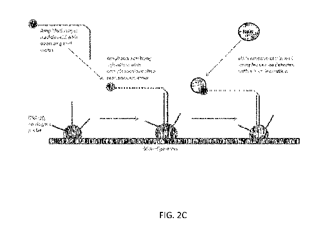
[0022] FIG. 2C shows a diagram of a universal array for nucleic acid
testing (NAT), in
accordance with some embodiments.
[0023] FIGS. 2D & 2E show the detection of a nucleic acid from Hepatitis C
virus (HCV)
using a universal array for nucleic acid testing (NAT), in accordance with
some embodiments.
Readouts obtained using a sample that lacks HCV nucleic acid (FIG. 2D) or a
sample that
contains an HCV nucleic acid (FIG. 2E) are shown.
[0024] FIG. 3A shows 15 individual probes on a universal array for NAT, in
accordance
with some embodiments.
[0025] FIG. 3B shows the effect of different Empigen BB concentrations in
sample
preparation for use with a universal array for NAT, in accordance with some
embodiments.
[0026] FIG. 3C shows the effect of different Hybridization Buffer
formulations for use with
a universal array for NAT, in accordance with some embodiments. The indicated
buffer
formulations are presented as x/y/z, where x is the strength of SSC buffer
(i.e., "3" indicates 3X
SSC buffer), y is the percentage of BSA, and z is the percentage of PEG-C.
[0027] FIG. 3D shows the effect of different NaOH concentrations on elution
efficiency for
the enrichment of a nucleic acid of interest from a sample, in accordance with
some
embodiments.
CA 03087624 2020-07-03
WO 2019/134835 PCT/EP2018/085945
[0028] FIG. 3E shows the effect of different NaOH concentrations in
combination with the
amount of enriched nucleic acid of interest used in amplification, in
accordance with some
embodiments.
[0029] FIG. 3F shows the effect of different elution strategies in
combination with the
amount of enriched nucleic acid of interest used in amplification, in
accordance with some
embodiments.
[0030] FIG. 3G shows the effect of different primer concentrations in
nucleic acid
enrichment protocols, in accordance with some embodiments.
[0031] FIG. 4A shows the enrichment of a nucleic acid of interest from a
sample in a pipette
tip, in accordance with some embodiments.
[0032] FIGS. 4B & 4C show the effect of the ratio of biotin-labeled
oligonucleotide to
neutravidin-labeled colloidal gold, in accordance with some embodiments.
Indicated ratios are
biotin-labeled probe:neutravidin-labeled colloidal gold. Dashed rectangles
indicate experimental
results, and solid rectangles indicate BSA-gold controls detected via 2-step
detection assay.
[0033] FIG. 5A shows a diagram of a continuous amplification system, in
accordance with
some embodiments.
[0034] FIGS. 5B & 5C show exemplary embodiments of continuous amplification
systems,
in accordance with some embodiments. FIG. 5B shows a robotic arm for obtaining
samples, a
pump system, optional pre-heating and activation zones, three stationary
temperature zones,
waste collection unit, temperature zone controller, and power supply. FIG. 5C
shows three
zones programmable to provide different temperatures, a temperature control
module, optional
fan control, power supply, pump module, and optional pre-heating and
activation zones.
[0035] FIG. 6A shows a diagram of asymmetric amplification for NAT, in
accordance with
some embodiments.
[0036] FIG. 6B shows ratios of reverse to forward primers in asymmetric
amplification for
NAT, in accordance with some embodiments.
21
CA 03087624 2020-07-03
WO 2019/134835 PCT/EP2018/085945
DETAILED DESCRIPTION
General Techniques
[0037] The techniques and procedures described or referenced herein are
generally well
understood and commonly employed using conventional methodology by those
skilled in the art,
such as, for example, the widely utilized methodologies described in Sambrook
et al., Molecular
Cloning: A Laboratory Manual 3d edition (2001) Cold Spring Harbor Laboratory
Press, Cold
Spring Harbor, N.Y.; Current Protocols in Molecular Biology (F.M. Ausubel, et
al. eds., (2003));
the series Methods in Enzymology (Academic Press, Inc.): PCR 2: A Practical
Approach (M.J.
MacPherson, B.D. Hames and G.R. Taylor eds. (1995)), Harlow and Lane, eds.
(1988)
Antibodies, A Laboratory Manual, and Animal Cell Culture (R.I. Freshney, ed.
(1987));
Oligonucleotide Synthesis (M.J. Gait, ed., 1984); Methods in Molecular
Biology, Humana Press;
Cell Biology: A Laboratory Notebook (J.E. Cellis, ed., 1998) Academic Press;
Animal Cell
Culture (R.I. Freshney), ed., 1987); Introduction to Cell and Tissue Culture
(J.P. Mather and
P.E. Roberts, 1998) Plenum Press; Cell and Tissue Culture: Laboratory
Procedures (A. Doyle,
J.B. Griffiths, and D.G. Newell, eds., 1993-8) J. Wiley and Sons; Handbook of
Experimental
Immunology (D.M. Weir and C.C. Blackwell, eds.); Gene Transfer Vectors for
Mammalian Cells
(J.M. Miller and M.P. Cabs, eds., 1987); PCR: The Polymerase Chain Reaction,
(Mullis et al.,
eds., 1994); Current Protocols in Immunology (J.E. Coligan et al., eds.,
1991); Short Protocols in
Molecular Biology (Wiley and Sons, 1999); Immunobiology (C.A. Janeway and P.
Travers,
1997); Antibodies (P. Finch, 1997); Antibodies: A Practical Approach (D.
Catty., ed., IRL Press,
1988-1989); Monoclonal Antibodies: A Practical Approach (P. Shepherd and C.
Dean, eds.,
Oxford University Press, 2000); Using Antibodies: A Laboratory Manual (E.
Harlow and D.
Lane (Cold Spring Harbor Laboratory Press, 1999); The Antibodies (M. Zanetti
and J. D. Capra,
eds., Harwood Academic Publishers, 1995); and Cancer: Principles and Practice
of Oncology
(V.T. DeVita et al., eds., J.B. Lippincott Company, 1993).
Microarrays
[0038] The universal array platform described herein can be used to detect
a variety of
nucleic acids or antigens of interest.
22
CA 03087624 2020-07-03
WO 2019/134835 PCT/EP2018/085945
Nucleic acid detection
[0039] Certain aspects of the present disclosure relate to methods for
detecting a nucleic acid
in a sample. In some embodiments, the methods include: a) amplifying at least
a portion of a
nucleic acid from a sample using a primer pair under conditions suitable for
amplification of an
amplicon comprising the portion of the nucleic acid if present in the sample,
wherein the primer
pair include: 1) a first primer comprising a label and a first oligonucleotide
sequence that
hybridizes with a first strand of the portion of the nucleic acid, and 2) a
second primer
comprising a second oligonucleotide sequence that hybridizes with a second
strand of the portion
of the nucleic acid opposite the first strand and a third oligonucleotide
sequence; b) after step (a),
contacting the amplicon, if present, to a plurality of single-stranded
oligonucleotide capture
sequences each affixed to a solid support, and wherein the amplicon, if
present, hybridizes with
at least one of the single-stranded oligonucleotide capture sequences on its
solid support via the
third oligonucleotide sequence or the complement of the third oligonucleotide
sequence; c) after
step (a), applying a colloidal detection reagent to the solid supports,
wherein the colloidal
detection reagent comprises a first moiety that binds to the label of the
amplicon if present and a
second moiety that comprises a colloidal metal; d) after (c), washing the
solid supports with a
wash solution; and e) after steps (a)-(d), detecting the colloidal detection
reagent, wherein
detection of the colloidal detection reagent on a solid support indicates the
presence of the
hybridized amplicon, thus detecting the nucleic acid in the sample.
[0040] The universal array platform provides an adaptable and versatile
approach for, e.g.,
diagnostic testing. As an example, an oligonucleotide 18 bases long with a
known sequence is
covalently attached to an activated glass slide or other surface as a small
spot. After binding and
blocking excess binding sites, a complementary oligonucleotide sequence is
covalently attached
to an antigen, such as HIV-1 gp41 immunodominant region. If a clinical sample,
such as blood,
contains antibodies to HIV-1 gp41 and is mixed with the HIV gp41 peptide
labeled with the
complementary oligo nucleotide, the antibody binds to the gp41 peptide, which
when put onto
the microarray described above, the complementary oligonucleotide binds to its
ligand partner
spotted onto the array. The unbound materials are washed off and the array
probed with a biotin
labeled anti-antibody (i.e., monoclonal anti-human Ig or protein A/G). An anti-
biotin molecule
(i.e., streptavidin) labeled gold is used to label antibodies bound to gp41,
which is bound to a
23
CA 03087624 2020-07-03
WO 2019/134835 PCT/EP2018/085945
specific spot on the microarray due to the complementary oligonucleotide
tethers described
above. Excess materials are washed off and the gold labeled microspot is then
detected directly
or the gold particles used to catalyze silver deposition, which can be easily
detected, thereby
indicating the presence/absence of anti-HIV antibodies to gp41 in the sample
thereby alerting the
user whether the person was infected with HIV based upon a detectable amount
of anti-HIV-1
gp41 antibodies present in the sample.
[0041] Importantly, the same oligonucleotide sequence can be used to label
different capture
reagents, which all will bind the complementary oligonucleotide sequence on
the array. Hence,
the next assay could be for detecting HBV, HCV, a toxin, hormone, or nucleic
acid, all of which
can bind to the same spot. The capture reagent will only bind to the same spot
based upon its
oligonucleotide label. Hence, a known set of oligonucleotides can be used to
create a generic
capture microarray and the same set of complementary oligos can be used to
label any capture
reagents. For example, a 16 x 16 microarray would have 256 spots, each with a
corresponding
oligonucleotide sequence. These oligonucleotide sequences can each be unique,
or the array
may include redundant spots used to confirm the results. Assume that each spot
is a duplicate,
one then has the potential to differentiate 128 different assays at the same
time. That array can
then become a standard, generic platform, and used to detect millions of
different targets by
simply labeling different capture reagents with the 128 different
complementary oligonucleotide
sequences, which can be provided as a generic kit. Additionally, the same
sample can be mixed
with different detector solutions that contain different tests and/or
overlapping tests. For
example, the sample can be screened for infectious diseases my mixing with
solution A, then
tested for cancer by mixing another portion of the sample with solution B,
then testing for toxins
by mixing another portion with solution C, and then detecting nucleic acids to
any of the targets
of interest by mixing with a solution D for direct detection of nucleic acids
or after an
amplification step. The microarray does not change¨it stays fixed¨but
different detector
solutions can be used to test for many different targets. This helps reduce
the cost for making the
microarrays and greatly expands their utility to detect virtually any target.
Exemplary features
and aspects of the universal array platform are described in greater detail
infra.
[0042] As used herein, unless otherwise specified, nucleic acids and/or
oligonucleotides refer
broadly to polymers of nucleic acids (e.g., DNA or RNA) and are meant to
include single-
24
CA 03087624 2020-07-03
WO 2019/134835 PCT/EP2018/085945
stranded and double-stranded species, as well as species comprising one or
more usually
naturally occurring and/or modified nucleosides/nucleotides (e.g., locked
nucleic acids, peptide
nucleic acids or PNAs, etc.).
[0043] As used herein, unless otherwise specified, an "amplicon" refers to
the product of any
of the types of nucleic acid amplification described herein, including but not
limited to
polymerase chain reaction (PCR), recombinase-polymerase assay (RPA), nucleic
acid
sequenced-based chain assay (NASBA), rolling circle amplification, branched
chain
amplification, ligation amplification, and loop-mediated isothermal
amplification. In some
embodiments, an amplicon is a double-stranded nucleic acid. In some
embodiments, an
amplicon is a single-stranded nucleic acid.
[0044] As used herein, the term "solid support" refers to any solid or semi-
solid structure
suitable for the attachment of biological molecules thereto, such as nucleic
acids. Solid supports
need not be flat or a single structure, and may be of any type of shape(s)
including spherical
shapes (e.g., beads). Solid supports may be arranged in any format. In some
embodiments, the
solid supports are arranged as a microarray (e.g., flat slide), a multiplex
bead array, or a well
array. In addition, the solid supports may be made of any suitable material,
including, but not
limited to, silicon, plastic, glass, polymer, ceramic, photoresist,
nitrocellulose, and hydrogel. In
some embodiments, the solid supports are nitrocellulose, silica, plastic, or
hydrogel.
[0045] Colloidal suspensions of nanoparticles such as colloidal gold can be
attached to
biological probes such as antibodies, useful as detection reagents for rapid
and sensitive
detection in immunostaining. Methods for preparing and using colloidal
detection reagents are
well known in the art (see, e.g., Hostetler etal., Langmuir 14:17-30, 1998;
Wang et al.,
Langmuir 17(19):5739-41, 2001). The colloidal detection reagent can be of any
material. In
some embodiments, the colloidal detection reagent comprises a metal. Examples
of colloidal
metal include, but are not limited to, gold (Au), silver (Ag), platinum (Pt),
palladium (Pd),
copper (Cu), nickel (Ni), ruthenium (Ru), and mixtures thereof. In some
embodiments, detecting
the colloidal detection reagent in step (e) comprises detection (e.g., direct
detection) of the
colloidal metal. In some embodiments, detecting the colloidal detection
reagent in step (e)
includes: 1) applying a developing reagent to the solid supports, wherein the
developing agent is
CA 03087624 2020-07-03
WO 2019/134835 PCT/EP2018/085945
suitable for forming a precipitate in the presence of the colloidal metal; and
2) detecting the
colloidal detection reagent by detecting the formation of the precipitate on a
solid support. In
some embodiments, the formation of the precipitate is detected by visual,
electronic, or magnetic
detection. In some embodiments, the formation of the precipitate is detected
by a mechanical
reader. In some of the embodiments described above, the developing reagent
comprises silver.
In some of the embodiments described above, silver nitrate and a reducing
agent (e.g.,
hydroquinone) are used. In some of the embodiments described above, a camera
(e.g., CCD
camera) is used to image the results of colloidal staining.
[0046] In some embodiments, the conditions in step (a) are suitable for
amplification by
polymerase chain reaction (PCR). In some embodiments, the conditions in step
(a) are suitable
for amplification by recombinase-polymerase assay (RPA), nucleic acid
sequenced-based chain
assay (NASBA), rolling circle amplification, branched chain amplification,
ligation
amplification, or loop-mediated isothermal amplification. In some embodiments,
the label
comprises biotin and the third oligonucleotide sequence hybridizes with at
least one of the
single-stranded oligonucleotide capture sequences.
[0047] In some embodiments, each single-stranded oligonucleotide capture
sequence is
coupled to a spacer reagent, and the spacer reagent is coupled to the
corresponding solid support.
In some embodiments, the spacer reagent comprises a serum albumin protein
(e.g., BSA). In
some embodiments, the spacer reagent comprises a dendrimer. In some
embodiments, the
method further comprises washing the solid supports with a wash solution after
step (b).
[0048] In some embodiments, the first primer is a forward primer that
amplifies in the sense
direction of the nucleic acid, and the second primer is a reverse primer that
amplifies in the
antisense direction of the nucleic acid. In some embodiments, the second
primer is a forward
primer that amplifies in the sense direction of the nucleic acid, and the
first primer is a reverse
primer that amplifies in the antisense direction of the nucleic acid. In some
embodiments, the
second primer comprises: the second oligonucleotide sequence, wherein the
second
oligonucleotide sequence allows for primer extension in the 5' to 3'
direction; and the third
oligonucleotide sequence, wherein the third oligonucleotide sequence is
oriented in the opposite
5' to 3' direction compared with the direction of primer extension from the
second
26
CA 03087624 2020-07-03
WO 2019/134835 PCT/EP2018/085945
oligonucleotide sequence. In some embodiments, the third oligonucleotide
sequence comprises a
modified nucleotide at the 3' terminus that blocks primer extension. In some
embodiments, the
second primer further comprises one or more linkers between the 5' end of the
third
oligonucleotide sequence and the 5' end of the second oligonucleotide
sequence.
[0049] In some embodiments, the label of the first primer comprises biotin.
In some
embodiments, the first moiety of the colloidal detection reagent comprises
neutravidin or
derivatives thereof, streptavidin or derivatives thereof, avidin or
derivatives thereof, or an
antigen-binding domain (e.g., antibody or antibody fragment) that specifically
binds biotin. In
some embodiments, the first moiety of the colloidal detection reagent
comprises neutravidin, and
wherein the second moiety of the colloidal detection reagent comprises a
colloidal gold ion. In
some embodiments, the colloidal detection reagent is applied to the solid
supports in step (c) at a
final dilution of 0.000010D to 200D. In some embodiments, the first moiety of
the colloidal
detection reagent comprises neutravidin, wherein the second moiety of the
colloidal detection
reagent comprises a colloidal gold ion, and wherein the colloidal detection
reagent is applied to
the solid supports in step (c) at a final dilution of 0.050D to 0.20D. Varying
amounts of the
colloidal detection reagent can be used. In some embodiments, 1pL to 1000)iL
of colloidal
detection reagent is applied to the solid supports in step (c) per ILEL of
amplicon. In some
embodiments, 100).EL of colloidal detection reagent is applied to the solid
supports in step (c) per
1.5 ILEL of amplicon.
[0050] In some embodiments, the method further comprises, prior to step
(a), exposing the
sample to a lysis buffer. In some embodiments, the lysis buffer comprises N,N-
dimethyl-N-
dodecylglycine betaine. In some embodiments, the lysis buffer comprises
greater than or equal
to 0.1% and less than or equal to 10% N,N-dimethyl-N-dodecylglycine betaine
(w/v). In some
embodiments, the lysis buffer comprises greater than or equal to 0.5% and less
than or equal to
4% N,N-dimethyl-N-dodecylglycine betaine (w/v). In some embodiments, the lysis
buffer
comprises greater than or equal to 1% and less than or equal to 2% N,N-
dimethyl-N-
dodecylglycine betaine (w/v).
[0051] In some embodiments, the sample is exposed to the lysis buffer at a
ratio between
1:50 sample:lysis buffer and 50:1 sample:lysis. In some embodiments, the
portion of the sample
27
CA 03087624 2020-07-03
WO 2019/134835 PCT/EP2018/085945
is exposed to the lysis buffer at a ratio of about 1:1 sample:lysis buffer. In
some embodiments,
the lysis buffer further comprises 0.1X to 5X phosphate buffered saline (PBS)
buffer or Tris
EDTA (TE) buffer. In some embodiments, the lysis buffer further comprises 1X
PBS.
[0052] In some embodiments, an amplicon is hybridized to the solid supports
in the presence
of a hybridization buffer. A variety of hybridization buffers are known in the
art. In some
embodiments, the amplicon is hybridized to the solid supports in step (b) in a
hybridization
buffer comprising 0.1X to 10X saline sodium citrate (SSC) buffer, 0.001% to
30% blocking
agent, and 0.01% to 30% crowding agent. In some embodiments, the blocking
agent comprises
bovine serum albumin (BSA), polyethylene glycol (PEG), casein, or polyvinyl
alcohol (PVA).
In some embodiments, the blocking agent comprises BSA, and the BSA is present
in the
hybridization buffer at 1% to 3%. In some embodiments, the crowding agent is
Polyethylene
Glycol Bisphenol A Epichlorohydrin Copolymer. In some embodiments, the
Polyethylene
Glycol Bisphenol A Epichlorohydrin Copolymer is present in the hybridization
buffer at 1% to
3%. In some embodiments, the hybridization buffer comprises 2X to 5X SSC
buffer.
[0053] In some embodiments, the method further comprises, prior to step
(b), blocking the
solid supports. In some embodiments, the solid supports are blocked using a
solution comprising
BSA. In some embodiments, the solid supports are blocked for 1 hour at 37 C
using 2% BSA
solution. In some embodiments, the method further comprises washing the solid
supports with a
wash solution after blocking the solid supports.
[0054] In some embodiments, the method further comprises, after step (b)
and prior to step
(c), washing the solid supports with a wash buffer. In some embodiments, the
wash buffer
comprises 0.1X to 10X SSC buffer and 0.01% to 30% detergent. In some
embodiments, the
detergent comprises 0.05% to 5% N-lauroylsarcosine sodium salt. In some
embodiments, the
wash buffer comprises lx to 5X SSC buffer.
[0055] In some embodiments, the method further comprises, prior to step
(a): (i) contacting
the sample with an oligonucleotide coupled to a solid substrate, wherein the
oligonucleotide
hybridizes with the nucleic acid if present in the sample; (ii) washing the
solid substrate under
conditions suitable to remove non-specific interactions with the solid
substrate but retain the
nucleic acid hybridized with the oligonucleotide, if present in the sample;
and (iii) eluting the
28
CA 03087624 2020-07-03
WO 2019/134835
PCT/EP2018/085945
nucleic acid, if present in the sample, from the oligonucleotide, wherein the
eluted nucleic acid is
subjected to PCR amplification in step (a). In some embodiments, the method
further comprises,
prior to step (a): (i) contacting the sample with an oligonucleotide, wherein
the oligonucleotide
hybridizes with the nucleic acid if present in the sample, (ii) simultaneous
with or after step (i),
contacting the sample with a solid substrate, wherein the solid substrate is
coupled to a first
binding moiety, wherein the oligonucleotide is coupled to a second binding
moiety that binds the
first binding moiety, and wherein the sample is contacted with the solid
substrate under
conditions suitable for the second binding moiety to bind the first binding
moiety; (iii) washing
the solid substrate under conditions suitable to remove non-specific
interactions with the solid
substrate but retain the oligonucleotide and the nucleic acid hybridized with
the oligonucleotide,
if present in the sample; and (iv) eluting the nucleic acid, if present in the
sample, from the
oligonucleotide, wherein the eluted nucleic acid is subjected to PCR
amplification in step (a). In
some embodiments, the oligonucleotide is coupled to the solid substrate via a
covalent
interaction. In some embodiments, the oligonucleotide is coupled to the solid
substrate via an
avidin:biotin or streptavidin:biotin interaction, or wherein the first binding
moiety comprises
avidin, neutravidin, streptavidin, or a derivative thereof and the second
binding moiety comprises
biotin or a derivative thereof. In some embodiments, the solid substrate is
positioned in a pipet
tip, and wherein step (i) comprises pipetting the sample into the pipet tip.
In some embodiments,
the solid substrate comprises a matrix or plurality of beads. In some
embodiments, the nucleic
acid comprises DNA. In some embodiments, the nucleic acid comprises RNA.
[0056] In
some embodiments, the method further comprises, prior to step (a), incubating
at
least a portion of the sample with a reverse transcriptase, primers, and
deoxyribonucleotides
under conditions suitable for generation of a cDNA synthesized from the
nucleic acid, wherein
the portion of the nucleic acid is amplified in step (a) using the cDNA. In
some embodiments,
the primers used prior to step (a) are random primers, poly-dT primers, or
primers specific for
the portion of the nucleic acid. In some embodiments, the portion of the
sample is incubated
with the reverse transcriptase, primers, and the deoxyribonucleotides in the
presence of an RNase
inhibitor. In some embodiments, the portion of the sample is incubated with
the reverse
transcriptase, primers, and the deoxyribonucleotides in the presence of
betaine. In some
embodiments, the betaine is present at a concentration of about 0.2M to about
1.5M. In some
embodiments, the nucleic acid comprises a viral nucleic acid. In some
embodiments, the viral
29
CA 03087624 2020-07-03
WO 2019/134835 PCT/EP2018/085945
nucleic acid is from a virus selected from the group consisting of HIV, HBV,
HCV, West Nile,
Zika, and parvovirus. In some embodiments, the nucleic acid comprises a
bacterial, archaean,
protozoan, fungal, plant, or animal nucleic acid. In some embodiments, the
sample comprises
whole blood, serum, saliva, urine, soil, tissue, or an environmental sample
(e.g., comprising
water, soil, etc.).
[0057] In some embodiments, any of the reagents described herein (e.g., a
lysis or
hybridization buffer) can comprise a positive control oligonucleotide, e.g.,
that hybridizes with a
capture oligonucleotide of the array. Advantageously, this can be used as a
positive control to
ensure that the correct reagents were used. For example, a plurality of
capture oligonucleotides
can be used to represent different reagents (e.g., lysis buffer, hybridization
buffer, and the like),
and a specific positive control in each reagent can be used to "encode" that
the proper reagent is
used. When viewed in aggregate, the plurality can indicate that some or all of
the steps of
microarray preparation/analysis were completed with reagents spiked with
positive control
oligonucleotide(s), thereby indicating the use of correct reagents.
[0058] In some embodiments, the present disclosure contemplates adjusting
the ratio of the
first primer relative to the second primer used in the step of amplifying a
nucleic acid in a
sample. The ratio of the primers can be skewed to preferentially amplify one
strand of the
nucleic acid more than the other¨a technique known as asymmetric amplification
(see, e.g.,
McCabe, PCR Protocols: A guide to Methods and Applications, 76-83, 1990).
Advantageously,
the skewed primer ratio in asymmetric amplification results in a predominantly
uniform product,
which may help increase the strength of the hybridization signal detected on a
universal array of
the present disclosure. In some embodiments of the present disclosure, the
portion of the nucleic
acid is amplified using an excess of the first primer relative to the second
primer, and wherein
the amplicon, if present, is a single-stranded nucleic acid that hybridizes
with at least one of the
single-stranded oligonucleotide capture sequences via the complement of the
third
oligonucleotide sequence. In some embodiments, the portion of the nucleic acid
is amplified
using a ratio of the first primer to the second primer of between about 12.5:1
and about 100:1. In
some embodiments, a ratio of the first primer to the second primer of at least
about 2:1, at least
about 5:1, at least about 10:1, at least about 15:1, at least about 20:1, at
least about 25:1, at least
about 30:1, at least about 35:1, at least about 40:1, at least about 45:1, at
least about 50:1, at least
CA 03087624 2020-07-03
WO 2019/134835 PCT/EP2018/085945
about 55:1, at least about 60:1, at least about 65:1, at least about 70:1, at
least about 75:1, at least
about 80:1, at least about 85:1, at least about 90:1, at least about 95:1, at
least about 100:1, at
least about 150:1, or at least about 200:1 is used.
Antigen detection
[0059] In addition to nucleic acids, the universal array platform described
herein can be used
to detect a variety of antigens of interest. Accordingly, some aspects of the
present disclosure
relate to methods for detecting an antigen in a sample. In some embodiments,
the method
includes: a) providing a plurality of single-stranded oligonucleotide capture
sequences each
affixed to a solid support; b) after step (a), contacting the solid supports
with an antigen-binding
domain that specifically binds an antigen, wherein the antigen-binding domain
is coupled to a
single-stranded oligonucleotide sequence that hybridizes with at least one of
the single-stranded
oligonucleotide capture sequences on the solid supports, and wherein the
microarray is contacted
with the antigen-binding domain under conditions suitable for the single-
stranded
oligonucleotide sequence of the antigen binding domain to hybridize with the
at least one single-
stranded oligonucleotide capture sequence on the solid supports; c) after step
(a), contacting the
solid supports with at least a portion of the sample under conditions suitable
for the antigen-
binding domain to bind the antigen, if present in the sample; d) after step
(a), applying a colloidal
detection reagent to the solid supports, wherein the colloidal detection
reagent comprises a first
moiety that specifically binds to the antigen if present and a second moiety
that comprises a
colloidal metal; e) after (d), washing the solid supports with a wash
solution; and f) after steps
(a)-(e), detecting the colloidal detection reagent, wherein detection of the
colloidal detection
reagent indicates the presence of the antigen in the sample.
[0060] In some embodiments, the antigen comprises a polypeptide, lipid, or
carbohydrate. In
certain embodiments, the antigen is a polypeptide antigen.
[0061] Any of the compositions and methods described above in reference to
nucleic acid
detection may also be used for antigen detection, including, without
limitation, the solid
supports, the colloidal detection reagent and method of detecting thereof, the
developing reagent,
the spacer reagent, the lysis buffer, the blocking agent, the crowding agent,
the hybridization
buffer, and the wash buffer.
31
CA 03087624 2020-07-03
WO 2019/134835 PCT/EP2018/085945
[0062] In some embodiments, the first moiety comprises a second antigen-
binding domain
that specifically binds to the antigen, wherein the second antigen-binding
domain is coupled to
biotin or a derivative thereof, and wherein the colloidal suspension is
coupled to avidin,
neutravidin, streptavidin, or a derivative thereof bound to the biotin. In
some embodiments, the
colloidal metal is gold, platinum, palladium, or ruthenium. In some
embodiments, the single-
stranded oligonucleotide capture sequence at each spot of the plurality is
coupled to a spacer
reagent, and the spacer reagent is coupled to the solid supports. In some
embodiments, the
spacer reagent comprises a serum albumin protein. In some embodiments, the
spacer reagent
comprises a dendrimer. In some embodiments, the method further comprises,
prior to step (c),
exposing the sample to a lysis buffer comprising greater than or equal to 0.1%
and less than or
equal to 10% N,N-dimethyl-N-dodecylglycine betaine (w/v). In some embodiments,
the antigen
is a viral antigen. In some embodiments, the viral antigen is from a virus
selected from the group
consisting of: HIV, HBV, HCV, West Nile, Zika, and parvovirus. In some
embodiments, the
antigen is a bacterial, archaean, protozoan, fungal, plant, or animal antigen.
In some
embodiments, the sample comprises whole blood, serum, saliva, urine, soil,
tissue, or an
environmental sample (e.g., comprising water, soil, etc.).
Devices
[0063] Other aspects of the present disclosure relate to a device or
apparatus for amplifying a
nucleic acid in a sample. In some embodiments, the device or apparatus
includes: capillary
tubing arranged around a support in a plurality of circuits, wherein each
circuit of the plurality
comprises a first, a second, and a third stationary temperature zone, and
wherein the capillary
tubing is heated to a first temperature in the first stationary temperature
zone, a second
temperature in the second stationary temperature zone, and a third temperature
in the third
stationary temperature zone; a robotic arm configured to introduce into the
capillary tubing a
sample comprising a nucleic acid in admixture with an amplification mixture
comprising
deoxyribonucleotides, a polymerase, and a primer pair; and a pump or vacuum
configured to
pass the sample comprising the nucleic acid in admixture with the
amplification mixture through
the plurality of circuits within the capillary tubing.
32
CA 03087624 2020-07-03
WO 2019/134835 PCT/EP2018/085945
[0064] To detect a nucleic acid in a sample using methods described herein,
amplification of
at least a portion of the nucleic acid can be used for hybridization to the
array. Thus, in some
aspects, the present disclosure contemplates apparatuses that may be useful
for amplifying
nucleic acids of interest. In some embodiments, the present disclosure relates
to an apparatus for
nucleic acid amplification having capillary tubing, a robotic arm, and a pump
or vacuum.
[0065] Amplification of the nucleic acid is carried out in the capillary
tubing. The capillary
tubing is arranged around a support in a plurality of circuits, wherein each
circuit of the plurality
comprises a first, a second, and a third stationary temperature zone, and
wherein the capillary
tubing is heated to a first temperature in the first stationary temperature
zone, a second
temperature in the second stationary temperature zone, and a third temperature
in the third
stationary temperature zone. Each of these zones may correspond to one portion
of a standard
PCR or other amplification reaction, namely denaturing, annealing, and
extension for PCR. For
isothermal amplification, each zone can be heated to the same temperature
(e.g., 37 C).
Advantageously, capillary tubing allows for an improved rate of heat
conduction, which means
target reaction temperatures can be rapidly achieved, and reaction times can
be shortened. The
capillary tubing in each circuit of the plurality may form any shape, for
example, without
limitation, a conical shape, a cylindrical shape, or a spiral shape. The
capillary tubing may
comprise any material, for example and without limitation,
polytetrafluoroethylene (PTFE). The
plurality of circuits of the capillary tubing may comprise any number of
circuits, for example and
without limitation, from about 25 to about 44 circuits (e.g., corresponding to
the number of
amplification cycles).
[0066] The pump or vacuum of the apparatus can be configured to pass the
sample
comprising the nucleic acid in admixture with the amplification mixture
through the plurality of
circuits within the capillary tubing. A variety of pumps and vacuums well
known in the art can
be used for the apparatus. In some embodiments, the pump or vacuum is a
peristaltic pump. In
some embodiments, the pump or vacuum is a high performance liquid
chromatography (HPLC)
pump. In some embodiments, the pump or vacuum is a precision syringe pump.
[0067] The robotic arm of the apparatus can be configured to introduce into
the capillary
tubing a sample comprising a nucleic acid in admixture with an amplification
mixture
33
CA 03087624 2020-07-03
WO 2019/134835 PCT/EP2018/085945
comprising deoxyribonucleotides, a polymerase, and a primer pair. In some
embodiments, the
robotic arm comprises a peristaltic or HPLC pump configured to introduce the
sample
comprising the nucleic acid target in admixture with an amplification mixture
into the capillary
tubing, and wherein the apparatus further comprises a secondary pump
configured to pull the
sample comprising the nucleic acid target in admixture with an amplification
mixture through the
capillary tubing.
[0068] Additional components may be added to the apparatus. In some
embodiments, the
apparatus further contains one or more processors, a memory, one or more
programs, wherein
the one or more programs are stored in the memory and configured to be
executed by the one or
more processors, the one or more programs including instructions for
controlling the temperature
of the first, second, and third stationary temperature zones. In some
embodiments, the apparatus
further contains an incubator for a cDNA synthesis zone (e.g., if the sample
is RNA) in which
the capillary tubing is heated to between about 37 C and about 42 C upstream
of the plurality of
circuits. In some embodiments, the incubator is a temperature bath, a Peltier
device, or a
resistance heater. In some embodiments, the sample in admixture with an
amplification mixture
is held in the cDNA synthesis zone for 15 seconds to 30 minutes. In some
embodiments, the
apparatus further contains an incubator for an activation zone in which the
capillary tubing is
heated to about 95 C upstream of the plurality of circuits. In some
embodiments, the apparatus
further contains an incubator for a PCR extension zone in which the capillary
tubing is heated to
between about 55 C and about 72 C downstream of the plurality of circuits.
[0069] The pump, robotic arm, and various temperature zones may all be
controlled by a
system control panel, which allows alteration of each of the components, for
example, the
temperature of the different zones can be changed. In addition, the control
panel can be used to
change the pumping speed, thereby altering the length of time spent in each
temperature zone of
the main amplification area. Thus, in some embodiments, the apparatus may
further include one
or more processors, a memory, one or more programs, wherein the one or more
programs are
stored in the memory and configured to be executed by the one or more
processors, the one or
more programs including instructions for controlling the temperature of the
first, second, and
third stationary temperature zones.
34
CA 03087624 2020-07-03
WO 2019/134835
PCT/EP2018/085945
[0070] The
temperature of the capillary tubing can be maintained at desired temperature
in
the different temperature zones according to standard techniques known in the
art. In some
embodiments, the temperature of one of more temperature zones is maintained by
Peltier or
resistance heaters. In some embodiments, polyimide tape (e.g., inside copper
tubing) is used to
insulate one or more temperature zones.
[0071] In
some embodiments, a robotic arm is used to apply an amplicon to the microarray
(e.g., solid supports of the present disclosure) after completion of the
circuits. In some
embodiments, a dye is added, e.g., to aid in visualizing the liquid spotted
onto the microarray. In
some embodiments, the amplicon is collected into a container with
hybridization buffer after
completion of the circuits, then added to the microarray (e.g., manually by
pipetting, or with a
robotic arm).
[0072] The
apparatuses described above may be used in any of the methods of the present
disclosure. For example, in some embodiments, the method includes: a)
incubating at least a
portion of the sample with an amplification mixture comprising
deoxyribonucleotides, a
polymerase, and a primer pair, wherein the primer pair comprises a first
primer comprising a
label and a first oligonucleotide sequence that hybridizes with a first strand
of a portion of the
nucleic acid, and a second primer comprising a second oligonucleotide sequence
that hybridizes
with a second strand of the portion of the nucleic acid opposite the first
strand and a first capture
moiety; b) passing the portion of the sample in admixture with the
amplification mixture through
first, second, and third stationary temperature zones for a plurality of
cycles through continuous
capillary tubing under conditions suitable for amplification of an amplicon
comprising the
portion of the nucleic acid, if present in the sample, wherein each cycle of
the plurality includes:
1) passing the portion of the sample in admixture with the amplification
mixture through the first
stationary temperature zone via the continuous capillary tubing at a first
temperature and for a
first duration suitable for denaturing the strands of the nucleic acid, if
present in the sample, 2)
after step (b)(1), passing the portion of the sample in admixture with the
amplification mixture
through the second stationary temperature zone via the continuous capillary
tubing at a second
temperature and for a second duration suitable for annealing the first and
second primers to the
respective strands of the nucleic acid, if present in the sample, and 3) after
step (b)(2), passing
the portion of the sample in admixture with the amplification mixture through
the third stationary
CA 03087624 2020-07-03
WO 2019/134835 PCT/EP2018/085945
temperature zone via the continuous capillary tubing at a third temperature
and for a third
duration suitable for amplifying the nucleic acid target, if present in the
sample, via the
polymerase and primer pair; c) after the plurality of cycles, associating the
amplicon, if present
in the sample, with a first capture moiety affixed to a solid support; and d)
detecting association
of the amplicon, if present in the sample, with the solid support, wherein
association of the
amplicon with the one or more solid supports indicates the presence of the
nucleic acid in the
sample.
[0073] General steps for using the above-described apparatuses to amplify
and detect a
nucleic acid in a sample are described below and may employ any of the
reagents or techniques
described supra.
[0074] The methods can include incubating in an initial container at least
a portion of the
sample with an amplification mixture comprising deoxyribonucleotides, a
polymerase, and a
primer pair, wherein the primer pair comprises a first primer comprising a
label and a first
oligonucleotide sequence that hybridizes with a first strand of a portion of
the nucleic acid, and a
second primer comprising a second oligonucleotide sequence that hybridizes
with a second
strand of the portion of the nucleic acid opposite the first strand and a
first capture moiety.
[0075] Then, a pump and a robotic arm are used to pass the portion of the
sample in
admixture with the amplification mixture through first, second, and third
stationary temperature
zones for a plurality of cycles through continuous capillary tubing under
conditions suitable for
amplification of an amplicon comprising the portion of the nucleic acid, if
present in the sample,
wherein each cycle of the plurality comprises: 1) passing the portion of the
sample in admixture
with the amplification mixture through the first stationary temperature zone
via the continuous
capillary tubing at a first temperature and for a first duration suitable for
denaturing the strands
of the nucleic acid, if present in the sample, 2) after step (b)(1), passing
the portion of the sample
in admixture with the amplification mixture through the second stationary
temperature zone via
the continuous capillary tubing at a second temperature and for a second
duration suitable for
annealing the first and second primers to the respective strands of the
nucleic acid, if present in
the sample, and 3) after step (b)(2), passing the portion of the sample in
admixture with the
amplification mixture through the third stationary temperature zone via the
continuous capillary
36
CA 03087624 2020-07-03
WO 2019/134835 PCT/EP2018/085945
tubing at a third temperature and for a third duration suitable for amplifying
the nucleic acid
target, if present in the sample, via the polymerase and primer pair.
[0076] Next, after the plurality of cycles, the amplicon, if present in the
sample, can be
associated with a first capture moiety affixed to a solid support.
[0077] Finally, association of the amplicon, if present in the sample, with
the solid support
can be detected, wherein association of the amplicon with the one or more
solid supports
indicates the presence of the nucleic acid in the sample.
[0078] In some embodiments, the first capture moiety comprises a third
oligonucleotide
sequence, and wherein the second capture moiety comprises a single-stranded
oligonucleotide
capture sequence that hybridizes with the third oligonucleotide sequence or
the complement of
the third oligonucleotide sequence in step (c). In some embodiments, detecting
association of the
amplicon, if present, with the solid support comprises: i) applying a
colloidal detection reagent to
the solid support, wherein the colloidal detection reagent comprises a first
moiety that binds to
the label of the amplicon if present and a second moiety that comprises a
colloidal metal; and ii)
detecting the colloidal detection reagent. In some embodiments, detecting the
colloidal detection
reagent in step (d)(ii) comprises detection of the colloidal metal. In some
embodiments,
detecting the colloidal detection reagent in step (d)(ii) comprises: a)
applying a developing
reagent to the solid support, wherein the developing agent is suitable for
forming a precipitate in
the presence of the colloidal metal; and b) detecting the colloidal detection
reagent by detecting
the formation of the precipitate at the solid support. In some embodiments,
the formation of the
precipitate is detected by visual, electronic, or magnetic detection. In some
embodiments, the
formation of the precipitate is detected by a mechanical reader. In some
embodiments, the
developing reagent comprises silver. In some embodiments, the label comprises
biotin or a
derivative thereof, and wherein the first moiety of the colloidal detection
reagent comprises
neutravidin, streptavidin, or an antigen-binding domain that specifically
binds biotin. In some
embodiments, the first moiety of the colloidal detection reagent comprises
neutravidin, and
wherein the second moiety of the colloidal detection reagent comprises a
colloidal gold ion. In
some embodiments, the conditions in step (b) are suitable for amplification by
polymerase chain
reaction (PCR). In some embodiments, the conditions in step (b) are suitable
for amplification
37
CA 03087624 2020-07-03
WO 2019/134835 PCT/EP2018/085945
by recombinase-polymerase assay (RPA), nucleic acid sequenced-based chain
assay (NASBA),
rolling circle amplification, branched chain amplification, ligation
amplification, or loop-
mediated isothermal amplification. In some embodiments, the portion of the
sample in
admixture with the PCR amplification mixture is passed through the continuous
capillary tubing
using a peristaltic pump, high performance liquid chromatography (HPLC) pump,
precision
syringe pump, or vacuum. In some of the embodiments described above, the
method may further
contain, prior to step (b): passing the portion of the sample in admixture
with the amplification
mixture through a preheating zone at between about 20 C and about 55 C via the
continuous
capillary tubing. In some embodiments, the preheating zone is between about 37
C and about
42 C. In some embodiments, the portion of the sample in admixture with the
amplification
mixture is passed through the preheating zone for up to 30 minutes. In some
embodiments, the
portion of the sample in admixture with the amplification mixture is passed
through the
preheating zone for about 15 minutes. In some of the embodiments described
above, the method
may further contain, prior to step (b): passing the portion of the sample in
admixture with the
amplification mixture through an activation zone at between about 80 C and
about 100 C via the
continuous capillary tubing. In some embodiments, the activation zone is
between about 90 C
and about 95 C. In some embodiments, the portion of the sample in admixture
with the
amplification mixture is passed through the activation zone for up to 20
minutes. In some
embodiments, the portion of the sample in admixture with the amplification
mixture is passed
through the activation zone for between about 5 minutes and about 10 minutes.
In some of the
embodiments described above, the method may further contain, after step (b)
and prior to step
(c): passing the portion of the sample in admixture with the amplification
mixture through an
extension zone at between about 55 C and about 72 C via the continuous
capillary tubing. In
some of the embodiments described above, the method may further contain, after
step (b) and
prior to step (c): i) mixing at least a portion of a second sample with an
amplification mixture
comprising deoxyribonucleotides, a polymerase, and a second primer pair,
wherein the second
primer pair comprises a third primer comprising a label and a fourth
oligonucleotide sequence
that hybridizes with a first strand of a portion of a second nucleic acid, and
a fourth primer
comprising a fifth oligonucleotide sequence that hybridizes with a second
strand of the portion of
the second nucleic acid opposite the first strand and a third capture moiety;
ii) passing the portion
of the second sample in admixture with the amplification mixture through the
first, second, and
38
CA 03087624 2020-07-03
WO 2019/134835 PCT/EP2018/085945
third stationary temperature zones for a second plurality of cycles through
the continuous
capillary tubing under conditions suitable for amplification of the portion of
the second nucleic
acid, if present in the sample, wherein each cycle of the second plurality
comprises: 1) passing
the portion of the second sample in admixture with the amplification mixture
through the first
stationary temperature zone via the continuous capillary tubing at the first
temperature and for
the first duration suitable for denaturing the strands of the second nucleic
acid, if present in the
second sample, 2) after step (ii)(1), passing the portion of the second sample
in admixture with
the amplification mixture through the second stationary temperature zone via
the continuous
capillary tubing at the second temperature and for the second duration
suitable for annealing the
third and fourth primers to the respective strands of the second nucleic acid,
if present in the
second sample, and 3) after step (ii)(2), passing the portion of the second
sample in admixture
with the amplification mixture through the third stationary temperature zone
via the continuous
capillary tubing at the third temperature and for the third duration suitable
for amplifying the
second nucleic acid, if present in the second sample, via the polymerase and
second primer pair;
wherein the second nucleic acid, if present in the second sample, is
associated concurrently with
the amplified first nucleic acid target, if present in the first sample, with
a fourth capture moiety
that associates with the third capture moiety, wherein the fourth capture
moiety is coupled to a
solid support; and wherein the association of the amplified second nucleic
acid, if present in the
second sample, with the solid support is detected concurrently with the
hybridization of the
amplified first nucleic acid, if present in the first sample, and wherein
association of the
amplified second nucleic acid target with the solid support indicates the
presence of the second
nucleic acid target in the second sample.
[0079] The first and the second samples may or may not be the same. In some
embodiments,
the first and the second samples are the same. In some embodiments, the first
and the second
samples are different samples. The first and the second nucleic acids may or
may not be the
same. In some embodiments, the first and the second nucleic acids are the
same. In some
embodiments, the first and the second nucleic acids are different. It is to be
noted that one may
detect the same nucleic acid from different samples using the apparatuses and
methods described
herein.
39
CA 03087624 2020-07-03
WO 2019/134835 PCT/EP2018/085945
[0080] In some of the embodiments described above, the method may further
contain, after
passing the portion of the first sample in admixture with the amplification
mixture through the
first, second, and third stationary temperature zones for the plurality of
cycles, and prior to
passing the portion of the second sample in admixture with the amplification
mixture through the
first, second, and third stationary temperature zones for the second plurality
of cycles: passing a
volume of air through the continuous capillary tubing sufficient to separate
the portion of the
first sample in admixture with the amplification mixture and the portion of
the second sample in
admixture with the amplification mixture. In some embodiments, the method may
further
contain, after passing the volume of air through the continuous capillary
tubing, and prior to
passing the portion of the second sample in admixture with the amplification
mixture through the
first, second, and third stationary temperature zones for the second plurality
of cycles: passing a
solution comprising sodium hypochlorite at a concentration of between about
0.1% and about
10% through the continuous capillary tubing. In some embodiments, the solution
comprises
sodium hypochlorite at a concentration of about 1.6%. In some embodiments, the
method may
further contain, after passing the bleach solution through the continuous
capillary tubing, and
prior to passing the portion of the second sample in admixture with the
amplification mixture
through the first, second, and third stationary temperature zones for the
second plurality of
cycles: passing a solution comprising thiosulfate at a concentration of
between about 5mM and
about 500mM through the continuous capillary tubing. In some embodiments, the
solution
comprises thiosulfate at a concentration of about 20mM. In some embodiments,
the method may
further include, after passing the thiosulfate solution through the continuous
capillary tubing, and
prior to passing the portion of the second sample in admixture with the
amplification mixture
through the first, second, and third stationary temperature zones for the
second plurality of
cycles: passing water through the continuous capillary tubing. In some
embodiments, the
method may further contain, after passing the water through the continuous
capillary tubing, and
prior to passing the portion of the second sample in admixture with the PCR
amplification
mixture through the first, second, and third stationary temperature zones for
the second plurality
of cycles: passing a volume of air through the continuous capillary tubing
sufficient to separate
the water and the portion of the second sample in admixture with the PCR
amplification mixture.
In some embodiments, a water and air passing scheme comprises four water
pulses of 20 seconds
each, separated by 20 seconds of air pulses, between samples. In some
embodiments that may be
CA 03087624 2020-07-03
WO 2019/134835 PCT/EP2018/085945
combined with any of the preceding embodiments, step (a) comprises inserting
the portion of the
sample into the continuous capillary tubing and mixing the portion of the
sample with the
amplification mixture using a robotic arm or valve system.
[0081] In some embodiments that may be combined with any of the preceding
embodiments,
the nucleic acid comprises DNA. In some embodiments that may be combined with
any of the
preceding embodiments, the nucleic acid comprises RNA. In some embodiments,
the method
may further contain, prior to step (a): incubating at least a portion of the
sample with a reverse
transcriptase, primers, and deoxyribonucleotides under conditions suitable for
generation of a
cDNA synthesized from the RNA, wherein the cDNA is mixed with the
amplification mixture in
step (a). In some embodiments, the primers used prior to step (a) are random
primers, poly-dT
primers, or primers specific for the portion of the RNA. In some embodiments,
wherein the
portion of the sample is incubated with the reverse transcriptase, primers,
and
deoxyribonucleotides while being passed through a cDNA synthesis zone between
about 37 C
and about 42 C via the continuous capillary tubing for a time sufficient for
generation of a
cDNA synthesized from the RNA. In some embodiments, the method may further
contain, after
passing the portion of the sample in admixture with the reverse transcriptase,
primers, and
deoxyribonucleotides through the cDNA synthesis zone, and prior to step (b):
passing the portion
of the sample in admixture with the reverse transcriptase, primers, and
deoxyribonucleotides
through an activation zone at about 95 C via the continuous capillary tubing.
In some
embodiments that may be combined with any of the preceding embodiments, during
each cycle
of the plurality, the portion of the sample in admixture with the
amplification mixture is passed
through the first stationary temperature zone at between about 80 C and about
100 C for 1
second to about 10 minutes. In some embodiments, the portion of the sample in
admixture with
the amplification mixture is passed through the first stationary temperature
zone at between
about 90 C and about 97 C for 2 seconds to about 20 seconds. In some
embodiments, the
portion of the sample in admixture with the amplification mixture is passed
through the first
stationary temperature zone at between about 95 C for at least 10 seconds. In
some
embodiments that may be combined with any of the preceding embodiments, during
each cycle
of the plurality, the portion of the sample in admixture with the
amplification mixture is passed
through the second stationary temperature zone between about 45 C and about 65
C for 2
seconds to about 60 seconds. In some embodiments, the portion of the sample in
admixture with
41
CA 03087624 2020-07-03
WO 2019/134835 PCT/EP2018/085945
the amplification mixture is passed through the second stationary temperature
zone between
about 55 C for at least 15 seconds. In some embodiments, the portion of the
sample in
admixture with the amplification mixture is passed through the second
stationary temperature
zone between about 50 C and about 57 C for 2 seconds to about 60 seconds. In
some
embodiments that may be combined with any of the preceding embodiments, during
each cycle
of the plurality, the portion of the sample in admixture with the
amplification mixture is passed
through the third stationary temperature zone at between about 57 C and about
74 C for 3
seconds to about 60 seconds. In some embodiments, the portion of the sample in
admixture with
the amplification mixture is passed through the third stationary temperature
zone at between
about 65 C and about 72 C for 3 seconds to about 60 seconds. In some
embodiments, the
portion of the sample in admixture with the amplification mixture is passed
through the third
stationary temperature zone at between about 65 C and about 72 C for at least
15 seconds. In
some embodiments, if isothermal or LAMP amplification is used, all three
stationary temperature
zones could have the same temperature, e.g., 37 C. In addition, for all
stationary temperature
zones, the speed of the pump or vacuum can be controlled to alter the length
of time the sample
in admixture with the amplification mixture spent in each stationary
temperature zone.
[0082] In some embodiments that may be combined with any of the preceding
embodiments,
during each cycle of the plurality, the portion of the sample in admixture
with the PCR
amplification mixture is passed through both the second stationary temperature
zone and the
third stationary temperature zone at between about 45 C and about 80 C for
between about 0.5
seconds and about 5 minutes. In some embodiments that may be combined with any
of the
preceding embodiments, the plurality of cycles comprises greater than or equal
to 2 cycles and
less than or equal to 100 cycles. In some embodiments that may be combined
with any of the
preceding embodiments, the method may further contain, prior to step (a),
incubating the portion
of the sample with a lysis buffer comprising greater than or equal to 0.1% and
less than or equal
to 10% N,N-dimethyl-N-dodecylglycine betaine (w/v). In some embodiments that
may be
combined with any of the preceding embodiments, the sample is further mixed in
step (a) with
betaine. In some embodiments that may be combined with any of the preceding
embodiments,
the sample is further mixed in step (a) with a fluorescent or colored dye. In
some embodiments
that may be combined with any of the preceding embodiments, the second primer
comprises: the
second oligonucleotide sequence, wherein the second oligonucleotide sequence
allows for primer
42
CA 03087624 2020-07-03
WO 2019/134835 PCT/EP2018/085945
extension in the 5' to 3' direction; and the third oligonucleotide sequence,
wherein the third
oligonucleotide sequence is oriented in the opposite 5' to 3' direction
compared with the
direction of primer extension from the second oligonucleotide sequence. In
some embodiments,
the third oligonucleotide sequence comprises a modified nucleotide at the 3'
terminus that blocks
primer extension. In some embodiments, the second primer further comprises one
or more
linkers between the 5' end of the third oligonucleotide sequence and the 5'
end of the second
oligonucleotide sequence. In some embodiments that may be combined with any of
the
preceding embodiments, the first capture moiety is affixed to a spacer reagent
and, wherein the
spacer reagent is coupled to the solid support. In some embodiments, the
spacer reagent
comprises a serum albumin protein. In some embodiments, the spacer reagent
comprises a
dendrimer. In some embodiments that may be combined with any of the preceding
embodiments,
the sample comprises whole blood, serum, saliva, urine, soil, tissue, or an
environmental sample.
In some embodiments that may be combined with any of the preceding
embodiments, the nucleic
acid comprises a viral nucleic acid. In some embodiments, the viral nucleic
acid is from a virus
selected from the group consisting of HIV, HBV, HCV, West Nile, Zika, and
parvovirus. In
some embodiments that may be combined with any of the preceding embodiments,
the nucleic
acid comprises a bacterial, archaean, protozoan, fungal, plant, or animal
nucleic acid. In some
embodiments, the sample comprises whole blood, serum, saliva, urine, soil,
tissue, or an
environmental sample (e.g., comprising water, soil, etc.).
Kits and Articles of Manufacture
[0083] Other aspects of the present disclosure relate to kits or articles
of manufacture for
detecting a nucleic acid or antigen in a sample.
[0084] In some embodiments, the present disclosure relates to a kit having:
a plurality of
primer pairs, wherein each primer pair of the plurality comprises a first
primer coupled to a label,
wherein the first primer hybridizes with a first strand of a nucleic acid, and
a second primer
comprising: 1) a first oligonucleotide sequence that allows for primer
extension in the 5' to 3'
direction and hybridizes with a second strand of the nucleic acid opposite the
first strand; 2) a
second oligonucleotide sequence, wherein the second oligonucleotide sequence
is oriented in the
opposite 5' to 3' direction compared with the direction of primer extension
from the second
43
CA 03087624 2020-07-03
WO 2019/134835 PCT/EP2018/085945
oligonucleotide sequence; and 3) one or more linkers between the 5' end of the
first
oligonucleotide sequence and the 5' end of the second oligonucleotide
sequence. In some
embodiments, the second oligonucleotide sequence comprises a modified
nucleotide at the 3'
terminus that blocks primer extension. In some embodiments, the label coupled
to the first
primer comprises biotin. In some of the embodiments described above, the kit
may further
include a plurality of single-stranded oligonucleotide capture sequences each
affixed to a solid
support, and wherein at least one single-stranded oligonucleotide sequence on
its solid support
hybridizes with the second oligonucleotide sequence of a second primer of a
primer pair of the
plurality.
[0085] In some embodiments, the present disclosure relates to a kit having:
a) a plurality of
single-stranded oligonucleotide capture sequences each affixed to a solid
support; and b) a
plurality of primer pairs, wherein each primer pair of the plurality
comprises: 1) a first primer
comprising a label and a first oligonucleotide sequence that hybridizes with a
first strand of the
portion of the nucleic acid, and 2) a second primer comprising a second
oligonucleotide
sequence that hybridizes with a second strand of the portion of the nucleic
acid opposite the first
strand and a third oligonucleotide sequence, wherein the third oligonucleotide
sequence of each
primer pair of the plurality hybridizes with a single-stranded oligonucleotide
capture sequence on
its solid support. In some embodiments, the second oligonucleotide sequence of
each primer pair
of the plurality allows for primer extension in the 5' to 3' direction,
wherein the third
oligonucleotide sequence of each primer pair of the plurality is oriented in
the opposite 5' to 3'
direction compared with the direction of primer extension from the second
oligonucleotide
sequence, and wherein the second primer of each primer pair of the plurality
further comprises
one or more linkers between the 5' end of the third oligonucleotide sequence
and the 5' end of
the second oligonucleotide sequence. In some embodiments, the third
oligonucleotide sequence
of each primer pair of the plurality comprises a modified nucleotide at the 3'
terminus that blocks
primer extension. In some of the embodiments described above, each of the
single-stranded
oligonucleotide capture sequences on its support is coupled to a spacer
reagent, and the spacer
reagent is coupled to the solid support. In some embodiments, the spacer
reagent comprises a
serum albumin protein. In some embodiments, the spacer reagent contains a
dendrimer.
44
CA 03087624 2020-07-03
WO 2019/134835 PCT/EP2018/085945
[0086] In some embodiments, the present disclosure relates to a kit having:
a) a plurality of
single-stranded oligonucleotide capture sequences each affixed to a solid
support; and b) a
plurality of antigen-binding domains, wherein each antigen-binding domain of
the plurality
specifically binds an antigen, and wherein each antigen-binding domain of the
plurality is
coupled to a single-stranded oligonucleotide sequence that is substantially
complementary to a
single-stranded oligonucleotide sequence affixed to the solid supports. In
some embodiments,
the kit further contains c) a second antigen-binding domain coupled to a
colloidal detection
reagent, wherein the second antigen-binding domain specifically binds an
antigen that is also
specifically bound by an antigen-binding domain of the plurality of antigen-
binding domains in
(b).
[0087] In some embodiments, a kit of the present disclosure further
includes primer
sequences. For example, for detection of HBV, a kit of the present disclosure
can further include
amplification primers for detecting an HBV nucleic acid, e.g., the HBV sAg. In
some
embodiments, an amplicon comprising the sequence TTC CTA GGA CCC CTG CTC GTG
TTA
CAG GCG GGG TTT TTC TTG TTG ACA AGA ATC CTC ACA ATA CCG CAG AGT CTA
GAC TCG TGG TGG ACT TCT CTC AAT TTT CTA GGG GG (SEQ ID NO:33) is amplified.
In some embodiments, the amplification primers comprise a first primer
comprising the sequence
CCC CCT AGA AAA TTG AGA GAA GTC CAC CAC G (SEQ ID NO:32) and a second
primer comprising the sequence ATT CCT AGG ACC CCT GCT CGT GTT A (SEQ ID
NO:31). In some embodiments, the first primer comprises biotin coupled to the
5' end.
Oligonucleotides
[0088] Other aspects of the present disclosure relate to single-stranded
oligonucleotides, e.g.,
tether or capture sequences. These sequences can be used interchangeably as
tether or capture
sequences. For example, provided herein are pluralities of single-stranded
oligonucleotide
capture sequences, where each sequence of the plurality is independently
selected from SEQ ID
NOs:1-15. Also provided herein are pluralities of single-stranded
oligonucleotide capture
sequences, where each sequence of the plurality is independently selected from
SEQ ID NOs:16-
30. Advantageously, these sequences have been identified from among thousands
of potential
CA 03087624 2020-07-03
WO 2019/134835 PCT/EP2018/085945
sequences for robust and consistent hybridization, lack of secondary
structure, and the absence of
homology with naturally-occurring sequences, e.g., related to the human
genome.
[0089] Further provided herein is a plurality of single-stranded
oligonucleotide capture
sequences each affixed to a solid support, wherein each single-stranded
oligonucleotide capture
sequence is independently selected from the group consisting of SEQ ID NOs:1-
15. In some
embodiments, the single-stranded oligonucleotide capture sequence at each
solid support is
coupled to a spacer reagent, and the spacer reagent is coupled to the solid
supports. In some
embodiments, the spacer reagent comprises a serum albumin protein. In some
embodiments, the
spacer reagent comprises a dendrimer. Further provided herein is a kit having:
a) the plurality of
any of the above embodiments; and b) a plurality of antigen binding domains,
wherein each
antigen binding domain of the plurality is coupled to a single-stranded
oligonucleotide sequence
independently selected from the group consisting of SEQ ID NOs:16-30. Further
provided
herein is a kit having: a) the plurality of any of the above embodiments; and
b) a plurality of
primer pairs, wherein each primer pair of the plurality comprises: 1) a first
primer comprising a
label and a first oligonucleotide sequence that hybridizes with a first strand
of a nucleic acid; and
2) a second primer comprising a second oligonucleotide sequence that
hybridizes with a second
strand of the portion of the nucleic acid opposite the first strand and a
third oligonucleotide
sequence, wherein the third oligonucleotide sequence of each first primer is
independently
selected from the group consisting of SEQ ID NOs:16-30.
[0090] Further provided herein is a plurality of single-stranded
oligonucleotide capture
sequences each affixed to a solid support, wherein each single-stranded
oligonucleotide capture
sequence is independently selected from the group consisting of SEQ ID NOs:16-
30. In some
embodiments, the single-stranded oligonucleotide capture sequence at each
solid support is
coupled to a spacer reagent, and the spacer reagent is coupled to the solid
supports. In some
embodiments, the spacer reagent comprises a serum albumin protein. In some
embodiments, the
spacer reagent contains a dendrimer. Further provided herein is a kit having:
the plurality of
sequences of any of the above embodiments; and b) a plurality of antigen
binding domains,
wherein each antigen binding domain of the plurality is coupled to a single-
stranded
oligonucleotide sequence independently selected from the group consisting of
SEQ ID NOs:1-
15. Further provided herein is a kit having: a) the plurality of sequences of
any of the above
46
CA 03087624 2020-07-03
WO 2019/134835 PCT/EP2018/085945
embodiments; and b) a plurality of primer pairs, wherein each primer pair of
the plurality
comprises: 1) a first primer comprising a label and a first oligonucleotide
sequence that
hybridizes with a first strand of a nucleic acid; and 2) a second primer
comprising a second
oligonucleotide sequence that hybridizes with a second strand of the portion
of the nucleic acid
opposite the first strand and a third oligonucleotide sequence, wherein the
third oligonucleotide
sequence of each first primer is independently selected from the group
consisting of SEQ ID
NOs:1-15.
[0091] In some embodiments that may be combined with any of the preceding
embodiments,
the solid supports are arranged as a microarray, a multiplex bead array, or a
well array. In some
embodiments that may be combined with any of the preceding embodiments, the
solid supports
are nitrocellulose, silica, plastic, or hydrogel. In some embodiments that may
be combined with
any of the preceding embodiments, the solid supports are arranged as a
microarray, a multiplex
bead array, or a well array. In some embodiments that may be combined with any
of the
preceding embodiments, the solid supports are nitrocellulose, silica, plastic,
or hydrogel.
[0092] The present disclosure will be more fully understood by reference to
the following
Examples. They should not, however, be construed as limiting any aspect or
scope of the present
disclosure in any way.
EXAMPLES
[0093] The present disclosure will be more fully understood by reference to
the following
examples. The examples should not, however, be construed as limiting the scope
of the present
disclosure. It is understood that the examples and embodiments described
herein are for
illustrative purposes only and that various modifications or changes in light
thereof will be
suggested to persons skilled in the art and are to be included within the
spirit and purview of this
application and scope of the appended claims.
Example 1: A "universal array" technology for detection of protein and nucleic
acid
biomarkers
[0094] The universal array concept employs an array with probe DNA
oligonucleotide
sequences printed to the array surface. Target DNA oligonucleotides
complementary to the
47
CA 03087624 2020-07-03
WO 2019/134835 PCT/EP2018/085945
probe oligos hybridize with the printed probes. Also attached to the target
DNA oligonucleotide
sequences are reagents that allow the detection of specific macromolecules.
For example, an
antibody that specifically binds a disease- or cancer-related biomarker can be
conjugated to a
DNA oligonucleotide sequence complementary to one of the sequences printed to
the array to
allow for specific detection of the biomarker. Since a portion of the target
DNA oligos hybridize
to the probes, different target DNA oligos can be employed for different
assays, thereby allowing
the same array (e.g., a "universal" array) to be configured for a variety of
different assays to
detect of biomarkers, proteins, antibodies, and/or nucleic acids of interest.
[0095] This concept is illustrated in FIG. 1A. FIG. lA depicts a single-
stranded
oligonucleotide probe ("cA") conjugated to BSA, producing a "BSA-cA conjugate
probe" which
is then printed to the array. For detection of a biomarker (HBsAg protein in
this example), a
primary antibody that specifically binds HBsAg was conjugated to an
oligonucleotide sequence
complementary to the probe sequence ("tA conjugate"), allowing the tA
conjugate to hybridize
to the probe at the array location on which the BSA-cA conjugate probe is
printed. Various
types of labels can be used to detect the presence of the biomarker at the
array location. In this
example, a gold-labeled secondary antibody that binds the biomarker was used
to detect the
presence of the biomarker at the array location after precipitation of silver
onto the gold, which
can be visualized as a spot via colorimetric detection with a CCD camera (see,
e.g., Alexandre, I.
etal. (2001) Anal. Biochem. 295:1-8).
[0096] FIG. 1B shows the results of an exemplary assay. As shown in FIG.
1B, HBsAg
protein was only detected at spots on the array when a specific oligo that
binds that probe at the
spots was used.
[0097] Similarly, the universal array concept can also be applied to
nucleic acid testing
(NAT), as shown in FIGS. 2A-2E. In this example, a nucleic acid of interest is
amplified using
polymerase chain reaction (PCR) with an amplification primer that has 3
components: a
sequence complementary to a probe on the universal array ("tC sequence") that
is oriented in the
3' to 5' direction, a spacer (e.g., 2 non-nucleotide linkers) that prevents
PCR extension into the
tC sequence, and an oligonucleotide primer specific to the nucleic acid of
interest oriented in the
5' to 3' direction (FIG. 2A). Thus, upon amplification using the primer, the
target sequence is
48
CA 03087624 2020-07-03
WO 2019/134835 PCT/EP2018/085945
incorporated into the amplicon product, allowing hybridization to a probe
printed on the array
(FIG. 2B). The PCR reaction also includes another primer specific for the
nucleic acid of
interest with a detectable label (in this case, biotin) to allow detection of
the amplicon hybridized
to the array.
[0098] A diagram of the universal array is shown in FIG. 2C. The amplicon
contains the
portion of the target nucleic acid amplified by the two primers, leading to
amplicons with a
biotin label at one end and an overhang (the tC sequence) that hybridizes to
the array at the other
end. Upon hybridization to the printed probe, the amplicon can be detected by
a variety of
means. In this example, gold-conjugated NeutrAvidin ("NAG") is detected using
silver
deposition, e.g., as described above.
[0099] Representative readouts of this assay are shown in FIGS. 2D & 2E. In
this example,
no hybridization was detected using an oligo specific for HCV in a sample that
lacks HCV
nucleic acid ("HCV-negative;" FIG. 2D). However, in a sample that contains HCV
nucleic acid,
hybridization was only detected when a specific oligo that hybridizes to
certain spots on the
array was used; non-specific oligos yielded no signal (FIG. 2E).
[00100] The adaptation of the universal array concept to protein biomarker
detection and NAT
is described in greater detail in the Examples provided below.
Example 2: Probe DNA oligonucleotide sequences
[00101] The universal array concept described in Example 1 was tested by
making arrays
containing 15 different probe DNA oligonucleotide sequences ("15 element
arrays").
Methods
[0100] To produce the arrays, a set of probe DNA oligonucleotide sequences
is conjugated to
a carrier protein and arrayed on a slide. Then, a set of target DNA
oligonucleotides
complementary to the probe oligos is used to amplify a sample (e.g., in the
form of an
amplification primer with three components), which allows the target DNA to
hybridize with the
printed probes.
49
CA 03087624 2020-07-03
WO 2019/134835 PCT/EP2018/085945
[0101] The probe sequences and complementary sequences used in the 15
element arrays are
provided in Table 1.
Table 1. Probe sequences and complementary sequences used in the 15 element
arrays.
Probe sequence Complementary sequence
GTTAAGAGCGTCTTCCTTGTTTA CTAAACAAGGAAGACGCTCTTAA
NS1
G (SEQ ID NO:1) C (SEQ ID NO:16)
GTGACGACTTAAATGTGGAGTA GATACTCCACATTTAAGTCGTCAC
NS2
TC (SEQ ID NO:2) (SEQ ID NO:17)
AT GGT CATTAC GACGAGGAT AA GCTTAT CCTCGTCGTAATGACCAT
NS3
GC (SEQ ID NO:3) (SEQ ID NO:18)
GTCAATGACTAGTTTCGAGTATA CTATACTCGAAACTAGTCATTGAC
N54 (SEQ lD NO:19)
G (SEQ ID NO:4)
GCGGCAACGAGCAAATATGCGT ATACGCATATTTGCTCGTTGCCGC
NS5
AT (SEQ ID NO:5) (SEQ ID NO:20)
GGGACGATCCAAGACTCATCAG CTCTGATGAGTCTTGGATCGTCCC
NS6
AG (SEQ ID NO:6) (SEQ ID NO:21)
TTGTTCATCGAGGTAAGGTCAGG GCCTGACCTTACCTCGATGAACAA
NS7
C (SEQ ID NO:7) (SEQ ID NO:22)
CGCAGCTAAGATCGGAGAGACG TACGTCTCTCCGATCTTAGCTGCG
NS8
TA (SEQ ID NO:8) (SEQ ID NO:23)
AACCCGAGAT CGTAGTATACTC TTGAGTATACTACGAT CT CGGGTT
NS9
AA (SEQ ID NO:9) (SEQ ID NO:24)
TCACCAAAGCTCGGTCACTTGTT CAACAAGTGACCGAGCTTTGGTG
NS 10
G (SEQ ID NO:10) A (SEQ ID NO:25)
GCGATCGCGTTTAGTTGTATTTC AGAAATACAACTAAACGCGATCG
NS11
T (SEQ ID NO:11) C (SEQ ID NO:26)
TTTATCGAATAAGTCTAATGCTC AGAGCATTAGACTTATTCGATAA
NS 12
T (SEQ ID NO:12) A (SEQ ID NO:27)
CA 03087624 2020-07-03
WO 2019/134835 PCT/EP2018/085945
TACCTCTATCCCCAACGTGCAAC TGTTGCACGTTGGGGATAGAGGT
NS 13
A (SEQ ID NO:13) A (SEQ ID NO:28)
ATGTCCATCCGTTTTGCCATATG TCATATGGCAAAACGGATGGACA
NS14
A (SEQ ID NO:14) T (SEQ ID NO:29)
CTATAGCCTCCGTGGATAAACTG CCAGTTTATCCACGGAGGCTATAG
NS 15
G (SEQ ID NO:15) (SEQ ID NO:30)
[0102] The 15 element arrays were tested using HBV DNA (5 ug in 1:1 plasma
lysate). HBV
primers comprised the sequences ATT CCT AGG ACC CCT GCT CGT GTT A (SEQ ID
NO:31; forward) and CCC CCT AGA AAA TTG AGA GAA GTC CAC CAC G (SEQ ID
NO:32; reverse). The probe sequences were synthesized and conjugated to HBV
forward
primer. The complementary sequence was the oligonucleotide conjugated to the
array. Biotin
was conjugated to 5' end of the reverse primer, and primers were used at 200
nm concentration.
Target amplicons generated using Promega GoTag 1-step RT-qPCR system (one-
step reverse
transcription-qPCR reagent system) per Manufacturer Instructions.
Results
[0103] The results are shown in FIG. 3A. Each panel represents a different
complementary
sequence used on the same 15 element array. The dark spots seen in all four
corners of each
panel were positive controls of BSA-gold conjugates (denoted by corner boxes
in FIG. 3A),
which were visualized as dark spots after precipitation of silver (see Example
1). Briefly, each
complementary sequence specifically detected the correct probe sequence on the
15 element
arrays (denoted by non-corner boxes in FIG. 3A). In some cases cross-
reactivity was seen in the
complementary sequences, such as NS1 to N55 or N53 to N57 (denoted by dotted
boxes in FIG.
3A).
[0104] This test using a 15 element array demonstrates the probe component
of the universal
array concept. Here, 15 probe sequences were used as well as 15 complementary
sequences
conjugated to HBV primers (i.e., one target sequence), but each complementary
sequence could
be conjugated to a different target primer. The universal probes can therefore
be used to
effectively differentiate a range of target sequences. Thus, the use of
universal probes removes
the need to design new array probes for each experiment/target.
51
CA 03087624 2020-07-03
WO 2019/134835 PCT/EP2018/085945
Example 3: Universal array reagents
[0105] The universal array concept described in Example 1 uses specific
reagents to prepare
samples and hybridize samples to the array. In this example, specific reagent
concentrations were
tested.
Methods
[0106] The first part of sample preparation used Lysis Buffer in a 1:1
ratio of sample to
buffer. The Lysis Buffer (Table 3) included Phosphate Buffered Saline (Table
2).
Table 2. 10X Phosphate Buffered Saline.
Ingredient Amount
Phosphate Buffer 100 mM
Potassium Chloride 27 mM
Sodium Chloride 1.37 M
Adjust to a final pH of 7.4.
Table 3. Lysis Buffer.
Ingredient Amount
10X Phosphate Buffered Saline 1X
Empigen BB 0.5% to 1%
[0107] The second part of sample preparation used PCR to amplify the
sample. This was
done using either the three component primer system described in Example 1 or
the asymmetric
amplification system described in Example 10. When using the three component
primer system,
100 nM to 200 nM of primer was used. When using the asymmetric amplification
system, 40 nM
to 80 nM of the forward primer, and 1 uM to 8 uM of the reverse primer were
used. If the
beginning sample was RNA, a reverse transcriptase mix was used at 0.1X to 5X.
52
CA 03087624 2020-07-03
WO 2019/134835 PCT/EP2018/085945
[0108] The first part of hybridizing samples to the array used
Hybridization Buffer (Table
5), which primarily consists of Saline-Sodium Citrate (SSC) (Table 4).
Table 4.20X Saline-Sodium Citrate.
Ingredient Amount
Sodium Chloride 3M
Citric Acid, Trisodium Salt 0.3M
Table 5. Hybridization Buffer.
Ingredient Amount
20X Saline-Sodium Citrate (SSC) 2X to 5X
Bovine Serum Albumin (BSA) 1% to 3%
Polyethylene Glycol Bisphenol A 1% to 3%
Epichlorohydrin Copolymer (PEG-C)
[0109] After the Hybridization Buffer was made, gold-conjugated NeutrAvidin
("NAG")
was added to the Buffer and diluted to a final dilution of 0.05 OD to 0.2 OD.
The amplicon
produced by PCR (described above) was added to the Hybridization/Neutravidin
Gold mixture
such that 1 ul to 5 ul amplicon was present in every 210 ul of the mixture.
[0110] Once the samples were hybridized to the array (see Example 4), the
array was
washed with a Hybridization Wash Buffer (Table 6), which again included SSC
buffer (Table
4). This Wash Buffer contained detergent in an amount sufficient to reduce
background signal on
the array.
Table 6. Hybridization Wash Buffer.
Ingredient Amount
20X SSC 1X to 5X
N-Lauroylsarcosine Sodium Salt 0.05% to 2%
Results
[0111] A test of different concentrations of Empigen BB in the Lysis Buffer
is illustrated in
FIG. 3B. In this test, the target was a part of HIV, which is an RNA virus.
Using Empigen BB
53
CA 03087624 2020-07-03
WO 2019/134835 PCT/EP2018/085945
promoted viral lysis, which released the RNA, and meant that the lysis product
(lysate) could be
used directly in RT-PCR or PCR. The asymmetric amplification system was used
to amplify the
HIV target. One complementary sequence was used, and one probe was dotted onto
the array
multiple times (dotted rectangles indicate location of probe on array in FIG.
3B) along with
BSA-Gold positive controls (indicated by solid rectangles on right side in
FIG. 3B). Empigen
BB concentrations of 0% to 1% allowed target amplification, while
concentrations of 2.5% to
10% inhibited target amplification.
Example 4: 10-minute, 1-step Hybridization and Array Processing
[0112] The universal array concept described in Example 1 was tested using
the following
processing protocols. These protocols allow PCR or RT-PCR amplification
products to be
directly hybridized to arrays (i.e., without an extra clean-up step).
Methods
[0113] Before hybridization, the array was blocked using 150 [tl of 2% BSA
in 1X PBS. The
array was then placed in a thermoshaker at 37 C and 250 RPM for 60 minutes.
After blocking,
the array was washed with 150 [tl Ultrapure H20 three times so that excess
unbound reagents
were removed. The final wash was left in the wells until the samples were
ready in order to
prevent the array from drying out.
[0114] To prepare the samples for hybridization, 2 ml of the Hybridization
Buffer/NAG
mixture (Hyb/NAG) were prepared per slide by adding 20 [tl 10 OD NAG to 2 ml
Hybridization
Buffer (see Example 3 for details). Then, 0.5 ml microfuge tubes were prepared
with 210 [tl of
Hyb/NAG for each amplicon, as each amplicon would be loaded into two wells and
each well
required 100 IA After preparing the tubes, 2.1 [tl of amplicon was added to
each tube and briefly
vortexed, after which all tubes were briefly centrifuged.
54
CA 03087624 2020-07-03
WO 2019/134835 PCT/EP2018/085945
[0115] To hybridize, the final wash was removed from the wells, and 100
I..tl of
Hyb/NAG/amplicon mix was added per well. Then, the array was placed in a
thermoshaker at
37 C and 250 RPM for 10 minutes.
[0116] After hybridization, the mix was pipetted out of the wells. Then,
the array was
washed with 100 I..tl of Hybridization Wash Buffer (see Example 3 for details)
three times to
remove unbound reagents. Immediately after the third wash, the array was
washed with 150 Ill of
Ultrapure H20 three times. The final wash was left in the wells until the
silver stain was ready in
order to prevent the array from drying out.
[0117] The silver stain used was the kit made by Intuitive Biosciences. The
slides were
imaged using GenePix Pro 7.
Results
[0118] The effect of different hybridization buffer formulations on
stringency is shown in
FIG. 3C. Here, the asymmetric amplification system was used to amplify an HCV
target. One
complementary sequence was used, and one probe was dotted onto the array
multiple times
along with BSA-Gold positive controls (indicated by solid rectangles in FIG.
3C). Two different
Hybridization Buffer formulations were used to prepare the samples for
hybridization in this test.
The first formulation, which was used to prepare the samples in the top row of
panels, consisted
included 3X SSC, 1% BSA, and 3% PEG-C (3/1/3 Hybridization Buffer). The second
formulation, which was used to prepare the samples in the bottom row of
panels, included 2X
SSC, 1% BSA, and 2% PEG-C (2/1/2 Hybridization Buffer). In comparing the two
formulations,
it can be seen that the 3/1/3 Hybridization Buffer is less stringent than the
2/1/2 Hybridization
Buffer, because a signal is seen in the absence of lysate (compare dotted
rectangles in top and
bottom panels on the left) and non-specific signals are seen at different
amounts of Lysate.
Example 5: Two-step bead enrichment method to prepare samples
[0119] Before the steps described in Example 1 for nucleic acid testing
using the universal
array concept, magnetic beads can be used to enrich nucleic acids of interest
from a sample. In
this example, streptavidin-coated magnetic beads were labeled with biotin-
labeled
CA 03087624 2020-07-03
WO 2019/134835 PCT/EP2018/085945
oligonucleotides complementary to the nucleic acid of interest. These beads
were then added to a
sample/Lysis Buffer mixture to bind the nucleic acid of interest. The nucleic
acid of interest was
then removed from the beads using sodium hydroxide, and the eluted target was
neutralized
using Tris.
Methods
[0120] The following protocol was used to bind the biotin-labeled
oligonucleotides to the
magnetic beads.
1. Add 400 1 of streptavidin-coated magnetic beads (here: Bangs Lab beads (Cat
#
BM568) to a 1.5 ml tube.
2. Magnetically separate beads for 30 seconds to isolate beads, then carefully
remove
supernatant by pipetting and discard it.
3. Resuspend beads in 200 1 Binding Buffer (20mM Tris pH 8.0/0.5M NaCl).
4. Add 2 1 of a biotin-labeled oligonucleotide (here: Biotinylated HIV
reverse primer),
then incubate the solution with rocking for 15 minutes at room temperature.
5. Magnetically separate beads for 30 seconds, then carefully remove
supernatant by
pipetting and discard it.
6. Wash beads using 200 1 Binding Buffer, while still on magnetic apparatus.
Discard
the buffer supernatant.
7. Repeat the wash again using 200 1 binding buffer. Discard the Buffer
supernatant.
8. Resuspend the magnetic beads with biotin-primer bound in 200 1 Binding
Buffer.
[0121] The following protocol was used to enrich the nucleic acid of
interest from the
sample.
1. Mix a sample (here: HIV RNA serum sample) 1:1 with Lysis Buffer (this
mixture is
called sample lysate). A total volume of 200 1 sample lysate is recommended.
56
CA 03087624 2020-07-03
WO 2019/134835 PCT/EP2018/085945
2. Add 5 1 of streptavidin-coated magnetic beads labeled with biotin-
labeled
oligonucleotides to sample lysate.
3. Mix by pipetting and incubate for 10 minutes at room temperature.
4. Magnetically separate the beads for 60 seconds.
5. Carefully remove the supernatant by pipetting and discard it.
6. Wash the beads using 100 ial Binding Buffer.
7. Carefully remove the Binding Buffer supernatant and discard it.
8. Resuspend the magnetic beads in 5 ial of 0.1M sodium hydroxide (NaOH).
9. Incubate at room temp for 30 seconds.
10. Magnetically separate the beads for 15 seconds.
11. Carefully remove the supernatant (-5 1) and transfer to a fresh 1.5 ml
tube containing
ial 100mM Tris and mix by pipetting (Elution 1).
12. Resuspend the beads still in the original tube in 10 1 100mM Tris (Elution
2).
Results
[0122] The effect of using different NaOH concentrations to remove the
nucleic acid of
interest from the magnetic beads is illustrated in FIG. 3D. Here, the sample
lysate used as input
was an HIV RNA serum sample diluted to 105 copies per ml, and then mixed 1:1
with Lysis
Buffer. Using concentrations of NaOH that are 0.05N and higher was seen to
effectively remove
the nucleic acid of interest from the beads (compare the signal within the
rectangles of the
bottom set of panels, labeled "Elution 1", to the signal within the rectangles
of the top set of
panels, labeled "Beads after Elution" in FIG. 3D). In contrast, when no NaOH
is used, the
nucleic acid of interest remains attached to the beads (compare the signals
within the rectangles
of the top and bottom right-most panels in FIG. 3D).
[0123] The effect of using different NaOH concentrations on the signal
strength of the
subsequent RT-PCR is illustrated in FIG. 3E. Here, the sample lysate used as
input was an HIV
RNA serum sample diluted to 103 copies per ml, and then mixed 1:1 with Lysis
Buffer. Less than
0.1 N NaOH can be used if 5 I..tl of the enriched nucleic acid of interest are
used as RT-PCR
input, but at least 0.1 N NaOH is required if 3 I..tl of the enriched nucleic
acid of interest are used
57
CA 03087624 2020-07-03
WO 2019/134835 PCT/EP2018/085945
as RT-PCR input (compare the signal within the rectangles of the top and
bottom sets of panels
in FIG. 3E).
[0124] A comparison between different methods of eluting the enriched
nucleic acid of
interest is illustrated in FIG. 3F. Here, the sample lysate used as input was
an HIV RNA serum
sample diluted to 103 copies per ml, and then mixed 1:1 with Lysis Buffer.
Using 100 mM Tris
to elute the enriched nucleic acid of interest from the magnetic beads
resulted in a stronger
downstream signal than using 10 mM TE, regardless of whether 3 ul or Sul were
used in the
subsequent RT-PCR (compare the signal within the blue rectangles of the top
and bottom sets of
panels in FIG. 3F).
Example 6: One-step bead enrichment method to prepare samples
[0125] Before nucleic acid testing using the universal array concept
described in Example 1,
magnetic beads can be used to enrich nucleic acids of interest. In this
example, streptavidin-
coated magnetic beads are mixed with biotin-labeled oligonucleotides
complementary to the
nucleic acid of interest as well as the sample lysate. Thus, the hybridization
of the
oligonucleotide to the nucleic acid of interest occurs in the same step as
binding of the biotin-
labeled oligonucleotide to the magnetic beads.
Method
[0126] The following protocol was used in the one-step bead enrichment
method.
1. Add 40 1 of magnetic beads (here: Nvigen beads (Cat # K61002)) to a
1.5m1 tube.
2. Using a magnetic apparatus, magnetically separate beads for 30 seconds to
isolate
beads, then carefully remove supernatant by pipetting and discard it.
3. While the tube is still on the magnetic apparatus, wash beads using 200
1 of Binding
Buffer, then discard the Buffer supernatant.
58
CA 03087624 2020-07-03
WO 2019/134835
PCT/EP2018/085945
4. Remove the tube from the magnetic apparatus and resuspend the unlabeled
magnetic
beads in 200 ial Binding Buffer (20mM Tris/0.5M NaCl).
5. Prepare 1X Lysis Buffer (1X PBS/1% Empigen BB) with biotin-labeled
oligonucleotide (here: biotin HIV-R3 primer).
a. Recommended concentrations are 5 picomole (pM), 25 pM, or 125 pM primer per
100
ial Lysis Buffer A.
6. Dilute sample (here: HIV serum) to preferred concentration in healthy
plasma or
Dilution Buffer (10mM Tris/0.1mM EDTA) in a 1.5 mL tube. A final volume of 100
ial diluted sample is recommended.
7. Add Lysis Buffer that contains primer at a 1:1 ratio to diluted sample from
step 6 to
produce sample lysate. A final volume of 200 ial of sample lysate is
recommended.
8. Incubate mixture at room temperature for 10 minutes to allow primer to bind
nucleic
acid of interest in lysed sample.
9. Add 5 ial unlabeled magnetic beads (from step 4) to mixture from step 8.
10. Mix by pipetting, then incubate for 5 minutes at room temperature.
11. Mix by pipetting a second time, and then incubate for an additional 5
minutes at room
temperature.
12. Using a magnetic apparatus, magnetically separate the beads for 60
seconds, then
carefully remove the supernatant by pipetting and discard it.
13. Wash the beads using 100 ial Binding Buffer, then carefully remove the
binding buffer
supernatant and discard it.
14. Remove the tube from the magnetic apparatus and resuspend the beads in 5
ial of 0.1 M
sodium hydroxide (NaOH).
15. Incubate at room temp for 30 seconds.
59
CA 03087624 2020-07-03
WO 2019/134835 PCT/EP2018/085945
16. Using the magnetic apparatus, magnetically separate the beads for 15
seconds.
17. Carefully remove the supernatant (-5 )il) and transfer to a fresh 1.5 mL
tube containing
)il 100 mM Tris and mix by pipetting (Elution 1)
18. Resuspend the beads still in the original tube in 10 1 100mM Tris (Elution
2).
Results
[0127] The effect of using different biotin-labels oligonucleotide (primer)
concentrations in
one-step enrichment is illustrated in FIG. 3G. Here, the input sample was an
HIV RNA serum
sample diluted to 105 copies per ml, which was compared with a negative
control. Primer
concentrations ranging from 5 pM to 125 pM were effective when used in one-
step enrichment
(compare the signal within the rectangles of the bottom set of panels, labeled
to the signal within
the rectangles of the top set of panels in FIG. 3G). Moreover, one-step
enrichment was more
effective than two-step enrichment (compare the signals within the rectangle
of the bottom right-
most panel to the other panels on the bottom of FIG. 3G).
Example 7: Anchored filters in pipette tips
[0128] Sample enrichment in a pipette tip is illustrated in FIG. 4A. Here,
an anchored filter
retains magnetic beads or a matrix with covalent coupling chemistry (e.g.,
streptavidin) inside
the tip. In addition, the tip contains oligonucleotides able to be attached to
the beads or matrix
(e.g., via biotin) that are complementary to the nucleic acid of interest (see
Examples 5 and 6 for
example embodiments of sample enrichment processes).
[0129] First, the sample is pipetted up and down through the pipette tip,
whereby the nucleic
acid of interest is captured by the oligonucleotide. Then, wash buffers are
pipetted up and down
through the pipette tip to remove any excess reagents or sample. Finally,
elution buffer is
pipetted up and down through the tip to elute the nucleic acid of interest.
[0130] The nucleic acid of interest can subsequently be used in the
universal array concept
described in Example 1.
Example 8: Ratio of biotin to neutravidin gold
CA 03087624 2020-07-03
WO 2019/134835 PCT/EP2018/085945
[0131] The ratio of biotin-labeled oligonucleotide to neutravidin-labeled
colloidal gold is
important for signal detection when using a universal array as described in
Example 1. This
concept is illustrated in FIGS. 4B & 4C. As with previous experiments (see
Example 4), one
complementary sequence was used and one probe was spotted onto the array
multiple times
along with BSA-Gold positive controls (indicated by solid rectangles). FIG. 4B
used a 1X
Target concentration and FIG. 4C twice the target amount as in FIG. 4B. Target
complementary to the spotted probe was incubated and then washed after which a
mixture of
Biotin-labeled probe complementary to the opposite end of the target bound to
the array and
Neutravidin-labeled colloidal Gold (NAG) in a range of ratios (Biotin Probe:
NAG) was added to
the slide, incubated, washed, and silver enhanced. On both FIGS. 4B & 4C,
decreasing the ratio
of Biotin-labeled probe to NAG increased signal in a 1-Step detection assay
(dotted rectangles).
The Positive Control was an example of a 2-Step detection assay where target
was incubated
with the array, washed away, then Biotin-labeled probe complementary to the
target was added,
excess washed away prior to adding NAG.
Example 9: Continuous amplification system
[0132] A continuous amplification system using capillary tubing can be used
to amplify
nucleic acids of interest. This concept is illustrated in FIG. 5A. First, a
peristaltic pump is used
to move the sample mixed with the amplification (e.g., PCR) mixture out of the
initial container
and into the capillary tubing. Then, it passes through an optional RT zone
(necessary if the
sample is RNA), which is held at a constant temperature (e.g., 37 C or 42 C).
The sample is
typically kept in this zone for 15 minutes (e.g., 15 loops of the capillary
tubing if a circular
heating system is used).
[0133] Next, the sample passes through an optional PCR activation zone,
which is held at a
constant temperature (e.g., 95'C). The sample is typically kept in this zone
for 10 minutes to
activate the PCR reaction components (e.g., 10 loops of the capillary tubing
if a circular heating
system is used). After these two optional areas, the sample reaches the main
amplification area,
which is a cylinder that is vertically divided (see top view in FIG. 5A) into
three constant
temperature zones (e.g., 95 C, 55 C, 65 C). Each of these zones corresponds to
one portion of a
standard PCR reaction, namely denaturing, annealing, and extension. The
capillary is wrapped
61
CA 03087624 2020-07-03
WO 2019/134835 PCT/EP2018/085945
around the main amplification cylinder multiple times (e.g., 44 wraps), so
that as the sample
passes through the loops, it goes repeatedly through each of the three zones
in sequence. The
divisions are proportioned such that the sample spends about 15 seconds in the
first zone, 15
seconds in the second zone, and 30 seconds in the third zone.
[0134] Finally, the sample exits the amplification system and enters the
collection system
where it is detected (e.g., MosaiQ detection chips). Indicator dyes within the
amplification
mixture are used to trigger the collection processes.
[0135] The pump, RT zone, PCR activation zone, and main amplification area
are all
controlled by a system control panel. This allows alteration of each of the
components, for
example the temperature of the different zones can be changed. In addition,
the control panel can
be used to change the pumping speed, thereby altering the length of time spent
in each
temperature zone of the main amplification area.
[0136] One example of a continuous amplification system is illustrated in
FIG. 5B. This unit
uses a robotic arm and a peristaltic (or HPLC) pump to pick up a sample that
has already been
extracted and added to an RT-PCR mix, and move the sample into capillary
tubing. Then, the
pump delivers the sample via capillary tubing to an optional preheat zone
(adjustable but set to
37 C) for 10-15 minutes, and after that the sample is delivered to an optional
PCR activation
zone (adjustable but set to 95 C) for 10 minutes. Subsequently, the sample is
delivered to the
PCR amplification module, where it cycles for about one minute per wrap of the
capillary tube
around three stationary, but adjustable, temperature zones. These zones allow
the sample to be
heated to 95 C for about 15 seconds, then heated to about 55 C to allow
primers to anneal for
about 15 seconds, then onto the amplification/extension zone of 65-72 C where
the amplification
occurs, which then loops back to the denaturing zone of 95 C for the next
loop. This continues
for 40-50 loops (cycles) of the capillary tube until the sample exits the
amplification module and
passes over an optional 72 C extension zone for 5 minutes and into the
collection tray, which
detects the amplified product as it contains a dye (such as blue or FITC).
[0137] A second example embodiment of a continuous amplification system is
illustrated in
FIG. SC. This example included the "Q coil system" (visible in the upper right
of the image
encased in a vented, clear, plastic encasement, which contains the 3
temperature zones in the
62
CA 03087624 2020-07-03
WO 2019/134835 PCT/EP2018/085945
coil) together incorporated with the power supply (not visible, internal),
case fan with analog
control for more accurate temperature control vs. no fan (such as the Q1000 as
seen in FIG. 5B),
and 3 digital, programmable displays (bottom left). A continuous tubing, which
is pumped
externally (not visible here) by peristaltic pump (or other pump mechanism
such as HPLC) from
the robotic sample plate collection apparatus (not shown), flows in to the
back of the Q1144
Case, then through the Q-coil system encasement (which contains the 3
temperature zones of
Z1=95'C, Z2=55'C, and Z3=65'C), and finally exits the Q114 Case for final
dispensing &
collection into sample tubes. This system holds the 3 temperature zones most
consistently &
gave results consistently down to 107'4 copies/mL of Serum Positive (luL
sample / reaction tube
mixture = ¨10 HIV DNA copies).
Methods
[0138] The reaction mixture and protocols used to detect HIV in a
continuous amplification
system (see FIGS. 5A -5C) are provided below.
Per reaction master mix (24u1 / rxn):
7 ul nuclease-free water
12.5 ul BIOTIUM mix (with Eva green in it)
1.5 ul 5M Betaine buffer
1 ul 0.2% blue dye
0.75 ul forward primer (Q-HIV-Fl-tc, 300nM)
0.75 ul reverse primer (Biotin-HIV-R3, 300nM)
0.5 ul Promega RT mix
24 ul master mix per tube, + 1 ul sample.
[0139] Samples were extracted by 1:1 dilution in Lysis buffer "Xl". The
Lysis buffer X1
formulation is [2% Triton X100 + lx PBS]. An alternative Lysis Buffer is "Al"
= [2% Empigen
BB + lx PBS], which works well in both the PCR system and Q system, however,
Lysis-Xl
buffer performed somewhat better in the Q system and was used for final
prototype testing.
Samples were extracted for 10 minutes prior to use.
[0140] Primers were used at 10 uM (diluted fresh from 100 uM stock before
use).
For sample loading:
63
CA 03087624 2020-07-03
WO 2019/134835
PCT/EP2018/085945
[0141] The robotic arm dipped the inlet to the peristaltic pump into
different wells or
columns. Each column corresponded to one test, and each test used different
amounts of water
washes, bleach, and neutralization chemicals. The system used small air gaps
between
disinfection and wash steps. The air, bleach, and wash steps were as follows:
I. Prepare 25 ul of sample and load each into plate in row 1 (Note: columns 1
and 12 are water
only, and 200 ul were loaded in each well for both columns). All water used
was nuclease-free
water.
II. Prep 100 ul of each of the other rows as follows:
20% bleach (1.6% final) in row 3, columns 2-11.
20 mM Thiosulfate in row 5, columns 2-11.
Water in rows 6, 7, 8...columns 2-11 each.
[0142] Sample loading program:
1. Collect sample from row 1 for 60s. Air for 30s.
2. 2x bleach pulses for 20s each from row 3 (approximately 15 pl each), 20s
air in between.
3. 2x thiosulfate pulses for 20s each from row 5 (approximately 15 ul each),
20s air in between.
4. lx water pulse from row 6 for 15s, 15s air.
5. lx water pulse from row 7 for 15s, 15s air.
6. 2x water pulse from row 8 for 15s, 15s air.
7. Delay 30s (air) before next sample collection.
[0143] Bleach was used to clear the amplicons from the lines before the
next sample was
introduced. Because bleach carryover would inactivate the next reaction,
various methods to
inactivate the bleach were tested. These included: water dilution, air
pockets, sodium meta-
bisulfite, sodium, and potassium thiosulfate. By using these methods, the
system did not need to
be extensively rinsed between samples. Thus, it was possible to perform
multiple amplifications
(e.g., of different targets) in succession. Many iterations of washes were
used and tried,
however, water alone was not sufficient to clear the previous samples a short
amount of time (it
took over 10 minutes of water pulses to avoid carry-over), while bleach alone
was too strong
without sufficient washes of water (and eventually discovered thiosulfate
worked best to
neutralize the bleach quickly), with optimal washes noted in section 140.
Bleach used was 20%
64
CA 03087624 2020-07-03
WO 2019/134835 PCT/EP2018/085945
of 8.2% hypochlorite stock (approximately 1.6% final), while thiosulfate was
20mM final, for
the washes listed in the program above.
Example 10: Asymmetric amplification
[0144] The asymmetric amplification concept employs a skewed primer ratio
to achieve a
predominantly uniform product. Asymmetric amplification is the second of the
two methods
described herein that can be used to prepare samples for hybridization to the
universal array (see
Example 1). Asymmetric amplification can be used in thermocycling
amplification processes,
such as polymerase chain reaction (PCR), or in isothermal amplification
processes, such as
recombinase polymerase amplification (RPA).
[0145] Asymmetric amplification uses two primers, namely a forward primer
(also known as
a 5' primer or a sense primer) and a reverse primer (also known as a 3' primer
or an antisense
primer). The forward primer has the same sequence as the capture on the
universal array as well
as a short portion of the 5' end of the nucleic acid of interest. The reverse
primer is specific for
the nucleic acid of interest (in this case, the 3' end of the nucleic acid of
interest) and has a
detectable label (in this case, biotin at its 5' end) to allow detection of
the amplicon hybridized to
the array. The reverse primer is used in an amount that is in excess of the
amount used for the
forward primer. For example, 15 -20 nM of the reverse primer and 5 - 10 nM of
the forward
primer can be used for a 2:1 or 3:1 ratio. The excess can also be larger, for
example a ratio of
reverse to forward primer from 12.5:1 to 100:1 can be used, e.g., 20:1. This
skewed primer ratio
results in the forward primer being used up before the reverse primer during
the amplification
process. Thus, the primary product in the final solution is that produced by
the reverse primer.
[0146] A diagram showing the steps of the asymmetric RPA process is shown
in FIG. 6A.
RPA allows the use of either a DNA or an RNA template by including reverse
transcriptase to
directly produce a DNA strand from an RNA template (first step shown in FIG.
6A). If a DNA
template is being used, the reverse strand is already present, and so does not
need to be
synthesized. The asymmetric amplification process begins with this reverse
strand.
[0147] Once a reverse strand is present, the forward primer binds to it and
the forward strand
is synthesized (second step shown in FIG. 6A). Because the forward primer
contains the
CA 03087624 2020-07-03
WO 2019/134835 PCT/EP2018/085945
universal array capture sequence, the synthesized forward strand now contains
the universal
array capture sequence at the 5' end of the template sequence.
[0148] In the next step, the reverse primer binds to the synthesized
forward strand and the
reverse strand is synthesized (third step shown in FIG. 6A). At the 3' end,
the tether sequence,
which is complementary to the universal array capture sequence, is
synthesized. The synthesized
reverse strand therefore contains (from 3' to 5') a tether sequence, a reverse
strand copy of the
template, and a biotin tag (product shown in FIG. 6A). Because the reverse
primer is used in
excess, the result of asymmetric PCR is predominantly this synthesized reverse
strand. The
synthesized reverse strand can then be hybridized to the universal array
(using the tether
sequence), and detected (using the biotin).
[0149] A test of different ratios of reverse primers to forward primers is
shown in FIG. 6B.
In this example, primers designed to an HCV target conjugated with one
complementary
sequence. One probe was dotted onto the array multiple times (dotted
rectangles indicate location
of probe on array in FIG. 6B) along with BSA-Gold positive controls (indicated
by solid
rectangles in FIG. 6B). The strength of the signal seen on the array increases
as the amount of
excess reverse primer increases.
[0150] Although the foregoing disclosure has been described in some detail
by way of
illustration and example for purposes of clarity of understanding, the
descriptions and examples
should not be construed as limiting the scope of the present disclosure. The
disclosures of all
patent and scientific literature cited herein are expressly incorporated in
their entirety by
reference.
66