Note: Descriptions are shown in the official language in which they were submitted.
ANATOMICAL CONCENTRIC SPHERES TOTAL HIP ARTHROPLASTY
[0001] This application is a divisional of Canadian Patent Application No.
2,859,510, filed
December 19, 2012.
RELATED ART
Field of the Invention
[0002] The present invention generally relates to hip replacement and revision
surgery, as
well as associated structure and methods used to carry out the foregoing.
BACKGROUND
[0003] Total hip arthroplasty (THA) is a surgical procedure that consists of
replacing both
the acetabulum and the femoral head. In contrast, hemiarthroplasty generally
only replaces
the femoral head. During THA, a surgeon makes an incision to directly access
the patient's
hip joint. The surgeon then dislocates the hip joint to separate the proximal
end of the femur
(including the femoral head) from the acetabulum. Without any point of
reference other than
experience, the surgeon makes a cut across the femur to remove the proximal
end of the
femur (including the femoral head and neck) and expose the intramedullary
canal of the
femur, which will be reamed or otherwise surgically prepared to accept a stern
of a prosthetic
femoral component. Likewise, without any reference point other than the
location of the
patient's natural acetabular cup, the surgeon reams the acetabulum to prepare
the acetabulum
to accept a prosthetic cup.
[0004] A significant problem resulting from THA is prosthetic ball and cup
separation,
whereas maximum contact area between the femoral head and the acetabular cup
is not
maintained. Most physicians and engineers refer to this as one of two clinical
concerns: (1)
femoral head separation; and, (2) the ball popping out of the cup socket
leading to
dislocation. When femoral head separation occurs, the femoral head slides out
of the cup,
mostly in the superolateral direction and the medial aspect of the femoral
head is no longer in
contact with the acetabular cup. This sliding phenomenon leads to shear forces
and moments
Date Recue/Date Received 2020-07-22
2
that were not present in the natural hip joint before surgery. When the ball
pops into and out
of the cup socket, shear forces and blunt impact forces are introduced between
the
components that are unintended and accordingly not accounted for in current
prosthetic
design. In a perfect world, the cup and socket would be in constant contact,
maintaining
maximum contact area with one another throughout a patient's entire range of
motion of a hip
joint, thereby significantly lessening shear forces and inhibiting blunt
impact forces
altogether.
[0005] As discussed in the present inventor's previous work, it is theorized
that a majority of
prosthetic ball and cup separation is the result of prosthetic components
failing to replicate
the natural biomechanics of the patient, most notably concentricity of the
spheres. This may
be the result of the design of the prosthetic components themselves or may
also be the result
of prosthetic components that are improperly implanted. More specifically, the
present
inventor has theorized that a patient's natural hip joint exhibits concentric
spheres throughout
motion. These concentric spheres are the spheres that result from picking a
first sphere that
best replicates the shape of the patient's proximal femoral head and picking a
second sphere
that best replicates the shape of the patient's acetabular cup. In a patient's
natural hip joint,
these spheres have the same center throughout motion. And the patient's soft
tissue provides
the necessary active forces and constraint forces to maintain this center post
THA, whereas
the geometry of the bones and the soft-tissues work together in unison. But
this cannot
happen if prosthetic THA components are implanted incorrectly or if implants
are not
designed with concentricity in mind. This also cannot happen using present day
jigs, guides,
and cutting instruments. Present day surgeons routinely cut the femoral head
and ream the
acetabulum without maintaining anatomical relationships with one another.
Present day
implants also do not allow for anatomical orientations as derived for
specificity of subjects.
Date Recue/Date Received 2020-07-22
3
INTRODUCTION TO THE INVENTION
[0006] The present invention is directed to hip replacement and revision
surgery, as well as
associated structure and methods used to carry out the foregoing.
[0007] It is a first aspect of the present invention to provide a trial for
use with total hip
arthroplasty, the trial comprising a first spherical insert having a plurality
of tabs mounted
thereto, each of the plurality of tabs at least partially defining an orifice
[0008] In a more detailed embodiment of the first aspect, the first spherical
insert includes a
first semispherical half and a second semispherical half that engage and
disengage one
another. In yet another more detailed embodiment, the first spherical insert
is sized to fit
within an unreamed acetabulum.
[0009] It is a second aspect of the present invention to provide a cutting
guide for use with
total hip arthroplasty, the cutting guide comprising a semispherical cutting
guide for coupling
to a proximal femur, the cutting guide including a concave section that mimics
the arc of a
natural femoral ball of a hip joint, the cutting guide including a retainer to
fasten the cutting
guide to the proximal femur.
[0010] In a more detailed embodiment of the second aspect, the retainer
comprises a plate
adapted to be adjacent an exterior of the proximal femur, the plate including
at least one
through orifice to receive a fastener. In yet another more detailed
embodiment, the fastener
includes at least one of a pin, a screw, a dowel, and a nail. In a further
detailed embodiment,
the retainer comprises at least two plates adapted to be adjacent an exterior
of the proximal
femur, at least one of the plates including at least one through orifice to
receive a fastener.
[0011] It is a third aspect of the present invention to provide a guide for
establishing the
spherical center of a femoral ball, the guide comprising a plurality of plates
repositionable
with respect to one another to overly and collectively approximate to the
circumferential
curvature of a distal femoral head, wherein the plurality of plates are
operative to retain this
approximation of circumferential curvature after dismounted from the distal
femoral head
[0012] In a more detailed embodiment of the third aspect, at least one of the
plurality of
plates is deformable. In yet another more detailed embodiment, the plurality
of plates are
interconnected with one another using at least one line extending through
orifices of the
plurality of plates. In a further detailed embodiment, the plurality of plates
comprise
overlapping flights that fan out to circumscribe the distal femoral head.
Date Recue/Date Received 2020-07-22
4
[0013] It is a fourth aspect of the present invention to provide a guide set
for use in a total
arthroplasty procedure, the guide set comprising a plurality of guides adapted
to interpose a
human acetabulum and a human proximal femur, each of the guides including an
acetabular
cup mounted to a partial femoral component, the partial femoral component
including an
endplate adapted to contact at least one of an exterior of the human proximal
femur and a
portion of the human proximal femur not exposed prior to a bone cut.
[0014] In a more detailed embodiment of the fourth aspect, at least two of the
plurality of
guides each allows the partial femoral component to be repositioned with
respect to
acetabular cup mounted thereto. In yet another more detailed embodiment, at
least two of the
plurality of guides each does not allow the partial femoral component to be
repositioned with
respect to acetabular cup mounted thereto. In a further detailed embodiment,
at least two of
the plurality of guides each includes an endplate having a non-uniform
thickness from medial
to lateral. In still a further detailed embodiment, at least two of the
plurality of guides each
includes an endplate having a non-uniform thickness from anterior to
posterior. In a more
detailed embodiment, at least two of the plurality of guides each includes an
acetabular
component having a plurality of tabs at least partially defining an orifice.
In a more detailed
embodiment, at least two of the plurality of guides each includes an endplate
at least partially
defining a plurality of orifices. In another more detailed embodiment, at
least two of the
plurality of guide each include a femoral ball as part of the partial femoral
component, each
femoral ball is mounted to respective endplate, and each respective endplate
is contoured to
approximate the exterior of the human proximal femur. In yet another more
detailed
embodiment, at least two of the plurality of guide each include a femoral ball
as part of the
partial femoral component, each femoral ball is mounted to respective
endplate, and each
respective endplate is free to rotate in four directions. In still another
more detailed
embodiment, at least two of the plurality of guide each include a femoral ball
as part of the
partial femoral component, each femoral ball is mounted to respective
endplate, and each
respective endplate is free to rotate in less than four directions.
[0015] In yet another more detailed embodiment of the fourth aspect, at least
two of the
plurality of guide each include a femoral ball as part of the partial femoral
component, and
each femoral ball is peimanently coupled to its respective acetabular cup. In
still another
more detailed embodiment, at least two of the plurality of guide each include
a femoral ball
as part of the partial femoral component, and each femoral ball is temporarily
coupled to its
'respective acetabular cup. In a further detailed embodiment, the endplate
mimics an angle of
Date Recue/Date Received 2020-07-22
5
an anatomical neck of the human proximal femur. In still a further detailed
embodiment, the
endplates include differing tapers to determine a preferred shape of the
femoral component.
[0016] It is a fifth aspect of the present invention to provide a light beam
instrument
comprising: (a) a light source operative to produce light; (b) at least one of
a lens and a mask
to utilize light from the light source to create a light image; (c) a
positional controller
operative to record the three dimensional position of at least one of the
light beam instrument
and the line of light; and, (d) a positional assembly to reposition at least
one of the light beam
instrument and the line of light.
[00171 In a more detailed embodiment of the fifth aspect, the light produced
by the light
source is a laser light. In yet another more detailed embodiment, the light
produced by the
light source is an infrared light. In a further detailed embodiment, the light
image comprises
an outline of a prosthetic trial. In still a further detailed embodiment, the
light produced by
the light source is a filament light. In a more detailed embodiment, the light
produced by the
light source is a emitting diode light.
[0018] It is a sixth aspect of the present invention to provide a light beam
instrument
comprising: (a) a light source operative to produce light; (b) at least one of
a lens and a mask
to utilize light from the light source to create a light image; (c) an image
controller; and, (d)
an image library communicatively coupled to the image controller.
[0019] In a more detailed embodiment of the sixth aspect, the light produced
by the light
source is a laser light. In yet another more detailed embodiment, the light
image comprises a
two dimensional image. In a further detailed embodiment, the two dimensional
image
comprises a hologram. In still a further detailed embodiment, the light image
comprises a
three dimensional image. In a more detailed embodiment, the three dimensional
image
comprises a hologram. In a more detailed embodiment, the light produced by the
light source
is an infrared light. In another more detailed embodiment, the light image
comprises an
outline of a prosthetic trial. In yet another more detailed embodiment, the
light image
comprises bone cut jig.
[0020] It is a seventh aspect of the present invention to provide a sleeve for
a prosthetic
insert, the sleeve comprising a support structure adapted to be secured within
an
intramedullary canal of a bone, the support structure including an inner
surface defining an
interior channel adapted to receive a prosthetic implant, the inner surface
having at least one
of two projections and two grooves that are adapted to align with
corresponding features of
Date Recue/Date Received 2020-07-22
6
the prosthetic implant to guarantee proper orientation between the support
structure and
prosthetic implant upon axial insertion.
[0021] In a more detailed embodiment of the seventh aspect, the inner surface
includes two
projections. In yet another more detailed embodiment, the two projections are
at least one of
linear and helical. In a further detailed embodiment, the inner surface
includes two grooves.
In still a further detailed embodiment, the two grooves are at least one of
linear and helical.
In a more detailed embodiment, the support structure includes a circular
exterior surface, the
support structure is circumscribed by a secondary support structure adapted to
contact the
wall of the bone defining the intramedullary canal, and the support structure
is rotationally
repositionable within the secondary support structure.
[0022] It is an eighth aspect of the present invention to provide a proximal
femoral prosthetic
device, the device comprising: (a) a femoral stem adapted to be seated within
an
intramedullary canal of a femur, and (b) an endplate mounted to the femoral
stem, the
endplate including a plurality of cut-outs at least partially accommodating
throughput of a
fastener.
[0023] In a more detailed embodiment of the eighth aspect, the fastener
comprises at least
one of a pin, a rod, a nail, and a screw.
[0024] It is a ninth aspect of the present invention to provide a method of
projecting an
image, the method comprising projecting an image onto an anatomical feature of
a human,
the image comprising at least one of a two dimensional image and a three
dimensional image,
wherein the anatomical feature comprises a bone.
[0025] In a more detailed embodiment of the ninth aspect, the image comprises
at least one
of a two dimensional image and a three dimensional image of a prosthetic
component. In yet
another more detailed embodiment, the image comprises a hologram. In a further
detailed
embodiment, the image comprises at least one of a two dimensional image and a
three
dimensional image of a cutting jig. In still a further detailed embodiment,
the image is
projected using a visible light source. In a more detailed embodiment, the
visible light source
projects laser light. In a more detailed embodiment, the image is projected
using an infrared
light source. In another more detailed embodiment, the infrared light source
projects laser
light.
[0026] It is a tenth aspect of the present invention to provide a method of
aligning bones of a
human, the method comprising: (a) mounting a first marker on a first bone and
a second
marker on a second bone while the first and second bone are aligned; (b)
repositioning the
Date Recue/Date Received 2020-07-22
7
first bone with respect to the second bone, where the repositioning no longer
results in the
first bone and the second bone being aligned; (c) displaying an image upon at
least one of the
first bone and the second bone; (d) repositioning the first bone with respect
to the second
bone using the image and the markers to align the first bone with respect to
the second bone;
and, (e) making a cut to at least one of the first bone and the second bone
after displaying the
image.
[0027] It is an eleventh aspect of the present invention to provide a method
of gathering data
on bones of a human, the method comprising: (a) taking a plurality of digital
photographs of
an exposed portion of a human bone; (b) applying a first algorithm to at least
one of the
plurality of digital photographs to construct a virtual outline of the exposed
portion; and, (c)
using the virtual outline to display a lighted outline onto the portion of the
human bone using
a light beam instrument.
[0028] In a more detailed embodiment of the eleventh aspect, the method also
includes
modifying the lighted outline to create a modified lighted outline that better
approximates the
anatomical outline of the human bone, recording the dimensions of the modified
lighted
outline, applying a second algorithm to the recorded dimensions to construct a
virtual image
of at least one of a trial prosthetic and a bone cutting jig, and using the
virtual image to
display a lighted image onto the portion of the human bone using the light
beam instrument.
[0029] It is a twelfth aspect of the present invention to provide a cutting
guide for use with
total hip arthroplasty, the cutting guide comprising an arcuate guide for
coupling to a
proximal femur, the cutting guide including a concave section that mimics the
arc of a natural
femoral ball of a hip joint, the cutting guide including a retainer to fasten
the cutting guide to
the proximal femur.
[0030] In a more detailed embodiment of the twelfth aspect, the retainer
comprises a plate
adapted to be adjacent an exterior of the proximal femur, the plate including
at least one
through orifice to receive a fastener. In yet another more detailed
embodiment, the fastener
includes at least one of a pin, a screw, a dowel, and a nail. In a further
detailed embodiment,
the retainer comprises at least two plates adapted to be adjacent an exterior
of the proximal
femur, at least one of the plates including at least one through orifice to
receive a fastener.
[0031] It is a thirteenth aspect of the present invention to provide a
measurement instrument
to measure at least one of diameter and circumference of removed femoral head.
Date Recue/Date Received 2020-07-22
8
[0032] It is a fourteenth aspect of the present invention to provide a
distraction measuring
device to determine a distraction force during leg manipulation of at least
one of an
acetabular cup, an acetabular insert, and a femoral head.
BRIEF DESCRIPTION OF THE DRAWINGS
[0033] FIG. 1 is a pair of X-ray images showing the implantation of a femoral
and acetabular
component within a human hip joint, in addition to showing the center of the
natural hip joint
being offset from the center of the prosthetic joint.
[0034] FIG. 2 is an elevated perspective view of a pair of acetabular cup
inserts showing
wear in the superior-lateral aspect.
[0035] FIG. 3 is a frontal view of a human pelvis and a right femur working
together to form
a hip joint.
[0036] FIG. 4 is a magnified view, from the front, of a human pelvis and a
right femur
working together to form a hip joint marked up to show measurements and
inserted pins to
document boney landmarks between the pelvis and the femur and alignment of the
femoral
neck with respect to the pelvis.
[0037] FIG. 5 is a magnified view, from the front, of a human pelvis and a
right femur
working together to form a hip joint with a plurality of inserted pins on the
femur and on the
pelvis that could be used to define specific lines and distances between those
pins.
[0038] FIG. 6 is a magnified view, from the front, of a human pelvis and a
right femur, where
the femoral head is coved with a plurality of deformable plates that may have
spherical
curvature.
[0039] FIG. 7 is a magnified view, from the front, of a human pelvis and a
right femur, where
a femoral cutting guide is mounted onto the femur, creating a cut through the
femoral neck
that may be straight, spherical, or rounded in shape to represent the
circumference of the
femoral head sphere.
[0040] FIG. 8 is a magnified view, from the front, of a human pelvis and a
right femur after a
cut is made to the femur of FIG. 7 to remove the femoral head.
[0041] FIG. 9 is a magnified view, from the front, of the human hip joint area
of FIG. 8 after
a positional guide is positioned in between the femur and pelvis.
[0042] FIG. 10 is a magnified view, from the front, of the human hip joint
area of FIG. 9
after half of the positional guide has been removed.
Date Recue/Date Received 2020-07-22
9
[0043] FIG. 11 is a magnified view, from the front, of the human hip joint
area of FIG. 10
after the positional guide has been removed.
[0044] FIG. 12 comprises a series of elevated perspective view of exemplary
hip joint trials.
[0045] FIG. 13 is a magnified view, from the front, of the human hip joint
area of FIG. 11
after installation of a hip joint trial.
[0046] FIG. 14 is a magnified view, from the front, of the human hip joint
area of FIG. 11
after installation of a hip joint trial and after installation of a plurality
of guide pins in the
pelvis.
[0047] FIG. 15 is a magnified view, from the front, of the human hip joint
area of FIG. 14
after removal of the hip joint trial and retention of the plurality of guide
pins in the pelvis.
[0048] FIG. 16 is a magnified view, from the front, of the human hip joint
area showing a
light image superimposed onto the proximal femur.
[0049] FIG. 17 is a magnified view, from the front, of the human hip joint
area showing a
light image superimposed onto the proximal femur.
[0050] FIG. 18 is an elevated perspective view of an exemplary operating room
showing the
position of an operating table, a patient positioned supine, and a light beam
instrument
positioned over the operating table.
[0051] FIG. 19 is a magnified view, from the front, of the human hip joint
area after
acetabular reaming, proximal femoral bone removal, and insertion of a femoral
stem.
[0052] FIG. 20 is a magnified view, from the front, of the human hip joint
area of FIG. 18
after attachment of the acetabular components, femoral neck, and femoral ball.
[0053] FIG. 21 includes a profile and overhead view of a proximal femur
showing insertion
of an exemplary femoral sleeve.
[0054] FIG. 22 comprises profile views of an exemplary femoral stem in
accordance with the
instant invention when the elements are deployed or retracted based upon the
position of the
screw.
[0055] FIG. 23 comprises profile views of an exemplary femoral stem in
accordance with the
instant invention when the elements are deployed or retracted based upon the
position of the
screw.
[0056] FIG. 24 comprises profile views of exemplary femoral trials in
accordance with the
instant invention used to determine the proper size the location of the
femoral bone cut.
[0057] FIG. 25 is a diagram depicting a fixed point in the Newtonian reference
frame with
respect to three points of a three dimensional image.
Date Recue/Date Received 2020-07-22
10
[0058] FIG. 26 are a series of diagrams showing how various vectors provide
relative
rotations of a three dimensional image with respect to a light beam
instrument.
DETAILED DESCRIPTION
[0059] The exemplary embodiments of the present disclosure are described and
illustrated
below to encompass devices and methods of correctly implanting prosthetic
components
during hip replacement or revision surgery. Of course, it will be apparent to
those of ordinary
skill in the art that the embodiments discussed below are exemplary in nature
and may be
reconfigured without departing from the scope and spirit of the present
disclosure. However,
for clarity and precision, the exemplary embodiments as discussed below may
include
optional steps, methods, and features that one of ordinary skill should
recognize as not being
a requisite to fall within the scope of the present disclosure.
[0060] Referencing FIGS. 1 and 2, an anatomical center 100 of a patient's hip
joint 102 is
superolateral of an implanted spherical center 104. In this depiction, a human
patient has had
a total hip arthroplasty (THA) procedure performed in order to replace the
bearing surfaces of
the patient's hip joint 102. In order to replace these bearings surfaces, THA
involves the
removal of a portion of the patient's femur 108, including the femoral ball
and a portion of
the femoral neck, as well revision of the acctabulum 110. The femoral bone
removal and
acetabulum reaming accommodates a femoral implant 112 and an acetabular
implant 114.
Most commonly, the femoral implant 112 will include a femoral stem 116 that is
received
within the intramedullary canal of the patient's femur, as well as a femoral
neck 118
interposing a femoral ball 120. The femoral ball 120 is received within an
acetabular cup
insert 124 that is received within an acetabular cup 126 mounted to the
patient's acetabulum
110.
[0061] Because the spherical center of the femoral implant 112 does not
coincide with the
anatomical center 100 of a patient's hip joint 102, the patient's soft tissue
surrounding the
femoral ball 120 will attempt, throughout the femoral ball's range of motion,
to translate the
femoral ball around the anatomical spherical center 100 of the hip joint 102.
And this motion
of the femoral ball 120 induced by the surrounding soft tissue, which does not
coincide with
the implanted spherical center 104, induces shear forces that were not present
in the patient's
natural hip joint. More specifically, these shear forces will induce a moment
attempting to
pivot the femoral ball 120 with respect to the acetabular cup insert 124,
instead of rotating it
Date Recue/Date Received 2020-07-22
11
within the acetabular cup insert that would mimic natural motion of the femur
108 with
respect to the acetabulum 110.
100621 A surgeon's inability to properly position the femoral ball 120 and the
acetabular cup
insert 124 to replicate the anatomical spherical center during THA is a major
concern. Even
a small offset of less than 1.0 mm may lead to an inducement of shear forces
between the
femoral ball 120 and the acetabular cup insert 124. Each time a patient takes
a step or
performs any motion, the implanted hip attempts to rotate around the
anatomical spherical
center, leading to an induced moment with respect to the anatomical sphere
center, further
inducing undesirable shear forces. In fact, common wear patterns have been
observed
superolateraly in polyethylene acetabular cup inserts removed from patients
during a
subsequent hip surgery. It has been hypothesized by the instant inventor that
soft tissue
surrounding the femoral implant 112 influences the motion of the femoral ball
120, rotating
around the anatomical center of the natural hip joint and that this influenced
motion causes
more than 95% of all hip replacements to experience separation between the
femoral ball 120
and the acetabular cup insert 124. Moreover, this influenced motion of the
femoral ball 120
may be the primary reason for dislocation of the femoral ball 120 from the
acetabular cup
insert 124.
[0063] At present, surgeons initially cut the neck 144 of the femur 108 and
detach the
femoral head 138 from the acetabulum 110. Then, the surgeon reams out the
acetabulum
110, without guides and/or knowledge of the original orientation of the
anatomical
acetabulum sphere. Thereafter, the surgeon prepares the femur for insertion of
the prosthetic
femoral stem 116. Unfortunately, no technology is used to maintain the
anatomical
concentric spheres as the acetabular and femoral components are inserted into
the bone
(femur and pelvis) separately and then the femoral head is "popped" into place
with the
acetabular cup. As discussed previously, these techniques lead to induced
shear forces,
torques, and stress on the implant components because the patient's
musculoskeletal structure
retains the memory of rotating the femur with respect to the pelvis around the
anatomical
center of the hip joint and not the hip implant's center. In other words, the
lack of
coincidence between the hip implant's center and the anatomical center induces
shear forces,
torques, and stresses on the implant components.
[0064] Referring to FIGS. 3 and 4, numerous methodologies may be used to
locate the
anatomical spherical center of the hip joint, which can include computer
assisted surgery,
differing imaging modalities such MRI, CT, fluoroscopy, ultrasound, x-rays,
and utilization
Date Recue/Date Received 2020-07-22
12
of bone pin markers or other marker techniques, as well as utilization of an
intra-operative jig
or guide. Some concerns associated with certain of these techniques include,
without
limitation: (1) the imaging techniques and computer assisted surgery are pre-
operative and
require the surgeon to do pre-operative planning; (2) the techniques induce
added time and
complexity to the surgery; (3) the techniques add significant expense to the
surgery; and (4)
the techniques have an inherent error that would not permit the surgeon to
accurately find the
anatomical center of the hip joint.
[0065] As will be described in greater detail hereafter, a novel technique and
associated
instruments for finding and maintaining the anatomical center of the hip joint
includes
utilization of a novel trial component allowing a surgeon to more easily find
the anatomical
center of the hip joint and to position the implanted components to mimic the
anatomical
center of the hip joint. This exemplary technique does not add significant
additional time or
money to the THA procedure, does not require pre-operative planning using an
imaging
modality, and does not require the surgeon to learn how to use a software
package associated
with a computer assisted surgical technique.
[0066] Initially, before the surgeon makes any bone cuts, he will assess the
orientation and
shape of the patient's natural femoral head 138 with respect to the pelvis 142
and locate the
spherical center of the hip joint, as shown in FIG. 3. As discussed
previously, the spherical
center of the hip joint may be located using many different techniques. But
locating the
spherical center of the hip joint as described herein will preferably be done
without
introducing significant extra cost, excessive time, and increased complexity
to the surgery.
[0067] As shown in FIG. 4, the surgeon keeps track of the relative orientation
and position of
the femur 108 with respect to the pelvis 142, which includes keeping track of
the angle of the
femoral neck 144 with respect to landmarks defined on the pelvis and noting
distances
between the femur and the pelvis at various points that are introduced by the
surgeon, but not
necessarily specific. Before any bone cuts are made, the surgeon marks at
least four points
148 (two on the femur 108 and two on the pelvis 142) on the two bones
comprising the hip
joint and records two distance measurements between corresponding sets of
points, identified
in FIG. 4 as distance A and distance B. However, it will be understood that
more than four
points 148 may be used to establish more than two distance measurements
between the pelvis
and femur. The points 148 may comprise physical or virtual pins or markers
inserted into or
otherwise mounted to the respective bone. In addition to the distance
measurements, one or
more pins or markers 150 may be mounted to the femur 108 and/or pelvis 142 to
record
Date Recue/Date Received 2020-07-22
13
anatomical angles, such as anteversion of the femoral ball and femoral neck
with respect to
the acetabulum. After the distance measurements and angular measurements have
been
taken, any pins or markers previously mounted to the femur and pelvis may be
removed. But
it is preferred that any mounting location be preserved for later attachment
of the pin or
marker.
[0068] It is also within the scope of the invention to utilize pins and
associated sleeves,
whereas the sleeves are inserted into the bone and the pin is then inserted
into the sleeve. In
such a circumstance, each pin may be removed but its associated sleeve, having
a slightly
larger or smaller radius than the pin or marker, will be maintained within the
respective bone.
This retained sleeve within the bone allows each pin to be replaced at any
time.
[0069] An alternative method that may be used for aligning femoral neck angles
and is
through the use of lasers and/or light beams or even three-dimensional
holographic images.
Essentially, a surgeon has a laser or some other light beam instrument above
the operating
room table. The use of light beams provides a relatively easy, less expensive,
and much less
complicated alternative to computer assisted orthopaedic surgery.
[0070] Referring to FIGS. 5 and 18, an even further alternative method makes
use of a laser
or other light beam instrument 300 above the operating table 302 to record the
orientation and
position of the femur 108 and the pelvis 142 (specifically, the acetabulum
110) prior to joint
separation and before any bone cuts are made. Although lasers are presently
used in other
industries like land development and carpentry and even in your home to hang a
picture on
the wall, the instant inventor is unaware of lasers being used during a
surgical procedure to
aid in bone cuts and implantation of prosthetic components. These lasers, for
other
industries, could be purchased off the shelf, but for the medical application
discussed in this
patent, a specialized instrument is disclosed.
[0071] In essence, a laser is a device that emits light (electromagnetic
radiation) through a
process of optical amplification based on the stimulated emission of photons.
The term
"laser" originated as an acronym for Light Amplification by Stimulated
Emission of
Radiation. The emitted laser light is notable for its high degree of spatial
and temporal
coherence, unattainable using other technologies. Spatial coherence typically
is expressed
through the output being a narrow beam which is diffraction-limited, often a
so-called a
"pencil beam." Laser beams can be focused to very tiny spots, achieving a very
high
irradiance. Or laser beams can be launched into a beam of very low divergence
in order to
concentrate its power at a large distance. Temporal (or longitudinal)
coherence implies a
Date Recue/Date Received 2020-07-22
14
polarized wave at a single frequency whose phase is correlated over a
relatively large
distance (the coherence length) along the beam. A beam produced by a thermal
or other
incoherent light source has an instantaneous amplitude and phase which vary
randomly with
respect to time and position, and thus a very short coherence length. Most so-
called "single
wavelength" lasers actually produce radiation in several modes having slightly
different
frequencies (wavelengths), often not in a single polarization. And although
temporal
coherence implies monochromaticity, there are even lasers that emit a broad
spectrum of
light, or emit different wavelengths of light simultaneously. There are some
lasers that are not
single spatial mode and consequently these light beams diverge more than
required by the
diffraction limit. However all such devices are classified as "lasers" based
on their method of
producing that light: stimulated emission.
[0072] For the instant medical application(s), a laser beam or light source is
focused to very
small spots on the bone or very thin lines representing anatomical landmarks
and/or bone or
implant component angles. Although lasers are presently used for eye surgery,
the
application for this invention is quite different. The present invention does
not use light to
ablate or make any cuts in tissue. Rather, the light is utilized to create
virtual jig or cutting
guide.
[0073] At present, it is difficult for a surgeon to align cutting guides
properly for THA. The
use of lasers to create a virtual jig or cutting guide is revolutionary for
hip joint surgery
because it allows the surgeon to properly place instruments and to make
accurate cuts without
the use of physical guides and/or jigs. Therefore, the virtual jig or cutting
guide is not
physically in the way of the surgeon, nor requires sterilization before every
surgery. In the
present invention, one or more laser or light beam sources or generators is
housed in a
projection device 300 above the operating room table 302 (see FIG. 18). The
main use of
lasers in other industries is to project a laser "beam". For purposes of the
instant application,
what is projected is a laser "line" or laser "shape". The projection device
300 also includes a
shutter or a variable opening so that a line of light may be created having a
variable distance,
on the order of 1.0 mm to 50 cm, and be projected onto the anatomical or
implanted structure.
In fact, this line may be used to define anatomical axes, such as the
mechanical axes, which
might require this projected laser line to be 2.0 meters in length. Exemplary
lines of light are
shown in FIG. 5. The thickness and distance of these light lines may be
modified using the
shutter or variable opening. In this manner, the surgeon is able to control
the thickness of
these lines using controls communicatively coupled to the projection device
300. It is also
Date Recue/Date Received 2020-07-22
15
within the scope of the invention that the controls incorporate a voice
recognition module in
order to allow the surgeon to change the line thickness, distance, and/or
orientation by verbal
commands. Moreover, the projection device 300 is not limited to projecting
lines of light.
Rather, the projection device 300 is also operative to project shapes (2D and
3D) including,
without limitation, images replicating physical jigs and cutting guides.
[0074] For example, a surgeon uses a light beam instrument 300 to orient a
beam of light in a
line ("light line") to appear on the femoral neck 144. The light beam
instrument 300 allows
the surgeon to rotate and translate this light line until the line appears,
for example, in the
middle of the femoral neck 144 or other locations with respect to the femoral
neck that mimic
the proper rotation of the femoral neck with respect to the pelvis 142. After
the surgeon is
satisfied with the position and orientation of the light line, the surgeon
mounts two pins 152
(also marked as "C" and "D") onto the pelvis 142 and the femur 108, passing
through the
light line, to allow the proper neck angle rotation to be defined at anytime.
Likewise, the
surgeon could record the distance between the corresponding pins, but this is
not necessary
when only assessing the orientation of the neck angle. In addition, the
surgeon may utilize
the light beam instrument 300 to position additional pins 152 (marked, "A",
"B", "E", "F") if
needed by the surgeon. Accordingly, at anytime during the THA procedure, even
if the
femur 108 has been re-oriented many times with respect to the pelvis 142, the
surgeon is able
to re-orient the femur 108 with respect to the pelvis 142 and recreate any of
the lines
(marked, "Ll", "L2", "L3") by turning on the light beam instrument 300 and
aligning the
pins 152 with respect to the light line.
100751 Although the instant application describes the use of a light beam
instrument with
respect to total hip arthroplasty surgery, it should be noted that the light
beam instrument may
be used directly in surgical joint replacement or revision procedures, in
addition to any form
of procedure beyond joint replacement or revision.
[0076] As shown in FIG. 6, after the orientation of the femur with respect to
the pelvis has
been recorded, the shape of the femoral head 138 is recorded in order to
locate the anatomical
spherical center of the hip joint. Recordation of the shape of the femoral
head 138 can be
accomplished in numerous ways. It should be understood that the following
discussion
includes but a subset of these numerous ways and therefore does not limit the
invention
disclosed herein to only these ways. A first exemplary method of recording the
shape of the
femoral head 138 is to use a series deformable plates 154 (four plates, for
example) that are
curved and/or spherical and repositionable along pins/rods 156 in order to
wrap the
Date Recue/Date Received 2020-07-22
16
deformable plates around the femoral head by compressing the plates against
the femoral
head using dials on a trial instrument that remains in contact with all of the
plates and allows
the surgeon to translate and orient the plates specifically on the surface of
the femoral head.
In this example, four plates are used, each representing one quadrant of the
surface area of the
femoral head. These plates may be disposable or re-usable and one or more of
these plates
may be securely fastened to a guided instrument (not shown). This guided
instrument may
have dials and levers that allow each plate to be translated to/from the bone
surface and re-
oriented on the bone surface. Therefore, the surgeon can translate and/or
orient the plates
towards the end of the femoral head and away from the pins/rods. Once the
plates 154 are
compressed against the femoral head 138, the orientation, size and angularity
of the femoral
head can be recorded. Depending on which femoral implant ball size a surgeon
chooses,
alternative plates 154 having a predefined curvature cOuld be fixated to the
pins/rods 156 and
dialed in either separately or simultaneously to record the appropriate
orientation, size and
angularity of the femoral head. Although each of the plates 156 may be
independently
repositionable, it is also within the scope of the invention to have the
plates repositioned in
unison or systematically repositioned until the plates come in contact with
the femoral head
138. By defining the outer geometry of the femoral head 138, the exact
location of the
spherical center of the hip joint can be located before the femoral head and
neck 144 are
removed from the femur.
[0077] Currently, trial components used in THA are not aligned with one
another. Instead,
the surgeon routinely places the acetabular component and the femoral
component in place
without aligning these components with each other. In contrast, the instant
invention may
make use of one or more trial components during surgery that is/are aligned
according to the
spherical center of the patient's natural hip joint being replaced or revised.
The THA trial
components may be either a single piece or multiple pieces and allow the trial
femoral head
to be securely placed into the acetabular trial component. This interaction
between the trial
femoral head and acetabular trial component allows the femoral head to freely
rotate and be
"popped" into place by inserting the head into the acetabular trial component.
Moreover, the
trial components may be generic, or have limited applicability (gender or race
specific), or be
patient-specific. In addition, the trial components may be reusable or may be
disposable.
[0078] In contrast to the techniques and trials currently used for THA, the
instant invention
may make use of an anatomical sphere interposing the femoral neck and pelvis
in order to
replicate the size and spherical curvature of at least one of the patient's
femoral head bearing
Date Recue/Date Received 2020-07-22
17
surface and acetabular cup bearing surface as a means to utilize a single
sphere necessarily
having one central point. More specifically, the correct acetabulum sphere is
one whose
anatomical femoral head sphere surface maintains contact with the weight-
bearing portion
during gait of the anatomical acetabulum sphere. Referencing FIG. 7, in order
to size the
anatomical sphere, a surgeon uses a cutting guide 160 replicating the
spherical shape of the
patient's natural femoral head 138. For instance, in a TKA, guides are
routinely used make
bone cuts. In THA, guides are not routinely used. This cutting guide 160 is
mounted onto
the femur 108 and provides for a spherical or uniform arcuate cut of the femur
that removes
the natural femoral head 138 and any potentially a portion of the femoral neck
144. The
guide could be mounted onto the femur 108 using a clamp, pins, as, or another
method for
fixating the guide to the femur. The surgeon could change the size and shape
of the
circumference of the cutting guide by using a dial on the handle. Once the
spherical shape of
the guide matches the spherical shape of the femoral head, the guide is
fixated to the femur
108. Although it is recommended for this cut to spherical or rounded,
representing the shape
of the femoral head circumference, this cut could also be straight
perpendicular to axis
through the femoral head, passing through the center of femoral neck. This cut
may be of
any shape, but it is advantageous that this cut be spherical in its arc.
[0079] As shown in FIG. 8, a cut to the proximal femur 108 is made to remove
the femoral
head 138 and a portion of the femoral neck 144. At this time, the surgeon can
visually
inspect the femoral head and view its curvature. Using a measuring instrument
that measures
the femoral head circumference and/or diameter and/or shape, the surgeon could
then choose
the proper femoral head guide. The measurement of the femoral head may be made
with a
measurement device, a digital recording device, or an instrumented jig that is
placed on the
femoral head, possibly in the shape of the jig in FIG. 7, whereas a dial may
be used to expand
and detract the curved prongs until one or more contacts the surface area of
the femoral head.
After the cut, the proximal femur 108 includes an arcuate depression 164 that
is sized to
receive a sphere. It should be noted that the cut into the proximal femur 108
may be
spherical, or the cut may have a constant arcuate profile from anterior to
posterior. This
constant arcuate profile has a uniform cross section from anterior to
posterior, unlike the
spherical cut, but is nonetheless operative to receive a prosthetic trial
sphere given that the
arcuate cut has the same radius as would be used for making a spherical cut
into the femur.
[0080] Referring to FIGS. 9-11, a positional guide 170 having a spherical
shape is inserted
into the acetabulum 110 and positioned in contact with the femoral neck 144.
In this
Date Recue/Date Received 2020-07-22
18
exemplary embodiment, the spherical shape is comprised of two semispherical
sections
mounted to one another and removable from one another. Once the guide 170 is
positioned
properly by fitting the inner portion of the spherical guide into the
acetabulum and the outer
portion next to the femoral neck, a series of holes are drilled through a
series of tabs 182
(using the tabs as an axial guide for the holes) that extend radially outward
from the exterior
of the proximal semispherical section and into the acetabulum 110. The surgeon
can re-
orient the guide until the position and orientation matches with the position
and orientation of
the femoral head that was removed. This guide could be a perfect sphere in
shape, or the
inner portion of the guide could be of a shape that is anatomical with respect
to the
acetabulum or the inner portion of the sphere could be just a rim inserts only
a small amount
into the acetabulum. In fact the inner portion of this sphere may take on any
shape, as long as
it is inserted into the acetabulum, but it may be advantageous for this shape
to be spherical or
anatomical. Although it is recommend that the outer portion of the guide be
spherical to mate
with a spherical cut in the proximal femur, it should be understood that the
outer portion may
be of any shape. For example, the outer portion may have a box-like shape
where the outer
edge may be in contact with a straight cut on the femur. If a box-like shape
is used, it is
preferable for the surgeon or another to measure the diameter along the three
principal axes,
to ensure that the box is shaped to mimic the circumference and diameters of
the femoral
head. After the holes are drilled into the acetabulum 110, a series of pins
180 are inserted
(one pin for each hole) through the tabs 182 and into the holes in the
acetabulum 110, thereby
locking the guide 170 in position with respect to the acetabulum. After the
pins 180 are
inserted through the tabs 182, the guide 170 is removed and a reamer (not
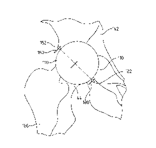
shown) is used to
ream the acetabulum 110 using the pins 180 as alignment guides. To use the
reamer, more
rigid guide pins may be used to guide the reamer, or the guide pins may be
used to insert a
central guide pin, thereby allowing the reamer to ream out the acetabulum
along the central
principal axis of the acetabulum sphere. Ideally, the inner portion of the
femoral head guide
is either spherical or anatomical in shape so that when the outer portion is
removed, the inner
portion is reminiscent of an acetabular cup so the surgeon could visible
inspect the position
and orientation of the femoral head guide cup, which will ultimately be the
position and
orientation of the implanted cup after all the final acetabulum cuts are made.
100811 If the acetabulum is damaged, for example by arthritis, the guide 170
may be aligned,
primarily off of the proximal femur and inserted into the acetabulum.
Likewise, if the
Date Recue/Date Received 2020-07-22
19
proximal femur is damaged, the guide 170 may be aligned more so off of the
acetabulum and
then inserted next to the proximal femur.
[0082] Referencing FIG. 12, after the acetabulum 110 has been reamed, a
portion of a
prosthetic trial 190 is inserted within the acetabulum 110 using the pins 180
as alignment
guides. The size of the spherical head in this trial 190 may be variable in
size and shape
depending on how much bone and/or cartilage was removed during the reaming
process.
Specifically, this prosthetic trial 190 includes an acetabular component 192
and a femoral ball
194 inserted therein. It is the acetabular component 192 that is temporarily
inserted into and
mounted to the reamed acetabulum 110. The femoral ball 194 of the trial 190 is
coupled to a
femoral neck 196 having an endplate 198.
[0083] As shown in FIG. 12, exemplary trials 190 may be modular so that the
femoral ball
194 is repositionable with respect to the acetabular component 192 and/or the
femoral neck
196 is repositionable with respect to the endplate 198. In contrast, the
exemplary trials 190
may be integrated or a single piece to inhibit movement between the respective
components
so that the orientation and position of the femoral ball 194 with respect to
the acetabular
component 192 is fixed and/or the orientation and position of the femoral neck
196 with
respect to the endplate 198 is fixed. Moreover, the exemplary trails 190 may
embody a
neutral shape position (A) or have a thicker base (B), or have variable neck
lengths (C), or
have an irregular base shapes (D), or have a clockwise rotated base shape (E),
or have a
counterclockwise rotated base shape (F).
[0084] Referring to FIGS. 12-14, one at a time, the various trials 190 may be
temporarily
mounted to the acetabulum 110 and aligned using the pins 180 to maintain
proper acetabular
cup orientation. At the same time, the femoral aspect of the trial 190 is used
by the surgeon
to properly maintain the orientation and position of the femur 108.
100851 Referring to FIGS. 13 and 14, after the orientation and position of the
femur 108 is
fixed with respect to the pelvis 142 using the trial 190, guide pins 200
inserted through the
femoral endplate 198, allowing for proper cuts to be made, maintaining
concentric anatomical
spheres. The femoral portion of this trial 190 may be free to rotate around
all three axes or a
locking mechanism may be used so that when the proper orientation of the
femoral neck
coincides with anatomical femoral neck, the angle is locked into place. While
the acetabular
component 192 is inserted within the reamed acetabulum 110, using the
previously inserted
guide pins 180, the surgeon is ready to mark the final cut of the femoral neck
144 using the
trial 190. By using the trial 190, which is mounted to the pelvis 142, the
resulting mark for
Date Recue/Date Received 2020-07-22
20
the final cut of the proximal femur 108 accounts for the orientation and
position of the trial
and allows concentric spheres (anatomical acetabulum sphere and femoral
sphere) to be
maintained. Although it is shown in FIGS. 13 and 14 that the trial 190 rests
on the proximal
femur, it could also be inserted into the proximal femur and/or guided around
the proximal
femur. Referencing FIGS. 14 and 15, after the guide pins 200 are inserted into
the femur
108, the trial 190 is removed from both the femur 108 and the pelvis 142.
Thereafter, a
cutting guide (not shown) is mounted onto the femur 108 using the pins 200 as
a guide to
ensure the alignment of the eventual femur cut.
100861 Various tapers may also be implemented in this trial shown in FIG. 12.
Therefore,
when the acetabular cup portion of the trial is inserted into the acetabulum,
it may be
temporarily fixed into place using nails or other fixating devices. Then, the
femoral portion
of the trial may be fixated to the superior aspect of the femur. Next, the
surgeon may
manipulate the leg into multiple positions, visually inspecting and
instrumentally measuring
for impingement, possible dislocation and any other concerns that could be
raised. The
surgeon may then replace the femoral portion of the trial using a different
taper to again
inspect and measure how the femoral component is rotating with respect to the
pelvis.
[00871 A distraction device may also be used that measures the amount of pull
of the
acetabular cup from the bone during manipulation of the femur. If the implants
have
concentric spheres, then the distractive and shear forces should be very low.
If during this
manipulation of the femur, the acetabular cup visually attempts to pull away
from the bone or
if the measurement device(s) detects irregular amounts of distractive or shear
forces, a
different trial may be used. This distraction measurement device may be a
spring loaded
mechanism or even a measurement device that measures distractive distance and
converts this
distance to a force, based on a mathematical model of the human hip joint that
derives intra-
operative forces using Newton's equations of motion. The mathematical models
of the
human body may be derived as an inverse model that measures the three
rotations and
translations of either the cup from the bone and/or the femoral head from the
cup. Then, this
motion is entered into the mathematical model to determine the forces in three
directions and
the torques around three directions.
100881 As shown in FIG. 24, an alternate method of marking the femur 108
includes using an
alignment/trial instrument 170 positioned so the acetabular portion is seated
within the
acetabulum and the femoral portion overlies the femoral neck. The surgeon may
then
reposition the instrument 300 to align with the proper orientation of the
intended proximal
Date Recue/Date Received 2020-07-22
21
femoral cut. The alignment/trial instruments 170 may be of normal implant
shape and
thickness 170A, or could of proper shape and thickness for the cup and femoral
ball, but
much thinner for the femoral neck and proximal femoral component 170B, 170C.
In
particular, the femoral neck portion of the alignment/trial instruments 170B,
170C comprises
a flat plate that is contoured to approximate the exterior contour of the
patient's proximal
femur so that the instrument may be easily placed directly on top of the
proximal femur to
ensure an accurate proximal femoral bone cut. The shape could also be
anatomical in nature.
[0089] Referring to FIGS. 16-18, a further alternative method of marking the
femur 108
includes using the light beam instrument 300 previously discussed to
superimpose various
shapes upon the proximal femur and distal pelvis 142. Those skilled in the art
will realize the
virtually any two dimensional shape could be superimposed upon the proximal
femur such as,
without limitation, a square, a rectangle, a trapezoid, and an outline of a
prosthetic hip trial.
A computer algorithm may be used with this instrument 300 so that three-
dimensional or
planer two-dimensional anatomical bone shapes are stored within a virtual
library of images.
These images may be created using one or more imaging modality including, but
not limited
to, MRI, CT, ultrasound, and X-rays. These images may also be stored in
various libraries
for size, gender and ethnicity.
[0090] During surgery, the surgeon uses a handheld instrument to generate
surgical data
including, but not limited to, boney landmarks, orientations, and distances.
This surgical data
is used by a computer algorithm to initially choose which image in the various
libraries best
matches the data entered and then, may modify one or more images stored in the
virtual
library to generate an image using the instrument 300 and project this image
onto the
patient's anatomical bone (in this case, the proximal femur). The library
images may then be
superimposed onto and compared with various images in the library, may be used
to create a
bone from various bones, or may be morphed from one or multiple library
images. Then,
using controls associated with the instrument 300, the image may be fine-tuned
to modify the
shape, size, thickness, position, and/or orientation to best match the
patient's bone. In
exemplary form, the instrument 300 projects virtual jigs, implants, and/or
bones onto the
patient's bone representative of the ideal location for each bone cut. In
addition or in the
alternative, the instrument may project an image of the final implant or
implant component
onto the patient's bone. For example, FIG. 16 depicts a rectangular shape 210
superimposed
=
onto the proximal femur 108 and distal pelvis 142, while FIG. 17 depicts an
outline of a
prosthetic hip trial 212 superimposed onto the proximal femur and distal
pelvis. Moreover, at
Date Recue/Date Received 2020-07-22
22
any time during the surgery, even after the surgeon has made all the bone
cuts, the surgeon
may turn on the light beam instrument 300 to verify the bone cuts made or to
revise the bone
cuts to match a particular shape, such as the outline of the intended implant.
As discussed
previously, even if the femur 108 has been repositioned and is out of
alignment, the surgeon
may utilize one or more of the pins 152 to properly orient and position the
femur with respect
to the pelvis 142. And after the femur 108 and pelvis 142 have been aligned,
the light beam
instrument 300 may be utilized to superimpose one or more shapes that
represent the best or
preferred implanted femoral neck shape and/or acetabular cup and/or femoral
head that
maintains proper biomechanics and concentric spheres of the pelvis 142 and
femur 108.
[0091] This light beam instrument 300 provides a relatively easy, less
expensive, and much
less complicated alternative to computer assisted orthopaedic surgery. At
present, many
surgeons are attempting to use computer navigation to define the orientation
and position of
the hip joint, but this methodology can be cumbersome and difficult to learn.
In the instant
technique, a light beam instrument 300, controls (such as dials and/or levers)
may be used to
change the position and orientation of a light beam (or image) directed from
the instrument
above the operating room table 302 to represent angles and/or positions of the
femur and/or
the pelvis during surgery. A surgeon can then turn on a light beam from the
instrument 300
and manually and/or audibly change the position and/or orientation of the
light beam to
define an anatomical feature of a bone, such as the anatomical femoral neck.
Once the light
beam has been positioned onto the femoral neck, defining its anatomical
position and
orientation, two or more pins may be inserted in the femur and/or pelvis.
These pins may be
used to define the anatomical bone or bone feature in question. Multiple light
beams may
also be used, defining as many bones or honey features as needed by the
surgeon. Therefore,
at anytime during the surgery, the surgeon may turn on a beam from the
instrument 300 and
re-orient the bones until the beam passes through the alignment pins.
[0092] Although the previous examples utilize the light beam instrument 300 to
project a
two-dimensional image, the projected images could also be three-dimensional
using
holographic images. Holographic imaging may be utilized to allow bone anatomy,
bone
landmarks, and implant components to be projected onto the bone using a light
source. The
light source, scattered from the object of reference, will be recorded and
later reconstructed
so that when an imaging system (a camera or an eye) views the reconstructed
beam, an image
of the bone and/or implant component is seen even when it is no longer present
in the
surgeon's field of view. The image changes as the position and orientation of
the surgeon
Date Recue/Date Received 2020-07-22
23
changes in exactly the same way as if the object were still present, thus
making the image
appear three-dimensional. This effect can be seen by the surgeon at all times,
right where the
orientation of the bone and/or implant component, even though each view of the
image may
appear to be significantly different by the surgeon, yet the three-dimensional
orientation and
position are correct. It should be noted that the holographic recording itself
is not an image ¨
it consists of an apparently random structure of either varying intensity,
density.
[0093] Similar to the foregoing technique used to project a two-dimensional
image upon the
patient's bone, a computer algorithm is used in order to generate a three-
dimensional image
and superimpose this image onto the requisite one. Unlike the two dimensional
image
projection, the surgeon will be required to measure distances and orientations
in all three
directions. In order to create the three-dimensional image, a series of
preexisting three-
dimensional images are stored in a virtual library. These images will contain
proper bone
landmarks and distances that define orientation and position with the human
body structure.
These images may be rigid or deformable bodies. During surgery, the handheld
device is
used to define anatomical distances, positions, and orientations on the bone
of the patient in
question and then, the computer algorithm chooses the best initial three-
dimensional bone fit
and projects this three dimensional image onto the anatomical bone. Unlike
using a two-
dimensional image, distances from the light beam source of the light beam
source instrument
to the anatomical bone must be known to properly project the three-dimensional
image.
Without defining this distance, at multiple locations on the anatomical bone,
the three-
dimensional image may not be properly projected. Therefore, three-dimensional
information
along all three directions must be measured and entered into the computer
algorithm.
[0094] An exemplary method of measuring and entering the data for processing
by the
computer algorithm includes using a digital camera or other recording source
to take multiple
photos or images of the boney anatomy, such as the femoral neck and head after
the surgeon
opens up the joint space. These image views may be proximal, distal, anterior,
posterior,
medial, and/or lateral. These images, in real-time may be sent to the light
beam instrument
300 and using the instant computer algorithm, a three-dimension image, either
holographic or
non holographic is constructed using the three-dimensional library of bone
images. Then, the
best fit bone image is projected onto the patient's bone. Using dials, levers
or other controls,
the three-dimensional image can be re-oriented, re-sized and/or re-positioned
onto the
anatomical bone. Once the surgeon deems the three-dimensional image to be an
accurate
Date Recue/Date Received 2020-07-22
24
representation of the anatomical bone, another algorithm is used to define
boney landmarks
and bone cuts that are ideal for that particular patient.
[0095] Creating the three-dimensional holographic images makes use of devices
that produce
so-called diffraction fringes, fine patterns of light and dark that can bend
the light passing
through them in predictable ways. A dense enough array of fringe patterns,
each bending
light in a different direction, can simulate the effect of light bouncing off
of a three-
dimensional object. In exemplary form, one exemplary commercially available
technology
uses a cylinder approximately one meter high by one-half meter in diameter.
Inside the
cylinder, a helix spins at high speed. A two-dimensional image is projected
onto the helix
and then the image is projected onto the bone. It is presumed, for purposes of
this example,
that the images are simple CAD-like drawings. These simple images are
constructed from
multiple digital camera images as discussed previously. An alternative method
and
technology that may be used incorporates a pair of lasers that emit beams that
intersect one
another inside of a cube of special material. The material inside the cube
glows at the
intersection point. Another method uses two lasers that intersect inside a
cube of a special
material. The material glows at the intersection, creating an image that may
then be projected
onto the bone.
[0096] Initially, the surgeon points the laser of the light beam instrument
300 at a beam
splitter, thereby causing the beam to be divided into two beams. Mirrors
within the light
beam instrument 300 are constructed along the path of the splitter so that the
laser hits the
bone in question. The light beam instrument 300 also includes diverging lenses
in front of
the mirrors so that the two beams passing through them become wide swathes of
light rather
than regular beams. One of the lights (object beam) will reflect off the bone
in question and
onto the holographic plate of the light beam instrument 300. The other light
(reference beam)
will hit the holographic plate only. Then, the surgeon projects the three-
dimensional
holographic image on the bone in question at anytime during surgery.
[0097] At present, surgeons routinely have four to ten trays of instruments
and jigs for use
during the surgery. Before every surgery, these instruments and jigs need to
be prepared and
sterilized. The foregoing light beam instrument may be used to project these
instruments and
jigs onto the bone, as needed by the surgeon. Each instrument is scanned using
a laser
scanner or is converted into three-dimensional solid objects using three-
dimensional
computer models. Once each instrument and various sizes are entered into the
virtual library
of images, the images may be re-oriented and displayed at anytime using a
computer
Date Recue/Date Received 2020-07-22
25
algorithm that instructs the light beam instrument to rotate and translate
with respect to either
a fixed or relative reference frame. The Newtonian reference frame is defined
within the
computer algorithm and relative reference frames are defined for each
instrument. Each
rotational and translational direction is defined as a function in an inverse
direction model or
as a generalized speed in a forward solution model. A change in direction or
rotation of the
displayed image may be made by the surgeon audibly, through the use of dials
and/or levers
(i.e., controls) or using a touch screen monitor. In fact, numerical changes
to the translation
matrix may also be input to define motion changes. Using for example, a touch
screen
instrument, the surgeon is able to touch a picture of a virtual instrument or
guide and the
computer algorithm instructs the light beam instrument to project it. Then a
secondary
library appears on the screen, whereas a surgeon can choose the correct size
of the image.
Then, by audible commands or using dials and levers or using his finger on the
screen, the
image, whether two-dimensional or three-dimensional can be repositioned.
Therefore, the
relative transformation matrix between the instrument and the Newtonian
reference frame
could be altered depending on where the instrument is in space with respect to
the origin
within the Newtonian reference frame. Once the surgeon has the instrument or
jig in place, a
stop is instituted and the relative reference frame of the instrument with
respect to the
Newtonian reference frame is recorded and stored for future use of the
instrument.
Therefore, within the computer algorithm the generalized coordinated and
generalized
positions, defined from the generalized speeds are changed and redefined based
on global
coordinate changes. This procedure may be conducted for each instrument, jig
and bone,
whether in two-dimensions or three-dimensions. These instruments, jigs and
bones may have
points, axes and cutting guides defined and positioned properly for surgical
use.
[0098] As stated previously, this process may be used for three-dimensional
images,
holographic or non-holographic. As stated previously, holography is a
technique that allows
the light scattered from an object to be recorded and later reconstructed so
that it appears as if
the object is in the same position relative to the recording medium as it was
when recorded.
The image changes as the position and orientation of the viewing system
changes in exactly
the same way as if the object was still present, thus making the recorded
image (hologram)
appear three dimensional. Holograms can also be made using other types of
waves.
[0099] Three-dimensional space is a geometric model of the physical universe
in which we
live. The three dimensions are commonly called length, width, and depth (or
height),
although any three mutually perpendicular directions can serve as the three
dimensions.
Date Recue/Date Received 2020-07-22
26
[0100] In mathematics, Cartesian geometry describes every point in three-
dimensional space
by means ofthree coordinates. This is the process previously described for
positioning and
orienting instruments, jigs and bones for surgical use. Three coordinate axes
are given, each
perpendicular to the other two at the origin, the point at which they cross.
The instant
inventor is a user of Kane's Dynamics. Thus, each body or massless frame that
is defined is
assigned three unit vectors (or relative axis), labeled as either the 1, 2 or
3 axis within a
particular reference frame. Relative to these axes, the position of any point
in three-
dimensional space is given by an ordered triple of real numbers, each number
giving the
distance of that point from the origin measured along the given axis, which is
equal to the
distance of that point from the plane determined by the other two axes. The
created three
dimensional holographic images may be constructed using generators and/or
other
components purchased from companies within the "International Hologram
Manufacturers
Association" specifically companies such as "The Hologram Company" and "API
Holographies". If one chooses to go this route, components and even full
holographic
projectors may be commercially purchased. Then, the holographic projector or
holographic
components are used with a digital device, instrumentation, and computer
algorithms to create
the images, as well as size, position, and orient the images into the proper
location during
surgery. Alternatively, a specialized holographic projector may be fabricated
from
commercially available components and ensuring that the resolution and quality
of the
holographic images is ideal for medical use. Similar to the two dimensional
images, each
light beam or point within the holographic image is defined with respect to
the fixed
Newtonian reference frame, defined on the light beam instrument. Unlike moving
a two-
dimensional image, two processes may be used to move the holographic image.
Either each
point could be defined and then redefined with respect to the origin in the
Newtonian
reference frame, or at least three points within the holographic image could
be defined with
respect to the Newtonian reference frame and then three relative axes with the
holographic
relative reference frame could be constructed and these axes could be oriented
and positioned
with respect to the Newtonian reference frame. Again, as the three-dimensional
image is
moved, the relative transformation matrix will be redefined. Once the three-
dimensional
image is positioned in place by the surgeon, the coordinates for this relative
transformation
matrix are locked in place and may be used at anytime during the surgery by
the surgeon.
Dal
27
10101] Referring to FIG. 25, if three points are defined on the holographic
image, one of
these points must be defined out of the plane the first two points were
defined within.
Therefore, for this example, each of the three points is defined as Pl, P2 and
P2 on a three
dimensional image taking the shape of an ellipse in any view or an elongated
sphere. In this
figure, the point 0 represents a fixed point in the Newtonian reference frame,
within the light
beam instrument or where the holographic image project is housed in the
instrument. The
image that is being projected is defined as body A and a relative reference
frame with the unit
vectors Al>, A2> and A3> are defined.
P P2 P1>
Al> ¨ ______________
IP P2 Pl> I
Unit vector Al> is defined by creating a line using the points P1 and P2 and
the dividing this
line by its magnitude. Next, the second unit vector is defined by cross
multiplying vector
Al> with the unit vector of the line between points P2 and P3 and dividing
this function by
its magnitude.
UnitVEC(P P2 P3>) x (Al>)
A2> =
UnitVEC(P P2 P3>) x (Al>) I
Finally, the third unit vector A3> is defined by cross multiplying Al> and
A2>. Next, the
transformation matrix can be derived using three successive rotations defined
in FIG. 26,
whereas the reference frame A represents a body, but the A' and A" reference
frames are
intermediate in nature and are defined as massless reference frames.
[0102] The sequential references frames could set up as follows:
{Al > Cos(03) 0 -Sin(03) 1 0 0
Cos(01) Sin(01) 0 N1>
A2> = 0 1 0 0
Cos(02) Sin(02) -Sin(01) Cos(01) 0 N2>
A3> Sin(03) 0 Cos(03) 0 -Sin(02) Cos(02) 0 0 1
N3>
Then, using matrix multiplication, the following relative transformation
between the
Newtonian reference frame within the light beam instrument and the relative
reference frame,
the three dimensional image could be described as:
Date Recue/Date Received 2020-07-22
28
1 Al > (COI 'CO3 - Sol-S02'S03) (S01 'CO3 + S02-S03C01) (-S03-0O2) N1>
A2> = - (S01 'CO2) (COI CO2) (S02)
N2>
A3> (S03C01 + SOl'S02'CO3) (S01 'S03 ¨ S02'C01*CO3) (CO2'CO3) N3>
[0103] In the transformation matrix listed above, 01, 02 and 03 represent
relative rotations of
the three dimensional image with respect to the light beam instrument and as
these angles
changes, so does the three dimensional image. These angles may be previously
defined
before surgery, may equate to temporal or depended functions or may be changed
intra-
operatively either using dials and/or levers, audibly or through another
measurement device.
If the surgeon chooses to move the three points Pl, P2 and P3 to navigate the
three-
dimensional image and would like to know the orientation of the image, more
specifically 01,
02 and 03, these angles may be mathematically derived using the following set
of equations:
PA11 = 001 .0O3 - SO1 -S02-S03
PA12 = S01 .0O3 + S02-S03-001
PA13 = -S03.0O2
PA21 = S01 =CO2
PA22 = C0102
PA23 = SO2
PA31 = S03.001+ S01.S02.0O3
PA32 = S01 =S03 - SO2C01.0O3
PA33 = CO2.0O3
The foregoing nine equations represent an over determinant system, meaning you
have too
many equations for only three unknown quantities. The three angles of
questions can also be
derived using the following three equations:
02 = Sin-1 (PA23)
03 = Cos-1(PA33/COS(02))
01 = Cos-1(PA22/Cos(03))
[0104] Although the use of three-dimensional images as disclosed herein
references
holographic or non-holographic images, these images could quite easily be
constructed using
Date Recue/Date Received 2020-07-22
29
any technique for defining and creating three-dimensional images. Also, the
application for
three dimensional images is disclosed in the context of total hip
arthroplasty, however, those
skilled in the art should understand that these three dimensional images and
associated
equipment could also be used for any joint, organ or structure within the
human and animal
body and could be used for other surgeries besides just total joint surgeries.
[0105] Referencing FIGS. 19 and 20, after the distal femur has been cut, the
final femoral
and acetabular implants are mounted to the pelvis 142 and the femur 108.
Mounting of the
final femoral implants may utilize the guide pins 200 or virtual
instrumentation through the
use of the light beam source instrument. For example, presuming any prefatory
reaming of
the intramedullary canal of the femur has taken place (and this reaming can
also utilize the
guide pins 200 for alignment), the final implanted femoral stem 220 is
inserted into the
intramedullary canal of the femur 108 using the guide pins 200. Presuming the
final femoral
implant is modular, the final femoral neck 224 and femoral ball 226 are
mounted to the
implanted femoral stem 220. In this exemplary embodiment, the femoral stem 220
includes
an endplate 222 that sits upon the outer proximal surface of the femur 108. In
addition, the
femoral neck 224 also includes an endplate 228 that couples to the endplate
222 of the
femoral stem to mount the neck to the stem.
[0106] In this exemplary embodiment, the femoral stem 220 is offset from the
femoral neck
224. While an offset between the stem 220 and neck 224 may occur infrequently,
it has been
shown to document the ability to reposition the neck independent from the stem
and the
modularity of this exemplary implant. In some cases, however, the final
implant will be
integrated or a single piece so that variable orientation and position of the
stem 220 with
respect to the neck 224 is not possible.
[0107] It should also be noted that the final acetabular implants may be
mounted to the
pelvis 142 utilizing the guide pins 180. For example, presuming all prefatory
reaming of the
acetabulum is complete (and this reaming can also utilize the guide pins 200
for alignment),
the final implanted acetabular cup and cup insert 232 are inserted into the
reamed acetabulum
110 using the guide pins 180. Presuming the final acetabular cup and cup
inserts are
modular, minor modifications can be made to the orientation and position of
each component
with respect to the other. After both the final femoral and acetabular
components are
implanted and secured, the femoral ball 226 is seated into the acetabular cup
insert 232.
Date Recue/Date Received 2020-07-22
30
[0108] Referring to FIG. 21, it is also within the scope of the invention to
include a femoral
sleeve 240 for insertion into the intramedullary canal of the femur 108. The
exemplary
sleeve 240 may be either press fit into the femur 108 or cemented. In
contrast, current
techniques commonly cement or press fit the femoral implant into the femur
108. Once this
femoral stem is secured to the bone using this preexisting technique, removal
for revision
surgery is very difficult. The instant invention introduces a modular type
femoral stem that
slides into the femoral sleeve 240. This femoral sleeve 240 may be designed
with internal
grooves and projections 242 so that a femoral stem 220 (see FIG. 19) may be
inserted into the
sleeve at multiple angles. The sleeve could also be smooth without grooves. If
grooves are
used, these grooves and projections 242 may alternate or include a pattern
that coincides with
a pattern on the femoral stem 220 to ensure proper orientation of the femoral
stem with
respect to the sleeve. The implanted femoral stem 220 is locked into place
within the sleeve
240. By using a sleeve 240 to interpose the femur 108 and the femoral stem
220, it provides
the advantage that if the femoral stem needs to be removed for any reason, the
femoral stem
can be unlocked from the sleeve and then removed without causing additional
damage to the
intramedullary canal. Thereafter, a new or revised femoral stem may be
inserted into the
sleeve 240. Accordingly, the use of a permanent sleeve 240 ensures that
initial femoral stem
alignment is maintained in any femoral version surgery. Also, if for some
reason it is deemed
that the femoral stem orientation is not proper for that patient, rather than
having to remove
the femoral stem from the femur, the femoral implant in this in this patient
may be removed
and then the version angle may be changed and thereafter the femoral stem is
re-positioned
back into the sleeve.
[0109] While the foregoing sleeve 240 has been described as having a fixed
orientation and
position with respect to the femur 108, it is also within the scope of the
invention for the
sleeve 240 to include mobile bearing functionality. This functionality may be
the result of a
pair of sleeves that are telescopic, with the inner (smaller diameter) sleeve
including the
internal grooves and projections 242. This structure allows the inner sleeve
to rotate with
respect to the femur 108 and may be limited to retard a fully 360 degrees of
femoral stem
rotation.
[0110] Another exemplary alternative includes a sleeve that has no internal
grooves and does
contain a locking mechanism. The sleeve may be cemented and/or press fit into
the femur.
Date Recue/Date Received 2020-07-22
31
The femoral stem is of a round or oval shape and free to rotate within the
sleeve. The
femoral stem may be locked in one direction so that it can be removed from the
sleeve, but is
free to rotate around its longitudinal axis or any defined axis that is
necessary to maintain
concentric spheres.
101111 Referring to FIGS. 22 and 23, in another exemplary embodiment, the
femoral stem
390 has an internal longitudinal screw (or gear mechanism) 400. After the
femoral stem 390
is implanted into the femur, the surgeon can turn the screw 400 on the
superior aspect of the
femoral stem, which will force elements 404 to protrude from the stem into the
bone, leading
to greater fixation of the femoral stem to the bone. If for some reason the
implant needs to be
removed during revision surgery, the surgeon can turn the screw 400 in the
opposite
direction, which operates to pull the elements 404 back into the stem 390
allowing for the
stem to be more easily removed. If the femoral stem is to be inserted into the
femoral sleeve
240 (see FIG. 21), the surgeon turns the screw 400 to reposition the elements
404 to protrude
away from the stem into grooves 242 in the sleeve. In exemplary form, the
elements 404
may take on various shapes and sizes and be located at various positions along
the femoral
stem and/or sleeve 240. For example, the elements 404 take on a triangular
profile in FIG.
22, while the elements take on a rectangular profile in FIG. 23.
[0112] It should be noted that the femoral stem and femoral sleeve may take on
any number
of shapes both on the interior and exterior. For instance, the inside of the
femoral sleeve may
be elliptical, while the exterior of the femoral stern is rectangular.
Likewise, the exterior of
the femoral sleeve may be rectangular, elliptical, or any other cross-section.
[0113] Following from the above description and invention summaries, it should
be apparent
to those of ordinary skill in the art that, while the methods and apparatuses
herein described
constitute exemplary embodiments of the present invention, the invention is
not limited to the
foregoing and changes may be made to such embodiments without departing from
the scope
of the invention as defined by the claims. Additionally, it is to be
understood that the
invention is defined by the claims and it is not intended that any limitations
or elements
describing the exemplary embodiments set forth herein are to be incorporated
into the
interpretation of any claim element unless such limitation or element is
explicitly stated.
Likewise, it is to be understood that it is not necessary to meet any or all
of the identified
advantages or objects of the invention disclosed herein in order to fall
within the scope of any
Date Recue/Date Received 2020-07-22
32
claims, since the invention is defined by the claims and since inherent and/or
unforeseen
advantages of the present invention may exist even though they may not have
been explicitly
discussed herein.
[0114] What is claimed is:
Date Recue/Date Received 2020-07-22