Note: Descriptions are shown in the official language in which they were submitted.
BLOOD PUMP SYSTEMS AND METHODS
FIELD OF THE INVENTION
[0001] The present invention relates to a blood pump system that includes a
pump,
conduits, a control unit, and a source of power, whereby the system may be
used for a variety of
peripheral vascular clinical indications. Specifically, this invention may be
useful for
persistently increasing the overall diameter and lumen diameter of veins and
arteries in patients
needing a vascular access site for hemodialysis, a bypass graft, or other type
of surgery or
procedure where a larger vein or artery diameter is desired. This invention
may also be useful
for increasing lower extremity venous return and reducing lower extremity
venous pressure in
patients with lower extremity venous hypertension, including those patients
with skin
discoloration and ulceration. This invention may be further useful for
providing increased local
blood flow to organs and tissues in need thereof, such as the lower
extremities of patients with
peripheral arterial disease (PAD).
BACKGROUND INFORMATION
[0002] There are over half a million chronic kidney disease (CKD) patients in
the United
States, with over 100,000 new CKD patients each year. There is a four percent
annual increase
in projected prevalence population due to such driving factors as, for
example, high blood
pressure, diabetes, and an aging population.
[0003] Hemodialysis is the treatment of choice for 92% of CKD patients,
because
without hemodialysis or some other form of treatment those CKD patients would
die. A typical
CKD patient undergoing hemodialysis treatment must have his or her vascular
system connected
to a hemodialysis machine two to three times per week. For hemodialysis, there
are three
common vascular access site options. The preferred access site option is an
arteriovenous fistula
(AVF), which is a direct, surgically created connection between an artery and
a vein, preferably
in the wrist, or alternatively, in the forearm, upper arm, leg, or groin.
Another access site option
is an arteriovenous graft (AVG), which is a surgically created connection
between an artery and
vein using an interposed synthetic conduit. The final major access site option
is a catheter
inserted into a large vein in the neck, chest, leg, or other anatomic
location.
1
Date Recue/Date Received 2020-07-24
[0004] Patients with an AVF have less morbidity, less mortality, and a
lower cost of
care compared with patients with an AVG or a catheter; therefore, an AVF in
the wrist is the
preferred form of vascular access for hemodialysis. Patients with an AVG or
catheter have
substantially higher rates of infection and death than patients having an AVF,
with catheter
patients having the worst outcomes. In addition, patients having an AVG or
catheter have a
higher average cost of care, with catheter patients having the highest costs.
If a patient is eligible
for an AVF, the wrist or forearm is generally preferred over an AVF in the
upper arm due to
higher rates of hand ischemia and the generally shorter and deeper vein
segments of the upper
arm.
[0005] Unfortunately, about 85 percent of patients are ineligible for
an AVF in the
wrist, mostly due to vein and artery diameters that are too small.
Furthermore, about 60 percent
of all AVFs created are not useable without additional surgical and
interventional procedures due
to an occurrence commonly referred to as "maturation failure," which is
correlated with small
vein and artery diameter. The availability of veins and arteries with larger
diameters is
correlated with higher AVF eligibility and lower rates of maturation failure.
[0006] Currently, there are few options for permanently and
persistently increasing
the diameter of a vein or artery. All current methods use mechanical methods
of dilation, such as
balloon angioplasty, that can lead to vein or artery injury. Since a patient
needs to have
peripheral veins and arteries of a certain size for a physician to create an
AVF, it is desirable to
have a method and system for persistently and permanently increasing the size
or diameter of
peripheral veins or arteries.
[0007] Approximately 7 million people in the US suffer from chronic
venous
insufficiency and hypertension, which can progress to venous ulceration. Lower
extremity ulcer
is the most common form of chronic wound, with an estimated prevalence of 1%
of the US
population. About 2.5 million people in the US have a lower extremity
ulceration and about
600,000 people seek treatment for a venous ulceration of the lower extremity
each year in the
US. The incidence of venous ulceration is expected to rise as the population
ages.
[0008] In a survey of patients with venous ulcers, 81% of patients
reported an adverse
effect on mobility, 56% reported spending up to 8 hours per week on ulcer
care, 68% reported a
negative emotional impact, including fear, social isolation, anger,
depression, and negative self-
image. In the survey, 80% of patients are not working outside the home; and of
the 20%
2
Date Recue/Date Received 2020-07-24
employed, leg ulceration correlated with time lost from work, job loss, and
adverse effects on
finances.
[0009] Lower extremity venous hypertension and ulceration is costly to
treat and
places a substantial burden on health care providers and systems. In a study
of 78 venous ulcer
patients at the Cleveland Clinic, median ulcer size was 2.8 cm2 (mean = 9.4
cm2) and 5% had
bilateral ulcers. The median time to ulcer healing was 77 days (mean = 108
days) and the mean
cost of treatment was $2,400 per month. The mean total cost of treatment to
heal an ulcer was
$9,685 per patient. For patients requiring more than a year to heal, the
average total cost per
patient was $18,534.
[0010] In a majority of cases, venous hypertension and ulceration
results from
valvular incompetence secondary to deep vein thrombosis or an unknown cause.
In a substantial
minority of cases, venous hypertension and ulceration results from femoral or
pelvic venous
obstruction secondary to deep vein thrombosis, vein injury, or extrinsic vein
compression.
Chronic tissue exposure to localized venous hypertension leads to dilation of
capillaries with
increased permeability and leakage of plasma and erythrocytes, trapping and
activation of
leukocytes in the microcirculation, and the release of free radicals and other
toxic products, such
as tumor necrosis factors and collagenase, which can promote cell death and
tissue damage.
Leakage of fibrinogen into surrounding tissues binds or "traps" growth factors
and cytokines, and
renders them unavailable for maintenance and repair of tissue integrity.
[0011] Lower extremity venous hypertension presents clinically as leg
redness and
discoloration, swelling, pain, edema, pruritus, scaling, discharge, and
lipodermatosclerosis.
Ulcers generally develop on the medial aspect of the leg and possess irregular
borders and can be
associated with severe pain. Venous ulcers are often complicated by
superimposed bacterial
infection. The arterial circulation is usually adequate. Current treatments
for lower extremity
venous hypertension and ulcer are often inadequate. Patients are mostly
offered palliative
treatments, with the goal of healing ulcers and preventing recurrence,
including aggressive
wound care, compression therapy to decrease lower extremity venous pressure
and increase
venous return, lower extremity vein stripping or ablation, and skin grafting.
However, current
treatments often fail to heal ulcers and recurrence rates for healed ulcers
are high.
[0012] Currently, small "heart pumps" exist; however, such pumps are
costly and not
designed and dimensioned for use in an extremity or for the uses described
herein. As such,
3
Date Recue/Date Received 2020-07-24
there is a need in the art for systems, components, methods, and pump devices
that can increase
the diameter of peripheral veins and arteries at a reasonable cost.
Additionally, there is a need
for a systems, components, methods, and pump devices that can increase lower
extremity venous
return, reduce lower extremity venous hypertension, and heal venous ulcers.
SUMMARY OF THE INVENTION
[0013] The present application relates to blood pump systems, including blood
pump
systems with wide operating ranges, low cost-of-goods-sold (COGS), and
intermediate duty
times. These blood pump systems are designed for use in a variety of clinical
situations and for a
variety of clinical indications, as described herein.
[0014] The blood pump systems described herein can be used for increasing the
diameter
of veins and arteries, preferably peripheral veins and arteries. The system
will function to move
blood in such a way as to cause an increase in vein or artery diameters. This
can be
accomplished by discharging ("pushing") blood into a vein or artery or by
removing ("pulling")
blood from a vein or artery. By either method, the system increases the flow
of blood in a vessel,
which ultimately leads to a persistent increase in vessel diameter. As such,
the system and, more
particularly, the pump use mechanical means to activate biological response
pathways resulting
in the enlargement or "remodeling" of veins or arteries. The system has a
blood pump, conduits
to carry or convey blood to and from the blood pump, a control system to
monitor the blood
pump and modify the operation of the blood pump, and a power source. As such,
the system
comprises a group of members that can be, for example, fluidly connected to an
artery at one end
and fluidly connected to a vein at the other, whereby, when activated, blood
is pumped at a rate
such that wall shear stress (WSS) on the endothelium of the vein, artery, or
both is elevated for a
period of time sufficient to causes a persistent enlargement in the vein or
artery. Any of a variety
of pumps and pump systems may be used so long as the flow of blood through the
pump system
can be controlled to produce the desired blood vessel diameter increase.
[0015] The blood pump systems described herein can be used to increase lower
extremity
venous return, reduce lower extremity venous hypertension, and heal venous
ulcers. The system
will function to move blood from a vein in the affected lower extremity, such
as a femoral,
saphenous vein, or iliac vein, to a location in the venous circulation such
that the return of
venous blood from the lower extremity to the heart is improved. Locations for
return to the
4
Date Recue/Date Received 2020-07-24
venous circulation include the jugular vein, the axillary vein, the subclavian
vein, the
brachiocephalic vein, the superior vena cava, and the right atrium. The system
has a blood
pump, one or more conduits to carry or convey blood to and from the blood
pump, a control
system to monitor the blood pump and modify the operation of the blood pump,
and a power
source. As such, the system comprises a group of members that can be, for
example, fluidly
connected at one end to a peripheral vein and fluidly connected to a
peripheral, central vein, or
right atrium at the other end, whereby, when activated, blood is pumped at a
rate such that
venous blood pressure is lowered in the treated lower extremity for a period
of time sufficient to
cause partial or complete healing of a venous ulcer to occur. Any of a variety
of pumps and
pump systems may be used so long as the flow of blood through the pump system
can be
controlled to produce the desired effect.
[0016] Various types of blood pumps may be employed, including positive
displacement
and rotary pumps, with rotary type pumps being preferred. In one embodiment, a
rotary blood
pump system includes a pump having a housing defining an inlet to receive
blood and an outlet
to discharge blood. The pump housing is designed and dimensioned to house a
rotating impeller
suspended on bearings. The pump housing can have a first bearing at the inlet
portion of the
housing and a second bearing at the outlet portion of the housing. Blood
enters and exits the
rotating impeller, whereby the impeller increases the exit velocity of the
blood. This increased
velocity is recovered or translated as increased pressure as the blood
decelerates within the pump
diffuser, which terminates in the pump outlet.
[0017] In other embodiments, various types of rotary blood pumps may
be used. For
example, an axial flow pump, a mixed flow pump, or preferably, a centrifugal
blood pump may
be used. In addition, a variety of pump impeller bearings may be used,
including, but not limited
to magnetic bearings, hydrodynamic bearings, and, preferably pivot (contact)
types. Similarly,
various types of pump diffusers may be used, including but not limited to a
collector diffuser, or
preferably a volute diffuser.
[0018] In one embodiment, a centrifugal blood pump with pivot bearings
includes a
pump housing defining a pump inlet having an inflow diffuser to receive blood
and direct blood
onto an impeller, the pump housing having a top bezel and top pivot bearing
extending from a
top of the housing into the inlet, and a bottom bezel and bottom pivot bearing
extending from a
bottom of the housing into the interior space of the housing. The pump also
includes the
Date Recue/Date Received 2020-07-24
impeller suspended within the housing, the impeller further having a bearing
lumen to receive an
impeller pivot. The impeller pivot has a first end to engage the inlet portion
(top) pivot bearing
and a second end to engage the outlet portion (bottom) pivot bearing. In one
embodiment, the
ends of the impeller pivot are convex and at least one end of each pivot
bearing is concave. In
another embodiment, the ends of the impeller pivot are concave and the pivot
bearings are
convex. The impeller can include a variety of fin or blade constructions
designed to contact and
accelerate blood into the volute. For example, the impeller defines a
plurality of blades on the
top surface of the impeller and extending radially from a center of the
impeller to an outer edge
of the impeller. The blades accelerate blood from the impeller's central inlet
to its peripheral
outlet. In another option, the impeller does not include blades or fins, but
does include means to
move or propel blood. The impeller optionally includes at least one washout
lumen, cut-away, or
bore extending generally parallel to a central axis of the impeller from a
bottom surface through
the impeller to a top surface. The lumen is designed to prevent stagnation of
blood under the
impeller and around the bottom pivot bearing.
[0019] The blood pump includes a motor, preferably electric, designed to
actuate the
impeller. In one embodiment, the blood pump includes a drive motor having at
least one magnet
mechanically attached to the impeller and at least one armature mechanically
attached to the
housing. The armature induces an electromotive force on the at least one
magnet attached to the
impeller. The pump motor can be an axial-gap brushless direct current (DC)
torque motor with
sensorless back electromotive force (back-EMF) commutation. The motor employs
a sintered
alloy of neodymium iron boron (NdFeB) for the magnets in the impeller and a 3-
phase planar
"racetrack" coil configuration in the stator. The motor has a pancake aspect
ratio, with a very
small axial length in comparison to its diameter.
[0020] In one embodiment, the blood pump system includes a centrifugal blood
pump
with an operating range between about 50 milliliters per minute and about 1500
milliliters per
minute. The system also includes a pump housing defining a pump inlet to
receive blood and
direct blood onto an impeller. The pump housing has a top pivot bearing
extending from a top of
the housing into the inlet, and a bottom pivot bearing extending from a bottom
of the housing
into the interior space of the housing. The pump also includes an impeller
suspended within the
housing wherein a first gap between the impeller and a top portion of the
housing is in a first
range between about 0.05 mm and about 0.2 mm.
6
Date Recue/Date Received 2020-07-24
[0021] The impeller includes an impeller pivot having a first end to engage
the top pivot
and a second end to engage the bottom pivot and a plurality of blades on the
top surface of the
impeller and extending radially away from a center of the impeller, the blades
to force blood
received at the inlet through the pump housing and to the outlet. The impeller
also includes at
least one lumen extending parallel to a central axis of the impeller from the
bottom surface
through the impeller to a top surface.
[0022] The pump further includes at least one magnet mechanically engaged to
the
impeller and an electric motor to magnetically engage the at least one magnet,
wherein the
electric motor rotates the at least one magnet and the impeller. In other
embodiments, the pump
also includes a ferromagnetic backplate to magnetically engage the at least
one magnet.
[0023] [0021] The blood pump system has one or more conduits including a first
(inflow) conduit having two ends, a first end that is fluidly connected to a
location in the vascular
system and receives blood from that location, and a second end that is fluidly
connected to the
pump. The inflow conduit delivers blood to the pump. The blood pump system has
a second
(outflow) conduit having two ends, a first end that is fluidly connected to
the pump and receives
blood from the pump, and a second end that is fluidly connected to a location
in the vascular
system. The outflow delivers blood to a location in the vascular system.
[0024] In various embodiments, the conduits of the blood pump system have an
individual length of between 2 cm and 110 cm and a combined length between 4
cm and 220 cm,
and may be trimmed to a desired length by a surgeon or other physician,
including during
implantation of the pump system. The conduits each have an inner diameter
between 2 mm and
mm, and preferably between 4 mm and 6 mm. The conduits may be formed at least
in part
from polyurethane (such as Pellethane 0 or Carbothane C)), polyvinyl chloride,
polyethylene,
silicone elastomer, polytetrafluoroethylene (PTFE), expanded
polytetrafluoroethylene (ePTFE),
polyethylene terephthalate (PET, e.g. Dacron), and combinations thereof. The
conduits may
further include an elastic reservoir.
[0025] All or portions of the conduits may be reinforced with a
braided or spiral
coiled shape memory material, such as nitinol, or other self-expanding or
radially expansive
material, such as stainless steel. For pump systems designed for the treatment
of lower extremity
venous hypertension and venous ulcers, the conduit that conveys blood from a
lower extremity
vein to the pump portion of the pump system may further comprise a distal
segment of ePTFE or
7
Date Recue/Date Received 2020-07-24
Dacron such this segment can be fluidly connected to the lower extremity vein
by a surgical
anastomosis. Further, this ePTFE or Dacron segment may comprise an external
reinforcement,
such as additional ePTFE or Dacron material, or with a self-expanding or
radially expansile
material such as nitinol or stainless steel. This external reinforcement may
take the form of a
spiral or a braid, or may comprise a more completely circumferential and
uniform support
structure, or may be configured in another manner that resists collapse,
compression, or coaption
when the pressure within the conduits is low or negative. The conduits may
have chamfered
ends that fluidly connect to the vascular system. The ends can be chamfered at
an angle between
degrees and 80 degrees. One or more of the conduits may have a number of holes
or
fenestrations in the walls of the distal ends, when configured for placement
within the lumen of a
blood vessel or other intravascular location. The conduits may be secured to
the pump using
radially-compressive connectors.
[0026] In another embodiment a blood pump system a centrifugal blood pump and
a
pump housing defining a pump inlet to receive blood and direct blood onto an
impeller. The
pump housing has a top pivot bearing extending from a top of the housing into
the inlet, and a
bottom pivot bearing extending from a bottom of the housing into the interior
space of the
housing. The pump also includes an impeller suspended within the housing
wherein a first gap
between the impeller and a top portion of the housing is in a first range
between about 0.05 mm
and about 0.2 mm.
[0027] The impeller includes an impeller pivot having a first end to engage
the top pivot
and a second end to engage the bottom pivot and a plurality of blades on the
top surface of the
impeller and extending radially away from a center of the impeller, the blades
to force blood
received at the inlet through the pump housing and to the outlet. The impeller
also includes at
least one lumen extending parallel to a central axis of the impeller from the
bottom surface
through the impeller to a top surface.
[0028] The pump further includes at least one magnet mechanically engaged to
the
impeller and an electric motor to magnetically engage the at least one magnet,
wherein the
electric motor rotates the at least one magnet and the impeller. The blood
pump also includes
having at least one conduit having an end in communication with the pump inlet
or pump outlet
and a distal end for insertion into a blood vessel. The distal end includes a
tapered, non-
chamfered distal tip defining an generally circular end opening coaxial with a
central
8
Date Recue/Date Received 2020-07-24
longitudinal axis of the distal end. The distal end also includes a first
plurality of side holes
symmetrically arranged about a circumference of the distal tip, where the
first plurality of side
holes are proximal to the circular end opening and oriented at an angle
relative to the central
longitudinal axis. The distal tip also includes a second plurality of side
holes symmetrically
arranged about a circumference of the distal tip.
[0029] In various other embodiments, the conduits of the blood pump systems
also
include one or more side ports in communication with the conduits. The blood
pump systems
also include one or more attachable conduit cuffs to engage the at least one
conduit.
[0030] In one embodiment, a blood pump system includes a blood pump
and a
control system to monitor the blood pump system and modify the operation of
the blood pump to
maintain an increased mean wall shear stress within an artery or vein fluidly
connected to the
blood pump. The control system is further configured to maintain mean wall
shear stress within
a vein in the range of 0.76 to 23 Pa, or preferably in the range of 2.5 to 10
Pa. In another
embodiment, the control system monitors and maintains an increased mean blood
velocity within
an artery or vein fluidly connected to the blood pump. In this embodiment, the
control system is
configured to maintain mean blood velocity within an artery or vein in the
range of 10 cm/s and
120 cm/s, or preferably in the range of 25 cm/s and 100 cm/s. In either
embodiment, the blood
pump system is configured to maintain increased mean wall shear stress or
increased mean blood
velocity for at least 1 day, 7 days, 14 days, 28 days, 42 days, 56 days, 84
days, or 112 days. As
used herein, term velocity may refer to speed of the blood regardless of
directional component or
vector.
[0031] The blood pump system has a control system to achieve and
maintain the
desired flow rate, which can optionally include a control device for receiving
information and
controlling the operation of the pump of the blood pumping system. At a
minimum, the control
system can be manually actuated to adjust speed of the motor. Alternately, an
automatic (i.e.
"smart") control system can be used. Optionally, the control system includes
sensors that can be
located in the pump, the conduits, or in the vascular system of the patient.
The control device
can measure the rotational speed of the motor based on the zero-crossings of
the back-EMF
waveform. These zero crossings indicate magnetic pole reversals of the
impeller. The speed of
the motor is controlled by pulse width modulation (PWM) of the input voltage,
and torque is
controlled by PWM of the input current. The control device also monitors other
state variables
9
Date Recue/Date Received 2020-07-24
of the pump motor, such as current and voltage, from which both the flow rate
through the blood
pumping system and the wall shear stress in the peripheral blood vessel can be
estimated and
controlled.
[0032] The control device preferably includes a "processor", which comprises a
sensing
stage, processing stage, and power stage to drive and control the pump motor.
The processor
energizes the motor windings and controls the motor speed by analyzing the
back-EMF in the
motor windings, as well as information from optional sensors. The processor
can execute control
algorithms encoded on a computer-readable medium. The blood pump system
includes a cable
for electrically connecting the control device to the pump and optional
sensors. The blood pump
system also includes a power source that, in various embodiments, may be
integrated into the
control device. In various embodiments, the power source for the blood pump
system may be
mobile (e.g. a rechargeable battery or fuel cell) or stationary (e.g. a power
base unit connected to
AC mains).
[0033] The control system may acquire information from various
sources. The
motor drive electronics within the control device can measure at least one of
the motor speed,
input power, or current required to operate the pump. In other embodiments,
the control system
includes sensors in the blood pump or conduits that measure at least one of a
blood velocity, a
blood flow rate, a resistance to blood flow in a peripheral blood vessel, a
blood pressure, a
pulsatility index, and combinations thereof. In other embodiments, the control
system includes
sensors in the vascular system of the patient that measure at least one of a
blood velocity, a blood
flow rate, a blood pressure, a pulsatility index, a vessel diameter, and
combinations thereof.
[0034] In various embodiments, the control system may estimate and
maintain a
desired and elevated level of wall shear stress in a target vessel or a
donating artery or vein,
using the information from the control device and/or sensors, such as a motor
speed, motor input
power, pump flow rate, pump pressure head, pressure near the junction of the
outflow conduit,
and the target vessel, pressure drop across a blood vessel, and combinations
thereof. For the
purpose of this application, "target vessel", "target blood vessel", "target
vein", or "target artery"
refers to a specific segment of an artery or a vein that is intended to
achieve a persistently
increased overall diameter and lumen diameter when a pump-conduit assembly is
implanted,
configured, and operated in such a manner as to result in the persistent
increase in the overall
diameter and lumen diameter.
Date Recue/Date Received 2020-07-24
[0035] Various control system methods may be used to automatically control the
operation of the blood pump system. In one embodiment, a method of determining
and
controlling a wall shear stress in a blood vessel includes the steps of
measuring a blood viscosity,
measuring a blood flow rate in a blood pump system or the blood vessel, and
measuring a radius
of the blood vessel. The steps also include determining the wall shear stress
in the blood vessel
from the measured blood viscosity, the measured flow rate, and the radius of
the blood vessel,
comparing the determined wall shear stress to a predetermined reference value,
and adjusting a
blood pump speed when the determined wall shear stress does not approximate
the
predetermined reference value. The steps are repeated until the determined
wall shear stress
approximates the predetermined reference value.
[0036] In another embodiment, a method of computing and controlling a
wall shear
stress in a blood vessel includes the steps of estimating a blood viscosity,
measuring a blood flow
rate in a blood pump system or the blood vessel, and measuring a radius of the
blood vessel. The
steps also include determining the wall shear stress from the estimated blood
viscosity, the
measured blood flow rate, and the radius of the blood vessel, comparing the
determined wall
shear stress with a predetermined reference value, and adjusting a blood pump
speed when the
determined wall shear stress does not approximate the predetermined reference
value. The steps
are repeated until the determined wall shear stress approximates the
predetermined reference
value.
[0037] In one embodiment, a method of estimating and controlling a
wall shear stress
in a blood vessel includes the steps of estimating a blood viscosity,
measuring at least one motor
state variable of a blood pump system selected from a voltage, a current, or a
pump speed, and
estimating a blood flow rate in the blood pump system. The steps also include
measuring a
pressure in the blood vessel, determining a vascular resistance of the blood
vessel from the
estimated blood flow rate and the measured pressure in the blood vessel,
estimating a radius of
the blood vessel. The steps further include determining the wall shear stress
from the estimated
blood viscosity, the estimated blood flow rate, and the radius of the blood
vessel, comparing the
determined wall shear stress with a predetermined reference value, and
adjusting the pump speed
when the determined wall shear stress does not approximate the predetermined
reference value.
The steps are repeated until the determined wall shear stress approximates the
predetermined
reference value.
11
Date Recue/Date Received 2020-07-24
[0038] In another embodiment, a method of estimating and controlling a
wall shear
stress in a blood vessel using a blood pump system includes the steps of
estimating a blood
viscosity, measuring at least one motor state variable of the blood pump
system selected from a
voltage, a current, or a pump speed, and estimating a blood flow rate and a
pressure head in the
blood pump system. The steps also include calculating a vascular resistance of
the blood vessel
from the estimated blood flow rate and the estimated pressure head, estimating
a radius of the
blood vessel, and determining the wall shear stress from the estimated blood
viscosity, the
estimated blood flow rate, and the estimated radius of the blood vessel. The
steps further include
comparing the determined wall shear stress with a predetermined reference
value and adjusting
the pump speed when the determined wall shear stress does not approximate the
predetermined
reference value. The steps are repeated the determined wall shear stress
approximates the
predetermined reference value.
[0039] In one embodiment, a method of estimating and controlling a
wall shear stress
in a blood vessel using a blood pump system includes the steps of estimating
at least one member
selected from a group consisting of a blood viscosity, a blood flow rate, a
pressure head in the
blood pump system, and a radius of the blood vessel, measuring at least one
motor state variable
of the blood pump system selected from a group consisting of a voltage, a
current, and a pump
speed, and determining the wall shear stress in the blood vessel. The steps
also include
comparing the determined wall shear stress with a predetermined reference
value and adjusting
the pump speed when the determined wall shear stress does not approximate the
predetermined
reference value. The steps are repeated until the determined wall shear stress
approximates the
predetermined reference value.
[0040] In yet another embodiment, a sensorless method to avoid a
collapse or
coaption of a blood vessel or atrial chamber fluidly connected to a blood pump
system upon
detecting an imminence of the collapse at an inlet of the blood pump system
includes the steps of
measuring a blood pump motor current and continually determining a spectral
analysis
representation of the blood pump motor current in a form of a Fourier series.
The steps also
include providing a detection indication when an amplitude of the second
harmonic term of the
Fourier series exceeds a reference value and decrementing a pump speed when
the amplitude of
the second harmonic term of the Fourier series exceeds the reference value.
The steps are
repeated until the amplitude of the second harmonic term falls below the
reference value.
12
Date Recue/Date Received 2020-07-24
[0041] In another embodiment, a blood pump system includes a blood
pump and a
control system to monitor the blood pump system and modify the operation of
the blood pump to
maintain a reduction in venous blood pressure in the treated lower extremity.
The blood pump is
also configured to maintain the lumen area of the inflow conduit and the
fluidly connected
peripheral vein segment during changes in body position, such as a change from
standing to
lying down. In one embodiment, the control system monitors blood pressure in
the lower
extremity vein fluidly connected to the inflow conduit of the blood pump
system and adjusts the
pump speed to maintain vein pressure in a desired range that is low enough to
result in adequate
venous return through the blood pump system while simultaneously avoiding vein
wall collapse,
coaption, or prolapse. In this embodiment, the control system is configured to
maintain a
pressure in the lower extremity vein segment adjacent to the inflow conduit in
the range of 5
mmHg and 100 mmHg, or preferably in the range of 10 mmHg and 50 mmHg or the
range of 10
mmHg and 25 mmHg. In either embodiment, the blood pump system is configured to
generally
maintain this lower extremity vein segment pressure range for at least 7 days,
28 days, 56 days,
112 days, 224 days, or 356 days.
[0042] ] The blood pump system has a control system to generally achieve and
maintain the desired lower extremity vein segment pressure range, which can
optionally include
a control device for receiving information and controlling the operation of
the pump of the blood
pumping system. At a minimum, the control system can be manually actuated to
adjust speed of
the motor. Alternately, an automatic (i.e. "smart") control system can be
used. Optionally, the
control system includes sensors that can be located in the pump, the conduits,
or the vascular
system of the patient. The sensors, including but not limited to position
sensors, may be located
in or on the patient at various other locations. The control device can
measure the rotational
speed of the motor based on the zero-crossings of the back-EMF waveform. These
zero
crossings indicate magnetic pole reversals of the impeller. The speed of the
motor is controlled
by pulse width modulation (PWM) of the input voltage, and torque is controlled
by PWM of the
input current. The control device also monitors other state variables of the
pump motor, such as
current and voltage, from which both the flow rate through the blood pumping
system can be
estimated and controlled. The control device preferably includes a memory, a
processor for
controlling the pump motor speed, analyzing the information coming from the
motor drive
electronics and optional sensors, and executing instructions encoded on a
computer-readable
13
Date Recue/Date Received 2020-07-24
medium. The blood pump system includes a cable for electrically connecting the
control device
to the pump and optional sensors. The blood pump system also includes a power
source that, in
various embodiments, may be integrated into the control device. In various
embodiments, the
power source for the blood pump system may be mobile (e.g. a rechargeable
battery or fuel cell)
or stationary (e.g. a power base unit connected to AC mains).
[0043] The control system may acquire information from various
sources. The
motor drive electronics within the control device can measure at least one of
the motor speed,
input power, or current required to operate the pump. In other embodiments,
the control system
includes sensors in the blood pump or conduits that measure at least one of a
blood velocity, a
blood flow rate, a blood pressure, a body position, and combinations thereof.
In other
embodiments, the control system includes sensors in the vascular system of the
patient that
measure at least one of a blood velocity, a blood flow rate, a blood pressure,
and combinations
thereof.
[0044] Various control system methods may be used to automatically control the
operation of the blood pump system. In one embodiment, a method of reducing
lower extremity
vein segment pressure includes the steps of estimating body position and
adjusting the speed of
the pump based on body position. In another embodiment, a method of reducing
lower extremity
vein segment pressure includes the steps of estimating body position,
measuring a blood pressure
in the inflow conduit or the segment of vein fluidly connected to the inflow
conduit, and
adjusting the speed of the pump based on body position and blood pressure in
the inflow conduit
or the segment of vein fluidly connected to the inflow conduit. In another
embodiment, a
method of reducing lower extremity vein segment pressure includes the steps of
measuring at
least one motor state variable of the blood pump system selected from a group
consisting of a
voltage, a current, and a pump speed, and setting the speed of the blood pump
system to provide
at least a certain minimum flow of blood through the blood pump system. In
another
embodiment, a method of reducing lower extremity vein segment pressure
includes the steps of
measuring a blood flow through the pump system, and setting the speed of the
blood pump
system to provide at least a certain minimum flow of blood through the blood
pump system.
[0045] In yet another embodiment, a sensorless method to avoid a
collapse or
coaption of a lower extremity vein segment fluidly connected to a blood pump
system upon
detecting an imminence of the collapse of the vein or an inflow conduit at or
near an inlet of the
14
Date Recue/Date Received 2020-07-24
blood pump system includes the steps of measuring a blood pump motor current
and continually
determining a spectral analysis representation of the blood pump motor current
in a form of a
Fourier series. The steps also include providing a detection indication when
an amplitude of the
second harmonic term of the Fourier series exceeds a reference value and
decrementing a pump
speed when the amplitude of the second harmonic term of the Fourier series
exceeds the
reference value. The steps are repeated until the amplitude of the second
harmonic term falls
below the reference value.
[0046]
In various other embodiments, the systems and methods disclosed herein may
be encoded on computer-readable media that may be executed by a processing
device. Any
reference values or predetermined standards used by the systems and methods
may be stored in a
database or other suitable storage medium.
BRIEF DESCRIPTION OF FIGURES
[0041] FIG. 1 is an isometric view of the pump.
[0042] FIG. 2 is an exploded isometric view of the pump showing its components
contained in the body identified in FIG. 1.
[0043] FIGS. 3A and 3B are, respectively, partial and full cross sectional
elevations of
the pump as taken along section line 3-3 in FIG. 1.
[0044] FIGS. 4A and 4B are, respectively, partial and full cross sectional
elevations of
the pump as taken along section line 4-4 in FIG. 1.
[0045] FIG. 4C is a cross sectional elevation of another embodiment of the
pump.
[0046] FIG. 4D is a perspective view of a backplate according to one
embodiment.
[0047] FIG. 4E is a cross sectional elevation of the pump according to one
embodiment.
[0048] FIG. 4F is a chart and illustration depicting the loads at the top and
bottom
bearings as a function of the backplate arrangement according to one
embodiment.
[0049] FIG. 4G is partial-section view of a blood pump illustrating the
surface area of the
impeller that provides a hydrodynamic bearing according to one embodiment.
[0050] FIG. 4H is a chart depicting the axial load at the top bearing as a
function of the
top gap between the impeller and the top casing as at 4000 RPM.
[0051] FIGS. 5A-B are enlarged views of the pivot axis area of FIGS. 3B and
4B.
[0052] FIGS. 6A-B, respectively, are top and bottom isometric views of the
impeller
pivot.
Date Recue/Date Received 2020-07-24
[0053] FIGS. 7A-B, respectively, are top and bottom isometric views of the
impeller
pivot
[0054] FIGS. 8A-B are side elevation views of embodiments of the impeller
pivot.
[0055] FIG. 8C is a side elevation view of an embodiment of the impeller
pivot.
[0056] FIGS. 8D-E are plan views of the top and the bottom surface,
respectively, of an
embodiment of the impeller pivot.
[0057] FIGS. 8F-G are close up plan views of the top and the bottom pivots,
respectively,
of an embodiment of the impeller pivot.
[0058] FIGS. 9A-B are, respectively, opposite end views of a representative
bearing pin
used on either end of the impeller pivot to support and allow rotation of the
impeller pivot.
[0059] FIG. 10 is a view of an embodiment of the top bearing pin.
[0060] FIGS. 11A-B are side elevation views of embodiments of the
representative
bearing pin.
[0061] FIG. 11C is a side elevation view of a representative bearing pin.
[0062] FIG. 11D is a plan view of one end of a representative bearing pin.
[0063] FIGS. 11E-F are cross sectional views of the representative bearing pin
and
bearing surface, respectively, of the representative bearing pin taken along
section line A-A in
FIG. 11C.
[0064] FIG. 12 is a longitudinal cross section of a representative bearing pin
assembly.
[0065] FIG. 13 is a plan view of the inlet cap and impeller casing.
[0066] FIGS. 14-16 are, respectively, cross sectional elevations taken along
section lines
14-14, 15-15, and 16-16 in FIG. 13.
[0067] FIG. 17 is an isometric partial cross section of the impeller chamber
inlet orifice.
[0068] FIGS. 18A and 18B are, respectively, a plan view of the inlet cap
portion defining
the inlet channel and an end elevation view of the same.
[0069] FIGS. 19A and 19B are the same respective views as FIGS. 18A and 18B,
except
of another embodiment.
[0070] FIGS. 20A and 20B are the same respective views as FIGS. 18A and 18B,
except
of another embodiment.
[0071] FIGS. 21-23 are the same views as FIG. 18A, except of three other
embodiments.
16
Date Recue/Date Received 2020-07-24
[0072] FIGS. 24A and 24B are, respectively, plan and side elevation views of
another
embodiment of the inlet cap and inlet channel similar to that described in
FIG. 21, except further
including an arcuate wedged portion.
[0073] FIG. 25 is an isometric view of the pump with the top impeller casing
removed to
reveal the impeller occupying the impeller chamber.
[0074] FIG. 26 is a perspective view of a blood pump system according to one
embodiment.
[0075] FIGS. 27A-27D are perspective views of the connection between the pump
and
conduits according to one embodiment.
[0076] FIGS. 28A and 28B are perspective views of the connection between the
pump
and conduits according to one embodiment.
[0077] FIGS. 29A and 29B are perspective views of the connection between the
pump
and conduits that include a side port according to one embodiment.
[0078] FIGS. 30A and 30B are perspective views of the connection between the
pump
and conduits that include a septum according to one embodiment.
[0079] FIG. 31 is a view of the distal portion of the outflow conduit
according to one
embodiment.
[0080] FIGS. 32A and 32B are views of the intravascular portion of an inflow
conduit
according to one embodiment.
[0081] FIG. 32C is perspective view of the intravascular portion of an inflow
or outflow
conduit according to one embodiment.
[0082] FIG. 32D is a plan view of the intravascular portion of an inflow or
outflow
conduit and a reinforcement coil of the conduit according to one embodiment.
[0083] FIG. 32E is a plan view of the intravascular portion of an inflow or
outflow
conduit and a marker band according to one embodiment.
[0084] FIG. 32F is a plan view of the intravascular portion of an inflow or
outflow
conduit according to one embodiment.
[0085] FIG. 32G is a cross-sectional view of the intravascular portion of the
inflow or
outflow conduit of FIG. 32F along line B-B according to one embodiment.
[0086] FIG. 32H is a plan view of the intravascular portion of an inflow or
outflow
conduit according to one embodiment.
17
Date Recue/Date Received 2020-07-24
[0087] FIG. 321 is a cross-sectional view of the intravascular portion of the
inflow or
outflow conduit of FIG. 32H along line C-C according to one embodiment.
[0088] FIG. 32J is a flowchart of a method of manufacturing a cannula tip
according to
one embodiment.
[0089] FIG. 33 is a schematic view of the pump system according to one
embodiment.
[0090] FIG. 34 is a schematic view of the pump system according to another
embodiment.
[0091] FIG. 35 is a schematic view of a control systems according to one
embodiment.
[0092] FIGS. 36A-36D are flowcharts of control system methods according to
various
embodiments.
[0093] FIGS. 36E is a plot of anastomosis pressures and blood flow rates for
an in vitro
model of the pump system according to one embodiment.
[0094] FIGS. 36F ¨ 36H are flowcharts of control system methods according to
various
embodiments.
[0095] FIG. 37A is a view of the pump system as applied to a circulatory
system of a
patient according to one embodiment.
[0096] FIG. 37B is a view of the pump system as applied to a circulatory
system of a
patient according to a second embodiment.
[0097] FIG. 38 is a schematic view of the pump system as applied to a
circulatory system
of a patient according to a third embodiment.
[0098] FIG. 39 is a schematic view of the system without a pump as applied to
a
circulatory system of a patient according to a fourth embodiment.
[0099] FIG. 40 is a schematic view of the pump system as applied to a
circulatory system
of a patient according to a fifth embodiment.
[00100] FIG. 41 is a longitudinal cross section of the junction between the
proximal
segment and distal segment.
[00101] FIG. 42 is a plan view of a medical kit.
[00102] FIG. 43 is a schematic diagram of a pump system controlled according
to
outflow pressure.
[00103] FIGS. 44A-D are schematic views of the pump system as applied to the
lower
extremity venous system of a patient for the treatment of venous hypertension
and venous ulcer.
18
Date Recue/Date Received 2020-07-24
[00104] FIG. 45A is a photograph of a portion of a conduit configured for
fluid
connection to the vascular system by surgical anastomosis.
[00105] FIG. 45B is a photograph of a portion of a conduit configured for
insertion
into the lumen of a portion of the vascular system.
[00106] FIGS. 46A-B are photographs of a wearable control device and a fixed
or
table-mounted control device, respectively.
[00107] FIGS. 47A-B are block diagrams of various arrangements a control
device and
a blood pump, where a motor drive processor may be located in the control
device or in the body
of the blood pump.
[00108] FIGS. 48A-D are perspective views of a portion of a cuff device that
may be
attached to the external surface of a segment of a conduit.
[00109] FIGS. 48E-F are photographs of a cuff device that may be attached to
the
external surface of a segment of a conduit.
[00110] FIGS. 49A-B are angiographic and histological results from an in vivo
feasibility study of the AFE System.
[0100] FIG. 50 is a photograph of a side port assembled to inflow and outflow
conduits
according to one embodiment.
[0101] FIGS. 51A-B are photographs of an unassembled and assembled "access
capable"
side port assembly, respectively, according to one embodiment.
[0102] FIGS. 52A-B are photographs of an unassembled and assembled "access
capable"
side port assembly, respectively, according to another embodiment.
[0103] FIG. 53 is an illustration of a mock circulatory loop used during
various studies
and experiments according to one embodiment.
[0104] FIG. 54 is a photograph of an experimental circulatory loop used during
various
studies and experiments according to one embodiment.
[0105] FIG. 55 is a graph depicting the unpaired results for test pumps units
comparing
BP-50 against mg N.I.H. Units.
[0106] FIG. 56 is a chart depicting paired results of a hemolysis test using
test pumps
units against BP-50 Units.
[0107] FIG. 57 is a chart depicting test pump hemolysis at various flow rates
expressed
in mg N.I.H. units according to one embodiment
19
Date Recue/Date Received 2020-07-24
[0108] FIG. 58 is a chart depicting test pump hemolysis at various flow rates
expressed
in BP-50 units according to one embodiment.
[0108] FIG. 59 is a mock test loop of a forearm AVF mock loop according to one
embodiment.
[0108] FIG. 60 is a graph depicting WSS doses against vein diameter according
to one
embodiment.
[0108] FIG. 61 is a graph depicting WSS doses against vein diameter according
to
another embodiment.
DETAILED DESCRIPTION OF THE INVENTION
[0109] The systems and components of the present application relate to a blood
pump
system. In various embodiments, the present application relates to a blood
pump designed and
dimensioned to discharge blood into a target vessel or withdraw blood from a
target vessel in
such a way and for such a period of time that the diameter of the target
vessel (vein or artery) is
persistently increased. Even more specifically, the present application
relates to a rotary blood
pump system configured to persistently increase the mean and/or peak blood
velocity and mean
and/or peak wall shear stress in selected segments of veins or arteries for a
period of time
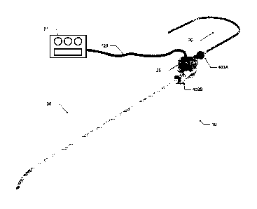
sufficient to persistently increase the overall diameter and the lumen
diameter of selected
segments of veins or arteries. The term "persistent increase" or "persistent
dilation" when used
to describe dilation or an increase in the overall diameter and lumen diameter
of an artery or
vein, is used herein to mean that even if the pump is turned off, an increase
in the overall
diameter or lumen diameter of a vessel can still be demonstrated when compared
to the overall
diameter or lumen diameter of the vessel prior to the period of blood pumping.
That is, the
overall diameter or lumen diameter of the vessel has become larger independent
of the pressure
generated by the pump. The blood pump system may therefore be useful to
certain patients,
including CKD patients in need of a vascular access site for hemodialysis. The
blood pump
system can include a rotary blood pump, one or more blood-carrying conduits, a
control system,
and a power source. The blood pump system withdraws blood from one location in
the vascular
system and discharges blood to another location in the vascular system. During
operation, such a
blood pump system may persistently increase mean and/or peak blood velocity
and mean and/or
peak WSS in a target blood vessel to a level and for a period of time
sufficient to persistently
Date Recue/Date Received 2020-07-24
increase the overall diameter and lumen diameter of the target blood vessel.
The system
functions in configurations where blood is withdrawn from the target blood
vessel or in
configurations where blood is discharged into the target blood vessel.
Further, the system can be
used simultaneously to increase the size of the donating and receiving
vessels.
[0110] In various other embodiments, the present application relates to a
blood pump
designed and dimensioned to move venous blood from a lower extremity to the
heart or to
another location in the venous system where it can more easily return to the
heart, in order to
reduce venous blood pressure in the lower extremity, and in some instances to
reduce swelling or
increase the rate of healing of an associated skin ulceration. Even more
specifically, the present
application relates to a rotary blood pump system configured to move venous
blood from a lower
extremity to the heart or to another location in the venous system where it
can more easily return
to the heart in order to reduce venous blood pressure in the lower extremity,
and in some
instances to reduce swelling or increase the rate of healing of an associated
skin ulceration. The
blood pump system may therefore be useful to certain patients including those
with venous
hypertension and/or venous ulceration of one or both lower extremities, such
as patients with
lower extremity venous obstruction or patients with damaged or incompetent
venous valves in
one or both lower extremities. The blood pump system can include a rotary
blood pump, one or
more blood-carrying conduits, a control system, and a power source. The blood
pump system
withdraws blood from a lower extremity vein segment and discharges blood to
another location
in the venous system. Locations for the return of blood to the venous
circulation include the
jugular vein, the axillary vein, the subclavian vein, the brachiocephalic
vein, the superior vena
cava, and the right atrium.
[0111] The optional blood-carrying conduits can include an inflow conduit to
carry
blood from a location in the vascular system (such as a donating vein, a
donating artery, or the
right atrium) to the blood pump and an outflow conduit to carry blood from the
blood pump to a
location in the vascular system (such as an accepting peripheral vein or
artery, or an accepting
location such as the right atrium). The blood pump system also includes a
control system. A
preferred control system is designed to collect information on the operating
parameters and
performance of the blood pump system, and changes in the vascular system, such
as changes in
the diameter of a donating artery, donating vein, accepting artery, or
accepting vein of a patient.
The blood pump system is primarily configured to pump a sufficient amount of
blood such that a
21
Date Recue/Date Received 2020-07-24
desired mean and/or peak wall shear stress (WSS) is achieved within a blood
vessel segment (the
"target blood vessel" or "target vessel") and for a sufficient period of time
such that the
permanent or persistent overall diameter and lumen diameter of the blood
vessel segment is
increased. The mean WSS can be calculated using the measured, estimated, or
assumed vessel
diameter and the measured, estimated, or assumed average blood flow rate
through the blood
pump system.
[0112] The diameter of blood vessels can be determined by measuring the
diameter of
the void within the center of the blood vessel. For the purpose of this
application, this
measurement is referred to as "lumen diameter". The diameter of blood vessels
can be
determined by measuring the diameter in a manner that includes the void within
the center of the
blood vessel and the wall of the blood vessel. For the purpose of this
application, this
measurement is referred to as "overall diameter". The invention relates to
simultaneously and
persistently increasing the overall diameter and lumen diameter of a
peripheral vein by moving
blood (preferably with low pulsatility) into the peripheral accepting vein,
thereby increasing the
velocity of the blood in the peripheral accepting vein and increasing the WSS
on the endothelium
of the peripheral accepting vein. Systems and methods are described wherein
the velocity of the
blood in a peripheral accepting vein and the WSS on the endothelium of the
peripheral accepting
vein is increased by using a pump. Systems and methods are also described that
withdraw or
"pull" blood such that the velocity of the blood and the WSS is increased in
the donating vessel,
either an artery or a vein. Preferably, the pump actively discharges blood
into the peripheral
accepting vein, wherein the pumped blood has reduced pulsatility, such as when
the pulse
pressure is lower than blood in a peripheral artery.
[0113] Blood pump systems described herein may have one or a group of
characteristics
that differ from many other blood pump systems. For example, a blood pump
system described
herein may operate safely within a wide operating range of blood flow, such as
a range from 50
mL/min to 1500 mL/min. In another example, a blood pump system described
herein can be
fabricated with a low cost-of-goods-sold (COGS), such as in the range of
$1,000 to $5,000. In
yet another example, a blood pump system described herein is designed to
operate reliably
outside of a hospital or clinic setting for an intermediate period of time,
such as for 1 hour to 12
months, or such as for 7 days to 12 months. In some examples, a blood pump
system described
herein can have one, several, or all of these factors, as one or more blood
pump systems
22
Date Recue/Date Received 2020-07-24
described herein can operate safely over a wide operating range of blood flow
including from 50
mL/min to 1500 mL/min, have low COGS of $1,000 to $5,000, and can operate
reliably outside
of a hospital or clinic setting for an intermediate period of time, such as
for 1 hour to 12 months,
or such as for 7 days to 12 months.
[0114] To begin a detailed discussion of the blood pump 25 of the system 10,
reference is
made to FIG. 1, which is an isometric view of the blood pump 25. In one
embodiment, the blood
pump 25 is a miniaturized centrifugal pump having a magnetic drive wherein the
impeller of the
pump is rotationally driven by rotating magnetic fields. For example, the
rotating magnetic
fields may be generated by energizing a number of electromagnets in a
particular sequence. In
another example, the rotating magnetic fields may be generated by rotating a
number of
permanent magnets or energized electromagnets. The pump can have a diameter
approximately
equal to that of a coin on the order of, for example, a United States quarter,
a United States half
dollar, or a larger diameter, as need be. For example, the pump 25 has a
diameter in a range
between about 2.0 cm and about 5.0 cm, according to various embodiments. As
shown in FIG.
1, the blood pump 25 includes a body 105, an inlet 110, an outlet 115, and a
power cable 120.
The power cable 120 connects the blood pump 25 to the control device 21 of a
control system 14
and power source. The power source can be part of the control device 21 or
separate. The
power cable allows for communication between the control device 21 and the
motor of the blood
pump 25. The cable can also be used to transfer power from a power source to
the motor or
pump. More particularly, the power cable 120 connects the electrical
components of the
magnetic drive inside the body 105 to an electrical power source (e.g., a
battery).
[0115] The inlet 110 is capable of being fluidly coupled to the inflow conduit
20 via a
coupling arrangement (e.g., a barbed-end, a flange, and a locking collar). The
inlet 110 provides
a fluid pathway into the intake region (i.e. center) of the pump impeller. The
intake region of the
impeller can be of a variety of constructions so long as blood is received out
of the outlet at a
velocity greater than the intake. The outlet 115 is capable of being fluidly
coupled to the outflow
conduit 30 via a coupling arrangement similar to the inlet (e.g., a barbed-
end, a flange, and a
locking collar). The outlet 115 provides a fluid pathway from the outlet
region (i.e. periphery) of
the pump impeller.
[0116] As illustrated in FIG. 2, which is an exploded isometric view of the
blood pump
25 showing its components contained in the body 105 identified in FIG. 1, the
blood pump 25
23
Date Recue/Date Received 2020-07-24
includes an inlet cap 125, a top bearing pin 130, a top impeller casing 135,
an impeller 140, an
impeller pivot 145, a magnet assembly 150, a magnet enclosure 155, a bottom
bearing pin 160, a
bottom impeller casing 165, an electrical coil assembly 170, and a coil
assembly enclosure lid
175. The inlet cap 125 and top impeller casing 135 each include approximately
half of the inlet
110.
[0117] As shown in FIGS. 3A and 3B, which are, respectively, partial and full
cross
sectional elevations of the blood pump 25 as taken along section line 3-3 in
FIG. 1, the
components mentioned with respect to FIG. 2 generally sandwich together to
form the pump.
For example, as can be understood from FIGS. 2-3A, the inlet cap 125 and top
impeller casing
135 respectively include a top horizontally extending inlet portion 110A and a
bottom
horizontally extending inlet portion 110B. Typically, the inlet and outlet are
opposed and
located in different planes. When the inlet cap 125 and top impeller casing
135 are sandwiched
together, they define an inlet fluid channel 180 leading through the inlet 110
to the impeller inlet
orifice 185. The inlet cap 125 and top impeller casing 135 respectively define
approximately a
top half and a bottom half of the channel 180. A seal groove 190 is defined in
the top impeller
casing 135 adjacent to the border of the channel 180 and is adapted to receive
a resilient fluid
seal member for creating a fluid tight seal between the inlet cap 125 and top
impeller casing 135.
[0118] FIGS. 4A and 4B are, respectively, partial and full cross sectional
elevations of
the blood pump 25 as taken along section line 4-4 in FIG. 1. As can be
understood from FIGS.
2, 4A, and 4B, the top impeller casing 135 and bottom impeller casing 165
respectively include a
top horizontally extending outlet portion 115A and a bottom horizontally
extending outlet
portion 115B. When top impeller casing 135 and bottom impeller casing 165 are
sandwiched
together, they define an outlet fluid channel 200 (i.e. volute) leading from
the impeller chamber
205 to the outlet 115. The top impeller casing 135 and bottom impeller casing
165 respectively
define approximately a top half and a bottom half of the channel 200. A seal
groove 211 is
defined in the bottom impeller casing 165 adjacent to the border of the
channel 200 and impeller
chamber 205 and is adapted to receive a resilient fluid seal member for
creating a fluid tight seal
between the top impeller casing 135 and bottom impeller casing 165.
[0119] As indicated in FIGS. 2-4B, the impeller magnet assembly 150 is a
plurality of
magnets in the form of a ring or disk. The magnets 150 are located in the
volume of the magnet
enclosure 155 and the volume of the impeller 140. The magnet enclosure 155 is
received in the
24
Date Recue/Date Received 2020-07-24
impeller 140. The magnet enclosure 155 and the impeller 140 respectively form
the bottom and
top portions of the volume in which the magnets 150 are located. The magnet
enclosure 155,
magnets 150, and impeller 140 are coupled together in a fixed integral
assembly that rotates as a
unit within the impeller chamber 205. Alternative constructions can be used
that cause rotation
of the impeller.
[0120] As illustrated in FIGS. 2-4B, the electrical coil assembly 170 is a
plurality of
electrical coils 210 arranged in a circular pattern on the lower impeller
casing and optionally
capped by a support disk 215. The electrical coil assembly 170 is fixed within
the coil chamber
220 defined in the bottom impeller casing 165 and capped by the coil enclosure
lid 175. An
internal floor structure 225 separates the impeller chamber 205 from the coil
chamber 220. In
one embodiment, the coil chamber 220 also contains one or more voids or
spaces, spacers 282,
and a ferrous backplate 284, as shown in FIG. 4C. An attractive magnetic force
is generated
between the impeller magnet 150 and the backplate 284, which counteracts the
upward force
imposed by the increased pressure of blood flowing in the gap 542 between the
bottom face of
the impeller 140 and the bottom impeller casing 165, as shown in FIG. 4E, and
the decrease
pressure at the impeller chamber inlet orifice 185 above the impeller. The net
effect is an
unloading of the top bearing pin 130. Depending upon the position of the
backplate 284 and the
speed of the pump 25, the axial load can be shared between the top and bottom
bearing pins 130
and 160 or it can be borne solely by the bottom bearing pin or the top bearing
pin. For example,
the force at the top bearing pin 130 may be less than approximately 3N during
operating speeds
up to approximately 6000 rpm. Similarly, the force on the bottom bearing pin
160 was less than
approximately 4N when operating at speeds up to approximately 6000 rpm.
Conversely, when at
rest (i.e. 0 rpm), the axial force experienced at the bottom force is at least
0.1N and may be up to
10N or greater.
[0121] A number of studies were performed to measure the load at the top and
bottom
bearing pins 130 and 160 with various pump speeds and backplate 284
orientations. The speed
at which the load changes from the bottom bearing pin 160 to the top bearing
pin 130 can be
tuned by varying the distance between the impeller 140 and backplate 284, such
as with one or
more spacers 282. Similarly, the load on the top and the bottom bearing pins
130 and 160 at a
particular impeller speed can be tuned by varying the distance between the
impeller 140 and
backplate 284. The ferrous backplate 284 also functions to increase the motor
performance and
Date Recue/Date Received 2020-07-24
motor torque, as the backplate causes the magnetic flux to penetrate deeper
into the coils 210
thereby providing a higher axial flux density.
[0122] One embodiment of the backplate 284 is shown in FIG. 4D. As shown, the
backplate 284 has a general disc shape and is composed of a ferrous metal or
alloy. In one
embodiment, the backplate 284 is composed of an iron-cobalt-vanadium soft
magnetic alloy,
such as Hiperco0 50, produced by Carpenter Technology. The backplate 284 has a
thickness in
a range from approximately .04 mm to about .07 mm and an outer diameter in a
range from
approximately 20 mm to approximately 40 mm. In a preferred embodiment, the
backplate 284 is
a solid disc having a thickness of approximately 0.53 mm and an outer diameter
of
approximately 31 mm. The backplate 284 may include a central opening 288 to
accommodate
the structural features of the pump 25; however, in other embodiments a solid
disc without the
opening 288 may be used. FIG. 4E is illustration of an embodiment of the pump
25. As shown,
in one embodiment, the backplate 284 is positioned a distance "D" away from
the magnet 150.
In one embodiment, the distance "D" is in a range between approximately 4 mm
and 8 mm. In a
preferred embodiment, the distance "D" is equal to approximately 6 mm. In
other embodiments,
the backplate 284 may be positioned closer to or farther from the magnets 150
to achieve the
desired gap 540 between the top face of the impeller 140 and the top impeller
casing 135 and the
gap 542 between the bottom of the impeller and the bottom impeller casing 165.
[0123] FIG. 4F is an illustration of the impeller 140 and the backplate 284
and a graph
depicting experimental results of the load measured at both the top pin and
the bottom pin as a
function of the backplate 284 position relative to the magnets 150. The
effective position of the
backplate 284 is configurable based on different arrangements of spacers 282
and the thickness
of the backplate 284. As shown, a preferred embodiment includes a single
backplate 284
positioned approximately 6 mm away from the motors using a 1.5 mm spacer 282.
Depending
upon the desired or tolerable loads at the top and bottom bearings other
backplate and spacer
combinations may be used. Similarly, FIG. 4H is a chart depicting the axial
load at the top
bearing as a function of the top gap 540 between the impeller 140 and the top
casing 135 when
the pump 25 is operating at approximately 4000 RPM.
[0124] The electrical cable 120 (see FIG. 1) extends through passage 230 in
the bottom
impeller casing 165 to the coil chamber 220 and the coils 210. Electrical
power supplied to the
coils 210 via the electrical cable 120 generates rotating magnetic fields,
which act on the
26
Date Recue/Date Received 2020-07-24
magnets 150 to cause the magnets, and the impeller 140 coupled to the magnets
to rotate. The
impeller rotation causes the impeller blades 235 to act upon the fluid (e.g.,
blood) present in the
impeller chamber, resulting in momentum being transferred to the fluid that is
recovered as a
pressure increase in the outlet fluid channel 200. The fluid is thus drawn
into the inlet 110 at low
pressure and discharged from the outlet 115 at a higher pressure.
[0125] As shown in FIGS. 3A-4B, the pivot axis for the impeller 140, magnets
150, and
enclosure 155 is the impeller pivot 145. As depicted in FIGS. 5A-B, the
impeller pivot 145 is
pivotally supported (i.e. restrained in all degrees of freedom except rotation
about a single axis)
via a top bearing pin 130 and a bottom bearing pin 160. The top bearing pin
130 is received and
fixed in a cylindrical recess 240 in the inlet cap 125, while the bottom
bearing pin 160 is
received and fixed in a cylindrical recess 245 in the bottom impeller casing
165. The impeller
pivot 145 extends through and is fixed to a center cylindrical opening 250 in
the impeller 140.
[0126] In one embodiment of the impeller assembly, the impeller pivot 145, the
top
bearing pin 130, and the bottom bearing pin 160 are formed from high purity
alumina (A1203),
such as CoorsTek0 AD-998. In another embodiment of the impeller assembly, the
impeller
pivot 145, the top bearing pin 130, and the bottom bearing pin 160 are formed
from silicon
carbide whisker-reinforced alumina, such as Greenleaf WG-300. In yet another
embodiment,
the impeller pivot 145, the top bearing pin 130, and the bottom bearing pin
160 are each formed
from alumina toughened zirconia (ATZ), which may provide a bearing more
resistant to wear
than bearings formed from alumina. Forming bearing components from ATZ may
also yield a
smoother surface finish than bearing components formed from alumina. In all
three
embodiments, the dimensions of the impeller pivot 145, the top bearing pin
130, and the bottom
bearing pin 160 are designed to limit the contact stresses to permissible
levels for high purity
alumina, silicon carbide toughened alumina, or ATZ, respectively, in view of
peak thrust loads
generated by hydrostatic forces and shock loads. In another embodiment of the
impeller
assembly, the impeller pivot 145 is formed from silicon carbide whisker-
reinforced alumina,
such as Greenleaf WG-300 or from high purity alumina, such as CoorsTek0 AD-
998, while
the top bearing pin 130, the bottom bearing pin 160, or both are formed from
ultrahigh molecular
weight polyethylene. In various other embodiments, portions or all of the top
bearing pin 130,
and the bottom bearing pin 160 can be formed from polyethylene. Additionally,
the geometry of
each component of the impeller assembly has been selected to limit fatigue and
wear in order to
27
Date Recue/Date Received 2020-07-24
satisfy the safety and durability requirements of the system 10. A number of
studies have been
conducted to illustrate the superior wear characteristics of ATZ over an
experimental lifetime of
the pump 25, which results in reduced changes to the overall height of the
bearing stack when
compared with bearing systems comprised of alumina and polyethylene.
[0127] As illustrated in FIGS. 6A-7B, the impeller pivot includes an upper
hemispherical
convex bearing surface 255 and a bottom hemispherical convex bearing surface
260. As
indicated in FIGS. 6A, 6B, and 8A, one embodiment of the impeller pivot has an
overall length
Li of approximately 10.15 mm, plus or minus 0.05 mm, and a pivot diameter D1
of
approximately 2 mm, plus or minus approximately 0.01 mm. The upper bearing
surface 255 has
a radius R1 of approximately 0.61 mm, plus or minus 0.02 mm and extends a
length L2 past an
adjacent lip 265 by approximately 0.55 mm, plus or minus 0.02 mm. The lower
bearing surface
260 has a radius R2 of approximately 0.31 mm, plus or minus 0.02 mm and
extends a length L21
past an adjacent lip 265 by approximately 0.55 mm, plus or minus 0.02 mm.
Similarly, an
alternate embodiment of the impeller pivot 145, as indicated in FIGS. 7A, 7B,
and 8B, has an
overall length Li of approximately 10.15 mm, plus or minus 0.05 mm, and a
pivot diameter D1
of approximately 2 mm, plus or minus approximately 0.01 mm. The upper bearing
surface 255
has a radius R1 of approximately 0.31 mm, plus or minus 0.02 mm and extends a
length L2 past
an adjacent lip 265 by approximately 0.55 mm, plus or minus 0.02 mm. The lower
bearing
surface 260 has a radius R2 of approximately 0.31 mm, plus or minus 0.02 mm
and extends a
length L21 past an adjacent lip 265 by approximately 0.55 mm, plus or minus
0.02 mm. Other
sizes and dimensions may be used depending upon the size and performance
requirements of the
pump. The sizes are such that the resultant pump can be used in a patient to
increase the
diameter of a vessel.
[0128] Similarly, an alternate embodiment of the impeller pivot 145, as
indicated in
FIGS. 7A, 7B, and 8B, has an overall length Li of approximately 10.15 mm, plus
or minus 0.05
mm, and a pivot diameter D1 of approximately 2 mm, plus or minus approximately
0.01 mm.
The upper bearing surface 255 has a radius R1 of approximately 0.31 mm, plus
or minus 0.02
mm and extends a length L2 past an adjacent lip 265 by approximately 0.55 mm,
plus or minus
0.02 mm. The lower bearing surface 260 has a radius R2 of approximately 0.31
mm, plus or
minus 0.02 mm and extends a length L21 past an adjacent lip 265 by
approximately 0.55 mm,
plus or minus 0.02 mm.
28
Date Recue/Date Received 2020-07-24
[0129] As can be understood from FIGS. 8C-8G, yet another embodiment of the
impeller
pivot 145 includes an upper hemispherical convex bearing surface 255 and a
bottom
hemispherical convex bearing surface 260. FIGS. 8D and 8E are plan views of
the upper
hemispherical convex bearing surface 255 and the bottom hemispherical convex
bearing surface
260, respectively as viewed along a longitudinal axis of the impeller pivot
140. FIGS. 8F and 8G
are close-up views of the bottom hemispherical convex bearing surface 260 and
the upper
hemispherical convex bearing surface 255, respectively. As indicated in FIG.
8C, one
embodiment of the impeller pivot has an overall length Li of approximately
10.45 mm, plus or
minus 0.05 mm, and a pivot diameter D1 of approximately 1.5 mm, plus or minus
approximately
0.005 mm. The upper bearing surface 255 has a radius R1 of approximately 0.6
mm, plus or
minus 0.02 mm and extends a length L2 past an adjacent taper point 266 by
approximately 1.4
mm, plus or minus 0.10 mm. The taper point 266 has a radius R3 of
approximately 0.20 mm
plus or minus 0.02 mm where the surface 267 of the impeller pivot tapers
inward along the
length L2 at a conical angle CA1 of approximately 20 degrees. The lower
bearing surface 260
has a radius R2 of approximately 0.60 mm, plus or minus 0.02 mm and extends a
length L21 past
an adjacent taper point 268 by approximately 0.5 mm, plus or minus 0.10 mm.
The taper point
268 has a radius R4 of approximately 0.05 mm where the surface 267 of the
impeller pivot tapers
inward along the length L21 at a conical angle CA2 of approximately 90
degrees.
[0130] As can be understood from FIGS. 5A and 5B, the upper bearings pin 130
and
bottom bearing pin 160 generally have the same configuration, but are
oppositely oriented. As
depicted in FIGS. 9A-B, the top bearing pin 130 and the bottom bearing pin
160, have a tea cup
or hemispherical concave bearing surface 270 on one end and a generally planar
surface 275 on
the opposite end. Similarly, FIG. 10 depicts a particular embodiment of the
top bearing pin 130,
which has a tea cup or hemispherical concave bearing surface 270 on one end
and a generally
planar surface 275 on the opposite end. In this embodiment, the hemispherical
concave bearing
surface 270 of the top bearing pin 130 has a larger radius than the concave
bearing surface on the
bottom bearing pin 160.
[0131] As illustrated in FIG. 11A, one embodiment of the bearing pin 130, 160
has an
overall length L3 of approximately 7.5 mm, plus or minus 0.1 mm, a minimum
pivot diameter
D2 of approximately 2 mm, plus or minus 0.01 mm, and a radius of approximately
0.6 mm at the
edge near the bearing surface 270. Near the non-bearing end 275 of the bearing
pin 130, 160, a
29
Date Recue/Date Received 2020-07-24
groove 280 extends circumferentially around the pin to provide a mechanical
interlock for
bonding the bearing pin in place within the blood pump 25. Similarly, an
alternate embodiment
of the bearing pins 130, 160, as illustrated in FIG. 11B, has an overall
length L3 of
approximately 7.5 mm, plus or minus 0.1 mm, a minimum pivot diameter D2 of
approximately 3
mm, plus or minus 0.01 mm, and a radius of approximately 0.2 mm at the edge
near the planar
end 275. Near the non-bearing end of the bearing pin 130, 160 there is a
groove 280
circumferentially extending around the pivot used to provide a mechanical
interlock for bonding
the bearing pin in place. Other sizes and dimensions may be used depending
upon the size of the
pump, the materials of the bearing pin, and the forces acting on the bearing
pin.
[0132] As can be understood from FIGS. 3B, 4B, and 5A-11B, the convex upper
bearing
surface 255 of the impeller pivot 145 is rotationally received against the
concave bearing surface
270 of the top bearing pin 130, and the convex lower bearing surface 260 of
the impeller pivot
145 is rotationally received against the concave bearing surface 270 of the
bottom bearing pin
160. Thus, the convex bearing ends 255, 260 of the impeller pivot 145 are
pivotally supported
by complementary concave bearing surfaces 270 of the top and bottom bearing
pins 130 and 160,
respectively. Accordingly, the impeller assembly may freely rotate in the
impeller chamber 205
on the impeller pivot 145, which is supported end to end with the bearing pins
130 and 160, in a
configuration commonly known as a "double pin bearing."
[0133] As can be understood from FIGS. 11C-11F, yet another embodiment of the
bearing pin 130, 160 has an overall length L3 of approximately 7.5 mm, plus or
minus 0.1 mm
and a minimum pivot diameter D2 of approximately 2.0 mm, plus or minus 0.01
mm. The
bearing end 271 has a radius R5 of approximately 0.3 mm at the edge near the
bearing surface
270. Near the non-bearing end 275 of the bearing pin 130, 160, a series of
grooves 281 extends
circumferentially around the pin to provide a mechanical interlock for bonding
the bearing pin in
place within the blood pump 25. The series of grooves 281 may be defined by
one or more
valleys 283 having a radius R6 of approximately 0.20 mm and a plateau 285
having an edge
radius R7 of approximately 0.03 mm. The distance V1 across each valley is
approximately 0.5
mm, while the distance P1 across the plateau 285 is approximately 0.3 mm. The
bearing pins
130 and 160 may also include a recess 286 having a diameter D3 of
approximately 0.8 mm plus
or minus 0.01 mm and a length L4 of approximately 2.0 mm, as shown in the
cross section view
of FIG. 11E. FIG. 11D is a view of the bearing surface 270 as viewed along a
longitudinal axis
Date Recue/Date Received 2020-07-24
of the bearing pin 130, 160. The bearing surface 270 may have a radius R8 of
approximately
0.65 mm plus or minus 0.01 mm and a depth L5 of approximately 0.3 mm, as shown
in the cross
section view of FIG. 11F.
[0134] In yet another embodiment of the impeller assembly, the impeller
assembly is a
composite of the impeller shaft 145, top bearing pin 130, and bottom bearing
pin 160. The
composite design is beneficial with regard to the simplicity, tolerances, and
cost of the machined
bearing components. All of these constructions are designed to allow the motor
to function in a
continuous state for around a day to 1-12 weeks or longer, without breakdown.
[0135] As illustrated in FIG. 12, the impeller shaft 145 comprises an impeller
pivot body
146 and two impeller pivot inserts 147. The impeller pivot body 146 comprises
a machinable
metal, such as stainless steel, and the impeller pivot inserts 147 comprise a
high purity alumina
(A1203), such as CoorsTek AD-998, a silicon carbide whisker-reinforced
alumina, such as
Greenleaf WG-300, or alumina toughened zirconia (ATZ). The impeller pivot
inserts 147 are
affixed to the impeller pivot body 146 by an adhesive and/or an interference
fit. Optionally, the
chamber 146A may be filled with an adhesive or other potting material that is
resistant to
compression. The aforementioned composite configuration and materials can be
applied to
embodiments of both the top bearing pin 130 and bottom bearing pin 160, where
the pin inserts
148 engage the impeller pivot inserts 147. Optionally, the chambers 148A for
each bearing pin
130 and 160, may be filled with an adhesive or other potting material that is
resistant to
compression.
[0136] The inlet cap 125 and its inlet channel 180 may have a variety of
configurations,
depending on the embodiment of the blood pump 25. For example, the inlet cap
125 depicted in
FIG. 2 is shown as being generally coextensive with the top impeller casing
135. In other
embodiments, the inlet cap 125 may be substantially smaller than, and not
coextensive with, the
top impeller casing 135, as depicted in FIGS. 13-15, which are views of the
inlet cap and
impeller casing.
[0137] As shown in FIGS. 14-16, which are, respectively, cross sectional
elevations
taken along section lines 14-14, 15-15, and 16-16 in FIG. 13, the inlet 110 is
a two part
construction having portions 110A and 110B that each form approximately half
of the inlet 110
and are respectively part of the inlet cap 125 and top impeller casing 135.
Each portion 110A
and 110B has defined therein approximately half of the inlet channel 180. As
illustrated in FIG.
31
Date Recue/Date Received 2020-07-24
14, the inlet channel 180 initially has a circular diameter D5 of
approximately 4 mm. As
indicated in FIG. 15, the inlet channel 180 transitions from a circular cross
section to a generally
rectangular cross section having a width W5 of approximately 8.4 mm and a
height 115 of
approximately 1.5 mm. Again, as dimensions change so will the listed
measurements.
[0138] As depicted in FIG. 16, the inlet channel 180 surrounds the impeller
chamber inlet
orifice 185, which extends around the top bearing 145 received in, and affixed
to, the inlet cap
125. As shown in FIG. 17, which is an isometric partial cross section of the
impeller chamber
inlet orifice 185, the impeller chamber inlet orifice 185 leads to the
impeller chamber 205 near
the intake region 300 of the impeller 140. The upper bearing end of the
impeller pivot 145
extends up through the orifice 185 to pivotally interface with the top bearing
pin 130 supported
in the inlet cap 125. Impeller blades 235 extend radially outward from the
intake region 300 of
the impeller 140.
[0139] As depicted in FIGS. 18A and 18B, which are, respectively, a plan view
of the
inlet cap portion 110A defining the inlet channel 180 and an end elevation
view of the same, in
one embodiment, the inlet channel 180 may be said to have an elliptic
configuration.
Specifically, a cylindrical channel portion 180A transitions in portion 180C
into an elliptical
channel portion 180B. A cylindrical island portion or bezel 305 supporting the
top bearing pin
130 is generally centered in the elliptical channel portion 180B and includes
a cylindrical hole
240 that receives the top bearing pin 130 similar to as illustrated in FIG.
17. In one embodiment,
the cylindrical channel portion 180A has a diameter D6 of approximately 4 mm.
The elliptical
channel portion 180B has a width W6 of approximately 12.4 mm. The distal
distance W7
between the wall of the bezel 305 and the distal end of the wall defining the
elliptical channel
portion 180B is approximately 1.5 mm. In other embodiments, the cylindrical
channel portion
180A has a diameter D6 of approximately 5 mm or 6 mm.
[0140] As depicted in FIGS. 19A and 19B, which are the same respective views
as FIGS.
18A and 18B, except of another embodiment, the inlet channel 180 may be said
to have a
circular configuration. Specifically, a cylindrical channel portion 180A
transitions in portion
180C into a circular channel portion 180B. A cylindrical island portion or
bezel 305 supporting
the top bearing pin 130 is generally centered in the circular channel portion
180B and includes a
cylindrical hole 240 that receives the top bearing pin 130 similar to as
illustrated in FIG. 17. In
one embodiment, the cylindrical channel portion 180A has a diameter D9 of
approximately 3.5
32
Date Recue/Date Received 2020-07-24
mm to 4.5 mm, preferably 4 mm. The circular channel portion 180B has a width
W9 of
approximately 11.5 mm to 13 mm, preferably 12.4 mm. The distal distance W10
between the
wall of the bezel 305 and the distal end of the wall defining the circular
channel portion 180B is
approximately 3.5 mm to 4.5 mm, preferably 4.2 mm. In other embodiments, the
cylindrical
channel portion 180A has a diameter D6 of approximately 5 mm or 6 mm.
[0141] As depicted in FIGS. 20A and 20B, which are the same respective views
as FIGS.
18A and 18B, except of another embodiment, the inlet channel 180 may be said
to have a
complex arcuate configuration. Specifically, a cylindrical channel portion
180A transitions in
portion 180C into a complex arcuate channel portion 180B. A cylindrical island
portion or bezel
305 supporting the top bearing pin 130 is generally centered in the complex
arcuate channel
portion 180B and includes a cylindrical hole 240 that receives the top bearing
pin 130 similar to
as illustrated in FIG. 17. In one embodiment, the cylindrical channel portion
180A has a
diameter D12 of approximately 4 mm. The complex arcuate channel portion 180B
has a width
W13 of approximately 8.4 mm. The distal distance W14 between the wall of the
bezel 305 and
the distal end dome 307 of the wall defining the complex arcuate channel
portion 180B is
approximately 1.75 mm. The distal distance W15 between the wall of the bezel
305 and the
distal end cleft 310 of the wall defining the complex arcuate channel portion
180B is
approximately 0.5 mm to 1.5 mm, preferably 1 mm. In other embodiments, the
cylindrical
channel portion 180A has a diameter D6 of approximately 5 mm or 6 mm.
[0142] As depicted in FIGS. 21-23, which are the same views as FIG. 18A,
except of
three other embodiments, the inlet channel 180 may be said to have a tear drop
configuration.
Specifically, a cylindrical channel portion 180A transitions into a tear drop
channel portion
180B. A cylindrical island portion or bezel 305 supporting the top bearing pin
130 is generally
centered in the tear drop channel portion 180B and includes a cylindrical hole
240 that receives
the top bearing pin 130 similar to as illustrated in FIG. 17. In one
embodiment, the cylindrical
channel portion 180A has a diameter D15 of approximately 4 mm. The tear drop
channel
portion 180B has a width W20 of approximately 8 mm. The bezel 305 has a
diameter D16 of 4
mm. A transition region 180C of the channel 180 between the tear drop portion
180B and the
cylindrical portion 180A has walls that diverge from each other at an angle
AN1 of
approximately 8 degrees. In other embodiments, the cylindrical channel portion
180A has a
diameter D6 of approximately 5 mm or 6 mm.
33
Date Recue/Date Received 2020-07-24
[0143] For the embodiment of FIG. 21, the distal distance W21 between the wall
of the
bezel 305 and the distal end of the wall defining the tear drop channel
portion 180B is
approximately 2 mm. For the embodiment of FIG. 22, the distal distance W21
between the wall
of the bezel 305 and the distal end of the wall defining the tear drop channel
portion 180B is
approximately 1 mm. For the embodiment of FIG. 23, the distal distance W21
between the wall
of the bezel 305 and the distal end of the wall defining the tear drop channel
portion 180B is
approximately 0 mm because the bezel intersects the distal end of the wall
defining the tear drop
channel portion.
[0144] As illustrated in FIGS. 24A and 24B, which are, respectively, plan and
side
elevation views of another embodiment of the inlet cap 110 and inlet channel
180 similar to that
described in FIG. 21, an arcuate wedged portion 320 may extend between the
distal wall of the
tear drop channel portion 180B to the distal side of the bezel 305. In such an
embodiment, the
cylindrical island portion or bezel 305 is generally centered in the tear drop
channel portion
180B and includes a cylindrical hole 240 that receives the top bearing pin 130
similarly to as
illustrated in FIG. 17. In one embodiment, the dimensional configuration of
the embodiment
depicted in FIGS. 24A and 24B is substantially the same as discussed with
respect to FIG. 21,
the significant difference being the presence of the arcuate wedge portion
320. As can be
understood from FIGS. 24A and 24B, the wedge portion 320 has walls that are
arcuate to
smoothly curve from the roof and adjacent wall of the tear drop channel
portion 180B to the
vertical extension of the bezel 305. Such a wedged portion 320 may be seen to
exist in the
embodiment depicted in FIGS. 3A, 3B, and 17 and may reduce areas of inlet
channel flow
stagnation and facilitate tangential inflow of fluid through the impeller
chamber inlet orifice 185.
[0145] As shown in FIG. 25, which is an isometric view of the blood pump 25
with the
top impeller casing removed to reveal the impeller 140 occupying the impeller
chamber 205, the
outlet fluid channel 200 exits the impeller chamber substantially tangential
to the outer
circumferential edge of the impeller. As indicated in FIGS. 3B, 4B, 17, and
25, a plurality of
bores 350 (i.e. washout holes) are circumferentially distributed about the
impeller pivot center
hole 250, and the bores 350 are generally parallel to the center hole 250 and
extend though the
full thickness of the impeller to daylight on both top and bottom boundaries
of the impeller. The
bottom openings of the bores 350 are located near the bottom bearing interface
between the
bottom bearing 165 and the impeller pivot bottom bearing surface 260 (see FIG.
8). As a result,
34
Date Recue/Date Received 2020-07-24
a fluid can be flowed through the bores 350 to cleanse the bottom bearing
interface. For
example, a fluid can be flowed through the impeller chamber inlet hole 185,
radially-outward
along the impeller blades 235, through the gap 542 under the impeller, and
then back to the
region of the impeller chamber inlet hole 185. This flow of blood serves to
cleanse the underside
of the impeller, the bottom bearing interface, the upper bearing interface,
and the region behind
the bezel 305.
[0146] As can be understood from FIGS. 3B, 5, 17, and 25, in one embodiment,
the
impeller 140 is rotationally supported in the impeller chamber 205 on a shaft
145 extending
through a center of the impeller. The shaft has an upper bearing end and a
bottom bearing end,
each end rotatably operably coupled to the pump housing. The impeller has a
top face, a bottom
face, and multiple bores 350 extending through the impeller from the top face
to the bottom face.
The multiple bores are generally evenly distributed radially about center of
the impeller. Further,
the multiple bores 350 extend through the impeller generally parallel to each
other and the shaft.
The inlet channel 180 leads to an inlet orifice 185 of the impeller chamber.
The inlet channel
opens into the impeller chamber generally perpendicular to the inlet channel.
The inlet orifice
extends along at least a portion of an outer circumferential surface of the
shaft near the upper
bearing end. The inlet orifice and the holes open in directions that are
generally parallel to each
other. During operation of the pump, at least a portion of the blood pumped
through the impeller
chamber circulates along the top and bottom faces of the impeller via the
bores. Thus, the bores
of the impeller eliminate flow dead ends around the impeller by generally
keeping blood flowing
along all blood contacting surfaces of the impeller. Accordingly, the bores
help to prevent blood
accumulation in the vicinity of the shaft/impeller intersection and along the
sides and bottom
face of the impeller.
[0147] In various embodiments, the gap between the top face of the impeller
140 and the
top impeller casing 135 is in a range between 0.05 mm and 0.3 mm, with
preferred embodiments
between 0.075 and 0.125 mm. Although counter to prevailing thoughts, it was
determined that a
smaller gap between the top face of the impeller 140 and the top impeller
casing 135 is
preferable as this takes advantage of the hydrodynamic flow behavior of the
blood flowing
around the impeller, which lowers the axial load applied to the top bearing
which, in some
instances, can function as a form of hydrodynamic bearing and can either
replace the upper
bearing or can supplement the upper bearing. The hydrodynamic bearing
effectively formed by
Date Recue/Date Received 2020-07-24
top surface of the impeller blades 235 with the smaller gap between the top
face of the impeller
140 and the top impeller casing 135 reduces the load and therefore wear on the
top bearing pin.
As a result, the pump 25 may be operated for longer durations before
replacement of the bearing
is required. By way of example, as shown in FIG. 4G, the total surface area of
the top of
impeller blades 235, indicated generally as 237, provides a hydrodynamic
bearing having an area
in a range between about 70 mm2 to about 120 mm2. In one embodiment, the total
surface area
of the impeller blades 235 that facilitates the hydrodynamic bearing is
approximately 96 mm2. In
this embodiment, the approximate area of the rotor top surface, excluding the
central and
washout holes, with the blades removed is approximately 677.7 mm2- Therefore,
if the area of
the blade top surfaces is approximately 96.1 mm2, then approximately 14% of
the surface area is
used to form the hydrodynamic bearing. In other embodiments, a greater ratio,
such as 20% or
more or a smaller ratio such as 10% or less of the impeller surface area may
be used to form the
hydrodynamic bearing.
[0148] In various embodiments, the gap 542 between the bottom face of the
impeller and
the bottom impeller casing 165 is in a range between approximately 0.1 mm and
0.5 mm, with
preferred embodiments having a gap between approximately 0.2 and 0.35 mm. A
larger gap 542
between the bottom face of the impeller 140 and the bottom impeller casing 165
is preferred as
this improves the washing of the bottom bearing and lowers shear stress on the
blood in the
bottom gap.
In various embodiments, a balance is made the low design point flow and the
broad
operating flow range of the blood pump system. The specified ranges of top and
bottom rotor-
housing gaps enable the system to simultaneously achieve its hydraulic
performance,
manufacturing cost, blood damage, and service life requirements. These were
verified in
numerous studies using actual working prototypes through in vitro life tests
demonstrating
negligible bearing wear over 6 weeks and in vivo studies showing dramatic vein
dilation over 9
days of treatment with no clinically significant hemolysis.
[0149] The body and impeller of the blood pump 25, including blood-contacting
surfaces, are made from a variety of rigid biocompatible materials. Preferred
options include
injection moldable plastics such as polycarbonate and polyetheretherketone
(PEEK). In various
embodiments, the blood-contacting surfaces of the blood pump 25 may comprise
Ti6A14V,
Ti6A17Nb, or other commercially pure titanium alloys. In one embodiment, the
surfaces of the
36
Date Recue/Date Received 2020-07-24
pump components to be exposed to the patient's blood may have antithrombotic
coatings. For
example, the luminal surfaces may be coated with Astute , a heparin based
antithrombotic
coating by BioInteractions Ltd., or ApplauseTM, a heparin coating by
SurModics, Inc.
[0150] In other embodiments, the surfaces of the blood pump system components
in
contact with the patient's tissue may have antimicrobial coatings. For
example, the external
surfaces of the synthetic conduits 16 and 18 or the external surfaces of the
pump or the power
cord 120 (which is also known as a "lead") may be coated with Avert , a
surface-active
antimicrobial coating by BioInteractions Ltd.
[0151] In various embodiments, the blood pump 25 may be implanted within a
patient.
Conversely, in other embodiments, the blood pump 25 may remain external to the
patient. For
example, when located externally to the patient, the blood pump 25 may be
secured to the patient
using tape, sutures, or other suitable means to affix the pump to the patient.
The system 10 may
be powered by wearable electronics having rechargeable batteries 28, as shown
in FIG. 34.
[0152] The pump for the pump system 10 disclosed herein may be a rotary pump,
including, for example, a centrifugal flow pump, an axial flow pump, a radial
flow pump, or a
mixed flow pump. As shown in FIGS. 1-15, in one embodiment, the pump is a
centrifugal
pump. Without recognizing specific limitations, the blood pump 25 can be
configured to
routinely pump about 0.05 to 1.5 L/min, 0.1 to 1.5 L, or 0.5 to 3.0 L/min, for
example.
[0153] While the pump configuration discussed above with respect to FIGS. 1-25
is
advantageous, other pump configurations may be employed with the pump systems
and methods
disclosed herein. Accordingly, the systems and methods disclosed herein should
not be limited
to the pump configuration discussed above with respect to FIGS. 1-25, but
should include all
types of pumps applicable for the systems and methods disclosed herein.
[0154] A preferred embodiment of the pump system 10 disclosed herein with
respect to
FIGS. 1-25 satisfies several unique needs that cannot be satisfied by any
blood pump systems
known in the art. Specifically, the Arteriovenous Fistula Eligibility ("AFE")
pump system
("AFE System") may be configured for up to 12 weeks of intended use. Further,
the AFE pump
system may be configured as a centrifugal rotary blood pump system for a low
flow rate (e.g., 50
to 1500 mL/min) and medium pressure range (e.g., 25 to 350 mmHg).
[0155] A control scheme used with the AFE System pump system may be optimized
to
maintain a steady and elevated mean WSS of 0.76 ¨23 Pa, or more preferably 2.5
Pa to 10 Pa, in
37
Date Recue/Date Received 2020-07-24
target veins that are directly fluidly connected to the blood pump or a
conduit of the blood pump
system, or target veins that are fluidly connected to a vein that is directly
fluidly connected to the
blood pump or a conduit of the blood pump system. With this control scheme,
the AFE System
is configured to operate for a period of time such that the overall diameter
and lumen diameter of
the target vein will persistently increase by 25%, 50%, or 100% or more,
utilizing sensing of
operating parameters and periodic speed adjustment. A control scheme used with
the AFE
System pump system may be optimized to maintain a steady pressure in the
segment of the
outflow conduit adjacent to the target vein in a range of 10 mmHg to 350 mmHg,
preferably
between 25 mmHg to 100 mmHg. With this control scheme, the AFE System is
configured to
operate for a period of time such that the overall diameter and lumen diameter
of the target vein
will persistently increase by 25%, 50%, or 100% or more, utilizing sensing of
operating
parameters and periodic speed adjustment.
[0156] For certain embodiments, the inflow conduit may be placed by
percutaneous
approach, with a portion of the inflow conduit residing in an intravascular
location, and the
outflow conduit may be placed by surgical approach adaptable to initial vein
diameters of
between 1-6 mm. In this setting, elevated mean WSS in the target blood vessel
results from
discharging blood into the target blood vessel.
[0157] For other embodiments, the outflow conduit may be placed by
percutaneous
approach, with a portion of the outflow conduit residing in an intravascular
location, and the
inflow conduit may be placed by surgical approach adaptable to initial vein or
artery diameters
of between 1-6 mm. In this setting, elevated mean WSS in the target blood
vessel results from
removing blood from the target blood vessel. In certain settings, WSS can be
elevated in both a
blood vessel where blood is removed and a blood vessel where blood is
discharged, making both
blood vessels target blood vessels. The pump system 10 achieves both ease of
insertion/removal
and resistance to infection. The pump system 10 is a mobile system with a pump
that is
adaptable for either implanted or extracorporeal placement. In various
embodiments, the pump
system 10 is powered by wearable electronics with rechargeable batteries.
[0158] The pump system 10 includes an inflow conduit 20 and an outflow conduit
30, as
shown in FIG. 26. The inflow conduit 20 is placed in fluid communication with
one location in
the vascular system, draws blood from this location, and carries it to the
blood pump 25. In
certain embodiments, the inflow conduit 20 is configured for placement of at
least a portion of
38
Date Recue/Date Received 2020-07-24
the inflow conduit within the lumen of the vascular system. In other
embodiments, the inflow
conduit 20 is joined to a blood vessel by a surgical anastomosis. The outflow
conduit 30 is
configured for making a fluid communication with another location in the
vascular system and
directs blood from the blood pump 25 to the other location in the vascular
system. In certain
embodiments, the outflow conduit 20 is configured for placement of at least a
portion of the
outflow conduit within the lumen of the vascular system. In other embodiments,
the outflow
conduit 30 is joined to a blood vessel by a surgical anastomosis.
[0159] The conduits 20 and 30 may each have a length that ranges between 2 cm
and 110
cm and a total combined length of 4 cm to 220 cm. The length of the each
conduit 20 and 30
may be trimmed to a desired length as determined by the location of the blood
pump 25 and the
location of the connections between the conduits and the vascular system. The
conduits 20 and
30 also have thin but compression-resistant and kink-resistant walls that have
a thickness of
between 0.5 mm and 4 mm and inner diameters that are between 2 mm and 10 mm.
Preferably,
the inner diameters for the conduits are 4 to 6 mm.
[0160] The inflow and outflow conduits 20 and 30 may be connected to the blood
pump
25 using any suitable connector that is durable, resists leaks, and is not
susceptible to
unintentional disengagement. Typically, the leading edge of the connector is
thin, in order to
minimize the step change in fluid path diameter between the inner diameter of
the conduits 20
and 30 and the inner diameter of the connector. Preferably, the step change in
fluid path
diameter should be less than 0.5 mm. In one embodiment, as shown FIGS. 27A-
27D, the
conduits 20 and 30 are connected to the blood pump 25 using barb fittings 400A
and 400B and
radially compressive retainers (i.e. locking collars) 402A and 402B. By way of
example, and not
limitation, the radially compressive retainers 402A and 402B, may be BarbLock
retainers
manufactured by Saint-Gobain Performance Plastics, a division of Saint-Gobain
S.A.
headquartered in Courbevoie, France. In another embodiment, the conduits 20
and 30 are
connected to the blood pump 25 using Pure-Fite sterile connectors, also
manufactured by Saint-
Gobain Performance Plastics.
[0161] The radial compressive retainers 402A and 402B are placed over the
proximal
ends 404 and 406 of the inflow and outflow conduits 20 and 30, respectively.
The conduits 20
and 30 are then placed over the barb fitting 400A and 400B to form a fluid
connection between
the conduits and the blood pump 25. Collets 408A and 408B of the radial
compressive retainers
39
Date Recue/Date Received 2020-07-24
402A and 402B are placed along the conduits 20 and 30 to encircle the conduits
and the barb-
fittings 400A and 400B. Outer sleeves 410A and 410B of the radial compressive
retainers 402A
and 402B are then moved along a longitudinal axis of the retainers to
compressively engage the
respective collets 408A and 408B, conduits 20 and 30, and the barb fittings
400A and 400B. In
one embodiment, the outer sleeves 410A and 410B are moved by a compressive
tool configured
to engage the outer sleeves and a support shelf 412A and 412B of the barb
fittings 400A and
400B, respectively. The compressive tool may also be configured to remove the
radial
compressive retainers 402A and 402B.
[0162] In other embodiments, alternative connectors may be used. Preferably,
the
alternative connectors are durable, resist leaks, and resist unintentional
dislodgment. For
example, as shown in FIGS. 28A-B, the conduits 20 and 30 engage barb fittings,
similar to barb
fittings 400A and 400B, to form a fluid connection between the conduits and
the blood pump 25.
The conduits 20 and 30 are secured to the barb fittings using circular clips
414A and 414B that
apply radial compressive force to the portion of the conduits on the barb
fittings by way of a
ratcheting mechanism 416A-416B of the clips. The circular clips 414A and 414B
provide a
leak-resistant and durable connection that may be removed with a removal tool
(not shown)
which releases the ratcheting mechanisms 416A-416B of the clips.
[0163] In another embodiment, the inflow conduit 20 and the outflow conduit 30
each
contain at least one side port 417, as shown in FIGS. 29A-B, 30A-B, and 50,
51A-B, and 52A-B,
that provides controlled access to the fluid path. Side ports 417 may be used
periodically to
introduce contrast into the fluid path to enable visualization of portions of
the AFE System or
portions of the vascular system in fluid communication with the conduit(s) of
the AFE System
by fluoroscopy. The side ports 417 may also be used to remove and return blood
from the
vascular system of a patient during hemodialysis, plasmapheresis, apheresis,
or other clinical
indications wherein blood is rapidly removed and returned to a patient. The
side ports 417 may
also be used to obtain blood samples, to infuse medications, or for other
clinically useful
purposes. Any side port design that allows periodic access to the fluid path
and does not leak or
alter the fluid flow path when not accessed is suitable. By way of example,
and not limitation,
the side port 417 may be a "T" port fitting that includes a check valve that
opens when a syringe
is inserted and closes when the syringe is removed. As shown in FIGS. 29A-B, a
"T" port
Date Recue/Date Received 2020-07-24
assembly 418 with auxiliary tubing 420 is in fluid communication with the pump
outlet 115 and
outflow conduit 30.
[0164] In another embodiment, a side port 417 for the inflow conduit 20, the
outflow
conduit 30, or both utilizes a septum access port 422 having a septum 424, as
shown in FIGS.
30A-B, through which a suitable hypodermic needle can be inserted for access
and then
removed, after which the septum closes, preventing fluid loss from the
conduit. Suitable
materials for the septum 424 include, but are not limited to silicone,
polyurethane, and other
elastomeric polymers. The segment of the inflow and/or outflow conduit 20 or
30, respectively,
which includes the septum 424, is of a suitable thickness to close a
hypodermic puncture hole
when the needle is removed. As shown in FIGS. 30A-B, the septum access port
422 is shown in
which the septum 424 makes up a portion of the outflow conduit 30. By way of
example, and
not limitation, the septum access port 422 may extend about one centimeter
over the length of
the outflow conduit 30. The septum 424 may be attached to the outflow conduit
30 by any
suitable means including, but not limited to, adhesive attachment, thermal
bonding, and thermal
bonding between inner and outer layers of the conduit tubing.
[0165] In various embodiments, the conduits 20 and 30 may be comprised of
materials
commonly used to make hemodialysis catheters such as polyurethane, polyvinyl
chloride,
polyethylene, silicone, and polytetrafluoroethylene (PTFE), and including
Pellethane0 or
Carbothane0. In other embodiments, the conduits may be comprised of materials
commonly
used to make hemodialysis grafts or synthetic peripheral bypass grafts such as
expanded
polytetrafluoroethylene (ePTFE) or Dacron. In further embodiments, conduits
may be
comprised of combinations of polyurethane, polyvinyl chloride, polyethylene,
silicone, PTFE,
Pellethane 0, Carbothane 0, Carbothane0 PC-3575, ePTFE, or Dacron.
[0166] For example, the entire length of the inflow conduit 20 may be composed
of
polyurethane. In another embodiment, shown in FIG. 31, a segment 500 of the
outflow conduit
30 configured to make a fluid communication with the blood pump 25 is composed
of
polyurethane while a segment 502 of the outflow conduit configured to make a
fluid
communication with the vascular system is composed of ePTFE.
[0167] By way of example and not limitation, and as shown in FIG. 41, which is
a
longitudinal cross section of the junction between the proximal segment 500
and distal segment
502, the proximal segment 500 of the outflow conduit 30 is joined to the
distal segment 502 of
41
Date Recue/Date Received 2020-07-24
the outflow conduit during the manufacturing process by placing one or more
layers 502A of
ePTFE from the distal segment between layers 500A of polyurethane from the
proximal
segment. The overlapping layers of polyurethane and ePTFE are then heat
laminated to bond the
proximal segment 500 and the distal segments 502 together.
[0168] In another example, one or more holes are made within the overlapped
sections of
the ePTFE of segment 502 prior to heat laminating the conduit. When the
outflow conduit 30 is
heated to a temperature that is sufficient to melt the polyurethane without
melting the ePTFE
(e.g. 200 F to 500 F), the molten polyurethane fills in and then cools
within the holes created in
the ePTFE segment 502. The inner and outer polyurethane layers of the segment
500 are joined
with in the holes to mechanically join the two segments 500 and 502 together
as well as
mechanically join the inner and outer layers of polyurethane in the overlapped
segment.
[0169] The embodiment of the outflow conduit 30 manufactured to have the ePTFE
layer
502A sandwiched between the polyurethane layers 500A is advantageous in that
the ePTFE layer
502A can be readily sutured to blood vessels using standard techniques. This
is also the case for
an inflow conduit 20 manufactured as discussed above with respect to FIG. 41.
[0170] As illustrated in FIG. 42, which is a plan view of a medical kit 1000,
the blood
pump 25, inflow conduit 20, outflow conduit 30, control device 21, and power
cord 120 can be
provided in a sterile package 1005 with instructions 1010 on how to assemble
and implant the
pump system in a patient. The medical kit 1000 may also include the barb
fittings 400A and
400B and the radially compressive retainers 402A and 402B. In one embodiment,
one or both
conduits 20, 30 are manufactured as described above with respect to FIG. 41
and enclosed within
the sterile package 1005 along with the blood pump 25. The medical kit 1000,
at a minimum,
includes a system for discharging or removing blood and instructions for
implementation and
usage.
[0171] In one embodiment, the operation of the blood pump 25 is controlled via
the
control unit 21 of a pump control system 14 by reading the outflow pressure
and adjusting the
pump speed accordingly. For example, as depicted in FIG. 43, which is a
schematic diagram of
a pump system 10 controlled according to outflow pressure, an outflow pressure
sensor 1050
may be operably coupled to the outlet 115 of the blood pump 25 or further
downstream, such as,
for example, somewhere along the length of the outflow conduit 30. The
processor 24 may
compare the pressure reading from the outflow pressure sensor 1050 to a range
of target outflow
42
Date Recue/Date Received 2020-07-24
pressures stored in the memory 27. The processor will then adjust the speed of
the pump drive
170 accordingly to cause the pressure reading from the outflow pressure sensor
1050 to be within
the range of target outflow pressures stored in the memory.
[0172] In one embodiment, the control system 14 also includes an inflow
pressure sensor
1060 that may be operably coupled to the inlet 110 of the blood pump 25 or
further upstream,
such as, for example, somewhere along the length of the inflow conduit 20. The
processor 24
may read both the pressure reading from the outflow pressure sensor 1050 and
the pressure
reading from the inflow pressure sensor 1060 and calculate a pressure
difference. This pressure
difference may then be compared to a range of target pressure differences
stored in the memory
1055. The processor will then adjust the speed of the pump drive 170 to cause
the calculated
pressure difference to be within the range of target pressure differences
stored in the memory.
[0173] In other embodiments, the inflow and outflow conduits 20 and 30 can be
any
material or combination of materials so long as the conduits 20 and 30 exhibit
desirable
characteristics, such as flexibility, sterility, resistance to kinking and
compression, and can be
connected to a blood vessel via an anastomosis or inserted into the lumen of a
blood vessel, as
needed. In addition, the conduits 20 and 30 preferably exhibit the
characteristics needed for
subcutaneous tunneling as desired, such as comprising lubricious external
surface coatings such
as HarmonyTM advanced lubricity coatings.
[0174] As another example, the inflow and outflow conduits 20 and 30 may have
an
exterior layer composed of a different material than the interior layer. All
or a portion of the
external layers of the inflow and outflow conduits 20 and 30 may also be
coated with a
lubricating agent, such as silicon or a hydrophilic coating to aid in
subcutaneous tunneling and
removal from the body, and to mitigate possible allergic reactions to latex.
In certain
embodiments, at least a portion of the surface of the exterior layer of the
inflow and outflow
conduits 20 and 30 may have an antimicrobial coating. In other embodiments, at
least a portion
of the surface of the blood pump 25 or the power cord 120 may have an
antimicrobial coating.
For example, Avert , a surface active antimicrobial coating may be used. In
certain
embodiments, a portion of the surface of the exterior layer of an inflow and
outflow conduit may
include a material to resist infection and encourage tissue incorporation,
such as Dacron,
polyester velour, or silicone. One such material is the VitaCuff
antimicrobial cuff by Vitaphore
Corp. The VitaCuff comprises two concentric layers of material. The internal
layer is
43
Date Recue/Date Received 2020-07-24
constructed of medical grade silicone. The external, tissue-interfacing layer
comprises a
collagen matrix with an antimicrobial activity that is attributable to silver
ions bound to the
collagen. In certain embodiments, this material absorbs physiological fluids,
quickly expands,
and helps provide a physical barrier at the exit site. Tissue ingrowth occurs,
further securing the
conduit in place, and reducing conduit movement to reduce the incidence of
exit site infection.
[0175] As can be understood from FIGS. 48A-F, an embodiment of a cuff 800 for
securing the inflow and outflow conduits 20 and 30 to the patient over time
and reducing ingress
of foreign matter such as bacteria at the skin insertion site or into the body
along the path of the
conduits. The cuff 800 may include a two-part design having a detachable upper
portion 802 and
a detachable lower portion 804 that are mechanically engaged to one another
and conduits. As
shown in FIGS. 48B and 48D, each of the upper and lower portions 802 and 804
includes one or
more latching members 806 and corresponding latching recesses 808. In one
embodiment, the
upper and lower portions 802 and 804 each include two latching members 806
that are received
in latching recesses 808 on the opposing portion to secure the two portions
together, as shown in
FIGS. 48A-B. Each portion 802 and 804 may also include a guidance member 810
to further
align the two halves and a corresponding guidance recess 812 for receiving the
guidance member
on the opposing portion. The upper and lower portions 802 and 804 each define
a channel 814
for receiving a conduit 20 or 30. The channel 814 further defines a series of
circumferentially
continuous or, alternately, interrupted projections 816 that project into the
channel 814. The
projections 816 securely engage the conduits 20 or 30 when the cuff 800 is
attached to the
conduits to prevent movement or slippage of the cuff relative to the conduit.
The projections 816
also provide a seal around the exterior surface of the conduits 20 and 30. In
various
embodiments, the exterior of the cuff may be coated or encased with a material
818 to encourage
tissue incorporation or resist infection, such as Dacron, polyester velour, or
silicone, as shown in
FIG. 48E-F. The material 818 may also comprise agents with antimicrobial
properties. The
material 818 provides a porous external surface to the cuff 800 to encourage
tissue ingrowth,
increase adhesion locally between the patient and the conduit 20 or 30, and
reduce ingress of
foreign matter and bacterial into the skin incision site, the patient's body,
or along the conduit
path.
[0176] A physician may adjust the length of a subcutaneous tunnel for a
conduit 20 or 30,
such that a cuff 800 affixed to the conduit at a location that is
appropriately located within the
44
Date Recue/Date Received 2020-07-24
tunnel. When the cuff 800 is configured for attachment and detachment to a
conduit 20 or 30
that may be trimmed to an appropriate length, the cuff 800 can be affixed to
the trimmed conduit
such that the cuff is appropriately located within the subcutaneous tunnel.
[0177] In certain embodiments, at least a portion of the blood-contacting
luminal surfaces
of the inflow and outflow conduits 20 and 30 may be coated with an
antithrombotic agent or
material. Similarly, at least a portion of the blood-contacting surfaces of
the blood pump 25 may
be coated with an antithrombotic agent or material. For example, the surfaces
may be coated
with the Applause coating from SurModics, Inc., or the Astute coating from
BioInteractions
Ltd., which are both hydrophilic copolymer coatings containing heparin.
[0178] In certain embodiments, at least a portion of the inflow conduit 20 and
outflow
conduit 30 are preferentially reinforced to resist kinking, compression,
collapse, and coaption.
For example, the conduits 20 and 30 may be reinforced with nitinol or another
shape memory
alloy or self-expanding or radially expansive material. Preferably, a layer of
braided nitinol is
wrapped around at least a portion of each of the conduits 20 and 30 or
incorporated into the walls
of conduits. In one embodiment, the inflow conduit 20 is reinforced by braided
nitinol
incorporated into the walls of the conduit. In another embodiment, the inflow
conduit may be
reinforced by braided stainless steel that is incorporated into the wall of
the conduits 20 and 30.
Alternately, a coil of nitinol or PTFE may be wrapped around portions of the
conduits 20 and 30
or incorporated therein. For example, as shown in FIG. 31, the distal segment
502 of the outflow
conduit 30 has a PTFE coil 504 incorporated around the ePTFE conduit forming
the wall 514 of
the conduit. In other embodiments, a coil of nitinol may be wrapped around
portions of the
conduits 20 and 30 or incorporated therein.
[0179] The braid density of the braided nitinol incorporated into both the
inflow and the
outflow conduits 20 and 30, commonly measured in pixels per inch ("PPI"), is
typically between
about 10 and 200, and preferably between about 20 and about 60. In various
embodiments, the
braid density may vary along the lengths of the inflow and the outflow
conduits 20 and 30. For
example, the braid density may be greater in portions of the conduits 20 and
30 adjacent to the
blood pump 25, in order to maintain greater stiffness of the conduits and
minimize the risk of
external conduit compression or conduit collapse during suction, while
allowing for more
flexibility in different segments of the conduits.
Date Recue/Date Received 2020-07-24
[0180] In one embodiment, as shown in FIGS. 32A-32B, the intravascular portion
506 of
the inflow conduit 20 is fenestrated by means of multiple side holes 508.
These side holes
enhance blood inflow and reduce the risk of suction of the vein or right
atrium wall by the end
hole in the event of partial obstruction of the conduit tip. The side holes
508 may be circular and
range in diameter from 1.0 mm to 3.0 mm. In preferred embodiments, the side
holes 508 may be
elliptical, or any other shape and size suitable for the intravascular
aspiration of blood.
[0181] As shown in FIGS. 31 and 32A-32B, the distal end 506 of the inflow
conduit 20
and the distal end 510 of the outflow conduit 30 may be cut and chamfered at
an angle between
about 100 and 80 . In certain embodiments, the chamfer reduces the risk of
suction of the vein
or right atrium wall by the end hole in the event of partial obstruction of
the tip of the conduit
during aspiration of blood. In other embodiments, the chamfer increases the
area of the conduit
as it joins the vascular system in an anastomotic connection. In certain
embodiments, the distal
ends 506 and 510 are chamfered at 45 . The inflow and outflow conduits 20 and
30 are adapted
for ease of insertion, subcutaneous tunneling, and removal, while also
providing a resistance to
infection and thrombosis.
[0182] In another embodiment, as shown in FIGS. 32C-32I, the intravascular
portion 506
of the inflow conduit 20 and/or the outflow cannula 30 has a distal tip 507
that is optimized to
reduce stagnant or recirculating flow within the conduit. The distal tip 507
is tapered and non-
chamfered, with a circular end hole 511 having a diameter in a range between
about 1.0 mm and
about 3.0 mm, preferably the diameter is approximately 2.0 mm. The distal tip
507 is fenestrated
by means of multiple sets of side holes 513 and 515. The side holes 513 and
515 may be of
various sizes, shapes, and orientations. For example, a set of four side holes
513 are
symmetrically arranged immediately behind the nose of the tip. Each of the
side holes in the set
513 are circular in shape and angled with respect to the center line 517 of
the inflow conduit
lumen. In one aspect, the side holes 513 have a diameter in a range between
approximately 0.8
mm and approximately 2.5 mm and are preferably approximately 1.7 mm in
diameter.
Moreover, the side holes 513 are oriented at an angle relative to the center
line 517 in a range
between approximately 30 and approximately 60'; preferably the holes are
oriented at
approximately 40 . Another set of four side holes 515 are symmetrically
arranged approximately
6.5 mm from the nose of the tip 507. The side holes 515 are generally
elliptical in shape with a
major axis in a range between approximately 2.5 mm and approximately 7.0 mm
long; preferably
46
Date Recue/Date Received 2020-07-24
the major axis approximately 4.8 mm in length. The side holes 515 also have a
minor axis in a
range ranging between approximately 1.0 mm and approximately 2.5 mm long;
preferably the
minor axis is about 1.7 mm in length. In various aspects, the edges of the
side holes 513 and 515
holes are rounded or radiused to avoid blood damage. Studies have demonstrated
that
embodiments of the cannula tip 507 as disclosed herein, are configured to
generate levels of
WSS at least one order of magnitude greater than existing cannulas. It is
believed that the
increase WSS is a function of the hole diameter difference (squared) and is
also driven by the
overall reduction in cannula diameter.
[0183] In various embodiments, the cannula tip 507 does not include the
reinforcement
coils of the inflow conduit 20 or outflow conduit 30. As shown in FIG. 32D, a
nitinol braid 519
embedded in the inflow or outflow conduits 20 and 30, respectively, does not
extend into
cannula tip 507. Rather, the reinforcement coil 519 terminates at or near the
cannula tip 507, as
indicated by 521. As shown in FIG. 32E, the cannula tip 507 may also include a
radiopaque
material, such as a ring or band 523. The marker band 523 aids in the
positioning of the inflow or
outflow conduits 20 and 30 during insertion into a blood vessel with
fluoroscopy
[0184] In one aspect, the present disclosure also relates to a method for
manufacturing
the cannula distal tip 507 as shown in FIGS. 32C -321. A flowchart depicting a
process 900 for
manufacturing the cannula tip 507 is shown in FIG. 32J. At step 902, a rigid
mandrel is inserted
through the distal tip opening 511 of the non-reinforced distal end of the
inflow cannula 20. In
example, the cannula tip 507 has an inner diameter of approximately 4.0 mm and
an outer
diameter of approximately 5.4 mm, while the mandrel has a diameter in a range
of approximately
1.5 mm to 2.0 mm. In various aspects, the mandrel may be composed of any rigid
material
including a metal, such as stainless steel. At step 904, a segment of thin
heat-shrink fluorinated
ethylene propylene (FEP) tubing is placed over the tip 507 and mandrel
assembly.
Approximately 0.5 cm to 2.5 cm of the distal portion of the tip 507 is heated
to about 400 F at
step 906. In one aspect, the distal portion is positioned within an
environment of heated air that
softens the cannula tip 507, which may be composed of polyurethane, as well as
causing the FEP
to shrink and compress the cannula tip against the mandrel and reduce the
inner diameter of the
distal tip opening 511 to approximately 1.5 mm to 2.0 mm. Moreover, by
positioning the distal
portion of the tip 507 and mandrel assembly in the heated environment, a
thermal gradient is
applied across the FEP tubing, which shrinks in differing amounts
corresponding to the different
47
Date Recue/Date Received 2020-07-24
temperatures along the thermal gradient. As a result, the polyurethane cannula
tip 507 is
compressed in a tapered manner, with the greatest compressive force exerted at
the distal portion,
where the temperature is the greatest, and decreasing in compressive force in
a proximal
direction.
[0185] In various embodiments, the degree of tamper imparted to the distal tip
507 may
be varied according to the configuration desired by the manufacturer or user,
as well as by
changes in process variables, including but not limited to the temperature of
the heated
environment, the material of the distal tip 507, the length and initial
diameter of the FEP tubing.
After forming a tapered configuration in the distal tip 507, the cannula tip
is allowed to cool and
the FEP tubing is removed at step 908, resulting in a smoothly tapered distal
tip 507.
[0186] In one embodiment, a radiopaque distal ring marker band 522 is adhered
to the
cannula tip at step 910. . In one aspect, the marker band has a diameter less
than outer diameter
of the distal end of the inflow cannula 20 and is forcibly inserted over the
tip 507 of the cannula
prior to the application of the FEP tubing and the tapering process of step
904-908. The marker
band is preferably attached at a position that will be placed within the
heated environment. As
the FEP tubing compresses against the marker band, the softened material of
the cannula (e.g.
polyurethane) flows around and over the band thereby embedding the band within
the cannula
wall.
[0187] At step 912, the side holes 513 and 515 are formed with in the cannula
tip 507. In
one aspect, the side holes 513 and 515 are formed by piercing the walls of the
cannula tip 507
using a length of a rigid conduit, such as but not limited to stainless steel
tubing. For example,
the round side holes 513 may be formed by piercing the cannula tip 507 side
walls with a
stainless steel tube having a wall thickness of approximately 0.5 mm. One end
of the tubing is
sharpened and configured to form a leading inner edge and a bevel surface
between the inner and
outer surfaces of the tubing of approximately 45 . To form the more elongated
side holes 515,
sharpened stainless steel tubing similar to that used to form the side holes
513 is used. However,
the tubing used to form the side holes 515 typically has a larger diameter and
is compressed until
the appropriate ellipsoid dimensions are achieved. The compressed tubing now
having an
elongated oval or elliptical cross-section is used to pierce the side walls of
the cannula tip 507.
[0188] In yet another aspect, the sharpened tip of the stainless steel tubing
used to
produce the side holes 513 and 515 may be heated to between about 250 F and
about 400 F
48
Date Recue/Date Received 2020-07-24
before piercing through the surface of the cannula tip 507 at step 912. In one
aspect, the heated
tubing heats and at least softens the material of the cannula tip 517 causing
it to "flow" and form
a smooth, rounded inner surface to the side holes 513 and 515. Conversely, in
other
embodiments, the side holes 513 and 515 may be formed by any suitable method,
including but
not limited to being cut by a laser or other precision cutting tool.
[0189] In one embodiment, a portion of the inflow conduit 20 may be inserted
into the
lumen of a blood vessel and advanced to the desired position using a
percutaneous approach or
an open surgical approach. To aid in the positioning of the inflow and outflow
conduits 20 and
30, the conduits may have radiopaque marker bands or other radiopaque
materials embedded
within the walls 512 and 514 of the inflow and outflow conduits, respectively,
that are visible
under fluoroscopy. For example, portions of the inflow and outflow conduits 20
and 30 may be
composed of Carbothane PC-3575 polyurethane embedded with barium sulfate
salts. In other
embodiments the portions of the inflow and outflow conduits 20 and 30 that are
configured to be
inserted into the lumen of the vascular system may have self-expanding or
radially expansive
(such as can be accomplished by incorporating nitinol) walls so that the
diameter of the
intravascular portion of the inflow and outflow conduits 20 and 30 will match
the diameter of the
vascular system at that location, such as is seen with the self-expanding
segment of the GORE
Hybrid Vascular Graft.
[0190] In various embodiments, including the embodiment shown in FIG. 37, the
inflow
and outflow conduits 20 and 30 may be attached to blood vessels using a
surgical anastomosis,
using suture in a running or divided fashion, henceforth described as an
"anastomotic
connection." An anastomotic connection can also be made with surgical clips
and other standard
ways of making an anastomosis. For example, an anastomotic connection may be
made between
the ePTFE distal segment 502 of the outflow conduit 30 and a blood vessel.
[0191] In certain embodiments where an anastomotic connection is made, the
outflow
conduit 30 is secured to blood vessels having an initial diameter between 1 mm
and 20 mm, and
preferably vessels having an initial diameter between 1.5 mm and 6 mm.
[0192] Conversely, in other embodiments shown in FIGS. 32A-B and 37-40,
portions of
the inflow and outflow conduits 20 and 30 are placed within a blood vessel or
the right atrium.
For example, the distal end 506 of the inflow conduit 20 may be positioned
within the right
49
Date Recue/Date Received 2020-07-24
atrium or the superior vena cava. As shown in FIGS. 32A-32B, the side holes
508 aid in the
aspiration or discharge of blood when the distal end 506 has been placed
intravascularly.
[0193] In various other embodiments, at least one of the inflow and outflow
conduits 20
and 30 may be compatible for use with a hemodialysis machine, or machines used
for
plasmapheresis or apheresis. For example, a patient using the blood pump
system 10 may also
need to receive a hemodialysis treatment. In this example, blood may be
withdrawn from the
blood pump system, passed through a hemodialysis machine, and then discharged
back into the
blood pump system for delivery back into the vascular system, thereby
eliminating the need to
create an additional vascular access site in the patient. Side ports 417 on
the inflow and outflow
conduits 20 and 30 may facilitate the removal and return of blood from the AFE
System during
hemodialysis, plasmapheresis, apheresis, or other procedures where blood is
removed and
returned to a patient. In certain embodiments, the side ports 417 may be
configured in such a
way as to enable the sterile insertion of endovascular devices, such as
guidewires, angioplasty
balloons, vascular stents, vascular occlusive devices, local drug delivery
catheters and
thrombolysis catheters, and thrombectomy devices such as Fogarty balloons. In
some of these
certain embodiments, the long axis of the side port 417 may be formed at an
angle to the long
axis of the conduit, such as at a 30 degree angle, a 40 degree angle, or at a
45 degree angle,
among others. In some of these embodiments, the side port 417 may comprise a
hemostatic
sheath to facilitate the rapid and simple insertion and removal of
endovascular devices.
[0194] The side ports 417 may be in attached to the inflow and outflow
conduits 20 and
30, respectively, by any suitable method. In one embodiment, an adhesive is
applied to the
surfaces of the side port 417 that will be received within the conduits 20 and
30. The side port
417 is engaged to the conduits and the adhesive is allowed to cure forming a
fluid-tight seat, as
shown in FIG. 50. In one aspect, the adhesive is an ultraviolet (UV) curable
medical-grade
adhesive.
[0195] FIGS. 51A-B and 52A-B depict embodiments of a side port assembly 419
that is
"access ready" or "access capable" and configured to permit the withdrawal of
fluid from the
conduits 20 or 30 and to introduce substances and other materials, including
but not limited to
medical tools and devices, into the conduits. In particular, FIGS. 51A and 52A
depict
unassembled assemblies 419, while FIGS. 51B and 52B depict corresponding
assembled side
port assemblies. In various embodiments, the side port 417 includes a cap to
seal the side port
Date Recue/Date Received 2020-07-24
when desired. By way of example and not limitation, the cap may be a hard or
rigid end cap 421
with a threaded luer fitting that can be screwed on and off of the side port.
In another example,
the cap is an infusion valve 423 that includes a plunger that is normally
closed until a syringe is
inserted into the cap, for infusion or aspiration. When the syringe is
removed, the plunger
returns to the closed position to seal the cap. In yet another example, the
cap may be a
hemostatic valve 425, similar to that in a standard angiography sheath. The
hemostatic valve
425 allow the cap to remain closed until a guidewire or catheter is inserted
through the valve.
This allows the operator to slide wires and catheters into and out of the side
port 417 without
manually opening or closing the cap. The side port 417 may also include a 3-
way side arm 427
that allows for concurrent infusion and/or aspiration. As shown in FIG. 51B,
an assembled
embodiment of the "access ready" side port 417 includes various combinations
of the caps, the 3-
way side arm 427, as well as one or more clamps 429. In other embodiments, few
or greater
caps in various other combinations may also be used.
[0196] When a blood pump system is in place with such "access ready" side
ports,
endovascular procedures can be readily performed on the conduits and the
associated vascular
system such as thrombectomy of conduits, balloon angioplasty of associated
vessels such as the
outflow vein of an AFE System, endovascular occlusion of vascular side
branches, and local
drug delivery in conduits and associated vessels, such as with catheter-
directed thrombolysis. In
one embodiment, the use of the AFE System is combined with the use of
endovascular occlusion
devices. For example, during treatment of a target vein with the AFE System,
one or more side
branches of the target vein may dilate in response to the elevated WSS,
thereby reducing the
WSS dose in the downstream vessel segment. In this situation, blood flow into
these vein side
branches can be blocked by placing an endovascular occlusion device into the
vein side
branches. Devices that could be used for this purpose include standard coils
for peripheral
vascular occlusion, Amplatz Vascular Plug devices (St. Jude Medical, Inc.), or
Blockstent
Microcatheters (Metactive Medical, LLC). These devices could be placed through
the side port
on the outflow conduit 30 or through a separate vascular access, such as a
sheath placed in a
peripheral vein such as the femoral vein or cephalic vein.
[0197] As shown in FIG. 35, one embodiment of the control system 14 includes a
control
device 21 having at least one processor 24 and memory 27 for delivering power
to the pump and
receiving information from the blood pump 25, whereby the information is used
to set and
51
Date Recue/Date Received 2020-07-24
control pump speed and estimate the flow rate of blood or fluid through the
pump system. The
processor 24 is configured to read, process, and execute systems, methods, and
instructions
encoded on a computer-readable medium. The control system 14 then estimates
the wall shear
stress in the target vessel using the measured or estimated vessel diameter
and the measured or
estimated average flow rate of the pump system. The control device also
includes a power
source 26, optionally having a battery 28.
[0198] In one embodiment, the control system 14 receives sensor feedback from
one or
more sensors 122. Any of a variety of suitable sensors may be used to detect
any of a variety of
changes in a physical quantity of the blood, blood pump 15, the blood pump
system 10, and/or
the target vessel. In some embodiments, sensors may be used to detect body
position or changes
in body position. The sensors 122 generate a signal indicative of the change
to be analyzed
and/or processed. Essentially, the sensors 122 monitor a variety of properties
of the blood pump
system 10, the blood flowing through the system, and the target blood vessel
for changes that can
be processed and compared to desired reference values or predetermined
standards. The desired
reference values or predetermined standards may be stored in a database or
other suitable
medium.
[0199] In various embodiments, one or more sensors 122 may be in communication
with
the blood pump 25, the inflow conduit 20, the outflow conduit 30, the donating
vessel or
location, or the accepting vessel or location. In various embodiments, the
control system 14 or
portions thereof may be located internally within the housing or casing of the
blood pump 25.
For example, one or more of the sensors 122 may be located in the inlet 110 or
outlet 115 of the
blood pump 25. In other embodiments, the control system 14 may be external to
the pump.
[0200] Wall shear stress can be used as a variable to configure the operation
of the pump
system 10 to result in an increase in the overall diameter and lumen diameter
of the target vessel
or an increase in the length of the target vessel.
[0201] Assuming Hagen-Poiseuille blood flow (i.e. laminar flow with a fully
developed
parabolic velocity profile) in the lumen of a vessel having a circular cross
section, then WSS can
be determined using the equation:
WS S (Pa) = 4Q[thcR3 [Eqn. 1]
where:
Q = flow rate (m3/s)
52
Date Recue/Date Received 2020-07-24
ji = viscosity of blood (Pals)
R = radius of vessel (m)
Wall shear stress control method #1: Manual
[0202] Mean and/or peak WSS in the target blood vessel can be controlled by
adjusting
pump speed, which affects the blood flow rate through the pump-conduit system
and therefore
blood flow through the target vessel. As shown in FIG. 36A, a manual control
method 600 may
involve the direct measurement of blood viscosity at block 602 (by sampling
the patient's blood
and analyzing it in a viscometer), blood flow rate in the blood pump system or
blood flow rate in
the target vessel at block 604 (by placement of an ultrasonic flow sensor on
either the inflow or
outflow conduit or by ultrasound or thermal dilution methods, respectively)
and vessel radius at
block 606 (by various imaging methods including angiography, ultrasound,
computed
tomography, or magnetic resonance imaging). The WSS acting on the vessel wall
is determined
at block 608, compared to the desired level at blocks 610 or 612, and then the
pump flow rate
(Q) is adjusted through changes in the rotational speed of the pump impeller
at blocks 614 or
616. Changes in pump speed are effected by varying the duty-cycle of the pulse
width
modulation of the motor input voltage.
Wall shear stress control method #2: Automatic with indirect blood viscosity,
direct blood
flow, and target blood vessel diameter measurements
[0203] An automatic WSS control system may involve direct measurement of blood
flow
rate in the pump system or the target vessel, and direct measurement of the
diameter of the target
vessel blood vessel. As shown in FIG. 36B, this automatic WSS control method
620 may
involve indirect measurements of blood viscosity at block 622 (estimated based
on its known
relationship with measured hematocrit and approximate mean WSS). Periodic
calibration of the
viscosity estimator at block 624 may be performed using direct measurements of
viscosity as
previously described. In clinical practice, the blood viscosity usually varies
slowly.
Wall shear stress control method #3: Automatic with indirect blood viscosity,
blood flow,
target blood vessel diameter measurements, and direct vein pressure
measurements
[0204] As shown in FIG. 36C, an automatic WSS control method 630 may involve
indirect measurements of blood viscosity (estimated based on its known
relationship with
53
Date Recue/Date Received 2020-07-24
measured hematocrit and approximate mean WSS) at block 622, blood flow rate
through the
blood pump system (estimated based on its relationship to motor state
variables) at block 632,
measurements of the target blood vessel pressure at block 634, and
measurements of the vessel
radius (estimated based on vascular resistance) at block 638. Vascular
resistance is calculated at
block 636 based on the estimated pump flow rate and the measured blood
pressure in the vessel.
Periodic calibration of the blood viscosity, pump flow, and target vessel
radius estimators
respectively, may be performed using direct measurements at blocks 624, 640,
and 642,
respectively, as previously described.
Wall shear stress control method #4: Automatic with indirect blood viscosity,
blood flow,
pump pressure head, and target blood vessel diameter measurements
[0205] As shown in FIG. 36D, an automatic WSS control method 650 may involve
indirect measurements of blood viscosity (estimated based on its known
relationship with
measured hematocrit and approximate mean WSS) at block 622, blood flow rate
through the
blood pump system (estimated based on its relationship to motor state
variables) at block 632,
and vessel radius (estimated based on vascular resistance) at block 638.
Vascular resistance is
calculated at block 636 based on the pump flow rate estimated at block 632 and
pump pressure
head, where pump pressure head is also estimated at block 652 based on its
relationship to motor
state variables. Periodic calibration of the blood viscosity, pump flow, and
target vessel radius
estimators may be performed using direct measurements at blocks 624, 640, and
642,
respectively, as previously described. Periodic calibration of the pump
pressure head estimator
may be performed by measuring pump inlet and pump outlet pressures with
separate pressure
transducers and calculating their difference at block 654, or by directly
measuring pressure head
across the pump with a differential pressure sensor.
Sensorless determination of blood pump system flow rate and pressure head:
[0206] Referring to FIG. 35, the processor 24 is adapted to detect and monitor
electric
current appearing in one or more of the electric coils of the coil assembly
170 of the pump via
the power cable 120 which, in conjunction with monitoring the voltage provided
to the coil
assembly permits the processor 24 to derive the input power (Pie) consumed by
the blood pump
25 and an actual rotational speed of the impeller 140 (w). The processor 24
can estimate pump
54
Date Recue/Date Received 2020-07-24
flow rate (Q) or changes in flow rate (AQ) as a function of Pin and w. For
example, Q = f[Pin, w].
More specifically, the following equation is used:
Q = a + b ln(Pin) + c = co' [Eqn. 2]
where:
Q = flow rate (L/min)
Pin = Motor input power (W)
w = Pump speed (rpm)
Motor input power is derived from the measured motor current and voltage. The
values for a, b,
and c are derived from curve fitting the plot of pump flow rate as a function
of motor speed and
input power.
[0207] The processor 24 can also estimate pump pressure head (Hp) or changes
in pump
pressure head (AHp) as a function of Pin and w. For example, Hp = f[Pin, w].
More specifically,
the following equation is used:
Hp = d + e ln(Pin) + f = w2-5 [Eqn. 3]
The values for d, e, and f are derived from curve fitting the plot of pump
pressure head as a
function of pump speed and motor input power, where Hp is measured across the
inflow conduit
20, pump 25, and outflow conduit 30.
Determination of vascular resistance and estimation of vessel radius:
[0208] Vascular resistance (Rv) is the resistance to flow that must be
overcome to push
blood through the circulatory system. Resistance is equal to driving pressure
(Hv) divided by the
flow rate. When the blood pump system is connected to a target vessel that is
a vein, the
vascular resistance is calculated using the following equation:
Rv = (Pv¨ CVP)/Q [Eqn. 4]
where:
Hv = pressure head lost across the peripheral vessel on the return
path of the blood to the heart (mmHg)
Pv = vein pressure at anastomosis (mmHg)
CVP = central venous pressure (mmHg)
Rv = vascular resistance ((mmHg = min)/L)
Date Recue/Date Received 2020-07-24
Normally, CVP ranges between 2-8 mmHg and can be neglected in the above
equation because
the operating ranges of Pv and Q are proportionally much greater. As
illustrated in FIG. 36E,
vascular resistance can be represented graphically as the slope of various Pv
vs. Q curves 660.
Since the curves 660 are nonlinear, the slope is a function of Q. As
illustrated by the following
equation, the vascular resistance may be derived by temporarily increasing
speed by several
hundred rpm (Aw), measuring the resulting change in vein pressure (AP), and
estimating the
resulting change in pump flow (AQ):
Rv (Q) = APv/AQ [Eqn. 5]
It is noted that the vascular resistance is a strong function of vessel
diameter or radius, with
smaller veins having high vascular resistance. Vascular resistance can be
quantified in various
units, for example, Wood units ((mmHg = min)/L) can be multiplied by eight to
convert to SI
units ((Pa- s)/m3).
[0209] Alternatively, pump pressure head (Hp) may be used as a basis for
calculating
vascular resistance. When the pump-conduit system is configured to withdraw
blood from one
location in the vascular system to discharge it into a peripheral artery or
vein it is a reasonable
assumption that the pressure head gained across the system (Hp) is exactly
equal to the pressure
head lost across the peripheral vessel on the return path of the blood to the
heart (Hy):
Hv Hp [Eqn. 6]
The radius of the peripheral vessel is inversely proportional to its vascular
resistance (Rv), the
ratio of Hv to Q. Assuming Hagen-Poiseuille blood flow in the vessel of
circular cross section,
the vascular resistance can be represented using the equation:
Rv (Pa-s/m3) = Pv/Q = 8. u.L/n.R4 [Eqn. 7]
where:
Pv is expressed in units of Pa
Q is expressed in units of (m3/s)
= viscosity of blood (Pals)
R = radius of vessel (m)
L = length of vessel (m)
In practice, Eqn. 7 would be refined based upon in vivo measurements of
pressure drop across
specific veins of known diameter. This provides an empirical form of the
equation:
56
Date Recue/Date Received 2020-07-24
R, (Pa-s/m3) = K = WWI [Eqn. 8]
where:
K is an empirical constant for the target vein (m)
Determination of wall shear stress:
[0210] The wall shear stress in the target vessel can be determined based on
the above
equations. Using Eqn. 4, the pump flow rate can be expressed according to the
following
equation:
Q = Pv / Rv [Eqn. 9]
Using Eqn. 8, vessel radius can be expressed according to the following
equation:
R = (K= 11/ Rv )1125 [Eqn. 10]
Using Eqns. 1, 9, and 10, the wall shear stress can be expressed according to
the following
equation:
WSS (Pa) = ((4-13v)/(7c=Ko.75)) . (11/ R)o.25 [Eqn. 11]
[0211] In various embodiments, the estimated variables used by the control
system are
periodically calibrated. For example, the estimates of flow rate and pressure
head are
periodically calibrated using actual measured values at an interval ranging
from 1 minute and up
to 30 days. Similarly, the estimate of artery or vein radius is periodically
calibrated using actual
measured values at an interval ranging from 1 minute and up to 30 days.
Safety features and alarms:
[0212] The automatic control system may also include safety features to avoid
hazards
associated with changes in the patient's cardiovascular system or malfunctions
of the pump
system or pump control system. As shown in FIG. 36F, a speed control method
670 can detect
characteristic changes in the motor current waveform associated with decreased
preload or
increase in afterload (e.g. due to thrombosis), suction, flow limitation, and
imminent collapse of
the vessel around the inflow conduit tip at block 672. Spectral analysis of
the motor current
waveform is performed using a Fourier transform at block 674. When the
amplitude of the
second harmonic term of the Fourier series exceeds a predetermined value at
block 676, suction
57
Date Recue/Date Received 2020-07-24
has occurred and collapse is deemed imminent. Pump speed is immediately
decreased at block
616 and an alarm is triggered at block 678A within the control device 21. When
normal
operation is restored, the alarm is canceled at block 678B.
[0213] As shown in FIG. 36G, a speed control method 680 can detect low flow
conditions. When the pump flow rate drops below the safe threshold level to
avoid thrombosis
of the pump-conduit system 10 at block 682, the pump speed is immediately
increased at block
614 and an alarm is triggered at block 678A within the control device 21. When
normal
operation is restored, the alarm is canceled at block 678B.
[0214] As shown in FIG. 36H, a speed control method 690 can detect high wall
shear
stress conditions. When the WSS rises above the safe threshold level to avoid
damage to the
vessel endothelium at block 692, the pump speed is immediately decreased at
block 616 and an
alarm is triggered at block 678A within the control device 21. When normal
operation is
restored, the alarm is canceled at block 678B.
[0215] In yet another embodiment in which the inflow conduit 20 is connected
to an
artery and the outflow conduit 30 is connected to a vein, the control system
14 monitors and
modifies the pulsatility of blood flow that is discharged into the accepting
vein. For example, the
control system 14 can monitor the electrocardiogram or monitor the cyclic
changes in the pulse
wave of blood coming into the blood pump system. During ventricular
contraction and pulse
wave propagation, the control system can decrease the rotational speed of the
pump. During
systole and after the pulse wave has passed, the control system can increase
the rotational speed
of the pump. In this manner, pulsatility in the blood entering the accepting
vein can be reduced.
Alternatively, the pulsatility of the blood in the accepting vein may be
periodically checked
manually, as may be accomplished with ultrasound, and the pump may be manually
adjusted, for
example, by tuning the head-flow characteristics of the pump, adding a
compliance reservoir or
elastic reservoir (a segmental or a diffuse change) to the pump inflow or
outflow, or modulating
the pump speed. Other adjustments may also be made. Alternatively, a
compliance reservoir or
elastic reservoir can be added to the inflow or outflow conduits at the time
of implantation of the
blood pump system.
[0216] In certain embodiments, a patient controller portion of the control
system 14 may
incorporate means for patients and care providers to make immediate changes in
pump speed in
response to urgent or emergent events, such as bleeding or pain. For example,
the patient or care
58
Date Recue/Date Received 2020-07-24
provider may stop the pump with an emergency stop function or may change the
pump operation
to a "safe mode" wherein the pump speed is reduced such that conduit pressure
and blood flow is
reduced but the blood flow through the pump system remains at a level
sufficient for thrombosis
free operation. These means may further comprise a system to provide
instruction to the patient
or care providers, such as to seek immediate medical care at the nearest
hospital or clinic.
[0217] In various other embodiments, the control system 14 is monitored and
adjusted
manually or with a software program or application encoded on a computer-
readable medium
and executable by the processor 24, or other automated systems. The computer-
readable
medium may include volatile media, nonvolatile media, removable media, non-
removable media,
and/or another available medium that can be accessed by control system 14. By
way of example
and not limitation, the computer-readable medium may include computer storage
media and
communication media. Computer storage media includes memory, volatile media,
nonvolatile
media, removable media, and/or non-removable media implemented in a method or
technology
for storage of information, such as computer readable instructions, data
structures, program
modules, or other data.
[0218] The software program may include executable instructions to
automatically adjust
the pump speed to maintain the desired amount of blood flow, mean blood
velocity or velocity,
and mean WSS in the vessel segment to be treated (the "target vessel" or the
"target blood
vessel") in which a persistent increase in overall diameter and lumen
diameter, or length, is
desired, whether it is a donating artery, a donating vein, an accepting
artery, or an accepting vein.
Alternatively, the overall diameter, lumen diameter, length, and blood flow in
the target vessel
may be periodically checked manually, as may be accomplished with ultrasound,
and the pump
may be manually adjusted, for example, by tuning the head-flow characteristics
of the pump or
modulating the pump speed. Other adjustments may also be made.
[0219] In one embodiment, the mean blood velocity is determined by calculating
an
average of multiple discrete measurements of blood velocity by summing the
discrete
measurements and dividing the total by the number of measurements. Mean blood
velocity can
be calculated by taking measurements over a period of milliseconds, seconds, 1
minute, 5
minutes, 15 minutes, 30 minutes, 1 hour, or multiple hours.
[0220] In another embodiment, the mean WSS is determined by making a series of
discrete measurements, making multiple discrete determinations of WSS (using
those
59
Date Recue/Date Received 2020-07-24
measurements), summing the discrete WSS determinations, and dividing the total
by the number
of determinations. Mean WSS can be calculated by taking measurements and
making discrete
WSS determinations over a period of seconds, 1 minute, 5 minutes, 15 minutes,
30 minutes, 1
hour, or multiple hours.
[0221] In one embodiment, the control system 14 receives information from
sensor 22 in
communication with the blood pump 25. In other embodiments, the control system
14 receives
information from a sensor 22 in communication with an inflow conduit 20 or an
outflow conduit
30 or in a vessel in fluid communication the inflow or outflow conduit. In
various embodiments,
all or portions of the control system 14 may be located within the pump body
25, while in other
embodiments all or a portion of the control system may be located within the
conduits, or within
the control device 21.
[0222] The systems and methods described herein increase the mean WSS level in
peripheral veins and arteries. Normal mean WSS for veins ranges between 0.076
Pa and 0.76 Pa.
The systems described herein are configured to increase the mean WSS level in
the accepting
peripheral vein to a range between 0.76 Pa and 23 Pa, preferably to a range
between 2.5 Pa and
Pa. Normal mean WSS for arteries ranges between 0.3 Pa and 1.5 Pa. For artery
dilation, the
systems and methods described herein increase the mean WSS level to a range
between 1.5 Pa
and 23 Pa, preferably to a range between 2.5 Pa and 10 Pa. In certain
instances, sustained mean
WSS less than 0.76 Pa in veins or less than 1.5 Pa in arteries may increase
the overall diameter
and lumen diameter of these vessels but the extent and rate of this increase
is not likely to be
clinically meaningful or compatible with routine clinical practice. Sustained
mean WSS greater
than 23 Pa in arteries or veins is likely to cause denudation (loss) of the
endothelium of the blood
vessels, or damage to the endothelium, which is known to retard dilation of
blood vessels in
response to increases in mean blood velocity and mean WSS. Pumping blood in a
manner that
increases mean WSS to the desired range for preferably 1 day to 84 days, and
more preferably
between about 7 and 42 days, for example, produces a persistent increase in
the overall diameter
and lumen diameter in an accepting vein, a donating vein, or a donating artery
such that veins
and arteries that were initially ineligible or suboptimal for use as a
hemodialysis access sites or
bypass grafts due to small vein or artery diameter become usable or more
optimal. The blood
pumping process may be monitored and adjusted periodically. For example, the
pump may be
adjusted over a period of minutes, hours, 1 day, 3 days, 1 week, or multiple
weeks to account for
Date Recue/Date Received 2020-07-24
changes in the peripheral vein or artery (such as a persistent increase in the
overall diameter and
lumen diameter) prior to achieving the desired persistent dilation.
[0223] Referring to FIGS. 37-40, a system 10 to increase the overall diameter
and lumen
diameter of veins and arteries is illustrated as used for a patient 1. In FIG.
37, the system 10
draws deoxygenated venous blood from the patient's venous system and
discharges that blood
into the accepting peripheral vessel 700. The system 10 also increases the
mean velocity of
blood in the accepting peripheral vessel 700 and increases the mean WSS
exerted on the
endothelium of the accepting peripheral vessel 700, to increase the overall
diameter and lumen
diameter of the accepting peripheral vessel 700 located, for example, in an
arm or leg. The
diameter of blood vessels such as peripheral veins can be determined by
measuring the diameter
of the lumen, which is the open space at the center of blood vessel where
blood is flowing or by
measuring the diameter of the overall vessel, which includes the open space
and the walls of the
blood vessel.
[0224] The invention also relates to simultaneously and persistently
increasing the
overall diameter and lumen diameter of a peripheral vein or artery by
directing blood into or out
of the peripheral vein or artery, thereby increasing the mean velocity of the
blood in the
peripheral vein or artery and increasing the mean WSS on the endothelium of
the peripheral vein
or artery. Systems are described wherein the mean velocity of the blood in a
peripheral vein or
artery and the mean WSS on the endothelium of the peripheral vein or artery is
increased by
using a blood pump system. Preferably, the pump directs blood into the
peripheral vein, wherein
the pumped blood has reduced pulsatility, such as when the pulse pressure is
lower than blood in
a peripheral artery.
[0225] The system 10 is suitable to maintain a flow rate preferably between 50
mL/min
and 2500 mL/min and optionally between 50 mL/min and 1500 mL/min or between
100 mL/min
and 1000 mL/min while also maintaining a pressure range in the outflow conduit
between 10
mmHg and 350 mmHg, preferably between 25 mmHg and 100 mmHg. As previously
described,
the control system 14 may be optimized to maintain a steady mean wall shear
stress of between
0.76 Pa and 23 Pa, preferably between 2.5 Pa and 10 Pa or between 2.5 Pa and
7.5 Pa, in
peripheral veins such that the overall diameter and lumen diameter of the
peripheral veins are
persistently increased by as much as 5% to more than 500%.
61
Date Recue/Date Received 2020-07-24
[0226] The systems described herein also increase the mean velocity of blood
in
peripheral veins. At rest, the mean velocity of blood in the cephalic vein in
humans (with an
average lumen diameter of 2.4 0.5 mm) is generally between 5 to 9 cm/s (0.05
to 0.09 m/s).
For the systems described herein, the mean velocity of blood in the peripheral
vein is increased
to a range between 5 cm/s and 235 cm/s (0.05 and 2.35 m/s), preferably to a
range between 15
cm/s and 100 cm/s (0.15 m/s and 1.0 m/s), depending on the initial overall
diameter or lumen
diameter of peripheral accepting vein and the final overall or lumen diameter
that is desired. The
systems described herein also increase the mean velocity of blood in
peripheral arteries. At rest,
the mean velocity of blood in the brachial artery in humans (with an average
lumen diameter of
3.7 0.7 mm) is generally between 10 and 15 cm/s (0.1 and 0.15 m/s). For the
systems and
methods described herein, the mean velocity of blood in the peripheral artery
is increased to a
range between 15 cm/s and 360 cm/s (0.1 and 3.6 m/s), preferably to a range
between 25 cm/s
and 160 cm/s (0.25 and 1.6 m/s), depending on the initial overall diameter or
lumen diameter of
artery the final overall or lumen diameter that is desired.
[0227] Preferably, the mean blood velocity is increased for between 1 day and
84 days,
or preferably, between 7 and 42 days, to induce a persistent increase in the
overall diameter and
lumen diameter in the peripheral accepting vein, peripheral accepting artery,
peripheral donating
vein, or peripheral donating artery such that veins and arteries that were
initially ineligible or
suboptimal for use as a hemodialysis access site or bypass graft due to a
small vein or artery
diameter become usable. This can also be accomplished by intermittently
increasing mean blood
velocity during the treatment period, with intervening periods of normal mean
blood velocity.
[0228] Studies have shown that baseline hemodynamic forces and changes in
hemodynamic forces within veins and arteries play a vital role in determining
the overall
diameter and lumen diameter, and the length of those veins and arteries. For
example, persistent
increases in mean blood velocity and mean WSS can lead to a persistent
increase in the lumen
diameter and overall diameter, and length, of veins and arteries. The elevated
mean blood
velocity and mean WSS are sensed by endothelial cells, which trigger signaling
mechanisms that
result in stimulation of vascular smooth muscle cells, attraction of monocytes
and macrophages,
and synthesis and release of proteases capable of degrading components of the
extracellular
matrix such as collagen and elastin. As such, the present invention relates to
increasing mean
blood velocity and mean WSS for a period of time sufficient to result in vein
and artery
62
Date Recue/Date Received 2020-07-24
remodeling and an increase in the overall diameter and the lumen diameter, and
length, of the
veins and arteries.
[0229] The systems described herein increase the mean WSS level in a
peripheral vein
or artery. Normal mean WSS for veins ranges between 0.076 Pa and 0.76 Pa. The
systems
described herein increase the mean WSS level in veins to a range between 0.76
Pa and 23 Pa,
preferably to a range between 2.5 Pa and 10 Pa. Normal mean WSS for arteries
ranges between
0.3 Pa and 1.5 Pa. To persistently increase the overall diameter and lumen
diameter of arteries,
the systems and methods described herein increase the mean WSS level to a
range between 1.5
Pa and 23 Pa, preferably to a range between 2.5 Pa and 10 Pa. Preferably, the
mean WSS is
increased for between 1 days and 84 days, or preferably, between 7 and 42
days, to induce a
persistent increase in the overall diameter and lumen diameter in the
peripheral accepting vein,
peripheral accepting artery, peripheral donating vein, or peripheral donating
artery such that
veins and arteries that were initially ineligible or suboptimal for use as a
hemodialysis access site
or bypass graft due to a small vein and artery diameter become usable. This
can also be
accomplished by intermittently increasing mean WSS during the treatment
period, with
intervening periods of normal mean WSS.
[0230] In some circumstances, sustained periods of mean WSS levels in the
peripheral
veins lower than 0. 76 Pa or in peripheral arteries lower than 1.5 Pa may
result in increased
overall diameter and lumen diameter of these veins and arteries, but the
extent and rate of this
increase is not likely to be clinically meaningful or compatible with routine
clinical practice.
Sustained mean WSS levels in peripheral veins and arteries higher than about
23 Pa are likely to
cause denudation (loss) of the endothelium of the veins or damage to the
endothelium of the
veins. Denudation of the endothelium or damage to the endothelium of blood
vessels is known
to reduce the increase in overall diameter and lumen diameter of blood vessels
in the setting of
increased in mean blood velocity and mean WSS. The increased mean WSS induces
sufficient
persistent increase in the overall diameter and lumen diameter, or length, in
the veins and
arteries, such that those that were initially ineligible or suboptimal for use
as a hemodialysis
access site or bypass graft due to a small vein or artery diameter become
usable or more optimal.
The diameter of the peripheral accepting vein, peripheral accepting artery,
peripheral donating
vein, or peripheral donating artery can be determined intermittently, such as
every 1 day, 3 days,
1 week, or multiple weeks for example, to allow for pump speed adjustment in
order to optimize
63
Date Recue/Date Received 2020-07-24
the rate and extent of the persistent increase in the overall diameter and
lumen diameter of the
vein and artery during the treatment period.
[0231] The systems described herein also increase the mean velocity of blood
in
peripheral veins. At rest, the mean velocity of blood in the cephalic vein in
humans (with an
average lumen diameter of 2.4 0.5 mm) is generally between 5 and 9 cm/s
(0.05 and 0.09 m/s).
For the systems described herein, the mean velocity of blood in the peripheral
vein is increased
to a range between 5 cm/s and 235 cm/s (0.05 and 2.35 m/s), preferably to a
range between 15
cm/s and 100 cm/s (0.15 m/s and 1.0 m/s), depending on the initial overall
diameter or lumen
diameter of the peripheral accepting vein and the desired final overall
diameter and lumen
diameter of the peripheral accepting vein. The systems described herein also
increase the mean
velocity of blood in peripheral arteries. At rest, the mean velocity of blood
in the brachial artery
in humans (with an average lumen diameter of 3.7 0.7 mm) is generally
between 10 ¨ 15 cm/s
(0.1 and 0.15 m/s). For the systems and methods described herein, the mean
velocity of blood in
the peripheral artery is increased to a range between 15 cm/s and 360 cm/s
(0.1 and 3.6 m/s),
preferably to a range between 25 cm/s and 160 cm/s (0.25 and 1.6 m/s),
depending on the initial
overall diameter or lumen diameter of the peripheral artery and the desired
final overall diameter
or lumen diameter of the peripheral artery. Preferably, the mean blood
velocity is increased for
between 1 day and 84 days, or preferably, between 7 and 42 days, to induce a
persistent increase
in the overall diameter and the lumen diameter, or length, of the peripheral
accepting vein,
peripheral accepting artery, peripheral donating vein, or peripheral donating
artery such that
veins and arteries that were initially ineligible or suboptimal for use as a
hemodialysis access site
or bypass graft due to a small vein or artery diameter or inadequate length
become usable. Mean
blood velocity levels in the peripheral accepting or donating vein lower than
5 cm/s to 15 cm/s
(0.05 m/s to 0.15 m/s) or mean blood velocity levels in the peripheral
accepting or donating
artery lower than 15 cm/s to 25 cm/s (0.15 m/s to 0.25 m/s) may result in
increased overall
diameter and lumen diameter of these veins and arteries, but the extent and
rate of this increase is
not likely to be clinically meaningful or compatible with routine clinical
practice. Mean blood
velocity levels in the peripheral accepting or donating vein higher than 160
cm/s to 235 cm/s
(0.16 m/s to 2.35 m/s) or mean blood velocity levels in the peripheral
accepting or donating
artery higher than 250 cm/s to 360 cm/s (0.25 m/s to 0.36 m/s) are likely to
cause denudation
(loss) of the endothelium of the veins or damage to the endothelium of veins.
Denudation or
64
Date Recue/Date Received 2020-07-24
damage of the endothelium of blood vessels is known to reduce the increase in
the overall
diameter and lumen diameter of blood vessels observed in the setting of
increased mean blood
velocity. The increased mean blood velocity in the desired range and for a
sufficient period of
time induces sufficient persistent increase in the overall diameter and lumen
diameter, or length,
in the veins and arteries, such that those that were initially ineligible or
suboptimal for use as a
hemodialysis access site or bypass graft due to a small vein or artery
diameter or inadequate
length become usable. The overall diameter or lumen diameter of the peripheral
accepting vein,
peripheral accepting artery, peripheral donating vein, and peripheral donating
artery can be
determined intermittently, such as every minute(s), hour(s), 1 day, 3 days, 1
week, or multiple
weeks for example, to allow for pump speed adjustment in order to optimize the
rate and extent
of the persistent increase in the overall diameter and lumen diameter of the
vein and artery
during the treatment period.
[0232] In one embodiment shown in FIG. 34, the system 10 includes the blood
pump 25,
the pair of conduits 12, and the control device 21 for moving deoxygenated
venous blood from a
donating vein or location in the venous system of a patient to a peripheral
accepting vein. In
various embodiments, the peripheral accepting vein may be a cephalic vein,
radial vein, median
vein, ulnar vein, antecubital vein, median cephalic vein, median basilic vein,
basilic vein,
brachial vein, lesser saphenous vein, greater saphenous vein, femoral vein, or
other veins. Other
veins that might be useful in the creation of a hemodialysis access site or
bypass graft or other
veins useful for other vascular surgery procedures requiring the use of veins
may be used. The
conduits 12 move the deoxygenated blood to the peripheral accepting vein. The
persistently
elevated mean velocity of the blood and the elevated mean WSS in the
peripheral vessel causes a
persistent and progressive increase in the overall diameter and lumen diameter
of the peripheral
accepting vein. Thus, the system 10 of the present invention advantageously
increases the
diameter or length of the peripheral vein 4 so that it can be used, for
example, to construct an
hemodialysis access site (such as an AVF or AVG), a bypass graft, or used in
another clinical
setting where a vein of a certain diameter or length is needed, as determined
by one skilled in the
art.
[0233] As used herein, deoxygenated blood is blood that has passed through the
capillary
system and had oxygen removed by the surrounding tissues and then passed into
the venous
system. A peripheral vein, as used herein, means any vein with a portion
residing outside of the
Date Recue/Date Received 2020-07-24
chest, abdomen, or pelvis. In the embodiment shown in FIG. 37A, the peripheral
accepting vein
712 is the cephalic vein. However, in other embodiments, the peripheral
accepting vein may be
a radial vein, median vein, ulnar vein, antecubital vein, median cephalic
vein, median basilic
vein, basilic vein, brachial vein, lesser saphenous vein, greater saphenous
vein, femoral vein, or
other veins. In addition to a peripheral vein, other veins that might be
useful in the creation of a
hemodialysis access site or bypass graft or other veins useful for other
vascular surgery
procedures requiring the use of veins may also be used as accepting veins,
such as those residing
in the chest, abdomen, and pelvis.
[0234] FIG. 37B illustrates another embodiment for using the system 10 to
increase the
overall diameter and lumen diameter of a blood vessel. In this embodiment, the
system 10 is
configured to remove deoxygenated blood from a donating vein 700 and move the
blood to the
superior vena cava or right atrium 702 of the heart 704. As shown, an inflow
conduit 706 is
connected in fluid communication with the donating vein 700, in this case the
cephalic vein. In
one embodiment, the connection may be made using a short ePTFE segment of the
inflow
conduit 706 that is used to secure the inflow conduit 706 to the donating vein
700 while the
remaining segment of the inflow conduit is made using polyurethane. In other
embodiments, at
least a portion of the inflow conduit or the outflow conduit further comprises
nitinol, for kink
and compression resistance. As shown, one end of the outflow conduit 710 is
connected to the
blood pump 25 while the other end of the outflow conduit is fluidly connected
to the superior
vena cava and the right atrium 702 by an intravascular portion. For the
embodiment of FIG. 37,
a blood pump is used increase the rate at which blood moves from the donating
vein 700 to the
superior vena cava and right atrium 702 of the heart 704 in order to achieve a
desired elevated
level of mean blood velocity and elevated level of mean WSS in the donating
vein 700. The
pump is operated at a rate and for a time sufficient to result in a desired
persistent increase in the
overall diameter and lumen diameter of the donating vein, such as a 10%
increase, a 25%
increase, a 50% increase, or an increase of 100% or more from the starting
diameter. In a further
embodiment, one or more venous valves between the junction of the inflow
conduit 706 and the
donating vein 700, and the right atrium 702 may be rendered incompetent or
less competent
(using any of the methods available to one skilled in the art) to allow blood
to flow in a
retrograde fashion in the donating vein 700 and then into the inflow conduit
706.
66
Date Recue/Date Received 2020-07-24
[0235] FIG. 38 illustrates another embodiment for using the system 10 to
increase the
overall diameter and lumen diameter of a blood vessel. In this embodiment, the
system 10 is
configured to remove oxygenated blood from a donating artery 712 (in this case
the brachial
artery) and move the blood to the superior vena cava and right atrium 702 of
the heart 704. As
shown, an inflow conduit 706 is connected in fluid communication with the
donating artery 712.
In one embodiment, the connection may be made using a short ePTFE segment of
the inflow
conduit 706 that is used to secure the inflow conduit to the donating artery
712 while the
remaining segment of the inflow conduit is made using polyurethane. In other
embodiments, one
or both segments of the inflow conduit 706 further comprise nitinol, such as
for kink and
compression resistance. As shown, one end of the outflow conduit 710 is
connected to the blood
pump 25 while the other end of the outflow conduit is fluidly connected to the
superior vena
cava and the right atrium 702 by an intravascular portion. For the embodiment
of FIG. 38, a
blood pump is used increase the rate at which blood moves from the donating
artery 712 to the
right atrium 702 of the heart 704 in order to achieve a desired elevated level
of mean blood
velocity and elevated mean level of WSS in the donating artery 712. The pump
is operated at a
rate and for a time sufficient to result in a desired persistent increase in
the overall diameter and
lumen diameter of the donating artery, such as a 10% increase, a 25% increase,
a 50% increase,
or an increase of 100% or more from the starting diameter.
[0236] In other embodiments, oxygenated arterial blood may be moved from a
donating
artery to an accepting location. Donating arteries may include, but are not
limited to, a radial
artery, ulnar artery, interosseous artery, brachial artery, anterior tibial
artery, posterior tibial
artery, peroneal artery, popliteal artery, profunda artery, superficial
femoral artery, or femoral
artery.
[0237] FIG. 39 illustrates another embodiment for using the system 10 to
increase the
overall diameter and lumen diameter of a blood vessel. In this embodiment, the
system 10 is
configured to remove oxygenated blood from a donating artery 712 (in this case
the brachial
artery) and move the blood to the superior vena cava and right atrium 702 of
the heart 704. As
shown, a conduit 716 is connected in fluid communication with the donating
artery 712. In one
embodiment, the connection may be made using a short ePTFE segment of the
conduit 716 that
is used to secure the inflow conduit to the donating artery 712 while the
remaining segment of
the inflow conduit is made using polyurethane. In other embodiments, one or
both segments of
67
Date Recue/Date Received 2020-07-24
the conduit 716 further comprise nitinol, such as for kink and compression
resistance. For the
embodiment of FIG. 39, there is no pump and blood moves passively from the
higher pressure
donating artery 712 to the lower pressure superior vena cava and right atrium
702, and the
conduit 716 is configured in length and lumen diameter to achieve a desired
elevated level of
mean blood velocity and mean WSS in the donating artery 712. The conduit 716
remains in
place for a time sufficient to result in a desired persistent increase in the
overall diameter and
lumen diameter of the donating artery 712, such as a 10% increase, a 25%
increase, a 50%
increase, or an increase of 100% or more from the starting diameter.
[0238] FIG. 40 illustrates another embodiment for using the system 10 to
increase the
overall diameter and lumen diameter of a peripheral artery. In this
embodiment, the system 10 is
configured to remove oxygenated blood from a target artery 718, such as the
radial artery, and
move the blood to an accepting artery 720, such as the brachial artery. As
shown, an inflow
conduit 706 is connected in fluid communication with the target artery 718. In
one embodiment,
the connection between the inflow conduit 706 and an artery or the outflow
conduit 710 and an
artery may be made using a short ePTFE segment of the respective conduit that
is used to fluidly
connect the inflow conduit to the target artery 718 or the outflow conduit 710
that is fluidly
connected to the accepting artery 720, while the remaining segments of the
inflow and outflow
conduits can be made using polyurethane. In other embodiments, one or both
segments of the
inflow conduit 706 or the outflow conduit 710 further comprise nitinol, such
as for kink and
compression resistance.
[0239] As shown, one end of the outflow conduit 710 is connected to the blood
pump 25
while the other end of the outflow conduit is fluidly connected to the
accepting artery 720. For
the embodiment of FIG. 40, the blood pump 25 is used increase the rate at
which blood is
withdrawn from the target artery 718 in order to achieve a desired elevated
level of mean blood
velocity and elevated mean level of WSS in the target artery. The pump is
operated at a rate and
for a time sufficient to result in a desired persistent increase in the
overall diameter and lumen
diameter of the target artery 718, such as a 10% increase, a 25% increase, a
50% increase, or an
increase of 100% or more from the starting diameter.
[0240] Referring now to FIGS. 44A-D, the pump system 10 may also be used to
increase
the return of venous blood from a lower extremity to the heart, reduce lower
extremity venous
hypertension, and heal venous ulcers by pumping venous blood from the lower
extremity, such
68
Date Recue/Date Received 2020-07-24
as a leg, to another location in the venous circulation, in this case the
superior vena cava and the
right atrium.
[0241] In one embodiment, as shown in FIGS. 45A, the inflow conduit 20
includes a
nitinol support structure, a hydrophilic coating, and a bonded ePTFE segment
503 that is
configured for forming an anastomosis 290 to the femoral vein 292. Collapse
and occlusion at
the inflow conduit tip may be prevented by the use of a suction detection
algorithm, as shown in
FIG. 36F, to adjust pump speed and/or a coil-reinforced ePTFE graft section,
as shown in FIG.
31, to resist collapse under negative pressures. The outflow conduit 30 also
includes a nitinol
support structure, a hydrophilic coating, and an unreinforced segment 509 with
side discharge
holes configured for insertion into the superior vena cava and right atrium,
as shown in FIG.
45B.
[0242] Various configurations of the control device 21 may be employed. For
example,
the pump system 10 may be controlled by a small portable control device 21
optimized for use
by ambulatory patients, as shown in FIG. 46A, which may be worn by the patient
on a belt, in a
pocket, or carried in a carrying case during treatment. The portable control
device 21 may
contain rechargeable batteries to provide power to the pump 25 through the
lead 120. The
control device 21 may also provide system status information to the patient
and adjust the pump
speed and other system parameters based on the patient's body position (e.g.
standing or supine,
etc.) or the blood pressure in an inflow conduit 20, an outflow conduit 30, in
a vein segment
adjacent to the inflow conduit or outflow conduit. In another embodiment, the
control device 21
may be a larger base unit optimized for use by non-ambulatory patients in
hospitals or clinics, or
nighttime use at home by ambulatory patients, as shown in FIG. 46B, and may be
configured for
placement on a table when powered by AC mains or on a cart when powered by
rechargeable
batteries.
[0243] In one aspect, the pump system 10 may convey venous blood from a lower
extremity to another location in the venous system in order to reduce lower
extremity venous
pressure, and assist in healing of an ulceration after approximately three
months of use, as shown
in FIG. 44C. The pump system 10 may be removed after the ulcer has fully
healed, as shown in
FIG. 44D.
[0244] In various embodiments of the control device 21, as shown in FIGS. 46A-
B, the
processor 24 for controlling the pump 25 may be located within the pump.
Placing the processor
69
Date Recue/Date Received 2020-07-24
24 within the pump 25 reduces wiring located within the power cord 120. This
reduction
improves the ability to detect the commutation timing via the back-EMF that
comes from the un-
driven leg of the three-phase motor coil configuration.
[0245] One embodiment of the control device 21, as shown in FIGS. 46B, that
includes a
base unit powered by AC mains and optimized for hospital or clinic use by non-
ambulatory
patients, is tethered to the blood pump 25 by a cable 120, as illustrated in
FIG. 47A. In this
embodiment, the processor 24 and power supply 26 are located within the
control device 21. As
the long cable 120 may act as an antenna, any motor commutation signals
generated at the blood
pump 25 to be received at the control device 21 and likewise, any AC motor
current pulses
generated at the control device to be receives at the blood pump, are highly
susceptible to radio
frequency (RF) noise. Therefore, attention must be given to RF shielding and
the grounding of
components to ensure reliable operation.
[0246] In embodiments of the control device 21 and pump 25, where the
processor 24 is
in closer proximity to the pump, whether either located within the blood pump
body 105, as
shown in FIG. 47B, or at least connected inline between the cable 120 and the
blood pump, the
effects of RF noise are diminished. In these embodiments, the DC current
provided over the
cable 120 is less affected by RF noise.
[0247] In other embodiments of the control device 21, as shown in FIG. 46A,
that
include a portable, battery-powered unit optimized for use by ambulatory
patients, a shorter
length of cable 120 that is less susceptible to RF noise is used. Therefore,
the processor 24 may
be located in either the control device 21 or the pump 25.
Example Studies and Experiments
[0248] In a series of in vivo feasibility studies, embodiments of the AFE
System were
implanted in pigs. In particular, the AFE system was placed in communication
with the left
jugular vein and the left hindlimb lateral saphenous vein (SV). In one study,
various
hemodynamic parameters including the mean right atrial pressure (RAP), mean
pulmonary artery
pressure (PAP), oxygen (02) saturation, arterial blood pressure (ABP), and
pump flow were
measured in an acute study of a 21 kg pig. During the acute study, pump flows
of 100-500
Date Recue/Date Received 2020-07-24
mL/min induced no changes in the hemodynamic parameters or cardiac function
from baseline
values.
[0249] Another study consisted of a chronic study of an anticoagulated 28 kg
pig, the
lateral saphenous vein was treated for 9 days with a WSS dose of approximately
4 Pa. During
the chronic study, pump flow increased from 270 mL/min on Day 0 to 947 mL/min
on Day 9,
and the outflow segment of the saphenous vein dilated from 3.7 mm to 13.8 mm,
as shown in
FIG. 49A, without angiographic evidence of stenosis. A necropsy performed on
Day 9 showed a
dilated saphenous vein that was elongated and easily mobilized. Histology
demonstrated
extensive dilatory remodeling and very minimal intimal hyperplasia, as
illustrated in FIG. 49B.
[0250] In order to compare results with the AFE System to the current standard
of care
arteriovenous fistula (AVF), a study was performed wherein the lateral
saphenous vein was
mobilized and connected to the femoral artery by a side (artery) to end (vein)
anastomosis to
make an AVF. The diameter and blood flow of the AVF outflow vein was
determined over 4
weeks by ultrasound and angiography. All four of the AVFs that were created
failed to mature
by KDOQI criteria (6 mm vein diameter and 600 mL/min blood flow) due to the
development of
severe intimal hyperplasia and stenosis in the outflow vein segment adjacent
to the artery. By
week 4, one AVF was occluded and the other three AVFs were nearly occluded.
[0251] A chronic study was completed on anticoagulated pigs weighing 20 ¨ 25
kg
wherein an arteriovenous fistula was made between the femoral artery and the
mobilized lateral
saphenous vein bilaterally in 2 pigs (n = 4 arteriovenous fistulas).
[0252] The results of these pilot studies demonstrated the efficacy of the AFE
System to
dilate and mature peripheral veins in vivo. In particular, the studies
demonstrated the a vein
dilation of approximately 10.1 mm, roughly equal to a 275% increase, was
achievable after nine
days of treatment with a maintained WSS of 4 Pa, with little intimal
hyperplasia formation in the
treated, dilated vein. These results with the AFE System stand in contrast to
results with the
standard of care AVF wherein vein dilation was poor and AVF blood flow was
limited by the
appearance of sever intimal hyperplasia and stenosis in the outflow vein.
[0253] In another study, the hemolytic properties of extracorporeal blood pump
(EBP)
units, including one similar to the pump 25, were evaluated both before and
after a series of
hydraulic performance tests. As a benchmark, the hemolytic properties of the
EBP test units
were assessed. A closed mock circulatory, non-pulsatile test loop was
constructed for each pump
71
Date Recue/Date Received 2020-07-24
in the hemolysis test. An example of the closed mock loop used during the
study is shown in
FIG. 54. Each loop comprised 4 mm ID PVC tubing (Tygon stock #AAC1S1518) for
inflow &
outflow conduits 5402 and 5404, a reservoir 5406, and a pump 5408. The inflow
and outflow
conduits measured 0.5 m in length. Bovine blood collected by venipuncture and
stored in a bag
with CPDA-1 was used within 48 hours (Lampire, CN# 7200805) in compliance with
ASTM
F1830-97. The blood was transferred into other blood bags (1L, Sorin Group #00-
700-1001)
which were used as reservoirs, each containing three ports 5410 used as the
inlet, outlet, and
sampling conduits. Straight barbed connectors were used to securely connect
the tubing to the
reservoir ports. A water bath 5412 was adjusted to 37 C. BBS was pumped
through each pump
and circuit for 30 minutes to rinse out the systems prior to testing. Prior to
the testing, the
reservoirs were supported above the water bath with the inflow and outflow
conduits suspended
in the bath to warm circulating blood to 37 C, as shown in FIG. 54
[0254] Pumps tested in the hemolysis analysis were the Medtronic BP-50, a pump
used
for pediatric cardiopulmonary bypass (CPB) and extracorporeal oxygenation
(ECMO), and EBP
test units. Pump speeds were selected to maintain a flow rate of 500 mL/min.
The speed of each
EBP was controlled via an mPBU, while the speed of the BP-50 5414 was
maintained using a
console (Medtronic Biomedicus 540 Bioconsole). Flow in each loop was measured
using a
custom ultrasonic flow sensor (Transonic Systems model ME3PXL) blood at 37 C
and a flow
meter (Transonic Systems model T5410). Each hemolysis test ran for 6 hours,
with 3-5 mL
samples collected from each pump in 15 minute intervals. A colorimetric assay
was used to
characterize blood damage using the methods previously described. Results were
plotted as
plasma free hemoglobin (PFH) concentration over time, and the slope of the
best fit line was
used to calculate hemolysis rates. These studies were conducted three times on
each pump both
before and after the life test. After each hemolysis study, the pumps were
flushed with room
temperature blood bank saline.
[0255] Hemolysis results were calculated as the milligram normalized index of
hemolysis
(mg N.I.H.), based on ASTM F-1841, the preferred measurement for data
comparison across the
literature, and BP-50 units. BP-50 units account for day-to-day and animal-to-
animal variations
in blood fragility by normalizing the EBP hemolysis rate using the BP-50 test
results obtained on
the same day using the same blood source. It is derived by dividing the EBP mg
N.I.H. rate by
the BP-50 mg N.I.H rate. mg N.I.H is determined by the formula:
72
Date Recue/Date Received 2020-07-24
mg N.I.H. = Afree Hb x V x (100-Ht)/100 x 100/(QxT); where
[0256] mg PFH added per 100 ml of blood pumped is corrected for plasma volume
and
normalized by flow rate and run time. As such higher values are expected at
higher flow rates if
the pumps are equally hemolytic. BP 50 Units are normalized by using mg NIH of
BP-50 at
same flow rate using the same blood source.
[0257] FIG. 55 shows the unpaired results for EBPs compared with BP-50 against
mg
N.I.H. Units. FIG. 56 shows the paired results of the Pre Life Test Hemolysis
Results for EBPs
against BP-50 Units. FIG. 57 is a chart depicting test pump hemolysis at
various flow rates
expressed in mg N.I.H. units, while FIG. 58 is a chart depicting test pump
hemolysis at various
flow rates expressed in BP-50 units.
[0258] Several studies were conducted to deterimine the optimal distances for
the gaps
540 and 542 between the impeller and the impeller casing. These gaps are
preferably optimized
to limit the destruction of red blood cells (RBCs) by exposure to shear
stress, as a result of
hemolysis. In addition, it is desirable to achieve a hydrodynamic bearing
effect in the upper gap
to counter the hydrostatic force of pressure acting on the bottom surface of
the rotor and reduce
forces on the upper bearing. The upper and lower rotor-housing gaps were
therefore selected to
provide minimal hemolysis and maximal hydrodynamic bearing effect for the EBPs
whose
application requires a design point speed, flow, and pressure head of 3800
RPM, 538 mL/min,
and 125 mmHg and an ideal operating flow range of 50 ¨ 1250 mL/min.
[0259] In highly simplified models of blood damage, hemolysis is a power law
function
of shear stress and exposure time. RBCs can tolerate high shear stresses (>
100 Pa) for short
exposure times (<1 s). In a laminar flow between a rotating plate and a
parallel stationary plate,
shear stress increases directly with surface velocity and inversely with gap
width. Small gaps on
the order of the RBC diameter (10 gm) exclude RBCs and limit hemolysis. Large
gaps on the
order of 1 mm are associated with recirculation that can extend exposure times
and promote
hemolysis. Through computational fluid dynamics modeling of the EBP, upper
gaps of 50, 75
gm, and 125 gm were tested and a lower gap of 250 gm was tested to evaluate
hemolysis. In
practice, these gaps have manufacturing tolerances, and manufacturing methods
are developed
on a situational basis to limit the tolerances for these gap distances as low
as possible, practical
or economical.
73
Date Recue/Date Received 2020-07-24
[0260] For the first study described below, EBPs were built with target rotor-
housing
upper gaps of 125 50 gm and target rotor-housing lower gaps of 250 50 gm.
The machined
components had tolerances of 100 gm. An average 3 measurements of total
(i.e. upper +
lower) gap on assembled pump was reported. Conical housing or rotor surfaces
were lapped to
achieve the target total gap. The upper bearing gap was set by potting the
upper bearing.
[0261] In vitro hemolysis tests of EBP prototypes with a 125 gm upper gap and
a 250 gm
lower gap demonstrated hemolysis rates averaging 14¨ 130 mg N.I.H. (or mg
plasma free
hemoglobin added per 100 L of blood pumped) across the 100 - 1000 mL/min
operating range of
pump flows (shown in FIG. 57). This compares favorably with concurrent tests
of the FDA-
approved Medtronic Model BP-50 Bio-Pump Centrifugal Blood Pump across the
same flow
range, with the EBP demonstrating normalized hemolysis rates of 1.1 ¨ 2.4 BP-
50 units (shown
in FIG. 58).
[0262] In vitro hemolysis tests of EBP prototypes with a 50 gm upper gap
demonstrated
hemolysis rates averaging 3.0 ¨4.2 mg N.I.H. (or mg plasma free hemoglobin
added per 100 L
of blood pumped) while operating at 500 mL/min (shown in FIG. 55). This
compares favorably
with concurrent tests of the FDA-approved Medtronic Model BP-50 Bio-Pump
Centrifugal
Blood Pump at the same flow rate, with the EBP demonstrating normalized
hemolysis rates of
0.8 ¨ 2.0 BP-50 units (shown in FIG. 56).
[0263] In vitro hemolysis tests of EBP prototypes with a 100 gm upper gap
demonstrated
hemolysis rates averaging 0.2 mg N.I.H. (or mg plasma free hemoglobin added
per 100 L of
blood pumped) while operating at 500 mL/min (shown in FIG. 55). This compares
favorably
with concurrent tests of the FDA-approved Medtronic Model BP-50 Bio-Pump
Centrifugal
Blood Pump at the same flow rate, with the EBP demonstrating normalized
hemolysis rates of <
0.1 BP-50 units (shown in FIG. 56).
[0264] Hydrodynamic bearing effects arise when a fluid film between a moving
and
stationary surface converges in the direction of sliding. Fluid is drawn into
and through the film
by the moving surface. The pressure within the fluid film is proportional to
surface speed times
fluid viscosity and to the inverse square of film thickness. Hydrodynamic
bearing forces
between the surfaces are proportional to the area over which this pressure
acts.
[0265] The upper surfaces of the 7 impeller blades of the EBP have a combined
area of
96.1 mm2 (with reference to FIG. 4G). In vitro bearing load studies of EBP
prototypes without
74
Date Recue/Date Received 2020-07-24
motor backplates demonstrate unloading of the upper bearing at 4000 RPM for
upper gaps of 0-
175 gm (Shown in Fig. 4H).
[0266] Based on the above analyses and testing, the upper and lower rotor-
housing gaps
in this embodiment of the EBP are in the range of 25 ¨ 225 gm and 150 ¨ 350
gm, respectively,
or preferably in the range of 75 ¨ 175 gm and 200 ¨ 300 gm, respectively, or
nominally 100 gm
and 250 gm, respectively.
[0100] An arteriovenous fistula (AVF) is created when a direct surgical
connection is
made between an artery and vein. In order to attempt to make an AVF for use as
a vascular
access site for routine hemodialysis, the patient generally needs a peripheral
vein with a diameter
> 2.5 ¨ 3.0 mm. After creation, the "inflow" artery and the "outflow" vein
that comprise the
AVF need to dilate and the blood flow in the AVF outflow vein needs to
increase for the AVF to
mature and become usable for hemodialysis. According to criteria established
by the National
Kidney Foundation (KDOQI) for an AVF to be deemed mature, the outflow vein
must dilate to
at least 6 mm and the outflow vein blood flow must increase to at least 600
mL/min.
Using the mock AVF loop shown in FIG. 59 a bench top experiment was performed
to
evaluate the effect of AVF outflow vein diameter on AVF outflow vein wall
shear stress (WSS)
when the inflow artery starting diameter was 4 mm (ID). A HeartMate 2000 IP
LVAS was
used to generate MAP = 120 mmHg in the mock circulatory loop. Approximately 50
cm of
Tygon tubing of 4 mm ID was used to simulate the AVF inflow radial artery.
Approximately 80
cm of Tygon tubing was used to simulate the AVF outflow cephalic vein with
diameters of 2, 3,
4, 5, or 6 mm ID. A Transonic (T5410/ME3PXL) ultrasonic flow sensor was used
to determine
blood flow rates in the AVF outflow vein. NETech (Digimano 200-2000IN)
pressure sensors
were placed at the pump inlet, pump outlet, and conduit-vein anastomosis. A
35% glycerine in
tap water solution @ 22 C was used to simulate blood. As shown in FIG. 60, AVF
outflow vein
WSS levels vary widely with AVF outflow vein diameters demonstrating that
arterial blood
pressure and vessel diameters determine AVF outflow vein WSS levels, factors
which cannot be
effectively controlled during AVF creation and maturation.
Using a mock AVF loop shown in Figure 53, a bench top experiment was performed
to
evaluate the effect of AFE System pump speed and AFE System outflow vein
diameter on AFE
System outflow vein wall shear stress (WSS). The test loop includes inflow and
outflow
conduits 5302 and 5304, a mock outflow vein 5306 and mock collateral vessels
5308 and 5310.
Date Recue/Date Received 2020-07-24
A 1 L reservoir was used to simulate venous system approximately 45 cm of 4 mm
ID Tygon
tubing was used to simulate the AFE System inflow and outflow conduits.
Approximately 80
cm of Tygon tubing was used to simulate the outflow vein with diameters of 2,
3, 4, 5, or 6 mm
ID. A Transonic (T5410/ME3PXL) ultrasonic flow sensor was used to determine
blood flow
rates in the AVF outflow vein. NETech (Digimano 200-2000IN) pressure sensors
were placed at
the pump inlet, pump outlet, and conduit-vein anastomosis. A 35% glycerine in
tap water
solution @ 22 C was used to simulate blood. As shown in FIG. 61, a consistent
WSS dose of 4
Pa could be administered to the AFE System outflow vein with vein diameters up
to 5 mm by
varying the speed of the pump.
[0267] While the invention has been explained in relation to exemplary aspects
and
embodiments, it is to be understood that various modifications thereof will
become apparent to
those skilled in the art upon reading the description. Therefore, it is to be
understood that the
invention disclosed herein is intended to cover such modifications as fall
within the scope of the
appended claims.
76
Date Recue/Date Received 2020-07-24