Note: Descriptions are shown in the official language in which they were submitted.
CA 03088176 2020-07-09
WO 2019/140299 PCT/US2019/013347
SYSTEMS AND METHODS FOR VASCULAR MAPPING
CROSS-REFERENCE TO RELATED PATENT APPLICATIONS
[0001] The present disclosure claims priority to, and the benefit of, U.S.
provisional
patent application serial no. 62/616,419, titled MAPPING OF CEREBRAL
VASCULATURE USING ULTRASOUND, and filed on January 11, 2018, which is
incorporated herein by reference in its entirety. The present disclosure
further claims priority
to, and the benefit of, U.S. provisional patent application serial no.
62/655,121, titled
SYSTEMS AND METHODS FOR VASCULATURE MAPPING, and filed on April 9,
2018, which is incorporated herein by reference in its entirety.
BACKGROUND
[0002] Ultrasound energy (e.g., pulse wave ultrasound) can be utilized to
insonate a range
of depths to represent velocity and intensity of flow over that particular
range. For example,
motion mode or multi-mode (M-Mode) energy (e.g., power or velocity M-Mode) of
ultrasound has been developed to address difficulties in locating acoustic
windows and blood
flow in vessels. Furthermore, a robotic setup that controls multiple degrees
of freedom
(DOF) positioning of an ultrasound probe has been developed to automatically
and precisely
control insonation of a subject's vasculature.
SUMMARY
[0003] According to various arrangements, there is provided a method for
mapping a
section of a vasculature of a subject. The method includes moving a probe to a
first position
at a body of the subject adjacent the section of the vasculature. The method
further includes
transmitting, by the probe, a first ultrasound beam into a first portion of
the section of the
vasculature through the body of the subject. The method further includes
receiving first
ultrasound data including at least one imaging parameter of the first portion
based on the first
ultrasound beam. The method further includes moving the probe to a second
position at the
body of the subject adjacent the section of the vasculature and different from
the first
position. The method further includes transmitting, by the probe, a second
ultrasound beam
CA 03088176 2020-07-09
WO 2019/140299
PCT/US2019/013347
into a second portion of the section of the vasculature through the body of
the subject. The
method further includes receiving second ultrasound data including the at
least one imaging
parameter of the second portion based on the second ultrasound beam. The
method further
includes constructing a map of the section of the vasculature based on the
first ultrasound
data and the second ultrasound data.
[0004] In some arrangements, the first position at the body of the subject
is adjacent the
head of the subject.
[0005] In some arrangements, the section of the vasculature includes the
circle of Willis
of the subject.
[0006] In some arrangements, the at least one imaging parameter includes
presence and
vector direction of blood flow.
[0007] In some arrangements, the vector direction indicates intensity of
the blood flow
away or towards the probe.
[0008] In some arrangements, the first position includes a first location
along the body of
the subject and a first orientation of the probe; the second position includes
a second location
along the body of the subject and a second orientation of the probe; and at
least one of the
first location is different from the second location or the first orientation
is different from the
second orientation.
[0009] In some arrangements, the first and second portions of the section
of the
vasculature are the same and the first and second ultrasound beams insonate
the first and
second portions at different angles.
[0010] In some arrangements, the first and second portions of the section
of the
vasculature are different.
[0011] In some arrangements, the first and second portions of the section
of the
vasculature overlap each other.
[0012] In some arrangements, the first and second ultrasound beams include
a
predetermined length.
[0013] In some arrangements, the predetermined length is about 60
millimeters.
2
CA 03088176 2020-07-09
WO 2019/140299
PCT/US2019/013347
[0014] In some arrangements, the first and second ultrasound data include
the at least one
imaging parameter at a plurality of different insonated depths along the first
and second
ultrasound beams, respectively.
[0015] In some arrangements, the plurality of insonated depths correspond
to depths
along the first and second ultrasound beams that overlap with the first and
second portions of
the section of the vasculature, respectively.
[0016] In some arrangements, constructing the map of the section of the
vasculature
includes generating coordinates of the first and second portions of the
section of the
vasculature in three-dimensional space.
[0017] In some arrangements, the coordinates of the first and second
portions of the
section of the vasculature are based on a plurality of insonated depths where
the first and
second ultrasound beams overlap the first and second portions.
[0018] In some arrangements, constructing the map of the section of the
vasculature
further includes connecting the coordinates of the first and second portions
of the section of
the vasculature in three-dimensional space.
[0019] In some arrangements, the first and second ultrasound beams include
transcranial
Doppler ultrasound.
[0020] In some arrangements, the probe is configured to move in at least
two degrees of
freedom (DOF).
[0021] According to various arrangements, a tool for mapping a section of a
vasculature
of a subject is provided. The tool includes a probe and a processing circuit.
The processing
circuit is configured to move the probe to a first position at a body of the
subject adjacent the
section of the vasculature. The probe is further configured to transmit, by
the probe, a first
ultrasound beam into a first portion of the section of the vasculature through
the body of the
subject. The probe is further configured to receive first ultrasound data
including at least one
imaging parameter of the first portion based on the first ultrasound beam. The
probe is
further configured to move the probe to a second position at the body of the
subject adjacent
the section of the vasculature and different from the first position. The
probe is further
configured to transmit, by the probe, a second ultrasound beam into a second
portion of the
3
CA 03088176 2020-07-09
WO 2019/140299 PCT/US2019/013347
section of the vasculature through the body of the subject. The probe is
further configured to
receive second ultrasound data including the at least one imaging parameter of
the second
portion based on the second ultrasound beam. The probe is further configured
to construct a
map of the section of the vasculature based on the first ultrasound data and
the second
ultrasound data.
[0022] According to various arrangements, there is provided a non-
transitory computer-
readable medium having computer-readable instructions such that, when executed
by a
processor, maps a section of a vasculature of a subject by moving a probe to a
first position at
a body of the subject adjacent the section of the vasculature; transmitting,
by the probe, a first
ultrasound beam into a first portion of the section of the vasculature through
the body of the
subject; receiving first ultrasound data including at least one imaging
parameter of the first
portion based on the first ultrasound beam; moving the probe to a second
position at the body
of the subject adjacent the section of the vasculature and different from the
first position;
transmitting, by the probe, a second ultrasound beam into a second portion of
the section of
the vasculature through the body of the subject; receiving second ultrasound
data including
the at least one imaging parameter of the second portion based on the second
ultrasound
beam; and constructing a map of the section of the vasculature based on the
first ultrasound
data and the second ultrasound data.
BRIEF DESCRIPTION OF THE FIGURES
[0023] Features and aspects of the present disclosure will become apparent
from the
following description and the accompanying example arrangements shown in the
drawings,
which are briefly described below.
[0024] FIG. 1 is a schematic diagram illustrating a system for vascular
mapping
according to various arrangements.
[0025] FIG. 2 is a schematic block diagram illustrating the system shown in
FIG. 1
according to various arrangements.
[0026] FIG. 3 is a process flow diagram illustrating a method for vascular
mapping using
the system shown in FIG. 1 according to various arrangements.
4
CA 03088176 2020-07-09
WO 2019/140299 PCT/US2019/013347
[0027] FIG. 4A is a schematic diagram illustrating an example workspace of
a subject
according to various arrangements.
[0028] FIG. 4B is a three-dimensional diagram illustrating a plurality of
ultrasound
beams of insonation within the subject shown in FIG. 4A according to various
arrangements.
[0029] FIG. 5A is a diagram illustrating an ultrasound beam insonating a
circle of Willis
according to various arrangements.
[0030] FIG. 5B is a three-dimensional diagram illustrating data collected
by the
ultrasound beam insonating the circle of Willis shown in FIG. 5A according to
various
arrangements.
DETAILED DESCRIPTION
[0031] The detailed description set forth below in connection with the
appended drawings
is intended as a description of various configurations and is not intended to
represent the only
configurations in which the concepts described herein may be practiced. The
detailed
description includes specific details for providing a thorough understanding
of various
concepts. However, it will be apparent to those skilled in the art that these
concepts may be
practiced without these specific details. In some instances, well-known
structures and
components are shown in block diagram form in order to avoid obscuring such
concepts.
[0032] In the following description of various arrangements, reference is
made to the
accompanying drawings which form a part hereof and in which are shown, by way
of
illustration, specific arrangements in which the arrangements may be
practiced. It is to be
understood that other arrangements may be utilized, and structural changes may
be made
without departing from the scope of the various arrangements disclosed in the
present
disclosure.
[0033] According to various arrangements, there are provided systems and
methods for
vascular mapping utilizing ultrasound energy capable of obtaining blood flow
information
across multiple depths and a robotic apparatus capable of finely spanning the
vasculature
with the three-dimensional ultrasound beam profile to construct a three-
dimensional
representation of a subject's insonated vasculature. Currently, duplex systems
are used to
acquire a visualization (two-dimensional or three-dimensional) of vasculature
in imaging
CA 03088176 2020-07-09
WO 2019/140299 PCT/US2019/013347
modes (e.g., B-mode and C-mode) to be used in conjunction with pulse wave
Doppler.
Typically, the duplex system is costly and does not provide adequate
resolution to provide
certain blood flow information (e.g., velocity and is used in addition to a
non-duplex
ultrasound system (e.g., TCD). According to various arrangements, because
mapping and
visualization of vasculature can be performed in non-duplex systems (e.g., TCD
systems that
provide sufficient resolution of blood flow information), a costly separate
duplex system is
not required for vasculature mapping and would therefore simplify and make
more efficient
the healthcare process (with the capability to perform all the functions,
including vascular
mapping or visualization, at the single non-duplex ultrasound system) and save
costs for
healthcare providers while providing adequate resolution of desired
information relating to
blood flow (e.g., velocity).
[0034] In some arrangements, measuring ultrasound data in M-Mode provides
signal
velocity or power information at every depth of an anatomic feature (e.g., a
brain) of a
subject, between a minimum and maximum depth value. Typically, vessel
insonation is
improved when the probe is lined up parallel to the vessel (e.g., such that a
length of the
probe, and therefore an ultrasound beam emitted from the probe, is oriented
along the same
direction as the length of a vessel, and therefore the flow of blood within
the vessel). Vessel
insonation refers to ultrasound penetration of blood vessels. For a particular
insonation target
point (e.g., a particular anatomic feature of interest, such as a particular
vessel or a particular
point in the brain), vessel insonation can be optimized using the widest M-
Mode band.
[0035] Further disclosure regarding M-Mode that can be utilized in
conjunction with
arrangements described herein can be found in patent no. 6,196,972, titled
DOPPLER
ULTRASOUND METHOD AND APPARATUS FOR MONITORING BLOOD FLOW, and
filed on November 11, 1998, which is incorporated herein by reference in its
entirety.
[0036] FIG. 1 is a schematic diagram illustrating a system 100 according to
various
arrangements. Referring to FIG. 1, the system 100 includes at least a device
110, a controller
130, and an output device 140.
[0037] In some examples, the device 110 is an ultrasound device (e.g.,
imaging
ultrasound or a TCD ultrasound device) configured to transmit and/or receive
acoustic energy
with respect to a subject (e.g., a head of the subject or other body part of
the subject). The
6
CA 03088176 2020-07-09
WO 2019/140299 PCT/US2019/013347
device 110 includes at least one transducer or probe 105 (e.g., at least one
ultrasound probe)
configured to transmit and/or receive ultrasound acoustic energy with respect
to the subject.
For example, the probe 105 includes at least one TCD transducer. The probe 105
can be
configured to collect the ultrasound data in the manner described to find a
high-quality signal
within an acoustic window (e.g., temporal acoustic window). In some
arrangements, an
acoustic window is a location along the body of the subject that allows
acoustic energy to
pass therethrough (e.g., such that bone or other internal or external body
part minimally
interfere with the acoustic energy signal transmitted or received by the probe
105, for
example, ultrasound energy signal). In other arrangements, the probe 105 can
be configured
to collect the ultrasound data in the manner described to find a high-quality
signal within
different acoustic windows such as but not limited to, a temporal window, a
transorbital
window, a suboccipital window, and so on. In some arrangements, the probe 105
is
configured to collect ultrasound data from other parts of the body, such as,
but not limited to,
the neck, the internal carotid artery, chest, abdomen, legs, and so on. In
particular
arrangements, the probe 105 is configured to collect ultrasound data from any
portion of a
subject's body that provides access to a section of vasculature. In some
arrangements, the
system 100 includes two devices 110, each device 110 including an ultrasound
probe 105,
which can be placed near or on the body in locations, such as, but not limited
to, the temporal
window region on either side of the head (e.g., a first device 110 including a
probe 105 at a
first side of the head and a second device 110 including a probe 106 at a
second side of the
head that is opposite to the first side of the head). An acoustic coupling gel
can be applied
between the head and the probe 105 to improve acoustic transmission or
reception.
[0038] The controller 130 is configured to receive the ultrasound data
collected and
output by the device 110 and to perform signal processing for the ultrasound
data (e.g.,
construction of a representation of the subject's vasculature based on the
ultrasound data). In
that regard, the device 110 is operatively coupled to the controller 130 via a
suitable network
120 to send the ultrasound data to the controller 130. The network 120 can be
wired or
wireless (e.g., 802.11X, ZigBee, Bluetoothg, Wi-Fi, or the like). The
controller 130 is
configured to assess signal quality of the ultrasound data in the manner
described. In some
examples, the controller 130 is further configured to perform signal
processing functions such
as but not limited to, beat segmentation, morphological feature
identification, digital signal
7
CA 03088176 2020-07-09
WO 2019/140299
PCT/US2019/013347
processing, and so on to facilitate a physician, clinician, technician, or
healthcare provider
with diagnosis. In some arrangements, the controller 130, the output device
140, and a
portion of the network 120 are incorporated into a single device (e.g., a
touchscreen tablet
device).
[0039] In some arrangements, the output device 140 includes any suitable
device
configured to display information, results, messages, and the like to an
operator (e.g., a
physician, clinician, technician, or care provider) of the system 100. For
example, the output
device 140 includes but is not limited to, a monitor, a touchscreen, audio
speaker, or any
other output device configured to display the ultrasound data (e.g., cerebral
blood flow
velocity (CBFV) waveforms, M-Mode data, spectral data), morphology indicators
corresponding to the ultrasound data, visualization of mapping of the
subject's vasculature,
and so on for facilitating diagnosis.
[0040] In some arrangements, the system 100 as described herein is used in
conjunction
with or for other diagnostic ultrasound procedures, such as, but not limited
to, needle
guidance, intravascular ultrasound (e.g., examination of vessels, blood flow
characteristics,
clot identification, emboli monitoring, and so on), echocardiograms, abdominal
sonography
(e.g., imaging of the pancreas, aorta, inferior vena cava, liver, gall
bladder, bile ducts,
kidneys, spleen, appendix, rectal area, and so on), gynecologic
ultrasonography (e.g.,
examination of pelvic organs such as uterus, ovaries, Fallopian tubes, and so
on), obstetrical
sonography, otolaryngological sonography (e.g., imaging of the thyroid (such
as for tumors
and lesions), lymph nodes, salivary glands, and so on), neonatal sonography
(e.g., assessment
of intracerebral structural abnormalities through soft spots of a skull of an
infant, bleeds,
ventriculomegaly, hyrdrocephalus, anoxic insults, and so on), ophthamological
procedures
(e.g., A-scan ultrasound biometry, B-scan ultrasonography, and so on),
pulmonological uses
(e.g., endobronchial ultrasound (EBUS)), urological procedures (e.g.,
determination of an
amount of fluid retained in a subject's bladder, imaging of pelvic organs
(such as uterus,
ovaries, urinary bladder, prostate, and testicles), and detection of kidney
stones), scrotal
sonography (e.g., to evaluate testicular pain, identify solid masses, and so
on),
musculoskeletal procedures (e.g., examination of tendons, muscles, nerves,
ligaments, soft
tissue masses, bone surfaces, and so on), bone fracture sonography, testing
for myopathic
8
CA 03088176 2020-07-09
WO 2019/140299 PCT/US2019/013347
disease, estimating lean body mass, proxy measures of muscle quality (e.g.,
tissue
composition), nephrological procedures (e.g., renal ultrasonography), and the
like.
[0041] In some arrangements, the system 100 as described herein is used in
conjunction
with therapeutic ultrasound procedures, such as, but not limited to, high-
intensity focused
ultrasound (HIFU), focused ultrasound surgery (FUS), Magnetic resonance-guided
focused
ultrasound (MRgFUS), lithotripsy (e.g., breaking up kidney stones, bezoars,
gall stones, and
the like), targeted ultrasound drug delivery, trans-dermal ultrasound drug
delivery, ultrasound
hemostasis, cancer therapy, ultrasound-assisted thrombolysis, dental hygiene
(e.g., cleaning
teeth), phacoemulsification, ablation (e.g., of tumors or other tissue),
acoustic targeted drug
delivery (ATDD), trigger release of drugs (e.g., anti-cancer drugs),
ultrasound-guided
treatments (sclerotherapy, endovenous laser treatment, liposuction, and so
on), and the like.
In some arrangements, ultrasound is used for physical therapy applications,
including, but not
limited to, stimulating tissue beneath the skin's surface (e.g., by using very
high frequency
sound waves, such as, as an example, between about 800,000 Hz and 2,000,000
Hz), treating
musculoskeletal ailments with ultrasound exposure (e.g., ligament sprains,
muscle strains,
tendonitis, joint inflammation, plantar fasciitis, metatarsalgia, facet
irritation, impingement
syndrome, bursitis, rheumatoid arthritis, osteoarthritis, and scar tissue
adhesion), and the like.
[0042] FIG. 2 is a schematic block diagram illustrating the system 100
shown in FIG. 1
according to various arrangements. Referring to FIGS. 1-2, the device 110
includes the probe
105 as described. Further disclosure regarding examples of the probe 105 that
can be used in
conjunction with the system 100 described herein can be found in non-
provisional patent
application no. 15/399,648, titled ROBOTIC SYSTEMS FOR CONTROL OF AN
ULTRASONIC PROBE, and filed on January 5, 2017, which is incorporated herein
by
reference in its entirety. In some arrangements, the device 110 is configured
to automatically
or robotically operate the probe 105.
[0043] In some arrangements, the device 110 includes robotics 214
configured to control
positioning of the probe 105. For example, the robotics 214 are configured to
translate the
probe 105 along a surface of the body of the subject (e.g., the subject's
head) and to move the
probe 105 with respect to (e.g., toward and away from) the subject's body
along various axes
in the Cartesian, spherical, and rotational coordinate systems. In particular,
the robotics 214
can include a multiple degree of freedom (DOF) TCD transducer positioning
system with
9
CA 03088176 2020-07-09
WO 2019/140299 PCT/US2019/013347
motion planning. In some arrangements, the robotics 214 are capable of
supporting one, two,
three, four, five, or six DOF movements of the probe 105 with respect to the
subject's body.
In some instances, the robotics 214 can translate in X and Y axes (e.g., along
a surface of the
head) to locate a temporal window region in translational axes, and in Z axis
with both force
and position feedback control to both position and maintain the appropriate
force against the
skull/skin to maximize signal quality by maintaining appropriate contact
force. Two angular
DOF (e.g., pan and tilt) may be used to maximize normal insonation of blood
vessels to
maximize velocity signals.
[0044] In some arrangements, an end of the probe 105 is operatively coupled
to or
otherwise interfaces with the robotics 214. The robotics 214 include
components, such as but
not limited to a motor assembly and the like for controlling the positioning
of the probe 105
(e.g., controlling z-axis pressure, normal alignment, or the like of the probe
105). In some
arrangements, the registration of the probe 105 against the head 105 is
accomplished using
the robotics 214 to properly position and align the probe 105 in the manner
described.
[0045] In some arrangements, the probe 105 includes a first end and a
second end that is
opposite to the first end. In some arrangements, the first end includes a
concave surface that
is configured to be adjacent to or contact a scanning surface on the head. The
concave
surface is configured with a particular pitch to focus generated energy
towards the scanning
surface. In some arrangements, the device 110 is a TCD apparatus such that the
first end of
the probe 105 is configured to be adjacent to or contact and align along a
side of the head.
The first end of the probe 105 is configured to provide ultrasound wave
emissions from the
first end and directed into the head (e.g., toward the brain). For example,
the first end of the
probe 105 can include a transducer (such as, but not limited to, an ultrasound
transducer,
TCD, transcranial color-coded sonography (TCCS), or acoustic ultrasound
transducer array
such as sequential arrays or phased arrays) that emits acoustic energy capable
of penetrating
windows in the skull/head or neck.
[0046] In some arrangements, the second end of the probe 105 is coupled to
the robotics
214. In some arrangements, the second end of the probe 105 includes a threaded
section
along a portion of the body of the probe 105. The second end is configured to
be secured in
the robotics 214 via the threads (e.g., by being screwed into the robotics
214). In other
arrangements, the probe 105 is secured in the robotics 214 by any other
suitable connecting
CA 03088176 2020-07-09
WO 2019/140299
PCT/US2019/013347
means, such as but not limited to welding, adhesive, one or more hooks and
latches, one or
more separate screws, press fittings, or the like.
[0047] The device 110 can further include a structural support 216
configured to support
the head of the subject and/or to support the device 110 on the head or other
parts of the body
of the subject. In some examples, the structural support 216 includes a
platform (e.g., a
baseplate) that allows the subject to lay down on a flat surface in a reclined
or supine position
while the device 110 is operational. The structural support 216 can be made
from any
suitably malleable material that allows for flexing, such as, but not limited
to, flexible
plastics, polyethylene, urethanes, polypropylene, ABS, nylon, fiber-reinforced
silicones,
structural foams, or the like.
[0048] In some arrangements, the system 100 includes an input device 250.
The input
device 250 includes any suitable device configured to allow an operator,
physician, or care
provider personnel to input information or commands into the system 100. In
some
arrangements, the input device 250 includes but is not limited to, a keyboard,
a keypad, a
mouse, a joystick, a touchscreen display, a microphone, or any other input
device performing
a similar function. In some arrangements, the input device 250 and the output
device 140 can
be a same input/output device (e.g., a touchscreen display device).
[0049] In some arrangements, the network interface 260 is structured for
sending and
receiving data (e.g., results, instructions, requests, software or firmware
updates, and the like)
over a communication network (e.g., the network 120). Accordingly, the network
interface
260 includes any of a cellular transceiver (for cellular standards), local
wireless network
transceiver (for 802.11X, ZigBee, Bluetoothg, Wi-Fi, or the like), wired
network interface, a
combination thereof (e.g., both a cellular transceiver and a Bluetooth
transceiver), and/or the
like. In some examples, the network interface 260 includes any method or
device configured
to send data from the device 110 to the controller 130. In that regard, the
network interface
260 may include Universal Serial Bus (USB), FireWire, serial communication,
and the like.
[0050] In some arrangements, the input device 250, the output device 140,
the network
interface 260, and the controller 130 form a single computing system that
resides on a same
node on the network 120. The device 110 is configured to be connected to the
computing
system via the network 120. The network interface 260 is configured to
communicate data to
11
CA 03088176 2020-07-09
WO 2019/140299 PCT/US2019/013347
and from the device 110 via the network 120. In such arrangements, the device
110 includes
a similar network interface (not shown) to communicate data to and from the
computing
device via the network 120. In other arrangements in which the device 110, the
controller
130, the output device 140, the input device 250, and the network interface
260 all reside in a
same computing device on a same node of a network, the network interface 260
is configured
to communicate data with another suitable computing system (e.g., cloud data
storage, remote
server, and the like).
[0051] In some arrangements, the controller 130 is configured for
controlling operations,
processing data, executing input commands, providing results, and so on. For
example, the
controller 130 is configured to receive input data or instructions from the
input device 250 or
the network interface 260, to control the system 100 to execute the commands,
to receive data
from the device 110, to provide information to the output device 140 or
network interface
260, and so on.
[0052] The controller 130 includes a processing circuit 232 having a
processor 234 and a
memory 236. In some arrangements, the processor 234 can be implemented as a
general-
purpose processor and is coupled to the memory 236. The processor 234 includes
any
suitable data processing device, such as a microprocessor. In the alternative,
the processor
234 includes any suitable electronic processor, controller, microcontroller,
or state machine.
In some arrangements, the processor 234 is implemented as a combination of
computing
devices (e.g., a combination of a Digital Signal Processor (DSP) and a
microprocessor, a
plurality of microprocessors, at least one microprocessor in conjunction with
a DSP core, or
any other such configuration). In some arrangements, the processor 234 is
implemented as an
Application Specific Integrated Circuit (ASIC), one or more Field Programmable
Gate
Arrays (FPGAs), a Digital Signal Processor (DSP), a group of processing
components, or
other suitable electronic processing components.
[0053] In some arrangements, the memory 236 includes a non-transitory
processor-
readable storage medium that stores processor-executable instructions. In some
arrangements, the memory 236 includes any suitable internal or external device
for storing
software and data. Examples of the memory 236 include but are not limited to,
Random
Access Memory (RAM), Read-Only Memory (ROM), Non-Volatile RAM (NVRAM), flash
memory, floppy disks, hard disks, dongles or other Recomp Sensor Board (RSB)-
connected
12
CA 03088176 2020-07-09
WO 2019/140299
PCT/US2019/013347
memory devices, or the like. The memory 236 can store an Operating System
(OS), user
application software, and/or executable instructions. The memory 236 can also
store
application data, such as an array data structure. In some arrangements, the
memory 236
stores data and/or computer code for facilitating the various processes
described herein.
[0054] As
used herein, the term "circuit" can include hardware structured to execute the
functions described herein. In some arrangements, each respective circuit can
include
machine-readable media for configuring the hardware to execute the functions
described
herein. The circuit can be embodied as one or more circuitry components
including, but not
limited to, processing circuitry, network interfaces, peripheral devices,
input devices, output
devices, sensors, etc. In some arrangements, a circuit can take the form of
one or more
analog circuits, electronic circuits (e.g., integrated circuits (IC), discrete
circuits, system on a
chip (SOCs) circuits, etc.), telecommunication circuits, hybrid circuits, and
any other suitable
type of circuit. In this regard, the circuit can include any type of component
for
accomplishing or facilitating achievement of the operations described herein.
For example, a
circuit as described herein can include one or more transistors, logic gates
(e.g., NAND,
AND, NOR, OR, XOR, NOT, XNOR, etc.), resistors, multiplexers, registers,
capacitors,
inductors, diodes, wiring, and so on.
[0055] The
circuit can also include one or more processors communicatively coupled to
one or more memory or memory devices. In this regard, the one or more
processors can
execute instructions stored in the memory or can execute instructions
otherwise accessible to
the one or more processors. In some arrangements, the one or more processors
can be
embodied in various ways. The one or more processors can be constructed in a
manner
sufficient to perform at least the operations described herein. In some
arrangements, the one
or more processors can be shared by multiple circuits (e.g., a first circuit
and a second circuit
can comprise or otherwise share the same processor which, in some example
arrangements,
can execute instructions stored, or otherwise accessed, via different areas of
memory).
Alternatively, or additionally, the one or more processors can be structured
to perform or
otherwise execute certain operations independent of one or more co-processors.
In other
example arrangements, two or more processors can be coupled via a bus to
enable
independent, parallel, pipelined, or multi-threaded instruction execution.
Each processor can
be implemented as one or more general-purpose processors, ASICs, FPGAs, DSPs,
or other
13
CA 03088176 2020-07-09
WO 2019/140299 PCT/US2019/013347
suitable electronic data processing components structured to execute
instructions provided by
memory. The one or more processors can take the form of a single core
processor, multi-core
processor (e.g., a dual core processor, triple core processor, quad core
processor, etc.),
microprocessor, etc. In some arrangements, the one or more processors can be
external to the
apparatus, for example, the one or more processors can be a remote processor
(e.g., a cloud-
based processor). Alternatively, or additionally, the one or more processors
can be internal
and/or local to the apparatus. In this regard, a given circuit or components
thereof can be
disposed locally (e.g., as part of a local server, a local computing system,
etc.) or remotely
(e.g., as part of a remote server such as a cloud-based server). To that end,
a circuit, as
described herein can include components that are distributed across one or
more locations.
[0056] An example system for implementing the overall system or portions of
the
arrangements can include a general-purpose computer, including a processing
unit, a system
memory, and a system bus that couples various system components including the
system
memory to the processing unit. Each memory device can include non-transient
volatile
storage media, non-volatile storage media, non-transitory storage media (e.g.,
one or more
volatile and/or non-volatile memories), etc. In some arrangements, the non-
volatile media
may take the form of ROM, flash memory (e.g., flash memory such as NAND, 3D
NAND,
NOR, 3D NOR, etc.), Electrically Erasable Programmable Read-Only Memory
(EEPROM),
Magnetoresistive Random Access Memory (MRAM), magnetic storage, hard discs,
optical
discs, etc. In other arrangements, the volatile storage media can take the
form of RAM,
Thyristor Random Access Memory (TRAM), Z-Capacitor Random Access Memory
(ZRAM), etc. Combinations of the above are also included within the scope of
machine-
readable media. In this regard, machine-executable instructions comprise, for
example,
instructions and data which cause a general-purpose computer, special purpose
computer, or
special purpose processing machines to perform a certain function or group of
functions.
Each respective memory device can be operable to maintain or otherwise store
information
relating to the operations performed by one or more associated circuits,
including processor
instructions and related data (e.g., database components, object code
components, script
components, etc.), in accordance with the example arrangements described
herein.
[0057] The controller 130 further includes a vascular mapping circuit 238,
which can be
implemented with the processing circuit 232 or another dedicated processing
circuit. In some
14
CA 03088176 2020-07-09
WO 2019/140299
PCT/US2019/013347
examples, the vascular mapping circuit 238 can be implemented with two or more
circuits.
The vascular mapping circuit 238 is configured to control the probe 105 and
other
components of the system 100 (e.g., the controller 130) to perform various
tasks associated
with mapping a section of vasculature of a subject, including, transmitting
the acoustic
energy into the vasculature, collecting and aggregating acoustic data based on
the transmitted
energy, constructing a mapping of the section of the vasculature based on the
collected
acoustic data, and other tasks described herein.
[0058] The controller 130 further includes a robotics control circuit 240,
which can be
implemented with the processing circuit 232 or another dedicated processing
circuit. The
robotics control circuit 240 is configured to control the robotics 214 to move
the probe 105 in
the manner described herein.
[0059] FIG. 3 is a process flow diagram illustrating a method 300 for
vascular mapping
using the system 100 according to various arrangements. Referring to FIGS. 1-
3, at 310, the
robotics 214 are configured by the robotics control circuit 240 to move the
probe 105 to a
first position on a subject. The probe 105 may be controlled to move within a
workspace at
the subject, as described with respect to FIG. 4A.
[0060] In some arrangements, a position of the probe 105 at a subject
includes a location
of the probe 105 at the subject and an orientation of the probe 105 with
respect to the subject
and at the location of the probe 105. Accordingly, the robotics control
circuit 240 is
configured to instruct the probe via the robotics 214 to move along the
subject and rotate the
probe 105 at various degrees of rotation to allow the probe 105, for example,
five or more
DOF. In particular arrangements, the robotics 214 that are controlled by the
robotics control
circuit 240 are configured to provide a five DOF automated robotic system that
can be used,
in conjunction with the probe 105 (e.g., an ultrasound probe, for example, a
TCD probe), to
locate an acoustic window (e.g., a transtemporal acoustic window) and measure
blood flow
characteristics of a section of vasculature in the subject using the probe
105. For example,
the robotics 214 are configured to translate and rotate the probe 105 in an X-
axis (e.g.,
laterally along the subject), Y-axis (e.g., laterally along the subject along
an axis
perpendicular to the X-axis), rotation in a direction along the X-axis, and
rotation in a
direction along the Y-axis, while keeping contact along a Z-axis (e.g.,
telescoping towards
CA 03088176 2020-07-09
WO 2019/140299 PCT/US2019/013347
and away from the subject along an axis that is perpendicular to the X-axis
and Y-axis) with
the subject's acoustic window with a constant force.
[0061] In some arrangements, the robotics 214 are configured to allow fine
increments in
translation and rotation of the probe 105 to allow the probe 105 to provide a
thorough
spanning of a section of the vasculature within a subject using acoustic
energy (e.g.,
ultrasound energy) emanated by the probe 105. For example, although the probe
105 can be
moved by the robotics 214 to a particular location at the subject.
Accordingly, in some
arrangements, the system 100 is capable of mapping a section of a vasculature
of a subject in
three-dimensional space by finely moving the probe to various positions along
a subject's
acoustic window and emanating a beam of ultrasound energy after each movement
to collect
and aggregate ultrasound data from the individual ultrasound beams that
insonate differing
portions of the section of the vasculature (e.g., different portions or the
same portion at
different angles of insonation) that can then be used to construct the
vasculature map, as
further described below.
[0062] As such, in some arrangements, at step 310, the robotics 214
controlled by the
robotics control circuit 240 are configured to move the probe 105 to the first
position that
includes a first location and a first orientation of the probe 105. The first
location is along the
subject (e.g., at an acoustic window of the subject) and is adjacent or
proximate the section of
vasculature that is to be insonated by the acoustic energy generated by the
probe 105. For
example, the probe 105 is external the body of the subject while the section
of the vasculature
is internal the body of the subject while the probe 105 and the section of the
vasculature are
adjacent or proximate to each other. The section of the vasculature to be
insonated can
include any section of vasculature in the subject that is desired to be
mapped, such as, but not
limited to the circle of Willis within the brain of the subject.
[0063] At 320, the probe 105 transmits a first ultrasound beam (e.g., as
controlled by the
vascular mapping circuit 238) into the section of vasculature that is to be
mapped. In some
arrangements, the first ultrasound beam includes an ultrasound beam configured
to insonate
the section of vasculature at multiple depths.
[0064] At 330, the vascular mapping circuit 238 receives first ultrasound
data that is
based on and results from the first ultrasound beam transmitted at step 320.
In particular
16
CA 03088176 2020-07-09
WO 2019/140299 PCT/US2019/013347
arrangements, because the first ultrasound beam is configured to insonate the
section of the
vasculature at multiple depths, the first ultrasound data includes information
of the section of
the vasculature at multiple insonated depth (assuming the first ultrasound
beam overlaps with
the section of the vasculature at the multiple depths). In other words, the
first ultrasound data
includes imaging information about the section of the vasculature at one or
more portions of
the section of the vasculature where the first ultrasound beam intersects with
the section of
the vasculature.
[0065] In some arrangements, the first ultrasound data includes information
relating to an
imaging parameter at the one or more insonated depths of the section of the
vasculature. For
example, the imaging parameter can include information relating to blood flow
at the one or
more insonated portions of the vasculature. In particular arrangements, the
imaging
parameter includes one or more of presence of blood flow, direction of blood
flow, and
intensity of blood flow at the one or more insonated portions of the section
of the vasculature.
For example, the imaging parameter can provide data pertaining to vector
direction of blood
flow, and so the direction and magnitude components of the vector direction
can help with
building up intensity-based vessel maps. In some arrangements, the first
ultrasound data
includes M-Mode ultrasound data, which is a result of processing the returning
first
ultrasound beam information at the multiple depths of the first ultrasound
beam.
[0066] At 340, the robotics 214 are configured by the robotics control
circuit 240 to move
the probe 105 to a second position on a subject. Step 340 is similar to step
310 and therefore
the description provided with respect to step 310 above is applicable to step
340. In some
arrangements, the second position is different than the first position. For
example, the probe
105 at the second position can have a second location along the body of the
subject and a
second orientation of the probe at the second location that are both different
from the first
location and the first orientation of the first position of the probe 105,
respectively. As
another example, the first location of the first position and the second
location of the second
position are the same, but the first orientation of the first position and the
second orientation
of the second position are different (e.g., the probe 105 remains at the same
location at the
body of the subject but at a different orientation with respect to the body of
the subject). As
yet another example, the first location of the first position and the second
location of the
second position are different, but the first orientation of the first position
and the second
17
CA 03088176 2020-07-09
WO 2019/140299 PCT/US2019/013347
orientation of the second position are the same (e.g., the probe 105 remains
at the same
orientation with respect to the body of the subject but at a different
location along the body of
the subject).
[0067] At 350, the probe 105 transmits a second ultrasound beam (e.g., as
controlled by
the vascular mapping circuit 238) into the section of vasculature that is to
be mapped. The
second ultrasound beam is transmitted while the probe is in the second
position that is
different from the first position such that the position of the second
ultrasound beam at the
section of the vasculature is different from that of the first ultrasound
beam. In some
arrangements, the second ultrasound beam also includes an ultrasound beam
configured to
insonate the section of vasculature at multiple depths.
[0068] At 360, the vascular mapping circuit 238 receives second ultrasound
data that is
based on and results from the second ultrasound beam transmitted at step 320.
Step 360 is
similar to step 330 and therefore the description provided with respect to
step 330 above is
applicable to step 360. In some arrangements, because the probe 105 is in the
second
position different from the first position the second ultrasound data reflects
a different portion
of the section of the vasculature than does the first ultrasound data. For
example, because the
robotics 214 are configured to move the probe 105 in fine increments, the
first ultrasound
data and the second ultrasound data include information regarding portions of
the section of
the vasculature that significantly overlap, but yet are still different in the
portions of the
section of the vasculature that are insonated. As another example, the first
and second
positions of the probe 105 can be far from each other or have a significant
difference in
orientation of the probe 105 such that the first ultrasound data and the
second ultrasound data
include information regarding portions of the section of the vasculature that
do not
significantly overlap or that are completely distinct.
[0069] At 370, the vascular mapping circuit 238 constructs a map of the
section of the
vasculature based on the first ultrasound data and the second ultrasound data.
In some
arrangements, the vascular mapping circuit 238 makes a determination as to
whether enough
ultrasound data has been acquired before initiating construction of the map.
For example, the
vascular mapping circuit 238 can track and base this determination on how many
ultrasound
insonations have occurred in a certain period of time, the amount of
ultrasound data received,
18
CA 03088176 2020-07-09
WO 2019/140299 PCT/US2019/013347
the number of movements of the probe 105, the amount of time that has lapsed
since the start
of the vascular mapping process, and so on.
[0070] In other arrangements, the vascular mapping circuit 238 adds or
refines the
vascular map as ultrasound data is received for continuous construction of the
map (e.g.,
based on blood flow directional vector from the previous positions). For
example, based on
the previous information received by the ultrasound beam, the vascular mapping
circuit 238
can guide the robotics control circuit 240 to control the probe 105 to move to
the next
position for insonation such that the insonation pattern or strategy or path
can be more
efficient in insonating at optimal angles and positions to find more portions
of the section of
the vasculature more quickly. As an example, the vascular mapping circuit 238
can use the
detected blood flow direction received from the ultrasound beam to inform a
next insonation
point (e.g., because the direction and magnitude of the blood flow of an
insonated portion of
the vasculature is received by the vascular mapping circuit 238, it can be
known where that
particular insonated vessel extends by following the blood flow such that the
robotics 214 can
move the probe 105, as controlled by the robotics control circuit 240, to
insonate the next
location where the vessel likely extends). In some arrangements, this can be
referred to as
vessel walking.
[0071] Further disclosure regarding guiding the position of the probe 105
based on
previous blood flow information (e.g., vessel walking) that can be used in
conjunction with
the system 100 described herein can be found in non-provisional patent
application no.
15/399,710, titled SYSTEMS AND METHODS FOR DETERMINING CLINICAL
INDICATIONS, and filed on January 5, 2017, which is incorporated herein by
reference in
its entirety.
[0072] In some arrangements, because the coordinates of the probe 105
within the robotic
system and the depths starting from the probe 105 of the insonated portions of
the section of
the vasculature are known (e.g., from the predetermined parameters of the
robotic system and
from the received ultrasound data, respectively), the vascular mapping circuit
238 can
construct the insonated portions of the section of the vasculature in three-
dimensional space).
In other words, in some arrangements, positional information of the probe 105
from the
robotic system are aligned with the ultrasound data based on the depths of
insonation of the
ultrasound beams. In some arrangements, Euler rotation matrices are used with
this known
19
CA 03088176 2020-07-09
WO 2019/140299
PCT/US2019/013347
information, and so the corresponding projections of the insonated portions of
the section of
the vasculature in the three-dimensional robotic reference frame can be
determined.
Accordingly, the vascular mapping circuit 238 can provide a visualization of
the obtained
coordinates in three-dimensional space that reflects topology of sections of
vasculature of a
subject.
[0073] In some arrangements, the probe 105 is held and manually manipulated
by a
human operator. In order to determine the position and orientation of the
probe so that the
image reconstruction can take place, additional methods of locating the probe
105 in space
could be used including: position sensor located within the probe which is
either an absolute
position sensor or a relative displacement sensor, linear, angular, or multi
axis position
sensors located within the probe, imaging of the probe using a camera and
determining its
position, simultaneous imaging of the probe 105 and anatomical features so
that anatomical
landmarks on the subject are known relative to the location of the probe 105,
adding markers
(e.g. fiducials) on the probe 105 such that imaging can interpret its location
as the marker is
moved through space, and accelerometers which sense parameters such as tilt,
movement and
speed. Based on the resolution of the measurement and the ability to
reconstruct a map from
multiple measurements of the same position in space, the knowledge of the
location of the
probe 105 in space can be reduced (e.g., five degrees of freedom including X,
Y, Z, pan and
tilt) to less because multiple measurements of the same location can be
stitched together.
[0074] In some arrangements, instead of using the method 300 for vascular
mapping,
other parts of the body of the subject can be imaged and a resulting map can
therefore be
constructed utilizing similar steps. For example, the ultrasound beams can be
used to
insonate an internal organ at multiple positions of the probe 105 to map and
image a
visualization of the organ (or bone, tissue, etc.).
[0075] In some arrangements, the vascular mapping circuit 238, for example
at step 330
and step 360, receives the ultrasound data from the probe 105 at certain times
that are
associated with certain locations and orientations (first and second
positions) of the probe 105
at those times. As such, in some arrangements, a time stamp of ultrasound data
is matched
with a time stamp of the robotics 214 attached to the probe 105. After
matching the time
stamps, using the position of the probe 105 and given the direction and length
of the
ultrasound beam originating from the probe 105, coordinates of the ultrasound
insonation can
CA 03088176 2020-07-09
WO 2019/140299 PCT/US2019/013347
be determined. Hence, the position of multiple portions of the vasculature
that are insonated
by the ultrasound beam at different depths can be cataloged in space, and all
or some of the
vasculature can be reconstructed by continuously moving the probe 105,
emanating an
ultrasound beam, and storing positional information of any resulting insonated
vasculature
portions.
[0076] Accordingly, in some arrangements, based on known anatomy, different
parts of
the vasculature can be identifiable based on insonation depths, blood flow
direction, and
vessel curvature. Furthermore, vascular mapping can help with vessel
identification and
navigation to ensure that ultrasound measurements are in correct regions
desired for
diagnostics. In addition, for stroke, vascular mapping can identify where an
occlusion resides
(e.g., by identifying a portion of the vasculature that should exhibit blood
flow, based on the
mapping and known anatomy, demonstrate poor or nonexistent blood flow),
helping with an
expedient intervention.
[0077] Accordingly, power M-Mode can be used to re-construct brain
vasculature in 3D
space. This non-invasive tool enables 3D vascular mapping, allowing for
insight into
cerebrovascular health unlocked by the rTCD system. Such mapping can help with
vessel
identification to assure TCD measurements are in regions required for
assessment or aid in
the comparison of different vessels for a wide range of pathologies.
[0078] In some arrangements, the mapping of the vasculature as described
herein can be
displayed (e.g., at the output device 140) in various forms to provide
healthcare providers
with convenient and informative visualizations. For example, while performing
traditional
ultrasound scans (e.g., to acquire blood flow velocity waveforms at particular
portions of the
cerebral vasculature, for example, at the middle cerebral artery), the mapping
of the
vasculature can be displayed while the ultrasound scan is performed so that
the healthcare
professional can conveniently and readily see exactly where in the vasculature
the ultrasound
beam is currently being projected so that the healthcare professional can be
guided or can
quickly find the desired section of vasculature that is to be insonated. In
some arrangements,
the mapping of the vasculature is used in generating medical reports so that
the viewer of the
reports can readily see where in the vasculature the blood flow information is
coming from.
21
CA 03088176 2020-07-09
WO 2019/140299 PCT/US2019/013347
[0079] In some arrangements, fusion imaging is accomplished with the help
of the
vascular mapping. For example, in some arrangements, fusion imaging for TCD
and other
imaging modalities (e.g. computed tomography angiography (CTA), magnetic
resonance
imaging (MRI), and so on) can be used to align real time search and vessel
mapping with
what has been found in these imaging modalities. In some arrangements, the
system 100, and
particularly the robotics 214 controlled by the robotics control circuit 240,
rely on fiducials at
the subject in space for maneuvering the probe 105. These fiducials provide
the system 100,
and particularly the robotics 214 controlled by the robotics control circuit
240, knowledge of
where the person is and therefore where the probe 105 is with respect to the
other imaging
modalities (e.g., CTA and MM). For example, the other imaging modalities can
also be
accomplished with fiducials at the same place as those used in connection with
the system
100, allowing for co-registration of the different imaging modalities.
[0080] Further disclosure regarding fiducials and registration of the
system 100 can be
found in non-provisional patent application no. 16/132,068, titled SYSTEMS AND
METHODS FOR REGISTERING HEADSET SYSTEM, and filed on September 14, 2018,
which is incorporated herein by reference in its entirety.
[0081] FIG. 4A is a schematic diagram illustrating an example workspace 400
of a
subject according to various arrangements.
[0082] Referring to FIGS. 1-4A, the workspace 400 of the probe 105 may
correspond to a
maximum allowable boundary which the robotics 214 can move the probe 105.
While shown
to be planar (e.g., in an XY-plane), the workspace 400 can be three-
dimensional (in an XYZ-
space). While the workspace 400 is shown to be square, the workspace 400 can
have any
suitable shape such as but not limited to, triangle, rectangle, circle,
pentagon, hexagon,
irregular shape, and so on.
[0083] In some arrangements, the workspace 400 corresponds to the subject's
temporal
acoustic window (e.g., the square between the subject's eye corner and
tragus). As shown in
FIG. 4, the workspace 400 is shown to have been scanned by the system 100 in
the manner
described herein at numerous positions 450 of the probe 105 (e.g., XY-
coordinate locations
along the subject's temporal region and at various angles of insonation of the
probe 105).
22
CA 03088176 2020-07-09
WO 2019/140299
PCT/US2019/013347
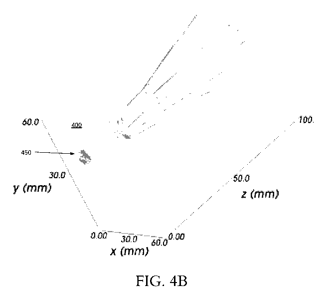
[0084] FIG. 4B is a three-dimensional diagram illustrating a plurality of
ultrasound
beams of insonation within the subject shown in FIG. 4A according to various
arrangements.
[0085] Referring to FIGS. 1-4B, FIG. 4B illustrates a perspective view of
the subject's
acoustic window and workspace 400 and the resulting ultrasound beams within
the subject's
head corresponding to the points of contact between the probe 105 and the
subject at the
workspace 400, as described with respect to FIG. 4A. As shown in FIG. 4B, each
point of
contact between the probe 105 and the subject is associated with a
corresponding ultrasound
beam of a given length that extends within the subject based on the
orientation of the probe
105 at the point of contact. Accordingly, by moving the probe 105 at different
locations
along a subject and by orienting the probe 105 at different angles at a given
point of contact
or at different points of contact, ultrasound data can be acquired to
accurately reconstruct a
subject's vasculature insonated by the plurality of ultrasound beams.
[0086] FIG. 5A is a diagram illustrating an ultrasound beam 500 insonating
a circle of
Willis according to various arrangements. FIG. 5B is a three-dimensional
diagram
illustrating data collected by the ultrasound beam 500 insonating the circle
of Willis shown in
FIG. 5A according to various arrangements.
[0087] Referring to FIGS. 5A and 5B, the ultrasound beam 500 is shown to
insonate
multiple depths of the circle of Willis vasculature, for example, portions of
the middle
cerebral artery and anterior cerebral artery. As described above, the
plurality of portions
insonated by the ultrasound beam 500 can provide imaging parameters at those
different
portions, such as, but not limited to, presence of blood flow, direction of
blood flow, and
intensity or magnitude of the blood flow. As shown in FIG. 5B, the same
ultrasound beam
can reflect this ultrasound data at the insonated portions of the vasculature
in three-
dimensional space. Accordingly, this information can then be used to construct
the
vasculature map as described herein.
[0088] The above used terms, including "held fast," "mount," "attached,"
"coupled,"
"affixed," "connected," "secured," and the like are used interchangeably. In
addition, while
certain arrangements have been described to include a first element as being
"coupled" (or
"attached," "connected," "fastened," etc.) to a second element, the first
element may be
23
CA 03088176 2020-07-09
WO 2019/140299 PCT/US2019/013347
directly coupled to the second element or may be indirectly coupled to the
second element via
a third element.
[0089] The previous description is provided to enable any person skilled in
the art to
practice the various aspects described herein. Various modifications to these
aspects will be
readily apparent to those skilled in the art, and the generic principles
defined herein may be
applied to other aspects. Thus, the claims are not intended to be limited to
the aspects shown
herein, but is to be accorded the full scope consistent with the language
claims, wherein
reference to an element in the singular is not intended to mean "one and only
one" unless
specifically so stated, but rather "one or more." Unless specifically stated
otherwise, the term
"some" refers to one or more. All structural and functional equivalents to the
elements of the
various aspects described throughout the previous description that are known
or later come to
be known to those of ordinary skill in the art are expressly incorporated
herein by reference
and are intended to be encompassed by the claims. Moreover, nothing disclosed
herein is
intended to be dedicated to the public regardless of whether such disclosure
is explicitly
recited in the claims. No claim element is to be construed as a means plus
function unless the
element is expressly recited using the phrase "means for."
[0090] It is understood that the specific order or hierarchy of steps in
the processes
disclosed is an example of illustrative approaches. Based upon design
preferences, it is
understood that the specific order or hierarchy of steps in the processes may
be rearranged
while remaining within the scope of the previous description. The accompanying
method
claims present elements of the various steps in a sample order, and are not
meant to be
limited to the specific order or hierarchy presented.
[0091] The previous description of the disclosed implementations is
provided to enable
any person skilled in the art to make or use the disclosed subject matter.
Various
modifications to these implementations will be readily apparent to those
skilled in the art, and
the generic principles defined herein may be applied to other implementations
without
departing from the spirit or scope of the previous description. Thus, the
previous description
is not intended to be limited to the implementations shown herein but is to be
accorded the
widest scope consistent with the principles and novel features disclosed
herein.
24
CA 03088176 2020-07-09
WO 2019/140299 PCT/US2019/013347
[0092] The various examples illustrated and described are provided merely
as examples
to illustrate various features of the claims. However, features shown and
described with
respect to any given example are not necessarily limited to the associated
example and may
be used or combined with other examples that are shown and described. Further,
the claims
are not intended to be limited by any one example.
[0093] The foregoing method descriptions and the process flow diagrams are
provided
merely as illustrative examples and are not intended to require or imply that
the steps of
various examples must be performed in the order presented. As will be
appreciated by one of
skill in the art the order of steps in the foregoing examples may be performed
in any order.
Words such as "thereafter," "then," "next," etc. are not intended to limit the
order of the
steps; these words are simply used to guide the reader through the description
of the methods.
Further, any reference to claim elements in the singular, for example, using
the articles "a,"
"an" or "the" is not to be construed as limiting the element to the singular.
[0094] The various illustrative logical blocks, modules, circuits, and
algorithm steps
described in connection with the examples disclosed herein may be implemented
as
electronic hardware, computer software, or combinations of both. To clearly
illustrate this
interchangeability of hardware and software, various illustrative components,
blocks,
modules, circuits, and steps have been described above generally in terms of
their
functionality. Whether such functionality is implemented as hardware or
software depends
upon the particular application and design constraints imposed on the overall
system. Skilled
artisans may implement the described functionality in varying ways for each
particular
application, but such implementation decisions should not be interpreted as
causing a
departure from the scope of the present disclosure.
[0095] The preceding description of the disclosed examples is provided to
enable any
person skilled in the art to make or use the present disclosure. Various
modifications to these
examples will be readily apparent to those skilled in the art, and the generic
principles
defined herein may be applied to some examples without departing from the
spirit or scope of
the disclosure. Thus, the present disclosure is not intended to be limited to
the examples
shown herein but is to be accorded the widest scope consistent with the
following claims and
the principles and novel features disclosed herein.
CA 03088176 2020-07-09
WO 2019/140299 PCT/US2019/013347
[0096] It should be noted that although the diagrams herein may show a
specific order
and composition of method blocks, it is understood that the order of these
blocks may differ
from what is depicted. For example, two or more blocks may be performed
concurrently or
with partial concurrence. Also, some method blocks that are performed as
discrete blocks
may be combined, blocks being performed as a combined block may be separated
into
discrete blocks, the sequence of certain processes may be reversed or
otherwise varied, and
the nature or number of discrete processes may be altered or varied. The order
or sequence of
any element or apparatus may be varied or substituted according to alternative
arrangements.
Accordingly, all such modifications are intended to be included within the
scope of the
present disclosure as defined in the appended claims. Such variations will
depend on the
machine-readable media and hardware systems chosen and on designer choice. It
is
understood that all such variations are within the scope of the disclosure.
Likewise, software
and web arrangements of the present disclosure could be accomplished with
standard
programming techniques with rule based logic and other logic to accomplish the
various
database searching blocks, correlation blocks, comparison blocks, and decision
blocks.
26