Note: Descriptions are shown in the official language in which they were submitted.
CA 03088786 2020-07-17
WO 2019/140534
PCT/CA2019/050074
TITLE
GENETICALLY ENGINEERED BACTERIOPHAGE
FIELD OF THE INVENTION
Prevention, diagnostics and treatment of human, animal, and plant bacterial
infections.
BACKGROUND
Bacteria are unicellular, biological entities that are mostly not harmful to
humans -
less than one percent of the different types make people sick. Many bacterial
species are beneficial to humans, such as those that help to digest food,
destroy
disease-causing cells, and provide needed vitamins.
Infectious bacteria (the harmful one percent) cause illness in humans and
animals.
They reproduce quickly in the body and produce toxic proteins that cause
tissue
damage and illness.
Bacteriophages (also referred to as phages) were discovered by Ernest Hankin
in
1896, and utilized as antibacterials against cholera. These bacteria-specific
viruses
can infect and destroy bacterial cells.
Bacteriophages are composed of proteins that encapsulate a DNA or RNA genonne.
Bacteriophages replicate within bacterium by injecting their viral genetic
material
(DNA or RNA) into the host cell effectively taking over the cells functions
for the
production of progeny bacteriophage leading to the rupture of the cell wall
and
subsequent bacterial cell death.
Bacteriophages continued to be used as antibacterials until the 1930's.
However,
it was found that bacteria naturally build up resistance to bacteriophages.
With
the introduction of chemical antibiotics, use of bacteriophages was abandoned.
While antibiotics are the usual treatment, bacterial mutations conferring
antibiotic
resistance are becoming increasingly common in pathogenic bacteria world-wide.
Methicillin-resistant Staphylococcus aureus (MRSA) bacteria, for example, is
an
increasingly common form of infection, often acquired through transmission in
hospitals. MRSA infections are extremely difficult to treat using conventional
antibiotics.
Bacteriophages can be very specific to the type of disease-causing bacterial
species.
Most bacteriophages have structures that enable it to bind to specific
molecules on
the surface of their target bacteria.
A key advantage of bacteriophages is that they enable the elimination of
antibiotic-
resistant bacteria without the need for increasingly toxic antibiotics or
harmful or
irritating chemical-exposure to humans, animals and the environment (see, e.g.
US
patent no. 6,699,701 to Intralytix).
1
CA 03088786 2020-07-17
WO 2019/140534
PCT/CA2019/050074
In natural settings, bacteriophages can be isolated from the environment in
which
the particular bacterium grows following a paired relationship, for example
from
sewage or feces. Repositories of different types of natural bacteriophages
have been
created to provide access to bacteriophages to treat difficult infections by
specific
bacterial species.
One problem with using bacteriophages has been that the patient's own body
will
often have an immune response against the bacteriophages and eliminate the
bacteriophages from blood. U.S. Pat. No. 5,660,812, U.S. Pat. No. 5,688,501,
U.S.
Pat. No. 5,811,093 and U.S. Pat. No. 5,766,892 all show methods of selecting
or
generating (using mutations) bacteriophages to improve the bacteriophage half-
life
within the blood of a patient to be treated.
Another problem associated with prior uses of phages to disinfect or treat
bacterial
contaminants or diseases, are that bacteria can become resistant to
bacteriophages.
The presence of, for example, a prophage within a bacterium may block the
expression of genes from an infectious bacteriophage, thus preventing
replication
of the infectious bacteriophage and preventing lysis and killing of the
bacterium. A
prophage may also cause the destruction of incoming phage DNA.
This has previously meant that either the bacteriophage needs to be matched to
the bacterium, often requiring complicated genetic analysis of the bacterium,
or
a number of different phages need to be used in combination. The production of
panels of different bacteriophages, such as panels of vir mutants derived from
temperate bacteriophage, is disclosed in WO 03/080823.
Currently, only natural bacteriophages exist, and natural bacteriophages that
have
been mutated and selected for specificity against certain bacteria (see, e.g.
US
patent no. 8,685,697 to Intralytix).
SUMMARY OF INVENTION
The invention is a template or platform technology for creating customized
genetically modified bacteriophages that target and destroy specific bacterial
organisms found in humans, animals and agricultural crops, as well as on
surfaces
in healthcare or food processing facilities.
Specific products can be developed using this template technology such as a
disinfectant spray for MRSA, food additive to prevent antibiotic use in animal
feed,
and treatment of bacterial infections in humans. The invention thus
encompasses
genetically modified bacteriophages as well as including gene products derived
from
bacteriophages, used to treat and or remove bacterial infections utilizing
bacteriophages.
According to one aspect, there is disclosed a method to manipulate the viral
genonne to cause functional changes in the life cycle of the virus.
In one embodiment, the invention provides a method of engineering
bacteriophages comprising:
2
CA 03088786 2020-07-17
WO 2019/140534
PCT/CA2019/050074
identifying a bacteriophage with only one attachment gene
isolating said bacteriophage;
removing said attachment gene from the genonne of said
bacteriophage; and
inserting a non-natural attachment gene into the genonne of said
bacteriophage wherein said non-natural attachment gene is
specific for attaching to a selected bacteria.
In another embodiment, the invention provides a method of engineering
bacteriophages comprising:
isolating a bacteriophage;
removing any attachment gene from a genonne of said
bacteriophage;
inserting a first unique open reading frame encoding one or more
attachment genes and inserting a second unique open reading
frame encoding one or more genes useful for overcoming bacterial
defenses;
inserting a non-natural attachment gene into said first open
reading frame, wherein said non-natural attachment gene is
specific for attaching to a selected bacteria. The one or more genes
useful for overcoming bacterial defenses are endolysins, bio-file
reducers, glycocalyx penetrators, or any combination thereof.
In another embodiment, the invention provides a method of engineering
bacteriophages comprising:
isolating a bacteriophage;
removing any attachment gene from a genonne of said
bacteriophage;
inserting a multiple restriction enzyme cassette in said genonne;
and
inserting a non-natural attachment gene into said cassette,
wherein said non-natural attachment gene is specific for attaching
to a selected bacteria.
In another embodiment, the invention provides a method of engineering
bacteriophages comprising:
isolating a bacteriophage;
removing all natural attachment genes from the genonne of said
bacteriophage; and
inserting a non-natural attachment gene into the genonne of said
bacteriophage;
wherein said non-natural attachment gene is specific for attaching
to a selected bacteria.
In another embodiment, the invention provides a method of growing
bacteriophages comprising:
- preparing a yeast culture comprising yeast and yeast nutrients;
- infecting said yeast with bacteriophages;
3
CA 03088786 2020-07-17
WO 2019/140534
PCT/CA2019/050074
- screening said yeast with colony-PCR for positive
transfornnants.
The bacteriophage may comprise a non-native attachment gene, wherein said
non-native attachment gene is specific for attaching to a selected bacteria.
The
bacteriophage may have no native attachment genes. The bacteriophage may
be lytic. The non-native attachment gene is specific for pathogenic/non-
pathogenic bacteria. The bacteriophage may be used for cleaning, treating, or
preventing a bacterial contaminant.
The invention also teaches bacteriophage for diagnosis of the presence or
absence of a specific bacteria.
The invention also teaches a method of producing a mutant bacteriophage, the
method comprising inactivating an attachment gene from a selected
bacteriophage, the selected bacteriophage being isolated from bacteriophages
from the environment; inserting, into the selected bacteriophage, a first
heterologous nucleic acid sequence comprising a first open reading frame
encoding a first specific attachment gene, the first specific attachment gene
being different than the inactivated attachment gene and being specific for a
selected bacteria, to produce the mutant bacteriophage. A second heterologous
nucleic acid sequence may be inserted in a second open reading frame encoding
a gene useful for overcoming bacterial defenses. The gene for overcoming
bacterial defenses may be a biofilnn degrading gene, a glycocalyx degrading
gene, a gene encoding an antibacterial protein, and a gene for an enzyme that
disrupts the bacterial wall, to produce the mutant bacteriophage. The step of
inactivating may inactivate all attachment genes from the selected
bacteriophage.
The invention also teaches a bacteriophage which is a lytic bacteriophage, a
bateriohage with a small genonne size, or a bacteriophage with structural and
functional genes to lyse gram negative and gram-positive bacteria, or any
combination thereof.
The invention also teaches an anti-microbial composition for sanitizing or
decontaminating a surface.
The invention also teaches a method of eliminating a microbial contaminant,
the
method comprising: obtaining one or more lytic enzymes produced by the mutant
bacteriophage; applying the one or more lytic enzymes to a bacterial
contaminant, without prior infection of the bacterial contaminant with a
bacteriophage, to eliminate the bacterial contaminant.
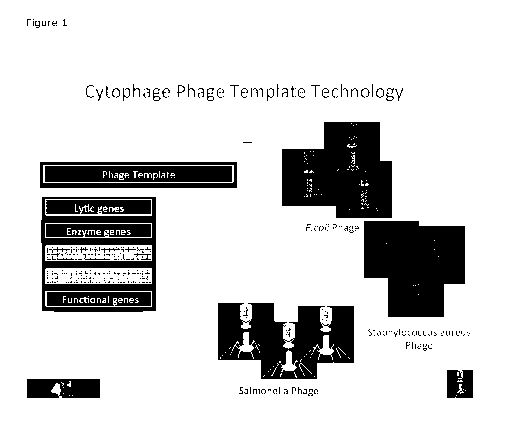
BRIEF DESCRIPTION OF THE DRAWINGS
Figure 1 shows an overview of a phage engineering platform, according to an
embodiment of the present invention.
Figure 2 shows an overview of a method to generate mutant bacteriophage using
a cell free cloning method, according to an embodiment of the present
invention.
4
CA 03088786 2020-07-17
WO 2019/140534
PCT/CA2019/050074
Figure 3 shows an overview of a method to generate mutant bacteriophages using
yeast strain, according to an embodiment of the present invention.
Figure 4 is an agarose plate of the titration of pp8 against E.coli DH5 alpha
after
rescue from the genetic template. Phage was spot plated on a lawn of E. coli.
Concentration was determined to be 108 for isolate one and 106 for isolate two
phage
units per 10u1.
Figure 5 shows a schematic representation of the entire genonne of the
disclosed
mutant bacteriophage, according to an embodiment of the present invention.
Figure 6 shows the nucleotide sequence of the entire genonne of PP8 and the
proteins encoded therein along with the restriction endonuclease sites
according
to an embodiment of the present invention.
Figure 7 is a detailed description of the PP8 molecule and proteins with
annotations according to an embodiment of the present invention.
Figure 8 is a gel electrophoresis photograph of PP8 DNA digestion using
enzymes
specific to remove inserts. EcoRI for ORF1 and ORF2 and TspRI for ORF 3 and
ORF
4, where Lane 1: 1 kb DNA ladder (NEB), Lane 2: space, Lane 3: undigested PP8
DNA, Lane 4: Digested PP8 ORF1 insertion SP5 attachment gene (46090) band size
1.1kb, Lane 5: Digested PP8 ORF2 insertion Endolysis gene (73195) band size
2.1,
Lane 6: Digested PP8 ORF3 insertion SP6 attachment gene (19991) band size
1.2kb,
Lane 7: Digested PP8 ORF4 insertion endolysis gene (60431) band size 2.1
Figure 9a - Shows a gel electrophoresis photograph where Lane 1: 1kb DNA
ladder
(NEB), 2: space, 3: Extracted bacteriophage genonne control, 4: Bacteria
control
(mock - bacteriophage infected), 5 - 7: Purified bacterial colonies with
potential
integration. Expected band size: 554 bases.
Figure 9b - is a gel electrophoresis photograph where Lane 1: Extracted
bacteriophage genonne control, 2: Bacteria control (mock - bacteriophage
infected),
3 - 5: Purified bacterial colonies with potential integration. Expected
band size:
613 bases.
Figure 10 shows an overview of the disclosed method for modifying the binding
sites, according to an embodiment of the present invention.
Figure 11 shows the results of the MRSA phage treatment experiment where
bacteriophage PP8 (5R5) insertion lysis of MRSA patient samples 1-6.
Bacteriophage
at a concentration of 107 was used to develop a kill curve of 6 MRSA positive
patient
samples. These samples were named patient 1-6.
Figure 12 shows the titration of PP8/5P5 against Staphylococcus aureus. Phage
was
spot plated on a lawn of Staphylococcus aureus. Concentration was determined
to
be 105 phage units per 10u1.
5
CA 03088786 2020-07-17
WO 2019/140534
PCT/CA2019/050074
Figure 13 shows the titration of PP8/SP6 against Staphylococcus aureus. Phage
was spot plated on a lawn of Staphylococcus aureus. Concentration was
determined
to be 108 phage units per 10u1.
Figure 14 shows the results of the new MRSA phage treatment where PP8 (5R5,
5R6) insertion kill curve of MRSA patient samples 1-6. Bacteriophage at a
concentration of 105 were used to develop a kill curve of 6 MRSA positive
patient
samples. Patient samples were tested for survivability at a concentration of
106
Figure 15 is a photograph showing a PP8 5P5/5P6 bacterial challenge.
Bacteriophage PP8 5P5/5P6 was flooded onto the agarose plate. Bacterial
strains
were tested for lysis. 50) E. coli 09 51) E.coli 01 52) E.coli 028 53) E.coli
DH5 alpha
54) Salmonella Enterica 55) Listeria nnonocytogenes 56) Entercoccus durans 57-
61)
MRSA patient sample 1-5 respectively.
DETAILED DESCRIPTION
The methods and techniques of the present disclosure are generally performed
according to conventional methods well known in the art and as described in
various general and more specific references that are cited and discussed
throughout the present specification unless otherwise indicated.
The terms "polypeptide", "peptide", and "protein" are typically used
interchangeably herein to refer to a polymer of amino acid residues. Amino
acids
may be referred to herein by either their commonly known three letter symbols
or by the one-letter symbols recommended by the IUPAC-TUB Biochemical
Nomenclature Commission. Each protein or polypeptide will have a unique
function. The invention includes polypeptides and functional fragments
thereof,
as well as mutants and variants having the same biological function or
activity.
In some embodiments, polymeric molecules (e.g., a polypeptide sequence or
nucleic acid sequence) are considered to be "homologous" to one another if
their
sequences are at least 25%, at least 30%, at least 35%, at least 40%, at least
45%, at least 50%, at least 55%, at least 60%, at least 65%, at least 70%, at
least 75%, at least 80%, at least 85%, at least 90%, at least 95%, or at least
99% identical.
In some embodiments a fragment of a nucleic acid sequence is a fragment of an
open reading frame sequence. In some embodiments, such a fragment encodes
a polypeptide fragment (as defined herein) of the protein encoded by the open
reading frame nucleotide sequence.
The term "nucleic acid fragment" as used herein refers to a nucleic acid
sequence
that has a deletion. In some embodiments a fragment of a nucleic acid sequence
is a fragment of an open reading frame sequence. In some embodiments, such
a fragment encodes a polypeptide fragment (as defined herein) of the protein
encoded by the open reading frame nucleotide sequence.
6
CA 03088786 2020-07-17
WO 2019/140534
PCT/CA2019/050074
The term "construct" refers to a nucleic acid sequence encoding a protein,
operably linked to a promoter and/or other regulatory sequences.
The term "genonnic sequence" refers to a sequence having non-contiguous open
reading frames, where introns interrupt the protein coding regions.
As used herein, the terms "encoding", "coding", or "encoded" when used in the
context of a specified nucleic acid mean that the nucleic acid comprises the
requisite information to guide translation of the nucleotide sequence into a
specified protein. The information by which a protein is encoded is specified
by
the use of codons. A nucleic acid encoding a protein may comprise non-
translated sequences (e.g., introns) within translated regions of the nucleic
acid
or may lack such intervening non-translated sequences (e.g., as in cDNA).
The term "percent sequence identity" or "identical" in the context of nucleic
acid
sequences refers to the residues in the two sequences which are the same when
aligned for maximum correspondence. For instance, polynucleotide sequences
.. can be compared using the computer program, BLAST (Altschul et al., 3. Mol.
Biol. 215:403-410 (1990); Gish and States, Nature Genet. 3:266-272 (1993).
The term "substantial homology" or "substantial similarity," when referring to
a
nucleic acid or fragment thereof, indicates that, when optimally aligned with
appropriate nucleotide insertions or deletions with another nucleic acid (or
its
complementary strand), there is nucleotide sequence identity in at least about
70%, 80%, 85%, or at least about 90%, or at least about 95%, 96%, 97%, 98%
or 99% of the nucleotide bases, as measured by any well-known algorithm of
sequence identity, such as BLAST, as discussed above.
As used herein, "heterologous nucleic acid sequence" is any sequence placed at
a location in the genonne where it does not normally occur. In some
embodiments, the heterologous nucleic acid sequence is a natural phage
sequence, albeit from a different phage.
A particular nucleic acid sequence also encompasses conservatively modified
variants thereof (such as degenerate codon substitutions) and complementary
sequences, as well as the sequence explicitly indicated. Thus, a nucleic acid
sequence encoding a protein sequence disclosed herein also encompasses
modified variants thereof as described herein. Substantially similar nucleic
acid
fragments of the instant invention may also be characterized by the percent
identity of the amino acid sequences that they encode to the amino acid
sequences disclosed herein, as determined by algorithms commonly employed
by those skilled in this art.
An "orgin bacteriophage" is a phage isolated from a natural or human made
environment that has not been modified by genetic engineering. A "mutant
bacteriophage" is a bacteriophage that comprises a genonne that has been
genetically modified by insertion of a heterologous nucleic acid sequence into
the genonne, or the genonne of the phage. In some embodiments the genonne of
7
CA 03088786 2020-07-17
WO 2019/140534
PCT/CA2019/050074
a origin bacteriophage is modified by recombinant DNA technology to introduce
a heterologous nucleic acid sequence into the genonne at a defined site.
"Operatively linked" or "operably linked" expression control sequences refers
to
a linkage in which the expression control sequence is contiguous with coding
sequences of interest to control expression of the coding sequences of
interest,
as well as expression control sequences that act in trans or at a distance to
control expression of the coding sequence.
A "coding sequence" or "open reading frame" is a sequence of nucleotides that
encodes a polypeptide or protein. The termini of the coding sequence are a
start
codon and a stop codon. The disclosure also includes native, isolated, or
recombinant nucleic acid sequences encoding a protein, as well as vectors
and/or
(host) cells containing the coding sequences for the protein.
Fragments and variants of the disclosed nucleotide sequences and proteins
encoded thereby are also encompassed by the present invention. By "fragment'
a portion of the nucleotide sequence or a portion of the amino acid sequence
and hence protein encoded thereby is intended. Fragments of a nucleotide
sequence may encode protein fragments that retain the biological activity of
the
native protein. Accordingly, the present disclosure relates to any nucleic
acid
fragment comprising a nucleotide sequence that encodes all or a substantial
portion of the amino acid sequences encoded thereby.
The present technology uses synthetic biology to generate bacteriophages that
can bind to specific bacterial strains. Since bacteriophages must attach to
host
bacterial cells to initiate infection of the bacteria, genetic selections or
manipulations in the viral DNA or RNA can define binding characteristics, thus
expanding the range of host cells beyond the natural paired relationship.
According to one embodiment there some characteristics of the disclosed
bacteriophages, including the following.
The phages are safe, non-corrosive, and non-toxic. The phages can be
engineered
so that they do not affect helpful bacteria, animal or human cells. Thus,
there is
no interference with the food chain, as with antibiotics.
The phages are designed, not discovered in nature. Thus, the technology is
adaptable to any bacterial infection. Undesirable genetic components are
eliminated. In contrast, the present methods of isolating natural phages for
specific
bacteria is like finding a "needle in a haystack" for target bacteria.
The phages are engineered to avoid mutation/adaptation of target bacteria
resulting
in superior kill rates and no resistance. Accordingly, the phages have
superior
efficacy over known phages. The phages also prevent biofilnn formation.
According to one embodiment, the platform is versatile. The disclosed
bacteriophages can be used to solve any bacterial problem. The disclosed
bacteriophages have application in human health (personalized medicine,
disinfectants, and diagnostics) such as for example, in MRSA and VRE, animal
health
8
CA 03088786 2020-07-17
WO 2019/140534
PCT/CA2019/050074
(livestock medicine, diagnostics) such as for example, ear drop for treating
dog ear
infections of Staphylococcus intermedius, and food safety (produce cleansing,
detection of bacterial contamination) such as for example, E. Coll, C. Jejuni,
Salmonella, and Listeria.
According to one embodiment, the bacteriophages can not only be used for the
treatment of antibiotic-resistant bacterial infections but also for prevention
of
bacterial-contamination in the environment and in food which may negatively
affect
human and animal health.
For example, the phages are useful for human health. Methicillin-resistant
Staphylococcus aureus (MRSA) bacteria are an increasingly common hospital-
acquired infection, often acquired through contact with contaminated surfaces.
For
facilities with a confirmed MRSA problem, this product can be used to
thoroughly
clean surfaces and reduce the development of new infections. According to an
embodiment, there is provided a multi-strain MRSA-specific disinfectant
cleanser
that can be used on porous and non-porous surfaces in hospitals including
beds,
curtains, tables, chairs, diagnostic and monitoring equipment, and medical
instruments.
According to an embodiment, the disclosed bacteriophages can be used to reduce
or eliminate any bacteria and/or resistant bacteria that are pathogenic to
humans
.. and/or animals. In aspects, the advantages of using this disinfectant over
the
commonly-used disinfectants, such as bleach, are multiple. First,
bacteriophage
are more effective in destroying bacteria than conventional means. Second,
phages can be left on surfaces to destroy new bacterial contamination events,
surviving for roughly 24 hours. Third, unlike bleach, bacteriophages do not
leave
a corrosive residue, and thus do not harm instruments, fabrics, and skin.
Fourth,
bacteriophages, customized for harmful bacteria, are non-toxic, unlike
cleaning
solutions.
The phages are also useful in animal health treatments. For example,
bacteriophage
are tailored to address bacterial infections in chickens, replacing the
antibiotic(s)
commonly used, resulting antibiotic-free chickens ¨ a commercial benefit in
today's
marketplace. This treatment also contributes to reducing the growing number of
antibiotic-resistant infections that occur as bacteria mutate and evolve to be
unaffected by antibiotics.
The phages are also useful in food safety. For example, bacteriophage-
cleansing
spray can be applied on agricultural crops for the prevention of food-borne
illnesses
from bacterial contamination during plant cultivation or during harvesting,
such as
Escherichia coli-contamination of strawberries.
The template technology is utilized to generate bacteriophages with various
specific
binding domains (thus selecting host range). The technology provides
bacteriophages in high concentrations.
9
CA 03088786 2020-07-17
WO 2019/140534
PCT/CA2019/050074
In some embodiments, bacteriophage-derived gene products may be useful for
"lysis-from-without" whereby bacteria can be eliminated without having to
become
infected.
According to an embodiment there is provided a method of eliminating a
bacterial
contaminant without prior infection of the bacterial contaminant with a
bacteriophage, the method comprising obtaining one or more lytic enzymes
produced by the disclosed bacteriophage; applying the one or more lytic
enzymes
to a bacterial contaminant to eliminate the bacterial contaminant.
A bacteriophage or phage is defined as a virus that infects bacteria.
Bacteriophages have a high specificity to their corresponding host bacteria.
To
infect bacteria, the bacteriophage attaches to specific receptors on the
surface of
the bacteria. This attachment determines the host range of each bacteriophage,
and normally is restricted to some genera, species, or even subspecies of
bacteria.
This bacteriophage specificity could provide clinicians, laboratory
technicians,
technicians in the field, as well as consumers, with the ability to identify
(detect
or diagnose) specific types of bacteria by exploiting this bacteriophage
characteristic.
Bacteriophages experience two types of natural life cycles, or methods of
viral
reproduction, known as the lytic cycle and the lysogenic cycle. In the lytic
cycle,
host cells will be broken and suffer death after replication of the virion. In
contrast,
the lysogenic cycle does not result in immediate lysing of the host cell and
consequential host cell death; rather, the bacteriophage genonne integrates
with
the host DNA, or establishes itself as a plasnnid, and replicates along with
the
organism's genonne. The endogenous bacteriophage remains dormant until the
host
is exposed to specific conditions (e.g., stress) at which point the
bacteriophage
may be activated, initiating the reproductive cycle resulting in the lysis of
the host
cell.
Endolysins are produced during the last stage of the phage lytic cycle from
within
their host and most are released into the periplasnnic space (Borysowski et
al.,
2006). From there on, endolysins cleave covalent bonds in the peptidoglycan to
release viral progeny (Fischetti, 2008). Within the endolysin subgroup, there
are
five classes: annidases, endopeptidases, nnurannidases, glucosanninidases and
transglycosylases (Gasset, 2010).
According to an embodiment, there is provided the use of lytic enzymes or
enzybiotics from bacterial viruses to combat antimicrobial resistance. An
enzybiotic
is defined to be a protein that degrades the bacterial cell wall, meaning that
it is
not subjected to bacteriophage proteins (Borysowski and Gorski, 2010). The
term
enzybiotics was first conceived in the paper 'Prevention and elimination of
upper
respiratory colonization of mice by group A streptococci by using
bacteriophage lytic
enzyme' (Nelson et al., 2001). The bacteriophage lytic enzymes are specific.
Phage
derived lytic enzyme and their destructive activity against certain components
of
the cell wall found in pathogenic bacterial strains but not the natural
nnicrobiota of
animals (Gasset. 2010). Two examples include group C streptococcal lysin,
effective in lysing group A streptococci but has no effect on normal oral
streptococci
CA 03088786 2020-07-17
WO 2019/140534
PCT/CA2019/050074
(Fischetti, 2006). A more relevant example is attained from the use of the
outer
membrane protein FyuA, commonly expressed in pathogenic Gram-negative
Escherichia coli. The fusion of FyuA binding domain to T4 lysozynne results in
translocation of the fusion from the outer membrane to the periplasnnic space
where
the lysozynne can destabilize the bacterial cell wall (Lukacik et al., 2013).
According to one embodiment, there is disclosed a method for providing an
endolysin protein or plurality of endolysin proteins, which overcome the
issues with
whole bacteriophages. The one or more endolysins specifically targets and
degrades the bacterial cell wall (peptidoglycan) from both within the cell or
from
.. outside of the cell resulting in lysis. In aspects, there is provided a
method to
generate various clones of endolysin genes from numerous bacteriophages and
using high-throughput screening, and to evaluate the success of the endolysin
clones against one or bacteria, such as for example, Escherichia coli strains,
Salmonella typhimurium and Campylobacterjejuni.
Thus, the technology extends the number of bacterial strains that may be
treated
with bacteriophage or bacteriophage gene products with and without infection.
Bacteriophages multiply themselves by infecting and killing bacteria. During
this
process, bacterial cell wall components are released along with the
bacteriophages.
These components may be toxic to humans, animal and bacteria. Thus, large
scale
preparations of bacteriophages using bacteria require post-manufacturing
treatments using harsh organic chemicals to reduce the toxicity to acceptable
levels
for clinical treatment.
Therefore, according to one embodiment, there is provided a method to grow the
disclosed bacteriophages in large-scale use of bacteria by using yeast
strains, such
.. as for example, Kluyveromyces lactis and Pichia pastoris. The disclosed
methods
circumvent the liberation of toxic end products.
Generating Mutant Bacteriophages
Environmental samples were isolated and fully characterized to determine
candidates which meet certain criteria. Preferably, a suitable origin
bacteriophage
is selected from candidates which includes one or more of the following
features:
- lytic phages;
- genetically different than known phages; and
- carries only one attachment gene
According to an embodiment, there is provided a method to genetically modify
one or more suitable origin bacteriophages.
In one embodiment, the origin bacteriophage includes one attachment gene. In
another embodiment, the origin bacteriophage includes more than one attachment
gene.
11
CA 03088786 2020-07-17
WO 2019/140534
PCT/CA2019/050074
In one embodiment, the method generates bacteriophage platforms configured to
allow for further interchanging of one or more desired proteins, such as for
example, attachment proteins.
According to one embodiment, the bacteriophage genonnes are manipulated to
change the virus' life cycle, creating gain of function, loss of function or
for virus
identification (reporter genes). A summary is shown in figure 1.
In one aspect, the origin bacteriophage is a lytic phage. In one aspect, the
origin
bacteriophage is a lytic phage that carries one or more attachment gene. In
another aspect, the origin bacteriophage is a lytic phage that carries only
one
attachment gene. In one aspect, the origin bacteriophage carries only one
attachment gene.
According to one embodiment, there is provided a method to produce a mutant
bacteriophage. In one aspect, the method comprises modifying the phage binding
sites of an origin bacteriophage so that the mutant bacteriophage can attach
to
different serotypes. In one embodiment, the mutant phage is then rescued and
the new binding domain is determined.
According to one embodiment, the engineered bacteriophage comprises only lytic
genes, wherein any and all lysogenic genes have been removed to ensure
integration cannot occur.
According to one embodiment, there is provided a method of 'cell free
cloning' to provide a template (or platform) technology that allows for the
modification/ insertion/ deletion of viral genes. The platform was generated
by constructing a mutant bacteriophage (defined as a phage which was
generated from known and unknown genetic codes) using isolated
environmental samples.
Genetic comparison of unknown phage types from environmental samples were
tested against known phage types allowing us to isolate known gene types.
In one embodiment, there is provided a mutant bacteriophage where genes of
interest were added and where unwanted genes were deleted. Together with
noncoding regions, the mutant bacteriophage is a genetic platform that carries
at least two unique open reading frames (ORF).
These unique ORFs can be used to add genes of interest. With reference to
figure
2, the genonnic compliment is divided into fragments with overlapping sections
to
adjacent fragments obtained by PCR amplification. Foreign genes are inserted
within respective fragments. Fragments were combined using bacterial cellular
extracts exploiting the homologous recombination methodology, where extracts
contain the necessary components to link fragments together into one
contiguous
fragment via homology. Rescue of bacteriophages from the fully assembled
genonnes is achieved by cell-free translation. This method involves mixing DNA
of
choice along with toxin free cellular extracts from E. coli along with amino
acids
12
CA 03088786 2020-07-17
WO 2019/140534
PCT/CA2019/050074
and energy, the transcription and translation proteins and enzymes from the
extract drives expression from the DNA leading to generation of bacteriophage.
In aspects, the mutant bacteriophage is a genetic platform that carries four
unique open reading frames (ORF).
In one embodiment, the first ORF can be used to insert an attachment gene for
a bacteria. In one aspect, the attachment gene can be selected from, but not
limited to, the following proteins:
= DNA-binding phage protein of Enterobacteriaceae (>CP007523.1:3585236-
3586111 Salmonella enterica subsp. enterica serovar Typhinnuriunn str.
CDC 2011K-0870, complete genonne) SEQ ID No: 125
= DNA-binding phage protein (>CP002910.1:3892390-3893265 Klebsiella
pneunnoniae KCTC 2242, complete genonne) SEQ ID No: 126
= DNA binding protein (>CM000724.1:300852-301217 Bacillus cereus
BDRD-5T26 chromosome, whole genonne shotgun sequence) SEQ ID No:
127
= Phage DNA-binding transcriptional regulator (>CP003678.1:c575894-
575136 Enterobacter cloacae subsp. dissolvens SDM, complete genonne)
SEQ ID No: 128
= Phage ssDNA binding protein (>CP009983.1:941901-942146 Vibrio
parahaennolyticus strain FORC 008 chromosome 2, complete sequence)
SEQ ID No: 129
= DNA binding protein (>CM000749.1:288493-288840 Bacillus thuringiensis.
T04001 chromosome, whole genonne shotgun sequence) SEQ ID No: 130
= phage nucleotide-binding protein (>CP006620.1:c2486999-2486259
Enterococcus faeciunn Aus0085, complete genonne) SEQ ID No: 131
= DNA-binding protein Bacteriophage P4 (>AE005174.2:318190-318450
Escherichia coli 0157:H7 str. EDL933 genonne) SEQ ID No: 132
= CP4-6 prophage; putative DNA-binding transcriptional regulator
(>HG738867.1:c269405-268512 Escherichia coli str. K-12 substr. MC4100
complete genonne) SEQ ID No: 133
= DNA-binding protein (Burkholderia pseudonnallei K96243 chromosome 1,
complete sequence) SEQ ID No: 134
= Putative DNA-binding prophage protein (>AL590842.1:c1239408-1238512
Yersinia pestis C092 complete genonne) SEQ ID No: 135
= Putative DNA-binding prophage protein (>AL590842.1:1235071-1235391
Yersinia pestis C092 complete genonne) SEQ ID No: 136
= Putative phage-related DNA-binding protein (>BX950851.1:4152092-
4152508 Erwinia carotovora subsp. atroseptica 5CRI1043, complete
genonne) SEQ ID No: 137
In one embodiment, the second ORF is used to insert a gene encoding a protein
useful for overcoming bacterial host defenses.
For example, the second ORF can be for introducing is to add enzymatic
functions to combat bacterial defenses. In one aspect, the second ORF can be
used to add endolysin genes, and/or biofilnn degrading genes.
13
CA 03088786 2020-07-17
WO 2019/140534
PCT/CA2019/050074
In an embodiment, the endolysin genes are selected from:
= PP1 phage endolysin SEQ ID No: 138 which is similar to Escherichia phage
B2 : 93% identical and 100% query coverage Accession Number
MG581355; Enterobacteria phage 311. : 92% identical and 100% query
coverage Accession Number : 3X865427; Shigella phage EP23 :
91% identical and 100% query coverage Accession Number :
3N984867;
Sodalis phage : 91%
identical and 100% query coverage Accession
Number : GQ502199.
= PP2 phage endolysin SEQ ID No: 139 which is similar to Escherichia phage
phiLLS : 99% identical and 100% query coverage Accession Number
: KY677846; Salmonella phage Stp1 : 98% identical and 100% query
coverage Accession Number : KY775453; Salmonella phage SPO1 :
98% identical and 100% query coverage Accession Number
KY114934; T5 phage-like p0rk29 : 97% identical and 100% query
coverage Accession Number : MF431732.
= PP3 phage endolysin SEQ ID No: 140 which is similar to Enterobacteria
phage ATK48 : 99% identical and 100% query coverage Accession
Number :
KT184310; Shigella phage SHSML-52-1 : 99% identical
and 100% query coverage Accession Number : KX130865;
Escherichia phage APCEc01 : 99% identical and 100% query coverage
Accession Number :
KR422352.1; E. coli 0157 typing phage 6 :
98% identical and 100% query coverage Accession Number
KP869104; Shigella phage 5hf125875 : 98% identical and 100% query
coverage Accession Number : KM407600;
Shigella phage phi25-
307 : 98% identical and 100% query coverage Accession Number
: MG589383; Klebsiella phage vB Kpn F48 : 73% identical and 98% query
coverage Accession Number : MG746602;
= PP7 phage endolysin SEQ ID No: 141 which is similar to Salmonella phage
5T11 : 95%
identical and 100% query coverage Accession Number
: MF370225; Salmonella phage Meda : 95% identical and
100% query
coverage Accession Number : MH586731;
Salmonella phage 5i3
: 95% identical and 100% query coverage Accession Number
KY626162; Escherichia phage EC6 : 95%
identical and 100% query
coverage Accession Number : 3)(560968; Bacteriophage Felix 01
: 95% identical and 100% query coverage Accession Number
AF320576; Enterobacteria phage KhF2 : 94% identical and 100% query
coverage Accession Number :
KT184314; Salmonella virus VSe102
: 94% identical and 100% query coverage Accession Number
MG251392; Salmonella phage Mushroom : 94% identical and 100%
query coverage Accession Number : KP143762;
Staphylococcus
phage SA1 : 94%
identical and 100% query coverage Accession Number
: GU169904; E. coli 0157 typing phage 15 : 94% identical and 100%
query coverage Accession Number : KP869113; Citrobacter phage
Mijalis : 83% identical and 99% query coverage Accession Number
: KY654690; Shigella phage Sf14 : 82% identical and
99% query
coverage Accession Number : MF327003;
= PP11 phage endolysin SEQ ID No: 142 which is similar to Enterobacteria
phage HK578 : 79% identical and 97% query coverage Accession Number
14
CA 03088786 2020-07-17
WO 2019/140534
PCT/CA2019/050074
: 3Q086375; Escherichia phage Sloth : 78% identical and 97% query
coverage Accession Number : KX534339; Escherichia phage Envy
: 78% identical and 97% query coverage Accession Number .
KX534335
= Enterobacter cloacae A1S1 phage endolysin SEQ ID No: 143
In one embodiment, the biofilnn degrading genes and glycocalyx degraders are
selected from:
Protein Name Accession Number
Cathelicidin antimicrobial peptide LL- NM 004345.5
37
Histatin 3 (HTN3) NM 000200.2
Nisin M24527.1
Dispersin B NZ NRDE01000005.1
Endo-1,4-8-glucanase (callulase) NM 001247953.1
Aureolysin EF070234.1
NucB HQ112343.1
Serine protease (SspA) AF309515.1
LapG protease KT186446.1
Melittin NM 001011607.2
Endo-1,4-8-nnannosidase (nnanA) AM920689.1
a-amylase A17930.1
For example, the second ORF can be for introducing antibacterial proteins used
in template to address bacterial lysis. In one aspect, an example protein is a
bacterial cell wall degrader used to degrade Staphylococcus aureus
(>ENAI3Q06632013Q066320.1 Staphylococcus aureus strain JP1 Psnn betel. (psnn
betel.) and Psnn beta2 (psnn beta2) genes, complete cds). SEQ ID No: 144
In other aspects, the second ORF can be for introducing enzymes which target
the key linking chemistries (amide, ester and glycolytic bonds) found in
bacterial cell walls. Examples include:
= M20 family peptidase [uncultured bacterium], ACCESSION AHZ45606
(uncultured bacterium, >KF835382.1:c34024-32630 Uncultured bacterium
clone SZR5 genonnic sequence) SEQ ID No: 145
= Lipolytic enzyme (uncultured bacterium ACCESSION AHZ45613
>KF835383.1:7038-8066 Uncultured bacterium clone WZR9 genonnic
sequence) SEQ ID No: 146
= peptidase M56 ([uncultured bacterium] ACCESSION AHZ45657
Uncultured bacterium clone WZR18 genonnic sequence
CA 03088786 2020-07-17
WO 2019/140534
PCT/CA2019/050074
(>KF835385.1:c14123-13038 Uncultured bacterium clone WZR18 genonnic
sequence) SEQ ID No: 147;
= Another example is Uncultured bacterium clone HOAb112C long-chain fatty
acid CoA-ligase gene ([uncultured bacterium] DBSOURCE accession
KF955286.1) SEQ ID No: 148;
= Bonnbyx nnori BnnGloverin1 nnRNA for gloverin-like protein 1, complete
ACCESSION AB190863 SEQ ID No:;149
= Bonnbyx nnori BnnGloverin2 nnRNA for gloverin-like protein 2, complete
ACCESSION AB190864 SEQ ID No: 150;
= Bonnbyx nnori BnnGloverin3 nnRNA for gloverin-like protein 3 ACCESSION
AB190865 SEQ ID No: 151; and
= Bonnbyx nnori BnnGloverin3 nnRNA for gloverin-like protein 4 ACCESSION
AB190866 SEQ ID No: 152;
.. According to an embodiment, there is provided a method of producing a
mutant
bacteriophage, the method comprising inactivating at least one attachment gene
from a selected bacteriophage, the selected bacteriophage can be isolated from
bacteriophages from the environment. The method further comprises inserting,
into the selected bacteriophage, one or more a heterologous nucleic acid
.. sequences comprising one or more attachment genes. The one or more inserted
attachment genes being different than the inactivated native attachment gene
and is/are choosen because of its specificity for a selected bacteria, to
produce
the mutant bacteriophage. In some embodiments, the provision of the selected
attachment gene(s) expands the range of possible host cells (i.e. bacteria)
.. beyond the natural paired relationship.
According to an embodiment, there is provided a method of producing a mutant
bacteriophage, the method comprising inactivating at least one attachment gene
from a selected bacteriophage, the selected bacteriophage can be isolated from
bacteriophages from the environment. The method further comprises inserting,
.. into the selected bacteriophage, a first heterologous nucleic acid sequence
comprising a first open reading frame encoding a first specific attachment
gene.
The first specific attachment gene is different than the inactivated
attachment
gene and is choosen because of its specificity for a selected bacteria, to
produce
the mutant bacteriophage.
.. In another embodiment, the method further comprises inserting a second
heterologous nucleic acid sequence in a second open reading frame encoding a
gene useful for overcoming bacterial defenses. In aspects, the gene for
overcoming bacterial defenses may be a biofilnn degrading gene, a glycocalyx
degrading gene, a gene encoding an antibacterial protein, and a gene for an
.. enzyme that disrupts the bacterial wall, to produce the mutant
bacteriophage.
In one aspect, the first open reading frame further encodes a second specific
attachment gene that is different than the first specific attachment gene.
In some embodiments, the method inactivates all the attachment genes from
the selected bacteriophage. In aspects, the step of inactivating comprises
16
CA 03088786 2020-07-17
WO 2019/140534
PCT/CA2019/050074
making an inactivating mutation in at least one native attachment gene. In
some aspects, the inactivating mutation is a point mutation.
According to an embodiment, there is provided an anti-microbial composition
for
sanitizing or decontaminating a surface. In aspects, the anti-microbial
composition comprises the disclosed mutant bacteriophage.
According to an embodiment, there is provided a method of decontaminating a
surface suspected of containing a bacteria. In aspects, the bacteria is an
infectious or a non-infectious bacteria. The method comprising applying the
disclosed anti-microbial composition comprising the disclosed mutant
bacteriophage to the surface. In aspects, the amount is effective to
decontaminate the surface of at least substantially or all of the
contaminating
bacteria.
In aspects, the surface is a biological surface (animal or plant).
According to an embodiment, there is provided a method to generate specific
mutant bacteriophage gene products. In aspects, there is provided a method of
eliminating or substantially eliminating a microbial contaminant (an
infectious or
non-infectious bacteria), the method comprising: obtaining one or more lytic
enzymes produced by the disclosed mutant bacteriophage and applying the one
or more lytic enzymes to a bacterial contaminant. In some aspects, the
elimination is accomplished without prior bacteriophage infection of the
microbial
contaminant and therefore leads to result of lysis from without.
All publications and patents mentioned in this specification are herein
incorporated by reference to the same extent as if each individual publication
or
patent was specifically and individually indicated to be incorporated by
reference.
The foregoing description and certain representative embodiments and details
of the invention have been presented for purposes of illustration and
description
of the invention. It is not intended to be exhaustive or to limit the
invention to
the precise forms disclosed. It will be apparent to practitioners skilled in
this
art that modifications and variations may be made therein without departing
from the scope of the invention.
Examples
Example 1 ¨ Bacteriophage Isolation
Samples from sewer and waste, environmental soils, and animal feces were
collected and purified to be used for the isolation of bacteriophages.
Purified
samples were then screened for the presence of bacteriophages against specific
bacteria. Protocols and methods for isolating bacteriophages from water
samples were adapted from Bonilla et al. (2016) and Bourdin et al. (2014);
solid and soil sample methods adapted from Sillankorva (2018), Pausz et al.
(2009), and Van Twest & Kropinski (2009).
17
CA 03088786 2020-07-17
WO 2019/140534
PCT/CA2019/050074
Solid samples were rehydrated using sterile water for a minimum of 1 hour to
allow the bacteriophages to disseminate. Samples are then centrifuged to
remove solid materials and large particulates and the supernatant is
collected.
The centrifuged environmental samples and water samples were then further
processed and purified using filters (0.2pM) to remove bacteria and smaller
unwanted particulates. Filtered samples can be further concentrated using
filter
tubes or stored at 4 C for future use.
Filtered samples were then tested against bacterial strains of interest using
an
agar overlay plaque assay technique (Kropinski et al. 2009). Liquid agar
overlay was inoculated with filtered environmental sample and the bacterial
strain of choice and mixed. It was then poured onto an agar culture plate
(bacterial strain dependent) and allowed to harden. Plates were then incubated
overnight (conditions are bacterial strain dependent) and observed the next
day for plaques against the chosen strains. Plaques containing bacteriophages
were then picked and further processed by 3-rounds of subsequent plaque
assay overlays to purify the selected phage(s).
Using the method outlined above, numerous (hundreds) EV samples were
collected and tested for suitability to develop the template. The function and
structural genes were characterized for each EV sample, tested for integration
(as detailed below). Candidate phages with a low copy number of lysogenic
genes, and the structural and functional genes to allow for gram negative and
gram-positive lysis was identified. A selected bacteriophage named PP8 was
sequenced and gene structure and function were examined as detailed below.
PP8 was selected as it had the desired genes. Although it also had lysogenic
genes, these were removed using ORF replacement.
Example 2 - Platform development
Using environmental sample EV3 1/PP8, after bacteriophage isolation we
purified genonnic material with PureLink viral DNA/RNA extraction kit. The
full-
length genonne was amplified (EV3 1/Full/F/pYESIL and EV3 1/Full/R/pYESIL
see sequence EV3 1) to have 30 bp homology with the pYESIL Sapphire vector.
PCR amplification was performed using Phusion high-fidelity DNA polynnerase
(modification use of touchdown technique for primer annealing starting at 69C
and dropping by 0.5C each cycle). PCR products were separated on agarose
gels and bands were excised, extracted, and assembled. The resulting
construct EV3 1pYES (unmodified) allowed for the genetic modification of EV3 1
and the determination of function of mutations in a phage rescue based
system.
Example 3 - Full-length Genome Assembly
In brief, the following provides for a method for the genetic manipulation of
yeast (Kluyveromyces lactis and Pichia pastoris) cells to include T7 DNA
(deoxyribonucleic acid)-dependent RNA (ribonucleic acid) polynnerase
transcription from Escherichia phage T7 followed by expression of
bacteriophage in yeast. There is also provided a method for the genetic
18
CA 03088786 2020-07-17
WO 2019/140534
PCT/CA2019/050074
manipulation of yeast (Kluyveromyces lactis and Pichia pastoris) cells to
include
transcriptional components from bacteria (Escherichia coli) and RNA
(ribonucleic acid) polynnerase (P) inside of yeast followed by expression of
the
bacteriophage in yeast.
With reference to figure 3, the genonnic compliment was divided into fragments
with overlapping sections to adjacent fragments obtained by PCR amplification.
Foreign genes were inserted within respective fragments. Fragments were
combined via homologous recombination into full-length genonnes and a yeast-
based plasnnid (as an additional PCR fragment) with a T7 promoter inside of
yeast strain Pichia pastoris. The stable plasnnid under T7 promoter control
drove
the rescue of bacteriophages upon induction of the P. pastoris which contains
T7 RNA polynnerase cells are then lysed using enzymatic and mechanical means
to release fully-formed bacteriophage particles.
Homologous recombination of EV31pYes (unmodified) with pYESIL vector was
achieved using 100 ng of each PCR product and transformed into chemically-
competent yeast cells. pYESIL vector (100 ng) and EV31 (100 ng) were
combined. Competent yeast cells were added and mixed gently followed by the
addition of 600 pl of polyethylene glycol (PEG) and lithium acetate (LiAc)
solution then mixed gently. The mixture was incubated at 30C for 30 minutes,
inverting in 10 minutes intervals. Immediately after incubation, 35.5p1 of
dinnethyl sulfoxide (DMSO) was added, mixed by inversion and subjected to
heat-shock for 20 min at 42C (with occasional inversion). Tubes were then
centrifuged at 200-400 xg for 5 minutes, supernatant was discarded and the
cell pellet was resuspended in 1 ml sterile 0.9% sodium chloride (NaC1).
Visualization of transformation was achieved by spread-plating 100p1 onto
selective agar plates (media without tryptophan) and a 3-day incubation period
at 30C. Colony-PCR screening can determine the presence of positive
transfornnants. Homologous recombination was achieved by standard cloning
techniques to make S. cerevisae strain 5150, chemically-competent. Briefly,
using the Gietz and Schiestl 2007 protocol, a spread plate of a single yeast
colony from stock was created and incubated overnight at 30C. The next day,
50p1 equivalent of cells was scraped and washed in a tube with 1nnl of sterile
nuclease-free water followed by a 13,000xg spin for 0.5 minutes. The following
was added to the cell pellet in order: 240p1 of PEG-3350 (50%w/v), 36p1 LiAc
(1M), 50p1 single - stranded carrier DNA (2nng/nn1 of pig sperm) and 34p1
plasnnid-nuclease free water mixture (< lug plasnnid). It was gently vortexed
to
mix, incubated at 42C for 20-180 minutes (timing is dependent on strain). For
EV31, 45 minutes was used. After transformation, it was spun for 13,000xg for
0.5 minutes, then the supernatant was removed and the pellet was resuspended
in 1 ml sterile nuclease-free water. The mixture was spread onto selective
media
plates, yeast synthetic drop out media without uracil and incubated at 30C for
3-4 days. Verification of clone was carried out using Colony-PCR screening.
Figure 4 shows the titration of PP8 after rescue from the genetic template.
A graphical representation which depicts the location of the genes of the
EV31/PP8 is shown in figure 5 and a detailed nucleotide sequence of the entire
19
CA 03088786 2020-07-17
W02019/140534
PCT/CA2019/050074
genonne of showing sense strand (SEQ ID NO: 1), the antisense strand of the
complementary sequence (SEQ ID NO:2), and the sequence of the proteins
encoded therein (SEQ IDs NO: 3-124) along with the restriction endonuclease
sites is provided in Figure 6. Figure 7 shows a detailed description of the
EV31/PP8 molecule and proteins with annotations.
Example 4 - Clone Verification by Colony PCR
Screening for positive-transfornnants (plate growth colonies) was carried out
as
follows. Individual yeast colonies were placed in into 15p1 of lysis buffer
for
inoculation. In a separate tube, 5p1 of each mixture was transferred and
stored
at 4C, until ready for large scale grow up of positive colonies. The remaining
10p1 of cell suspension was boiled for 5 minutes at 95C, then immediately
placed
on ice, adding 40p1 of nuclease-free water and mix. 0.5p1 of lysate was added
to
each PCR reaction in a total volume of 50p1 and visualized by agarose gel
electrophoresis. The resulting gel of the PP8 DNA digestion is shown in figure
8.
Example 5 - Development of unique ORF's
Using the PP8 template, a mutant bacteriophage was generated. Native
attachment
proteins were removed by generating point mutations using homologous
recombination.
Gene Disruption 14452-13316 (tail protein):
>PP8-F1F
ACAAATAGTGAAGAGATAAACCAGGTTGAGCAAG SEQ ID No: 185
>PP8-tail-nnut-R
TTGACGTTGAATCTGGAGTCGATAGGTGCGACAGGTTACCAATGG SEQ ID No: 186
>PP8-tail-nnut-F
GTCGCACCTATCGACTCCAGATTCAACGTCAAGGTCTCACC SEQ ID No: 187
>PP8-F1R
TTCCAAGACGGATTCGAACCGTCACTAGTACAAGG SEQ ID No: 188
Gene Disruption 14823-14446 (tail protein):
>PP8-F1F
ACAAATAGTGAAGAGATAAACCAGGTTGAGCAAG SEQ ID No: 185
>PP8-hyp1-nnut-R
TTAATGATGTTATCTCGATAACGTCGACATGGAGACTCAGTAAATGG SEQ ID No: 189
> PP8- hypl-nnut-F
TCTCCATGTCGACGTTATCGAGATAACATCATTAAGGTTGTACC SEQ ID No: 190
>PP8-F1R
TTCCAAGACGGATTCGAACCGTCACTAGTACAAGG SEQ ID No: 188
Gene Disruption 16522-17937 (tail protein):
>PP8-F1F
ACAAATAGTGAAGAGATAAACCAGGTTGAGCAAG SEQ ID No: 185
>PP8-hyp2-nnut-R
TTGAATAAACCGTTATCGCCTTCTTAAAGCAACCTGTATTGCGTTCTGC SEQ ID No: 191
> PP8- hyp2-nnut-F
TTGCTTTAAGAAGGCGATAACGGTTTATTCAACAAACCCTCATTTCATTG SEQ ID No:
192
CA 03088786 2020-07-17
WO 2019/140534
PCT/CA2019/050074
> PP8 - F1 R
TTCCAAGACGGATTCGAACCGTCACTAGTACAAGG SEQ ID No: 188
Gene Disruption 34777-37020 (tail protein):
> PP8-F3F
TTCTTAAG G AG G GTTATG AATG TG TTATAC AG G SEQ ID NO: 158
>PP8-tape-nnut-R
TCTGTGTAGTTCGGCCAACTGTAGTGTGCGAATGATGCAGCGAACATTC SEQ ID No:
193
>PP8-tape-nnut-F
TTCGCACACTACAGTTGGCCGAACTACACAGATACCATGAAGCAGTACTC SEQ ID No:
194
>PP8-F3R
GTGGTAAGGTAAGGTATGGAAGGATGGCAGTAG SEQ ID NO: 167
The mutant bacteriophage can comprises four ORFs: ORF 1 is located at position
46090; ORF 2 is located at position 73195; ORF 3 is located at position 19991;
ORF
4 is located at position 60431.
Using modification primers EV31/ORF1/F and EV31/ORF1/R for ORF1 (between
nucleotide positions 46,090 and 46,091) and EV31/ORF2/F and ORF2/R for ORF 2
(between nucleotide positions 73,195 and 73,196) and cell free cloning we
generated three EV31 mutant constructs. EV31 (ORF1), EV31 (ORF2), and EV31
(ORF1/2). In the construction of ORF2 we first removed the natural binding
domain
from EV31 and added a multiple restriction enzyme cassette. This cassette is
then
used to add new bacterial binding domains.
Both homologues recombination and insertion using restriction digests. For
restriction digest, the enzyme TspRI allows insertion of a multiple cloning
site
(MCS). In one example, ORF3 is located at 19991 in ev31/pp8 sequence. In this
example, the insertion of the MCS would be done by using TspRI. Once the MCS
is
inserted, the insertion an attachment gene of choice can done achieved by
using
restriction enzymes sites found in the MCS. Example of MCS for ORF3:
GCCGGCAGTGGATCCCCGGGGAAGATATTC SEQ ID NO: 153. This MCS carries
enzymes sites for Neel, TspRI, XnnnI, SnnaI. The primers used for adding the
MCS
to site 19991 are: EV31 ORF3 primer f GCTACACTGCTGAGA SEQ ID NO: 154; EV31
ORF3 primer r TCTCAGCAGTGTAGC SEQ ID NO: 155.
The fourth ORF is located at 60431 in ev31/pp. In this example, the insertion
of
the MCS would be done by using TspRI. Once the MCS is inserted, the insertion
an
attachment gene of choice can done achieved by using restriction enzymes sites
found in the MCS. The primers used for adding the MCS to site 60431 are: EV31
ORF4 primer f CATCAGATGCTGG SEQ ID NO: 156; EV31 ORF4 primer r
CCAGCATCTGATG SEQ ID NO: 157.
21
CA 03088786 2020-07-17
WO 2019/140534
PCT/CA2019/050074
Example 6 - Integration
An analysis of the genonne of the EV31/PP8 revealed a possible lysogenic gene
located at 60351-62336. Mutation of the gene by generating an ORF at the site
of
60431 (ORF4). Once the lysogenic gene were inactivated, we carried out
integration studies to ensure integration did not occur. The results are shown
in
figures 9a and 9b.
Under conditions that promote integration we confirmed that PP8 lacks the
ability
to integrate. The gel electrophoresis photograph identifies integration events
demonstrated by a bacteriophage (bacteriophage induction control), determined
by
polynnerase chain reaction (PCR) on whole bacterial cells. A respective primer
set
for each bacteriophage would give a positive PCR signal (right panel; lane 5)
if the
bacteriophage genetic material was integrated inside of the purified
(bacteriophage
particle-free) bacterial colonies. Contrarily, PP8 cannot integrate into the
bacterial
host cells, as indicated by the absence of a positive signal for the PP8
sequence in
the photograph (left panel; lanes 5 - 7).
Creating conditions for integration
Fresh overnight cultures of the bacterial host (Escherichia coil C) from
glycerol
stocks were prepared in Luria-Bertani (LB) broth. Once saturated, the cultures
were
diluted (1:100) in fresh LB broth, supplemented with 2 nnM CaCl2 and incubated
until an 0D600 of 0.6. Mixtures of host (100 pL of E. coli C) and
bacteriophage (100
pL at multiplicity of infection of 5) in 3 nnL of molten, soft agar were
overlaid onto
previously, dried LB-agar plates. Following an overnight incubation, three
colonies
from each plate were picked, re-streaked onto fresh LB-agar plates and
incubated
overnight for three rounds. The purified colonies (free of contaminating
bacteriophage particles) were inoculated into LB - 2nnM CaCl2 broth and
incubated
overnight.
Analyzing potential integration events
Polynnerase chain reaction (PCR) master mixes of GoTaqC) DNA polynnerase
(Pronnega) were set up following the manufacture's recommendations along with
the respective primers for each bacteriophage to be evaluated. Five pL from
each
overnight culture were spiked into their respective PCR reaction. Cycling
conditions
were altered to include whole-bacterial cell boiling in the initial
denaturation period
(950C for 10 minutes) and an annealing temperature of 590C. Five pL of
completed
PCR reactions were subjected 1% agarose gel electrophoresis, stained and
visualized under ultraviolet (UV) light. The results are shown in figures 9a
and 9b.
Example 7 - ORF1 insertion SP5 attachment protein (between 46,090 and
46,091)
Using PP8 we developed of a MRSA specific PP8 binding phage by utilizing the
PP8
template we removed native attachment genes and added attachment protein SP5
at the ORF 1 location (between 46,090 and 46,091) using homologous
recombination. The primer sets used for this homologous recombination are:
22
CA 03088786 2020-07-17
WO 2019/140534
PCT/CA2019/050074
1. Primer set for PP8 Fragment (bold) and homologous recombination with SP5
gene (underline) on 5'
>PP8-F3F
TAATACTCTACAGACACCACTAACTGATGCTGCTG SEQ ID NO: 158
>PP8-5P5-R
CTCGTTTCAACATCTTTTATTTTGTACATACAAGGGATTAAGCAGTTCTTACCC SEQ
ID NO: 159
2. Amplification of 5P5 (underline):
>5P5-F
ATGTACAAAATAAAAGATGTTGAAACGAG SEQ ID NO: 161
>5P5-R
CACCCCTTAATTAAATAAAGTGTATTAGGGTC SEQ ID NO: 162
3. Primer set for PP8 Fragment (bold) and homologous recombination with 5P5
gene (underline) on 3'
>PP8-SP5-F
CACTTTATTTAATTAAGGGGTGATGACTGATTGTTAAGATGGTGTTAATATTC SEQ
ID NO: 163
> PP8-F3R
GTGGTAAGGTAAGGTATGGAAGGATGGCAGTAG SEQ ID NO: 167
The sequence of the insertion (MRSA attachment protein 5R5) is shown in SEQ ID
NO: 168.
Example 8 ¨ Phage efficacy against MRSA
We tested our new PP8(5P5) phage against MRSA infected patient samples 1
through 6. These are MRSA positive patient samples from clinical isolation. An
overview of the method is shown in figure 10 and the results are shown in
figures
11 and 12. Patient samples 1, 2, 4, 5, 6 were all lysed using PP8 (5R5).
Patient
sample 3 showed only a partial binding profile, which suggested that the
binding
may not have been specific enough to give a 100% lysis rate. Positive control
was
PP8 bacteriophage with in SA attachment site.
After sequence analysis of patient sample 3, through sequence analysis and
blast
searching for attachment sites, a new binding site was revealed.
Example 9 - ORF1 insertion SP6 attachment protein (between 46,090 and
46,091)
We also generated a PP8 5R6 mutant and tested this against Staphylococcus
aureus. The result is shown in figure 13.
23
CA 03088786 2020-07-17
WO 2019/140534
PCT/CA2019/050074
We then generated a new PP8 strain to attach to and lyse patient sample 3. We
further generated a generate PP8 (SR5, SR6) mutant using homologous
recombination by adding attachment protein SP6 to ORF 1 of the original PP8
template to generate PP8 (SR5, SR6). The primer sets used for this homologous
recombination are:
1. Primer set for PP8 Fragment (bold) and homologous recombination with SP6
gene (underline) on 5'
>PP8-F3F
TAATACTCTACAGACACCACTAACTGATGCTGCTG SEQ ID NO: 158
>PP8-SP6-R
CTCGTTTCAACATCTTTTATTTTGTACATACAAGGGATTAAGCAGTTCTTACCC SEQ
ID NO: 160
2. Amplification of 5P6 (underline):
>SP6-F
ATGTACAAAATAAAAGATGTTGAAACGAG SEQ ID NO: 164
>5P6-R
TCACCCCTTAATTAAGTAAAGTGTATTAGGGTC SEQ ID NO: 165
3. Primer set for PP8 Fragment (blue) and homologous recombination with 5P6
gene (underline) on 3'
>PP8-SP6-F
AGACCCTAATACACTTTACTTAATTAAGGGGTGATGACTGATTGTTAAGATGGTG SEQ
ID NO: 166
>PP8-F3R
GTGGTAAGGTAAGGTATGGAAGGATGGCAGTAG SEQ ID NO: 167
The sequence of the insertion (MRSA attachment protein 5P6) is shown in SEQ ID
NO: 169.
The resultant new strain of bacteriophage was called PP8(5P5, 5P6). We used
this
new bacteriophage in conjunction with PP8(5P5) to determine if we could lyse
patient samples 1 through 6 using these two new modified bacteriophages. As
see
in figures 14 and 15, the new mutant bacteriophage lysed all six patient
samples
demonstrating that addition of a new attachment gene to our PP8 template
allows
for the specific targeting of a bacterium.
Example 10 - ORF2 endolysis gene insertion (inserted at 73195 in
PPS)
1. Primer set for PP8 Fragment (bold) and homologous recombination with
foreign gene (underline) on 5'
>PP8-F5F
AAGACTCGGAAGAAGGTAGTCACTAAGGAAAGTG SEQ ID NO: 170
>PP8-endolysin-R
24
CA 03088786 2020-07-17
WO 2019/140534
PCT/CA2019/050074
CCGTAAATCTTAGACCGTTGTCACTGAATCGCATGTCAAGTTTTACATAGAAATCC
SEQ ID NO: 171
>endolysin-F
ATGCGATTCAGTGACAACGGTCTAAGATTTACGGCAGC SEQ ID NO: 172
>endolysin-R
TTATGCTGCGTTACGCCCGATTTTCTCGGCAACGTCC SEQ ID NO: 173
>PP8-endolysin-F
TTGCCGAGAAAATCGGGCGTAACGCAGCATAAAAGGTGATGTGGGTCTTGATAGG
SEQ ID NO: 174>PP8-F5R
GCAACACTGTATCGGCTACTTCAAAGTCTTCTCTG SEQ ID NO: 175
The insertion of the endolysis gene was carried out using normal molecular
biology techniques. The sequence of the insertion is shown in SEQ ID NO: 176.
Example 11 ¨ Insertion of attachment proteins in ORF 1 and ORF 2
Homologous recombination in ORF1
1. Primer set for PP8 Fragment (bold) and homologous recombination with
foreign
gene (underline) on 5'
>PP8-F3F
TAATACTCTACAGACACCACTAACTGATGCTGCTG SEQ ID NO: 158
>PP8-attachnnent protein-R
CATATCCTGCGCCAGTCGCGACATACAAGGGATTAAGCAGTTCTTACCCAAGC SEQ
ID NO: 177
2. Amplification of foreign gene (underline):
>attachment protein-F
ATGTCGCGACTGGCGCAGGATATGAAAAAACTGG SEQ ID NO: 178
>attachment protein-R
TCAATCAGTATACCCGTATACCTGCTC SEQ ID NO: 179
3. Primer set for PP8 Fragment (bold) and homologous recombination with
foreign
gene (underline) on 3'
>PP8-attachnnent protein-F
TTGAGCAGGTATACGGGTATACTGATTGATGACTGATTGTTAAGATGGTG SEQ ID
NO: 180
>PP8-F3R
GTGGTAAGGTAAGGTATGGAAGGATGGCAGTAG SEQ ID NO: 167
Homologous recombination in ORF2
1. Primer set for PP8 Fragment (bold) and homologous recombination with
foreign
gene (underline) on 5'
>PP8-F5F
AAGACTCGGAAGAAGGTAGTCACTAAGGAAAGTG SEQ ID NO: 170
CA 03088786 2020-07-17
WO 2019/140534
PCT/CA2019/050074
>PP8-attachnnent protein-R
CATATCCTGCGCCAGTCGCGACATGTCAAGTTTTACATAGAAATCCTGTCA SEQ ID
NO: 181
2. Amplification of foreign gene (underline):
>attachment protein-F
ATGTCGCGACTGGCGCAGGATATGAAAAAACTGG SEQ ID NO: 182
>attachment protein-R
TCAATCAGTATACCCGTATACCTGCTC SEQ ID NO: 183
3. Primer set for PP8 Fragment (bold) and homologous recombination with
foreign
gene (underline) on 3'
>PP8-attachnnent protein-F
TTGAGCAGGTATACGGGTATACTGATTGAAAGGTGATGTGGGTCTTGATAGG SEQ ID
NO: 184
>PP8-F5R
GCAACACTGTATCGGCTACTTCAAAGTCTTCTCTG SEQ ID NO: 175
Example 12 - Bacteriophage Development to Target Escherichia coli,
Salmonella enterica and Clostridium perfringens species
A sequence analysis of all pathogenic Escherichia coli, Salmonella enterica
and
Clostridium perfringens species currently causing mortality in Canadian
poultry
farms allowed evaluation and generation of a universal binding domain, which
was used to genetically design phages to destroy these pathogenic bacteria.
The
process to achieve this was as follows:
1) Sequence analysis:
Samples of feces and other excrement were collected from Manitoba poultry
farms for the identification of E. co/i. and Salmonella enteric. Clostridium
.. perfringens samples were supplied by an industry partner. All of these
samples
were used to isolate pathogenic bacteria as well as bacteriophage (to be used
to
build upon our bacteriophage library) present in the Canadian poultry
population. Once pure cultures of pathogenic bacteria were attained, sequence
analysis of the bacterium was carried out using an Illumine Miseq 2000. After
analysis of these sequences, a ubiquitous attachment region for each bacterium
was obtained. Using a Clone Manager genetic program, conserved attachment
regions on the surface of various bacterial species were determined, and the
generation of a genetic clone which can attach to the conserved bacterial
binding
domain was reverse engineered.
2) Insertion of conserved attachment region into template and propagation
in yeast strain (CP109):
This ubiquitous attachment construct was sub-cloned into the disclosed
bacteriophage template. Infectious bacteriophages were generated by
26
CA 03088786 2020-07-17
WO 2019/140534
PCT/CA2019/050074
transforming and propagating in yeast strain CP 109, which has the capability
of
holding multiple copies of the bacteriophage template. This was achieved using
two methodologies:
a) in vivo by transformation and eventual induction of the bacteriophage
template in yeast cells, or
b) in vitro by using cellular extracts of the yeast cells.
Regardless of the method used, the advantage of propagating using these
methods lies in the avoidance of classical bacteriophage propagation in which
potentially dangerous levels of bacterial endotoxins contaminate the
preparations. These methods of phage production remove this hurdle, as yeast
cells are used to grow the bacteriophage.
3) Determination of phage ability to target and infect multiple E.coli,
Salmonella enterica and Clostridium perfringens pathogenic bacterial species:
Growth characteristics were carried out on the bacteriophage to ensure that
the
ubiquitous attachment protein was properly inserted. E.coli spp., Salmonella
enterica and Clostridium perfringens were infected and phage growth analyzed.
Lytic testing was carried out to ensure no integration took place. Cellular
toxicity
testing was carried out to validate the non-toxic extraction methods in yeast.
The phages have been analyzed for binding ability and are ready for evaluation
of phage treatment in broiler chickens.
27
CA 03088786 2020-07-17
WO 2019/140534
PCT/CA2019/050074
REFERENCES
Bonilla, N., Rojas M.I., Netto Flores Cruz, G., Hung, S.H., Rohwer, F., Barr,
J.J.
2016. Phage on tap-a quick and efficient protocol for the preparation of
bacteriophage laboratory stocks. Peer]., 4, e2261.
Bourdin, G., Schmitt, B., Marvin Guy, L., Gernnond, J.E., Zuber, S., Michot,
L.,
Reuteler, G., Brussow, H. 2014. Amplification and purification of T4-like
escherichia coli phages for phage therapy: from laboratory to pilot scale.
Appl Environ Microbiol. 80, 1469-1476.
Kropinski, A.M., Mazzocco, A., Waddell, T.E., Lingohr, E., Johnson, R.P. 2009.
Enumeration of bacteriophages by double agar overlay plaque assay.
Methods Mol Biol. 501, 69-76.
Pausz, C., Clasen, J.L., Suttle, C.A. 2009. Isolation independent methods of
characterizing phage communities 1: strain typing using fingerprinting
methods. Methods Mol Biol. 502, 255-278.
Sillankorva, S. 2018. Isolation of Bacteriophages for Clinically Relevant
Bacteria.
Methods Mol Biol. 1693, 23-30.
Van Twest, R., Kropinski, A.M. 2009. Bacteriophage enrichment from water and
soil. Methods Mol Biol. 501, 15-21.
Gasset, M. (2010). Bacteriophage Holins and their Membrane Disrupting Ability,
123-148. https://doLorq/10.1002/9780470570548.ch6
Fischetti, V. A. (2008). Bacteriophage lysins as effective antibacterials.
Current
Opinion in Microbiology, 11(5), 393-400.
https://doLorg/10.1016/j.nnib.2008.09.012
Borysowski, J., Weber-Dabrowska, B., & Gorski, A. (2006) Bacteriophage
Endolysins as a Novel Class of Antibacterial Agents. Experimental Biology
and Medicine, 366-377.
http://journals.sagepub.conn/doi/10.1177/153537020623100402
28