Note: Descriptions are shown in the official language in which they were submitted.
Specification
TEST PAPER FOR SEMI-QUANTITATIVE DETECTION OF HUMAN CHORIONIC
GONADOTROPIN, REAGENT CUP, PREPARATION METHOD THEREFOR AND USE
THEREOF
CROSS REFERENCE TO RELATED APPLICATIONS.
This application claims priority to Chinese Patent Application No.
201910693957.1 filed on
July 30, 2019, entitled "Test paper for semi-quantitative detection of human
chorionic
gonadotropin, reagent cup, preparation method therefor and use thereof.
TECHNICAL FIELD
The present application relates to a field of biological detection, and
particularly relates to a
test paper for semi-quantitative detection of human chorionic gonadotropin,
reagent cup,
preparation method therefor and use thereof.
BACKGROUND
Human chorionic gonadotropin (hCG) is a glycoprotein hormone secreted by
placental
trophoblast cells during pregnancy and consists of two different subunits of a
and (3 connected by
a non-covalent bond. During production, secretion, and metabolism of hormones,
hCG molecules
will undergo various changes such as rupture and dissociation, so as to exist
in various molecular
forms in blood and urine. hCG is the only placenta hormone that does not
increase with the
increase in placental weight. Reference values of hCG of pregnant women are
described in
"Chinese Clinical Test Operating Procedures": the reference value of hCG is 5-
50 IU/ml for 0.2-1
week of pregnancy, the reference value of hCG is 50-500 IU/ml for 1-2 weeks of
pregnancy, the
reference value of hCG is 100-5000 IU/ml for 2-3 weeks of pregnancy, the
reference value of
hCG is 500-10000 IU/ml for 3-4 weeks of pregnancy, the reference value of hCG
is 1000-50,000
IU/ml for 4-5 weeks of pregnancy, the reference value of hCG is 10,000-100,000
IU/ml for 5-6
weeks of pregnancy, the reference value of hCG is 15,000-200,000 IU/m for 6-8
weeks of
pregnancy, the reference value of hCG is 10,000-100,000 IU/ml for 8-12 weeks
of pregnancy,
and decreases rapidly after the next 1-2 weeks, and then gradually decreases
and maintains at
about 1/5-1/10 of the peak level until delivery. Therefore, the pregnancy
cycle can be
estimated by detecting the concentration of hCG in blood and urine.
Moreover, the detection of human chorionic gonadotropin (hCG) has also been
widely used
in the diagnosis and observation of the effect of diseases involving
threatened abortion, inevitable
abortion, missed abortion, ectopic pregnancy, trophoblastic disease, etc. By
detecting the
concentration of hCG, various diseases can be diagnosed, and the course of the
disease can be
observed, which has great clinical significance.
At present, colloidal gold early pregnancy test paper is commonly used for hCG
detection,
but it can only make qualitative identification, and its application range is
limited.
1
Date Recue/Date Received 2021-10-15
Specification
Chinese patent CN108761099A discloses a hCG cycle test paper comprising a
first test paper
strip and a second test paper strip. Whether the pregnancy time is the first
three weeks or more
than three weeks can be identified by comparing the degree of color
development of detection
lines of two test paper strips. The patent also discloses a hCG cycle test kit
comprising the hCG
cycle test paper. The test paper and the test kit have a simple operation
method, and thus are
suitable for on-site, instant, and rapid detection and are suitable for the
detection of the number
of weeks of pregnancy in a family. However, the test paper strip only
comprises two test paper
strips, including the first test paper strip with a minimum detectable
quantity of 25m1U/m1 and the
second test paper strip with a minimum detectable quantity of 100mIU/ml, and
can only determine
whether the pregnancy time is the first three weeks or more than three weeks.
When the person is
pregnant for a longer period of time and has a hCG concentration of more than
100mIU/ml, the
quality control line is easy to fail to develop color because the free 13
antibody used in the second
test paper strip is a murine antibody. Therefore, it is impossible to identify
the higher
concentration range of hCG, and impossible to diagnose and identify various
diseases, and the
concentration detection range is narrow. Moreover, the identification using
the test paper strip is
performed by comparing the degree of color development of detection lines of
two test paper
strips, thus the test results are subjective and not accurate enough.
SUMMARY
Base on the above, the technical problem to be solved by the present
application is to
overcome the defects that concentration detection range of the hCG detection
test paper in the
prior art is narrow, and the test results are subjective and not accurate
enough. Therefore, the
present application provides a test paper for semi-quantitative detection of
human chorionic
gonadotropin with a wide concentration detection range, accurate test results,
and high sensitivity,
reagent cup, preparation method therefor and use thereof.
In the first aspect, the present application provides a test paper for semi-
quantitative detection
of human chorionic gonadotropin, comprising at least three test paper strips,
each including a
substrate, and a sample pad, a colloidal gold adsorption pad, an antibody
carrying film and a water
absorption pad sequentially adhered to the substrate; wherein the sample pad,
the colloidal gold
adsorption pad, the antibody carrying film, and the water absorption pad are
partially overlapped
and lapped with each other;
the antibody carrying film is provided with a detection line on an end close
to the colloidal
gold adsorption pad and a quality control line on an end close to the water
adsorption pad; the
detection line is coated with an anti-human chorionic gonadotropin a.-hCG
monoclonal antibody;
the quality control line is coated with an anti-mouse IgG polyclonal antibody;
the colloidal gold adsorption pad of one of the at least three test paper
strips is adsorbed with
colloidal gold-anti-human chorionic gonadotropin 13-hCG monoclonal antibody
conjugate, and the
colloidal gold adsorption pad of the rest of the at least three test paper
strips is adsorbed with
colloidal gold-anti-human chorionic gonadotropin 13-hCG monoclonal antibody
conjugate and a
2
Date recue/Date Received 2020-09-18
Specification
free 13 antibody with different weight ratio; wherein the free 13 antibody is
a non-murine antibody.
In the test paper for semi-quantitative detection of human chorionic
gonadotropin, the free 13
antibody is a humanized, equine, rabbit or goat antibody.
The test paper for semi-quantitative detection of human chorionic gonadotropin
comprises
six test paper strips of a first test paper strip, a second test paper strip,
a third test paper strip, a
fourth test paper strip, a fifth test paper strip and a sixth test paper
strip.
In the test paper for semi-quantitative detection of human chorionic
gonadotropin, the weight
ratios of colloidal gold-anti-human chorionic gonadotropin 13-hCG monoclonal
antibody conjugate
to free 13 antibody adsorbed on colloidal gold adsorption pad of the second
test paper strip, the
third test paper strip, the fourth test paper strip, the fifth test paper
strip, and the sixth test paper
strip are 1:1, 1:2, 1:40, 1:80, 1:100, respectively.
In the test paper for semi-quantitative detection of human chorionic
gonadotropin, the
antibody carrying film is a nitrocellulose membrane with a pore size of 3-10
[tm.
In the test paper for semi-quantitative detection of human chorionic
gonadotropin, the
interval between the detection line and the quality control line is 0.3-1.0
cm, preferably, 0.5 cm.
In the test paper for semi-quantitative detection of human chorionic
gonadotropin, a
protective film is disposed on both of the sample pad and the water adsorption
pad.
In the second aspect, the present application provides a method for preparing
a test paper for
semi-quantitative detection of human chorionic gonadotropin, comprising the
steps of:
Si: coating an antibody carrying film with anti-human chorionic gonadotropin
a.-hCG
monoclonal antibody and anti-mouse IgG polyclonal antibody, respectively, to
obtain a detection
line and a quality control line, performing sealing treatment in a sealing
treatment solution, and
drying for use;
S2: taking colloidal gold and adjusting pH thereof, adding anti-human
chorionic
gonadotropin 13-hCG monoclonal antibody therein, adding a stabilizer with
stirring, centrifuging
and collecting a precipitate, redissolving the precipitate with a colloidal
gold complex solution to
obtain colloidal gold-anti-human chorionic gonadotropin 13-hCG monoclonal
antibody conjugate
complex solution;
S3: casting the colloidal gold-anti-human chorionic gonadotropin 13-hCG
monoclonal
antibody conjugate complex solution obtained in step S2 on a colloidal gold
adsorption pad of one
test paper strip, followed by drying, sealing and storing for use;
S4: adding free 13 antibody to the colloidal gold-anti-human chorionic
gonadotropin 13-hCG
monoclonal antibody conjugate complex solution obtained in step S2, and mixing
well to obtain
different mixed complex solutions with different weight ratio of colloidal
gold-anti-human
chorionic gonadotropin 13-hCG monoclonal antibody conjugate to free 13
antibody, casting the
different mixed complex solutions on colloidal gold adsorption pads of the
rest of test paper strips,
followed by drying, sealing and storing for use;
S5: taking a sample pad, a colloidal gold adsorption pad, an antibody carrying
film, and a
water adsorption pad which are pasted sequentially along a length direction of
a substrate in a
3
Date recue/Date Received 2020-09-18
Specification
partially overlapping manner, and obtaining a test paper strip, and preparing
at least three test
paper strips in this way;
wherein the free 13 antibody is a non-murine antibody.
In the method for preparing a test paper for semi-quantitative detection of
human chorionic
gonadotropin, there are six test paper strips of a first test paper strip, a
second test paper strip, a
third test paper strip, a fourth test paper strip, a fifth test paper strip
and a sixth test paper strip.
In the method for preparing a test paper for semi-quantitative detection of
human chorionic
gonadotropin, in the first, second, and third test paper strips, the anti-
human chorionic
gonadotropin 3 -hCG monoclonal antibody is labeled with colloidal gold at 4
[tg/ml, and in the
fourth, fifth, and sixth test paper strips, the anti-human chorionic
gonadotropint3-hCG monoclonal
antibody is labeled with colloidal gold at 5 [tg/ml.
In the method for preparing a test paper for semi-quantitative detection of
human chorionic
gonadotropin, in the second, third, fourth, fifth, and sixth test paper
strips, the weight ratios of the
anti-human chorionic gonadotropint3-hCG monoclonal antibody to the free 13
antibody are 1:1, 1:2,
1:40, 1:80, and 1:100, respectively.
In the method for preparing a test paper for semi-quantitative detection of
human chorionic
gonadotropin, the anti-human chorionic gonadotropin a.-hCG monoclonal antibody
has a
concentration of 2-3 mg/ml, the anti-mouse IgG polyclonal antibody has a
concentration of 1-2
mg/ml.
In the method for preparing a test paper for semi-quantitative detection of
human chorionic
gonadotropin, the sealing treatment solution comprises 0.08-0.12Mol buffer
solution, 0.3-0.7wt%
sugar, and 0.8-1.2wt% sealing protein, and 0.03-0.07wt% preservative, wherein
the buffer solution
is phosphate buffer, the sugar is sucrose or trehalose, the preservative is
NaN3 or thimerosal, and
the blocking protein is casein or bovine serum albumin. Preferably, the
sealing treatment solution
comprises 0.1Mol buffer solution, 0.5wt% sugar, lwt% sealing protein, and
0.05wt% preservative.
In the method for preparing a test paper for semi-quantitative detection of
human chorionic
gonadotropin, in step S2, the pH is adjusted to 6.5-7Ø
In the method for preparing a test paper for semi-quantitative detection of
human chorionic
gonadotropin, the colloidal gold-anti-human chorionic gonadotropin 13-hCG
monoclonal antibody
conjugate complex solution and the mixture solution of colloidal gold-anti-
human chorionic
gonadotropin 13-hCG monoclonal antibody conjugate and free 13 antibody are
cast on the colloidal
gold adsorption pad with an amount of 50 0/cm2 respectively.
In the third aspect, the present application provides a reagent cup for semi-
quantitative
detection of human chorionic gonadotropin, comprising the test paper for semi-
quantitative
detection of human chorionic gonadotropin or the test paper for semi-
quantitative detection of
human chorionic gonadotropin prepared by the method.
The reagent cup for semi-quantitative detection of human chorionic
gonadotropin further
comprises a cup body, and a cartridge disposed inside the reagent cup and
provided with parallel
slots, wherein, one end of the slot is provided with an opening for the test
paper strip to be inserted
4
Date recue/Date Received 2020-09-18
Specification
therein, a sample pad of a test paper strip extends from the opening to an
outside of the cartridge.
The reagent cup for semi-quantitative detection of human chorionic
gonadotropin further
comprises a cup cover detachably closed on the cup body.
In the reagent cup for semi-quantitative detection of human chorionic
gonadotropin, the
cartridge is disposed vertically inside the cup body, and the opening of the
slot is close to the
bottom of the cup body.
In the reagent cup for semi-quantitative detection of human chorionic
gonadotropin, the cup
body is provided with a detection cavity, a liquid storage cavity and a liquid
guide cavity; wherein
the liquid guide cavity is provided with a first through hole
intercommunicating with the liquid
storage cavity at an upper part thereof and a second through hole
intercommunicating with the
detection cavity at a lower part thereof; a piston sliding along the liquid
guide cavity is disposed
inside the liquid guide cavity, and has an initial state for
intercommunicating the liquid guide
cavity with the liquid storage cavity and a detection state for
intercommunicating the liquid
chamber guide cavity with the detection chamber; a third through hole is
disposed on a cup wall of
the cup body away from the detection chamber and intercommunicated with the
liquid guide
cavity; and the cartridge is disposed vertically inside the detection cavity.
In the reagent cup for semi-quantitative detection of human chorionic
gonadotropin, a liquid
storage tank is disposed on the piston; when the piston is in the initial
state, the liquid storage tank
of the piston is aligned with the first through hole, so that the liquid guide
cavity is
intercommunicated with the liquid storage cavity; when the piston is in the
detection state, the
liquid storage tank of the piston is aligned with the second through hole, so
that the liquid guide
cavity is intercommunicated with the detection cavity.
In the reagent cup for semi-quantitative detection of human chorionic
gonadotropin, a boost
member for boosting the piston is provided on the cup cover, and detachably
fitted on the cup
cover.
In the reagent cup for semi-quantitative detection of human chorionic
gonadotropin, the
cartridge is fixed in parallel inside the cup cover.
In the reagent cup for semi-quantitative detection of human chorionic
gonadotropin, a
support body is disposed on an outer edge of the cup cover for fixing the
reagent cup placed on its
side, and a scale line is arranged on the cup body.
In the reagent cup for semi-quantitative detection of human chorionic
gonadotropin, the
cartridge is made of transparent material.
In the fourth aspect, the present application provides a method for semi-
quantitative detection
of human chorionic gonadotropin using the reagent cup, which comprises
collecting a urine
sample in the cup body, and observing whether the detection line and the
quality control line of the
test paper strip develop color within 5 minutes from the time when the test
paper strip adsorbs the
sample.
In the fifth aspect, the present application provides a use of a test paper
for semi-quantitative
detection of human chorionic gonadotropin, the test paper for semi-
quantitative detection of
5
Date recue/Date Received 2020-09-18
Specification
human chorionic gonadotropin prepared by the method, or a reagent cup for semi-
quantitative
detection of human chorionic gonadotropin for detecting human pregnancy cycle,
ectopic
pregnancy, miscarriage and trophoblastic disease.
The technical solutions provided by the present application have the following
advantages.
1. The test paper for semi-quantitative detection of human chorionic
gonadotropin provided
by the present application, comprises at least three test paper strips, each
including a substrate, and
a sample pad, a colloidal gold adsorption pad, an antibody carrying film and a
water absorption
pad sequentially adhered to the substrate; wherein the sample pad, the
colloidal gold adsorption
pad, the antibody carrying film, and the water absorption pad are partially
overlapped and lapped
with each other; the antibody carrying film is provided with a detection line
on an end close to the
colloidal gold adsorption pad and a quality control line on an end close to
the water adsorption pad;
the detection line is coated with an anti-human chorionic gonadotropin a.-hCG
monoclonal
antibody; the quality control line is coated with an anti-mouse IgG polyclonal
antibody; the
colloidal gold adsorption pad of one of the at least three test paper strips
is adsorbed with colloidal
gold-anti-human chorionic gonadotropin 13-hCG monoclonal antibody conjugate,
and the colloidal
gold adsorption pad of the rest of the at least three test paper strips is
adsorbed with colloidal
gold-anti-human chorionic gonadotropin 13-hCG monoclonal antibody conjugate
and a free 13
antibody with different weight ratio; wherein the free 13 antibody is a non-
murine antibody. The
test paper can adjust the detectable quantity of the test paper to a wider
range by using a
non-murine free 13 antibody. If a murine free 13 antibody is used, when the
concentration of hCG in
the testing sample is high, it needs to use a large amount of free 13 antibody
to adjust the detectable
quantity of the test paper strip to a higher level. However, since the free 13
antibody is a murine
antibody, a large amount of free 13 antibody may bind with the anti-mouse IgG
polyclonal antibody
disposed on the quality control line, but only a very small amount of
colloidal gold-labeled hCG
may bind with the quality control line, thus the quality control line does not
develop color, which
makes it difficult to detect a higher concentration of hCG. The concentration
detection range
becomes wider by using a non-murine 13 antibody. In addition, the test paper
is provided with at
least three test paper strips, each has different types and concentrations of
antibodies. When
detecting the concentration of hCG, the color development of each test paper
strip is different. By
determining whether the detection line of each test paper strip develops
color, a semi-quantitative
identification of the concentration in a wider range can be made without
subjective identification
of color depth, thus the concentration range is identified more accurately.
2. In the method for preparing a test paper for semi-quantitative detection of
human chorionic
gonadotropin provided by the present application, a non-murine free 13
antibody and colloidal
gold-anti-human chorionic gonadotropin 13-hCG monoclonal antibody conjugate
are mixed and
cast into the colloidal gold adsorption pad, which can overcome the defect
that when the murine
free 13 antibody is used, the concentration of hCG in the testing sample is
high and the
concentration of the murine free 13 antibody is high, the quality control line
does not develop color,
6
Date recue/Date Received 2020-09-18
Specification
which makes it difficult to detect a higher concentration of hCG. The
concentration detection
range becomes wider by using a non-murine 13 antibody. In addition, the test
paper is provided
with at least three test paper strips, each has different types and
concentrations of antibodies.
When detecting the concentration of hCG, the color development of each test
paper strip is
different. By determining whether the detection line of each test paper strip
develops color, a
semi-quantitative identification of the concentration in a wider range can be
made without
subjective identification of color depth, thus the concentration range is
identified more accurately.
3. In the method for preparing a test paper for semi-quantitative detection of
human chorionic
gonadotropin provided by the present application, the weight ratios of
colloidal gold-anti-human
chorionic gonadotropin 13-hCG monoclonal antibody conjugate to free 13
antibody adsorbed on
colloidal gold adsorption pad of the second test paper strip, the third test
paper strip, the fourth test
paper strip, the fifth test paper strip, and the sixth test paper strip are
1:1, 1:2, 1:40, 1:80, 1:100,
respectively. By setting the above ratio gradients, the test paper strips with
the minimum
detectable quantities of 5mIU/ml, 25mIU/ml, 100mIU/ml, 500mIU/ml, 2500mIU/ml,
and
10000mIU/m1 respectively can be prepared, so as to obtain semi-quantitative
test paper with high
sensitivity, wide concentration detection range, and accurate test results.
4. The reagent cup for semi-quantitative detection of human chorionic
gonadotropin provided
by the present application, comprises the test paper for semi-quantitative
detection of human
chorionic gonadotropin or the test paper for semi-quantitative detection of
human chorionic
gonadotropin prepared by the method. The reagent cup is used to detect the
concentration of hCG,
and the detection range is wider. Moreover, the reagent cup can be used to
make a
semi-quantitative identification of the concentration by determining whether
the detection line of
each test paper strip develops color, without subjective identification of
color depth, thus the
concentration range is identified more accurately.
5. The reagent cup for semi-quantitative detection of human chorionic
gonadotropin provided
by the present application, further comprises a cup body, and a cartridge
disposed inside the
reagent cup and provided with parallel slots, wherein, one end of the slot is
provided with an
opening for the test paper strip to be inserted therein, a sample pad of a
test paper strip extends
from the opening to an outside of the cartridge. The cartridge with a slot is
configured to fix the
test paper strip in the reagent cup, which is convenient for sucking the
sample. The sample pad of
the test paper strip extends from the opening to the outside of the cartridge,
it is convenient for the
sample pad to suck the sample. When using the reagent cup, as long as the
urine sample is placed
in the cup body, the test can be completed, which is easy to use and hygienic.
6. The reagent cup for semi-quantitative detection of human chorionic
gonadotropin provided
by the present application further comprises a cup cover detachably closed on
the cup body.
During the test, the cup cover is covered after putting the urine sample into
the cup body, on the
one hand, which can isolate the sample from the external environment, ensuring
the stability of the
test environment, and preventing the external environment from affecting the
test effect, and on
the other hand, can prevent the emission of odor and make it more hygienic.
7
Date recue/Date Received 2020-09-18
Specification
7. In the reagent cup for semi-quantitative detection of human chorionic
gonadotropin
provided in the present application, the cup body is provided with a detection
cavity, a liquid
storage cavity and a liquid guide cavity; wherein the liquid guide cavity is
provided with a first
through hole intercommunicating with the liquid storage cavity at an upper
part thereof and a
second through hole intercommunicating with the detection cavity at a lower
part thereof; a piston
sliding along the liquid guide cavity is disposed inside the liquid guide
cavity, and has an initial
state for intercommunicating the liquid guide cavity with the liquid storage
cavity and a detection
state for intercommunicating the liquid chamber guide cavity with the
detection chamber; a third
through hole is disposed on a cup wall of the cup body away from the detection
chamber and
intercommunicated with the liquid guide cavity; and the cartridge is disposed
vertically inside the
detection cavity. The detection cavity comprising the cartridge and the test
paper, and a liquid
storage cavity for collecting urine, are configured to make the test paper in
the detection cavity
separate from the urine sample under the initial state. The piston is
configured to make the liquid
storage cavity communicate with the liquid guide cavity under the initial
state, and the urine enters
into the liquid guide cavity from the liquid storage cavity. When the piston
slides in the liquid
guide cavity to the detection state, the urine enters into the detection
cavity from the liquid guide
cavity, thus the test paper strip in the cartridge of the detection cavity
contacts with the urine
sample, thus completing the detection. Through the above structure, the time
when starts the test
can be more conveniently controlled, and the test can be started after the
sample collection is
completed, so as to prevent the problem that the test result is not accurate
enough when collecting
samples while simultaneously performing the test, making the result more
accurate and easy to
operate. The liquid guide cavity intercommunicates with the third through
hole, so that the sliding
of the piston can be easily controlled.
8. In the reagent cup for semi-quantitative detection of human chorionic
gonadotropin
provided by the present application, a liquid storage tank is disposed on the
piston; when the
piston is in the initial state, the liquid storage tank of the piston is
aligned with the first through
hole, so that the liquid guide cavity is intercommunicated with the liquid
storage cavity; when the
piston is in the detection state, the liquid storage tank of the piston is
aligned with the second
through hole, so that the liquid guide cavity is intercommunicated with the
detection cavity. The
liquid storage tank is configured to make the urine sample flow into the
liquid storage tank from
the liquid storage cavity under the initial state, the piston is pushed, so
that the urine sample can
flow into the detection cavity from the liquid storage tank under the
detection state, and the test
paper strip in the detection cavity can suck samples, thereby starting the
detection. The liquid
storage tank is configured to make the amount of urine samples introduced into
the detection
cavity constant, thereby preventing excessive urine samples from entering the
detection cavity and
affecting the test results. The accuracy of the test results is ensured
9. In the reagent cup for semi-quantitative detection of human chorionic
gonadotropin
provided by the present application, a boost member for boosting the piston is
provided on the cup
cover, and detachably fitted on the cup cover. The boost member is configured
to more easily push
8
Date recue/Date Received 2020-09-18
Specification
the piston from the initial state to the detection state. When the reagent cup
is not used, the boost
member can be fitted on the cup cover to facilitate storage and prevent its
loss.
10. In the reagent cup for semi-quantitative detection of human chorionic
gonadotropin
provided by the present application, the cartridge is fixed in parallel inside
the cup cover. By
fixing the cartridge on the cup cover, the test paper strip in the cartridge
can be separated from the
urine sample in the cup body when the test is not started. At the beginning of
the test, the test can
be started by tightening the cup cover, and placing the reagent cup on its
side, so as to prevent the
problem that the test result is not accurate enough when collecting samples
while simultaneously
performing the test, making the result more accurate and easy to operate.
11. In the reagent cup for semi-quantitative detection of human chorionic
gonadotropin
provided by the present application, a support body is disposed on an outer
edge of the cup cover
for fixing the reagent cup placed on its side, and a scale line is arranged on
the cup body. The
support body is configured to dispose on the edge of the cup cover, the
reagent cup is fixed when
it is placed on its side, preventing the reagent cup from rolling. When
collecting urine, it is
necessary to ensure that the urine exceeds the scale line, ensuring that the
urine samples are
sufficient when the reagent cup is placed on its side, so that the test paper
strip can carry out
adsorption detection successfully.
12. In the use of a test paper for semi-quantitative detection of human
chorionic gonadotropin,
the test paper for semi-quantitative detection of human chorionic gonadotropin
prepared by the
method, or a reagent cup for semi-quantitative detection of human chorionic
gonadotropin for
detecting human pregnancy cycle, ectopic pregnancy, miscarriage and
trophoblastic disease
provided by the present application, the above test paper or reagent cup can
be used to conduct a
semi-quantitative detection of the concentration of hCG in a wider range. The
detection range is
wide, the sensitivity is high and the result is accurate, so that human
pregnancy cycle can be
identified more accurately, and diseases such as ectopic pregnancy,
miscarriage and trophoblastic
lesions can be diagnosed and identified by the detected concentration range.
DESCRIPTION OF THE DRAWING
In order to more clearly illustrate the technical solutions of the embodiments
of the present
application or the prior art, the drawings used in the embodiments of the
present application or the
prior art will be briefly described below. Obviously, the drawings in the
following description are
only some examples of the present application, and those skilled in the art
can obtain other
drawings based on these drawings without any creative efforts.
Figure 1 is a schematic view of a test paper for semi-quantitative detection
of human chorionic
gonadotropin in Examples 1-6 of the present application;
Figure 2 is a schematic view of a test paper and a cartridge in the reagent
cup for semi-quantitative
detection of human chorionic gonadotropin in Examples 4-6 of the present
application;
Figure 3 is a schematic view of a reagent cup for semi-quantitative detection
of human chorionic
gonadotropin in Example 4 of the present application;
9
Date recue/Date Received 2020-09-18
Specification
Figure 4 is a schematic view of a reagent cup for semi-quantitative detection
of human chorionic
gonadotropin in Example 5 of the present application;
Figure 5 is a schematic view of a reagent cup for semi-quantitative detection
of human chorionic
gonadotropin in an initial state of Example 5 of the present application;
Figure 6 is a schematic view of a reagent cup for semi-quantitative detection
of human chorionic
gonadotropin in a detection state in Example 5 of the present application; and
Figure 7 is a schematic view of a reagent cup for semi-quantitative detection
of human chorionic
gonadotropin in Example 6 of the present application;
In the figures, the reference numerals are:
1-substrate; 2-sample pad; 3-colloidal gold adsorption pad; 4-antibody
carrying film; 5-water
adsorption pad; 6-detection line; 7-quality control line; 8-protective film; 9-
cup body; 10-cup
cover; 11-first test paper strip; 12-second test paper strip; 13-third test
paper strip; 14-fourth test
paper strip; 15-fifth test paper strip; 16-sixth test paper strip; 17-
cartridge; 18-slot; 19-detection
cavity; 20-liquid storage cavity; 21-liquid guide cavity; 211-first through
hole; 212-second
through hole; 213-third through hole; 22-piston; 221-liquid storage tank; 23-
boost member; 24-
support body; 25- scale line; 26-liquid guide tank.
DETAILED DESCRIPTION OF THE EMBODIMENTS
Humanized free 13 antibody from hamster, equine free 13 antibody from horse,
rabbit free 13
antibody from rabbit, anti-human chorionic gonadotropin a.-hCG monoclonal
antibody, anti-mouse
IgG polyclonal antibody, anti-human chorionic gonadotropin 13-hCG monoclonal
antibody used in
the following examples are purchased from Hangzhou Zhengzhi Biotechnology Co.,
Ltd.
Example 1
The present example provides a test paper for semi-quantitative detection of
human chorionic
gonadotropin, which comprises six test paper strips of a first test paper
strip 11, a second test paper
strip12, a third test paper strip13, a fourth test paper strip14, a fifth test
paper strip 15 and a sixth
test paper strip 16. The structure of each test paper strip, as shown in
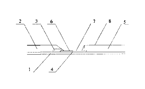
Figure 1, includes a substrate
1 and a sample pad 2, a colloidal gold adsorption pad 3, an antibody carrying
film 4 and a water
adsorption pad 5 sequentially adhered to the substrate. The sample pad 2, the
colloidal gold
adsorption pad 3, the antibody carrying film 4 and the water adsorption pad 5
are partially
overlapped and lapped with each other. Specifically, the antibody carrying
film 4 is located under
the colloidal gold adsorption pad 3 and the water adsorption pad 5, the sample
pad 2 is located
above the colloidal gold adsorption pad 3, and the length of the overlapping
part is lmm.
The antibody carrying film 4 is provided with a detection line 6 on an end
close to the
colloidal gold adsorption pad 3 and a quality control line 7 on an end close
to the water adsorption
pad 5. The detection line 6 is coated with an anti-human chorionic
gonadotropin a.-hCG
monoclonal antibody (anti-oc-hCG monoclonal antibody). The quality control
line 7 is coated with
Date recue/Date Received 2020-09-18
Specification
an anti-mouse IgG polyclonal antibody.
The colloidal gold adsorption pad 3 of the first test paper strip 11 is
adsorbed with colloidal
gold-anti-human chorionic gonadotropin 13-hCG monoclonal antibody conjugate
(colloidal
gold-anti-13-hCG monoclonal antibody conjugate). The colloidal gold adsorption
pad 3 of the
second test paper strip 12, the third test paper strip 13, the fourth test
paper strip 14, the fifth test
paper strip 15 and the sixth test paper strip 16 is adsorbed with colloidal
gold-anti-13-hCG
monoclonal antibody conjugate and free 13 antibody. The weight ratio of gold-
anti-13-hCG
monoclonal antibody conjugate to free 13 antibody of the second test paper
strip 12, the third test
paper strip 13, the fourth test paper strip 14, the fifth test paper strip 15
and the sixth test paper
strip 16 is 1:1, 1:2, 1:40, 1:80, and 1:100, respectively. The free 13
antibody is non-murine antibody.
In the example, the free p antibody is a humanized antibody derived from
hamster.
Moreover, the detection line 6 and the quality control line 7 are arranged in
parallel with a
pitch of 0.3-1.0 cm. In the example, the pitch is 0.5 cm.
Moreover, a protective film 8 is disposed on both of the sample pad 2 and the
water
adsorption pad 3.
Moreover, the antibody carrying film 4 is a nitrocellulose film with a pore
size of 3-10 [an.
The present example provides a method for preparing the above test paper for
semi-quantitative detection of human chorionic gonadotropin, comprises the
following steps.
1. Preparation of an antibody carrying film:
(1) A nitrocellulose film with a pore size of 3-10 [an is cut into a film
having specifications
with a width of 2.0 cm and a length of 30.5cm, as needed.
(2) 2-3mg/m1 anti-oc-hCG monoclonal antibody is prepared with 0.1M phosphate
buffer for
coating of the detection line, in this example, the concentration of anti-oc-
hCG monoclonal
antibody is 2.5 mg/ml. 1-2.0 mg/ml anti-mouse IgG polyclonal antibody is
prepared with 0.85
wt% sodium chloride buffer for coating of the quality control line, in this
example, the
concentration of anti-mouse IgG polyclonal antibody is 1.5 mg/ml.
(3) An antibody coating surface of the nitrocellulose film is labeled. The
antibody solution of
the detection line to be coated and the antibody solution of the quality
control line to be coated
should be uniformly coated on the film in parallel. The detection line and the
quality control line
are disposed at an interval of 0.3-0.7 cm. In this example, the interval is
0.5cm. The nitrocellulose
film is dried at a constant temperature of 2-30 C for use.
(4) A sealing treatment soaking solution is prepared: an actual production
volume of purified
water is added to a mixing tank; and then buffer, sugar, sealing protein and
preservative are
respectively weighed and then directly added to a mixing tank with stirring
until completely
dissolved, purified water is added to reach a required volume, followed by
stirring well for not less
than 10 minutes. The sealing treatment soaking solution comprises 0.1Mol
phosphate buffer,
0.5wt% sugar, 1 wt% sealing protein and 0.05wt% preservative, wherein the
buffer solution is
phosphate buffer, the sugar is sucrose, the preservative is thimerosal, and
the sealing protein is
bovine serum albumin.
11
Date recue/Date Received 2020-09-18
Specification
(5) The film coated with the detection line and the quality control line is
placed in a
processing tank, and the sealing treatment soaking solution prepared in step
(4) is added therein. It
should be ensured that each film is completely immersed in the sealing
treatment soaking solution
for 30 minutes and the film does not move and overlap. The film is taken from
the processing tank,
and then the sealing treatment soaking solution is discarded. After that, the
film is placed on a
gauze with tweezers to dry it a little, so as to obtain an antibody carrying
film.
(6) Pasted on a board and drying
A white paper in the middle of a cutting line on a double-sided tape of a tape
board is
removed. An operator places the film in the blank space in the center of the
tape board, and make
sure that the right side of the tape board is flush with the right side of the
film. In order to avoid
errors in the production process, it is necessary to ensure that the color
development position is
relatively accurate. The film is pasted on the board by aligning with the top
of one end of the
quality control line. After the film is pasted on the tape board, the film
surface is smoothed across
double-sided tape to avoid air bubbles. The temperature in the room is
controlled to 18-28 C, and
the relative humidity is It is also
necessary to ensure that an air in a drying room could
circulate and a wind of a dehumidifier will not directly blow on the film
surface. The drying time
is ?4 hours.
2. Preparation of the colloidal gold adsorption pad of the first test paper
strip
(1) Preparation of colloidal gold complex solution
The actual production volume of purified water is added to the mixing tank;
and then
trehalose, bovine serum albumin, trisodium citrate, polyethylene glycol, and
NaN3 are weighed
with an electronic analytical balance and then directly added to the mixing
tank with stirring until
completely dissolved, purified water is added to reach a required volume,
followed by stirring well
for not less than 30 minutes, thus preparing a colloidal gold complex solution
containing 5wt%
trehalose, 2wt% bovine serum albumin, 0.5wt% trisodium citrate, 0.05wt%
polyethylene glycol,
and 0.05w1% NaN3.
(2) A required amount of colloidal gold is measured with a measuring cylinder,
adjusting PH
to 6.5-7.0 by adding 0.2 mol/L potassium carbonate solution with 0.53% by
volume of the
colloidal gold with stirring on a magnetic stirrer for 15 minutes, thus
obtaining an adjusted
colloidal gold. Anti-I3-hCG monoclonal antibody is diluted with double
distilled water, and labeled
with the colloidal gold at 4 [tg/ml, i.e. the anti-I3-hCG monoclonal antibody
is added into the
colloidal gold, followed by stirring on a magnetic stirrer for 30 minutes, and
then 0.5% by
volume of stabilizer polyethylene glycol is added with stirring for 30
minutes, followed by
centrifuging, collecting a labeled colloidal gold precipitate which is re-
dissolved with the colloidal
gold complex solution at 3% by volume, and stirring on a magnetic stirrer
until the mixture is
homogeneous, thus obtaining 3% by volume of complex solution of colloidal gold-
anti-13-hCG
monoclonal antibody conjugate.
(3) 3% by volume of the complex solution of colloidal gold-anti-13-hCG
monoclonal antibody
conjugate of the above step (2) is taken and re-dissolved with the colloidal
gold complex solution
12
Date recue/Date Received 2020-09-18
Specification
at 50% by volume, followed by mixing on a magnetic stirrer, and then cast on
the prepared
colloidal gold adsorption pad at 50 0/cm2, followed by placing in a drying
room to dry for? 4
hours, and controlling the temperature in the drying room at 18-28 C, and the
relative humidity
40%. It should be ensured that the air is unobstructed and that the air flow
cannot be blown
directly onto the colloidal gold adsorption pad. The dried colloidal gold
adsorption pad is placed
into an aluminum foil bag containing a desiccant, sealed for storage, and
labeled as the colloidal
gold adsorption pad of the first test paper strip.
3. Preparation of the colloidal gold adsorption pad of the second test paper
strip
(1) A required amount of colloidal gold is measured with a measuring cylinder,
adjusting PH
to 6.5-7.0 by adding 0.2 mol/L potassium carbonate solution with 0.53% by
volume of the
colloidal gold with stirring on a magnetic stirrer for 15 minutes, thus
obtaining an adjusted
colloidal gold. Anti-P-hCG monoclonal antibody is diluted with double
distilled water, and labeled
with the colloidal gold at 4 [tg/ml, i.e. the anti-P-hCG monoclonal antibody
is added into the
colloidal gold, followed by stirring on a magnetic stirrer for 30 minutes, and
then 0.5%0 by volume
of stabilizer polyethylene glycol is added with stirring for 30 minutes,
followed by centrifuging,
collecting a labeled colloidal gold precipitate which is re-dissolved with the
colloidal gold
complex solution at 3% by volume, and stirring on a magnetic stirrer until the
mixture is
homogeneous, thus obtaining 3% by volume of complex solution of colloidal gold-
anti-p-hCG
monoclonal antibody conjugate.
(2) 3% by volume of the complex solution of colloidal gold-anti-P-hCG
monoclonal antibody
conjugate of the above step (1) is taken and re-dissolved with the colloidal
gold complex solution
at 40% by volume, then added free 13 antibody according to the weight ratio of
anti-P-hCG
monoclonal antibody to free 13 antibody of 1:1, wherein the free 13 antibody
is a humanized
antibody derived from hamster, followed by mixing on a magnetic stirrer, and
then cast on the
prepared colloidal gold adsorption pad at 50 0/cm2, followed by placing in a
drying room to dry
for? 4 hours, and controlling the temperature in the drying room at 18-28 C,
and the relative
humidity 40%. It
should be ensured that the air is unobstructed and that the air flow cannot be
blown directly onto the colloidal gold adsorption pad. The dried colloidal
gold adsorption pad is
placed into an aluminum foil bag containing a desiccant, sealed for storage,
and labeled as the
colloidal gold adsorption pad of the second test paper strip.
4. Preparation of the colloidal gold adsorption pad of the third test paper
strip
(1) A required amount of colloidal gold is measured with a measuring cylinder,
adjusting PH
to 6.5-7.0 by adding 0.2 mol/L potassium carbonate solution with 0.53% by
volume of the
colloidal gold with stirring on a magnetic stirrer for 15 minutes, thus
obtaining an adjusted
colloidal gold. Anti-P-hCG monoclonal antibody is diluted with double
distilled water, and labeled
with the colloidal gold at 4 [tg/ml, i.e. the anti-P-hCG monoclonal antibody
is added into the
colloidal gold, followed by stirring on a magnetic stirrer for 30 minutes, and
then 0.5%0 by volume
of stabilizer polyethylene glycol is added with stirring for 30 minutes,
followed by centrifuging,
collecting a labeled colloidal gold precipitate which is re-dissolved with the
colloidal gold
13
Date recue/Date Received 2020-09-18
Specification
complex solution at 3% by volume, and stirring on a magnetic stirrer until the
mixture is
homogeneous, thus obtaining 3% by volume of complex solution of colloidal gold-
anti-P-hCG
monoclonal antibody conjugate.
(2) 3% by volume of the complex solution of colloidal gold-anti-P-hCG
monoclonal antibody
conjugate of the above step (1) is taken and re-dissolved with the colloidal
gold complex solution
at 40% by volume, then added free 13 antibody according to the weight ratio of
anti-P-hCG
monoclonal antibody to free 13 antibody of 1:2, wherein the free 13 antibody
is a humanized
antibody derived from hamster, followed by mixing on a magnetic stirrer, and
then cast on the
prepared colloidal gold adsorption pad at 50 [t1/cm2, followed by placing in a
drying room to dry
for? 4 hours, and controlling the temperature in the drying room at 18-28 C,
and the relative
humidity 40%. It should be ensured that the air is unobstructed and that
the air flow cannot be
blown directly onto the colloidal gold adsorption pad. The dried colloidal
gold adsorption pad is
placed into an aluminum foil bag containing a desiccant, sealed for storage,
and labeled as the
colloidal gold adsorption pad of the third test paper strip.
5. Preparation of the colloidal gold adsorption pad of the fourth test paper
strip
(1) A required amount of colloidal gold is measured with a measuring cylinder,
adjusting PH
to 6.5-7.0 by adding 0.2 mol/L potassium carbonate solution with 0.53% by
volume of the
colloidal gold with stirring on a magnetic stirrer for 15 minutes, thus
obtaining an adjusted
colloidal gold. Anti-P-hCG monoclonal antibody is diluted with double
distilled water, and labeled
with the colloidal gold at 5 [tg/ml, i.e. the anti-P-hCG monoclonal antibody
is added into the
colloidal gold, followed by stirring on a magnetic stirrer for 30 minutes, and
then 0.5%0 by volume
of stabilizer polyethylene glycol is added with stirring for 30 minutes,
followed by centrifuging,
collecting a labeled colloidal gold precipitate which is re-dissolved with the
colloidal gold
complex solution at 3% by volume, and stirring on a magnetic stirrer until the
mixture is
homogeneous, thus obtaining 3% by volume of complex solution of colloidal gold-
anti-P-hCG
monoclonal antibody conjugate.
(2) 3% by volume of the complex solution of colloidal gold-anti-P-hCG
monoclonal antibody
conjugate of the above step (1) is taken and re-dissolved with the colloidal
gold complex solution
at 40% by volume, then added free 13 antibody according to the weight ratio of
anti-p-hCG
monoclonal antibody to free 13 antibody of 1:40, wherein the free 13 antibody
is a humanized
antibody derived from hamster, followed by mixing on a magnetic stirrer, and
then cast on the
prepared colloidal gold adsorption pad at 50 [tl/cm2, followed by placing in a
drying room to dry
for? 4 hours, and controlling the temperature in the drying room at 18-28 C,
and the relative
humidity 40%. It should be ensured that the air is unobstructed and that
the air flow cannot be
blown directly onto the colloidal gold adsorption pad. The dried colloidal
gold adsorption pad is
placed into an aluminum foil bag containing a desiccant, sealed for storage,
and labeled as the
colloidal gold adsorption pad of the fourth test paper strip.
6. Preparation of the colloidal gold adsorption pad of the fifth test paper
strip
(1) A required amount of colloidal gold is measured with a measuring cylinder,
adjusting PH
14
Date recue/Date Received 2020-09-18
Specification
to 6.5-7.0 by adding 0.2 mol/L potassium carbonate solution with 0.53% by
volume of the
colloidal gold with stirring on a magnetic stirrer for 15 minutes, thus
obtaining an adjusted
colloidal gold. Anti-P-hCG monoclonal antibody is diluted with double
distilled water, and labeled
with the colloidal gold at 5 Him', i.e. the anti-P-hCG monoclonal antibody is
added into the
colloidal gold, followed by stirring on a magnetic stirrer for 30 minutes, and
then 0.5%0 by volume
of stabilizer polyethylene glycol is added with stirring for 30 minutes,
followed by centrifuging,
collecting a labeled colloidal gold precipitate which is re-dissolved with the
colloidal gold
complex solution at 3% by volume, and stirring on a magnetic stirrer until the
mixture is
homogeneous, thus obtaining 3% by volume of complex solution of colloidal gold-
anti-P-hCG
monoclonal antibody conjugate.
(2) 3% by volume of the complex solution of colloidal gold-anti-p-hCG
monoclonal antibody
conjugate of the above step (1) is taken and re-dissolved with the colloidal
gold complex solution
at 40% by volume, then added free P antibody according to the weight ratio of
anti-P-hCG
monoclonal antibody to free P antibody of 1:80, wherein the free P antibody is
a humanized
antibody derived from hamster, followed by mixing on a magnetic stirrer, and
then cast on the
prepared colloidal gold adsorption pad at 50 [i1/cm2, followed by placing in a
drying room to dry
for? 4 hours, and controlling the temperature in the drying room at 18-28 C,
and the relative
humidity 40%. It should be ensured that the air is unobstructed and that
the air flow cannot be
blown directly onto the colloidal gold adsorption pad. The dried colloidal
gold adsorption pad is
placed into an aluminum foil bag containing a desiccant, sealed for storage,
and labeled as the
colloidal gold adsorption pad of the fifth test paper strip.
7. Preparation of the colloidal gold adsorption pad of the sixth test paper
strip
(1) A required amount of colloidal gold is measured with a measuring cylinder,
adjusting PH
to 6.5-7.0 by adding 0.2 mol/L potassium carbonate solution with 0.53% by
volume of the
colloidal gold with stirring on a magnetic stirrer for 15 minutes, thus
obtaining an adjusted
colloidal gold. Anti-P-hCG monoclonal antibody is diluted with double
distilled water, and labeled
with the colloidal gold at 5 Him', i.e. the anti-P-hCG monoclonal antibody is
added into the
colloidal gold, followed by stirring on a magnetic stirrer for 30 minutes, and
then 0.5%0 by volume
of stabilizer polyethylene glycol is added with stirring for 30 minutes,
followed by centrifuging,
collecting a labeled colloidal gold precipitate which is re-dissolved with the
colloidal gold
complex solution at 3% by volume, and stirring on a magnetic stirrer until the
mixture is
homogeneous, thus obtaining 3% by volume of complex solution of colloidal gold-
anti-P-hCG
monoclonal antibody conjugate.
(2) 3% by volume of the complex solution of colloidal gold-anti-P-hCG
monoclonal antibody
conjugate of the above step (1) is taken and re-dissolved with the colloidal
gold complex solution
at 40% by volume, then added free P antibody according to the weight ratio of
anti-P-hCG
monoclonal antibody to free P antibody of 1:100, wherein the free P antibody
is a humanized
antibody derived from hamster, followed by mixing on a magnetic stirrer, and
then cast on the
prepared colloidal gold adsorption pad at 50 [il/cm2, followed by placing in a
drying room to dry
Date recue/Date Received 2020-09-18
Specification
for? 4 hours, and controlling the temperature in the drying room at 18-28 C,
and the relative
humidity 40%. It should be ensured that the air is unobstructed and that
the air flow cannot be
blown directly onto the colloidal gold adsorption pad. The dried colloidal
gold adsorption pad is
placed into an aluminum foil bag containing a desiccant, sealed for storage,
and labeled as the
colloidal gold adsorption pad of the sixth test paper strip.
Notes: The colloidal gold solution is cast, because the cast colloidal gold
can be cut freely,
thus it is easy to adjust the color depth of the product.
8. Assembly and cutting:
(1) A transparent substrate semi-finished product that has been pasted with
the antibody
carrying film is taken, and the colloidal gold adsorption pad of the first
test paper strip is cut into
0.5cm x 30cm strip with a width of 0.5cm which is pasted on the transparent
substrate close to the
detection line and kept to lap about lmm with the antibody carrying film. The
water adsorption
pad of the first test paper strip is compounded on the transparent substrate
film close to the quality
control line and lapped about lmm with the antibody carrying film. The sample
pad of the first
test paper strip is compounded on the end of the colloidal gold adsorption pad
of the first test
paper strip away from the antibody carrying film, and lapped about lmm
therewith, labeling for
use.
(2) The assembled substrate is cut into a strip test paper labeled as a first
test paper strip.
(3) A transparent substrate semi-finished product that has been pasted with
the antibody
carrying film is taken, and the colloidal gold adsorption pad of the second
test paper strip is cut
into 0.5cm x 30cm strip with a width of 0.5cm which is pasted on the
transparent substrate close
to detection line and kept to lap about lmm with the antibody carrying film.
The water adsorption
pad of the second test paper strip is compounded on the transparent substrate
film close to the
quality control line and lapped about lmm with the antibody carrying film. The
sample pad of the
second test paper strip is compounded on the end of the colloidal gold
adsorption pad of the
second test paper strip away from the antibody carrying film, and lapped about
lmm therewith,
labeling for use.
(4) The assembled substrate is cut into a strip test paper labeled as a second
test paper strip.
(5) A transparent substrate semi-finished product that has been pasted with
the antibody
carrying film is taken, and the colloidal gold adsorption pad of the third
test paper strip is cut into
0.5cm x 30cm strip with a width of 0.5cm which is pasted on the transparent
substrate close to
detection line and kept to lap about lmm with the antibody carrying film. The
water adsorption
pad of the third test paper strip is compounded on the transparent substrate
film close to the
quality control line and lapped about lmm with the antibody carrying film. The
sample pad of the
third test paper strip is compounded on the end of the colloidal gold
adsorption pad of the third
test paper strip away from the antibody carrying film, and lapped about lmm
therewith, labeling
for use.
(6) The assembled substrate is cut into a strip test paper labeled as a third
test paper strip.
(7) A transparent substrate semi-finished product that has been pasted with
the antibody
16
Date recue/Date Received 2020-09-18
Specification
carrying film is taken, and the colloidal gold adsorption pad of the fourth
test paper strip is cut into
0.5cm x 30cm strip with a width of 0.5cm which is pasted on the transparent
substrate close to
detection line and kept to lap about lmm with the antibody carrying film. The
water adsorption
pad of the fourth test paper strip is compounded on the transparent substrate
film close to the
quality control line and lapped about lmm with the antibody carrying film. The
sample pad of the
fourth test paper strip is compounded on the end of the colloidal gold
adsorption pad of the fourth
test paper strip away from the antibody carrying film, and lapped about lmm
therewith, labeling
for use.
(8) The assembled substrate is cut into a strip test paper labeled as a fourth
test paper strip.
(9) A transparent substrate semi-finished product that has been pasted with
the antibody
carrying film is taken, and the colloidal gold adsorption pad of the fifth
test paper strip is cut into
0.5cm x 30cm strip with a width of 0.5cm which is pasted on the transparent
substrate close to
detection line and kept to lap about lmm with the antibody carrying film. The
water adsorption
pad of the fifth test paper strip is compounded on the the transparent
substrate film close to the
quality control line and lapped about lmm with the antibody carrying film. The
sample pad of the
fifth test paper strip is compounded on the end of the colloidal gold
adsorption pad of the fifth test
paper strip away from the antibody carrying film, and lapped about lmm
therewith, labeling for
use.
(10) The assembled substrate is cut into a strip test paper labeled as a fifth
test paper strip.
(11) A transparent substrate semi-finished product that has been pasted with
the antibody
carrying film is taken, and the colloidal gold adsorption pad of the sixth
test paper strip is cut into
0.5cm x 30cm strip with a width of 0.5cm which is pasted on the transparent
substrate close to
detection line and kept to lap about lmm with the antibody carrying film. The
water adsorption
pad of the sixth test paper strip is compounded on the transparent substrate
film close to the
quality control line and lapped about lmm with the antibody carrying film. The
sample pad of the
sixth test paper strip is compounded on the end of the colloidal gold
adsorption pad of the sixth
test paper strip away from the antibody carrying film, and lapped about lmm
therewith, labeling
for use.
(12) The assembled substrate is cut into a strip test paper labeled as a sixth
test paper strip.
Example 2
The present example provides a test strip for semi-quantitative detection of
human chorionic
gonadotropin, which is different from the test strip of example 1 in that free
13 antibody is an
equine antibody, and the interval between the detection line 6 and the quality
control line 7 is
0.3cm.
The present example also provides a method for preparing the above test strip
for
semi-quantitative detection of human chorionic gonadotropin, which is
different from the
preparation method of example 1 in that the free 13 antibody used is an equine
antibody; during the
preparation process of the antibody carrying film, the concentration of anti-a-
HCG monoclonal
17
Date recue/Date Received 2020-09-18
Specification
antibody is 2 mg/ml, the concentration of anti-mouse IgG polyclonal antibody
is 1 mg/ml, and the
interval between the detection line and the quality control line is 0.3cm; and
the sealing treatment
solution used includes 0.08Mol buffer solution, 0.3wt% sugar, 0.8 wt% sealing
protein, and 0.03
wt% preservative, wherein the buffer solution is phosphate buffer, the sugar
is trehalose, the
preservative is NaN3, and the sealing protein is casein.
Example 3
The present example provides a test strip for semi-quantitative detection of
human chorionic
gonadotropin, which is different from the test strip of example 1 in that free
13 antibody is a rabbit
antibody, and the interval between the detection line 6 and the quality
control line 7 is 1.0cm.
The present example also provides a method for preparing the above test strip
for
semi-quantitative detection of human chorionic gonadotropin, which is
different from the
preparation method of example 1 in that the free 13 antibody used is a rabbit
antibody; during the
preparation process of the antibody carrying film, the concentration of anti-a-
HCG monoclonal
antibody is 3 mg/ml, the concentration of anti-mouse IgG polyclonal antibody
is 2 mg/ml, and the
interval between the detection line and the quality control line is 0.7cm; and
the sealing treatment
solution used includes 0.12Mol buffer solution, 0.7wt% sugar, 1.2 wt% sealing
protein, and 0.07
wt% preservative, wherein the buffer solution is phosphate buffer, the sugar
is sucrose, the
preservative is thimerosal, and the sealing protein is bovine serum protein.
Example 4
As shown in Figure 3, the present example provides a reagent cup for semi-
quantitative
detection of human chorionic gonadotropin, which comprises the test paper for
semi-quantitative
detection of human chorionic gonadotropin prepared in example 1, and further
comprises a cup
bod 9, a cup cover 10 and a cartridge 17. The cartridge 17 is placed
vertically inside the cup body
9, and the cup cover 10 is detachably closed on the cup body 9. As shown in
Figure 2, the
cartridge 17 is provided with six or more parallel slots 18, and the slots 18
have an opening near
the bottom of the cup body 9. The first test paper strip 11, the second test
paper strip 12, the third
test paper strip 13, the fourth test paper strip 14, the fifth test paper
strip 15 and the sixth test paper
strip 16 are successively inserted into the six slot 18 via the opening. The
sample pad 2 of the test
paper strip extends from the opening to the outside of the cartridge 17.
Cartridge 17 is made of
plastic, of course, as an alternative embodiment, cartridge17 can also be made
of other transparent
materials.
Specifically, the structure of each of the first test paper strip 11, the
second test paper strip 12,
the third test paper strip 13, the fourth test paper strip 14, the fifth test
paper strip 15 and the sixth
test paper strip 16 is shown in Figure 1, which respectively includes a
substrate 1 and a sample
pad 2, a colloidal gold adsorption pad 3, an antibody carrying film 4 and a
water adsorption pad 5
sequentially adhered to the substrate. The sample pad 2, the colloidal gold
adsorption pad 3, the
antibody carrying film 4 and the water adsorption pad 5 are partially
overlapped and lapped with
18
Date recue/Date Received 2020-09-18
Specification
each other. Specifically, the antibody carrying film 4 is located under the
colloidal gold adsorption
pad 3 and the water adsorption pad 5, the sample pad 2 is located above the
colloidal gold
adsorption pad 3, and the length of the overlapping part is lmm.
The antibody carrying film 4 is provided with a detection line 6 on an end
close to the
colloidal gold adsorption pad 3 and a quality control line 7 on an end close
to the water adsorption
pad 5. The detection line 6 is coated with an anti-u-hCG monoclonal antibody.
The quality control
line 7 is coated with an anti-mouse IgG polyclonal antibody.
The colloidal gold adsorption pad 3 of the first test paper strip 11 is
adsorbed with colloidal
gold-anti--hCG monoclonal antibody conjugate. The colloidal gold adsorption
pad 3 of the
second test paper strip 12, the third test paper strip 13, the fourth test
paper strip 14, the fifth test
paper strip 15 and the sixth test paper strip 16 is adsorbed with colloidal
gold-anti--hCG
monoclonal antibody conjugate and free 13 antibody. The free P antibody is non-
murine antibody.
In the example, the free P antibody is a humanized antibody derived from
hamster.
Moreover, the detection line 6 and the quality control line 7 are arranged in
parallel with a
pitch of 0.3-1.0 cm. In the example, the pitch is 0.5 cm. Moreover, a
protective film 8 is disposed
on the sample pad 2 and the water adsorption pad 3 respectively. Moreover, the
antibody carrying
film 4 is a nitrocellulose film with a pore size of 3-10 um
When in use, the cup cover 10 is opened, the urine sample is collected in the
cup body 9, the
cup cover 10 is fitted on the cup body 9, the cup body 9 is placed upright,
and the sample pads 2
of the six test paper strips are immersed in the sample. The sample is inhaled
into the test paper
strip from the sample pad 2 due to the capillarity, and then passes through
the colloidal gold
adsorption pad 3 and the antibody carrying film 4 successively to reach to the
water adsorption
pad 5, the hCG in the sample binds with the colloidal gold-anti-3-hCG
monoclonal antibody
conjugate or free P antibody adsorbed in the colloidal gold adsorption pad 3.
With the flow of
liquid, the hCG, bound to the colloidal gold-anti-3-hCG monoclonal antibody
conjugate, reaches
to the antibody carrying film 4, and binds to anti-u-hCG monoclonal antibody
coated in the
detection line and anti-mouse IgG polyclonal antibody coated in the quality
control line,
respectively, and develops color at the detection line and the quality control
line because it has
colloidal gold labeling. While the hCG, bound to free 13 antibody, does not
develop color at the
detection line because it does not have colloidal gold labeling, and does not
bind to anti-mouse
IgG polyclonal antibody coated in the quality control line because the free P
antibody is not
murine antibody. Even if the concentration of hCG in the sample is high and a
large amount of
hCG binds to free P antibody, it will not cause most of antibodies in the
quality control line to bind
to hCG-free P antibody conjugate, so that the quality control line will not
develop color. As a
result, the samples with higher hCG concentration can be measured, and the
detectable quantity
can reach to 10000mIU/ml, making the range of concentration detection wider.
Since the weight
ratios of colloidal gold-anti-3-hCG monoclonal antibody conjugate to free P
antibody in the
colloidal gold adsorption pad of six test paper strips are different, the
detection line and quality
control line of the six test paper strips will show different color
development According to the
19
Date recue/Date Received 2020-09-18
Specification
color development, the concentration of hCG in the sample can be detected semi-
quantitatively.
Example 5
As shown in Figures 4-6, the present example provides a reagent cup for semi-
quantitative
detection of human chorionic gonadotropin, which comprises the test paper for
semi-quantitative
detection of human chorionic gonadotropin prepared in example 1, and further
comprises a cup
bod 9 and a cartridge 17.
The cup body 9 is provided with a detection cavity 19, a liquid storage cavity
20 and a liquid
guide cavity 21. The liquid guide cavity 21 is provided with a first through
hole 211
intercommunicating with the liquid storage cavity at an upper part thereof and
a second through
hole 212 intercommunicating with the detection cavity 19 at a lower part
thereof. A piston 22
sliding along the liquid guide cavity is disposed inside the liquid guide
cavity 21, has an initial
state for intercommunicating the liquid guide cavity 21 with the liquid
storage cavity 20 and a
detection state for intercommunicating the liquid chamber guide cavity 21 with
the detection
chamber 19. A third through hole 213 is disposed on a cup wall of the cup body
9 away from the
detection chamber 19 and intercommunicated with the liquid guide cavity 21. A
liquid storage
tank 221 is disposed on the piston 22. As shown in Figure 5, when the piston
22 is in the initial
state, the liquid storage tank 221 of the piston is aligned with the first
through hole 211, so that the
liquid guide cavity 21 is intercommunicated with the liquid storage cavity 20;
when the piston 22
is in the detection state, the liquid storage tank 221 of the piston is
aligned with the second
through hole 212, so that the liquid guide cavity 21 is intercommunicated with
the detection cavity
19.
Moreover, a liquid guide tank 26 is provided inside the bottom of the cup body
9, the liquid
guide tank 26 intercommunicates with the detection cavity 19, and the liquid
guide tank 26 is
located below the second through hole 212, so that the liquid guide tank 26
intercommunicates
with the second through hole 212.
The structure and number of the cartridge 17 and the test paper strip are the
same as those in
Example 4, except that the cartridge 17 inserted with the test paper strip is
vertically placed inside
the detection cavity 19.
By setting the cartridge 17, the reagent cup can fix the test paper strip in
the detection cavity
19 of the reagent cup. When in use, the cup body can be kept upright, so that
the test paper strip
can also be kept upright, which is convenient for sample absorption. The
reagent cup is configured
to comprise six test paper strips, and the hCG concentration in urine can be
semi-quantitatively
detected according to the color development of different test paper strips.
The detectable
concentration range is wider, and there is no need to identify by comparing
the degree of color
development, thus the test result is more accurate. The detection cavity 19
comprising the
cartridge and the test paper, and a liquid storage cavity 20 for collecting
urine, are configured to
make the test paper in the detection cavity 19 separate from the urine sample
under the initial state.
The piston 22 and the liquid storage tank 221 disposed on the piston are
configured to make the
Date recue/Date Received 2020-09-18
Specification
urine enter into the liquid storage tank 221 from the liquid storage cavity 20
via the first through
hole 211. When the piston 22 slides in the liquid guide cavity 21 to the
detection state, the urine
enters into the liquid guide tank 26 from the liquid storage tank 221 via the
second through hole
212, thus the test paper strip in the cartridge 17 of the detection cavity 19
contacts with the urine
sample, so as to complete the detection. Through the above structure, the time
when starts the test
can be more conveniently controlled, and the test can be started after the
sample collection is
completed, so as to prevent the problem that the test result is not accurate
enough when collecting
samples while simultaneously performing the test, making the result more
accurate and easy to
operate. The liquid guide cavity 21 intercommunicates with the third through
hole 213, so that the
sliding of the piston 22 can be easily controlled. The liquid storage tank 221
is configured to make
the amount of urine samples introduced into the detection cavity 19 constant,
thereby preventing
excessive urine samples from entering the detection cavity 19 and affecting
the test results. The
accuracy of the test results is ensured. When using the reagent cup, the user
only needs to collect
the urine sample directly in the cup body, and then pushes the piston 22, the
test and result reading
can be performed, which is convenient and hygienic.
The reagent cup further includes a cup cover 10 detachably closed on the cup
body 9. A boost
member 23 for boosting the piston 22 is provided on the cup cover 10, and
detachably fitted on the
cup cover 10. The boost member 23 is configured to more easily push the piston
22 from the
initial state to the detection state. When the reagent cup is not used, the
boost member 23 can be
fitted on the cup cover 10 to facilitate storage and prevent its loss.
When in use, the cup cover 10 is opened, the urine sample is collected in the
liquid storage
cavity 20 of the cup body 9, the cup cover 10 is fitted on the cup body 9, the
cup body 9 is placed
upright. At this time, as shown in Figure 5, when the reagent cup is in the
initial state, the urine
sample in liquid storage cavity 20 enters into the liquid storage tank 221 of
the piston 22 through
the first through hole 211. The boost member 23 fitted on the cup cover 10 is
taken down, and
inserted into the third through hole 213, and the piston 22 is pushed to the
detection state through
the boost member 23. As shown in Figure 6, the urine sample in the liquid
storage tank 221 flows
into the liquid guide tank 26 at the bottom through the second through hole
212, and then flows
into the detection cavity 19, the sample pads 2 of six test paper strips in
detection cavity 19 are
immersed in the sample. The sample is inhaled into the test paper strip from
the sample pad 2 due
to the capillarity, and then passes through the colloidal gold adsorption pad
3 and the antibody
carrying film 4 successively to reach to the water adsorption pad 5, the hCG
in the sample binds
with the colloidal gold-anti-3-hCG monoclonal antibody conjugate or free P
antibody adsorbed in
the colloidal gold adsorption pad 3. With the flow of liquid, the hCG, bound
to the colloidal
gold-anti--hCG monoclonal antibody conjugate, reaches to the antibody carrying
film 4, and
binds to anti-a-hCG monoclonal antibody coated in the detection line and anti-
mouse IgG
polyclonal antibody coated in the quality control line, respectively, and
develops color at the
detection line 6 and the quality control line 7 because it has colloidal gold
labeling. While the hCG,
bound to free P antibody, does not develop color at the detection line 6
because it does not have
21
Date recue/Date Received 2020-09-18
Specification
colloidal gold labeling, and does not bind to anti-mouse IgG polyclonal
antibody coated in the
quality control line 7 because the free 13 antibody is not murine antibody.
Since the weight ratios of
colloidal gold-anti-13-hCG monoclonal antibody conjugate to free 13 antibody
in the colloidal gold
adsorption pad of six test paper strips are different, the detection line 6 of
the six test paper strips
will show different color development. According to the color development, the
concentration of
hCG in the sample can be detected semi-quantitatively. Compared with two test
paper strips, the
six test paper strips of the present application have a wider detectable
concentration range, and
there is no need to identify by comparing the degree of color development,
thus the test result is
more accurate. When using the reagent cup, user only needs to collect the
urine sample directly in
the cup body, and then pushes the piston 22, the test and result reading can
be performed, which is
convenient and hygienic.
Example 6
As shown in Figure 7, the present example provides a reagent cup for semi-
quantitative
detection of human chorionic gonadotropin, which comprises a cup bod 9, a
cartridge 17 and the
test paper of example 1, and further comprises a cup cover 10, which is
detachably fitted on the
cup body 9. The structure and number of the cartridge 17 and the test paper
strip are the same as
those in Example 4, except that the cartridge 17 is fixed in parallel inside
the cup cover 10.
By setting the cartridge 17, the reagent cup can fix the test paper strip in
the detection cavity
19 of the reagent cup. When in use, the cup body 9 is in an on its side state,
which is convenient
for absorbing sample with the test paper strip. The reagent cup is configured
to comprise six test
paper strips, and the hCG concentration in urine can be semi-quantitatively
detected according to
the color development of different test paper strips. The detectable
concentration range is wider,
and there is no need to identify by comparing the degree of color development,
thus the test result
is more accurate. By fixing the cartridge 17 on the cup cover 10, the test
paper strip in the
cartridge 17 can be separated from the urine sample in the cup body10 when the
test is not started.
At the beginning of the test, the test can be started by tightening the cup
cover 10, and placing the
reagent cup on its side, so as to prevent the problem that the test result is
not accurate enough
when collecting samples while simultaneously performing the test, make the
result more accurate
and easy to operate.
A support body 24 is disposed on an outer edge of the cup cover 20 for fixing
the reagent cup
placed on its side, and a scale line 25 is arranged on the cup body 10. The
support body 24 is
configured to dispose on the edge of the cup cover 10, the reagent cup is
fixed when it is placed on
its side, preventing the reagent cup from rolling. When collecting urine, it
is necessary to ensure
that the urine exceeds the scale line 25, ensuring that the urine samples are
sufficient when the
reagent cup is placed on its side, so that the test paper strip can carry out
adsorption detection
successfully.
When in use, the cup coven l 0 is opened, the urine sample is collected in the
cup body 9 to
make the volume of urine sample exceed the scale 1ine25 on the cup body 9. The
cup cover 10 is
22
Date recue/Date Received 2020-09-18
Specification
fitted on the cup body 9 and tightened, the reagent cup is placed on its side
and fixed with the
support body 24. The sample pads 2 of six test paper strips are immersed in
the sample. The
sample is inhaled into the test paper strip from the sample pad 2 due to the
capillarity, and then
passes through the colloidal gold adsorption pad 3 and the antibody carrying
film 4 successively to
reach to the water adsorption pad 5, the hCG in the sample binds with the
colloidal
gold-anti-13-hCG monoclonal antibody conjugate or free p antibody adsorbed in
the colloidal gold
adsorption pad 3. With the flow of liquid, the hCG, bound to the colloidal
gold-anti-13-hCG
monoclonal antibody conjugate, reaches to the antibody carrying film 4, and
binds to anti-a-hCG
monoclonal antibody coated in the detection line and anti-mouse IgG polyclonal
antibody coated
in the quality control line, respectively, and develops color at the detection
line 6 and the quality
control line 7 because it has colloidal gold labeling. While the hCG, bound to
free p antibody, does
not develop color at the detection line 6 because it does not have colloidal
gold labeling, and does
not bind to anti-mouse IgG polyclonal antibody coated in the quality control
line 7 because the
free 13 antibody is not murine antibody. Since the weight ratios of colloidal
gold-anti-13-hCG
monoclonal antibody conjugate to free 13 antibody in the colloidal gold
adsorption pad of six test
paper strips are different, the detection line 6 of the six test paper strips
will show different color
development. According to the color development, the concentration of hCG in
the sample can be
detected semi-quantitatively. Compared with two test paper strips, the six
test paper strips of the
present application have a wider detectable concentration range, and there is
no need to identify by
comparing the degree of color development, thus the test result is more
accurate. When using the
reagent cup, user only needs to collect the urine sample directly in the cup
body, and then pushes
the piston 22, the test and result reading can be performed, which is
convenient and hygienic.
Example 7
The present example provides a method for using the reagent cup for semi-
quantitative
detection of human chorionic gonadotropin in Example 4, which includes steps
of: equilibrating
the sample and the reagent cup at room temperature; then opening the cup cover
10 and collecting
the urine sample in the cup body 9, fitting the cup cover 10 on the cup body
9, placing the cup
body 9 upright down, immersing the sample pad 2 of the six test paper strips
in the sample, and
observing whether the detection line and quality control line of the six test
paper strips develop
color within 5 minutes. The test results include the following situations:
Table 1: Interpretation of displayed results of test paper strips
Displayed Results hCG Concentration Range
The quality control lines of the six test paper strips do not Invalid
develop pruno sus
The quality control lines of the six test paper strips develop Negative (-):
pruno sus; Less than 5mIU/m1
The detection lines of the six test paper strips do not develop
23
Date recue/Date Received 2020-09-18
Specification
prunosus
The quality control lines of the six test paper strips develop Positive (+):
prunosus; 5-25m1U/m1
The detection line of the first test paper strip develops
prunosus;
The detection lines of the rest test paper strips do not develop
prunosus
The quality control lines of the six test paper strips develop Positive (++):
prunosus; 25-100m1U/m1
The detection lines of the first and second test paper strips
develop prunosus;
The detection lines of the rest test paper strips do not develop
prunosus
The quality control lines of the six test paper strips develop Positive (+++):
prunosus; 100-500mIU/m1
The detection lines of the first, second and third test paper strips
develop prunosus;
The detection lines of the rest test paper strips do not develop
prunosus
The quality control lines of the six test paper strips develop Positive
(++++):
prunosus; 500-2500m1U/m1
The detection lines of the first, second, third and fourth test
paper strips develop prunosus;
The detection lines of the rest test paper strips do not develop
prunosus
The quality control lines of the six test paper strips develop Positive
(+++++):
prunosus; 2500-10000mIU/m1
The detection lines of the first, second, third, fourth and fifth
test paper strips develop prunosus;
The detection line of the sixth test paper strip does not develop
prunosus
The quality control lines of the six test paper strips develop Positive
(++++++):
prunosus; Greater than or equal to
10000
The detection lines of the six test paper strips develop prunosus mIU/m1
The test begins on the day when menstruation should come but does not come. If
the test
begins in advance, it should be performed again on the expected date of
menstruation. If
menstruation is delayed, the test should be performed again after three days.
If the menstrual
period is irregular, the longest menstrual period in the most recent months
should be calculated
before the test is performed. If you don't know when menstruation is expected
to begin, you
should perform the test at least nineteen days after the last time you had sex
without protective
measures.
Example 8
24
Date recue/Date Received 2020-09-18
Specification
The present example provides a use of the test paper for semi-quantitative
detection of human
chorionic gonadotropin in Example 4 for detecting human pregnancy cycle,
ectopic pregnancy,
miscarriage and trophoblastic disease.
The urine sample is tested using the method of using the reagent cup in
Example 7, and the
concentration range of human chorionic gonadotropin in the urine sample is
semi-quantitatively
detected according to the Interpretation in Table 1.
Human pregnancy cycle is identified according to reference values of hCG of
pregnant
women described in "Chinese Clinical Test Operating Procedures". The reference
value of hCG is
5-50 IU/ml for 0.2-1 week of pregnancy, the reference value of hCG is 50-500
IU/ml for 1-2
weeks of pregnancy, the reference value of hCG is 100-5000 IU/ml for 2-3 weeks
of pregnancy,
the reference value of hCG is 500-10000 IU/ml for 3-4 weeks of pregnancy, the
reference value of
hCG is 1000-50,000 IU/ml for 4-5 weeks of pregnancy, the reference value of
hCG is
10,000-100,000 IU/ml for 5-6 weeks of pregnancy, the reference value of hCG is
15,000-200,000
IU/m for 6-8 weeks of pregnancy, the reference value of hCG is 10,000-100,000
IU/ml for 8-12
weeks of pregnancy.
In the aspect of clinical diagnosis, the concentration range of human
chorionic gonadotropin
in the urine samples is detected semi-quantitatively to diagnose ectopic
pregnancy, miscarriage
and trophoblastic disease, and observe the course of disease. Specifically, in
the diagnosis of
ectopic pregnancy, the hCG of ectopic pregnancy is often 312-625mIU/ml, and
hCG result can
still be positive after three days of uterine bleeding, so hCG test can be
used as a method to
distinguish ectopic pregnancy from other acute abdomen. In the diagnosis of
miscarriage, if there
is still placental tissue remaining in the uterus, hCG result can still be
positive, while when
complete abortion or stillbirth occurs, hCG result changes from positive to
negative. Therefore,
hCG test can be used as a reference basis for fetal protection or uterine
aspiration treatment. In the
diagnosis of threatened abortion, if the urine hCG has been kept at a high
level, there will be no
inevitable abortion, but if hCG is below 2500mIU/m1 and gradually decreases,
there is the
possibility of abortion or stillbirth, and when hCG falls to 600mIU/ml, there
will be inevitable
abortion. Therefore, in the treatment of protecting fetus, if hCG continues to
decline, it means that
the protecting fetus is ineffective, while if hCG continues to rise, it means
that the protecting fetus
is successful. In addition, urinary hCG levels in patients with hydatidiform
mole, malignant
hydatidiform mole and chorionic epithelioma are significantly increased,
ranges from 100,000 to
millions of mIU/ml. The urine hCG of the patients with trophoblastic tumor
should be less than
50mIU/m1 at 3 weeks and negative at 8-12 weeks after operation. If the hCG
does not decrease or
change to negative, it may indicate that there may be residual lesions.
Comparative Example 1
This comparative example provides a test paper for semi-quantitative
quantitative detection
of human chorionic gonadotropin similar to that in Example 1, except that the
free 13 antibody used
is a murine antibody, and a national level human chorionic gonadotropin
standard is prepared into
Date recue/Date Received 2020-09-18
Specification
a standard solution with a concentration of 10000mIU/ml. When test is
performed using the test
paper, the result is that of the six test paper strips, the quality control
lines of the fourth, fifth, and
sixth test paper strips do not develop color, thus the result is invalid.
Comparative Example 2
The national level human chorionic gonadotropin standard is prepared into a
standard
solution with a concentration of 5mIU/ml. When test is performed using hCG
cycle test paper
disclosed in Chinese Patent CN108761099A and the hCG cycle test kit including
the test paper,
the result is that of the quality control lines of the two test paper strips
develop color, and the
detection lines of the two test paper strips do not develop color.
Experimental example 1
1. The required reagents are as follows:
0 mIU/m1 sample solution: protein-containing phosphate buffer (PBS) is 0
mIU/m1 of the
HCG sample solution.
5m IU/ml, 25 mIU/ml, 100mIU/ml, 500mIU/ml, 2500mIU/ml, and 10000mIU/m1 human
chorionic gonadotropin (hCG) sample solution prepared by human chorionic
gonadotropin (hCG)
standard and the protein-containing phosphate buffer (0.01M PBS) to reach a
corresponding
concentration respectively.
500m IU/ml human luteinizing hormone (hLH): After hLH standard is re-dissolved
with the
protein-containing phosphate buffer (PBS), Om IU/ml HCG sample solution is
used to prepare
hLH A solution with a concentration of 500m IU/ml; 5m IU/ml human chorionic
gonadotropin
sample solution is used to prepare hLH B solution with a concentration of 500m
IU/ml.
1000m IU/ml human follicle stimulating hormone (hFSH): After the hFSH standard
is
re-dissolved with the protein-containing phosphate buffer (PBS), Om IU/ml HCG
sample solution
is used to prepare hFSH A solution with a concentration of 1000m IU/ml; 5m
IU/ml human
chorionic gonadotropin sample solution is used to prepared hFSH B solution
with a concentration
of 1000m IU/ml.
1000 IU/ml human thyroid stimulating hormone (hTSH): After the hTSH standard
is
re-dissolved with the protein-containing phosphate buffer (PBS), Om IU/ml HCG
sample solution
is used to prepare hTSH A solution with a concentration of 1000 IU/ml; 5m
IU/ml human
chorionic gonadotropin sample solution is used to prepare hTSH B solution with
a concentration
of 1000 IU/ml; and 5m IU/ml human chorionic gonadotropin sample solution is
used to prepare
hTSH B solution with a concentration of 1000 IU/ml.
2. Testing Physical properties:
Testing appearance properties: The test paper strip is taken out and observed
with eyesight.
The paper strip should be neat and complete, have no burrs, no damage, and no
pollution, and be
firmly attached with the material.
Film strip width: Two of each six kinds of sensitivity test paper strips are
taken and measured
26
Date recue/Date Received 2020-09-18
Specification
the width of the film on the paper strips with vernier caliper, and the
average value of the two
measurement results is calculated.
Chromatographic status: regular, the colloid gold not layered with water, and
a background is
basically clear within 3-5 minutes.
Liquid migration speed: two test paper strips, of each of the six
sensitivities test paper strips
are taken, and the operation is carried out according to the method used in
Example 7. The time
used is recorded as (t) from the time when the test paper strip is immersed in
the sample solution
with a stopwatch (with an accuracy of 0.01s) until the liquid reaches the
boundary between the
antibody carrying film and the water adsorption pad. The length is recorded as
(L) from the
sample loading end of the sample pad to the boundary between the antibody
carrying film and the
water adsorption pad measured with Vernier caliper (precision 0.02mm). The
migration velocity,
i.e. L/t is calculated. The average values of repeated measurement of two test
paper strips, of each
of six sensitivities test paper strips, are taken respectively. The results
are consistent with the
liquid migration speed of no less than 1 Omm/min. The chromatographic state is
regular, the
colloidal gold is not delaminated with water, and the background is basically
clear within 5
minutes.
3. The minimum detectable quantity of the test:
A national level human chorionic gonadotropin standard is prepared into
standard solutions
with concentrations of 2.5 mIU/ml, 5 mIU/ml, and 10 mIU/ml, respectively. The
standard solution
with each concentration is tested three times, using the reagent cup of
Example 4 of the present
application, and the results are observed within 5 minutes, showing that the
minimum detectable
quantity of the first test paper strip is 5 mIU/ml. A national level human
chorionic gonadotropin
standard is prepared into standard solutions with concentrations of 12.5
mIU/ml, 25 mIU/ml, and
50 mIU/ml. The standard solution with each concentration is tested three
times, and the results are
observed within 5 minutes, showing that the minimum detectable quantity of the
second test paper
strip is 25 mIU/ml. A national level human chorionic gonadotropin standard is
prepared into
standard solutions with concentrations of 50 mIU/ml, 100 mIU/ml, and 200
mIU/ml. The standard
solution with each concentration is tested three times, and the results are
observed within 5
minutes, showing that the minimum detectable quantity of the third test paper
strip is 100 mIU/ml.
A national level human chorionic gonadotropin standard is prepared into
standard solutions with
concentrations of 400mIU/ml, 500mIU/ml, and 600mIU/ml. The standard solution
with each
concentration is tested three times, and the results are observed within 5
minutes, showing that the
minimum detectable quantity of the fourth test paper strip is 500 mIU/ml. A
national level human
chorionic gonadotropin standard is prepared into standard solutions with
concentrations of
2400mIU/ml, 2500mIU/ml, and 2600mIU/ml. The standard solution with each
concentration is
tested three times, and the results are observed within 5 minutes, showing
that the minimum
detectable quantity of the fifth test paper strip is 2500 mIU/ml. A national
level human chorionic
gonadotropin standard is prepared into standard solutions with concentrations
of 9900mIU/ml,
10000mIU/ml, and 10100mIU/ml. The standard solution with each concentration is
tested three
27
Date recue/Date Received 2020-09-18
Specification
times, and the results are observed within 5 minutes, showing that the minimum
detectable
quantity of the sixth test paper strip is 10000 mIU/ml.
5. Accuracy and repeatability of test:
national level human chorionic gonadotropin standard is prepared into standard
solutions
with concentrations of 5mIU/m, 25mIU/ml, 100mIU/ml, 500mIU/ml, 2500mIU/ml, and
10000mIU/ml. The standard solution with each concentration is tested twenty
times to obtain the
test result. The forward difference between the test results and the labeled
value of the
corresponding standard solution does not exceed one order of magnitude, There
is no backward
difference. The positive standard solution does not show a negative result.
5. Specificity:
Negative specificity: 500m IU/m1 hLH A solution, 1000m IU/m1 hFSH A solution,
1000
IU/ml hTSH A solution are tested for 3 times respectively. Results of the
three times of each
solution show that the detection lines are negative.
Positive specificity: 500m IU/ml hLH B solution, 1000m IU/ml hFSH B solution,
and 1000
IU/ml hTSH B solution are tested for 3 times respectively. Results of the
three times of each
solution show that the detection line is positive.
6. Stability of test:
Physical properties, accuracy, repeatability, and specificity of the test
paper strip and the
reagent cup are tested after one month after the expiration date, the results
meet the requirements.
It is apparent that the above embodiments are merely examples for clarity of
illustration, and
are not intended to limit the embodiments. Other variations or modifications
in various forms may
be made by those skilled in the art in view of the above description. There is
no need and no way
to present all of embodiments herein. The obvious variations or modifications
derived therefrom
are still within the scope of protection of the present application.
28
Date recue/Date Received 2020-09-18