Note: Descriptions are shown in the official language in which they were submitted.
CA 03089826 2020-07-28
WO 2019/152848
PCT/US2019/016362
LIPID-LIKE NANOCOMPLEXES AND USES THEREOF
CROSS-REFERENCE TO RELATED APPLICATION
This application claims the benefit of priority to U.S. Provisional Patent
Application serial no. 62/625,153, filed February 1, 2018, the contents of
which are
incorporated herein by reference.
GOVERNMENT SUPPORT
This invention was made with government support under grant 1452122
awarded by the National Science Foundation, grants EB027170 and TR002636
awarded by the National Institutes of Health, and grant N00014-16-1-2550
awarded
by the United States Navy. The government has certain rights in the invention.
BACKGROUND
Protein-based therapeutics are used for transient and accurate manipulation of
is cell functions because of their high specificities and low off-target
effects. For
example, clustered regularly interspaced short palindromic repeat associated
protein
9, i.e., CRISPR/Cas9, demonstrates high flexibility and specificity for genome
editing
either via gene deletion, insertion, activation, and repression or via
epigenetic
modification. CRISPR/Cas9 facilitates disease modeling and identification of
new
treatments for various genetic disorders and infectious diseases.
A protein such as CRISPR/Cas9 must be delivered to its target site, i.e., an
intracellular target, to achieve therapeutic effects. Yet, it has been a long-
standing
challenge to develop safe and efficient carriers for intracellular delivery of
therapeutic
proteins.
Conventional methods for delivering proteins include mechanical/physical
techniques (e.g., microinjection, electroporation, and hydrodynamic injection)
and
carrier-based biochemical modifications (e.g., nuclear localization signal
peptides,
lipid or lipid-like nanocomplexes, and polymeric assemblies). The
mechanical/physical techniques, although not requiring carriers, turn out to
be
invasive, raising practical issues for in vivo application. On the other hand,
carriers
used in biochemical modifications, while capable of delivering proteins
1
CA 03089826 2020-07-28
WO 2019/152848
PCT/US2019/016362
intracellularly, exhibit significant limitations, e.g., low transfection
efficiency and
high cytotoxicity.
There is a need to develop a new carrier without the above-mentioned
limitations for delivering a protein to its target site.
SUMMARY
The present invention relates to certain lipophilic compounds for forming
lipid-like nanocomplexes that can be used for delivering a protein, e.g.,
CRISPR/Cas9, to its target site. Unexpectedly, these lipid-like nanocomplexes
demonstrate higher transfection efficiency and lower cytotoxicity than
Lipofectamine
1() 2000 (Lpf2k), a commonly used commercial agent for delivering proteins.
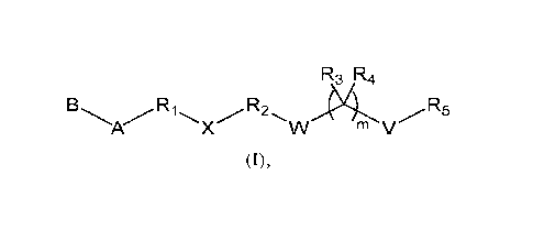
In one aspect of this invention, it covers two sets of lipid-like compounds of
formula (I) below:
R3 R4
A X m V
(I).
In one set, referring to formula (I), A is a hydrophilic head selected from
Ra
Ra
\+711- RN
Ra¨N Ra'¨N
+/Z
+2 ZµZ Ra"¨N
Ra¨N N Ra.-14 Ra"I4 ¨ .4ss
riV Ra' 04. prµe , and Ra"' , in which each of Ra, Ra',
Ra", and Ra.", independently, is H, Ci-C20 alkyl, C2-C20 alkenyl, C2-C20
alkynyl, C3-
C20 cycloalkyl, Ci-C20 heteroalkyl, Ci-C20 heterocycloalkyl, aryl, or
heteroaryl; and Z
is a Ci-C20 bivalent aliphatic radical, a Ci-C20 bivalent heteroaliphatic
radical, a
bivalent aryl radical, or a bivalent heteroaryl radical; B is Ci-C24 alkyl, C2-
C24
alkenyl, C2-C24 alkynyl, C3-C24 cycloalkyl, Ci-C24 heteroalkyl, Ci-C24
R3 R4
R1 R2 R5
heterocycloalkyl, aryl, or heteroaryl, or X W V ; each of
Ri and R2, independently, is a Ci-C20 bivalent aliphatic radical; each of R3
and R4,
independently, is H or Ci-Cio alkyl, or R3 and R4, together with the atom to
which
they are attached, form C3-C10 cycloalkyl; R5 is Cl-C24 alkyl, C2-C24 alkenyl,
C2-
C24 alkynyl, C3-C24 cycloalkyl, Ci-C24 heteroalkyl, Ci-C24 heterocycloalkyl,
aryl, or
2
CA 03089826 2020-07-28
WO 2019/152848
PCT/US2019/016362
G
r
heteroaryl; W is 0, S, or Se; V is a bond, 0, S, or Se; X, a linker, is 1 \
2
or t , in
which each of Li, L2, L3, and L4, independently, is a bond, 0,
S, or NRe; G is 0, S, or NR); Q is ORi., SRg, or NRbRi; and each of r and t,
independently, is 1-6, each of Re, Rd, Rf, Rg, Rh, and Rõ independently, being
H,
Ci-
Cio alkyl, Ci-Cio heteroalkyl, aryl, or heteroaryl; and m is 0 or 1, provided
that m is 1
when V is S.
In the other set, referring to formula (I) again, A is a hydrophilic head
selected
Ra
Ra
\+/ Ra'¨N
Ra¨N Ra'¨N
Ra"¨N
Ra¨N N R '-1\1 Ra"¨N,
from rAr ,r\l' a X PPV. , and Ra'" , in
which each of Ra,
Ra', Ra", and Ra'", independently, is H, Ci-C20 alkyl, C2-C20 alkenyl, C2-C20
alkynyl,
1() C3-C20 cycloalkyl, Ci-C20 heteroalkyl, Ci-C20 heterocycloalkyl, aryl,
or heteroaryl;
and Z is a Ci-C20 bivalent aliphatic radical, a Ci-C20 bivalent
heteroaliphatic radical, a
bivalent aryl radical, or a bivalent heteroaryl radical; B is Ci-C24 alkyl, C2-
C24
alkenyl, C2-C24 alkynyl, C3-C24 cycloalkyl, Ci-C24 heteroalkyl, Ci-C24
R3 R4
R2 /R5
heterocycloalkyl, aryl, or heteroaryl, or X W m V ; Ri is a
is Ci-C20 bivalent aliphatic radical; R2 is a bond or Ci-C20 bivalent
aliphatic radical;
each of R3 and R4, independently, is H or Ci-Cio alkyl, or R3 and R4, together
with the
atom to which they are attached, form C3-Cio cycloalkyl; R5 is
IRb
77 R7
L1 L2 , in which R6 is a bond or Ci-C20 bivalent aliphatic
radical; each of Rh and Rb' is F or, Rh and Rb', together with the atom to
which they
20 are attached, form C=0; R7 is F or an aliphatic lipid moiety; each of Li
and L2,
independently, is a bond, 0, S, or NRe, Re being H, Ci-Cio alkyl, Ci-Cio
heteroalkyl,
aryl, or heteroaryl; and n is 1 to 20; each of W and V, independently, is a
bond, 0, S,
3
CA 03089826 2020-07-28
WO 2019/152848 PCT/US2019/016362
G
or Se; X, a linker, is \
3 L5
t , in which each of L3, L4,
or
L5, and L6, independently, is a bond, 0, S, or NRe; G is 0, S, or NRd; Q is
OR(, SRg,
or NRhR,; and each of r and t, independently, is 1-6, each of Re, Rd, Re, Rt.,
Rg, Rh, and
independently, being H, Ci-Cio alkyl, Ci-Cio heteroalkyl, aryl, or heteroaryl;
and
iS 0 or 1.
Typically, the above-described lipid-like compounds have variable A as either
Ra¨N
Ra¨N Ra'-14
r.rs'r or rlj:r , each of Ra and Ra', independently, being a Ci-Cio
monovalent
aliphatic radical, a Ci-Cio monovalent heteroaliphatic radical, a monovalent
aryl
radical, or a monovalent heteroaryl radical; and Z being a Ci-Cio bivalent
aliphatic
1() -- radical, a Ci-Cio bivalent heteroaliphatic radical, a bivalent aryl
radical, or a bivalent
heteroaryl radical. These compounds preferably have variable B as
R3 R4
R5
X m V
The term "lipid-like compounds" herein refers to compounds that contain one
or more hydrophilic (or polar) amine-containing head groups and one or more
hydrophobic (or nonpolar) hydrocarbon-containing tails. See, e.g., Love et
al.,
PNAS, 2010, 107(5), 1864-1869. The term "lipid-like nanocomplexes" refers to
nanocomplexes that contain one of lipid-like compounds. See, e.g., Wang et
al.,
Angew. Chem. Int. Ed., 2014, 53(11), 2893-2898.
The term "aliphatic" herein refers to a saturated or unsaturated, linear or
-- branched, acyclic, cyclic, or polycyclic hydrocarbon moiety. Examples
include, but
are not limited to, alkyl, alkylene, alkenyl, alkenylene, alkynyl, alkynylene,
cycloalkyl, cycloalkylene, cycloalkenyl, cycloalkenylene, cycloalkynyl, and
cycloalkynylene moieties.
The term "aliphatic lipid moiety" herein refers to a hydrophobic moiety that
contains long-chain, saturated or unsaturated, linear or branched, acyclic,
cyclic, or
polycyclic hydrocarbons, alcohols, aldehydes, or carboxylic acids. Examples
include,
but are not limited to, cholesterol, desmosterol, and lanosterol.
4
CA 03089826 2020-07-28
WO 2019/152848
PCT/US2019/016362
The term "alkyl" or "alkylene" refers to a saturated, linear or branched
hydrocarbon moiety, such as methyl, methylene, ethyl, ethylene, propyl,
propylene,
butyl, butylenes, pentyl, pentylene, hexyl, hexylene, heptyl, heptylene,
octyl,
octylene, nonyl, nonylene, decyl, decylene, undecyl, undecylene, dodecyl,
dodecylene, tridecyl, tridecylene, tetradecyl, tetradecylene, pentadecyl,
pentadecylene,
hexadecyl, hexadecylene, heptadecyl, heptadecylene, octadecyl, octadecylene,
nonadecyl, nonadecylene, icosyl, icosylene, triacontyl, and triacotylene. The
term
"alkenyl" or "alkenylene" refers to a linear or branched hydrocarbon moiety
that
contains at least one double bond, such as -CH=CH-CH3 and ¨CH=CH-CH2-. The
term "alkynyl" or "alkynylene" refers to a linear or branched hydrocarbon
moiety that
contains at least one triple bond, such as -CC-CH3 and -CC-CH2-. The term
"cycloalkyl" or "cycloalkylene" refers to a saturated, cyclic hydrocarbon
moiety, such
as cyclohexyl and cyclohexylene. The term "cycloalkenyl" or "cycloalkenylene"
refers to a non-aromatic, cyclic hydrocarbon moiety that contains at least one
double
is bond, such as cyclohexenyl cyclohexenylene. The term "cycloalkynyl" or
"cycloalkynylene" refers to a non-aromatic, cyclic hydrocarbon moiety that
contains
at least one triple bond, cyclooctynyl and cyclooctynylene.
The term "heteroaliphatic" herein refers to an aliphatic moiety containing at
least one heteroatom selected from N, 0, P, B, S, Si, Sb, Al, Sn, As, Se, and
Ge.
The term "alkoxy" herein refers to an -0-alkyl. Examples of alkoxy include
methoxy, ethoxy, n-propoxy, isopropoxy, n-butoxy, iso-butoxy, sec-butoxy, and
tert-
butoxy.
The term "aryl" herein refers to a C6 monocyclic, Cio bicyclic, C14 tricyclic,
C20 tetracyclic, or C24 pentacyclic aromatic ring system. Examples of aryl
groups
.. include phenyl, phenylene, naphthyl, naphthylene, anthracenyl,
anthrcenylene,
pyrenyl, and pyrenylene. The term "heteroaryl" herein refers to an aromatic 5-
8
membered monocyclic, 8-12 membered bicyclic, 11-14 membered tricyclic, and 15-
20 membered tetracyclic ring system having one or more heteroatoms (such as 0,
N,
S, or Se). Examples of heteroaryl groups include furyl, furylene, fluorenyl,
fluorenylene, pyrrolyl, pyrrolylene, thienyl, thienylene, oxazolyl,
oxazolylene,
imidazolyl, imidazolylene, benzimidazolyl, benzimidazolylene, thiazolyl,
thiazolylene, pyridyl, pyridylene, pyrimidinyl, pyrimidinylene, quinazolinyl,
5
CA 03089826 2020-07-28
WO 2019/152848
PCT/US2019/016362
quinazolinylene, quinolinyl, quinolinylene, isoquinolyl, isoquinolylene,
indolyl, and
indolylene.
Unless specified otherwise, aliphatic, heteroaliphatic, alkoxy, alkyl,
alkylene,
alkenyl, alkenylene, alkynyl, alkynylene, cycloalkyl, cycloalkylene,
cycloalkenyl,
cycloalkenylene, cycloalkynyl, cycloalkynylene, heterocycloalkyl,
heterocycloalkylene, heterocycloalkenyl, heterocycloalkenylene, aryl, and
heteroaryl
mentioned herein include both substituted and unsubstituted moieties. Possible
substituents on cycloalkyl, cycloalkylene, cycloalkenyl, cycloalkenylene,
cycloalkynyl, cycloalkynylene, heterocycloalkyl, heterocycloalkylene,
heterocycloalkenyl, heterocycloalkenylene, aryl, and heteroaryl include, but
are not
limited to, Ci-Cio alkyl, C2-Cio alkenyl, C2-Cio alkynyl, Ci-C20 alkoxy, C3-
C20
cycloalkyl, C3-C20 cycloalkenyl, C3-C20 heterocycloalkyl, C3-C20
heterocycloalkenyl,
Ci-Cio alkoxy, aryl, aryloxy, heteroaryl, heteroaryloxy, amino, Ci-Cio
alkylamino,
C20 dialkylamino, arylamino, diarylamino, Ci-Cio alkylsulfonamino,
arylsulfonamino,
is Ci-Cio alkylimino, arylimino, Ci-Cio alkylsulfonimino, arylsulfonimino,
hydroxyl,
halo, thio, Ci-Cio alkylthio, arylthio, Ci-Cio alkylsulfonyl, arylsulfonyl,
acylamino,
aminoacyl, aminothioacyl, amido, amidino, guanidine, ureido, thioureido,
cyano,
nitro, nitroso, azido, acyl, thioacyl, acyloxy, carboxyl, and carboxylic
ester. On the
other hand, possible substituents on aliphatic, heteroaliphatic, alkyl,
alkylene, alkenyl,
alkenylene, alkynyl, and alkynylene include all of the above-recited
substituents
except Ci-Cio alkyl. Cycloalkyl, cycloalkylene, cycloalkenyl, cycloalkenylene,
heterocycloalkyl, heterocycloalkylene, heterocycloalkenyl,
heterocycloalkenylene,
aryl, and heteroaryl can also be fused with each other.
The lipid-like compounds described above include the compounds themselves,
as well as their salts and solvates, if applicable. A salt, for example, can
be formed
between an anion and a positively charged group (e.g., amino) on a lipid-like
compound. Suitable anions include chloride, bromide, iodide, sulfate, nitrate,
phosphate, citrate, methanesulfonate, trifluoroacetate, acetate, malate,
tosylate,
tartrate, fumurate, glutamate, glucuronate, lactate, glutarate, and maleate.
Likewise, a
salt can also be formed between a cation and a negatively charged group (e.g.,
carboxylate) on a lipid-like compound. Suitable cations include sodium ion,
potassium ion, magnesium ion, calcium ion, and an ammonium cation such as
tetramethylammonium ion. The lipid-like compounds also include those salts
containing quaternary nitrogen atoms. A solvate refers to a complex formed
between
6
CA 03089826 2020-07-28
WO 2019/152848
PCT/US2019/016362
a lipid-like compound and a pharmaceutically acceptable solvent. Examples of
pharmaceutically acceptable solvents include water, ethanol, isopropanol,
ethyl
acetate, acetic acid, and ethanolamine.
Another aspect of this invention relates to a pharmaceutical composition
containing a nanocomplex formed of a lipid-like compound described above and
and
a protein or a nucleic acid. In this composition, the nanocomplex has a
particle size of
50 to 1000 nm (e.g., 50 to 500 nm, 50 to 300 nm, and 50 to 180 nm). The lipid-
like
compound binds to the protein or nucleic acid via a non-covalent interaction,
a
covalent bond, or both.
The term "protein" refers to a polymer of natural or non-natural amino acids
linked together by amide bonds and having a molecular weight of 800 Dalton or
higher. The term "nucleic acid" refers to a polymer of nucleotides linked
together by
phosphodiester bonds, having a molecular weight of 800 Dalton or higher. Both
of
these polymers can be chemically modified. Examples of protein modification
is include PEGylation and carboxylation of amine groups in lysine residues
contained
therein. More specifically, carboxylation of proteins or peptides can be
achieved by
using cis-aconitic anhydride. See Lee et al., Angew. Chem. Int. Ed., 2009, 48,
5309-
5312; Lee et al., Angew. Chem. Int. Ed., 2010, 49, 2552-2555; and Maier et
al.,
Journal of the American Chemical Society, 2012, 134, 10169-10173.
The term "non-covalent interaction" refers to any non-covalent binding, which
includes ionic interaction, hydrogen bonding, van der Waals interaction, and
hydrophobic interaction.
The pharmaceutical composition typically contains a pharmaceutically
acceptable carrier. The carrier in the pharmaceutical composition must be
"acceptable" in the sense that it is compatible with the active ingredient of
the
composition (and preferably, capable of stabilizing the active ingredient) and
not
deleterious to the subject to be treated. One or more solubilizing agents can
be
utilized as pharmaceutical excipients for delivery of an active glycoside
compound.
Examples of other carriers include colloidal silicon oxide, magnesium
stearate,
cellulose, sodium lauryl sulfate, and D&C Yellow # 10.
Further covered by this invention is a method of treating a medical condition,
e.g., a lung disease. The method includes a step of administering to a subject
in need
thereof an effective amount of an above-described pharmaceutical composition.
7
CA 03089826 2020-07-28
WO 2019/152848
PCT/US2019/016362
The details of the invention are set forth in the description below. Other
features, objects, and advantages of the invention will be apparent from the
following
drawings and detailed description of several embodiments, and also from the
appending claims.
BRIEF DESCRIPTION OF THE DRAWINGS
Figure 1 is a schematic depiction of synthesis of lipid-like compounds
(lipidoids) and encapsulation of proteins into lipidoid nanoparticles. (a)
Encapsulation of negatively charged GFP-Cre and Cas9:sgRNA into synthetic
cationic lipidoid nanoparticles (LNPs) for intracellular protein delivery and
genome
editing. (b) Synthetic route and lipidoids nomenclature. (c) Chemical
structures of
amine heads for lipidoids synthesis.
Figure 2 is a schematic depiction of characterization of lipidoids and LNPs.
(a) and (b) NMR and ESI-MS spectra of 76-0170, 76-017S, and 76-0175e (see
is exemplary lipid-like compounds below). (c) Statistical analysis of
averaged
hydrodynamic diameter (<Dh>) distribution of LNPs. (d) Typical hydrodynamic
diameter distributions of 76-0170, 76-017S, and 76-0175e LNPs.
Figure 3 is a schematic depiction of another characterization of lipidoids and
LNPs. (a) and (b) Typical transmission electron microscopy (TEM) images and
relative size variations of 76-0170, 76-017S, and 76-0175e LNPs (scale bar
being
100 nm). (c) Fluorescent emission intensities and FRET ratios of DiO/DiI
loaded 76-
0175e LNPs during storage.
Figure 4 is a schematic depiction of in vitro screening of LNPs for protein
delivery. (a) Typical images of (-30)GFP-Cre protein and (-30)GFP-Cre loaded
76-
0170, 76-017S, and 76-0175e LNPs treated HeLa-DsRed cells. Scale bar = 200 pm.
(b) Percentage of GFP-positive cells shown for 51 LNPs tested. Data points
marked in
red for LNPs induced high level of transfection. (c) The tails (0170, 017S,
and
0175e) influenced (-30)GFP-Cre protein transfection activity.
Figure 5 is a schematic depiction of structure-activity relationship for LNPs.
(a) and (b) Apparent pKa values and phospholipid bilayer membrane disruption
ability influenced (-30)GFP-Cre protein delivery efficiency. (c) Relative hit
rates of
efficacious LNPs having none, one, or two properties. (d) Relative hit rates
of
efficacious LNPs having 0170, 017S, or 0175e tails.
8
CA 03089826 2020-07-28
WO 2019/152848
PCT/US2019/016362
Figure 6 shows the efficiency of (-30)GFP-Cre delivery with LNPs. (a)
DsRed expression of HeLa-DsRed cells treated with (-30)GFP-Cre and (-30)GFP-
Cre
loaded LNPs. (b) Cell viability of HeLa-DsRed cells treated with (-30)GFP-Cre
and
(-30)GFP-Cre loaded LNPs
Figure 7 is a schematic depiction of typical fluorescence images of sections
of
lungs obtained from Ai14 mice treated with PBS and GFP-Cre/LNPs (the first
column
being 4',6-diamidino-2-phenylindole or DAPI; the second column being tdTomato;
the third column shwoing the merging of the first and second columns; and
scale bar
being 100 pm).
Figure 8 shows the efficiency of Cas9:sgRNA delivery with LNPs. (a) GFP
knockout of GFP-HEK cells treated with Cas9:sgRNA and Cas9:sgRNA/LNPs. (b)
Cell viability of GFP-HEK cells treated with Cas9:sgRNA and Cas9:sgRNA/LNPs.
Figure 9 is a schematic presentation of cholesterol-based and reduction-
responsive combinatorial lipidoids for intracellular delivery. (A) Chemical
structures
is -- of cationic lipidoids and amine head groups. (B) Lipidoids nanoparticles
as a versatile
platform for anticancer drugs, mRNA and protein delivery.
Figure 10 shows the characterization of lipioids and nanoparticles. (A)
MALDI-TOF spectra of lipidoids. (B) Hydrodynamic diameter and polydispercity
of
lipidoid nanoparticles measured by DLS. (C) TEM images of lipidoid
nanoparticles.
-- Scal bar = 200 nm. (D) Relative size change of blank nanoparticles under
storage. (E)
Cytotoxicity tests of OcholB, 016B and Lpf2k nanoparticles. P < 0.05,
student's t-
test.
Figure 11 shows the thiol-triggered morphological variation and cargo
release. (A) Time-dependent relative size variation of the lipidoid
nanoparticles with
-- DTT and Cysteine treatment. (B) TEM images of lipidoid nanoparticles
treated with
DTT. Scale bar = 600 nm. (C) Relative size change of lipidoid nanoparticle
after 24 h
of DTT treatment. (D) Fluorescent emission spectra of cargoes loaded
nanoparticles.
(E) Time-dependent NR release profile. (F) Fluorescent intensity of calcein
encapsulated lipidoid nanoparticles treated with DTT or Cysteine. (G) RNA
binding
-- test of lipidoid nanoparticles with and without DTT treatment.
Figure 12 shows the internalization study of cargo-loaded lipidoid
nanoparticles. (A) Time-dependent FRET ratio variation of DiO-DiI loaded
nanoparticles. (B) Time-dependent NR cells portions of HeLa cells treated
with NR
loaded nanoparticles. (C) NW cells portions of lipidoid nanoparticles after 8
h of
9
CA 03089826 2020-07-28
WO 2019/152848
PCT/US2019/016362
exposure. (D) Fluorescent images of HeLa cells treated by NR loaded
nanoparticles.
Scale bar = 100 pm. (E) Mean fluorescent intensity of HeLa cells treated with
free or
nanoparticles encapsulated calcein. (F) Transfection efficiencies of (-30)GFP-
Cre
protein by lipidoid nanoparticles against HeLa-DsRed cells. (G) Mean
fluorescent
intensity, (H) flow cytometry histogram, (I) fluorescent images and (J) bright
field
images of (-30)GFP-Cre/LNPs treated HeLa-DsRed cells. Scale bar = 110 pm.
Figure 13 shows the intracellular delivery of anticancer drugs. (A) Absorption
and fluorescent emission spectra of CPT and Dox loaded nanoparticles. (B) Mean
fluorescent intensity of free and nanoparticle encapsulated Dox treated HeLa
cells.
(C) Dose-dependent cytotoxicity of free Dox, and blank and Dox loaded
nanoparticles. (D) Cytotoxicity of free and nanoparticle encapsulated CPT and
Oxa.
Figure 14 shows the intracellular delivery of mRNA. (A) LNP/mRNA weight
ratio and (B) mRNA dose-dependent transfection efficacy. (C) Fluorescent
images of
mRNA/LNPs treated HeLa cells. Scale bar = 100 pm. (D) Transfection efficiency
and
is (E) cytotoxicity of mRNA/LNPs. (F) Bright field images of mRNA/LNPs
treated
HeLa cells. Scale bar = 110 pm. (G) Cre mRNA and (H) Cas9 mRNA and sgRNA
delivery by OCholB LNPs.
Figure 15 shows the intracellular delivery of genome editing protein. (A)
Internalization mechanism study. (B) Genome editing efficiency, (C) flow
cytometry
histogram, (D) cytotoxicity and (E) bright field images of (-30)GFP-Cre/LNPs
treated
HeLa-DsRed cells. Scale bar = 200 pm. (F) Genome editing efficacy was plotted
against cell viability for each tested conditions.
Figure 16 shows the In vivo toxicity tests. (A) Time-dependent body weight
and (B) biochemical blood analysis of blank LNPs injected Balb/c mice.
Figure 17 shows the mRNA and protein delivery for in vivo genome editing
using adult Ai14 mice. (A) Cre-mediated gene recombination. Protocols used for
(B)
intramuscular and (C) intravenous injections. Fluorescent images of (D)
intramuscular
protein/LNPs (scale bar = 270 pm) and (E) intramuscular mRNA/LNPs (scale bar =
270 pm) injected skeletal muscles. Fluorescent images of (F) lungs from
control and
intravenous protein/LNPs injected mice (scale bar = 135 pm) and (G) spleens
from
intravenous mRNA/LNPs injected mice (scale bar = 190 pm). Red channel in the
original image, tdTomato; Blue channel in the original image, DAPI. Images in
up
panels are from nanoparticles injected mice and images in low panels are from
untreated control mice.
CA 03089826 2020-07-28
WO 2019/152848
PCT/US2019/016362
Figure 18 is a schematic presentation showing (a) Encapsulation of AmB into
synthetic cationic lipidoids nanoparticles and effect on fungus cells. (d) The
quatemized lipidoids were combinatorial synthesized of the amine and alkyl-
eposxide molecules, lipidoids are named as follows(Carbon numbers of tail)-
(Amine
number)
Figure 19 shows the visual stability states after preparation: AmB/(75-04B,
78-014B, 87-014B) encapsulates demonstrated opaque suspention and all
precipitated within 1 week, AmB/(75-014B, 78-014B, 87-014B)-F encapsulates
demonstrated translucent solutions after preparation and not homogenize at the
end of
1() 2 week, AmB/(Q75-014B, Q78-014B, Q87-014B) and AmB/(Q75-014B, Q78-
014B, Q87-014B) -F encapsulates exhibit homogenous transparent yellow
solutions
and stable in the following 2 weeks.
Figure 20 shows (a) Hydrodynamic sizes of AmB encapsulates and
Fungizone after preparation and in following 2 weeks(n=9), (b) Polydispersity
indexes of AmB encapsulates after preparation and in following 2 weeks
determined
by DLS(n=9). *p < 0.05 and ** p < 0.001vs the particle size and PDI after
preparation.
Figure 21 shows (a) The DLC of AmB encapsulates and Fungizone (n=3).
(b) The MIC of different AmB encapsulates, free AmB and Fungizone against
Candida. albicans(SC5314) after 24h and 48h incubation from range of 14.0 to
0.109375pg/mL (n=9), *P < 0.05 vs Fungizone .
Figure 22 shows (a) Hemolysis of human RBCs by AmB encapsulates, free
AmB and Fungizone at equivalent of AmB concentrations (25, 50, 100, 200pg/mL)
after lh incubation at 37 C(n=9). (b) In vitro MTT test of different AmB
encapsulates,
free AmB and Fungizone towards HEK293 cell line at equivalent of AmB
concentrations (25, 50, 100, 200pg/mL) after 24h incubation. All data present
as
mean SD (n=9), *p< 0.05 and **p< 0.001 vs Fungizone
Figure 23 shows (a) Plasma concentrations of AmB after intravenous
injection of AmB/Q78-014B-F and Fungizone at a dose of 2mg AmB/kg to SD rats;
(b-e) The AmB concentrations in mice tissues after 48h and 72h intravenous
treatment
of AmB/Q78-014B-F (10mg, 5mg, 2mg AmB/kg, respectively) and Fungizone (2
mg AmB/kg) by HPLC, (b) Liver, (c)Spleen, (d) Lung, (e) Kidney, *p <0.05 and
**p
<0.001 vs Fungizone .
11
CA 03089826 2020-07-28
WO 2019/152848
PCT/US2019/016362
Figure 24 shows the In vivo toxicity of (a) Creatinine, (b) BUN, (c) ALT, (d)
AST level in healthy BALB/c mice 48h and 72h after intravenous administrated
with
AmB/Q78-014B-F at dose of 10mg, 5mg and 2mg AmB/kg and Fungizone at dose
of 2mg AmB/kg (n=3), *p <0.05 and **p <0.001 vs Fungizone .
Figure 25 is a bar graph showing the percentage of GFP positive cells (GFP+)
as a function of the fluorine-containing lipidoid used for protein delivery.
Figure 26 is a bar graph showing the percentage of DsRed positive cells as a
function of the fluorine-containing lipidoid used for protein delivery.
Figure 27 is a bar graph showing the percentage of GFP+ cells as a function
1() of the lipidoid (derived from lipids with different hydrophobic tails
and synthesized
from amine 200) used for protein delivery.
Figure 28 is a bar graph showing the percentage of GFP+ cells as a function
of the lipidoid (derived from lipids synthesized from different cyclic amine
analogues) used for protein delivery for.
Figure 29 is a bar graph showing the observed luminescence stemming from
mRNA delivery to CD8+ T cells as a function of the lipidoid (derived from
lipids
synthesized from different imidazole-containing amine analogues).
DETAILED DESCRIPTION
Disclosed in detail herein are lipid-like compounds of the present invention.
More specifically, two embodiments are described in order below.
In the first embodiment, referring to formula (I) shown above, A is a
Ra
\
Ra¨N Ra 1'1
+2'1-
Ra¨N N Ra.-14
IA' Iskr
hydrophilic head selected from , and
Ra
\
Ra'¨N
'
Ra"¨N
Ram , in which each of Ra, Ra', Ra", and Ra.", independently, is H, Ci-
C20 alkyl,
C2-C20 alkenyl, C2-C20 alkynyl, C3-C20 cycloalkyl, Ci-C20 heteroalkyl, Ci-C20
heterocycloalkyl, aryl, or heteroaryl; and Z is a Ci-C20 bivalent aliphatic
radical, a Cl-
C20 bivalent heteroaliphatic radical, a bivalent aryl radical, or a bivalent
heteroaryl
12
CA 03089826 2020-07-28
WO 2019/152848
PCT/US2019/016362
radical; B is Ci-C24 alkyl, C2-C24 alkenyl, C2-C24 alkynyl, C3-C24 cycloalkyl,
Ci-C24
heteroalkyl, Ci-C24 heterocycloalkyl, aryl, or heteroaryl, or
R3 Ret
vR1 R2 R5
X W V ; each of
Ri and R2, independently, is a Ci-C2o
bivalent aliphatic radical; each of R3 and R4, independently, is H or Ci-Cio
alkyl, or
R3 and R4, together with the atom to which they are attached, form C3-Cio
cycloalkyl;
R5 is Ci-C24 alkyl, C2-C24 alkenyl, C2-C24 alkynyl, C3-C24 cycloalkyl, Ci-C24
heteroalkyl, Ci-C24 heterocycloalkyl, aryl, or heteroaryl; W is 0, S, or Se; V
is a
L)3C-
4 t
bond, 0, S, or Se; X, a linker, is 1 or , in which
each
of Li, L2, L3, and L4, independently, is a bond, 0, S, or NRe; G is 0, S, or
NRd; Q is
io ORf, SRg, or NRfiRi; and each of r and t, independently, is 1-6, each of
Re, Rd, Rf, Rg,
Rh, and Rõ independently, being H, Ci-Cio alkyl, Ci-Cio heteroalkyl, aryl, or
heteroaryl; and m is 0 or 1, provided that m is 1 when V is S.
This embodiment preferably includes compounds that typically have variable
Ra¨N R3 R4
Ra¨N Ra R2/R5
A as Pi' or , and variable B as X Wm V
71-t.
Ra¨N
is Exemplary compounds have variables A, B, and Ri-R5 as follows: A is
P's'r or
Ra¨N R3 R4
Ra R2R5
rkr ; B is X W V ; each of Ri and R2,
independently, is a Ci-C4 bivalent aliphatic radical; each of R3 and R4,
independently,
is H or Ci-C4 alkyl; and R5 is Cl-C20 alkyl.
Preferably, A is an amino moiety formed from one of the following amines:
13
CA 03089826 2020-07-28
WO 2019/152848
PCT/US2019/016362
1 r
N
NH2 -'1\jNFI2 C-ININFI2 01 N H2
/
-Th
78 75 76 77
0 0
0
NNH2 N N H2
N.'"''''......sNH2 L'"'NNH NH2 2 .....'0 I
82 90 17 80 81
N\...., .j.._'N -------'''..--.' N H2 H2N N N H2
HOJ I
87 93 306
HN
H
?
HOf N 0H
NNN112
I H H H H H
103 400 63 304 .
As described above, X is a linker. Examples of X include, but are not limited
0 0 0
0 k A A. kNANA,
OH II cz, Y.LNA, 0 0 N =
`2,-Lcs 2,Cr; 1 1 A '22.?_ 1 1 1
to, ... 7 '. , RC (:) CY ' RC , and
Rd Rc , each
, ,
of Re and Rd, independently, being H or Ci-Cio alkyl. These compounds
preferably
5 have each of Ri and R2, independently, as a Ci-C4 bivalent aliphatic
radical; each of
R3 and R4, independently, as H or Ci-C4 alkyl; and R5 as Ci-C20 alkyl.
Turning to variables W, V, and m, this embodiment can include, based on
these three variables, the following three subsets of compounds.
Subset (i) includes the compounds of formula (I), in which each of W and V,
1() independently, is 0 or Se; and m is 0.
This subset of compounds can have their
R3 R4
Xv R1 R2 R5
X W m V moiety formed
from one of the following
molecules:
14
CA 03089826 2020-07-28
WO 2019/152848 PCT/US2019/016362
o 0
JL IS,s'
NS'S1'.'r =)(CDS'SPr 0 a
H a a
o o
Se Se
)(Nse %, =======s,,A ....^..õ_õõSe _
_ ,,,..,, .....õ<_y
Se
H -1" 0 Se- 0 -/
' ' a
a a
, in which q is an integer of 8-12.
Subset (ii) includes the compounds of formula (I), in which W is 0, S, or Se;
V is a bond; and m is 0 or 1.
This subset of compounds can have their
R3 R4
Xc, Ri R2 R5
X W m V moiety formed from one of the following
molecules:
o 0
o a
H a a
o o
)L NS
H
a
o o
)LNSe
.)(0Se 4,,r rx-
--...õ,Se......,...õi_r
0
H a
a a
, in which q is an integer of 8-12.
Subset (iii) includes the compounds of formula (I), in which each of W and V
is 0 or S and m is 1.
This subset of compounds can have their
R3 R4
X
X W m V moiety formed from one of the following
molecules:
CA 03089826 2020-07-28
WO 2019/152848 PCT/US2019/016362
0 0
\
, in which q is an integer of 8-12.
Alternatively, this subset of compounds can have their
R3 R4
R R2 R5
X W m V moiety formed from one of the following
molecules:
0 0
X
v RR q
VRR" q
0 0
v RAR q RAR
, in which X is 0, S, or NH; R is H or Me; p is an integer of 0-3; q is an
integer of 1-
16; and v is an integer of 1-10.
In the second embodiment, referring to the above formula (I) again, A is a
Ra
\-F/'
Ra¨N Ra 1'1
Ra +711-
RaNN R Ra÷-14
fAr X
hydrophilic head selected from pr\l' a , and
Ra
\ +711-
Ra'¨N
=J's
Ra"' , in which each of Ra, Ra', Ra", and Ra", independently, is H, Ci-
C20 alkyl,
C2-C20 alkenyl, C2-C20 alkynyl, C3-C20 cycloalkyl, Ci-C20 heteroalkyl, Ci-C20
heterocycloalkyl, aryl, or heteroaryl; and Z is a Ci-C20 bivalent aliphatic
radical, a Cl-
C20 bivalent heteroaliphatic radical, a bivalent aryl radical, or a bivalent
heteroaryl
radical; B is Ci-C24 alkyl, C2-C24 alkenyl, C2-C24 alkynyl, C3-C24 cycloalkyl,
Ci-C24
heteroalkyl, Ci-C24 heterocycloalkyl, aryl, or heteroaryl, or
16
CA 03089826 2020-07-28
WO 2019/152848
PCT/US2019/016362
R3 R4
vR1 R2/R5
X W m V ; Ri is a
Ci-C20 bivalent aliphatic radical; R2 is a
bond or Ci-C20 bivalent aliphatic radical; each of R3 and R4, independently,
is H or
Ci-Cio alkyl, or R3 and R4, together with the atom to which they are attached,
form
Rbl
R6 R7
C3-Cio cycloalkyl; R5 is L 1 n 1_7 2
, in which R6 is a bond or Ci-
C20 bivalent aliphatic radical; each of Rh and Rh' is F or, Rh and Rb',
together with the
atom to which they are attached, form C=0; R7 is F or an aliphatic lipid
moiety; each
of Li and L2, independently, is a bond, 0, S, or NRe, Re being H, Ci-Cio
alkyl, Ci-Cio
heteroalkyl, aryl, or heteroaryl; and n is 1 to 20; each of W and V,
independently, is a
L4 6
bond, 0, S, or Se; X, a linker, is 3 or t , in which
each
io of L3, L4, L5, and L6, independently, is a bond, 0, S, or NRe; G is 0,
S, or NRd; Q is
ORf, SRg, or NRbRi; and each of r and t, independently, is 1-6, each of Re,
Rd, Re, Rf,
Rg, Rh, and Ri, independently, being H, Ci-Cio alkyl, Ci-Cio heteroalkyl,
aryl, or
heteroaryl; and m is 0 or 1.
Like the first embodiment, the second embodiment can also include
Ra¨N
Ra¨N Ra.-14
compounds having variable A as X or X , and variable B as
R3 R4
R2 R5
m V
An exemplary compound of this embodiment has variables A, B, and Ri-R4 as
Ra¨N R3 R4
71-6
Ra¨N Ra'-14 /v\ R5
follows: A is 0:ri or ; B is A X W V ; each
of Ri and R2, independently, is a Ci-C4 bivalent aliphatic radical; and each
of R3 and
R4, independently, is H or Ci-C4 alkyl.
Again, A can be an amino moiety formed from one of the following amines:
17
CA 03089826 2020-07-28
WO 2019/152848 PCT/US2019/016362
CV--
1 r
N H2 / N ....'s N H2 N N H2 N H2 i N H2
78 75 76 77
-----'µ.1 o-Th
0
0
NN H2 N N H2
0 NH2
I
1 \j ''''''NH2 N NH2 ........
82 90 17 80 81
HO
NN H2 Nx.,_______N N H2 H2NNN H2
1
HO)
87 93 306
HN
H
?
fNNII
HO .()Fi
N-1\1 r\j-NN
I\JNN H2
H H H H
1 H
103 400 63 304
18
CA 03089826 2020-07-28
WO 2019/152848 PCT/US2019/016362
OH NH2
HONH2 HOOH N=N
H H NNH2 a
NH2
22 63 75 76
I
.....NNH2 Isli NH2
NCINH2 1 aNH2
/
77 78 80 81 82
H 0õõ..-.N......õ.....N N2 0 C ..-Ni
I NH2 Isk,___I NH2 I H
H2NNNH2 H2NNNN2
HO,)
87 90 93 113 114
H
N='Nl'=NH2 H2NNNH2 rkiN'NH2 H2NN/--\rsINE12
) I I H
123 306 400 401
=T' ,,,a = , =.._ , õ,,,,,,,:.
.:
4,,
9310 9311 9312 9313 9314 9215 9316
rlõ 171. N:.
fs,,,,,. ,...."4
.\.W = itt': ' It ' W. A
.1 j =;.) ,
.,..
,=,-
I =1 f.
4A=
= .4. HA, ..Nt.' .. 10.
9321 .9.22 9323: :9324
..:õ, . .
rl
N n,
.. ..: .-'''''.,---,' -=g,. .'-'.. -#4... = =
9331 .9.32 33,33 9334
iliN . N'-'=====k. .:*=""$µ
'''''4A, .L.õ. Vt'''\:,4y = t. N
e :NN-.. 144P . :k,..,==1*i.:
:9341. 9351. 9352 935*
19
CA 03089826 2020-07-28
WO 2019/152848 PCT/US2019/016362
1,4-Bis(3-aminopropyl)piperazine 1-(2-Aminoethyl)piperidine-3-
carboxamide
piperazine-2-carboxamide 4-
Aminopiperidine
o
/--\ _/--\
H2N_/N N NH2 - 0 NH2
/4-NH2 /-N\-
HN NH H2N HND-NH2-'
4-(aminomethyl)piperidineinformamidine dihydrochloride 1-Amino 4 (2
hydroxyethyl)piperazine
2-(4-aminopiperidin-1-yl)acetamide /--\
HN NH j-N N-NH2
HO
H2N HCI ND-NH2
H2N-
/- 0
1-(2-Aminoethyl)piperazine 2-(Aminomethyl)piperidine
/--\ NH2
/-N H2N-' NHCcH
2-Methylpiperazine 2,3-Dimethylpiperazine
1-Methylpiperazine Piperazine
/--\ /--\
-N NH HN NH HN NH HN NH
2-(Trifluoromethyl)piperazine 2-0xopiperazine 1-(3-aminopropyl)piperazine 1-
(3-pyrrolidinopropyl)piperazine
F3C q
)_\
?-\ H2N\_/-N/-\NH I*1FIC\NH
HN NH HN NH
1-methyl-4-(2-piperidin-4-yl-ethyl)-piperazine 2-(4-Methyl-piperazin-1-yI)-
ethylamine
1-Cyclopentylpiperazine
/--\
O-
/-C\N-
N N-
HND¨r \¨' /--\ N NH H2N-' \¨/
1-(2-diisopropylaminoethyl)piperazine
4 /-1*1--\NH
In the second embodiment, examples of X include, but are not limited to,
0 0 0
0 k A A, kNANA,
OH II
Y.LNN 0
c& A \ 1 1 1
R, 0 CY Re , and Rd Rc , each of
9 9 9
RC and Rd, independently, being H or Ci-Cio alkyl. These compounds preferably
have
each of Ri and R2 as a Ci-C4 bivalent aliphatic radical; each of R3 and R4,
independently, as H or Ci-C4 alkyl; and R5 as Ci-C20 alkyl.
CA 03089826 2020-07-28
WO 2019/152848
PCT/US2019/016362
As to variables W, V, and m, the second embodiment can include compounds
having each of R2, W, and V as a bond, and m as 0.
Rb
R6
=
Referring to variable R5, 1.e., L1 n 1_72
R7, compounds in this
embodiment can have each of Li and L2 as a bond, and each of Rb, R6', and R7
as F.
R3 R4
vR1 R2
Exemplary compounds have their X Wm V moiety
formed from one of the following molecules:
0
N j 0 j 0
H k
in which j is an integer of 0-10 and k is an integer of 1-20.
Alternatively, this embodiment includes those compounds, in which R6 is Cl-
C4 bivalent aliphatic radical; each of Li and L2, independently, is 0 or NRe,
Re being
H or Ci-Cio alkyl; R6 and Rb', together with the atom to which they are
attached, form
C=0; n is 1 or 2; and R7 is an aliphatic lipid moiety. The aliphatic lipid
moiety can
be cholesterol. Exemplary compounds have their
R3 R4
vR1 ,R9 R5
X/
m V moiety formed
from one of the following
molecules:
0
0
1-r*
0
0
in which X is 0 or NH and W is 0, S, or Se.
21
CA 03089826 2020-07-28
WO 2019/152848
PCT/US2019/016362
The lipid-like compounds of this invention can be prepared by methods well
known in the art. See, e.g., Wang et al., ACS Synthetic Biology, 2012, 1, 403-
407;
Manoharan et al., WO 2008/042973; and Zugates et al., US Patent 8,071,082.
The synthetic route shown below exemplifies synthesis of certain lipid-like
compounds described above:
R3 R4
0
X M V
R3 R4
0 IR,'NI R3 R4
Ra¨NH2 +x R5 0
R2 R5
X M V
in which each of variables Ra, R2-R5 , X, W, V, and m are defined above.
In this exemplary synthetic route, an amine compound, i.e., compound D,
reacts with a vinyl carbonyl compound E to afford the final product, i.e.,
compound F.
Amino compound D can be one of the above-described Compounds 10, 17, 63, 75-
78,
80-82, 87, 90, 93, 103, 304, 306, and 400.
Other lipid-like compounds of this invention can be prepared using other
suitable starting materials through the above-described synthetic route and
others
known in the art. The method set forth above can include an additional step(s)
to add
or remove suitable protecting groups in order to ultimately allow synthesis of
the
lipid-like compounds. In addition, various synthetic steps can be performed in
an
alternate sequence or order to give the desired material. Synthetic chemistry
transformations and protecting group methodologies (protection and
deprotection)
useful in synthesizing applicable lipid-like compounds are known in the art,
including, for example, R. Larock, Comprehensive Organic Transformations (2nd
Ed.,
VCH Publishers 1999); P. G. M. Wuts and T. W. Greene, Greene's Protective
Groups
in Organic Synthesis (4th Ed., John Wiley and Sons 2007); L. Fieser and M.
Fieser,
Fieser and Fieser's Reagents for Organic Synthesis (John Wiley and Sons 1994);
and
L. Paquette, ed., Encyclopedia of Reagents for Organic Synthesis (2nd ed.,
John Wiley
and Sons 2009) and subsequent editions thereof.
Certain lipid-like compounds may contain a non-aromatic double bond and
one or more asymmetric centers. Thus, they can occur as racemates and racemic
22
CA 03089826 2020-07-28
WO 2019/152848
PCT/US2019/016362
mixtures, single enantiomers, individual diastereomers, diastereomeric
mixtures, and
cis- or trans- isomeric forms. All such isomeric forms are contemplated.
As mentioned above, these lipid-like compounds are useful for delivery of
proteins or nucleic acids. They can be preliminarily screened for their
efficacy in
delivering pharmaceutical agents by an in vitro assay and then confirmed by
animal
experiments and clinic trials. Other methods will also be apparent to those of
ordinary skill in the art.
Not to be bound by any theory, the lipid-like compounds facilitate delivery of
proteins or nucleic acids by forming complexes, e.g., nanocomplexes and
microparticles. The hydrophilic head of such a lipid-like compound, positively
or
negatively charged, binds to a moiety of a protein or nucleic acid that is
oppositely
charged and its hydrophobic moiety binds to a hydrophobic moiety of the
protein or
nucleic acid. Either binding can be covalent or non-covalent.
The above described complexes can be prepared using procedures described in
is publications such as Wang et al., ACS Synthetic Biology, 2012, 1, 403-
407.
Generally, they are obtained by incubating a lipid-like compound and a protein
or
nucleic acid in a buffer such as a sodium acetate buffer or a phosphate
buffered saline
("PBS").
Further covered by this invention is a pharmaceutical composition containing
a nanocomplex formed of a lipid-like compound described above and and a
protein or
a nucleic acid. Again, the lipid-like compound binds to the protein or nucleic
acid via
a non-covalent interaction, a covalent bond, or both.
Examples of the protein or nucleic acid include, but are not limited to,
clustered regularly interspaced short palindromic repeat associated protein 9
(CRISPR/Cas9), Cre recombinase ((-30)GFP-Cre), and Cas9:single-guide RNA
(Cas9:sgRNA) ribonucleoprotein (RNP) or Cas9:sgRNA RNP.
Still within the scope of this invention is a method of treating a medical
condition, e.g., a lung disease, with the above-described pharmaceutical
composition.
The method includes administering to a subject (e.g., a patient) in need
thereof an
effective amount of the pharmaceutical composition.
The term "an effective amount" refers to the amount of complexes that is
required to confer a therapeutic effect on the treated subject. Effective
doses will
vary, as recognized by those skilled in the art, depending on the types of
diseases
23
CA 03089826 2020-07-28
WO 2019/152848
PCT/US2019/016362
treated, route of administration, excipient usage, and the possibility of co-
usage with
other therapeutic treatment.
To practice the method of the present invention, a composition having the
above-described complexes can be administered parenterally, orally, nasally,
rectally,
topically, or buccally. The term "parenteral" as used herein refers to
subcutaneous,
intracutaneous, intravenous, intramuscular, intraarticular, intraarterial,
intrasynovial,
intrastemal, intrathecal, intralesional, or intracranial injection, as well as
any suitable
infusion technique.
A sterile injectable composition can be a solution or suspension in a non-
toxic
parenterally acceptable diluent or solvent, such as a solution in 1,3-
butanediol.
Among the acceptable vehicles and solvents that can be employed are mannitol,
water, Ringer's solution, and isotonic sodium chloride solution. In addition,
fixed oils
are conventionally employed as a solvent or suspending medium (e.g., synthetic
mono- or diglycerides). Fatty acid, such as oleic acid and its glyceride
derivatives are
is useful in the preparation of injectables, as are natural
pharmaceutically acceptable
oils, such as olive oil or castor oil, especially in their polyoxyethylated
versions.
These oil solutions or suspensions can also contain a long chain alcohol
diluent or
dispersant, carboxymethyl cellulose, or similar dispersing agents. Other
commonly
used surfactants such as Tweens or Spans or other similar emulsifying agents
or
bioavailability enhancers which are commonly used in the manufacture of
pharmaceutically acceptable solid, liquid, or other dosage forms can also be
used for
the purpose of formulation.
A composition for oral administration can be any orally acceptable dosage
form including capsules, tablets, emulsions and aqueous suspensions,
dispersions, and
solutions. In the case of tablets, commonly used carriers include lactose and
corn
starch. Lubricating agents, such as magnesium stearate, are also typically
added. For
oral administration in a capsule form, useful diluents include lactose and
dried corn
starch. When aqueous suspensions or emulsions are administered orally, the
active
ingredient can be suspended or dissolved in an oily phase combined with
emulsifying
.. or suspending agents. If desired, certain sweetening, flavoring, or
coloring agents can
be added.
A nasal aerosol or inhalation composition can be prepared according to
techniques well known in the art of pharmaceutical formulation. For example,
such a
composition can be prepared as a solution in saline, employing benzyl alcohol
or
24
CA 03089826 2020-07-28
WO 2019/152848
PCT/US2019/016362
other suitable preservatives, absorption promoters to enhance bioavailability,
fluorocarbons, and/or other solubilizing or dispersing agents known in the
art.
A composition containing the nanocomplexes can also be administered in the
form of suppositories for rectal administration.
EXAMPLES
Without further elaboration, it is believed that one skilled in the art can,
based
on the above description, utilize the present invention to its fullest extent.
The
following specific examples are therefore to be construed as merely
illustrative, and
1() not limitative of the remainder of the disclosure in any way
whatsoever.
Methods and Materials
General
All chemicals used for lipidoids synthesis were purchased from Sigma-Aldrich
without further purification unless otherwise noted. (-30)GFP-Cre recombinase,
S.
is pyogenes Cas9 (spCas9) and sgRNA were generated following the protocols
reported
in Wang at al., Proc. Natl. Acad. Sci. USA, 2016, 113, 2868-2873 ("Wang").
HeLa-
DsRed and GFP-HEK cells were cultured in Dulbecco's modified eagle's medium
(DMEM, Sigma-Aldrich) with 10% fetal bovine serum (PBS, Sigma-Aldrich) and 1%
penicillin-streptomycin (Gibco). All 1H NMR spectra were recorded on a Bruker
20 AVIII 500 MHz NMR spectrometer operated in the Fourier transform mode.
Hydrodynamic size and polydispersity index of nanoparticles were measured by
Zeta-
PALS particle size analyzer (Brookhaven Instruments). The apparent pKa values
of
lipidoids were determined using 2-(p-toluidinynaphthalene-6-sulphonic acid)
(TNS,
Sigma-Aldrich) as fluorescent probe following the protocols reported in Heyes
et al.,
25 J. Controlled Release, 2005, 107, 276-287. TEM measurements were
performed on a
FBI Technai Transmission Electron Microscope. Fluorescence images of tissue
slices
were obtained using BZ-X Analyzer fluorescence microscope.
Synthesis of lipid-like compounds (i.e., lipidoids)
Head amines (Sigma-Aldrich) were mixed with acrylates tails (e.g., 0170,
30 017S, and 0175e) at a molar ratio of 1:2.4 in teflon-lined glass screw-
top vials for 48
hours at 70 C. The crude products were purified using a Teledyne Isco
Chromatography system.
CA 03089826 2020-07-28
WO 2019/152848 PCT/US2019/016362
One class of lipid-like compounds of formula (I) were synthesized by
following the synthetic route shown below:
0
CI)
R X R Y0 H -Ow-
Y
......-- H 0 H ¨ill'
triethylamine
X = CI, Br, or!
Y = 0, S. or Se
0
0 Ra¨NH2 RYR a
0 N
R,.......,...X...õ..."..,0,
R
Head amines Ra-NH2 shown in the above scheme were selected from
Compounds 10, 17, 63, 75-78, 80-82, 87, 90, 93, 103, 304, 306, and 400.
1 r
N
NH2 aN 1-1 NH C N H2 2 /
78 75 76 77
10 0
. 0
N..N H N N H2
N NH2 .N NH2 I
2
0 N H2
82 90 17 80 81
HONNH2 ..-.-N N H2 H2 N NN H2
Ho) Nv...:rj
I
87 93 306
HN
H
?
HOf NNF-(:)Fi
-...N.---,..,......---..N.---\õ..--. N
NH2
I H H H H H
103 400 63 304
Shown in the table below are the codes, chemical formulas, and analytical data
(ESI-MS) of 51 exemplary lipid-like compounds ("lipidoids") of formula (I).
Note
that each lipidoid is coded as X-017Y, in which X represents the number of an
amino
1() compound and Y represents 0, S, or Se. Code X-017Y indicates that a
lipidoid is
formed from an amine of Compound X and a lipid molecule of 017Y (Y being 0, S,
or Se).
For example, lipidoid 10-0170 is formed from amine Compound 10 and lipid
molecule 0170 as follows:
26
CA 03089826 2020-07-28
WO 2019/152848
PCT/US2019/016362
NE12
10-0170
0170
Each code in the table below contains 0170, 017S, or 017Se, which
represents one of the three molecules:
0
0c)). 0170
0
So). 017S
0
017Se
5
Lipidoid Code Chemical Formula Cal. 11\4+Hr Found 11\4+Hr
10-0170 C41 H82N07 700.61 700.70
10-017S C411-182N05s2 732.56 732.59
10-017Se C411-182N05Se2 828.45 828.27
17-0170 C481-188N08 806.65 806.63
17-017S C481-188N06S2 838.60 838.49
17-017Se C481-188N06Se2 934.49 934.27
63-0170 C43H87 N206 727.66 727.67
63-017S C431-187N204S2 759.61 759.62
63-017Se C431-187N204Se2 855.50 855.35
75-0170 C44H89 N206 741.67 741.71
75-017S C441-189N204S2 773.63 773.69
75-017Se C441-189N204Se2 869.51 869.67
76-0170 C44H87 N206 739.66 739.74
76-017S C441-187N204S2 771.61 771.69
76-017Se C441-187N204Se2 867.50 867.46
77-0170 C45 H89 N206 753.67 753.75
27
CA 03089826 2020-07-28
WO 2019/152848
PCT/US2019/016362
77-017S C451-189N204S2 785.63 785.64
77-017Se C451-189N204Se2 881.51 881.46
78-0170 C42H85 N206 713.64 713.79
78-017S C42H85 N2 04S 2 745.59 745.57
78-017Se C421-185N204Se2 841.48 841.43
80-0170 C43H87 N206 727.66 727.68
80-017S C431-187N204S2 759.61 759.70
80-017Se C431-187N204Se2 855.50 855.46
81-0170 C45 H91 N206 755.69 755.71
81-017S C45H91 N2 04S 2 787.64 787.64
81-017Se C45H9iN204Se2 883.53 883.45
82-0170 C45 H89 N206 753.67 753.88
82-017S C451-189N204S2 785.63 785.70
82-017Se C451-189N204Se2 881.51 881.42
87-0170 C45H91 N208 787.68 787.71
87-017S C45H91 N2 06S 2 819.63 819.52
87-017Se C45H9iN206Se2 915.52 915.39
90-0170 C44H87 N207 755.65 755.96
90-017S C44H87 N2 05 S 2 787.61 787.59
90-017Se C441-187N205Se2 883.49 883.38
93-0170 C44H84N3 06 750.64 750.69
93-017S C44H84N3 04S 2 782.59 782.69
93-017Se C441-184N304Se2 878.48 878.41
103-0170 C441189N208 773.66 773.72
103-017S C441189N206S2 805.62 805.53
103-017Se C441189N206Se2 901.50 901.45
304-0170 C661-1133N409 1126.01 1125.97
304-017S C661-1133N406S3 1173.94 1173.88
304-017Se C661-1133N406Se3 1317.77 1317.63
306-0170 C831-1164N3012 1395.23 1395.24
306-017S C831-1164N308S4 1459.14 1459.89
28
CA 03089826 2020-07-28
WO 2019/152848
PCT/US2019/016362
306-017Se C8311164N3OsSe4 1650.92 1650.77
400-0170 C65H130N309 1096.98 1096.90
400-017S C65H130N306S3 1144.91 1144.74
400-017Se C65Hi3oN306Se3 1288.74 1288.60
Another class of lipid-like compounds of formula (I) were synthesized by
following the synthetic route shown below:
0
01fl
). OH
H /01.r
triethylamine 0 n = 1-10
/
C)10C))'r
Ra¨NH2 k /n
Ra
010C)1(
PPTS
0
0
PPTS = pyridinium p-toluenesulfonate
Again, head amines Ra-NH2 shown in this above scheme were selected from
Compounds 10, 17, 63, 75-78, 80-82, 87, 90, 93, 103, 304, 306, and 400.
Still another class of lipid-like compounds of formula (I) were synthesized by
following the synthetic route shown below:
0 0
0
F F
Ra¨NH2 /X
H,x :j F` F CI /HxF.r F Ra--N
X j
0 \
F k
.4
FikF k
triethylamine
X=OorNH j = 0-10 `F k
k = 1-20
to Head amines Ra-NH2 shown in the above scheme were selected from
Compounds 10, 17, 63, 75-78, 80-82, 87, 90, 93, 103, 304, 306, and 400.
Fabrication of nanocomplexes from lipidoids and proteins.
Lipidoids were fabricated into nanoparticles for delivery proteins or nucleic
acids. Briefly, lipidoids were mix with sodium acetate buffer (25 mM, pH 5.2),
is sonicated for 30 mm in ultrasonic bath and followed by another 30 mm of
vigorous
vortex to form lipid-like nanoparticles or LNPs. The LNPs thus obtained were
stored
at 4 C. For protein/LNP complexation, LNPs were mixed with (-30)GFP-Cre or
29
CA 03089826 2020-07-28
WO 2019/152848
PCT/US2019/016362
Cas9:sgRNA in PBS buffer (25 mM, pH 7.4) following the protocols reported in
Wang and incubated at room temperature for 30 minutes.
Evaluation of phospholipid bilayer membrane disruption
Human red blood cells (hRBCs) were washed with PBS buffer three times and
collected after centrifugation at 1000 rpm for 5 minutes. The resulting stock
solution
(about 10% v/v hRBCs) was diluted 3 fold in PBS buffer to give the assay
solution.
90 L of assay solution was mixed with 10 L of LNPs solutions (final
concentration
of lipidoids = 3.3 mg/L) and incubated at 37 C for 60 minutes. Then the
samples
were centrifuged again at 1000 rpm for 10 mm. 10 L of the supernatant was
further
diluted into 90 L of PBS buffer, and the absorbance at 405 nm (0D405) was
recorded using a microplate reader. The PBS buffer and Triton X-100 (1% v/v)
were
used as negative and positive controls respectively.
Intracellular delivery of (-30)GFP-Cre/LNP
For the intracellular uptake study, HeLa-DsRed cells were seeded in 48-well
is plate with a density of 2 x 10⁴ cell/well. After 24 h of incubation at
37 C, 5% CO2, (-
30)GFP-Cre/LNP nanoparticles were added to the cells and incubated for 6 h
before
fluorescence microscopy and flow cytometry (BD FACS Calibur, BD Science, CA)
analysis (green emission from GFP). The final (-30)GFP-Cre protein
concentration is
25 nM, and lipidoid concentration is 3.3 mg/L. For the gene recombination
functional
study, HeLa-DsRed cells were treated with same conditions and the red
fluorescence
emission from DsRed was analyzed by flow cytometry 24 h after delivery.
Intracellular delivery of Cas9:sgRNA/LNP
For CRISPR/Cas9 gene knockout study, GFP-HEK cells were seeded in 48-
well plate with a density of 2 x 10⁴ cell/well. After 24 h of incubation,
Cas9:sgRNA/LNP nanoparticles were added to the cells and incubated for 4 h,
followed by media changed. After 48 h of incubation, the green emission from
GFP
was analyzed by flow cytometry. The final Cas9:sgRNA RNP concentration was 25
nM, and lipidoid concentration was 3.3 mg/L.
In vitro cytotoxicity assay.
Cell viability was measured by the standard MTT assay. HeLa-DsRed or GFP-
HEK cells were seeded into 96-well plate with a density of 5 x 10³ cell/well.
(-
30)GFP-Cre/LNP or Cas9:sgRNA/LNP nanoparticles were added after 24 h of
incubation. The final concentration of protein is 25 nM and LNP is 3.3 mg/L.
After
incubating for 24 h or 48 h, the MTT reagent (5 mg/mL, in 30 pL PBS buffer)
was
CA 03089826 2020-07-28
WO 2019/152848
PCT/US2019/016362
added and the cells were incubated for another 4 h at 37 C. The cell culture
media
were then carefully removed and 200pL of DMSO were added. The DMSO solution
was transferred into another 96-well plate and the absorbance at 570 nm was
recorded
by microplate reader. All experiments were performed in quadruplicate.
In vivo protein delivery to Ai14 mouse
Formulated LNPs (lipidoid/Cholesterol/DOPE/DSPE-PEG2k = 16/4/1/4,
weight ratio) were prepared for protein loading and mice injection. Ai14 mice
were
housed in a temperature and humidity controlled facility with a 12 h
light/dark cycle.
Two mice in each group were injected with (-30)GFP-Cre/LNPs formulations on
day
0 and 5, with 100 pg protein for each injection. Organs including heart,
liver, spleen,
lung and kidney from all groups were collected 20 days after injections. The
tissues
were fixed overnight in 4% paraformaldehyde (PFA) before being sectioning into
10
pm slices. The slices were collected and stained with DAPI for fluorescence
imaging.
is EXAMPLE 1: Preparation and Characterization of Lipid-Like Nanoparticles
(LNPs)
Certain lipid-like nanoparticles (LNPs) were prepared from lipid-like
compounds of formula (I), i.e., lipidoids, by following the procedures
described
below.
Synthesis of 0170
The following scheme was followed for synthesizing 0170.
'01.4
_____________________________________ lsie=
Na14, Ki
6 0
)0, 0170
'TEA
Sodium hydride (0.72 g, 30 mmol) was added to the solution of ethylene
glycol (5.6 g, 90 mmol) in anhydrous DMF (30 mL) and stirred for 10 min at 0
C. 1-
Bromotetradecane (6.0 g, 20 mmol) and KI (3.3 g, 20 mmol) were then added and
the
reaction mixture was kept at 95 C for another 4 h. After cooling to room
temperature, the mixture was diluted with cold water, extracted with ethyl
acetate, and
dried over anhydrous sodium sulfate. Compound 1 (3.3 g, yield about 65%) was
obtained after column chromatography purification on silica gel using n-
hexane/ethyl
31
CA 03089826 2020-07-28
WO 2019/152848
PCT/US2019/016362
acetate as mobile phase. Then, compound 1 (3.3 g, 12.8 mmol) and triethylamine
(TEA, 1.9 g, 19.2 mmol) were dissolved in anhydrous DCM (80 mL). Acryloyl
chloride (1.4 g, 15.4 mmol) was added dropwise at 0 C, and the reaction
mixture was
stirred overnight. After column chromatography purification, 0170 was obtained
as
colorless oil (3.2 g, yield about 82%). The structure of 0170 was confirmed by
11-1
NMR spectrum recorded in CDC13.
Synthesis of 017S
The following scheme was followed for synthesizing 017S.
2
6
017$
'TEA
To a solution of 2-mercaptoethanol (1.1 g, 14 mmol) in acetonitrile (20 mL)
was added 1-bromotetradecane (5.0 g, 18 mmol) and potassium carbonate (3.6 g,
26
mmol). The reaction solution was stirred overnight at 40 C, filtered and
concentrated. Compound 2 (1.8 g, yield about 48%) was obtained after column
chromatography purification on silica gel using n-hexane/ethyl acetate as
mobile
phase. In a manner similar to that for the preparation of 0170, 017S was
synthesized
and purified as oil-like liquid (3.5 g, yield about 75%). The structure of
017S was
confirmed by 11-1 NMR spectrum recorded in CDC13.
Synthesis of 0175e
The following scheme was followed for synthesizing 017Se.
K.U.01 Naf3H4
HO
1-1
4
1-1$o
6
017U
TEA
32
CA 03089826 2020-07-28
WO 2019/152848
PCT/US2019/016362
Potassium selenocyanate (1.5 g, 10 mmol) was added in portion to a solution
of 2-bromoethanol (1.6 g, 13 mmol) in acetone (50 mL) at room temperature. The
solution was heated to reflux for 2 h. After cooling to room temperature, the
white
precipitate was filtering off and acetone was removed by rotary evaporation
under
vacuum. Compound 3 was then dissolved in ethanol (25 mL) and sodium
borohydride (0.9 g, 24 mmol) was added slowly at 0 C. After the reaction
solution
turned to colorless, 1-bromotetradecane (4.1 g, 15 mmol) was added through a
dropping funnel. The reaction was stopped by adding DI water (10 mL) after 30
mm.
Then the ethanol was removed under reduced pressure, reaction mixture was
diluted
with saturated sodium chloride aqueous solution (50 mL), and extracted with
DCM (3
x 50 mL). Compound 5 (1.5 g, yield about 46%) was obtained after column
chromatography purification on silica gel using n-hexane/ethyl acetate as
elute. In a
manner similar to that for the preparation of 0170 and 017S, 017Se was
obtained as
oil-like liquid (2.7 g, yield about 72%). The structure of 017Se was confirmed
by 41
is NMR spectrum recorded in CDC13.
Lipidoids synthesis
Commercially available amine heads, e.g., Compounds 10, 17, and 63, were
mixed with acrylate tails 0170, 017S, or 017Se stoichiometrically. The mixture
thus obtained was stirred at 70 C for 48 h. See Figure 1. Lipidoids were
purified by
Teledyne Isco Chromatography system, characterized by 1H NMR and ESI-MS, and
coded as amine number (X) and 017Y (R-017Y, Y being 0, S or Se) as shown in
the
table above. The typical 1H NMR and ESI-MS spectra of 76-0170, 76-0-17S and
76-017Se are shown in Figures 2a and 2b.
Lipidoid nanoparticles fabrication and characterization.
Lipidoids nanoparticles (LNPs) were fabricated in sodium acetate buffer (25
mM, pH 5.2) by following the simple ultrasonication and vortex procedures
described
above. Hydrodynamic sizes and polydispersity index (PDI) of LNPs were measured
by dynamic laser scattering (DLS) analysis. As shown in Figure 2c, most of the
0, S
and Se ethers containing LNPs had the averaged hydrodynamic diameter (<Dh>)
between 100-300 nm, and the PDI in the range 0.1-0.3, suitable for
intracellular
protein delivery application. Further, as also shown in Figure 2c, it was
found that
about 53% of LNPs with 0170 tails, about 82% of 017S LNPs, and about 65% of
0175e LNPs had <Dh> less than 200 nm, resulted from the effect of incorporated
chalcogen atoms on the supramolecular self-assembly behaviors in aqueous
solutions.
33
CA 03089826 2020-07-28
WO 2019/152848
PCT/US2019/016362
Typical size distribution profiles of 76-0170 (<Dh> being 170.1 nm, 1t2/r2
being
0.37), 76-017S (<Dh> being 114.3 nm, //2/r2 being 0.24) and 76-017Se (<Dh>
being
129.4 nm, 1,121r2 being 0.18) LNPs are shown in Figure 2d.
The morphologies of LNPs were further studied by the transmission electron
microscopy (TEM). As shown in Figure 3a, spherical particles were observed in
the
images of 76-0170, 76-017S and 76-017Se LNPs, and the measured number-
averaged sizes (145 nm, 94 nm, and 133 nm for 76-0170, 76-017S, and 76-017Se,
respectively) are comparable with the hydrodynamic diameters as determined by
DLS. See Figure 2d. The morphologies of other LNPs including 80-0170, 80-017S,
and 80-017Se, were also examined by their TEM images, which showed presence of
spherical particles. Subsequently, the stability of LNPs thus prepared was
examined
by DLS and fluorescence measurements. As shown in Figure 3b, the time-
dependent
DLS measurements revealed that no evident aggregation of the 76-0170, 76-017S,
and 76-017Se LNPs occurred during five days of storage under room temperature,
is with the relative size change being less than 15%. Fluorescence
resonance energy
transfer (FRET) pair, Di0 and DiI, loaded 76-0175e LNPs also showed negligible
FRET ratio (15751(1575+1505)) variations in five days of storage, as shown in
Figure 3c,
which indicated the structure integrity and long-term storage stability of the
LNPs.
EXAMPLE 2: Evaluation of LNPs for Protein Delivery
A study was performed to evaluate the effct of LNPs prepared in EXAMPLE
1 on protein delivery as follows.
In vitro screening of LNPs for protein delivery
A Cre recombinase protein fused to a negatively supercharged GFP variant ( (-
30)GFP-Cre) was used as a model cargo protein. The(-30)GFP-Cre protein was
able
to complex with cationic LNPs through electrostatic attraction and other types
supramolecular interactions. The cellular uptake of LNPs could be determined
by
direct analysis of intracellular GFP fluorescent intensity as reported in
Wang. HeLa-
DsRed cells were used in this study, which expressed red fluorescent DsRed
oupon
Cre-mediated recombination to facilitate the functional study of delivered
proteins in
the following study.
The (-30)GFP-Cre protein loaded LNPs (GFP-Cre/LNPs) were prepared at
first by simply mixing precalculated amount of aqueous solution of LNPs and
protein
at ambient conditions. For the intracellular delivery, after incubation with
GFP-
34
CA 03089826 2020-07-28
WO 2019/152848
PCT/US2019/016362
Cre/LNPs nanoparticles for 6 h, the GFP-positive cells were observed using
fluorescence microscopy, harvested and counted by flow cytometry. As shown in
Figure 4a, comparing with the control group, i.e., untreated HeLa-DsRed cell,
bright
green fluorescence emission was observed from the GFP-Cre/Lpf2k (Lpf2k being
Lipofectamine 2000, a commercial transfection agent), GFP-Cre/76-0170, GFP-
Cre/76-017S, and GFP-Cre/76-017Se treated cells. Cells treated with the naked
protein, (-30)GFP-Cre, however, showed negligible fluorescence emission, as
compared with lipid-facilitated delivery systems, which indicated that the
naked (-
30)GFP-Cre protein could not efficiently enter into the HeLa-DsRed cells. The
intracellular (-30)GFP-Cre protein delivery efficiencies were further
quantified by
flow cytometry. As shown in Figure 4b, both the naked (-30)GFP-Cre protein and
the
control group showed low portions of GFP-positive cells, consistent with the
results
of fluorescence microscopy shown in Figure 4a.
On the other hand, in the presence of LNPs, the proportions of GFP-positive
is cells were increased, located in the range of 4-42%, with most of them
being around
12-18%. Delivery efficiencies of LNPs were comparable with that of Lpf2k
(about
31% of GFP-positive cells). For instance, the proportions of GFP-positive
cells
treated with (-30)GFP-Cre protein loaded 400-017Se, 80-017Se, and 77-017Se
LNPs were found to be 42%, 39% and 37%, respectively.
Investigation of structure-activity relationship.
The library of 51 0, S and Se ether-containing lipidoids thus prepared was
utilized to study the structure-activity relationship between LNPs and
intracellular
protein delivery efficacies.
More specifically, lipidoids with greater than 20% GFP-positive cells treated
with (-30)GFP-Cre protein/LNP nanoparticles were defined as efficacious LNPs
(red
data points in Figure 4b), as compared with the bulk LNPs (black data points).
The
lipidoids library was then categorized into three groups according to their
hydrophobic tail structures (0170, 017S, and 0175e); each tail made up 33.3%
of
the library. In the efficacious LNPs group, 21.4%, 28.6%, and 50% of lipidoids
were
with 0170, 017S, and 0175e tails respectively. Therefore, the relative hit
rates of
LNPs with 0170, 017S, and 0175e tails were -11.9%, -4.7%, and 16.7%,
respectively, relative to the initial library (Figure 4c). In other words,
lipidoids with
0170 and 017S tails were significantly underrepresented among LNPs with
delivery
CA 03089826 2020-07-28
WO 2019/152848
PCT/US2019/016362
efficacy greater than 20%, while lipidoids with 017Se tail was
overrepresented,
suggesting that 017Se tails were associated with efficacious LNPs.
It was determined that the delivery efficiencies of LNPs were related to the
chemical structures of amine heads, hydrophobic tails, substitution numbers
and
apparent pKa values. In this study, to further elucidate the structure-
activity
relationship of 0, S, Se ethers containing lipidoids, effects of apparent pKa
value and
phospholipids bilayer membrane disruption ability of the LNPs were further
analyzed.
Apparent pKa values were measured following the previously reported procedures
using 2-(p-toluidinyl)naphthalene-6-sulphonic acid (TNS) as fluorescent probe.
The phospholipids bilayer membrane disruption ability of LNPs was evaluated
using human red blood cells (hRBCs) as model and hemoglobin as the chromophore
reporter agent. Absorbance at 405 nm (0D405) was recorded to assess the amount
of
released hemoglobin, using PBS buffer and Triton X-100 (1% v/v) as negative
and
positive controls, respectively, in which higher 0D405 values indicate
stronger
is membrane disruption capabilities. As shown in Figures 5a and 5b, the
apparent pKa
and 0D405 values of LNPs were plotted against the percentages of GFP-positive
cells
for each LNP, and it was found that most of the efficacious nanoparticles
(with GFP-
positive cells greater than 20%) were located in the regions of pKa > 5.1 and
0D405
> 0.2 (gated with blue dash lines in Figures 5a and 5b). After further
examination, it
was found that these two properties have striking effects on (-30)GFP-Cre
protein
transfection efficiencies in HeLa-DsRed cells. As shown in Figure Sc, when
LNPs
possess both of properties (i.e., pKa > 5.1 and 0D405 > 0.2), the relative hit
rate to be
able to mediate high transfection efficiency was 77%. When one or two of the
properties was removed from the LNPs, the likelihood of achieving high
transfection
efficiency of (-30)GFP-Cre protein into HeLa-DsRed cells dropped significantly
to 8-
33%.
Furthermore, as to the structure-activity relationship, it was found that, for
LNPs with 0170, 017S, and 0175e tails, the relative hit rates of above
mentioned
efficacy criteria were -1.9%/-14.6%, 0.99%/4.2%, and 0.99%/10.5%, respectively
(pKa > 5.1/0D405 > 0.2). See Figure 5d. It was clear that both of the two
properties
were underrepresented in the group of LNPs with 0170 tails, consistent with
the
results shown in Figure 4c, in which 0170 tail was underrepresented in the
efficacious lipidoid. While both properties of high pKa and 0D405 values were
overrepresented in the group of LNPs with 017S and 0175e tails.
36
CA 03089826 2020-07-28
WO 2019/152848
PCT/US2019/016362
Furthermore, according to the results shown in Figures Sc and 5d, the
membrane disruption ability of these LNPs appeared to be the more influential
factor
in determining in vitro (-30)GFP-Cre protein delivery efficiency into HeLa-
DsRed
cells, as compared with the apparent pKa values.
EXAMPLE 3: (-30)GFP-Cre Protein Delivery for Gene Recombination and
Cytotoxicity
A study was performed to evaluate the effct of LNPs prepared in EXAMPLE
1 on (-30)GFP-Cre protein delivery for gene recombination and cytotoxicity as
io follows.
The top 12 of LNPs identified through intracellular delivery screening
experiments were further tested for gene recombination using HeLa-DsRed model
cells. The expression of DsRed from Cre protein mediated gene recombination
was
analyzed after 24 h of co-incubation with free (-30)GFP-Cre protein and
protein
is loaded LNPs. As shown in Figure 6a, naked (-30)GFP-Cre protein did not
induce
DsRed expression, due to its low internalization ability, consistent with the
fluorescence microscopy observation and flow cytometry analysis demonstrated
in
Figure 4a. Most of the test LNPs, on the other hand, efficiently delivered (-
30)GFP-
Cre protein and induced gene recombination, with 14-46% of the cells positive
for
20 DsRed.
More specifically, certain LNPs exhibited high protein transfection
efficiencies, namely, 76-017S (40.8%), 76-017Se (36.1%), 77-017S (38.6%), 77-
017Se (31.0%), 78-017Se (37.8%), and 80-017S (45.6%). These LNPs exhibited
higher or similar transfection efficiencies when compared with Lpf2k (33.5%).
25 Through MTT assay against HeLa-DsRed cells, 76-017S, 76-017Se, 77-
017S, and 77-017Se LNPs showed low cytotoxicity as greater than 80% cells were
alive, as compared to Lpf2k, 400-017Se 78-017Se, 80-017S, and 80-017Se, of
which the cell viability was 67-77%. See Figure 6b.
These results indicate that 76-017S, 76-017Se, 77-017S, and 77-017Se
30 exhibited high intracellular protein delivery and Cre-mediated genome
recombination
efficacies, with lower cytotoxicity than Lpf2k.
37
CA 03089826 2020-07-28
WO 2019/152848
PCT/US2019/016362
EXAMPLE 4: In vivo GFP-Cre Delivery for Gene Recombination in Ai14 Mice
A study was performed to evaluate the effct of LNPs prepared in EXAMPLE
1 on GFP-Cre delivery for gene recombination in Ai14 mice as follows.
Delivering genome editing proteins in vivo has the therapeutic potential for
treating a wide range of genetic diseases. Based on the in vitro screening
results, this
study was conducted to evaluate the effect of the above 0, S, and Se ethers
containing
LNPs on delivering (-30)GFP-Cre protein in vivo for Cre-mediated gene
recombination.
The study used an Ai14 mouse model, which had a genetically integrated
loxP-flanked STOP cassette that prevents the transcription of red fluorescent
protein,
tdTomato. Upon Cre mediated gene recombination, the STOP cassette was removed,
resulting in tdTomato expression. Considering the different performances of
cargo
loaded LNPs in vitro and in vivo, three LNPs with same amine heads and
different
tails (76-0170, 76-017S and 76-0175e) were tested in this study. Formulated
LNPs
is (lipidoid/cholesterol/DOPE/DSPE-PEG2k = 16/4/1/4, weight ratio) were
prepared.
Mice were injected (intravenous injection) with (-30)GFP-Cre loaded the
formulated
LNPs (GFP-Cre/76-0170, GFP-Cre/76-0175, and GFP-Cre/76-0175e) at day 0 and
day 5 (100 pg of protein for each injection). Organs including heart, liver,
spleen,
lung, and kidney were collected at day 20 for measuring and analyzing the
tdTomato
expression. As shown in Figure 7, under the same preparation and imaging
conditions, strong tdTomato signals were observed in the sections of lung from
GFP-
Cre/76-0175 and GFP-Cre/76-0175e injected mice. Fluorescence images with lower
magnification and larger field of view were obtained. It was unexpectedly
observed
that the GFP/76-0175 and GFP/76-0175e injection induced Cre-mediated genome
recombination efficiently in the lung, as compared with the control group and
the
group treated with GFP/76-0170. Therefore, a composition containing LNPs of
this
invention is useful for lung disease treatment.
Notably, both the in vitro screening results and the in vivo tests showed that
lipidoids with same amine heads and different hydrophobic tails possessed very
different physicochemical properties, intracellular delivery efficacies, and
genome
recombination profiles.
38
CA 03089826 2020-07-28
WO 2019/152848
PCT/US2019/016362
EXAMPLE 5: Delivery of Cas9:sgRNA RNP for Genome Modification
A study was performed to evaluate the effct of LNPs prepared in EXAMPLE
1 on the delivery of Cas9:sgRNA RNP for genome modification as follows.
The Cas9:sgRNA RNP targeting genomic GFP reporter gene and GFP-HEK
cells were used in this study. The morphologies of Cas9:sgRNA loaded LNPs were
examined by TEM, and typical image of Cas9:sgRNA loaded 76-017Se LNP
(Cas9:sgRNA/76-017Se) was obtained. For the intracellular delivery, GFP-HEK
cells
were harvested after treating with Cas9:sgRNA/LNPs nanocomplexes for 48 h. GFP
gene knockout efficacy was further evaluated by flow cytometry. As shown in
Figure
8a, naked Cas9:sgRNA RNP did not induce GFP gene knockout, while the knockout
efficiency of Cas9:sgRNA/Lpf2k was relatively high, with 63% of GFP-negative
cells. When using 0, S, and Se ethe-containing LNPs as delivery vehicles, the
GFP-
HEK cells showed a loss of 14%-58% GFP expression. In particular, 50.2%,
57.7%,
54.7% and 57.4% of GFP knockout were observed when cells were treated with
is Cas9:sgRNA loaded with 76-017Se, 80-017Se, 81-017Se, and 400-017Se LNPs.
These lipidoids could efficiently deliver genome editing proteins into
different
mammalian cell lines in vitro, based on the results of gene recombination of
Cre
protein in HeLa-DsRed cells and GFP gene knockout of Cas9:sgRNA RNP delivery
in GFP-HEK cells.
In vitro cytotoxicity of Cas9:sgRNA/LNPs against GFP-HEK cells was also
evaluated by the MTT assay. As shown in Figure 8b, the cell viabilities were
determined to be 67%-119% after incubation with Cas9:sgRNA/LNPs at 37 C for
48
h, indicating that the certain LNPs were non-cytotoxic to GFP-HEK cells, while
some
showing cell viability the same as that of Lpf2k (cell viability about 66% )
under the
same experimental conditions. It was also observed that two LNPs with high
Cas9:sgRNA delivery efficiencies, i.e. 80-017Se and 400-017Se, exhibited cell
viability similar to that for Lpf2k, namely, 68.2% and 66.7% of cell viability
for 80-
017Se and 400-017Se, respectively. Unexpectedly, 76-017Se and 81-017Se LNPs
showed both high Cas9:sgRNA transfection efficiency (50.2% and 54.7%) and low
cytotoxicity (76.3% and 97.7% of cell viability after 48 h of incubation).
These results indicate that LNPs formed from lipid-like compounds of formula
(I) exhibited high protein transfection efficiency and low cytotoxicity.
39
CA 03089826 2020-07-28
WO 2019/152848
PCT/US2019/016362
Methods and Materials
Preparation of blank and cargo-loaded lipidoid nanoparticles
Lipidoids were fabricated into nanoparticles for all delivery applications.
Briefly, lipidoids were mixed with sodium acetate buffer (25 mM, pH 5.2),
sonicated
for 30 min in an ultrasonic bath, followed by another 30 mm of vigorous
vortexing.
The as-prepared blank LNPs were stored at 4 oC. For Cy5-RNA/LNP, mRNA/LNP
and protein/LNP complexation, blank lipidoid nanoparticles were mixed with RNA
molecules or (-30)GFP-Cre protein in PBS buffer (pH 7.4) following our
previously
reported procedures and incubated at room temperature for another 30 mm before
use.
io Typical procedures for Nile red encapsulation are as follows: 5 pL of
Nile red stock
solution in acetone was added into an empty vial, which was then placed in a
vacuum
oven to completely remove the organic solvent. Then, a predetermined amount of
blank LNP stock solution (1.0 mg/mL) was added into the vial. The mixture was
sonicated for 40 mm in an ultrasonic bath and stirred overnight at room
temperature.
is The final concentration of Nile red was adjusted to 6.6 x 10⁻⁷ mol L-1
and 6 x 10⁻⁷
mol L-1 for thiol triggered release study and cell incubation, respectively,
by diluting
with PBS as necessary. Typical procedures for CPT and DiO/DiI FRET pair
encapsulation are as follows: 100 pL of DiO/DiI stock solution in Me0H was
charged
into an empty vial and placed in a vacuum oven to remove the organic solvent.
20 Lipidoids (2.0 mg) in 200 pL of methanol were then added into the vial
and stirred to
produce a homogeneous solution. Then, 600 pL of DI water was added dropwise in
mm with continous stirring. The resulting mixture was dialyzed against DI
water
for 24 h (Thermo Scientific Slide-A-Lyzer Dialysis Cassette, MWCO = 3500 Da),
and
fresh water was replaced every 4 h. Typical procedures for encapsulation of
calcein
25 and doxonorubicin hydrochloride are as follows: precalculated amounts of
calcein or
Dox stock solutions in DI water was diluted into 800 pL with sodium acetate
buffer,
and used as the selective solvents to trigger the self-assembly process of
lipidoids in
methanol (5 mg/mL), respectively. The unloaded calcein or Dox was removed by
dialysis against DI water (Thermo Scientific Slide-A-Lyzer Dialysis Cassette,
MWCO
30 = 3500 Da).
Intracellular delivery of cargo-loaded lipidoid nanoparticles
For the intracellular uptake study, HeLa or HeLa-DsRed cells were seeded in
48-well plate with an initial seeding density of 2 x 10⁴ cell/well. After 24 h
of
incubation at 37 oC, 5% CO2, NR or (-30)GFP-Cre loaded nanoparticles were
added
CA 03089826 2020-07-28
WO 2019/152848
PCT/US2019/016362
to the cells and incubated for certain time (1-8 h) before fluorescence
microscopy
(BZ-X Analyzer) observation and flow cytometry (BD FACS Calibur, BD Science,
CA) analysis (red fluorescence emission from NR and green fluorescence
emission
from GFP). The final concentration of NR is 6 x 10⁻⁷ mol L-1. The final
concentration of (-30)GFP-Cre protein concentration is 25-100 x 10-9 mol L-1.
For
small molecular anticancer drugs delivery, HeLa cells were seeded in 96-well
plate
with an initial seeding density of 5 x 10³ cell/well. After 24 h of incubation
at 37 oC,
5% CO2, Dox, CPT or Oxa loaded nanoparticles were added to the cells and
incubated for 8 h followed by media change. The cells were then incubated for
1() another 40 h before cell viability analysis. For mRNA delivery, HeLa, B
16F10, HEK
293, NIH 3T3 or Jurkat cells were seeded in 48-well plate with an initial
seeding
density of 2 x 10⁴ cell/well. After 24 h of incubation at 37 oC, 5% CO2, mRNA
loaded nanoparticles were added to the cells and incubated for another 24 h
before
fluorescence microscopy and flow cytometry analysis. For protein delivery,
HeLa-
is DsRed cells were seeded in 48-well plate with an initial seeding density
of 2 x 10⁴
cell/well. After 24 h of incubation at 37 oC, 5% CO2, (-30)GFP-Cre protein
loaded
nanoparticles were added to the cells and incubated for 8 h followed by a
complete
media change. The cells were then incubated for another 16 h (24 h of
incubation in
total) before fluorescence microscopy and flow cytometry analysis.
20 In vitro and in vivo toxicity assay
Cell viabilities of HeLa and HeLa-DsRed were measured using the standard
MTT assay. In a 96-well plate, after incubating HeLa or HeLa-DsRed cells with
blank
or cargo-loaded nanoparticles, the MTT reagent (5 mg/mL, in 30 pL PBS buffer)
was
added and the cells were incubated for another 4 h at 37 oC. The cell culture
media
25 was then carefully removed and 200 pL of DMSO was added to each well.
The
DMSO solution was then transferred into a clean 96-well plate and the
absorbance at
570 nm was recorded by a microplate reader. All experiments were performed in
quadruplicate.
For in vivo toxicity studies, the body weights of untreated and nanoparticles
30 injected Balb/c mice (housed in a temperature and humidity controlled
facility with a
12 h light/dark cycle) were measured at day 1, 3, 5, 7, 9, 11, 13 and 14.
Biological
functions of kidney and liver were examined by the serum biochemical tests and
concentrations of creatinine, urea, aspartate aminotransferase (AST), and
alanine
41
CA 03089826 2020-07-28
WO 2019/152848 PCT/US2019/016362
aminotransferase (ALT) were measured using corresponding detection kits
(MilliporeSigma) following manufacturers' protocols.
In vivo protein and mRNA delivery to Ai14 mouse
Similar to the in vitro transfection study, lipidoid nanoparticles were
prepared
for mRNA or protein loading and in vivo delivery. Ai14 mice were housed in a
temperature and humidity controlled facility with a 12 h light/dark cycle.
Three mice
in each group were injected (intravenously or intramuscularly) with Cre mRNA-
loaded
or (-30)GFP-Cre protein-loaded LNPs formulations. Organs including heart,
liver,
spleen, lung and kidney from all groups were collected at day 10
(intramuscular
io injection) or 14 (intravenous injection)after injection. The tissues
werefixed overnight
in 4%paraformaldehyde (PFA)and dehydrated in 30%sucrosebefore beingfrozein in
OCTandsectionedinto 10-15 gmslices.The sliceswere then collected and stained
withDAPIfor fluorescence imaging(BZ-X Analyzerfluorescence microscopy).
is EXAMPLE 6: Cholesteryl Lipidoid Synthesis, Nanoparticles Fabrication and
Characterization.
(4) 4, ,S,s,,,,..4:4=2 (b) S
tifiz
P;p4SAN;,
+ CI, 0 FVF
X'
Py.SS.NiIz
0
"NJ-- \\....) 0 =--1
t_y
ON-SS-CW
Synthesis of Py-SS-Chol
Cholesteryl chloroformate (10.71 g, 23.85 mmol) was dissolved in anhydrous
20 DCM (50 mL) and added into the DCM solution of Py-SS-NH2 (4.47 g, 23.99
mmol)
and TEA (3.71 g, 36.69 mmol) dropwise at 0 oC. The reaction mixture was
stirred
overnight and Py-SS-Chol was obtained as slightly yellow viscous solid (4.89
g, yield
¨ 34.26%) after silica gel column chromatography purification using ethyl
acetate,
dichloromethane and n-hexane as the mobile phase.
42
CA 03089826 2020-07-28
WO 2019/152848
PCT/US2019/016362
Synthesis of OH-SS-Chol
Py-SS-Chol (3.55 g, 5.93 mmol) and acetic acid (600 pL) were dissolved in
DCM (100 mL). 2-Mecaptoethanol (0.51 g, 6.52 mmol) was then added dropwise,
and the reaction mixture was maintained at 35 oC for another 24 h with
continuous
stirring. OH-SS-Chol was purified by silica gel column chromatography using
ethyl
acetate and n-hexane as mobile phase and a colorless solid was obtained (2.74
g, yield
¨ 81.63%).
Synthesis of OCholB
OH-SS-Chol (2.41 g, 4.26 mmol) and TEA (0.65 g, 6.39 mmol) were
dissolved in anhydrous DCM (100 mL). Acryloyl chloride (0.46 g, 5.11 mmol) was
added dropwise at 0 oC. The reaction mixture was stirred overnight and OCholB
was
obtained as a colorless solid (2.52 g, yield ¨ 95.68%) after silica gel column
chromatography purification using ethyl acetate, dichloromethane and n-hexane
as
mobile phase.
Synthesis of lipidoids
The cholesterol-containing acrylates tails shown above were reacted with head
amines Ra-NH2 (i.e., Compounds 75-78, 80, 81, 87, 90, and 304.) to afford the
following lipid-like compounds:
0
H
75-chol 0
0
HN
k 0
0
IND
400H
M oleo utar Weight: 1357.13
43
CA 03089826 2020-07-28
WO 2019/152848 PCT/US2019/016362
A 0
wid 0 At
76-chol 0 0 141r(r..
1/41
0
Ss
HN
0
O.
41161
H
Molecular Weight: 1355.12
0
77-chol S N 11.0
0
0
0 H H
Ss
HN
11164
w
H
Molecular Weight: 1369.15
44
CA 03089826 2020-07-28
WO 2019/152848 PCT/US2019/016362
A 0 H
78-choi
0 H'
Ss
HN
o't0
111
,40
11
H
Molecular Weight: 1329.08
N N0
A 0
0 4IP
80-chol 0 H
0
SS
HN
CP
1116
445 H
Molecular Weight: 1343.11
CA 03089826 2020-07-28
WO 2019/152848
PCT/US2019/016362
p 0
8 1-chol N s
0
0
H
0
HN
,k 0
0
1116k
AlW
H
4
Molecular Weight: 1371.16
o H
N N NtrOt
87.-chol
OH 0
H H
Molecular Weight 1403.16
46
CA 03089826 2020-07-28
WO 2019/152848
PCT/US2019/016362
Fi
0
90-chol
-
v
H,
0
HN
R
Mo I ec ular Weight 137112
304-chol H ,H
411P H
le
0 0
0 0
HN HN
1
0 0
0 0 Molecular Weight: 2049.21
IN
0
H' H
Preparation of blank and cargo-loaded lipidoid nanoparticles
Lipidoids were fabricated into nanoparticles for all delivery applications. As
shown in Figure 10B, most nanoparticles showed average diameters in the range
of
47
CA 03089826 2020-07-28
WO 2019/152848
PCT/US2019/016362
70-300 nm and PDI 0.1-0.3. The sizes of LNPs self-assembled from lipidoids
with
OCholB tails are similar to our previously reported LNP libraries with alkyl
chains.
The relative low PDI values indicated the uniformity of these nanoparticles.
The morphologies of OCholB fully substituted LNPs were then examined by
transmission electron microscopy (TEM). As shown in Figure 10C, spherical
vesicle-
like structures, which are hollow spheres with hydrophobic bilayer walls
sandwiched
by hydrophilic internal and external coronas, were observed from 75-OCholB, 76-
OCholB, 77-OCholB, 78-OCholB, 80-OCholB, 81-OCholB, and 304-OCholB. In
contrast, the vesicular structures were not well-formed for 87-OCholB and 90-
OCholB
compared to their counterparts, and amorphous aggregates were observed
instead. TEM
imaging revealed the largest particles to be 81-OCholB (216.9 nm) and 304-
OCholB
(394.0 nm), which is consistent with the DLS measurement results as shown in
Figure
10B.
The cytotoxicity of OCholB fully substituted LNPs (75-OCholB, 76-OCholB
is and 77-OCholB) and 016B LNPs (75-016B, 76-016B and 77-016B) were tested
side-by-side under different conditions, i.e., low dosage/short exposure time
and
higher dosage/long exposure time, against HeLa cell line using the standard
MTT
assay. As shown in Figure 10E, at the low dosage/short exposure time
conditions (i.e.,
llipidoidl = 1.0 or 3.3 pg mL-1, exposure time = 8 h), all the OCholB and 016B
LNPs
.. showed negligible cytotoxicity, with cell viability of >83% reported for
all lipidoids
(e.g. when llipidoidl = 3.3 pg mL-1, the viabilities of 75-OCholB and 75-016B
treated
cells are 86.5% and 87.7%, respectively). When the dosage and exposure
duration
were both increased (i.e., llipidoidl = 47 or 91 pg mL-1, exposure time = 24
h),
OCholB fully substituted LNPs treated cells showed significantly higher
viabilities
comparing to those treated with 016B LNPs (when llipidoidl = 47 pg mL-1, the
cell
viabilities are 75-OCholB/75-016B = 57.4%/41.9%, 76-OCholB/76-016B =
65.5%/32.8%, 77-OCholB/77-016B = 79.6%/29.0%; when llipidoidl = 91 pg mL-1,
the cell viabilities are 75-OCholB/75-016B = 55.1%/29.5%, 76-OCholB/76-016B =
60.2%/24.3%, 77-OCholB/77-016B = 64.1%/21.9%). These results show that the
.. cholesteryl lipidoids have lower cytotoxicity comparing with the lipidoids
with the
linear alky chain. Furthermore, we compared the biocompatibility of our newly
developed OCholB LNPs against that of the commercially available and widely
used
cationic transfection agent, lipofectamine 2000 (Lpf2k). Lpf2k is shown to be
highly
efficient for both protein and nucleic acids delivery; however its
cytotoxicity is often
48
CA 03089826 2020-07-28
WO 2019/152848
PCT/US2019/016362
a major concern, especially when the targeted cells are exposed to a relative
high
dosage and long incubation duration. As shown in Figure 10E, when the HeLa
cells
were treated with 47 and 91 pg mL-1 of Lpf2k for 24 h, their viabilities were
determined to be 8.9% and 6.8%, which are much lower than that of OCholB LNPs
treated cells under the same conditions. Above all, the in vitro cytotoxicity
tests
demonstrated the excellent biocompatibility of the newly developed cholesteryl-
containing (OCholB) LNPs.
EXAMPLE 7: Thiol-Responsiveness, Loading and Triggered Release of Guest
io Molecules.
The thiol-trigged degradation and dissociation of the OCholB LNPs were
studied by time-dependent DLS measurements and TEM observation. Typically, as
shown in Figure 11A, in the presence of 10 mM of 1,4-dithiothreitol (DTT),
which
has been widely used in previous studies for mimicking intracellular reductive
is conditions,25 we observed the increase of the relative sizes of 75-
OCholB, 76-OCholB
and 77-OCholB gradually increased along incubation duration, as 566.4%, 498.5%
and 1591.4% respectively, in first 2 h. Nanoparticle size was then typically
maintained over the following 4 h, with the exception of 75-OCholB, which
showed
1315.7% increase in size at 6 h. The typical morphologies of DTT treated LNPs
were
20 then examined by TEM and images are shown in Figure 11B. The absence of
well-
formed vesicular structures as shown in Figure 10C and large aggregates at
micrometer scales (which are consistent with the results obtained from DLS
measurements; Figure 11A) with amorphous structures were observed for 75-
OCholB, 76-OCholB and 77-OCholB LNPs. The disruption of the vesicle structure
is
25 considered to be resulted from the thiol-exchange and disulfide bond
cleavage
reactions of OCholB lipidoids. We next investigated whether the thiol-
containing
molecules (e.g. albumin, cysteine, homocysteine, cysteinylglycine, etc.) in
the serum
may induce the structural disintegration of these disulfide bond-containing
LNPs.
The stabilities of LNPs in the presence of 20 pM of L-cysteine (Cys), which
mimics
30 the free thiols on the small- and macromolecules presented in the serum,
were
examined. Shown in Figure 11A, we observed <25% changes in hydrodynamic
diameter at any of the 1 h intervals over the span of this study for all the
three tested
LNPs, and in fact 12.2%, 14.6% and 7.2% decreases in size were observed for 75-
OCholB, 76-OCholB and 77-OCholB after 6 h of incubation, respectively. The
size
49
CA 03089826 2020-07-28
WO 2019/152848
PCT/US2019/016362
changes of cysteine-treated LNPs showed negligible difference when compared
with
the untreated control groups (Figure 11A), indicating the good stability of
OCholB
LNPs under the conditions mimicking the concentration of free thiols in the
blood
serum. Furthermore, the relative size variations of the OCholB LNPs after 24 h
of
incubation in the presence of either 10 mM DTT or 20 pM Cys were determined
and
the results are shown in Figure 11C. With 10 mM of DTT and after 24 h of
treatment,
75-OCholB, 78-OCholB and 304-OCholB LNPs showed the greatest size changes,
with 2872.4%, 4642.8% and 3849.6% increase in averaged hydrodynamic diameters
observed; a moderate size increase of 766.9%-1266.4% were recorded for 76-
77-OCholB, 80-OCholB and 81-OCholB LNPs; while both 87-OCholB
(210.3%) and 90-OCholB (179.5%), which were unable to form consistent vesicles
based on the TEM images (Figure 10C), showed a minimal size increase over 24
hours. On the other hand, all LNPs incubated with 20 M Cysteine for 24 h
showed
minimal size changes, similar to the untreated control groups (Figure 11C).
These
is results
demonstrated the kinetics of the degradation of these OCholB LNPs in relation
to intracellular and extracellular reducible environments. While the degrees
of size
changes vary between lipidoids with different amine head groups, all lipidoids
were
more responsive to DTT (modeling intracellular conditions) than to Cysteine
(modeling conditions in the blood serum). Furthermore, the OCholB LNPs showed
relative good stability in the presence of low concentrations of thiols
(mimicked by 20
pM Cysteine treatment) which indicates that these new LNPs could be used for
systemic drug delivery.
EXAMPLE 8: Drug encapsulation using OCholB LNPs:
Next, the capabilities of OCholB LNPs as nanocarriers to encapsulate cargo
molecules with various physical properties were studied. In this context,
coumarin
(Excitation (Ex.) 350 nm, Emission (Em.) 448 nm) and Nile red (NR; Ex. 520 nm,
Em.
613 nm) as representative small molecular hydrophobic cargoes, calcein (Ex.
475 nm,
Em. 529 nm) as a representative small molecular hydrophilic cargo, and (-
30)GFP-Cre
recombinant fluorescent protein (Ex. 420 nm, Em. 510 nm) and double stranded,
Cy5
labeled RNA (Cy5-RNA, 13kDa; Ex. 625 nm, Em. 672 nm) as representative
macromolecular hydrophilic cargoes were used as the model cargoes. 75-OCholB
was
chosen as the model lipid carrier in the study. Figure 11D showed that the all
the
cargoes can be successfully loaded into the 75-OCholB LNPs. The cargo
molecules
CA 03089826 2020-07-28
WO 2019/152848
PCT/US2019/016362
were loaded into LNPs either through hydrophobic interactions (coumarin and
NR),
electrostatic interactions (calcein, (-30)GFP-Cre and Cy5-RNA), or physical
encapsulation. Furthermore, as shown in Figure 3D, simultaneous encapsulation
of
cargo molecules could be also achieved using the hydrophobic fluorescence
resonance
energy transfer (FRET) pair, Di0 and DiI (Ex. 425 nm, Em. 504 nm (DiO) and 578
nm
(DiI)), as a model, which demonstrates the possibility of using these OCholB
LNPs to
load multiple types bioactive molecules simultaneously for combination
therapies.3 By
taking advantages of the supramolecular interactions (e.g. electrostatic
interaction and
hydrogen bonding) and/or encapsulating cargoes during the self-assembly
process, both
small and macromolecular hydrophilic molecules (e.g. genome editing platforms
and
cell signal inhibitors) could be readily loaded and delivered by the newly
developed
LNPs.
The reduction triggered release behavior of encapsulated cargoes was then
studied by using nile red loaded 75-OCholB LNPs (NR/75-OCholB), taking
is advantage of the microenvironmental polarity sensitive photophysical
property of nile
red. As shown in Figure 11E, in the presence of 1 mM, 5 mM and 10 mM of DTT,
33.5%, 61.7% and 67.4% of encapsulated nile red were released from nile red/75-
OCholB LNPs within 2h, and 44.0%, 84.2% and 86.4% were released in 6 h of
incubation, respectively. In the meantime, 20 pM Cysteine treated NR/75-OCholB
released 4.2% of nile red in 2h and 6.9% in 6 h, which could be ascribed to
the
previously discussed minimal structural and morphological changes of the LNPs
under the stimulus of low concentration of thiols (Figure 11A and 11C). The
triggered
release of hydrophilic fluorescent dye, calcein, which possesses a self-
quenching
feature at high concentrations, was further studied.' The fluorescence
intensities of
DTT (1 mM, 5 mM and 10 mM) treated calcein/75-OCholB LNPs increased 4.7-6.4
folds after 12 h incubation comparing to untreated control LNPs and 20 pM of
Cysteine treated groups (Figure 11F). Furthermore, the binding affinity of
OCholB
LNPs with negatively charged macromolecular cargo, Cy5-RNA, was examined. It
was found that at a 10/1 weight ratio (lipidoid/Cy5-RNA), 82.9% of the RNA
molecules could be efficiently complexed with 75-OCholB LNPs, while the
binding
efficacy dramatically reduced to 15.5% when DTT (10 mM, 24 h) treated
nanoparticles were used (as 84.5% unbound Cy5-RNA was determined; Figure 11G).
Additionally, similar to the responsiveness study, it is reasonable that the
cargo
release profiles could depend on both of the species and concentrations of the
thiols-
5 I
CA 03089826 2020-07-28
WO 2019/152848
PCT/US2019/016362
containing regents. Above all, OCholB LNPs loaded with cargoes are relatively
stable
in the presence of low concentration of thiols and the triggered release
behaviors of
cargo molecules with different physicochemical properties
(hydrophobic/hydrophilic,
low/high molecular weight, etc.) could be expected.
EXAMPLE 9: Internalization Studies.
The cell (HeLa and HeLa-DsRed cell lines) internalization studies were
conducted using small molecular hydrophobic (nile red) and hydrophilic
(calcein)
fluorescent dyes and macromolecular fluorescent recombinant protein ((-30)GFP-
Cre)
loaded OCholB LNPs. The stabilities of cargo-loaded LNPs were examined at
first
using time-dependent DLS and fluorescence measurements. As shown in Figure
12A,
the fluorescence intensity of FRET pair Di0 and DiI encapsulated LNPs (DiO-
DiI/75-
0CholB, DiO-DiI/76-OCholB and DiO-DiI/77-OCholB) showed negligible variations
on FRET ratio (1575/15754505) after 7 days of storage.
The internalization kinetics and efficiencies of OCholB LNPs were then
studied using NR loaded LNPs and HeLa cells. As shown in Figure 12B, comparing
to the untreated control cells, all the cells incubated with NR/LNPs (NR/75 -
OCholB,
NR/76-OCholB and NR/77-OCholB) showed the percentage of NR positive (NW')
cells gradually increasing over time, which means the internalization process
of
NR/LNPs is exposure time dependent over the time scale of this study.
Furthermore,
both NR/75-OCholB and NR/77-OCholB showed a similar NW population growth
pattern and decreased growth rate after 4h of exposure (at 4 h , the NW'
percentages
for NR/75-OCholB, NR/76-OCholB and NR/77-OCholB treated cells are 85.2%,
65.0% and 90.3%, respectively); while in general, NR/76-OCholB showed a
constant
increase rate and a lower NW' percentage after 8h comparing to NR/75-OCholB
and
NR/77-OCholB. Next, all of the NR delivery efficiencies of eight OCholB LNPs
were
determined after 8 h of exposure. As shown in Figure 12C, NR/75-OCholB, NR/76-
OCholB, NR/77-OCholB, NR/78-OCholB and NR/80-OCholB showed highest
delivery efficiencies, with 85.2%, 65.0%, 90.3%, 92.3% and 71.4% of cells
determined as NR; 27.0%, 12.9% and 22.8% NW' cells were recorded for NR/81-
OCholB, NR/90-OCholB and NR/304-OCholB; NR/87-OCholB showed lowest
transfection efficacy, comparable to the untreated control group, which means
87-
OCholB cannot efficiently deliver NR into HeLa cells under the tested
conditions.
Representative fluorescent images of NR/LNPs (NR/75-OCholB, NR/76-OCholB and
52
CA 03089826 2020-07-28
WO 2019/152848
PCT/US2019/016362
NR/77-OCholB) treated HeLa cells are shown in Figure 12D, from which it is
easily
observed that NR was delivered into the cells and mainly distributed in
cytoplasm, by
OCholB LNPs. Next, the internalization of negatively charged hydrophilic
fluorescent
dye, calcein, was studied. As shown in Figure 12E, after 8 h of exposure, the
free
calcein molecules cannot enter into the cells efficiently, which is consistent
with
previously reported results. Calcein/75-OCholB treated cells showed relatively
high
green fluorescent intensity, as ¨9.4 folds higher mean fluorescent intensity
was
recorded when compared to the free calcein-treated cells. These results proved
that
the OCholB LNP may serve as efficient nanocarriers for intracellular delivery
of both
it) hydrophilic and hydrophobic cargo molecules. Next, the use of OCholB
LNPs for the
intracellular delivery of macromolecular cargo was explored, using fluorescent
recombinant (-30)GFP-Cre protein and HeLa-DsRed cells as the model system. As
shown in Figure 12F, naked (-30)GFP-Cre and (-30)GFP-Cre loaded Lpf2k ((-
30)GFP-Cre/Lpf2k) were used as negative and positive controls, as it has been
is demonstrated that the naked (-30)GFP-Cre protein cannot enter into the
cells and
Lpf2k is highly efficient for (-30)GFP-Cre delivery. Delivery efficiency was
tested at
a range of (-30)GFP-Cre protein concentrations (25, 50 and 100 nM), with a
consistent lipid/protein ratio (i.e., delivery with the final lipid
concentration of 1.7,
3.3, and 6.6 pg mL-lrespectively). 33.2%, 43.8% and 74% of GFP positive (GFP )
20 cells were recorded for (-30)GFP-Cre/Lpf2k treated cells with the
concentration of (-
30)GFP-Cre 25 nM, 50 nM and 100 nM, respectively. At the 25 nM (-30)GFP-Cre
concentration, (-30)GFP-Cre/76-OCholB (66.4%), (-30)GFP-Cre/77-OCholB
(66.4%), (-30)GFP-Cre/80-OCholB (54.7%), and (-30)GFP-Cre/81-OCholB (40.9%)
all showed higher transfection efficiencies than (-30)GFP-Cre/Lpf2k. At the
higher (-
25 30)GFP-Cre concentrations (i.e. 50 nM and 100 nM), (-30)GFP-Cre/75-
OCholB
(42.3% and 93.1% GFP cells with 50 nM and 100 nM respectively), (-30)GFP-
Cre/76-0CholB (96.3% and 98.7%), (-30)GFP-Cre/77-OCholB (90.7% and 97.9%),
(-30)GFP-Cre/78-OCholB (46.1% and 90.1%), (-30)GFP-Cre/80-OCholB (88.9% and
97.0%), and (-30)GFP-Cre/81-OCholB (87.4% and 91.5%) showed comparable or
30 even higher transfection efficacies than Lpf2k. 48.8% of GFP cells were
obtained
from (-30)GFP-Cre/304-OCholB treated cells with the concentration of protein
100
nM, while both (-30)GFP-Cre loaded 87-OCholB and 90-OCholB showed lowest
delivery efficiencies comparing to other OCholB LNPs. This result is
consistent with
the small molecular NR delivery results as shown in Figure 12C, indicating
these two
53
CA 03089826 2020-07-28
WO 2019/152848
PCT/US2019/016362
nanoparticles are probably inefficient for intracellular delivery
applications. Although
many of the OCholB LNPs as well as Lpf2k possessed similar GFP cells
percentages
(Figure 4F), as shown in Figure 12G (llipidoidl = 6.6 pg mL-1 and R-30)GFP-
Crel =
100 nM), further analysis revealed that the mean fluorescence intensities of (-
30)GFP-
s Cre loaded nanoparticles treated cells varied significantly, indicating
some of the
LNPs (75-OCholB, 76-OCholB, 77-OCholB, 80-OCholB and 81-OCholB) are much
more efficient than others (Lpf2k and 78-OCholB), as higher mean fluorescence
intensities represent larger amount of (-30)GFP-Cre proteins were successfully
delivered into the cells. The typical flow cytometry profiles of naked (-
30)GFP-Cre
protein and (-30)GFP-Cre loaded nanoparticles ((-30)GFP-Cre/75-OCholB, (-
30)GFP-Cre/76-0CholB, (-30)GFP-Cre/77-OCholB and (-30)GFP-Cre/Lpf2k)
treated HeLa-DsRed cells were shown in and Figure 12H (llipidoidl = 6.6 pg mL-
1
and R-30)GFP-Crel = 100 nM), which is consistent with the statistical results
shown
in Figure 12G. Furthermore, the representative fluorescent images from the
protein
is and protein/nanoparticle treated HeLa-DsRed cells are also shown in
Figure 121.
Strong green fluorescence signals from (-30)GFP-Cre/75-OCholB, (-30)GFP-Cre/76-
0CholB, (-30)GFP-Cre/77-OCholB and (-30)GFP-Cre/Lpf2k and negligible signals
from (-30)GFP-Cre treated- and untreated cells were detected, which is also
consistent
with the results from flow cytometry analysis. Figure 12J shows typical bight
field
images of (-30)GFP-Cre/LNPs ((-30)GFP-Cre/75-OCholB, (-30)GFP-Cre/76-OCholB
and (-30)GFP-Cre/77-OCholB) treated cells (llipidoidl = 6.6 pg mL-1 and R-
30)GFP-
Crel = 100 nM) and no evident morphological change were observed comparing to
the untreated cells, which further demonstrate the biocompatibility of the
OCholB
LNPs. Taken together, this data indicates that most of the newly developed
fully
substituted OCholB LNPs are efficient for the delivery of small molecular
hydrophobic and hydrophilic cargoes as well as macromolecular cargoes into
mammalian cells in vitro.
EXAMPLE 10: Intracellular Delivery of Small Molecular Anticancer Drugs.
The possibility of using OCholB LNPs to deliver both hydrophobic and
hydrophilic small molecular drugs was explored. Doxorubicin hydrochloride
(Dox)
(water soluble), and camptothecin (CPT) and oxaliplatin (Oxa) (water
insoluble)
were encapsulated into LNPs (see experimental section) and tested against HeLa
cells.
The successful encapsulation of small molecular drugs was demonstrated by
54
CA 03089826 2020-07-28
WO 2019/152848
PCT/US2019/016362
examining the absorption and fluorescence emission spectra of Dox (Ex. 495 nm,
Em.
594 nm) and CPT (Ex. 360 nm, Em. 446 nm) loaded 75-OCholB LNP, in which the
characteristic absorbance and emission peaks of Dox and CPT were observed, as
shown in Figure 13A. Using corresponding standard curves, the drug loading
contents
(DLC%=
liWloaded drugli[Wloaded drug+Whpidoidr 100%) were determined to be 19.2% and
5.2% for Dox and CPT, respectively. Then the internalization of Dox loaded 75-
OCholB LNP (Dox/75-OCholB) was studied after 8 h of exposure using flow
cytometry. As shown in Figure 13B, in sharp contrast to calcein (Figure 12E)
and (-
30)GFP-Cre (Figure 4f), free Dox could be readily internalized by HeLa cells
after 8
h of incubation. Dox/75-OCholB treated HeLa cells also showed a comparable
mean
fluorescence intensity as free Dox treated cells, and are ¨28.9 folds higher
comparing
to the untreated control cells, indicating the Dox/75-OCholB nanoparticles
could be
efficient for intracellular delivery of Dox under this condition.
Dose-dependent cytotoxicity was then examined. From Figure 13C, the
is Dox/75-OCholB showed a similar concentration-dependent cytotoxicity
profile as
free Dox against HeLa cells (8 h of exposure; MTT assay after 48 h of
incubation).
Meanwhile, blank 75-OCholB LNPs showed negligible toxicity under the same
conditions. The cell viabilities treated by blank LNPs maintained to be >80%,
which
further validates the safety of the OCholB LNPs. Next, hydrophobic anticancer
drugs,
CPT and Oxa were encapsulated into 75-OCholB LNPs (CPT/75-OCholB and
Oxa/75-OCholB) and the cell viabilities of CPT/75-OCholB and Oxa/75-OCholB
treated HeLa cells (both with 8 h of exposure; [CPT] = 1.8 pg mL-1; [Oxa] =
2.4 pg
mL-1) after 48 h of incubation were determined as 42.4% and 66.9% (Figure
13D).
Overall, the Dox and Oxa encapsulated 75-OCholB LNPs showed comparable or even
higher toxicities than their free counterparts; while CPT loaded 75-OCholB was
less
efficient than the free CPT. This indicates that the physicochemical
properties of the
cargo drugs could have a huge impact on the delivery performances of OCholB
LNPs,
which in principle may also be true for other carrier systems.
EXAMPLE 11: Intracellular Delivery of mRNA.
Messenger RNA delivery has great potentials for cancer therapy, protein
replacement therapy and neurological disorder treatments.42 The intracellular
delivery
of mRNA using OCholB LNPs was studied using GFP mRNA and different cell lines
(HeLa, Bl6F10, HEK-293, NIH/3T3 and Jurkat). The weight ratio of LNP/mRNA
CA 03089826 2020-07-28
WO 2019/152848
PCT/US2019/016362
was optimized at first using HeLa cells. As shown in Figure 14A, by fixing the
final
concentration of mRNA as 0.86 pg mL-1 and increasing the LNP/mRNA weight ratio
from 0/1 (i.e. free mRNA, without LNP) to 15/1, minimal GFP + cells were
determined
after 24 h of exposure when the LNP/mRNA ratio is less than 1/1 (0.7%, 0.8%
and
1.4% of GFP + cells were determined for LNP/mRNA = 0/1, 0.5/1 and 1/1,
respectively). Gradual increase on GFP -F populations were observed when the
ratio is
increased from 2/1 to 15/1, and 24.3%, 39.2%, 64.7% and 68.9% of GFP + cells
were
recorded for LNP/mRNA ratios of 2/1, 5/1, 10/1 and 15/1. The weight ratio of
LNP/mRNA = 10/1 was then chose for the following mRNA delivery studies. The
intracellular delivery of GFP mRNA was also found to be dose dependent in the
mRNA concentration range of 0.027-1.5 pg mL-1, as continuous increase in GFP+
cells (from 3.3% to 76.3%) were observed when increasing the dosage of
mRNA/LNPs (Figure 14B). Then the intracellular delivery efficiencies of all
fully
substituted OCholB LNPs were tested against HeLa cells, and Lpf2k and naked
GFP
is mRNA were used as controls (LNP/mRNA = 10/1; [mRNA] = 0.86 pg mL-1; 24 h
exposure). As shown in Figure 14D, naked mRNA induced neglectable GFP + cells,
which is comparable to the untreated cells, while Lpf2k is highly efficient
for mRNA
delivery, with 98.1% GFP + HeLa cells. As to the OCholB LNPs, 75-OCholB (62.0%
of GFP + cells), 76-OCholB (79.8%), 77-OCholB (73.5%), 78-OCholB (67.1%), 80-
OCholB (55.6%), 81-OCholB (51.6%) and 304-OCholB (52.1%) are all determined to
be effective. Typical fluorescent images of mRNA/LNPs (mRNA/75-OCholB,
mRNA/76-OCholB and mRNA/77-OCholB) treated HeLa cells are shown in Figure
14C. Comparing to untreated HeLa cells, strong green fluorescent signals were
recoded from nanoparticles incubated cells, which is consistent with the flow
cytometry data as shown in Figure 14D. Meanwhile, both 87-OCholB (6.1%) and 90-
OCholB (2.5%) are found to be inefficient for GFP mRNA delivery into HeLa
cells,
which is consistent with the internalization studies using NR (Figure 12C) and
(-
30)GFP-Cre protein (Figure 12F) as the fluorescent reporters. It was revealed
that
consistency of the delivery performances may exist among these OCholB LNPs,
and
the internalization efficacies of non-active LNPs stayed minimal regardless of
the
properties of loaded cargoes.
To examine the delivery spectrum of the newly developed LNPs, the GFP
mRNA loaded OCholB LNPs were then challenged against other four types of cell
lines.
B 16F10 (mouse melanoma cells), HEK 293 (human embryonic kidney cells), NIH
3T3
56
CA 03089826 2020-07-28
WO 2019/152848
PCT/US2019/016362
(mouse embryonic fibroblast cells) and Jurkat (human T lymphocyte cells) cells
were
tested (LNP/mRNA = 10/1; [mRNA] = 0.86 pg mL-1; 24 h exposure) and the results
are shown in Figure 14D. As the positive control, Lpf2k turned out to be very
efficient
to deliver GFP mRNA into B16F10 (63.2% of GFP -F cells), HEK 293 (80.1%) and
NIH
3T3 (70.1%) cells, while slightly less efficient to Jurkat cell (28.7%), which
is usually
considered to be one of the most difficult-to-transfect cell lines. As to the
OCholB LNPs,
in general, 87-OCholB, 90-OCholB and 304-OCholB were proved to be less
efficient
to deliver mRNA into all these cell lines; while other six OCholB LNPs were
found to
be much more efficient. For example, 63.0%, 60.9%, 54.1% and 50.1% of GFP -F
cells
were determined from mRNA/75-OCholB treated B 16F10, HEK 293, NIH 3T3 and
Jurkat cells; and the numbers for mRNA/76-OCholB treated cells were 70.7%,
75.8%,
24.1% and 7.7%, respectively. It was obvious that not only the types of
lipidoids, but
also the target cell lines could have huge impacts on the delivery efficacy.
Overall, the
positive control, Lpf2k, is a relatively high-activity broad-spectrum
transfection reagent;
is most of the
OCholB LNPs (6 out of 9) are also effective broad-spectrum transfection
reagents under the tested conditions. Some of the OCholB LNPs (e.g. 75-OCholB,
77-
OCholB and 78-OCholB) showed particular advantages over Lpf2k regarding the
efficacy of delivery to Jurkat cells. It was expected that through further
formulation
optimization, such as adding excipients (helper lipids like small molecular
phospholipids (e.g. DOPE and DSPC) and macromolecules lipids (e.g. PEG-DSPE
and
PEG-Ceramide)) into the LNPs and/or using more controllable self-assembly
procedures, improved transfection efficiency of the fully substituted OCholB
LNPs
could be achieved.
Next, the MTT assay was conducted to examine the cytotoxicity of mRNA-
loaded nanoparticles against HeLa cells. As shown in Figure 14E, it was
revealed that
after 24 h of exposure (lipidoid/mRNA = 10/1; [mRNA] = 0.86 pg mL-1), even
though
highest GFP cells percentage was obtained from mRNA/Lpf2k treated group
(Figure
14D), the mRNA/Lpf2k complex showed significant cytotoxicity against HeLa
cells,
as 37.5% cell viability was recorded. On the other hand, all of the GFP mRNA
loaded
OCholB LNPs showed negligible cytotoxicities under the same conditions (e.g.
the cell
viabilities were determined to be 84.7%, 94.5% and 100.1% for mRNA/75-OCholB,
mRNA/76-OCholB, and mRNA/77-OCholB incubated samples), which is consistent
with previous toxicity studies of blank OCholB LNPs (Figure 10E). This result
indicate
that the excellent compatibility of OCholB LNPs and the possibility to further
increase
57
CA 03089826 2020-07-28
WO 2019/152848
PCT/US2019/016362
the intracellular delivery efficiencies by increasing the total dosage and/or
exposure
time of the mRNA/LNPs complexes. From the bight field images shown in Figure
14F,
it is obvious that after 24 h of exposure, significant morphological changes
were
observed from mRNA/Lpf2k treated cells, while no obvious variations were
observed
for both the mRNA/LNPs (mRNA/75-OCholB, mRNA/76-OCholB, and mRNA/77-
OCholB) and naked mRNA treated cells, comparing the untreated control group.
This
result is consistent with the cell viability study as shown in Figure 14E and
further
validated the advantage of OCholB LNPs as relative safe transfection
nanocarriers.
Next, the possibility of using OCholB LNPs to deliver mRNA for genome
editing (Cre-loxP and CRISPR/Cas9 systems) purposes was examined. First, Cre
mRNA was complexed with OCholB LNPs and tested against HeLa-DsRed cells. The
HeLa-DsRed cells express red fluorescent protein, DsRed, only upon Cre protein-
mediated recombination. After 24 h of incubation with mRNA/LNPs (lipidoid/mRNA
= 10/1; lmRNAl = 0.86 pg mL-1), the DsRed+ cell portions were determined by
flow
is cytometry.
As shown in Figure 14G, 75-OCholB, 76-OCholB, and 77-OCholB were
all effective, as 67.2%, 48.7% and 72.8% of DsRed+ cells were recorded,
respectively.
Then the Cas9 mRNA that expresses a version of Streptococcus pyo genes SF370
Cas9
protein with an N and C terminal nuclear localization signal (NLS) were loaded
into
LNPs, along with single-guide RNA (sgRNA) that targets a sequence on GFP gene.
GFP-HEK cells which steadily express GFP proteins were used a cell model in
this case.
GFP- cells, indicating a successful Cas9-mediated knockdown of GFP expression,
were
analyzed using flow cytometry. After 48 h of incubation (lipidoid/mRNA/sgRNA =
10/1/1; lriaRNAl = lsgRNAl = 0.86 pg mL-1), it was found that all the three
tested LNPs
(75-OCholB, 76-OCholB and 77-OCholB) were unable to induce any evident GFP
knockout under this condition. The GFP- cells portions recoded for mRNA and
sgRNA
loaded 75-OCholB, 76-OCholB and 77-OCholB LNPs treated GFP-HEK were 6.1%,
7.7% and 4.4%, respectively, which are comparable to that of untreated control
cells
(7.0%). The intracellular delivery results of GFP, Cre and Cas9 mRNA molecules
as
shown in Figure 14D and 14G indicated that the efficacies of mRNA/LNPs are
dependent on both of the tested cell types and the functions of protein
expressed by
cargo mRNA molecules. This was also found to be applicable to protein delivery
in our
previous studies.
In order to further demonstrate the potentials of newly developed OCholB
LNPs library in intracellular delivery applications, formulation optimization
was
58
CA 03089826 2020-07-28
WO 2019/152848
PCT/US2019/016362
explored for improved Cas9 mRNA delivery for genome editing. In this context,
two
strategies were tested, i.e., synthesizing new OCholB -tailed lipidoids with
single tail
rather than full substitution, and adding helper lipids (phospholipids) into
fully
substituted OCholB LNPs. Single-tailed lipidoids, 75-OCholB-1, 76-OCholB-1 and
76-OCholB-1 were synthesized at first following similar protocols as described
before
and characterized by ESI-MS ([75-OCholB-1+Hr, 736.55; [76-OCholB-1+Hr,
734.64; [77-OCholB-1+Hr, 748.73). Nanoparticles were than fabricated using the
same sonication/vortex procedures and the obtained LNPs were measured by DLS
(75-OCholB-1, <Dh> = 302.6 nm, y2/F2 = 0.30; 76-OCholB-1, <Dh> = 294.5 nm,
1,1211-2= 0.30; 77-OCholB-1, <Dh> = 254.2 nm, //2/T2 = 0.33). The delivery
efficacies
of the single-tailed LNPs were first tested using GFP mRNA against HeLa cells
(lipidoid/mRNA = 10/1; lmRNAl = 0.86 pg mL-1; 24 h of exposure). 69.4%, 72.5%
and 68.9% of GFP + cells were determined for mRNA/75-OCholB-1, mRNA/76-
0Cho1B-1 and mRNA/77-OCholB-1 treated HeLa cells, respectively. Cre mRNA
is could also be efficiently delivered into HeLa-DsRed cells, as 87.3%
(mRNA/75-
0Cho1B-1), 82.8% (mRNA/76-0Cho1B-1) and 81.5% (mRNA/77-0Cho1B-1) of cells
were determined to be DsRed+ after 24 h of exposure (Figure 14G). However, it
was
found that the Cas9 mRNA and sgRNA complexed single-tailed OCholB LNPs also
induced negligible GFP knockout against GFP-HEK cells. Single-tailed OCholB
lipidoids showed comparable or slightly higher delivery efficacies regarding
to GFP
and Cre mRNA, while failed with Cas9 mRNA and sgRNA delivery, similar to their
two-tailed counterparts. We then tried to add helper lipids into the original
two-tailed
OCholB lipidoid nanoparticles formulations. As a proof-of-concept, DOPE was
mixed with OCholB lipidoids (lipidoid/DOPE = 1/1, weight ratio) and
nanoparticles
were fabricated (noted as 75-OCholB-F, 76-OCholB-F, and 77-OCholB-F) and
loaded with Cas9 mRNA and sgRNA (mRNA/sgRNA = 1/1, weight ratio). As shown
in Figure 14H, after 48 h of exposure (OCholB lipidoid/mRNA = 10/1; lnaRNAl =
0.86 pg mL-1), 76-OCholB-F showed similar GFP- portion (6.4%) as untreated
(6.3%)
and naked Cas9 mRNA and sgRNA treated (5.6%) cells. However, increased amount
of GFP knockout cells was recorded for 75-OCholB-F and 77-OCholB-F treated
GFP-HEK cells, as 15.3% and 12.9% of the cells were determined to be GFP-.
These
results indicated that nanoparticle formulation optimization could be an
effective
strategy to achieve improved performances. Overall, it was noted that the GFP
knockout efficacies of mRNA loaded OCholB LNPs were relatively low when
59
CA 03089826 2020-07-28
WO 2019/152848
PCT/US2019/016362
compared to our previously reported ribonucleoprotein (RNP) delivery results;
however, by further molecular design (e.g. incorporating new types of cationic
amine
head groups to expand the combinatorial library) and supramolecular structural
optimization (e.g. screening different species as well as compositions of
excipients,
optimization of cargo/carrier ratios and incubation conditions like exposure
duration
and dosage), optimized intracellular delivery and subsequent genome editing
performances could be expected.
EXAMPLE 12: Intracellular Delivery of Genome-Editing Protein.
Protein- and peptide-based therapeutics have attracted tremendous attention
during last three decades owing to their relatively high specificity and low
off-target
effects. Formulations for treatment of cancer, infection, inflammation and
degenerative diseases have been developed. Effective intracellular delivery
methods
for proteins and peptides could further expand their therapeutic modalities.
As the
is intracellular delivery of protein using OCholB LNPs has been
successfully
demonstrated in the previous internalization study using (-30)GFP-Cre protein
as
cargo and GFP as the fluorescent reporter, the functionality study was
conducted
using HeLa-DsRed cell line and DsRed protein as the fluorescent reporter.
In this context, the internalization mechanism of the (-30)GFP-Cre/LNPs
complexes was studied at first, by introducing different endocytosis
inhibitors, i.e.,
sucrose (clathrin-mediated endocytosis inhibitor), methyl-P-cyclodextrin (M-0-
CD,
cholesterol-depleting agent), dynasore (dynamin II inhibitor) and nystatin
(caveolin-
mediated endocytosis inhibitor), following our previously reported procedures.
As
shown in Figure 15A, the internalization efficiencies (llipidoidl = 6.6 pg mL-
1, R-
30)GFP-Crel = 100 nM; exposure duration = 6 h) of all three tested
protein/LNPs ((-
30)GFP-Cre/75-0CholB, (-30)GFP-Cre/76-OCholB, (-30)GFP-Cre/77-OCholB) were
significantly suppressed by sucrose and dynasore. M-0-CD and nystatin, on the
other
hand, did not induce obvious suppression of the cellular uptake of these
nanoparticles.
This indicates that clathrin and dynamin play important roles in the cellular
uptake of
.. these (-30)GFP-Cre protein complexed OCholB LNPs. Comparing to other
combinatorial library studies, it is clear that even loaded with same cargoes
and tested
against same cell line, different lipidoids with different chemical structures
could be
internalized through very distinct pathways. Next, the genome-editing
efficiencies of
(-30)GFP-Cre/LNPs were determined by flow cytometry after 24 h of incubation
CA 03089826 2020-07-28
WO 2019/152848
PCT/US2019/016362
(with 8 h of (-30)GFP-Cre/LNPs complex exposure). Three different
concentrations
of protein/lipidoid complexes (25 nM/1.7 pg mL-1, 50 nM/3.4 pg mL-1, and 100
nM/6.6 pg mL-1) were tested for each lipidoid nanoparticle. As shown in Figure
15B,
naked (-30)GFP-Cre protein induced negligible genome editing efficacy
regardless of
the protein concentrations; while all the tested nanoparticles including Lpf2k
showed
a dose-dependent DsRed + cell percentage pattern, i.e., higher protein
concentration
correlate with higher genome editing and DsRed expression level. Three lipids
were
found to be less efficient at delivery, namely 87-OCholB, 90-OCholB and 304-
OCholB (which are also showed to be inefficient for NR and mRNA delivery). All
other fully substituted OCholB LNPs (75-OCholB, 76-OCholB, 77-OCholB, 78-
OCholB, 80-OCholB, and 81-OCholB) showed comparable or even much higher
DsRed + cells than the positive control, Lpf2k. For example, the DsRed + cells
were
recorded as 10.5%/24.9%/88.9%, 13.5%/55.4%/88.8%, and 27.7%/47.8%/94.7% for
(-30)GFP-Cre loaded 75-OCholB, 76-OCholB and 77-OCholB, respectively, at the
is protein concentration of 25, 50 and 100 nM. In particular, six of the
OCholB LNPs
(75-OCholB, 76-OCholB, 77-OCholB, 78-OCholB, 80-OCholB, and 81-OCholB)
out-performed Lpf2k when tested at 100 nM of (-30)GFP-Cre, which further
showed
the advantage of newly developed LNPs. Furthermore, the typical flow cytometry
profiles of (-30)GFP-Cre/LNPs ((-30)GFP-Cre/75-OCholB, (-30)GFP-Cre/76-
OCholB, (-30)GFP-Cre/77-OCholB),(-30)GFP-Cre/Lpf2k and naked (-30)GFP-Cre
treated HeLa-DsRed cells were shown in Figure 15C, from which the enhanced
DsRed fluorescent signal intensities were observed for the nanoparticles-based
delivery systems, which are consistent with the results shown in Figure 15B.
Then,
the cytotoxicity profiles of (-30)GFP-Cre/LNPs, (-30)GFP-Cre/Lpf2k and naked (-
30)GFP-Cre at different concentrations against HeLa-DsRed cells (8 h of
exposure; (-
30)GFP-Cre/LNPs = 25 nM/1.7 pg mL-1, 50 nM/3.4 pg mL-1, and 100 nM/6.6 pg mL-
1) are measured using MTT assay after 24 h of incubation. As shown in Figure
15D,
in general, for all the samples tested, higher (-30)GFP-Cre concentrations
induced
lower cell viabilities. All nine of protein loaded fully substituted OCholB
LNPs
showed relatively high cell viabilities. For (-30)GFP-Cre/75-OCholB treated
cells, the
viabilities were determined to be 95.5%, 91.8% and 83.1%, at the protein
concentration of 25, 50 and 100 nM, respectively; the numbers for (-30)GFP-
Cre/76-
0CholB and (-30)GFP-Cre/77-OCholB are 97.8%/98.2%/97.2 and
100.9%/96.6%/98.6%. The (-30)GFP-Cre loaded 78-0Ch1B, 80-0ChlB and 304-
61
CA 03089826 2020-07-28
WO 2019/152848
PCT/US2019/016362
OChlB treated cells showed 79.1-81.4% of viabilities at 100 nM of protein,
while all
other samples were demonstrated to be non-toxic against HeLa-DsRed cells under
the
tested conditions. In sharp contrast, Lpf2k showed sever cytotoxicity under
the same
conditions, as 58.9%, 56.7% and 51.2% of cell viabilities were determined with
the
concentration of (-30)GFP-Cre at 25 nM, 50 nM and 100 nM, respectively.
Meanwhile, the morphology changes of HeLa-DsRed cell treated with different
nanoparticle formulations were studied and the results are shown in Figure
15E. It is
clear that similar to the GFP mRNA loaded Lpf2k treated HeLa cells (Figure
14F),
the HeLa-DsRed cells exposed to (-30)GFP-Cre/Lpf2k were unhealthy and shrunk;
while those treated with OCholB LNPs ((-30)GFP-Cre/75-OCholB, (-30)GFP-Cre/76-
0CholB and (-30)GFP-Cre/77-OCholB) were less affected comparing to the
untreated
and naked protein treated control groups. The DsRed+ cell percentage was then
plotted against the corresponding cell viability for all tested conditions (11
samples
with 3 different concentrations), as shown in Figure 15F, with dotted lines
denoting
is 80% of cell viability and 50% of genome editing efficacy (DsRed+ cell
portion),
respectively. Samples found in upper left quadrant are non-toxic and
inefficient for
delivery; samples in lower left quadrant are toxic and inefficient; samples in
lower
right quadrant are efficient but toxic; samples in upper right quadrant are
non-toxic
and efficient, which would be top candidates for further study. It is clear
that Lpf2k
.. (shown in dotted purple circle) at high concentration is relatively
efficient for genome
editing, while also toxic to the target cells. On the other hand, most of the
(-30)GFP-
Cre loaded OCholB LNPs are almost non-toxic when compared to (-30)GFP-
Cre/Lpf2k, similar to naked (-30)GFP-Cre protein. 87-OCholB, 90-OCholB and 304-
OCholB (shown in dotted dark blue circle) are less efficient for genome-
editing; while
high genome editing efficacy and excellent tolerability were achieved by using
75-
OCholB, 76-OCholB and 77-OCholB (shown in dotted green circle). Above all,
these
results indicated that the newly developed OCholB LNPs could serve as highly
efficient and safe nanocarriers for Cre recombinase protein delivery for in
vitro
genome editing.
EXAMPLE 13: In Vivo Toxicity Study.
Both the blank (Figure 10E and 13C) and cargo (GFP mRNA and genome
editing protein) loaded (Figure 14E and 15D) OCholB LNPs showed relative high
biocompatibility in vitro. The in vivo toxicity of the OCholB LNPs was further
62
CA 03089826 2020-07-28
WO 2019/152848
PCT/US2019/016362
examined by measuring body weight change and biological functions of kidney
and
liver through serum biochemical tests using Balb/c mice. 4-6 weeks old Balb/c
mice
(n = 3) were injected with blank 75-OCholB, 76-OCholB and 77-OCholB LNPs (50
pg LNPs for each injection) through tail vein at day 1 and day 5, body weights
were
monitored for 14 days, and blood were collected and analyzed at day 14. As
shown in
Figure 16A, comparing to the untreated control group, the body weights of LNPs
(75-
OCholB, 76-OCholB and 77-OCholB) injected mice showed negligible differences
throughout the study. Serum concentrations of creatinine, urea, aspartate
aminotransferase (AST), and alanine aminotransferase (ALT) of LNPs injected
mice
io were very similar to control mice (Figure 16B).
These results indicated that these OCholB LNPs would not induce significant
body weight change or serve organ damages through systemic administration
under
the tested conditions, indicating these LNPs could be used as safe carriers
for in vivo
delivery purposes.
EXAMPLE 14: In Vivo Protein and mRNA Delivery for Genome Editing.
Cre-loxP system and transgenic Ai14 mouse model were used in the in vivo
genome editing study. As shown in Figure 17A, this mouse model has a
genetically
integrated loxP-flanked STOP cassette that prevents the transcription of red
fluorescent protein, tdTomato. When the Cre recombinase mediated gene
reorganization occurs, the STOP cassette could be removed, resulting in the
expression of fluorescent tdTomato reporter protein.
Local delivery through intramuscular injection (IM injection; rear leg) using
(-
30)GFP-Cre protein and Cre mRNA loaded 76-OCholB LNPs (Figure 17B). Ai14 mice
(n = 3) received single dose of (-30)GFP-Cre/LNPs (50 pg of protein) or
mRMA/LNPs
(10 pg of mRNA) injection at day 1 and were sacrificed at day 10. Skeletal
muscles
were collected, fixed, cryosectioned and imaged for tdTomato expression
analysis (see
experimental sections). As shown in Figure 17D and 17E (Blue channel, DAPI;
Red
channel, tdTomato), contrary to untreated control muscle, strong tdTomato
fluorescent
signals from both (-30)GFP-Cre /LNPs and mRNA/LNPs injected muscles were
recorded. A larger portion of tdTomato positive cells were found in the
protein/LNPs
injected muscle samples than the mRNA/LNPs counterpart.
The OCholB LNPs was further investigated if they can induce successful gene
editing in vivo through a systemic administration pathway. At first, Ai14 mice
(n = 3)
63
CA 03089826 2020-07-28
WO 2019/152848
PCT/US2019/016362
were injected through tail vein (intravenous (IV) injection) with (-30)GFP-Cre
protein
loaded LNPs at day 1 and 5 (50 pg protein for each injection; 100 pg in
total), then
sacrificed at day 14 for analysis (Figure 17C). In this case, five of the top
OCholB LNPs
that have been demonstrated to be effective in vitro as shown in Figure 17F
are tested,
i.e., 75-OCholB, 76-OCholB, 77-OCholB, 78-OCholB, 80-OCholB. The heart, liver,
spleen, lung and kidney from each group were collected and analyzed. Relative
high
genome editing efficacy was achieved in the lung and spleen of (-30)GFP-Cre/80-
0CholB and (-30)GFP-Cre/76-OCholB injected Ai14 mice, respectively, as shown
in
Figure 17F. Like most intravenous nano-therapeutics, the (-30)GFP-Cre protein
it) complexed OCholB nanoparticles injected through tail vein would travel
first to the
heart, and from there directly to the lung. The nanoparticle formulations
(which may
have complexed with serum proteins) could be easily trapped in the vasculature
structures in the capillary bed of the lung, delaying or inhibiting the
redistribution of
LNPs to the liver, spleen and other organs. During the in vivo transport and
is redistribution process, some of the protein loaded LNPs may successfully
enter into the
cells in lung or spleen to induce the genome editing and tdTomato expression
cascade.
Degradation, aggregation and/or immune cells sequestration of the cargo
protein and
carrier LNPs would dramatically reduce or perhaps even prohibit genome editing
events.
Nevertheless, 80-OCholB and 76-OCholB were demonstrated to be efficient for (-
20 .. 30)GFP-Cre protein delivery into lung and spleen, respectively, in vivo
through
systemic administration.
Next, in vivo systemic mRNA delivery using OCholB LNPs was tested using a
similar intravenous injection protocol (Figure 17C). Cre mRNA loaded 76-OCholB
LNPs were injected at day 1 and day 5 (10 pg mRNA for each injection; 20 pg in
25 total), and mice were sacrificed at day 14. All the major organs (heart,
liver, spleen,
lung and kidney) were collected and analyzed. As shown in Figure 17G,
significant
amount of tdTomato positive cells were recorded in the spleen, and positive
signals
were not found in other organs. It was noted that both of the (-30)GFP-Cre
protein
and Cre mRNA loaded 76-OCholB LNPs induced genome editing in the spleen,
30 which indicated that the nature of carrier lipidoids may impact the
metabolism and
biodistribution of the whole delivery system. Overall, the genome editing
efficacies of
systemically administrated nanoparticles (both of the (-30)GFP-Cre protein and
Cre
mRNA loaded LNPs) seemed much lower than that of local injection, which is
understandable as the formulations injected through vein would encounter much
more
64
CA 03089826 2020-07-28
WO 2019/152848
PCT/US2019/016362
physical as well as biochemical barriers. However, it is still worth pursuing
as
systemic administration supplies a wide range of possibilities for the
treatment of
human conditions or diseases. Above all, these in vivo genome editing results
suggested the possibility of using OCholB LNPs as nanocarriers to deliver
functional
proteins as well mRNA in vivo both through the systemic and local
administrations
routes for genome editing purposes.
Materials and Methods
General
The chemicals used for lipidoids synthesis, amphotericin B and commercial kits
used to assess hepatotoxicity and nephrotoxicity were purchased from Sigma-
Aldrich.
1,2 -dis tearoyl- sn-glycero-3 -pho sphoethanolamine-N- [amino (polyethylene
glycol)-
20001(DSPE-PEG2000 Amine) was ordered from Avanti. HEK293 cells were cultured
in Dulbecco's modified eagle's medium (DMEM, Sigma-Aldrich) with 1% penicillin-
streptomycin (Gibco) and 10% fetal bovine serum (1-BS, Sigma-Aldrich).
Hydrodynamic sizes and polydispersity indexes (PDI) of AmB nanoparticles were
measured by Zeta-PALS particle size analyzer (Brookhaven Instruments). The
concentration, RBC hemolysis and cell viability of AmB encapsulates were
measured
by SpectraMax M2e microplate readers. The AmB encapsulates were lyophilized by
freeze Dryer (Labconco). Human whole blood was ordered from Reaserch Blood
Component, LLC. The strain of C. albicans (SC5314) was obtained from the
laboratory
of Professor Carol A. Kumamoto in Department of Molecular Biology and
Microbiology of Tufts medical center. Tissue samples (100 mg) were ground by
bead
bug microtube homogenizer (Benchmark scientific). The plasma and tissue
concentration of AmB were measured by high performance liquid chromatography
(HPLC) (Agilent 1200) in chemistry department of Tufts University. Female
BALB/c
mice (age 6-8 weeks, weight 20-30g) and female Sprague Dawley rats (age 8-
10weeks,
weight 200-250g) were ordered from Charles River. The animal protocol of this
study
was approved by the Institutional Animal Care and Use Committee (IACUC) of
Tufts
University (B2018-73) and all in vivo experiments were performed under the
approved
animal care guidelines.
Preparation of AmB nanoparticles.
The AmB encapsulates were prepared as follows: Briefly, lmg each lipidoid
was mixed with lmg AmB which was already dissolved in 300p1 Dimethyl sulfoxide
CA 03089826 2020-07-28
WO 2019/152848
PCT/US2019/016362
(DMSO). The mixtures were sonicated for 30 minutes and then vortexed for 10
minutes
until each was completely dissolved. Then the AmB nanoparticles were
formulated
with 10mg/mL DSPE-PEG dissolved in ethanol with the mole ratio of 1:6.8 (DSPE-
PEG to lipidoids). As a control group, the AmB nanoparticles were not
formulated with
DSPE-PEG. Each solution was added drop-wise to a glass bottle containing 600p1
sodium acetate buffer (pH 5.0) with continuous homogenization at 700 rpm. Then
the
solutions were further dialyzed against distilled water by using the dialysis
bag
(MWCO: 3500Da) for 4h to remove the DMSO and sodium acetate buffer with a
stirring speed of 600 rpm/min. The AmB encapsulates were transferred to 2m1
glasses
bottles to observe their visual transparency for 2 weeks. The data recorded
are the mean
of three experiments carried out independently.
Stability and Particle size
The particle sizes and PDI were assayed by dynamic light scattering (DLS), 1
and 2-week endpoints to evaluate the stability of AmB nanoparticles. Mean size
(nm)
is and PDI were
determined based on size distribution by number. The AmB nanoparticles
were dispersed in deionized water with 10-fold dilution before measurement.
Three
runs of 60 s per sample were carried out at a detection angle of 90 in the
same
conditions. All nanoparticles were prepared and measured in triplicate.
Drug Loading Content
In order to quantify the amount of AmB loaded, regression calibration curve of
AmB concentration was calculated by studying the absorbance of different AmB
concentrations (0.001-1.0mg/mL) dissolved in DMSO by SpectraMax M2e. The
wavelength ranging from 300 to 450 nm was selected for UV-Vis absorbance
spectrum.
The amounts of AmB encapsulated into liposome were determined by dissolving
the
nanoparticles in DMSO and then their absorbance at 392nm wavelength were
measured. The drug loading content (DLC) of AmB was calculated according to
linear
regression calibration curve and then the following equation: Drug loading
content (%)
= W loacledX1001W polymer W loaded, where Wkiatted is the weight of AmB
loaded in the
liposomes after encapsulation, and Wpmymer is the weight of lipidoids. The
data recorded
are the mean of three experiments carried out independently.
In Vitro Antifungal Activity
The minimum inhibitory concentration (MIC) and C. albicans (SC5314) strain
were used to test antifungal efficacy of AmB encapsulates in vitro according
to the
Reference Method for Broth Dilution Antifungal Susceptibility Testing of
Yeasts.
66
CA 03089826 2020-07-28
WO 2019/152848
PCT/US2019/016362
Briefly, the yeast was grown on Sabouraud Dextrose Agar (SDA) plates and
inoculated into water to yield a final inoculum concentration of 1-5x10⁶ yeast
cells/mL. C. albicans cells suspension was diluted 1:20 in RPMI-MOPS growth
medium and 100p1 dispensed into a microliter tray containing a serial
concentration of
AmB range from 0.125 to 32p g/mL and 0.109375 pg/mL to 14.0p g/mL. Three wells
containing drug-free medium and inoculum were used as positive and negative
controls. The inoculated plates were incubated at 35 C for 48 h. The growth in
each
well was visually estimated at 24h and 48h. The MIC was recorded to be the
lowest
concentration of AmB that prevented visible growth of C. albicans and
expressed in
pg/mL. The data recorded are the mean of three experiments carried out
independently.
Hemolysis test from human erytrocytes
In order to screen the optimized AmB encapsulates, high doses of AmB
encapsulates were needed in toxicity evaluation. The AmB encapsulates were
is lyophilized by cryoprotectant, then were reconstituted to properly
volume with filtered
deionized water followed by shaking to get homogenous liposomal dispersion.
The
hemolysis was performed as previously described. Venous blood obtained from a
healthy volunteer stored at 6 2 C. Whole blood was centrifuged (30 mm at 1,600
x g)
and the supernatant was pipetted off and discarded. RBCs were then washed
three times
with isotonic PBS of pH 7.4 and were finely dispersed in PBS at 2% stock
solution.
Subsequently, 90p1 of the RBCs suspension were mixed with 10p1 of PBS
containing
different AmB encapsulates, free AmB and Fungizone in triplicate. The final
AmB
concentration was 200, 100, 50 and 25 lig/mL respectively in all
nanoparticles. Each
sample was then incubated at 37 C. After 1 h incubation, hemolysis was stopped
and
RBCs not lysed were removed by centrifugation (5 min at 5000 x g). The
supernatants
were collected for analysis to determine the extent of hemolysis by reading
the
absorption of hemoglobin at 540 nm by SpectraMax M2e. Hemolysis (%) = Abso)
x 100/ (Abs/00-Abso), where Abs is the absorbance of AmB encapsulates, Absioo
is
absorbance of the 100% lysed sample treated with 1% Triton X100 sample and
Abso is
the absorbance of unlysed sample treated with PBS.
In Vitro toxicity in mammalian cells
Human embryonic kidney HEK293 cells were used to evaluate cell viability of
AmB encapsulates. The cells were transferred to 96-well tissue culture plates
at 5x10³
cells per well and incubated for 24 h at 37 C prior to drug treatment which
containing
67
CA 03089826 2020-07-28
WO 2019/152848
PCT/US2019/016362
different concentrations of AmB encapsulates, free AmB and Fungizone
(equivalent
of AmB 200, 100, 50 and 25pg/mL). 30p1 of MTT stock solution (5 mg/mL) was
added
to each well and the plates were incubated for 4 h at 37 C. After discarding
the culture
medium, 200p1 DMSO was added to dissolve the blue formazan crystals converted
from MTT. Cell viability was assessed by measuring the absorbance at 570 nm by
SpectraMax M2e. The cell viability was expressed as percentage calculated with
the
absorbance obtained from control well without drug treatment using the
following
equation: Cell viability (%) = Abst/Abs, x 100%, where Abst is the absorbance
of drug-
treated well and Abs, is the absorbance of control well without drug
treatment.
Pharmacokinetics analysis studies
For this experiment, six female Sprague Dawley rats were fasted overnight for
about 12 h with free water access and were divided randomly in two groups.
Considering the maximum tolerated dose(MTD) of Fungizone is 2mg AmB/kg, the
rats were intravenously administered via tail vein with either screened AmB
is encapsulate or Fungizone at a single dose equivalent of AmB 2 mg/kg.
The blood
samples (-0.5m1) of each group were collected in heparinized tubes by retro-
orbital
puncture at each time point (10, 30 min and 1, 2, 4, 6, 8, 12, 24, 36 h) after
administration. Each blood sample was centrifuged at 10000 rpm for 10 min and
plasma
was collected for the determination of the AmB concentration. Two parts of
methanol
was added into one part of the plasma. The mixtures were vortexed for 5 mm
followed
by centrifugation (13000 g, 4 C and 30 mm). The supernatants were collected
for HPLC
as described previously. HPLC analysis of each sample was performed with a
modular
liquid chromatograph system (Agilent TM). The mobile phase consisted of
acetonitrile
and 10 mM sodium acetate buffer, pH 4.0 (40:60, v/v) and the flow rate kept at
1
mUmin. Compounds were separated on a 4.6 x 100 mm, 3.5 pm size eclipse plus
C18
reverse-phase column. The relative retention time of AmB was 4 mm. The
effluent was
monitored at 408 nm. Plasma AmB concentrations were calculated from linear
regression calibration curves. Non-compartment pharmacokinetic analysis of Pks
software designed by Zhang was used to evaluate the AmB plasma concentrations
versus time data.
Tissue biodistribution test
Twenty-four BALB/c mice were randomly divided into four groups (n=6) for
the tissue distribution study. Three groups were injected with screened AmB
encapsulates via tail vein at a single dose of 10mg, 5mg, 2mg AmB/kg
respectively.
68
CA 03089826 2020-07-28
WO 2019/152848
PCT/US2019/016362
One group was intravenously injected with Fungizone at a single dose of 2mg
AmB/kg. Three mice of each group were sacrificed by CO2 inhalation, and
tissues
(liver, spleen, lungs, kidney, heart and brain) were taken out at 48 h and 72
h post
administration respectively and kept at -80 C until they were further
processed. Tissue
samples (100 mg) were ground and homogenized with 200p1 DI water in a high-
speed
by bead bug tissue homogenizer (2 mm, 4000 rpm). Two parts of methanol were
added
into one part of the homogenate. The resulting mixtures were vortexed for 2
min
followed by centrifugation (13000 g, 4 C and 30 mm). The supernatants were
used for
HPLC analysis in the same way as pharmacokinetics analysis.
Hepatotoxicity and nephrotoxicity tests
Fifteen female BALB/c mice were randomly divided into five groups (n=3).
Three groups were injected with screened AmB encapsulate via tail vein at a
single
dose of 10mg, 5mg, 2mg AmB/kg, respectively. One group was administrated in
the
same way with Fungizone at single a dose of 2 mg AmB/kg. The control group
was
is injected with PBS. The blood samples (-0.2 mL) were collected by the
mandibular vein
puncture at 48 h and 72 h after injection and were allowed to coagulate at 4 C
and then
centrifuged for 10 mm at 5000 rpm to collect the serum. Kidney and liver
biochemical
parameters were performed as per the manufacturer's guidelines to analyse the
nephrotoxicity and hepatotoxicity investigations including Creatinine (Cr),
Blood urea
nitrogen (BUN), Alanine aminotransferase (ALT) and Aspartate aminotransferase
(AST). The concentrations were calculate based on the regression calibration
curves of
each kit.
Statistical analysis
All data expressed as mean standard deviation (SD). The difference among
the groups was evaluated by two-way analysis of variance (ANOVA) followed by
the
Turkey-Kramer multiple comparison test for more than two groups, and student t-
test
for comparing two groups using Prism software (Graph Prism7.0 Software Inc.
CA,
USA). The differences were considered significant when p <0.05. Whereas*p <
0.05
and ** p <0.00 lversus control group described in the legends.
EXAMPLE 15: Optimization of AmB lipidoids encapsulates and stability
evaluation
AmB is poorly soluble in aqueous and organic solvents. Its water solubility at
physiological pH is less than 1 mg/L. The amphipathic property rendered a
challenge
for efficient and economical deliver. The amphipathic characteristic rises
from the
69
CA 03089826 2020-07-28
WO 2019/152848
PCT/US2019/016362
apolar and polar sides of the lactone ring, while the amphoteric property is
due to the
presence of ionizable carboxyl and amine groups (Fig. 18a). AmB was first
formulated with different lipidoids 75-014B, 78-014B or 87-014B, opaque
suspensions were obtained, but all precipitated in less than 1 week as show in
Fig. 2.
The particle sizes increased dramatically and the PDI increase to more than
0.7 at the
end of 2 weeks. As we all know, the particle size plays an important role in
pharmacokinetics and toxicity. Because particle size larger than 100nm in
diameter is
easily interacts with plasma proteins, then can be easily recognized by RES
and
eliminated more rapidly from blood such as Amphocil . However, too small
particle
io size could increase glomerular filtration and drug renal excretion such
as Fungizone .
The optimizable size of nanoparticle is 50-100nm.
To increase the solubility and stability, AmB was either formulated with
DSPE-PEG or encapsulated in QLDs. As a result, the nanoparticles demonstrated
better drug solubility with more clearly yellowish color translucent
solutions, but still
is a little cloudy when formulated with DSPE-PEG2000 (Fig. 19). The
particle sizes
increased to 500-900nm and PDI increased to more than 0.5 at the end of 2
weeks(Fig. 20a and 20b). However, when AmB was loaded by QLDs, homogenous
transparent yellow solutions were obtained and remained stable in the
following 2
weeks (Fig. 2). The particle sizes were decreased to100-160nm, but still a
little higher
20 than the optimizable size of nanoparticles(Fig. 20a and 20b).Therefore,
we further
formulated DSPE-PEG with QLDs to encapsulate AmB. The particle sizes of
AmB/Q75-014B-F and AmB/Q78-014B-F decreased to 70-100nm (Fig. 20a).
Although the particle size of AmB/Q87-014B-F was a little high(110-120nm), but
it
still decreased as compared to AmB/Q87-014B. The quatemized liposomal vesicles
25 either formulated with DSPE-PEG or not were all homogeneous and similar
in nature
with regards to particle size and PDI after preparation even following 2 weeks
period
(Fig. 20b).
QLDs and DSPE-PEG enable the formation of stable AmB encapsulates and
facilitate produce smaller condensed structure, in which AmB was intercalated
30 between the lipid bilayer (Fig. 18a). The stability of liposome depended
on the nature
of the phospholipid molecules contained in their structure. QLDs having two
quatemized amine heads characterized by its higher solubility, easier and
economic
combinatorial synthesis as well as higher delivery efficiencies, which make
the QLDs
attractive. PEG is biocompatible, but a large amount is needed for the water-
soluble
CA 03089826 2020-07-28
WO 2019/152848
PCT/US2019/016362
AmB complex. QLDs increased the AmB solubility property and decreased the
amount of DSPE-PEG with 1:6.8 PEG-to-lipidoid molar ratio. The lipidoids were
quatemized by being dissolved in THF and reacting with excessive amount of
methyl
iodide overnight at room temperature in the dark. The precipitations were
filtered,
washed with diethyl ether then dried in vacuum (Fig. 18a). Another superior
aspect of
AmB encapsulates described here is less-cost excipients and easier preparation
as
compared to Ambisome . Ambisome is formulated with injectable good
manufacturing practice (GMP)-grade cholesterol because of the agents of bovine
spongiform encephalopathy/transmissible spongiform encephalopathy and the
related
io analysis procedure, which make the final product expensive.
EXAMPLE 16: Drug Loading Content
AmB dissolved in DMSO exhibited three main spectrophotometric peaks in the UV
range consistent as previously reported. AmB concentrations were measured by
is absorbance of properly diluted ratio at 392nm and calculated by
calibration curve with
correlation coefficient equivalent to 0.9985. The DLC of AmB encapsulates were
38.9-49.9% indicating excellent association of AmB with the liposomes as show
in
Fig. 21a. AmB/Q78-014B-F showed the highest DLC about 49.9% among these
encapsulates. The DLC efficiency depending on the polarity and partition
coefficient
20 determined its localization in liposomal membrane. Because the AmB is
amphipathic,
it resides in the acyl hydrocarbon chain, adjacent to the water-lipid
interface (Fig.
18a).
EXAMPLE 17: In Vitro Antifungal Activity
25 AmB has high affinity to ergosterol in fungal cell membrane, leading to
the
pore formation, intercellular ion leakage and untimately fungal cell death.
The MIC
test with 24 and 48h incubation showed the lower MIC for the all AmB
encapsulates
when compared to free AmB and Fungizone against yeast strains C. albicans
(SC5314) (Fig. 21b). The MIC of Fungizone was 0.875pg/mL and free AmB was
30 1.75pg/mL after 48h incubation against C. albicans (Fig. 21b),
consistent with the
results obtained by Radwan. Among the all AmB encapsulates, AmB/Q78-014B-F
present the lowest MIC (0.29 0.13pg/mL), which was almost 6-fold lower than
free
AmB, 3-fold lower than Fungizone as shown in Fig. 21b (p <0.05). The
structure
characteristic of the quaternary amino group may contribute to the higher
antifungal
71
CA 03089826 2020-07-28
WO 2019/152848
PCT/US2019/016362
efficacy by increasing AmB concentrations in fungal cell membranes and the
synergistic antifungal effect with AmB.
EXAMPLE 18: Hemolysis test from human erytrocytes (RBCs)
To evaluate the toxicity of AmB encapsulates, hemolysis induced by different
concentrations of AmB were compared with free AmB and Fungizone . Free AmB
exhibited almost 80.69 2.39% and 102.47 1.04% of hemolysis at 100 and 200ug
AmB/mL, respectively (Fig. 22a). Fungizone showed almost 52.05 9.83% and
68.84 10.28% of hemolysis at the same concentration of AmB. The hemolytic
properties of AmB encapsulates were little affected up to 200pg AmB/mL except
AmB/Q75-014B-F encapsulates which exhibit 21.39 3.58% at 200pg AmB/mL as
shown in Fig. 22a (p < 0.05). Therefore, AmB encapsulates were less
hematotoxic
than Fungizone , because AmB released from bilayer unilamellar was lower than
from micellar formulation. The micelles of Fungizone are a relatively weak
barrier
is compared to lipid bilayers and the drug in Fungizone is more available
than AmB
encapsulates, resulting in faster leakage of hemoglobin and potassium. Another
reason
for increased of hemolysis for Fungizone is that the component of sodium
deoxycholate which acts as a surfactant can induce hemolysis itself.
EXAMPLE 19: In Vitro toxicity in mammalian cells
Fig. 22b showed the cell viabilities of AmB encapsulates, Fungizone and free
AmB at concentrations ranging from 25 to 200 pg AmB/mL. Free AmB and
Fungizone showed obvious cytotoxicity to HEK293 only after 24 h incubation
even
at low concentration. After formulated with the QLDs, the cell viabilities of
AmB/(Q75 -014B , Q78- 014B , Q87 -014B ) encapsulates were slight decreased
compared to AmB/(75-014B, 78-014B 87-014B)-F encapsulates. Simultaneously, the
cell viability of all AmB nanoparticles dramatically increased when compared
to
Fungizone and free AmB (p < 0.05). After formulated with DSPE-PEG, the cell
viabilities of AmB/(Q75-014B, Q78-014B, Q87-014B)-F encapsulates remained at
70-80 % up to 200 pg AmB/mL. This perhaps contributed to biocompatible and
relatively nontoxic DSPE-PEG which is capable of interacting with the positive
amino
group of AmB to form an ionic complex in the bilayers. Another reason is that
QLDs
effectively encapsulated AmB resulting in slow and sustained AmB release and
reducing the toxicity.
72
CA 03089826 2020-07-28
WO 2019/152848
PCT/US2019/016362
Based on the results from the in vitro evaluation, the AmB/Q78-014B-F
demonstrating minimally toxicity, MIC and most stability was finally screened
to be
the most effective delivery system for further analysis in vivo.
EXAMPLE 20: Pharmacokinetics analysis studies
Pharmacokinetics impact the accumulation of the drug in the tissues. AmB/Q78-
014B-F and Fungizone were intravenously injected into rats at a dose of 2mg
AmB/kg body weight for comparison of their pharmacokinetic profiles. The
estimated
plasma concentration-versus-time profiles were shown in Fig. 23a and
corresponding
mean pharmacokinetic parameters were summarized in Table 1.
The results demonstrated that plasma concentration profiles of both AmB/Q78-
014B-F and Fungizone showed a rapid initial distributive phase. Meanwhile,
AmB/Q78-014B-F yielded higher maximal plasma concentration (Cmax) for AmB
than Fungizone (25.13 7.05 and 2.66 0.81pg/mL, respectively, p < 0.05)( Table
1).
is The AmB concentration of Fungizone could not detectable in all rats at
24h and in
one rat at 12h. The AmB was still detectable at 24h (0.74 0.12pg/mL) after
administration and remain above the MIC (0.39 0.13pg/mL). AmB would show
fungistatic activity if the concentration is less than 0.5 to 1-fold MIC and
perform strong
fungicidal activity when its concentration is more than 0.5 to 1 time of the
MIC. The
results indicated that AmB/Q78-014B-F still have fungicidal activity after 24h
administration beneficial for blood-borne infection such as disseminated
candidiasis.
Moreover, AmB/Q78-014B-F showed higher AUC (46.58 6.28mg*h/L) over
4-fold against that of Fungizone (10.98 5.02 mg*h/L) and the smaller volume
of
distribution (Vd) (177.08 46.05L/kg) almost half against that of Fungizone
(296.86 12.02 L/kg) (p < 0.05) (Table 1). The pharmacokinetic behavior of
AmB/Q78-
014B-F seems to be similar to Ambisome which also exhibits a high Cmax, AUC,
slow CI and small Vd. One explanation is that amino group of AmB, with its
positive
charge forms an ionic complex with QLDs. This mechanism thereby promotes the
retention of AmB within the liposomal bilayer and released it slowly,
resulting in a
longer circulation in blood. Another reason is DSPE-PEG possesses properties
of its
biocompatibility and varied conformational flexibility which prolongs blood
circulation
time by being attaching on the surface of anionic lipids and thus further
facilitates the
retention of AmB within bilayer.
73
CA 03089826 2020-07-28
WO 2019/152848
PCT/US2019/016362
It is very important to avoid the uptake by RES and prolong the plasma
circulation time to improve the distribution and effect when the infected
target is a
tissue except for liver and spleen. Fungizone displayed low AUC, Cmax, large
CI
and wide Vd (Table 1) consistent with previously reported results. The low AmB
plasma concentration of Fungizone could be explained by the fast release of
AmB
from micellar formulation of Fungizone and high uptake of AmB by RES of the
liver and spleen. We also observed an interesting phenomenon that Fungizone
displayed a second peak in plasma levels 4h after administration which have
already
been reported respectively before by Swenson and Serrano (Fig. 6a). This may
be
related to the redistribution from the tissues such as liver.
Table.1 Pharmacokinetic parameters of AmB after intravenous injection of
AmB/Q78-014B-F and Fungizone in rats at a dose of 2mg AmB/kg.
Parameters Fungizone AmB/Q78-014B-F
Dose(mg/kg) 2 2
AUC0_24 (mg*h /L) 10.98 5.02 46.58 6.28*
MRT(h) 27.92 32.0 21.87 5.48
C. (mg/L) 2.66 0.81 25.13 7.05**
T112(h) 19.11 22.94 21.14 6.91
CL (L/Kg/h) 19.04 12.02 5.93 0.98*
Vd(L/Kg) 296.86 159.06 177.08 46.5*
Note. All data represent as mean SD(n=3). Abbreviations: AUC Area under the
concentration time curve; MRT Mean Residence Time, Cmax Maximal plasma
concentration, Ti/2 half-life, CL clearance, V volume of distribution, *p
<0.05 and
**p <0.001 vs Fungizone .
EXAMPLE 21: Tissue biodistribution test
Once the nanoparticles leave blood circulation, it is very important to know
where the drug goes and how long it remains in a particular tissue, because
tissues are
also the primary site of systemic fungal infection. The results of tissues
distribution
after 48h and 72h intravenous administration were shown in Fig. 23b-23e. All
the mice
74
CA 03089826 2020-07-28
WO 2019/152848
PCT/US2019/016362
were alive when administrated with AmB/Q78-014B-F at a single dose of
equivalent
of AmB 5mg/kg and 2mg/kg. Each group has one deceased mouse when administrated
with the dose equivalent to AmB 10mg/kg of AmB/Q78-014B-F and 2mg/kg of
Fungizone
The results indicated that AmB/Q78-014B-F exhibited lower concentrations in
liver (2.07 0.30pg/g) and spleen(5.10 0.97pg/g) compared to that of Fungizone
(5.80 1.43pg/mL in liver and 6.25 1.30pg/mL in spleen) after 48h injection at
a single
dose of 2mg AmB/kg(Figs. 23b and 23c). Because AmB/Q78-014B-F avoided been
immediate recognized by RES leading to prolonged circulation in plasma. The
recognition of particle is mediate by opsonization in blood, depending on the
distance
between the particle and opsonins. When the distance is short like Fungizone ,
opsonins bind to the surface of the particle then are recognizable by RES.
Furthermore,
the AmB concentration of AmB/Q78-014B-F decline to the equivalent level in
liver
(1.46 0.06pg/g) and spleen (1.37 0.06pg/g) in comparison to Fungizone (1.80
0.10
is and1.23
0.14pg/mL, respectively) after 72h injection, and still remain above the MIC
(Fig. 23b and 23c). The long-term tissue retention suggests that the drug
could be given
intermittently, instead of daily, without losing efficacy and this would
reduce the cost
and possible toxic side-effects. Unfortunately, AmB/Q78-014B-F exhibited none
AmB
distribution in brain tissue, which was not beneficial for the intracranial
fungal infection
.. such as cryptococcal meningitis.
We noticed that the AmB concentrations were low in kidneys (48h,
0.79 0.70pg/g, 72h, 0.45 0.39pg/g) as compared with Fungizone (48 h,
1.93 0.23pg/g; 72h, 0.83 0.74pg/g)(Fig. 23e), indicating reduced distributions
of
AmB to kidneys. The explanation is that liposome is large enough to avoid
glomerular
filtration and drug renal excretion and led to reduced nephrotoxicity of AmB
encapsulates.
There was another superior attribute of this nanoparticle that AmB/Q78-014B-
F accumulates in the lungs at higher concentrations than Fungizone (2.96 1.06
vs
1.45 0.24pg/g, respectively, p <0.05) (Fig. 23d). After 72h injection, the
concentration
in lung of AmB encapsulates was 2.12 0.27pg/g, however, low AmB concentrations
were detectable in the lungs of Fungizone treated mice (p < 0.05) (Fig. 23d).
It is
beneficial for pulmonary fungal infection when the target of AmB/Q78-014B-F is
lung
site such as invasive aspergillosis. Unfortunately, AmB/Q78-014B-F did not
exhibit
CA 03089826 2020-07-28
WO 2019/152848
PCT/US2019/016362
any distribution in brain tissues, which was not beneficial for the
intracranial fungal
infection such as cryptococcal meningitis.
An increase in the dose-dependent response was noted in the tissues of
AmB/Q78-014B-F treated mice (Fig. 23b-23e). When the injection dose was
increased to 5mg AmB/Kg, higher concentrations were detected in all organ
tissues of
mice. All mice were survived in the experiment, no toxicities were identified
in
subsequent in vivo toxicity test. However, one mouse died in 12h when the dose
of
AmB/Q78-014B-F was increased to 10mg AmB/kg. It means the toxicity increased
when higher concentration of AmB accumulated in the tissues post
administration.
io .. Low concentrations of AmB were found in heart tissues after 48h
administration at a
dose of 10mg AmB/kg. One mouse succumbed to Fungizone at the single dose of
2mg Amb/kg intravenous administration. These results indicated AmB/Q78-014B-F
have wider and safer therapeutic window as compared with Fungizone , which was
confirmed in following in vivo toxicity test.
EXAMPLE 22: In Vivo toxicity tests
In vivo toxicity evaluations, the results suggested AmB/Q78-014B-F did not
affect liver (ALT and AST) and kidney (Cr and BUN) functions at the dose of
either
2mg or 5mg AmB/kg treated mice compared to that of the control group (Fig.
24). The
results were consistent with the reduction of AmB concentration accumulation
in
kidneys of 2mg or 5mg AmB/kg of AmB/Q78-014B-F treated mice (Fig. 23e). Thus,
glomerular filtration is reduced and nephrotoxicity is minimized. However,
AmB/Q78-
014B-F increased the Cr and BUN level or the liver enzymes AST and ALT when
the
dose was elevated to 10 mg AmB/kg and all have significant differences
compared with
that of the control group (p <0.05) (Fig. 24).
The hepatotoxicity and nephrotoxicity may relate to the AmB retention in
kidney and liver after increasing dose administration. In comparison,
Fungizone
induced significant increases in Cr, BUN, ALT and AST after 72h administration
at
similar dose of 2mg AmB/kg when compared to AmB/Q78-014B-F (p < 0.05) (Fig.
24). The findings demonstrated that AmB/Q78-014B-F present a substantial
reduction in toxicity and an increase in the therapeutic window of AmB in
comparison to Fungizone .
76
CA 03089826 2020-07-28
WO 2019/152848 PCT/US2019/016362
EXAMPLE 23: Lipids with fluorine chains
0 F
F
0 ).L FF R-NH2 7 F
0 R.
F N 0 F
7 F
010F
7 F
F
R-010F
OH NH2
HONH2 HOOH
H H
¨ NH2
22 25
63 75 76
I
C
NH2 N'-"NH2 Isl'NH2 a
õ,N NH2
) NH2
i
77 78 80 81 82
O
HONNH2 4.*-NNH2 I H
Nv......4
H2NNNH2 H2NNNH2
HO,,) 'ItINH2
87 90 93 113 114
H
IsiN`NH2 H2NNNH2 rsi'NJ'NH2 H2Nisi--\NNH2
)
123 306 400 401
Synthesis
A fluorine-containing tail (2.5 equiv.) was mixed with an amine head (1
equiv.) in a clean glass vial. The mixture was kept under 70 C with continuous
stirring
for 48 h. The reaction was then stopped, and the crude product was purified
via silica
gel column chromatography, using methanol and dichloromethane as the mobile
phase.
Assay
The results of the percentage of GFP positive and DsRed positive cells for the
above different lipids with fluorine chain are summarized in a bar graph in
Figure 25
and Figure 26.
77
CA 03089826 2020-07-28
WO 2019/152848 PCT/US2019/016362
EXAMPLE 24: New Library 1 ¨ Amine 200 with different hydrophobic tails
H
200
Molecular Weight 215.35
0126 ri) 0 0 0
018B Molecular Weight: 276.45 Molecular Weight 304.51
Molecular Weight 332.56 Molecular Weight: 360.62
0
014
Molecular Weight. 312.49
0
S10 0 0
S19
818
Molecular Weight. 272 45 Molecular Weight 328.56 Molecular Weight
389.66
0
Se14
Molecular Weight 375.46
EC12
Molecular Weight: 189.32
The results of the percentage of GFP+ cells for the above lipids with
different
hydrophobic tails (synthesized from amine 200) are summarized in a bar graph
in
Figure 27.
78
CA 03089826 2020-07-28
WO 2019/152848 PCT/US2019/016362
EXAMPLE 25: New library 2 ¨ Cyclic amine analogues
1,4-Bis(3-aminopropyl)piperazine 1-(2-Aminoethyl)piperidine-3-
carboxamide
piperazine-2-carboxamide 0 4-
Aminopiperidine
H2N\_/-NN-/- \N H2 0 NH2
/4\-NH2 HND-NH2
HN NH H2NNj ¨'
4-(aminomethyl)piperidineinformamidine dihydrochloride 1-Amino-4-(2-
hydroxyethyl)piperazine
2-(4-aminopiperidin-1-yl)acetamide
HNNH2 /¨N N-NH2
HO-1
H2N HCI NN H2
H2N-
/-0
1-(2-Aminoethyl)piperazine 2-(Aminomethyl)piperidine
NH2
/¨N NH
H2N¨' CcH
2-Methylpiperazine 2,3-Dimethylpiperazine
1-Methylpiperazine Piperazine
¨N NH HN NH HN NH HN NH
2-(Trifluoromethyl)piperazine 2-0xopiperazine 1-(3-aminopropyl)piperazine 1-
(3-pyrrolidinopropyl)piperazine
FC 0
)¨\ H2N\_/¨N NH N N NH
HN NH HN NH \__/¨
1-methyl-4-(2-piperidin-4-yl-ethyl)-piperazine 2-(4-Methyl-piperazin-111)-
ethylamine
1-Cyclopentylpiperazine
/¨m
N N¨ N¨
HND¨r 0¨C\NH H2N¨i
1-(2-diisopropylaminoethyl)piperazine
Ir \NH
The results of the percentage of GFP+ cells for lipids synthesized from
different cyclic amine analogues are summerized in a bar graph in Figure 28.
79
CA 03089826 2020-07-28
WO 2019/152848 PCT/US2019/016362
EXAMPLE 26: New library 3 ¨ imidazole containting amine analogues
--44
4 k3. .41
PeNsk:NH ti,h) e"'N''V \-r--)" rY
!I: Q
2 ..k ''''1:s 1 k s, 0 .4
kA0 s. \s
: tM.
Ni
9310 9311 9312 9313 9314 5315
9316
rN N N
IV te.....,
, )
õ) )
f'
1.1W's1
Wb t40:1-` H:Oe
9,32:1 9322 9323 9324
r" s') kei 30
1 iL II ttk:
....:. ...,......õ ._ 5331 5332 9333 5334
HA'
1
i)
ftZSO 1,,,,,,,e.NKI
lift.
9341 9351 9352 9361
The results of the efficiency of mRNA delivery to CD8+ T cells for the above
lipids synthesized from different imidazole-containing amine analogues are
summerzied in a bar graph in Figure 29.
ADDITIONAL EMBODIMENTS
All of the features disclosed in this specification may be combined in any
combination. Each feature disclosed in this specification may be replaced by
an
alternative feature serving the same, equivalent, or similar purpose. Thus,
unless
io expressly stated otherwise, each feature disclosed is only an example of
a generic
series of equivalent or similar features.
From the above description, one skilled in the art can easily ascertain the
essential characteristics of the described embodiments, and without departing
from
the spirit and scope thereof, can make various changes and modifications of
the
embodiments to adapt it to various usages and conditions. Thus, other
embodiments
are also within the claims. It will be apparent to those skilled in the art
that various
modifications and variations can be made to the disclosed embodiments. It is
intended that the specification and examples be considered as exemplary only,
with a
CA 03089826 2020-07-28
WO 2019/152848
PCT/US2019/016362
true scope of the disclosure being indicated by the following claims and their
equivalents.
81