Note: Descriptions are shown in the official language in which they were submitted.
CA 03091231 2020-08-12
WO 2019/191552 PCT/US2019/024774
MODULATING LACTOGENIC ACTIVITY IN MAMMALIAN CELLS
CROSS REFERENCE TO RELATED APPLICATIONS
This application claims the benefit of U.S. Provisional Application No.
62/649,963, filed March 29, 2018, the disclosure of which is incorporated
herein by
reference in its entirety.
SEQUENCE LISTING
The present application contains a Sequence Listing which has been submitted
in
ASCII format via EFS-Web and is hereby incorporated by reference in its
entirety. Said
ASCII copy, created on March 28, 2019, is named 00B206 0785 SL.txt and is
444,511
bytes in size.
1. FIELD OF INVENTION
The present disclosure relates to methods and compositions for producing a
product of interest, e.g., a recombinant protein. In particular, the present
disclosure is
directed to mammalian cells expressing a product of interest, where the cells
(e.g., Chinese
Hamster Ovary (CHO) cells) have modulated lactogenic activity. The present
disclosure
is also directed to methods and compositions for modulating pyruvate kinase
muscle
(PKM) expression (e.g., PKM-1 expression) in a mammalian cell to reduce or
eliminate
the lactogenic activity of the cell, as well compositions comprising one or
more cells that
have reduced or eliminated lactogenic activity and methods of using the same.
2. BACKGROUND
Chinese hamster ovary (CHO) cells have been widely used in the production of
therapeutic proteins for clinical applications because of their capacity for
proper protein
folding, assembly and post translational application. Normally, CHO cells,
like other
immortalized cell lines, tend to consume glucose and generate lactate through
aerobic
glycolysis, a process known as the Warburg effect (Warburg, 1956, Science
123(3191):309-14). Accumulation of lactate in the production culture can
adversely affect
cell growth, viability and productivity. Such lactogenic behavior (i.e.,
lactate generating
behavior) of CHO cells during manufacturing processes can cause a decline in
viability
and productivity and can alter the quality of the produced therapeutic
proteins.
Several approaches targeting the process conditions have been developed to
1
CA 03091231 2020-08-12
WO 2019/191552 PCT/US2019/024774
mitigate lactate generation in lactogenic CHO cell lines. For example,
optimizing copper
levels has been shown to be effective in preventing lactogenic behavior in
some CHO cell
lines (Luo et al., 2012, Biotechnol. Bioeng. 109(1):146-56; Xu et al., 2016,
Bioprocess
Biosyst. Eng. 39(11):1689-702). Another approach involving controlled nutrient
feeding
triggered by rising pH in culture (HIgh-end pH-controlled Delivery of Glucose,
or
HIPDOG) has also been shown to be effective in reducing or eliminating lactate
accumulation in large scale CHO cultures (Gagnon et al., 2011, Biotechnol.
Bioeng.
108(6):1328-37). However, these approaches have their limitations as the
former
approach cannot apply to all lactogenic CHO cell lines, and the latter can
complicate the
process of large-scale manufacturing. Furthermore, these approaches do not
target the
underlying mechanisms of lactogenic behavior in CHO cells.
Therefore, there is a need in the art for techniques for reducing lactate
production
in cell cultures.
3. SUMMARY
The present disclosure relates to methods, cells and compositions for
producing a
product of interest, e.g., a recombinant protein. In particular, the methods,
cells and
compositions described herein include improved mammalian cells expressing the
product
of interest, where the cells (e.g., Chinese Hamster Ovary (CHO) cells) have
modulated
lactogenic activity. The methods and compositions described herein modulate
the
lactogenic activity of mammalian cells, and thus reduce or eliminate the
undesired effects
associated with the lactogenic activity, e.g., reduced viability and
productivity of the
mammalian cells and altered quality of the produced products of interest.
This disclosure is further directed to methods and compositions for modulating
pyruvate kinase muscle (PKM) expression (e.g., PKM-1 expression) in a
mammalian cell
to thereby reduce or eliminate the lactogenic activity of the cell, as well
cells having
reduced or eliminated lactogenic activity and methods of using the same.
In one aspect, the present disclosure relates to a mammalian cell having
reduced or
eliminated lactogenic activity, in which the expression of a pyruvate kinase
muscle (PKM)
polypeptide isoform, or isoforms, is knocked down or knocked out. In certain
embodiments, the PKM polypeptide isoform knocked out or knocked down is the
PKM-1
polypeptide isoform. In certain embodiments, the PKM polypeptide isoforms that
are
knocked out or knocked down are both the PKM-1 polypeptide isoform and the PKM-
2
polypeptide isoform. In certain embodiments, the lactogenic activity of the
mammalian
2
CA 03091231 2020-08-12
WO 2019/191552 PCT/US2019/024774
cell is less than about 50%, e.g., less than about 20%, of the lactogenic
activity of a
reference cell. In certain embodiments, the reference cell is a cell that
comprises one or
more wild-type alleles of the PM/ gene, e.g., both alleles of the PM/ gene are
wild-type
or unmodified. In certain embodiments, the lactogenic activity of the
mammalian cell is
determined at day 14 or day 15 of a production phase. In certain embodiments,
the
mammalian cell produces less than about 1.0 g/L or less than about 2.0 g/L of
lactate
during a production phase, e.g., produces less than about 1.0 g/L or less than
about 2.0 g/L
of lactate during a production phase in a shake flask. In certain embodiments,
the
mammalian cell produces less than about 2.0 g/L of lactate during a production
phase in a
bioreactor. The present disclosure provides a mammalian cell comprising an
allele of a
PM/ gene that comprises a nucleotide sequence selected from the group
consisting of
SEQ ID NOs: 39-41, or comprises the nucleotide sequences set forth in SEQ ID
NOs: 37
and 38. The present disclosure further provides compositions comprising one or
more
cells, e.g., mammalian cells, disclosed herein.
In certain embodiments, the mammalian cell comprises a nucleic acid sequence
encoding a product of interest. In certain embodiments, the nucleic acid
sequence is
integrated in the cellular genome of the mammalian cell at a targeted
location.
Alternatively and/or additionally, the nucleic acid encoding the product of
interest is
randomly integrated into the cellular genome of the mammalian cell. In certain
embodiments, the mammalian cell is a CHO cell.
In certain embodiments, the product of interest comprises a protein, e.g., a
recombinant protein. In certain embodiments, the product of interest comprises
an
antibody or an antigen-binding fragment thereof. For example, but not by way
of
limitation, the antibody is a multispecific antibody or an antigen-binding
fragment thereof
In certain embodiments, the antibody consists of a single heavy chain sequence
and a
single light chain sequence or antigen-binding fragments thereof In certain
embodiments,
the antibody is a chimeric antibody, a human antibody or a humanized antibody
and/or a
monoclonal antibody.
In another aspect, the present disclosure relates to a method for reducing or
eliminating the lactogenic activity in a cell. In certain embodiments, the
method includes
knocking down or knocking out the expression of a pyruvate kinase muscle (PKM)
polypeptide isoform. In certain embodiments, the method includes administering
to the
cell a genetic engineering system, in which the genetic engineering system
knocks down
or knocks out the expression of a pyruvate kinase muscle (PKM) polypeptide
isoform. In
3
CA 03091231 2020-08-12
WO 2019/191552 PCT/US2019/024774
certain embodiments, the genetic engineering system is selected from the group
consisting
of a CRISPR/Cas system, a zinc-finger nuclease (ZFN) system, a transcription
activator-
like effector nuclease (TALEN) system and a combination thereof In certain
embodiments, the method results in the cell having a lactogenic activity that
is less than
about 50%, e.g., less than about 20%, of the lactogenic activity of a
reference cell. In
certain embodiments, the reference cell is a cell that comprises one or more
wild-type
alleles of the PKM gene, e.g., both alleles of the PKM gene are wild-type or
unmodified.
In certain embodiments, the lactogenic activity of the cell is determined at
day 14 or day
of a production phase. In certain embodiments, the cell produces less than
about 1.0
10 g/L or less than about 2.0 g/L of lactate during a production phase,
e.g., produces less than
about 1.0 g/L or less than about 2.0 g/L of lactate during a production phase
in a shake
flask. In certain embodiments, the cell produces less than about 2.0 g/L of
lactate during
a production phase in a bioreactor.
In certain non-limiting embodiments, a genetic engineering system for use in
the
15 present disclosure is a CRISPR/Cas9 system that includes a Cas9
molecule, and one or
more guide RNAs (gRNAs) comprising a targeting domain that is complementary to
a
target sequence of the PKM gene. In certain embodiments, the target sequence
is selected
from the group consisting of: a portion of the MI gene, a 5' intron region
flanking exon
9 of the PKA1 gene, a 3' intron region flanking exon 9 of the PKA1 gene, a 3'
intron region
flanking exon 10 of the P1cA1 gene, a region within exon 1 of the P1cA1 gene,
a region
within exon 2 of the P1cA1 gene, a region within exon 12 of the P1cA1 gene and
combinations thereof. In certain embodiments, the one or more gRNAs comprise a
first
gRNA comprising a target sequence that is complementary to a 5' intron region
flanking
exon 9 of the P1cA1 gene and a second gRNA comprising a target domain that is
complementary to a 3' intron region flanking exon 9 of the P1cA1 gene. In
certain
embodiments, the one or more gRNAs comprise a first gRNA comprising a target
sequence that is complementary to a region within exon 2 of the P1cA1 gene and
a second
gRNA comprising a target domain that is complementary to a region within exon
12 of the
PAM gene. For example, but not by way of limitation, the one or more gRNAs
comprise
a sequence selected from the group consisting of SEQ ID NOs: 33-34 and 42-43,
and a
combination thereof In certain embodiments, the expression of the PKM
polypeptide
isoform is knocked out or knocked down, and the lactogenic activity in the
cell is
eliminated. In certain embodiments, the PKM polypeptide isoform is a PKM-1
polypeptide isoform or a combination of the PKM-1 and PKM-2 polypeptide
isoforms.
4
CA 03091231 2020-08-12
WO 2019/191552 PCT/US2019/024774
In certain embodiments, a genetic engineering system for use in the present
disclosure is a zinc-finger nuclease (ZFN) system or a transcription activator-
like effector
nuclease (TALEN) system. In certain non-limiting embodiments, the genetic
engineering
system includes an RNA selected from the group consisting of a short hairpin
RNA
(shRNA), a small interference RNA (siRNA) and a micro RNA (miRNA) and the RNA
is
complementary to an mRNA expressed by the P1cA1 gene. In certain embodiments,
the
mRNA expressed by the P1cA1 gene encodes a PKM-1 polypeptide isoform. In
certain
embodiments, the expression of PKM-1 polypeptide isoform is knocked down, and
the
lactogenic activity of the cell is reduced. In certain embodiments, the
genetic engineering
system further comprises a second RNA selected from the group consisting of a
shRNA,
an siRNA and a microRNA miRNA, wherein the second RNA is complementary to a
portion of an mRNA expressed by the P1cA1 gene that encodes the PKM-2
polypeptide
isoform. In certain embodiments, the expression of PKM-1 and PKM-2 polypeptide
isoforms are knocked out or knocked down, and the lactogenic activity of the
cell is
reduced.
In a further aspect, the present disclosure provides methods for producing a
product
of interest, e.g., from the cells disclosed herein. For example, a method of
producing a
product of interest comprises culturing mammalian cells to produce the product
of interest,
wherein the mammalian cells have reduced or eliminated lactogenic activity. In
certain
embodiments, the present disclosure provides a method of culturing a
population of
mammalian cells expressing a product of interest, wherein the mammalian cells
have
reduced or eliminated lactogenic activity. In certain embodiments, the
reduction or
elimination of lactogenic activity results from knocking down or knocking out
the
expression of a pyruvate kinase muscle (PKM) polypeptide isoform in the
mammalian
cells. In certain embodiments, the PKM polypeptide isoform is the PKM-1
polypeptide
isoform. In certain embodiments, expression of the PKM-2 polypeptide isoform
is also
knocked down or knocked out. In certain embodiments, the method can further
comprise
isolating the product of interest from the cell culture.
4. BRIEF DESCRIPTION OF THE DRAWINGS
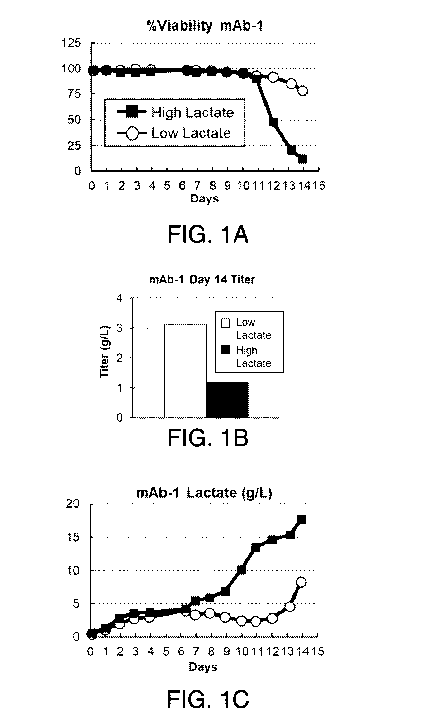
Figures IA-1F. Lactate levels of mAb-1 cell line directly correlate with the
PKM-
1 levels. Percent cell viability (Figure 1A), day 14 titer (Figure 1B), and
lactate levels
(Figure 1C) in mAb-1 cell line under low and high lactate processes. (Figure
1D) PKM-1
and PKM-2 western blot analysis of mAb-1 cell lysates at different days during
production
5
CA 03091231 2020-08-12
WO 2019/191552 PCT/US2019/024774
under the low and high lactate processes. Actin was used as the loading
control.
Quantification of relative PKM-1 (Figure 1E) and PKM-2 (Figure 1F) levels,
normalized
against Actin.
Figures 2A-2F. Lactate levels of mAb-2 cell line directly correlate with the
PKM-
1 levels. Percent cell viability (Figure 2A), day 14 titer (Figure 2B), and
lactate levels
(Figure 2C) in mAb-2 cell line under low and high lactate processes. (Figure
2D) PKM-1
and PKM-2 western blot analysis of mAb-2 cell lysates at different days during
production
under the low and high lactate processes. Actin was used as the loading
control.
Quantification of relative PKM-1 (Figure 2E) and PKM-2 (Figure 2F) levels,
normalized
against Actin.
Figures 3A-3E. CHO cells do express PKL/R enzymes, but levels of these
enzymes do not correlate with lactate levels in production culture. (Figure
3A) Western
blot analysis of intracellular expression of PKL/R proteins in two different
CHO host cell
lines (K1 and DHFR -/-) as well as human 293 cells. Actin is used as loading
control.
(Figures 3B and 3C) Intracellular protein levels of PKL/R at different days
during the low
and high lactate production processes of mAb-1 (Figure 3B) and mAb-2 (Figure
3C) cell
lines. (Figures 3D and 3E) Quantification of relative PKL/R levels normalized
against
Actin in mAb-1 (Figure 3D) and mAb-2 (Figure 3E) cell lines.
Figures 4A-4C. Generation of PKM-1 knock out cell lines. (Figure 4A) Schematic
of exons 8-10 ofPKA1 gene and gRNA and screening primers used to evaluate
deletion of
exon-9 in mAb-2 cell line. Two gRNAs flanking the exon 9 of MI gene were co-
transfected with Cas9 transgene to induce exon 9 deletion. Transfected cells
were single
cell cloned. PCR primers flanking the deletion region were used to screen cell
lines with
successful deletion of exon-9 in PKM allele(s). (Figure 4B) Wild type (WT) mAb-
2 cells
and cell lines with targeted deletion of exon-9 via gRNAs and Cas9 transgene
were
analyzed by screening PCR primers. Upper band represents WT PKM allele(s)
while lower
band represents PKM allele(s) with exon 9 deletions. KO: knock out cell lines,
HET:
heterozygous cell lines. (Figure 4C) Sequence comparison of PKM WT allele in
contrast
with exon-9 KO allele(s) in targeted cell lines. Note that KO-15 cell line
bears larger than
.. intended (by 122 bp) deletions in 5' region of exon-9 in targeted PKM
allele(s).
Figures 5A-511. Abolishing or reducing PKM-1 expression averted lactogenic
behaviors in mAb-2 cell line even under high-lactate process. Viable cell
count (Figure
5A), Percent cell viability (Figure 5B), day 14 titer (Figure 5C), day 14
specific
productivity (Figure 5D), and lactate levels (Figure 5E) of WT, and PKM-1 HET
and KO
6
CA 03091231 2020-08-12
WO 2019/191552 PCT/US2019/024774
mAb-2 cell lines during a 14-day fed-batch production assay using AMBR15
bioreactors.
Process control was the same as the high lactate process in Figure 2. (Figure
5F) Western
blot analysis of cell lysates of indicated cell lines for PKM-1 and PKM-2
proteins. Actin
is used as a loading control. (Figures 5G and 5H) Antibody product qualities
including
percent aggregation (Figure 5G) and percent charge variant (Figure 5H) of mAb-
2 from
indicated cell lines.
Figures 6A-6G. Lactogenic behavior is averted or reduced in PKM-1 KO or HET
mAb-2 cell lines, using the high-lactate process, irrespective of cell age or
post thaw.
Viable cell count (Figure 6A), percent cell viability (Figure 6B), day 14
titer (Figure 6C),
day 14 specific productivity (Figure 6D), and lactate levels (Figure 6E) of
indicated cell
lines, at a young cell age, in a 14-day fed-batch production assay using
AMBR15
bioreactors. (Figures 6F and 6G) Antibody product qualities including percent
aggregation
(Figure 6F) and percent charge variant (Figure 6G) of mAb-2 from indicated
cell lines.
Process control was the same as in Figure 5. Error bars represent standard
error from four
individual experiments.
Figure 7. MAb-3 producing pools derived from PKM and PKM-1 KO host cell
lines had comparable or higher Qp and titer, but lower growth (in shake flask
production).
Figures 8A-8B. MAb-3 producing pools derived from PKM and PKM-1 KO host
cell lines generated lower lactate (Figure 8A), but consumed more glucose
(Figure 8B) in
shake flask production.
Figure 9A. MAb-3 producing pools derived from PKM and PKM-1 KO host cell
lines exhibited different amino acid synthesis/consumption rates in shake
flask production.
Figure 9B. Bars and pathways enclosed in rectangles show that PKM KO host
cell lines accumulated 3-phosphoglycerate.
Figure 9C. Bars and pathways enclosed in rectangles show that WT host cell
lines
accumulated pyruvate.
Figure 9D. Bars and pathways enclosed in rectangles show that PKM-1 KO host
cell lines accumulated the TCA cycle product, alpha-ketoglutarate (a-KG).
Figure 9E. Bars and pathways enclosed in rectangles show that PKM-1 KO host
cell lines accumulated the TCA cycle product, oxaloacetate.
Figure 10. Host cell specific consumption or generation of glucose, lactate
and
amino acids.
Figure 11. MAb-3 producing pools derived from PKM and PKM-1 KO host cell
lines exhibited different glycosylation profiles in shake flask production.
PKM KO host
7
CA 03091231 2020-08-12
WO 2019/191552 PCT/US2019/024774
had decreased galactosylation, and PKM KO and PKM-1 KO host cell lines had
slightly
decreased fucosylation.
5. DETAILED DESCRIPTION
For clarity, but not by way of limitation, the detailed description of the
presently
disclosed subject matter is divided into the following subsections:
5.1 Definitions;
5.2 Modulating PKM expression;
5.3 Cells with reduced or eliminated lactogenic activity;
5.4 Cell culture methods;
5.5 Products; and
5.6 Exemplary embodiments.
5.1. Definitions
The terms used in this specification generally have their ordinary meanings in
the
art, within the context of this disclosure and in the specific context where
each term is
used. Certain terms are discussed below, or elsewhere in the specification, to
provide
additional guidance to the practitioner in describing the compositions and
methods of the
present disclosure and how to make and use them.
As used herein, the use of the word "a" or "an" when used in conjunction with
the
term "comprising" in the claims and/or the specification can mean "one," but
it is also
consistent with the meaning of "one or more," "at least one" and "one or more
than one."
The terms "comprise(s)," "include(s)," "having," "has," "can," "contain(s)"
and
variants thereof, as used herein, are intended to be open-ended transitional
phrases, terms
or words that do not preclude the possibility of additional acts or
structures. The present
disclosure also contemplates other embodiments "comprising," "consisting of'
and
"consisting essentially of" the embodiments or elements presented herein,
whether
explicitly set forth or not.
As used herein, the term "lactogenic behavior" or "lactogenic activity" refers
to
the lactate producing activity of a cell, for example, by consuming glucose
and generating
lactate through aerobic glycolysis. In certain embodiments, the lactogenic
activity of a
cell can be measured by the level of accumulated lactate in the cell culture
medium.
The term "about" or "approximately" means within an acceptable error range for
the particular value as determined by one of ordinary skill in the art, which
will depend in
8
CA 03091231 2020-08-12
WO 2019/191552 PCT/US2019/024774
part on how the value is measured or determined, i.e., the limitations of the
measurement
system. For example, "about" can mean within 3 or more than 3 standard
deviations, per
the practice in the art. Alternatively, "about" can mean a range of up to 20%,
preferably
up to 10%, more preferably up to 5%, and more preferably still up to 1% of a
given value.
Alternatively, particularly with respect to biological systems or processes,
the term can
mean within an order of magnitude, preferably within 5-fold, and more
preferably within
2-fold, of a value.
The terms "cell culture medium" and "culture medium" refer to a nutrient
solution
used for growing mammalian cells that typically provides at least one
component from
one or more of the following categories:
1) an energy source, usually in the form of a carbohydrate such as glucose;
2) all essential amino acids, and usually the basic set of twenty amino acids
plus
cysteine;
3) vitamins and/or other organic compounds required at low concentrations;
4) free fatty acids; and
5) trace elements, where trace elements are defined as inorganic compounds or
naturally occurring elements that are typically required at very low
concentrations,
usually in the micromolar range.
The nutrient solution can optionally be supplemented with one or more
components from
any of the following categories:
1) hormones and other growth factors as, for example, insulin, transferrin,
and
epidermal growth factor;
2) salts and buffers as, for example, calcium, magnesium, and phosphate;
3) nucleosides and bases such as, for example, adenosine, thymidine, and
hypoxanthine; and
4) protein and tissue hydrolysates.
"Culturing" a cell refers to contacting a cell with a cell culture medium
under
conditions suitable to the survival and/or growth and/or proliferation of the
cell.
"Batch culture" refers to a culture in which all components for cell culturing
(including the cells and all culture nutrients) are supplied to the culturing
bioreactor at the
start of the culturing process.
"Fed-batch cell culture," as used herein refers to a batch culture wherein the
cells
and culture medium are supplied to the culturing bioreactor initially, and
additional culture
nutrients are fed, continuously or in discrete increments, to the culture
during the culturing
9
CA 03091231 2020-08-12
WO 2019/191552 PCT/US2019/024774
process, with or without periodic cell and/or product harvest before
termination of culture.
"Perfusion culture," sometimes referred to as continuous culture, is a culture
by
which the cells are restrained in the culture by, e.g., filtration,
encapsulation, anchoring to
microcarriers, etc., and the culture medium is continuously, step-wise or
intermittently
introduced (or any combination of these) and removed from the culturing
bioreactor.
As used herein, the term "cell," refers to animal cells, mammalian cells,
cultured
cells, host cells, recombinant cells and recombinant host cells. Such cells
are generally cell
lines obtained or derived from mammalian tissues which are able to grow and
survive
when placed in media containing appropriate nutrients and/or growth factors.
The terms "host cell," "host cell line" and "host cell culture" are used
interchangeably and refer to cells into which exogenous nucleic acid has been
introduced,
including the progeny of such cells. Host cells include "transformants" and
"transformed
cells," which include the primary transformed cell and progeny derived
therefrom without
regard to the number of passages. Progeny does not need to be completely
identical in
nucleic acid content to a parent cell, but can contain mutations. Mutant
progeny that have
the same function or biological activity as screened or selected for in the
originally
transformed cell are included herein.
The term "mammalian host cell" or "mammalian cell" refers to cell lines
derived
from mammals that are capable of growth and survival when placed in either
monolayer
culture or in suspension culture in a medium containing the appropriate
nutrients and
growth factors. The necessary growth factors for a particular cell line are
readily
determined empirically without undue experimentation, as described for example
in
Mammalian Cell Culture (Mather, J. P. ed., Plenum Press, N.Y. 1984), and
Barnes and
Sato, (1980) Cell, 22:649. Typically, the cells are capable of expressing and
secreting
large quantities of a particular protein, e.g., glycoprotein, of interest into
the culture
medium. Examples of suitable mammalian host cells within the context of the
present
disclosure can include Chinese hamster ovary cells/-DHFR (CHO, Urlaub and
Chasin,
Proc. Natl. Acad. Sci. USA, 77:4216 1980); dp12.CHO cells (EP 307,247
published 15
Mar. 1989); CHO-K 1 (ATCC, CCL-61); monkey kidney CV1 line transformed by 5V40
(COS-7, ATCC CRL 1651); human embryonic kidney line (293 or 293 cells
subcloned for
growth in suspension culture, Graham et al., J. Gen Virol., 36:59 1977); baby
hamster
kidney cells (BHK, ATCC CCL 10); mouse sertoli cells (TM4, Mather, Biol.
Reprod.,
23:243-251 1980); monkey kidney cells (CV1 ATCC CCL 70); African green monkey
kidney cells (VERO-76, ATCC CRL-1587); human cervical carcinoma cells (HELA,
CA 03091231 2020-08-12
WO 2019/191552 PCT/US2019/024774
ATCC CCL 2); canine kidney cells (MDCK, ATCC CCL 34); buffalo rat liver cells
(BRL
3A, ATCC CRL 1442); human lung cells (W138, ATCC CCL 75); human liver cells
(Hep
G2, HB 8065); mouse mammary tumor (MMT 060562, ATCC CCL51); TRI cells (Mather
et al., Annals N.Y. Acad. Sci., 383:44-68 1982); MRC 5 cells; FS4 cells; and a
human
hepatoma line (Hep G2). In certain embodiments, the mammalian cells include
Chinese
hamster ovary cells/-DHFR (CHO, Urlaub and Chasin, Proc. Natl. Acad. Sci. USA,
77:4216 1980); dp12.CHO cells (EP 307,247 published 15 Mar. 1989).
The term "peptone" within the context of the present disclosure is meant to
refer
to a media supplement that is essentially hydrolyzed animal protein. The
source of this
protein can be animal by-products from slaughter houses, purified gelatin, or
plant
material. The protein is typically hydrolyzed using acid, heat or various
enzyme
preparations.
"Growth phase" of the cell culture refers to the period of exponential cell
growth
(the log phase) where cells are generally rapidly dividing. The duration of
time for which
the cells are maintained at growth phase can vary based on the cell-type, the
rate of growth
of cells and/or the culture conditions, for example. In certain embodiments,
during this
phase, cells are cultured for a period of time, usually between 1-4 days, and
under such
conditions that cell growth is maximized. The determination of the growth
cycle for the
host cell can be determined for the particular host cell envisioned without
undue
experimentation. "Period of time and under such conditions that cell growth is
maximized" and the like, refer to those culture conditions that, for a
particular cell line,
are determined to be optimal for cell growth and division. In certain
embodiments, during
the growth phase, cells are cultured in nutrient medium containing the
necessary additives
generally at about 30 -40 C in a humidified, controlled atmosphere, such that
optimal
growth is achieved for the particular cell line. In certain embodiments, cells
are maintained
in the growth phase for a period of about between one and four days, usually
between two
to three days.
"Transition phase" of the cell culture refers to the period of time during
which
culture conditions for the production phase are engaged. During the transition
phase
environmental factors such as temperature of the cell culture, medium
osmolality and the
like are shifted from growth conditions to production conditions.
"Production phase" of the cell culture refers to the period of time during
which cell
growth is/has plateaued. The logarithmic cell growth typically decreases
before or during
this phase and protein production takes over. During the production phase,
logarithmic
11
CA 03091231 2020-08-12
WO 2019/191552 PCT/US2019/024774
cell growth has ended, and protein production is primary. During this period
of time the
medium is generally supplemented to support continued protein production and
to achieve
the desired glycoprotein product. Fed-batch and/or perfusion cell culture
processes
supplement the cell culture medium or provide fresh medium during this phase
to achieve
and/or maintain desired cell density, viability and/or recombinant protein
product titer. A
production phase can be conducted at large scale.
The term "expression" or "expresses" are used herein to refer to transcription
and
translation occurring within a host cell. The level of expression of a product
gene in a host
cell can be determined on the basis of either the amount of corresponding mRNA
that is
present in the cell or the amount of the protein encoded by the product gene
that is
produced by the cell. For example, mRNA transcribed from a product gene is
desirably
quantitated by northern hybridization. Sambrook et al., Molecular Cloning: A
Laboratory
Manual, pp. 7.3-7.57 (Cold Spring Harbor Laboratory Press, 1989). Protein
encoded by a
product gene can be quantitated either by assaying for the biological activity
of the protein
or by employing assays that are independent of such activity, such as western
blotting or
radioimmunoassay using antibodies that are capable of reacting with the
protein.
Sambrook et al., Molecular Cloning: A Laboratory Manual, pp. 18.1-18.88 (Cold
Spring
Harbor Laboratory Press, 1989).
As used herein, "polypeptide" refers generally to peptides and proteins having
more than about ten amino acids. The polypeptides can be homologous to the
host cell, or
preferably, can be exogenous, meaning that they are heterologous, i.e.,
foreign, to the host
cell being utilized, such as a human protein produced by a Chinese hamster
ovary cell, or
a yeast polypeptide produced by a mammalian cell. In certain embodiments,
mammalian
polypeptides (polypeptides that were originally derived from a mammalian
organism) are
used, more preferably those which are directly secreted into the medium.
The term "protein" is meant to refer to a sequence of amino acids for which
the
chain length is sufficient to produce the higher levels of tertiary and/or
quaternary
structure. This is to distinguish from "peptides" or other small molecular
weight drugs that
do not have such structure. Typically, the protein herein will have a
molecular weight of
at least about 15-20 kD, preferably at least about 20 kD. Examples of proteins
encompassed within the definition herein include all mammalian proteins, in
particular,
therapeutic and diagnostic proteins, such as therapeutic and diagnostic
antibodies, and, in
general proteins that contain one or more disulfide bonds, including multi-
chain
polypeptides comprising one or more inter- and/or intrachain disulfide bonds.
12
CA 03091231 2020-08-12
WO 2019/191552 PCT/US2019/024774
The term "antibody" is used herein in the broadest sense and encompasses
various
antibody structures including, but not limited to, monoclonal antibodies,
polyclonal
antibodies, monospecific antibodies (e.g., antibodies consisting of a single
heavy chain
sequence and a single light chain sequence, including multimers of such
pairings),
multispecific antibodies (e.g., bispecific antibodies) and antibody fragments
so long as
they exhibit the desired antigen-binding activity.
An "antibody fragment," "antigen-binding portion" of an antibody (or simply
"antibody portion") or "antigen-binding fragment" of an antibody, as used
herein, refers
to a molecule other than an intact antibody that comprises a portion of an
intact antibody
that binds the antigen to which the intact antibody binds. Examples of
antibody fragments
include, but are not limited to, Fv, Fab, Fab', Fab' -SH, F(ab')2; diabodies;
linear
antibodies; single-chain antibody molecules (e.g., scFv, and scFab); single
domain
antibodies (dAbs); and multi specific antibodies formed from antibody
fragments. For a
review of certain antibody fragments, see Holliger and Hudson, Nature
Biotechnology
23:1126-1136 (2005).
The term "chimeric" antibody refers to an antibody in which a portion of the
heavy
and/or light chain is derived from a particular source or species, while the
remainder of the
heavy and/or light chain is derived from a different source or species.
The "class" of an antibody refers to the type of constant domain or constant
region
possessed by its heavy chain. There are five major classes of antibodies: IgA,
IgD, IgE,
IgG and IgM, and several of these can be further divided into subclasses
(isotypes), e.g.,
IgG2, IgG3, IgG4, IgAi, and IgA2. In certain embodiments, the antibody is of
the
IgGi isotype. In certain embodiments, the antibody is of the IgG2isotype. The
heavy chain
constant domains that correspond to the different classes of immunoglobulins
are called
a, 6, 6, y and , respectively. The light chain of an antibody can be assigned
to one of two
types, called kappa (x) and lambda (k), based on the amino acid sequence of
its constant
domain.
The term "titer" as used herein refers to the total amount of recombinantly
expressed antibody produced by a cell culture divided by a given amount of
medium
volume. Titer is typically expressed in units of milligrams of antibody per
milliliter or
liter of medium (mg/ml or mg/L). In certain embodiments, titer is expressed in
grams of
antibody per liter of medium (g/L). Titer can be expressed or assessed in
terms of a relative
measurement, such as a percentage increase in titer as compared obtaining the
protein
13
CA 03091231 2020-08-12
WO 2019/191552 PCT/US2019/024774
product under different culture conditions.
The term "nucleic acid," "nucleic acid molecule" or "polynucleotide" includes
any
compound and/or substance that comprises a polymer of nucleotides. Each
nucleotide is
composed of a base, specifically a purine- or pyrimidine base (i.e., cytosine
(C), guanine
(G), adenine (A), thymine (T) or uracil (U)), a sugar (i.e., deoxyribose or
ribose), and a
phosphate group. Often, the nucleic acid molecule is described by the sequence
of bases,
whereby said bases represent the primary structure (linear structure) of a
nucleic acid
molecule. The sequence of bases is typically represented from 5' to 3'.
Herein, the term
nucleic acid molecule encompasses deoxyribonucleic acid (DNA) including, e.g.,
complementary DNA (cDNA) and genomic DNA, ribonucleic acid (RNA), in
particular
messenger RNA (mRNA), synthetic forms of DNA or RNA, and mixed polymers
comprising two or more of these molecules. The nucleic acid molecule can be
linear or
circular. In addition, the term nucleic acid molecule includes both, sense and
antisense
strands, as well as single stranded and double stranded forms. Moreover, the
herein
described nucleic acid molecule can contain naturally occurring or non-
naturally occurring
nucleotides. Examples of non-naturally occurring nucleotides include modified
nucleotide
bases with derivatized sugars or phosphate backbone linkages or chemically
modified
residues. Nucleic acid molecules also encompass DNA and RNA molecules which
are
suitable as a vector for direct expression of an antibody of the disclosure in
vitro and/or in
vivo, e.g., in a host or patient. Such DNA (e.g., cDNA) or RNA (e.g., mRNA)
vectors,
can be unmodified or modified. For example, mRNA can be chemically modified to
enhance the stability of the RNA vector and/or expression of the encoded
molecule so that
mRNA can be injected into a subject to generate the antibody in vivo (see,
e.g., Stadler et
al, Nature Medicine 2017, published online 12 June 2017, doi:10.1038/nm.4356
or EP 2
.. 101 823 B1).
As used herein, the term "vector" refers to a nucleic acid molecule capable of
transporting another nucleic acid to which it has been linked.
The term "hybridoma" refers to a hybrid cell line produced by the fusion of an
immortal cell line of immunologic origin and an antibody producing cell. The
term
encompasses progeny of heterohybrid myeloma fusions, which are the result of a
fusion
with human cells and a murine myeloma cell line subsequently fused with a
plasma cell,
commonly known as a trioma cell line. Furthermore, the term is meant to
include any
immortalized hybrid cell line which produces antibodies such as, for example,
quadromas.
See, e.g., Milstein et al., Nature, 537:3053 (1983).
14
CA 03091231 2020-08-12
WO 2019/191552 PCT/US2019/024774
A "human antibody" is one which possesses an amino acid sequence which
corresponds to that of an antibody produced by a human or a human cell or
derived from
a non-human source that utilizes human antibody repertoires or other human
antibody-
encoding sequences. This definition of a human antibody specifically excludes
a
humanized antibody comprising non-human antigen-binding residues.
A "humanized" antibody refers to a chimeric antibody comprising amino acid
residues from non-human CDRs and amino acid residues from human FRs. In
certain
aspects, a humanized antibody will comprise substantially all of at least one,
and typically
two, variable domains, in which all or substantially all of the CDRs
correspond to those of
a non-human antibody, and all or substantially all of the FRs correspond to
those of a
human antibody. A humanized antibody optionally can comprise at least a
portion of an
antibody constant region derived from a human antibody. A "humanized form" of
an
antibody, e.g., a non-human antibody, refers to an antibody that has undergone
humanization.
The term "hypervariable region" or "HVR" as used herein refers to each of the
regions of an antibody variable domain which are hypervariable in sequence and
which
determine antigen binding specificity, for example "complementarity
determining
regions" ("CDRs").
Generally, antibodies comprise six CDRs: three in the VH (CDR-H1, CDR-H2,
CDR-H3), and three in the VL (CDR-L1, CDR-L2, CDR-L3). Exemplary CDRs herein
include:
(a) hypervariable loops occurring at amino acid residues 26-32 (L1), 50-52
(L2),
91-96 (L3), 26-32 (H1), 53-55 (H2), and 96-101 (H3) (Chothia and Lesk, I Mot.
Biol.
196:901-917 (1987));
(b) CDRs occurring at amino acid residues 24-34 (L1), 50-56 (L2), 89-97 (L3),
31-35b (H1), 50-65 (H2), and 95-102 (H3) (Kabat et al., Sequences of Proteins
of
Immunological Interest, 5th Ed. Public Health Service, National Institutes of
Health,
Bethesda, MD (1991)); and
(c) antigen contacts occurring at amino acid residues 27c-36 (L1), 46-55 (L2),
89-
96 (L3), 30-35b (H1), 47-58 (H2), and 93-101 (H3) (MacCallum et al. I Mol.
Biol. 262:
732-745 (1996)).
Unless otherwise indicated, the CDRs are determined according to Kabat et al.,
supra. One of skill in the art will understand that the CDR designations can
also be
determined according to Chothia, supra, McCallum, supra, or any other
scientifically
CA 03091231 2020-08-12
WO 2019/191552 PCT/US2019/024774
accepted nomenclature system.
An "immunoconjugate" is an antibody conjugated to one or more heterologous
molecule(s), including but not limited to a cytotoxic agent.
The term "monoclonal antibody" as used herein refers to an antibody obtained
from
a population of substantially homogeneous antibodies, i.e., the individual
antibodies
comprising the population are identical and/or bind the same epitope, except
for possible
variant antibodies, e.g., containing naturally occurring mutations or arising
during
production of a monoclonal antibody preparation, such variants generally being
present in
minor amounts. In contrast to polyclonal antibody preparations, which
typically include
different antibodies directed against different determinants (epitopes), each
monoclonal
antibody of a monoclonal antibody preparation is directed against a single
determinant on
an antigen. Thus, the modifier "monoclonal" indicates the character of the
antibody as
being obtained from a substantially homogeneous population of antibodies, and
is not to
be construed as requiring production of the antibody by any particular method.
For
example, the monoclonal antibodies in accordance with the presently disclosed
subject
matter can be made by a variety of techniques, including but not limited to
the hybridoma
method, recombinant DNA methods, phage-display methods, and methods utilizing
transgenic animals containing all or part of the human immunoglobulin loci,
such methods
and other exemplary methods for making monoclonal antibodies being described
herein.
The term "variable region" or "variable domain" refers to the domain of an
antibody heavy or light chain that is involved in binding the antibody to
antigen. The
variable domains of the heavy chain and light chain (VH and VL, respectively)
of a native
antibody generally have similar structures, with each domain comprising four
conserved
framework regions (FRs) and three complementary determining regions (CDRs).
(See,
e.g., Kindt et al. Kuby Immunology, 6th ed., W.H. Freeman and Co., page 91
(2007).) A
single VH or VL domain can be sufficient to confer antigen-binding
specificity.
Furthermore, antibodies that bind a particular antigen can be isolated using a
VH or VL
domain from an antibody that binds the antigen to screen a library of
complementary VL
or VH domains, respectively. See, e.g., Portolano et al., I Immunol. 150:880-
887 (1993);
Clarkson et al., Nature 352:624-628 (1991).
As used herein, the term "cell density" refers to the number of cells in a
given
volume of medium. In certain embodiments, a high cell density is desirable in
that it can
lead to higher protein productivity. Cell density can be monitored by any
technique known
in the art, including, but not limited to, extracting samples from a culture
and analyzing
16
CA 03091231 2020-08-12
WO 2019/191552 PCT/US2019/024774
the cells under a microscope, using a commercially available cell counting
device or by
using a commercially available suitable probe introduced into the bioreactor
itself (or into
a loop through which the medium and suspended cells are passed and then
returned to the
bioreactor).
As used herein, the term "seeding" refers to the addition or inoculation of
growing
cells into a culture medium at the beginning of the production phase. Further,
as used
herein, the term "seed train" refers to a continual passaging of cells in
volumes of culture
medium of about 20 L or less for the maintenance of the cell line.
As used herein, the term "recombinant cell" refers to cells which have some
genetic
modification from the original parent cells from which they are derived. Such
genetic
modification can be the result of an introduction of a heterologous gene for
expression of
the gene product, e.g., a recombinant protein.
As used herein, the term "recombinant protein" refers generally to peptides
and
proteins, including antibodies. Such recombinant proteins are "heterologous,"
i.e., foreign
to the host cell being utilized, such as an antibody produced by CHO cells.
As used herein, a "PKM polypeptide" refers to a polypeptide that is encoded by
the P1cA1 gene. A PKM polypeptide includes the PKM-1 polypeptide isoform
and/or the
PKM-2 polypeptide isoform.
5.2. Modulating PKM expression
Glycolysis is the process of glucose metabolism used by mammalian cells, e.g.,
CHO cells, to generate energy. Glycolysis can occur at high or low flux
states. The cell
response to glucose levels and the switch between these flux states can vary
depending on
the cell lines and the levels and the combinations of isozymes present in the
glycolytic
pathway (Mulukutla et al., 2014, PLoS One 9(6):e98756). Under normal culture
conditions, CHO cells, like other immortalized cell lines, tend to consume
glucose and
generate lactate through aerobic glycolysis, a process known as the Warburg
effect
(Warburg, 1956, Science 123(3191):309-14). Accumulation of lactate in
production
cultures can adversely affect cellular growth, viability and productivity.
Therefore,
regulation of cellular energy flux and metabolism can be leveraged to avoid
undesirable
outcomes caused by lactate accumulation during the cell culture production
phase
(Mulukutla et al., 2010, Trends Biotechnol. 28(9):476-84; Luo et al., 2012,
Biotechnol.
Bioeng. 109(1):146-56; Ahn and Antoniewicz, 2012, Biotechnol. J. 7(1):61-74).
The final step of the glycolysis process involves conversion of
17
CA 03091231 2020-08-12
WO 2019/191552 PCT/US2019/024774
phosphoenolpyruvate (PEP) to pyruvate, which is mediated by the pyruvate
kinase (PK)
enzymes. Four different isoforms of PK have been identified: PK liver (PKL),
PK red
blood cells (PKR), PK muscle 1 (PKM-1) and PK muscle 2 (PKM-2). The PK enzymes
are expressed by two different genes, PKLR and PK M. Alternative exon splicing
gives
rise to different PK isoforms. The presence of both exons 1 and 2 in an mRNA
transcript
from the PKLR gene results in expression of PKR protein, whereas an mRNA
transcript
that starts with exon 2 results in the expression of PKL protein. Alternative
splicing of
exon 9 or 10 in the P1cA1 gene transcript gives rise to PKM-1 or PKM-2,
respectively.
Specifically, PKM-1 includes exon 9 and excludes exon 10 of the P1cA1 gene,
and PKM-2
includes exon 10 and excludes exon 9 of the P1cA1 gene. Tissue specific
promoters,
transcription factors, and alternative splicing regulate expression of these
isoforms in
different tissues and cell lines (Chaneton and Gottlieb, 2012, Trends.
Biochem. Sci.
37(8):309-16; Israelsen and Vander Heiden, 2015, Semin Cell Dev Biol 43:43-51;
Mazurek, 2011, Int. J. Biochem. Cell Biol 43(7):969-80; Harada et al., 1978,
Biochim.
Biophys. Acta. 524(2):327-39; Noguchi et al., 1986, J. Biol. Chem.
261(29):13807-12;
Noguchi et al., 1987, J. Biol. Chem. 262(29):14366-71).
PKM-2 has been extensively studied due to its central role in cancer cell
metabolism and tumor growth, hallmarked by high glucose consumption and
lactate
production. While the PKM-1 enzyme is constitutively active, PKM-2 activity is
regulated
by oligomerization, substrate binding and post-translational modifications
(Christofk et
al., 2008, Nature 452(7184):230-3; Chaneton and Gottlieb, 2012, Trends
Biochem. Sci.
37(8):309-16; Israelsen and Vander Heiden, 2015, Semin. Cell Dev. Biol 43:43-
51). For
example, fructose 1,6-bisphosphate (FBP) reversibly binds to PKM-2, promoting
its
tetramerization and hence activation (Ashizawa et al., 1991, J. Biol. Chem.
266(25):16842-6; Dombrauckas et al., 2005, Biochemistry 44(27):9417-29).
Phosphorylation of PKM-2 at tyrosine 105 or its binding to other
phosphotyrosine proteins
inactivates PKM-2 by blocking FBP binding and preventing PKM-2 tetramerization
(Christofk et al., 2008, Nature 452(7184):181-6; Hitosugi et al., 2009, Sci.
Signal
2(97):ra73). Increased levels of glycolysis, however, promote acetylation of
PKM-2 at
lysine 305 as part of a metabolic feedback loop, which targets PKM-2 for its
degradation
via chaperone-mediated autophagy (Lv et al., 2011, Mol. Cell 42(6):719-30)
(Macintyre
and Rathmell, 2011, Mol. Cell 42(6):713-4). Additionally, PKM-2 dimers have
been
shown to localize to the cell nucleus acting as protein kinases, utilizing PEP
as a phosphate
donor, to promote cell proliferation through phosphorylation of STAT3 and
activation of
18
CA 03091231 2020-08-12
WO 2019/191552 PCT/US2019/024774
MEK5 (Gao et al., 2012, Mol. Cell 45(5):598-609).
In accordance with one aspect, the present disclosure relates to methods for
modulating lactogenic activity of a mammalian cell by modulating PKM
expression, e.g.,
PKM polypeptide expression, in the cell. For example, but not by way of
limitation,
methods for modulating lactogenic activity of a mammalian cell include
knocking out or
knocking down PKM polypeptide expression in the cell. In certain embodiments,
the
expression of PKM-1 is knocked down or knocked out. In certain embodiments,
the
expression of PKM-2 is knocked down or knocked out. In certain embodiments,
the
expression of both PKM-1 and PKM-2 are knocked down or knocked out. As used
herein,
knocked out expression refers to the elimination of the expression of a PKM
polypeptide,
e.g., a PKM-1 polypeptide and/or a PKM-2 polypeptide, in the cell as compared
to a
reference cell. As used herein, knocked down expression refers to a reduction
in the
expression of a PKM polyp eptide, e.g., a PKM-1 polypeptide and/or a PKM-2
polyp epti de,
in the cell as compared to a reference cell.
In certain embodiments, the reference cells are cells where the expression of
a
PKM polypeptide, e.g., PKM-1 and/or PKM-2, is not modulated, e.g., reduced. In
certain
embodiments, a reference cell is a cell that comprises at least one or both
wild-type alleles
of the PKA1 gene. For example, but not by way of limitation, a reference cell
is a cell that
has both wild-type PKA1 alleles. In certain embodiments, the reference cells
are WT CHO
cells.
In certain embodiments, the expression of a PKM polypeptide, e.g., PKM-1
and/or
PKM-2, in a cell that has been modified to knock down expression of the PKM
polypeptide
is less than about 90%, less than about 80%, less than about 70%, less than
about 60%,
less than about 50%, less than about 40%, less than about 30%, less than about
20%, less
than about 10%, less than about 5%, less than about 4%, less than about 3%,
less than
about 2% or less than about 1% of the PKM polypeptide expression of a
reference cell,
e.g., a WT CHO cell. In certain embodiments, the expression of a PKM-1
polypeptide in
a cell that has been modified to knock down expression of the PKM-1
polypeptide is less
than about 90%, less than about 80%, less than about 70%, less than about 60%,
less than
about 50%, less than about 40%, less than about 30%, less than about 20%, less
than about
10%, less than about 5%, less than about 4%, less than about 3%, less than
about 2%, or
less than about 1% of the PKM-1 polypeptide expression of a reference cell,
e.g., a WT
CHO cell.
In certain embodiments, the expression of a PKM polypeptide, e.g., PKM-1
and/or
19
CA 03091231 2020-08-12
WO 2019/191552 PCT/US2019/024774
PKM-2, in a cell that has been modified to knock down expression of the PKM
polypeptide
is at least about 90%, at least about 80%, at least about 70%, at least about
60%, at least
about 50%, at least about 40%, at least about 30%, at least about 20%, at
least about 10%,
at least about 5%, at least about 4%, at least about 3%, at least about 2% or
at least about
1% of the PKM polypeptide expression of a reference cell, e.g., a WT CHO cell.
In certain
embodiments, the expression of a PKM-1 polypeptide in a cell that has been
modified to
knock down expression of the PKM-1 polypeptide is at least about 90%, at least
about
80%, at least about 70%, at least about 60%, at least about 50%, at least
about 40%, at
least about 30%, at least about 20%, at least about 10%, at least about 5%, at
least about
.. 4%, at least about 3%, at least about 2%, or at least about 1% of the PKM-1
polypeptide
expression of a reference cell, e.g., a WT CHO cell.
In certain embodiments, the expression of a PKM polypeptide, e.g., PKM-1
and/or
PKM-2, in a cell that has been modified to knock down expression of the PKM
polypeptide
is no more than about 90%, no more than about 80%, no more than about 70%, no
more
than about 60%, no more than about 50%, no more than about 40%, no more than
about
30%, no more than about 20%, no more than about 10%, no more than about 5%, no
more
than about 4%, no more than about 3%, no more than about 2% or no more than
about 1%
of the PKM polypeptide expression of a reference cell, e.g., a WT CHO cell. In
certain
embodiments, the expression of a PKM polypeptide, e.g., PKM-1 and/or PKM-2, in
a cell
that has been modified to knock down expression of the PKM polypeptide is no
more than
about 40% of the PKM polypeptide expression of a reference cell, e.g., a WT
CHO cell.
In certain embodiments, the expression of a PKM-1 polypeptide in a cell that
has been
modified to knock down expression of the PKM-1 polypeptide is no more than
about 90%,
no more than about 80%, no more than about 70%, no more than about 60%, no
more than
.. about 50%, no more than about 40%, no more than about 30%, no more than
about 20%,
no more than about 10%, no more than about 5%, no more than about 4%, no more
than
about 3%, no more than about 2% or no more than about 1% of the PKM-1
polypeptide
expression of a reference cell, e.g., a WT CHO cell.
In certain embodiments, the expression of a PKM polypeptide, e.g., PKM-1
and/or
.. PKM-2, in a cell that has been modified to knock down expression of the PKM
polypeptide
is between about 1% and about 90%, between about 10% and about 90%, between
about
20% and about 90%, between about 25% and about 90%, between about 30% and
about
90%, between about 40% and about 90%, between about 50% and about 90%, between
about 60% and about 90%, between about 70% and about 90%, between about 80%
and
CA 03091231 2020-08-12
WO 2019/191552 PCT/US2019/024774
about 90%, between about 85% and about 900 o, between about 100 and about 800
o,
between about 10% and about 80%, between about 20% and about 80%, between
about
30% and about 80%, between about 40% and about 80%, between about 50% and
about
80%, between about 60% and about 80%, between about 70% and about 80%, between
about 7500 and about 80%, between about 10o and about 70%, between about 10%
and
about 70%, between about 2000 and about 70%, between about 30% and about 70%,
between about 40% and about 70%, between about 500o and about 70%, between
about
60% and about 70%, between about 65% and about 70%, between about 1% and about
60%, between about 10% and about 60%, between about 20% and about 60%, between
about 30% and about 60%, between about 40% and about 60%, between about 50%
and
about 60%, between about 55% and about 60%, between about 1% and about 50%,
between about 10% and about 50%, between about 20% and about 50%, between
about
30% and about 50%, between about 40% and about 50%, between about 45% and
about
50%, between about 1% and about 40%, between about 10% and about 40%, between
about 20% and about 40%, between about 30% and about 40%, between about 35%
and
about 40%, between about 1% and about 30%, between about 10% and about 30%,
between about 20% and about 30%, between about 25% and about 30%, between
about
1% and about 20%, between about 5% and about 20%, between about 10% and about
20%,
between about 15% and about 20%, between about 1% and about 10%, between about
5%
and about 10%, between about 5% and about 20%, between about 5% and about 30%,
between about 5% and about 40% of the PKM-1 polypeptide expression of a
reference
cell, e.g., a WT CHO cell. In certain embodiments, the expression of a PKM-1
polypeptide
in a cell that has been modified to knock down expression of the PKM-1
polypeptide is
between about 1% and about 90%, between about 10% and about 90%, between about
20% and about 90%, between about 25% and about 90%, between about 30% and
about
90%, between about 40% and about 90%, between about 50% and about 90%, between
about 60% and about 90%, between about 70% and about 90%, between about 80%
and
about 90%, between about 85% and about 90%, between about 1% and about 80%,
between about 10% and about 80%, between about 20% and about 80%, between
about
30% and about 80%, between about 40% and about 80%, between about 50% and
about
80%, between about 60% and about 80%, between about 70% and about 80%, between
about 75% and about 80%, between about 1% and about 70%, between about 10% and
about 70%, between about 20% and about 70%, between about 30% and about 70%,
between about 40% and about 70%, between about 50% and about 70%, between
about
21
CA 03091231 2020-08-12
WO 2019/191552 PCT/US2019/024774
600 o and about 70%, between about 65% and about 700 o, between about 100 and
about
60%, between about 10% and about 60%, between about 2000 and about 60%,
between
about 30% and about 60%, between about 40% and about 60%, between about 50%
and
about 60%, between about 5500 and about 60%, between about 10o and about 50%,
between about 10% and about 50%, between about 20% and about 50%, between
about
30% and about 50%, between about 40% and about 50%, between about 45% and
about
50%, between about 1% and about 40%, between about 10% and about 40%, between
about 20% and about 40%, between about 30% and about 40%, between about 35%
and
about 40%, between about 1% and about 30%, between about 10% and about 30%,
between about 20% and about 30%, between about 25% and about 30%, between
about
1% and about 20%, between about 5% and about 20%, between about 10% and about
20%,
between about 15% and about 20%, between about 1% and about 10%, between about
5% and about 10%, between about 5% and about 20%, between about 5% and about
30%,
between about 5% and about 40% of the PKM-1 polypeptide expression of a
reference
cell, e.g., a WT CHO cell.
In certain embodiments, the expression of a PKM polypeptide, e.g., PKM-1
and/or
PKM-2, in a cell that has been modified to knock down expression of the PKM
polypeptide
is between about 5% and about 40% of the PKM polypeptide expression of a
reference
cell, e.g., a WT CHO cell. In certain embodiments, the expression of a PKM-1
polypeptide
in a cell that has been modified to knock down expression of the PKM-1
polypeptide is
between about 5% and about 40% of the PKM-1 polypeptide expression of a
reference
cell, e.g., a WT CHO cell. The expression level of the PKM polypeptide, e.g.,
PKM-1
and/or PKM-2, in different reference cells (e.g., cells that comprise at least
one or both
wild-type alleles of the PKM gene) can vary. For example, seed train cells or
low-lactate
producing cells may produce low levels of PKM-1, whereas high-lactate
producing cells
may produce high levels of PKM-1.
Alternative splicing of exon 9 or 10 in the PKAIgene transcript gives rise to
PKM-
1 or PKM-2, respectively. The P1cA1 gene that is knocked down or knocked out
can be a
P1cA1 gene from a human. In certain embodiments, the P1cA1 gene can be from a
non-
human, e.g., a Rhesus Monkey, canine, green monkey, chicken, cattle, pig,
mouse, Chinese
hamster, rat or rabbit.
In certain embodiments, the P1cA1 gene that is knocked down or knocked out is
a
Chinese hamster P1cA1 gene. In certain embodiments, the Chinese hamster P1cA1
gene
sequence has a NCBI Reference Sequence ID NW 003613709.1 (range:
200602..223561)
22
CA 03091231 2020-08-12
WO 2019/191552 PCT/US2019/024774
(SEQ ID NO: 44). In certain embodiments, the PM/ gene sequence differs by 1,
2, 3, 4,
5, 6, 7, 8, 9, 10, 11, 12, 13, 14, 15, 16, 17, 18, 19, 20 or more nucleotides
from the
NW 003613709.1 sequence. In certain embodiments, the PM/ gene sequence differs
by
1%, 2%, 3%, 4%, 5%, 6%, 7%, 8%, 9%, 10%, 11%, 12%, 13%, 14%, 15%, 16%, 17%,
18%, 19%, 20% or more from the sequence set forth in SEQ ID NO: 44.
Additional non-limiting examples of PM/ genes include the human PM/ gene
(e.g., NC 000015.10 (range 72199029..72231624) (SEQ ID NO: 45)), the Rhesus
monkey
PM/gene (e.g., NC 027899.1 (range 49211766..49245983) (SEQ ID NO: 46)), the
green
monkey PM/ gene (e.g., NC 023667.1 (range 11224332..11255538) (SEQ ID NO:
47)),
the canine PM/ gene (e.g., NC 006612.3 (range 35712853..35737643) (SEQ ID NO:
48)), the mouse PM/ gene (e.g., NC 000075.6 (range 59656368..59679375) (SEQ ID
NO: 49)), the rat PM/ gene (e.g., NC 005107.4 (range 64480963..64502957) (SEQ
ID
NO: 50)), the rabbit PM/gene (e.g., NC 013685.1 (range 304096..317843) (SEQ ID
NO:
51)), the chicken PM/gene (e.g., NC 006097.4 (range 1506428..1523684) (SEQ ID
NO:
52)), the pig PM/ gene (e.g., NC 010449.5 (range 60971807..61032780) (SEQ ID
NO:
53)) and the cattle PM/ gene (e.g., AC 000167.1 (range 18965981..18992644)
(SEQ ID
NO: 54)). In certain embodiments, the PM/gene sequence differs by 1, 2, 3,4,
5, 6, 7, 8,
9, 10, 11, 12, 13, 14, 15, 16, 17, 18, 19, 20 or more nucleotides from any one
of the
sequences set forth in SEQ ID NOs: 45-54. In certain embodiments, the PM/ gene
sequence differs by 1%, 2%, 3%, 4%, 5%, 6%, 7%, 8%, 9%, 10%, 11%, 12%, 13%,
14%,
15%, 16%, 17%, 18%, 19%, 20% or more from any one of the sequences set forth
in SEQ
ID NOs: 45-54.
One skilled in the art would know that different mammalian cell lines, even
those
from the same species, e.g., two CHO distinct host cell lines, may not share
identical PM/
gene sequences. Small differences in the sequences of PM/ gene, e.g.,
differences of 1,
2, 3, 4, 5, 6, 7, 8, 9, 10, 11, 12, 13, 14, 15, 16, 17, 18, 19 or 20
nucleotides, can exist
between two mammalian cell lines from the same species.
5.2.1 Methods for Modulating PK1VI expression
In certain embodiments, a genetic engineering system is employed to modulate
(e.g., knock down or knock out) the expression of a PKM polypeptide (e.g., PKM-
1
expression). Various genetic engineering systems known in the art can be used
for the
methods disclosed herein. Non-limiting examples of such systems include the
CRISPR/Cas system, the zinc-finger nuclease (ZFN) system, the transcription
activator-
23
CA 03091231 2020-08-12
WO 2019/191552 PCT/US2019/024774
like effector nuclease (TALEN) system and the use of other tools for protein
knockdown
by gene silencing, such as small interfering RNAs (siRNAs), short hairpin RNA
(shRNA),
and microRNA (miRNA). Any CRISPR/Cas systems known in the art, including
traditional, enhanced or modified Cas systems, as well as other bacterial
based genome
excising tools such as Cpf-1 can be used with the methods disclosed herein.
Any PKM inhibitors known in the art can also be used with the methods
disclosed
herein to modulate PKM activity, and thus modulate lactogenic activity of the
cells
disclosed herein. Non-limiting examples of PKM inhibitors include sodium
monofluorophosphate, L-phenylalanine, creatine phosphate, Ca2+, flurophosphate
and
pyridoxal 5' -phosphate.
In certain embodiments, a portion of the P1cA1 gene is deleted to modulate,
e.g.,
knock down or knock out, expression of a PKM polypeptide. In certain
embodiments, at
least about 2%, at least about 5%, at least about 10%, at least about 15%, at
least about
20%, at least about 25%, at least about 30%, at least about 35%, at least
about 40%, at
least about 45%, at least about 50%, at least about 55%, at least about 60%,
at least about
65%, at least about 70%, at least about 75%, at least about 80%, at least
about 85% or at
least about 90% of the PAM gene is deleted. In certain embodiments, no more
than about
2%, no more than about 5%, no more than about 10%, no more than about 15%, no
more
than about 20%, no more than about 25%, no more than about 30%, no more than
about
35%, no more than about 40%, no more than about 45%, no more than about 50%,
no
more than about 55%, no more than about 60%, no more than about 65%, no more
than
about 70%, no more than about 75%, no more than about 80%, no more than about
85%
or no more than about 90% of the PAM gene is deleted. In certain embodiments,
between
about 2% and about 90%, between about 10% and about 90%, between about 20% and
about 90%, between about 25% and about 90%, between about 30% and about 90%,
between about 40% and about 90%, between about 50% and about 90%, between
about
60% and about 90%, between about 70% and about 90%, between about 80% and
about
90%, between about 85% and about 90%, between about 2% and about 80%, between
about 10% and about 80%, between about 20% and about 80%, between about 30%
and
about 80%, between about 40% and about 80%, between about 50% and about 80%,
between about 60% and about 80%, between about 70% and about 80%, between
about
75% and about 80%, between about 2% and about 70%, between about 10% and about
70%, between about 20% and about 70%, between about 30% and about 70%, between
about 40% and about 70%, between about 50% and about 70%, between about 60%
and
24
CA 03091231 2020-08-12
WO 2019/191552 PCT/US2019/024774
about 70%, between about 65% and about 700 o, between about 2% and about 600
o,
between about 10% and about 60%, between about 20% and about 60%, between
about
30% and about 60%, between about 40% and about 60%, between about 50% and
about
60%, between about 5500 and about 60%, between about 2% and about 50%, between
.. about 10% and about 5000, between about 20% and about 500o, between about
30% and
about 500o, between about 40% and about 500o, between about 450 and about
500o,
between about 2% and about 40%, between about 10% and about 40%, between about
2000 and about 40%, between about 30% and about 40%, between about 35% and
about
40%, between about 2% and about 30%, between about 10% and about 30%, between
.. about 20% and about 30%, between about 25% and about 30%, between about 2%
and
about 20%, between about 5% and about 20%, between about 10% and about 20%,
between about 15% and about 20%, between about 2% and about 10%, between about
5% and about 10%, or between about 2% and about 50 of the PKA1 gene is
deleted.
In certain embodiments, at least one exon of the PKA1 gene is at least
partially
deleted. "Partially deleted," as used herein, refers to at least about 2%, at
least about 50
,
at least about 10%, at least about 15%, at least about 20%, at least about
25%, at least
about 30%, at least about 350, at least about 40%, at least about 450, at
least about 50%,
at least about 55%, at least about 60%, at least about 65%, at least about
70%, at least
about 750, at least about 80%, at least about 85%, at least about 90%, at
least about 95%,
.. no more than about 2%, no more than about 5%, no more than about 10%, no
more than
about 15%, no more than about 20%, no more than about 25%, no more than about
30%,
no more than about 35%, no more than about 40%, no more than about 45%, no
more than
about 50%, no more than about 55%, no more than about 60%, no more than about
65%,
no more than about 70%, no more than about 75%, no more than about 80%, no
more than
.. about 85%, no more than about 90%, no more than about 95%, between about 2%
and
about 90%, between about 10% and about 90%, between about 20% and about 90%,
between about 25% and about 90%, between about 30% and about 90%, between
about
40% and about 90%, between about 50% and about 90%, between about 60% and
about
90%, between about 70% and about 90%, between about 80% and about 90%, between
about 85% and about 90%, between about 2% and about 80%, between about 10% and
about 80%, between about 20% and about 80%, between about 30% and about 80%,
between about 40% and about 80%, between about 50% and about 80%, between
about
60% and about 80%, between about 70% and about 80%, between about '75% and
about
80%, between about 2% and about 70%, between about 10% and about 70%, between
CA 03091231 2020-08-12
WO 2019/191552 PCT/US2019/024774
about 2000 and about 7000, between about 30% and about 7000, between about 40%
and
about 70%, between about 50% and about 70%, between about 60% and about 70%,
between about 65% and about 70%, between about 2% and about 60%, between about
10% and about 60%, between about 20% and about 60%, between about 30% and
about
60%, between about 40% and about 60%, between about 50% and about 60%, between
about 55% and about 60%, between about 2% and about 5000, between about 10%
and
about 50%, between about 2000 and about 50%, between about 30% and about 50%,
between about 40% and about 50%, between about 45% and about 50%, between
about
2% and about 40%, between about 10% and about 40%, between about 20% and about
40%, between about 30% and about 40%, between about 350 and about 40%, between
about 2% and about 30%, between about 10% and about 30%, between about 20% and
about 30%, between about 25% and about 30%, between about 2% and about 20%,
between about 5% and about 20%, between about 10% and about 20%, between about
1500 and about 20%, between about 2% and about 10%, between about 500 and
about
10%, or between about 2% and about 5% of a region, e.g., of the exon, is
deleted. For
example, but not by way of limitation, exon 9 of the P1cA1 gene can be at
least partially
deleted or completely deleted. In certain embodiments, exon 10 of the P1cA1
gene can be
at least partially deleted or completely deleted. In certain embodiments,
exons 9 and 10
of the PKA1 gene can be at least partially deleted or completely deleted. In
certain
embodiments, the region that encompasses exons 1-12 is at least partially
deleted or
completed deleted.
In certain non-limiting embodiments, a CRISPR/Cas9 system is employed to
modulate the expression of a PKM polypeptide. A clustered regularly-
interspaced short
palindromic repeats (CRISPR) system is a genome editing tool discovered in
prokaryotic
cells. When utilized for genome editing, the system includes Cas9 (a protein
able to
modify DNA utilizing crRNA as its guide), CRISPR RNA (crRNA, contains the RNA
used by Cas9 to guide it to the correct section of host DNA along with a
region that binds
to tracrRNA (generally in a hairpin loop form) forming an active complex with
Cas9), and
trans-activating crRNA (tracrRNA, binds to crRNA and forms an active complex
with
Cas9). The terms "guide RNA" and "gRNA" refer to any nucleic acid that
promotes the
specific association (or "targeting") of an RNA-guided nuclease such as a Cas9
to a target
sequence such as a genomic or episomal sequence in a cell. gRNAs can be
unimolecular
(comprising a single RNA molecule, and referred to alternatively as chimeric)
or modular
(comprising more than one, and typically two, separate RNA molecules, such as
a crRNA
26
CA 03091231 2020-08-12
WO 2019/191552 PCT/US2019/024774
and a tracrRNA, which are usually associated with one another, for instance by
duplexing).
CRISPR/Cas9 strategies can employ a vector to transfect the mammalian cell.
The guide
RNA (gRNA) can be designed for each application as this is the sequence that
Cas9 uses
to identify and directly bind to the target DNA in a cell. Multiple crRNAs and
the
.. tracrRNA can be packaged together to form a single-guide RNA (sgRNA). The
sgRNA
can be joined together with the Cas9 gene and made into a vector in order to
be transfected
into cells.
In certain embodiments, the CRISPR/Cas9 system for use in modulating
expression of one or more PKM polypeptides comprises a Cas9 molecule and one
or more
gRNAs comprising a targeting domain that is complementary to a target sequence
of the
P1cA1 gene. In certain embodiments, the target gene is a region of the P1cA1
gene. The
target sequence can be any exon or intron region within the P1cA1 gene, e.g.,
the targeting
of which eliminates or reduces the expression of PKM1 and/or PKM2
polypeptides. In
certain embodiments, the target sequence can be a 5' region flanking exon 1, a
region
within exon 1, a 5' region flanking exon 2, a region within exon 2, a 5'
region flanking
exon 9, a 3' region flanking exon 9, a 3' region flanking exon 10, a region
within exon 12
and/or a 3' region flanking exon 12 of the P1cA1 gene. For example, but not by
way of
limitation, the target sequence is selected from the group consisting of a 5'
intron region
flanking exon 9 of P1cA1 gene, a 3' intron region flanking exon 9 of P1cA1
gene, a 3' intron
.. region flanking exon 10 of P1cA1 gene and a combination thereof
In certain embodiments, a 5' intron region flanking exon 9 of the PKA1 gene
and a
3' intron region flanking exon 9 of the P1cA1 gene are both targeted using a
CRISPR/Cas9
system disclosed herein. For example, but not by way of limitation, the
CRISPR/Cas9
system comprises a Cas9 molecule, a gRNA targeting a 5' intron region flanking
exon 9
of PM/ gene and a gRNA targeting a 3' intron region flanking exon 9 of the
P1cA1 gene,
e.g., for generating cells that have PKM-1 knocked down or knocked out. In
certain
embodiments, the gRNA targeting a 5' intron region flanking exon 9 of P1cA1
gene
comprises the sequence set forth in SEQ ID NO: 33. In certain embodiments, the
gRNA
targeting a 3' intron region flanking exon 9 of PKA1 gene comprises the
sequence set forth
in SEQ ID NO: 34.
In certain embodiments, the target sequence can be a 5' region flanking exon
1, a
region within exon 1, a 5' region flanking exon 2, a region within exon 2, a
region within
exon 12, a 3' region flanking exon 12 or a combination thereof For example,
and not by
way for limitation, a CRISPR/Cas9 system of the present disclosure can
comprise a Cas9
27
CA 03091231 2020-08-12
WO 2019/191552 PCT/US2019/024774
molecule, a gRNA targeting a region within exon 1 of the PKA1 gene and a gRNA
targeting
a region within exon 12 of the PKA1 gene, e.g., for generating cells that have
both PKM-1
and PKM-2 knocked down or knocked out. In certain embodiments, the CRISPR/Cas9
system of the present disclosure can comprise a Cas9 molecule, a gRNA
targeting a region
within exon 2 of the PKA1 gene and a gRNA targeting a region within exon 12 of
the P1cA1
gene, e.g., for generating cells that have both PKM-1 and PKM-2 knocked down
or
knocked out. In certain embodiments, the gRNA targeting a region within exon 2
of the
PAM gene comprises the sequence set forth in SEQ ID NO: 42. In certain
embodiments,
the gRNA targeting a region within exon 12 of the PAM gene comprises the
sequence set
forth in SEQ ID NO: 43.
In certain embodiments, the gRNAs are administered to the cell in a single
vector
and the Cas9 molecule is administered to the cell in a second vector. In
certain
embodiments, the gRNAs and the Cas9 molecule are administered to the cell in a
single
vector. Alternatively, each of the gRNAs and Cas9 molecule can be administered
by
separate vectors. In certain embodiments, the CRISPR/Cas9 system can be
delivered to
the cell as a ribonucleoprotein complex (RNP) that comprises a Cas9 protein
complexed
with one or more gRNAs, e.g., delivered by electroporation (see, e.g., DeWitt
et al.,
Methods 121-122:9-15 (2017) for additional methods of delivering RNPs to a
cell). In
certain embodiments, administering the CRISPR/Cas9 system to the cell results
in the
knock out or knock down of the expression of the PKM-1 polypeptide. In certain
embodiments, administering the CRISPR/Cas9 system to the cell results in the
knock out
or knock down of the expression of both the PKM-1 and PKM-2 polypeptides.
In certain embodiments, the genetic engineering system is a ZFN system for
modulating the expression of a PKM polypeptide in a mammalian cell. The ZFN
can act
as restriction enzyme, which is generated by combining a zinc finger DNA-
binding
domain with a DNA-cleavage domain. A zinc finger domain can be engineered to
target
specific DNA sequences which allows the zinc-finger nuclease to target desired
sequences
within genomes. The DNA-binding domains of individual ZFNs typically contain a
plurality of individual zinc finger repeats and can each recognize a plurality
of base pairs.
The most common method to generate a new zinc-finger domain is to combine
smaller
zinc-finger "modules" of known specificity. The most common cleavage domain in
ZFNs
is the non-specific cleavage domain from the type IIs restriction endonuclease
FokI. ZFN
modulates the expression of proteins by producing double-strand breaks (DSBs)
in the
target DNA sequence, which will, in the absence of a homologous template, be
repaired
28
CA 03091231 2020-08-12
WO 2019/191552 PCT/US2019/024774
by non-homologous end-joining (NHEJ). Such repair can result in deletion or
insertion of
base-pairs, producing frame-shift and preventing the production of the harmful
protein
(Durai et al., Nucleic Acids Res.; 33 (18): 5978-90 (2005)). Multiple pairs of
ZFNs can
also be used to completely remove entire large segments of genomic sequence
(Lee et al.,
Genome Res.; 20 (1): 81-9 (2010)). In certain embodiments, the target gene is
part of the
PKAI gene. In certain embodiments, the target sequence is exon 9 ofPKAIgene.
In certain
embodiments, the target sequence is exon 9 and exon 10 of PAM gene.
In certain embodiments, the genetic engineering system is a TALEN system for
modulating the expression of a PKM polypeptide in a mammalian cell. TALENs are
restriction enzymes that can be engineered to cut specific sequences of DNA.
TALEN
systems operate on a similar principle as ZFNs. TALENs are generated by
combining a
transcription activator-like effectors DNA-binding domain with a DNA cleavage
domain.
Transcription activator-like effectors (TALEs) are composed of 33-34 amino
acid
repeating motifs with two variable positions that have a strong recognition
for specific
nucleotides. By assembling arrays of these TALEs, the TALE DNA-binding domain
can
be engineered to bind desired DNA sequence, and thereby guide the nuclease to
cut at
specific locations in genome (Boch et al., Nature Biotechnology; 29(2):135-6
(2011)). In
certain embodiments, the target gene is part of the P1cA1 gene. In certain
embodiments,
the target sequence is exon 9 of PAM gene. In certain embodiments, the target
sequence
is exon 9 and exon 10 of PAM gene.
In certain embodiments, the expression of PKM polypeptide can be knocked down
using oligonucleotides that have complementary sequences to PKM nucleic acids
(e.g.,
PKM mRNA, PKM-1 mRNA or PKM-2 mRNA). Non-limiting examples of such
oligonucleotides include small interference RNA (siRNA), short hairpin RNA
(shRNA),
and micro RNA (miRNA). In certain embodiments, such oligonucleotides can be
homologous to at least a portion of a PKM nucleic acid sequence, e.g., a PKM,
a PKM-1
or a PKM-2 nucleic acid sequence, wherein the homology of the portion relative
to the
PKM nucleic acid sequence is at least about 75 or at least about 80 or at
least about 85 or
at least about 90 or at least about 95 or at least about 98 percent. In
certain non-limiting
embodiments, the complementary portion can constitute at least 10 nucleotides
or at least
15 nucleotides or at least 20 nucleotides or at least 25 nucleotides or at
least 30 nucleotides
and the antisense nucleic acid, shRNA, mRNA or siRNA molecules can be up to 15
or up
to 20 or up to 25 or up to 30 or up to 35 or up to 40 or up to 45 or up to 50
or up to 75 or
up to 100 nucleotides in length. Antisense nucleic acid, shRNA, mRNA or siRNA
29
CA 03091231 2020-08-12
WO 2019/191552 PCT/US2019/024774
molecules can comprise DNA or atypical or non-naturally occurring residues,
for example,
but not limited to, phosphorothioate residues.
The genetic engineering system disclosed herein can be delivered into the
mammalian cell using a viral vector, e.g., retroviral vectors such as gamma-
retroviral
vectors, and lentiviral vectors. Combinations of retroviral vector and an
appropriate
packaging line are suitable, where the capsid proteins will be functional for
infecting
human cells. Various amphotropic virus-producing cell lines are known,
including, but
not limited to, PA12 (Miller, et al. (1985) Mol. Cell. Biol. 5:431-437); PA317
(Miller, et
al. (1986) Mol. Cell. Biol. 6:2895-2902); and CRIP (Danos, et al. (1988) Proc.
Natl. Acad.
Sci. USA 85:6460-6464). Non-amphotropic particles are suitable too, e.g.,
particles
pseudotyped with VSVG, RD114 or GALV envelope and any other known in the art.
Possible methods of transduction also include direct co-culture of the cells
with producer
cells, e.g., by the method of Bregni, et al. (1992) Blood 80:1418-1422, or
culturing with
viral supernatant alone or concentrated vector stocks with or without
appropriate growth
factors and polycations, e.g., by the method of Xu, et al. (1994) Exp. Hemat.
22:223-230;
and Hughes, et al. (1992) J. Clin. Invest. 89:1817.
Other transducing viral vectors can be used to modify the mammalian cell
disclosed herein. In certain embodiments, the chosen vector exhibits high
efficiency of
infection and stable integration and expression (see, e.g., Cayouette et al.,
Human Gene
Therapy 8:423-430, 1997; Kido et al., Current Eye Research 15:833-844, 1996;
Bloomer
et al., Journal of Virology 71:6641-6649, 1997; Naldini et al., Science
272:263-267, 1996;
and Miyoshi et al., Proc. Natl. Acad. Sci. U.S.A. 94:10319, 1997). Other viral
vectors that
can be used include, for example, adenoviral, lentiviral, and adena-associated
viral vectors,
vaccinia virus, a bovine papilloma virus, or a herpes virus, such as Epstein-
Barr Virus (also
see, for example, the vectors of Miller, Human Gene Therapy 15-14, 1990;
Friedman,
Science 244:1275-1281, 1989; Eglitis et al., BioTechniques 6:608-614, 1988;
Tolstoshev
et al., Current Opinion in Biotechnology 1:55-61, 1990; Sharp, The Lancet
337:1277-
1278, 1991; Cornetta et al., Nucleic Acid Research and Molecular Biology
36:311-322,
1987; Anderson, Science 226:401-409, 1984; Moen, Blood Cells 17:407-416, 1991;
Miller
et al., Biotechnology 7:980-990, 1989; LeGal La Salle et al., Science 259:988-
990, 1993;
and Johnson, Chest 107:77S-83S, 1995). Retroviral vectors are particularly
well
developed and have been used in clinical settings (Rosenberg et al., N. Engl.
J. Med
323:370, 1990; Anderson et al., U.S. Pat. No. 5,399,346).
Non-viral approaches can also be employed for genetic engineering of the
CA 03091231 2020-08-12
WO 2019/191552 PCT/US2019/024774
mammalian cell disclosed herein. For example, a nucleic acid molecule can be
introduced
into the mammalian cell by administering the nucleic acid in the presence of
lipofection
(Feigner et al., Proc. Natl. Acad. Sci. U.S.A. 84:7413, 1987; Ono et al.,
Neuroscience
Letters 17:259, 1990; Brigham et al., Am. J. Med. Sci. 298:278, 1989;
Staubinger et al.,
Methods in Enzymology 101:512, 1983), asialoorosomucoid-polylysine conjugation
(Wu
et al., Journal of Biological Chemistry 263:14621, 1988; Wu et al., Journal of
Biological
Chemistry 264:16985, 1989), or by micro-injection under surgical conditions
(Wolff et al.,
Science 247:1465, 1990). Other non-viral means for gene transfer include
transfection in
vitro using calcium phosphate, DEAE dextran, electroporation and protoplast
fusion.
Liposomes can also be potentially beneficial for delivery of nucleic acid
molecules into a
cell. Transplantation of normal genes into the affected tissues of a subject
can also be
accomplished by transferring a normal nucleic acid into a cultivatable cell
type ex vivo
(e.g., an autologous or heterologous primary cell or progeny thereof), after
which the cell
(or its descendants) are injected into a targeted tissue or are injected
systemically.
5.3 Cells with reduced or eliminated lactogenic activity
In one aspect, the present disclosure relates to cells or compositions
comprising
one or more cells, e.g., mammalian cells, having reduced or eliminated
lactogenic activity
and methods of using the same. The expression of a PKM polypeptide (e.g., PKM-
1
expression) is knocked down or knocked out in the cell, which results in
reduced or
eliminated lactogenic activity of the cell. Non-limiting examples of the cells
include CHO
cells (e.g., DHFR CHO cells), dp12.CHO cells, CHO-Kl (ATCC, CCL-61), monkey
kidney CV1 line transformed by 5V40 (e.g., COS-7 ATCC CRL-1651), human
embryonic
kidney line (e.g., 293 or 293 cells subcloned for growth in suspension
culture), baby
hamster kidney cells (e.g., BHK, ATCC CCL 10), mouse sertoli cells (e.g. TM4),
monkey
kidney cells (e.g., CV1 ATCC CCL 70), African green monkey kidney cells (e.g.,
VERO-
76, ATCC CRL-1587), human cervical carcinoma cells (e.g., HELA, ATCC CCL 2),
canine kidney cells (e.g., MDCK, ATCC CCL 34), buffalo rat liver cells (e.g.,
BRL 3A,
ATCC CRL 1442), human lung cells (e.g., W138, ATCC CCL 75), human liver cells
(e.g.,
.. Hep G2, HB 8065), mouse mammary tumor (e.g., MMT 060562, ATCC CCL51), TRI
cells, MRC 5 cells, F54 cells, human hepatoma line (e.g., Hep G2), myeloma
cell lines
(e.g., YO, NSO and 5p2/0). In certain embodiments, the cells are CHO cells.
Additional
non-limiting examples of CHO host cells include CHO K1SV cells, CHO DG44
cells, a
CHO DUKXB-11 cells, CHOK1S cells and CHO KM cells. In
certain embodiments,
31
CA 03091231 2020-08-12
WO 2019/191552 PCT/US2019/024774
only one allele of the PM/ gene is modified in a cell of the present
disclosure. In certain
embodiments, both alleles of the PM/ gene are modified. In certain
embodiments, a cell
of the present disclosure comprises at least one allele of the PM/ gene that
comprises a
sequence selected from the group consisting of SEQ ID NOs: 39-41, or comprises
the
nucleotide sequences set forth in SEQ ID NOs: 37 and 38 (see Figure 4C). For
example,
but not by way of limitation, a cell of the present disclosure comprises an
allele of a PM/
gene that comprises a nucleotide sequence selected from the group consisting
of SEQ ID
NOs: 39-41, or comprises the nucleotide sequences set forth in SEQ ID NOs: 37
and 38.
In certain embodiments, a cell of the present disclosure comprises at least
one allele of the
PAM gene that comprises a sequence selected from the group consisting of SEQ
ID NOs:
39-41. In certain embodiments, the cell is a CHO cell. Non-limiting examples
of the cell
having reduced or eliminated lactogenic activity include HET-3, HET-18, KO-2
and KO-
15, disclosed herein.
In certain embodiments, the expression of a PKM polypeptide (e.g., PKM-1
and/or
PKM-2) is knocked out, which results in eliminating the lactogenic activity of
the cell as
compared to a reference cell. In certain embodiments, the expression of a PKM
polypeptide is knocked down in the cell, which results in reduced lactogenic
activity of
the cell as compared to a reference cell. In certain embodiments, the
reference cells are
cells where the expression of a PKM polypeptide is not modulated. In certain
embodiments, the reference cells are cells that have at least one wild-type
PKM allele. In
certain embodiments, the reference cells are cells having both wild-type PKM
alleles. In
certain embodiments, the reference cells are WT CHO cells.
In certain embodiments, the lactogenic activity of cells is indicated by the
lactate
concentration in the cell culture media during the culturing period of the
cells. In certain
embodiments, the lactogenic activity of cells is indicated by the lactate
concentration in
the cell culture media on day 2, day 3, day 4, day 5, day 6, day 7, day 8, day
9, day 10, day
11, day 12, day 13, day 14, day 15, day 16, day 17, day 18, day 19, day 20,
day 25, day
30, day 35, day 40, day 45, day 50, day 55, day 60, day 65, day 70 or day 80
of the culturing
period. In certain embodiments, the lactogenic activity of cells is indicated
by the lactate
concentration in the cell culture media more than 80 days after the start of
the culturing.
In certain embodiments, the lactogenic activity of the presently disclosed
cells is indicated
by the lactate concentration in the cell culture media during the production
phase, e.g., on
day 2, day 3, day 4, day 5, day 6, day 7, day 8, day 9, day 10, day 11, day
12, day 13, day
14, day 15, day 16, day 17, day 18, day 19 or day 20 of the production phase
of a cell
32
CA 03091231 2020-08-12
WO 2019/191552 PCT/US2019/024774
culture process. In certain embodiments, the lactate concentration is
determined on day 6,
day 7, day 10, day 11, day 14 and/or day 15 of the production phase. In
certain
embodiments, the lactate concentration is determined on day 7, day 10 and/or
day 14 of
the production phase.
Any methods known in the art for measuring lactogenic activity of a cell
and/or for
measuring lactate production by cells in culture can be used with the subject
matter
disclosed herein. Non-limiting exemplary methods include those disclosed in
TeSlaa and
Teitell, Methods Enzymol (2014) 542:91-114 and in Lehman et al., Med Sci
Sports Exerc.
(1991) 23(8):935-8, which are incorporated for reference in their entireties
herein. For
example, but not by way of limitation, such techniques include using
commercial
extracellular lactate kits, use of an extracellular bioanalyzer, measuring the
extracellular
acidification rate (ECAR), e.g., by using a Seahorse XF analyzer, measuring
the activity
of rate-limiting glycolytic enzymes and measuring lactate production by using
tracers.
In certain embodiments, the lactogenic activity of the cells having reduced
lactogenic activity (e.g., because of the modulation of the PM/ gene in the
cells to knock
down or knock out a PKM polypeptide) is about 80%, about 70%, about 60%, about
50%,
about 40%, about 30%, about 20%, about 10%, about 5%, about 4%, about 3%,
about 2%
or about 1% of the lactogenic activity of a reference cell. In certain
embodiments,
lactogenic activity of the cells having reduced lactogenic activity (e.g.,
because of the
modulation of the PM/ gene in the cells to knock down or knock out a PKM
Polypeptide)
is less than about 80%, about 70%, about 60%, about 50%, about 40%, about 30%,
about
20%, about 10%, about 5%, about 4%, about 3%, about 2% or about 1% of the
lactogenic
activity of the reference cell. In certain embodiments, the lactogenic
activity of the cell is
less than about 50% of the lactogenic activity of a reference cell, e.g., as
observed at day
14 or day 15 of the production phase of the cell culture. In certain
embodiments, the
lactogenic activity of the cell is less than about 20% of the lactogenic
activity of a reference
cell, e.g., as observed at day 14 or day 15 of the production phase of the
cell culture. In
certain embodiments, the lactogenic activity of the cell is less than about
10% of the
lactogenic activity of a reference cell, e.g., as observed at day 14 or day 15
of the
production phase of the cell culture. In certain embodiments, the reference
cell is a cell
that comprises at least one or both wild-type alleles of the PM/ gene.
In certain embodiments, the lactate concentration in the cell culture media
produced by cells of the present disclosure is less than about 15 g/L, less
than about 14
g/L, less than about 13 g/L, less than about 12 g/L, less than about 101 g/L,
less than about
33
CA 03091231 2020-08-12
WO 2019/191552 PCT/US2019/024774
g/L, less than about 9 g/L, less than about 8 g/L, less than about 7 g/L, less
than about
6 g/L, less than about 5 g/L, less than about 4 g/L, less than about 3 g/L,
less than about 2
g/L, less than about 1 g/L or about less than about 0.5 g/L, e.g., in the
production phase of
the cell culture (e.g., on day 2, day 3, day 4, day 5, day 6, day 7, day 8,
day 9, day 10, day
5 11, day 12, day 13, day 14, day 15, day 16, day 17, day 18, day 19 or day
20 of the
production phase). In certain embodiments, the lactate concentration in the
cell culture
media produced by cells of the present disclosure is less than about 5 g/L,
less than about
2 g/L, or less than about 1 g/L. In certain embodiments, the lactate
concentration in the
cell culture media produced by cells of the present disclosure is less than
about 5 g/L, less
10 than about 2 g/L, or less than about 1 g/L on day 7, day 10, day 14 or
day 15 of the
production phase of the cell culture. In certain embodiments, the lactate
concentration in
the cell culture media produced by cells of the present disclosure is less
than about 1 g/L,
or less than about 2 g/L during the production phase of a shake flask culture.
In certain
embodiments, the lactate concentration in the cell culture media produced by
cells of the
.. present disclosure is less than about 2 g/L during the production phase of
a cell culture in
a bioreactor.
In certain embodiments, the cells with reduced or eliminated lactogenic
activity as
disclosed herein (e.g., cells generated by knocking out or knocking down of
the expression
of a PKM polypeptide) have comparable titer and/or specific productivity as
cells with
regular lactogenic activity (e.g., wildtype cells). In certain embodiments,
the difference in
titer and/or specific productivity between the cells with reduced or
eliminated lactogenic
activity and the cells with regular lactogenic activity is less than about 1%,
less than about
5%, less than about 10%, less than about 15%, less than about 20%, less than
about 25%,
or less than about 30% of the cells with regular lactogenic activity (e.g., a
reference cell).
In certain embodiments, the cells with reduced or eliminated lactogenic
activity have
higher titer and/or specific productivity than the cells with regular
lactogenic activity. In
certain embodiments, the titer of the cells with reduced or eliminated
lactogenic activity is
about 1%, about 5%, about 10%, about 15%, about 20%, about 25%, about 30%,
about
35%, about 40%, about 45%, or about 50% higher than the titer of the cells
with regular
lactogenic activity. In certain embodiments, the titer of the cells with
reduced or
eliminated lactogenic activity is more than about 50% higher than the titer of
the cell with
regular lactogenic activity. In certain embodiments, the specific productivity
(Qp) of the
cells with reduced or eliminated lactogenic activity is about 1%, about 5%,
about 10%,
about 15%, about 20%, about 25%, about 30%, about 35%, about 40%, about 45%,
about
34
CA 03091231 2020-08-12
WO 2019/191552 PCT/US2019/024774
50%, about 55%, about 60%, about 65%, about 70%, about 75%, about 80%, about
85%,
or about 90% higher than the specific productivity of the cells with regular
lactogenic
activity. In certain embodiments, the specific productivity of the cell with
reduced or
eliminated lactogenic activity is more than about 90% higher than the specific
productivity
of the cell with regular lactogenic activity.
The lactate concentration in the cell culture media can be measured by a
chemistry
analyzer or a lactate assay kit. Non-limiting examples of chemistry analyzer
include
Bioprofile 400 (Nova Biomedical), Piccolo Xpress Chemistry Analyzer, Excel ¨
Semi-
automated Chemistry Analyzer, Indiko Clinical and Specialty Chemistry System,
and
ACE Axcel Clinical Chemistry System. Non-limiting examples of lactate assay
kit
include L-Lactate Assay Kit (Colorimetric) (ab65331, Abcam), BioVision Lactate
Colorimetric/Fluorometric Assay Kit, PicoProbeTM Lactate Fluorometric Assay
Kit,
Lactate Colorimetric Assay Kit II, Cell Biolabs Lactate Assay Kits.
In certain embodiments, the cells disclosed herein express a product of
interest. In
certain embodiments, the product of interest is a recombinant protein. In
certain
embodiments, the product of interest is a monoclonal antibody. Additional non-
limiting
examples of products of interest are provided in Section 5.5. In certain
embodiments, the
cells disclosed herein can be used for production of commercially useful
amounts of the
product of interest.
In certain embodiments, the cells disclosed herein can comprise a nucleic acid
that
encodes a product of interest. In certain embodiments, the nucleic acid can be
present in
one or more vectors, e.g., expression vectors. One type of vector is a
"plasmid," which
refers to a circular double stranded DNA loop into which additional DNA
segments can
be ligated. Another type of vector is a viral vector, where additional DNA
segments can
be ligated into the viral genome. Certain vectors are capable of autonomous
replication in
a host cell into which they are introduced (e.g., bacterial vectors having a
bacterial origin
of replication and episomal mammalian vectors). Other vectors (e.g., non-
episomal
mammalian vectors) are integrated into the genome of a host cell upon
introduction into
the host cell, and thereby are replicated along with the host genome.
Moreover, certain
vectors, expression vectors, are capable of directing the expression of genes
to which they
are operably linked. In general, expression vectors of utility in recombinant
DNA
techniques are often in the form of plasmids (vectors). Additional non-
limiting examples
of expression vectors for use in the present disclosure include viral vectors
(e.g.,
replication defective retroviruses, adenoviruses and adeno-associated viruses)
that serve
CA 03091231 2020-08-12
WO 2019/191552 PCT/US2019/024774
equivalent functions.
In certain embodiments, the nucleic acid encoding a product of interest can be
introduced into a host cell, disclosed herein. In certain embodiments, the
introduction of
a nucleic acid into a cell can be carried out by any method known in the art
including, but
not limited to, transfection, electroporation, microinjection, infection with
a viral or
bacteriophage vector containing the nucleic acid sequences, cell fusion,
chromosome-
mediated gene transfer, microcell-mediated gene transfer, spheroplast fusion,
etc. In
certain embodiments, the host cell is eukaryotic, e.g., a Chinese Hamster
Ovary (CHO)
cell or lymphoid cell (e.g., YO, NSO, Sp20 cell).
In certain embodiments, the nucleic acid encoding a product of interest can be
randomly integrated into a host cell genome ("Random Integration" or "RI").
For
example, but not by way of limitation, a nucleic acid encoding a product of
interest can be
randomly integrated into the genome of a cell that has been modulated to have
knocked
down or knocked out expression of a PKM polypeptide, e.g., PKM-1.
In certain embodiments, the nucleic acid encoding a product of interest can be
integrated into a host cell genome in a targeted manner ("Targeted
Integration" or "TI").
For example, but not by way of limitation, a nucleic acid encoding a product
of interest
can be integrated into the genome of a cell that has been modulated to have
knocked down
or knocked out expression of a PKM polypeptide, e.g., PKM-1, in a targeted
manner. An
"integration site" comprises a nucleic acid sequence within a host cell genome
into which
an exogenous nucleotide sequence is inserted. In certain embodiments, an
integration site
is between two adjacent nucleotides on the host cell genome. In certain
embodiments, an
integration site includes a stretch of nucleotide sequences. In certain
embodiments, the
integration site is located within a specific locus of the genome of the TI
host cell. In
certain embodiments, the integration site is within an endogenous gene of the
TI host cell.
Any integration site known in the art can be regulated and used with the
subject matter
disclosed herein. The targeted integration can be mediated by methods and
systems known
in the art. For example, but not by way of limitation, methods and systems
disclosed in
International Application No. PCT/US18/067070, filed December 21, 2018, the
content of
which is incorporated herein by its entirely, can be used for targeted
integration.
In certain embodiments, the nucleic acid encoding a product of interest can be
integrated into a host cell genome using transposase-based integration.
Transposase-based
integration techniques are disclosed, for example, in Trubitsyna et al.,
Nucleic Acids Res.
45(10):e89 (2017), Li et al., PNAS 110(25):E2279-E2287 (2013) and WO
2004/009792,
36
CA 03091231 2020-08-12
WO 2019/191552 PCT/US2019/024774
which are incorporated by reference herein in their entireties.
In certain embodiments, the nucleic acid encoding a product of interest can be
randomly integrated into a host cell genome ("Random Integration" or "RI"). In
certain
embodiments, the random integration can be mediated by any method or systems
known
in the art. In certain embodiments, the random integration is mediated by
MaxCyte STX
electrop oration system.
In certain embodiments, targeted integration can be combined with random
integration. In certain embodiments, the targeted integration can be followed
by random
integration. In certain embodiments, random integration can be followed by
targeted
integration. For example, but not by way of limitation, a nucleic acid
encoding a product
of interest can be randomly integrated into the genome of a cell that has been
modulated
to have knocked down or knocked out expression of a PKM polypeptide, e.g., PKM-
1, and
a nucleic acid encoding the same product of interest can be integrated in the
genome of the
cell in a targeted manner.
In certain embodiments, the host cell is a RI host cell. In certain
embodiments, the
host cell is a TI host cell.
5.4. Cell culturing methods
In one aspect, the present disclosure provides a method for producing a
product of
interest comprising culturing a cell disclosed herein. Suitable culture
conditions for
mammalian cells known in the art can be used for culturing the cells herein
(J. Immunol.
Methods (1983) 56:221-234) or can be easily determined by the skilled artisan
(see, for
example, Animal Cell Culture: A Practical Approach 2nd Ed., Rickwood, D. and
Hames,
B. D., eds. Oxford University Press, New York (1992)).
Mammalian cell culture can be prepared in a medium suitable for the particular
cell
being cultured. Commercially available media such as Ham's F10 (Sigma),
Minimal
Essential Medium (MEM, Sigma), RPMI-1640 (Sigma) and Dulbecco's Modified
Eagle's
Medium (DMEM, Sigma) are exemplary nutrient solutions. In addition, any of the
media
described in Ham and Wallace, (1979) Meth. Enz., 58:44; Barnes and Sato,
(1980) Anal.
Biochem., 102:255; U.S. Pat. Nos. 4,767,704; 4,657,866; 4,927,762; 5,122,469
or U.S.
Pat. No. 4,560,655; International Publication Nos. WO 90/03430; and WO
87/00195; the
disclosures of all of which are incorporated herein by reference, can be used
as culture
media. Any of these media can be supplemented as necessary with hormones
and/or other
growth factors (such as insulin, transferrin, or epidermal growth factor),
salts (such as
37
CA 03091231 2020-08-12
WO 2019/191552 PCT/US2019/024774
sodium chloride, calcium, magnesium, and phosphate), buffers (such as HEPES),
nucleosides (such as adenosine and thymidine), antibiotics (such as gentamycin
(gentamicin), trace elements (defined as inorganic compounds usually present
at final
concentrations in the micromolar range) lipids (such as linoleic or other
fatty acids) and
their suitable carriers, and glucose or an equivalent energy source. Any other
necessary
supplements can also be included at appropriate concentrations that would be
known to
those skilled in the art.
In certain embodiments, the mammalian cell that has been modified to reduce
and/or eliminate the expression of a PKM polypeptide is a CHO cell. Any
suitable medium
can be used to culture the CHO cell. In certain embodiments, a suitable medium
for
culturing the CHO cell can contain a basal medium component such as a DMEM/HAM
F-
12 based formulation (for composition of DMEM and HAM F12 media, see culture
media
formulations in American Type Culture Collection Catalogue of Cell Lines and
Hybridomas, Sixth Edition, 1988, pages 346-349) (the formulation of medium as
described
in U.S. Pat. No. 5,122,469 are particularly appropriate) with modified
concentrations of
some components such as amino acids, salts, sugar, and vitamins, and
optionally
containing glycine, hypoxanthine, and thymidine; recombinant human insulin,
hydrolyzed
peptone, such as Primatone HS or Primatone RL (Sheffield, England), or the
equivalent; a
cell protective agent, such as Pluronic F68 or the equivalent pluronic polyol;
gentamycin;
and trace elements.
In certain embodiments, the mammalian cell that has been modified to reduce
and/or eliminate the expression of a PKM polypeptide is a cell that expresses
a
recombinant protein. The recombinant protein can be produced by growing cells
which
express the products of interest under a variety of cell culture conditions.
For instance, cell
culture procedures for the large or small-scale production of proteins are
potentially useful
within the context of the present disclosure. Procedures including, but not
limited to, a
fluidized bed bioreactor, hollow fiber bioreactor, roller bottle culture,
shake flask culture,
or stirred tank bioreactor system can be used, in the latter two systems, with
or without
microcarriers, and operated alternatively in a batch, fed-batch, or continuous
mode.
In certain embodiments, the cell culture of the present disclosure is
performed in a
stirred tank bioreactor system and a fed batch culture procedure is employed.
In the fed
batch culture, the mammalian host cells and culture medium are supplied to a
culturing
vessel initially and additional culture nutrients are fed, continuously or in
discrete
increments, to the culture during culturing, with or without periodic cell
and/or product
38
CA 03091231 2020-08-12
WO 2019/191552 PCT/US2019/024774
harvest before termination of culture. The fed batch culture can include, for
example, a
semi-continuous fed batch culture, wherein periodically whole culture
(including cells and
medium) is removed and replaced by fresh medium. Fed batch culture is
distinguished
from simple batch culture in which all components for cell culturing
(including the cells
and all culture nutrients) are supplied to the culturing vessel at the start
of the culturing
process. Fed batch culture can be further distinguished from perfusion
culturing insofar as
the supernatant is not removed from the culturing vessel during the process
(in perfusion
culturing, the cells are restrained in the culture by, e.g., filtration,
encapsulation, anchoring
to microcarriers etc. and the culture medium is continuously or intermittently
introduced
and removed from the culturing vessel).
In certain embodiments, the cells of the culture can be propagated according
to any
scheme or routine that can be suitable for the specific host cell and the
specific production
plan contemplated. Therefore, the present disclosure contemplates a single
step or
multiple step culture procedure. In a single step culture, the host cells are
inoculated into
a culture environment and the processes of the instant disclosure are employed
during a
single production phase of the cell culture. Alternatively, a multi-stage
culture is
envisioned. In the multi-stage culture cells can be cultivated in a number of
steps or phases.
For instance, cells can be grown in a first step or growth phase culture
wherein cells,
possibly removed from storage, are inoculated into a medium suitable for
promoting
.. growth and high viability. The cells can be maintained in the growth phase
for a suitable
period of time by the addition of fresh medium to the host cell culture.
In certain embodiments, fed batch or continuous cell culture conditions are
devised
to enhance growth of the mammalian cells in the growth phase of the cell
culture. In the
growth phase cells are grown under conditions and for a period of time that is
maximized
for growth. Culture conditions, such as temperature, pH, dissolved oxygen
(d02) and the
like, are those used with the particular host and will be apparent to the
ordinarily skilled
artisan. Generally, the pH is adjusted to a level between about 6.5 and 7.5
using either an
acid (e.g., CO2) or a base (e.g., Na2CO3 or NaOH). A suitable temperature
range for
culturing mammalian cells such as CHO cells is between about 30 to 38 C and a
suitable
d02 is between 5-90% of air saturation.
At a particular stage the cells can be used to inoculate a production phase or
step
of the cell culture. Alternatively, as described above the production phase or
step can be
continuous with the inoculation or growth phase or step.
In certain embodiments, the culturing methods described in the present
disclosure
39
CA 03091231 2020-08-12
WO 2019/191552 PCT/US2019/024774
can further include harvesting the product from the cell culture, e.g., from
the production
phase of the cell culture. In certain embodiments, the product produced by the
cell culture
methods of the present disclosure can be harvested from the third bioreactor,
e.g.,
production bioreactor. For example, but not by way of limitation, the
disclosed methods
can include harvesting the product at the completion of the production phase
of the cell
culture. Alternatively or additionally, the product can be harvested prior to
the completion
of the production phase. In certain embodiments, the product can be harvested
from the
cell culture once a particular cell density has been achieved. For example,
but not by way
of limitation, the cell density can be from about 2.0 x i07 cells/mL to about
5.0 x
cells/mL prior to harvesting.
In certain embodiments, harvesting the product from the cell culture can
include
one or more of centrifugation, filtration, acoustic wave separation,
flocculation and cell
removal technologies.
In certain embodiments, the product of interest can be secreted from the host
cells
or can be a membrane-bound, cytosolic or nuclear protein. In certain
embodiments,
soluble forms of the polypeptide can be purified from the conditioned cell
culture media
and membrane-bound forms of the polypeptide can be purified by preparing a
total
membrane fraction from the expressing cells and extracting the membranes with
a
nonionic detergent such as TRITON X-100 (EMD Biosciences, San Diego, Calif).
In
certain embodiments, cytosolic or nuclear proteins can be prepared by lysing
the host cells
(e.g., by mechanical force, sonication and/or detergent), removing the cell
membrane
fraction by centrifugation and retaining the supernatant.
5.5 Products
The cells and/or methods of the present disclosure can be used to produce any
product of interest that can be expressed by the cells disclosed herein. In
certain
embodiments, the cells and/or methods of the present disclosure can be used
for the
production of polypeptides, e.g., mammalian polypeptides. Non-limiting
examples of
such polypeptides include hormones, receptors, fusion proteins, regulatory
factors, growth
factors, complement system factors, enzymes, clotting factors, anti-clotting
factors,
kinases, cytokines, CD proteins, interleukins, therapeutic proteins,
diagnostic proteins and
antibodies. The cells and/or methods of the present disclosure are not
specific to the
molecule, e.g., antibody, that is being produced.
In certain embodiments, the methods of the present disclosure can be used for
the
CA 03091231 2020-08-12
WO 2019/191552 PCT/US2019/024774
production of antibodies, including therapeutic and diagnostic antibodies or
antigen-
binding fragments thereof In certain embodiments, the antibody produced by
cell and
methods of the present disclosure can be, but are not limited to, monospecific
antibodies
(e.g., antibodies consisting of a single heavy chain sequence and a single
light chain
sequence, including multimers of such pairings), multispecific antibodies and
antigen-
binding fragments thereof. For example, but not by way of limitation, the
multispecific
antibody can be a bispecific antibody, a biepitopic antibody, a T-cell-
dependent bispecific
antibody (TDB), a Dual Acting FAb (DAF) or antigen-binding fragments thereof.
5.5.1 Multispecific Antibodies
In certain aspects, an antibody produced by cells and methods provided herein
is a
multispecific antibody, e.g., a bispecific antibody. "Multispecific
antibodies" are
monoclonal antibodies that have binding specificities for at least two
different sites, i.e.,
different epitopes on different antigens (i.e., bispecific) or different
epitopes on the same
antigen (i.e., biepitopic). In certain aspects, the multispecific antibody has
three or more
binding specificities. Multispecific antibodies can be prepared as full length
antibodies or
antibody fragments as described herein.
Techniques for making multispecific antibodies include, but are not limited
to,
recombinant co-expression of two immunoglobulin heavy chain-light chain pairs
having
different specificities (see Milstein and Cuello, Nature 305: 537 (1983)) and
"knob-in-
hole" engineering (see, e.g., U.S. Patent No. 5,731,168, and Atwell et al., J.
Mol. Biol.
270:26 (1997)). Multispecific antibodies can also be made by engineering
electrostatic
steering effects for making antibody Fc-heterodimeric molecules (see, e.g.,
WO 2009/089004); cross-linking two or more antibodies or fragments (see, e.g.,
US Patent
No. 4,676,980, and Brennan et al., Science, 229: 81(1985)); using leucine
zippers to
produce bi-specific antibodies (see, e.g., Kostelny et al., J. Immunol.,
148(5):1547-1553
(1992) and WO 2011/034605); using the common light chain technology for
circumventing the light chain mis-pairing problem (see, e.g., WO 98/50431);
using
"diabody" technology for making bispecific antibody fragments (see, e.g.,
Hollinger et al.,
Proc. Natl. Acad. Sci. USA, 90:6444-6448 (1993)); and using single-chain Fv
(sFv) dimers
(see, e.g., Gruber et al., J. Immunol., 152:5368 (1994)); and preparing
trispecific
antibodies as described, e.g., in Tutt et al. J. Immunol. 147: 60 (1991).
Engineered antibodies with three or more antigen binding sites, including for
example, "Octopus antibodies", or DVD-Ig are also included herein (see, e.g.,
WO
2001/77342 and WO 2008/024715). Other non-limiting examples of multispecific
41
CA 03091231 2020-08-12
WO 2019/191552 PCT/US2019/024774
antibodies with three or more antigen binding sites can be found in WO
2010/115589, WO
2010/112193, WO 2010/136172, WO 2010/145792 and WO 2013/026831. The bispecific
antibody or antigen binding fragment thereof also includes a "Dual Acting FAb"
or "DAF"
(see, e.g., US 2008/0069820 and WO 2015/095539).
Multispecific antibodies can also be provided in an asymmetric form with a
domain
crossover in one or more binding arms of the same antigen specificity, i.e.,
by exchanging
the VH/VL domains (see, e.g., WO 2009/080252 and WO 2015/150447), the CH1/CL
domains (see, e.g., WO 2009/080253) or the complete Fab arms (see, e.g., WO
2009/080251, WO 2016/016299, also see Schaefer et al, PNAS, 108 (2011) 1187-
1191,
and Klein at al., MAbs 8 (2016) 1010-20). In certain embodiments, the
multispecific
antibody comprises a cross-Fab fragment. The term "cross-Fab fragment" or
"xFab
fragment" or "crossover Fab fragment" refers to a Fab fragment, wherein either
the
variable regions or the constant regions of the heavy and light chain are
exchanged. A
cross-Fab fragment comprises a polypeptide chain composed of the light chain
variable
region (VL) and the heavy chain constant region 1 (CH1), and a polypeptide
chain
composed of the heavy chain variable region (VH) and the light chain constant
region
(CL). Asymmetrical Fab arms can also be engineered by introducing charged or
non-
charged amino acid mutations into domain interfaces to direct correct Fab
pairing. See,
e.g., WO 2016/172485.
Various further molecular formats for multi specific antibodies are known in
the art
and are included herein (see, e.g., Spiess et al., Mol. Immunol. 67 (2015) 95-
106).
In certain embodiments, particular type of multispecific antibodies, also
included
herein, are bispecific antibodies designed to simultaneously bind to a surface
antigen on a
target cell, e.g., a tumor cell, and to an activating, invariant component of
the T cell
receptor (TCR) complex, such as CD3, for retargeting of T cells to kill target
cells.
Additional non-limiting examples of bispecific antibody formats that can be
useful
for this purpose include, but are not limited to, the so-called "BiTE"
(bispecific T cell
engager) molecules wherein two scFv molecules are fused by a flexible linker
(see, e.g.,
WO 2004/106381, WO 2005/061547, WO 2007/042261, and WO 2008/119567, Nagorsen
and Banerle, Exp Cell Res 317, 1255-1260 (2011)); diabodies (Holliger et al.,
Prot. Eng.
9, 299-305 (1996)) and derivatives thereof, such as tandem diabodies
("TandAb";
Kipriyanov et al., J Mol Biol 293, 41-56 (1999)); "DART" (dual affinity
retargeting)
molecules which are based on the diabody format but feature a C-terminal
disulfide bridge
for additional stabilization (Johnson et al., J Mol Biol 399, 436-449 (2010)),
and so-called
42
CA 03091231 2020-08-12
WO 2019/191552 PCT/US2019/024774
triomabs, which are whole hybrid mouse/rat IgG molecules (reviewed in Seimetz
et al.,
Cancer Treat. Rev. 36, 458-467 (2010)). Particular T cell bispecific antibody
formats
included herein are described in WO 2013/026833, WO 2013/026839, WO
2016/020309;
Bacac et al., Oncoimmunology 5(8) (2016) e1203498.
5.5.2 Antibody Fragments
In certain aspects, an antibody produced by the cells and methods provided
herein
is an antibody fragment. For example, but not by way of limitation, the
antibody fragment
is a Fab, Fab', Fab'-SH or F(ab')2 fragment, in particular a Fab fragment.
Papain digestion
of intact antibodies produces two identical antigen-binding fragments, called
"Fab"
fragments containing each the heavy- and light-chain variable domains (VH and
VL,
respectively) and also the constant domain of the light chain (CL) and the
first constant
domain of the heavy chain (CH1). The term "Fab fragment" thus refers to an
antibody
fragment comprising a light chain comprising a VL domain and a CL domain, and
a heavy
chain fragment comprising a VH domain and a CH1 domain. "Fab' fragments"
differ from
Fab fragments by the addition of residues at the carboxy terminus of the CH1
domain
including one or more cysteines from the antibody hinge region. Fab' -SH are
Fab'
fragments in which the cysteine residue(s) of the constant domains bear a free
thiol group.
Pepsin treatment yields an F(ab')2 fragment that has two antigen-binding sites
(two Fab
fragments) and a part of the Fc region. For discussion of Fab and F(ab')2
fragments
comprising salvage receptor binding epitope residues and having increased in
vivo half-
life, see U .S . Patent No. 5,869,046.
In certain embodiments, the antibody fragment is a diabody, a triabody or a
tetrabody. "Diabodies" are antibody fragments with two antigen-binding sites
that can be
bivalent or bispecific. See, for example, EP 404,097; WO 1993/01161; Hudson et
al., Nat.
Med. 9:129-134 (2003); and Hollinger et al., Proc. Natl. Acad. Sci. USA 90:
6444-6448
(1993). Triabodies and tetrabodies are also described in Hudson et al., Nat.
Med. 9:129-
134 (2003).
In a further aspect, the antibody fragment is a single chain Fab fragment. A
"single
chain Fab fragment" or "scFab" is a polypeptide consisting of an antibody
heavy chain
variable domain (VH), an antibody heavy chain constant domain 1 (CH1), an
antibody
light chain variable domain (VL), an antibody light chain constant domain (CL)
and a
linker, wherein said antibody domains and said linker have one of the
following orders in
N-terminal to C-terminal direction: a) VH-CH1-linker-VL-CL, b) VL-CL-linker-VH-
CH1, c) VH-CL-linker-VL-CH1 or d) VL-CH1-linker-VH-CL. In particular, said
linker is
43
CA 03091231 2020-08-12
WO 2019/191552 PCT/US2019/024774
a polypeptide of at least 30 amino acids, preferably between 32 and 50 amino
acids. Said
single chain Fab fragments are stabilized via the natural disulfide bond
between the CL
domain and the CH1 domain. In addition, these single chain Fab fragments might
be
further stabilized by generation of interchain disulfide bonds via insertion
of cysteine
residues (e.g., position 44 in the variable heavy chain and position 100 in
the variable light
chain according to Kabat numbering).
In another aspect, the antibody fragment is single-chain variable fragment
(scFv).
A "single-chain variable fragment" or "scFv" is a fusion protein of the
variable domains
of the heavy (VH) and light chains (VL) of an antibody, connected by a linker.
In
particular, the linker is a short polypeptide of 10 to 25 amino acids and is
usually rich in
glycine for flexibility, as well as serine or threonine for solubility, and
can either connect
the N-terminus of the VH with the C-terminus of the VL, or vice versa. This
protein retains
the specificity of the original antibody, despite removal of the constant
regions and the
introduction of the linker. For a review of scFv fragments, see, e.g.,
PlUckthun, in The
Pharmacology of Monoclonal Antibodies, vol. 113, Rosenburg and Moore eds.,
(Springer-
Verlag, New York), pp. 269-315 (1994); see also WO 93/16185; and U.S. Patent
Nos.
5,571,894 and 5,587,458.
In another aspect, the antibody fragment is a single-domain antibody. "Single-
domain antibodies" are antibody fragments comprising all or a portion of the
heavy chain
variable domain or all or a portion of the light chain variable domain of an
antibody. In
certain aspects, a single-domain antibody is a human single-domain antibody
(Domantis,
Inc., Waltham, MA; see, e.g.,U U.S. Patent No. 6,248,516 B1).
Antibody fragments can be made by various techniques, including but not
limited
to proteolytic digestion of an intact antibody.
5.5.3 Chimeric and Humanized Antibodies
In certain aspects, an antibody produced by the cells and methods provided
herein
is a chimeric antibody. Certain chimeric antibodies are described, e.g., in
U.S. Patent No.
4,816,567; and Morrison et al., Proc. Natl. Acad. Sci. USA, 81:6851-6855
(1984)). In one
example, a chimeric antibody comprises a non-human variable region (e.g., a
variable
region derived from a mouse, rat, hamster, rabbit, or non-human primate, such
as a
monkey) and a human constant region. In a further example, a chimeric antibody
is a
"class switched" antibody in which the class or subclass has been changed from
that of the
parent antibody. Chimeric antibodies include antigen-binding fragments thereof
In certain aspects, a chimeric antibody is a humanized antibody. Typically, a
non-
44
CA 03091231 2020-08-12
WO 2019/191552 PCT/US2019/024774
human antibody is humanized to reduce immunogenicity to humans, while
retaining the
specificity and affinity of the parental non-human antibody. Generally, a
humanized
antibody comprises one or more variable domains in which the CDRs (or portions
thereof)
are derived from a non-human antibody, and FRs (or portions thereof) are
derived from
human antibody sequences. A humanized antibody optionally will also comprise
at least
a portion of a human constant region. In certain embodiments, some FR residues
in a
humanized antibody are substituted with corresponding residues from a non-
human
antibody (e.g., the antibody from which the CDR residues are derived), e.g.,
to restore or
improve antibody specificity or affinity.
Humanized antibodies and methods of making them are reviewed, e.g., in Almagro
and Fransson, Front. Biosci. 13:1619-1633 (2008), and are further described,
e.g., in
Riechmann et al., Nature 332:323-329 (1988); Queen et al., Proc. Nat'l Acad.
Sci. USA
86:10029-10033 (1989); US Patent Nos. 5, 821,337, 7,527,791, 6,982,321, and
7,087,409;
Kashmiri et al., Methods 36:25-34 (2005) (describing specificity determining
region
(SDR) grafting); Padlan, Mol. Immunol. 28:489-498 (1991) (describing
"resurfacing");
Dall'Acqua et al., Methods 36:43-60 (2005) (describing "FR shuffling"); and
Osbourn et
al., Methods 36:61-68 (2005) and Klimka et al., Br. J. Cancer, 83:252-260
(2000)
(describing the "guided selection" approach to FR shuffling).
Human framework regions that can be used for humanization include but are not
limited to: framework regions selected using the "best-fit" method (see, e.g.,
Sims et al.
J. Immunol. 151:2296 (1993)); framework regions derived from the consensus
sequence
of human antibodies of a particular subgroup of light or heavy chain variable
regions (see,
e.g., Carter et al. Proc. Natl. Acad. Sci. USA, 89:4285 (1992); and Presta et
al.
Immunol., 151:2623 (1993)); human mature (somatically mutated) framework
regions or
human germline framework regions (see, e.g., Almagro and Fransson, Front.
Biosci.
13:1619-1633 (2008)); and framework regions derived from screening FR
libraries (see,
e.g., Baca et al., I Biol. Chem. 272:10678-10684 (1997) and Rosok et al., J.
Biol. Chem.
271:22611-22618 (1996)).
5.5.4 Human Antibodies
In certain aspects, an antibody produced by the cells and methods provided
herein
is a human antibody. Human antibodies can be produced using various techniques
known
in the art. Human antibodies are described generally in van Dijk and van de
Winkel, Curr.
Op/n. Pharmacol. 5: 368-74 (2001) and Lonberg, Curr. Op/n. Immunol. 20:450-459
(2008).
CA 03091231 2020-08-12
WO 2019/191552 PCT/US2019/024774
Human antibodies can be prepared by administering an immunogen to a transgenic
animal that has been modified to produce intact human antibodies or intact
antibodies with
human variable regions in response to antigenic challenge. Such animals
typically contain
all or a portion of the human immunoglobulin loci, which replace the
endogenous
immunoglobulin loci, or which are present extrachromosomally or integrated
randomly
into the animal's chromosomes. In such transgenic mice, the endogenous
immunoglobulin
loci have generally been inactivated. For review of methods for obtaining
human
antibodies from transgenic animals, see Lonberg, Nat. Biotech. 23:1117-1125
(2005). See
also, e.g., U.S. Patent Nos. 6,075,181 and 6,150,584 describing XENOMOUSETm
technology; U.S. Patent No. 5,770,429 describing HUMAB technology; U.S.
Patent No.
7,041,870 describing K-M MOUSE technology, and U.S. Patent Application
Publication
No. US 2007/0061900, describing VELOCIMOUSE technology). Human variable
regions from intact antibodies generated by such animals can be further
modified, e.g., by
combining with a different human constant region.
Human antibodies can also be made by hybridoma-based methods. Human
myeloma and mouse-human heteromyeloma cell lines for the production of human
monoclonal antibodies have been described. (See, e.g., Kozbor I Immunol. ,
133: 3001
(1984); Brodeur et al., Monoclonal Antibody Production Techniques and
Applications, pp.
51-63 (Marcel Dekker, Inc., New York, 1987); and Boerner et al., J. Immunol.,
147: 86
(1991).) Human antibodies generated via human B-cell hybridoma technology are
also
described in Li et al., Proc. Natl. Acad. Sci. USA, 103:3557-3562 (2006).
Additional
methods include those described, for example, in U.S. Patent No. 7,189,826
(describing
production of monoclonal human IgM antibodies from hybridoma cell lines) and
Ni,
Xiandai Mianyixue, 26(4):265-268 (2006) (describing human-human hybridomas).
Human hybridoma technology (Trioma technology) is also described in Vollmers
and
Brandlein, Histology and Histopathology, 20(3):927-937 (2005) and Vollmers and
Brandlein, Methods and Findings in Experimental and Clinical Pharmacology,
27(3): 185-
91(2005).
5.5.5 Target molecules
Non-limiting examples of molecules that can be targeted by an antibody
produced
by the cells and methods disclosed herein include soluble serum proteins and
their
receptors and other membrane bound proteins (e.g., adhesins). In certain
embodiments,
an antibody produced by the cells and methods disclosed herein is capable of
binding to
one, two or more cytokines, cytokine-related proteins, and cytokine receptors
selected
46
CA 03091231 2020-08-12
WO 2019/191552 PCT/US2019/024774
from the group consisting of 8MPI, 8MP2, 8MP38 (GDFIO), 8MP4, 8MP6, 81V1138,
CSFI
(M-CSF), CSF2 (GM-CSF), CSF3 (G-CSF), EPO, FGF1 (aFGF), FGF2 (f3FGF), FGF3
(int-2), FGF4 (HST), FGF5, FGF6 (HST-2), FGF7 (KGF), FGF9, FGF1 0, FGF11,
FGF12, FGF12B, FGF14, FGF16, FGF17, FGF19, FGF20, FGF21, FGF23, IGF1, IGF2,
IFNA1, IFNA2, IFNA4, IFNA5, IFNA6, IFNA7, IFN81, IFNG, IFNWI, FEL1, FEL1
(EPSELON), FEL1 (ZETA), IL 1A, IL 1B, IL2, IL3, IL4, IL5, IL6, IL7, IL8, IL9,
IL1 0,
IL 11, IL 12A, IL 12B, IL 13, IL 14, IL 15, IL 16, IL 17, IL 17B, IL 18, IL
19, IL20, IL22,
IL23, IL24, IL25, IL26, IL27, IL28A, IL28B, IL29, IL30, PDGFA, PDGFB, TGFA,
TGFB1, TGFB2, TGFBb3, LTA (TNF-f3), LTB, TNF (TNF-a), TNFSF4 (0X40 ligand),
TNFSF5 (CD40 ligand), TNFSF6 (FasL), TNFSF7 (CD27 ligand), TNFSF8 (CD30
ligand), TNFSF9 (4-1 BB ligand), TNFSF10 (TRAIL), TNFSF11 (TRANCE), TNFSF12
(APO3L), TNFSF13 (April), TNFSF13B, TNFSF14 (HVEM-L), TNFSF15 (VEGI),
TNFSF18, HGF (VEGFD), VEGF, VEGFB, VEGFC, IL1R1, IL1R2, IL1RL1, IL1RL2,
IL2RA, IL2RB, IL2RG, IL3RA, IL4R, IL5RA, IL6R, IL7R, IL8RA, IL8RB, IL9R,
ILlORA, ILlORB, IL 11RA, IL12RB1, IL12RB2, IL13RA1, IL13RA2, IL15RA, IL17R,
IL18R1, IL20RA, IL21R, IL22R, IL1HY1, IL1RAP, IL1RAPL1, IL1RAPL2, IL1RN,
IL6ST, IL18BP, IL18RAP, IL22RA2, AIF1, HGF, LEP (leptin), PTN, and THPO.k
In certain embodiments, an antibody produced by cells and methods disclosed
herein is capable of binding to a chemokine, chemokine receptor, or a
chemokine-related
protein selected from the group consisting of CCLI (1-309), CCL2 (MCP -
1/MCAF),
CCL3 (MIP-Ia), CCL4 (MIP-If3), CCL5 (RANTES), CCL7 (MCP-3), CCL8 (mcp-2),
CCL11 (eotaxin), CCL 13 (MCP-4), CCL 15 (MIP-I6), CCL 16 (HCC-4), CCL 17
(TARC), CCL 18 (PARC), CCL 19 (MDP-3b), CCL20 (MIP-3a), CCL21 (SLC/exodus-
2), CCL22 (MDC/ STC-1), CCL23 (MPIF-1), CCL24 (MPIF-2 /eotaxin-2), CCL25
(TECK), CCL26 (eotaxin-3), CCL27 (CTACK / ILC), CCL28, CXCLI (GROI), CXCL2
(GRO2), CXCL3 (GRO3), CXCL5 (ENA-78), CXCL6 (GCP-2), CXCL9 (MIG), CXCL
10 (IP 10), CXCL 11 (1-TAC), CXCL 12 (SDFI), CXCL 13, CXCL 14, CXCL 16, PF4
(CXCL4), PPBP (CXCL7), CX3CL 1 (SCYDI), SCYEI, XCLI (lymphotactin), XCL2
(SCM-43), BLRI (MDR15), CCBP2 (D6/JAB61 ), CCRI (CKR11HM145), CCR2 (mcp-
IRB IRA), CCR3 (CKR3/CMKBR3), CCR4, CCR5 (CMKBR5/ChemR13), CCR6
(CMKBR6/CKR-L3/STRL22/DRY6), CCR7 (CKR7/EBII), CCR8 (CMKBR8/
TER1/CKR- L1), CCR9 (GPR-9-6), CCRL1 (VSHK1), CCRL2 (L-CCR), XCR1
(GPR5/CCXCR1), CMKLR1, CMKOR1 (RDC1), CX3CR1 (V28), CXCR4, GPR2
(CCR10), GPR31, GPR81 (FKSG80), CXCR3 (GPR9/CKR-L2), CXCR6
47
CA 03091231 2020-08-12
WO 2019/191552 PCT/US2019/024774
(TYMSTR/STRL33/Bonzo), H1V174, IL8RA (IL8Ra), IL8RB (IL8Rf3), LTB4R (GPR16),
TCP10, CKLFSF2, CKLFSF3, CKLFSF4, CKLFSF5, CKLFSF6, CKLFSF7, CKLFSF8,
BDNF, C5, C5R1, CSF3, GRCC10 (C10), EPO, FY (DARC), GDF5, HDF1, HDF la,
DL8, PRL, RGS3, RGS13, SDF2, SLIT2, TLR2, TLR4, TREM1, TREM2, and VHL.
In certain embodiments, an antibody produced by methods disclosed herein
(e.g.,
a multispecific antibody such as a bispecific antibody) is capable of binding
to one or more
target molecules selected from the following: 0772P (CA125, MUC16) (i.e.,
ovarian
cancer antigen), ABCF1; ACVR1; ACVR1B; ACVR2; ACVR2B; ACVRL1; ADORA2A;
Aggrecan; AGR2; AICDA; AIF1; AIG1; AKAP1; AKAP2; AMH; AMHR2; amyloid
beta; ANGPTL; ANGP T2; ANGPTL3; ANGPTL4; ANPEP; APC; APOC1; AR;
ASLG659; ASPHD1 (aspartate beta-hydroxylase domain containing 1; L0C253982);
AZGP1 (zinc-a-glycoprotein); B7.1; B7.2; BAD; BAFF-R (B cell -activating
factor
receptor, BLyS receptor 3, BR3; BAG1; BAIl; BCL2; BCL6; BDNF; BLNK; BLRI
(MDR15); BMPl; BMP2; BMP3B (GDF10); BMP4; BMP6; BMP8; BMPR1A;
BMPR1B (bone morphogenic protein receptor-type D3); BMPR2; BPAG1 (plectin);
BRCAl; Brevican; C19orf10 (IL27w); C3; C4A; C5; C5R1; CANT1; CASP1; CASP4;
CAV1; CCBP2 (D6/JAB61); CCL1 (1-309); CCL11 (eotaxin); CCL13 (MCP-4); CCL15
(MIP16); CCL16 (HCC-4); CCL17 (TARC); CCL18 (PARC); CCL19 (MIP-30); CCL2
(MCP-1); MCAF; CCL20 (MIP-3a); CCL21 (MTP-2); SLC; exodus-2; CCL22
(MDC/STC-1); CCL23 (MPIF-1); CCL24 (MPIF-2/eotaxin-2); CCL25 (TECK); CCL26
(eotaxin-3); CCL27 (CTACK/ILC); CCL28; CCL3 (MTP-Ia); CCL4 (MDP-If3);
CCL5(RANTES); CCL7 (MCP-3); CCL8 (mcp-2); CCNAl; CCNA2; CCND1; CCNE1;
CCNE2; CCR1 (CKRI/ HM145); CCR2 (mcp-IRWRA);CCR3 (CKR/ CMKBR3); CCR4;
CCR5 (CMKBR5/ChemR13); CCR6 (CMKBR6/CKR-L3/STRL22/ DRY6); CCR7
(CKBR7/EBI1); CCR8 (CMKBR8/TER1/CKR-L1); CCR9 (GPR-9-6); CCRL1
(VSHK1); CCRL2 (L-CCR); CD164; CD19; CD1C; CD20; CD200; CD22 (B-cell
receptor CD22-B isoform); CD24; CD28; CD3; CD37; CD38; CD3E; CD3G; CD3Z;
CD4; CD40; CD4OL; CD44; CD45RB; CD52; CD69; CD72; CD74; CD79A (CD79a,
immunoglobulin-associated alpha, a B cell-specific protein); CD79B; CDS; CD80;
CD81;
CD83; CD86; CDH1 (E-cadherin); CDH10; CDH12; CDH13; CDH18; CDH19; CDH20;
CDH5; CDH7; CDH8; CDH9; CDK2; CDK3; CDK4; CDK5; CDK6; CDK7; CDK9;
CDKN1A (p21/WAF1/Cipl); CDKN1B (p27/Kipl); CDKN1C; CDKN2A (P16INK4a);
CDKN2B; CDKN2C; CDKN3; CEBPB; CER1 ; CHGA; CHGB; Chitinase; CH S T 10;
CKLFSF2; CKLFSF3; CKLFSF4; CKLFSF5; CKLFSF6; CKLFSF7; CKLFSF8;
48
CA 03091231 2020-08-12
WO 2019/191552 PCT/US2019/024774
CLDN3;CLDN7 (claudin-7); CLL-1 (CLEC12A, MICL, and DCAL2); CLN3; CLU
(clusterin); CMKLR1; CMKOR1 (RDC1); CNR1; COL 18A1; COL1A1; COL4A3;
COL6A1; complement factor D; CR2; CRP; CRIPTO (CR, CR1, CRGF, CRIPTO,
TDGF1, teratocarcinoma-derived growth factor); CSFI (M-CSF); CSF2 (GM-CSF);
CSF3
(GCSF); CTLA4; CTNNB1 (b-catenin); CTSB (cathepsin B); CX3CL1 (SCYDI);
CX3CR1 (V28); CXCL1 (GRO1); CXCL10 (IP-10); CXCL11 (I-TAC/IP-9); CXCL12
(SDF1); CXCL13; CXCL14; CXCL16; CXCL2 (GRO2); CXCL3 (GRO3); CXCL5
(ENA-78/LIX); CXCL6 (GCP-2); CXCL9 (MIG); CXCR3 (GPR9/CKR-L2); CXCR4;
CXCR5 (Burkitt's lymphoma receptor 1, a G protein-coupled receptor); CXCR6
(TYMSTR/STRL33/Bonzo); CYB5; CYCl; CYSLTR1; DAB2IP; DES;
DKFZp451J0118; DNCLI; DPP4; E16 (LAT1, SLC7A5); E2F1; ECGF1; EDG1; EFNAl;
EFNA3; EFNB2; EGF; EGFR; ELAC2; ENG; EN01; EN02; EN03; EPHB4; EphB2R;
EPO; ERBB2 (Her-2); EREG; ERK8; ESR1; ESR2; ETBR (Endothelin type B receptor);
F3 (TF); FADD; FasL; FASN; FCER1A; FCER2; FCGR3A; FcRH1 (Fc receptor-like
protein 1); FcRH2 (IFGP4, IRTA4, SPAP1A (5H2 domain containing phosphatase
anchor
protein la), SPAP1B, SPAP1C); FGF; FGF1 (aFGF); FGF10; FGF11; FGF12; FGF12B;
FGF13; FGF14; FGF16; FGF17; FGF18; FGF19; FGF2 (bFGF); FGF20; FGF21; FGF22;
FGF23; FGF3 (int-2); FGF4 (HST); FGF5; FGF6 (HST-2); FGF7 (KGF); FGF8; FGF9;
FGFR; FGFR3; FIGF (VEGFD); FEL1 (EPSILON); FIL1 (ZETA); FLJ12584; F1125530;
FLRTI (fibronectin); FLT1; FOS; FOSL1 (FRA-1); FY (DARC); GABRP (GABAa);
GAGEB1; GAGEC 1; GALNAC 4 S -6 S T ; GATA3; GDF 5 ; GDNF-Ral (GDNF family
receptor alpha 1; GFRAl; GDNFR; GDNFRA; RETL1; TRNR1; RET1L; GDNFR-
alphal; GFR-ALPHA-1); GEDA; GFIl; GGT1; GM-CSF; GNASI; GNRHI; GPR2
(CCR10); GPR19 (G protein-coupled receptor 19; Mm.4787); GPR31; GPR44; GPR54
(1(I551 receptor; KISS1R; GPR54; H0T7T175; AX0R12); GPR81 (FKSG80);
GPR172A (G protein-coupled receptor 172A; GPCR41; F1111856;
D15Ertd747e);GRCCIO (C10); GRP; GSN (Gelsolin); GSTP1; HAVCR2; HDAC4;
HDAC5; HDAC7A; HDAC9; HGF; HIF1A; HOPI; histamine and histamine receptors;
HLA-A; HLA-DOB (Beta subunit of MHC class II molecule (Ia antigen); HLA-DRA;
HM74; HMOXI ; HUMCYT2A; ICEBERG; ICOSL; 1D2; IFN-a; IFNAl; IFNA2;
IFNA4; IFNA5; IFNA6; IFNA7; IFNB 1; IFNgamma; DFNW1; IGBP 1; IGF 1; IGF 1R;
IGF2; IGFBP2; IGFBP3; IGFBP6; IL-1; IL10; ILlORA; ILlORB; IL11; IL11RA; IL-12;
IL12A; IL12B; IL12RB1; IL12RB2; IL13; IL13RA1; IL13RA2; IL14; IL15; IL15RA;
IL16; IL17; IL17B; IL17C; IL17R; IL18; IL18BP; IL18R1; IL18RAP; IL19; ILIA;
IL1B;
49
CA 03091231 2020-08-12
WO 2019/191552 PCT/US2019/024774
ILIF10; IL1F5; IL1F6; IL1F7; IL1F8; IL1F9; IL1HY1; IL1R1; IL1R2; IL1RAP;
IL1RAPL1; IL1RAPL2; IL1RL1; IL1RL2, ILIRN; IL2; IL20; IL20Ra; IL21 R; IL22; IL-
22c; IL22R; IL22RA2; IL23; IL24; IL25; IL26; IL27; IL28A; IL28B; IL29; IL2RA;
IL2RB; IL2RG; IL3; IL30; IL3RA; IL4; IL4R; IL5; IL5RA; IL6; IL6R; IL6ST
(glycoprotein 130); influenza A; influenza B; EL7; EL7R; EL8; IL8RA; DL8RB;
IL8RB;
DL9; DL9R; DLK; INHA; INHBA; INSL3; INSL4; IRAK1; IRTA2 (Immunoglobulin
superfamily receptor translocation associated 2); ERAK2; ITGAl; ITGA2; ITGA3;
ITGA6 (a6 integrin); ITGAV; ITGB3; ITGB4 (b4 integrin); a4(37 and aEf37
integrin
heterodimers; JAG1; JAK1; JAK3; JUN; K6HF; KAIl; KDR; KITLG; KLF5 (GC Box
BP); KLF6; KLKIO; KLK12; KLK13; KLK14; KLK15; KLK3; KLK4; KLK5; KLK6;
KLK9; KRT1; KRT19 (Keratin 19); KRT2A; KHTHB6 (hair-specific type H keratin);
LAMAS; LEP (leptin); LGR5 (leucine-rich repeat-containing G protein-coupled
receptor
5; GPR49, GPR67); Lingo-p75; Lingo-Troy; LPS; LTA (TNF-b); LTB; LTB4R (GPR16);
LTB4R2; LTBR; LY64 (Lymphocyte antigen 64 (RP105), type I membrane protein of
the
leucine rich repeat (LRR) family); Ly6E (lymphocyte antigen 6 complex, locus
E;
Ly67,RIG-E,SCA-2,TSA-1); Ly6G6D (lymphocyte antigen 6 complex, locus G6D; Ly6-
D, MEGT1); LY6K (lymphocyte antigen 6 complex, locus K; LY6K; HSJ001348;
FLJ35226); MACMARCKS; MAG or 0Mgp; MAP2K7 (c-Jun); MDK; MDP; MD31;
midkine; MEF; MIP-2; MKI67; (Ki-67); MMP2; MMP9; MPF (MPF, MSLN, SMR,
megakaryocyte potentiating factor, mesothelin); MS4A1; MSG783 (RNF124,
hypothetical protein FLJ20315);MSMB; MT3 (metallothionectin-111); MTSS1; MUC1
(mucin); MYC; MY088; Napi3b (also known as NaPi2b) (NAPI-3B, NPTIIb, SLC34A2,
solute carrier family 34 (sodium phosphate), member 2, type II sodium-
dependent
phosphate transporter 3b); NCA; NCK2; neurocan; NFKB1; NFKB2; NGFB (NGF);
NGFR; NgR-Lingo; NgR-Nogo66 (Nogo); NgR-p75; NgR-Troy; NME1 (NM23A);
NOX5; NPPB; NR0B1; NROB2; NR1D1; NR1D2; NR1H2; NR1H3; NR1H4; NR112;
NR113; NR2C1; NR2C2; NR2E1; NR2E3; NR2F 1; NR2F2; NR2F6; NR3C1; NR3 C2;
NR4A1; NR4A2; NR4A3; NR5A1; NR5A2; NR6A1; NRP1; NRP2; NT5E; NTN4;
ODZI; OPRD1; 0X40; P2RX7; P2X5 (Purinergic receptor P2X ligand-gated ion
channel
5); PAP; PART1; PATE; PAWR; PCA3; PCNA; PD-Li; PD-L2; PD-1; POGFA; POGFB;
PECAM1; PF4 (CXCL4); PGF; PGR; phosphacan; PIAS2; PIK3CG; PLAU (uPA); PLG;
PLXDC1; PMEL17 (silver homolog; SILV; D12553E; PMEL17; SI; SIL); PPBP
(CXCL7); PPID; PRI; PRKCQ; PRKDI; PRL; PROC; PROK2; PSAP; PSCA hlg
(2700050C12Rik, C530008016Rik, RIKEN cDNA 2700050C12, RIKEN cDNA
CA 03091231 2020-08-12
WO 2019/191552 PCT/US2019/024774
2700050C12 gene); PTAFR; PTEN; PTGS2 (COX-2); PTN; RAC2 (p21 Rac2); RARB;
RET (ret proto-oncogene; MEN2A; HSCR1; MEN2B; MTC1; PTC; CDHF12;
Hs.168114; RET51; RET-ELE1); RGSI; RGS13; RGS3; RNF110 (ZNF144); ROB02;
S100A2; SCGB1D2 (lipophilin B); SCGB2A1 (mammaglobin2); SCGB2A2
(mammaglobin 1); SCYEI (endothelial Monocyte-activating cytokine); SDF2; Sema
5b
(F1110372, KIAA1445, Mm.42015, SEMA5B, SEMAG, Semaphorin 5b Hlog, sema
domain, seven thrombospondin repeats (type 1 and type 1-like), transmembrane
domain
(TM) and short cytoplasmic domain, (semaphorin) 5B); SERPINAl; SERPINA3;
SERP1NB5 (maspin); SERPINE1(PAI-1); SERPDMF1; SHBG; SLA2; SLC2A2;
SLC33A1; SLC43A1; SLIT2; SPPI; SPRR1B (Sprl); ST6GAL1; STABI; STAT6; STEAP
(six transmembrane epithelial antigen of prostate); STEAP2 (HGNC 8639, IPCA-1,
PCANAP1, STAMP1, STEAP2, STMP, prostate cancer associated gene 1, prostate
cancer
associated protein 1, six transmembrane epithelial antigen of prostate 2, six
transmembrane prostate protein); TB4R2; TBX21; TCPIO; TOGFI; TEK; TENB2
(putative transmembrane proteoglycan); TGFA; TGFBI; TGFB1II; TGFB2; TGFB3;
TGFBI; TGFBRI; TGFBR2; TGFBR3; THIL; THBSI (thrombospondin-1 ); THBS2;
THBS4; THPO; TIE (Tie-1 ); TMP3; tissue factor; TLR1; TLR2; TLR3; TLR4; TLR5;
TLR6; TLR7; TLR8; TLR9; TLR10; TMEFF1 (transmembrane protein with EGF-like and
two follistatin-like domains 1; Tomoregulin-1); TMEM46 (shisa homolog 2); TNF;
TNF-
a; TNFAEP2 (B94); TNFAIP3; TNFRSFIIA; TNFRSF1A; TNFRSF1B; TNFR5F21;
TNFRSF5; TNFRSF6 (Fas); TNFRSF7; TNFRSF8; TNFRSF9; TNFSF10 (TRAIL);
TNF SF11 (TRANCE); TNF SF 12 (APO3L); TNF SF 13 (April); TNF SF 13B ; TNF SF14
(HVEM-L); TNF5F15 (VEGI); TNF5F18; TNFSF4 (0X40 ligand); TNFSF5 (CD40
ligand); TNFSF6 (FasL); TNFSF7 (CD27 ligand); TNFSFS (CD30 ligand); TNFSF9 (4-
1
BB ligand); TOLLIP; Toll-like receptors; TOP2A (topoisomerase Ea); TP53; TPM1;
TPM2; TRADD; TMEM118 (ring finger protein, transmembrane 2; RNFT2; F1114627);
TRAF1; TRAF2; TRAF3; TRAF 4 ; TRAF5; TRAF 6; TREM1; TREM2; TrpM4
(BR22450, F1120041, TRPM4, TRPM4B, transient receptor potential cation
channel,
subfamily M, member 4); TRPC6; TSLP; TWEAK; Tyrosinase (TYR; OCAIA; OCA1A;
tyrosinase; SHEP3);VEGF; VEGFB; VEGFC; versican; VHL C5; VLA-4; XCL1
(lymphotactin); XCL2 (SCM-1b); XCRI(GPR5/ CCXCRI); YY1; and ZFPM2.
In certain embodiments, an antibody produced by the cells and methods
disclosed
herein is capable of binding to CD proteins such as CD3, CD4, CD5, CD16, CD19,
CD20,
CD21 (CR2 (Complement receptor 2) or C3DR (C3d/Epstein Barr virus receptor) or
51
CA 03091231 2020-08-12
WO 2019/191552 PCT/US2019/024774
Hs.73792); CD33; CD34; CD64; CD72 (B-cell differentiation antigen CD72, Lyb-
2);
CD79b (CD79B, CD7913, IGb (immunoglobulin-associated beta), B29); CD200
members
of the ErbB receptor family such as the EGF receptor, HER2, HER3, or HER4
receptor;
cell adhesion molecules such as LFA-1, Macl, p150.95, VLA-4, ICAM-1, VCAM,
a1pha4/beta7 integrin, and alphav/beta3 integrin including either alpha or
beta subunits
thereof (e.g., anti-CD1 1 a, anti-CD18, or anti-CD1 lb antibodies); growth
factors such as
VEGF-A, VEGF-C; tissue factor (TF); alpha interferon (alphalFN); TNFalpha, an
interleukin, such as IL-1 beta, IL-3, IL-4, IL-5, IL-6, IL-8, IL-9, IL-13, IL
17 AF, IL-1S,
IL-13R alphal, IL13R a1pha2, IL-4R, IL-5R, IL-9R, IgE; blood group antigens;
flk2/f1t3
receptor; obesity (OB) receptor; mpl receptor; CTLA-4; RANKL, RANK, RSV F
protein,
protein C etc.
In certain embodiments, the cells and methods provided herein can be used to
produce an antibody (or a multispecific antibody, such as a bispecific
antibody) that
specifically binds to complement protein C5 (e.g., an anti-CS agonist antibody
that
specifically binds to human C5). In certain embodiments, the anti-CS antibody
comprises
1, 2, 3, 4, 5 or 6 CDRs selected from (a) a heavy chain variable region CDR1
comprising
the amino acid sequence of SSYYMA (SEQ ID NO:1); (b) a heavy chain variable
region
CDR2 comprising the amino acid sequence of AIFTGSGAEYKAEWAKG (SEQ ID
NO:26); (c) a heavy chain variable region CDR3 comprising the amino acid
sequence of
DAGYDYPTHAMHY (SEQ ID NO: 27); (d) a light chain variable region CDR1
comprising the amino acid sequence of RASQGISSSLA (SEQ ID NO: 28); (e) a light
chain variable region CDR2 comprising the amino acid sequence of GASETES (SEQ
ID
NO: 29); and (f) a light chain variable region CDR3 comprising the amino acid
sequence
of QNTKVGSSYGNT (SEQ ID NO: 30). For example, in certain embodiments, the anti-
.. C5 antibody comprises a heavy chain variable domain (VH) sequence
comprising one, two
or three CDRs selected from: (a) a heavy chain variable region CDR1 comprising
the
amino acid sequence of (SSYYMA (SEQ ID NO: 1); (b) a heavy chain variable
region
CDR2 comprising the amino acid sequence of AIFTGSGAEYKAEWAKG (SEQ ID NO:
26); (c) a heavy chain variable region CDR3 comprising the amino acid sequence
of
DAGYDYPTHAMHY (SEQ ID NO: 27); and/or a light chain variable domain (VL)
sequence comprising one, two or three CDRs selected from (d) a light chain
variable region
CDR1 comprising the amino acid sequence of RASQGISSSLA (SEQ ID NO: 28); (e) a
light chain variable region CDR2 comprising the amino acid sequence of GASETES
(SEQ
ID NO: 29); and (f) a light chain variable region CDR3 comprising the amino
acid
52
CA 03091231 2020-08-12
WO 2019/191552 PCT/US2019/024774
sequence of QNTKVGSSYGNT (SEQ ID NO: 30). The sequences of CDR1, CDR2 and
CDR3 of the heavy chain variable region and CDR1, CDR2 and CDR3 of the light
chain
variable region above are disclosed in US 2016/0176954 as SEQ ID NO: 117, SEQ
ID
NO: 118, SEQ ID NO: 121, SEQ ID NO: 122, SEQ ID NO: 123, and SEQ ID NO: 125,
respectively. (See Tables 7 and 8 in US 2016/0176954.)
In certain embodiments, the anti-05 antibody comprises the VH and VL sequences
QVQLVESGGG LVQPGRSLRL SCAASGFTVH SSYYMAWVRQ APGKGLEWVG
AIFTGSGAEY KAEWAKGRVT ISKDTSKNQV VLTMTNMDPV DTATYYCASD
AGYDYPTHAM HYWGQGTLVT VS S (SEQ ID NO: 31)
and
DIQMTQSPSS LSASVGDRVT ITCRASQGIS SSLAWYQQKP GKAPKLLIYG
ASETESGVPS RFSGSGSGTD FTLTISSLQP EDFATYYCQN TKVGSSYGNT
FGGGTKVEIK (SEQ ID NO: 32), respectively, including post-translational
modifications of those sequences. The VH and VL sequences above are disclosed
in US
2016/0176954 as SEQ ID NO: 106 and SEQ ID NO: 111, respectively. (See Tables 7
and
8 in US 2016/0176954.) In certain embodiments, the anti-CS antibody is 305L015
(see
US 2016/0176954).
In certain embodiments, an antibody produced by methods disclosed herein is
capable of binding to 0X40 (e.g., an anti-0X40 agonist antibody that
specifically binds to
human 0X40). In certain embodiments, the anti-0X40 antibody comprises 1, 2, 3,
4, 5 or
6 CDRs selected from (a) a heavy chain variable region CDR1 comprising the
amino acid
sequence of DSYMS (SEQ ID NO: 2); (b) a heavy chain variable region CDR2
comprising
the amino acid sequence of DMYPDNGDSSYNQKFRE (SEQ ID NO: 3); (c) a heavy
chain variable region CDR3 comprising the amino acid sequence of APRWYFSV (SEQ
ID NO: 4); (d) a light chain variable region CDR1 comprising the amino acid
sequence of
RASQDISNYLN (SEQ ID NO: 5); (e) a light chain variable region CDR2 comprising
the
amino acid sequence of YTSRLRS (SEQ ID NO: 6); and (f) a light chain variable
region
CDR3 comprising the amino acid sequence of QQGHTLPPT (SEQ ID NO: 7). For
example, in certain embodiments, the anti-0X40 antibody comprises a heavy
chain
variable domain (VH) sequence comprising one, two or three CDRs selected from:
(a) a
heavy chain variable region CDR1 comprising the amino acid sequence of DSYMS
(SEQ
ID NO: 2); (b) a heavy chain variable region CDR2 comprising the amino acid
sequence
of DMYPDNGDSSYNQKFRE (SEQ ID NO: 3); and (c) a heavy chain variable region
CDR3 comprising the amino acid sequence of APRWYFSV (SEQ ID NO: 4) and/or a
53
CA 03091231 2020-08-12
WO 2019/191552 PCT/US2019/024774
light chain variable domain (VL) sequence comprising one, two or three CDRs
selected
from (a) a light chain variable region CDR1 comprising the amino acid sequence
of
RASQDISNYLN (SEQ ID NO: 5); (b) a light chain variable region CDR2 comprising
the
amino acid sequence of YTSRLRS (SEQ ID NO: 6); and (c) a light chain variable
region
CDR3 comprising the amino acid sequence of QQGHTLPPT (SEQ ID NO: 7). In
certain
embodiments, the anti-0X40 antibody comprises the VH and VL sequences
EVQLVQSGAE VKKPGASVKV SCKASGYTFT DSYMSWVRQA PGQGLEWIGD
MYPDNGDS SY NQKFRERVTI TRDTSTSTAY LELSSLRSED TAVYYCVLAP
RWYFSVWGQG TLVTVSS (SEQ ID NO: 8)
and
DIQMTQSPSS LSASVGDRVT ITCRASQDIS NYLNWYQQKP GKAPKLLIYY
TSRLRSGVPS RFSGSGSGTD FTLTISSLQP EDFATYYCQQ GHTLPPTFGQ
GTKVEIK (SEQ ID NO: 9), respectively, including post-translational
modifications of
those sequences.
In certain embodiments, the anti-0X40 antibody comprises 1, 2, 3, 4, 5 or 6
CDRs
selected from (a) a heavy chain variable region CDR1 comprising the amino acid
sequence
of NYLIE (SEQ ID NO: 10); (b) a heavy chain variable region CDR2 comprising
the
amino acid sequence of VINPGSGDTYYSEKFKG (SEQ ID NO: 11); (c) a heavy chain
variable region CDR3 comprising the amino acid sequence of DRLDY (SEQ ID NO:
12);
(d) a light chain variable region CDR1 comprising the amino acid sequence of
HASQDISSYIV (SEQ ID NO: 13); (e) a light chain variable region CDR2 comprising
the
amino acid sequence of HGTNLED (SEQ ID NO: 14); and (f) a light chain variable
region
CDR3 comprising the amino acid sequence of VHYAQFPYT (SEQ ID NO: 15). For
example, in certain embodiments, the anti-0X40 antibody comprises a heavy
chain
variable domain (VH) sequence comprising one, two or three CDRs selected from:
(a) a
heavy chain variable region CDR1 comprising the amino acid sequence of NYLIE
(SEQ
ID NO: 10); (b) a heavy chain variable region CDR2 comprising the amino acid
sequence
of VINPGSGDTYYSEKFKG (SEQ ID NO: 11); and (c) a heavy chain variable region
CDR3 comprising the amino acid sequence of DRLDY (SEQ ID NO: 12) and/or a
light
chain variable domain (VL) sequence comprising one, two or three CDRs selected
from
(a) a light chain variable region CDR1 comprising the amino acid sequence of
HASQDISSYIV (SEQ ID NO: 13); (b) a light chain variable region CDR2 comprising
the
amino acid sequence of HGTNLED (SEQ ID NO: 14); and (c) a light chain variable
region
CDR3 comprising the amino acid sequence of VHYAQFPYT (SEQ ID NO: 15). In
54
CA 03091231 2020-08-12
WO 2019/191552 PCT/US2019/024774
certain embodiments, the anti-0X40 antibody comprises the VH and VL sequences
EVQLVQ SGAE VKKP GA S VKV SCKASGYAFT NYLIEWVRQA P GQGLEWIGV
INPGSGDTYY SEKFKGRVTI TRDTSTSTAY LELSSLRSED TAVYYCARDR
LDYWGQGTLV TVSS (SEQ ID NO: 16)
and
DIQMTQSPSS LSASVGDRVT ITCHASQDIS SYIVWYQQKP GKAPKLLIYH
GTNLEDGVPS RFSGSGSGTD FTLTISSLQP EDFATYYCVH YAQFPYTFGQ
GTKVEIK (SEQ ID NO: 17), respectively, including post-translational
modifications of
those sequences.
Further details regarding anti-0X40 antibodies are provided in WO 2015/153513,
which is incorporated herein by reference in its entirety.
In certain embodiments, an antibody produced by the cells and methods
disclosed
herein is capable of binding to influenza virus B hemagglutinin, i.e., "fluB"
(e.g., an
antibody that binds hemagglutinin from the Yamagata lineage of influenza B
viruses, binds
hemagglutinin from the Victoria lineage of influenza B viruses, binds
hemagglutinin from
ancestral lineages of influenza B virus, or binds hemagglutinin from the
Yamagata lineage,
the Victoria lineage, and ancestral lineages of influenza B virus, in vitro
and/or in vivo).
Further details regarding anti-FluB antibodies are described in WO
2015/148806, which
is incorporated herein by reference in its entirety.
In certain embodiments, an antibody produced by the cells and methods
disclosed
herein is capable of binding to low density lipoprotein receptor-related
protein (LRP)-1 or
LRP-8 or transferrin receptor, and at least one target selected from the group
consisting of
beta-secretase (BACE1 or BACE2), alpha-secretase, gamma-secretase, tau-
secretase,
amyloid precursor protein (APP), death receptor 6 (DR6), amyloid beta peptide,
alpha-
synuclein, Parkin, Huntingtin, p75 NTR, CD40 and caspase-6.
In certain embodiments, an antibody produced by the cells and methods
disclosed
herein is a human IgG2 antibody against CD40. In certain embodiments, the anti-
CD40
antibody is RG7876.
In certain embodiments, the cells and methods of the present disclosure can be
used
to product a polypeptide. For example, but not by way of limitation, the
polypeptide is a
targeted immunocytokine. In certain embodiments, the targeted immunocytokine
is a
CEA-IL2v immunocytokine. In certain embodiments, the CEA-IL2v immunocytokine
is
RG7813. In certain embodiments, the targeted immunocytokine is a FAP-IL2v
immunocytokine. In certain embodiments, the FAP-IL2v immunocytokine is RG7461.
CA 03091231 2020-08-12
WO 2019/191552 PCT/US2019/024774
In certain embodiments, the multispecific antibody (such as a bispecific
antibody)
produced by the cells or methods provided herein is capable of binding to CEA
and at least
one additional target molecule. In certain embodiments, the multispecific
antibody (such
as a bispecific antibody) produced according to methods provided herein is
capable of
binding to a tumor targeted cytokine and at least one additional target
molecule. In certain
embodiments, the multispecific antibody (such as a bispecific antibody)
produced
according to methods provided herein is fused to IL2v (i.e., an interleukin 2
variant) and
binds an IL1-based immunocytokine and at least one additional target molecule.
In certain
embodiments, the multispecific antibody (such as a bispecific antibody)
produced
.. according to methods provided herein is a T-cell bispecific antibody (i.e.,
a bispecific T-
cell engager or BiTE).
In certain embodiments, the multispecific antibody (such as a bispecific
antibody)
produced according to methods provided herein is capable of binding to at
least two target
molecules selected from: IL-1 alpha and IL- 1 beta, IL-12 and IL-1S; IL-13 and
IL-9; IL-
13 and IL-4; IL-13 and IL-5; IL-5 and IL-4; IL-13 and IL-lbeta; IL-13 and IL-
25; IL-13
and TARC; IL-13 and MDC; IL-13 and MEF; IL-13 and TGF--; IL-13 and LHR
agonist;
IL-12 and TWEAK, IL-13 and CL25; IL-13 and SPRR2a; IL-13 and SPRR2b; IL-13 and
ADAMS, IL-13 and PED2, IL17A and IL17F, CEA and CD3, CD3 and CD19, CD138
and CD20; CD138 and CD40; CD19 and CD20; CD20 and CD3; CD3S and CD13S; CD3S
and CD20; CD3S and CD40; CD40 and CD20; CD-S and IL-6; CD20 and BR3, TNF
alpha and TGF-beta, TNF alpha and IL-1 beta; TNF alpha and IL-2, TNF alpha and
IL-3,
TNF alpha and IL-4, TNF alpha and IL-5, TNF alpha and IL6, TNF alpha and IL8,
TNF
alpha and IL-9, TNF alpha and IL-10, TNF alpha and IL-11, TNF alpha and IL-12,
TNF
alpha and IL-13, TNF alpha and IL-14, TNF alpha and IL-15, TNF alpha and IL-
16, TNF
.. alpha and IL-17, TNF alpha and IL-18, TNF alpha and IL-19, TNF alpha and IL-
20, TNF
alpha and IL-23, TNF alpha and IFN alpha, TNF alpha and CD4, TNF alpha and
VEGF,
TNF alpha and MIF, TNF alpha and ICAM-1, TNF alpha and PGE4, TNF alpha and
PEG2,
TNF alpha and RANK ligand, TNF alpha and Te38, TNF alpha and BAFF,TNF alpha
and
CD22, TNF alpha and CTLA-4, TNF alpha and GP130, TNF a and IL-12p40, VEGF and
.. Angiopoietin, VEGF and HER2, VEGF-A and HER2, VEGF-A and PDGF, HER1 and
HER2, VEGFA and ANG2,VEGF-A and VEGF-C, VEGF-C and VEGF-D, HER2 and
DR5,VEGF and IL-8, VEGF and MET, VEGFR and MET receptor, EGFR and MET,
VEGFR and EGFR, HER2 and CD64, HER2 and CD3, HER2 and CD16, HER2 and
HER3; EGFR (HER1) and HER2, EGFR and HER3, EGFR and HER4, IL-14 and IL-13,
56
CA 03091231 2020-08-12
WO 2019/191552 PCT/US2019/024774
IL-13 and CD4OL, IL4 and CD4OL, TNFR1 and IL-1 R, TNFR1 and IL-6R and TNFR1
and IL-18R, EpCAM and CD3, MAPG and CD28, EGFR and CD64, CSPGs and RGM
A; CTLA-4 and BTN02; IGF1 and IGF2; IGF1/2 and Erb2B; MAG and RGM A; NgR
and RGM A; NogoA and RGM A; OMGp and RGM A; POL-1 and CTLA-4; and RGM A
and RGM B.
In certain embodiments, the multispecific antibody (such as a bispecific
antibody)
produced according to methods provided herein is an anti-CEA/anti-CD3
bispecific
antibody. In certain embodiments, the anti-CEA/anti-CD3 bispecific antibody is
RG7802.
In certain embodiments, the anti-CEA/anti-CD3 bispecific antibody comprises
the amino
acid sequences set forth in SEQ ID NOs: 18-21 are provided below:
DIQMTQSPSS LSASVGDRVT ITCKASAAVG TYVAWYQQKP GKAPKLLIYS ASYRKRGVPS
RFSGSGSGTD FTLTISSLQP EDFATYYCHQ YYTYPLFTFG QGTKLEIKRT VAAPSVFIFP
PSDEQLKSGT ASVVCLLNNF YPREAKVQWK VDNALQSGNS QESVTEQDSK DSTYSLSSTL
TLSKADYEKH KVYACEVTHQ GLSSPVTKSF NRGEC (SEQ ID NO: 18)
QAVVTQEPSL TVSPGGTVTL TCGSSTGAVT TSNYANWVQE KPGQAFRGLI GGTNKRAPGT
PARFSGSLLG GKAALTLSGA QPEDEAEYYC ALWYSNLWVF GGGTKLTVLS SASTKGPSVF
PLAPSSKSTS GGTAALGCLV KDYFPEPVTV SWNSGALTSG VHTFPAVLQS SGLYSLSSVV
TVPSSSLGTQ TYICNVNHKP SNTKVDKKVE PKSC (SEQ ID NO: 19)
QVQLVQSGAE VKKPGASVKV SCKASGYTFT EFGMNWVRQA PGQGLEWMGW INTKTGEATY
VEEFKGRVTF TTDTSTSTAY MELRSLRSDD TAVYYCARWD FAYYVEAMDY WGQGTTVTVS
SASTKGPSVF PLAPSSKSTS GGTAALGCLV KDYFPEPVTV SWNSGALTSG VHTFPAVLQS
SGLYSLSSVV TVPSSSLGTQ TYICNVNHKP SNTKVDKKVE PKSCDGGGGS GGGGSEVQLL
ESGGGLVQPG GSLRLSCAAS GFTFSTYAMN WVRQAPGKGL EWVSRIRSKY NNYATYYADS
VKGRFTISRD DSKNTLYLQM NSLRAEDTAV YYCVRHGNFG NSYVSWFAYW GQGTLVTVSS
ASVAAPSVFI FPPSDEQLKS GTASVVCLLN NFYPREAKVQ WKVDNALQSG NSQESVTEQD
SKDSTYSLSS TLTLSKADYE KHKVYACEVT HQGLSSPVTK SFNRGECDKT HTCPPCPAPE
AAGGPSVFLF PPKPKDTLMI SRTPEVTCVV VDVSHEDPEV KFNWYVDGVE VHNAKTKPRE
EQYNSTYRVV SVLTVLHQDW LNGKEYKCKV SNKALGAPIE KTISKAKGQP REPQVYTLPP
CRDELTKNQV SLWCLVKGFY PSDIAVEWES NGQPENNYKT TPPVLDSDGS FFLYSKLTVD
KSRWQQGNVF SCSVMHEALH NHYTQKSLSL SPGK (SEQ ID NO: 20)
QVQLVQSGAE VKKPGASVKV SCKASGYTFT EFGMNWVRQA PGQGLEWMG WINTKTGEATY
VEEFKGRVTF TTDTSTSTAY MELRSLRSDD TAVYYCARWD FAYYVEAMD YWGQGTTVTVS
SASTKGPSVF PLAPSSKSTS GGTAALGCLV KDYFPEPVTV SWNSGALTS GVHTFPAVLQS
57
CA 03091231 2020-08-12
WO 2019/191552 PCT/US2019/024774
SGLYSLSSVV TVPSSSLGTQ TYICNVNHKP SNTKVDKKVE PKSCDKTHT CPPCPAPEAAG
GPSVFLFPPK PKDTLMISRT PEVTCVVVDV SHEDPEVKFN WYVDGVEVH NAKTKPREEQY
NSTYRVVSVL TVLHQDWLNG KEYKCKVSNK ALGAPIEKTI SKAKGQPRE PQVCTLPPSRD
ELTKNQVSLS CAVKGFYPSD IAVEWESNGQ PENNYKTTPP VLDSDGSFF LVSKLTVDKSR
WQQGNVFSCS VMHEALHNHY TQKSLSLSPG K (SEQ ID NO: 21)
Further details regarding anti-CEA/anti-CD3 bispecific antibodies are provided
in
WO 2014/121712, which is incorporated herein by reference in its entirety.
In certain embodiments, a multispecific antibody (such as a bispecific
antibody)
produced by the cells and methods disclosed herein is an anti-VEGF/anti-
angiopoietin
bispecific antibody. In certain embodiments, the anti-VEGF/anti-angiopoietin
bispecific
antibody bispecific antibody is a Crossmab. In certain embodiments, the anti-
VEGF/anti-
angiopoietin bispecific antibody is RG7716. In certain embodiments, the anti-
CEA/anti-
CD3 bispecific antibody comprises the amino acid sequences set forth in SEQ ID
NOs:
22-25 are provided below:
EVQLVESGGG LVQPGGSLRL SCAASGYDFT HYGMNWVRQA PGKGLEWVGW INTYTGEPTY
AADFKRRFTF SLDTSKSTAY LQMNSLRAED TAVYYCAKYP YYYGTSHWYF DVWGQGTLVT
VSSASTKGPS VFPLAPSSKS TSGGTAALGC LVKDYFPEPV TVSWNSGALT SGVHTFPAVL
QSSGLYSLSS VVTVPSSSLG TQTYICNVNH KPSNTKVDKK VEPKSCDKTH TCPPCPAPEA
AGGPSVFLFP PKPKDTLMAS RTPEVTCVVV DVSHEDPEVK FNWYVDGVEV HNAKTKPREE
QYNSTYRVVS VLTVLAQDWL NGKEYKCKVS NKALGAPIEK TISKAKGQPR EPQVYTLPPC
RDELTKNQVS LWCLVKGFYP SDIAVEWESN GQPENNYKTT PPVLDSDGSF FLYSKLTVDK
SRWQQGNVFS CSVMHEALHN AYTQKSLSLS PGK (SEQ ID NO: 22)
QVQLVQSGAE VKKPGASVKV SCKASGYTFT GYYMHWVRQA PGQGLEWMGW INPNSGGTNY
AQKFQGRVTM TRDTSISTAY MELSRLRSDD TAVYYCARSP NPYYYDSSGY YYPGAFDIWG
QGTMVTVSSA SVAAPSVFIF PPSDEQLKSG TASVVCLLNN FYPREAKVQW KVDNALQSGN
SQESVTEQDS KDSTYSLSST LTLSKADYEK HKVYACEVTH QGLSSPVTKS FNRGECDKTH
TCPPCPAPEA AGGPSVFLFP PKPKDTLMAS RTPEVTCVVV DVSHEDPEVK FNWYVDGVEV
HNAKTKPREE QYNSTYRVVS VLTVLAQDWL NGKEYKCKVS NKALGAPIEK TISKAKGQPR
EPQVCTLPPS RDELTKNQVS LSCAVKGFYP SDIAVEWESN GQPENNYKTT PPVLDSDGSF
FLVSKLTVDK SRWQQGNVFS CSVMHEALHN AYTQKSLSLS PGK (SEQ ID NO: 23)
DIQLTQSPSS LSASVGDRVT ITCSASQDIS NYLNWYQQKP GKAPKVLIYF TSSLHSGVPS
RFSGSGSGTD FTLTISSLQP EDFATYYCQQ YSTVPWTFGQ GTKVEIKRTV AAPSVFIFPP
SDEQLKSGTA SVVCLLNNFY PREAKVQWKV DNALQSGNSQ ESVTEQDSKD STYSLSSTLT
LSKADYEKHK VYACEVTHQG LSSPVTKSFN RGEC (SEQ ID NO: 24)
58
CA 03091231 2020-08-12
WO 2019/191552 PCT/US2019/024774
SYVLTQPPSV SVAPGQTARI TCGGNNIGSK SVHWYQQKPG QAPVLVVYDD SDRPSGIPER
FSGSNSGNTA TLTISRVEAG DEADYYCQVW DSSSDHWVFG GGTKLTVLSS ASTKGPSVFP
LAPSSKSTSG GTAALGCLVK DYFPEPVTVS WNSGALTSGV HTFPAVLQSS GLYSLSSVVT
VPSSSLGTQT YICNVNHKPS NTKVDKKVEP KSC (SEQ ID NO: 25)
In certain embodiments, the multispecific antibody (such as a bispecific
antibody)
produced by methods disclosed herein is an anti-Ang2/anti-VEGF bispecific
antibody. In
certain embodiments, the anti-Ang2/anti-VEGF bispecific antibody is RG7221. In
certain
embodiments, the anti-Ang2/anti-VEGF bispecific antibody is CAS Number 1448221-
05-
3.
Soluble antigens or fragments thereof, optionally conjugated to other
molecules,
can be used as immunogens for generating antibodies. For transmembrane
molecules,
such as receptors, fragments of these (e.g., the extracellular domain of a
receptor) can be
used as the immunogen. Alternatively, cells expressing the transmembrane
molecule can
be used as the immunogen. Such cells can be derived from a natural source
(e.g., cancer
cell lines) or can be cells which have been transformed by recombinant
techniques to
express the transmembrane molecule. Other antigens and forms thereof useful
for
preparing antibodies will be apparent to those in the art.
In certain embodiments, the polypeptide (e.g., antibodies) produced by the
cells
and methods disclosed herein is capable of binding to can be further
conjugated to a
chemical molecule such as a dye or cytotoxic agent such as a chemotherapeutic
agent, a
drug, a growth inhibitory agent, a toxin (e.g., an enzymatically active toxin
of bacterial,
fungal, plant, or animal origin, or fragments thereof), or a radioactive
isotope (i.e., a
radioconjugate). An immunoconjugate comprising an antibody or bispecific
antibody
produced using the methods described herein can contain the cytotoxic agent
conjugated
to a constant region of only one of the heavy chains or only one of the light
chains.
5.5.6 Antibody Variants
In certain aspects, amino acid sequence variants of the antibodies provided
herein
are contemplated, e.g., the antibodies provided in Section 5.5.5. For example,
it can be
desirable to alter the binding affinity and/or other biological properties of
the antibody.
Amino acid sequence variants of an antibody can be prepared by introducing
appropriate
modifications into the nucleotide sequence encoding the antibody, or by
peptide synthesis.
Such modifications include, for example, deletions from, and/or insertions
into and/or
59
CA 03091231 2020-08-12
WO 2019/191552 PCT/US2019/024774
substitutions of residues within the amino acid sequences of the antibody. Any
combination of deletion, insertion, and substitution can be made to arrive at
the final
construct, provided that the final construct possesses the desired
characteristics, e.g.,
antigen-binding.
5.5.6.1 Substitution, Insertion, and Deletion Variants
In certain aspects, antibody variants having one or more amino acid
substitutions
are provided. Sites of interest for substitutional mutagenesis include the
CDRs and FRs.
Conservative substitutions are shown in Table 1 under the heading of
"preferred
substitutions". More substantial changes are provided in Table 1 under the
heading of
"exemplary substitutions", and as further described below in reference to
amino acid side
chain classes. Amino acid substitutions can be introduced into an antibody of
interest and
the products screened for a desired activity, e.g., retained/improved antigen
binding,
decreased immunogenicity, or improved ADCC or CDC.
Table!
Original Exemplary Preferred
Residue Substitutions Substitutions
Ala (A) Val; Leu; Ile Val
Arg (R) Lys; Gln; Asn Lys
Asn (N) Gln; His; Asp, Lys; Arg Gln
Asp (D) Glu; Asn Glu
Cys (C) Ser; Ala Ser
Gln (Q) Asn; Glu Asn
Glu (E) Asp; Gln Asp
Gly (G) Ala Ala
His (H) Asn; Gln; Lys; Arg Arg
Ile (I) Leu; Val; Met; Ala; Phe; Leu
Norleucine
Leu (L) Norleucine; Ile; Val; Met; Ala; Phe Ile
Lys (K) Arg; Gln; Asn Arg
Met (M) Leu; Phe; Ile Leu
Phe (F) Trp; Leu; Val; Ile; Ala; Tyr Tyr
Pro (P) Ala Ala
Ser (S) Thr Thr
CA 03091231 2020-08-12
WO 2019/191552 PCT/US2019/024774
Original Exemplary Preferred
Residue Substitutions Substitutions
Thr (T) Val; Ser Ser
Trp (W) Tyr; Phe Tyr
Tyr (Y) Trp; Phe; Thr; Ser Phe
Val (V) Ile; Leu; Met; Phe; Ala; Norleucine Leu
Amino acids can be grouped according to common side-chain properties:
(1) hydrophobic: Norleucine, Met, Ala, Val, Leu, Ile;
(2) neutral hydrophilic: Cys, Ser, Thr, Asn, Gln;
(3) acidic: Asp, Glu;
(4) basic: His, Lys, Arg;
(5) residues that influence chain orientation: Gly, Pro;
(6) aromatic: Trp, Tyr, Phe.
Non-conservative substitutions will entail exchanging a member of one of these
classes for a member of another class.
One type of substitutional variant involves substituting one or more
hypervariable
region residues of a parent antibody (e.g., a humanized or human antibody).
Generally,
the resulting variant(s) selected for further study will have modifications
(e.g.,
improvements) in certain biological properties (e.g., increased affinity,
reduced
immunogenicity) relative to the parent antibody and/or will have substantially
retained
certain biological properties of the parent antibody. An exemplary
substitutional variant
is an affinity matured antibody, which can be conveniently generated, e.g.,
using phage
display-based affinity maturation techniques such as those described herein.
Briefly, one
or more. CDR residues are mutated and the variant antibodies displayed on
phage and
screened for a particular biological activity (e.g., binding affinity).
Alterations (e.g., substitutions) can be made in CDRs, e.g., to improve
antibody
affinity. Such alterations can be made in CDR "hotspots", i.e., residues
encoded by codons
that undergo mutation at high frequency during the somatic maturation process
(see, e.g.,
Chowdhury, Methods Mol. Biol. 207:179-196 (2008)), and/or residues that
contact
antigen, with the resulting variant VH or VL being tested for binding
affinity. Affinity
maturation by constructing and reselecting from secondary libraries has been
described,
e.g., in Hoogenboom et al. in Methods in Molecular Biology 178:1-37 (O'Brien
et al., ed.,
Human Press, Totowa, NJ, (2001).) In some aspects of affinity maturation,
diversity is
61
CA 03091231 2020-08-12
WO 2019/191552 PCT/US2019/024774
introduced into the variable genes chosen for maturation by any of a variety
of methods
(e.g., error-prone PCR, chain shuffling, or oligonucleotide-directed
mutagenesis). A
secondary library is then created. The library is then screened to identify
any antibody
variants with the desired affinity. Another method to introduce diversity
involves CDR-
directed approaches, in which several CDR residues (e.g., 4-6 residues at a
time) are
randomized. CDR residues involved in antigen binding can be specifically
identified, e.g.,
using alanine scanning mutagenesis or modeling. CDR-H3 and CDR-L3 in
particular are
often targeted.
In certain aspects, substitutions, insertions, or deletions can occur within
one or more
CDRs so long as such alterations do not substantially reduce the ability of
the antibody to
bind antigen. For example, conservative alterations (e.g., conservative
substitutions as
provided herein) that do not substantially reduce binding affinity can be made
in the CDRs.
Such alterations can, for example, be outside of antigen contacting residues
in the CDRs.
In certain variant VH and VL sequences provided above, each CDR either is
unaltered, or
contains no more than one, two or three amino acid substitutions.
A useful method for identification of residues or regions of an antibody that
can be
targeted for mutagenesis is called "alanine scanning mutagenesis" as described
by
Cunningham and Wells (1989) Science, 244:1081-1085. In this method, a residue
or group
of target residues (e.g., charged residues such as arg, asp, his, lys, and
glu) are identified
and replaced by a neutral or negatively charged amino acid (e.g., alanine or
polyalanine)
to determine whether the interaction of the antibody with antigen is affected.
Further
substitutions can be introduced at the amino acid locations demonstrating
functional
sensitivity to the initial substitutions. Alternatively, or additionally, a
crystal structure of
an antigen-antibody complex can be used to identify contact points between the
antibody
.. and antigen. Such contact residues and neighboring residues can be targeted
or eliminated
as candidates for substitution. Variants can be screened to determine whether
they contain
the desired properties.
Amino acid sequence insertions include amino- and/or carboxyl-terminal fusions
ranging in length from one residue to polypeptides containing a hundred or
more residues,
as well as intrasequence insertions of single or multiple amino acid residues.
Examples of
terminal insertions include an antibody with an N-terminal methionyl residue.
Other
insertional variants of the antibody molecule include the fusion to the N- or
C-terminus of
the antibody to an enzyme (e.g., for ADEPT (antibody directed enzyme prodrug
therapy))
or a polypeptide which increases the serum half-life of the antibody.
62
CA 03091231 2020-08-12
WO 2019/191552 PCT/US2019/024774
5.5.6.2 Glycosylation variants
In certain aspects, an antibody provided herein is altered to increase or
decrease the
extent to which the antibody is glycosylated. Addition or deletion of
glycosylation sites
to an antibody can be conveniently accomplished by altering the amino acid
sequence such
that one or more glycosylation sites is created or removed.
Where the antibody comprises an Fc region, the oligosaccharide attached
thereto can
be altered. Native antibodies produced by mammalian cells typically comprise a
branched,
biantennary oligosaccharide that is generally attached by an N-linkage to
Asn297 of the
CH2 domain of the Fc region. See, e.g., Wright et al. TIB TECH 15:26-32
(1997). The
.. oligosaccharide can include various carbohydrates, e.g., mannose, N-acetyl
glucosamine
(GlcNAc), galactose, and sialic acid, as well as a fucose attached to a GlcNAc
in the "stem"
of the biantennary oligosaccharide structure. In some aspects, modifications
of the
oligosaccharide in an antibody of the disclosure can be made in order to
create antibody
variants with certain improved properties.
In one aspect, antibody variants are provided having a non-fucosylated
oligosaccharide, i.e. an oligosaccharide structure that lacks fucose attached
(directly or
indirectly) to an Fc region. Such non-fucosylated oligosaccharide (also
referred to as
"afucosylated" oligosaccharide) particularly is an N-linked oligosaccharide
which lacks a
fucose residue attached to the first GlcNAc in the stem of the biantennary
oligosaccharide
.. structure. In one aspect, antibody variants are provided having an
increased proportion of
non-fucosylated oligosaccharides in the Fc region as compared to a native or
parent
antibody. For example, the proportion of non-fucosylated oligosaccharides can
be at least
about 20%, at least about 40%, at least about 60%, at least about 80%, or even
about 100%
(i.e., no fucosylated oligosaccharides are present). The percentage of non-
fucosylated
oligosaccharides is the (average) amount of oligosaccharides lacking fucose
residues,
relative to the sum of all oligosaccharides attached to Asn 297 (e. g.
complex, hybrid and
high mannose structures) as measured by MALDI-TOF mass spectrometry, as
described
in WO 2006/082515, for example. Asn297 refers to the asparagine residue
located at
about position 297 in the Fc region (EU numbering of Fc region residues);
however,
Asn297 can also be located about 3 amino acids upstream or downstream of
position
297, i.e., between positions 294 and 300, due to minor sequence variations in
antibodies.
Such antibodies having an increased proportion of non-fucosylated
oligosaccharides in the
Fc region can have improved FcyRIIIa receptor binding and/or improved effector
function,
in particular improved ADCC function. See, e.g., US 2003/0157108; US
2004/0093621.
63
CA 03091231 2020-08-12
WO 2019/191552 PCT/US2019/024774
Examples of cell lines capable of producing antibodies with reduced
fucosylation
include Lec13 CHO cells deficient in protein fucosylation (Ripka et al. Arch.
Biochem.
Biophys. 249:533-545 (1986); US 2003/0157108; and W02004/056312, especially at
Example 11), and knockout cell lines, such as alpha-1,6-fucosyltransferase
gene, FUT8,
knockout CHO cells (see, e.g., Yamane-Ohnuki et al. Biotech. Bioeng. 87:614-
622 (2004);
Kanda, Y. et al., Biotechnol. Bioeng., 94(4):680-688 (2006); and WO
2003/085107), or
cells with reduced or abolished activity of a GDP-fucose synthesis or
transporter protein
(see, e.g., U52004259150, US2005031613, U52004132140, U52004110282).
In a further aspect, antibody variants are provided with bisected
oligosaccharides,
e.g., in which a biantennary oligosaccharide attached to the Fc region of the
antibody is
bisected by GlcNAc. Such antibody variants can have reduced fucosylation
and/or
improved ADCC function as described above. Examples of such antibody variants
are
described, e.g., in Umana et al., Nat Biotechnol 17, 176-180 (1999); Ferrara
et al.,
Biotechn Bioeng 93, 851-861 (2006); WO 99/54342; WO 2004/065540, WO
2003/011878.
Antibody variants with at least one galactose residue in the oligosaccharide
attached
to the Fc region are also provided. Such antibody variants can have improved
CDC
function. Such antibody variants are described, e.g., in WO 1997/30087; WO
1998/58964;
and WO 1999/22764.
5.5.6.3 Fc region variants
In certain aspects, one or more amino acid modifications can be introduced
into the
Fc region of an antibody provided herein, thereby generating an Fc region
variant. The Fc
region variant can comprise a human Fc region sequence (e.g., a human IgGi,
IgG2, IgG3
or IgG4 Fc region) comprising an amino acid modification (e.g., a
substitution) at one or
more amino acid positions.
In certain aspects, the present disclosure contemplates an antibody variant
that
possesses some but not all effector functions, which make it a desirable
candidate for
applications in which the half life of the antibody in vivo is important yet
certain effector
functions (such as complement-dependent cytotoxicity (CDC) and antibody-
dependent
cell-mediated cytotoxicity (ADCC)) are unnecessary or deleterious. In vitro
and/or in vivo
cytotoxicity assays can be conducted to confirm the reduction/depletion of CDC
and/or
ADCC activities. For example, Fc receptor (FcR) binding assays can be
conducted to
ensure that the antibody lacks FcyR binding (hence likely lacking ADCC
activity), but
64
CA 03091231 2020-08-12
WO 2019/191552 PCT/US2019/024774
retains FcRn binding ability. The primary cells for mediating ADCC, NK cells,
express
FcyRIII only, whereas monocytes express FcyRI, FcyRII and FcyRIII. FcR
expression on
hematopoietic cells is summarized in Table 3 on page 464 of Ravetch and Kinet,
Annu.
Rev. Immunol. 9:457-492 (1991). Non-limiting examples of in vitro assays to
assess
ADCC activity of a molecule of interest is described in U.S. Patent No.
5,500,362 (see,
e.g., Hellstrom, I. et al. Proc. Nat'l Acad. Sci. USA 83:7059-7063 (1986)) and
Hellstrom,
Jet al., Proc. Nat'l Acad. Sci. USA 82:1499-1502 (1985); 5,821,337 (see
Bruggemann, M.
et al., I Exp. Med. 166:1351-1361(1987)). Alternatively, non-radioactive
assays methods
can be employed (see, for example, ACTITm non-radioactive cytotoxicity assay
for flow
cytometry (CellTechnology, Inc. Mountain View, CA; and CytoTox 96 non-
radioactive
cytotoxicity assay (Promega, Madison, WI). Useful effector cells for such
assays include
peripheral blood mononuclear cells (PBMC) and Natural Killer (NK) cells.
Alternatively,
or additionally, ADCC activity of the molecule of interest can be assessed in
vivo, e.g., in
a animal model such as that disclosed in Clynes et al. Proc. Nat'l Acad. Sci.
USA 95:652-
656 (1998). Clq binding assays can also be carried out to confirm that the
antibody is
unable to bind Clq and hence lacks CDC activity. See, e.g., Clq and C3c
binding ELISA
in WO 2006/029879 and WO 2005/100402. To assess complement activation, a CDC
assay can be performed (see, for example, Gazzano-Santoro et at., I Immunol.
Methods
202:163 (1996); Cragg, M.S. et al., Blood 101:1045-1052 (2003); and Cragg,
M.S. and
M.J. Glennie, Blood 103:2738-2743 (2004)). FcRn binding and in vivo
clearance/half life
determinations can also be performed using methods known in the art (see,
e.g., Petkova,
S.B. et al., Intl. Immunol. 18(12):1759-1769 (2006); WO 2013/120929 Al).
Antibodies with reduced effector function include those with substitution of
one or
more of Fc region residues 238, 265, 269, 270, 297, 327 and 329 (U.S. Patent
No.
6,737,056). Such Fc mutants include Fc mutants with substitutions at two or
more of
amino acid positions 265, 269, 270, 297 and 327, including the so-called
"DANA" Fc
mutant with substitution of residues 265 and 297 to alanine (US Patent No.
7,332,581).
Certain antibody variants with improved or diminished binding to FcRs are
described. (See, e.g.,U U.S. Patent No. 6,737,056; WO 2004/056312, and Shields
et al.,
Biol. Chem. 9(2): 6591-6604 (2001).)
In certain aspects, an antibody variant comprises an Fc region with one or
more
amino acid substitutions which improve ADCC, e.g., substitutions at positions
298, 333,
and/or 334 of the Fc region (EU numbering of residues).
CA 03091231 2020-08-12
WO 2019/191552 PCT/US2019/024774
In certain aspects, an antibody variant comprises an Fc region with one or
more
amino acid substitutions which diminish FcyR binding, e.g., substitutions at
positions 234
and 235 of the Fc region (EU numbering of residues). In one aspect, the
substitutions are
L234A and L235A (LALA). In certain aspects, the antibody variant further
comprises
D265A and/or P329G in an Fc region derived from a human IgGi Fc region. In one
aspect,
the substitutions are L234A, L235A and P329G (LALA-PG) in an Fc region derived
from
a human IgGi Fc region. (See, e.g., WO 2012/130831). In another aspect, the
substitutions
are L234A, L235A and D265A (LALA-DA) in an Fc region derived from a human IgGi
Fc region.
In some aspects, alterations are made in the Fc region that result in altered
(i.e., either
improved or diminished) C 1 q binding and/or Complement Dependent Cytotoxicity
(CDC), e.g., as described in US Patent No. 6,194,551, WO 99/51642, and
Idusogie et al.
Immunol. 164: 4178-4184 (2000).
Antibodies with increased half lives and improved binding to the neonatal Fc
receptor (FcRn), which is responsible for the transfer of maternal IgGs to the
fetus (Guyer
et al., I Immunol. 117:587 (1976) and Kim et al., I Immunol. 24:249 (1994)),
are
described in U52005/0014934 (Hinton et al.). Those antibodies comprise an Fc
region
with one or more substitutions therein which improve binding of the Fc region
to FcRn.
Such Fc variants include those with substitutions at one or more of Fc region
residues:
238, 252, 254, 256, 265, 272, 286, 303, 305, 307, 311, 312, 317, 340, 356,
360, 362, 376,
378, 380, 382, 413, 424 or 434, e.g., substitution of Fc region residue 434
(See, e.g., US
Patent No. 7,371,826; Dall'Acqua, W.F., et al. J. Biol. Chem. 281 (2006) 23514-
23524).
Fc region residues critical to the mouse Fc-mouse FcRn interaction have been
identified by site-directed mutagenesis (see e.g. Dall'Acqua, W.F., et al. J.
Immunol 169
(2002) 5171-5180). Residues 1253, H310, H433, N434, and H435 (EU index
numbering)
are involved in the interaction (Medesan, C., et al., Eur. J. Immunol. 26
(1996) 2533; Firan,
M., et al., Int. Immunol. 13 (2001) 993; Kim, J.K., et al., Eur. J. Immunol.
24 (1994) 542).
Residues 1253, H310, and H435 were found to be critical for the interaction of
human Fc
with murine FcRn (Kim, J.K., et al., Eur. J. Immunol. 29 (1999) 2819). Studies
of the
human Fc-human FcRn complex have shown that residues 1253, S254, H435, and
Y436
are crucial for the interaction (Firan, M., et al., Int. Immunol. 13 (2001)
993; Shields, R.L.,
et al., J. Biol. Chem. 276 (2001) 6591-6604). In Yeung, Y.A., et al. (J.
Immunol. 182
(2009) 7667-7671) various mutants of residues 248 to 259 and 301 to 317 and
376 to 382
and 424 to 437 have been reported and examined.
66
CA 03091231 2020-08-12
WO 2019/191552 PCT/US2019/024774
In certain aspects, an antibody variant comprises an Fc region with one or
more
amino acid substitutions, which reduce FcRn binding, e.g., substitutions at
positions 253,
and/or 310, and/or 435 of the Fc-region (EU numbering of residues). In certain
aspects,
the antibody variant comprises an Fc region with the amino acid substitutions
at positions
.. 253, 310 and 435. In one aspect, the substitutions are I253A, H310A and
H435A in an Fc
region derived from a human IgG1 Fc-region. See, e.g., Grevys, A., et al., J.
Immunol. 194
(2015) 5497-5508.
In certain aspects, an antibody variant comprises an Fc region with one or
more
amino acid substitutions, which reduce FcRn binding, e.g., substitutions at
positions 310,
and/or 433, and/or 436 of the Fc region (EU numbering of residues). In certain
aspects,
the antibody variant comprises an Fc region with the amino acid substitutions
at positions
310, 433 and 436. In one aspect, the substitutions are H3 10A, H433A and Y436A
in an Fc
region derived from a human IgG1 Fc-region. (See, e.g., WO 2014/177460 Al).
In certain aspects, an antibody variant comprises an Fc region with one or
more
.. amino acid substitutions which increase FcRn binding, e.g., substitutions
at positions 252,
and/or 254, and/or 256 of the Fc region (EU numbering of residues). In certain
aspects,
the antibody variant comprises an Fc region with amino acid substitutions at
positions 252,
254, and 256. In one aspect, the substitutions are M252Y, 5254T and T256E in
an Fc
region derived from a human IgGi Fc-region. See also Duncan & Winter, Nature
322:738-
40 (1988); U.S. Patent No. 5,648,260; U.S. Patent No. 5,624,821; and WO
94/29351
concerning other examples of Fc region variants.
The C-terminus of the heavy chain of the antibody as reported herein can be a
complete C-terminus ending with the amino acid residues PGK. The C-terminus of
the
heavy chain can be a shortened C-terminus in which one or two of the C
terminal amino
acid residues have been removed. In one preferred aspect, the C-terminus of
the heavy
chain is a shortened C-terminus ending PG. In one aspect of all aspects as
reported herein,
an antibody comprising a heavy chain including a C-terminal CH3 domain as
specified
herein, comprises the C-terminal glycine-lysine dipeptide (G446 and K447, EU
index
numbering of amino acid positions). In one aspect of all aspects as reported
herein, an
antibody comprising a heavy chain including a C-terminal CH3 domain, as
specified
herein, comprises a C-terminal glycine residue (G446, EU index numbering of
amino acid
positions).
67
CA 03091231 2020-08-12
WO 2019/191552 PCT/US2019/024774
5.5.6.4 Cysteine engineered antibody variants
In certain aspects, it can be desirable to create cysteine engineered
antibodies, e.g.,
THIOMABTm antibodies, in which one or more residues of an antibody are
substituted
with cysteine residues. In particular aspects, the substituted residues occur
at accessible
.. sites of the antibody. By substituting those residues with cysteine,
reactive thiol groups
are thereby positioned at accessible sites of the antibody and can be used to
conjugate the
antibody to other moieties, such as drug moieties or linker-drug moieties, to
create an
immunoconjugate, as described further herein. Cysteine engineered antibodies
can be
generated as described, e.g., in U.S. Patent No. 7,521,541, 8,30,930,
7,855,275, 9,000,130,
.. or WO 2016040856.
5.5.6.5 Antibody Derivatives
In certain aspects, an antibody provided herein can be further modified to
contain
additional nonproteinaceous moieties that are known in the art and readily
available. The
moieties suitable for derivatization of the antibody include but are not
limited to water
.. soluble polymers. Non-limiting examples of water soluble polymers include,
but are not
limited to, polyethylene glycol (PEG), copolymers of ethylene glycol/propylene
glycol,
carboxymethylcellulose, dextran, polyvinyl alcohol, polyvinyl pyrrolidone,
poly-1, 3-
dioxolane, poly-1,3,6-trioxane, ethylene/maleic anhydride copolymer,
polyaminoacids
(either homopolymers or random copolymers), and dextran or poly(n-vinyl
.. pyrrolidone)polyethylene glycol, propropylene glycol homopolymers,
prolypropylene
oxide/ethylene oxide co-polymers, polyoxyethylated polyols (e.g., glycerol),
polyvinyl
alcohol, and mixtures thereof Polyethylene glycol propionaldehyde can have
advantages
in manufacturing due to its stability in water. The polymer can be of any
molecular weight,
and can be branched or unbranched. The number of polymers attached to the
antibody can
.. vary, and if more than one polymer are attached, they can be the same or
different
molecules. In general, the number and/or type of polymers used for
derivatization can be
determined based on considerations including, but not limited to, the
particular properties
or functions of the antibody to be improved, whether the antibody derivative
will be used
in a therapy under defined conditions, etc.
5.5.7 Immunoconjugates
The present disclosure also provides immunoconjugates comprising an antibody
disclosed herein conjugated (chemically bonded) to one or more therapeutic
agents such
as cytotoxic agents, chemotherapeutic agents, drugs, growth inhibitory agents,
toxins (e.g.,
68
CA 03091231 2020-08-12
WO 2019/191552 PCT/US2019/024774
protein toxins, enzymatically active toxins of bacterial, fungal, plant, or
animal origin, or
fragments thereof), or radioactive isotopes.
In one aspect, an immunoconjugate is an antibody-drug conjugate (ADC) in which
an antibody is conjugated to one or more of the therapeutic agents mentioned
above. The
antibody is typically connected to one or more of the therapeutic agents using
linkers. An
overview of ADC technology including examples of therapeutic agents and drugs
and
linkers is set forth in Pharmacol Review 68:3-19 (2016).
In another aspect, an immunoconjugate comprises an antibody as described
herein
conjugated to an enzymatically active toxin or fragment thereof, including but
not limited
to diphtheria A chain, nonbinding active fragments of diphtheria toxin,
exotoxin A chain
(from Pseudomonas aeruginosa), ricin A chain, abrin A chain, modeccin A chain,
alpha-
sarcin, Aleurites fordii proteins, dianthin proteins, Phytolaca americana
proteins (PAPI,
PAPII, and PAP-S), momordica charantia inhibitor, curcin, crotin, sapaonaria
officinalis
inhibitor, gelonin, mitogellin, restrictocin, phenomycin, enomycin, and the
tricothecenes.
In another aspect, an immunoconjugate comprises an antibody as described
herein
conjugated to a radioactive atom to form a radioconjugate. A variety of
radioactive
isotopes are available for the production of radioconjugates. Examples include
At211, 1131,
1125, y90, Re186, Re188, sm153, Bi212, F.32, Pb 212
and radioactive isotopes of Lu. When the
radioconjugate is used for detection, it can comprise a radioactive atom for
scintigraphic
studies, for example tc99m or 1123, or a spin label for nuclear magnetic
resonance (NMR)
imaging (also known as magnetic resonance imaging, mri), such as iodine-123
again,
iodine-131, indium-111, fluorine-19, carbon-13, nitrogen-15, oxygen-17,
gadolinium,
manganese or iron.
Conjugates of an antibody and cytotoxic agent can be made using a variety of
bifunctional protein coupling agents such as N-succinimidy1-3-(2-
pyridyldithio)
propionate (SPDP), succinimi dy1-4-(N-m al eimi dom ethyl) cycl ohex ane-l-
carb oxyl ate
(SMCC), iminothiolane (IT), bifunctional derivatives of imidoesters (such as
dimethyl
adipimidate HC1), active esters (such as disuccinimidyl suberate), aldehydes
(such as
glutaraldehyde), bis-azido compounds (such as bis (p-azidobenzoyl)
hexanediamine), bis-
diazonium derivatives (such as bis-(p-diazoniumbenzoy1)-ethylenediamine),
diisocyanates (such as toluene 2,6-diisocyanate), and bis-active fluorine
compounds (such
as 1,5-difluoro-2,4-dinitrobenzene). For example, a ricin immunotoxin can be
prepared as
described in Vitetta et al., Science 238:1098 (1987).
Carbon-14-labeled 1-
isothiocyanatobenzy1-3-methyldiethylene triaminepentaacetic acid (MX-DTPA) is
an
69
CA 03091231 2020-08-12
WO 2019/191552 PCT/US2019/024774
exemplary chelating agent for conjugation of radionucleotide to the antibody.
See WO
94/11026. The linker can be a "cleavable linker" facilitating release of a
cytotoxic drug in
the cell. For example, an acid-labile linker, peptidase-sensitive linker,
photolabile linker,
dimethyl linker or disulfide-containing linker (Chari et al., Cancer Res.
52:127-131
(1992); U.S. Patent No. 5,208,020) can be used.
The immunuoconjugates or ADCs herein expressly contemplate, but are not
limited
to such conjugates prepared with cross-linker reagents including, but not
limited to,
BMPS, EMCS, GMBS, HBVS, LC-SMCC, MBS, MPBH, SBAP, SIA, STAB, SMCC,
SMPB, SMPH, sulfo-EMCS, sulfo-GMBS, sulfo-KMUS, sulfo-MBS, sulfo-SIAB, sulfo-
SMCC, and sulfo-SMPB, and SVSB (succinimidy1-(4-vinylsulfone)benzoate) which
are
commercially available (e.g., from Pierce Biotechnology, Inc., Rockford, IL.,
USA).
5.6 Exemplary embodiments
A. In certain non-limiting embodiments, the presently disclosed subject matter
provides for a mammalian cell having reduced or eliminated lactogenic
activity, wherein
the expression of a pyruvate kinase muscle (PKM) polypeptide isoform is
knocked down
or knocked out, and wherein the PKM polypeptide isoform comprises a PKM-1
polypeptide isoform.
Al. The foregoing mammalian cell of A, wherein the expression of the PKM-2
polypeptide isoform is knocked down or knocked out.
A2. The foregoing mammalian cell of A or Al, wherein the cell is a CHO cell.
A3. The foregoing mammalian cell of any one of A-A2, comprising a nucleic acid
sequence encoding a product of interest.
A4. The foregoing mammalian cell of A3, wherein the product of interest
comprises a protein.
AS. The foregoing mammalian cell of A3 or A4, wherein the product of interest
comprises a recombinant protein.
A6. The foregoing mammalian cell of any one of A3-A5, wherein the product of
interest comprises an antibody or an antigen-binding fragment thereof
A7. The foregoing mammalian cell of A6, wherein the antibody is a
multispecific
antibody or an antigen-binding fragment thereof
A8. The foregoing mammalian cell of A6, wherein the antibody consists of a
single heavy chain sequence and a single light chain sequence or antigen-
binding
fragments thereof.
CA 03091231 2020-08-12
WO 2019/191552 PCT/US2019/024774
A9. The foregoing mammalian cell of any one of A6-A8, wherein the antibody
comprises a chimeric antibody, a human antibody or a humanized antibody.
A10. The foregoing mammalian cell of any one of A6-A9, wherein the antibody
comprises a monoclonal antibody.
All. The foregoing mammalian cell of any one of A3-Al 0, wherein the nucleic
acid sequence is integrated in the cellular genome of the mammalian cell at a
targeted
location.
Al2. The foregoing mammalian cell of All, further comprising a nucleic acid
encoding the product of interest that is randomly integrated in the cellular
genome of the
mammalian cell.
A13. The foregoing mammalian cell of any one of A-Al2, wherein the lactogenic
activity of the mammalian cells is less than about 50% of the lactogenic
activity of a
reference cell.
A14. The foregoing mammalian cell of A13, wherein the lactogenic activity of
the mammalian cells is less than about 20% of the lactogenic activity of a
reference cell.
A15. The foregoing mammalian cell of A13 or A14, wherein the reference cell
is a cell that comprises wild-type alleles of the PKA1 gene.
A16. The foregoing mammalian cell of any one of A-A15, wherein the lactogenic
activity of the mammalian cell is determined at day 14 or day 15 of a
production phase.
A17. The foregoing mammalian cell of any one of A-A16, wherein the
mammalian cell produces less than about 2.0 g/L of lactate during a production
phase.
A18. The foregoing mammalian cell of any one of A-A16, wherein the
mammalian cell produces less than about 2.0 g/L of lactate during a production
phase in a
shake flask.
A19. The foregoing mammalian cell of any one of A-A16, wherein the
mammalian cell produces less than about 2.0 g/L of lactate during a production
phase in a
bioreactor.
B. In certain non-limiting embodiments, the presently disclosed subject matter
provides for a mammalian cell comprising an allele of a PKA1 gene that
comprises a
nucleotide sequence selected from the group consisting of SEQ ID NOs: 39-41,
or the
nucleotide sequences set forth in SEQ ID NOs: 37 and 38.
C. In certain non-limiting embodiments, the presently disclosed subject matter
provides for a composition comprising a mammalian cell of any of the foregoing
mammalian cell of A-A19.
71
CA 03091231 2020-08-12
WO 2019/191552 PCT/US2019/024774
D. In certain non-limiting embodiments, the presently disclosed subject matter
provides for a method for reducing or eliminating lactogenic activity in a
cell, comprising
knocking down or knocking out the expression of a pyruvate kinase muscle (PKM)
polypeptide isoform.
E. In certain non-limiting embodiments, the presently disclosed subject matter
provides for a method for reducing or eliminating lactogenic activity in a
cell, comprising
administering to the cell a genetic engineering system, wherein the genetic
engineering
system knocks down or knocks out the expression of a pyruvate kinase muscle
(PKM)
polypeptide isoform.
El. The foregoing method of E, wherein the genetic engineering system is
selected
from the group consisting of a CRISPR/Cas system, a zinc-finger nuclease (ZFN)
system,
a transcription activator-like effector nuclease (TALEN) system and a
combination
thereof
E2. The foregoing method of E or El, wherein the genetic engineering system is
a CRISPR/Cas9 system.
E3. The foregoing method of E2, wherein the CRISPR/Cas9 system comprises:
(a) a Cas9 molecule, and
(b) one or more guide RNAs (gRNAs) comprising a targeting sequence that is
complementary to a target sequence in a PKM gene.
E4. The foregoing method of E3, wherein the target sequence is selected from
the
group consisting of: a portion of the PKA1 gene, a region within exon 1, a 5'
region flanking
exon 2, a region within exon 2, a 5' intron region flanking exon 9 of the PKA1
gene, a 3'
intron region flanking exon 9 of the P1cA1 gene, a 3' intron region flanking
exon 10 of the
PAM gene, a region within exon 1 of the PAM gene, a region within exon 12 of
the P1cA1
gene and combinations thereof.
E5. The foregoing method of any one of E3-E4, wherein the one or more gRNAs
comprises a sequence selected from the group consisting of SEQ ID NOs: 33-34
and 42-
43 and a combination thereof
E6. The foregoing method of E3 or E4, wherein the one or more gRNAs comprises
(1) a first gRNA comprising a target sequence that is complementary to a 5'
intron region
flanking exon 9 of the PKA1 gene; and (2) a second gRNA comprising a target
domain that
is complementary to a 3' intron region flanking exon 9 of the PKA1 gene.
E7. The foregoing method of any one of E3-E6, wherein the one or more gRNAs
comprises a sequence selected from the group consisting of SEQ ID NOs: 33-34
and a
72
CA 03091231 2020-08-12
WO 2019/191552 PCT/US2019/024774
combination thereof
E8. The foregoing method of E3 or E4, wherein the one or more gRNAs comprises
(1) a first gRNA comprising a target sequence that is complementary to a
region within
exon 2 of the PM/ gene; and (2) a second gRNA comprising a target domain that
is
complementary to region within exon 12 of the PAM gene.
E9. The foregoing method of any one of E3-E5 and E8, wherein the one or more
gRNAs comprises a sequence selected from the group consisting of SEQ ID NOs:
42-43
and a combination thereof
E10. The foregoing method of any one of D and E-E9, wherein the expression of
the PKM polypeptide isoform is knocked out, and the lactogenic activity in the
cell is
eliminated or reduced compared to the lactogenic activity of a reference cell.
El 1 . The foregoing method of any one of D and E-E9, wherein the expression
of
the PKM polypeptide isoform is knocked down, and the lactogenic activity in
the cell is
reduced compared to the lactogenic activity of a reference cell.
E12. The foregoing method of El 0 or Ell, wherein the lactogenic activity of
the
cell is less than about 50% of the lactogenic activity of the reference cell.
E 13. The foregoing method of El 0 or Ell, wherein the lactogenic activity of
the
cell is less than about 20% of the lactogenic activity of the reference cell.
E14. The foregoing method of any one of D and E-E13, wherein the lactogenic
activity of the cell is determined at day 14 or day 15 of a production phase.
E15. The foregoing method of any one of D and E-E14, wherein the cell produces
less than about 2.0 g/L of lactate during a production phase.
E16. The foregoing method of any one of D and E-E14, wherein the cell produces
less than about 2.0 g/L of lactate during a production phase in a shake flask.
E17. The foregoing method of any one of D and E-E14, wherein the cell produces
less than about 2.0 g/L of lactate during a production phase in a bioreactor.
E18. The foregoing method of any one of Ell-E17, wherein the reference cell is
a
cell that comprises wild-type alleles of the 131011 gene.
E19.The foregoing method of any one of D and E-E18, wherein the PKM
polypeptide isoform is the PKM-1 polypeptide isoform.
E20.The foregoing method of any one of D and E-E19, wherein the PKM
polypeptide isoform is the PKM-1 polypeptide isoform and the PKM-2 polypeptide
isoform.
E21. The foregoing method of E, wherein the genetic engineering system
comprises
73
CA 03091231 2020-08-12
WO 2019/191552 PCT/US2019/024774
an RNA selected from the group consisting of: a short hairpin RNA (shRNA), a
small
interference RNA (siRNA), and a microRNA (miRNA), wherein the RNA is
complementary to a portion of an mRNA expressed by the P1cA1 gene.
E22. The foregoing method of E21, wherein the mRNA expressed by the PAM gene
encodes a PKM-1 polypeptide isoform.
E23.The foregoing method of E22, wherein the expression of the PKM-1
polypeptide isoform is knocked out or knocked down and the lactogenic activity
of the cell
is reduced as compared to the lactogenic activity of a reference cell.
E24. The foregoing method of any one of E21-E23, wherein the genetic
engineering
system further comprises a second RNA selected from the group consisting of:
shRNA, an
siRNA, and a microRNA miRNA, wherein the second RNA is complementary to a
portion
of an mRNA expressed by the PAM gene that encodes a PKM-2 polypeptide isoform.
E25.The foregoing method of E24, wherein the expression of the PKM-1 and
PKM-2 polypeptide isoforms are knocked out or knocked down, and the lactogenic
activity of the cell is reduced.
E26. The foregoing method of E, wherein the genetic engineering system is a
zinc-
finger nuclease (ZFN) system or a transcription activator-like effector
nuclease (TALEN)
system.
E27. The foregoing method of D and E-E26, wherein the cell is a mammalian
cell.
E28. The foregoing method of E27, wherein the mammalian cell is a CHO cell.
E29. The foregoing method of D and E-E28, wherein the cell expresses a product
of interest.
E30. The foregoing method of E29, wherein the product of interest expressed by
the cells is encoded by a nucleic acid sequence.
E31. The foregoing method of E30, wherein the nucleic acid sequence is
integrated
in the cellular genome of the cell at a targeted location.
E32. The foregoing method of any one of E26-E31, wherein the product of
interest
expressed by the cells is further encoded by a nucleic acid sequence that is
randomly
integrated in the cellular genome of the mammalian cell.
E33. The foregoing method of E26-E31, wherein the product of interest
comprises
a protein.
E34. The foregoing method of E33, wherein the product of interest comprises a
recombinant protein.
E35. The foregoing method of any one of E26-E33, wherein the product of
interest
74
CA 03091231 2020-08-12
WO 2019/191552
PCT/US2019/024774
comprises an antibody or an antigen-binding fragment thereof.
E36.The foregoing method of E35, wherein the antibody is a multispecific
antibody or an antigen-binding fragment thereof
E37.The foregoing method of E35, wherein the antibody consists of a single
heavy
chain sequence and a single light chain sequence or antigen-binding fragments
thereof
E38. The foregoing method of any one of E35-E37, wherein the antibody is a
chimeric antibody, a human antibody or a humanized antibody.
E39. The foregoing method of any one of E35-E38, wherein the antibody is a
monoclonal antibody.
F. In certain non-limiting embodiments, the presently disclosed subject matter
provides a method of producing a product of interest comprising culturing
mammalian
cells expressing the product of interest, wherein the mammalian cells express
the product
of interest and have reduced or eliminated lactogenic activity.
G. In certain non-limiting embodiments, the presently disclosed subject matter
provides a method of culturing a population of mammalian cells expressing a
product of
interest, wherein the mammalian cells have reduced or eliminated lactogenic
activity.
G1 . The foregoing method of F or G, wherein the reduction or elimination of
lactogenic activity results from the knock out or knock down of the expression
of a
pyruvate kinase muscle (PKM) polypeptide isoform in the mammalian cells.
G2. The foregoing method of Gl, wherein the PKM polypeptide isoform is the
PKM-1 polypeptide isoform.
G3. The foregoing method of Gl, wherein the PKM polypeptide isoform is the
PKM-1 polypeptide isoform and the PKM-2 polypeptide isoform.
G4. The foregoing method of any one of F and G-G3, wherein the lactogenic
activity of the mammalian cells is less than about 50% of the lactogenic
activity of a
reference cell.
G5. The foregoing method of any one of F and G-G3, wherein the lactogenic
activity of the mammalian cells is less than about 20% of the lactogenic
activity of a
reference cell.
G6. The foregoing method of any one of F and G-G5, wherein the lactogenic
activity of the mammalian cells is determined at day 14 or day 15 of a
production phase.
G7. The foregoing method of any one of F and G-G6, wherein the mammalian
cells produce less than about 2.0 g/L of lactate during a production phase.
G8. The foregoing method of any one of F and G-G6, wherein the mammalian
CA 03091231 2020-08-12
WO 2019/191552 PCT/US2019/024774
cells produce less than about 2.0 g/L of lactate during a production phase in
a shake flask.
G9. The foregoing method of any one of F and G-G6, wherein the mammalian
cells produce less than about 2.0 g/L of lactate during a production phase in
a bioreactor.
G10. The foregoing method of any one of F and G-G9, wherein the reference cell
is a cell that comprises at least one or both wild-type alleles of the PK71
gene.
G11. The foregoing method of any one of F and G-10, wherein the mammalian
cells are CHO cells.
G12. The foregoing method of any one of F and G-Gl 1, wherein the product of
interest expressed by the mammalian cells is encoded by a nucleic acid
sequence.
G13. The foregoing method of G12, wherein the nucleic acid sequence is
integrated in the cellular genome of the mammalian cells at a targeted
location.
G14. The foregoing method of any one of F and G-G13, wherein the product of
interest expressed by the cells is further encoded by a nucleic acid sequence
that is
randomly integrated in the cellular genome of the mammalian cells.
G15. The foregoing method of any one of F and G-G14, wherein the product of
interest comprises a protein.
G16. The foregoing method of any one of F and G-G15, wherein the product of
interest comprises a recombinant protein.
G17. The foregoing method of any one of F and G-G16, wherein the product of
interest comprises an antibody or an antigen-binding fragment thereof
G18. The foregoing method of G17, wherein antibody is a multispecific antibody
or an antigen-binding fragment thereof.
G19. The foregoing method of G17, wherein the antibody consists of a single
heavy chain sequence and a single light chain sequence or antigen-binding
fragments
thereof
G20. The foregoing method of any one of G17-G19, wherein the antibody is a
chimeric antibody, a human antibody or a humanized antibody.
G21. The foregoing method of any one of G17-G20, wherein the antibody is a
monoclonal antibody.
G22. The foregoing method of any one of F and G-G21, further comprising
harvesting the product of interest.
EXAMPLE S
The following examples are merely illustrative of the presently disclosed
subject
76
CA 03091231 2020-08-12
WO 2019/191552 PCT/US2019/024774
matter and should not be considered as limitations in any way.
Example 1: PKM-1 expression drove lactogenic behavior in CHO cell lines,
triggering
lower viability and productivity
In the process of cell line development (CLD), two lactogenic cell lines
expressing
different antibody molecules were identified. The lactogenic behaviors of
these cell lines
could be differentially mitigated through optimization of either nutrient
feeds or culture
pH, depending on the cell line. Analysis of various proteins involved in the
glycolysis
pathway revealed a direct correlation between the pyruvate kinase muscle-1
(PKM-1)
isoform levels and lactogenic behavior.
CRISPR/Cas9 was used for targeted deletion of exon-9 to knockout PKA1-1
expression. Knocking outPW-1 expression completely abolished lactogenic
behavior in
the selected two cell lines, eliminating the needs for mitigation strategies.
A single cell
line was identified in which expression of both 131011-1 and 131011-2 genes
were fully
disrupted without any adverse effects on growth and viability. Further
analysis of paternal
and CHO cell lines revealed that they all expressed both PKL and PKR versions
of
pyruvate kinase, which, without being bound by theory, could enable them to
tolerate
complete lack of PKM expression. The unique mitigation strategies used to
control the
lactogenic behavior of these cell lines shifted their metabolic pathways,
altering the pattern
of PKM-1 transcription and expression, preventing or delaying the lactogenic
behavior.
PKA1 gene is knocked out entirely in order to analyze the behavior of the
resulting cell
lines in culture and in bioreactors.
In summary, the present disclosure delineates a direct correlation between
lactogenic behavior and high PKM-1 levels in production cultures. Furthermore,
elimination of PKM-1 expression was tolerated and reversed the lactogenic
behavior in
the selected CHO cell lines. Hence, permanent deletion of PKM-1, or the entire
PKAIgene
for that matter, in CHO cells can be beneficial in reducing occurrences of
lactogenic
behavior during production in bioreactors.
Materials and Methods
Cell Lines
Cell lines secreting recombinant monoclonal antibody mAb-1 or mAb-2 were
derived from a CHO-Kl host utilizing a glutamine synthetase (GS) selection
marker. Seed
trains were maintained in a proprietary DMEM/F12-based medium containing
methionine
sulfoximine (MSX) as a selection agent at 150 rpm shaking speed, 37 C, and 5%
CO2.
77
CA 03091231 2020-08-12
WO 2019/191552 PCT/US2019/024774
Cells were passaged every 3 or 4 days.
2-L Bioreactor Production
2-L bioreactor production was performed in glass stirred-tank bioreactors
(Applikon, Foster City, CA) with Finesse controllers (Applikon, Foster City,
CA). All
.. production cultures utilized a single inoculum train stage (N-1) for 3 days
in the bioreactors
before inoculating the production stage (N), lasting 14 days. The N-1
inoculation cultures
were inoculated between 1.5 and 1.7 L of working volume to a target packed
cell volume
(PCV) of 0.17%. They were operated at 37 C, a pH set point of 7.0, a dissolved
oxygen
(DO) set point of 30%, and an agitation of 275 rpm. After three days, cells
were transferred
to inoculate the production (N) bioreactors to a volume between 1.4 and 1.7 L
at a target
PCV of 0.25%. All bioreactors were operated at 37 C with a temperature shift
to 35 C at
72 hours. Cultures were run at a pH of 7.00 with a dead band of 0.03 using CO2
as acid
control and 1 M sodium carbonate as the base control. The only exception to
this pH
operation was the mAb-2 pH level that was operated at a constant pH of 6.80
with the
same dead band of 0.03. All cultures were set to operate at 350 rpm and a DO
of 30%
utilizing a combination of air and oxygen gas flows to keep DO constant. For
the control
process cultures of mAb-1, feeds occurred on day 3 of production (72 hours)
and were
20% of the total volume of the culture at that time. For the enhanced feed
process, feeds
occurred on day 3 (72 hours) and on day 6 (144 hours) at 15% of the total
volume culture
for each feed.
AMBR15 Operation
Production cultures in the AMBR15 system (Sartorius, Goettingen Germany) were
operated at set points of a temperature of 37 C, a DO of 30%, a pH of 7.0, and
an agitation
rate of 1400 rpm. The N-1 inoculation trains were run for 4 days and then the
production
.. (N) stage was inoculated at 1 million cells/mL with a total volume of 13 mL
with a
temperature of 37 C, a DO of 30%, a pH of 7.0, and an agitation rate of 1400
rpm. For
these cultures, a scaled down process of 2-L bioreactor was performed.
Off-line sample analyses
Supernatant and cell pellet samples were collected for submission to assays or
Western blotting analysis. Samples were analyzed for viable cell concentration
(VCC)
and viability using the Vi-Cell XR (Beckman Coulter), and for p02, pH, pCO2,
Nat,
glucose, and lactate using the Bioprofile 400 (Nova Biomedical). All samples
from 2-L
and AMBR bioreactors were analyzed on the BioProfile 400 within a few minutes
after
sampling to minimize off-gassing. The same Vi-Cell XR, BioProfile 400, and
osmometer
78
CA 03091231 2020-08-12
WO 2019/191552 PCT/US2019/024774
(Model 2020, Advanced Instruments) were used for all samples to eliminate
instrument-
to-instrument variability. Antibody titer was measured using high pressure
liquid
chromatography (HPLC) with a protein A column. Antibody product quality assays
were
conducted using cell culture supernatant samples purified by PhyTip (PhyNexus,
San Jose,
.. CA) protein A column. Antibody molecular size distribution was analyzed by
size-
exclusion chromatography (SEC). Protein charge heterogeneity was measured
using
imaged capillary isoelectric focusing (icIEF), and all charge heterogeneity
samples were
pretreated with carboxypeptidase B. All protein product quality assays were
developed
in-house, and detailed protocols have been published (Hopp et al., 2009,
Biotechnol. Prog.
.. 25(5):1427-32).
Immunoblotting
Cell pellets were lysed in lysis buffer (10 mM Tris pH 8.0, 0.5% NP40, 150 mM
NaCl, 5 mM MgCl2, with protease inhibitors) and incubated on ice for 20
minutes.
Samples were then spun down at 13,000 rpm for 10 minutes and the supernatant
was
transferred to a new tube to determine protein concentration using a Nanodrop
2000
(Thermo Scientific, Wilmington DE) using the absorbance at 280 nm. 15 tL of
the lysate
was combined with 5 tL of 4x running buffer (400 [IL of 2x Invitrogen loading
buffer +
400 [IL of 50% glycerol + 200 [IL of 20% SDS + 100 [IL beta-mercaptoethanol)
and heated
at 90 C for 5 minutes. Equivalent amounts of protein were then loaded onto a
12 well 4-
20% Tris-Glycine gel and run for 1.5 hours at 150 V using SDS running buffer.
Afterward
the proteins were transferred to nitrocellulose membrane using the iBlot2 from
Thermo
Fisher. After washing with lx TBST buffer, blots were blocked in of 5% milk
solution
for a minimum of 1 hour followed by incubation with the primary antibodies
overnight.
The blots were then washed and incubated in the secondary antibody for a
minimum of
one hour and washed with TBST buffer again. ECL reagents were used followed by
imaging of the blots using Bio-Rad imaging machine (Bio-Rad, Hercules, CA).
131011-1 knockout
To knock out M1-1, guide RNAs (gRNAs) targeting to both the 5' and the 3'
intron regions flanking the exon 9 of P1cA1 gene were cloned into a Genentech
gRNA-
expression vector. This construct was then co-transfected with Cas9 expression
plasmid
into mAb-2 expressing cells. Transfected cells were single cell cloned and
deletion of
exon 9 was confirmed by genomic DNA PCR. The following gRNA oligoes were most
effective in deleting exon-9 (PKM-1) and the pools targeted with these oligoes
were used
to isolate 13M1-1 knockout mAb-2 cell lines:
79
CA 03091231 2020-08-12
WO 2019/191552 PCT/US2019/024774
5' gRNA: GTCCTTTGGGCAGAGACAG (SEQ ID NO: 33)
3' gRNA: GACCAGAGTACTCCCTCGT (SEQ ID NO: 34)
Sequence of genomic DNA PCR primers:
5'-primer: CCAGATTTGGTGAGGACGAT (SEQ ID NO: 35)
3'-primer: AGCTGTGTTGTGAGGCATTG (SEQ ID NO: 36)
Results
Increase in PKM-1 levels during production correlates with CHO cells
lactogenic
behavior
A cultured seed train of the mAb-1 expressing cell line was used to source
.. production cultures in bioreactors using standard or enhanced feeding
strategies. Standard
feeding triggered lactogenic behavior in mAb-1 cell line while an enhanced
feeding
strategy mitigated the lactogenic behavior. The high lactate conditions
triggered a drop of
cell viability after day 10 in the production culture (Figure 1A), resulting
in lower day 14
titers compared to low lactate conditions (Figure 1B). While both culture
conditions had
comparable levels of lactate till day 7, the high lactate conditions showed a
sharp increase
in lactate accumulation from day 7 in production culture, reaching upwards of
15 g/L on
day 14. For low lactate conditions, a delay of 3-4 days was observed before
lactate levels
started to trend higher (between days 12 to 14) reaching only 8 g/L by day 14
(Figure 1C).
To identify enzyme(s) that might correlate with the observed lactogenic
behavior,
Western blot analysis was performed on a variety of proteins involved in cell
growth
signaling and glycolysis pathways. Since in high lactate conditions cell
viability dropped
sharply after day 10, samples from days 0, 2, 7 and 10 of this set were
compared to samples
from days 0, 7, 10, and 14 of the low lactate condition (Figures 1D-1F). Among
all the
different proteins analyzed (data not shown), only PKM-1 levels showed a
direct
correlation with lactogenic behavior of the mAb-1 expressing cell line. Figure
1D-1F
showed PKM-1 and PKM-2 protein levels in cell pellets. PKM-1 expression levels
in high
lactate samples started to divergently increase after day 7 in production
culture relative to
the low lactate samples. Interestingly, the increase in PKM-1 levels tightly
trended with
the increase in lactate accumulation since even under low lactate conditions
the increase
in PKM-1 levels towards the end of production culture (Figure 1E, between days
10 and
14) trended with the higher lactate levels in culture (Figure 1C). The levels
of PKM-2,
however, correlated inversely with lactogenic behavior in mAb-1 expressing
cell line
(Figure 1F).
Similar bioreactor experiments were conducted for the mAb-2 expressing cell
line.
CA 03091231 2020-08-12
WO 2019/191552 PCT/US2019/024774
Such cell line was cultured in production media with conditions promoting
lactogenic or
non-lactogenic behaviors. Cell viability for mAb-2 cell line was comparable
between high
and low lactate conditions until days 13 and 14 of the production culture
(Figure 2A).
This resulted in a titer reduction of approximately 25% for the high lactate
compared to
the low lactate condition (Figure 2B). Throughout the production process,
lactate
accumulation in the media was low for the low lactate control arm of mAb-2
cell line
experiment. However, under the high lactate conditions lactate levels started
to diverge
from day 7, reaching upwards of 15 g/L by day 14 of the production culture
(Figure 2C).
Western blot analysis revealed that PKM-1 protein levels started to increase
in the high
lactate samples, relative to the low lactate ones, around day 7 in production
and linearly
increased until the end of the production culture (Figures 2D and 2E). This
increase in
PKM-1 levels correlated tightly with the increase in accumulated lactate
(Figures 2C and
2E). Similar to mAb-1, overall levels of PKM-2 in low lactate mAb-2 samples
were higher
than those of high lactate samples as illustrated in Figures 2D (lower panels)
and Figure
2F. Taken together, these data suggest a direct correlation between lactogenic
behavior in
mAb-1 and mAb-2 expressing cells and the PKM-1 levels in these cells.
PKL/R proteins are expressed in CHO cells but do not correlate with lactogenic
behavior
To evaluate whether other forms of PK (other than PKM) are expressed in the
proprietary CHO host cells, two different CHO host cell lines (CHO-K1 and DHFR-
/-)
and a human (HEK 293) cell line were analyzed for expression of PKL and PKR
(PKL/R)
proteins. Both CHO cell lines as well as the control HEK 293 cell line showed
expression
of PKL/R enzyme (Figure 3A), indicating that all isoforms of PK genes
(including PKM-
1, PKM-2, and PKL/R) are expressed in the selected CHO hosts. Whether
expression of
PKL/R was differentially regulated when comparing high versus low lactate
conditions in
mAb-1 and/or mAb-2 expressing cell lines was evaluated next. PKL/R expression
levels
were comparable (once the protein levels were normalized against actin as
internal control)
between high and low lactate conditions in both mAb-1 and mAb-2 cell lines,
showing no
correlation with lactogenic behavior in these cell lines (Figures 3B-3D). The
lower levels
of PKL/R in mAb-2 high lactate samples for days 7 and 10 in Figure 3C, was due
to lower
overall protein loading as reflected by lower levels of actin (as loading
control) in the same
lanes. Besides PK isoforms, expression of several different proteins involved
in the cell
growth signaling and glycolysis pathways were also analyzed. However, no clear
correlation between the expression of these proteins and the lactogenic
behavior observed
81
CA 03091231 2020-08-12
WO 2019/191552 PCT/US2019/024774
in mAb-1 or mAb-2 expressing cell lines was observed (data not shown). The
fact that
different PK isoforms were expressed in selected CHO host cells suggested that
these cell
lines might be able to tolerate targeted deletion of one or more isoforms of
PK. The
observed direct correlation between lactogenic behaviors of mAb-1 and mAb-2
cell lines
with PKM-1 levels during production phase made this enzyme a good candidate
for
targeted deletion.
CRISPR/Cas9 mediated targeted deletion of exon-9 in P1cA1 gene
The MI gene is responsible for expression of both PKM-1 and PKM-2 enzymes,
which differ only in alternative inclusion of exon 9 or exon 10, respectively.
Since the
selected CHO cell lines were not sequenced and annotated, no information
regarding PKM
allele types, gene copy numbers, or sequence were available. Therefore, to
disrupt
expression of PKM-1 gene, guide RNA constructs targeting intron regions
flanking exon
9 using available CHO sequences in the public databases were designed (Kent et
al. 2002,
Genome Res. 12(6):996-1006). The 5' screening PCR primer(s) was designed to
match
the intron region between exons 8 and 9, and the 3' screening PCR primer was
designed
to matched exon 10 (Figure 4A). Exon sequence information was obtained based
on
amplifying and sequencing PKM cDNA from the selected CHO hosts' mRNAs. The
mAb-2 expressing cell line was transfected with Cas9 and various gRNA
constructs. After
initial screening, the pool showing the most efficient targeting of exon 9 was
subjected to
single cell cloning. Twenty cell lines from this pool were screened for
targeted deletion
of exon-9, and two heterozygous (HET-3 and HET-18) and two knockout (K0-2 and
KO-
15) cell lines were identified for which the targeted allele's deletion were
fully confirmed
by PCR and sequencing (Figures 4B and 4C). Cultures for these cell lines were
then
expanded for further evaluation in bioreactors. Sequencing of the targeted
allele(s)
confirmed deletion of exon 9 in all selected cell lines (Figure 4C). Three
cell lines (KO-
2, HET-3, and HET-18) had the deletion of exon 9 exactly at the 5' and 3' gRNA
targeting
sites (Figure 4C). For KO-15 cell line, the PCR size of targeted allele(s) was
smaller than
those observed for other cell lines (Figure 4B). Sequencing analysis confirmed
that 122
base pairs (bp) upstream of 5' gRNA was deleted in the KO-15 cell line and
partially
replaced with an unrelated insertion (Figure 4C and data not shown). Deletion
in this
region could affect the integrity of the intron between exons 8 and 9.
Irrespective of this,
exon 9 was fully deleted in all selected cell lines according to the
sequencing analysis.
82
CA 03091231 2020-08-12
WO 2019/191552 PCT/US2019/024774
Blocking or decreasing PKM-1 expression averted or reduced lactogenic behavior
in mAb-2 cell line, respectively.
The four mAb-2 cell lines with deletion of exon-9 in some (HET) or all (KO)
alleles
and the WT mAb-2 line were all sourced to set up a 14-day production culture
in AMBR
bioreactors. Growth and viability of PKM-1 KO, HET and WT cell lines were
shown in
Figures 5A and 5B. The PKM-1 HET or KO mAb-2 lines had relatively similar
titers
and specific productivities to that of WT mAb-2 cell line (Figure 5C and 5D).
PKM-1
KO (K0-2 and KO-15) and one of the HET mAB-2 cell lines (HET-3) had very low
lactate
levels compared to the WT mAb-2 cell line (Figure 5E) when cultured under
control
(lactogenic) condition. The HET-18 line on the other hand displayed similar
lactogenic
behavior compared to the WT mAb-2 till day 10. After day 10 the rate of
lactate
accumulation in the WT mAb-2 line increased, approximately doubled relative to
the
HET-18 line by day 14 (Figure 5E).
Western blot analysis of these cell lines showed that KO-2 and KO-15 lines
were
fully deficient in expression of PKM-1, and HET-3 cell line had lower PKM-1
expression
compared to the HET-18 cell line (Figure 5F). The fact that lactogenic
behavior of PKM-
1 HET and KO cell lines directly correlated with the PKM-1 protein expression
in several
cell lines suggested that the observed change in lactogenic behavior is not
due only to other
clonal differences. Since a band corresponding to the full length WT allele
could be
amplified from the genomic DNA of the HET cell lines (Figure 4B), possible
explanations
for these observations could include, but not be limited to: differential
expression of PKM-
1 from different CHO PKM allele(s); targeting of the WT PKM allele(s) by only
one
gRNA construct, resulting in disruption of proper exon splicing; inversion of
the gRNA
targeted region, or 4-homogenitization mediated by allele recombination.
PKM-2 levels were low for the WT mAb-2 cell line (the relative difference was
considered with the caveat that actin loading control was also lower for this
sample), while
it was higher for the HET-3, HET-18, and KO-2 cell lines (Figure 5F).
Interestingly, the
KO-15 mAb-2 cell line was completely deficient in the expression of PKM-2 in
addition
to PKM-1 (Figure 5F). This was likely due to the larger deletion(s) observed
upstream of
5' gRNA targeted region of the PKM allele(s), possibly affecting functionality
of intron
region between exons 8 and 9 (Figure 4C). This suggested that the selected CHO
cells
could tolerate complete lack of both PKM-1 and PKM-2 enzymes, perhaps due to
their
ability to express PKL/R enzyme(s) (Figure 3A).
Product quality attributes such as high molecular weight (aggregates) and
charge
83
CA 03091231 2020-08-12
WO 2019/191552 PCT/US2019/024774
variant species between WT and PKM-1 KO and HET mAb2 expressing cell lines
were
investigated. Although no significant differences in product aggregation
levels between
WT and PKM-1 KO and HET cell lines were observed (Figure 5G), some variations
were
observed for mAb-2 product charge variant (Figures 511) profiles. Since there
were no
clear trends or correlations between lactate levels and product quality
attributes (Figures
5G and 511), the observed differences could be due to normal variations that
could occur
during cell line derivation. Additionally, the media and feed used in these
experiments
were optimized for the WT mAb-2 cell line, not the PKM-1 KO or HET cell lines.
Since
altering PKM levels could affect the overall metabolic profile of these cell
lines, fine-
tuning media and feed could be required in order to explore their full effects
and influence
on product quality attributes.
Since it is important to analyze the behavior of any cell line post cell
banking and
thaw, the PKM-1 KO and HET mAb-2 cell lines were banked and then thawed and
maintained in culture for 2.5 weeks, along with the WT cell line as control.
These cells
were then sourced to set up production cultures, in quadruplet, for each cell
line, in order
to evaluate their performance post-thaw. The data showed that growth,
viability, titer,
specific productivity, and lactogenic profiles of PKM-1 KO and HET cell lines
were
comparable prior to, and post cell banking (Figures 5A-5D and 6A-6D). As was
previously observed (data not shown), the freshly thawed WT mAb-2 cells were
less
lactogenic (10 g/L, Figure 6E) relative to the older cells (15 g/L, Figure
5E). However,
this behavior was unique to the WT mAb-2 cell line, since cell age had no
significant
effects on the lactogenic behavior of mAb-2 PKM-1 KO and HET cell lines
(Figures 5E
and 6E). Note that for all the cell lines a gradual accumulation of lactate in
the media was
observed by day 7 of the production culture, after which PKM-1 KO and HET-3
cell lines
consumed the lactate while a lactate run-away profile was observed for both WT
and HET-
18 cell lines (Figure 6E). Although it was observed that all the younger cell
lines (post-
thaw) had relatively lower % acidic peaks (Figure 6G) compared to the older
cell lines
(Figure 5G), a trait that was perhaps related to the cell age, there were no
major differences
with regards to product quality between lactogenic (WT or PKM-1 RET-18) and
non-
lactogenic (PKM-1 KO or HET-3) cell lines (Figures 6F-6G).
Discussion
Lactogenic behavior in CHO cell cultures could trigger culture acidification
and
high osmolarity as the result of base addition to control the culture pH,
negatively
impacting viability, VCC, and eventually production titer (Li et al., 2010,
MAbs 2(5):466-
84
CA 03091231 2020-08-12
WO 2019/191552 PCT/US2019/024774
79). Lactate generation results from aerobic glycolysis, a behavior that is
commonly
known as the Warburg effect (Warburg, 1956, Science 123(3191):309-14) and it
is
observed in many cultured and cancer cells. These cells utilize the glycolysis
process to
generate energy and regulate this process in response to various energy and
metabolic
fluxes (Mulukutla et al., 2014, PLoS One 9(6):e98756; Mulukutla et al. 2010,
Trends
Biotechnol. 28(9):476-84; Luo et al., 2012, Biotechnol. Bioeng. 109(1):146-56;
Ahn and
Antoniewicz, 2012, Biotechnol. J. 7(1):61-74). Although some lactogenic
behaviors could
be curbed by process engineering (Luo et al., 2012, Biotechnol. Bioeng.
109(1):146-56;
Gagnon et al., 2011, Biotechnol. Bioeng. 108(6):1328-37), these approaches
however did
__ not reveal the root cause of the observed behavior. To have a better
understanding of
lactogenic behavior in CHO host, two lactogenic cell lines expressing mAb-1 or
mAb-2
were identified for which lactogenic behavior could be curbed by either
modifying feeding
strategy or culture pH. These independently derived cell lines were utilized
as tools where
we analyzed a panel of proteins and enzymes involved in cell growth signaling
or
glycolysis pathways, in order to delineate their possible correlation with
lactogenic
behavior. These findings revealed a direct correlation between PKM-1
expression and
lactogenic behavior in both mAb-1 and mAb-2 cell lines (Figures 1D-1F and 2D-
2F).
The detected levels of PKM-1 in both mAb-1 and mAb-2 production cultures were
low at early time points during production and increased in a manner
correlated with
increase in lactate levels in the culture. PKM-2 levels were inversely
correlated with
lactogenic behavior in these cell lines (Figures 1D-1F and 2D-2F). This could
be due to
the function of proteins involved in alternative splicing of PKM-1 or PKM-2
transcripts
from P1cA1 gene (Chaneton and Gottlieb, 2012, Trends Biochem. Sci. 37(8):309-
16;
Israelsen and Vander Heiden, 2015, Semin. Cell Dev. Biol. 43:43-51; Mazurek,
2011, Int.
.. J. Biochem. Cell Biol. 43(7):969-80; Harada et al., 1978, Biochim. Biophys.
Acta.
524(2):327-39; Noguchi et al., 1986, J. Biol. Chem. 261(29):13807-12; Noguchi
et al.,
1987, J. Biol. Chem. 262(29):14366-71). Of the two isoforms of PM/ gene, PKM-2
is
reported to act as a central switch for altering metabolic pathways, allowing
cancer cells
to survive and thrive under physiologically unfavorable conditions such as
within a tumor
.. environment (Christofk et al., 2008, Nature 452(7184):230-3; Chaneton and
Gottlieb,
2012, Trends Biochem. Sci. 37(8):309-16; Israelsen and Vander Heiden, 2015,
Semin.
Cell Dev. Biol. 43:43-51). On the other hand, PKM-1 isoform is constitutively
active once
expressed. Without wishing to be bound by theory, the correlation of PKM-1
level with
lactogenic behavior late in the production culture could be due to
uncontrollable
CA 03091231 2020-08-12
WO 2019/191552 PCT/US2019/024774
conversion of PEP to pyruvate by this enzyme, followed by LDH mediated
conversion of
pyruvate to lactate.
CRISPR mediated deletion of exon-9 and hence PKM-1 in the mAb-2 expressing
cell line resulted in complete reversal of lactogenic behavior in PKM-1 KO
cells under
conditions where WT mAb-2 expressing cells displayed lactogenic behavior
(Figure 5E).
The two PKM-1 HET mAb-2 cell lines displayed distinct behaviors. While the HET-
3
KO cell line produced very little lactate (only a little more than its PKM-1
KO
counterparts), the HET-18 cell line was lactogenic and generated approximately
2/3 as
much lactate as the WT mAb-2 cells (Figure 5E). A direct correlation between
the
lactogenic behavior of these PKM-1 HET cell lines and the higher levels of PKM-
1 in
these cells was also observed (Figure 5F). Differences in PKM-1 levels
observed in HET-
18 compared to HET-3 cell lines could be due to: differential expression of
PKM-1 from
different PKM alleles, or partial targeting of PKM allele(s) by a gRNA
construct(s) in
HET-3 cell line, or inversion of targeted region within the targeted PKM
allele(s).
A PKM-1 knockout cell line (K0-15) that was unable to express PKM-2 protein
(Figure 5F) was identified. The cell line has a 122-bp deletion and insertion
of random
bases at the intron upstream of 5' gRNA targeting region (Figure 3C). The KO-
15 cell
line can tolerate loss of both PKM-1 and PKM-2 enzymes because the CHO cell
lines also
express PKL/R (Figure 3A), which can compensate for lack of PKM expression.
The
expression of PKL/R protein(s) in the WT and all the PKM-1 HET and KO cell
lines were
confirmed via Western blot analysis (data not shown). Nevertheless, lack of or
reduced
PKM-1 expression resulted in lower lactate generation in all the PKM-1 KO or
HET cell
lines, respectively, without an effect on titer or specific productivity
(Figures 5C-5D and
6C-6D). Absence or attenuation of lactogenic behavior in these cell lines were
reproducible and consistent in post-thaw younger cell ages and among all
replicates
(Figures 5A-5E and 6A-6E).
Controlling lactogenic behavior during production culture is important for
obtaining optimal titer and product quality from manufacturing runs,
therefore, knocking
out PKM-1 expression from hosts can be advantageous in dealing with lactogenic
behavior
of CHO cells. It is important to confirm and monitor the expression of other
PK enzymes
in CHO cells prior to targeting PKM-1 or the entire PKAIgene for deletion,
because PKL/R
enzymes can compensate PKM ablation. Knocking out of the entire P1cA1 gene can
be
tolerated in CHO hosts, and the PKM KO CHO hosts are capable of expressing
antibodies
or products of interest with comparable titer and product quality to that of
WT host. These
86
CA 03091231 2020-08-12
WO 2019/191552 PCT/US2019/024774
PKM KO hosts can reduce lactogenic behavior and have a better growth profile
than the
WT host as observed in KO-2 and KO-15 cell lines (Figures 5A and 6A).
Example 2: Production of mAb-3 in PKM knockout and PKM-1 knockout CHO cell
lines
The PM/ gene or 131011-1 gene was knocked out in a CHO-K1M cell line,
generating one PKM KO host cell line and four PKM-1 KO host cell lines. A
transgene
encoding mAb-3 was introduced into the WT CHO-K1M, PKM KO, and PKM-1 KO host
cell lines by target integration, generating three mAb-3 expressing WT pools
from the
same host cell line, three mAb-3 expressing PKM KO pools from the same host
cell line,
and four mAb-3 expressing PKM-1 KO pools from four different host cell lines.
The following experiments were performed as disclosed in Example 1 except that
the gRNAs for generating the PKM KO host are different and provided below. The
gRNAs used to generate the PKM-1 KO host cells are the same as used in Example
1.
The gRNAs for generating the PKM KO host cells had the following sequences:
5'-gRNA: CCCATCACGGCCCGCAACAC (SEQ ID NO: 42, targeting a region
within exon 2 of the MI gene)
3' -gRNA: CTTCTTCAAGACGGGGGATG (SEQ ID NO: 43, targeting a region
within exon 12 of the PKA1 gene)
MAb-3 producing pools derived from PKM and PKM-1 KO host cell lines had
comparable or higher Qp and titers than WT pools, but lower growth
(represented by
integral viable cell concentration (IVCC)) in shake flask production (Figure
7). MAb-3
producing pools derived from PKM and PKM1 KO host cell lines also generated
lower
lactate than WT host cell lines (Figure 8A), but consumed more glucose (Figure
8B) in
shake flask production. As shown in Figure 8A, WT host cells produced about
1.0 g/L of
lactate by day 15 of production in a shake flask. During shake flask
production, a lactate
concentration between 1-2.5 g/L is considered high. In contrast, the PKM-1 KO
host cells
produced very little lactate and the PKM KO host cells produced less than 0.2
g/L of lactate
by day 15, resulting in a reduction in the production of lactate of greater
than an 80% when
PKM-1 or both PKM-1 and PKM-2 are knocked out.
MAb-3 producing pools derived from PKM/PKM-1 KO host cell lines had
different amino acid synthesis/consumption rates in shake flask production
(Figure 9A
and Figure 10). For example, PKM KO host cell lines accumulated 3-
phosphoglycerate,
which led to the increased production of serine and glycine as compared to WT
and PKM-
87
CA 03091231 2020-08-12
WO 2019/191552 PCT/US2019/024774
1 host cells (Figure 9B). WT host cell lines accumulated pyruvate compared to
PKM KO
and PKM-1 host cells (Figure 9C). Without being limited to a particular
theory, the
accumulation of pyruvate in WT host cells can lead to the accumulation of
lactate and
increased production of alanine (Figure 10). PKM-1 KO host cell lines
accumulated TCA
cycle products such as oxaloacetate, which led to the accumulation of aspartic
acid and
asparagine (Figure 9D), and alpha-ketoglutarate (a-KG), which led to the
accumulation of
glutamic acid, arginine and glutamine (Figure 9E). Without being limited to a
particular
theory, these results suggest that PKM-1 and PKM-2 can function to regulate
the level of
pyruvate production so pyruvate preferentially enters the TCA cycle. A summary
of the
.. above findings is shown in Figure 10. Based on these data, the cell culture
media and
conditions used to culture the PKM-1 and PKM KO cells can be adjusted to
compensate
for the amount of glucose that is consumed and/or the amount of amino acids
that is
consumed or synthesized.
MAb-3 producing pools derived from PKM/PKM1 KO host cell lines had different
glycosylation profiles in shake flask production. PKM KO host had decreased
galactosylation, and PKM KO and PKM-1 KO host cell lines had slightly
decreased
fucosylation (Figure 11). Without being limited to a particular theory, these
changes in
glycosylation will likely not affect the activity of the antibody generated.
The contents of all figures and all references, patents and published patent
applications and Accession numbers cited throughout this application are
expressly
incorporated herein by reference.
88