Note: Descriptions are shown in the official language in which they were submitted.
CA 03091531 2020-08-18
WO 2019/162334 PCT/EP2019/054232
PATIENT ASSESSMENT METHOD
Background and field of the invention
The invention concerns the examination of subjects admitted to, or presenting
at, a
hospital emergency department (hereinafter "ED", also named Acute Care
Department,
or Accident & Emergency Department).
Rapid and safe risk stratification is a necessary and important task in
emergency
medicine. "Risk stratification" in this context means classifying patients
into bands or
groups according to the perceived risk of their needing in-hospital care.
Identifying
subjects at high and low risk shortly after admission can guide clinical
decision-making
towards the patients in need, regarding treatment, observation and allocation
of
resources and those not in need of a hospital admission. Several studies have
suggested biomarkers as a supplement to enhance risk stratification; however,
they have
only been studied retrospectively, which is why an interventional study was
both
warranted and required, in order to quantify the effects of implementing a
prognostic
biomarker in emergency medicine. The current invention results from a study
that was to
our knowledge the first of its kind. The study focused on whether the
availability of a
prognostic biomarker influences the treatment strategy and overall prognosis
of subjects
admitted to the ED.
A biomarker reflecting the level of urgency or comorbidity (two or more co-
existing
diseases) burden is potentially very useful, but the value of a biomarker with
a strong
negative predictive value must not be underestimated. The availability of a
biomarker
reflecting healthiness or non-urgency is particularly interesting in the
setting of
emergency departments where crowding is a serious concern. High bed occupancy
rates are associated with an increased mortality (i.e. death) rate, delays in
initiation of
time-critical care and diagnosis, increased costs and an overall poor quality
of care and
concerns of patient safety. Furthermore, hospitalization is associated with a
number of
adverse outcomes such as falls, medication errors, in-hospital infections, and
delirium.
Early discharge is associated with decreased mortality and increased patient
outcome,
illustrated by an American study and a British study that found 26 % or 20%,
respectively, of all hospitalizations were potentially avoidable. A
more efficient
identification of subjects who do not need to be admitted is desirable.
1
CA 03091531 2020-08-18
WO 2019/162334 PCT/EP2019/054232
The present invention aims to provide a novel means by which medical personnel
can (in
conjunction with other clinical observations and medical history etc) assess
the state of a
subject and, in particular, the subject's risk of mortality within a short
time frame. This
enables more accurate assessments to be made concerning whether a subject
should
be admitted or discharged.
Prior art
WO 2008/077958 (Hvidovre Hospital) discloses the use of soluble urokinase-type
plasminogen activator receptor (suPAR) as a biomarker for low-grade
inflammation
(LGI), diseases associated with LGI, and metabolic syndrome. It also discloses
the
measurement of suPAR levels in apparently healthy subjects as a means of
assessing
the risk of developing a disease (such as cardiovascular disease) and the
overall risk of
mortality within ten years, principally so that lifestyle changes can be made
in order to
reduce those risks. Determining the risk of developing a disease (as opposed
to having
the disease) and the risk of mortality within ten years in an apparently
healthy subject is
not relevant to the sort of assessments that are needed in an ED.
Rasmussen et al (2016) Emerg. Med. J. 0, 1-7 discloses the use of suPAR levels
as a
prognostic marker in patients admitted to an ED. It was a retrospective study
and the
results were equivocal. For example, the authors concluded that "the
association we
found between high suPAR and readmission at the time of admission may not be
clinically applicable per se, but support that suPAR is a surrogate marker of
disease
severity or additional underlying disease and could raise awareness of
morbidity other
than the acute illness already from the point of admission".
Similar equivocal disclosures are to be found in Ostervig et al (2015) Sc. J.
Trauma,
Resusc. and Emerg. Med. 23 (Suppl 1) A31; Haupt eta! (2012) Critical Care 16,
R130;
Nayak et al (2015) Dan. Med. J. 62, A5146; and on the ClinicalTrials.gov
website ref
N0T02643459.
Accordingly, a clinical trial was devised in order to determine whether
measuring suPAR
levels would be useful in deciding whether to admit, keep in, or discharge a
subject in an
ED. The design of the trial has been published in Sando et al (2016) Sc. J.
Trauma,
Resusc. and Emerg. Med. 24, 100-106 but the results have not yet been
published.
Thus, according to the state of the art, it is not currently known whether the
suPAR
measurements are useful in this context. The present invention is based on
(unpublished) results showing that the suPAR measurements are useful in this
context.
2
CA 03091531 2020-08-18
WO 2019/162334 PCT/EP2019/054232
Summary of the invention
One aspect of the invention provides a method of applying risk stratification
to
a human subject who has been admitted to, or presents at, a hospital emergency
department (ED), the method comprising measuring the subject's suPAR level and
comparing it with a reference value.
The risk stratification may comprise triaging the subject, determining the ED-
relevant
health status of the subject, improving the disease risk identification in
acute medical
patients, identifying whether serious disease is present or not at time of
presentation in
the ED, and/or providing support for the clinical decision of discharge or
admittance of
the acute medical patient.
The triaging method may comprise determining the morbidity of the subject
(including
risk of in-hospital death), or the risk of death within 28 days, 30 days, 90
days or 6, 10 or
12 months of the subject, or the need to admit the subject into the hospital,
or the ability
to discharge the patient from the hospital. "Morbidity" is the state or extent
of being
diseased.
The measurement of the suPAR level is typically carried out in vitro on a
sample taken
from the subject. The sample is typically blood, blood serum, blood plasma,
cerebrospinal fluid or urine. The sample may undergo processing before the
measurement is carried out. For example, it might be centrifuged, frozen and
thawed,
diluted, concentrated, stabilised, filtered, dried onto filter paper or
treated with
preservative.
Detailed description of the invention
Urokinase-type Plasminogen Activator Receptor (uPAR, 0D87) is the cellular
receptor
for urokinase (uPA), and is expressed by most leukocytes, including monocytes,
macrophages, neutrophils and platelets. uPAR is an activation antigen in
monocytes
and T cells. uPAR may be shed from the cell surface, generating a soluble form
of the
receptor (suPAR) lacking the GPI-anchor. The shedding mechanism is poorly
understood but may occur by cleavage of the GPI-anchor catalyzed by a GPI-
specific
phospholipase D. Soluble forms of uPAR (suPAR) have been identified in cell
culture
supernatants and in diverse biological fluids such as tumor ascites, cystic
fluid, serum,
cerebrospinal fluid, plasma and urine. The cellular origin of circulating
suPAR is not
3
CA 03091531 2020-08-18
WO 2019/162334 PCT/EP2019/054232
known. Many, if not all, cells which express uPAR also shed soluble forms of
the
receptor when cultured in vitro.
The protein suPAR (NCB! Accession no. AAK31795 and isoforms of the receptor,
NP_002650, 003405, NP_002650, NP_001005376) is the soluble portion of
Urokinase-
type Plasminogen Activator Receptor (uPAR), which is released by cleavage of
the GPI
anchor of membrane-bound uPAR. suPAR is a family of glycosylated proteins
consisting
of full length suPAR (277 amino acids (1-277)) and suPAR fragments D1 (1-83),
and
D2D3 (84-277) generated by urokinase cleavage or human airway trypsin-like
protease,
D1 (1-87) and D2D3 (88-277) generated by MMP cleavage, D1 (1-89) and D2D3 (90-
277) also generated by urokinase cleavage or human airway trypsin-like
protease, D1 (1-
91) and D2D3 (92-277) generated by cleavage by plasmin.
Continuous and
discontinuous epitopes present in the protein suPAR and its cleavage products
may be
used to monitor their presence and abundance in a biological fluid by
immunodetection
with mono- or polyclonal antibodies. Antibodies directed to accessible
epitopes common
to suPAR and its cleavage products (e.g. D2D3) can be used to detect both
suPAR and
its cleavage products in a biological fluid. Since there is a one-to-one
relationship
between suPAR and its cleavage products, an antibody that is directed to an
epitope that
is common to both full length suPAR and, say, the D2D3 cleavage product will
at the
same time directly and indirectly measure the suPAR level. That is to say, a
value of,
say, 3 ng/ml as measured in the assay is regarded as indicating a suPAR level
of 3
ng/ml, even though some of the protein that was detected may have been the
D2D3
cleavage product. In the context of the assay, therefore, "suPAR" refers to
full length
suPAR and its cleavage product D2D3. The term D2D3 is used to denote any suPAR-
derived fragment corresponding to the 84-277 region of suPAR and having an N-
terminus lying in the 84-92 amino acid region of suPAR and a C-terminus
corresponding
to the C-terminus of suPAR (amino acid 277), for example 84-277, 88-277, 90-
277 and
92-277.
suPAR is a broadly applicable prognostic biomarker with potential use in a
broad variety
of acute and chronic diseases, and it is also a predictor of long term disease
development in the general population. It was known that suPAR is an
unspecific
biomarker with prognostic value across various diseases but we now show for
the first
time that it is a useful biomarker for risk stratification in an ED, as the
staff can target
intervention, resources, and clinical focus where most beneficial and, through
this
knowledge and intervention, reduce mortality.
4
CA 03091531 2020-08-18
WO 2019/162334 PCT/EP2019/054232
When a subject presents at the Emergency Department (ED) with an acute medical
condition, vital signs, scoring systems and a range of biomarkers are used in
a triage
process to determine the urgency of the subject's needs and to diagnose and
prognosticate the subject. A range of biomarkers including soluble urokinase
plasminogen activator receptor (suPAR) have shown prognostic value in
retrospective
studies. The suPAR biomarkers reflect the severity and prognosis of the
subject, but until
the present invention it was unknown whether this knowledge, in addition to
the
knowledge already available to the physician, could alter the outcome of the
subjects.
Outcomes can be defined as morbidity, admissions, readmissions or mortality
(following
discharge from hospital or in-hospital mortality) within a specified period,
for example 1,
2, 3, 4, 5, 6, 7, 8, 9, 10, 11 or 12 months with reference to those with a
high level of
suPAR or number of patients discharged within 24 hours or mean length of stay
in
hospital, with reference to the use of low values of suPAR (negative
predictive value).
Outcome can also be related to the negative predictive value of suPAR, e.g.
low suPAR
resulting in quick discharge, shorter length of stay. In other words, the
methods of the
invention can be used in identifying those with a low risk of disease, thereby
improving
patient flow in the hospital, and reducing the number of unnecessary
admissions, and
thereby also lead to a shortening of length of stay.
This can also be seen in the light of a significant effect of measuring suPAR
in the
TRIAGE III trial with regard to reducing number of patients admitted to
hospital, and
shortening their length of stay, even if there is no effect on overall
mortality.
The risk stratification method of the invention can additionally measure
and/or process
one or more of: the subject's sex, age, medical history, haemoglobin level, C
Reactive
Protein level, creatinine level, leucocyte count, sodium level, potassium
level,
adrenomedullin level, albumin level, D-dimer level, troponin level (HEART
Score),;
recording clinical symptoms and signs such as physiological parameters, such
as pulse,
cognition, blood pressure, temperature and respiratory rate; the output of a
risk algorithm
such as Early warning score and similar and locally adapted variables thereof
(e.g.
Decision-tree early warning score (DTEWS) or National Early Warning Score
(NEWS),
Acute Physiology and Chronic Health Evaluation (APACHE), Glasgow coma scale,
electrocardiogram, age, risk factors, quick Sepsis Related Organ Failure
Assessment
(qS0FA), or the Model for Endstage Liver Disease (MELD), based on bilirubin,
INR
(international normalized ratio), and creatinine). An account of the Early
Warning Score,
for example, can be found in Alam et al (2014) Resuscitation 85, 587-
594.Further
examples include the American Society of Anesthesiologists (ASA)
classification (which
CA 03091531 2020-08-18
WO 2019/162334 PCT/EP2019/054232
is a simple six-point scale used in the preoperative setting, used to assess
the surgical
patients' overall physical status); the Physiologic and Operative Severity
Score for the
enUmeration of Mortality and Morbidity (POSSUM) score; and other risk scores
for
outcome prediction of acute hospitalized patients, such as the GRACE ACS Risk
and
Mortality Calculator (which estimates admission-6 month mortality for patients
with acute
coronary syndrome), the Thrombolysis in Myocardial Infarction risk score (TIMI
RS),
Platelet glycoprotein Ilb/Illa in Unstable angina: Receptor Suppression Using
Integrilin
Therapy risk score (PURSUIT RS), and Global Registry of Acute Cardiac Events
risk
score (GRACE RS) for in-hospital and 1 year mortality across the broad
spectrum of
non-ST-elevation acute coronary syndromes (ACS).
A further aspect of the invention provides apparatus for applying risk
stratification to a
human subject who has been admitted to, or presents at, a hospital emergency
department (ED), the apparatus comprising:
means to accommodate a sample obtained from the subject,
a detector configured to measure the level of soluble urokinase type
plasminogen
activator (suPAR) in the sample,
a processing module to compare the level of suPAR with a reference suPAR
value, and
means to output a risk stratification.
The means to output the risk stratification may be a visual display or a
printout.
In order to output the risk stratification, the apparatus may additionally
measure and/or
process one or more of: the subject's sex, age, medical history, haemoglobin
level, C
Reactive Protein level, creatinine level, leucocyte count, sodium level,
potassium level,
adrenomedullin level, albumin level, D-dimer level, troponin level (HEART
Score),;
recording clinical symptoms and signs such as physiological parameters, such
as pulse,
cognition, blood pressure, temperature and respiratory rate; the output of a
risk algorithm
such as Early warning score and similar and locally adapted variables thereof
(e.g.
Decision-tree early warning score (DTEWS) or National Early Warning Score
(NEWS),
Acute Physiology and Chronic Health Evaluation (APACHE), Glasgow coma scale,
electrocardiogram, age, risk factors, quick Sepsis Related Organ Failure
Assessment
(qS0FA), or the Model for Endstage Liver Disease (MELD), based on bilirubin,
INR
(international normalized ratio), and creatinine). An account of the Early
Warning Score,
6
CA 03091531 2020-08-18
WO 2019/162334 PCT/EP2019/054232
for example, can be found in Alam et al (2014) Resuscitation 85, 587-
594.Further
examples include the American Society of Anesthesiologists (ASA)
classification (which
is a simple six-point scale used in the preoperative setting, used to assess
the surgical
patients' overall physical status); the Physiologic and Operative Severity
Score for the
enUmeration of Mortality and Morbidity (POSSUM) score; and other risk scores
for
outcome prediction of acute hospitalized patients, such as the GRACE ACS Risk
and
Mortality Calculator (which estimates admission-6 month mortality for patients
with acute
coronary syndrome), the Thrombolysis in Myocardial Infarction risk score (TIMI
RS),
Platelet glycoprotein Ilb/Illa in Unstable angina: Receptor Suppression Using
Integrilin
Therapy risk score (PURSUIT RS), and Global Registry of Acute Cardiac Events
risk
score (GRACE RS) for in-hospital and 1 year mortality across the broad
spectrum of
non-ST-elevation acute coronary syndromes (ACS).
Biological samples suitable for detection of suPAR as a marker
suPAR and its cleavage products (e.g., D2D3) can be used as a marker for the
purposes
of the invention by measuring the level of suPAR in a biological fluid derived
from a
human subject, as illustrated in the examples herein. suPAR and its cleavage
products
are present in all biological fluids derived from a human subject, including
cerebrospinal
fluid, plasma, serum, blood, urine, semen, saliva and sputum.
Preferably, the sample is plasma or serum.
Where the biological sample is urine, the measurements may be based on the
urine
suPAR/creatinine value from a subject, since this value is known to be highly
correlated
to the concentration of suPAR in a plasma sample derived from the same
subject. Thus,
urine samples may also be employed for the measurement of suPAR, where the
measured level in urine is normalized for protein content (e.g. using
creatinine). These
normalized values may be employed as a marker for the purposes of the present
invention.
Detection and quantitation of suPAR and its cleavage products
Accurate methods for measuring the level of suPAR in a biological fluid
derived from a
subject include immunodetection methods, e.g. Enzyme-Linked ImmunoSorbent
Assay
(ELISA), which are particularly suitable as such methods are relatively cheap
and simple
to perform in the clinical setting. ELISAs can be adapted to analyze both
small and large
numbers of samples, and include both an ELISA plate format with wells coated
with
suPAR specific antibodies, or adapted to a lateral flow format incorporating
components
7
CA 03091531 2020-08-18
WO 2019/162334 PCT/EP2019/054232
of the ELISA assay. Additionally, suPAR levels can be measured by proteomic
approaches such as western blot, Luminex, MALDI-TOF, HPLC and automated immune
analyzer platforms such as Bayer Centaur, Abbott Architect, Abbott AxSym,
Roche
COBAS and the Axis Shield Afinion. A suitable ELISA or lateral flow device,
suPARnostic quick test or turbidimetric assay suPARnostic Turb are available
commercially from Virogates NS, Birkerod, Denmark, under the trade name
suPARnostie.
Monoclonal antibodies to the said receptor or receptor peptides used in the
method of
the present invention may be prepared using any technique which provides for
the
production of antibody molecules by continuous cell lines in culture. These
include, but
are not limited to, the hybridoma technique, the human B-cell hybridoma
technique, and
the EBV-hybridoma technique. See, e.g., Kohler, et al, 1975, Nature 256: 495-
497;
Kozbor eta!, 1985, J. lmmunol. Methods 81: 31-42; Cote eta!, 1983, Proc. Natl.
Acad.
Sci. USA 80: 2026-2030; Cole eta!, 1984, Mo/. Cell Biol. 62: 109-120.
Specifically, the
method comprises the following steps: (a) immunizing an animal with an
immunogenic
receptor peptide; (b) isolating antibody producing cells from the animal; (c)
fusing the
antibody producing cells with immortalized cells in culture to form monoclonal
antibody-
producing hybridoma cells; (d) culturing the hybridoma cells; and (e)
isolating from the
culture monoclonal antibodies which bind to said polypeptide.
Antigenic specificity is conferred by variable domains and is independent of
the constant
domains, as is known from experiments involving the bacterial expression of
antibody
fragments, all containing one or more variable domains. These molecules
include Fab-
like molecules (Better eta! (1988) Science 240, 1041); Fv molecules (Skerra
eta! (1988)
Science 240, 1038); single-chain Fv (ScFv) molecules where the VH and VL
partner
domains are linked via a flexible oligopeptide (Bird eta! (1988) Science 242,
423; Huston
et al (1988) Proc. Natl. Acad. Sci. USA 85, 5879) and single domain antibodies
(dAbs)
comprising isolated V domains (Ward et al (1989) Nature 341, 544). A general
review of
the techniques involved in the synthesis of antibody fragments which retain
their specific
binding sites is to be found in Winter & Milstein (1991) Nature 349, 293-299.
By "ScFv
molecules" we mean molecules wherein the VH and VL partner domains are linked
via a
flexible oligopeptide. These molecules may be used in the present invention.
Various immunoassays may be used for screening to identify antibodies having
the
desired specificity. Numerous protocols for competitive binding or
immunoradiometric
assays using either polyclonal or monoclonal antibodies with established
specificities are
well known in the art. Such immunoassays typically involve the measurement of
8
CA 03091531 2020-08-18
WO 2019/162334 PCT/EP2019/054232
complex formation between the polypeptide(s) of the present invention and its
specific
antibody.
The reference value with which the subject's suPAR level is compared is
typically 0-16
ng/ml in terms of the plasma level. The test can be applied to whole blood, in
which
case there will be a barrier to hold back the red blood cells, such that the
test effectively
measures the level in plasma.
Today, there are many patients that are admitted to hospital that, with the
knowledge of
suPAR, could be discharged without increasing risk of readmittance or
mortality. It is the
patients with a suPAR level of lower than 4 ng/ml, especially lower than 3
ng/ml, that
need not be admitted as a patient, and can be discharged from the hospital.
In patients that have suPAR above 3 ng/ml and especially above 4 ng/ml, but
below 6
ng/ml, the suPAR level is an indicator of the presence of disease and supports
the doctor
in acknowledging that the patient is diseased.
A suPAR level of higher than 6 ng/ml is a strong factor indicating that a
subject should be
admitted as a patient, or kept in as a patient, even if other components of
the risk
stratification procedure are factors indicating that the subject need not be
admitted or can
be discharged. That is to say, it is likely that a decision will be made to
admit the subject
as a patient, or to keep them in as a patient, even if there is no other
factor indicating that
this should be done.
A suPAR level above 9 ng/ml is a strong factor that the patient is of risk of
mortality and
should be admitted and given a high level of clinical attention, even if other
parameters
suggest that the patient could be discharged.
Preferably, the subject's suPAR level is measured within 1, 2, 3, 4, 5 or 6
hours of the
subject's arrival at the hospital emergency department or even in the
ambulance before
arrival at the hospital.
Figures
Figure 1 shows linear correlation between fasting plasma suPAR versus
overnight
fasting urine suPAR corrected for urine creatinine in a sub-sample of 24 HIV-
infected
patients, where both scales are log transformed. The strength of the
correlation is given
as R2.
9
CA 03091531 2020-08-18
WO 2019/162334 PCT/EP2019/054232
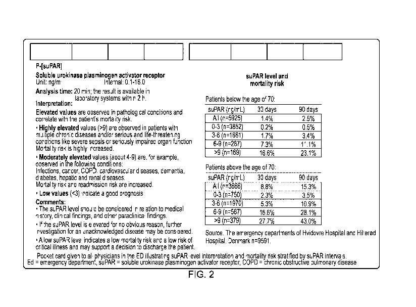
Figure 2 shows a pocket assessment card to be used by medical staff when
making use
of the method of the invention. It illustrates suPAR level interpretation and
mortality risk
stratified by suPAR intervals. ED ¨ emergency department, suPAR = soluble
urokinase
plasminogen activator receptor, COPD = chronic obstructive pulmonary disease.
Figure 3 shows the flow-diagram of the included patients.
Figure 4 shows the number of patients discharged within 24 hours in the group
with a
suPAR measurement and the controls.
Figure 5 shows the length of hospital stay in patients with suPAR measured at
inclusion
and patients without (controls).
Figure 6 is a ROC curve analysis for single markers and their ability to
predict 30-day
mortality.
Figure 7 is a suPAR patient-flow guideline from the TRIAGE III study.
Figure 8 shows how the addition of a suPAR measurement and comparison with a
reference value increases the specificity and sensitivity of a 30 day
mortality
assessment.
Figure 9 shows how the addition of a suPAR measurement and comparison with a
reference value increases the specificity and sensitivity of a 90 day
mortality
assessment.
Example 1 ¨ measurement of suPAR level
suPAR levels may be measured in body fluids by the methods taught in WO
2008/077958, which is incorporated herein for that purpose.
More specifically, suPAR levels may be determined by ELISA assay as follows:
Nunc
Maxisorp ELISA-plates (Nunc, Roskilde, Denmark) are coated overnight at 4 C
with a
monoclonal rat anti-suPAR antibody (VG-1, ViroGates NS, Copenhagen, Denmark, 3
pg/ml, 100 p1/well). Plates are blocked with PBS buffer + 1% BSA and 0.1%
Tween 20,
1 hour at room temperature, and washed 3 times with PBS buffer containing 0.1
%
Tween 20. 85 pl dilution buffer (100 mm phosphate, 97.5 mm NaCI, 10 g L-1
bovine
serum albumin (BSA, Fraction V, Roche Diagnostics GmbH Penzberg, Germany), 50
U
mL-1 heparin sodium salt (Sigma Chemical Co., St. Louis, MO), 0.1% (v/v) Tween
20, pH
7.4) containing 1.5 pg/ml HRP labeled mouse anti-suPAR antibody (VG-2-HRP,
CA 03091531 2020-08-18
WO 2019/162334 PCT/EP2019/054232
ViroGates) and 15 pl plasma (or serum or urine) sample is added in duplicates
to the
ELISA plate. After 1 hour of incubation at 37 C, plates are washed 10 times
with PBS
buffer + 0.1 % Tween 20 and 100 p1/well HRP substrate added (Substrate Reagent
Pack, R&D Systems Minneapolis, Minnesota). The colour reaction is stopped
after 30
min using 50 pl per well 1M H2504 and measured at 450 nm.
Furthermore, suPAR can be measured in bodily fluids using commercially
available
CE/IVD approved assays such as the suPARnostic product line according to the
manufacturer's instructions. In the TRIAGE III trials, suPAR was quantified
using the
suPARnostic Quick Triage lateral flow assay.
Example 2¨ correlation of plasma and urine levels of suPAR
WO 2008/077958 shows that plasma levels of suPAR in HIV-infected patients on
stable
HAART correlate with urine suPAR, as has been demonstrated previously in HIV
negative individuals, and that diurnal changes in urine suPAR are small (Sier
et al., 1999,
Lab Invest 79:717-722). A sub-sample of 24 of 36 patients had provided
overnight-
fasting urine. The effect of differences in dilution of the urine on suPAR
levels was
corrected with the amount of creatinine, as described previously (Sier et al,
1999, Lab
Invest. 79:717-722). Urine creatinine was measured as described (Mustjoki et
al, 2000,
Cancer Res. 60:7126-7132).
Figure 1 shows that fasting plasma suPAR and urine suPAR are highly correlated
in HIV-
infected patients on stable HAART. Since urine suPAR is shown to be a robust
estimate
of plasma suPAR, the level of suPAR can be performed on urine as well as
plasma
samples from such individuals. There is no reason to suppose that a similar
correlation,
and an equivalent correction factor, cannot be used in all subjects.
Example 3¨ clinical trial structure
A randomized intervention study was carried out at two large hospitals in the
capital
region of Denmark (ClinicalTrials.gov number, NCT02643459). The hypothesis of
the
study was that the introduction, fast measurement and immediate reporting
(knowledge)
of the suPAR level to attending physicians or other hospital professionals in
the EDs will
be associated with a reduction in all-cause mortality at least 10 months after
admission.
The primary aim of the study was to evaluate whether the determination of the
subject's
suPAR level can be used as a part of risk stratification of unselected acutely
admitted
subjects in order to reduce all-cause mortality.
11
CA 03091531 2020-08-18
WO 2019/162334 PCT/EP2019/054232
The secondary aims included:
= All cause mortality after index admission, after 30 days.
= Number of discharges from the emergency room within 24 hours.
= Length of stay during admission. [Time Frame: In-hospital stay].
= Number of readmissions [Time Frame: 30 and 90 days]. All new admissions
within 91 days of the same patient are defined as readmissions.
= Economical expenses [Time Frame: in-hospital stay, 30 days and 10 months
after inclusion period ends].
The main hypothesis was to assess if all-cause mortality at 10 months after
admission is
lower when the suPAR biomarker is measured on acutely admitted patients. Using
a
% level of significance and a power of 80 %, a sample of 7340 subjects was
needed in
each randomization group to detect an absolute risk reduction in mortality at
least 10
months after admission of 1.5 %.
Table 1: Trial structure
Cycle 1 2 3 4 5 6
Hospital 1 +suPAR Control +suPAR Control +suPAR Control
Hospital 2 Control +suPAR Control +suPAR Control -- +suPAR
Each cycle consisted of three weeks with (+suPAR) or without (Control) suPAR
measurements in the ED.
Quantification of suPAR
Blood samples (6 mL EDTA plasma tubes) for measurement of plasma suPAR were
drawn along with the routine blood work. For quantification of suPAR, blood
collection
tubes were spun for 60 s at 6000 RPM. 10 pL of plasma was added to a
prefabricated
tube containing 100 pL of running buffer. Using a 60 pL pipette, the plasma
and buffer
were mixed by pipetting the solution up and down 5 times. From this mixture,
60 pL was
added to the suPARnostic Quick Triage stick, a lateral flow device (also
called
suPARnostic Quick Test). After 20 min, the lateral flow device was visually
inspected
for test and control line, and the suPAR test line quantified using a
suPARnostic Quick
test device reader (Qiagen, Germany) [20]. According to the test manufacturer
(ViroGates NS, Birkeroed, Denmark), the limit of Detection (LOD) for the
suPARnostic
12
CA 03091531 2020-08-18
WO 2019/162334 PCT/EP2019/054232
quick test was 0.3 ng/ml. The limit of quantification (LOQ) was 2 ng/mL
defined at the
lowest concentration with a CV% that does not exceed 25 %. The intra- and
interserial
measured CV% on 5 samples x 4 concentrations (2.0; 4.0; 8.4; 13.7 ng/mL)
measured
on the same day or with 5 days interval was less than 25 %. The r2 of the
suPARnostic
Quick Test compared to the suPARnostic ELISA is 0.875. Analysis of suPAR level
was
handled by trained medical students according to the manufacturer's
instructions,
available on-site full-time for non-stop inclusion of eligible subjects. All
suPAR levels
were analyzed as quickly as possible and always within two hours following
blood
sampling and immediately reported.
Information to physicians
The suPAR level was presented to the attending physicians through the
electronic
systems LABKA, OPUS and Cetrea. LABKA II (v. 2.5ØH2, Computer Sciences
Corporation (CSC)) is the clinical laboratory information system used to
request blood
work and view results from laboratory analysis. OPUS (OPUS Arbejdsplads, v.
2.5Ø0,
Computer Sciences Corporation (CSC)) is the electronic database of medical
records.
The emergency wards in the EDs are monitored by the Cetrea system, which is
presented by several large screen monitors in the ED and presents a rough
overview of
the ward (patient data and status, possible diagnosis, route of admission)
used by
physicians and nurses. Prior to the study, all physicians working in the
emergency
department were informed in writing about the prognostic abilities of suPAR in
unselected subjects, and in regard to specific diagnoses in the form of a
review of
published literature, as well as pocket cards providing unadjusted mortality
rates from
10,000 subjects from similar EDs.
The participating doctors and nurses were informed that they should consider
the high
risk connected with increased suPAR levels, and clinical reconsideration was
advised
when encountering a subject with an unexplained high suPAR, in which case an
individual intervention should be scheduled based on symptoms and objective
findings
for the particular clinical issue, for example referral to a specialist,
follow-up consultation
with general practitioner, positron emission tomography scan or other
diagnostic
procedures or scanning methods. On the other hand, a low suPAR should promote
faster discharge. The doctors were informed of specific cut-of values with
regard to
suPAR and age and the mortality risk associated with those values (Figure 2).
The data
in these information charts was based on retrospective patient data obtained
from North
Zeeland and Copenhagen University Hospital Hvidovre, Denmark.
13
CA 03091531 2020-08-18
WO 2019/162334 PCT/EP2019/054232
For the sake of clarity, the information on the card, as shown in Fig. 2, is
as follows
(between the two lines of asterisks):
***********
Soluble urokinase plasminogen activator receptor levels are shown in units of
ng/ml, with a range of 0.1-16Ø The analysis time is 20 min; the result is
available in laboratory systems within 2 h.
Interpretation
Elevated values are observed in pathological conditions and correlate with the
patient's mortality risk.
= Highly elevated values (>9) are observed in patients with multiple
chronic
diseases and/or serious and life-threatening conditions like severe sepsis
or seriously impaired organ function. Mortality risk is highly increased.
= Moderately elevated values (about 4-9) are, for example, observed in the
following conditions: Infections, cancer, COPD, cardiovascular diseases,
dementia, diabetes, hepatic and renal diseases.
Mortality risk and
readmission risk are increased.
= Low values (<3) indicate a good prognosis.
Comments
= The suPAR level should be considered in conjunction with medical history,
clinical findings, and other paraclinical findings.
= If the suPAR level is elevated for no obvious reason, further
investigation
for an unacknowledged disease may be considered.
= A low suPAR level indicates a low mortality risk and a low risk of
critical
illness and may support a decision to discharge the subject.
suPAR level and mortality risk
Subjects below the age of 70:
suPAR (ng/mL) 30 days 90 days
14
CA 03091531 2020-08-18
WO 2019/162334 PCT/EP2019/054232
All (n=5925) 1.4% 2.5%
0-3 (n=3852) 0.2% 0.5%
3-6 (n=1661) 1.7% 3.4%
6-9(n=287) 7.3% 11.1%
>9 (n=169) 16.6% 23.1%
Subjects above the age of 70:
suPAR (ng/mL) 30 days 90 days
All (n=3666) 8.8% 15.3%
0-3 (n=750) 2.3% 3.5%
3-6 (n=1970) 5.3% 10.9%
6-9 (n=567) 16.6% 28.1%
>9 (n=379) 27.7% 43.0%
Source: The emergency departments at Hvidovre Hospital and Hillerod Hospital,
Denmark n=9591.
***********
To assess the quality of the data, and whether the physicians received and
considered
the suPAR level in the initial evaluation of subjects, a questionnaire was
sent to 200
randomly selected physicians at the participating hospitals, asking:
Did you see the suPAR level of your subject?
Did you feel informed in the prognostic ability of suPAR?
How often did you include suPAR in your combined assessment of your subject?
How often did the suPAR level influence your clinical decision?
How often were you surprised by a high suPAR level?
How often were you surprised by a low suPAR level?
CA 03091531 2020-08-18
WO 2019/162334 PCT/EP2019/054232
Data collection
Results of blood sample analyses including suPAR level were obtained from the
LABKA
ll database. Using the unique Danish central person registration number (CPR-
number),
demographic data and mortality were obtained from the Central Civil Registry
where all
residents in Denmark are registered. Data on admissions, discharges, and
diagnoses
were obtained from the National Patient Registry (NPR). NPR contains
information
coded according to the International Statistical Classification of Disease,
10th revision
(ICD-10) on primary diagnosis of discharge (A-diagnosis) and comorbidity (B-
diagnoses).
Laboratory values were obtained through LABKA (the clinical laboratory
information
system research database in Northern and Central Denmark; Grann et al (2011)
Clin.
Epidemiol. 3, 133-138). In the data analysis, the suPAR level from the index
admission
was linked with the data above to examine the primary and secondary outcomes.
Statistical analysis
Patients admitted in each intervention or control cycle were followed as a
single cohort
and data were analyzed as randomized. The two groups were assessed for
comparability of the following variables: age, sex, and Char!son score.
Differences in
mean age of more than 5 years and/ or an absolute Char!son Comorbidity Index
score of
2 or more were adjusted for in the final analysis. Patient data were analyzed
according
to the arm of the trial to which the patient was admitted during index
admission,
according to the randomization scheme (Table 1) corresponding to the intention-
to-treat
principle. A weighted Cox model was used to compare mortality at 10 months
after
inclusion of the last subject. Subjects were censored if their first
readmission was in the
opposite group to their index admission. As this censoring is likely to be
dependent
censoring (a readmission is rarely a positive prognostic signal), we employed
Inverse
Probability of Censoring Weighting (IPCW) where subjects readmitted to their
own
treatment group were up-weighted to compensate. We employed stabilized weights
such that the reweighted sample had the same implied sample size throughout
follow-up.
Due to the design, time since index admission was the only covariate that
needs to be
included in the weights. Reweighing was done for every two weeks of follow-up.
We did
not censor nor reweight for 2nd or later readmissions, since the weights would
become
highly unstable and it was not likely that the presence or absence of an
initial suPAR
measurement would be important for clinical decisions at this stage.
Furthermore, a
traditional intention-to-treat analysis was performed. Notable difference
between the
results of the two analysis strategies were considered critically. Kaplan-
Meier plots were
used to illustrate survival. Unpaired T-test was used to compare length of
stay. P < 0.05
16
CA 03091531 2020-08-18
WO 2019/162334 PCT/EP2019/054232
was considered significant. Subgroup analysis of the following groups was
performed:
subjects aged 65 years and above, and patients discharged with diagnoses of
surgical
conditions, cancer, infections, and cardiovascular disease.
At follow-up (10 months after inclusion of last patient) the following data
was collected
from the central Danish Patient Registry:
= Contacts with the healthcare system (including all historical contacts)
= Information regarding admissions (date, time and place of admittance and
discharge)
= Diagnoses (historical and in relation to index admission).
= Date of death or emigration
Diagnoses obtained from the national patient registry were coded with the ICD-
10
system. The original chapters were used to group patients according to
diagnoses.
Primary diagnosis was used with construction subgroups, and both primary and
secondary diagnoses will be used to calculate the Char!son score. The
following will
define the subgroups: Cancer: Chapter II: Neoplasms (COO-D48). Cardiovascular
disease: Chapter IX (100-199). Infections: Chapter 1: A00-699 + J00-J22 + +
N10-N11+
N30-N31. Neurological disease: Chapter VI(GOO-G99). Surgical conditions:
Presence of
surgical procedure code divided into different specialities (general,
orthopedic, other).
Example 4
The negative predictive value of suPAR aids in discharge decisions
Background: The TRIAGE 111-trial is a cross-over, cluster-randomized, parallel-
group,
prospective, interventional trial, with the hospitals as units of
randomization and the
patients as the units of analysis. The trial design has been published
previously (Sando
A, Schultz M, Eugen-Olsen J, et al (2016) "Introduction of a prognostic
biomarker to
strengthen risk stratification of acutely admitted patients: rationale and
design of the
TRIAGE III cluster randomized interventional trial" Scand J Trauma Resusc
Emerg Med.
24(1):100. doi:10.1186/513049-016-0290-8). We conducted the TRIAGE 111-trial
at the
EDs of two large hospitals: Bispebjerg University Hospital and Herlev
University Hospital,
both located in the Capital Region of Denmark and with 70,000 and 85,000
annual
admissions, respectively. By using cluster design and designating hospitals as
the units
of randomization, we ensured that unselected patients with different chronic-
and acute
diseases were included in both groups as well as a consecutive and full
inclusion rate.
The trial had five months of inclusion from January 11, 2016 and ended as
planned on
17
CA 03091531 2020-08-18
WO 2019/162334 PCT/EP2019/054232
June 6, 2016 with a subsequent 10-month follow-up concluded on April 6, 2017.
The
patients included are shown in Figure 3.
Aim of study: To determine whether providing the doctors and nurses in the ED
with the
patient suPAR value can affect the decision of "admit or discharge" and
whether
providing suPAR can lead to shorter hospital length of stay.
Methods:
suPAR levels were measured using the CE/IVD approved suPARnostic quick triage
test
and reader (ViroGates NS, Denmark). Data were acquired from the Danish
National
Patient Registry (NPR) and the Civil Registration System (CRS) at the end of
follow-up
(10 months after the last patient were included). All patient contacts are
registered in the
NPR and vital status is registered in the CRS. Data on blood tests, including
plasma
suPAR level, was extracted from the electronical hospital database "LABKA".
For
inclusion in the trial, patients were required to have a contact in the NPR
within six hours
of registered blood tests in LABKA within the inclusion period and an age 16
years.
Admissions at the pediatric, obstetric and gynaecological departments were not
included.
The index admission was defined as the first admission in the trial inclusion-
period.
Analysis included all patients participating in the TRIAGE III trial and
compared those
who had a suPAR measurement (N=7,905) with those who did not (N=8,896).
Differences were compared using student's T- and Wilcoxon tests. P<0.05 was
considered statistically significant. Statistics were carried out using R
version 1Ø136
(The R Foundation for Statistical Computing).
Outcomes
The endpoints for the negative predictive value of suPAR were:
(I) Short admissions (<24 h) to the ED. Is there a difference in the number of
patients discharged from hospital (stay shorter than 24 hours from Index) when
comparing those patients who had their suPAR measured compared to those who
did not?
(II) Length of stay. Is there a difference in the length of hospital stay of
patients when
comparing those patients who had their suPAR measured compared to those who
did not?
18
CA 03091531 2020-08-18
WO 2019/162334 PCT/EP2019/054232
Results: During the study, 16801 patients were included. Mean age was 60 years
(SD
20) and 47.8% were men. 7905 patients had a suPAR measurement at admission and
8896 patients did not have suPAR measured (controls) (Figure 3).
With regard to endpoint I, patients who had a suPAR measurement were
significantly
more often discharged within 24 hours compared to those without suPAR
measurement
(50.2% (3,966 patients) vs. 48.6% (4,317 patients), absolute difference: 1.6%
(95% Cl
0.08-3.12); P=0.039) (Figure 4).
With regard to endpoint II, patients with a suPAR measurement had a 6.5 hour
shorter
length of hospital stay compared to patients without suPAR measurement (4.31
days
(7.35) vs. 4.58 days (9.37), difference: 0.27 days (95% Cl 0.01-0.53),
P=0.043) (Figure
5).
Mortality in patients discharged within 24 hours
All-cause mortality within 30 days among early discharged patients occurred in
52
patients (1.3%) in the suPAR group and in 77 patients (1.8%) in the control
group. The
unadjusted Cox model found a trend towards lower mortality in the suPAR group
compared to control: Hazard ratio (HR), 0.73; 95% confidence interval (Cl)
0.52 to 1.04;
P=0.084.
During the median 12-months of follow-up, 225 (5.7%) of the patients died,
which was
less than among early discharged patients in the control arm where 256 (6.7%)
died
during follow-up (P=0.05). In patients that were discharged within 24 hours,
the AUC for
predicting 30-day mortality was 0.92 (95`)/0CI: 0.90-0.95)
Readmissions in patients discharged within 24 hours
With regard to 30-day readmission, 336 (8.5%) patients in the suPAR group were
readmitted, while 331 (7.7%) patients in the control group were readmitted,
P=0.18. For
90-day readmission, 490 patients (12.4%) vs. 552 patients (12.8%) were
readmitted in
the suPAR group and control group, respectively (P=0.57).
Discussion: The study showed that knowledge of patient's suPAR level at the
Emergency Department led to earlier discharged patients and overall shorter
length of
stay. Even though more patients were discharged in the suPAR group compared
with
controls, there was no difference with regard to readmissions or mortality.
Thus, early
discharge based on suPAR is safe and feasible. Improving patient flow and
earlier
discharge of patients where admission might not be necessary will benefit both
patients
19
CA 03091531 2020-08-18
WO 2019/162334 PCT/EP2019/054232
in need of hospital treatment and low-risk patients who can be discharged
without being
exposed to the risks of hospitalization, such as in-hospital infections, loss
of muscle
mass and loss of personal income if the patient is working. For the hospital,
the shorter
admission observed in patients that had suPAR measured at admission (6 hours
shorter
in the suPAR arm), leads to economic savings.
The fact that the AUC of suPAR became very high among those early discharged
shows
that the doctors used the positive predictive value of suPAR and kept patients
more than
24 hours in hospital if suPAR was elevated. The high AUC of 0.92 thus reflects
that
those early discharged were the low risk patients and those who were sent home
to die
(e.g. to hospice or retirement home).
Example 5
Positive predictive value of suPAR
Background: suPAR has previously been shown to be a strong predictor of
outcome in
retrospective studies. However, it was unknown whether giving the doctors
information
on the suPAR level could alter the outcome/change the prognosis. In the TRIAGE
III
Intervention study, suPAR was measured at time of admission using the
suPARnostic
Quick Test in 7,905 patients. Comparison is made to the 8896 patients in the
control
arm (without suPAR measurement) (Figure 3).
Methods: suPAR levels were measured using the CE/IVD approved suPARnostic
quick
triage test and reader (ViroGates NS, Denmark). The discriminative ability of
suPAR
with regard to mortality at one and ten months was assessed by using area
under the
curve (AUC) for receiver operating characteristics (ROC).
P<0.05 was considered statistically significant. Statistics were performed in
R version
1Ø136 (The R Foundation for Statistical Computing) and figures were created
with
Graphpad Prism, version 7.02.
Results:
suPAR and mortality. The median suPAR level of patients who survived was
significantly
lower than the suPAR level of patients who died during follow-up, both at 30
days (4.0
ng/ml (IQR 2.9-5.7) vs. 8.3 ng/ml (IQR 5.9-11.7), p<0.001) and 10 months (3.8
ng/ml
(IQR 2.8-5.3) vs.6.9 ng/ml (IQR 5.1-10.1), p<0.001). SuPAR had a high
prognostic
power for predicting 30-days and 10-months mortality (AUCs: 30 days: 0.83 (95%
Cl:
0.81-0.84); 10 months: 0.80 (95`)/0CI: 0.79-0.82). In comparison with age and
routine
CA 03091531 2020-08-18
WO 2019/162334 PCT/EP2019/054232
biomarkers, suPAR had superior prognostic power regarding mortality at all
follow-up
times (Table 2: AUC for suPAR and other routine biomarkers and age) (Figure 6,
ROC
curve analysis for single markers and their ability to predict 30-day
mortality; the dashed
line to the left of the figure is the level of suPAR).
Mortality 30 days Mortality 90 days Mortality
All follow-up
Age 0.777 0.774 0.781
C-reactive
protein 0.738 0.729 0.702
Hemoglobin 0.701 0.721 0.729
Sodium 0.582 0.597 0.604
Potassium 0.578 0.574 0.564
Albumin 0.777 0.763 0.732
Creatinine 0.622 0.607 0.604
Leucocytes 0.654 0.627 0.580
ALAT1 0.511 0.530 0.550
suPAR 0.835 0.815 0.802
Table 2. Area Under the Curve (AUC) for the routine measured biomarkers and
age
Adding suPAR to algorithm significantly improves outcome prediction
To determine whether suPAR provides an additional and independent value to a
combined model of all predictive routine markers, two models were made: one
without
suPAR but containing all the variables found significant in Table 2, and
another model
including these variables and suPAR.
For the prediction of 30-day mortality, the first model (without suPAR)
provides an AUC
of 0.860 (95`)/0C1 0.84-0.86). Addition of suPAR significantly improved this
model, AUC
0.896 (95`)/0C1 0.88-0.90), p= 0.007. The increase in sensitivity and
specificity can be
seen in Figure 8.
Similarly, for the determination of 90-day mortality, the model without suPAR
provided an
AUC of 0.854 (95`)/0CI: 0.84-0.85). When including suPAR, the model
significantly
improved to an AUC of 0.878 (95`)/0CI: 0.86-0.88), p=0.001 (Figure 9).
Measuring suPAR at admission and difference in mortality between patients with
or without suPAR measurement
1 Alanine aminotransferase
21
CA 03091531 2020-08-18
WO 2019/162334 PCT/EP2019/054232
With regard to mortality in the suPAR intervention arm versus the control, we
observed a
mortality rate of 13.9% in the intervention arm compared to 14.3% in the
control arm
corresponding to 36 fewer mortalities in the intervention arm. The difference
in mortality
between the suPAR Intervention arm and control arm was strongly observed at
Bispebjerg Hospital, Copenhagen, Denmark. At Bispebjerg Hospital, 3451
patients were
included in the suPAR intervention arm and 3569 in the control arm. During
follow-up,
427 patients died in the suPAR intervention arm (12,4%) which was a
significant lower
mortality than was observed in the control arm (515 died (14,4%), p<0.05.
Discussion: In this study, it is shown that suPAR is superior to other
biomarkers with
regard to outcome prediction compared with other investigated biomarkers,
including a
combined model of commonly used routine blood tests, in predicting short-term
mortality.
It is of interest that suPAR, in contrast to other biomarkers, is stronger
than age in
prediction of outcome. Also, adding suPAR to an algorithm of all the routine
biomarkers
significantly improved the prediction of both 30- and 90-day mortality. With
regard to
prevention of mortality, less mortality was observed in the intervention arm
compared
with the control arm. The effect of informing the doctors of suPAR level was
of most
value in patients with well-functioning clinical signs, e.g. in those triaged
in the low risk
category or having a low Early warning score (EWS or NEWS) where a severe
disease,
if present, is not recognised without the suPAR measurement.
The prognostic abilities of suPAR have been studied retrospectively before,
and the
biomarker has been shown to be associated with risk of mortality and adverse
events.
However, previous studies have not investigated the clinical impact of
interventions on
patients when giving the doctors "real time" information on the suPAR level
while the
patient was present in the ED. Hence, it was until now unknown whether
knowledge of
suPAR while the patient is present can change the outcome of the patient.
This study shows for the first time that knowledge of suPAR led to more early
discharges
in the Intervention arm compared with control. With regard to mortality in
those early
discharged, fewer patients died in the intervention arm compared with control,
demonstrating that both the negative and positive predictive value of
providing "real time"
suPAR levels to the doctors and nurses aids in better admission and discharge
decisions
in the Emergency Departments.
22