Note: Descriptions are shown in the official language in which they were submitted.
CA 03092870 2020-09-01
WO 2019/173475
PCT/US2019/020952
EMBOLIC PROTECTION DEVICE
CROSS REFERENCE TO RELATED APPLICATION
[0001] This PCT application claims the benefit of U.S. provisional application
no.
62/639,618, filed on March 7, 2018, and U.S. provisional application no.
62/812,391, filed on
March 1, 2019. Each of these documents is hereby incorporated by reference in
its entirety.
TECHNICAL FIELD
[0002] This application relates to embolic protection devices including a
catheter and
methods of using such embolic protection devices in medical procedures (e.g.,
closed-heart
surgical procedures).
BACKGROUND
[0003] Traditional pigtail catheters are used during percutaneous cardiac
procedures where
the positioning of various instruments and devices within the vasculature of a
patient is
important. These pigtail catheters comprise a curved distal end that can rest
within the
patient's anatomy (e.g., an artery (e.g., aorta)) and hold the catheter in
place while other
instrumentation and devices are delivered into the patient's vasculature. Some
traditional
pigtail catheters include a lumen and small apertures at their distal ends
through which a
contrast agent can be injected into a patient's vasculature for imaging the
relevant portion of
the patient's anatomy and identifying anatomical landmarks.
[0004] However, the use of traditional pigtail catheters in percutaneous
cardiac procedures
often results in serious and life-threatening complications for the patient.
For example,
cerebral embolism is a common complication in cardiac procedures, such as
valve
replacement and repair, where a traditional pigtail catheter is deployed.
During such
procedures, plaque, calcium, thrombi, or any combination thereof, in the
vessels, valves,
and/or cardiac chambers can be dislodged by the catheter or other medical
devices introduced
into the patient's vasculature. The dislodged plaque, calcium, thrombi or any
combination
thereof can be carried into the patient's brain via blood flow from the aorta
and can cause
blockages therein leading to an embolic event such as stroke. Approximately
2.9%-6.7% of
patients undergoing transfemoral transcatheter aortic-valve implantation
(TAVI) have a
stroke within 30 days, and even more (4.5%-10.6%) have a stroke within a year,
often
leading to death. Furthermore, up to 85% of patients undergoing TAVI have
evidence of
embolic phenomenon to the brain based on neuroimaging studies. Although
clinically silent,
such embolic phenomena are associated with cognitive decline (Astraci 2011;
Ghanem 2010;
Kahlert 2010; Rodes-Caban 2011).
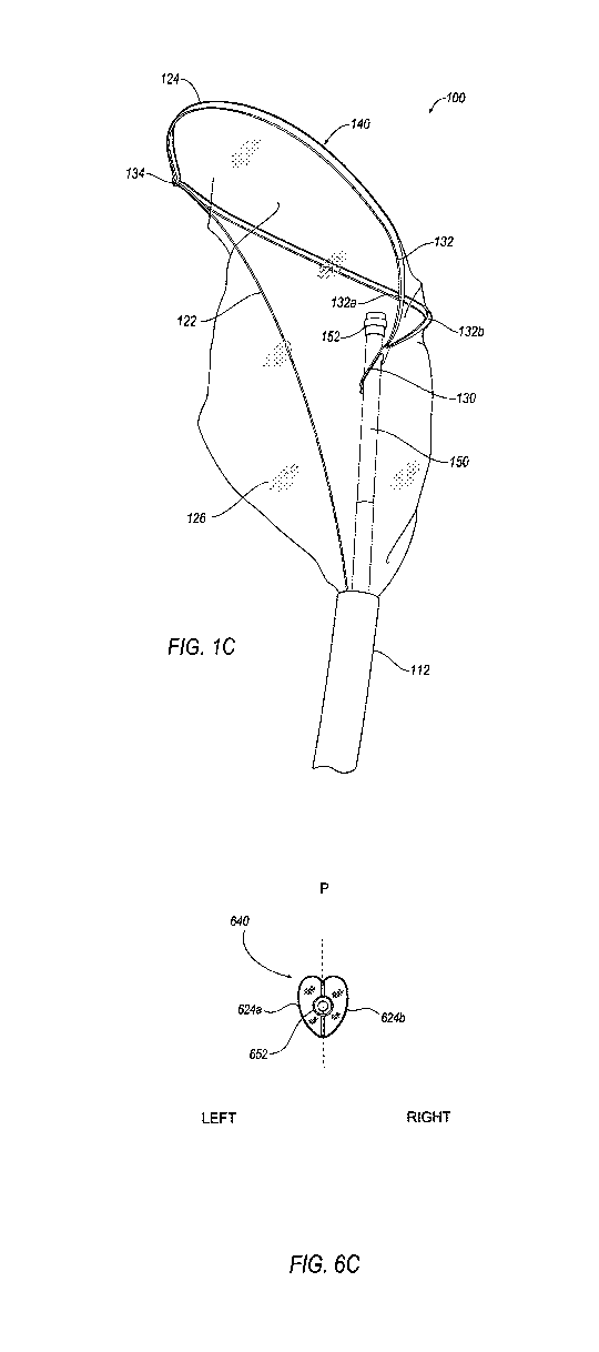
1
CA 03092870 2020-09-01
WO 2019/173475
PCT/US2019/020952
[0005] Presently, there are a few devices on the market designed to protect
the brain,
abdominal organs, and carotid arteries from emboli, and these devices suffer
from various
significant drawbacks. For instance, the Embrella Embolic Deflector ,
available from
Edwards Lifesciences of Irvine, California, employs a deflector that deflects
emboli from the
carotid arteries into the descending aorta, but the device does not trap the
emboli, so emboli
are free to travel to other areas of the body and cause deleterious
complications. The
EMBOL-X , also available from Edwards Lifesciences, employs a filtering
screen, but this
device is designed for use in open heart procedures, which present additional
medical risks
and increased morbidity. Additionally, the use of multiple devices, for
example a catheter for
visualization and a separate filter device, lengthens the procedure time and
increases the risk
of complications to the patient.
SUMMARY
[0006] These and other needs are met by the present invention, which presents
an embolic
protection device comprising a deployable embolic filter that is disposed
around a catheter
having a distal portion that can assume an arcuate configuration being at
least a semi-circle,
and having a wire that is operable to manipulate the embolic filter into a
configuration that
more fully engages a body lumen.
[0007] The combination of the catheter and the embolic filter in the same
device may provide
the benefits of both devices individually, as well as provide a synergistic
effect. For example,
the integration of the catheter and the embolic filter can decrease the
duration of the medical
procedure and reduce the occurrence of complications (e.g., complications
caused by
dislodged emboli). In other examples, the expansion of the embolic filter may
help to anchor
the catheter into position to provide a more accurate position of the catheter
than if the
position of the catheter is susceptible to the influences of blood flow,
tissue movement, and
the like. In a valve replacement procedure, anchoring of the catheter and more
accurate
positioning of the catheter may help ensure that the valve prosthesis is
properly positioned
and stabilized. In another example, the position of the catheter may ensure
that the filter is
being properly positioned.
[0008] In some aspects, the embolic protection device comprises a catheter, a
self-expanding
embolic filter coupled to the catheter, a pull wire for reorienting the filter
by bending a frame
of the filter, and an outer sheath movable with respect to the embolic filter
and the catheter.
The outer sheath holds the embolic filter in a collapsed configuration when
surrounding the
embolic filter and is proximally retracted to deploy the embolic filter. The
outer sheath may
recapture the embolic filter and any debris captured therein by being distally
advanced. The
filter and outer sheath might both be movable with respect to the catheter,
for example to be
2
CA 03092870 2020-09-01
WO 2019/173475
PCT/US2019/020952
able to move the embolic filter longitudinally without having to move the
entire catheter
longitudinally. The pull wire is advantageous due to its ability to bend the
frame, thereby
facing the filter opening towards the distal end of the device and causing the
embolic filter to
more fully engage the body lumen.
[0009] In some aspects, the catheter has a proximal end and a distal end. A
lumen extends
from the proximal end of the catheter to the distal end of the catheter. In
some embodiments,
the lumen may be configured to house a guidewire.
[0010] In some aspects, the catheter is a pigtail catheter. A pigtail catheter
is configured to
curl at the distal end of the catheter, forming a generally arcuate shape that
is at least a semi-
circle. The pigtail may have a radiopaque marker viewable on x-rays or other
medical
imaging devices. The radiopaque marker is on the distal section of the curled
pigtail in the
form of a longitudinal marker, circumferential bands, or the like. The pigtail
may
additionally have one or more apertures to dispense drugs and/or contrast
agents through the
lumen.
[0011] In some aspects, a guidewire is inserted through the patient's skin and
into a body
lumen such as a femoral, radial, or brachial artery and steered near a target
site. The
guidewire is inserted into a lumen of the embolic protection device, and the
embolic
protection device is pushed or tracked over the guidewire to the target site.
When the
guidewire is retracted from at least the distal portion of the catheter, the
catheter assumes a
generally arcuate shape. The radiopaque marker on the catheter is used to
visualize and
position the catheter. Once the catheter is in position, the outer sheath is
retracted to deploy
the embolic filter and the pull wire is retracted to bend the frame of the
filter to position the
distal opening of the filter across the vessel. The user can then perform a
procedure such as
valve replacement, valve repair, radio frequency ablation, and the like. When
the procedure
is completed, the pull wire is advanced and the outer sheath is advanced to
recapture the
embolic filter and any debris trapped in the embolic filter. The device is
then retracted from
the vessel, with the catheter being atraumatic to vessels during retraction.
[0012] Another aspect is a method of capturing embolic debris during a closed-
heart surgical
procedure comprising inserting the distal end of the catheter of the embolic
protection device
into a body lumen. The method further comprises allowing the embolic filter to
assume an
expanded, deployed configuration and retracting the pull wire to bend the
frame of the filter,
so that a distal opening of the filter spans the body lumen.
[0013] In some aspects, the embolic protection device comprises a catheter, a
self-expanding
embolic filter coupled to the catheter, a push wire for reorienting the filter
by bending a frame
of the filter in a longitudinal direction and extending the frame in a radial
direction, and an
3
CA 03092870 2020-09-01
WO 2019/173475
PCT/US2019/020952
outer sheath movable with respect to the embolic filter and the catheter. The
outer sheath
holds the embolic filter in a collapsed configuration when surrounding the
embolic filter and
is proximally retracted to deploy the embolic filter. The outer sheath may
recapture the
embolic filter and any debris captured therein by being distally advanced. The
push wire is
advantageous due to its ability to bend and extend the frame, thereby facing
the filter opening
towards the distal end of the device and causing the embolic filter to more
fully engage the
body lumen.
[0014] In some aspects, the catheter has a proximal end and a distal end. A
lumen extends
from the proximal end to the distal end along a longitudinal axis of the
catheter. In some
embodiments, the lumen may be configured to house a guidewire.
[0015] In some aspects, the catheter is a pigtail catheter. A pigtail catheter
is configured to
curl at the distal end of the catheter, forming a generally arcuate shape that
is at least a semi-
circle. The pigtail may have a radiopaque marker viewable on x-rays or other
medical
imaging devices. The radiopaque marker is on the distal section of the curled
pigtail in the
form of a longitudinal marker, circumferential bands, or the like. The pigtail
may
additionally have one or more apertures to dispense drugs and/or contrast
agents through the
lumen.
[0016] In some aspects, a guidewire is inserted through the patient's skin and
into a body
lumen such as a femoral, radial, or brachial artery and steered near a target
site. The
guidewire is inserted into a lumen of the embolic protection device, and the
embolic
protection device is pushed or tracked over the guidewire to the target site.
When the
guidewire is retracted from at least the distal portion of the catheter, the
catheter assumes a
generally arcuate shape. The radiopaque marker on the catheter is used to
visualize and
position the catheter. Once the catheter is in position, the outer sheath is
retracted to deploy
the embolic filter and the push wire is advanced to bend and extend the frame
of the filter to
position the distal opening of the embolic filter across the vessel. The user
can then perform
a procedure such as valve replacement, valve repair, radio frequency ablation,
and the like.
When the procedure is completed, the push wire is retracted and the outer
sheath is advanced
to recapture the embolic filter and any debris trapped in the embolic filter.
The device is then
retracted from the vessel, with the catheter being atraumatic to vessels
during retraction.
[0017] Another aspect is a method of capturing embolic debris during a closed-
heart surgical
procedure comprising inserting the distal end of the catheter of the embolic
protection device
into a body lumen. The method further comprises allowing the embolic filter to
assume an
expanded, deployed configuration and advancing the push wire to bend and
extend the frame
of the filter, so that a distal opening of the filter spans the body lumen.
4
CA 03092870 2020-09-01
WO 2019/173475
PCT/US2019/020952
BREF DESCRIPTION OF THE FIGURES
[0018] The following figures are provided by way of example and are not
intended to limit
the scope of the claimed invention.
[0019] FIGS. IA and 1B illustrate partial side views of an embodiment of an
embolic
protection device of the present invention. In FIG. 1A, an embolic filter of
the embolic
protection device is illustrated in a collapsed (undeployed) configuration. In
FIG. 1B, the
embolic filter is illustrated in an expanded (deployed) configuration wherein
a pull wire
affixed to a frame of the embolic filter is advanced to a distal position so
that the frame
assumes it's self-expanded and undeflected (i.e., unbent) configuration.
[0020] FIG. IC illustrates a side perspective view of an embodiment of an
embolic filter of
the present invention assuming a partially deflected (i.e., partially bent)
configuration
wherein the pull wire affixed to the frame of the embolic filter is partially
longitudinally
retracted to a proximal position.
[0021] FIG. ID illustrates a transverse cross-sectional view of an embodiment
of an embolic
filter of the present invention assuming a fully deflected (e.g., fully bent)
configuration
wherein the pull wire is fully longitudinally retracted thereby deflecting the
filter.
[0022] FIGS. lE and IF illustrate front views of an embodiment of an embolic
filter frame of
the present invention. In FIG. 1E, the filter frame is undeployed wherein the
frame is
collapsed and enclosed by an outer sheath. In FIG. IF, the outer sheath is
longitudinally
retracted and the filter frame is deployed to its self-expanded configuration.
[0023] FIGS. 2A ¨ 2B illustrate partial side views of an embodiment of an
embolic
protection device of the present invention comprising a shoulder.
[0024] FIGS. 3A ¨ 3D illustrate partial side views of an embodiment of an
embolic
protection device of the present invention comprising an intermediate tube.
[0025] FIGS. 4A ¨ 4C illustrate partial side views of an embodiment of an
embolic
protection device of the present invention comprising a deflector.
[0026] FIG. 5A illustrates an embodiment of an embolic protection device
comprising a
handle. FIG. 5B illustrates a distal portion of the embolic protection device
comprising the
embolic filter and pigtail catheter.
[0027] FIG. 6A illustrates a partial side view of an embodiment of an embolic
protection
device of the present invention with an embolic filter in a collapsed
(undeployed)
configuration.
[0028] FIGS. 6B and 6C illustrate a side view and a front end view of the
embolic filter in an
self-expanded (deployed) configuration, respectively, wherein a push wire
coupled to a frame
CA 03092870 2020-09-01
WO 2019/173475
PCT/US2019/020952
of the embolic filter is retracted to a proximal position so that the frame
assumes an
undeflected (i.e., unbent) configuration.
[0029] FIGS. 6D and 6E illustrate a side view and a front end view of the
embolic filter in an
partially expanded configuration, respectively, wherein the push wire coupled
to the frame of
the embolic filter is longitudinally advanced to a first distal position so
that the frame
assumes a deflected (i.e., bent) configuration.
[0030] FIGS. 6E and 6F illustrate a side view and a front end view of the
embolic filter in an
fully expanded configuration, respectively, wherein the push wire coupled to
the frame of the
embolic filter is longitudinally advanced to a second distal position farther
than the first distal
position shown in FIG. 6C so that the frame assumes an extended configuration.
[0031] FIGS. 7A ¨ 7C illustrate partial side views of an embodiment of an
embolic
protection device of the present invention having an actuating mechanism for
operating an
embolic filter.
[0032] FIGS. 8A and 8B illustrate an embodiment of an embolic protection
device of the
present invention having a handle for manually operating an embolic filter.
[0033] FIGS. 8C ¨ 8F illustrate an example of the handle.
[0034] FIGS. 9A ¨ 9E illustrate a stepwise method of using an embolic
protection device of
the present invention.
[0035] FIG. 10 illustrates the deflection and capture of embolic debris by an
embolic
protection device of the present invention comprising a deflector.
[0036] FIG. 11 illustrates the deflection and capture of embolic debris by an
embolic
protection device of the present invention wherein a second catheter device is
present.
[0037] FIGS. 12A ¨ 12D illustrate a stepwise method of using an embolic
protection device
of the present invention operating an embolic filter.
[0038] FIGS. 13A and 13B are photographs of distal portions of embolic
protection devices
of the present invention situated within a cadaver's vasculature according to
Example 1. In
FIG. 13A, the embolic protection device comprises a longitudinal groove in
which a second
catheter is inserted alongside the embolic protection device. In FIG. 13B, the
second catheter
is situated adjacent to the embolic protection device that lacks a
longitudinal groove.
[0039] FIG. 14 is a bar graph of performance data of an embolic protection
device of the
present invention (the EPD-1 device) according to Example 2.
[0040] FIGS. 15A ¨ 15J are images generated from diffusion-weighted magnetic
resonance
imaging (DW-MRI) of representative subjects according to Example 2.
[0041] FIG. 16A is a photograph of thrombi captured by an embolic protection
device of the
present invention (the EPD-1 device) according to Example 2.
6
CA 03092870 2020-09-01
WO 2019/173475
PCT/US2019/020952
[0042] FIG. 16B is a photograph of a collagenous fragment captured within the
filter of the
embolic protection device (the EPD-I device) according to Example 2.
[0043] Like reference numerals in the various drawings indicate like elements.
DETAILED DESCRIPTION
[0044] The present invention provides an embolic protection device and methods
of using the
embolic protection device for capturing embolic debris during surgical
procedures.
[0045] I. DEFINITIONS
[0046] As used herein, the term "self-expanding" means to increase, spread
out, or unfold
from a collapsed state upon the withdrawal or removal of a restricting or
confining force.
[0047] As used herein, the term "closed-heart" refers to any surgical
procedure involving the
heart, wherein the chest cavity is not opened.
[0048] As used herein, the term "woven" refers to any material that comprises
a plurality of
strands, wherein the strands are interlaced to form a net, mesh, or screen.
Without limitation,
examples of woven materials include netting or mesh comprising a polymer,
metal, or metal
alloy.
[0049] As used herein, the term "non-woven" refers to any material that
comprises a
continuous film. Non-woven material may be permeable, semi-permeable, or non-
permeable.
For example, permeable or semi-permeable non-woven material may optionally
include one
or more pores through which a fluid may pass.
[0050] As used herein, the term "alloy" refers to a homogenous mixture or
solid solution
produced by combining two or more metallic elements, for example, to give
greater strength
or resistance to corrosion. For example, alloys include brass, bronze, steel,
nitinol, chromium
cobalt, MP35N, 35NLT, elgiloy, and the like.
[0051] As used herein, "nitinol" and "nickel titanium" are used
interchangeably to refer to an
alloy of nickel and titanium.
[0052] As used herein, "chromium cobalt" refers to an alloy of chromium and
cobalt.
[0053] As used herein, "MP35N" refers to an alloy of nickel and cobalt.
[0054] As used herein, "35NLT" refers to a cobalt-based alloy that may also
comprise
chromium, nickel, molybdenum, carbon, manganese, silicon, phosphorus, sulphur,
titanium,
iron, and boron.
[0055] As used herein, "elgiloy" refers to an alloy of cobalt, chromium,
nickel, iron,
molybdenum, and manganese.
[0056] As used herein, a "body lumen" refers to the inside space of a tubular
structure in the
body, such as an artery, intestine, vein, gastrointestinal tract, bronchi,
renal tubules, and
urinary collecting ducts. In some instances, a body lumen refers to the aorta.
7
CA 03092870 2020-09-01
WO 2019/173475
PCT/US2019/020952
[0057] 11. EMBOLIC PROTECTION DEVICES
[0058] Although certain embodiments and examples are described below, those
skilled in the
art will recognize that the disclosure extends beyond the specifically
disclosed embodiments
and/or uses and obvious modifications and equivalents thereof. Thus, it is
intended that the
scope of the disclosure herein presented should not be limited by any
particular embodiments
described below.
[0059] For purposes of this disclosure, the terms "upper," "lower," "right,"
"left," "rear,"
"front," "vertical," "horizontal," and derivatives thereof shall relate to the
invention as
oriented in FIGS. 1B and IF (or in FIGS. 6B and 6C). However, it is to be
understood that
the invention may assume various alternative orientations, except where
expressly specified
to the contrary. Also, for purposes of this disclosure, the term "coupled" (in
all of its forms,
couple, coupling, coupled, etc.) generally means the joining of two components
(electrical or
mechanical) directly or indirectly to one another. Such joining may be
stationary in nature or
movable in nature; may be achieved with the two components (electrical or
mechanical) and
any additional intermediate members being integrally formed as a single
unitary body with
one another or with the two components; and may be permanent in nature or may
be
removable or releasable in nature, unless otherwise stated.
[0060] FIGS. IA and 1B illustrate embodiments of an embolic protection device
100. In
these embodiments, the device 100 comprises a catheter 102 (e.g., a pigtail
catheter) having a
proximal end 114, a distal end 116, and a lumen 118 extending from the
proximal end 114 to
the distal end 116. The lumen 118 may be configured to house a guidewire 990
(see FIGS.
9A and 9B) that is longitudinally moveable through this lumen to coil or
straighten the distal
portion 104 of the catheter 102 depending on whether the guidewire is
retracted (to coil the
distal portion) or extended (to straighten the distal portion). In some
embodiments, the
catheter 102 includes a distal portion 104 configured to assume a generally
arcuate shape
being at least a semi-circle. A side wall of the catheter 102 may optionally
include one or
more apertures 108 in the distal portion 104 that are configured to deliver
one or more fluids
(e.g., imaging dye, contrast agent, oxygenated blood, saline, any combination
thereof, or the
like) to a body lumen 992 (see FIG. 9A). The apertures 108 (the plural
intended to include
embodiments in which the distal portion includes one aperture 108) are in
fluid
communication with the lumen 118. In some embodiments, the distal portion 104
of the
catheter 102 includes one or more radiopaque markers 106. In some embodiments,
the
radiopaque markers 106 are wrapped around the circumference of the distal
portion of the
catheter and can have the same or different widths. In other embodiments, the
radiopaque
markers are co-linear with the lumen and extend to the distal end of the
catheter. The device
8
CA 03092870 2020-09-01
WO 2019/173475
PCT/US2019/020952
100 further comprises a self-expanding embolic filter 110 defined by a frame
124 and a filter
medium 126, and a deployment mechanism 112 (e.g., a longitudinally retractable
outer sheath
or a longitudinally retractable ring). The embolic filter 110 is disposed
around the catheter
102.
[0061] As illustrated in FIG. 1B, in its deployed configuration, the embolic
filter 110
includes a distal opening 140 that is defined by the frame 124, faces the
distal end 116 of the
catheter 102, and extends proximally from the distal opening 140 to a closed
proximal end
142. The device 100 further comprises a pull wire 122 that is coupled to the
frame 124 and
can be retracted to deflect or bend the frame 124 and change the orientation
and shape of the
distal opening 140.
[0062] In some embodiments, retracting the pull wire 122 may cause the distal
opening 140
of the embolic filter 110 to engage at least a portion of the interior body
lumen 992 (see FIG.
9D) wall. FIG. 1B illustrates the pull wire 122 in an advanced, i.e., un-
retracted or self-
expanded, configuration with the frame oriented generally to extend in a
distal longitudinal
direction, albeit angled back somewhat (e.g., less than about 45 degrees) in a
lateral direction.
The catheter 102 may be partially surrounded towards its proximal end 114 by a
support
catheter 150 that terminates at a head 152, proximal to the distal portion 104
of the catheter
102. The support catheter 150 may be made of a thicker, stiffer material to
add rigidity and
provide a protective or supporting layer surrounding the catheter 102.
[0063] FIG. 1C illustrates the embolic filter 110 deployed (e.g., self-
expanded) by retraction
of the deployment mechanism (e.g., outer sheath) 112 with the frame 124
partially deflected,
i.e., partially bent, by retraction of the pull wire 122. The pull wire 122 is
coupled to the
frame 124 at a distal coupling 134. The distal opening 140 is primarily
defined by a first
portion 132 of the frame 124. The first portion 132 of the frame 124 defines a
shape of the
distal opening 140 that is substantially elliptical (i.e., shaped like an
ellipse), or alternatively,
substantially oval-shaped or circular. In this embodiment, the portion 132 of
the frame 124
may be substantially elliptical and may terminate a V-shaped point at its
proximal end, i.e.,
the portion 132 of the frame 124 may invert its curvature at one end of its
substantially
elliptical shape (e.g., at its distal end) and come to a point at its proximal
end. The distal
opening 140 may substantially be defined by the frame 124, but may span across
the frame
124 adjacent to the section of the frame 124 that comes to a point. The filter
medium 126
may define a portion of the distal opening 140 where the filter medium 126
spans across the
frame 124, i.e., adjacent to a point of attachment of the frame 124 to the
catheter 102 or
support catheter 150.
9
CA 03092870 2020-09-01
WO 2019/173475
PCT/US2019/020952
[0064] The attachment of the frame 124 to the support catheter 150 (or
alternatively, directly
to the catheter 102) is accomplished via a second portion 130 of the frame
124, which
encircles the support catheter 150 (or catheter 102) and is at an angle with
respect to the
longitudinal axis of the catheter 102. The second portion 130 of the frame 124
may be fixed
in its position by friction and by tension of the embolic filter 110 in the
lateral and/or
longitudinal directions. In other embodiments, the fixed attachment of the
second portion
130 of the frame 124 to the support catheter 150 (or catheter 102) may also be
accomplished
via adhesives, welding, or the like.
[0065] The first portion 132 of the frame 124 may extend in a first lateral
direction away
from the catheter 102 and away from the second portion 130 of the catheter 102
and loop
back across the catheter 102 and extend in the opposite lateral direction. In
this embodiment,
the first portion 132 of the frame 124 comprises two sides (132a, 132b) that
each extend
generally in a first lateral direction away from the catheter 102 and then
loop back on
opposite sides around the catheter 102 and extend generally in the opposite
lateral direction
before converging and meeting to form the substantially elliptical shape. As
shown in FIG.
1F, the embolic filter 110 is symmetrical about the pull wire 122. For ease of
discussion, the
embolic filter 110 is referred as having a left side and a right side.
Elements on the left side
of the embolic filter 110 are mirrored by elements on the right side of the
embolic filter 110.
[0066] When the pull wire 122 is in its advanced state (or partially, but not
fully, retracted
state), the frame 124 extends in a distal longitudinal direction as it extends
from its
attachment to the catheter 102 (or support catheter 150). When the pull wire
122 is in its
retracted state (i.e., fully retracted) (see FIG. ID and FIG. 9E), the frame
124 extends in a
distal longitudinal direction near its point of attachment to the catheter
102, but then is bent
such that it extends substantially perpendicular to the longitudinal axis of
the catheter 102.
[0067] FIG. ID presents a cross-sectional view of the distal opening 140 of
the embolic filter
110 when the embolic filter 110 assumes an expanded configuration and when the
pull wire
122 is in a fully retracted state, fully deflecting (or bending) the frame
124. The pull wire
122 deflects or bends the frame 124 in a proximal longitudinal direction and
laterally
outward. In a fully deflected configuration (i.e., when the pull wire 122 is
fully retracted),
the distal opening 140 of the embolic filter 110 may be substantially
perpendicular to the
longitudinal axis of the catheter 102 and may span laterally across the body
lumen 992 (see
FIGS. 9D and 9E), substantially perpendicular to the longitudinal axis of the
body lumen 992.
The fully deflected (or bent) configuration may allow the embolic filter 110
to more fully
engage the body lumen 992. In this fully deflected configuration, the distal
opening 140 is
substantially perpendicular to the longitudinal axis of the catheter 102. In
the fully deflected
CA 03092870 2020-09-01
WO 2019/173475
PCT/US2019/020952
configuration, the width, x, across the distal opening 140 may be increased
compared to the
corresponding dimension in the undeflected configuration. Likewise, in the
fully deflected
configuration, the length, y, across the distal opening 140 may be decreased
compared to the
corresponding dimension in the undeflected configuration. By increasing the
width, x, in the
bent configuration, the frame 124 defining the distal opening 140 may more
fully engage the
body lumen 992.
[0068] In the embodiments illustrated in each of FIGS. IA ¨ 1D, the catheter
102 extends
through the distal opening 140 of the embolic filter 110, and the frame 124
extends away
from the catheter 102 in a first lateral direction and then curves back around
the catheter 102
in the opposite direction.
[0069] The embolic protection device 100, with the embolic filter 110
deployed, i.e., the
deployment mechanism 112 is retracted), may assume an undeflected (FIG. 1B),
partially
deflected (FIG. IC), or fully deflected (FIGS. ID and 5E) configuration. These
configurations are achieved by engaging the pull wire 122 to a fully advanced,
partially
retracted (or partially advanced), or fully retracted state. In the fully
advanced state, the pull
wire 122 is in a distal position. In the fully retracted state, the pull wire
122 is in a proximal
position. When longitudinally retracted to a proximal position, the pull wire
122 is
configured to deflect (or bend) the frame 124 so that the distal opening 140
of the filter 110 is
substantially perpendicular to the longitudinal direction of the catheter 102
and the distal
opening 140 faces the distal end 116 of the catheter 102. When longitudinally
advanced to a
distal position, the pull wire 122 is configured to position the frame 124 so
that the distal
opening 140 of the filter 110 defined by the frame 124 is substantially
parallel or angled less
than about 45 degrees with respect to longitudinal direction of the catheter
102.
[0070] In some embodiments, the distal opening 140 of the embolic filter 110
has a diameter
of from about 2 cm to about 6 cm (e.g., from about 2.5 cm to about 5 cm or
about 4.5 cm).
The embolic filter 110 can comprise any suitable size or diameter to
accommodate anatomic
variability in patients' body lumens 992 (see FIG. 9C). In some embodiments,
the embolic
filter 110 is coupled to the catheter 102 at the proximal and/or distal ends
of the embolic filter
110 and/or at any other points there between. For example, the embolic filter
110 may be
coupled to the catheter 102 via the frame 124, specifically the second portion
130 of the
frame 124 (distal attachment) and also coupled to the catheter 102 via the
filter medium 126
at an attachment point within the sheath 112.
[0071] FIGS. 1E and 1F illustrate the frame 124 of the embolic filter 110. In
the embodiment
illustrated in FIG. 1E, the frame 124 is collapsed within the outer sheath
112, i.e., with the
sheath 112 advanced over the frame 124. In the embodiment illustrated in FIG.
IF, the frame
11
CA 03092870 2020-09-01
WO 2019/173475
PCT/US2019/020952
124 is deployed outside the sheath 112, i.e., with the sheath 112 retracted.
The pull wire 122
is coupled to the frame 124 at a distal coupling 134. The pull wire 122 may be
coupled to the
frame 124 at the distal coupling 134 by a variety of methods, including by
means of a hole in
the frame 124 through which the pull wire 122 is threaded and crimped to hold
it in place.
The distal coupling 134 may also include a variation in the curvature of the
frame 124, i.e.,
by inverting the curvature of the frame 124 and coming to a point. This
curvature, along with
the curvature of the frame 124 adjacent to the point of attachment of the
frame 124 to the
catheter 102, may aid in collapsing the frame 124 in order to advance the
sheath 112 over the
embolic filter 110. In some embodiments, the frame 124 comprises a shape
memory material
(e.g., a metal alloy or polymer). Examples of shape memory materials include,
without
limitation, nitinol, chromium cobalt, and/or other metal alloys such as MP35N,
35NLT,
elgiloy, and the like. In some embodiments, the frame 124 is laser cut from a
tube or a sheet.
[0072] FIGS. 2A and 2B illustrate embodiments of an alternative deployment
mechanism for
an embolic protection device 200 comprising a catheter 202, an embolic filter
210, and a
movable outer sheath 212. In some embodiments, the outer sheath 212 can
include an
optional lip 260 protruding inwardly from the distal end of the outer sheath
212. The catheter
202 can include one or more shoulders 262 (e.g., a distal shoulder 262a and a
proximal
shoulder 262b) protruding outwardly from an outer wall of the catheter 202.
The lip 260 of
the outer sheath 212 is configured to engage the shoulder or shoulders 262 of
the catheter 202
to inhibit or prevent the outer sheath 212 from moving excessively in either
the proximal or
distal direction. The lip 260 and shoulder 262 may be arcuate, pronged, and
combinations
thereof, and the like.
[0073] In some embodiments, the outer sheath 212 and/or the catheter 202
comprise nubs
and/or detents configured to provide information to the user about the
longitudinal position of
the outer sheath without inhibiting further movement. In some embodiments, the
outer
sheath 212 and the catheter 202 comprise lips 260, shoulders 262, and detents
and nubs (e.g.,
to inhibit longitudinal movement of the outer sheath 212 excessively in either
direction, and
to provide information about the extent of movement of the outer sheath 212
relative to the
catheter 202 (e.g., 1/2 retracted, 1/4 retracted, etc.)).
[0074] Benefits of the outer sheath 212 deployment mechanism may include its
simplicity,
ease of operation, and small number of moving parts. The embolic protection
device 200 is
well-suited for use in conjunction with delicate cardiac procedures having
serious risks. As
the duration of the procedure increases, the risk of complications typically
increases as well.
Therefore, it can be advantageous that the user be able to quickly and easily
deploy and
recapture the embolic filter 210. A more complicated device could be more
difficult to
12
CA 03092870 2020-09-01
WO 2019/173475 PCT/US2019/020952
operate and could be more likely to malfunction or cause adverse effects. The
ability to
move the outer sheath 212 relative to the embolic filter 210 can
advantageously allow the
user to partially recapture the embolic filter 210, for example to adjust the
width of the distal
opening 140. In some embodiments, narrowing the distal opening 140 allows the
user to
introduce a second catheter or instrument to the patient's body lumen 992 (see
FIG. 9D) and
maneuver the second catheter or instrument around and past the catheter 202
and embolic
filter 210, as described herein. In some embodiments, an embolic protection
device as
described herein may have a longitudinally extending groove (not shown) along
its surface,
e.g., along the catheter 102, along the support catheter 150 or along the
deployment
mechanism (e.g. outer sheath) 112. In such embodiments, a second catheter or
instrument
may be inserted while engaging the groove to guide the second device alongside
the embolic
protection device.
[0075] FIGS. 3A ¨ 3D illustrate embodiments of an embolic protection device
300 in which
an embolic filter 310 is movably coupled to a catheter 302 by way of a frame
324 and is
longitudinally movable with respect to the catheter 302. In some embodiments,
the embolic
filter 310 is coupled to an intermediate tube 330 that at least partially
circumferentially
surrounds the catheter 302. The intermediate tube 330 is longitudinally
movable with respect
to the catheter 302. An outer sheath 312 is configured to at least partially
circumferentially
surround both the catheter 302 and the intermediate tube 330. The intermediate
tube 330 and
the outer sheath 312 can be moved simultaneously and independently. The
longitudinal
position of the embolic filter 310 with respect to the catheter 302 can be
adjusted while the
embolic filter 310 is in the collapsed configuration or in a deployed or
partially deployed, .
expanded configuration. In some embodiments, the perimeter of the distal
opening of the
embolic filter 310 comprises one or more radiopaque markers to allow the user
to visualize
the position of the distal opening, for example, with respect to various
anatomical landmarks.
For example, if the user is performing a procedure on a patient's aortic valve
and wants to
prevent emboli from entering the cerebral arteries, the radiopaque markers can
be used to
ensure the distal opening of the embolic filter 310 is positioned in the
ascending aorta
upstream from the carotid arteries.
[0076] FIG. 3A illustrates the embolic filter 310 confined in a closed
configuration by the
outer sheath 312 and a distal end of intermediate tube 330 at position (a). If
the intermediate
tube 330 is held stationary at position (a), the outer sheath 312 can be
retracted to deploy the
embolic filter 310, as shown in FIG. 3C. If the intermediate tube 330 and
outer sheath 312
are instead moved simultaneously, the embolic filter 310 remains confined by
the outer
sheath 312 while the longitudinal position of the embolic filter 310 is
adjusted. For example,
13
CA 03092870 2020-09-01
WO 2019/173475
PCT/US2019/020952
FIG. 3B illustrates the embolic filter 310 still confined by outer sheath 312,
while the
intermediate tube 330 has been retracted so that the distal end of the
intermediate tube 330 is
at position (b). If the intermediate tube 330 is then held stationary at
position (b), the outer
sheath 312 can be retracted to deploy the embolic filter 310, as shown in FIG.
3D. The
intermediate tube 330 and outer sheath 312 can be moved to adjust the
longitudinal position
of the embolic filter 310 in a deployed or partially deployed configuration.
For example, the
intermediate tube 330 and outer sheath 312 can be moved simultaneously to
retract the
intermediate tube 330 from the position as shown in FIG. 3C to position (b) as
shown in FIG.
3D.
[0077] In addition to those described in detail herein, a wide variety of
deployment
mechanisms for embolic filters are possible. For example, a deployment system
may
comprise a portion of an annular sheath including inward end protrusions that
are guided in
tracks along the catheter body. Certain such embodiments may advantageously
reduce the
profile of the catheter. For another example, a deployment system may comprise
a threaded
sheath that longitudinally moves upon twisting by the user. For yet another
example, a
deployment system may comprise a plurality of annular bands that can capture
the embolic
filter longitudinally and/or circumferentially. Combinations of the deployment
systems
described herein and other deployment systems are also possible.
[0078] FIGS. 4A ¨ 4C illustrate another embodiment of an embolic protection
device 400
comprising a catheter 402, a deflector 460, an embolic filter 410, and a
movable outer sheath
412. In some embodiments, the embolic protection device 400 is similar to
embolic
protection device 100 with the addition of the deflector 460.
[0079] Various types and designs of deflectors can be used with an embolic
protection device
such as embolic protection device 400. Such deflectors can have different
shapes and/or
sizes and can vary in where and how they are coupled to the catheter. For
example,
deflectors can be made in various sizes, for example to accommodate
differences in patient
anatomy. In some embodiments, the deflector comprises a shape memory material,
for
example including nitinol, chromium cobalt, and/or alloys such as MP35N,
35NLT, elgiloy,
and the like. In some embodiments, the deflector comprises a porous membrane,
for example
a semi-permeable polyurethane membrane/material, mounted to a self-expanding
frame, for
example a frame comprising a shape memory material.
10080] An example of the deflector 460 shown in FIGS. 4A ¨ 4C has a generally
butterfly or
elliptical shape with two wings or petals 460a and 460b extending to either
side of a central
axis 464. The wings or petals 460a and 460b may be the same or different in
size shape,
material, and the like. The deflector 460 is coupled to a side of the catheter
402 via an
14
CA 03092870 2020-09-01
WO 2019/173475
PCT/US2019/020952
elongate member 462 that is coupled (e.g., by adhering, welding, soldering,
coupling using a
separate component, combinations thereof, and the like) at one end to the
central axis 464 of
the deflector 460 and at the other end to the catheter 402. In some
embodiments, the elongate
member 462 comprises a shape memory material, for example including nitinol,
chromium
cobalt, and/or alloys such as MP35N, 35NLT, elgiloy, and the like that is
configured (e.g.,
shape set) to bias the deflector away from the catheter 402. The deflector 460
is configured
to release to an open configuration, shown in FIGS. 4B and 4C, when not
confined by, for
example, an outer sheath 412. In some embodiments, the deflector 460 is
configured to fold
along the central axis 464 away from the elongate member 462 so that the wings
or petals
460a and 460b come together and the deflector 460 can be contained in, for
example, an outer
sheath 412, as shown in FIG. 4A. As shown in FIG. 4A, the deflector 460 can
initially be
folded and contained in the outer sheath 412 such that the wings or petals
460a and 460b are
positioned distal to the central axis 464. In some embodiments, the deflector
460 can initially
be folded in the opposite direction such that the wings or petals 460a and
460b are positioned
proximal to the central axis 464.
[0081] In some embodiments, the catheter 402 is a pigtail-type catheter as
shown in FIGS.
4A and 4B and described herein. The catheter 402 includes a distal portion 404
configured to
assume a generally arcuate shape being at least a semi-circle. In some
embodiments, the
distal portion 404 of the catheter 402 includes one or more radiopaque markers
406. A side
wall of the catheter 402 may optionally include one or more apertures 408 in
the distal
portion 404 that are configured to deliver one or more fluids (e.g., imaging
dye, contrast
agent, oxygenated blood, saline, any combination thereof, or the like) to a
body lumen.
[0082] The catheter 402 has a proximal end 414 and a distal end 416. As shown
in the FIG.
4B, an example of the catheter 402 is partially surrounded towards its
proximal end 414 by a
support catheter 450 that terminates at a head 452, proximal to the distal
portion 404 of the
catheter 402. The support catheter 450 may be made of a thicker, stiffer
material to add
rigidity and provide a protective or supporting layer surrounding the catheter
402.
[0083] As illustrated in FIG. 4B, the embolic filter 410 comprises a frame 424
and a filter
medium 426. In its deployed configuration, the embolic filter 410 includes a
distal opening
440 defined by the frame 424, faces the distal end 416 of the catheter 402,
and extends
proximally from the distal opening 440 to a closed proximal end 442. The
device 400 further
comprises a pull wire 422 that is coupled to the frame 424 and can be
retracted to deflect or
bend the frame 424 and change the orientation and shape of the distal opening
440, in manner
similar to that described above with reference to FIGS. 1B ¨ 1D.
CA 03092870 2020-09-01
WO 2019/173475
PCT/US2019/020952
[0084] In some embodiments, the deflector 460 and embolic filter 410 can be
coupled to
another type of catheter, for example a catheter without a distal portion
configured to assume
an arcuate shape. The embolic filter 410 can be similar to the embolic filters
110 and 210
shown in FIGS. IA ¨ ID; FIGS. 2A and 2B; and described herein. In some
embodiments,
the embolic filter 410 is coupled to the catheter 402 proximal to the
deflector 460, for
example as shown in FIGS. 4A ¨ 4B. In some embodiments, the embolic filter 410
is
coupled to the catheter 402 distal to the deflector 460. The embolic filter
410 is coupled so
that it is disposed around the catheter 402. This configuration advantageously
allows the
embolic filter 410 to engage the interior body lumen 992 (see FIG. 9D) wall,
as the position
of the catheter 402 within the body lumen 992 (see FIG. 9D) may be affected by
the deployed
deflector 460.
[0085] The combination of the deflector 460 and the embolic filter 410 can
advantageously
provide additional protection against potential complications resulting from
thrombi in the
blood stream. For example, if the embolic filter 410 (e.g., the distal end of
the embolic filter
410) is distal to the deflector 460, the embolic filter 410 can serve as the
primary means of
embolic protection and the deflector 460 can serve as the secondary means of
embolic
protection. If some blood is able to flow around the embolic filter 410 rather
than through it,
the deflector 460 serves as a secondary (or back-up) protection device and
prevents any
debris not captured by the embolic filter 410 from entering the cerebral
arteries and traveling
to the brain. If the embolic filter 410 is proximal to the deflector 460, the
deflector 460 can
serve as the primary means of embolic protection and the embolic filter 410
can serve as the
secondary means of embolic protection. The deflector 460 first deflects debris
away from the
carotid arteries, then the embolic filter 410 captures debris (e.g., including
deflected debris)
as blood flows through the descending aorta.
[0086] In some embodiments, the catheter 402 and outer sheath 412 can have
lips, shoulders,
nubs, and/or detents, for example similar to those shown in FIGS. 2A and 2B
and described
herein. For example, lips, shoulders, nubs, and/or detents can be positioned
on the catheter
402 distal to the deflector 460, between the deflector 460 and embolic filter
410, and
proximal to the embolic filter 410 to engage corresponding lips, shoulders,
nubs, and/or
detents on the outer sheath 412. The lips, shoulders, nubs, and/or detents can
advantageously
provide the user with information about the longitudinal position of the outer
sheath 412 so
that the user knows when neither, one, or both of the deflector 460 and
embolic filter 410 are
deployed. In some embodiments, either or both of the deflector 460 and embolic
filter 410
can be movably coupled to the catheter 402 via an intermediate tube similar to
that shown in
FIGS. 3A ¨ 3D and described herein.
16
CA 03092870 2020-09-01
WO 2019/173475
PCT/US2019/020952
[0087] An embodiment of an embolic protection device 500, similar to the
embolic
protection device 100 in FIGS. lA ¨ 1E, is shown in FIGS. 5A and 5B. The
embolic
protection device 500 comprises a catheter 502, an embolic filter 510, a
movable outer sheath
512, and a handle 570. In some embodiments, the catheter 502 is a pigtail-type
catheter as
shown in the close up view of FIG. 5B and described herein. The catheter 502
includes a
distal portion 504 configured to assume a generally arcuate shape being at
least a semi-circle.
In some embodiments, the distal portion 504 of the catheter 502 includes one
or more
radiopaque markers 506. A side wall of the catheter 502 may optionally include
one or more
apertures 508 in the distal portion 504 that are configured to deliver one or
more fluids (e.g.,
imaging dye, contrast agent, oxygenated blood, saline, any combination
thereof, or the like)
to a body lumen.
[0088] As illustrated in FIG. 5B, the embolic filter 510 comprises a frame 524
and a filter
medium 526. In its deployed configuration, the embolic filter 510 opens
towards a distal end
516 of the catheter 502. The device 500 further comprises a pull wire 522 that
is coupled to
the frame 524 and can be retracted to deflect or bend the frame 524 and change
the
orientation and shape of the embolic filter 510, in manner similar to that
described above with
reference to FIGS. 1B ¨ ID.
[0089] Returning to FIG. 5A, the handle 570 has a wire-engagement mechanism
574
configured to advance or retract the pull wire 522 by movement of a first
slider 572. The
handle 570 also has a sheath-engagement mechanism 578 configured to advance or
retract the
deployment mechanism (e.g. outer sheath) 512 by movement of a second slider
576.
[0090] FIGS. 6A ¨ 6G illustrate embodiments of an embolic protection device
600. In these
embodiments, the embolic protection device 600 comprises a catheter 602 (e.g.,
a pigtail
catheter) having a proximal end 614, a distal end 616, and a lumen 618
extending from the
proximal end 614 to the distal end 616 along a longitudinal axis of catheter
602. The lumen
618 may be configured to house a guidewire 1290 (see FIG. 12A) that is
longitudinally
movable through this lumen to coil or straighten the distal portion 604 of the
catheter
depending on whether the guidewire is retracted (to coil the distal portion)
or extended (to
straighten the distal portion). In some embodiments, the catheter 602 includes
a distal portion
604 configured to assume a generally arcuate shape being at least a semi-
circle. A side wall
of the catheter 602 may optionally include one or more apertures 608 in the
distal portion 604
that are configured to deliver one or more fluids (e.g., imaging dye, contrast
agent,
oxygenated blood, saline, any combination thereof, or the like) to a body
lumen 1292 (see
FIG. 12A). The apertures 608 (the plural intended to include embodiments in
which the
distal portion 604 includes one aperture 608) are in fluid communication with
the lumen 618.
17
CA 03092870 2020-09-01
WO 2019/173475 PCT/US2019/020952
In some embodiments, the distal portion 604 of the catheter 602 includes one
or more
radiopaque markers 606. In some embodiments, the radiopaque markers 606 are
wrapped
around the circumference of the distal portion 604 of the catheter 602 and can
have the same
or different widths. The embolic protection device 600 further comprises a
self-expanding
embolic filter 610 defined by a frame 624 and a filter medium 626, and a
deployment
mechanism 612 (e.g., a longitudinally retractable outer sheath or a
longitudinally retractable
ring). The embolic filter 610 is disposed around the catheter 602.
[0091] FIG. 6B illustrates the embolic filter 610 deployed in a self-expanded
configuration .
by retraction of the deployment mechanism (e.g., outer sheath) 612. The
embolic filter 610
includes a distal opening 640 that is defined by the frame 624, faces the
distal end 616 of the
catheter 602, and extends proximally from the distal opening 640 to a closed
proximal end
642. The embolic protection device 600 further comprises a push wire 622 that
is coupled to
the frame 624. The push wire 622 can be advanced, in the distal direction, to
deflect (or
bend) and extend the frame 624; and, in turn, change the configuration of the
embolic filter
610 between self-expanded, partially expanded, and fully expanded. In some
embodiments,
advancing the push wire 622 may cause the distal opening 640 of the embolic
filter 610 to
change orientation, shape, and/or size to engage at least a portion of the
interior body lumen
1292 (see FIG. 12D) wall. FIG. 6B illustrates the push wire 622 in a
retracted, i.e., un-
advanced, state with the frame 624 extending in a distal, longitudinal
direction, albeit angled
back somewhat (e.g., less than about 45 degrees) in a lateral direction toward
the proximal
end 614. The catheter 602 may be partially surrounded towards its proximal end
614 by a
support catheter 650 that terminates at a head 652, proximal to the distal
portion 604 of the
catheter 602. The support catheter 650 may be made of a thicker, stiffer
material to add
rigidity and provide a protective or supporting layer surrounding the catheter
602.
[0092] FIGS. 6C, 6E, and 6G show front-end views of the embolic filter 610, as
viewed from
the distal opening 640, in the self-expanded, partially expanded, and fully
expanded
configurations, respectively. The catheter 602 is removed from these views for
clarity. The
frame 624 comprises two sides (624a, 624b) that each extend generally in a
first lateral
direction away from the catheter 602/support catheter 650 and then loop back
on opposite
sides around the catheter 602/support catheter 650 and extend generally in the
opposite lateral
direction before converging and meeting to form a substantially elliptical
(i.e., shaped like an
ellipse), or alternatively, a substantially ovular (i.e. shaped like an oval),
or circular shape.
As shown, the embolic filter 610 is symmetrical about a plane (identified in
the figure as a
dotted line labeled "P"). For ease of discussion, the embolic filter 610 is
referred to as having
18
CA 03092870 2020-09-01
WO 2019/173475
PCT/US2019/020952
a left side and a right side. Elements on the left side of the embolic filter
610 are mirrored by
elements on the right side of the embolic filter 610.
[0093] FIGS. 6D and 6E illustrate the embolic filter 610 in the partially
expanded
configuration with the frame 624 deflected (i.e., bent) by advancement of the
push wire 622
in the distal direction. The frame 624 comprises a movable portion 630 and a
fixed portion
632. The movable portion 630 of the frame 624 can move, longitudinally, with
respect to the
catheter 602/support catheter 650. With respect to the catheter 602/support
catheter 650, the
movable portion 630 can move, longitudinally, while the fixed portion 632
cannot. The
frame 624 is coupled to the push wire 622 at the movable portion 630. In a
convenient
embodiment, the push wire 622 and movable portion 630 are joined by a crimp.
In other
embodiments, the push wire 622 and movable portion 630 are joined by a weld,
adhesive, or
threads. The frame 624 is attached to the support catheter 650 (or
alternatively, directly to
the catheter 602) by the fixed portion 632. The fixed portion 632 of the frame
624 may be
attached to the catheter 602/support catheter 650 by a weld, an adhesive, or
the like.
[0094] Starting at the fixed portion 632, the frame 624 extends in a distal,
longitudinal
direction and then bends at an angle with respect to the longitudinal axis of
the catheter
602/support catheter 650. When the push wire 622 is in its retracted state,
the frame 624
bends at an acute angle and extends in a proximal, longitudinal direction such
that the frame
624 folds onto itself (see FIG. 6B). Advantageously, in this configuration,
the embolic filter
610 may more effectively retain embolic debris captured during a procedure.
The curvature
of the frame 624 adjacent the movable portion 630 may aid in collapsing the
frame 624 in
order to advance the outer sheath 612 over the embolic filter 610.
[0095] FIG. 6E shows the front-end view of the embolic filter 610, as viewed
from the distal
opening 640, when the push wire 622 is advanced and the embolic filter 610
assumes a
partially expanded configuration. The advancing push wire 622 urges the
movable portion
630 forward relative to the catheter 602/support catheter 650. (Shown in FIG.
6D as an arrow
pointing away from the support catheter 650.) This in turn deflects or bends
the frame 624
longitudinally in the distal direction and laterally outward. In a deflected
configuration (i.e.,
when the push wire 622 is advanced), the distal opening 640 of the embolic
filter 610 may be
substantially perpendicular to the longitudinal axis of the catheter
602/support catheter 650
and may span laterally across the body lumen 1292 (see FIG. 12D),
substantially
perpendicular to the longitudinal axis of the body lumen 1292. In the
deflected configuration,
the width, Xbeni, across the distal opening 640 is increased compared to the
corresponding
dimension in the non-deflected configuration. By increasing the width, Xbent,
in the bent
configuration, the frame 624 defining the distal opening 640 engages the body
lumen 1292.
19
CA 03092870 2020-09-01
WO 2019/173475
PCT/US2019/020952
[0096] FIGS. 6F and 6G illustrate the embolic filter 610 in the fully expanded
configuration
with the frame 624 extended by the further advancement of the push wire 622 in
the distal
direction. Moving the push wire 622 further, distally, urges the movable
portion 630
sideways relative to the catheter 602/support catheter 650. This in turn
extends the frame 624
radially outward, away from the catheter 602/support catheter 650. (Shown in
FIG. 6G as a
left directional arrow and right directional arrow pointing away from the
support catheter
650.) In some embodiments, in addition to extending the frame 624 in the
radial direction,
the advancing push wire 622 moves the movable portion 630 forward relative to
the catheter
602/support catheter 650; which, in turn, bends the frame 624, further, in the
longitudinal
direction. In one embodiment, the movable portion 630 is formed with a curve
or bend to aid
in extending the frame 624 in the radial direction.
[0097] In an extended configuration, the width, Xextended, across the distal
opening 640 is
increased compared to the corresponding dimension (Xbent) in the partially
expanded
configuration of the embolic filter 610. By increasing the width, Xextended,
in the extended
configuration, the frame 624 defining the distal opening 640 engages the body
lumen 1292.
The increase in the width across the distal opening 640 between the partially
expanded
configuration (Xbent) and the fully expanded configuration (Xextended) of the
embolic filter 610
(and intermediate configurations in between) may represent a range of filter
sizes or
diameters, e.g., 25 millimeters (mm) to 40 mm. The range of filter sizes
accommodates
variations in patient vasculature. Advantageously, instead of a one-size-fits-
all device or
multiple devices of different sizes, certain embodiments of the embolic
protection device 600
provide a single device that can be tailored to a particular patient and/or a
particular surgical
procedure. For example, a surgeon can expand the embolic filter 610 to a first
size and then
adjust the embolic filter 610 to a second size to achieve a better fit within
a patient's
vasculature.
[0098] In some embodiments, the distal opening 640 of the embolic filter 610
has a diameter
of from about 2 centimeters (cm) to about 6 cm (e.g., from about 2.5 cm to
about 4 cm or to
about 4.5 cm). The embolic filter 610 can comprise any suitable size or
diameter to
accommodate anatomic variability in patients' body lumens 1292 (see FIG. 12A).
[0099] FIGS. 7A ¨ 7C illustrate another embodiment of an embolic protection
device 700
comprising a catheter 702, an embolic filter 710, a movable outer sheath 712,
and an
actuating mechanism for operating the embolic filter 710. A portion of the
catheter 702 is
slidably received and supported by a fixed inner catheter 750 that terminates
at a head 752.
The fixed inner catheter 750 may be made of a thicker, stiffer material to add
rigidity and
provide a protective or supporting layer surrounding the catheter 702. The
embolic filter 710
CA 03092870 2020-09-01
WO 2019/173475
PCT/US2019/020952
is disposed around the fixed inner catheter 750 and is configured to self-
expand to a radially
expanded configuration, as shown in FIG. 7A, when not confined or restrained
by the outer
sheath 712.
[00100] The embolic filter 710 includes a frame 724 and a filter medium
726. The
frame 724 defines a distal opening 740 of the embolic filter 710 and includes
a movable
portion 730 for controlling the size or diameter of the distal opening 740.
The embolic filter
710 extends proximally from the distal opening 740 to a closed proximal end
742. The frame
724 further includes a fixed portion 732 for attaching the frame 724 to the
fixed inner catheter
750 at a location adjacent to the closed proximal end 742 of the embolic
filter 710. In some
embodiments, the embolic protection device 700 is similar to the embolic
protection device
600 of FIGS. 6A ¨ 6G with the addition of the actuating mechanism.
[00101] The actuating mechanism comprises an inner catheter 756 and an
outer
catheter 758. The inner catheter 756 slides over the fixed inner catheter 750.
The outer
catheter 758 slides over the inner catheter 756. The movement of the inner
catheter 756 and
outer catheter 758 relative to the fixed inner catheter 750 controls the size
or diameter of the
embolic filter 710, as will be described in greater detail below.
[00102] The embolic protection device 700 further includes a push wire 722
coupled to
a distal portion 764 of the outer catheter 758. The push wire 722 is
longitudinally movable
between a fully retracted state, a partially advanced (or partially retracted)
state, and a fully
advanced state by the outer catheter 758. The push wire 722 is further coupled
to the
movable portion 730 of the frame 724. Moving the outer catheter 758, relative
to the fixed
inner catheter 750, translates into moving the push wire 722 between the fully
retracted,
partially advanced, and fully advanced states. This in turn urges the movable
portion 730,
causing the frame 724 to deflect (or bend) or extend.
[00103] In various embodiments of the embolic protection device 700, the
foregoing
device components may be coupled to each other, as described above, by any
number of
means and techniques. For example, in a convenient embodiment, sleeves made
from
polyether block amide (PEBAX8) or other similar biocompatible material attach
the push
wire 722 to the distal portion 764 of the outer catheter 758, attach the top
guide 760 to the
distal portion 766 of the inner catheter 756, and attach the bottom guide 762
to the fixed inner
catheter 750. Additionally or alternatively, the device components may be
joined together
with a biocompatible adhesive(s).
[00104] The actuating mechanism further comprises a top guide 760 and a
bottom
guide 762 for directing the deflection and extension of the frame 724 so that
the distal
opening 740 of the embolic filter 710 faces towards a distal end (or working
end) of the
21
CA 03092870 2020-09-01
WO 2019/173475
PCT/US2019/020952
device 220 as it expands. In some embodiments, the top guide 760 and the
bottom guide 762
keep the movable portion 730 and the fixed portion 732 of the frame 724
straight,
respectively. The top guide 760 and the bottom guide 762 are arranged at
opposite points
around the fixed inner catheter 750 with portions disposed along the fixed
inner catheter 750.
The top guide 760 is coupled at one end to a distal portion 766 of the inner
catheter 756. A
portion of the top guide 760, distal to the distal portion 766, is in slidable
engagement with
the fixed inner catheter 750 at or otherwise adjacent to the closed proximal
end 742 of the
embolic filter 710. For example, a portion of the top guide 760 slides under
the filter medium
726 along the fixed inner catheter 750 and passes through the closed proximal
end 742 of the
embolic filter 710. The bottom guide 762 is fixedly attached to the fixed
inner catheter 750 at
or otherwise adjacent to the closed proximal end 742 of the embolic filter
710.
[00105] At the distal opening 740 of the embolic filter 710, the top guide 760
and the
bottom guide 762 are movable away from the fixed inner catheter 750. The top
guide 760
slidably receives the movable portion 730 of the frame 724 and the bottom
guide 762
receives the fixed portion 732. The arrangement causes the top guide 760 and
bottom guide
762 to flare or flex outward away from the fixed inner catheter 750 (as one
moves from the
closed proximal end 742 of the embolic filter 710 to the distal opening 740),
thereby, giving
the embolic filter 710 a general funnel-like appearance. The top guide 760 and
the bottom
guide 762 may also support the filter medium 726, in the longitudinal and
lateral directions,
between the distal opening 740 and the closed proximal end 742 of the embolic
filter 710. In
a convenient embodiment, the top guide 760 and the bottom guide 762 are
hypotubes made
from stainless steel, polyetheretherketone (PEEK), or other biocompatible
material.
[00106] FIG. 7A further illustrates the outer sheath 712 fully retracted
over the embolic
filter 710 and the embolic filter 710 exposed. The inner catheter 756 and
outer catheter 758
are in their initial positions (labeled "A" in the figure) relative to the
fixed inner catheter 750.
With the embolic filter 710 unsheathed, the movable portion 730 and the fixed
portion 732 of
the frame 724, with the top guide 760 and bottom guide 762, flex outwardly
away from the
fixed inner catheter 750. This causes the distal opening 740 of the embolic
filter 710 to lie at
an angle with respect to the fixed inner catheter 750. For example, the frame
724 and the
fixed inner catheter 750 are at an angle of 45 degrees or less. At this stage
in deployment, the
embolic filter 710 is in a self-expanded configuration with the frame 724
unbent.
[00107] FIG. 7B illustrates the distal opening 740 partial expanded to a
first size or
diameter. The inner catheter 756 and outer catheter 758 are advanced in
unison, distally, over
the fixed inner catheter 750. The inner catheter 756 and outer catheter 758
are moved from
their initial positions (labeled "A" in the figure) to their intermediate
positons (labeled "B" in
22
CA 03092870 2020-09-01
WO 2019/173475
PCT/US2019/020952
the figure), relative to the fixed inner catheter 750. The concerted movement
of the inner
catheter 756 and the outer catheter 758 advances the push wire 722 and the top
guide 760
together; and, in turn, urges the movable portion 730 of the frame 724,
longitudinally, in the
distal direction (forward direction). This rotates the distal opening 740 of
the embolic filter
710 into an orientation substantially perpendicular to the longitudinal axis
of the fixed inner
catheter 750 and expands the distal opening 740 to the first size (e.g., a
diameter of about 25
mm).
[00108] FIG. 7C illustrates the distal opening 740 fully expanded to a second
size larger
than the first size. In FIG. 7E, the outer catheter 758 is distally advanced
over the inner
catheter 756 and the fixed inner catheter 750. Without the inner catheter 756
moving, the
outer catheter 758 moves from its intermediate positon (labeled "B" in the
figure) to its final
position (labeled "C" in the figure), relative to the fixed inner catheter
750. The continued
distal movement of the outer catheter 758 moves the push wire 722 without
moving the top .
guide 760. A length of the movable portion 730 of the frame 724 is radially
played out from
the top guide 760 (i.e., out of the plane of the page), extending the frame
724 and further
expanding the distal opening 740 of the embolic filter 710 to the second size
(e.g., a diameter
of about 40 mm).
[00109] FIGS. 8A ¨ 8F illustrate embodiments of an embolic protection device
800
comprising a catheter 802, an embolic filter 810, a movable outer sheath 812,
and a handle
870 for manually operating the embolic filter 810. In FIG. 8B, the embolic
protection device
800 further comprises a push wire 822, a filter frame 824, a filter media 826,
a movable
portion 830, a fixed portion 832, a fixed inner catheter 850, an inner
catheter 856, an outer
catheter 858, a top guide 860, and a bottom guide 862 arranged in a
configuration similar to
the configuration described above with reference to FIGS. 7A ¨ 7C. For
example, the push
wire 822 is coupled to a distal portion 864 of the outer catheter 858, and the
top guide 860 is
coupled at one end to a distal portion 866 of the inner catheter 856. In some
embodiments,
the embolic protection device 800 is similar to the embolic protection device
700 of FIGS.
7A ¨ 7C with the addition of the handle 870.
[00110] FIG. 8A illustrates the handle 870 having a first slider 872
operable for manually
retracting the outer sheath 812 over the catheter 802 and the embolic filter
810 to deploy the
embolic filter 810 in a self-expanded configuration. The first slider 872 is
further used to
manually advance the outer sheath 812 over the catheter 802 and the embolic
filter 810, and
collapse/recover the embolic filter 810. The handle 870 further includes a
second slider 874
operable for manually increasing and decreasing the size or diameter of a
distal opening 840
23
CA 03092870 2020-09-01
WO 2019/173475
PCT/US2019/020952
of the embolic filter 810. (The embolic filter 810 extends proximally from the
distal opening
840 to a closed proximal end 842.)
[00111] In some embodiments, the catheter 802 is a pigtail-type catheter as
shown in FIG.
8B and described herein. The catheter 802 includes a distal portion 804
configured to assume
a generally arcuate shape being at least a semi-circle. In some embodiments,
the distal
portion 804 of the catheter 802 includes one or more radiopaque markers 806. A
side wall of
the catheter 802 may optionally include one or more apertures 808 in the
distal portion 804
that are configured to deliver one or more fluids (e.g., imaging dye, contrast
agent,
oxygenated blood, saline, any combination thereof, or the like) to a body
lumen.
[00112] The catheter 802 has a proximal end, a distal end 816, and a lumen 818
extending
between the proximal end and the distal end 816. The lumen 818 may be
configured to house
a guidewire 1290 (see FIGS. 12A and 12B) that is longitudinally moveable
through this
lumen to coil or straighten the distal portion 804 of the catheter 802
depending on whether
the guidewire is retracted (to coil the distal portion) or extended (to
straighten the distal
portion). The apertures 808 and the lumen 818 may in fluid communication with
each other
in order to deliver one or more fluids to a body lumen as described above.
[00113] As shown in the FIG. 8B, an example of the catheter 802 is partially
surrounded
towards its proximal end by the fixed inner catheter 850 that terminates at a
head 852,
proximal to the distal portion 804 of the catheter 802. The fixed inner
catheter 850 may be
made of a thicker, stiffer material to add rigidity and provide a protective
or supporting layer
surrounding the catheter 802.
[00114] FIG. 8C illustrates an example of the handle 870 (with the handle
cover removed
for clarity) including a sheath-engagement mechanism 876 configured to advance
or retract
the outer sheath 812 by movement of the first slider 872. The outer sheath 812
is joined to
the sheath-engagement mechanism 876. Any number of suitable means, (e.g.,
fastener and/or
adhesive) or techniques (e.g., sonic welding, solvent welding, and
overmolding) can be used
to join the outer sheath 812 and sheath-engagement mechanism 876.
[00115] The sheath-engagement mechanism 876 is movable within the handle 870
between a distal, initial position (shown in FIG. 8C) and a proximal, final
position (shown in
FIG. 8D). The initial position of the sheath-engagement mechanism 876
corresponds with
the outer sheath 812 circumferentially disposed around at least a portion of
embolic filter 810
and the embolic filter 810 housed in the collapsed configuration. The final
position of the
sheath-engagement mechanism 876 corresponds with the outer sheath 812
longitudinally
retracted over the embolic filter 810 and the embolic filter 810 deployed in
the self-expanded
configuration.
24
CA 03092870 2020-09-01
WO 2019/173475
PCT/US2019/020952
[00116] The sheath-engagement mechanism 876 is selectively operable by the
first slider
872. For example, an operator presses down on the first slider 872 with their
thumb to
unlock the sheath-engagement mechanism 876 from the handle 870 in order to
move the
sheath-engagement mechanism 876 from the initial position (shown in FIG. 8C)
to the final
position (shown in FIG. 8D). The operator moves the first slider 872,
proximally, using their
thumb to retract the outer sheath 812 and expose the embolic filter 810. To
collapse/recover
the embolic filter 810, the operator moves the first slider 872, distally, and
advances the outer
sheath 812 over the embolic filter 810.
[00117] The example of the handle 870 shown in FIG. 8C further includes an
engagement
mechanism 878 configured to change the size or diameter of the distal opening
840 of the
embolic filter 810 by movement of the second slider 874. The engagement
mechanism 878
comprises a top pull 880 and a bottom pull 882. The top pull 880 is coupled to
a proximal
portion of the outer catheter 858 and the bottom pull 882 is coupled to a
proximal portion of
the inner catheter 856 (shown in FIG. 8F).
[00118] The engagement mechanism 878 is movable within the handle 870 between
an
initial (proximal) position (shown in FIGS. 8C and 8D), an intermediate
position (shown in
FIG. 8E), and a final (distal) position (shown in FIG. 8F). The initial
position of the
engagement mechanism 878 corresponds with the embolic filter 810 in the self-
expanded
configuration with the filter frame 824 undeflected (or unbent). The
intermediate position of
the engagement mechanism 878 corresponds with the embolic filter 810 in a
partially
expanded configuration with the filter frame 824 deflected (or bent) in the
longitudinal
direction. The final position of the engagement mechanism 878 corresponds with
the
embolic filter 810 in a fully expanded configuration with the filter frame 824
extended in the
radial direction.
[00119] The engagement mechanism 878 is selectively operable by the second
slider 874.
For example, with the engagement mechanism 878 at the initial position (shown
in FIG. 8D),
a user presses the second slider 874 down. The applied force causes a
projection (not shown)
extending from the second slider 874 to move downward through a hole (not
shown) in the
top pull 880 and into a recess (not shown) in the bottom pull 882.
[00120] In FIG. 8E, with combined reference to FIG. 8B, with the second slider
874
depressed and engaged with both the top pull 880 and the bottom pull 882, the
operator
moves the second slider 874, distally, using their thumb to advance the outer
catheter 858 and
the inner catheter (hidden from view) together. The concerted movement of the
outer
catheter 858 and the inner catheter moves the push wire 822 and the top guide
860 together
(i.e., moved in unison). This in turn, advances the movable portion 830,
longitudinally, in the
CA 03092870 2020-09-01
WO 2019/173475
PCT/US2019/020952
distal direction (forward direction), and expands the distal opening 840 of
the embolic filter
810.
[00121] The distal opening 840 continues to expand with the distal movement of
the
second slider 874 until the engagement mechanism 878 reaches the intermediate
position
shown in FIG. 8E. At the intermediate position, the distal opening 840 is at a
first size (e.g.,
a diameter of about 25 mm) and the second slider 874 partially disengages from
the
engagement mechanism 878. For example, a spring and ball plunger (not shown),
located
within the handle 870, lifts the projection out of the recess in the bottom
pull 882. The
second slider 874 disengages from the bottom pull 882 but remains engaged with
the top pull
880. It may be convenient to refer to the engagement between the top pull 880
and the
bottom pull 882 as temporary.
[00122] In FIG. 8F, with combined reference to FIG. 8B, the operator continues
to move
the second slider 874, distally, to advance the outer catheter 858 farther in
the distal direction.
With the bottom pull 882 disengaged, the inner catheter 856 and the top guide
860 are fixed
in position, while the push wire 822 advances farther in the distal direction.
As a result, a
length of the movable portion 830 is radially played out from the top guide
860 (i.e., out of
the plane of the page) and further expands the distal opening 840 of the
embolic filter 810 to
a next size (e.g., a diameter of about 30 mm). The distal opening 840 expands
to its
maximum size (e.g., a diameter of about 40 mm) when the engagement mechanism
878 is at
the final position as shown in FIG. 8F. To recover the embolic filter 810, the
process
described above with reference to FIGS. 8C ¨ 8F is carried out in reverse.
[00123] In some embodiments, a wire of an embolic protection device as
described herein,
e.g., the pull wire 122 of the embolic protection device 100 of FIG. 1B or the
push wire 622
of the embolic protection device 600 of FIG. 6B, comprises a metal material,
for example,
stainless steel. Alternatively, the wire may comprise a plastic material or
other suitable
material. In some embodiments, the wire is stainless steel coated in
polytetrafluoroethylene
(PTFE). In the case of the wire being a pull wire, similar to the pull wire
122 of FIG. 1B, the
pull wire is flexible but may have sufficient rigidity to deflect (or bend) a
frame of an
embolic filter in a proximal direction when the pull wire is retracted in a
manner similar to
that described above with reference to FIGS. 1C and ID. In the case of the
wire being a
push wire, similar to the push wire 622 of FIG. 6B, the push wire is flexible
but may have
sufficient rigidity to deflect/bend a frame of an embolic filter in a distal
direction when the
pull wire is advanced; and to extend the frame in a radial direction when the
pull wire is
father advanced in a manner similar to that described above with reference to
FIGS. 6D ¨ 6F.
26
CA 03092870 2020-09-01
WO 2019/173475
PCT/US2019/020952
[00124] In some embodiments, a filter medium (e.g., the filter medium 126 of
FIG. IA or
the filter medium 626 of FIG. 6B) comprises a braided mesh, for example
braided nitinol
mesh. In some embodiments, the filter medium comprises a porous membrane, for
example a
semi-permeable polyurethane membrane. In other embodiments, the filter medium
has a pore
size of from about 100 microns to about 150 microns (e.g., about 125 microns).
[00125] In some
embodiments, an embolic filter (e.g., the embolic filter 110 of FIG. 1B or
the embolic filter 610 of FIG. 6B) comprises an anti-thrombogenic coating
(e.g., a heparin
coating or other coating comprising a thrombin or platelet inhibitor) to
advantageously reduce
thrombogenicity.
[00126] The embolic filter is configured to self-expand to a radially expanded
configuration illustrated in, for example FIGS. I B and 1C, and FIGS. 6B and
6C, when not
confined or restrained by an deployment device, such as the outer sheath 112
of FIG. lA or
the outer sheath 612 of FIG. 6A.
[00127] In some embodiments wherein the deployment mechanism comprises an
outer
sheath (e.g., the movable outer sheath 112 of FIG. IA or the movable outer
sheath 612 of
FIG. 6A), the outer sheath is configured to be circumferentially disposed
around at least a
portion of a catheter and a embolic filter (e.g., the catheter 102 and the
embolic filter 110 of
FIG. IA; or the catheter 602 and the embolic filter 610 of FIG. 6A). The outer
sheath is
configured to contain or house the embolic filter in a collapsed
configuration. The outer
sheath is longitudinally movable with respect to the catheter, and can be
longitudinally
retracted (i.e., moved longitudinally in a proximal direction) to deploy the
embolic filter and
longitudinally advanced (i.e., moved longitudinally in a distal direction) to
recapture the
embolic filter and any embolic material collected by the embolic filter. The
embolic filter is
configured to self-expand upon longitudinal retraction of the outer sheath.
[00128] In some embodiments, an embolic filter of an embolic protection device
as
described herein (e.g., the embolic filter 110 of FIG. IA and the embolic
filter 610 of FIG.
6A) is configured to at least partially collapse upon longitudinal extension
of an outer sheath
(e.g., the outer sheath 112 of FIG. lA and the outer sheath 612 of FIG. 6A).
In these
embodiments, a distal opening of the embolic filter (e.g., the distal opening
140 of FIG. 1B
and the distal opening 640 of FIG. 6B) assumes a substantially closed
configuration thereby
sequestering or substantially sequestering the filtered material.
[00129] In some embodiments, a catheter of an embolic protection device as
described
herein (e.g., the catheter 102 of FIG. IA and the catheter 602 of FIG. 6A) may
comprise a
flexible material so as to be maneuverable within a body lumen (e.g., the body
lumen 992 of
FIG. 9A and the body lumen 1292 of FIG. 12A) as further described herein. For
example, in
27
CA 03092870 2020-09-01
WO 2019/173475
PCT/US2019/020952
some embodiments, the catheter comprises a metal or metal alloy. In other
embodiments, the
catheter comprises a polymer (e.g., polyurethane, silicone, latex,
polytetrafluoroethylene
(PTFE), a plastic material, any combination thereof, or the like). In some
embodiments, the
catheter comprises a metal-reinforced plastic (e.g., including nitinol,
stainless steel, and the
like). Other materials are also possible. In some embodiments, the catheter is
substantially
free of latex (natural or synthetic), which may cause allergic reactions in
some patients. In
some embodiments, the catheter comprises braid-reinforced tubing to
advantageously
increase the strength of the catheter. In some embodiments, the catheter
comprises a braided
catheter shaft including a layer of braided wire between two layers of
catheter tubing, which
may increase the strength of the catheter. In some embodiments, the catheter
does not
include a braided layer, which may increase the flexibility of the catheter.
In some
embodiments, the catheter comprises a lubricious coating, for example a
coating having a low
friction coefficient, to advantageously allow for smoother navigation through
tortuous
vasculature. In some embodiments, the catheter coating has anti-thrombotic
properties to
advantageously inhibit thrombus formation. In some embodiments, the catheter
has a size
(i.e., outside diameter) between about 3 French and about 5 French (between
about 2 mm and
about 3 mm). Other sizes are also possible, for example depending on the size
of the target
body lumen of a particular patient. In some embodiments, the catheter has a
length between
about 65 centimeters (cm) and about 135 cm. Other lengths are also possible,
for example to
allow for insertion of the catheter in the femoral, radial, brachial, or
subclavian artery. The
catheter can be manufactured, for example, by extrusion, injection molding, or
another
suitable process.
[00130] In some embodiments, an embolic protection device as described herein
may
include one or more radiopaque marker bands located at a distal portion of a
catheter. For
example, the embolic protection device 100 of FIGS. IA and 1B with the
radiopaque markers
106 located at the distal portion 104 of the catheter 102. As another example,
the embolic
protection device 600 of FIGS. 6A and 6B with the radiopaque markers 606
located at the
distal portion 604 of the catheter 602. When the distal portion assumes a
generally arcuate
shape, the circumferential radiopaque marker bands may be visualized to
confirm that the
distal portion is generally arcuate. In some embodiments, the radiopaque
marker bands are
located so that when the distal portion assumes its generally arcuate
configuration, the marker
bands are at the distal most point of the catheter, i.e., actually beyond a
distal end of the
catheter (e.g., beyond the distal end 116 of the catheter 102 shown in FIGS.
lA and IB; or
beyond the distal end 616 of the catheter 602 shown in FIGS. 6A and 6B).
28
CA 03092870 2020-09-01
WO 2019/173475
PCT/US2019/020952
[00131] The radiopaque markers comprise a radiopaque material, for example
platinum,
tantalum, tungsten, palladium, and/or iridium. Other radiopaque materials are
also possible.
In some embodiments, a material may be considered radiopaque, for example, if
the average
atomic number is greater than 24 or if the density is greater than about 9.9
g/cm3. In some
embodiments a distal portion of the catheter (e.g., the distal portion 104 of
the catheter 102 of
FIGS. lA and 1B; and the distal portion 604 of the catheter 602 of FIGS. 6A
and 6B) may be
infused with a radiopaque material so that the entire distal portion is
visible using imaging
techniques.
[00132] In some embodiments, an outer sheath of an embolic protection device
as
described herein comprises a hollow tube configured to circumferentially
surround at least a
portion of the catheter. For example, the outer sheath 112 of the embolic
protection device
100 of FIGS. IA ¨ I F or the outer sheath 612 of the embolic protection device
600 of FIGS.
6A ¨ 6G. The outer sheath is longitudinally movable with respect to the
catheter and is
configured to at least partially contain or house the embolic filter in a
collapsed configuration
when circumferentially surrounding the embolic filter, for example, as shown
in FIG. IA and
FIG. 6A. The outer sheath is longitudinally proximally retractable to release
the embolic
filter to the expanded, open configuration when not contained by the outer
sheath.
[00133] In some embodiments, the outer sheath extends proximally to a proximal
end of
the catheter (e.g., the proximal end 114 of the catheter 102 shown in FIG. IA
or the proximal
end 614 of the catheter 602 shown in FIG. 6A) so that the user can grasp and
manipulate the
outer sheath directly. In some embodiments, the outer sheath extends
proximally over only a
portion of the catheter, and a secondary device (e.g., a push-rod such as
found in stent
deployment systems) is coupled to the outer sheath (e.g., to the proximal end
of the outer
sheath) to allow for indirect manipulation of the outer sheath. Manipulation
of the outer
sheath may be mechanical, electronic, manual, combinations thereof, and the
like.
[00134] In some embodiments, an embolic protection device as described herein
may have
a longitudinally extending groove (not shown) along its outer surface. For
example, the
embolic protection device 100 of FIG. 1B includes a longitudinally extending
groove along
the catheter 102, along the support catheter 150, or along the deployment
mechanism (e.g.
outer sheath) 112. In another example, the embolic protection device 600 of
FIG. 6B
includes a longitudinally extending groove along the catheter 602, along the
support catheter
650, or along the deployment mechanism/outer sheath 612. In some embodiments,
the
groove may extend substantially from the proximal end to the distal end of the
embolic
protection device. The groove may be useful for guiding another catheter
device alongside
the embolic protection device. For example, the groove may be useful for
guiding a valve
29
CA 03092870 2020-09-01
WO 2019/173475
PCT/US2019/020952
delivery device alongside and beyond the distal end of the embolic protection
device.
Advantageously, the second device may be tracked along the groove and pass
beyond the
embolic protection device while the embolic filter is deployed as shown, for
example, in FIG.
13A.
[00135] A device according to the disclosure herein can comprise some or all
of the
features of the embolic protection device 100, 200, 300, 400, 500, 600, 700,
and 800 as
shown in FIGS. IA ¨ IF; FIGS. 2A and 2B; FIGS. 3A ¨ 3D; FIGS. 4A ¨ 4C; FIGS.
5A and
5B; FIGS. 6A ¨ 6G; FIGS. 7A ¨ 7C; and FIGS. 8A ¨ 8F; and is described herein
in various
combinations.
[00136] III. METHODS OF CAPTURING EMBOLIC DEBRIS
[00137] Another aspect of the present invention provides a method 900 of
capturing
embolic debris during a closed-heart medical procedure (e.g., an aortic valve
replacement
procedure), as illustrated in a stepwise fashion in FIGS. 9A ¨ 9E, using an
embolic protection
device of the present invention (e.g., the embolic protection device 100, 200,
300, 400, or 500
as described herein).
[00138] Referring to FIG. 9A, in one embodiment, a guidewire 990 is
percutaneously
inserted into a body lumen 992 of a patient, for example a femoral, radial,
brachial, or
subclavian artery, and navigated to the desired anatomical location, for
example, the
ascending aorta. The guidewire 990 can be a J-tipped wire having a diameter of
about 0.035
in. (approx. 0.089 cm). Other types and dimensions of guidewires 990 useful
for this method
are also possible.
[00139] In some embodiments, the proximal end of the guidewire 990 is inserted
into the
opening at the distal end 116 of the catheter 102. When the guidewire 990 is
in the lumen
118 of the catheter 102 at the distal portion 104 of the catheter 102, the
distal portion 104 of
the catheter is straightened or assumes the curvature of the guidewire 990.
The distal end 116
of the catheter 102 is inserted into the body lumen 992 by tracking the lumen
118 of the
catheter 102 over the guidewire 990, as shown in FIG. 9A. The outer diameter
of the
guidewire 990 is smaller than the inner diameter of the embolic protection
device 100 such
that the embolic protection device 100 may be tracked over the guidewire 990.
The inner
surface of the lumen 118 and/or the outer surface of the guidewire 990 may
include a
lubricious coating to reduce friction during tracking. The guidewire 990 keeps
the distal
portion 104 of the catheter 102 substantially straight (e.g., from being in
the generally arcuate
state) as the catheter 102 is inserted into and navigated within the patient's
body.
[00140] The radiopaque marker(s) 106 are used to visualize and position the
distal portion
104 of the catheter 102 during tracking. The guidewire 990 is retracted, i.e.,
moved
CA 03092870 2020-09-01
WO 2019/173475
PCT/US2019/020952
longitudinally in a proximal direction, a sufficient distance to allow the
distal portion 104 of
the catheter 102 to assume the generally arcuate shape, as shown in FIG. 9B.
The distal
portion 104 of the catheter 102 is positioned at the desired anatomical
landmark, for example,
the lower border of the noncoronary cusp of the aortic valve. The radiopaque
marker(s) 106
are on the distal-most section of the distal portion 104 when the distal
portion 104 assumes its
generally arcuate shape. In some embodiments the distal portion 104 of the
catheter 102 may
be infused with a radiopaque material so that the entire distal portion 104 is
visible using
imaging techniques.
[001411 In some embodiments of the method, the proximal end 114 of the
catheter 102 is
connected to a contrast material injector, and contrast material is injected
into the lumen 118
of the catheter 102, for example to visualize the anatomy around the device
100. The contrast
material exits the catheter 102 lumen 118 through the opening at the distal
end 116 of the
catheter 102 and/or through one or more apertures 108 in the side wall of the
catheter 102.
Injecting contrast material can aid in visualizing and positioning the
catheter 102.
[00142] In some embodiments, a second guidewire is percutaneously inserted
into a
second body lumen, for example the other femoral artery, and a second catheter
is tracked
over the second guidewire. The second catheter can carry a medical device or
instrument, for
example, a replacement valve, a valve repair system, or a radio frequency
ablation system.
Once the second catheter and associated device or instrument are properly
positioned, the
outer sheath 112 of the catheter 102 is longitudinally proximally retracted,
allowing the
embolic filter 110 to assume the expanded, deployed configuration, as shown in
FIG. 9C.
[00143] Next, the pull wire 122 can be retracted to bend the frame 124 of the
embolic filter
110. The pull wire 122 bends the frame 124 in a proximal longitudinal
direction and laterally
outward. In a fully bent configuration (i.e., with pull wire fully retracted),
as shown in FIGS.
9D and 9E, the distal opening 140 of the embolic filter 110 may be
substantially
perpendicular to the catheter 102 and may span laterally across the body lumen
992,
substantially perpendicular to the longitudinal axis of the body lumen 992.
The fully bent
configuration may engage the body lumen 992, thereby capturing embolic debris
994 in the
embolic filter 110 without allowing embolic debris to travel around the
outside of the embolic
filter 110. The second guidewire and/or the second catheter can also be
positioned after the
embolic filter 110 is deployed. The distal opening 140 of the embolic filter
110 is located in
the ascending aorta so that blood flows through the filter before flowing into
the carotid
arteries or descending aorta. In some embodiments, when the embolic filter 110
is deployed,
the catheter 102 rests against the interior lumen wall, thereby stabilizing
the catheter 102.
31
CA 03092870 2020-09-01
WO 2019/173475
PCT/US2019/020952
The procedure can then be performed, and embolic debris dislodged or otherwise
in the blood
stream during the procedure is captured by the embolic filter 110.
[00144] After the procedure, the pull wire 122 is advanced and the outer
sheath 112 is
longitudinally distally advanced to recapture the embolic filter 110,
returning the frame to the
unbent configuration and returning the embolic filter 110 to the collapsed
configuration and
capturing any embolic debris 994 (see FIG. 9E) contained within the embolic
filter 110. The
second catheter and catheter 102 can then be withdrawn from the patient's
body. The catheter
102 can be retracted over the guidewire 990 or without straightening the
distal portion 104 of
the catheter 102 because the arcuate shape of the distal portion 104 is
atraumatic to the blood
vessels.
[00145] In some embodiments, the procedure performed is a cardiac valve
replacement
procedure, for example an aortic valve replacement procedure. The embolic
protection
device 100 is introduced into the patient and navigated to the aortic valve as
described herein
and shown in FIGS. 9A-9E. The radiopaque marker(s) 106 assist in delineating
the lower
border of the noncoronary cusp to assist in proper positioning of a
percutaneously implanted
replacement aortic valve. Once the catheter 102 is positioned, a second
guidewire can be
percutaneously inserted into a second body lumen and navigated to the level of
the ascending
aorta or left ventricle. A balloon can be tracked over the second guidewire to
the aortic
valve. The outer sheath 112 is then retracted to deploy the embolic filter 110
and the pull
wire 122 is retracted to bend the frame 124 to a bent configuration. Balloon
inflation of the
valve can then be performed, and the embolic filter 110 captures embolic
debris 994
dislodged during the procedure or otherwise in the blood stream. After balloon
pre-dilation,
the pull wire 122 is advanced and the outer sheath 112 is advanced to
recapture the embolic
filter 110 and any embolic debris 994 contained within the embolic filter 110.
The balloon is
removed, and a second catheter carrying a valvular prosthesis is advanced to
the level of the
ascending aorta by tracking the catheter over the second guidewire. The outer
sheath 112 is
again retracted to redeploy the embolic filter 110 and the pull wire 122 is
again retracted.
The radiopaque marker(s) 106 allow the user to properly position the valve
prosthesis, for
example about 4 mm to about 6 mm below the lower border of the noncoronary
cusp. After
the procedure is completed, the pull wire 122 is advanced and the outer sheath
112 is
advanced to recapture the embolic filter 110 and any captured embolic debris
994, and the
catheters are removed from the body. In some embodiments, the second catheter
can be
removed prior to recapturing the embolic filter 110 and embolic debris 994.
[00146] In some embodiments, the procedure is a cardiac valve repair
procedure. The
method described herein can also be adapted for a mitral valve repair or
replacement
32
CA 03092870 2020-09-01
WO 2019/173475
PCT/US2019/020952
procedure. In some embodiments, the procedure is a radio frequency ablation
procedure, for
example to treat atrial fibrillation. In some embodiments, the procedure is a
catheterization
procedure or structural heart procedure.
[00147] In some embodiments, a method of capturing embolic debris as described
herein
may include inserting a second catheter device through the same vessel as the
embolic
protection device. The second catheter device may be inserted after the
embolic protection
device and may be tracked along a longitudinal groove in the outer surface of
the embolic
protection device. For example, a valve delivery catheter device may be guided
alongside
the embolic protection device and beyond the distal end of the embolic
protection device by
tracking the valve delivery device along the groove. Advantageously, the
second device may
be tracked along the groove and pass beyond the embolic protection device
while the embolic
filter is deployed as shown, for example, in FIG. 13A.
[00148] FIG. 10 illustrates another embodiment of a method 1000 of deflecting
and
capturing embolic debris during a medical procedure using an embolic
protection device
1001. The embolic protection device 1001 is similar to the embolic protection
device 300
that is described in FIGS. 3A ¨ 3D, in that it has an intermediate tube 1030.
The embolic
protection device 1001 further comprises an embolic filter 1010 that is
movably coupled to a
catheter 1002 by way of a frame 1024 and is longitudinally movable with
respect to the
catheter 1002. As shown in the figure, the catheter 1002 is at least partially
surrounded by a
support catheter 1050 that terminates at a head 1052, proximal to a distal
portion 1004 of the
catheter 1002. The embolic filter 1010 is coupled to the intermediate tube
1030 that at least
partially circumferentially surrounds the support catheter 1050. The
intermediate tube 1030
is longitudinally movable with respect to the catheter 1002.
[00149] The embolic protection device 1001 further comprises an outer sheath
(not shown)
configured to at least partially circumferentially surround both the catheter
1002/support
catheter 1050 and the intermediate tube 1030. The intermediate tube 1030 and
the outer
sheath can be moved simultaneously and independently. The longitudinal
position of the
embolic filter 1010 with respect to the catheter 1002 can be adjusted while
the embolic filter
1010 is in the collapsed configuration or in a deployed or partially deployed,
expanded
configuration.
[00150] The method 1000 includes capturing emboli using the embolic
protection
device 1001 in a manner similar to the method 900 described above with
reference to FIGS.
9A ¨ 9E. For example, a distal end 1016 of the catheter 1002 is inserted into
a body lumen
1080 of a patient by tracking a lumen 1018 of the catheter 1002 over a
guidewire, which was
previously percutaneously inserted into the body lumen 1080. The guidewire
keeps a distal
33
CA 03092870 2020-09-01
WO 2019/173475
PCT/US2019/020952
portion 1004 of the catheter 1002 substantially straight (e.g., from being in
the generally
arcuate state) as the catheter 1002 is inserted into and navigated within the
patient's body.
The radiopaque marker 1006 is used to visualize and position the distal
portion 1004 of the
catheter 1002 during tracking. Visualization may also be accomplished by
perfusing imaging
dye or contrast agent through apertures 1008 in the distal portion 1004 of the
catheter 1002.
Once positioned at the desired anatomical landmark (e.g., the lower border of
the
noncoronary cusp of the aortic valve), the guidewire is retracted a sufficient
distance to allow
the distal portion 1004 of the catheter 1002 to assume the generally arcuate
shape, as shown
in FIG. 10.
[00151] The longitudinal position of the embolic filter 1010 within the
body lumen
1080 can be adjusted by simultaneously moving the intermediate tube 1030 and
the outer
sheath. When the embolic filter 1010 is in the desired longitudinal position
within the body
lumen 1080, the intermediate tube 1030 is held stationary while the outer
sheath is retracted
to deploy the embolic filter 1010. Next, the pull wire 1022 is retracted to
bend the frame
1024 and open the embolic filter 1010 to capture emboli.
[00152] The method 1000 further includes deflecting emboli. The embolic
protection
device 1001 also comprises a deflector 1060 similar to that shown in FIGS. 4A
¨ C. Once the
embolic protection device 1001 is in position (as described above), the
deflector 1060 is
deployed from the outer sheath to cover the brachiocephalic and left common
carotid artery.
In some patients, the deflector 1060 might also cover the left subclavian
artery. During a
subsequent medical procedure, the deflector 1060 can prevent emboli from
entering the
carotid arteries, and the embolic filter 1010 can capture emboli deflected by
the deflector
1060 before it travels to other parts of the patient's body. The method 1000
can also be
performed with various other embolic protection devices, for example as
described herein,
and deflector devices that may vary in configuration and how they are
introduced into the
body and navigated to the aortic arch.
[00153] FIG. 11 illustrates another embodiment of a method 1100 of deflecting
and
capturing embolic debris. An embolic protection device 1101 comprises a
catheter 1102
(e.g., a pigtail catheter) with a radiopaque marker 1106 and an embolic filter
1110 disposed
around the catheter 1102 similar to the embolic filter 110 illustrated in
FIGS. IA ¨ IF and
described herein. As shown in the figure, the catheter 1102 is partially
surrounded by a
support catheter 1150 that terminates at a head 1152, proximal to a distal
portion 1104 of the
catheter 1102.
[00154] The method 1100 includes capturing emboli using the embolic protection
device
1101 in a manner similar to the method 900 described above with reference to
FIGS. 9A ¨
34
CA 03092870 2020-09-01
WO 2019/173475
PCT/US2019/020952
9E. For example, a distal end 1116 of the catheter 1102 is inserted into a
body lumen 1180 of
a patient by tracking a lumen 1118 of the catheter 1102 over a guidewire,
which was
previously percutaneously inserted into the body lumen 1180. The guidewire
keeps a distal
portion 1104 of the catheter 1102 substantially straight (e.g., from being in
the generally
arcuate state) as the catheter 1102 is inserted into and navigated within the
patient's body.
The radiopaque marker 1106 is used to visualize and position the distal
portion 1104 of the
catheter 1102 during tracking. Visualization may also be accomplished by
perfusing imaging
dye or contrast agent through apertures 1108 in the distal portion 1104 of the
catheter 1102.
[00155] Once positioned at the desired anatomical landmark (e.g., the lower
border of the
noncoronary cusp of the aortic valve), the guidewire is retracted a sufficient
distance to allow
the distal portion 1104 of the catheter 1102 to assume the generally arcuate
shape, as shown
in FIG. 11. An outer sheath (not shown) of the catheter 1102 is
longitudinally, proximally
retracted, allowing the embolic filter 1110 to assume the expanded, deployed
configuration,
as shown in FIG. 11. Next, the pull wire 1122 is retracted to bend the frame
1124 and open
the embolic filter 1110 to capture emboli.
[00156] The method 1100 further includes deflecting emboli with a deflector
1160. As
shown, the deflector 1160 is mounted to a shaft 1162 and contained in an
introducer 1168
during insertion. The introducer 1168 is introduced into the patient's body
through the artery
(e.g., right radial artery) and navigated to the aortic arch via the
brachiocephalic artery. Once
in position, the deflector 1160 is deployed from the introducer 1168 and
pulled back to cover
the brachiocephalic and left common carotid artery. In some patients, the
deflector 1160
might also cover the left subclavian artery. In some embodiments, the
deflector 1160 can be
introduced and deployed before the catheter 1102 is navigated to the aortic
arch. During a
subsequent medical procedure, the deflector 1160 can prevent emboli from
entering the
carotid arteries, and the embolic filter 1110 can capture emboli deflected by
the deflector
1160 before it travels to other parts of the patient's body. The method 1100
can also be
performed with various other embolic protection devices, for example as
described herein,
and deflector devices that may vary in configuration and how they are
introduced into the
body and navigated to the aortic arch.
[00157] Another aspect of the present invention provides a method of capturing
embolic
debris during a closed-heart procedure, comprising inserting a distal end of a
embolic
protection device into a body lumen, the embolic protection device comprising
a catheter
having a proximal end, a distal end, and a lumen extending from the proximal
end of the
catheter to the distal end of the catheter, wherein the lumen is configured to
house a
guidewire, and a distal portion of the catheter that assumes a generally
arcuate shape being at
CA 03092870 2020-09-01
WO 2019/173475
PCT/US2019/020952
least a semi-circle when the guidewire is at least partially longitudinally
retracted; a self-
expanding embolic filter that is disposed around the catheter proximal to the
distal portion,
wherein the embolic filter comprises a frame, and the frame defines an opening
of the
embolic filter; a deployment mechanism that is disposed around at least a
portion of the
catheter, wherein the deployment mechanism is longitudinally movable with
respect to the
catheter, the deployment mechanism is configured to contain the embolic filter
in a collapsed
configuration, and the embolic filter is configured to self-expand upon
longitudinal retraction
of the deployment mechanism; and a pull wire coupled to the frame of the
embolic filter,
wherein the wire is longitudinally movable, and when longitudinally retracted,
bends the
frame longitudinally toward the proximal end of the catheter and laterally
outward from the
catheter, such that the opening of the embolic filter generally faces the
distal end of the
catheter. The method further includes tracking the lumen of the catheter over
the guidewire
that is percutaneously inserted into the body lumen.
[00158] Some embodiments further comprise at least partially longitudinally
retracting the
guidewire from the lumen of the catheter, so that the distal portion of the
catheter assumes a
generally arcuate shape being at least a semi-circle.
[00159] In some embodiments, the distal portion of the catheter comprises a
radiopaque
marker; and the method further comprises positioning the catheter by
visualizing the
radiopaque marker using an imaging technique.
1001601 Some embodiments comprise at least partially longitudinally
retracting the
deployment mechanism and allowing the self-expanding embolic filter to assume
an
expanded, deployed configuration.
[00161] Some embodiments comprise longitudinally retracting the wire, thereby
bending
the frame longitudinally toward the proximal end of the catheter and laterally
outward from
the catheter, wherein the opening defined by the frame substantially spans the
body lumen.
1001621 Some embodiments comprise longitudinally retracting the wire to a
proximal
position, thereby bending the frame so that the opening of the filter defined
by the frame is
substantially perpendicular to the longitudinal direction of the catheter,
wherein the opening
defined by the frame substantially spans the body lumen.
1001631 In some embodiments, the embolic filter is movably coupled to the
catheter and is
longitudinally moveable with respect to the catheter, and the method comprises
longitudinally
moving the embolic filter with respect to the catheter.
[00164] In some embodiments, the embolic protection device comprises a self-
expanding
deflector coupled to the catheter proximal to the distal portion, and the
method comprises
deploying the self-expanding deflector to direct embolic debris toward the
embolic filter.
36
CA 03092870 2020-09-01
WO 2019/173475
PCT/US2019/020952
[00165] In some embodiments, the deployment mechanism is a sheath that is
circumferentially disposed around at least a portion of the catheter.
[00166] In some embodiments, the distal portion of the catheter comprises one
or more
apertures that communicate with the lumen of the catheter; the method further
comprising
perfusing a fluid into the body lumen through the one or more apertures.
[00167] In some embodiments, the embolic protection device comprises a
longitudinal
groove along an outer surface of the embolic protection device; the method
further
comprising inserting a second catheter device alongside the embolic protection
device by
tracking the second catheter device along the groove.
[00168] In some embodiments, the second catheter device is advanced past the
embolic
filter of the embolic protection device while the embolic filter is in a
deployed configuration.
[00169] Another aspect of the present invention provides a method of capturing
embolic
debris during a closed-heart procedure, the method comprising inserting a
distal end of a
embolic protection device into a body lumen, the embolic protection device
comprising a
catheter having a proximal end, a distal end, and a lumen extending from the
proximal end of
the catheter to the distal end of the catheter, wherein the lumen is
configured to house a
guidewire, and a distal portion of the catheter assumes a generally arcuate
shape being at least
a semi-circle when the guidewire is at least partially longitudinally
retracted; a self-expanding
embolic filter that is disposed around the catheter proximal to the distal
portion, wherein the
embolic filter comprises a frame, and the frame defines an opening of the
embolic filter; a
deployment mechanism that is disposed around at least a portion of the
catheter, wherein the
deployment mechanism is longitudinally movable with respect to the catheter,
the
deployment mechanism is configured to contain the embolic filter in a
collapsed
configuration, and the embolic filter is configured to self-expand upon
longitudinal retraction
of the deployment mechanism; a wire coupled to the frame of the self-expanding
filter,
wherein the wire is longitudinally movable, and when longitudinally retracted,
bends the
frame longitudinally toward the proximal end of the catheter and laterally
outward from the
catheter, such that the opening of the embolic filter generally faces the
distal end of the
catheter
[00170] The method further includes tracking a lumen of the catheter over a
guidewire that
is percutaneously inserted into the body lumen and at least partially
longitudinally retracting
the guidewire from the lumen of the catheter, so that the distal portion of
the catheter assumes
a generally arcuate shape being at least a semi-circle upon retracting the
guidewire from the
distal portion of the catheter. The method further includes longitudinally
retracting the
deployment mechanism and deploying the self-expanding embolic filter. The
method further
37
CA 03092870 2020-09-01
WO 2019/173475
PCT/US2019/020952
includes longitudinally retracting the wire and bending the frame of the
embolic filter
longitudinally toward the proximal end of the catheter and laterally outward
from the
catheter.
[00171] Yet another aspect of the present invention provides a method 1200 of
capturing
embolic debris during a closed-heart medical procedure (e.g., an aortic valve
replacement
procedure), as illustrated in a stepwise fashion in FIGS. 12A ¨ 12D, using an
embolic
protection device of the present invention (e.g., the embolic protection
device 600, 700, or
800 as described herein).
[00172] Referring to FIG. 12A, in one embodiment, a guidewire 1290 is
percutaneously
inserted into a body lumen 1292 of a patient, for example a femoral, radial,
brachial, or
subclavian artery, and navigated to the desired anatomical location, for
example, the
ascending aorta. The guidewire 1290 can be a J-tipped wire having a diameter
of about 0.035
in. (approx. 0.089 cm). Other types and dimensions of guidewires useful for
this method are
also possible.
[00173] In other
embodiments, the proximal end of the guidewire 1290 is inserted into the
opening at the distal end 616 of the catheter 602. When the guidewire 1290 is
in the lumen
618 of the catheter 602 at the distal portion 604 of the catheter 602, the
distal portion 604 of
the catheter is straightened or assumes the curvature of the guidewire 1290.
The distal end
616 of the catheter 602 is inserted into the body lumen 1292 by tracking the
lumen 618 of the
catheter 602 over the guidewire 1290, as shown in FIG. 12A. The outer diameter
of the
guidewire 1290 is smaller than the inner diameter of the embolic protection
device 600 such
that the embolic protection device 600 may be tracked over the guidewire 1290.
The inner
surface of the lumen 618 and/or the outer surface of the guidewire 1290 may
include a
lubricious coating to reduce friction during tracking. The guidewire 1290
keeps the distal
portion 604 of the catheter 602 substantially straight (e.g., from being in
the generally arcuate
state) as the catheter 602 is inserted into and navigated within the patient's
body.
[00174] The radiopaque marker(s) 606 are used to visualize and position the
distal portion
604 of the catheter 602 during tracking. The guidewire 1290 is retracted,
i.e., moved
longitudinally in a proximal direction, a sufficient distance to allow the
distal portion 604 of
the catheter 602 to assume the generally arcuate shape, as shown in FIG. 12B.
The distal
portion 604 of the catheter 602 is positioned at the desired anatomical
landmark, for example,
the lower border of the noncoronary cusp of the aortic valve. The radiopaque
marker(s) 606
are on the distal-most section of the distal portion 604 when the distal
portion 604 assumes its
generally arcuate shape. In some embodiments, the distal portion 604 of the
catheter 602
38
CA 03092870 2020-09-01
WO 2019/173475
PCT/US2019/020952
may be infused with a radiopaque material so that the entire distal portion
604 is visible using
imaging techniques.
[00175] In other embodiments of the method, the proximal end 614 of the
catheter 602 is
connected to a contrast material injector, and contrast material is injected
into the lumen 618
of the catheter 602, for example to visualize the anatomy around the embolic
protection
device 600. The contrast material exits the lumen 618 through the opening at
the distal end
616 of the catheter 602 and/or through one or more apertures 608 in the side
wall of the
catheter 602. Injecting contrast material can aid in visualizing and
positioning the catheter
602.
[00176] In other embodiments, a second guidewire is percutaneously inserted
into a
second body lumen, for example the other femoral artery, and a second catheter
is tracked
over the second guidewire. The second catheter can carry a medical device or
instrument, for
example, a replacement valve, a valve repair system, or a radio frequency
ablation system.
Once the second catheter and associated device or instrument are properly
positioned, the
outer sheath 612 is longitudinally retracted in the proximal direction,
allowing the embolic
filter 610 to assume the self-expanded, deployed configuration, as shown in
FIG. 12C.
[00177] Next, the push wire 622 can be advanced to bend the filter frame of
the embolic
filter 610. The push wire and the filter frame are not shown in FIGS. 12A ¨
12D, but can be
seen in FIGS. 6B ¨ 6F as the push wire 622 and the frame 624, respectively.
The push wire
bends the filter frame in a distal longitudinal direction and laterally
outward. In the bent
configuration (i.e., with the pull wire advanced in the distal direction), as
shown in FIG. 12D,
the distal opening 640 of the embolic filter 610 may be substantially
perpendicular to the
catheter 602 and may span laterally across the body lumen 1292, substantially
perpendicular
to the longitudinal axis of the body lumen 1292. To accommodate the size of
the body lumen
1292, the push wire can be advanced farther to extend the frame in the radial
direction and
further expand the embolic filter 610.
[00178] The bent configuration may engage the body lumen 1292, thereby
capturing
embolic debris 1294 in the embolic filter 610 without allowing embolic debris
to travel
around the outside of the embolic filter 610. The second guidewire and/or the
second
catheter can also be positioned after the embolic filter 610 is deployed. The
distal opening
640 of the embolic filter 610 is located in the ascending aorta so that blood
flows through the
embolic filter 610 before flowing into the carotid arteries or descending
aorta. In some
embodiments, when the embolic filter 610 is deployed, the catheter 602 rests
against the
interior lumen wall, thereby stabilizing the catheter 602. The procedure can
then be
39
CA 03092870 2020-09-01
WO 2019/173475
PCT/US2019/020952
performed and embolic debris 1294 dislodged or otherwise in the blood stream
during the
procedure is captured by the embolic filter 610.
[00179] After the procedure, the push wire 622 is retracted and the outer
sheath 612 is
longitudinally and distally advanced to recapture the embolic filter 610,
returning the filter
frame to the unbent configuration and returning the embolic filter 610 to the
collapsed
configuration. And in turn capturing any embolic debris 1294 (see FIG. 12D)
contained
within the embolic filter 610. The second catheter and catheter 602 can then
be withdrawn
from the patient's body. The catheter 602 can be retracted over the guidewire
1290 or
without straightening the distal portion 604 of the catheter 602 because the
arcuate shape of
the distal portion 604 is atraumatic to the blood vessels.
1001801 In other embodiments, the procedure performed is a cardiac valve
replacement
procedure, for example an aortic valve replacement procedure. The embolic
protection
device 600 is introduced into the patient and navigated to the aortic valve as
described herein
and shown in FIGS. 12A ¨ 12D. The radiopaque marker(s) 606 assist in
delineating the
lower border of the noncoronary cusp to assist in proper positioning of a
percutaneously
implanted replacement aortic valve. Once the catheter 602 is positioned, a
second guidewire
can be percutaneously inserted into a second body lumen and navigated to the
level of the
ascending aorta or left ventricle. A balloon can be tracked over the second
guidewire to the
aortic valve. The outer sheath 612 is then retracted to deploy the embolic
filter 610 and the
push wire 622 is advanced to bend the frame 624 to a bent configuration. And
if needed to
engage the interior body lumen 1292, the push wire 622 may be advanced even
farther to
extend the frame 624 to an extended configuration. Balloon inflation of the
valve can then be
performed, and the embolic filter 610 captures embolic debris 1294 dislodged
during the
procedure or otherwise in the blood stream. After balloon pre-dilation, the
push wire 622 is
retracted and the outer sheath 612 is advanced to recapture the embolic filter
610 and any
embolic debris 1294 contained within the embolic filter 610. The balloon is
removed, and a
second catheter carrying a valvular prosthesis is advanced to the level of the
ascending aorta
by tracking the catheter over the second guidewire. The outer sheath 612 is
again retracted to
redeploy the embolic filter 610 and the push wire 622 is again advanced. The
radiopaque
marker(s) 606 allow the user to properly position the valve prosthesis, for
example about 4
mm to about 6 mm below the lower border of the noncoronary cusp. After the
procedure is
completed, the push wire 622 is retracted and the outer sheath 612 is advanced
to recapture
the embolic filter 610 and any captured embolic debris 1294, and the catheters
are removed
from the body. In some embodiments, the second catheter can be removed prior
to
recapturing the embolic filter 610 and embolic debris 1294.
CA 03092870 2020-09-01
WO 2019/173475
PCT/US2019/020952
[00181] In other embodiments, the procedure is a cardiac valve repair
procedure. The
method described herein can also be adapted for a mitral valve repair or
replacement
procedure. In some embodiments, the procedure is a radio frequency ablation
procedure, for
example to treat atrial fibrillation. In some embodiments, the procedure is a
catheterization
procedure or structural heart procedure.
[00182] In other embodiments, a method of capturing embolic debris as
described herein
may include inserting a second catheter device through the same vessel as the
embolic
protection device. The second catheter device may be inserted after the
embolic protection
device and may be tracked along a longitudinal groove in the outer surface of
the embolic
protection device. For example, a valve delivery catheter device may be guided
alongside
the embolic protection device and beyond the distal end of the embolic
protection device by
tracking the valve delivery device along the groove. Advantageously, the
second device may
be tracked along the groove and pass beyond the embolic protection device
while the embolic
filter is deployed as shown, for example, in FIG. 13A.
[00183] Another aspect of the present invention provides a method of capturing
embolic
debris during a closed-heart procedure, comprising inserting a distal end of a
embolic
protection device into a body lumen, the embolic protection device comprising
a catheter
having a proximal end, a distal end, and a lumen extending from the proximal
end of the
catheter to the distal end of the catheter, wherein the lumen is configured to
house a
guidewire, and a distal portion of the catheter that assumes a generally
arcuate shape being at
least a semi-circle when the guidewire is at least partially longitudinally
retracted; a self-
expanding embolic filter that is disposed around the catheter proximal to the
distal portion,
wherein the embolic filter comprises a frame, and the frame defines an opening
of the
embolic filter; a deployment mechanism that is disposed around at least a
portion of the
catheter, wherein the deployment mechanism is longitudinally movable with
respect to the
catheter, the deployment mechanism is configured to contain the embolic filter
in a collapsed
configuration, and the embolic filter is configured to self-expand upon
longitudinal retraction
of the deployment mechanism; and a wire coupled to the frame of the embolic
filter, wherein
the wire is longitudinally movable with respect to the catheter; when the wire
is
longitudinally advanced, in a distal direction, to a first position, the wire
is configured to bend
the frame longitudinally towards the distal end of the catheter and laterally
outward from the
catheter, such that the opening of the embolic filter generally faces the
distal end of the
catheter and expands to a first diameter; and when the wire is longitudinally
advanced, in the
distal direction, to a second position distally farther than the first
position, the wire is
configured to extend the frame radially outward from the catheter, such that
the opening of
41
CA 03092870 2020-09-01
WO 2019/173475
PCT/US2019/020952
the embolic filter expands to a second diameter larger than the first
diameter. The method
further includes tracking the lumen of the catheter over the guidewire that is
percutaneously
inserted into the body lumen.
[00184] Other embodiments further comprise at least partially longitudinally
retracting the
guidewire from the lumen of the catheter, so that the distal portion of the
catheter assumes a
generally arcuate shape being at least a semi-circle.
[00185] In other embodiments, the distal portion of the catheter comprises a
radiopaque
marker; and the method further comprises positioning the catheter by
visualizing the
radiopaque marker using an imaging technique.
[00186] Other embodiments comprise at least partially longitudinally
retracting the
deployment mechanism and allowing the self-expanding embolic filter to assume
an
expanded, deployed configuration.
[00187] Other embodiments comprise longitudinally advancing the wire, thereby
bending
the frame longitudinally toward the proximal end of the catheter and laterally
outward from
the catheter, wherein the opening defined by the frame substantially spans the
body lumen.
[00188] Other embodiments comprise longitudinally advancing the wire to the
first
position, thereby bending the frame longitudinally towards the distal end of
the catheter and
laterally outward from the catheter, and expanding the opening of the embolic
filter to the
first diameter, which substantially spans the body lumen.
[00189] Other embodiments comprise longitudinally advancing the wire to the
second
position distally farther than the first position, thereby extending the frame
radially outward
from the catheter and expanding the opening of the embolic filter to the
second diameter
larger than the first diameter, which substantially spans the body lumen.
[00190] In other embodiments, the deployment mechanism is a sheath that is
circumferentially disposed around at least a portion of the catheter.
[00191] In other embodiments, the distal portion of the catheter comprises one
or more
apertures that communicate with the lumen of the catheter; the method further
comprising
perfusing a fluid into the body lumen through the one or more apertures.
[00192] In other embodiments, the embolic protection device comprises a
longitudinal
groove along an outer surface of the embolic protection device; the method
further
comprising inserting a second catheter device alongside the embolic protection
device by
tracking the second catheter device along the groove.
[00193] In other embodiments, the second catheter device is advanced past the
embolic
filter of the embolic protection device while the embolic filter is in a
deployed configuration.
42
CA 03092870 2020-09-01
WO 2019/173475
PCT/US2019/020952
[00194] Another aspect of the present invention provides a method of capturing
embolic
debris during a closed-heart procedure, the method comprising inserting a
distal end of a
embolic protection device into a body lumen, the embolic protection device
comprising a
catheter having a proximal end, a distal end, and a lumen extending from the
proximal end of
the catheter to the distal end of the catheter, wherein the lumen is
configured to house a
guidewire, and a distal portion of the catheter assumes a generally arcuate
shape being at least
a semi-circle when the guidewire is at least partially longitudinally
retracted; a self-expanding
embolic filter that is disposed around the catheter proximal to the distal
portion, wherein the
embolic filter comprises a frame, and the frame defines an opening of the
embolic filter; a
deployment mechanism that is disposed around at least a portion of the
catheter, wherein the
deployment mechanism is longitudinally movable with respect to the catheter,
the
deployment mechanism is configured to contain the embolic filter in a
collapsed
configuration, and the embolic filter is configured to self-expand upon
longitudinal retraction
of the deployment mechanism; a wire coupled to the frame of the self-expanding
filter,
wherein the wire is longitudinally movable.
[00195] The method further includes tracking the lumen of the catheter over
the guidewire
that is percutaneously inserted into the body lumen and at least partially
longitudinally
retracting the guidewire from the lumen of the catheter, so that the distal
portion of the
catheter assumes a generally arcuate shape being at least a semi-circle upon
retracting the
guidewire from the distal portion of the catheter. The method further includes
longitudinally
retracting the deployment mechanism and deploying the self-expanding embolic
filter. The
method further includes longitudinally advancing the wire, in a distal
direction, to a first
position, thereby bending the frame longitudinally towards the distal end of
the catheter and
laterally outward from the catheter, and expanding the opening of the embolic
filter to a first
diameter.
[00196] IV. EXAMPLES
[00197] Example 1: Cadaver Model
[00198] Referring to FIGS. 13A and 13B, an embolic protection device of the
present
invention (EPD-1) was tested in a human cadaver model to visually assess the
device's ability
to cover all cerebral vessels with an embolic filter while an endovascular
device was passed
through the aorta and alongside the EPD-1. In the photographs of FIGS. 13A and
13B, the
EPD-1 is deployed and covering the opening of cerebral vessels of the cadaver
while at the
same time, a TAVR delivery system passes above the filter. In FIG. 13A, the
TAVR delivery
system is tracked along a longitudinal groove on the outer surface of the EPD-
1 catheter. In
FIG. 13B, the TAVR delivery system is tracked outside the groove of the EPD-1
catheter.
43
CA 03092870 2020-09-01
WO 2019/173475
PCT/US2019/020952
[00199] Example 2: Clinical Study
[00200] Referring to FIG. 14 and FIGS. 15A¨ 15J, the safety and performance of
an
embolic protection device according to the present invention ("EPD-1") was
assessed during
transcatheter aortic valve replacement (TAVR) procedures on human subjects.
The primary
objective was to evaluate the performance and the treatment of effect of the
use of the EPD-1
during TAVR with respect to procedure-related cerebral embolic burden as
determined by
diffusion-weighted magnetic resonance imaging (DW-MRI). A secondary objective
was to
analyze the safety profile and type of captured debris from the EPD-1 filter
after TAVR.
[00201] The study was designed as a multi-center non-randomized trial
including up to 5
clinical sites to evaluate the performance and the treatment effect of the use
of the EPD-1
during TAVR with respect to procedure-related silent ischemic damage and
cerebral embolic
burden, as determined by DW-MRI studies performed before and after the
procedure. A
secondary objective was to analyze the safety profile and the type of captured
debris from the
EPD-1 filter after TAVR. The potential risk of neurological compromise and
stroke was
assessed based on neurological evaluations pre and post procedure. The study
population
was comprised of up to thirty (30) subjects with severe native aortic valve
stenosis who meet
the commercially approved indications for TAVR and complied with the
inclusion/exclusion
criteria.
[00202] Primary Endpoints: 1) Device performance: defined as the successful
insertion,
placement, and removal of the EPD-1. Device performance was evaluated during
and after
completion of the TAVR index procedure. 2) Acute cerebral embolic burden
reduction after
TAVR, defined as number and volume of brain lesions detected with DW MRI at
Day 2-5
post TAVR procedure compared with baseline.
[00203] Secondary Endpoints: 1) Rate of major adverse cardiac and
cerebrovascular
events at 30-days post TAVR index procedure compared to historical data. Major
Adverse
Cardiac and Cerebrovascular Events (MACCE) are defined as: All-cause
mortality; All
stroke (major, minor, TIA); Acute Kidney Injury (Class 3). 2) Clinical
assessment of
subject's neurological status pre- and post-index procedure using the NIH
stroke scale.
[00204] Eleven
subjects were enrolled in a multi-center, non-randomized, prospective pilot
study. The performance characteristics of the EPD-1 were evaluated post-
procedurally and
scored on a 5-point score (1, unacceptable to 5, excellent). The average
performance across
all patients of all characteristics for the EPD-1 was 4.8 at clinical site 1
and 3.4 at clinical site
2. Average performance scores (at each of the clinical sites) for each
assessed characteristic
EPD-1 performance are illustrated in the bar graphs of FIG. 14. The
characteristics scored
were: vessel access, tracking, use of sheath and deployment buttons,
positioning, re-
44
CA 03092870 2020-09-01
WO 2019/173475
PCT/US2019/020952
sheathing, removal, visualization during aortography, deployment, positioning,
repositioning,
retrieval, stability, visibility in place, ease to deploy, and ease to sheath.
[00205] Pre-to-post procedure aortic gradient measurements averaged 86.4%
reduction in
all eleven (11) subjects confirming success of TAVR treatments.
[00206] All subjects underwent DW-MRI pre-and-post-procedure, and evaluation
of
images were consistent with identification of some ischemic lesions. MRI was
performed at
the Baseline and Pre-Discharge (Day 2-5) visits in the eleven (11) subjects
that underwent a
Transcatheter Aortic Valve Replacement (TAVR) procedure at each of the two
clinical sites.
The MRI protocol consisted of the following sequences: Axial DWI, Axial FLAIR
and 3D
Ti-weighted IR-GRE. DWI contrast is sensitive to water molecules and helps
locate and
quantify fresh lesions. Total lesions were counted, lesion location, size and
volume was
assessed, and total lesion volume were analyzed. FIGS. 15A ¨ 15J show the DW-
MRI
images of the brains for three (3) representative human subjects (001-05, 001-
06 and 002-01).
[00207] A median lesion count of 6 and a median lesion volume of 193.9 mm3
were
observed among the eleven (11) subjects. A breakdown of lesions by location is
detailed in
Table 1. These results indicate a lower lesion count and volume when compared
to both
historical controls and clinical trials involving cleared and investigational
embolic protection
devices.
[00208] Table 1: Brain lesions by location for all patients (clinical sites
1 and 2) from the
clinical study.
Vascular Territory Lesion Count
Anterior Choroidal Artery 2
Anterior Cerebral Artery 3
Middle Cerebral Artery 40
Posterior Cerebral Artery 22
Vertebrobasilar Artery 1
Anterior Inferior Cerebellar Artery
Posterior Inferior Cerebellar Artery 12
Total Lesion Count (Entire Brain) 80
[00209] Table 2 provides a detailed comparison of lesion count and volume
between the
clinical study of this Example 2 and clinical studies for comparable devices.
These results
demonstrate that protection using the EPD-1 could reduce the number of
ischemic lesions or
their volume, thus supporting the utility of the procedure.
CA 03092870 2020-09-01
WO 2019/173475
PCT/US2019/020952
[00210] Table 2: Comparison of EPD-1 performance to that of cleared and
investigational
devices.
Study Device # of Median Median Time
Subjects Lesion Lesion Range of
Count Volume Imaging
(mm)
CLEAN-TA VI None (control) 45 16 800 2 D
EXAMPLE 2 EPD-1 11* 6 193.9 <48
hours
SENTINEL Claret Medical 91 3 294 2 ¨ 7D
Sentinel Protected
areas
only
PROTAVI-C Edwards 42 8 305 7D
Lifesciences
Embrella Embolic
Deflector System
(investigational)
DEFLECT-III TriGuardTm HDH 46 N/A 46%> 150 2 ¨ 6D
Embolic Deflection
Device
(investigational)
[00211] The time point at which MRI was taken differs between these studies.
Whereas
DW-MRI was performed within 48 hours post-procedure for all patients in
Example 2; for
other referenced studies, imaging was performed at a longer time point.
Because the
appearance of hyper-intensity during DW-MRI imaging is known to evolve over
time, these
other referenced studies would have likely observed a higher lesion volume,
had DW-MRI
been taken within 48 hours post-procedure. Nonetheless, the EPD-1 outperformed
the
referenced, comparable devices with respect to acute cerebral embolic burden
reduction.
Three patients had elevated lesion counts; however, they were considered
outliers as the filter
was recaptured and the TAVR device post dilated. During these outlier
procedures, the
operators were concerned about interaction of the balloon catheter with the
filter frame due to
the small anatomy of the aorta. This typically results in liberation of
debris.
[00212] The EPD-1 captured thrombi in all procedures. Two examples of captured
thrombi are shown in the photographs of FIGS. 16A and 16B. The photograph of
FIG. 16A
shows a thrombi captured by the EPD-1 of Example 2. The photograph of FIG. 16B
shows
an actual pathologic finding of a 4.6 mm collagenous fragment captured within
the EPD-1
filter during a TAVR procedure. Neurological evaluation of all patients using
NIHSS at
discharge and 30 days post-procedure showed that scores for all patients
remained at baseline
levels, except for one patient developing limb ataxia. No serious adverse
events were
46
CA 03092870 2020-09-01
WO 2019/173475
PCT/US2019/020952
recorded. Debris captured by the embolic filter of the EPD-I included
collagen, fibrin,
thrombi, and calcium.
[00213] A summary of endpoints is shown in Table 3.
[00214] Table 3: Summary of endpoints from the clinical studies of Example 2.
Endpoints Result
Success Failure
Primary Endpoints
Device performance successful 100% 0%
deployment and retrieval
Acute cerebral embolic burden The EPD-1 device showed reduction in acute
cerebral
reduction after TAVR embolic burden when compared to both historical
controls
and other marketed and investigational devices.
Secondary Endpoints
MACCE, 30-days post- 100% 0%
procedure (No Events)
NIH stroke scale pre-and-post- 100% (Scores=0) 0%
procedure
Gross histologic evaluation of 100% 0%
embolic debris captured
OTHER EMBODIMENTS
[00215] It is to be understood that while the invention has been described
in conjunction
with the detailed description thereof, the foregoing description is intended
to illustrate and not
limit the scope of the invention, which is defined by the scope of the
appended claims. Other
aspects, advantages, and modifications are within the scope of the following
claims.
[00216] It is to be understood by one having ordinary skill in the art that
the specific
devices and processes illustrated in the attached drawings and described in
this specification
are simply example embodiments of the inventive concepts defined in the
appended claims.
Hence, specific dimensions and other physical characteristics relating to the
embodiments
disclosed herein are not to be considered as limiting, unless the claims
expressly state
otherwise. It is also to be understood that construction of the described
invention and other
components is not limited to any specific material. Other example embodiments
of the
invention disclosed herein may be formed from a wide variety of materials,
unless described
otherwise herein.
[00217] Changes and modifications in the specifically-described embodiments
may be
carried out without departing from the principles of the present invention,
which is intended
to be limited only by the scope of the appended claims as interpreted
according to the
principles of patent law including the doctrine of equivalents.
47