Note: Descriptions are shown in the official language in which they were submitted.
CA 03093086 2020-09-03
WO 2019/173361
PCT/US2019/020787
A THERAPY FOR GLAUCOMA AND OPTIC NEUROPATHY BY
TARGETING COLONY STIMULATING FACTORS
CROSS REFERENCE TO RELATED APPLICATIONS
This Application claims the benefit of U.S. Provisional Application 62/638,884
filed
on March 5, 2018, the entire contents of which are incorporated herein by
reference in their
entirety
STATEMENT AS TO FEDERALLY SPONSORED RESEARCH
This invention was made with government support under RO1 EY025259 awarded by
the National Eye Institute. The Government has certain rights in the
invention.
FIELD OF THE INVENTION
The present invention relates to ocular disorders.
BACKGROUND
Glaucoma is a group of ocular disorders associated with elevated intraocular
pressure
(I0P) and death of retinal ganglion cells (RGCs) and optic nerve degeneration.
Glaucoma is a
leading cause of irreversible blindness worldwide. Treatment options have been
limited
solely to lowering intraocular pressure (I0P), which slows down disease
progression but does
not halt the disease.
SUMMARY OF THE INVENTION
The invention provides compositions and methods that address a fundamental
underlying defect in the cause of blindness (independent of or associated with
high IOP).
Accordingly, a method for treating an optic neuropathic disorder in a subject
is carried
out by locally administering to the eye an inhibitor of colony stimulating
factor-1 (CSF1) or a
receptor thereof. For example, the inhibitor is an antibody. Alternatively,
the method is
carried out by locally administering to the eye a colony stimulating factor-2
(CSF2) protein or
polypeptide.
The subject is diagnosed with glaucoma or is identified as being predisposed
to or at
risk of developing glaucoma, e.g., the subject does not yet exhibit elevated
intraocular
1
CA 03093086 2020-09-03
WO 2019/173361
PCT/US2019/020787
pressure (I0P). Thus, the compositions and methods are particularly valuable
for early
treatment for this disorder.
In certain embodiments, a method for treating an optic neuropathic disorder in
a
subject comprising locally administering to the eye an inhibitor of colony
stimulating factor-1
(CSF1) or a receptor thereof or by locally administering to the eye a colony
stimulating
factor-2 (CSF2) protein or polypeptide. In certain embodiments, the subject is
diagnosed with
glaucoma. In certain embodiments, the inhibitor or polypeptide is administered
intravitreally.
In certain embodiments, the inhibitor comprises an antibody which specifically
binds to
CSF1 or a CSF1 receptor.
In certain embodiments, a method preventing or treating an optic neuropathic
disorder
in a subject comprising locally administering to the eye, a pharmaceutical
composition
comprising a therapeutically effective amount of an inhibitor of colony
stimulating factor-1
(CSF1) or a receptor thereof and a colony stimulating factor-2 (CSF2)
polypeptide, thereby
preventing or treating the optic neuropathic disorder. In certain embodiments,
the inhibitor of
CSF1 or a receptor thereof and a CSF2 protein or polypeptide recombinant
protein suppress
microglial activation. In certain embodiments, the inhibitor of CSF1 or a
receptor thereof and
a CSF2 protein or polypeptide recombinant protein protect loss of retinal
ganglion cells
(RGCs) and vision function. In certain embodiments, the inhibitor of CSF1 or a
receptor
thereof and a CSF2 protein or polypeptide recombinant protein suppress
microglial
activation, protect the loss of retinal ganglion cells (RGCs) and vision
function. In certain
embodiments, the inhibitor of CSF1 or a receptor thereof, comprises
antibodies, antibody
fragments, aptamers, small molecules, antisense oligonucleotides, siRNA
reagents, Fab, Fab',
F(ab')2 fragments, Fv fragments, single chain antibodies, antibody mimetics,
peptoids,
cytokines, cellular factors, enzymes or combinations thereof. In certain
embodiments, the
.. pharmaceutical composition comprises an anti-CSF1 antibody and a CSF2
recombinant
peptide. In certain embodiments, the pharmaceutical composition comprises an
anti-CSF1
receptor antibody and a CSF2 recombinant peptide. In certain embodiments, the
pharmaceutical composition comprises an anti-CSF1 antibody and an anti-CSF1
receptor
antibody. In certain embodiments, the pharmaceutical composition comprises an
anti-CSF1
antibody, an anti-CSF1 receptor antibody and a CSF2 polypeptide. In certain
embodiments,
the pharmaceutical composition comprises an inhibitor of CSF1 antibody and/or
an inhibitor
of CSF1 receptor and/or a CSF2 polypeptide.
2
CA 03093086 2020-09-03
WO 2019/173361
PCT/US2019/020787
In certain embodiments, a pharmaceutical composition comprises a
therapeutically
effective amount of an inhibitor of colony stimulating factor-1 (CSF1) or a
receptor thereof
and a colony stimulating factor-2 (CSF2) protein or polypeptide. In certain
embodiments, the
inhibitor of CSF1 or a receptor thereof, comprises antibodies, antibody
fragments, aptamers,
small molecules, antisense oligonucleotides, siRNA reagents, Fab, Fab',
F(ab')2 fragments,
Fv fragments, single chain antibodies, antibody mimetics, peptoids, cytokines,
cytokine
agonists, cytokine antagonists, cellular factors, enzymes or combinations
thereof.
In certain embodiments, a method of suppressing microglial activation in a
subject,
comprises administering to the subject an inhibitor of colony stimulating
factor-1 (CSF1) or a
receptor thereof or by locally administering to the eye a colony stimulating
factor-2 (CSF2)
protein or polypeptide. In certain embodiments, the subject is diagnosed with
glaucoma. In
certain embodiments, the CSF1 inhibitor, CSF1 receptor inhibitor or CSF2
polypeptide is
administered intravitreally. In certain embodiments, the inhibitor of CSF1 or
a receptor
thereof, comprises antibodies, antibody fragments, aptamers, small molecules,
antisense
oligonucleotides, siRNA reagents, Fab, Fab', F(ab')2 fragments, Fv fragments,
single chain
antibodies, antibody mimetics, peptoids, cytokines, cytokine agonists,
cytokine antagonists,
cellular factors, enzymes or combinations thereof.
In certain embodiments, a CSFlor CSF1 receptor inhibitors are formulated for
ocular
administration. In certain embodiments, a CSF1 inhibitor is formulated for
ocular
administration. In certain embodiments, a CSF2 polypeptide or protein
formulated for ocular
administration.
In some examples, the inhibitor or polypeptide is administered intravitreally.
Exemplary inhibitors of CSF1 or CSF receptor include antibody specific for
CSF1 or a CSF1
receptor. For example, a CSF1 inhibitor formulated for ocular administration.
Alternatively or in conjunction with CSF1 treatment, the therapy includes a
CSF2
polypeptide or protein formulated for ocular administration.
Each embodiment disclosed herein is contemplated as being applicable to each
of the
other disclosed embodiments. Thus, all combinations of the various elements
described
herein are within the scope of the invention.
3
CA 03093086 2020-09-03
WO 2019/173361
PCT/US2019/020787
Other features and advantages of the invention will be apparent from the
following
description of the preferred embodiments thereof, and from the claims. Unless
otherwise
defined, all technical and scientific terms used herein have the same meaning
as commonly
understood by one of ordinary skill in the art to which this invention
belongs. Although
.. methods and materials similar or equivalent to those described herein can
be used in the
practice or testing of the present invention, suitable methods and materials
are described
below. All published foreign patents and patent applications cited herein are
incorporated
herein by reference. Genbank and NCBI submissions indicated by accession
number cited
herein are incorporated herein by reference. All other published references,
documents,
.. manuscripts and scientific literature cited herein are incorporated herein
by reference. In the
case of conflict, the present specification, including definitions, will
control. In addition, the
materials, methods, and examples are illustrative only and not intended to be
limiting.
DESCRIPTION OF THE DRAWINGS
FIGS. 1A-1C are images of CSF1 expression on retinal sections of normal mice
(FIG.
.. 1A; Ctr) and mice at 3 (3D MB) and 7 (7D MB) days post microbead-injection
(FIGS. 1B-
1C, respectively). FIG. 1D is a bar graph showing relative fold change of CSF1
mRNA levels
detected in the normal retina (Ctr) and retina taken from mice at 3 (3) and 7
(7) days post-
microbead injection. Abbreviations: ONL (outer nuclear layer); OPL (outer
plexiform layer;
INL (inner nuclear layer); IPL (inner plexiform layer); GCL (ganglion cell
layer). **
.. represents P value <0.001. These figures (FIGS. 1A-1D) show expression of
CSF1 in normal
and glaucomatous retina.
FIG. 2A is a series of representative images of retinal sections of normal
(Ctr) and
glaucomatous mice at 7 (MB 7D) and 14 (MB 14D) days post microbead injection.
FIG. 2B
is a bar graph showing quantification of CSF2 mRNA levels in control and the
glaucomatous
.. retinas determined by RT-PCR. FIG. 2C is a photograph of the results of a
representative
Western blot showing the protein levels of CSF2 and 13-actin as a loading
control in the
control and glaucomatous retinas. FIG. 2D is a bar graph showing
quantification of CSF2
protein levels in normal and glaucomatous retinas at 7 and 14 days post-
microbead injection
that was normalized to the normal retina. * and *** represent P value <0.05
and 0.001.
.. GCL=ganglion cell layer; IPL=inner plexiform layer; INL=inner nuclear
layer; OPL=outer
plexiform layer, and ONL=outer nuclear layer. These figures (FIGS. 2A-D) show
expression
of CSF2 in normal and glaucoma retina.
4
CA 03093086 2020-09-03
WO 2019/173361
PCT/US2019/020787
FIG. 3A is a line graph showing the intraocular pressure (I0P) level at
baseline and
up to 42 days/6 weeks post-MB injection of mice receiving either intravitreal
injections of
CSF1R Ab, CSF2, CSF1R Ab + CSF2 or control vehicle at D3 and D7. FIG. 3B is a
bar
graph showing the ratio of positive scotopic threshold response (pSTR) of
glaucoma eye to
contralateral normal eye. FIG. 3C is a bar graph showing the contrast
sensitivity (CS) of the
OMR. FIG. 3D is a bar graph showing Retinal Ganglion Cells (RGC) density of
control (Ctr),
vehicle (Veh) and CSF1R Ab, CSF2 and CSF2+CSF1 Ab -treated groups. Note Ctr
group are
the uninjured eyes. FIGS. 3E-3G are photomicrographs of Brn3a+ RGCs (red) at 6
week
post-injection of microbeads. In these figures (FIGS. 3A-G), `#' and `*'
represents statistical
analysis comparing to control and vehicle group, respectively. *, ** and ***
represent P
value <0.05, 0.01 and 0.001, respectively. #, ## and ### represent P value
<0.05, <0.01 and
<0.001. These figures (FIGS. 3A-3G) demonstrate that modulating CSFs protects
retinal
function, visual performance and neuronal loss in an art-recognized mouse
model
(microbead-induced) of human glaucoma.
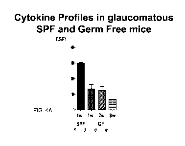
FIGS. 4A and 4B are graphs showing the cytokine profiles in glaucomatous SPF
and
germ free mice. Quantification of serum levels of (FIG. 4A) colony stimulating
factors 1
(CSF1) and (FIG. 4B) CSF2 in the retinas of specific pathogen free (SPF) and
germ free (GF)
mice at weeks 1, 2 and 8 after elevation of intraocular pressure (I0P), by
Luminex assay.
Note the consistent down-regulation of CSF1 and upregulation of CSF2 in the
glaucomatous
GF mice compared to SPF mice.
FIGS. SA and 5B are graphs showing upregulated CSF1/CSF1R expression in
microbead (MB)-injected mice. The results from qPCR quantification
demonstrated
upregulation of CSF1 (FIG. SA) and CSF1R (FIG. 5B) expression in the mouse
retinas after
microbead-induced elevation of intraocular pressure.
FIGS. 6A, 6B and 6C are a series of graphs and a Western blot demonstrating
the
downregulation of CSF2 expression in MB-injected mice. Results of qPCR (FIG.
6A) and
Western blot (FIG. 6B, 6C) quantifications showing downregulation of CSF2 and
CSF2R
expression in the mouse retinas after MB- induced elevation of intraocular
pressure.
FIG. 7 is a schematic representation showing the experimental design. It was
hypothesized that inhibiting CSF1 by administration of CSF1R antibody (CSF1R
Ab) and/or
promoting CSF2 signaling protect RGC against glaucomatous damage. Four
experimental
5
CA 03093086 2020-09-03
WO 2019/173361
PCT/US2019/020787
groups were proposed: following MB-injection to induce high IOP, mice received
treatment
of vehicle (PBS), CSF1R Ab, CSF2, or CSF1R+CSF2 were studied.
FIG. 8 is a graph showing the intraocular pressure (I0P) levels in all groups
of mice.
IOP levels were monitored during the entire period. IOP levels in all
experimental groups
were increased from 10 mmHg baseline to and maintained above 16 mmHg after MB
injection.
FIG. 9 is a graph showing decreased RGC loss by the treatment of CSF1RAb
and/or
CSF2 after MB injection. RGC counts from control (Ctr) mice and glaucomatous
(MB-
injected) mice received vehicle, CSF1RAb, CSF2 and CSF1RAb+CSF2 treatment.
FIG. 10 is a graph showing that pSTR increased by treatment of CSF1RAb/CSF2
after MB injection. pSTR amplitudes assessed in MB-induced glaucomatous mice
received
vehicle, CSF1RAb, CSF2 or CSF1RAb+CSF2 treatment at before (Ctr), 4 (G4w) and
6
(G4w) weeks after MB injection.
FIGS. 11A and 11 B are graphs demonstrating that visual function increased by
treatment of CSF1RAb/CSF2 after MB injection. Assessment of vision contrast
sensitivity
and visual acuity in MB-induced glaucomatous mice received vehicle, CSF1RAb,
CSF2 or
CSF1RAb+CSF2 treatment at before (Ctr), 4 (G4w) and 6 (G4w) weeks after MB
injection.
FIG. 12 is a graph showing that high IOP induced lba-1 expression. qPCR
quantification of lba-1 levels in the retinas of mice at day 0, 3, 7, 14 after
MB injection.
FIG. 13 is a Western blot and a graph demonstrating that high IOP activates
Muller
glia. Western blot quantification of the expression of activated Muller glia
marker, GFPA, in
the mouse retinas at day 0, 3, 7, 14 after MB injection.
DETAILED DESCRIPTION
Activation of microglia plays a critical role in the progression of
neurodegeneration in
glaucoma. Colony stimulating factor 1 (CSF1) and colony stimulating factor 2
(CSF2) are
involved in glaucomatous neuron loss by regulating microglia function. Mice
with glaucoma
showed upregulated CSF1 levels and downregulation of CSF2. Moreover, addition
of CSF2
recombinant protein or neutralizing CSF1 by intravitreal injection of anti-
CSF1 or antibody
against CSF1 receptor in a glaucoma mouse model significantly suppressed
microglial
6
CA 03093086 2020-09-03
WO 2019/173361
PCT/US2019/020787
activation and protected the loss of retinal ganglion cells (RGCs) and vision
function in a
glaucoma mouse model. The data indicates that modulating CSF pathways is
useful to confer
a clinical benefit to subjects with glaucoma and/or optic neuropathy.
Colony Stimulating Factors
CSF-1 is present in the circulation, predominantly as the proteoglycan form,
at
biologically active concentrations of approximately 10 ng/mL. It is produced
constitutively
by a wide variety of cells of mesenchymal and epithelial origin. The level in
the circulation
increases in many different pathologies, including infections, cancer, and
chronic
inflammatory disease, regardless of etiology. CSF1 controls the survival,
proliferation, and
differentiation of mononuclear phagocytes and regulates cells of the females
reproductive
tract. CSF1 may also play an autocrine and/or paracrine role in cancers of the
ovary,
endometrium, breast, and myeloid and lymphoid tissues.CSF1 levels are also
elevated in the
circulation during pregnancy and contribute to placentation. In both mice and
humans, there
is a perinatal surge of tissue and circulating CSF1. In inflammation, CSF1 may
also be
produced by recruited macrophages themselves, although in the mouse at least,
most
macrophages do not produce CSF1 and undergo cell death in the absence of the
protein.
Under normal steady-state conditions, the production of CSF1 is balanced by
its consumption
by tissue macrophages, through receptor-mediated endocytosis by the CSF1
receptor
(CSF1R) followed by intracellular destruction.
Granulocyte-macrophage colony-stimulating factor, also known as GM-CSF and
CSF2, is a monomeric glycoprotein secreted by macrophages, T cells, mast
cells, natural
killer cells, endothelial cells and fibroblasts that functions as a cytokine.
CSF2 controls the
production, differentiation, and function of granulocytes and macrophages. The
pharmaceutical analogs of naturally occurring CSF2 are called sargramostim and
molgramostim. CSF2 can be used as a medication to stimulate the production of
white blood
cells following chemotherapy. It may also be used as a vaccine adjuvant in HIV-
infected
patients.
Colony-stimulating factor 3 (CSF 3), is a glycoprotein that stimulates the
bone
marrow to produce granulocytes and stem cells and release them into the
bloodstream.
Functionally, it is a cytokine and hormone, a type of colony-stimulating
factor, and is
produced by a number of different tissues. The pharmaceutical analogs of
naturally occurring
7
CA 03093086 2020-09-03
WO 2019/173361
PCT/US2019/020787
CSF3 are called filgrastim and lenograstim. CSF3 also stimulates the survival,
proliferation,
differentiation, and function of neutrophil precursors and mature neutrophils.
CSF1 and CSF2 play a primary role in mediating the actions of
monocytes/macrophages, while CSF2 and CSF3 regulate granulocytes
(neutrophils).
.. Macrophages are polarized into M1 and M2 subtypes after activation. CSF2
promotes the M1
phenotype, which secretes pro-inflammatory cytokines, such as TNF-a and IL-12,
and
enhances the defense against bacteria or tumor by stimulating immune
responses. In contrast,
CSF1 stimulates the M2 phenotype that secretes anti-inflammatory cytokines and
promotes
tissue repair and angiogenesis.
The receptors of CSF1 and CSF2 belong to the tyrosine kinase family and
activate the
downstream pathways of PI-3/AKT, MAPK, STAT pathways to signal cell
survival/proliferation, differentiation, and activation.
Prior to the invention, a role for CSF1 and/or CSF2 in glaucoma or retinal
neurodegenerative disease had not been identified.
The data (FIGS. 1A-1D, FIGS. 2A-1D, FIGS. 3A-3F, FIGS. 5A, 5B, FIG. 9, FIG.
10,
FIGS. 11A, 11B) showed that CSF1 or CSF1 receptor blockade, as well as CSF2
protein
administration/augmentation protected retinal ganglion cell degeneration and
protected visual
performance in a mouse model of human glaucoma. These data unveil new insights
into the
pathogenesis of glaucomatous neural damage and demonstrate the therapeutic
potential of
blocking CSF1/CSF1 receptor activity and/or administration of CSF2 recombinant
proteins
via intravitreal injection for glaucomatous patients. Multiple injections of
such agents into the
vitreous are a common and low risk practice to patients in an ophthalmology
clinic.
Glaucoma is the most common form of optic neuropathy. The compositions and
methods are
also applicable to the treatment of other forms of optic neuropathy or for
similar
neurodegeneration conditions in the brain and spinal cord.
Regulation of microglia activation by CSFs drives neurodegeneration in
glaucoma
The studies described herein indicated that mice raised in the absence of
microflora
(germ-free mice) do not develop retinal ganglion cells (RGC) damage following
elevation of
intraocular pressure (I0P). Moreover, data indicated that the expression of
colony
stimulating factor 1 (CSF1) was downregulated, while CSF2 was upregulated in
germ-free
8
CA 03093086 2020-09-03
WO 2019/173361
PCT/US2019/020787
mice. Studies were then carried out to determine whether CSF1 and CSF2 played
an
opposing role mediating RGC loss in glaucoma. Herein, the expression and
involvement of
CSF1 and CSF2 in a standard mouse model of glaucoma, is reported.
Microbeads (MB) were injected into the anterior chamber of adult B6 mice to
induce
high IOP. The expression of CSF1/2 and their receptors was examined by
immunostaining
and quantitative polymerase chain reaction (qPCR) at different time points
after MB
injection. CSF2 and/or neutralizing antibody of CSF1 were adminstered
intravitreally to mice
with high IOP. Anti-bm3a staining was used to label RGC in the whole-mount
retina.
The expression of CSF1 was found to be upregulated, while CSF2 was
downregulated
in the retina 2 weeks after MB injection. The data also showed that
administration of either
CSF2 or antibody specific for CSF1, e.g., a neutralizing antibody, reduced or
attenuated
glaucomatous RGC loss compared to saline-treated control mice. CSF1 receptor
was found to
associate with microglia and RGCs, while CSF2 receptor was expressed by Muller
cells and
RGCs.
These data indicate that CSF1 and CSF2 play opposing roles on microglia and
Muller
cells activation under elevated IOP that drive glaucomatous RGC degeneration.
These
findings indicate inhibition of CSF signaling and/or augmentation of CSF
signaling is
effective to reduce or prevent RGC loss and vision loss in subjects with a
optic neuropathic
disorder such as glaucoma.
CSF1, CSF2
UniProtKB - P09603 (CSFl_HUMAN) is provided below (SEQ ID NO: 1):
10 20 30 40 50
MTAPGAAGRC PPTTWLGSLL LLVCLLASRS IlLEVSEYCS HMIGSGHLQS
60 70 80 90 100
LQRLIDSQME TSCQITI,EFV DQEQLKDPVC YLKKAFLLVQ DIMEDTMRFR
110 120 130 140 150
DNTPNAIAIV QLQELSLRLK SCFTKDYEEH DKACVRTFYE TPLQLLEKVK
160 170 180 190 200
NVFNETKNLL DKDWNIFSKN CNNSFAECSS QDVVTKPDCN CLYPKAIPSS
210 220 230 240 250
DPASVSPHQP LAPSMAPVAG LTWEDSEGTE GSSLLPGEQP LHTVDPGSAK
260 270 280 290 300
9
CA 03093086 2020-09-03
WO 2019/173361
PCT/US2019/020787
QRPPRSTCQS FEPPETPVVK DSTIGGSPQP RPSVGAFNPG MEDILDSAMG
310 320 330 340 350
TNWVPEEASG EASEIPVPQG TELSPSRPGG GSMQTEPARP SNFLSASSPL
360 370 380 390 400
PASAKGQQPA DVTGTALPRV GPVRPTGQDW NHTPQKTDHP SALLRDPPEP
410 420 430 440 450
GSPRISSLRP QGLSNPSTLS AQPQLSRSHS SGSVLPLGEL EGRRSTRDRR
460 470 480 490 500
SPAEPEGGPA SEGAARPLPR FNSVPLTDTG HERQSEGSFS PQLQESVFHL
510 520 530 540 550
LVPSVILVLL AVGGLLFYRW RRRSHQEPQR ADSPLEQPEG SPLTQDDRQV
ELPV
Human Colony Stimulating Factor 1 Receptor (CSF1-R) amino acid sequence
AAH47521.1 is provided below (SEQ ID NO: 2):
1 MGPGVLLLLL VATAWHGQGI PVIEPSVPEL VVKPGATVTL RCVGNGSVEW DGPPSPHWTL
61 YSDGSSSILS TNNATFQNTG TYRCTEPGDP LGGSAAIHLY VKDPARPWNV LAQEVVVFED
121 QDALLPCLLT DPVLEAGVSL VRVRGRPLMR HTNYSFSPWH GFTIHRAKFI QSQDYQCSAL
181 MGGRKVMSIS IRLKVQKVIP GPPALTLVPA ELVRIRGEAA QIVCSASSVD VNFDVFLQHN
241 NTKLAIHQQS DFHNNRYQKV LTLNLDQVDF QHAGNYSCVA SNVQGKHSTS MFFRVVESAY
301 LNLSSEQNLI QEVTVGEGLN LKVMVEAYPG LQGFNWTYLG PFSDHQPEPK LANVTTKDTY
361 RHTFTLSLPR LKPSEAGRYS FLARNPGGWR ALTFELTLRY PPEVSVIWTF INGSGTLLCA
421 ASGYPQPNVT WLQCSGHTDR CDEAQVLQVW DDPYPEVLSQ EPFHKVTVQS LLTVETLEHN
481 QTYECRAHNS VGSGSWAFIP ISAGAHTHPP DEFLFTPVVV ACMSIMALLL LLLLLLLYKY
541 KQKPKYQVRW KIIESYEGNS YTFIDPTQLP YNEKWEFPRN NLQFGKTLGA GAFGKVVEAT
601 AFGLGKEDAV LKVAVKMLKS TAHADEKESL MSELKIMSHL GQHENIVNLL GACTHGGPVL
661 VITEYCCYGD LLNFLRRKAE AMLGPSLSPG QDPEGGVDYK NIHLEKKYVR RDSGFSSQGV
721 DTYVEMRPVS TSSNDSFSEQ DLDKEDGRPL ELRDLLHFSS QVAQGMAFLA SKNCIHRDVA
781 ARNVLLTNGH VAKIGDFGLA RDIMNDSNYI VKGNARLPVK WMAPESIFDC VYTVQSDVWS
841 YGILLWEIFS LGLNPYPGIL VNSKFYKLVK DGYQMAQPAF APKNIYSIMQ ACWALEPTHR
901 PTFQQICSFL QEQAQEDRRE RDYTNLPSSS RSGGSGSSSS ELEEESSSEH LTCCEQGDIA
961 QPLLQPNNYQ FC
SEQ ID NO: 2; GenBank Accession AAH47521.1, incorporated herein by reference.
Exemplary landmark residues, domains, and fragments of CSF1-R include, but are
not
limited to residues 28 ¨ 85 (Immunoglobulin like domain), residues 207 - 293
(Immunoglobulin like domain), residues 209 ¨ 290 (Immunoglobulin like domain),
residues
299 ¨ 400 (Fourth immunoglobulin (Ig)-like domain of stem cell factor receptor
(SCFR)),
residues 401 ¨ 495 (Immunoglobulin like domain), 542 ¨ 914 (Protein Kinases,
catalytic
domain), or residues 588 to 591, 594, 596, 614, 616, 647, 663 to 666, 778,
782..783, 785, and
795 and 796 (ATP binding sites). A fragment of a CSF1-R protein is less than
the length of
CA 03093086 2020-09-03
WO 2019/173361
PCT/US2019/020787
the full length protein, e.g., a fragment is at least 3, 4, 5, 6, 7, 8, 9, 10,
20, 30, 40, 50, 100,
200 or more residues in length, but less than e.g., 972 residues in the case
of CSF1-R above.
Human CSF1-R nucleic acid sequence BC047521.1 (start and stop codons
underlined) (SEQ ID NO: 3):
1 ggtggccttg cctagctaaa aggggaagaa gaggatcagc ccaaggagga ggaagaggaa
61 aacaagacaa acagccagtg cagaggagag gaacgtgtgt ccagtgtccc gatccctgcg
121 gagctagtag ctgagagctc tgtgccctgg gcaccttgca gccctgcacc tgcctgccac
181 ttccccaccg aggccatggg cccaggagtt ctgctgctcc tgctggtggc cacagcttgg
241 catggtcagg gaatcccagt gatagagccc agtgtccccg agctggtcgt gaagccagga
301 gcaacggtga ccttgcgatg tgtgggcaat ggcagcgtgg aatgggatgg ccccccatca
361 cctcactgga ccctgtactc tgatggctcc agcagcatcc tcagcaccaa caacgctacc
421 ttccaaaaca cggggaccta tcgctgcact gagcctggag accccctggg aggcagcgcc
481 gccatccacc tctatgtcaa agaccctgcc cggccctgga acgtgctagc acaggaggtg
541 gtcgtgttcg aggaccagga cgcactactg ccctgtctgc tcacagaccc ggtgctggaa
601 gcaggcgtct cgctggtgcg tgtgcgtggc cggcccctca tgcgccacac caactactcc
661 ttctcgccct ggcatggctt caccatccac agggccaagt tcattcagag ccaggactat
721 caatgcagtg ccctgatggg tggcaggaag gtgatgtcca tcagcatccg gctgaaagtg
781 cagaaagtca tcccagggcc cccagccttg acactggtgc ctgcagagct ggtgcggatt
841 cgaggggagg ctgcccagat cgtgtgctca gccagcagcg ttgatgttaa ctttgatgtc
901 ttcctccaac acaacaacac caagctcgca atccatcaac aatctgactt tcataataac
961 cgttaccaaa aagtcctgac cctcaacctc gatcaagtag atttccaaca tgccggcaac
1021 tactcctgcg tggccagcaa cgtgcagggc aagcactcca cctccatgtt cttccgggtg
1081 gtagagagtg cctacttgaa cttgagctct gagcagaacc tcatccagga ggtgaccgtg
1141 ggggaggggc tcaacctcaa agtcatggtg gaggcctacc caggcctgca aggttttaac
1201 tggacctacc tgggaccctt ttctgaccac cagcctgagc ccaagcttgc taatgttacc
1261 accaaggaca catacaggca caccttcacc ctctctctgc cccgcctgaa gccctctgag
1321 gctggccgct actccttcct ggccagaaac ccaggaggct ggagagctct gacgtttgag
1381 ctcacccttc gatacccccc agaggtaagc gtcatatgga cattcatcaa cggctctggc
1441 acccttttgt gtgctgcctc tgggtacccc cagcccaacg tgacatggct gcagtgcagt
1501 ggccacactg ataggtgtga tgaggcccaa gtgctgcagg tctgggatga cccataccct
1561 gaggtcctga gccaggagcc cttccacaag gtgacggtgc agagcctgct gactgttgag
1621 accttagagc acaaccaaac ctacgagtgc agggcccaca acagcgtggg gagtggctcc
1681 tgggccttca tacccatctc tgcaggagcc cacacgcatc ccccggatga gttcctcttc
1741 acaccagtgg tggtcgcctg catgtccatc atggccttgc tgctgctgct gctcctgctg
1801 ctattgtaca agtataagca gaagcccaag taccaggtcc gctggaagat catcgagagc
1861 tatgagggca acagttatac tttcatcgac cccacgcagc tgccttacaa cgagaagtgg
1921 gagttccccc ggaacaacct gcagtttggt aagaccctcg gagctggagc ctttgggaag
1981 gtggtggagg ccacggcctt tggtctgggc aaggaggatg ctgtcctgaa ggtggctgtg
2041 aagatgctga agtccacggc ccatgctgat gagaaggagt ccctcatgtc cgagctgaag
2101 atcatgagcc acctgggcca gcacgagaac atcgtcaacc ttctgggagc ctgtacccat
2161 ggaggccctg tactggtcat cacggagtac tgttgctatg gcgacctgct caactttctg
2221 cgaaggaagg ctgaggccat gctgggaccc agcctgagcc ccggccagga ccccgaggga
2281 ggcgtcgact ataagaacat ccacctcgag aagaaatatg tccgcaggga cagtggcttc
2341 tccagccagg gtgtggacac ctatgtggag atgaggcctg tctccacttc ttcaaatgac
2401 tccttctctg agcaagacct ggacaaggag gatggacggc ccctggagct ccgggacctg
2461 cttcacttct ccagccaagt agcccagggc atggccttcc tcgcttccaa gaattgcatc
2521 caccgggacg tggcagcgcg taacgtgctg ttgaccaatg gtcatgtggc caagattggg
2581 gacttcgggc tggctaggga catcatgaat gactccaact acattgtcaa gggcaatgcc
2641 cgcctgcctg tgaagtggat ggccccagag agcatctttg actgtgtcta cacggttcag
2701 agcgacgtct ggtcctatgg catcctcctc tgggagatct tctcacttgg gctgaatccc
2761 taccctggca tcctggtgaa cagcaagttc tataaactgg tgaaggatgg ataccaaatg
2821 gcccagcctg catttgcccc aaagaatata tacagcatca tgcaggcctg ctgggccttg
2881 gagcccaccc acagacccac cttccagcag atctgctcct tccttcagga gcaggcccaa
2941 gaggacagga gagagcggga ctataccaat ctgccgagca gcagcagaag cggtggcagc
3001 ggcagcagca gcagtgagct ggaggaggag agctctagtg agcacctgac ctgctgcgag
3061 caaggggata tcgcccagcc cttgctgcag cccaacaact atcagttctg ctgaggagtt
3121 gacgacaggg agtaccactc tcccctcctc caaacttcaa ctcctccatg gatggggcga
11
CA 03093086 2020-09-03
WO 2019/173361
PCT/US2019/020787
3181 cacggggaga acatacaaac tctgccttcg gtcatttcac tcaacagctc ggcccagctc
3241 tgaaacttgg gaaggtgagg gattcagggg aggtcagagg atcccacttc ctgagcatgg
3301 gccatcactg ccagtcaggg gctgggggct gagccctcac cccccgcctc ccctactgtt
3361 ctcatggtgt tggcctcgtg tttgctatgc caactagtag aaccttcttt cctaatcccc
3421 ttatcttcat ggaaatggac tgactttatg cctatgaagt ccccaggagc tacactgata
3481 ctgagaaaac caggctcttt ggggctagac agactggcag agagtgagat ctccctctct
3541 gagaggagca gcagatgctc acagaccaca ctcagctcag gccccttgga gcaggatggc
3601 tcctctaaga atctcacagg acctcttagt ctctgcccta tacgccgcct tcactccaca
3661 gcctcacccc tcccaccccc atactggtac tgctgtaatg agccaagtgg cagctaaaag
3721 ttgggggtgt tctgcccagt cccgtcattc tgggctagaa ggcaggggac cttggcatgt
3781 ggctggccac accaagcagg aagcacaaac tcccccaagc tgactcatcc taactaacag
3841 tcacgccgtg ggatgtctct gtccacatta aactaacagc attaatacaa aaaaaaaaaa
3901 aaaa
The sequence of CSF2 (also known as GM-CSF is described in NP_000749.2
granulocyte-macrophage colony-stimulating factor precursor; hereby
incorporated by
reference in its entirety.) (SEQ ID NO: 4):
1 MWLQSLLLLG TVACSISAPA RSPSPSTQPW EHVNAIQEAR RLLNLSRDTA AEMNETVEVI
61 SEMFDLQEPT CLQTRLELYK QGLRGSLTKL KGPLTMMASH YKQHCPPTPE TSCATQIITF
121 ESFKENLKDF LLVIPFDCWE PVQE
Antibodies and Inhibitors of CSF1 and CSF1R
A humanized immunoglobulin (Ig) G2 monoclonal antibody (mAb) directed against
the cytokine colony stimulating factor 1 (CSF1; CSF-1; macrophage colony-
stimulating
factor; M-CSF), with potential immunomodulating and antineoplastic activities,
anti-CSF1
monoclonal antibody PD-0360324 targets, binds to and neutralizes CSF1. This
prevents the
binding of CSF1 to its receptor CSF1R (CD115; M-CSPR), which is expressed on
various
immune cells, such as monocytes and macrophages. This prevents CSF1R
activation and
CSF1R-mediated signaling in these cells, leading to inhibition of monocyte
differentiation,
blocking the activity of macrophages, and reducing their production of
inflammatory
mediators, which reduces inflammation. By blocking the activity and
proliferation of CSF1R-
dependent tumor-associated macrophages (TAMs) in the tumor microenvironment,
PD-
0360324 reduces TAM-mediated immune suppression, decreases regulatory T cells
(Tregs),
re-activates the immune system, and improves anti-tumor cell responses
mediated by
increasing infiltration by cytotoxic T cells. TAMs play key roles in immune
suppression, and
tumor cell proliferation and survival. CSF-1 plays a key role in the
regulation of the
proliferation, differentiation and survival of monocytes and macrophages.
Exemplary antibodies include those available from R&D systems (Minneapolis,
MN),
e.g., Mouse M-CSF Antibody neutralized; Cat #: MAB416-SP, or eBioscience
12
CA 03093086 2020-09-03
WO 2019/173361
PCT/US2019/020787
(ThermoFischer); CD115 (c-fms) Monoclonal Antibody; Cat #: AFS9 or Peprotech
(Rocky
Hill, NJ 08553) United States; Recombinant Murine GM- CSF; Cat #: 315-03.
An orally bioavailable inhibitor of colony stimulating factor 1 receptor (CSF-
1R;
CSF1R), with potential antineoplastic activity, CSF1R inhibitor BLZ945 (4--
[2((1R,2R)-2-
hydroxycyelohexylamino) - benzothiazol -6 -yloxyll-pyridine- 2-carboxylic acid
me thylamide)
selectively binds to CSF1R expressed on tumor-associated macrophages (TAMs),
blocks the
activity of CSF1R, and inhibits CSF1R-mediated signal transduction pathways.
This inhibits
the activity and proliferation of TAMs, and reprograms the immunosuppressive
nature of
existing TAMs. Altogether, this reduces TAM-mediated immune suppression in the
tumor
microenvironment, re-activates the immune system, and improves anti-tumor cell
responses
mediated by T-cells. CSF1R, also known as macrophage colony-stimulating factor
receptor
(M-CSFR) and CD115 (cluster of differentiation 115), is a cell-surface
receptor for its ligand,
colony stimulating factor 1 (CSF1); this receptor is overexpressed by TAMs in
the tumor
microenvironment, and plays a major role in both immune suppression and the
induction of
.. tumor cell proliferation.
Another inhibitor of the tyrosine kinase receptor colony stimulating factor 1
receptor
(CSF1R; CSF-1R; C-FMS; CD115; M-CSFR), with potential antineoplastic,
macrophage
checkpoint-inhibitory and immunomodulating activities, DCC-3014, targets and
binds to
CSF1R expressed on monocytes, macrophages, and osteoclasts and inhibits the
binding of the
CSF1R ligands colony-stimulating factor-1 (CSF-1) and interleukin-34 (IL-34),
to CSF1R.
This prevents CSF1R activation and CSF1R-mediated signaling in these cells.
This blocks
the production of inflammatory mediators by macrophages and monocytes and
reduces
inflammation. By blocking the recruitment to the tumor microenvironment and
activity of
CSF1R-dependent tumor-associated macrophages (TAMs), DCC-3014 inhibits the
immunomodulating activity by macrophages and enhances T-cell infiltration and
antitumor
T-cell immune responses, which inhibits the proliferation of tumor cells. TAMs
play key
roles in the tumor microenvironment and allow for immune suppression; TAMs
promote
inflammation, tumor cell proliferation, angiogenesis, invasiveness and
survival.
Examples of other inhibitors of CSF1 receptors undergoing clinical phase
trials in
cancer patients, include: Pexidartinib (PLX3397, PLX108-01), PLX7486, ARRY-
382, JNJ-
40346527, BLZ945, Emactuzumab (RG7155), AMG820, IMC-054 (LY3022855), MCS110,
GW-2580, Gleevec (imatinib mesylate). (Cannarile, Michael A et al. "Colony-
stimulating
13
CA 03093086 2020-09-03
WO 2019/173361
PCT/US2019/020787
factor 1 receptor (CSF1R) inhibitors in cancer therapy" Journal for
immunotherapy of
cancer vol. 5,1 53. 18 Jul. 2017, doi:10.1186/540425-017-0257-y).
PLX73086 (AC708) which is a small molecule inhibitor of CSF1R, which leads to
reduced CSF1R activation and may restore resistance to angiogenesis inhibition
through a
decrease in tumor associated macrophages (Lyons, Y.A. et al., Oncotarget. 2017
Aug
24;8(57):96496-96505. doi: 10.18632/oncotarget.20410. eCollection 2017 Nov
14).
Chiauranib (CS2164) is a multi-kinase inhibitor that inhibits AURKB, CSF-1R,
VEGFRs,
KIT, and PDGFRA, resulting in decreased tumor growth and angiogenesis (Zhou,
Y. et al.,
Cancer Sci. 2017 Mar;108(3):469-477. doi: 10.1111/cas.13141. Epub 2017 Mar 7).
Sprycel
(dasatinib) is an inhibitor of the SRC-family of protein kinases, BCR-ABL, and
ABL, and
has additional activity against other kinases including KIT, DDR1/2, PDGFRA/B,
and
EPHA2, which prevents cell growth (Kothiwale S. et al., Drug Discov Today.
2015
Feb;20(2):255-61. doi: 10.1016/j.drudis.2014.09.025. Epub 2014 Oct 7). DCC-
3014 inhibits
CSF1R, potentially resulting in increased anti-tumor immune response in
combination with
other agents (Cancer Res 2016;76(14 Suppl):Abstract nr 4889). Debio 0617B is a
multi-
kinase inhibitor of SRC, JAK, and ABL, the class III kinases, CSF1R, FLT3,
KIT, and
PDGFR, and the class V kinases, VEGFR 1/2/3, which may result in inhibition of
Stat3 and
Stat5 signaling, leading to inhibition of tumor cell growth and metastasis
(Murone, M. et al.,
Mol Cancer Ther. 2016 Oct;15(10):2334-2343. Epub 2016 Jul 20). Dovitinib
(TKI258)
targets multiple receptor tyrosine kinases including Flt3, c-Kit, CSF1R, FGFR
1-4, VEGFR
1-3, and PDGFR alpha and beta, potentially resulting in decreased tumor growth
(Lesca E., et
al., J Mol Biol. 2014 Nov 11;426(22):3744-3756. doi:
10.1016/j.jmb.2014.09.004. Epub 2014
Sep 16; Andre F. et al., Clin Cancer Res. 2013 Jul 1;19(13):3693-702. doi:
10.1158/1078-
0432.CCR-13-0190. Epub 2013 May 8). Emactuzumab (RG7155) is a monoclonal
antibody
that inhibits dimerization of CSF1R, resulting in decreased ligand-dependent
and ligand-
independent signaling (Ries C. H. et al., Cancer Cell. 2014 Jun 16;25(6):846-
59. doi:
10.1016/j.ccr.2014.05.016. Epub 2014 Jun 2). FF-10101 is a second generation
and
irreversible inhibitor of Flt3, including the internal tandem duplication
(FLT3-ITD) and
known resistance mutations (D835Y, Y842C, Y842H, or F691L) and also inhibits
Kit and
Csflr (Fms) (Yamaura T. et al., Blood. 2018 Jan 25;131(4):426-438. doi:
10.1182/blood-
2017-05-786657. Epub 2017 Nov 29). GW2580 is an ATP-competitive selective
inhibitor of
CSF-1R, which may lead to decreased tumor cell growth (Ryder M. et al., PLoS
One.
2013;8(1):e54302. doi: 10.1371/journal.pone.0054302. Epub 2013 Jan 23). JNJ-
40346527 is
14
CA 03093086 2020-09-03
WO 2019/173361
PCT/US2019/020787
a small molecule inhibitor of CSF1R (von Tresckow B. et al., Clin Cancer Res.
2015 Apr
15;21(8):1843-50. doi: 10.1158/1078-0432.CCR-14-1845. Epub 2015 Jan 27).
Ki20227
inhibits CSF1R, which may result in decreased CSF-dependent cell growth (Ohno
H. et al.,
Mol Cancer Ther. 2006 Nov;5(11):2634-43). Lestaurtinib (CEP-701) (Hexner E. 0.
et al.,
Blood. 2008 Jun 15;111(12):5663-71. Epub 2007 Nov 5). Linifanib (ABT-869) is a
receptor
tyrosine kinase inhibitor with specificity against FLT1 (VEGFR1), CSF-1R, KDR
(VEGFR2), FLT3, and KIT, which may result in inhibition of cell proliferation
and tumor
growth, and tumor regression (Albert D. H. et al., Mol Cancer Ther. 2006
Apr;5(4):995-
1006). Rydapt (midostaurin) is a multi-kinase inhibitor (Ashman L.K. et al.,
Expert Opin
Investig Drugs. 2013 Jan;22(1):103-15. doi: 10.1517/13543784.2013.740010. Epub
2012
Nov 6). Tasigna (nilotinib) inhibits several tyrosine kinases including BCR-
ABL, PDGFR,
KIT, DDR and CSF-1R (Blay J.Y. et al., Semin Oncol. 2011 Apr;38 Suppl 1:S3-9.
doi:
10.1053/j.seminonco1.2011.01.016). Pexidartinib (PLX3397) inhibits multiple
receptor
tyrosine kinases, including KIT, CSF1R, FLT3, and FLT3/ITD (Smith C. C. et
al., Cancer
.. Discov. 2015 Jun;5(6):668-79. doi: 10.1158/2159-8290.CD-15-0060. Epub 2015
Apr 6).
PLX7486 binds to and inhibits CSF1R, TRKA, TRKB, and TRKC. Nexavar (sorafenib)
is a
multikinase inhibitor with activity against several kinases, including RAF
kinases, VEGFR2,
VEGFR3, PDGFR-beta, KIT, FLT3, RET, and CSF1R (Ullrich K. et al., Br J
Haematol.
2011 Nov;155(3):398-402. doi: 10.1111/j.1365-2141.2011.08685.x. Epub 2011 Apr
22).
Sutent (sunitinib) inhibits KDR (VEGFR2), PDGFR, c-KIT, FLT3, RET, and CSF1R
(Subbiah V. et al., J Hematol Oncol. 2014 Aug 1;7:52. doi: 10.1186/513045-014-
0052-x).
In certain embodiments a CSF1R inhibitor comprises PLX3397 (Tahmasebi F. et
al.,
J Cell Biochem. 2019 Jan 10. doi: 10.1002/jcb.28344), GW-2580 (Gerber Y. N. et
al., Front
Cell Neurosci. 2018; 12: 368.), BLZ-945 (Pyonteck S. M. et al., Nat Med. 2013
Oct;19(10):1264-72. doi: 10.1038/nm.3337) or combinations thereof. In certain
embodiments a CSF1R neutralizing antibody comprises: RG-7155, FPA-008, M279
(publication available, e.g., from AMGEN) or combinations thereof.
The antibodies which specifically bind to CSF1 or receptors thereof, inhibit
the
function or activity of the CSF1 molecule. The antibodies can be produced by
any means
known in the art directed to SEQ ID NOS: 1 or 2. In certain embodiments, the
antibodies or
fragments thereof, specifically bind to a CSF1 peptide or CSF1R having at
least a 50%
sequence identity to SEQ ID NOS: 1 or 2 respectively. In certain embodiments,
the
CA 03093086 2020-09-03
WO 2019/173361
PCT/US2019/020787
antibodies or fragments thereof, specifically bind to a CSF1 peptide or CSF1R
having at least
a 75% sequence identity to SEQ ID NOS: 1 or 2 respectively. In certain
embodiments, the
antibodies or fragments thereof, specifically bind to a CSF1 peptide or CSF1R
having at least
a 95% sequence identity to SEQ ID NOS: 1 or 2 respectively. In certain
embodiments, the
antibodies or fragments thereof specifically bind to epitopes in SEQ ID NOS: 1
or 2.
In certain embodiments, a CSF1 and receptor thereof inhibitor(s)
preferentially inhibit
CSF1 and receptors thereof, as compared to CSF2. In certain embodiments, a
CSF1 inhibitor
or a CSF1R inhibitor, inhibit the expression and/or activity and/or function
of CSF1 by about
1-fold as compared to a CSF2 activity or function. In certain embodiments, a
CSF1 inhibitor
or a CSF1R inhibitor, inhibit the expression and/or activity and/or function
of CSF1 by about
2-fold, 3-fold, 5-fold, 10-fold, 20-fold, 50-fold or more as compared to CSF2
activity or
function. In certain embodiments, a CSF1 inhibitor or a CSF1R inhibitor,
inhibit the
expression and/or activity and/or function of CSF1 by at least 5% as compared
to CSF2
activity or function. In certain embodiments, a CSF1 inhibitor or a CSF1R
inhibitor, inhibit
the expression and/or activity and/or function of CSF1 by at least 10%, 15%,
20%, 30%,
40%, 50%, 60%, 70%, 80%, 85%, 86%, 87%, 88%, 89%, 90%, 91%, 92%, 93%, 94%,
95%,
96%, 97%, 98%, 99%, or more as compared to CSF2 activity or function.
The activity of CSF1 and CSF2 can be determined by any number of assays known
in
the art and also detailed herein. For example, mRNA expression, protein
expression (FIGS.
1A-1C, FIGS. 2A-2D, FIGS. 5A-5B, FIGS. 6A-6C), intraocular pressure (FIGS. 3A-
3G, FIG.
8, FIG. 12), cytokine profiles (FIGS. 5A, 5B), RGC loss (FIG. 9), positive
scotopic threshold
response (pSTR) (FIG. 10), visual function (FIGS. 11A, 11B), lba-1 expression
(FIG. 12),
activation of Muller glia (FIG. 13). Whether an inhibitor specifically
inhibits CSF1 or
receptor thereof versus CSF2 can be determined by similar assays. For example,
expression
of CSF1, CSF1R versus CSF2. These assays can be combined with clinical
examination
which are routine.
The function of retinal ganglion cells (RGCs) can be non-invasively assessed
in
experimental and genetic models of glaucoma by means of variants of the ERG
technique
that emphasize the activity of inner retina neurons. The best understood
technique is the
Pattern Electroretinogram (PERG) in response to contrast-reversing gratings or
checkerboards, which selectively depends on the presence of functional RGCs.
In glaucoma
models, the PERG can be altered before histological loss of RGCs; PERG
alterations may be
16
CA 03093086 2020-09-03
WO 2019/173361
PCT/US2019/020787
either reversed with moderate IOP lowering or exacerbated with moderate IOP
elevation.
Under particular luminance-stimulus conditions, the Flash-ERG displays
components that
may reflect electrical activity originating in the proximal retina and be
altered in some
experimental glaucoma models (positive Scotopic Threshold response, pSTR;
negative
Scotopic Threshold Response, nSTR; Photopic Negative Response, PhNR;
Oscillatory
Potentials, OPs; multifocal ERG, mfERG) (Vittorio Porciatti, Exp Eye Res. 2015
Dec; 141:
164-170).
In some embodiments, the antigen-binding domain is a humanized antibody of
fragments thereof. A "humanized" antibody is an antibody in which all or
substantially all
complementarity determining region (CDR) amino acid residues are derived from
non-human
CDRs and all or substantially all framework region (FR) amino acid residues
are derived
from human FRs. A humanized antibody optionally may include at least a portion
of an
antibody constant region derived from a human antibody. A "humanized form" of
a non-
human antibody, refers to a variant of the non-human antibody that has
undergone
humanization, typically to reduce immunogenicity to humans, while retaining
the specificity
and affinity of the parental non-human antibody. In some embodiments, some FR
residues in
a humanized antibody are substituted with corresponding residues from a non-
human
antibody (e.g. , the antibody from which the CDR residues are derived), e.g. ,
to restore or
improve antibody specificity or affinity.
In some embodiments, the heavy and light chains of an antibody can be full-
length or
can be an antigen-binding portion (a Fab, F(ab')2, Fv or a single chain Fv
fragment (scFv)).
In other embodiments, the antibody heavy chain constant region is chosen from,
e.g., IgGl,
IgG2, IgG3, IgG4, IgM, IgAl, IgA2, IgD, and IgE, particularly chosen from,
e.g., IgGl, IgG2,
IgG3, and IgG4, more particularly, IgGl (e.g., human IgGl). In another
embodiment, the
antibody light chain constant region is chosen from, e.g., kappa or lambda,
particularly
kappa.
Among the provided antibodies are antibody fragments. An "antibody fragment"
refers to a molecule other than an intact antibody that comprises a portion of
an intact
antibody that binds the antigen to which the intact antibody binds. Examples
of antibody
fragments include but are not limited to Fv, Fab, Fab', Fab'-SH, F(ab')2;
diabodies; linear
antibodies; variable heavy chain (VII) regions, single-chain antibody
molecules such as scFvs
and single-domain VH single antibodies; and multispecific antibodies formed
from antibody
17
CA 03093086 2020-09-03
WO 2019/173361
PCT/US2019/020787
fragments. In particular embodiments, the antibodies are single-chain antibody
fragments
comprising a variable heavy chain region and/or a variable light chain region,
such as scFvs.
In certain embodiments, the antibodies are single- domain antibodies. Single-
domain
antibodies are antibody fragments comprising all or a portion of the heavy
chain variable
domain or all or a portion of the light chain variable domain of an antibody.
In certain
embodiments, a single-domain antibody is a human single-domain antibody.
Antibody fragments can be made by various techniques, including but not
limited to
proteolytic digestion of an intact antibody as well as production by
recombinant host cells. In
some embodiments, the antibodies are recombinantly-produced fragments, such as
fragments
comprising arrangements that do not occur naturally, such as those with two or
more
antibody regions or chains joined by synthetic linkers, e.g., peptide linkers,
and/or that are
may not be produced by enzyme digestion of a naturally-occurring intact
antibody. In some
aspects, the antibody fragments are scFvs.
In certain embodiments, the antibody or antibody fragments have high binding
affinity for CSF1 or CSF1 receptors. In embodiments, the increased binding
affinity is greater
than effected by a reference antigen.
In certain embodiments, inhibitors of CSF1 are selected based on their ability
to
inhibit CSF1 expression or activity. In certain embodiments, the inhibitors of
CSF1 are
selected based on their ability to inhibit expression or function of the CSF1
receptor or
inhibiting CSF1 from binding to CSF1 receptors. In certain embodiments, these
potential
therapeutic agents identified based on the screening assays are selected for
testing their
therapeutic activity. In certain embodiments, the therapeutic activity is
suppression of
microglial activation, protection against loss of retinal ganglion cells
(RGCs) and vision
function.
Candidate/Test Agents: Various candidate agents, e.g. inhibitors of CSF1 and
receptor
thereof, can be employed in the screening methods of the invention, including
any naturally
existing or artificially generated agents. They can be of any chemistry class,
such as
antibodies, small molecules, proteins, peptides, small organic compounds,
saccharides, fatty
acids, steroids, purines, pyrimidines, nucleic acids, and various structural
analogs or
combinations thereof. In some embodiments, the screening methods utilize
combinatorial
libraries of candidate agents. Combinatorial libraries can be produced for
many types of
18
CA 03093086 2020-09-03
WO 2019/173361
PCT/US2019/020787
compounds that can be synthesized in a step-by-step fashion. Such compounds
include
polypeptides, beta-turn mimetics, nucleic acids, polysaccharides,
phospholipids, hormones,
prostaglandins, steroids, aromatic compounds, heterocyclic compounds,
benzodiazepines,
oligomeric N-substituted glycines and oligocarbamates. Large combinatorial
libraries of the
compounds can be constructed by the encoded synthetic libraries (ESL) method
described in
Affymax, WO 95/12608, Affymax, WO 93/06121, Columbia University, WO 94/08051,
Pharmacopeia, WO 95/35503 and Scripps, WO 95/30642 (each of which is
incorporated
herein by reference for all purposes). Peptide libraries can also be generated
by phage display
methods. See, e.g., Devlin, WO 91/18980.
Candidate agents include numerous chemical classes, though typically they are
organic compounds including small organic compounds, nucleic acids including
oligonucleotides, peptides or antibodies. Small organic compounds suitably may
have e.g. a
molecular weight of more than about 40 or 50 yet less than about 2,500.
Candidate agents
may comprise functional chemical groups that interact with proteins and/or
DNA.
Candidate agents may be obtained from a wide variety of sources including
libraries
of synthetic or natural compounds. For example, numerous means are available
for random
and directed synthesis of a wide variety of organic compounds and
biomolecules, including
expression of randomized oligonucleotides. Alternatively, libraries of natural
compounds in
the form of e.g. bacterial, fungal and animal extracts are available or
readily produced.
Chemical Libraries: Developments in combinatorial chemistry allow the rapid
and
economical synthesis of hundreds to thousands of discrete compounds. These
compounds are
typically arrayed in moderate-sized libraries of small molecules designed for
efficient
screening. Combinatorial methods can be used to generate unbiased libraries
suitable for the
identification of novel compounds. In addition, smaller, less diverse
libraries can be
generated that are descended from a single parent compound with a previously
determined
biological activity.
A combinatorial chemical library is a collection of diverse chemical compounds
generated by either chemical synthesis or biological synthesis, by combining a
number of
chemical "building blocks," such as reagents. For example, a linear
combinatorial chemical
library, such as a polypeptide library, is formed by combining a set of
chemical building
blocks (amino acids) in a large number of combinations, and potentially in
every possible
19
CA 03093086 2020-09-03
WO 2019/173361
PCT/US2019/020787
way, for a given compound length (i.e., the number of amino acids in a
polypeptide
compound). Millions of chemical compounds can be synthesized through such
combinatorial
mixing of chemical building blocks.
A "library" may comprise from 2 to 50,000,000 diverse member compounds.
Preferably, a library comprises at least 48 diverse compounds, preferably 96
or more diverse
compounds, more preferably 384 or more diverse compounds, more preferably,
10,000 or
more diverse compounds, preferably more than 100,000 diverse members and most
preferably more than 1,000,000 diverse member compounds. By "diverse" it is
meant that
greater than 50% of the compounds in a library have chemical structures that
are not identical
to any other member of the library. Preferably, greater than 75% of the
compounds in a
library have chemical structures that are not identical to any other member of
the collection,
more preferably greater than 90% and most preferably greater than about 99%.
The preparation of combinatorial chemical libraries is well known to those of
skill in
the art. For reviews, see Thompson et al., Synthesis and application of small
molecule
libraries, Chem Rev 96:555-600, 1996; Kenan et al., Exploring molecular
diversity with
combinatorial shape libraries, Trends Biochem Sci 19:57-64, 1994; Janda,
Tagged versus
untagged libraries: methods for the generation and screening of combinatorial
chemical
libraries, Proc Nail Acad Sci USA. 91:10779-85, 1994; Lebl et al., One-bead-
one-structure
combinatorial libraries, Biopolymers 37:177-98, 1995; Eichler et al., Peptide,
.. peptidomimetic, and organic synthetic combinatorial libraries, Med Res Rev.
15:481-96,
1995; Chabala, Solid-phase combinatorial chemistry and novel tagging methods
for
identifying leads, Curr Opin Biotechnol. 6:632-9, 1995; Dolle, Discovery of
enzyme
inhibitors through combinatorial chemistry, Mol. Divers. 2:223-36, 1997;
Fauchere et al.,
Peptide and nonpeptide lead discovery using robotically synthesized soluble
libraries, Can J.
Physiol Pharmacol. 75:683-9, 1997; Eichler et al., Generation and utilization
of synthetic
combinatorial libraries, Mol Med Today 1: 174-80, 1995; and Kay et al.,
Identification of
enzyme inhibitors from phage-displayed combinatorial peptide libraries, Comb
Chem High
Throughput Screen 4:535-43, 2001.
Other chemistries for generating chemical diversity libraries can also be
used. Such
chemistries include, but are not limited to, peptoids (PCT Publication No. WO
91/19735);
encoded peptides (PCT Publication WO 93/20242); random bio-oligomers (PCT
Publication
No. WO 92/00091); benzodiazepines (U.S. Pat. No. 5,288,514); diversomers, such
as
CA 03093086 2020-09-03
WO 2019/173361
PCT/US2019/020787
hydantoins, benzodiazepines and dipeptides (Hobbs, et al., Proc. Nat. Acad.
Sci. USA,
90:6909-6913 (1993)); vinylogous polypeptides (Hagihara, et al., J. Amer.
Chem. Soc.
114:6568 (1992)); nonpeptidal peptidomimetics with 0-D-g1ucose scaffolding
(Hirschmann,
et al., J. Amer. Chem. Soc., 114:9217-9218 (1992)); analogous organic
syntheses of small
compound libraries (Chen, et al., J. Amer. Chem. Soc., 116:2661(1994));
oligocarbamates
(Cho, et al., Science, 261:1303 (1993)); and/or peptidyl phosphonates
(Campbell, et al., J.
Org. Chem. 59:658 (1994)); nucleic acid libraries (see, Ausubel, Berger and
Sambrook, all
supra); peptide nucleic acid libraries (see, e.g., U.S. Pat. No. 5,539,083);
antibody libraries
(see, e.g., Vaughn, et al., Nature Biotechnology, 14(3):309-314 (1996) and
PCT/U596/10287); carbohydrate libraries (see, e.g., Liang, et al., Science,
274:1520-1522
(1996) and U.S. Pat. No. 5,593,853); small organic molecule libraries (see,
e.g.,
benzodiazepines, Baum C&E News, January 18, page 33 (1993); isoprenoids (U.S.
Pat. No.
5,569,588); thiazolidinones and metathiazanones (U.S. Pat. No. 5,549,974);
pyrrolidines
(U.S. Pat. Nos. 5,525,735 and 5,519,134); morpholino compounds (U.S. Pat. No.
5,506,337);
benzodiazepines (U.S. Pat. No. 5,288,514); and the like.
Devices for the preparation of combinatorial libraries are commercially
available (see,
e.g., 357 MPS, 390 MPS, Advanced Chem. Tech, Louisville Ky., Symphony, Rainin,
Woburn, Mass., 433A Applied Biosystems, Foster City, Calif., 9050 Plus,
Millipore,
Bedford, Mass.). In addition, numerous combinatorial libraries are themselves
commercially
available (see, e.g., ComGenex, Princeton, N.J., Asinex, Moscow, Ru, Tripos,
Inc., St. Louis,
Mo., ChemStar, Ltd., Moscow, RU, 3D Pharmaceuticals, Exton, Pa., Martek Bio
sciences,
Columbia, Md., etc.).
The screening assays of the invention suitably include and embody, animal
models,
cell-based systems and non-cell based systems. Identified genes, variants,
fragments, or
oligopeptides thereof are used for identifying agents of therapeutic interest,
e.g. by screening
libraries of compounds or otherwise identifying compounds of interest by any
of a variety of
drug screening or analysis techniques. The gene, allele, fragment, or
oligopeptide thereof
employed in such screening may be free in solution, affixed to a solid
support, borne on a cell
surface, or located intracellularly. The measurements will be conducted as
described in detail
in the examples section which follows.
21
CA 03093086 2020-09-03
WO 2019/173361
PCT/US2019/020787
In some embodiments, a method of identifying candidate therapeutic agents
comprises screening a sample containing the specific target molecule in a high-
throughput
screening assay.
In another embodiment, a method of identifying therapeutic agents comprises
contacting: (i) a target molecule with a candidate therapeutic agent;
determining whether (i)
the agent modulates a function of the peptide or interaction of the peptide
with a partner
molecule; or (ii) the agent modulates expression and/or function of the
nucleic acid sequence
of the target.
In another embodiment, a method of identifying candidate therapeutic agents
for
treatment of disease, comprises culturing an isolated cell expressing a target
molecule,
administering a candidate therapeutic agent to the cultured cell; correlating
the target
molecules expression, activity and/or function in the presence or absence of a
candidate
therapeutic agent as compared to control cells, wherein a drug is identified
based on desirable
therapeutic outcomes. For example, a drug which modulates levels of the target
molecule
whereby such levels are responsible for the disease state or the target
molecule modulates the
activity or amount of another molecule whether upstream or downstream in a
pathway. In
other examples the assays measure kinase activity. In other examples, the
assay measure
binding partners. In other examples, the assay measures amounts of candidate
therapeutic
agents which provide a desired therapeutic outcome.
Another suitable method for diagnosis and candidate drug discovery includes
contacting a test sample with a cell expressing a target molecule, and
detecting interaction of
the test agent with the target molecule, an allele or fragment thereof, or
expression product of
the target molecule an allele or fragment thereof.
In another embodiment, a sample, such as, for example, a cell or fluid from a
patient
is isolated and contacted with a candidate therapeutic molecule. The genes,
expression
products thereof, are monitored to identify which genes or expression products
are regulated
by the drug.
Pharmaceutical Compositions
As described above, the compositions of the present invention can be prepared
in a
variety of ways known to one of ordinary skill in the art. Regardless of their
original source
or the manner in which they are obtained, the compositions of the invention
can be
22
CA 03093086 2020-09-03
WO 2019/173361
PCT/US2019/020787
formulated in accordance with their use. For example, the nucleic acids and
vectors described
above can be formulated within compositions for application to cells in tissue
culture or for
administration to a patient or subject. Any of the pharmaceutical compositions
of the
invention can be formulated for use in the preparation of a medicament, and
particular uses
.. are indicated below in the context of treatment. These compositions can be
prepared in a
manner well known in the pharmaceutical art, and can be administered by a
variety of routes,
depending upon whether local or systemic treatment is desired and upon the
area to be
treated. Administration may be topical (including ophthalmic and to mucous
membranes
including intranasal, vaginal and rectal delivery), pulmonary (e.g., by
inhalation or
.. insufflation of powders or aerosols, including by nebulizer; intratracheal,
intranasal,
epidermal and transdermal), ocular, oral or parenteral. Methods for ocular
delivery can
include topical administration (eye drops), subconjunctival, periocular or
intravitreal injection
or introduction by balloon catheter or ophthalmic inserts surgically placed in
the conjunctival
sac. Parenteral administration includes intravenous, intra-arterial,
subcutaneous,
intraperitoneal or intramuscular injection or infusion; or intracranial, e.g.,
intrathecal or
intraventricular administration. Parenteral administration can be in the form
of a single bolus
dose, or may be, for example, by a continuous perfusion pump. Pharmaceutical
compositions
and formulations for topical administration may include transdermal patches,
ointments,
lotions, creams, gels, drops, suppositories, sprays, liquids, powders, and the
like.
Conventional pharmaceutical carriers, aqueous, powder or oily bases,
thickeners and the like
may be necessary or desirable.
This invention also includes pharmaceutical compositions which contain, as the
active
ingredient, polypeptides, nucleic acids and vectors described herein in
combination with one
or more pharmaceutically acceptable carriers. The term pharmaceutically
acceptable carrier,
includes any and all solvents, dispersion media, coatings, antibacterial,
isotonic and
absorption delaying agents, buffers, excipients, binders, lubricants, gels,
surfactants and the
like, that may be used as media for a pharmaceutically acceptable substance.
In making the
compositions of the invention, the active ingredient is typically mixed with
an excipient,
diluted by an excipient or enclosed within such a carrier in the form of, for
example, a
capsule, tablet, sachet, paper, or other container. When the excipient serves
as a diluent, it can
be a solid, semisolid, or liquid material (e.g., normal saline), which acts as
a vehicle, carrier
or medium for the active ingredient. Thus, the compositions can be in the form
of tablets,
pills, powders, lozenges, sachets, cachets, elixirs, suspensions, emulsions,
solutions, syrups,
23
CA 03093086 2020-09-03
WO 2019/173361
PCT/US2019/020787
aerosols (as a solid or in a liquid medium), lotions, creams, ointments, gels,
soft and hard
gelatin capsules, suppositories, sterile injectable solutions, and sterile
packaged powders. As
is known in the art, the type of diluent can vary depending upon the intended
route of
administration. The resulting compositions can include additional agents, such
as
.. preservatives. In some embodiments, the carrier can be, or can include, a
lipid-based or
polymer-based colloid. In some embodiments, the carrier material can be a
colloid formulated
as a liposome, a hydrogel, a microparticle, a nanoparticle, or a block
copolymer micelle. As
noted, the carrier material can form a capsule, and that material may be a
polymer-based
colloid.
In some instances, the topical ocular formulation is a solution, a suspension,
creams,
ointments, gels, gel-forming liquid, suspension containing liposomes or
micelles, spray
formulation, or an emulsion. In some cases, the topical ocular formulation
also includes one
or more pharmaceutically acceptable excipients selected from stabilizers,
surfactants,
polymer base carriers, gelling agents, organic co-solvents, pH active
components, osmotic
active components and with or without preservatives. In some cases, the
sustained release
semi-solid formulation, sustained release solid formulation or ocular implant
is injected into
the affected eye. In some embodiments, the sustained release semi-solid
formulation,
sustained release solid formulation or ocular implant further comprises a
pharmaceutically
acceptable excipient. In some cases, the sustained release semi-solid
formulation, sustained
release solid formulation or ocular implant includes a CSF1 or receptor
thereof inhibitor,
CSF2 polypeptide, or combinations thereof; and a biodegradable polymer
selected from
polylactic acid (PLA), polyglycolic acid (PLGA) and polylactic acid and
polyglycolic acid
copolymers.
The ophthalmic formulations further comprise at least one ophthalmically
acceptable
excipient such as, but not limited to, demulcent, tonicity adjusting agent,
preservative,
buffering agent, pH adjusting agent, solubilizing agent, surfactant, chelating
agent,
penetration enhancer, emulsifying agent, suspending agent, stabilizing agent,
antioxidant,
carrier, plasticizer, release modifying or controlling excipients, ion
exchange resins and the
like. Suitable demulcents include, but are not limited to, glycerin, polyvinyl
pyrrolidone,
polyethylene oxide, polyethylene glycol (PEG) such as but not limited to PEG
400, PEG 300
and the like or combinations thereof; propylene glycol, sorbitol and
polyacrylic acid and the
like or combinations thereof. Tonicity adjusting agents useful in the
compositions of the
present invention may include, but are not limited to, salts such as, but not
limited to, sodium
24
CA 03093086 2020-09-03
WO 2019/173361
PCT/US2019/020787
chloride, potassium chloride and calcium chloride, non- ionic tonicity agents
may include,
but are not limited to, propylene glycol, glycerol, mannitol, dextran and the
like or
combinations thereof.
Suitable chelating agents may include, but are not limited to, EDTA and its
salts.
Solubilizing agents, that may be employed include, but are not limited to,
CREMOPHOR
EL , tween 80, cyclodextrin and the like or combinations thereof. Suitable
cyclodextrins
may be employed, such as, but not limited to, a-cyclodextrin, 13- cyclodextrin
y-cyclodextrin,
hydroxypropy143-cyclodextrin, hydroxypropyl- y-cyclodextrin, dimethyl-P-
cyclodextrin and
dimethyl-y -cyclodextrin, and the like or combinations thereof. pH adjusting
agents may
include sodium hydroxide, hydrochloric acid, boric acid, Tris, triethanolamine
and sodium
hydroxide. Suitable buffering agents include, but are not limited to,
phosphates, acetates and
the like, and amino alcohols such as 2-amino-2-methyl-1-propanol (AMP),
ascorbates,
borates, hydrogen carbonate/carbonates, citrates, gluconates, lactates,
propionates and TRIS
(tromethamine) buffers, and the like or combinations thereof. Suitable
preservatives include,
but are not limited to, benzalkonium chloride, polyquatemium-1 , p-
hydroxybenzoic acid
ester, sodium perborate, sodium chlorite, alcohols such as chlorobutanol,
benzyl alcohol or
phenyl ethanol, guanidine derivatives such as polyhexamethylene biguanide,
sodium
perborate, sorbic acid, and the like or combinations thereof. Suitable
penetration enhancers
that may optionally be employed include, but are not limited to,
polyoxyethylene glycol
lauryl ether, polyoxyethylene glycol stearyl ether, polyoxyethylene glycol
oleyl ether, sodium
taurocholate, saponins, CREMOPHOR EL, and the like or combinations thereof.
Suitable surfactants that may be employed include, but are not limited to,
ionic and
nonionic surfactants, and the like or combinations thereof. Suitable nonionic
surfactants
include, but are not limited to, poloxamers, tyloxapol, polysorbates,
polyoxyethylene castor
oil derivatives, sorbitan esters, polyoxyl stearates and a mixture of two or
more thereof.
Suitable pharmaceutical carriers include sterile water; electrolytes such as
sodium chloride;
dextrose; dextrose in water or saline; lower alkanols, ointment bases such as
but not limited
to, natural wax e.g. white bees wax, camauba wax, wool wax (wool fat),
purified lanolin,
anhydrous lanolin; petroleum wax e.g. solid paraffin, microcrystalline wax;
hydrocarbons e.g.
liquid paraffin, white petrolatum (e.g. white PROTOPETO), yellow petrolatum,
and the like
or combinations thereof. Suitable emulsifying agent may be included such as,
but not limited
to, mono- or di-glyceride of a fatty acid, phosphatide, e.g., lecithin,
polysorbates, macrogols,
poloxamers, tyloxapol, polyethylene glycol derivatives, polyvinyl alcohol and
the like, and
CA 03093086 2020-09-03
WO 2019/173361
PCT/US2019/020787
mixtures thereof. Suitable stabilizing agent such as, but not limited to,
polyethylene glycol
hydroxystearate, thiourea, thiosorbitol, sodium dioctyl sulfosuccinate,
monothioglycerol and
the like, or combinations thereof may be employed. Antioxidants such as, but
not limited to,
ascorbic acid, acetylcysteine, cysteine, sodium hydrogen sulfite, butylated
hydroxyanisole,
butylated hydroxytoluene or alpha-tocopherol acetate may be employed.
Plasticizers, such as,
but not limited to, glycerol, and the like may be employed.
Release modifying or controlling excipients, such as but not limited to,
polymeric
release modifying or controlling excipients, non-polymeric release modifying
or controlling
excipients or combinations thereof may be included in the compositions of the
present
invention. Exemplary release modifying or controlling excipients include
glyceryl behenate,
chitosan, carrageenan, cellulose derivatives such as ethylcellulose, acrylic
acid and
methacrylic acid polymers or copolymers and the like, or derivatives or
combinations thereof.
The ophthalmic formulations of the present invention may optionally include
additional
viscosity enhancing agents such as, but not limited to, cellulose and
cellulose derivatives,
such as, but not limited to, methylcellulose, hydroxypropylcellulose,
hydroxyethyl cellulose,
ethylhydroxyethyl cellulose, hydroxypropylmethylcellulose, sodium
carboxymethylcellulose,
cellulose acetophthalate, and the like or combinations thereof; alginic acid,
sodium alginate,
propylene glycol alginate, polyvinylpyrrolidone, carboxyvinyl polymers or
carbomers
(CARBOPOLO), polyvinyl alcohol, glycerin, polyethylene glycol, triblock
copolymers of
polyoxypropylene and polyoxyethylene, polyethoxylated sorbitan, polysorbate
80,
chondroitin sulfate, dimethicone, perfluorononyl dimethicone, cyclomethicone,
dextrans,
proteoglycans, natural polysaccharides, such as, but not limited to,
hyaluronic acid and salts
thereof, guar gum, karaya, xyloglucan gum, chitosan, gellan gum, pectin,
collagen, modified
collagen and like or combinations thereof.
The ophthalmic formulations of the present invention may optionally include
additional gelling agents such as, but not limited to, polysaccharide gums
such as, but not
limited to, gellan gum, tamarind gum, tragacanth, locust bean gum, agarose,
carageenans,
guar gum, hydroxypropyl guar gum, hyaluronic acid, chitosan, konj ac, acacia,
pectin, arabic,
curdlan, glucan gum, scleroglucan and sulfated glucan sulfate and the like or
combinations
thereof; cellulose and its derivatives such as, but not limited to, methyl
cellulose,
carboxymethyl cellulose, methyl cellulose, hydroxypropyl cellulose, methyl
hydroxypropyl
cellulose, hydroxypropyl methyl cellulose, cellulose acetate, ethyl cellulose,
methyl
26
CA 03093086 2020-09-03
WO 2019/173361
PCT/US2019/020787
hydroxyethyl cellulose, hydroxyethyl cellulose, cellulose gum, and the like or
combinations
thereof; cross-linked acrylic polymers or carbomer (CARBOPOLTm), aloe vera
gel, polyvinyl
alcohol, polyacrylamide, poloxamer, polymethylvinylether-maleic anhydride,
swellable
water-insoluble polymers such as, but not limited to, hydrogel and the like or
combinations
.. thereof. Ion exchange resins such as, but not limited to, inorganic
zeolites or synthetically
produced organic resins may be employed in the compositions of the present
invention. The
ophthalmic formulations of the present invention may optionally include
additional
mucodhesive agents such as, but not limited to, polyacrylic acid, hyaluronans,
chitosan,
pullulan, cellulose derivatives such as, but not limited to, methyl cellulose,
hydroxypropyl
.. methyl cellulose, sodium carboxymethylcellulose, poly (galacturonic) acid,
sodium alginate,
pectin, xyloglucan, xanthan gum, carbomers (CARBOPOLTm), polyvinyl alcohol,
polyvinylpyrrolidone, polyethylene glycol, poloxamer, and the like or
combinations thereof.
The above listing of examples is given for illustrative purposes and is not
intended to
be exhaustive. Examples of other agents useful for the foregoing purposes are
well known in
ophthalmic formulation and are contemplated by the present invention. It is
also
contemplated that the concentrations of the excipients in the formulations of
the present
invention can vary. The ophthalmic formulations of the present invention can
be in the form
of eye drops, eye lotions, suspensions, dispersions, gels, ointments,
emulsions, colloidal
solutions, ocular inserts, ocular hydrogels, films, minitablets,
nanoemulsions, and particulate
systems such as but not limited to, liposomes, microparticles, nanoparticles,
and the like. In
one embodiment, the ophthalmic formulation of the present invention is in the
form of an in-
situ gelling system. In another embodiment, the in-situ type gelling
composition of the
present invention may comprise one or more cross-linking agent, such as but
not limited to
borate, and the like. In another embodiment, the in-situ type gelling
composition of the
present invention does not comprise one or more cross-linking agent.
In a further embodiment, the ophthalmic formulation of the present invention
in the
form of ocular insert is a bioerodible ocular insert. In another embodiment,
the ophthalmic
formulation of the present invention in the form of ocular insert is a non-
bioerodible ocular
insert.
The ophthalmic formulations of the present invention may be in the form of
liquid,
solid or semisolid dosage form. Further, in one embodiment, the ophthalmic
formulations of
the present invention are formulated so as to have a pH and osmolality that
are compatible
27
CA 03093086 2020-09-03
WO 2019/173361
PCT/US2019/020787
with the eye. The ophthalmic formulations of the present invention may
comprise depending
on the final dosage form suitable ophthalmically acceptable excipients. In one
embodiment,
the ophthalmic formulations are formulated to maintain a physiologically
tolerable pH range.
In one embodiment, the pH range of the ophthalmic formulation is in the range
of from 5 to
9. In another embodiment, pH range of the ophthalmic formulation is in the
range of from 6
to 8.
In a further embodiment, the ophthalmic formulations of the present invention
are for
topical administration to the eye. In another embodiment, the ophthalmic
formulations of the
present invention are for intraocular or periocular administration. In a
further embodiment,
the ophthalmic formulations of the present invention are for immediate release
of active agent
in the ocular cavity.
In another embodiment, the ophthalmic formulations of the present invention
are for
sustained or controlled release in the ocular cavity. In a further embodiment,
the ophthalmic
formulations of the present invention are for at once-a-day administration. In
one
embodiment, the sustained or controlled release delivery of the active agent
from the
ophthalmic formulation is for a sustained period of time of about 24 hours. In
another
embodiment, the sustained or controlled release delivery of the active agent
from the
ophthalmic formulation is for a sustained period of time of about 12 hours. In
a further
embodiment, the sustained or controlled release delivery of the active agent
from the
ophthalmic formulation is for a sustained period of time of about 10 hours. In
yet another
embodiment, the sustained or controlled release delivery of the active agent
from the
ophthalmic formulation is for a sustained period of time of about 8 hours. In
one
embodiment, the sustained or controlled release delivery of the active agent
from the
ophthalmic formulation is for a sustained period of time of about 6 hours. In
a further
embodiment, the sustained or controlled release delivery of the active agent
from the
ophthalmic formulation is for a sustained period of time of about 4 hours to
about 24 hours.
Depending on the dosage form of the ophthalmic formulations of the present
invention, appropriate method of preparation is employed. Various methods for
preparation
of ophthalmic formulations known in the art may be employed. Further depending
on the
dosage form, the ophthalmic formulations or excipients and/or active agents
employed
therein are suitably sterilized by one or more methods known to a person
skilled in the art. In
one embodiment, the ophthalmic formulations of the present invention in the
form of ocular
28
CA 03093086 2020-09-03
WO 2019/173361
PCT/US2019/020787
insert, is prepared by molding or extrusion procedures well known in the art.
In another
embodiment, the ophthalmic formulation of the present invention in the form of
ophthalmic
solution is prepared by either by dissolving or suspending prescribed amount
of a drug in a
prescribed volume of a carrier solvent along ophthalmically acceptable
excipients. Particle
size of certain ophthalmic formulations of the present invention is within
ophthalmically
acceptable limits known to a person skilled in the art.
The compositions of the present invention are useful for the treatment of
humans or
animals.
Administration of a composition or formulation can be once a day, twice a day,
three
times a day, four times a day or more often. Frequency may be decreased during
a treatment
maintenance phase of the treatment, e.g., once every second or third day
instead of every day
or twice a day. The dose and the administration frequency can be adjusted
based on the
judgment of the treating physician, for example, taking into account the
clinical signs,
pathological signs and clinical and subclinical symptoms of a disease of the
conditions
treated with the present methods, as well as the patient's clinical history.
It will be appreciated that the amount of an agent disclosed herein required
for use in
treatment will vary with the route of administration, the nature of the
condition for which
treatment is required, and the age, body weight and condition of the patient,
and will be
ultimately at the discretion of the attendant physician. Compositions will
typically contain an
effective amount of a CSF1 or receptor thereof inhibitor, CSF2 polypeptide, or
combinations
thereof. Preliminary doses can be determined according to animal tests, and
the scaling of
dosages for human administration can be performed according to art-accepted
practices.
Length of treatment, i.e., number of days, will be readily determined by a
physician
treating the subject; however, the number of days of treatment may range from
about 1 day to
about 365 days. As provided by the present methods, the efficacy of treatment
can be
monitored during the course of treatment to determine whether the treatment
has been
successful, or whether additional (or modified) treatment is necessary.
Dosage, toxicity and therapeutic efficacy of the therapeutic compounds can be
determined by standard pharmaceutical procedures in cell cultures or
experimental animals,
e.g., for determining the LD50 (the dose lethal to 50% of the population) and
the ED50 (the
dose therapeutically effective in 50% of the population). Dosage forms for the
CSF1 and
29
CA 03093086 2020-09-03
WO 2019/173361
PCT/US2019/020787
CSF1R inhibitors, and CSF2 can be readily determined by the ordinarily skilled
artisan, and
can e.g., be obtained in animal models and in clinical studies reported in the
literatures, for
determining dosage, safety and efficacy according to standard methods known in
the art. The
exact formulation, route of administration and dosage can be chosen by the
individual
physician in view of the patient's condition.
In certain embodiments, the pharmaceutical composition comprises an anti-CSF1
antibody and a CSF2 recombinant peptide. In certain embodiments, the
pharmaceutical
composition comprises an anti-CSF1 receptor antibody and a CSF2 recombinant
peptide. In
certain embodiments, the pharmaceutical composition comprises an anti-CSF1
antibody and
an anti-CSF1 receptor antibody. In certain embodiments, the pharmaceutical
composition
comprises an anti-CSF1 antibody, an anti-CSF1 receptor antibody and a CSF2
polypeptide.
In certain embodiments, the pharmaceutical composition comprises an inhibitor
of CSF1
antibody and/or an inhibitor of CSF1 receptor and/or a CSF2 polypeptide.
In certain embodiments, a pharmaceutical composition comprises a
therapeutically
effective amount of an inhibitor of colony stimulating factor-1 (CSF1) or a
receptor thereof
and a colony stimulating factor-2 (CSF2) protein or polypeptide. In certain
embodiments, the
inhibitor of CSF1 or a receptor thereof, comprises antibodies, antibody
fragments, aptamers,
small molecules, antisense oligonucleotides, siRNA reagents, Fab, Fab',
F(ab')2 fragments,
Fv fragments, single chain antibodies, antibody mimetics, peptoids, cytokines,
cytokine
agonists, cytokine antagonists, cellular factors, enzymes or combinations
thereof.
In certain embodiments, the pharmaceutical compositions embodied herein,
include
cytokines, cytokine agonists, cytokine antagonist or combinations thereof. For
example, a
pharmaceutical composition comprises a therapeutically effective amount of an
inhibitor of
colony stimulating factor-1 (CSF1) or a receptor thereof and/or a colony
stimulating factor-2
(CSF2) protein or polypeptide and/ or a cytokine(s), cytokine agonists,
cytokine antagonists
or combinations thereof.
In certain embodiments, a CSFlor CSF1 receptor inhibitors are formulated for
ocular
administration. In certain embodiments, a CSF1 inhibitor is formulated for
ocular
administration. In certain embodiments, a CSF2 polypeptide or protein
formulated for ocular
administration.
CA 03093086 2020-09-03
WO 2019/173361
PCT/US2019/020787
In some examples, the inhibitor or polypeptide is administered intravitreally.
Exemplary inhibitors of CSF1 or CSF receptor include antibody specific for
CSF1 or a CSF1
receptor. For example, a CSF1 inhibitor formulated for ocular administration.
Alternatively or in conjunction with CSF1 treatment, the therapy includes a
CSF2
polypeptide or protein formulated for ocular administration.
The present invention provides formulations of inhibitors of CSF1, CSF1R, CSF2
polypeptides or combinations thereof, formed as a solution with viscosity
similar to water.
The solution includes pharmaceutically acceptable agents/excipients, for
example, without
being limiting, cyclodextrin. The solution thus formed is clear and colorless
solution, suitable
for topical administration to the eye.
The solutions of the present invention reduce anterior segment exposure of the
active
agent; thereby they allow increased concentration of the active agent in the
solution and
increased frequency of delivery, thus, promoting maintained high concentration
of the active
agent in the posterior segment of the eye.
The solutions of the invention comprise about 0.005% to about 95% w/v of the
active
agent of inhibitors of CSF1, CSF1R, CSF2 polypeptides or combinations thereof,
or a
pharmaceutically acceptable salt thereof. In some embodiments, the
concentration of
inhibitors of CSF1, CSF1R, CSF2 polypeptides or combinations thereof, in the
solutions is
about 0.005%-0.01%, about 0.01%-0.05%, about 0.05%-0.1%, about 0.1%-0.2%,
about
0.2%-0.3%, about 0.3%-0.4%, about 0.4%-0.5%, about 0.5%-0.6%, about 0.6%-0.7%,
about
0.7%-0.8%, about 0.8%-0.9%, about 0.9%-1.0%, about 1.0%-2.0%, about 2.0%-3.0%,
about
3.0%-4.0%, about 4.0%-5.0%, 0.005%-20%, about 0.005%-%-25%, about 0.005%-30%,
about 0.005%-40%, about 0.005%-50% or greater w/v for topical administration.
In some
embodiments, the solutions include about 0.005%, 0.05%, 0.1%, 0.2%, 0.3%,
0.4%, 0.5%,
0.6%, 0.7%, 0.8%, 0.9%, 1.0%, 2.0%, 3.0%, 4.0%, 5.0%, 10%, 15%, 20%, 25%, 30%,
40%,
50% or greater w/v of inhibitors of CSF1, CSF1R, CSF2 polypeptides or
combinations
thereof.
In some embodiments, the formulation comprises cyclodextrin for improving
solubility of any of the inhibitors. Compound-I. Cyclodextrin, an
oligosaccharide made up of
six to eight dextrose units joined through one or four bonds increases
solubility of active
31
CA 03093086 2020-09-03
WO 2019/173361
PCT/US2019/020787
agents that have poor or low solubility in water or aqueous solutions (e.g.,
in PBS buffer).
Cyclodextrins form hydrophilic complexes with hydrophobic active agents.
One or more cyclodextrins can be used are used in the solution of the present
invention. Non-limiting examples of cyclodextrins for use in formulation of
the current
invention are, for example: 2-hydroxypropy1J3-cyclodextrin, methyl-P-
cyclodextrin,
randomly methylated-O-cyclodextrin, ethylated-.beta.-cyclodextrin, triacety143-
cyclodextrin,
peracetylated-O-cyclodextrin, carboxymethyl-P-cyclodextrin, hydroxyethy1J3-
cyclodextrin, 2-
hydroxy-3-(trimethylammonio)propyl-.beta.-cyclodextrin, glucosy1J3-
cyclodextrin, maltosyl-
P-cyclodextrin, sulfobutyl ether-.beta.-cyclodextrin, branched-O-cyclodextrin,
hydroxypropyl-
y-cyclodextrin, randomly methylated-y-cyclodextrin, trimethyl-y-cyclodextrin,
or
combinations thereof.
Definitions
Unless specifically defined otherwise, all technical and scientific terms used
herein
shall be taken to have the same meaning as commonly understood by one of
ordinary skill in
the art (e.g., in cell culture, molecular genetics, and biochemistry).
As used herein, the singular forms "a," "an," and "the" include the plural
reference
unless the context clearly dictates otherwise. Thus, for example, a reference
to "a disease,"
"a disease state", or "a nucleic acid" is a reference to one or more such
embodiments, and
includes equivalents thereof known to those skilled in the art and so forth.
As used herein, the term "about" in the context of a numerical value or range
means
10% of the numerical value or range recited or claimed, unless the context
requires a more
limited range.
As used herein, the term "agent" is meant to encompass any molecule, chemical
entity, composition, drug, therapeutic agent, chemotherapeutic agent, or
biological agent
capable of preventing, ameliorating, or treating a disease or other medical
condition. The
term includes small molecule compounds, antisense oligonucleotides, siRNA
reagents,
antibodies, antibody fragments bearing epitope recognition sites, such as Fab,
Fab', F(ab')2
fragments, Fv fragments, single chain antibodies, antibody mimetics (such as
DARPins,
affibody molecules, affilins, affitins, anticalins, avimers, fynomers, Kunitz
domain peptides
and monobodies), peptoids, aptamers, enzymes, peptides organic or inorganic
molecules,
32
CA 03093086 2020-09-03
WO 2019/173361
PCT/US2019/020787
natural or synthetic compounds and the like. An agent can be assayed in
accordance with the
methods of the invention at any stage during clinical trials, during pre-trial
testing, or
following FDA-approval.
The term "antibody" herein is used in the broadest sense and includes
polyclonal and
monoclonal antibodies, including intact antibodies and functional (antigen-
binding) antibody
fragments, including fragment antigen binding (Fab) fragments, F(ab')2
fragments, Fab'
fragments, Fv fragments, recombinant IgG (r1gG) fragments, variable heavy
chain (VI))
regions capable of specifically binding the antigen, single chain antibody
fragments,
including single chain variable fragments (scFv), and single domain antibodies
(e.g., sdAb,
sdFv, nanobody) fragments. The term encompasses genetically engineered and/or
otherwise
modified forms of immunoglobulins, such as intrabodies, peptibodies, chimeric
antibodies,
fully human antibodies, humanized antibodies, and heteroconjugate antibodies,
multispecific,
e.g., bispecific, antibodies, diabodies, triabodies, and tetrabodies, tandem
di-scFv, tandem tri-
scFv. Unless otherwise stated, the term "antibody" should be understood to
encompass
functional antibody fragments thereof. The term also encompasses intact or
full-length
antibodies, including antibodies of any class or sub-class, including IgG and
sub-classes
thereof, IgM, IgE, IgA, and IgD. The term "antibody" is inclusive of all
species, including
human and humanized antibodies and the antigenic target, can be from any
species. Thus, an
antibody, for example, which binds to an antigen "X" can be mouse anti-human
X, human
anti-human X; humanized anti-human X, goat anti-human X; goat anti-mouse X;
rat anti-
human X; mouse anti-rat X and the like. The combinations of antibody generated
in a certain
species against an antigen target, e.g. "X", from another species, or in some
instances the
same species(for example, in autoimmune or inflammatory response) are
limitless and all
species are embodied in this invention.
"Aptamers" are DNA or RNA molecules that have been selected from random pools
based on their ability to bind other molecules. The aptamer binds specifically
to a target
molecule wherein the nucleic acid molecule has sequence that comprises a
sequence
recognized by the target molecule in its natural setting. Alternately, an
aptamer can be a
nucleic acid molecule that binds to a target molecule wherein the target
molecule does not
naturally bind to a nucleic acid. The target molecule can be any molecule of
interest. For
example, the aptamer can be used to bind to a ligand-binding domain of a
protein, thereby
preventing interaction of the naturally occurring ligand with the protein.
This is a non-
33
CA 03093086 2020-09-03
WO 2019/173361
PCT/US2019/020787
limiting example and those in the art will recognize that other embodiments
can be readily
generated using techniques generally known in the art (see, e.g., Gold et al.,
Annu. Rev.
Biochem. 64:763, 1995; Brody and Gold, J. Biotechnol. 74:5, 2000; Sun, Curr.
Opin. Mol.
Ther. 2:100, 2000; Kusser, J. Biotechnol. 74:27, 2000; Hermann and Patel,
Science 287:820,
2000; and Jayasena, Clinical Chem. 45:1628, 1999).
In the descriptions above and in the claims, phrases such as "at least one of'
or "one
or more of' may occur followed by a conjunctive list of elements or features.
The term
"and/or" may also occur in a list of two or more elements or features. Unless
otherwise
implicitly or explicitly contradicted by the context in which it is used, such
a phrase is
intended to mean any of the listed elements or features individually or any of
the recited
elements or features in combination with any of the other recited elements or
features. For
example, the phrases "at least one of A and B;" "one or more of A and B;" and
"A and/or B"
are each intended to mean "A alone, B alone, or A and B together." A similar
interpretation
is also intended for lists including three or more items. For example, the
phrases "at least one
of A, B, and C;" "one or more of A, B, and C;" and "A, B, and/or C" are each
intended to
mean "A alone, B alone, C alone, A and B together, A and C together, B and C
together, or A
and B and C together." In addition, use of the term "based on," above and in
the claims is
intended to mean, "based at least in part on," such that an unrecited feature
or element is also
permissible.
It is understood that where a parameter range is provided, all integers within
that
range, and tenths thereof, are also provided by the invention. For example,
"0.2-5 mg" is a
disclosure of 0.2 mg, 0.3 mg, 0.4 mg, 0.5 mg, 0.6 mg etc. up to and including
5.0 mg.
A "comparison window" refers to a segment of any one of the number of
contiguous
positions (e.g., least about 10 to about 100, about 20 to about 75, about 30
to about 50, 100 to
500, 100 to 200, 150 to 200, 175 to 200, 175 to 225, 175 to 250, 200 to 225,
200 to 250) in
which a sequence may be compared to a reference sequence of the same number of
contiguous positions after the two sequences are optimally aligned. In various
embodiments,
a comparison window is the entire length of one or both of two aligned
sequences. In some
embodiments, two sequences being compared comprise different lengths, and the
comparison
window is the entire length of the longer or the shorter of the two sequences.
Methods of
alignment of sequences for comparison are well-known in the art. Optimal
alignment of
sequences for comparison can be conducted, e.g., by the local homology
algorithm of Smith
34
CA 03093086 2020-09-03
WO 2019/173361
PCT/US2019/020787
& Waterman, Adv. Appl. Math. 2:482 (1981), by the homology alignment algorithm
of
Needleman & Wunsch, J. Mol. Biol. 48:443 (1970), by the search for similarity
method of
Pearson & Lipman, Proc. Nat'l. Acad. Sci. USA 85:2444 (1988), by computerized
implementations of these algorithms (GAP, BESTFIT, FASTA, and TFASTA in the
.. Wisconsin Genetics Software Package, Genetics Computer Group, 575 Science
Dr.,
Madison, Wis.), or by manual alignment and visual inspection (see, e.g.,
Current Protocols in
Molecular Biology (Ausubel et al., eds. 1995 supplement)).
In various embodiments, an algorithm that is suitable for determining percent
sequence identity and sequence similarity are the BLAST and BLAST 2.0
algorithms, which
are described in Altschul et al., Nuc. Acids Res. 25:3389-3402 (1977) and
Altschul et al., J.
Mol. Biol. 215:403-410 (1990), respectively. BLAST and BLAST 2.0 may be used,
with the
parameters described herein, to determine percent sequence identity for
nucleic acids and
proteins. Software for performing BLAST analyses is publicly available through
the
National Center for Biotechnology Information, as known in the art. This
algorithm involves
first identifying high scoring sequence pairs (HSPs) by identifying short
words of length W in
the query sequence, which either match or satisfy some positive-valued
threshold score T
when aligned with a word of the same length in a database sequence. T is
referred to as the
neighborhood word score threshold (Altschul et al., supra). These initial
neighborhood word
hits act as seeds for initiating searches to find longer HSPs containing them.
The word hits
are extended in both directions along each sequence for as far as the
cumulative alignment
score can be increased. Cumulative scores are calculated using, for nucleotide
sequences, the
parameters M (reward score for a pair of matching residues; always >0) and N
(penalty score
for mismatching residues; always <0). For amino acid sequences, a scoring
matrix is used to
calculate the cumulative score. Extension of the word hits in each direction
are halted when:
the cumulative alignment score falls off by the quantity X from its maximum
achieved value;
the cumulative score goes to zero or below, due to the accumulation of one or
more negative-
scoring residue alignments; or the end of either sequence is reached. The
BLAST algorithm
parameters W, T, and X determine the sensitivity and speed of the alignment.
The BLASTN
program (for nucleotide sequences) uses as defaults a wordlength (W) of 11, an
expectation
(E) of 10, M=5, N=-4 and a comparison of both strands. For amino acid
sequences, the
BLASTP program uses as defaults a wordlength of 3, and expectation (E) of 10,
and the
BLOSUM62 scoring matrix (see Henikoff & Henikoff, Proc. Natl. Acad. Sci. USA
89:10915
CA 03093086 2020-09-03
WO 2019/173361
PCT/US2019/020787
(1989)) alignments (B) of 50, expectation (E) of 10, M=5, N=-4, and a
comparison of both
strands.
The transitional term "comprising," which is synonymous with "including,"
"containing," or "characterized by," is inclusive or open-ended and does not
exclude
additional, unrecited elements or method steps. By contrast, the transitional
phrase
"consisting of' excludes any element, step, or ingredient not specified in the
claim. The
transitional phrase "consisting essentially of' limits the scope of a claim to
the specified
materials or steps "and those that do not materially affect the basic and
novel
characteristic(s)" of the claimed invention.
As used herein, the term "cytokine" refers generically to proteins released by
one cell
population that act on another cell as intercellular mediators or have an
autocrine effect on
the cells producing the proteins. Examples of such cytokines include
lymphokines,
monokines; interleukins ("ILs") such as IL-1, IL-la, IL-2, IL-3, IL-4, IL-5,
IL-6, IL-7, IL-8,
IL-9, IL-10, IL-11, IL-12, IL-13, IL-15, IL-17A-F, IL-18 to IL-29 (such as IL-
23), IL-31,
including PROLEUKINTM rIL-2; a tumor-necrosis factor such as TNF-a or TNF-0,
TGF-
131-3; and other polypeptide factors including leukemia inhibitory factor
("LIF"), ciliary
neurotrophic factor ("CNTF'), CNTF-like cytokine ("CLC"), cardiotrophin
("CT"), and kit
ligand ("KL").
As used herein, "effective" when referring to an amount of a therapeutic
compound
refers to the quantity of the compound that is sufficient to yield a desired
therapeutic response
without undue adverse side effects (such as toxicity, irritation, or allergic
response)
commensurate with a reasonable benefit/risk ratio when used in the manner of
this disclosure.
"Excipient" is used herein to include any other compound that may be contained
in or
combined with one or more of the disclosed inhibitors or CSF2 polypeptides
that is not a
therapeutically or biologically active compound. As such, an excipient should
be
pharmaceutically or biologically acceptable or relevant (for example, an
excipient should
generally be non-toxic to the subject). "Excipient" includes a single such
compound and is
also intended to include a plurality of excipients. For the purposes of the
present application
the term "excipient" and "carrier" are used interchangeably throughout the
description of the
present application and said terms are defined herein as, "ingredients which
are used in the
practice of formulating a safe and effective pharmaceutical composition."
36
CA 03093086 2020-09-03
WO 2019/173361
PCT/US2019/020787
The term "high affinity" for an antibody refers to an antibody having a KD of
lx10-7
M or less, more preferably 5x10⁻⁸ M or less, even more preferably 1x10⁻⁸ M or
less, even
more preferably 5x10⁻⁹ M or less and even more preferably lx10-9 M or less for
a target
antigen. However, "high affinity" binding can vary for other antibody
isotypes. For example,
"high affinity" binding for an IgM isotype refers to an antibody having a KD
of 10-6 M or less,
10-7 M or less, 10-8 M or less.
The term "identical" or percent "identity," in the context of two or more
nucleic acids
or polypeptide sequences, refer to two or more sequences or subsequences that
are the same
or have a specified percentage of amino acid residues or nucleotides that are
the same (e.g.,
50%, 55%, 60%, 65%, 70%, 75%, 80%, 85%, 90%, 91%, 92%, 93%, 94%, 95%, 96%,
97%,
98%, 99%, or more identity over a specified region, e.g., of an entire
polypeptide sequence or
an individual domain thereof), when compared and aligned for maximum
correspondence
over a comparison window, or designated region as measured using a sequence
comparison
algorithm or by manual alignment and visual inspection. Such sequences that
are at least
about 80% identical are said to be "substantially identical." In some
embodiments, two
sequences are 100% identical. In certain embodiments, two sequences are 100%
identical
over the entire length of one of the sequences (e.g., the shorter of the two
sequences where
the sequences have different lengths). In various embodiments, identity may
refer to the
complement of a test sequence. In some embodiments, the identity exists over a
region that is
at least about 10 to about 100, about 20 to about 75, about 30 to about 50
amino acids or
nucleotides in length. In certain embodiments, the identity exists over a
region that is at least
about 50 amino acids in length, or more preferably over a region that is 100
to 500, 100 to
200, 150 to 200, 175 to 200, 175 to 225, 175 to 250, 200 to 225, 200 to 250 or
more amino
acids in length.
As used herein, an "isolated" or "purified" nucleic acid molecule,
polynucleotide,
polypeptide, or protein, is substantially free of other cellular material, or
culture medium
when produced by recombinant techniques, or chemical precursors or other
chemicals when
chemically synthesized. Purified compounds are at least 60% by weight (dry
weight) the
compound of interest. Preferably, the preparation is at least 75%, more
preferably at least
90%, and most preferably at least 99%, by weight the compound of interest. For
example, a
purified compound is one that is at least 90%, 91%, 92%, 93%, 94%, 95%, 98%,
99%, or
100% (w/w) of the desired compound by weight. Purity is measured by any
appropriate
37
CA 03093086 2020-09-03
WO 2019/173361
PCT/US2019/020787
standard method, for example, by column chromatography, thin layer
chromatography, or
high-performance liquid chromatography (HPLC) analysis. A purified or isolated
polynucleotide (ribonucleic acid (RNA) or deoxyribonucleic acid (DNA)) is free
of the genes
or sequences that flank it in its naturally-occurring state. A purified or
isolated protein or
peptide is free of amino acids or amino acid sequences that flank it its
naturally-occurring
state. Purified also defines a degree of sterility that is safe for
administration to a human
subject, e.g., lacking infectious or toxic agents. Similarly, by
"substantially pure" is meant a
nucleotide or polypeptide that has been separated from the components that
naturally
accompany it. Typically, the nucleotides and polypeptides are substantially
pure when they
are at least 60%, 70%, 80%, 90%, 95%, or even 99%, by weight, free from the
proteins and
naturally-occurring organic molecules with they are naturally associated.
As used in this specification and the appended claims, the term "or" is
generally
employed in its sense including "and/or" unless the content clearly dictates
otherwise.
"Percentage of sequence identity" is determined by comparing two optimally
aligned
sequences over a comparison window, wherein the portion of the polynucleotide
or
polypeptide sequence in the comparison window may comprise additions or
deletions (i.e.,
gaps) as compared to the reference sequence (which does not comprise additions
or deletions)
for optimal alignment of the two sequences. The percentage is calculated by
determining the
number of positions at which the identical nucleic acid base or amino acid
residue occurs in
both sequences to yield the number of matched positions, dividing the number
of matched
positions by the total number of positions in the window of comparison and
multiplying the
result by 100 to yield the percentage of sequence identity. For sequence
comparison,
typically one sequence acts as a reference sequence, to which test sequences
are compared.
In various embodiments, when using a sequence comparison algorithm, test and
reference
sequences are entered into a computer, subsequence coordinates are designated,
if necessary,
and sequence algorithm program parameters are designated. Preferably, default
program
parameters can be used, or alternative parameters can be designated. The
sequence
comparison algorithm then calculates the percent sequence identities for the
test sequences
relative to the reference sequence, based on the program parameters.
As used herein, "pharmaceutically acceptable" carrier or excipient refers to a
carrier
or excipient that is suitable for use with humans and/or animals without undue
adverse side
effects (such as toxicity, irritation, and allergic response) commensurate
with a reasonable
38
CA 03093086 2020-09-03
WO 2019/173361
PCT/US2019/020787
benefit/risk ratio. It can be, e.g., a pharmaceutically acceptable solvent,
suspending agent or
vehicle, for delivering the instant compounds to the subject.
By "reference" is meant a standard or control condition.
A "small molecule" is a compound that is less than 2000 daltons in mass. The
molecular mass of the small molecule is preferably less than 1000 daltons,
more preferably
less than 600 daltons, e.g., the compound is less than 500 daltons, 400
daltons, 300 daltons,
200 daltons, or 100 daltons.
As used herein, an antibody that "specifically binds" to a target is intended
to refer to
a targeting ligand, e.g. an antibody that binds to a target with a KD of 1 x10-
7 M or less, more
preferably 5x10⁻⁸ M or less, more preferably 3x10⁻⁸ M or less, more preferably
1x10⁻⁸ M or
less, even more preferably 5x10⁻⁹ M or less.
The terms "subject," "patient," "individual," and the like as used herein are
not
intended to be limiting and can be generally interchanged. That is, an
individual described as
a "patient" does not necessarily have a given disease, but may be merely
seeking medical
advice. The term "subject" as used herein includes a subject diagnosed with an
optic
neuropathy. For example, the subject has been diagnosed with an elevated IOP.
Alternatively, the subject is characterized as comprising an optic neuropathy
in the absence of
elevated IOP. In some instances, early glaucoma is characterized by aberrant
CSF1 and/or
CSF2 levels in the absence of elevated IOP. Such patients benefit from early
treatment using
the compositions and methods described herein.
As used herein, a "symptom" associated with a disorder includes any clinical
or
laboratory manifestation associated with the disorder, and is not limited to
what the subject
can feel or observe.
As used herein, "treating" encompasses, e.g., inhibition, regression, or
stasis of the
progression of a disorder. Treating also encompasses the prevention or
amelioration of any
symptom or symptoms of the disorder. As used herein, "inhibition" of disease
progression or
a disease complication in a subject means preventing or reducing the disease
progression
and/or disease complication in the subject.
39
CA 03093086 2020-09-03
WO 2019/173361
PCT/US2019/020787
The term "variable region" or "variable domain", when used in reference to an
antibody, such as an antibody fragment, refers to the domain of an antibody
heavy or light
chain that is involved in binding the antibody to antigen. The variable
domains of the heavy
chain and light chain (VII and VL, respectively) of a native antibody
generally have similar
structures, with each domain comprising four conserved framework regions (PRs)
and three
CDRs. (See, e.g., Kindt et al. Kuby Immunology, 6th ed., W.H. Freeman and Co.,
page 91
(2007). A single VH or VL domain may be sufficient to confer antigen-binding
specificity.
Furthermore, antibodies that bind a particular antigen may be isolated using a
VH or VL
domain from an antibody that binds the antigen to screen a library of
complementary VL or
VH domains, respectively. See, e.g., Portolano et al., J. Immunol. 150:880-887
(1993);
Clarkson et al., Nature 352:624-628 (1991).
All genes, gene names, and gene products disclosed herein are intended to
correspond
to homologs from any species for which the compositions and methods disclosed
herein are
applicable. Thus, the terms include, but are not limited to genes and gene
products from
humans and mice. It is understood that when a gene or gene product from a
particular species
is disclosed, this disclosure is intended to be exemplary only, and is not to
be interpreted as a
limitation unless the context in which it appears clearly indicates. Thus, for
example, for the
genes or gene products disclosed herein, which in some embodiments relate to
mammalian
nucleic acid and amino acid sequences, are intended to encompass homologous
and/or
orthologous genes and gene products from other animals including, but not
limited to other
mammals, fish, amphibians, reptiles, and birds. In preferred embodiments, the
genes, nucleic
acid sequences, amino acid sequences, peptides, polypeptides and proteins are
human.
Genbank and NCBI submissions indicated by accession number cited herein are
incorporated herein by reference. All other published references, documents,
manuscripts and
scientific literature cited herein are incorporated herein by reference. In
the case of conflict,
the present specification, including definitions, will control. In addition,
the materials,
methods, and examples are illustrative only and not intended to be limiting.
OTHER EMBODIMENTS
While the invention has been described in conjunction with the detailed
description
thereof, the foregoing description is intended to illustrate and not limit the
scope of the
CA 03093086 2020-09-03
WO 2019/173361
PCT/US2019/020787
invention, which is defined by the scope of the appended claims. Other
aspects, advantages,
and modifications are within the scope of the following claims.
The patent and scientific literature referred to herein establishes the
knowledge that is
available to those with skill in the art. All United States patents and
published or unpublished
United States patent applications cited herein are incorporated by reference.
All published
foreign patents and patent applications cited herein are hereby incorporated
by
reference. Genbank and NCBI submissions indicated by accession number cited
herein are
hereby incorporated by reference. All other published references, documents,
manuscripts
and scientific literature cited herein are hereby incorporated by reference.
While this invention has been particularly shown and described with references
to
preferred embodiments thereof, it will be understood by those skilled in the
art that various
changes in form and details may be made therein without departing from the
scope of the
invention encompassed by the appended claims.
41