Note: Descriptions are shown in the official language in which they were submitted.
CA 03093326 2020-09-08
WO 2019/169446 PCT/AU2019/050202
Response monitoring
Field of the invention
[0001] This invention relates to response monitoring. More particularly, the
invention
concerns a method for intra-operative monitoring of the effectiveness of renal
denervation in a patient, to assist in guiding the procedure.
Background of the invention
[0002] Hypertension is the most commonly diagnosed medical condition and a
global
health crisis, affecting approximately 1 in 3 adults and causing deaths from
cardiovascular disease at a rate of 9.4 million deaths a year world-wide.
Globally,
hypertension has seen an alarming rise in recent times, with 600 million
people affected
in 1980 growing to 1 billion in 2008, with the highest prevalence rates in
developing
countries. For every 20mmHg increase in systolic pressure and lOmmHg in
diastolic
pressure above 115mmHg/75mmHg, there is a doubling of cardiovascular
mortality. It is
estimated that if prevention of cardiovascular disease is not addressed, the
global
economic toll from 2011 to 2030 will total 15.6 trillion US dollars.
[0003] In a western population, despite the availability of medical therapy,
only half of
patients with hypertension achieve target blood pressure control, and up to 1
in 8 have
resistant hypertension, defined as uncontrolled blood pressure despite using 3
or more
antihypertensives of different classes at maximal tolerated doses. Clearly,
current
medical therapies for hypertension, even if ubiquitously available, will be
inadequate to
fully remedy this growing epidemic. Without new therapies for hypertension,
immense
health and socioeconomic consequences will have to be faced.
[0004] The paradigm that renal nerve hyperactivity contributes to driving
resistant
hypertension via increasing total body sympathetic output and promoting renal
salt and
fluid retention is supported by numerous physiological studies and by the
historical
success of surgical renal denervation for treating hypertension. More
recently,
transcatheter radiofrequency ablation from within the renal artery has emerged
as a
potential method for renal denervation, supported by efficacy data from
controlled trials
and clinical registry data.
1
CA 03093326 2020-09-08
WO 2019/169446 PCT/AU2019/050202
[0005] A microwave transcatheter ablation device and method of its use is
described in
International Patent Application Publication No. WO 2016/197206. This device
is
designed for controlled circumferential denervation in a renal artery, the
device
introduced via a peripheral artery such as the femoral artery, within a
guiding sheath
which engages the ostium of the renal artery. The entire content of WO
2016/197206 is
incorporated herein by reference.
[0006] Notwithstanding the potential therapeutic benefits of renal denervation
procedures, trial results have been mixed. The largest randomised controlled
trial to
date, Symplicity HTN-3, failed to show efficacy when the intervention was
compared to
a sham procedure. After radiofrequency renal denervation therapy,
norepinephrine spill-
over measurements in patients have revealed incomplete and non-uniform
denervation
and subsequent large animal studies have shown the capacity for histological
neuroregeneration and physiological recovery of renal nerve function after
radiofrequency ablation. Without an effective, consistent and durable method
to perform
transcatheter renal denervation, there are real challenges in assessing with
certainty in
clinical trials its potential as a therapeutic intervention.
[0007] An important cause for the inconsistent efficacy of transcatheter
denervation
procedures is the lack of a means to monitor the effect of catheter ablation
on renal
nerve activity during the procedures. This lack of an intra-operative endpoint
means that
it is not possible to ascertain whether the ablations performed have led to
renal nerve
injury and how complete this injury is.
[0008] Renal nerve stimulation is known to dramatically reduce renal blood
flow
through activation of efferent renal nerves and cause arterial
vasoconstriction while
increasing blood pressure immediately though activation of afferent sensory
fibres that
increase peripheral arterial resistance.
[0009] Studies of renal nerve stimulation during open surgery in animal models
have
been conducted in the past, and have demonstrated that renal nerve stimulation
can
lead to renal vasoconstriction together with a hypertensive response. As far
as the
present inventors are aware, concurrent efferent response of renal
vasoconstriction has
never been examined with the afferent response of blood pressure change,
because
nerve stimulation has been applied within (or very close to) the renal artery
itself, thus
precluding meaningful assessment of the effect of electrical stimulation on
properties of
2
CA 03093326 2020-09-08
WO 2019/169446 PCT/AU2019/050202
the renal artery (such renal vascular calibre, renal artery flow, pressure
drop or vascular
resistance), due to the difficulty of segregating the effect of pacing on
renal nerve
stimulation from that of direct mechanical stimulation of the renal
vasculature.
[0010] Furthermore, from the relevant literature, it has remained uncertain
whether
some blood pressure responses when pacing are due to stimulation of pain
fibres in the
retroperitoneal region. Hence, it seems clear that blood pressure elevation
from pacing
within the region of the renal arteries cannot be a basis of a reliable
technique to
localise and stimulate renal nerves.
[0011] In regard to the relevant prior art, direct aorticorenal ganglion (ARG)
pacing in
open surgery in dogs and its effect on blood pressure and heart rate has been
studied.
This study suggested its possible use in respect of observing the effect of
local
denervation. Further, the prior art includes literature publications
concerning renal
arterial vasodilation (in human patients and in dogs) after radiofrequency
renal
denervation. However, these studies required waiting between 30 minutes and 6
months after the ablation before the effect could be observed. Clearly, this
not a
practical method for guiding any sort of surgical procedure.
[0012] In summary, no techniques have been hitherto developed for efferent
renal
nerve assessment during transcatheter renal denervation procedures.
Prior art citations
[0013] 'Renal Artery Vasodilation May Be An Indicator of Successful
Sympathetic
Nerve Damage During Renal Denervation Procedure'; WeijieChen, Huaan Du,
Jiayi Lu, Zhiyu Ling, Yi Long, YanpingXu, PeilinXiao, Laxman Gyawali,
Kamsang Woo, Yuehui Yin and Bernhard Zrenner; 16 Nov. 2016, Scientific
Reports 6:37218 DOI: 10.1038/5rep37218.
(https://www.ncbi.nlm.nih.gov/pmc/articles/PMC5110962)
[0014] 'Effects of Renal Denervation on Renal Artery Function in Humans:
Preliminary Study'; Doltra A, Hartmann A, Stawowy P, Goubergrits L, Kuehne T,
etal.; 22 March 2016;. PLOS ONE 11(3): e0150662.
(https://doi.org/10.1371/journal.pone.0150662)
3
CA 03093326 2020-09-08
WO 2019/169446 PCT/AU2019/050202
[0015] There is therefore a need to provide a means of reliable
intraprocedural
monitoring of the effect of renal artery denervation, ideally to afford a
procedural
endpoint for the denervation.
[0016] Reference to any prior art in the specification is not an
acknowledgment or
suggestion that this prior art forms part of the common general knowledge in
any
jurisdiction or that this prior art could reasonably be expected to be
understood,
regarded as relevant, and/or combined with other pieces of prior art by a
person skilled
in the art.
Summary of the invention
[0017] In a first aspect, the invention provides a method for monitoring renal
denervation in a patient through transcatheter ablation, the method including:
introducing one or more intraluminal electrodes via a peripheral vein and/or
artery of the patient;
applying an electrical pacing stimulus by way of the one or more electrodes at
a
particular site or sites in the vicinity of the renal artery ostium;
monitoring stimulation of the renal nerves and or one or more proximate
ganglia
involved in kidney innervation by observing blood pressure response and/or
renal artery
calibre changes, an observation of resulting increased blood pressure and/or
renal
artery vasoconstriction indicating an appropriate site application of the
electrical pacing
stimulus;
performing a renal denervation procedure by transcatheter ablation;
monitoring the effect on renal artery calibre after or during the ablation
procedure to determine efficacy of denervation.
[0018] The step of monitoring the effect on renal artery calibre after or
during the
ablation procedure may involve further observing renal artery calibre changes
in
response to applied electrical pacing stimulus at said particular site or
sites, or
observing dilation of the renal artery in response to renal denervation after
sustained
renal arterial vasoconstriction produced by the application of the electrical
pacing
stimulus prior to the denervation.
4
CA 03093326 2020-09-08
WO 2019/169446 PCT/AU2019/050202
[0019] As will be understood, the invention provides an effective method of
monitoring
the effect of transcatheter ablation of renal nerves, so providing feedback to
a surgeon
at the time of the denervation, to assist in monitoring the effectiveness and
in guiding
the procedure, eg. the dosing and the localisation of the ablation. Hence, the
invention
can provide a reliable endpoint for the denervation intervention.
[0020] Further, it will be understood that the technique provides a patient-
specific way
of testing a 'before and after' response change to guide renal denervation.
The applied
pacing increases the state of activation of efferent renal sympathetic nerves,
which
during procedural sedation may allow the renal artery to be otherwise in a
dilated state,
so to create an increased local sympathetic tone. The relief of this
sympathetic tone can
indicate a reliable endpoint for the denervation procedure.
[0021] The renal artery (and its blood flow) is monitored by one or more known
methods, including but not limited to:
a) By angiogram of the renal artery;
b) By bioimpedance measurement of the renal artery lumen between
proximal and distal points within the artery (or from the artery to the renal
vein); as the
lumen decreases (or renal vascular bed contracts), the impedance will rise;
c) By thermodilution measurement of renal artery flow; as the lumen
decreases, flow will also decrease;
d) By ultrasound imaging of the kidneys showing changes in Doppler
blood flow either in the renal artery or the renal tissue itself.
[0022] As will be understood, other suitable techniques for monitoring the
renal artery
can be employed. For example, a suitable pressure-temperature sensor-tipped
wire (eg.
0.014" wire) can be inserted and used both to determine pressure and to take
thermodilution measurements. Vascular resistance can be determined once the
pressure gradient and the flow rate are known.
[0023] In a preferred form, the or each intraluminal electrode is provided in
a catheter
device introduced into the inferior vena cava and/or aorta percutaneously via
a
peripheral vein or artery.
CA 03093326 2020-09-08
WO 2019/169446 PCT/AU2019/050202
[0024] Preferably, the electrical pacing stimulus is applied to a target site
or sites in a
region between 10cm above and 10cm below (preferably between 5cm above and 5cm
below) the renal artery ostium, in order to identify a site or sites that
result
simultaneously in an increased blood pressure response and renal artery
vasoconstriction, the response occurring within a period of 2 minutes
(preferably within
a period of 30 seconds) from the commencement of the application of the
electrical
pacing stimulus.
[0025] The target sites are small, generally of less than lOmm diameter, and
found by
experiment. The inventors have determined that the target sites generally lie
between
the ipsilateral renal artery ostium and a point approximately 5cm above it,
closely
associated with the aorta, posterior aspect of the inferior vena cava and the
adipose
tissue in that region. When found, pacing of the points has a nearly immediate
effect on
blood pressure and renal artery calibre and can be easily identified from a
rise in blood
pressure tracings, with or without other means of determining renal artery
calibre.
Preferably, both blood pressure elevation and renal arterial vasoconstriction
are used to
confirm capture of the ipsilateral ARG.
[0026] The electrical pacing stimulus may take the form of relatively high
frequency
pacing. Preferably, this is at a frequency of at least 10Hz, and may be up to
around
2kHz. For example, the electrical impulse may be of 2ms duration applied every
100ms.
Preferably the electrical stimulus is a current in the range of 10mA to 30mA.
Suitable
electrical pacing can be obtained from a conventional cardiac pacing console,
such as
the Micropace EP5320, delivered in such a fashion as to minimise muscular
stimulation
if encountered.
[0027] The electrical pacing stimulus may be applied as a unipolar pacing
between the
catheter electrode and a surface indifferent electrode, or alternatively as
bipolar pacing
between two intraluminal electrodes applied at appropriate sites.
[0028] Trials have indicated that appropriate sites are approximately 3-4 cm
above the
renal artery ostium and may be paired, one on either side of the aorta, these
sites
understood to correspond substantially to the ARG. In one approach, therefore,
the
electrical pacing is applied to the right side of the aorta by way of a
catheter device
introduced into the inferior vena cava, and to the left side of the aorta by
way of a
catheter device introduced into the aorta. Trials have also shown that it may
be
6
CA 03093326 2020-09-08
WO 2019/169446 PCT/AU2019/050202
possible, depending on anatomical relationship, to capture both ARGs from the
IVC,
IVC pacing being preferable due to the lower risk associated with access via
the venous
system. Pacing is performed on the right and left sites at the time of
denervation of the
respective kidney.
[0029] The efficacy of the denervation procedure may be determined by:
(1) the return of renal artery calibre during or soon after the renal nerve
ablation to pre-pacing dimensions at a site where repeated or prolonged
application of
the electrical pacing stimulus was observed to produce sustained renal
vasoconstriction, and/or
(2) failure to observe reversible renal artery constriction with
application of
the electrical pacing stimulus at a site or sites where said application of
the electrical
pacing stimulus was previously observed to produce reversible renal vascular
constriction.
[0030] The transcatheter renal ablation procedure is preferably carried out by
a
circumferential renal denervation system which does not create significant
renal artery
spasm which may give rise to vasoconstriction during operation (thus
potentially
interfering with real-time monitoring of renal vascular response). In one
form, a
transcatheter microwave ablation system is used. As will be understood,
alternative
ablation procedures may be employed, such as targeted spot neural ablation
without
arterial involvement.
[0031] As noted above, the invention addresses the need for a procedural
endpoint for
renal artery denervation, and in particular the need for a physiological
intraoperative
endpoint in transcatheter renal artery denervation. Endovascular pace-capture
of
aorticorenal ganglia can produce renal arterial vasomotor responses to provide
operator
feedback regarding efferent renal nerve function.
[0032] Further aspects of the present invention and further embodiments of the
aspects described in the preceding paragraphs will become apparent from the
following
description, given by way of example and with reference to the accompanying
figures.
7
CA 03093326 2020-09-08
WO 2019/169446 PCT/AU2019/050202
Detailed description of the embodiments
[0033] It will be understood that the invention disclosed and defined in this
specification
extends to all alternative combinations of two or more of the individual
features
mentioned or evident from the text or drawings. All of these different
combinations
constitute various alternative aspects of the invention.
[0034] As noted above, renal nerve stimulation is known to reduce renal blood
flow
through activation of efferent renal nerves and causing arterial
vasoconstriction, while
increasing blood pressure though activation of afferent sensory fibres that
increase
peripheral arterial resistance. With this in mind, the inventors of the
present invention
looked at ways to stimulate the renal nerves or a nearby ganglion innervating
the
kidney, with a view to the renal vascular changes providing a testable
procedural
endpoint during transcatheter ablation for renal denervation.
[0035] In accordance with an embodiment of the invention, using cardiac
electrophysiology catheters with an end electrode, the inferior vena cava
(IVC) or aorta
is entered percutanously via a peripheral vein or artery. High frequency
unipolar pacing
at greater or equal to 10Hz using 10 to 30mA is performed in the vicinity of
the renal
artery ostium and up to 5cm above and below to find sites that produce
simultaneously
an increased blood pressure response and renal artery vasoconstriction within
2
minutes of pacing. These sites tend to be around 3-4cm above the renal artery
ostium
and are often paired one on either side of the aorta. The right side is
generally
accessible by pacing from the IVC, but the left sided structure can require
pacing from
within the aorta. These sites may correspond to the ARG.
[0036] Pacing is performed prior to circumferential renal denervation and the
efficacy
of denervation gauged by 1) the return of renal arterial calibre during and
immediately
after ablation to pre-pacing dimensions after renal vasoconstriction is
produced by
repetitive or prolonged pacing at the target site, and 2) the loss of
reversible renal
constriction with pacing at the target site where reversible renal vascular
constriction
was previously demonstrated with pacing. This method is used in conjunction
with a
circumferential renal denervation system which during operation does not
create
significant renal artery spasm and which does not occlude renal artery flow
(allowing
8
CA 03093326 2020-09-08
WO 2019/169446 PCT/AU2019/050202
renal arterial vascular changes to be assessed), such as the system described
in
International Patent Application Publication No. WO 2016/197206.
[0037] Alternative pacing methods or devices can be used, such as bipolar
pacing from
a catheter in the IVC to another in the aorta. Alternatively, devices that
have multiple
electrodes that can be placed in the IVC or aorta can be used, and pacing from
selected
electrodes or between selected electrode pairs can increase the chance of pace-
capture of this putative ARG.
Testing and validation
[0038] Progressive prototype developments and extensive animal testing and in
vitro
testing have validated the feasibility of the invention.
Experiment 1
[0039] Methods: High-frequency pacing was performed at multiple sites in the
inferior
vena cava (IVC) and aorta at 25mA and 10Hz in 8 sheep. Aorticorenal ganglia
pace-
capture was inferred if a hypertensive and renal vasoconstrictor response was
simultaneously observed. Renal artery dimensions were measured with
quantitative
coronary analysis software.
[0040] Results: Discrete regions 32 4 mm superior to the right renal artery
ostium and
38 3 mm superior to the left renal artery ostium could be captured from the
IVC and left
anterior aorta respectively, correlating to ganglionic tissue seen
histologically. Pacing
produced a mean arterial pressure increase of 23 (IQR 18-28) mmHg without
significant
heart rate change, and ipsilateral renal artery mean diameter change of -13
11%,
p=0.0005, without consistent effect on the contralateral renal artery -5 14%,
p=0.18.
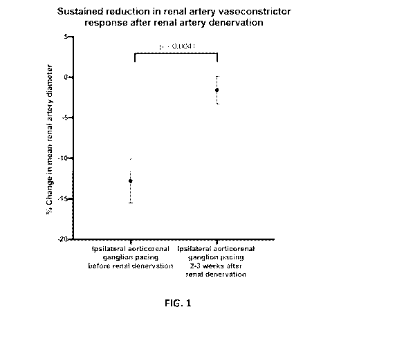
[0041] The results are illustrated in Figure 1, demonstrating relief of
repetitive ARG
pacing induced vasoconstriction with circumferential renal artery denervation.
The
angiograms (third page of Figure 1) show the renal artery state immediately
prior to,
during, immediately after and two weeks after the renal denervation. The
graphs (first
two sheets of Figure 1) show renal arterial diameter at the different stages,
and the
sustained reduction in vasoconstrictor response after renal artery
denervation.
9
CA 03093326 2020-09-08
WO 2019/169446 PCT/AU2019/050202
[0042] Conclusion: High-frequency pacing from the IVC and aorta appears
feasible for
localising aorticorenal ganglia that produce consistent ipsilateral renal
arterial
vasoconstriction and offers a potential means to test renal sympathetic
efferent nerve
function during transcatheter renal artery denervation.
Experiment 2
[0043] Methods: 8 sheep underwent unilateral microwave renal artery
denervation after
attempts to identify and repetitively pace the ipsilateral ARG to maximise
renal artery
vasoconstriction. Capture of the ARG was inferred by concurrent hypertensive
and
ipsilateral renal vasoconstrictor responses during high-frequency pacing at
25mA and
10Hz (100ms period, 2ms pulse) from the inferior vena cava and the aorta.
[0044] Results: In 6 of 8 renal arteries prior to denervation, pacing reduced
renal
arterial diameter from 5.8 1.2 mm to 4.0 1.5 mm, p value=0.007. Whenever
vasoconstriction was induced by pacing, microwave renal denervation caused
progressive vasodilation during ablation to restore renal artery diameter, 5.3
0.7 mm vs
5.8 1.2 mm at baseline, p=0.14. At 2-3 weeks, the ipsilateral aorticorenal
ganglia could
no longer be pace-captured in three of six arteries where it was previously
possible and,
in the remaining three, pacing produced insignificant changes in renal
arterial diameter
5.7 0.5 mm to 4.8 1.3 mm, p=0.38. Renal cortical norephinephrine content on
the
denervated side was reduced by 73%, p=0.0004.
[0045] The results are illustrated in Figure 2, showing the haemodynamic and
vasoconstrictive responses to the ARG pacing.
[0046] Conclusion: When renal sympathetic tone is increased, effective
circumferential
renal artery denervation may be appreciated by immediate renal artery
vasodilation and
diminished vasoconstrictive response to ARG pacing.
Experiment 3
[0047] Methods: In 3 sheep, using a modified radiofrequency ablation catheter
with a
retractable needle tip, ink mixed with intravenous contrast (50:50%) was
injected under
fluoroscopic guidance, at the site of pacing which elicited ipsilateral renal
arterial
constriction together with blood pressure elevation. Histological analysis was
performed
CA 03093326 2020-09-08
WO 2019/169446 PCT/AU2019/050202
after formalin fixation and sectioning every 4mm in the area of the
retroperitoneum
where the stain was evident.
[0048] Results: 4 pacing sites in the 3 sheep yielded ipsilateral renal artery
constriction
concurrent with hypertensive responses. Ink injection was directed into the
perivascular
adipose tissue posterior to the IVC and/or anterior to the aorta. Histological
analysis
demonstrated abundant ganglionic tissue at injection sites.
[0049] Right putative ARG site injected: Figure 3.
[0050] Left putative ARG site injected: Figure 4.
[0051] Ganglionic tissue was observed at injection labelled sites
histologically: Figure
5.
[0052] Conclusion: sites with pacing response consistent with stimulation of
ARG
correlate with histological evidence of ganglionic tissue.
[0053] The results of these experiments clearly demonstrate that renal
arterial
vasoconstriction from high renal sympathetic tone can allow intraprocedural
arterial
vasodilation to serve as a renal denervation endpoint, thus assisting in
guiding the
dosing of renal denervation procedures (such as transcatheter circumferential
renal
denervation) to achieve more complete or more elective renal denervation, so
improving
procedural efficiency.
[0054] Moreover, the results demonstrate that pace-capture of the ARG may
enable
physiological testing of renal sympathetic efferent nerves.
[0055] Further tests carried out by the inventors using both RF and MW
ablation
provided additional confirmation of the above findings, namely that it is
possible to
localise ARG using transvascular pacing through observation of renovascular
and
haemodynamic changes, that the pacing site corresponding to a sympathetic
ganglion
is indeed an ARG (through demonstration of ipsilateral renal denervation with
ganglion
ablation), and that renal artery denervation can abolish ARG pacing-induced
renal
vasoconstriction. Again, histological assessment was used to confirm the
correlation of
the pacing sites with sympathetic ganglionic tissue.
11
CA 03093326 2020-09-08
WO 2019/169446 PCT/AU2019/050202
[0056] Additional findings from these further tests (providing inter alia
further evidence
that the ARG was successfully pace-captured) included:
= The renovascular changes were lateralised and therefore consistent with a
neurogenic rather than humoral response.
= Ink injection and ablation demonstrated a sympathetic ganglion was
present at
the pace capture site.
= Ablation injury to the ganglion was associated with ipsilateral renal
denervation,
implicating its role in innervating the ipsilateral kidney. It was noted that
the left
ARG was more difficult to locate with pacing than the right, likely due to its
variable depth within periaortic fat and the routine transaortic approach for
the left
side used in the trials. Histological analysis suggested that a paired
leftward
sympathetic ganglion (likely the left ARG) is often close to the ostium of the
left
renal vein and therefore may be accessible from the left aspect of the IVC.
The
vasodilatory response with microwave ablation was seen only if the ipsilateral
ARG was captured, suggesting that the mechanism is likely due to relief of
sympathetic tone rather than a direct effect on the vascular smooth muscle.
[0057] Figure 6 provides a diagrammatic illustration of the process of the
invention,
showing renal artery 10 supplying blood to kidney 20, from aorta 30 (Figure
6A). The
aorticorenal ganglia and renal sympathetic fibres are indicated by reference
40. End-
electrode equipped catheter 50 produces electrical pacing 55 at a suitable
site, selected
to correspond to a sympathetic ganglion, resulting in renal vasoconstriction
(and
concurrent blood pressure elevation) in artery 10, as illustrated in Figure
6B.
Transcatheter renal denervation (indicated by ablation zone 60 in Figure 6C)
blocks
renal nerve activation, reducing or abolishing renovascular response to the
ARG pacing.
Further details of test procedures and equipment used
[0058] In these tests, high frequency unipolar transvascular pacing at 10Hz at
up to
25mA was applied using a Micropace EP stimulation source supplying either a
deflectable Webster quadrapolar catheter or a 3.5mm Thermocool ablation
catheter
(Biosense Webster). Renal angiography was performed using either an 8.5F
epicardial
Agilis Sheath (St Jude Medical) or a 6F diagnostic angiography catheter via a
7F
femoral arterial short sheath. Invasive blood pressure was monitored via
either a
dedicated 6F short sheath inserted on the left femoral artery or from the
angiography
12
CA 03093326 2020-09-08
WO 2019/169446 PCT/AU2019/050202
guide catheter and recorded on a Prucka CardioLab system (GE Healthcare). The
tip of
the pacing catheter was positioned at multiple sites above and below the level
of the
ipsilateral renal artery ostium. Skeletal muscle stimulation was avoided by
reducing
pacing current output. If no change in blood pressure was observed within 30s
of
stimulation of a site, the pacing catheter tip position was moved a few
millimetres to a
new position.
[0059] Hemodynamic pressure data was extracted from the Prucka CardioLab
system,
and with main renal artery calibre determined using quantitative coronary
analysis
software (Siemens AG), while quantitative analysis of renal arterial tree
vasoconstriction
beyond the branch renal arteries was performed by (1) obtaining a digital
subtraction
angiography (Horos2k, version 2Ø2), (2) reducing background noise in ImageJ
(ImageJ, version 1.515) using the 'subtract background' function, (3)
selecting a circular
region of interest with a diameter defined by the first renal artery
bifurcation and the
furthermost point on the renal cortex, (4) obtaining a mean measure of
greyscale, and
(5) computing a pixel density index being the complement of greyscale (pixel
density
index = 255 - greyscale value). GraphPad Prism 7 (GraphPad Software Inc.) was
used
for statistical analysis.
[0060] ARG pace capture was inferred when a rise in mean invasive blood
pressure
within 30s of pacing was accompanied by constriction in the ipsilateral main
renal
artery. After cessation of pacing, blood pressure was permitted to return to
steady state,
defined as less than 5mmHg change in mean arterial pressure over 60s.
!psilateral and
contralateral renal angiography was performed at baseline prior to pacing and
at the
peak of blood pressure elevation during pacing stimulation.
[0061] The invention thus provides a repeatable physiological patient-specific
method
to test a 'before and after' response change to guide renal denervation. The
state of
activation of efferent renal sympathetic nerves, which during procedural
sedation may
allow the renal artery to be otherwise in a dilated state, can be increased
using pacing
to create an increased local sympathetic tone, and the relief of this
sympathetic tone
can become a reliable endpoint for the denervation procedure.
[0062] Further, the method of locating perivascular ganglia in the manner
described
above also has potential future application in locating sites to apply
ablation energy to
13
CA 03093326 2020-09-08
WO 2019/169446 PCT/AU2019/050202
produce denervation of the organ innervated by the ganglia. Such applications
include
renal denervation, as well as other sites in the aorta and IVC external to the
renal artery.
[0063] It will be understood that the invention disclosed and defined in this
specification
extends to all alternative combinations of two or more of the individual
features
mentioned or evident from the text or drawings. All of these different
combinations
constitute various alternative aspects of the invention.
[0064] As used herein, except where the context requires otherwise, the term
"comprise" and variations of the term, such as "comprising", "comprises" and
"comprised", are not intended to exclude further additives, components,
integers or
steps.
14