Note: Descriptions are shown in the official language in which they were submitted.
CA 03093422 2020-09-08
WO 2019/183131
PCT/US2019/023020
PERIOSTIN ANTIBODIES AND METHODS OF USING THE SAME
CROSS REFERENCE TO RELATED APPLICATIONS
This application claims priority to United States Provisional Application No.
62/644,681,
filed March 19, 2018 and United States Provisional Application No. 62/727,915,
filed September
6, 2018, both of which are incorporated herein by reference in their
entireties.
STATEMENT REGARDING FEDERALLY SPONSORED RESEARCH
This invention was made with United States government support awarded by the
United
States National Institute of Health ("NIH") grant number U01CA168870-01. The
United States
has certain rights in this invention.
SEQUENCE LISTING
A Sequence Listing accompanies this application. The sequences are listed by
SEQ ID
NO: in the specification and the corresponding sequences are found in the
Sequence Listing
filred herewith which is incorporated herein by reference.
INTRODUCTION
Antibody-based therapy and diagnosis of cancer has become an important
strategy for
treating and diagnosing cancer patients. Cell surface antigens that are
selectively expressed by
cancer cells as compared to normal cells provide an attractive means of
developing targeted
cancer therapies and diagnostic tools. A key challenge in the field, however,
has been to identify
antigens that may be used to selectively target cancer cells. Peptide antigens
are commonly used
to develop cancer cell-specific antibodies although the applicability of such
antigens may be
limited in certain contexts, for example, when the expression of the peptide
antigen is similar in
normal and cancer cells.
Cancer-specific glycosylation changes in proteins are another attractive group
of antigens
that may be able to distinguish cancer cells from normal cells and may be
useful in the
development of both diagnostic and therapeutic applications. Few antibodies,
however, have
been developed that specficially target the carbohydrate moieties that are
selectively expressed
1
CA 03093422 2020-09-08
WO 2019/183131
PCT/US2019/023020
on cancer cells. Thus, there remains a need in the art for new antibodies that
specifically target
glycosylation differences between cancer cells and normal cells.
SUMMARY
In one aspect of the present invention, antigen-binding reagents are provided.
The
antigen-binding reagents may specifically bind to a human Periostin
glycoprotein, preferably, a
gycan epitope of the human Periostin glycoprotein. In some embodiments, the
antigen-binding
reagent may include the following complementarity-determining regions (CDRs):
CDR H1,
GFIFDDYAMH (SEQ ID NO: 1), CDR H2, NSGHIDYADSVEGRFT (SEQ ID NO: 2), CDR
H3, VSYLSTASSLDY (SEQ ID NO: 3), CDR L3, QRYNRAPYT (SEQ ID NO: 4) or a heavy
chain variable region comprising SEQ ID NO: 5 and a light chain variable
region comprising
SEQ ID NO: 6.
In another aspect, antigen-binding conjugates are provided.
The antigen-binding
conjugates may include any one of the antigen-binding reagents described
herein linked to an
agent.
In a further aspect, cells are provided. The cells may include any of the
antigen-binding
reagents or any of the antigen-binding conjugates described herein.
In another aspect, pharmaceutical compositions are provided. The
pharmaceutical
compositions may include any of the antigen-binding reagents, any of the
antigen-binding
conjugates, or any of the cells disclosed herein and a pharmaceutical carrier,
excipient, or
diluent.
In another aspect, the present invention relates to methods for imaging cancer
cells in a
subject. The methods may include administering in an effective amount any of
the antigen-
binding reagents, any of the antigen-binding conjugates, or any of the
pharmaceutical
compositions described herein to the subject, and generating an image of at
least a portion of the
subject using an imaging modality. Preferably in these method embodiments, the
imaging of
cells bound to the antigen-binding reagent, antigen-binding conjugate, or
pharmaceutical
composition is indicative of the cells being cancer cells.
In a further aspect, the present invention relates to methods of detecting
cancer cells in a
subject sample. The methods may include obtaining a sample from the subject,
contacting the
sample with any of the antigen-binding reagents or any of the antigen-binding
conjugates
2
CA 03093422 2020-09-08
WO 2019/183131
PCT/US2019/023020
disclosed herein, and detecting binding of the antigen-binding reagent or
antigen-binding
conjugate to cells in the sample. Suitably, binding of the antigen-binding
reagent or the antigen-
binding conjugate to the cells is indicative of the cells being cancer cells.
In a still further aspect, the present invention relates to methods of
treating cancer cells in
a subject. The methods may include administering to the subject an effective
amount any of the
antigen-binding reagents, any of the antigen-binding conjugates, any of the
cells, or any of the
pharmaceutical compositions disclosed herein to treat the cancer in the
subject.
BRIEF DESCRIPTION OF DRAWINGS
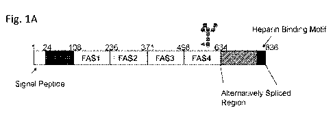
Figure IA-1C shows the Periostin domain structure and location of complex N-
linked glycosylation. Fig. 1A shows the Domain map of the human periostin
protein with the
glycosylation site in the last FAS1 domain marked. Fig. 1B shows the NMR
structure (PDB
5WT7) of the FAS4 domain showing the unstructured loop where asparagine 599 is
located 35.
Crystal structure (PDB 5YJG) of the FAS1-FAS4 domains for human periostin with
the N599
solvent exposed Fig. 1C is a Western blot analysis of periostin protein
purified from culture
supernatant on anti-Flag resin. The top cropped image is detected using the
lectin E-PHA
(Vector Labs) and the bottom cropped image is the detection of the same blot
with periostin
antibody (Santa Cruz Biotechmologies). Examples of previously detected glycan
structures for
each cell line are shown above each lane
Figure 2 shows a schematic flow of the selection, purification, and validation
approach. The ovarian cancer yeast-display scFv library was first subtracted
using 6 rounds
each on the non-malignant Pro5-PN and Lec4-PN cells. Non-binders were grown
and added to
OVCAR3-PN cells for multiple rounds of selection. Clonal populations of
binders were
evaluated using yeast-cell ELISA and yeast that had binding specificity for
bisecting glycans
were made into secreted scFv. Clone 9 was converted to a biotin labeled
antibody known as a
biobody with the indicated tags and evaluated using cell lines and xenograft
tumor models.
Figure 3 shows representative yeast-cell ELISA results. Differential binding
of
candidate clonal yeast populations were measured on Pro5-PN (blue), Lec4-PN
(red) , and
OVCAR3-PN (green) cells plated at 90% confluence on 24-well plates. Bound
yeast (labeled
with Calcofluor) were measured before and after washes. Representative data
shown reflect the
percentage of yeast bound after each wash for each cell line for the indicated
clones.
3
CA 03093422 2020-09-08
WO 2019/183131
PCT/US2019/023020
Figure 4A-Fig. 4E shows the specificity, cellular localization and antibody-
dependent cytotoxicity for scFvC9 biobody. Fig. 4A shows Flow cytometry
analysis of
OVCAR3-PN Control ShRNA and OVCAR3-PN GnT-III ShRNA cells stained with scFvC9
biobody premixed with streptavidin APC (red lines) or streptavidin APC onity
(blue lines). Fig.
4B shows representative images of scFvC9 biobody binding and internalization
into OVCA26
cells, bar 10 p.m. Fig. 4C shows functional analysis of cell cytotoxicity
using a cell titer glow
luminescence viability assay. ScFvC9 biobody was premixed with anti-myc mAb
and serial
dilutions were added to cell for 48 hr at 37 C. The results shown are
representative of 3
independent experiments. Fig. 4D and Fig. 4E shows scFvC9 cell binding and
specificity in
human glioblastoma cells. Fig. 4D shows Crispr/Cas9 KO of the Mgat3 gene in
single cell
isolated LN18 clone known as C2 is confirmed by the absenceof E-PHA binding
indicating a
loss of bisecting N-glycan. The non-targeted single cell isolated clone known
as control Al has
Mgat3 expression confirmed by the binding of E-PHA lectin, bar 20 p.m. Fig. 4E
shows the scFv
C9 biobody binds to LN18 Control Al clone and has nno binding to the LN18
Crispr/Cas9
Mgat3 KO clone C2.
Figure 5 shows the IVIS imaging of ovarian tumors. Top panel: immune
compromised
NSG female mice with 6 wk subcutaneous xenograft tumor from A1847 human
ovarian cancer
cells were imaged before and after retro-orbital injection of scFvC9/IRB680W
complexes or
negative control (IRB680W only). Middle panel: immune competent C57B1/6 female
mice with
8 wk intraovary Luc-ID8 murine ovarian cancer cells were imaged before and
after injection of
scFvC9/IRB680W complexes or negative control 9IRB680W only). Lower panel:
immune
competent C57B1/6 female mice with 8 wk intraperitoneal Luc-ID8 murine ovarian
cancer cells
were imaged before and after scFvC9/IRB680W complex injection or IRB680W only.
Figure 6 shows the detection of scFvC9 biobody in tumors and tissues at the 24
hr
time point. Immune compromised female NSG mice with subcutaneous A1847
xenograft
tumors were injected with scFvC9 biobody IV 24 hr before necropsy and tissue
collection.
Sections were stained with Streptavidin Qdot (1:50 in 1X PBS) prior to
counterstain with DAPI.
White arrows mark regions of interest discussed in the text, Bar 100 p.m.
Figure 7A and Figure 7B shows the MR studies with scFvC9 biobody. Fig. 7A
shows
the phantom tubes layered with cells only, anti-flag magnetic bead only, or
cells with scFvC9
biobody and anti-flag magnetic beads were MR imaged. Representative image
shown and
4
CA 03093422 2020-09-08
WO 2019/183131
PCT/US2019/023020
results in graph to the right represent mean decreased signal intensity from 3
independent
experiments, SEM P.0001. Fig. 7B shows Immune compromised NSG female mice
with
A1847 subcutaneous tumor were injected with scFvC9 coupled 1:2 with magnetic
avidin beads.
Representative 1 hour images are shown and cumulative normalized (SI tumor/SI
muscle for
given ROI) signal intensity for each time point are graphed to the right.
Figure 8 shows a mouse mesothelinhh Luc-ID8 mouse ovarian cancer model 8 weeks
after intraperitoneal injection and in vivo imaging with luciferin.
Figure 9 shows a mouse mesothelin' Luc-ID8 ovarian cancer in C57B1/6 female
mice (8
weeks after IP injection) and in vivo imaging with a biotinylated anti-
mesothelin nanobody (MN)
or an anti-N-glycosylated periostin (C9) coupled to labeled streptavidin (SA).
Negative control
(SA only): C57B1/6 mouse, injected with labeled streptavidin only.
Figure 10 shows a human mesothelinhi EKVX lung cancer in NSG female mice (4
weeks
after IV injection) and in vivo imaging with a biotinylated anti-mesothelin
nanobody (MN) or an
anti-N-glycosylated periostin (C9) coupled to labeled streptavidin (SA).
Negative control (SA):
tumor-bearing NSG mouse, injected with labeled streptavidin only.
Figure 11 shows a human mesothelinhh H460 lung cancer in NSG female mice (IV
injection, 4 weeks) and in vivo imaging with a biotinylated anti-mesothelin
nanobody (MN) or an
anti-N-glycosylated periostin (C9) coupled to labeled streptavidin (SA).
Negative control (SA):
tumor-bearing NSG mouse, injected with labeled streptavidin only.
Figure 12 shows a human mesothelinl A549 lung cancer in NSG female mice (4
weeeks
after IV injection) and in vivo imaging with a biotinylated anti-mesothelin
nanobody (MN) or an
anti-N-glycosylated periostin (C9) coupled to labeled streptavidin (SA).
Negative control (SA
only): tumor-bearing NSG mouse, injected with labeled streptavidin only.
DETAILED DESCRIPTION
Here, the present inventors have developed antigen-binding reagents that
recognize a
cancer-specific glycan (carbohydrate) modification on the human Periostin
protein. In a
previous study, the present inventors discovered an unusual bisecting N-linked
glycan structure
on the human Periostin protein that is specifically expressed in cancer cells
such as ovarian
cancer cells. See Abbott et al., Proteomics 10(3): 470-481 (2010). The N-
linked glycan
structure is unusual due to the lack of galactose capping and sialic acid
extensions and has been
described, for example, in Allam, Heba et al. "Glycomic Analysis of Membrane
Glycoproteins
5
CA 03093422 2020-09-08
WO 2019/183131
PCT/US2019/023020
with Bisecting Glycosylation from Ovarian Cancer Tissues Reveals Novel
Structures and
Functions." Journal of Proteome Research 14.1 (2015): 434-446. PMC.
Tumor cells typically display tumor-specific changes in glycosylation on
surface
glycoproteins and glycolipids that can serve as biomarkers for diagnosis as
well as candidates for
immunotherapy 1-4. Such changes in glycosylation are due to altered expression
levels of unique
glycosyltransferases and glycoproteins that lead to their surface expression
and potential
secretion from tumor cells. However, this area of research has been hampered
by having only a
few specific anti-carbohydrate antibodies useful for targeting tumor cell-
specific changes in
glycosylation.
One approach to develop such specific anti-carbohydrate antibodies is yeast
display.
These technologies can improve the affinity and specificity of recognition
reagents 5-7. In this
method, recombinant antibodies are displayed on the yeast surface as a fusion
protein to a cell
wall component (Aga-2) and library generation is facilitated by the homologous
recombination
system inherent in yeast ". Coupling flow cytometry with cell surface display
of recombinant
antibodies expressed as single chain Fragment variables (scFv) permits the
monitoring of both
scFv expression at the yeast surface and scFv binding to the antigen 1 . Yeast
display has also
proven to be highly effective for various directed evolution applications "15.
These methods
translate into time-and cost-efficient production and screening of scFvs that
have enabled the
identification of many functional scFvs directed toward numerous medically
relevant proteins,
including scFv directed against mesothelin 16, TEM1 17, mannose receptor 18,
glypican 19, and
B7-H4 20.
We have utilized the powerful advantages of the yeast display method to
isolate scFv that
recognize the tumor-specific bisecting glycan structures discovered in ovarian
cancer 3. These
glycans are generated in part by a unique glycosyltransferase GnT-III, encoded
by the Mgat3
gene, which creates bisecting complex-type N-glycans by addition of a 131-4-
linked GlcNAc to
the core (3-mannose of N-glycans 21. We previously discovered that the Mgat3
gene was highly
amplified in ovarian cancer 22. The Mgat3 gene is amplified in several human
cancers due to
hypomethylation changes in the promoter near the transcription start site 23.
The structures of
bisecting N-glycans in ovarian cancer are different than those bisecting N-
glycans found in non-
malignant cells. Unexpectedly, the bisecting N-glycans from ovarian cancers
show reduced
6
CA 03093422 2020-09-08
WO 2019/183131
PCT/US2019/023020
branching, lack of galactose and sialic acid, with or without core fucose
making this glycan
structure a biomarker for ovarian cancer and possibly several other human
cancers 3.
Our laboratory has used a targeted glycoproteomic approach to identify
glycoproteins
that carry tumor-associated bisecting glycan structures in ovarian cancer. Our
analysis of
.. secreted and membrane proteins from primary ovarian cancer tissues led to
the discovery of
periostin, also known as osteoblast-specific factor 2 (OSF-2) as a potential
biomarker 3'24.
Periostin is a secreted glycoprotein that is present in circulation and also
associates with the cell
membranes evidenced by the presence of periostin in membrane fractions by
proteomic analysis
3. The likely mechanism of cell surface binding is due to presence of FAS1
domains that have
been demonstrated to interact with the membrane in the protein fasciclin 25.
Despite the
elevated levels of periostin in human cancers, this glycoprotein has not been
utilized as a
biomarker due to variable expression in inflammatory conditions 26-28. This
complicates the use
of the protein itself as a biomarker for cancer because detection of the
periostin protein levels
may not correlate with the disease burden. The ability to detect the cancer-
specific bisecting
glycoform on periostin would be a superior biomarker for diagnostic
applications and may lead
to the development of new therapeutic approaches. Here, we describe our
subtraction/selection
process to identify a yeast-displayed scFv (scFvC9) and characterization of
its specificity for
tumor-specific bisecting glycan structures. We further validate the use of
scFvC9 to target
ovarian cancer xenograft tumors in vivo. Together these finding suggest the
potential use for this
antibody in diagnostic and therapeutic applications for cancers that have
amplification of the
Mgat3 gene.
Briefly, the present inventors produced cell lines that eliminate the enzyme
that adds the
bisecting glycan as well as control cell lines that produce this enzyme. They
also produced a
mutant version of the human periostin protein that is missing the N-linked
glycosylation site.
Using these cell lines, they developed a selective panning strategy for use
with a scFv yeast
display library derived from B cells of ovarian cancer patients. To subtract
scFvs that interact
with the peptide portion of the human periostin protein or interact with other
glycan structures,
they first panned with the cell lines that do not express the bisecting
glycans yet express the
periostin protein. Next, they panned with cell lines that express bisecting
glycans and express
the periostin protein to select scFvs that bind to the N-linked glycosylation
moiety. These binders
were then further screened to select clones that specifically bind to the
glycan. One of the
7
CA 03093422 2020-09-08
WO 2019/183131
PCT/US2019/023020
positive clones, known as C9, was further characterized and shown to
specifically target the
cancer-specific N-linked glycan structure on the human periostin protein and
to specifically
target human xenograft ovarian and lung tumors growing in several mouse cancer
models.
Based on this data, it becomes readily apparent that the C9 scFv may serve as
the basis for the
antigen-binding reagents disclosed herein, which may futher be used in a
variety of compositions
and methods.
Antigen-binding Reagents
In one aspect of the present invention, antigen-binding reagents are provided.
As used
herein, the term "antigen-binding reagent(s)" is used in the broadest sense to
refer to polypeptide
affinity agents based on antibodies. For example, the antigen-binding reagent
may include,
without limitation, a single chain antibody (e.g., single-chain Fvs (scFvs),
biobodies, disulfide-
linked Fvs (sdFvs), etc.) monoclonal antibody, or antibody fragments such as
Fab, Fab', F(ab')2,
Fv fragments, diabodies, linear antibodies, or multispecific antibodies (e.g.,
bispecific
antibodies) formed from antibody fragments. The antigen-binding reagent may be
a chimeric, a
humanized, or a fully human polypeptide sequence. The antigen-binding reagent
may be any
one of the known major classes of immunoglobulins including IgA, IgD, IgE,
IgG, IgY, and
IgM, any class (e.g., IgGl, IgG2, IgG3, IgG4, IgAl and IgA2), or any subclass
(e.g., IgG2a and
IgG2b) of immunoglobulin molecules. In some embodiments, the antigen-binding
reagent may
be a scFv, a Fab, or an IgG monoclonal antibody.
The antigen-binding reagents include amino acid residues that interact with an
"antigen"
such as the human Periostin protein and confer on the antigen-binding reagent
the capability of
specifically binding to the antigen. An "antigen" is a molecule or a portion
of a molecule
capable of being bound by an antibody. An antigen may have one or more than
one epitope. An
"epitope" refers to that portion of any molecule capable of being recognized
by, and bound by,
an antigen-binding reagent. Generally, epitopes include a surface grouping of
molecules, for
example, amino acids or carbohydrate moeities that form a specific three-
dimensional structure
recognized by the antigen-binding reagent.
The antigen-binding reagents further may include the "framework" amino acid
residues
necessary to maintain the proper conformation of the antigen-binding amino
acid residues and/or
amino acid residues commonly found in some types of antibodies that modulate
the immune
8
CA 03093422 2020-09-08
WO 2019/183131
PCT/US2019/023020
system (e.g., Fe effector functions such as complement-dependent cytotoxicity
(CDC), antibody-
dependent cellular cytotoxicity (ADCC), and/or antibody-dependent cell
phagocytosis (ADCP)).
The antigen-binding amino acid residues of the antigen-binding reagents are
commonly
known as the "complementarity determining regions" or "CDR" regions. These CDR
regions
account for the basic specificity of the antigen-binding reagent for a
particular antigenic
determinant structure. The CDRs are non-contiguous stretches of amino acids
within the variable
regions of antibodies. The variable heavy and light chains of some antibodies
each have three
CDR regions, each non-contiguous with the others (termed Li, L2, L3, H1, H2,
H3) for the
respective light (L) and heavy (H) chains. Surprisingly, the present inventors
have found that the
C9 biobody disclosed in the Examples contains three heavy chain CDR regions
(H1, H2, H3)
and only a single light chain CDR region (L3).
The antigen-binding reagent may be capable of specifically binding to a human
Periostin
glycoprotein. Periostin (also known as POSTN, PN, or osteoblast-specific
factor OSF-2) is a
human glycoprotein that functions as a ligand for alpha-V/beta-3 and alpha-
V/beta-5 integrins to
control cell motility. Periostin is also known to be glycosylated and, in a
previous study, the
present inventors discovered an unusual bisecting N-linked glycan structure on
the human
Periostin protein that is specifically expressed in cancer cells such as
ovarian cancer cells. See
Abbott et al., Proteomics 10(3): 470-481 (2010). An exemplary protein sequence
of human
Periostin including an N-terminal sequence peptide is provided as SEQ ID NO:
9.
Optionally, the antigen-binding reagent may specifically bind to a human
Periostin
glycoprotein with an affinity of at least 10' M, 10' M, 10-8 M, 10-9 M, 10-10
NI--,
or 10-11 M.
Methods for determining the affinity of an antigen-binding reagent are known
by those of
ordinary skill in the art. See, e.g., Antibodies: A Lab. Manual (Harlow et
al., eds., Cold Spring
Harbor Lab. Press, Cold Spring Harbor, N.Y., 1988).
The antigen-binding reagent may specifically bind to a glycan epitope of the
human
Periostin glycoprotein.
The glycan epitope may be specifically present on Periostin
glycoproteins present on cancer cells. In some embodiments, the glycan epitope
includes an N-
linked glycan structure.
In accordance with the present invention, cancer cells may include, without
limitation,
epithelial cancer cells, ovarian cancer cells, lung cancer cells, breast
cancer cells, pancreatic
cancer cells, prostate cancer cells, bladder cancer cells, gastric cancer
cells, esophagealcancer
9
CA 03093422 2020-09-08
WO 2019/183131
PCT/US2019/023020
cells, colon cancer cells, skin cancer cells, testicular cancer cells,
colorectal cancer cells,
urothelial cancer cells, renal cancer cells, hepatocellular cancer cells,
leukemia cancer cells,
lymphoma cancer cells, multiple myeloma cancer cells, and central nervous
system cancer cells.
The antigen-binding reagent may include the following complementarity-
determining
regions (CDRs): CDR H1, GFIFDDYAMH (SEQ ID NO: 1), CDR H2,
NSGHIDYADSVEGRFT (SEQ ID NO: 2), CDR H3, VSYLSTASSLDY (SEQ ID NO: 3), CDR
L3, QRYNRAPYT (SEQ ID NO: 4). In some embodiments, the antigen-binding reagent
may
include a heavy chain variable region including SEQ ID NO: 5 and a light chain
variable region
including SEQ ID NO: 6. In some embodiments, the antigen-binding reagent may
include SEQ
ID NO: 7 (C9 scFv protein sequence).
Antigen-binding conjugates
In another aspect of the present invention, antigen-binding conjugates are
provided. The
antigen-binding conjugates may include any one of the antigen-binding reagents
described herein
linked to an agent. An "agent" may be any substance that provides additional
functionality to the
antigen-binding reagents. Suitable agents include, without limitation,
detectable imaging agents,
therapeutic agents, immunoprotein domains, or combinations thereof
A "detectable imaging agent" may be any suitable chemical or substance that
may be
detected as a signal or contrast using imaging techniques. Suitable detectable
imaging agents
may be, without limitation, a fluorophore moiety, an enzyme moiety, an optical
moiety, a
magnetic moiety, a radiolabel moiety, an X-ray moiety, an ultrasound imaging
moiety, a
nanoparticle-based moiety, or a combination of two or more of the listed
moieties.
A "fluorophore moeity" may include any molecule capable of generating a
fluorescent
signal. Various fluorophore moieties are well-known in the art and/or
commercially available.
Exemplary fluorophore moeities include, without limitation, fluorescein, FITC,
Alexa Fluor 488,
Alexa Fluor 660, Alexa Fluor 680, Alexa Fluor 750, and Alexa Fluor 790 (Life
Technologies);
Cy2, Cy3, Cy3.5, Cy5, Cy5.5 and Cy7 (GE Healthcare); DyLight 350, DyLight 488,
DyLight
594, DyLight 650, DyLight 680, DyLight 755 (Life Technologies); IRDye 800CW,
IRDye
80016, and IRDye 700DX (Li-Cor); VivoTag680, VivoTag-5680, and VivoTag-5750
(PerkinElmer).
CA 03093422 2020-09-08
WO 2019/183131
PCT/US2019/023020
An "enzyme moiety" refers to polypetides that catalyze the production of a
detectable
signal. Exemplary enzyme moieties may include, without limitation, horseradish
peroxidase
(HRP), alkaline phosphatase (AP), glucose oxidase, or P-galactosidase.
"Optical moieties" may include, for example, any agents that may be used to
produce
contrast or signal using optical imaging such as luminescence or acousto-
optical moieties.
"Magnetic moieties" may include, for example, a chelating agent for magnetic
resonance
agents. Chelators for magnetic resonance agents can be selected to form stable
complexes with
paramagnetic metal ions, such as Gd(III), Dy(III), Fe(III), and Mn(II).
Other exemplary detectable imaging agents may include radiolabel moieties.
Exemplary
radioactive labels may include, without limitation,99Mo, 99mTne, 64cn, 67Ga,
186Re, 188Re, 153sm,
177Lu, 67cti, 1231, 1241, 1251, nc, x3N, 150, and BF.
"X-ray moieties" may include, for example, any agents that may be used to
produce
contrast or signal using X-ray imaging such as iodinated organic molecules or
chelates of heavy
metal ions.
Ultrasound imaging moieties may include, for example, any agents that may be
used to
produce contrast or signal using ultrasound imaging such as Levovist, Albunex,
or Echovist.
A detectable imaging agent may also be a nanoparticle-based moiety. A
nanoparticle-
based moiety is a nanoparticle that is capable of generating a signal. For
example, silicon
containing nanoparticles may be used to produce fluoresecence, luminescence,
or another type of
signal. Other exemplary nanoparticle-based moieties include, without
limitation, nanospheres
such as Kodak X-SIGHT 650, Kodak X-SIGHT 691, Kodak X-SIGHT 751 (Fisher
Scientific);
metal oxide nanoparticles; and quantum dots such as EviTags (Evident
Technologies) or Qdot
probes (Life Technologies). Nanoparticles may also be used to link or
conjugate the antigen-
binding reagents to a toxin or other cytotoxic agent or cytotoxic compound.
A "therapeutic agent" may be any substance that provides a therapeutic
functionality
when conjugated to an antigen-binding reagent. For example, antibody-drug
conjugates
including the antigen-binding reagents disclosed herein are contemplated.
Suitable therapeutic
agents may include, without limitation, cytotoxic compounds, and particularly
those shown to be
effective in other antibody-drug conjugates. As used herein, a "cytotoxic
compound" refers to
any substance that disrupts the functioning of cells and/or causes the death
of cells. Various
therapeutic cytotoxic compounds are known in the art and may include, without
limitation, DNA
11
CA 03093422 2020-09-08
WO 2019/183131
PCT/US2019/023020
damaging agents, anti-metabolites, natural products and their analogs.
Exemplary classes of
cytotoxic compounds include enzyme inhibitors such as dihydrofolate reductase
inhibitors, and
thymidylate synthase inhibitors, tubulin inhibitors, DNA intercalators, DNA
cleavers,
topoisomerase inhibitors, the anthracycline family of drugs, the vinca drugs,
the mitomycins, the
bleomycins, the cytotoxic nucleosides, the pteridine family of drugs,
diynenes, the
podophyllotoxins, dolastatins, auristatins, maytansinoids, differentiation
inducers, and taxols.
More specifically, suitable cytoxic compounds may include 5-fluorouracil,
aclacinomycin, activated cytoxan, bisantrene, bleomycin, carmofur, CCNU, cis-
platinum,
daunorubicin, doxorubicin, DTIC, melphalan, methotrexate, mithromycin,
mitomycin,
mitomycin C, peplomycin pipobroman, plicamycin, procarbazine, retinoic acid,
tamoxifen, taxol,
tegafur, VP16, VM25, diphtheria toxin, botulinum toxin, geldanamycin,
maytansinoids
(including DM1), monomethylauristatin E (MMAE), monomethylauristatin F (MMAF),
and
maytansinoids (DM4) and their analogues.
Exemplary cyotoxic compounds may also include therapeutic radiopharmaceuticals
including, without limitation,186Re, 188Re, 153sm, 67cti, 105¨ +
Kn inAg, and 192Ir.
In one embodiment the antigen-binding reagents may be used to initiate
antibody-
dependent cellular cytotoxicity (ADCC) and may thus be used to kill the cancer
cells. The
ADCC data in the examples demonstrates that when the scFvC9 is linked to full
length IgG (such
as the anti-cmyc used int eh Examples), it can initiaite an effective ADCC
response. The V5 tag
antibody described herein may perform similarly.
Preparation of antibody-drug conjugates is generally known in the art and can
be
performed by conventional methods analogous to those described in, for
example, Doronina et
al., Bioconjugate Chem. 2006, 17, 114-124. See also for example U.S. Pat. Nos.
8,067,546,
8,039,273, 7,989,434, 7,851,437, 7,837,980, 7,829,531, 7,705,045 8,034,959,
8,034,787,
7,968,586, 7,847,105, and 7,223,837.
An "immunopolypeptide" may be any polypeptide that facilitates an immune
function.
For example, the antigen-binding reagents disclosed herein may be combined
with further
immunopolypeptides to produce new chimeric antigen receptors (CARs) specific
for Periostin.
CARs may include a targeting moiety such as any of the antigen-binding
reagents disclosed
herein, and additional "immunopolypeptides" such as a transmembrane domain,
and intracellular
signaling/activation domain(s). Intracellular signaling/activation
domain(s) suitable as
12
CA 03093422 2020-09-08
WO 2019/183131
PCT/US2019/023020
immunopolypeptides include, without limitation, CD3C signaling domains, 41BB -
signaling
domains, CD28-signaling domains, or combinations thereof. The
immunopolypeptide may also
be immunoglobulin domains important in developing dendritic based vaccines.
The antigen-binding reagent and agent may be linked directly by a covalent
bond or may
be linked using a linker or spacer moiety. Useful linker or spacer moieties
include peptides,
amino acids, nucleic acids, as well as homofunctional linkers or
heterofunctional linkers.
Particularly useful conjugation reagents that can facilitate formation of a
covalent bond between
an antigen-binding reagent and agent may include a N-hydroxysuccinimide (NHS)
ester and/or a
maleimide. In some embodiments, the antigen-binding reagent and agent are
linked at the N-
terminal end of the antigen-binding reagent. In some embodiments, the antigen-
binding reagent
and are are linked at the C-terminal end of the antigen-binding reagent. In
some embodiments,
the linker is at least 2, 3, 4, 5, 6, 7, 8, or more amino acids long.
In embodiments covering antibody-drug conjugates, the linker may be cleavable
under
intracellular or extracellular conditions, such that cleavage of the linker
releases the therapeutic
agent from the antigen-binding reagent in the appropriate environment. For
example, the linker
may be cleavable by extracellular or intracellular proteases including,
without limitation,
lysosomal or endosomal proteases. Suitable linkers cleavable by an
intracellular protease may
include a Val-Cit linker or a Phe-Lys linker. See, e.g., U.S. Pat. No.
6,214,345.
In some embodiments, the therapeutic agent may be released after degradation
of the
antigen-binding reagent and/or linker in, for example, lysosomes. See, e.g.,
U.S. Publication No.
2005/0238649.
The linker may be cleavable by a cleaving agent that is present in the
intracellular
environment (e.g., within a lysosome or endosome or caveolea).
In some embodiments, the linker may be cleavable by cathepsins B and D and
plasmin,
all of which are known to hydrolyze dipeptide drug derivatives resulting in
the release of active
drug inside target cells.
The linker may be pH-sensitive, for example, sensitive to hydrolysis at
certain pH values.
Typically, a pH-sensitive linker is hydrolyzable under acidic conditions. For
example, an acid-
labile linker that is hydrolyzable in the lysosome (for example, a hydrazone,
semicarbazone,
thiosemicarbazone, cis-aconitic amide, orthoester, acetal, ketal, thioether,
or the like) may be
used. See, e.g., U.S. Pat. Nos. 5,122,368; 5,824,805; 5,622,929. Such linkers
are relatively
13
CA 03093422 2020-09-08
WO 2019/183131
PCT/US2019/023020
stable under neutral pH conditions, like in the blood, but are unstable at
below pH 5.5, the
approximate pH of the lysosome.
In some embodiments, the linker may be cleavable under reducing conditions
(e.g., a
disulfide linker). A variety of disulfide linkers are known in the art,
including, for example,
those that can be formed using SATA (N-succinimidy1-5-acetylthioacetate) and
SPDB (N-
succinimidy1-3-(2-pyridyldithio)butyrate).
In some embodiments, the linker is self-immolative. See, e.g., WO
2007059404A2,
W006110476A2, W005112919A2, W02010/062171, W009/017394, W007/089149, WO
07/018431, W004/043493 and W002/083180.
A variety of exemplary linkers that can be used with the present invention are
described
in WO 2004010957, U.S. Publication No. 2006/0074008, U.S. Publication No.
20050238649,
and U.S. Publication No. 2006/0024317.
In some embodiments, the antigen-binding reagent and the agent are linked by a
tag
system. A tag system includes any group of agents capable of binding one
another with a high
affinity. Several tag systems are well-known in the art and include, without
limitation,
biotin/avidin, biotin/streptavidin, or digoxigenin (DIG) systems. In some
embodiments, the tag
system includes biotin/avidin or biotin/streptavidin. In such embodiments, the
antigen-binding
reagent may be modified at either the N-terminus or C-terminus to include
biotin while the agent
may be modified to include streptavidin or avidin. Alternatively, the antigen-
binding reagent
may be modified at either the N-terminus or C-terminus end to include
streptavidin or avidin
while the agent may be modified to include biotin.
Cells
In a further aspect of the present invention, cells are provided. The cells
may include any
one of the antigen-binding reagents or any one of the antigen-binding
conjugates described
herein. The cells may be mammalian cells such as, without limitation, human
cells.
In some embodiments, the cells may be cancerous cells such as, without
limitation,
ovarian cancer cells or lung cancer cells.
In some embodiments, the cells may be immune cells such as, without
limitation, T cells
or Natural Killer (NK) cells. For example, the immune cells may be engineered
immune cells,
such as T cells or NK cells, including the chimeric antigen receptors (CARs)
described herein.
Pharmaceutical Compositions
14
CA 03093422 2020-09-08
WO 2019/183131
PCT/US2019/023020
In a still further aspect of the present invention, pharmaceutical
compositions are
provided. The pharmaceutical compositions may include any of the antigen-
binding reagents,
any of the antigen-binding conjugates, or any of the cells disclosed herein
and a pharmaceutical
carrier, excipient, or diluent, which are nontoxic to the cell or subject
being exposed thereto at
the dosages and concentrations employed. Often a pharmaceutical diluent is in
an aqueous pH
buffered solution. Examples of pharmaceutical carriers include buffers such as
phosphate,
citrate, and other organic acids; antioxidants including ascorbic acid; low
molecular weight (less
than about 10 residues) polypeptide; proteins, such as serum albumin, gelatin,
or
immunoglobulins; hydrophilic polymers such as polyvinylpyrrolidone; amino
acids such as
glycine, glutamine, asparagine, arginine or lysine; monosaccharides,
disaccharides, and other
carbohydrates including glucose, mannose, or dextrins; chelating agents such
as EDTA; sugar
alcohols such as mannitol or sorbitol; salt-forming counterions such as
sodium; and/or nonionic
surfactants such as TWEENTm brand surfactant, polyethylene glycol (PEG), and
PLURONICSTm
surfactant.
Methods
Various in vitro and in vivo diagnostic and/or therapeutic methods using the
compositions
disclosed herein are contemplated.
In a further aspect, the present invention relates to methods for imaging
cancer cells in a
subject. The methods may include administering in an effective amount any of
the antigen-
binding reagents, any of the antigen-binding conjugates, or any of the
pharmaceutical
compositions described herein to the subject, and generating an image of at
least a portion of the
subject using an imaging modality. Preferably in these method embodiments, the
imaging of
cells bound to the antigen-binding reagent, antigen-binding conjugate, or
pharmaceutical
composition is indicative of the cells being cancer cells.
As used herein, the term "subject" refers to both human and non-human animals.
The
term "non-human animals" of the disclosure includes all vertebrates, e.g.,
mammals and non-
mammals, such as non-human primates, sheep, dog, cat, horse, cow, chickens,
amphibians,
reptiles, and the like. Suitably, the subject is a human patient.
As used herein, "imaging modality" may include any technology capable of
generating
an image of a subject. In some embodiments, the imaging modality may be
selected from the
group consisting of ultrasound, positron-emission tomography (PET), photon
emission computed
CA 03093422 2020-09-08
WO 2019/183131
PCT/US2019/023020
tomography (SPECT), nuclear magnetic resonance imaging (NMRI), optical imaging
(0I) and
computed tomography (CT). For example, in some embodiments of the present
methods, the
present inventors contemplate that some of the compositions disclosed herein
may be used in
conjunction with ultrasound technologies to image pelvic masses in a subject
to determine
whether such masses are benign or cancerous. Such diagnostic imaging methods
would be
useful prior to removal of the pelvic mass because the prognosis of the
subject after removal of
the pelvic mass is directly related to the type of surgeon that performs the
surgery. If the
imaging methods indicate that the pelvic mass is cancerous, the subject may be
directed to a
surgeon specializing in removing cancerous tissue. On the other hand, if the
imaging methods
indicate that the pelvic mass is benign, the subject may be directed to a
general surgeon whom
may remove the mass and may not have any particular experience in removing
cancerous tissue.
In some embodiments of such methods, antigen-binding conjugates disclosed
herein including
one or more ultrasound imaging moieties may be administered to the subject and
then ultrasound
images may generated of the pelvic region of the subject using, for example,
transvaginal or
other ultrasound imaging technol4es. If the ultrasound image shows significant
detectable
signal from the ultrasound imaging moiety in or around the pelvic mass this
would indicate that
the pelvic mass is cancerous.
In another aspect, the present invention relates to methods of detecting
cancer cells in a
subject sample. The methods may include obtaining a sample from the subject,
contacting the
sample with any of the antigen-binding reagents or any of the antigen-binding
conjugates
disclosed herein, and detecting binding of the antigen-binding reagent or
antigen-binding
conjugate to cells in the sample. Suitably, binding of the antigen-binding
reagent or the antigen-
binding conjugate to the cells is indicative of the cells being cancer cells.
Alternatively, the
methods may include admininstering an imaging or other detectable agent linked
to the antigen-
binding reagents provided herein to the subject and then detecting binding of
the antigen-binding
reagent or antigen-binding conjugate to cells in the subject. The ability of
the antigen-binding
reagent to bind cells in the subject and produce a detectable signal is
indicative of the subject
having cancer. The administration can be carried out by any means available to
those skilled in
the art and will vary depending on the type of cancer suspected.
The "sample" may include cells. In particular, the methods described herein
may be
performed without requiring a tissue sample or biopsy. "Sample" is intended to
include any
16
CA 03093422 2020-09-08
WO 2019/183131
PCT/US2019/023020
sampling of cells, tissues, or bodily fluids in which cancer cells may be
detected. Examples of
such samples include, without limitation, blood, serum, urine, synovial fluid,
saliva, or any other
bodily secretion or derivative thereof Blood can include whole blood, plasma
(citrate, EDTA,
heparin), serum, or any derivative of blood. Samples may be obtained from a
subject by a
variety of techniques available to those skilled in the art. Methods for
collecting various samples
are well known in the art. In some embodiments, the sample is serum or plasma.
As used herein, "contacting" may be carried out through any of the variety of
procedures
used to apply compositions to samples that will be apparent to the skilled
artisan including,
without limitation, simple addition of the composition to the sample.
Methods suitable for "detecting" the binding of the antigen-binding reagent or
antigen-
binding conjugate to cells in the sample are known to those of skill in the
art and may include,
without limitation, ELISA, immunofluorescence, FACS analysis, Western blot,
magnetic
immunoassays, and antibody-based microarrays. In the past, the gold standard
for detection of
cells in blood was the use of ELISAs; however, liquid biopsy technologoies
offer an attractive
alternative approach for cellular analysis.
In a further aspect, the present invention relates to methods of treating
cancer cells in a
subject. The methods may include administering to the subject an effective
amount any of the
antigen-binding reagents, any of the antigen-binding conjugates, any of the
cells, or any of the
pharmaceutical compositions disclosed herein to treat the cancer in the
subject. The cancer and
cancer cells include cancers and cancer cells with increased expression of the
Mgat3 gene.
These cancers include, but are not limited to, ovarian, lung, glioblastoma,
kidney clear cell,
uterine corpus endometriroid, rectum adenocarcinoma, colon, and
adenocarcinoma. In lung
cancers lung squamous cell and lung adenocarcinoma are reported to have
increased Mgat3
expression and thus would be candidates for the methods provided herein.
Several cancers have
been identified (see ref 23) and the inventors expect additional cancers will
be identified that
have epigenetic hypomethylation changes to Mgat3.
Treating cancer cells includes, without limitation, reducing the number of
cancer cells or
the size of a tumor in the subject, reducing progression of a cancer to a more
aggressive form,
reducing proliferation of cancer cells or reducing the speed of tumor growth,
killing of cancer
cells, reducing metastasis of cancer cells or reducing the likelihood of
recurrence of a cancer in a
subject. Treating a subject as used herein refers to any type of treatment
that imparts a benefit to
17
CA 03093422 2020-09-08
WO 2019/183131
PCT/US2019/023020
a subject afflicted with cancer or at risk of developing cancer or facing a
cancer recurrence.
Treatment includes improvement in the condition of the subject (e.g., in one
or more symptoms),
delay in the progression of the disease, delay in the onset of symptoms or
slowing the
progression of symptoms, etc.
In some embodiments of the present methods, the methods may further include
administering an effective amount of an anti-cancer therapeutic agent to the
subject.
The "anti-cancer therapeutic agent" may be any therapeutic agent that is used
to treat
cancer in a subject. Suitable anti-cancer therapeutic agents may include,
without limitation,
radiation, chemotherapy agents, anti-cancer biologics, or immunotherapy
agents. Chemotherapy
agents are chemotherapeutic compounds that may be used to treat cancer.
Suitable
chemotherapy agents may include, without limitation, 5-fluorouracil,
aclacinomycin, activated
cytoxan, bisantrene, bleomycin, carmofur, CCNU, cis-platinum, daunorubicin,
doxorubicin,
DTIC, melphalan, methotrexate, mithromycin, mitomycin, mitomycin C, peplomycin
pipobroman, plicamycin, procarbazine, retinoic acid, tamoxifen, taxol,
tegafur, VP16, or VM25.
Anti-cancer biologics are biomolecules (e.g., polynucleotides, polypeptides,
lipids, or
carbohydrates) that may be used to treat cancer. Anti-cancer biologics may
include, without
limitation, hormones, cytokines such as IL-la, IL-2, IL-213, IL-3, IL-4, CTLA-
2, IFN-a, IFN-y,
granulocyte-macrophage colony stimulating factor (GM-C SF), IL-12, IL-23, IL-
15, IL-7, or any
combination thereof; or anti-cancer antibodies such as Rituximab, Trastuzumab,
Gemtuzumab,
Alemtuzumab, Ibritumomab tiuxetan, Tositumomab, Cetuximab, Bevacizumab,
Panitumumab,
Ofatumumab, Brentuximab Vedotin, Pertuzumab, Adotrastuzumab emtansine, and
Obinutuzumab.
The term "immunotherapy agent(s)" refers to any therapeutic that is used to
treat cancer in
a subject by inducing and/or enhancing an immune response in that subject.
Immunotherapy
agents may include, without limitation, checkpoint inhibitors, cancer
vaccines, immune cells
such as engineered T cells, anti-cancer viruses, or bispecific antibodies.
Checkpoint inhibitors are therapeutics, such as antibodies, that block the
immune
checkpoint pathways in immune cells that are responsible for maintaining self-
tolerance and
modulating the degree of an immune response. Tumors often exploit certain
immune checkpoint
pathways as a major mechanism of immune resistance against T cells that are
specific for tumor
antigens. Many of the immune checkpoints are initiated by receptor-ligand
interactions and thus
18
CA 03093422 2020-09-08
WO 2019/183131
PCT/US2019/023020
may be blocked by antibodies to either the ligand or receptor or may be
modulated by soluble
recombinant forms of the ligands or receptors. Such immune checkpoint blockade
allows tumor-
specific T cells to continue to function in an otherwise immunosuppressive
tumor
microenvironment. Exemplary checkpoint inhibitors include, without limitation,
antibodies or
other therapeutics targeting programmed cell death protein 1 (PD1, also known
as CD279),
programmed cell death 1 ligand 1 (PD-L1, also known as CD274), PD-L2,
cytotoxic T-
lymphocyte antigen 4 (CTLA4, also known as CD152), A2AR, CD27, CD28, CD40,
CD80,
CD86, CD122, CD137, 0X40, GITR, ICOS, TIM-3, LAG3, B7-H3, B7-H4, BTLA, DO,
KIR,
or VISTA. Suitable anti-PD1 antibodies include, without limitation,
lambrolizumab (Merck
MK-3475), nivolumab (Bristol-Myers Squibb BMS-936558), AMP-224 (Merck), and
pidilizumab (CureTech CT-011). Suitable anti-PD-Li antibodies include, without
limitation,
MDX-1105 (Medarex), MEDI4736 (Medimmune) MPDL3280A (Genentech/Roche) and BMS-
936559 (Bristol-Myers Squibb). Exemplary anti-CTLA4 antibodies include,
without limitation,
ipilimumab (Bristol-Myers Squibb) and tremelimumab (Pfizer).
Cancer vaccines stimulate the body's immune system to attack cancer cells.
Cancer
vaccines generally include a tumor antigen in an immunogenic formulation that
activates tumor
antigen-specific helper T cells and/or cytotoxic T cells and B cells. Vaccines
can be in a variety
of formulations, including, without limitation, dendritic cells, monocytes,
viral, liposomal and
DNA vaccines. Suitably, the dendritic cells are autologous and transfected
with tumor cells or
tumor antigens. Dendritic cells are immune cells that present antigens to T
cells, which
prompted their application in therapeutic cancer vaccines. Following the
loading of dendritic
cells with tumor antigens ex vivo, the dendritic cells may be administered as
a cellular vaccine
which has been found to induce protective and therapeutic anti-tumor immunity.
Exemplary
cancer vaccines include, without limitation, Sipuleucel-T (Provengeg, or
APC8015).
Sipuleucel-T is an FDA-approved cancer vaccine developed from autologous
dendritic cells
(DC) loaded with engineered fusion protein of prostatic acid phosphatase (PAP)
and
granulocyte-macrophage colony-stimulating factor (GM-CSF).
An immunotherapy agent may include immune cells (i.e., T cells or B cells)
that are
adoptively transferred into a subject to attack or reduce cancer cells or
cancer cell growth. The
immune cells may be autologous or derived from a subject that is different
from the subject
receiving the immune cells and modified to reduce rejection. The immune cells
may also have a
19
CA 03093422 2020-09-08
WO 2019/183131
PCT/US2019/023020
natural or genetically engineered reactivity to a subject's cancer. For
example, natural
autologous T cells have been shown to be effective in treating metastatic
cancers. See, e.g.,
Rosenberg SA et al., Nat. Rev. Cancer 8 (4): 299-308 (2008). Natural
autologous T cells may be
found within a resected subject's tumor. Such T cells can be induced to
multiply in vitro using
high concentrations of IL-2, anti-CD3 and allo-reactive feeder cells. These T
cells are then
transferred back into the subject along with, for example, exogenous
administration of IL-2 to
further boost their anti-cancer activity.
The T cells may also include engineered T cells. Engineered T cells are T
cells that have
been genetically modified so as to direct T cells to specifically destroy a
subject's cancer cells.
Engineered T cells may, for example, include T cells that have been
genetically modified to
express chimeric antigen receptor (CAR) proteins or "CAR T cells."
An immunotherapy agent may include an oncolytic virus. As used herein, an
"oncolytic
virus" refers to any virus that may be used to treat cancer. Exemplary
oncolytic viruses include,
without limitation, PVS-RIPO, T-VEC, and Onyx-015. PVS-RIPO is a genetically
modified oral
poliovirus that has been fast-tracked by the FDA for the treatment of
recurrent glioblastoma
multiforme (GBM). T-VEC (Imlygic) is an FDA-approved oncolytic virus for the
treatment of
melanoma in patients with inoperable tumors. Onyx-015 is an oncolytic
adenovirus.
Bispecific antibodies may also be used as an immunotherapy agent in accordance
with the
present invention. A bispecific antibody is an antibody having binding sites
for a tumor-
associated antigen and for a T-cell surface receptor that can direct the lysis
of specific tumor
cells by T cells. Bispecific antibodies have been used, for example, to
successfully treat brain
tumors in human patients. See, e.g., Nitta et al., Lancet 355:368-371 (1990).
Numerous methods
to produce bispecific antibodies are known in art including, without
limitation, the quadroma
method (See, e.g., Milstein and Cuello, Nature, 305:537-540 (1983)), use of
heterobifunctional
cross-linkers to chemically tether two different antibodies or antibody
fragments (See, e.g.,
Staerz et al., Nature 314:628-631 (1985); European Patent Application
0453082), or
DOCK-AND-LOCK methods (See, e.g., U.S. Patent Numbers 7,550,143; 7,521,056;
7,534,866;
7,527,787 and 7,666,400).
A bispecific antibody may include a trifunctional antibody that includes two
heavy and
two light chains, one each from two different antibodies. The two Fab regions
are directed
against two antigens while the Fc region is made up from the two heavy chains
and forms the
CA 03093422 2020-09-08
WO 2019/183131
PCT/US2019/023020
third binding site, which typically may elicit effector functions. A
bispecific antibody may
include chemically linked Fab regions, various types of bivalent and trivalent
single-chain
variable fragments (scFvs), or fusion proteins mimicking the variable domains
of two antibodies.
Suitable bispecific antibodies include, without limitation, Removab (Trion
Pharma), Blincyto
(Amgen), AMG-110 (Amgen), ABT-122 (Abbvie), ABT-981 (Abbvie), AFM13 (Affimed
Therapeutics), MM-111 (Merrimack Pharmaceuticals), 5AR156597 (Sanofi), RG7221
(Roche),
RG6013 (Roche), RG7597 (Roche), ALX-0761 (Ablynx), MCLA-128 (Merus), MEDI-565
(AMG-211), MGD006 (Macrogenics), and REGN1979 (Regeneron).
An "effective amount" or a "therapeutically effective amount" as used herein
means the
amount of a composition (e.g. antigen-binding reagents, antigen-binding
conjugates, cells,
pharmaceutical compositions or anti-cancer therapeutic agents) that, when
administered to a
subject for treating a state, disorder or condition is sufficient to effect a
treatment (as defined
above). The therapeutically effective amount will vary depending on the
compound, formulation
or composition, the disease and its severity and the age, weight, physical
condition and
responsiveness of the subject to be treated.
In accordance with the present methods, the compositions (e.g., antigen-
binding reagents,
antigen-binding conjugates, cells, or anti-cancer therapeutic agents) and
pharmaceutical
compositions described herein may be "administered" by any means known to
those skilled in
the art, including, without limitation, intravenously, intra-tumoral, intra-
lesional, intradermal,
topical, intraperitoneal, intramuscular, parenteral, subcutaneous and topical
administration Thus
the compositions may be formulated as an injectable, topical or ingestible,
suppository
formulation. Administration of the compositions and pharmaceutical
compositions to a subject
in accordance with the present invention may exhibit beneficial effects (e.g.,
therapeutically or
diagnostically) in a dose-dependent manner. Thus, within broad limits,
administration of larger
quantities of the compositions is expected to achieve increased beneficial
biological effects than
administration of a smaller amount. Moreover, efficacy is also contemplated at
dosages below
the level at which toxicity is seen.
It will be appreciated that the specific dosage of a composition (e.g. antigen-
binding
reagents, antigen-binding conjugates, cells, pharmaceutical compositions or
anti-cancer
therapeutic agents) administered in any given case will be adjusted in
accordance with the
composition or compositions being administered, the volume of the composition
that can be
21
CA 03093422 2020-09-08
WO 2019/183131
PCT/US2019/023020
effectively delivered to the site of administration, the disease to be treated
or inhibited, the
condition of the subject, and other relevant medical factors that may modify
the activity of the
compositions or the response of the subject, as is well known by those skilled
in the art. For
example, the specific dose of a composition (e.g. antigen-binding reagents,
antigen-binding
conjugates, cells, pharmaceutical compositions or anti-cancer therapeutic
agents) for a particular
subject depends on age, body weight, general state of health, diet, the timing
and mode of
administration, the rate of excretion, medicaments used in combination and the
severity of the
particular disorder to which the therapy is applied. Dosages for a given
patient can be
determined using conventional considerations, e.g., by customary comparison of
the differential
activities of the compositions described herein and of a known agent, such as
by means of an
appropriate conventional pharmacological protocol. The compositions can be
given in a single
dose schedule, or in a multiple dose schedule.
The maximal dosage of a (e.g. antigen-binding reagents, antigen-binding
conjugates,
cells, pharmaceutical compositions or anti-cancer therapeutic agents) for a
subject is the highest
dosage that does not cause undesirable or intolerable side effects. The number
of variables in
regard to an individual treatment regimen is large, and a considerable range
of doses is expected.
The route of administration will also impact the dosage requirements. It is
anticipated that
dosages of the compositions will treat cancer by, for example, by reducing
tumor size or
decreasing the rate of tumor growth by least 10%, 20%, 30%, 40%, 50%, 60%,
70%, 80%, 90%,
100% or more as compared to no treatment.
The effective dosage amounts of a (e.g. antigen-binding reagents, antigen-
binding
conjugates, cells, pharmaceutical compositions or anti-cancer therapeutic
agents) herein refer to
total amounts administered, that is, if more than one composition is
administered, the effective
dosage amounts of a composition corresponds to the total amount administered.
The
.. compositions can be administered as a single dose or as divided doses. For
example, the
composition may be administered two or more times separated by 4 hours, 6
hours, 8 hours, 12
hours, a day, two days, three days, four days, one week, two weeks, or by
three or more weeks.
The compositions (e.g. antigen-binding reagents, antigen-binding conjugates,
cells,
pharmaceutical compositions or anti-cancer therapeutic agents) described
herein may be
administered one time or more than one time to the subject to effectively
treat cancer. Suitable
dosage ranges for a composition may be of the order of several hundred
micrograms of the
22
CA 03093422 2020-09-08
WO 2019/183131
PCT/US2019/023020
inhibitor and/or agent with a range from about 0.001 to 10 mg/kg/day,
preferably in the range
from about 0.01 to 1 mg/kg/day. Precise amounts of a composition required to
be administered
depend on the judgment of the practitioner and may be peculiar to each
subject. It will be
apparent to those of skill in the art that the therapeutically effective
amount of the compositions
and pharmaceutical compositions described herein will depend, inter alia, upon
the
administration schedule, the unit dose of agent administered, whether the
composition is
administered in combination with other therapeutic agents, the status and
health of the recipient,
and the therapeutic activity of the particular composition.
The effectiveness of an anti-cancer therapeutic agent may be enhanced by at
least 10%, at
least 15%, at least 20%, at least 25%, at least 30%, at least 35%, at least
40%, at least 45%, at
least 50%, at least 55%, at least 60%, at least 65%, at least 70%, at least
75%, at least 80%, at
least 85%, at least 90%, at least 95%, or at least 100% when combined with a
composition (e.g.
antigen-binding reagents, antigen-binding conjugates, cells, pharmaceutical
compositions)
disclosed herein and relative to a control treated with the anti-cancer
therapeutic agent alone.
Suitably, the compositions and methods described herein may reduce the size of
a tumor or the
spread of a tumor in a subject by at least 5%, preferably at least 10%, at
least 15%, at least 20%,
at least 25%, at least 30%, at least 35%, at least 40%, at least 45%, at least
50%, at least 55%, at
least 60%, at least 65%, at least 70%, at least 75%, at least 80%, at least
85%, at least 90%, at
least 95% or at least 99% relative to a control such as saline or relative to
administration of the
anti-cancer therapeutic agent alone.
The present disclosure is not limited to the specific details of construction,
arrangement
of components, or method steps set forth herein. The compositions and methods
disclosed herein
are capable of being made, practiced, used, carried out and/or formed in
various ways that will
be apparent to one of skill in the art in light of the disclosure that
follows. The phraseology and
terminology used herein is for the purpose of description only and should not
be regarded as
limiting to the scope of the claims. Ordinal indicators, such as first,
second, and third, as used in
the description and the claims to refer to various structures or method steps,
are not meant to be
construed to indicate any specific structures or steps, or any particular
order or configuration to
such structures or steps. All methods described herein can be performed in any
suitable order
unless otherwise indicated herein or otherwise clearly contradicted by
context. The use of any
and all examples, or exemplary language (e.g., "such as") provided herein, is
intended merely to
23
CA 03093422 2020-09-08
WO 2019/183131
PCT/US2019/023020
facilitate the disclosure and does not imply any limitation on the scope of
the disclosure unless
otherwise claimed. No language in the specification, and no structures shown
in the drawings,
should be construed as indicating that any non-claimed element is essential to
the practice of the
disclosed subject matter. The use herein of the terms "including,"
"comprising," or "having,"
and variations thereof, is meant to encompass the elements listed thereafter
and equivalents
thereof, as well as additional elements. Embodiments recited as "including,"
"comprising," or
"having" certain elements are also contemplated as "consisting essentially of'
and "consisting
of' those certain elements.
Recitation of ranges of values herein are merely intended to serve as a
shorthand method
of referring individually to each separate value falling within the range,
unless otherwise
indicated herein, and each separate value is incorporated into the
specification as if it were
individually recited herein. For example, if a concentration range is stated
as 1% to 50%, it is
intended that values such as 2% to 40%, 10% to 30%, or 1% to 3%, etc., are
expressly
enumerated in this specification. These are only examples of what is
specifically intended, and
all possible combinations of numerical values between and including the lowest
value and the
highest value enumerated are to be considered to be expressly stated in this
disclosure. Use of
the word "about" to describe a particular recited amount or range of amounts
is meant to indicate
that values very near to the recited amount are included in that amount, such
as values that could
or naturally would be accounted for due to manufacturing tolerances,
instrument and human
error in forming measurements, and the like. All percentages referring to
amounts are by weight
unless indicated otherwise.
No admission is made that any reference, including any non-patent or patent
document
cited in this specification, constitutes prior art. In particular, it will be
understood that, unless
otherwise stated, reference to any document herein does not constitute an
admission that any of
these documents forms part of the common general knowledge in the art in the
United States or
in any other country. Any discussion of the references states what their
authors assert, and the
applicant reserves the right to challenge the accuracy and pertinence of any
of the documents
cited herein. All references cited herein are fully incorporated by reference
in their entirety,
unless explicitly indicated otherwise. The present disclosure shall control in
the event there are
any disparities between any definitions and/or description found in the cited
references.
24
CA 03093422 2020-09-08
WO 2019/183131
PCT/US2019/023020
Unless otherwise specified or indicated by context, the terms "a", "an", and
"the" mean
"one or more." For example, "a protein" or "an RNA" should be interpreted to
mean "one or
more proteins" or "one or more RNAs," respectively.
The following examples are meant only to be illustrative and are not meant as
limitations
on the scope of the invention or of the appended claims.
EXAMPLES
Example 1 ¨ Selection and Characterization of a Novel scFv Antibody that
Targets Tumor-
Specific N-linked Glycans
MATERIALS AND METHODS
Cell lines
Periostin cDNA cloned into a retroviral vector was a gift from Dr. Xiao-Fan
Wang (Duke
University, Durham, NC). Virus was produced using 293-GP2 packaging cells and
the VSV-G
envelope prior to transduction into recipient cells (Lec4, Pro5, OVCAR3 ) to
create periostin
(PN) expressing cell lines used for depletions and enrichments. The CHO cell
lines Lec4 and
Pro5 were gifts from Dr. Pamela Stanley (Albert Einstein College of Medicine,
Bronx,NY). The
OVCAR3 and OVCA26 control and GnT-III shRNA cell lines have previously been
described
3'29. Human mesothelin A1847, C30, and human mesothelin Luc-ID8 cell lines
were generated
by Dr. Scholler (SRI International, Menlo Park, CA).
Western blot analysis
Cell culture supernatant (50 mL) was collected from OVCAR3-PN, Pro5-PN, and
Lec4-
PN cells with the addition of protease inhibitors. Periostin was purified on
anti-Flag resin
(Sigma-Aldrich) according to the manufacturer instructions. Proteins were
separated on NuPage
4-12 % BisTris gel using lx IVIES buffer prior to transfer to PVDF membrane.
Blots were
blocked in 3% BSA/1X TBST before detection of bisecting glycans using
(1:5,000) dilution of
biotin labeled E-PHA (Vector Labs) and (1:10,000) dilution of streptavidin HRP
(Vector Labs)
followed by enhanced chemiluminescent detection. The blot was stripped in
Pierce (Thermo)
stripping buffer, blocked in 5% nonfat milk 1XTBST and detected using (1:250)
dilution of
antibody to periostin (Santa Cruz Biotechnologies).
Selection of bisecting glycan-selective scFv by screening a yeast-display scFv
library
A yeast display library of scFvs isolated from infiltrating B cells and PBMCs
derived
from 11 ovarian cancer patients has been previously described 30. This library
was grown in SD-
CA 03093422 2020-09-08
WO 2019/183131
PCT/US2019/023020
CAA ( 0.67% yeast nitrogen base, and 0.5% Casamino acids) and the induction of
cell surface
display of scFv was induced as previously described 31. Multiple rounds of
library depletion were
performed as follows: 1 x10⁸ induced yeast-display scFv in phosphate buffered
saline (PBS)
were added to PBS rinsed adherent Lec4-PN cells (95% confluent T175 flask).
Non-adherent
.. yeast after 30 min of incubation were taken to another T175 flask of Lec4-
PN cells and this
process was repeated for a total of 6 flasks. This process was repeated using
Pro5-PN flasks.
Next, this new depleted sub-library was grown and induced again and used to
enrich for scFv
binding to the tumor-specific glycosylation on periostin using the OVCAR3-PN
cells. Following
6 rounds of enrichment with manual selection of bound yeast using a cell
selector probe the level
of enrichment was monitored using yeast-cell ELISA as follows: Yeast in the
scFv enriched
pool were spread on SD-CAA plates and allowed to grow for 2-3 days to allow
colonies to
develop. Individual colonies were streaked onto separate SD-CAA plates and
induced with
SGR-CAA to allow scFv expression on the yeast cell surface. Yeast scFv were
labeled using
fluorescent brightener 28 (Sigma-Aldrich, calcofluor) lmg/mL in H20/NaOH.
Briefly, yeast
with scFv on the cell surface were resuspended at 1 x 10' in calcofluor
solution (10% final) for 5
min at room temperature followed by washes in PBS. Labeled yeast were panned
on Lec4-
PN/Pro5-PN/OVCAR3-PN cells at 90% confluence on 24-well plates for 30 min at
room
temperature. Differential yeast binding to cells were measured with an
Envision 2104 multilabel
reader at (Ex355/Em405) before and after each 5 minutes wash with gentle
shaking. Post wash
readings were made following removal of wash buffer and addition of fresh PBS.
Transformation of yeast-display scFv into soluble scFv
ScFv DNA was PCR amplified from lysed yeast. Briefly, 5 tL of yeast grown at
saturation were suspended in 20 tL of 20 mM NaOH and microwaved 3 min, to lyse
yeast.
DNA corresponding to the scFv fragment was amplified by PCR using Phire DNA
polymerase
and gel purified prior to cotransformation with linearlized p416BCCP vector
into the VYH10
yeast strain by electroporation 17. Yeast were grown overnight in SD CAA media
supplemented
with tryptophan (TRP) and further induced in 1 mL of SGR CAA/TRP as previously
described 6.
Soluble scFv were confirmed using an ELISA assay using the HIS and V5 tags for
detection.
Soluble scFv clones were transformed into site-specific biotinylated soluble
antibodies
(biobodies) as described previously 32.
ADCC Assay
26
CA 03093422 2020-09-08
WO 2019/183131
PCT/US2019/023020
OVCAR5 cells (3 wells per condition) were plated at 0.8 x 10⁴ cells/well 48
hrs prior to
addition of scFvC9 antibody alone, anti-myc antibody alone, or serial
dilutions of scFvC9 mixed
with anti-myc antibody. Complexes with scFvC9 at 0.5 mg/mL and anti-myc
antibody at 1
mg/mL were formed at 4 C for 30 min. prior to addition to cells. Serial
dilutions of complexes
and control scFvC9 alone (0.5 mg/mL or anti-myc antibody alone (1 mg/mL) were
added to cells
for 48 hrs. Equal volume of CellTiter-Glo Reagent (Promega) was added to each
well. The plate
was shaken on an orbital shaker for 2 minutes and placed at room temperature
for 10 minutes
prior to recording luminescence. The resulting cell lysis generates
luminescent signal
proportional to ATP present in the number of viable cells. Three independent
experiments were
performed.
Immunochemistry Cell Staining
Ovarian cancer cells were plated on poly L-lysine coverslips and grown to 50 %
confluence prior to immunofluorescent staining. scFvC9 biobody antibody (50
g/mL) in PBS
was added to cells for 5 min. or 30 min. time points. Cells were washed with
PBS before
fixation in ice cold methanol for 5 min. Cells were blocked with PBS/I% BSA
for 10 min.
before detection of scFvC9 biobody using streptavidin conjugated Alexa Fluor
594. Nuclei were
counterstained with a 1:10,000 solution of DAPI for 10 seconds before mounting
in Vectashield
media.
Xenograft scFvC9 Imaging
Immune compromised NSG female mice were injected subcutaneously with 1.0 x 10⁶
A1847 human ovarian cancer cells six wk before imaging studies. Immune
competent C57B1/6
female mice were injected intraovary or intraperitoneal with 1.0 x 10⁶
luciferase transduced ID8
murine ovarian cancer cells 8 wk prior to imaging 3'. Luc-ID8 tumors were
monitored with
luciferin injections prior to the imaging study. Mice were anesthetized with
isoflurane and
imaged prior to antibody injection for baseline and then at the 2 min., 5
min., 30 min., 60 min., 4
hr, 24 hr, and 48 hr time points after injection of antibody complexes. The
scFvC9 complexes
included 30 [tg scFvC9 biobody pre-incubated with 1:1 fluorescently labeled
streptavidin
IRB680W for 30 min at 4 C to form complexes. IV injection of complexes was
performed retro
orbitally for all mice.
Immunofluorescence Localization of scFvC9 in Tissues
27
CA 03093422 2020-09-08
WO 2019/183131
PCT/US2019/023020
NSG mice bearing subcutaneous A1847 tumors were injected with 30 tg scFvC9
biobody and sacrificed 24 hr later to harvest tumor, kidney, spleen, lung, and
liver. All tissues
were immediately fixed in formalin and stored in 70% ethanol until tissue
section. Slides were
deparaffinized by sequentially dipping in xylene and grated ethanol series.
Tissue was incubated
with Streptavidin-Qdot 800 (diluted 1:50) in PBS for 1 hr at room temperature
in the dark.
Slides were washed 3 times in PBS/0.05% tween 20 and counterstain was
performed with DAPI
at 1:10,000 for 15 min. Slides were washed 2X in PBS and fluorsave reagent was
used to mount
the slides.
Magnetic Resonance Imaging
In Vitro Analysis---MRI imaging was performed on a 1.5 T MR system (Bruker
PharmaScan 70/16). Phantom tubes were generated with A1847, C30, or ID8 cells
(0.4-1 x 10⁶
cells) layered between spacers of agarose gel before or after incubation with
25 pg/mL or 50
pg/mL of scFvC9 coupled to anti-flag magnetic beads. The scFvC9/magnetic bead
complexes
were incubated with the cells for 30 min at 4 C before washing and fixing with
2%
paraformaldehyde for 20 minutes at 4 C. Fixed cells and scFvC9/magnetic
complexes were then
resuspended in 100 in 1% agarose gel and finally layered between spacers
of 2% ultralow gel
temperature agarose to generate phantom tubes. Optimal Ti and T2 weighted
sequences were
determined and regions of interest for each cell layer were measured for
control cells only and
control magnetic beads only for comparison with cells incubated with
C9/magnetic beads.
Results from three separate experiments were calculated and the SEM for
normalized signal
intensities were calculated.
In Vivo Analysis¨MRI imaging was performed on a 1.5T MR system. NSG mice
bearing 6 wk subcutaneous A1847 xenograft tumors were injected with avidin-
coated magnetic
beads only or scFvC9 biobody coupled 1:2 with avidin-coated magnetic beads in
100 tL of PBS
for 30 minutes at 4 C. Regions of interest were calculated for tumor and
control (muscle) across
each 2 mm slice. The temperature during MR imaging was 28 C and the time of
acquisition was
min. Signal intensity (SI) values of tumor were divided by control (muscle) to
yield the
normalized signal intensity. Normalized signal intensities were calculated
before and 1 hr, 4 hr,
or 24 hr following magnetic bead only or C9/magnetic bead injections via retro
orbital injection.
30 Animal Study Ethics
28
CA 03093422 2020-09-08
WO 2019/183131
PCT/US2019/023020
All animal studies and procedures were conducted under a protocol approved by
the SRI
International Institutional Animal Care and Use Committee. All methods were
performed in
accordance to guidelines and regulations at SRI International. SRI
International maintains a
centralized animal care and use program registered with the U.S. Department of
Agriculture
.. (USDA), accredited by the Associatiion for Assessment and Accreditation of
Laboratory Animal
Care International (AAALAC) and has an assurance on file with the Office of
Laboratory
Animal Welfare (OLAW).
RESULTS
Selection of human scFvs binding with tumor-specific glycans
Periostin has one highly conserved N-linked glycosylation site located in the
last FAS1
domain near the C-terminus of the protein (Fig. 1A). The functions of the
glycosylation present
on periostin are not known; however, this site is highly conserved in sequence
implying its
potential importance, and this site is present in all known isoforms of
periostin. The
conservation and location of the N-glycosylation site in an unstructured,
solvent exposed region
34'35 (Fig. 1B) led us to the hypothesis that we could use the perostin
protein as a scaffold to
display different glycoforms of periostin allowing subtraction and enrichment
of specific scFv
antibodies to glycoforms of periostin. The top image displays the NMR
structure of the last
FAS1 domain of human periostin (Fig. 1B) indicates that asparagine 599 (the
amino acid that is
glycosylated) is located in the unstructured loop. The location of this region
within the crystal
structure of all FAS1 domains is shown in the bottom image further validating
the exposure of
the N-glycosylation site. There are three main forms of N-glycans: high
mannose-type, hybrid-
type, or complex-type. Typical glycoproteins have several N-glycosylation
sites that can consist
of any of these three forms. It is not yet well understood why certain sites
have a tendency to be
high mannose and other sites are hybrid or complex. However, prior research
studies indicate
there is site specificity for these glycan forms within glycoproteins 36. We
have determined that
the single N-glycosylation site in periostin displays complex N-glycans due to
the glycosylation
pattern changes in different cancers. Our previous glycoproteomic analysis of
breast cancer
tissues indicates that periostin displays tetra-antennary sialylated complex N-
linked glycans
In ovarian cancer tissues our previous studies indicate that periostin
displays truncated,
agalactosylated, asialylated N-glycan structures with or without core fucose
3'24. Despite the
high expression in human cancer tissues, human cancer cell lines grown under
adherent growth
29
CA 03093422 2020-09-08
WO 2019/183131
PCT/US2019/023020
conditions do not express periostin. Cell lines that are grown under non-
adherent conditions
permit the formation of spheroids that begin to express periostin. We created
stable periostin
expression in the ovarian cancer cell line OVCAR3 as well as the non-malignant
Chinese
hamster ovary (CHO) cell lines Pro5 (parental) and Lec4 (lacking GnT-V
expression) to allow
.. expression under adherent growth conditions. As shown in Fig. 1C, periostin
isolated using a
Flag tag antibody bound to the lectin E-PHA, a lectin known to recognize
bisecting N-glycans 38,
only for the OVCAR3-PN cell line indicating the presence of bisecting glycans.
There are
additional higher molecular weight bands reacting bound by E-PHA indicating
that other
glycoproteins were isolated with periostin that also carry this form of
glycosylation in ovarian
.. cancer cells. The Pro5-PN and Lec4-PN flag tag pull downs are negative for
E-PHA binding
reflecting an absence of bisecting glycosylation in these cell lines (Fig.
1C). Previously
published mass spectrometry analysis of N-glycosylated glycoforms found on
glycoproteins
isolated from Pro5 and Lec4 cells lines suggests that tetra-antennary and tri-
antennary complex-
type N-glycans are prominent in these cell lines 39. All cell lines express
similar levels of
periostin protein (Fig. 1C). These results confirm that periostin is expressed
in these cell lines
with different forms of complex-type N-glycans enabling us to use these for
the subtraction and
selection of scFv antibodies.
The scFv yeast-display library used was isolated from the B cells of ovarian
cancer
patients. Our enrichment strategy described in Fig. 2 consists of multiple
rounds of subtraction
using the Pro5-PN and Lec4-PN cell lines to create a new sub-library that is
then added to
OVCAR3-PN cells to select binding yeast clones. Yeast-display binding clonal
populations
(n=21) were further screened by panning onto adherent OVCAR3-PN, Lec4-PN, and
Pro5-PN
cells using a yeast cell-ELISA procedure. Figure 3 shows a representative
analysis of scFv
binding clones using these cell lines. Certain clones such as #1, #4, and #7
bound with similar
affinity to all cell lines following sequential washes suggesting that these
clones do not
demonstrate specificity and affinity for any cell line; other clones such as
#13, #15, and #18
bound best to Lec4-PN and OVCAR3-PN (#13 and #15) or Pro5-PN (#18) indicating
these scFv
clones show affinity to protein elements or glycan elements that are not
ovarian cancer specific.
However, other clones such as #9, #11, and #12 differentially bound with
affinity to OVCAR3-
PN cells following each sequential wash indicating specificity for ovarian
cancer. Clone #9,
#11, and #12 were transformed into soluble scFv antibody as previously
described 17
CA 03093422 2020-09-08
WO 2019/183131
PCT/US2019/023020
In vitro analysis of scFvC9 binding specificity, distribution, and antibody-
initiated cytotoxicity
Clone #9 had the optimal yields of soluble biotin labeled scFv antibody
production and
was further analyzed for binding specificity to bisecting N-glycans using
OVCAR3 cells. We
established stable periostin expression in OVCAR3 cells that have stable
expression of control
ShRNA not targeting any gene or ShRNA targeting GnT-III (Mgat3 gene )329. Flow
cytometry
data shown in Fig. 4A show that scFvC9 has increased binding to control OVCAR3-
PN cells
compared with GnT-III ShRNA OVCAR3-PN cells indicating binding specificity for
bisecting
N-glycans. OVCAR3-PN Control ShRNA and OVACR3-PN GnT-III ShRNA both express
periostin protein indicating that the binding is specific to the bisecting N-
glycan and not the
protein. Next, to evaluate the potential targeting and internalization of
scFvC9 we used
microscopy to track the binding and distribution of scFvC9 in ovarian cancer
cells using the
patient-derived cell line OVCA26 previously described 29. Cell staining of
OVCA26 Control
ShRNA cells at 5 min indicates an accumulation of scFvC9 at the cell surface
(Fig. 4B). The
antibody is fully internalized at the 30 min time point. We observed no
binding of scFvC9 to
OVCA26 GnT-III ShRNA cells further validating the specificity for bisecting N-
glycans. We
have further evaluated the binding of scFvC9 to glioblastoma cells since this
tumor type also has
elevated levels of GnT-III expression. Our 30 minute binding data shown in
Fig. 4D and Fig. 4E
shows that scFvC9 binds to control LN18 cells that display bisecting glycans
and there is no
binding to LN18 Crispr/Cas9 KO of Mgat3 (Fig. 4E). These data confirm that
scFvC9 requires
the bisecting glycan for binding and that bisecting structures from other
cancers can be targeted.
The accumulation of scFvC9 at the cell surface suggests that scFvC9 may be
capable of
functional in initiating antibody-dependent cell cytotoxicity. The scFvC9
biobody contains a
myc tag (Fig. 2) allowing us to expose cells to scFvC9/anti-myc ab complexes
to evaluate
cytotoxicity. The ovarian cancer cell line OVCAR5 was premixed with serial
dilutions of
antibody complexes for 48 hrs before cell viability was measured using a
luminescent viability
assay. The results indicate that scFvC9 alone or anti-myc ab alone did not
induce cytotoxicity
(Fig. 4C). However, exposure of the cells to the first two serial dilutions of
the complex (2.5
i.tg/mL and 1.25 i.tg/mL) had cytotoxic activity.
Targeting, stability, and specificity of scFvC9 for tumors in vivo
31
CA 03093422 2020-09-08
WO 2019/183131
PCT/US2019/023020
We used in vivo imaging (IVIS) to evaluate the ability of scFvC9 to target
tumors in vivo
using both human xenograft and syngeneic mouse models. The top panel of Fig. 5
shows the
localization and accumulation of scFvC9 complexed 1:1 with fluorescent-labeled
streptavidin in
NOD/Scid mice with human A1847 ovarian cancer subcutaneous xenograft tumors
established 6
wk prior. The scFvC9 antibody targets the tumor and accumulates in the tumor
with a peak at 24
hrs and a gradual decline beginning at 48 hrs. Next, we evaluated the ability
of scFvC9 to target
luciferase transduced ID8 murine ovarian cancer cells (Luc-ID8) in the immune
competent
C57B1/6 female mice. Cells were injected intraovary or intraperitoneal 8 wk
prior to IVIS
imaging. The scFvC9 antibody complexed 1:1 with fluorescent-labeled
streptavidin was injected
retro orbitally at the indicated times prior to imaging. The intraovary
injections (Fig. 5 middle
panel) accumulated at the maximum in the 24 hr time point as observed for the
human
subcutaneous xenograft injections (Fig. 5, top panel). However, the decline at
48 hr was more
substantial. The syngeneic intraperitoneal model reached a maximum
accumulation of
scFvC9/fluorescent streptavidin complexes at the 4 hr time point. These
results confirm that
scFvC9 can target both human and mouse ovarian tumors in vivo by retro orbital
injection.
To evaluate the specificity of the scFvC9 antibody for tumor tissues and not
normal
tissues we evaluated antibody localization following injection. NSG mice
bearing A1847
subcutaneous tumors were injected with scFvC9 biobody or vehicle only. Mice
were sacrificed
24 hr later and tissues were harvested for immunofluorescent staining with
streptavidin Qdot 800
to localize the scFvC9 biobody. We observed very punctate signals localized to
the periphery of
the nuclei in the tumor cells indicative of endosomal compartment localization
(Fig. 6 first image
lower panel white arrow marks examples). The kidney, an organ known to express
non-
malignant bisecting N-glycans was negative for the punctate epithelial cell
staining of scFvC9
seen in the tumor. While we do observe staining in the blood vessel of the
kidney, the epithelial
cells of the kidney tissue were negative. Some background staining could be
seen in the spleen;
but this staining can be observed in areas between nuclei suggesting possible
extracellular
localization (Fig. 6 third image lower panel arrows show examples) rather than
accumulation of
the antibody perinuclear as observed with tumor cells (Fig.4B) and tumor
tissue (Fig. 6 first
image lower panel). We also notice some accumulation of scFvC9 in the
extracellular spaces in
the lung. Overall, the scFvC9 antibody demonstrates the ability to
preferentially target
malignant epithelial cells in vivo via the vasculature.
32
CA 03093422 2020-09-08
WO 2019/183131
PCT/US2019/023020
Magnetic resonance imaging (MN) validation studies
Due to the successful targeting of tumors with scFvC9 we tested whether the
scFvC9
biobody could target magnetic beads to the tumor for amplification of signal
by magnetic
resonance imaging studies. Successful development of scFvC9 as a targeted MR
imaging probe
would require specificity, magnitude of accumulation, and stability. We
started the evaluation of
scFvC9 as an MR imaging probe by measuring the ability to detect scFvC9
magnetic bead
complexes in ovarian cancer cells in vitro by MM using phantom tubes. A1847,
ID8, and C30
cells were embedded in agarose and layered. Layers of cells only (washed and
fixed), anti-flag
tag magnetic beads only, or cells (incubated with scFvC9/ anti-flag tag
magnetic beads prior to
wash and fix) were measured using MM. The results demonstrated a significant
reduction of
normalized signal intensity was detectable in the layers containing cells with
scFvC9/ magnetic
bead versus cells alone (Fig. 7A). These results illustrate the accumulation
of signal
amplification.
Next, we initiated subcutaneous A1847 xenograft tumors in NSG mice to test the
ability
of scFvC9/magnetic bead complexes to target tumors in vivo. Avidin-coated
magnetic beads
alone or complexed 1:2 with site-specific biotinylated scFv (C9 biobody) were
injected IV retro-
orbitally. Animals were MR imaged before and at 1 hr, 4 hr, or 24 hr post
injection. Regions of
interest (ROT) for tumor and control (muscle) were measured across a 2 mm
slice. The
normalized signal intensity differences between magnetic beads alone and
scFvC9/magnetic
bead complexes were highly significant at all post injection time points (Fig.
7B). Representative
images from the 1 hr post injection time point are shown (Fig. 7B). These data
illustrate that
scFvC9/magnetic bead complexes have specificity to target tumor and show
signal amplification,
specificity, and stability as the reduction in MM signal for the tumor was
consistent from the 1
hr to 24 hr time points.
DISCUSSION
Our results indicate the successful development of an effective screening
platform that
led to the isolation and purification of a fully human scFv antibody scFvC9
that targets a
prominent tumor-specific glycosylation change. We characterized the binding
specificity and
targeting of this antibody for ovarian cancer and our initial microscopy data
using the LN18
glioblastoma cell line indicate that scFvC9 should bind other tumors that
exhibit amplification of
the Mgat3 gene 23 . We have developed the scFvC9 clone into a biobody allowing
large scale
33
CA 03093422 2020-09-08
WO 2019/183131
PCT/US2019/023020
purification and demonstrated the specificity of scFvC9 biobody for tumor
glycans in vitro and
in vivo. The cell surface binding and internalization of the antibody with
enhanced stability in
vivo are qualities that should enable future development of diverse imaging
and therapeutic
applications. The scFvC9 biobody could be conjugated to diverse therapeutic
molecules such as
immune-conjugates, toxins, or drug-conjugates. In addition to these potential
therapeutic
innovations; the biobody can be useful for tumor imaging and potentially
pairing of imaging and
therapy options.
Most antibodies developed against tumor antigens target protein despite the
fact that there
are numerous well-known tumor carbohydrate antigens such as the Tn, sialyl-Tn,
Thomsen-
Friedenreich (TF), LeX, sialyl-LeX, and LeY Antibodies that have been
isolated to many of
these tumor-glycan epitopes are IgM leading to limited applications in
clinical use. The isolation
of antibodies against membrane protein glycoforms or secreted protein
glycoforms from human
patient-derived antibody libraries has been limited and this may be due to
lower abundance of
antibodies that target these antigens within the libraries. Therefore, we
employed new strategies
in this study to overcome this limitation allowing the isolation of a fully
human scFv that targets
a prominent tumor-glycan (scFvC9). The repeated subtractions of a patient-
derived library with
an intact glycoprotein expressing non-tumor glycoforms prior to antigen
enrichment using the
intact glycoprotein expressing tumor-glycoforms is a key component of our
strategy. Our use of
mammalian cells to screen the library rather than purified glycoprotein or
synthetic synthesized
glycopeptides is also unique. To our knowledge, this is the first isolation
and description of a
human scFv that targets a complex-type N-linked tumor glycan.
Single-chain antibodies have been utilized previously to select for antibodies
against
glycans. Most of the previously published studies utilized phage-display
rather than yeast-
display. Yeast antibody libraries display posttranslational modifications
similar to mammalian
cells and this may offer advantages in solubility and folding. Phage-display
was used to isolate
human single-chain antibodies toward the glycolipid carbohydrate antigen G(M3)
with
specificity for melanoma and breast cancer cells in vitro 41. Another study
using phage-display
demonstrated that human single-chain antibodies that target sialyl-LeX and LeX
could be
isolated from a patient-derived library 42. The most famous tumor carbohydrate
antigens, Tn and
STn, present a challenge due to the smaller size of these carbohydrate
antigens. Single-chain
antibodies that target the Tn antigen were isolated due to a strategy that
included construction of
34
CA 03093422 2020-09-08
WO 2019/183131
PCT/US2019/023020
a mouse scFv library from mice immunized with Jurkat cells that display
prominent Tn and STn
antigens along with a coordinated subtraction and enrichment strategy led to
the isolation of scFv
targeting the Tn antigen Our strategy builds on these studies utilizing the
following features:
(i) we have screened a patient-derived scFv library developed from the B cells
of 11 different
ovarian cancer patients (from peripheral blood lymphocytes and ascites)
increasing the depth of
the library, (ii) we have panned our library using mammalian cells expressing
a glycoprotein that
displays the tumor-glycans allowing optimal presentation of the tumor glycan,
(iii) we utilized
multiple rounds of subtraction and enrichment, and (iv) our method uses
complementary yeast
systems that permit the production of cell surface scFv and secreted scFv with
similar
conformations minimizing changes in antibody binding specificity.
We are confident that scFvC9 binds tumor-specific bisecting N-glycoforms and
is not
dependent on periostin protein expression due to our yeast cell-ELISA data,
flow cytometry
analysis, and cell staining; however, we do not know at this time the exact
structures of the N-
glycoforms that scFvC9 is binding. The antibody was isolated using a human
ovarian cancer cell
line that may express differences in the bisecting N-glycoforms from the
structures we have
previously determined from primary ovarian cancer tissues 3. Our validation
analysis using
human ovarian cancer cell lines (OVCA26, C30, A1847), murine ovarian cancer
cells (ID8-Luc),
and human glioblastoma cells (LN18) that are each distinct from the OVCAR3
cell line that was
used to isolate the antibody add confidence that scFvC9 recognizes a broad
range of tumor
bisecting N-glycans.
There are powerful advantages for antibodies that recognize tumor-glycans.
Patients
make antibodies against tumor-associated antigens, including glycans. It is
known that tumor-
specific glycoforms on proteins can overcome immune tolerance 44. Attempts to
elicit humoral
immune response to MUC1 peptides failed; yet chemoenzymatically synthesized
MUC1 peptide
with cancer associated 0-glycan Tn and STn epitopes elicited a cancer-specific
humoral
response 44. Antibodies that target tumor-glycans may work with checkpoint
inhibitors to
improve strategies to overcome the immune suppression for solid tumors.
Antibodies that target
tumor-glycans could improve targeted chemotherapy strategies due to the
abundance of the
tumor carbohydrate antigen on multiple proteins. Single-chain antibodies to
tumor glycans, due
to the small size, can be developed into novel therapeutics for glycoproteins
that may not have
CA 03093422 2020-09-08
WO 2019/183131
PCT/US2019/023020
been thought of as traditional drug targets. In summary, our results
demonstrate a new approach
useful for the isolation of human antibodies that target tumor-specific
glycans.
Sequencing of biobody #C9 (Bb #C9)
We sequenced two clones (Ell and F11) of Biobody #C9. DNA fragments encoding
Bb
#C9 from Ell and F 11 clones were PCR amplified and sequenced. Clones were
identical at the
protein and DNA level as expected.
The final DNA and protein sequences of biobody #C9 is disclosed in the
sequence listing
provided herewith. The C9 biobody included a heavy chain with the following
heavy chain
complementarity-determining regions (CDRs): CDR H1, GFIFDDYAMH (SEQ ID NO: 1),
CDR H2, NSGHIDYADSVEGRFT (SEQ ID NO: 2), CDR H3, VSYLSTASSLDY (SEQ ID
NO: 3). Surprisingly, the C9 biobody included a truncated light chain with a
single light chain
CDR - CDR L3, QRYNRAPYT (SEQ ID NO: 4). The sequence of the C9 biobody heavy
chain
variable region is provided as SEQ ID NO: 5 and the C9 biobody light chain
variable region is
provided as SEQ ID NO: 6. The protein and DNA sequences of the full-length C9
biobody,
including linkers and the V5 tag, are provided as SEQ ID NO: 7 and SEQ ID NO:
8,
respectively.
Example 2 ¨In vivo Imaging Studies
Methods
Model Systems
Several lung cancer models were imaged using a labeled anti-mesothelin
nanobody(MN) or
an anti-N-glycan scFv (C9) antibody. The models included:
= WT C57B1/6 mice with intra peritoneal mouse mesothelinint Luc-ID8 mouse
ovarian
cancer (FIGS. 8-9)
= NSG mice with orthotopic human mesothelinlo A549 human lung cancer (FIG.
10)
= NSG mice with orthotopic human mesothelinInt H460 human lung cancer (FIG.
11)
= NSG mice with orthotopic human mesothelinhi EKVX human lung cancer (FIG. 12)
Injections
36
CA 03093422 2020-09-08
WO 2019/183131
PCT/US2019/023020
Retro-ocular injections were performed in the models with 1) biotinylated anti-
mesothelin nanobody (MN, 30 ng) coupled to labeled streptavidin IRB680W, 2)
biotinylated
anti-N-glycan scFy (C9, 30 ng) coupled to labeled streptavidin IRB680W, and 3)
labeled
streptavidin IRB680W only (negative control).
Image Normalization
= Luc-ID8 Ortho ovary mesoint Min:1.25e9 Max:5e9
= Luc-ID8 IP ovary mese'. Min:2e9
Max:9e9
= NSG SC ovary mesohi Min:1.5e10 Max:8e9
= NSG SC lung mesohji"/1 Min:1.89e9
Max:4e9
Results
The Anti-mesothelin nanobody detected both human and mouse mesothelin
expressed by
ovary and lung cancers injected intra ovary, IV, IP or SC. Sensitivity was
high. Tumors that
were barely visualized by luciferin (FIG. 8) or not visible at all (FIG. 10)
were detected by the
anti-mesothelin nanobody, supporting suitability for cancer early detection.
Specificity was
high. Large lung tumors that did not express mesothelin were not detected
(FIG. 12).
Anti-N-glycan periostine scFy detected both human and mouse ovarian cancers
injected
intra ovary, IP or SC. C9 biodistribution was faster than mesonano
biodistribution, with earlier
increase and decrease of signal after injection in all 3 ovarian cancer models
tested.
References
1 Hakomori, S., Wang, S. M. St Young, W. W., Jr. Isoantigenic expression of
Forssman
glycolipid in human gastric and colonic mucosa: its possible identity with "A-
like
antigen" in human cancer. Proceedings of the National Academy of Sciences of
the
United States of America 74, 3023-3027 (1977).
2 Christiansen, M. N. et aL Cell surface protein glycosylation in
cancer. Proteomics 14,
525-546, doi:10.1002/pmic.201300387 (2014).
3 Allam, H. etal. Glycomic analysis of membrane glycoproteins with
bisecting
glycosylation from ovarian cancer tissues reveals novel structures and
functions.
Journal of proteome research 14, 434-446, doi:10.1021/pr501174p (2015).
4 Lakshminarayanan, V. etal. Immune recognition of tumor-associated
mucin MUC1 is
achieved by a fully synthetic aberrantly glycosylated MUC1 tripartite vaccine.
Proceedings of the National Academy of Sciences of the United States of
America 109,
261-266, doi:10.1073/pnas.1115166109 (2012).
37
CA 03093422 2020-09-08
WO 2019/183131
PCT/US2019/023020
Boder, E. T. & Wittrup, K. D. Yeast surface display for screening
combinatorial
polypeptide libraries. Nature biotechnology 15, 553-557, doi:10.1038/nbt0697-
553
(1997).
6 Scholler, N. Selection of antibody fragments by yeast display.
Methods in molecular
5 biology 907, 259-280, doi:10.1007/978-1-61779-974-7_15 (2012).
7 Siegel, R. W. Antibody affinity optimization using yeast cell
surface display. Methods
in molecular biology 504, 351-383, doi:10.1007/978-1-60327-569-9_20 (2009).
8 Swers, J. S., Kellogg, B. A. & Wittrup, K. D. Shuffled antibody
libraries created by in
vivo homologous recombination and yeast surface display. Nucleic acids
research 32,
e36, doi:10.1093/nar/gnh030 (2004).
9 Weaver-Feldhaus, J. M. et al. Yeast mating for combinatorial Fab
library generation
and surface display. FEBS letters 564, 24-34, doi:10.1016/50014-5793(04)00309-
6
(2004).
10 Weaver-Feldhaus, J. M., Miller, K. D., Feldhaus, M. J. St Siegel, R.
W. Directed evolution
for the development of conformation-specific affinity reagents using yeast
display.
Protein engineering, design & selection : PEDS 18, 527-536,
doi:10.1093/protein/gzi060 (2005).
11 Kieke, M. C. et aL Selection of functional T cell receptor mutants
from a yeast
surface-display library. Proceedings of the National Academy of Sciences of
the United
States of America 96, 5651-5656 (1999).
12 Boder, E. T., Midelfort, K. S. & Wittrup, K. D. Directed evolution
of antibody
fragments with monovalent femtomolar antigen-binding affinity. Proceedings of
the
National Academy of Sciences of the United States of America 97, 10701-10705,
doi:10.1073/pnas.170297297 (2000).
13 Shusta, E. V., Holler, P. D., Kieke, M. C., Kranz, D. M. & Wittrup, K.
D. Directed
evolution of a stable scaffold for T-cell receptor engineering. Nature
biotechnology
18, 754-759, doi:10.1038/77325 (2000).
14 Colby, D. W. et aL Potent inhibition of huntingtin aggregation and
cytotoxicity by a
disulfide bond-free single-domain intracellular antibody. Proceedings of the
National
Academy of Sciences of the United States of America 101, 17616-17621,
doi:10.1073/pnas.0408134101 (2004).
15 Colby, D. W. et al. Development of a human light chain variable
domain (V(L))
intracellular antibody specific for the amino terminus of huntingtin via yeast
surface
display. Journal of molecular biology 342, 901-912,
doi:10.1016/j.jmb.2004.07.054
(2004).
16 Bergan, L., Gross, J. A., Nevin, B., Urban, N. & Scholler, N.
Development and in vitro
validation of anti-mesothelin biobodies that prevent CA125/Mesothelin-
dependent
cell attachment. Cancer letters 255, 263-274, doi:10.1016/j.canlet.2007.04.012
(2007).
17 Zhao, A. et al. Rapid isolation of high-affinity human antibodies
against the tumor
vascular marker Endosialin/TEM1, using a paired yeast-display/secretory scFy
library platform. Journal of immunological methods 363, 221-232,
doi:10.1016/j.jim.2010.09.001 (2011).
18 Dangaj, D. et aL Mannose receptor (MR) engagement by mesothelin GPI
anchor
polarizes tumor-associated macrophages and is blocked by anti-MR human
38
CA 03093422 2020-09-08
WO 2019/183131
PCT/US2019/023020
recombinant antibody. PloS one 6, e28386, doi:10.1371/journal.pone.0028386
(2011).
19 Li, Y., Siegel, D. L., Scholler, N. St Kaplan, D. E. Validation of
glypican-3-specific scFy
isolated from paired display/secretory yeast display library. BMC
biotechnology 12,
23, doi:10.1186/1472-6750-12-23 (2012).
20 Dangaj, D. St Scholler, N. Blocking the B7-H4 pathway with novel
recombinant
antibodies enhances T cell-mediated antitumor responses. Oncoimmunology 2,
e25913, doi:10.4161/onci.25913 (2013).
21 Bhaumik, M., Seldin, M. F. St Stanley, P. Cloning and chromosomal
mapping of the
mouse Mgat3 gene encoding N-acetylglucosaminyltransferase III. Gene 164, 295-
300 (1995).
22 Abbott, K. L. et aL Focused glycomic analysis of the N-linked glycan
biosynthetic
pathway in ovarian cancer. Proteomics 8, 3210-3220, doi:10.1002/pmic.200800157
(2008).
23 Kohler, R. S. et aL Epigenetic activation of MGAT3 and corresponding
bisecting
GlcNAc shortens the survival of cancer patients. Oncotarg et 7, 51674-51686,
doi:10.18632/oncotarget.10543 (2016).
24 Abbott, K. L. et al. Identification of candidate biomarkers with
cancer-specific
glycosylation in the tissue and serum of endometrioid ovarian cancer patients
by
glycoproteomic analysis. Proteomics 10, 470-481, doi:10.1002/pmic.200900537
(2010).
Hortsch, M. St Goodman, C. S. Drosophila fasciclin I, a neural cell adhesion
molecule,
has a phosphatidylinositol lipid membrane anchor that is developmentally
regulated. The Journal of biological chemistry 265, 15104-15109 (1990).
25 26 Tang, Y. et aL Periostin promotes migration and osteogenic
differentiation of human
periodontal ligament mesenchymal stem cells via the Jun amino-terminal kinases
(INK) pathway under inflammatory conditions. Cell proliferation,
doi:10.1111/cpr.12369 (2017).
27 Li, W. et aL Periostin: its role in asthma and its potential as a
diagnostic or
therapeutic target. Respiratory research 16, 57, doi:10.1186/s12931-015-0218-2
(2015).
28 Conway, S. J. et aL The role of periostin in tissue remodeling
across health and
disease. Cellular and molecular life sciences: CMLS 71, 1279-1288,
doi:10.1007/500018-013-1494-y (2014).
29 Allam, H. et al. The glycosyltransferase GnT-III activates Notch
signaling and drives
stem cell expansion to promote the growth and invasion of ovarian cancer. The
Journal of biological chemistry, doi:10.1074/jbc.M117.783936 (2017).
30 Dangaj, D. et aL Novel recombinant human b7-h4 antibodies overcome
tumoral
immune escape to potentiate T-cell antitumor responses. Cancer research 73,
4820-
4829, doi:10.1158/0008-5472.CAN-12-3457 (2013).
31 Dangaj, D. St Scholler, N. Isolation and Validation of Anti-B7-H4
scFvs from an
Ovarian Cancer scFy Yeast-Display Library. Methods in molecular biology 1319,
37-
49, doi:10.1007/978-1-4939-2748-7_2 (2015).
32 Scholler, N., Garvik, B., Quarles, T., Jiang, S. St Urban, N. Method
for generation of in
vivo biotinylated recombinant antibodies by yeast mating. Journal of
immunological
methods 317, 132-143, doi:10.1016/j.jim.2006.10.003 (2006).
39
CA 03093422 2020-09-08
WO 2019/183131
PCT/US2019/023020
33 Prantner, A. M. etal. Molecular Imaging of Mesothelin-Expressing
Ovarian Cancer
with a Human and Mouse Cross-Reactive Nanobody. Molecular pharmaceutics 15,
1403-1411, doi:10.1021/acs.molpharmaceut.7b00789 (2018).
34 Liu, J. et aL Structural characterizations of human periostin
dimerization and
cysteinylation. FEBS letters 592, 1789-1803, doi:10.1002/1873-3468.13091
(2018).
35 Yun, H., Kim, E. H. St Lee, C. W. (1)H, (13)C, and (15)N resonance
assignments of
FAS1-IV domain of human periostin, a component of extracellular matrix
proteins.
Biomolecular NMR assignments 12, 95-98, doi:10.1007/512104-017-9786-z (2018).
36 Chandler, K. B., Leon, D. R., Meyer, R. D., Rahimi, N. St Costello,
C. E. Site-Specific N-
Glycosylation of Endothelial Cell Receptor Tyrosine Kinase VEGFR-2. Journal of
proteome research 16, 677-688, doi:10.1021/acs.jproteome.6b00738 (2017).
37 Abbott, K. L. et aL Targeted glycoproteomic identification of
biomarkers for human
breast carcinoma. Journal of pro teome research 7, 1470-1480,
doi:10.1021/pr700792g (2008).
38 Cummings, R. D. St Kornfeld, S. Characterization of the structural
determinants
required for the high affinity interaction of asparagine-linked
oligosaccharides with
immobilized Phaseolus vulgaris leukoagglutinating and erythroagglutinating
lectins.
The Journal of biological chemistry 257, 11230-11234 (1982).
39 North, S. J. et aL Glycomics profiling of Chinese hamster ovary cell
glycosylation
mutants reveals N-glycans of a novel size and complexity. The Journal of
biological
chemistry 285, 5759-5775, doi:10.1074/jbc.M109.068353 (2010).
40 Ragupathi, G. Carbohydrate antigens as targets for active specific
immunotherapy.
Cancer immunology, immunotherapy : CII 43, 152-157 (1996).
41 Lee, K. J. et al. Phage-display selection of a human single-chain fv
antibody highly
specific for melanoma and breast cancer cells using a chemoenzymatically
synthesized G(M3)-carbohydrate antigen. Journal of the American Chemical
Society
124, 12439-12446 (2002).
42 Mao, S. et al. Phage-display library selection of high-affinity
human single-chain
antibodies to tumor-associated carbohydrate antigens sialyl Lewisx and Lewisx.
Proceedings of the National Academy of Sciences of the United States of
America 96,
6953-6958 (1999).
43 Kubota, T., Matsushita, T., Niwa, R., Kumagai, I. St Nakamura, K.
Novel anti-Tn single-
chain Fv-Fc fusion proteins derived from immunized phage library and antibody
Fc
domain. Anticancer research 30, 3397-3405 (2010).
44 Sorensen, A. L. et aL Chemoenzymatically synthesized multimeric Tn/STn
MUC1
glycopeptides elicit cancer-specific anti-MUC1 antibody responses and override
tolerance. Glyco biology 16, 96-107, doi:10.1093/glycob/cwj044 (2006).
40