Note: Descriptions are shown in the official language in which they were submitted.
CA 03093723 2020-09-10
WO 2019/183320 PCT/US2019/023320
CANCER VACCINE COMPOSITIONS AND METHODS OF USE THEREOF
CROSS REFERENCE TO RELATED APPLICATION
[0001] This application claims priority to U.S. Provisional Application Serial
No.
62/755,741, filed November 5, 2018; U.S. Provisional Application Serial No.
62/688,051,
filed June 21, 2018; and U.S. Provisional Application Serial No. 62/645,975,
filed March
21, 2018, each of which is incorporated by reference herein in its entirety
for all purposes.
TECHNICAL FIELD
[0002] The disclosure is generally related to compositions and methods for
inhibiting
tumor growth and promoting anti-tumor immune responses. More specifically, the
disclosure is related to cancer vaccine compositions and methods that activate
the
immune system's response against a tumor. The disclosure also relates to
methods for
producing cancer cell vaccines.
BACKGROUND
[0003] Cancer immunotherapy involves the use of compositions and methods to
elicit
and enhance an individual's own immune system against cancerous cells, or
infections
that predispose to cancer. Cancer vaccines function by triggering the immune
system to
mount a response to an antigen (e.g., typically a protein, peptide, or
carbohydrate) that is
introduced into the body in a non-carcinogenic form and triggers the body to
confer
immunity or obtain a long-lived "memory" immune response. Once the immune
system
response is established, exposure of the immune system to this antigen (e.g.,
in the form
of a cancerous tumor) results in a rapid and robust immune response.
[0004] One challenge for cancer immunotherapy is that clinical responses often
vary
considerably from one patient to another. Some patients can have remarkable
and
durable responses while other patients derive no apparent clinical benefit.
Thus, there
exists a need in the art for compositions that can reliably and effectively
stimulate the
immune system as a cancer immunotherapeutic.
1
CA 03093723 2020-09-10
WO 2019/183320 PCT/US2019/023320
SUMMARY
[0005] Provided herein is a cancer vaccine composition, the composition
comprising
inactivated cancer cells, wherein the inactivated cancer cells are incapable
of replication.
The cancer cells may be isolated or derived from a patient suffering from one
or more
types of cancer.
[0006] Also provided is a method for treating cancer in a patient in need
thereof, the
method comprising administering a cancer vaccine of the disclosure to the
patient.
[0007] Also provided is a method for producing a cancer vaccine composition,
the
method comprising treating cancer cells with light (e.g., UV light) in the
presence of a
photosensitizer (e.g., riboflavin).
[0008] Also provided is a cancer vaccine composition for use in a method of
treating
cancer.
[0009] Also provided is a cancer vaccine composition for use as a medicament
for
treating cancer, and use of the cancer vaccine composition in the manufacture
of a
medicament for treating cancer. These and other aspects are described in
further detail
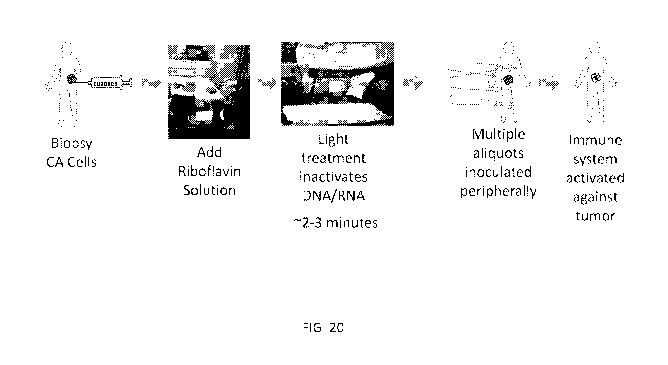
below.
BRIEF DESCRIPTION OF THE DRAWINGS
[0010] FIG. 1 illustrates proliferation of CAMA cells following treatment with
riboflavin
and UV light on the day of treatment (Day 0), and 2, 4, 6, and 8 days after
treatment.
Cells were treated using the Mirasol PRT Illumination device, at 10%, 20%7
30%7 40%7
50% or 100% illumination intensities. Cells that were not treated with UV
light (Live) were
included as a control.
[0011] FIG. 2 shows viability of CAMA cells after treatment with riboflavin
and UV light.
[0012] FIG. 3 shows expression of EpCAM, a surface marker, in CAMA cells after
treatment with riboflavin and UV light.
[0013] FIG. 4 provides fluorescence microscopy images comparing surface marker
expression on CAMA cells at various time intervals post-treatment with
riboflavin and UV
light (20% illumination intensity).
2
CA 03093723 2020-09-10
WO 2019/183320 PCT/US2019/023320
[0014] FIG. 5 shows relative expression of surface markers EpCAM (front row of
bars)
and CD38 (back row of bars) within the viable cell population after treatment
with varying
doses of UV light.
[0015] FIG. 6 shows Caspase-3 concentration in CAMA cells after treatment with
riboflavin and UV light.
[0016] FIG. 7 depicts the correlation between surface marker expression and
viability of
CAMA cells after treatment with riboflavin and UV light.
[0017] FIG. 8 shows tumor growth curves for mice bearing PyMT breast carcinoma
tumors that were injected with saline (control, no vaccine) compared to those
receiving
inactivated whole cell vaccine and those receiving a lysate vaccine (4T1
Spheroid Lysate
Vax, as described in WO 2016/161309, which is incorporated herein by reference
in its
entirety). Results indicated a statistically significant reduction in tumor
cell growth
observed for the inactivated whole cell vaccine versus untreated control
group, starting
at Day 23 post injection (p = 0.02 at day 23 and p < 0.001 at day 25).
[0018] FIG. 9 shows overall survival in vaccinated (inactivated whole cell
vaccine)
versus untreated/saline (control) groups of mice bearing PyMT tumors. The mice
receiving the inactivated whole cell vaccine had a significantly extended
survival time (p
= 0.009, Mantel-Cox Log Rank test) as compared to the saline-treated control
group.
[0019] FIG. 10 shows the size of 4T1 tumors in mice, prior to surgical
removal. Following
surgery, mice were arranged into the 3 groups shown so that each group had a
similar
average tumor size (p = 0.9) and spread. PBS ("control"), n = 5 mice;
Adjuvant, n = 8
mice; inactivated whole cell vaccine, n = 8 mice.
[0020] FIG. 11A and FIG. 11B show the results of an experiment wherein mice
were
treated with PBS (Control), with losartan and cationic liposome-DNA complexes
(CLDC)
(Adjuvant), or with the inactivated whole cell vaccine (Adjuvant + vaccine)
weekly starting
24 hours after surgical removal of the primary tumor. Metastatic disease in
the lungs was
quantitated using IVIS imaging following i.p. injection of 100p1 of luciferin.
As shown in
FIG. 11A, there was a significant decrease in measured metastatic burden in
mice treated
with the vaccine compared to adjuvant treated mice (Day 14, p =0. 0157) and
compared
to both the control mice and adjuvant treated mice (Day 16, p = 0.0119 and p =
0.0021,
3
CA 03093723 2020-09-10
WO 2019/183320 PCT/US2019/023320
respectively). FIG 11B shows the photon flux data over time for the individual
mice in
each group.
[0021] FIG. 12 shows the frequency of regrowth of the primary tumor due to
incomplete
removal of the primary tumor in the various treatment groups.
[0022] FIG. 13 is a survival curve showing that survival of the mice was
enhanced in the
group that received the inactivated whole cell vaccine. Mice were euthanized
when
moribund (i.e. weight loss > 10%, seizures, decreased mobility, unkempt
appearance,
etc.). The median survival of the control and adjuvant group was 17.5 days
while the
inactivated whole cell vaccine treated group was 24 days. This difference in
survival was
not statistically significant (p = 0.1), but biologically relevant given the
aggressive nature
of the 4T1 tumor.
[0023] FIG. 14 is a graph showing doubling time of tumor size, as described in
Example
3. Doubling time was greater in mice treated with inactivated whole cell
vaccine (p = 0.01).
[0024] FIG. 15 is a graph showing tumor growth area at 3, 5, 7, 10, 13, and 19
days post
tumor injection of mice bearing subcutaneous Lewis Lung Carcinoma tumors (LLC)
and
treated with either PBS control or inactivated LLC vaccine. Tumor growth was
significantly reduced in mice vaccinated with the inactivated LLC vaccine at
13 (p = 0.02)
and 19 (p = 0.001) days post tumor cell injection.
[0025] FIG. 16A-C are graphs showing subtypes of T cells in tumors obtained
from
control and vaccinated mice from the LLC study. FIG. 16A shows percent of T
cells that
were either CD4+CD25+ (presumed T regulatory T cells) or CD8+CD25+. There was
a
significant decrease in the CD4+CD25+ T cells in the vaccinated mice. FIG. 16B
shows
percent CD8+ T cells expressing the immune suppressive proteins PD-1, Lag3 or
Tim3.
FIG. 16C shows percent CD4+ T cells expressing the immune suppressive proteins
PD-
1, Lag3 or Tim3. For each data set shown in FIG. 16A-C, control is shown on
the left and
vaccine is shown on the right.
[0026] FIG. 17 shows production of IFNg (pg/ml) after spleen cells were
isolated from
healthy, naïve B6 mice, which were vaccinated and boosted with the inactivated
whole
cell vaccine (derived from 4T1 mouse tumor cells) with various immune
adjuvants. The
spleen cells were then restimulated with inactivated 4T1 tumor cells in vitro
for 72 hours
4
CA 03093723 2020-09-10
WO 2019/183320 PCT/US2019/023320
and IFNg was measured via ELISA. The CLDC adjuvant system produced the best
IFNg
response.
[0027] FIG. 18 shows mean fluorescent intensity (MFI) of serum IgG antibodies
at a
1:1000 dilution, derived from the blood of mice shown in FIG. 17, binding to
live 4T1 cells.
All of the vaccine/adjuvant systems produced significantly higher binding than
control or
inactivated cells alone.
[0028] FIG. 19 shows the results of an experiment wherein metastatic disease
in the
lungs was quantitated using IVIS imaging after mice were injected with 4T1
mammary
tumor cells that were then surgically removed and used to generate the
inactivated whole
cell vaccine. The mouse shown in the right panel was treated with an
inactivated whole
cell vaccine, and the mouse shown in the left panel was not given any vaccine.
A scale
bar for luminescence is also provided. 62% of mice receiving the vaccination
were
negative for lung metastases at similar time points wherein 80% of untreated
mice had
developed lung tumors at day 16 post-tumor cell removal.
[0029] FIG. 20 depicts an exemplary scheme for inactivating cells using UV
light and
riboflavin, preparing a vaccine composition, and treating a patient in need
thereof.
[0030] FIG. 21A-21B shows cell surface staining of mouse LLC cells following
UV+Rf
(UV light + Riboflavin) inactivation.
[0031] FIG. 22 shows surface staining of the mouse 4T1 breast carcinoma cells
following
UV+Rf inactivation.
[0032] FIG. 23A-D shows GFP expression of mouse melanoma GFP+616 tumor cells
following inactivation by either UV+RF or gamma radiation ex vivo.
[0033] FIG. 24A-B shows expression of the mouse tumor-associated antigen,
gp70,
following UV+Rf inactivation of mouse colon carcinoma, CT26, tumor cells.
[0034] FIG. 25A-D shows surface protein staining of inactivated, ex vivo,
canine tumor
tissues at lhr and 48hrs after UV+Rf inactivation. Cells were maintained at 4
C for the
48 hrs after inactivation.
[0035] FIG. 26A-D shows UV+RF inactivation of two additional ex vivo canine
tumor
tissues.
[0036] FIG. 27 shows staining of inactivated human liver carcinoma cells,
HepG2, for
surface marker GLUT1. Inactivated cells are shown in the left panel, and live
cells are
CA 03093723 2020-09-10
WO 2019/183320 PCT/US2019/023320
shown in the right panel. The chart below shows percent antibody-positive and
antibody-
negative cells when HepG2 cells were stained for surface markers GLUT1 and
HLA1.
[0037] FIG. 28A-B shows proliferation of T cells from spleen of 4T1-tumor
bearing mice,
that received no treatment. T cells proliferated ex vivo when cultured with
inactivated 4T1
tumor cells.
[0038] FIG. 29 shows lack of proliferation of inactivated 4T1 mouse breast
carcinoma
cells in culture for various time points following inactivation.
[0039] FIG. 30 shows lack of proliferation of inactivated human liver
carcinoma cells,
HepG2 (left panel) and data for lack of proliferation of HepG2 and human colon
carcinoma
cells, CRL-2577 (right panel).
DETAILED DESCRIPTION
[0040] Provided herein is a method for inactivating cells and preventing their
replication
using UV light and riboflavin. This chemical process is specific to the
DNA/RNA present
in the cells. Thus, cellular DNA and/or RNA is modified, while leaving protein
(including
cell surface antigens, enzymes, etc.) untouched in the process. By preventing
replication
processes while preserving cell antigens and phenotype, the treated cancer
cell
preparations can be used as vaccine compositions. The fact that the antigens
are present
in their native state on the cells of the vaccine compositions may boost
immune responses
above the level observed with single antigens or protein formulations that are
intended to
elicit the same responses. The combination of inactivated whole cells with an
adjuvant
further boosts this immunological effect.
[0041] This technology can be used in an autologous or allogenic fashion, i.e.
using
tumor cells isolated from the patient, cancer stem cell preparations, or those
grown in
culture systems, etc. When administered to a patient, the whole cell vaccine
reduces
tumor growth, decreases metastasis, and prolongs survival time.
[0042] Thus, the technology described herein provides a rapid method to
isolate,
prepare and administer cancer cell vaccines to patients, producing a response
in the
patient that rivals use of standard chemotherapy drugs.
[0043] Unless otherwise defined, all technical and scientific terms used
herein have the
same meaning as commonly understood by one of ordinary skill in the art to
which this
6
CA 03093723 2020-09-10
WO 2019/183320 PCT/US2019/023320
disclosure belongs. The terminology used in the detailed description herein is
for the
purpose of describing particular embodiments only and is not intended to be
limiting.
Definitions
[0044] The following terms are used in the description herein and the appended
claims:
[0045] The singular forms "a," "an" and "the" are intended to include the
plural forms as
well, unless the context clearly indicates otherwise.
[0046] Furthermore, the term "about" as used herein when referring to a
measurable
value such as an amount, dose, time, temperature, and the like, is meant to
encompass
variations of 20%7 10%7 5%7 1%7 0.5%,
or even 0.1% of the specified amount.
[0047] Also as used herein, "and/or" refers to and encompasses any and all
possible
combinations of one or more of the associated listed items, as well as the
lack of
combinations when interpreted in the alternative ("or").
[0048] Unless the context indicates otherwise, it is specifically intended
that the various
features described herein can be used in any combination.
[0049] As used herein, the terms "reduce," "reduces," "reduction" and similar
terms
mean a decrease of at least about 10%, about 15%, about 20%, about 25%, about
35%,
about 50%, about 75%, about 80%, about 85%, about 90%, about 95%, about 97% or
more.
[0050] As used herein, the terms "enhance," "enhances," "enhancement" and
similar
terms indicate an increase of at least about 10%, about 15%, about 20%, about
25%,
about 50%, about 75%, about 100%, about 150%, about 200%, about 300%, about
400%,
about 500% or more.
[0051] By the terms "treat," "treating" or "treatment of" (and grammatical
variations
thereof) it is meant that the severity of the patient's condition is reduced,
at least partially
improved or stabilized and/or that some alleviation, mitigation, decrease or
stabilization
in at least one clinical symptom is achieved and/or there is a delay in the
progression of
the disease or disorder.
[0052] The terms "prevent," "preventing" and "prevention" (and grammatical
variations
thereof) refer to prevention and/or delay of the onset of a disease, disorder
and/or a
clinical symptom(s) in a patient and/or a reduction in the severity of the
onset of the
7
CA 03093723 2020-09-10
WO 2019/183320 PCT/US2019/023320
disease, disorder and/or clinical symptom(s) relative to what would occur in
the absence
of the methods of the disclosure. The prevention can be complete, e.g., the
total absence
of the disease, disorder and/or clinical symptom(s). The prevention can also
be partial,
such that the occurrence of the disease, disorder and/or clinical symptom(s)
in the patient
and/or the severity of onset is less than what would occur in the absence of
the present
disclosure.
[0053] "Therapeutically effective amount" as used herein refers to an amount
that, when
administered to a patient for treating a disease, or at least one of the
clinical symptoms
of a disease, is sufficient to affect such treatment of the disease or symptom
thereof. The
"therapeutically effective amount" may vary depending, for example, on the
disease
and/or symptoms of the disease, severity of the disease and/or symptoms of the
disease
or disorder, the age, weight, and/or health of the patient to be treated, and
the judgment
of the prescribing physician. An appropriate amount in any given instance may
be
ascertained by those skilled in the art or capable of determination by routine
experimentation.
Cancer Vaccine Compositions
[0054] Provided herein are cancer vaccine compositions. The compositions
comprise,
consist essentially of, or consist of inactivated cancer cells, optionally in
combination with
an adjuvant. The cancer cells are inactivated by modifying their DNA and/or
RNA,
rendering them replication incompetent. The modification of the cellular DNA
and/or RNA
does not kill the cells, i.e. the cancer vaccines are live, replication-
inactivated vaccines.
Because cell viability is maintained, the vaccines present live antigenic
targets to the
patient's immune system. Peripheral inoculation stimulates immune response to
primary
tumor and metastases.
[0055] In some embodiments, the cancer vaccine comprises, consists essentially
of, or
consists of cancer cells that were inactivated using a photochemical process
to inactivate
tumor cell DNA and/or RNA replication while preserving protein structure and
phenotype.
In some embodiments, the DNA and/or RNA of the cancer cells in the cancer cell
vaccine
comprises modified bases. For example, in some embodiments, the DNA of the
cancer
8
CA 03093723 2020-09-10
WO 2019/183320 PCT/US2019/023320
cells in the vaccine may comprise modified guanine bases, such as oxidized
guanine
bases.
[0056] In some embodiments, the cancer cells are autologous cancer cells. As
used
herein "autologous" refers to cells that were removed from or derived from the
same
patient to whom the vaccine is administered. In some embodiments, the cancer
cells are
allogeneic cells. As used herein, "allogeneic" refers to cells that were
removed from or
derived from a donor who is not the patient to whom the vaccine is
administered.
[0057] In some embodiments, the cancer cells are from a patient suffering from
one or
more types of cancer. For example, the cancer cells may be isolated or derived
from a
patient suffering from cancer. The cancer may be a solid tumor or a liquid
tumor. The
cancer cells may be isolated or derived from a primary tumor, or a metastatic
tumor. The
cancer may be stage I, stage II, stage III, or stage IV. In some embodiments,
the cancer
cells may be derived from a patient suffering from breast cancer, lung cancer,
liver cancer,
bladder cancer, gynecological cancer, brain cancer, stomach cancer, prostate
cancer,
skin cancer, thyroid cancer, pancreatic cancer, colon cancer, or blood cancer.
In some
embodiments, the skin cancer is a melanoma. In some embodiments, the blood
cancer
is a leukemia, a lymphoma, or a myeloma. In some embodiments, the leukemia is
Acute
Lymphocytic Leukemia or Acute Myeloid Leukemia. In some embodiments, the
lymphoma is Hodgkin's Lymphoma or Non-Hodgkins Lymphoma. In some embodiments,
the myeloma is multiple myeloma.
[0058] In some embodiments, the cancer cells are derived from an immortalized
cancer
cell line. As used herein, a "cancer cell line" refers to a transformed cell
line derived from
a cancer sample. Usually, a cancer cell line is capable of generating a tumor
upon explant
into an appropriate host. A cancer cell line usually retains, in vitro,
properties in common
with the cancer from which it is derived, including, e.g., loss of
differentiation, loss of
contact inhibition, and will undergo essentially unlimited cell divisions in
vitro. Cancer cell
lines may include cell lines which have been genetically modified, for
example, to express
a protein that allows the cells to be recognized better by antigen-presenting
cells.
[0059] In some embodiments, the cancer cells are cancer stem cells.
[0060] In some embodiments, the cells are derived from a non-cancerous but
abnormal
growth, i.e., a benign tumor or growth.
9
CA 03093723 2020-09-10
WO 2019/183320 PCT/US2019/023320
[0061] In some embodiments, the cancer vaccine comprises, consists essentially
of, or
consists of white blood cells (e.g., tumor-associated macrophages), tumor-
associated
endothelial cells, tumor-associated fibroblasts, or any other cell type
present in the tumor
micro-environment.
[0062] In some embodiments, the cancer vaccine composition further comprises
an
adjuvant. The effect of the adjuvant is to boost the immunological response.
In some
embodiments, the adjuvant modifies monocyte function.
[0063] Examples of suitable adjuvants include saponin formulations, virosomes,
virus
like particles, non-toxic derivatives of enterobacterial lipopolysaccharide
(LPS),
immunostimulatory oligonucleotides (e.g. an immunostimulatory oligonucleotide
containing a CpG motif), mineral containing compositions, oil-emulsions,
polymers,
micelle-forming adjuvants (e.g., a liposome), immunostimulating complex
matrices (e.g.,
ISCOMATRIX), particles, squalene, phosphate, cationic liposome-DNA complexes
(CLDC), DDA, DNA adjuvants, gamma-insulin, ADP-ribosylating toxins, detoxified
derivatives of ADP-ribosylating toxins, Freund's complete adjuvant, Freund's
incomplete
adjuvant, muramyl dipeptides, monophosphoryl Lipid A (MPL), poly IC, CpG
oligodeoxynucleotides (ODNs), imiquimod, adjuvant system AS01, adjuvant system
AS02, adjuvant system AS03, MF59 and aluminum or aluminum salts (e.g. alum,
aluminum phosphate, aluminum hydroxide). Other suitable adjuvants include TLR
agonists, NOD agonists, and lipid-DNA agonist complexes.
[0064] In some embodiments, the cancer vaccine composition further comprises
one or
more agonists or antagonists.
[0065] In some embodiments, the agonist comprises a Toll-Like Receptor (TLR)
agonist.
In some embodiments, the TLR agonist is an agonist of TLR1, TLR2, TLR3, TLR4,
TLR5,
TLR6, TLR7, TLR8, TLR9, TLR10, TLR11, or TLR12. In particular embodiments, the
agonist is a TLR3 and/or a TLR9 agonist.
[0066] In some embodiments, the antagonist is a C-C chemokine receptor type 2
(CCR2) antagonist.
[0067] In some embodiments, the antagonist is an angiotensin receptor blocker
(ARB),
such as losartan, telmisartan, irbesartan, azilsartan, candesartan,
eprosartan,
olmesartan, or valsartan. In some embodiments, the ARB is administered at a
dose of
CA 03093723 2020-09-10
WO 2019/183320 PCT/US2019/023320
between about 5 and about 100 mg/kg, for example about 5, about 10, about 15,
about
20, about 25, about 30, about 35, about 40, about 45, about 50, about 55,
about 60,
about 65, about 70, about 75, about 80, about 85, about 90, about 95, or about
100 mg/kg.
[0068] In some embodiments, the cancer vaccine comprises at least one of (i.e.
one of,
two of, or all three of) a TLR agonist, a CCR2 antagonist and an ARB.
[0069] In some embodiments, the agonist or antagonist (e.g., TLR3 and/or a
TLR9
agonist) is contained within or coupled to a liposome. Liposomes are
spherical, self-
enclosed vesicles composed of amphipathic lipids. Liposomes may be
unilamellar,
having one lipid bilayer membrane, or multilamellar, having two or more
concentrically
arranged bilayers. Suitable liposomes may have a selected mean particle size
diameter
of about 200-500 nm. Various methods of preparing liposomes and encapsulation
of
therapeutic agents therein are well documented (see, for example, U.S. Pat.
Nos.
3,932,657, 4,311,712, and 5,013,556, all of which are incorporated herein by
reference).
Known methods include the reverse phase evaporation method as described in
U.S. Pat.
No. 4,235,871, which is incorporated herein by reference.
[0070] Lipids for use in forming the liposomes described herein include
vesicle-forming
lipids having two hydrocarbon chains, typically acyl chains, and a polar head
group.
Included in this class are the phospholipids, such as phosphatidylcholine
(PC),
phosphatidylethanolamine (PE), phosphatidic acid (PA), phosphatidylinositol
(PI), and
sphingomyelin (SM), where the two hydrocarbon chains are typically between
about 14-
22 carbon atoms in length, and have varying degrees of unsaturation. The
selection of
lipids and proportions can be varied to achieve a desired degree of fluidity
or rigidity, to
control stability, and/or to control the rate of release of an entrapped
agent. Where more
than one type of lipid is used, a suitable amount of a relatively unsaturated
lipid (such as
PC), may be used in order to form stable liposomes. In one embodiment, at
least 45-50
mol % of the lipids used to form the liposome are PC.
[0071] The liposomes may also include lipids derivatized with a hydrophilic
polymer such
as polyethylene glycol (PEG). Suitable hydrophilic polymers include
polyvinylpyrrolidone,
polyvinyl m ethylether, polymethyloxazoline,
polyethyloxazoline,
polyhydroxypropyloxazoline, polyhydroxypropylmethacrylamide,
polymethacrylamide,
polydimethylacrylam ide, polyhydroxypropylmethacrylate,
polyhydroxyethylacrylate,
11
CA 03093723 2020-09-10
WO 2019/183320 PCT/US2019/023320
hydroxymethylcellulose, hydroxyethylcellulose, polyethyleneglycol,
polyaspartamide, and
hydrophilic peptide sequences. Methods of preparing lipids derivatized with
hydrophilic
polymers are known (see e.g. U.S. Pat. No, 5,395,619, which is incorporated
herein by
reference).
[0072] In some embodiments, the cancer vaccine comprises cationic liposome-DNA
complexes (CLDC).
[0073] In some embodiments, the cancer vaccine further comprises a
photosensitizer
such as riboflavin (vitamin B2). In some embodiments, the cancer vaccine is
substantially
free of photosensitizer.
[0074] In some embodiments, the cancer vaccine composition further comprises a
carrier. In some embodiments, the cells and/or the photosensitizer are
suspended in the
carrier. In some embodiments, the carrier comprises normal saline (e.g., 0.9%
sodium
chloride), dextrose saline (e.g., dextrose 5% in 0.9% sodium chloride),
phosphate
buffered saline (e.g., 137 mmol/L NaCI, 2.7 mmol/L KCI, 10 mmol/L Na2HPO4, 2
mmol/L
KH2PO4).
[0075] In some embodiments, the cancer vaccine composition further comprises
one or
more additional pharmaceutically acceptable ingredients well known to those
skilled in
the art, including, but not limited to, pharmaceutically acceptable carriers,
diluents,
excipients, adjuvants, fillers, buffers, preservatives, anti-oxidants,
lubricants, stabilizers,
solubilizers, surfactants (e.g., wetting agents), masking agents, coloring
agents, flavoring
agents, and sweetening agents. Suitable carriers, diluents, excipients, etc.
can be found
in standard pharmaceutical texts. See, for example, Handbook of Pharmaceutical
Additives, 2nd Edition (eds. M. Ash and I. Ash), 2001 (Synapse Information
Resources,
Inc., Endicott, New York, USA), Remington's Pharmaceutical Sciences, 20th
edition, pub.
Lippincott, Williams & Wilkins, 2000; and Handbook of Pharmaceutical
Excipients, 2nd
edition, 1994.
Methods of Producing Cancer Cell Vaccines
[0076] The cancer cell vaccines described herein are produced using an
innocuous
chemical agent in a selective process that prevents cellular replication
processes while
preserving antigenic protein structure. More specifically, the cancer cell
vaccines are
12
CA 03093723 2020-09-10
WO 2019/183320 PCT/US2019/023320
produced by the combined application of a photosensitizer and light for
rendering cancer
cells replication deficient while retaining other biological functions of the
treated cells and
proteins. An exemplary scheme for producing and using cancer cell vaccines is
shown
in FIG. 20. The process for producing the cancer vaccines of the disclosure is
described
in detail below.
[0077] Initially, cancer cells are provided. The cancer cells may be
autologous, i.e.
removed from or derived from the subject to be vaccinated. In some
embodiments, the
cancer cells may be allogeneic. The cancer cells may also be derived from a
cancer cell
line.
[0078] In some embodiments, the cancer cells are cancer stem cells. In some
embodiments, the cancer vaccine comprises, consists essentially of, or
consists of white
blood cells (e.g., tumor-associated macrophages), tumor-associated endothelial
cells,
tumor-associated fibroblasts, or any other cell type present in the tumor
micro-
environment.
[0079] In some embodiments, the cancer cells are provided as a single cell
suspension
during inactivation. In some embodiments, the cells are suspended in media
during
inactivation. Exemplary medias which may be used include, but are not limited
to,
RPMI1640, MEM, DMEM, IMDM, DMEM-F12, Opti-MEM, Ham's F12, Media 199, or
combinations thereof.
[0080] Next, the cancer cells are inactivated using photochemical technology.
This is
achieved using photosensitizers that can act as electron transfer agents. The
application
of photosensitizer agents that can be placed into an excited state in
proximity to a guanine
base in DNA or RNA constructs allows for selective modification (e.g.
oxidation, cross-
linking, fragmentation, deamination) of these bases. Because electron
chemistry can only
occur over short distances, the photosensitizer agent must be bound or
associated with
(i.e. intercalated with) the nucleic acid in order to carry out the desired
chemistry.
[0081] In some embodiments, the photosensitizer is a flavin, for example
riboflavin
(Vitamin B2), flavin mononucleotide, or flavin adenine dinucleotide.
In some
embodiments, the photosensitizer is a tertiary aliphatic amine (e.g., 1,4-
diazabicyclo(2,2,2)octane), a piperazine, (e.g., N-2-hydroxyethylpiperazine-N'-
2-
ethanesulfonic acid and 1,4-dimethylpiperazine), an amino acid (e.g.,
tyrosine,
13
CA 03093723 2020-09-10
WO 2019/183320 PCT/US2019/023320
tryptophan, histidine, methionine), an enzyme (e.g., superoxide dismutase) or
EDTA
(ethylenediaminetetraacetic acid). In some embodiments, the photosensitizer
is
riboflavin.
[0082] The cells are added to a solution containing the photosensitizer (e.g.
riboflavin),
or the photosensitizer is added to a solution containing the cells (e.g., a
single cell
suspension of the cells in media).
[0083] In some embodiments, the concentration of photosensitizer used during
inactivation is about 10 pM to about 100 pM, such as about 10 pM, about 15 pM,
about
20 pM, about 25 pM, about 30 pM, about 35 pM, about 40 pM, about 45 pM, about
50
pM, about 55 pM, about 60 pM, about 65 pM, about 70 pM, about 75 pM, about 80
pM,
about 85 pM, about 90 pM, about 95 pM, or about 100 pM. In some embodiments,
the
solution contains the photosensitizer at a concentration of about 1 pM to
about 50 pM,
such as about 2 pM, about 3 pM, about 4 pM, about 5 pM, about 6 pM, about 7
pM, about
8 pM, about 9 pM, about 10 pM, about 15 pM, about 20 pM, about 25 pM, about 30
pM,
about 35 pM, about 40 pM, about 45 pM, or about 50 pM. In some embodiments,
the
photosensitizer concentration is less than about 10 pM, such as less than
about 9 pM,
about 8 pM, about 7 pM, about 6 pM, about 5 pM, about 4 pM, about 3 pM, about
2 pM,
or about 1 pM.
[0084] The solution containing the photosensitizer and the cells (optionally,
in media) is
then subjected to light treatment. The light treatment may comprise treatment
with visible
light, ultraviolet light, and/or infrared light. The light treatment
inactivates DNA and/or RNA
in the cancer cells by modifying bases of these nucleic acids. In some
embodiments,
guanine bases are selectively modified. In some embodiments, guanine bases are
selectively oxidized. Oxidized guanine bases cannot be repaired by natural
enzymatic
and cell repair mechanisms. As such, there is no possibility for reversion of
the induced
change to a form that would restore the ability of the cells to replicate.
[0085] In some embodiments, the light treatment comprises, consists
essentially of, or
consists of treatment with ultraviolet (UV) light. The UV light may be UV-A,
UV-B, or UV-
C light. The UV light may have a wavelength of 170 to 400 nm, including all
ranges and
subranges therebetween. For example, in some embodiments, the UV light has a
wavelength of 315 to 400 nm, 310 to 320 nm, 280 to 360 nm, 280 to 315 nm, or
180 to
14
CA 03093723 2020-09-10
WO 2019/183320 PCT/US2019/023320
280 nm. The UV light may be provided by UV light sources known in the art,
such as the
Mirasol PRT Illumination device (TerumoBCT, Lakewood, Colorado).
In some
embodiments, the cells may be treated with multiple wavelengths of light
simultaneously.
[0086] In particular embodiments, when riboflavin is used as a
photosensitizer, UV light
having a wavelength of 310 to 320 nm is used. The inventors have determined
that this
wavelength prevents riboflavin from reacting in free solution, which results
in production
of undesirable oxygen free radicals. At these wavelengths, riboflavin will
selectively react
when intercalated with nucleic acid.
[0087] The dose of the UV light may vary depending on the volume of solution
being
treated. For example, the dose of the UV light may be between 200-400 Joules
(e.g.,
300 Joules) for a volume of about 170 to 370 mls of solution. As will be
understood by
those of skill in the art, the dosage may be adjusted up or down if the volume
to be treated
is above or below this range.
[0088] In some embodiments, the dose of UV light may be from about 200 Joules
to
about 600 Joules, for example about 200, about 225, about 250, about 275,
about 300,
about 325, about 350, about 375, about 400, about 425, about 450, about 475,
about 500,
about 525, about 550, about 575, or about 600 Joules. In some embodiments, the
volume
of cancer cell preparations for illumination may be from about 200 ml to about
600 ml, for
example about 200, about 225, about 250, about 275, about 300, about 325,
about 350,
about 375, about 400, about 425, about 450, about 475, about 500, about 525,
about 550,
about 575, or about 600 ml. In some embodiments, the dose of UV light may be
from
about 0.5 J/ml to about 3.0 J/m I. For example, the dose of UV light may be
about 0.5,
about 0.6, about 0.7, about 0.8, about 0.9, about 1.0, about 1.1, about 1.2,
about 1.3,
about 1.4, about 1.5, about 1.6, about 1.7, about 1.8, about 1.9, about 2.0,
about 2.1,
about 2.2, about 2.3, about 2.4, about 2.5, about 2.6, about 2.7, about 2.8,
about 2.9, or
about 3.0 Joules/ml.
[0089] The cells may be treated with UV light for about 1 minute to about 60
minutes,
for example, about 1, about 2, about 3, about 4, about 5, about 6, about 7,
about 8, about
9, about 10, about 15, about 20, about 25, about 30, about 35, about 40, about
45, about
50, about 55, or about 60 minutes. In some embodiments, the cells are treated
with UV
CA 03093723 2020-09-10
WO 2019/183320 PCT/US2019/023320
light for about 1 minute to about 10 minutes, about 1 minute to about 5
minutes, or about
1 minute to about 3 minutes.
[0090] In some embodiments, the cancer cells are preincubated for a
predetermined
period of time in the solution containing the photosensitizer (e.g.,
riboflavin) before
subjecting the cells to the light treatment.
[0091] In some embodiments, the cells are not subjected to any additional
purification
or modification steps after light treatment. In other embodiments, the cancer
cells are
isolated and/or washed after the light treatment. For example, the cells may
be pelleted
and optionally washed after the light treatment. Pelleting and/or washing the
cells may
substantially remove photosensitizer (e.g., riboflavin) from the composition.
In some
embodiments, the cancer cells are concentrated after the light treatment.
[0092] In some embodiments, the cancer cells are resuspended or combined with
one
or more additional pharmaceutically acceptable ingredients as described above
after light
treatment. In some embodiments, the cancer cells are resuspended in a solution
comprising an adjuvant after light treatment.
[0093] In some embodiments, the cells remain viable for 1, 2, 3, 4, 5, 6, 7,
8, 9 or 10
days after light treatment. In some embodiments, the cells die (e.g., by an
apoptotic
mechanism) 1, 2, 3, 4, 5, 6, 7, 8, 9, or 10 days after treatment.
[0094] The cells generated using this method are incapable of replication
processes, but
substantially maintain and preserve the antigen and epitope profile of the
original, native
cell or antigen under treatment. In some embodiments, the inactivation process
does not
substantially change the metabolic processes, phenotype, or structure of the
cancer cells.
For example, in some embodiments, the inactivation process does not
substantially
change cell-surface marker expression in the cancer cells. In some
embodiments, the
inactivation process does not substantially change expression levels of cell
surface
markers such as EpCAM, CD38, CD34, CD117, CD44, CD24, Sca1, HLA, Glut1, MHC
Class I, PDL-L1, CD45, gp70, GFP and/or CD90 in the cells. In some
embodiments, the
inactivation process does not compromise the cell membrane and nuclear
membrane
integrity of the cells.
[0095] The fact that the cells are replication incompetent protects against
native forms
of the disease (cancer) or altered cell compositions in the body responsible
for formation
16
CA 03093723 2020-09-10
WO 2019/183320 PCT/US2019/023320
of tumorous lesions. Thus, because the specificity of the chemistry preserves
the antigen
profile and cellular integrity, and maintains protein structure in its native
state, the
inactivated cells that are produced by this process provide an improved source
for antigen
presentation.
Methods of Treatment
[0096] The cancer cell vaccine compositions described herein can be used as
vaccine
agents or stimulants for immune system priming and recognition that foster
immune
responses in cancer patients. This targeted therapy results in fewer side
effects
compared to traditional treatments such as chemotherapy or radiation. Notably,
because
the cancer cells of the vaccine maintain a normal phenotype, the potential for
them to
induce undesired side effects is extremely low or nonexistent.
[0097] In some embodiments, the cancer cell vaccine may be administered to a
patient
to treat or prevent cancer in the patient. The cancer that is treated or
prevented may be
a solid tumor or a liquid tumor. For example, the cancer that is treated or
prevented may
be breast cancer, lung cancer, liver cancer, bladder cancer, gynecological
cancer, brain
cancer, stomach cancer, prostate cancer, skin cancer, thyroid cancer,
pancreatic cancer,
colon cancer, or blood cancer. In some embodiments, the skin cancer is a
melanoma. In
some embodiments, the blood cancer is a leukemia, a lymphoma, or a myeloma. In
some
embodiments, the leukemia is Acute Lymphocytic Leukemia or Acute Myeloid
Leukemia.
In some embodiments, the lymphoma is Hodgkin's Lymphoma or Non-Hodgkin's
Lymphoma. In some embodiments, the myeloma is multiple myeloma.
[0098] In some embodiments, the vaccine may be administered to a patient to
treat or
prevent a non-cancerous but abnormal growth, i.e., a benign tumor or growth,
in a patient.
While most benign tumors/growths are treatable with surgery, some are in
locations
where surgery is not possible, and/or radiation may not be adequate. Examples
of non-
cancerous growths that may be treated include, but are not limited to,
adenomas,
fibromas, neuromas, hemangiomas, seborrheic keratoses, dermatosis papulosa
nigra, and
sebaceous hyperplasia.
[0099] In some embodiments, the patient is assessed for immune function and
immune
status prior to administration of the cancer vaccine. Such assessments may
include, but
17
CA 03093723 2020-09-10
WO 2019/183320 PCT/US2019/023320
are not limited to, DTH skin testing, blood tests, lymph node aspirate tests,
tumor tissue
tests, and/or determination of whether the patient is anergic, B cell
responsive, etc. In
some embodiments, the patient is not assessed for immune function and immune
status
prior to administration of the cancer vaccine.
[0100] In some embodiments, the patient may be immunocompetent. In other
embodiments, the patient may be immunocompromised. Optionally, the vaccine may
be
used in combination with genetic testing to quantify the degree of immune-
responders, or
immune non-responders.
[0101] It will be appreciated by one of skill in the art that appropriate
number of cells in
the cancer vaccine composition can vary from patient to patient. In some
embodiments,
the cancer vaccine comprises about 1 x 10³, about 1 x 10⁴, about 1 x 10⁵,
about 1 x 10⁶,
about 1 x 10⁷, about 1 x 10⁸, about 1 x 10⁹, or about 1 x 10¹⁹ cells. In some
embodiments,
a cancer vaccine comprises about 1 x 10⁵ to about 1 x 10⁸ cells.
[0102] In some embodiments, about 1x10⁵ to about 1x10⁸ cells are administered
to a
patient per administration. For example, about 1x10⁵, about 5x10⁵, about
1x10⁶, about
5x10⁶, about 1x10⁷, about 5x10⁷, or about 1x10⁸ cells may be administered to a
patient
per administration. In some embodiments, the administered dose is a split
dose, wherein
the total number of cells for administration is divided into 2, 3, 4, 5, 6, 7,
8, 9, or 10 sub-
doses. One or more sub-dose may be administered to the patient peripherally,
at different
locations on the patient's body. Each sub-dose may be administered at
approximately
the same time, or administration of the sub-doses may be staggered. For
example, sub-
doses may be administered at intervals of 15 minutes, 20 minutes, 30 minutes,
45
minutes, 1 hour, or 3 hours.
[0103] In some embodiments, the cancer vaccine is administered once, or more
than
once to the patient. In some embodiments, the cancer vaccine is administered
once,
twice, three times, four times, five times, six times, seven times, eight
times, nine times,
or ten times to a patient.
[0104] The cancer vaccine may be administered to the patient every day, about
every 3
days, about every 7 days, about every fourteen days, about once per month, or
about
once per year. In some embodiments, the cancer vaccine is administered at
least once
per week, at least every two weeks, or at least once every six months. In some
18
CA 03093723 2020-09-10
WO 2019/183320 PCT/US2019/023320
embodiments, the cancer vaccine is administered once, twice, three times, four
times,
five times, six times, seven times, eight times, nine times, ten times, twelve
times, fifteen
times, twenty times, or twenty-five times in a year.
[0105] In some embodiments, a first cancer vaccine and a second cancer vaccine
are
administered to the patient. In some embodiments, the second cancer vaccine is
administered after the first vaccine to boost the immune response. In some
embodiments,
immune response and/or tumor growth in the patient are monitored between
administration of the first vaccine and the second vaccine. In some
embodiments, the
second cancer vaccine is administered when it is determined that the patient
has not
exhibited a satisfactory immune response following administration of the first
vaccine, or
when it is determined that the tumor has continued to grow or metastasize
after
administration of the first vaccine. In some embodiments, the first cancer
vaccine and
the second vaccine comprise cells isolated or derived from a first tumor
extraction. For
example, a tumor removed from a patient may be used to produce the first and
second
vaccine, and after the first vaccine is administered, the second vaccine is
stored for later
use. In some embodiments, the first vaccine and the second vaccine comprise
cells
isolated or derived from separate tumor extractions. For example, a tumor
removed from
a patient may be used to produce the first vaccine, and after the tumor recurs
or
metastasizes, the recurrent tumor or metastatic tumor is removed and used to
produce
the second vaccine.
[0106] The cancer vaccine may be delivered to the patient intramuscularly,
intramucosally, intranasally, subcutaneously, intratumorally, intradermally,
transdermally,
intravaginally, intraperitoneally, intrarectally, intra-articularly or intra-
lymphatically, orally
or intravenously. In some embodiments, administration may be by sublingual,
buccal,
intra-organ (e.g., intrasplenic), or inhaled routes. For intravenous,
cutaneous or
subcutaneous injection, or injection at the site of the tumor, the cancer cell
vaccine may
be in the form of a parenterally acceptable aqueous solution which has
suitable pH,
isotonicity and stability. Those of relevant skill in the art are well able to
prepare suitable
solutions using, for example, isotonic vehicles such as Sodium Chloride
Injection,
Ringer's Injection, Lactated Ringer's Injection. Preservatives, stabilizers,
buffers,
antioxidants and/or other additives may be included, as required.
19
CA 03093723 2020-09-10
WO 2019/183320 PCT/US2019/023320
[0107] In some embodiments, the vaccine is administered peripherally to the
patient. In
some embodiments, multiple aliquots of the cancer vaccine are administered
peripherally
to the patient, in different locations.
[0108] In some embodiments, the cancer vaccine is administered simultaneously
or
sequentially (either before or after) with a vaccine-enhancing agent. In some
embodiments, the vaccine-enhancing agent is an angiotensin receptor blocker
(ARB) or
a beta blocker (BB). Exemplary vaccine-enhancing agents include losartan,
telmisartan,
irbesartan, azilsartan, candesartan, eprosartan, olmesartan, valsartan,
propranolol,
acebutolol, atenolol, betaxolol, bisoprolol, carteolol, carvedilol, esmolol,
labetalol,
metoprolol, nadolol, nebivolol, penbutolol, pindolol, propranolol, sotalol,
timolol. In some
embodiments, the vaccine-enhancing agent is selected from the group consisting
of
losartan and propranolol. In some embodiments, the vaccine-enhancing agent is
losartan.
In some embodiments, the vaccine-enhancing agent is propranolol.
[0109] In some embodiments, the vaccination protocol described herein
comprises
administering a cancer cell vaccine composition comprising inactivated, live
cancer cells,
and a potent adjuvant comprising TLR3 and/or TLR9 agonists attached to
liposomes, and
also comprises sequential or simultaneous administration of a vaccine-
enhancing agent
(e.g., losartan), which is given at or around the time of vaccination and
reduces
recruitment of immune suppressive myeloid cells.
[0110] In some embodiments, the vaccination protocol described herein
comprises
administering a cancer cell vaccine composition comprising inactivated, live
cancer cells
to a patient in need thereof. An adjuvant may optionally be administered at
the time of
vaccination. In some embodiments, an adjuvant is administered after
vaccination to boost
the immune response, for example about 6 hours, about 12 hours, about 24
hours, about
36 hours, about 48 hours, about 60 hours, or about 72 hours after vaccination.
In some
embodiments, the adjuvant comprises liposomes, e.g., CLDC. In some
embodiments, a
vaccine-enhancing agent such as losartan may be administered at or around the
time of
the vaccination. In some embodiments, a vaccine-enhancing agent such as
losartan may
be administered after vaccination, for example, about 6 hours, about 12 hours,
about 24
hours, about 36 hours, about 48 hours, about 60 hours, or about 72 hours after
vaccination. In some embodiments, a vaccine-enhancing agent such as losartan
may be
CA 03093723 2020-09-10
WO 2019/183320 PCT/US2019/023320
administered to the patient daily for a therapeutically effective number of
days, optionally
beginning on the day that the vaccine is administered. In some embodiments,
the
vaccine-enhancing agent (e.g., losartan) is administered at a dose of between
about 5
and about 100 mg/kg, for example about 5, about 10, about 15, about 20, about
25, about
30, about 35, about 40, about 45, about 50, about 55, about 60, about 65,
about 70, about
75, about 80, about 85, about 90, about 95, or about 100 mg/kg.
[0111] In some embodiments, the treatment reduces tumor growth or regrowth by
at
least 10%, at least 20%, at least 30%, at least 40%, at least 50%, at least
60%, at least
70%, at least 80%, at least 85%, at least 90%, at least 95%, or 100% compared
to tumor
growth in an unvaccinated patient. In some embodiments, the treatment prolongs
survival
of the patient by at least 10%, at least 20%, at least 30%, at least 40%, at
least 50%, at
least 60%, at least 70%, at least 80%, at least 85%, at least 90%, at least
95%, or 100%
compared to an unvaccinated patient. In some embodiments, the treatment
reduces the
occurrence of metastasis by at least 10%, at least 20%, at least 30%, at least
40%, at
least 50%, at least 60%, at least 70%, at least 80%, at least 85%, at least
90%, at least
95%, or 100% compared to an unvaccinated patient.
[0112] The cancer cell vaccine may elicit an immune response in the patient.
In some
embodiments, the immune response may include one or more of the following: (i)
upregulation of immunoglobulin (e.g., IgG, IgM), (ii) T-cell activation (e.g.,
multiple T-cell
generations matched to multiple cancer neoantigens), (iii) modulation of
innate immune
cells (e.g., myeloid cells), and (iv) revival of "exhausted" T-Cell
populations.
[0113] Suitable patients include both avians and mammals. The term "avian" as
used
herein includes, but is not limited to, chickens, ducks, geese, quail,
turkeys, pheasant,
parrots, parakeets, and the like. The term "mammals" as used herein includes,
but is not
limited to, humans, non-human primates, bovines, ovines, caprines, equines,
felines,
canines, lagomorphs, etc. Human subjects include neonates, infants, juveniles,
adults
and geriatric subjects. The terms "subject" and "patient" are used
interchangeably herein.
[0114] The cancer cell vaccines may be administered to patients with pre-
existing
conditions, for example pre-existing conditions that would prevent treatment
with other
therapies such as radiation, chemotherapy, or surgical resection.
21
CA 03093723 2020-09-10
WO 2019/183320 PCT/US2019/023320
Combination Therapies
[0115] The cancer cell vaccines may be administered alone or in combination
with other
treatments/therapies, either simultaneously or sequentially, depending upon
the condition
to be treated. Examples of treatments and therapies include, but are not
limited to,
chemotherapy (the administration of active agents, including, e.g. drugs, such
as
chemotherapeutics); surgery; and radiation therapy. Further examples of
treatments and
therapies include immune-based therapies, such as antibody therapy, adoptive
cell
therapy (ACT), and vaccine-based therapy. In some embodiments, the cancer cell
vaccines described herein may be administered after another treatment/therapy
to
eliminate any remaining tumor cells.
[0116] In some embodiments, the cancer vaccines may be administered in
combination
with one or more of the following therapies: checkpoint inhibitors (e.g., PD-1
or PDL-1
inhibitors, antibody therapies, genetically engineered dendritic cells, or
genetically
engineered T-cells (e.g., CAR-T cells).
[0117] In some embodiments, the cancer vaccines may be administered alone or
in
combination with a chemotherapeutic agent. A "chemotherapeutic agent" is a
chemical
compound useful in the treatment of cancer, regardless of mechanism of action.
Classes
of chemotherapeutic agents include, but are not limited to: alkylating agents,
antimetabolites, spindle poison plant alkaloids, cytotoxic/antitumor
antibiotics,
topoisomerase inhibitors, antibodies, photosensitizers, and kinase inhibitors.
Chemotherapeutic agents include compounds used in "targeted therapy" and
conventional chemotherapy.
[0118] Examples of suitable chemotherapeutic agents include: erlotinib
(TARCEVA ,
Genentech/OSI Pharm.), docetaxel (TAXOTERE , Sanofi-Aventis), 5-FU
(fluorouracil, 5-
fluorouracil, CAS No. 51-21-8), gemcitabine (GEMZAR , Lilly), PD-0325901 (CAS
No.
391210-10-9, Pfizer), cisplatin (cis-diamine, dichloroplatinum(II), CAS No.
15663-27-1),
carboplatin (CAS No. 41575-94-4), paclitaxel (TAXOL , Bristol-Myers Squibb
Oncology,
Princeton, N.J.), trastuzumab (HERCEPTIN , Genentech), temozolomide (4-methy1-
5-
oxo- 2,3,4,6,8-pentazabicyclo [4.3.0] nona-2,7,9-triene- 9-carboxamide, CAS
No. 85622-
93-1, TEMODAR , TEMODAL , Schering Plough), tamoxifen ((Z)-2-[4-(1,2-
diphenylbut-
22
CA 03093723 2020-09-10
WO 2019/183320 PCT/US2019/023320
1-enyl)phenoxy]-N,N-dimethylethanamine, NOLVADEX , ISTUBAL , VALODEXC,), and
doxorubicin (ADRIAMYCINC,), Akti-1/2, HPPD, and rapamycin.
[0119] More examples of chemotherapeutic agents include: oxaliplatin (ELOXATIN
,
Sanofi), bortezomib (VELCADE , Millennium Pharm.), sutent (SUNITINIB ,
SU11248,
Pfizer), letrozole (FEMARA , Novartis), imatinib mesylate (GLEEVEC ,
Novartis), XL-
518 (Mek inhibitor, Exelixis, WO 2007/044515), ARRY-886 (Mek inhibitor,
AZD6244,
Array BioPharma, Astra Zeneca), SF-1126 (P I3K inhibitor, Semafore
Pharmaceuticals),
BEZ-235 (PI3K inhibitor, Novartis), XL-147 (PI3K inhibitor, Exelixis),
PTK787/ZK 222584
(Novartis), fulvestrant (FASLODEX , AstraZeneca), leucovorin (folinic acid),
rapamycin
(sirolimus, RAPAMUNE , Wyeth), lapatinib (TYKERB , GSK572016, Glaxo Smith
Kline), lonafarnib (SARASARTM, SCH 66336, Schering Plough), sorafenib (NEXAVAR
,
BAY43-9006, Bayer Labs), gefitinib (IRESSA , AstraZeneca), irinotecan
(CAMPTOSAR , CPT-11, Pfizer), tipifarnib (ZARNESTRATm, Johnson & Johnson),
ABRAXANETM (Cremophor-free), albumin-engineered nanoparticle formulations of
paclitaxel (American Pharmaceutical Partners, Schaumberg, II), vandetanib
(rINN,
ZD6474, ZACTIMA , AstraZeneca), chloranmbucil, AG1478, AG1571 (SU 5271;
Sugen),
temsirolimus (TORISEL , Wyeth), pazopanib (GlaxoSmithKline), canfosfamide
(TELCYTA , Telik), thiotepa and cyclosphosphamide (CYTOXAN , NEOSARC)); alkyl
sulfonates such as busulfan, improsulfan and piposulfan; aziridines such as
benzodopa,
carboquone, meturedopa, and uredopa; ethylenimines and methylamelamines
including
altretam ine, triethylenemelam ine, triethylenephosphoram ide,
triethylene-
thiophosphoramide and trimethylomelamine; acetogenins (especially bullatacin
and
bullatacinone); a camptothecin (including the synthetic analog topotecan);
bryostatin;
callystatin; CC-1065 (including its adozelesin, carzelesin and bizelesin
synthetic
analogs); cryptophycins (particularly cryptophycin 1 and cryptophycin 8);
dolastatin;
duocarmycin (including the synthetic analogs, KW-2189 and CB1-TM1);
eleutherobin;
pancratistatin; a sarcodictyin; spongistatin; nitrogen mustards such as
chlorambucil,
chlornaphazine, chlorophosphamide, estramustine, ifosfamide, mechlorethamine,
mechlorethamine oxide hydrochloride, melphalan, novembichin, phenesterine,
prednimustine, trofosfamide, uracil mustard; nitrosoureas such as carmustine,
chlorozotocin, fotemustine, lomustine, nimustine, and ranimnustine;
antibiotics such as
23
CA 03093723 2020-09-10
WO 2019/183320 PCT/US2019/023320
the enediyne antibiotics (e.g. calicheamicin, calicheamicin gamma1I,
calicheamicin
omegal1 (Angew Chem. Intl. Ed. Engl. (1994) 33:183-186); dynemicin, dynemicin
A;
bisphosphonates, such as clodronate; an esperamicin; as well as
neocarzinostatin
chromophore and related chromoprotein enediyne antibiotic chromophores),
aclacinomysins, actinomycin, authramycin, azaserine, bleomycins, cactinomycin,
carabicin, carminomycin, carzinophilin, chromomycinis, dactinomycin,
daunorubicin,
detorubicin, 6-diazo-5-oxo-L-norleucine, morpholino-doxorubicin,
cyanomorpholino-
doxorubicin, 2-pyrrolino-doxorubicin and deoxydoxorubicin), epirubicin,
esorubicin,
idarubicin, nemorubicin, marcellomycin, mitomycins such as mitomycin C,
mycophenolic
acid, nogalamycin, olivomycins, peplomycin, porfiromycin, puromycin,
quelamycin,
rodorubicin, streptonigrin, streptozocin, tubercidin, ubenimex, zinostatin,
zorubicin; anti-
metabolites such as methotrexate and 5-fluorouracil (5-FU); folic acid analogs
such as
denopterin, methotrexate, pteropterin, trimetrexate; purine analogs such as
fludarabine,
6-mercaptopurine, thiamiprine, thioguanine; pyrimidine analogs such as
ancitabine,
azacitidine, 6-azauridine, carmofur, cytarabine, dideoxyuridine,
doxifluridine, enocitabine,
floxuridine; androgens such as calusterone, dromostanolone propionate,
epitiostanol,
mepitiostane, testolactone; anti-adrenals such as am inoglutethimide,
mitotane, trilostane;
folic acid replenisher such as frolinic acid; aceglatone; aldophosphamide
glycoside;
aminolevulinic acid; eniluracil; amsacrine; bestrabucil; bisantrene;
edatraxate;
defofamine; demecolcine; diaziquone; elfornithine; elliptinium acetate; an
epothilone;
etoglucid; gallium nitrate; hydroxyurea; lentinan; lonidainine; maytansinoids
such as
maytansine and ansamitocins; mitoguazone; mitoxantrone; mopidanmol;
nitraerine;
pentostatin; phenamet; pirarubicin; losoxantrone; podophyllinic acid; 2-
ethylhydrazide;
procarbazine; PSK polysaccharide complex (JHS Natural Products, Eugene, OR);
razoxane; rhizoxin; sizofiran; spirogermanium; tenuazonic acid; triaziquone;
2,2',2"-
trichlorotriethylamine; trichothecenes (especially T-2 toxin, verracurin A,
roridin A and
anguidine); urethan; vindesine; dacarbazine; mannomustine; mitobronitol;
mitolactol;
pipobroman; gacytosine; arabinoside ("Ara-C"); cyclophosphamide; thiotepa; 6-
thioguanine; mercaptopurine; methotrexate; platinum analogs such as cisplatin
and
carboplatin; vinblastine; etoposide (VP-16); ifosfamide; mitoxantrone;
vincristine;
vinorelbine (NAVELBINEC)); novantrone; teniposide; edatrexate; daunomycin;
24
CA 03093723 2020-09-10
WO 2019/183320 PCT/US2019/023320
aminopterin; capecitabine (XELODA , Roche); ibandronate; CPT-11; topoisomerase
inhibitor RFS 2000; difluoromethylornithine (DMF0); retinoids such as retinoic
acid; and
pharmaceutically acceptable salts, acids and derivatives of any of the above.
[0120] Also included in the definition of "chemotherapeutic agent" are: (i)
anti-hormonal
agents that act to regulate or inhibit hormone action on tumors such as anti-
estrogens
and selective estrogen receptor modulators (SERMs), including, for example,
tamoxifen
(including NOLVADEXC); tamoxifen citrate), raloxifene, droloxifene, 4-
hydroxytamoxifen,
trioxifene, keoxifene, LY117018, onapristone, and FARESTON (toremifine
citrate); (ii)
aromatase inhibitors that inhibit the enzyme aromatase, which regulates
estrogen
production in the adrenal glands, such as, for example, 4(5)-im idazoles,
aminoglutethimide, MEGASE (megestrol acetate), AROMASIN (exemestane;
Pfizer),
formestanie, fadrozole, RIVISOR (vorozole), FEMARA (letrozole; Novartis),
and
ARIMIDEX (anastrozole; AstraZeneca); (iii) anti-androgens such as flutamide,
nilutamide, bicalutamide, leuprolide, and goserelin; as well as troxacitabine
(a 1,3-
dioxolane nucleoside cytosine analog); (iv) protein kinase inhibitors such as
MEK
inhibitors (WO 2007/044515); (v) lipid kinase inhibitors; (vi) antisense
oligonucleotides,
particularly those which inhibit expression of genes in signaling pathways
implicated in
aberrant cell proliferation, for example, PKC-alpha, Raf and H-Ras, such as
oblimersen
(GENASENSE , Genta Inc.); (vii) ribozymes such as VEGF expression inhibitors
(e.g.,
ANGIOZYMEC) and HER2 expression inhibitors; (viii) vaccines such as gene
therapy
vaccines, for example, ALLOVECTIN , LEUVECTIN , and VAXIDC); PROLEUKIN rIL-
2; topoisomerase 1 inhibitors such as LURTOTECANC); ABARELIX rmRH; (ix) anti-
angiogenic agents such as bevacizumab (AVASTIN , Genentech); and
pharmaceutically
acceptable salts, acids and derivatives of any of the above.
[0121] Also included in the definition of "chemotherapeutic agent" are
therapeutic
antibodies such as alemtuzumab (Campath), bevacizumab (AVASTIN , Genentech);
cetuximab (ERBITUX , Imclone); panitumumab (VECTIBIX , Amgen), rituximab
(RITUXAN , Genentech/Biogen Idec), ofatumumab (ARZERRA , GSK), pertuzumab
(PERJETATm, OMNITARGTm, 2C4, Genentech), trastuzumab (HERCEPTIN ,
Genentech), tositumomab (Bexxar, Corixia), and the antibody drug conjugate,
gemtuzumab ozogamicin (MYLOTARG , Wyeth).
CA 03093723 2020-09-10
WO 2019/183320 PCT/US2019/023320
[0122] Humanized monoclonal antibodies with therapeutic potential as
chemotherapeutic agents in combination with the vaccines of the disclosure
include:
alemtuzumab, apolizumab, aselizumab, atlizumab, bapineuzumab, bevacizumab,
bivatuzumab mertansine, cantuzumab mertansine, cedelizumab, certolizumab
pegol,
cidfusituzumab, cidtuzumab, daclizumab, eculizumab, efalizumab, epratuzumab,
erlizumab, felvizumab, fontolizumab, gemtuzumab ozogamicin, inotuzumab
ozogamicin,
ipilimumab, labetuzumab, lintuzumab, matuzumab, mepolizumab, motavizumab,
motovizumab, natalizumab, nimotuzumab, nolovizumab, numavizumab, ocrelizumab,
omalizumab, palivizumab, pascolizumab, pecfusituzumab, pectuzumab, pertuzumab,
pexelizumab, ralivizumab, ranibizumab, reslivizumab, reslizumab, resyvizumab,
rovelizumab, ruplizumab, sibrotuzumab, siplizumab, sontuzumab, tacatuzumab
tetraxetan, tadocizumab, talizumab, tefibazumab, tocilizumab, toralizumab,
trastuzumab,
tucotuzumab celmoleukin, tucusituzumab, umavizumab, urtoxazumab, and
visilizumab.
NUMBERED EMBODIMENTS OF THE INVENTION
[0123] 1. A cancer vaccine composition, the composition comprising
inactivated
cancer cells, wherein the inactivated cancer cells are incapable of
replication.
[0124] 2. The cancer vaccine composition of embodiment 1, wherein the
cancer cells
are from a patient suffering from one or more types of cancer.
[0125] 3. The cancer vaccine composition of embodiment 2, wherein the
patent is
suffering from one or more of breast cancer, lung cancer, liver cancer,
bladder cancer,
gynecological cancer, brain cancer, stomach cancer, prostate cancer, skin
cancer, thyroid
cancer, pancreatic cancer, colon cancer, and blood cancer.
[0126] 4. The cancer vaccine composition of embodiment 3, wherein the skin
cancer
is a melanoma.
[0127] 5. The cancer vaccine composition of embodiment 3, wherein the blood
cancer is a leukemia, a lymphoma, or a myeloma.
[0128] 6. The cancer vaccine composition of embodiment 5, wherein the
leukemia is
Acute Lymphocytic Leukemia or Acute Myeloid Leukemia.
[0129] 7. The cancer vaccine composition of embodiment 5, wherein the
lymphoma
is Hodgkin's Lymphoma or Non-Hodgkin's Lymphoma.
26
CA 03093723 2020-09-10
WO 2019/183320 PCT/US2019/023320
[0130] 8. The cancer vaccine composition of embodiment 5, wherein the
myeloma is
multiple myeloma.
[0131] 9. The cancer vaccine composition of embodiment 2, wherein the
patent is
suffering from a benign tumor.
[0132] 10. The cancer vaccine composition of embodiment 2, wherein the cancer
is
metastatic cancer.
[0133] 11. The cancer vaccine composition of any one of embodiments 1-10,
wherein
the cancer cells are derived from an immortalized cell line.
[0134] 12. The cancer vaccine composition of any one of embodiments 1 to 11,
wherein the cancer cells are autologous.
[0135] 13. The cancer vaccine composition of any one of embodiments 1 to 11,
wherein the cells are allogeneic.
[0136] 14. The cancer vaccine composition of any one of embodiments 1 to 13,
wherein the composition comprises about 1x10⁵ to about 1x10⁸ cancer cells.
[0137] 15. The cancer vaccine composition of any one of embodiments 1 to 14,
wherein the DNA of the cancer cells comprises modified guanine bases.
[0138] 16. The cancer vaccine composition of any one of embodiments 1 to 15,
wherein the composition further comprises an adjuvant.
[0139] 17. The cancer vaccine composition of embodiment 16, wherein the
adjuvant
modifies monocyte function.
[0140] 18. The cancer vaccine composition of embodiment 16, wherein the
adjuvant
comprises aluminum hydroxide.
[0141] 19. The cancer vaccine composition of embodiment 16, wherein the
adjuvant
comprises CLDC.
[0142] 20. The cancer vaccine composition of embodiment 16, wherein the
adjuvant
comprises poly IC, CpG oligodeoxynucleotides (ODN), or imiquimod.
[0143] 21. The cancer vaccine composition of embodiment 16, wherein the
adjuvant
comprises liposomes.
[0144] 22. The cancer vaccine composition of embodiment 21, wherein the
liposomes
are conjugated to an agonist.
27
CA 03093723 2020-09-10
WO 2019/183320 PCT/US2019/023320
[0145] 23. The cancer vaccine composition of embodiment 22, wherein the
agonist is
an agonist of at least one of TLR3 and TLR9.
[0146] 24. The cancer vaccine composition of any one of embodiments 1 to 23,
wherein the composition further comprises a pharmaceutically acceptable
carrier.
[0147] 25. The cancer vaccine composition of embodiment 24, wherein the
pharmaceutically acceptable carrier is normal saline, dextrose saline, or
phosphate
buffered saline.
[0148] 26. The cancer vaccine composition of any one of embodiments 1 to 25,
wherein the cancer cells are inactivated using light treatment.
[0149] 27. The cancer vaccine composition of embodiment 26, wherein the light
treatment lasts for about 1 minute to about 3 minutes.
[0150] 28. The cancer vaccine composition of embodiment 26 or 27, wherein the
light
treatment does not substantially alter the structure of the antigenic proteins
on the cancer
cells.
[0151] 29. The cancer vaccine composition of any one of embodiments 26 to 28,
wherein the light treatment alters the DNA of the cancer cells.
[0152] 30. The cancer vaccine composition of embodiment 29, wherein the light
treatment selectively oxidizes guanine bases in the DNA of the cancer cells.
[0153] 31. The cancer vaccine composition of any one of embodiments 26 to 30,
wherein the light treatment does not substantially change the metabolic
processes,
phenotype, or structure of the cancer cells.
[0154] 32. The cancer vaccine composition of any one of embodiments 26 to 31,
wherein the light treatment does not substantially change surface marker
expression or
activity in the cancer cells.
[0155] 33. The cancer vaccine composition of embodiment 32, wherein the light
treatment does not change expression levels of EpCAM, CD38, CD34, CD117, CD44,
CD24, Sca1, HLA, Glut1, MHC Class I, PDL-L1, CD45, gp70, GFP or CD90 in the
cells.
[0156] 34. The cancer vaccine composition of any one of embodiments 26 to 33,
wherein the light treatment does not compromise the cell membrane or nuclear
membrane integrity of the cells.
28
CA 03093723 2020-09-10
WO 2019/183320 PCT/US2019/023320
[0157] 35. The cancer vaccine composition of any one of embodiments 26 to 34,
wherein the light treatment comprises treatment with UV light.
[0158] 36. The cancer vaccine composition of embodiment 35, wherein the UV
light
has a wavelength of 170 to 400 nm.
[0159] 37. The cancer vaccine composition of embodiment 35, wherein the UV
light
has a wavelength of 315 to 400 nm.
[0160] 38. The cancer vaccine composition of embodiment 35, wherein the UV
light
has a wavelength of 310 to 320 nm.
[0161] 39. The cancer vaccine composition of embodiment 35, wherein the UV
light
has a wavelength of 280 to 360 nm.
[0162] 40. The cancer vaccine composition of embodiment 35, wherein the UV
light
has a wavelength of 280 to 315 nm.
[0163] 41. The cancer vaccine composition of embodiment 35, wherein the UV
light
has a wavelength of 180 to 280 nm.
[0164] 42. The cancer vaccine composition of embodiment 35, wherein the UV
light
has a wavelength of 170 to 200 nm.
[0165] 43. The cancer vaccine composition of any one of embodiments 35 to 42,
wherein the dose of UV light is about 200 Joules to about 600 Joules.
[0166] 44. The cancer vaccine composition of embodiment 43, wherein the dose
of UV
light is about 200 Joules to 400 Joules.
[0167] 45. The cancer vaccine composition of embodiment 44, wherein the dose
of UV
light is about 300 Joules.
[0168] 46. The cancer vaccine composition of any one of embodiments 26 to 45,
wherein the light treatment is performed by contacting the cancer cells with
light in the
presence of a photosensitizer.
[0169] 47. The cancer vaccine composition of embodiment 46, wherein the
concentration of the photosensitizer is about 1 pM to about 50 pM.
[0170] 48. The cancer vaccine composition of embodiment 46 or 47, wherein the
concentration of the photosensitizer is less than about 10 pM.
[0171] 49. The cancer vaccine composition of any one of embodiments 46 to 48,
wherein the photosensitizer is riboflavin.
29
CA 03093723 2020-09-10
WO 2019/183320 PCT/US2019/023320
[0172] 50. A method for treating cancer in a patient in need thereof, the
method
comprising administering the cancer vaccine composition of any one of
embodiments 1
to 49 to the patient.
[0173] 51. The method of embodiment 50, wherein the cancer vaccine composition
is
administered simultaneously or sequentially with a vaccine-enhancing agent.
[0174] 52. The method of embodiment 51, wherein the vaccine-enhancing agent is
an
angiotensin receptor blocker (ARB) or a beta blocker (BB).
[0175] 53. The method of embodiment 51 or 52, wherein the vaccine-enhancing
agent
is losartan.
[0176] 54. The method of embodiment 53, wherein the dose of losartan is
between
about Sand about 100 mg/kg.
[0177] 55. The method of embodiment 54, wherein the dose of losartan is about
60
mg/kg.
[0178] 56. The method of embodiment 51 or 52, wherein the vaccine-enhancing
agent
is propranolol.
[0179] 57. The method of any one of embodiments 50 to 56, wherein the cancer
vaccine composition is administered once to the patient.
[0180] 58. The method of one of embodiments 50 to 56, wherein the cancer
vaccine
composition is administered more than once to the patient.
[0181] 59. The method of embodiment 58, wherein the cancer vaccine composition
is
administered 2, 3, 4, 5, 6, 7, 8, 9, or 10 times to the patient.
[0182] 60. The method of embodiment 58 or 59, wherein the cancer vaccine
composition is administered to the patient at least once every 7 days.
[0183] 61. The method of embodiment 58 or 59, wherein the cancer vaccine
composition is administered to the patient at least once every 14 days.
[0184] 62. The method of embodiment 58 or 59, wherein the cancer vaccine
composition is administered to the patient at least once every 6 months.
[0185] 63. The method of any one of embodiments 50 to 62, wherein the cancer
vaccine composition is administered by a route selected from subcutaneous,
intramuscular, intravenous, intranasal, sublingual, buccal, inhaled,
intradermal,
intratumoral, intra-organ, oral, and intraperitoneal.
CA 03093723 2020-09-10
WO 2019/183320 PCT/US2019/023320
[0186] 64. The method of embodiment 63, wherein the cancer vaccine composition
is
administered by subcutaneous injection.
[0187] 65. The method of embodiment 63, wherein the cancer vaccine composition
is
administered by intravenous injection.
[0188] 66. The method of embodiment 63, wherein the cancer vaccine composition
is
administered by intramuscular injection.
[0189] 67. The method of any one of embodiments 50 to 62, wherein the patient
is
immunocompetent.
[0190] 68. The method of any one of embodiments 50 to 62, wherein the patient
is
im m unocom promised.
[0191] 69. The method of any one of embodiments 50 to 68, wherein the
treatment
reduces tumor growth by at least 10% compared to tumor growth in an
unvaccinated
patient.
[0192] 70. The method of embodiment 69, wherein the treatment reduces tumor
growth by at least 20% compared to tumor growth in an unvaccinated patient.
[0193] 71. The method of embodiment 70, wherein the treatment reduces tumor
growth by at least 50% compared to tumor growth in an unvaccinated patient.
[0194] 72. The method of any one of embodiments 50 to 71, wherein the
treatment
prolongs survival of the patient by at least 10% compared to an unvaccinated
patient.
[0195] 73. The method of embodiment 72, wherein the treatment prolongs
survival of
the patient by at least 20% compared to an unvaccinated patient.
[0196] 74. The method of embodiment 73, wherein the treatment prolongs
survival of
the patient by at least 50% compared to an unvaccinated patient.
[0197] 75. The method of any one of embodiments 50 to 74, wherein the
treatment
upregulates IgG and/or IgM in the patient.
[0198] 76. The method of any one of embodiments 50 to 75, wherein the
treatment
activates T-cells in the patient.
[0199] 77. The method of any one of embodiments 50 to 76, wherein the cancer
is
breast cancer, lung cancer, liver cancer, bladder cancer, gynecological
cancer, brain
cancer, stomach cancer, prostate cancer, skin cancer, thyroid cancer,
pancreatic cancer,
colon cancer, or blood cancer.
31
CA 03093723 2020-09-10
WO 2019/183320 PCT/US2019/023320
[0200] 78. The method of embodiment 77, wherein the skin cancer is a melanoma.
[0201] 79. The method of embodiment 77, wherein the blood cancer is a
leukemia, a
lymphoma, or a myeloma.
[0202] 80. The method of embodiment 79, wherein the leukemia is Acute
Lymphocytic
Leukemia or Acute Myeloid Leukemia.
[0203] 81. The method of embodiment 79, wherein the lymphoma is Hodgkin's
Lymphoma or Non-Hodgkin's Lymphoma.
[0204] 82. The method of embodiment 79, wherein the myeloma is multiple
myeloma.
[0205] 83. The method of any one of embodiments 50 to 82, wherein the cancer
is
metastatic cancer.
[0206] 84. The method of any one of embodiments 50 to 83, wherein the cancer
vaccine composition is administered to the patient in combination with one or
more
additional therapies.
[0207] 85. The method of embodiment 84, wherein the one or more additional
therapies are selected from the group consisting of checkpoint inhibitors,
antibody
therapies, genetically engineered dendritic cells, genetically engineered T-
cells, and
chemotherapy.
[0208] 86. A method for producing a cancer vaccine, the method comprising
contacting
cancer cells with UV light in the presence of riboflavin.
[0209] 87. The method of embodiment 86, wherein the UV light alters the DNA of
the
cancer cells.
[0210] 88. The method of embodiment 87, wherein the UV light selectively
oxidizes
guanine bases in the DNA of the cancer cells.
[0211] 89. The method of embodiment 86, wherein the light treatment does not
substantially alter the structure of the antigenic proteins on the cancer
cells.
[0212] 90. The method of any one of embodiments 86 to 89, wherein the UV light
does
not substantially change the metabolic processes, phenotype, or structure of
the cancer
cells.
[0213] 91. The method of any one of embodiments 86 to 90, wherein the UV light
does
not substantially change surface marker expression or activity in the cancer
cells.
32
CA 03093723 2020-09-10
WO 2019/183320 PCT/US2019/023320
[0214] 92. The method of embodiment 91, wherein the UV light does not
substantially
change expression levels of EpCAM, CD38, CD34, CD117, CD44, CD24, Sca1, HLA,
Glut1, MHC Class I, PDL-L1, CD45, gp70, GFP and/or CD90 in the cells.
[0215] 93. The method of any one of embodiments 86 to 92, wherein the
inactivation
does not compromise the cell membrane and nuclear membrane integrity of the
cells.
[0216] 94. The method of any one of embodiments 86 to 93, wherein the cancer
cells
are contacted with UV light in the presence of riboflavin for about 1 minute
to about 3
minutes.
[0217] 95. The method of any one of embodiments 86 to 94, wherein the UV light
has
a wavelength of 170 to 400 nm.
[0218] 96. The method of any one of embodiments 86 to 94, wherein the UV light
has
a wavelength of 315 to 400 nm.
[0219] 97. The method of any one of embodiments 86 to 94, wherein the UV light
has
a wavelength of 310 to 320 nm.
[0220] 98. The method of any one of embodiments 86 to 94, wherein the UV light
has
a wavelength of 280 to 360 nm.
[0221] 99. The method of any one of embodiments 86 to 94, wherein the UV light
has
a wavelength of 280 to 315 nm.
[0222] 100. The method of any one of embodiments 86 to 94, wherein the UV
light has
a wavelength of 180 to 280 nm.
[0223] 101. The method of any one of embodiments 86 to 94, wherein the UV
light has
a wavelength of 170 to 200 nm.
[0224] 102. The method of any one of embodiments 86 to 101, wherein the dose
of UV
light is about 200 Joules to about 600 Joules.
[0225] 103. The method of embodiment 102, wherein the dose of UV light is
about 200
Joules to about 400 Joules.
[0226] 104. The method of embodiment 103, wherein the dose of UV light is
about 300
Joules.
[0227] 105. The method of any one of embodiments 86 to 104, wherein the cancer
cells
are in a single-cell suspension.
33
CA 03093723 2020-09-10
WO 2019/183320 PCT/US2019/023320
[0228] 106. The method of embodiment 105, wherein the riboflavin is added to
the
single-cell suspension.
[0229] 107. The method of any one of embodiments 86 to 106, wherein the cancer
cells
are preincubated in a solution containing riboflavin before contacting the
cells with the UV
light.
[0230] 108. The method of embodiment 107, wherein the solution comprises 10 to
100
pM riboflavin.
[0231] 109. The method of embodiment 107, wherein the solution comprises about
1
pM to about 50 pM riboflavin.
[0232] 110. The method of embodiment 107, wherein the solution comprises less
than
about 10 pM riboflavin.
[0233] 111. The cancer vaccine composition of any one of embodiments 1 to 49
for use
as a medicament.
[0234] 112. The cancer vaccine composition of any one of embodiments 1 to 49
for use
as a medicament for treating cancer.
[0235] 113. The cancer vaccine composition of any one of embodiments 1 to 49
for use
in a method of treating cancer.
[0236] 114. Use of the cancer vaccine composition of any one of embodiments 1
to 49
in the manufacture of a medicament for treating cancer.
[0237] 115. The cancer vaccine composition of any one of embodiments 111 to
113, or
the use of embodiment 114, wherein the cancer is breast cancer, lung cancer,
liver
cancer, bladder cancer, gynecological cancer, brain cancer, stomach cancer,
prostate
cancer, skin cancer, thyroid cancer, pancreatic cancer, colon cancer, or blood
cancer.
[0238] 116. The cancer vaccine composition or use of embodiment 115, wherein
the
skin cancer is a melanoma.
[0239] 117. The cancer vaccine composition or use of embodiment 115, wherein
the
blood cancer is a leukemia, a lymphoma, or a myeloma.
[0240] 118. The cancer vaccine composition or use of embodiment 117, wherein
the
leukemia is Acute Lymphocytic Leukemia or Acute Myeloid Leukemia.
[0241] 119. The cancer vaccine composition or use of embodiment 117, wherein
the
lymphoma is Hodgkin's Lymphoma or Non-Hodgkin's Lymphoma.
34
CA 03093723 2020-09-10
WO 2019/183320 PCT/US2019/023320
[0242] 120. The cancer vaccine composition or use of embodiment 117, wherein
the
myeloma is multiple myeloma.
[0243] 121. The cancer vaccine composition of any one of embodiments 111 to
113, or
the use of embodiment 114, wherein the cancer is metastatic cancer.
EXAMPLES
[0244] The following examples, which are included herein for illustration
purposes only,
are not intended to be limiting.
EXAMPLE 1. Inactivation of tumor cells derived from a tumor cell line
[0245] CAMA cells, a human mammary tumor line, were inactivated using UV light
treatment in the presence of riboflavin. Cells were treated using the Mirasol
PRT
Illumination device, at 10% (190 Joules), 20% (380 Joules), 30% (570 Joules),
40% (760
Joules), 50% (950 Joules) or 100% (1896 Joules) illumination intensities.
Cells that were
not treated with UV light (Live) were included as a control. Proliferation
(FIG. 1), viability
(FIG. 2), cell surface marker expression (FIG. 3, FIG. 4, FIG. 5), caspase
activity (FIG.
6), and cell membrane and nuclear membrane integrity were examined the day of
(day
0) and 2, 4, 6, and 8 days after treatment.
[0246] As shown in FIG. 1, treatment with riboflavin/UV light rendered the
cells unable
to replicate in culture. This effect was seen immediately, on the day of
treatment, even
at doses as low as 190-380 Joules (10-20% illumination intensity). Even though
they had
been inactivated, the cells remained substantially viable after treatment
(FIG. 3).
Specifically, the cells remained intact and metabolically functional (FIG. 4).
After four
days, the concentration of caspase-3 increased significantly (FIG. 6),
consistent with cell
death by an apoptotic mechanism. Thus, after treatment, the cells no longer
proliferate
and slowly die off over time.
[0247] Cell surface marker expression (EpCAM and CD38) was maintained at
relatively
consistent level after treatment, from doses as low as 190-380 Joules (10-20%
illumination intensity) to doses as high as 1896 Joules (100% illumination
intensity) (FIG.
3, FIG. 5). Cell viability was positively correlated with surface marker
expression (FIG.
CA 03093723 2020-09-10
WO 2019/183320 PCT/US2019/023320
7). This indicates that the cells maintained the cell surface antigens
required to generate
an immune response after UV treatment, even over a wide range of UV doses.
[0248] This data establishes a dynamic therapeutic range for preparing
inactivated cells
without damaging the cell surface marker proteins needed to stimulate antibody
generation.
EXAMPLE 2. Vaccine safety: Inactivation of autologous tumor cells and
injection
into healthy test mice
[0249] PyMT tumor cells were injected into wildtype C57616 mice. After tumors
grew,
tumor tissue was harvested to generate a cancer cell vaccine. A total of 2 x
108 PyMT ex
vivo tumor cells collected from seven C571316 mice were resuspended in
solution
comprising (i) 265 mls of DMEM media supplemented with 20% fetal bovine serum
and
glutamine (no antibiotics) and (ii) 35 m Is of riboflavin. The cells were
treated with UV light
at a dose of 300 Joules in total.
[0250] 1 x 10⁶ of the treated cells were placed in culture media and incubated
under
optimum conditions. No evidence of growth or proliferation was observed after
a month
in culture, consistent with 100% inactivation of the replication potential for
the treated
cells.
[0251] Additionally, a total of 10 C57/616 mice were injected subcutaneously
with 1 x 10⁶
inactivated cells. Additional doses were administered one, two and three weeks
later
(total of 4 doses, 1 x 10⁶ cells each dose). The animals were monitored for
160 days after
the first injection. No tumors were observed in any test subject during the
160-day
monitoring period, and no side effects of injection were observed, consistent
with
complete inactivation of the cells.
[0252] Lastly, 8 immune deficient NOD/SCID mice were injected with 1 x 10⁶
inactivated
PyMT cells in the flank. The animals were monitored for 5 months after
injection. No
tumor growth was observed during this monitoring period.
[0253] Taken together, these data suggest that there are no safety concerns
for the
injected cell preparation.
36
CA 03093723 2020-09-10
WO 2019/183320 PCT/US2019/023320
EXAMPLE 3. Vaccine efficacy: Injection of inactivated tumor cells into test
mice
with breast cancer inhibits tumor growth
[0254] C5761/6 mice were injected with 2.5 x 10⁵ viable PyMT cells in the
mammary fat
pad. Three days later, mice were treated with either saline (control, n = 10),
vaccine with
the inactivated tumor cells (n = 10), or a previously-studied lysate vaccine
(positive
control, n = 6). The vaccine comprised 1 x 10⁶ inactivated cells per mouse, co-
mixed with
the CLDC adjuvant system (lipids + TLR agonists) and was administered
subcutaneously
into both forelimbs under anesthesia (typically 100-110 p1/limb). The mice
also received
losartan (60 mg/kg) by intraperitoneal injection once a day for three days,
for a total of
three doses starting on the day the vaccine was given. Finally, mice received
a CLDC
adjuvant booster (100 pl, i.p.) 24 hours after the vaccine was given.
Vaccination was
repeated weekly for a total of 5 vaccine/losartan/CLDC booster cycles. Tumor
growth
over time was measured using a caliper (length x width). The extent of
mortality seen
over a 2 month period post-injection was also monitored.
[0255] FIG. 8 shows tumor growth curves for saline-injected (control, no
vaccine)
compared to those receiving inactivated whole cell vaccine and those receiving
the lysate
vaccine (4T1 Spheroid Lysate Vax). A statistically significant reduction in
tumor cell
growth was observed for the inactivated whole cell vaccine group versus the
untreated
control group, starting at Day 23 post injection (p = 0.02 at day 23 and p <
0.0001 at day
25).
[0256] FIG. 9 shows overall survival. In this experiment, mice were euthanized
when
the longest tumor diameter exceeded 15 mm. Mice receiving the inactivated
whole cell
vaccine had a significantly extended survival time (p = 0.0038) as compared to
the saline-
treated control group. The overall median survival time of mice receiving
inactivated
whole cell vaccine was 34 days compared to control at 26 days (-30% increase).
[0257] FIG. 14 shows doubling time. Doubling time of tumor growth was
significantly
greater in the mice treated with the inactivated whole cell vaccine as
compared to control
mice (p = 0.01).
37
CA 03093723 2020-09-10
WO 2019/183320 PCT/US2019/023320
EXAMPLE 4. Vaccine efficacy: Injection of inactivated tumor cells into test
mice
with aggressive breast cancer reduces lung metastasis, limits tumor regrowth
and
increases survival
[0258] The 4T1 mammary carcinoma is a transplantable tumor cell line that is
highly
tumorigenic and invasive and, unlike most tumor models, can spontaneously
metastasize
from the primary tumor in the mammary gland to multiple distant sites
including lymph
nodes, blood, liver, lung, brain and bone. Balb/c mice were injected with 1 x
106 4T1-
luciferase tumor cells in the mammary fat pad. 11 days after injection (when
the average
tumor area was 52 mm2), the primary tumors were measured and then surgically
removed. One mouse died during the procedure. The remaining 21 mice were
grouped
according to pre-surgical tumor size such that the average tumor size was
equal (FIG.
10). The following groups were determined: PBS ("control"), n = 5 mice;
Adjuvant, n = 8
mice; inactivated whole cell vaccine, n = 8 mice.
[0259] Surgically removed tumor tissue was then placed in media at 4 C
overnight. The
following day, the tumor tissue was minced and then treated with collagenase.
Cells were
filtered to remove tissue debris and then quantitated. A portion of the cells
was used to
create the first vaccine. Mice were vaccinated once a week with the
inactivated 4T1 tumor
cells (1.7x10⁶ cells) + adjuvant, boosted with adjuvant 24 hrs after
vaccination, and
provided 3 daily doses of 60 mg/kg losartan starting the day the vaccine was
given. This
cycle was repeated weekly.
[0260] Mice were imaged using an IVIS machine on a regular basis to detect
metastatic
disease development. Mice were injected with 100 pl luciferin, then put on the
IVIS 10
minutes later. Photon flux numbers were calculated using the IVIS-based
software and
compared between groups (FIG. 11A-B).
[0261] Mice were treated with PBS only (Control), with cationic liposome-DNA
complexes (CLDC) and losartan (Adjuvant), or with the inactivated whole cell
vaccine
(Adjuvant + vaccine) weekly starting 24 hours after surgical removal of the
primary tumor.
Metastatic disease in the lungs was quantitated using IVIS imaging (FIG. 19)
following
i.p. injection of 100p1 of luciferin. As shown in FIG. 11A, there was a
significant decrease
in measured metastatic burden in mice treated with the vaccine compared to
adjuvant
treated mice (Day 14, p =0.0157) and compared to both the control mice and
adjuvant
38
CA 03093723 2020-09-10
WO 2019/183320 PCT/US2019/023320
treated mice (Day 16, p = 0.0119 and p = 0.0021, respectively). FIG 11B shows
the
photon flux data over time of the individual mice in each group.
[0262] In some mice, there was regrowth of the primary tumors due to
incomplete
excision. The number of mice with primary regrowth 17 days after surgery was
documented (FIG. 12). Interestingly, less mice treated with the inactivated
whole cell
vaccine had regrowth of their primary tumor compared to the other treatment
groups. In
the control, 60% of the mice had regrowth of the primary tumor, 63% in the
adjuvant only
group, and only 38% in the inactivated whole cell vaccine group.
[0263] The effect of the vaccine on survival was also tested. Mice were
euthanized
when they displayed signs of morbidity (decreased weight by 10% or greater,
poor
mobility, seizures, etc.) and day post-surgery was evaluated (FIG. 13). Median
survival
time of mice treated with inactivated whole cell vaccine was 24 days compared
to 18 days
for the control mice, and 17.5 days post-surgical removal of the primary tumor
for the
adjuvant only mice. Thus, treatment with the vaccine increased median survival
by about
6 days. All mice did eventually succumb to metastatic disease in all groups.
The 4T1
metastatic model is extremely aggressive and typically unresponsive to many
conventional therapies, so any observed effect is noteworthy.
EXAMPLE 5. Testing efficacy of the inactivated whole cell vaccine in a
different
mouse tumor model (LLC)
[0264] To test the efficacy of the inactivated whole cell vaccine in a
different mouse
tumor model, healthy B6 mice were injected with Lewis Lung Carcinoma (LLC)
cells.
When primary tumors grow, they were excised and the tumor cells were
inactivated with
300J. Another 19 B6 mice were injected with 5x10⁵ LLC cells subcutaneously in
the flank.
Three days after tumor cell injection, mice received their first vaccine of
1.7x10⁶
inactivated cells per mouse per vaccine. They also received the CLDC adjuvant,
including
losartan and the 24 hour post-vaccine CLDC booster. Mice received their
vaccines weekly
for a total of three vaccines. Mice were all euthanized on day 19 and tumor
tissue was
collected and stained for immune cells.
[0265] As shown in FIG. 15, tumor growth was significantly decreased/delayed
in the
inactivated whole cell vaccine treated mice (p = 0.02 on day 13 and p = 0.001
on day 19).
Two of the mice in the vaccine treated group did not have tumors on day 19
(Complete
39
CA 03093723 2020-09-10
WO 2019/183320 PCT/US2019/023320
response = 20%). The final tumor weights of the mice that had tumors was
decreased in
the vaccine treated mice, although not significantly (p = 0.0547). The tumor
doubling time
was also not significantly decreased, but was less in the vaccine treated mice
(p =
0.0570). In the tumors, there was a significant decrease in CD4+CD25+ T cells
in the
vaccinated mice (p = 0.004, putative immune suppressive T regulatory cells) as
well as a
decrease in CD4+ T cells expressing GITR (p = 0.02), another marker of T-
regulatory
cells (FIG. 16A). Relatedly, there was a significant decrease in CD8+ T cells
expressing
the immune suppressive proteins Lag3 (p = 0.01) and Tim3 (p = 0.05) (FIG. 16B)
and a
significant decrease in CD4+ T cells expressing the proteins Lag3 (p = 0.005)
and Tim3
(p = 0.02) (FIG. 16C) in the vaccinated mice tumors.
EXAMPLE 6. Determining whether ex vivo canine tumor tissue contains tumor
cells
that maintain surface expression of proteins
[0266] Two tumor tissues were surgically obtained from dogs with spontaneous
cancer
undergoing treatment. One tissue was from an anal gland adenocarcinoma (ASA)
and
the other from a thyroid carcinoma (TC). Tissue was collagenase digested and
the single
cell suspension obtained was frozen in multiple vials at -80 C. Tumor cells
were later
thawed and one bullet of cells was used as control cells and the other as the
inactivated
cells. The canine cells were inactivated using 300J. The cell numbers used
were less
than that previously used for inactivating tumor cells. It took 1 minute and
38 seconds to
inactivate each group of cells. The tumor cells were then stained for canine
CD44, CD90,
PD-L1 and CD45 expression. CD45 was used to gate out all hematopoetic cells
(FIG.
25A-D). Analysis of the "tumor" cells was done on CD45 negative cells
(including tumor
cells, fibroblasts, endothelial cells, and other cell types).
[0267] For the ASA, 1 hr after inactivation, 15% of the cells expressed CD44
(compared
to 20% of control), 1% expressed CD90 (compared to 0.3%) and 7.7% expressed PD-
L1
(compared to 3.1%). For TC, after inactivation, 7.1% expressed CD44 (compared
to 6.8%
of control cells), 0.6% expressed CD90 (compared to 0.4%) and 0% expressed PD-
L1
(compared to 0.2%). The remaining cells were placed at 4 C for 48 hours and
then stained
again. After 48 hours, for ASA: CD44 = 10% (11 A control), CD90 = 2% (3.2%
control),
PD-L1 = 9.5% (3% control). For TC: CD44 = 5.8% (8.9% control), CD90 = 3.1%
(3.2%
control) and PD-L1 = 3% (2.9% control).
CA 03093723 2020-09-10
WO 2019/183320 PCT/US2019/023320
[0268] Additional pieces of other tumor tissue were obtained from two
different dogs
undergoing surgery. One was a GI mass and the other was a lung mass. The tumor
tissue
was digested using collagenase, washed and then strained to make a single cell
suspension. The cells were then all frozen in cell freezing media. Later,
cells were thawed
and half of the cells were inactivated using a UV+RF (UV light + riboflavin)
protocol. The
other half were kept on ice. The cells were then stained for surface
expression of canine
MHC Class I, CD44, CD90, and PD-L1 (FIG. 26).
[0269] Taken together, these data indicate that the surface markers are
maintained on
ex vivo canine tumor cells following inactivation.
EXAMPLE 7. Testing different adjuvants to produce a significant cellular and
humoral immune response against inactivated tumor cells
[0270] Healthy Balb/c mice were either vaccinated with PBS or inactivated
cells alone,
or vaccinated with the inactivated 4T1 vaccine (used in the metastatic mouse
tumor study
described in Example 4) with either the CLDC adjuvant system, topical
imiquimod applied
to the skin prior to vaccination (2mg/kg), or admixed with CpG ODN (50pg per
vaccine).
The vaccines were given s.c. in equal volumes in both the left and right
forelimb of the
mice, close to the foot. Mice received 1.7x10⁶ cells per mouse per vaccine.
Vaccines were
given on day 1 and then again on day 14. Mice were euthanized 8 days after the
booster
vaccine was given and spleen and blood were collected. Spleen cells were
cultured with
inactivated 4T1 cells (to prevent replication) and IFNg production was
measured after 72
hours (FIG. 17). Spleen cells were cultured at a ratio of 25 spleen cells to 1
inactivated
4T1 cell.
[0271] As shown in FIG. 17, the CLDC adjuvant system had the best recall
response in
terms of IFNg production. This was followed by CpG ODN. The vaccine alone also
produced some level of IFNg in 3 of the 4 mice, but there was no significant
difference
compared to control.
EXAMPLE 8. Further testing adjuvants to produce a significant cellular and
humoral immune response against inactivated tumor cells
[0272] Healthy Balb/c mice were either vaccinated with PBS, inactivated cells
alone or
vaccinated with the inactivated 4T1 vaccine (used in the metastatic mouse
tumor study
41
CA 03093723 2020-09-10
WO 2019/183320 PCT/US2019/023320
described in Example 4) with either the CLDC adjuvant system, topical
imiquimod applied
to the skin prior to vaccination (2mg/kg), or admixed with CpG ODN (50pg per
vaccine).
The vaccines were given subcutaneously in equal volumes in both the left and
right
forelimb of the mice, close to the foot. Mice received 1.7x10⁶ cells per mouse
per vaccine.
Vaccines were given on day 1 and then again on day 14. Mice were euthanized 8
days
after the booster vaccine was given and spleen and blood was collected. The
serum from
these mice was screened against live 4T1 tumor cells for IgG antibody binding
by
incubating serum with the cells at either a 1:500 or a 1:1000 dilution,
staining with a
fluorescently-labeled donkey anti-mouse secondary antibody, then subtracting
out the
background staining (normal mouse serum at 1:500 and 1:1000).
[0273] As shown in FIG. 18, unvaccinated and inactivated whole cell vaccine
alone did
not produce any IgG specific for 4T1 tumor cells. lmiquimod produced variable
amounts
with 3 of the 5 mice having very high staining. CpG ODN produced a reliable
and
significant increase in staining at 1:1000 dilution. Not very much staining
above
background was detected at a 1:500 dilution.
EXAMPLE 9. Preservation of Surface Antigens
[0274] The mouse lung carcinoma line, LLC, maintained expression of surface
antigens
following inactivation (FIG. 21A-21B). LLC cells were treated with riboflavin
(RF, 50pM)
and UV light (300J) and stained for surface expression of CD34, CD117, CD44
and CD90.
All 4 antigens were maintained on the surface of the cells. The mouse breast
carcinoma
cell line, 4T1, was also assessed for surface marker expression of CD44, Sca1,
and
EpCAM before and after UV+RF inactivation (FIG. 22).
[0275] The mouse melanoma tumor cell line B16 was transfected with green-
fluorescent
protein, injected into C571316 mice, removed from the mice, made into a single
cell
suspension and then was analyzed pre and post UV+RF inactivation and post
gamma
irradiation (100Gy) for expression of GFP+ tumor cells. Neither forms of cell
inactivation
negatively affected the expression of GFP by the tumor cells in the ex vivo
tumor tissue
(FIG. 23).
42
CA 03093723 2020-09-10
WO 2019/183320 PCT/US2019/023320
[0276] Lastly, the mouse colon carcinoma cell line, CT26, was inactivated with
UV+RF
and then analyzed for surface expression of the known tumor-associated
antigen, gp70
(FIG. 24A-24B). UV+RF inactivation increased the expression of gp70 on the
CT26 cells.
[0277] In addition, the human liver carcinoma cell line, HepG2, was
inactivated and
stained for expression of human HLA and GLUT1 following UV+RF inactivation
(300J,
FIG. 27) and imaged on an adherent cell cytometer.
[0278] The results obtained from all of these studies indicate that the
inactivation can be
carried out without significant modification to cell-specific antigens on the
cell surface. In
addition, these markers are maintained for an extended period of storage post-
treatment
in morphologicaly intact cells, and these observations have been noted in
cells derived
from all three species tested (mouse, canine and human).
EXAMPLE 10. Immune system triggering by inactivated cells.
[0279] Spleen cells were removed from mice that had 4T1 tumors growing, but
did not
receive any therapies, and placed them in culture to study T-cell immune
response to
either live 4T1 cell line derived cells compared to UV+RF-inactivated 4T1 cell
line-derived
cells (FIG. 28). Spleen cells were plated at 1x10⁶ cells per well, then co-
cultured with the
tumor cells (live or inactivated) at 4x10⁵ cells per well for 72 hours. The
proliferation dye
EdU was added after 48 hours of culture. The spleen cells were then collected
and stained
for proliferation of CD4+ and CD8+ T cells. There was a significant increase
in
proliferation of both CD4+ and CD8+ T cells when the cells were co-cultured
with the
UV+RF inactivated 4T1 tumor cells.
EXAMPLE 11: Pharmacokinetics: Proliferation and Persistence of Tumor Cells in
vitro and in vivo
[0280] To test the safety and efficacy of the inactivation process, PyMT tumor
cells were
removed from solid tumors on B6 mice and inactivated using UV light (300J).
1.5x10⁶
"viable" inactivated PyMT cells were injected s.c. into 5 healthy B6 mice.
This was
repeated on days 7, 14, and 21 for a total of 4 doses. Mice were euthanized
around 160
days after initial inactivated tumor cell injections and dissected. No
evidence of tumor
growth was noted. To test the safety of the inactivated cells in immune
deficient mice, 8
NOD/SCID mice were injected with 1x10⁶ inactivated PyMT cells s.c. in the
right flank.
43
CA 03093723 2020-09-10
WO 2019/183320 PCT/US2019/023320
Mice have been monitored since then. Three mice died during the study likely
due to
unrelated causes (likely a viral or bacterial infection). Necropsy was
performed on one
and no tumor was noted, skin analysis was performed on the other 2 and no
tumors were
noted. Mice were euthanized 252 days post tumor cell injection and no tumors
were noted
in the remaining 5 mice.
[0281] Cellular proliferation in culture following inactivation was also
assessed (FIG. 29).
4T1 cells were injected into the fat pad of Balb/c mice and tumor growth was
monitored
until tumors reached about 10 mm in diameter. The tissue was removed, digested
using
collagenase, washed and inactivated with UV+Rf and then cultured for 24 and 72
hrs in
37 C. No proliferation of 4T1 cells was noted at 24 and 72 hours-post
inactivation. This
was also compared to the non-inactivated ex vivo tumor cells (which
proliferated some)
and with the 4T1 tumor cell line (which proliferated). A similar study was
performed using
the human liver carcinoma cell line, HepG2 and the human colon carcinoma cell
line,
CRL-2577 (FIG. 30). Proliferation testing was conducted with Click-iT EdU
which labels
newly formed DNA. Using an adherent cell cytometer the fluorescence of labeled
new
DNA is compared to a total DNA stain. This test identified many thousands of
replicating
cells amounting to 20-35% high EdU cells in the live control for CRL-2577 and
approximately 75% in the HepG2 population which was labeled for longer.
[0282] Although the disclosed teachings have been described with reference to
various
applications, methods, and compositions, it will be appreciated that various
changes and
modifications can be made without departing from the teachings herein and the
claims
below. The foregoing examples are provided to better illustrate the disclosed
teachings
and are not intended to limit the scope of the teachings presented herein.
While the
present teachings have been described in terms of these exemplary embodiments,
the
skilled artisan will readily understand that numerous variations and
modifications of these
exemplary embodiments are possible without undue experimentation. All such
variations
and modifications are within the scope of the current teachings.
[0283] All references cited herein, including patents, patent applications,
papers, text
books, GenBankTM or other accession numbers, and the like, and the references
cited
therein, to the extent that they are not already, are hereby incorporated by
reference in
their entirety. In the event that one or more of the incorporated literature
and similar
44
CA 03093723 2020-09-10
WO 2019/183320 PCT/US2019/023320
materials differs from or contradicts this application, including but not
limited to defined
terms, term usage, described techniques, or the like, this application
controls.
[0284] The foregoing description and Examples detail certain specific
embodiments of
the invention and describes the best mode contemplated by the inventors. It
will be
appreciated, however, that no matter how detailed the foregoing may appear in
text, the
invention may be practiced in many ways and the invention should be construed
in
accordance with the appended claims and any equivalents thereof.