Note: Descriptions are shown in the official language in which they were submitted.
CA 03093912 2020-09-14
WO 2019/178378
PCT/US2019/022303
GALLOYLATED PROCYANIDINS FOR INCREASING
INTRACELLULAR NITRIC OXIDE PRODUCTION
CROSS-REFERENCE TO RELATED APPLICATIONS
This nonprovisional application claims priority to U.S. Provisional Patent
Application No.
62/642,846, entitled "Galloylated Procyanidins for Increasing Intracellular
Nitric Oxide
Production," filed March 14, 2018 by the same inventors, the entirety of which
is incorporated
herein by this reference.
TECHNICAL FIELD
This invention relates, generally, to diseases or conditions associated with
small or
vasoconstricted blood vessels. More specifically, it relates to formulations
for increasing
intracellular nitric oxide levels to effectuate vasodilation.
BACKGROUND
An intrinsic role of nitric oxide (NO) in vascular physiology is capillary
dilation, subsequently
increasing oxygen and blood flow to muscle tissue. Since its discovery, there
have been a litany
of scientific papers that have been published acknowledging nitric oxide's
crucial role in
vasodilation and cell communication. The vasodilation effect of nitric oxide
is relevant to
athletics and exercise, as increased blood flow would increase endurance,
muscle healing and
protein anabolism, subsequently attenuating lactic acid levels. Nitric oxide
reduces the amount
of lactic acid produced during exercise and extends activity duration and
intensity before
exhaustion. In addition, it has been demonstrated to shorten healing time
following strenuous
exercise. By accelerating the delivery of oxygen and nutrients to muscles
under stress, nitric
oxide has a transient effect on endurance. In addition, continuous exposure of
the muscle to
nitric oxide has been shown to increase protein anabolism and subsequently
increase muscle
mass and strength. Additionally, nitric oxide has been shown to enhance both
the uptake of
glucose and the removal of ammonia in the muscle.
Attempts have been made to increase intracellular nitric oxide, including U.S.
Patent Nos.
6,706,756 and 7,132,446. Other references have described the biomedical
significance of Nitric
Oxide, such as European Patent No. EP 1,549,300 to Mantione et al. Nitric
oxide (NO) is a
major signaling molecule in the mammalian immune, cardiovascular and nervous
systems. NO
produced at one site can have an effect on tissues at a distance. NO is
produced from L-
arginine by the enzyme, nitric oxide synthase (NOS). NOS occurs in three
forms: endothelial
(e), neuronal (n), and inducible (i) NOS. The first two forms are
constitutively expressed and
Ca2+ dependent. Inducible (i) NOS is Ca2+ independent. The three forms of NOS
are encoded
for on three distinct genes on chromosomes. In general, n- and e- NOS depend
on intracellular
1
CA 03093912 2020-09-14
WO 2019/178378 PCT/US2019/022303
calcium transients and release NO in the nM range, whereas iNOS, following an
induction/latency period, can release NO in the pM range for extended periods
of time.
The presence of constitutive and inducible forms of NOS suggest that they may
have distinct
functions. c- and i- NOS can be distinguished on the basis of the length of
time necessary to
see an increase in levels of NO and the length of time these elevated levels
can be maintained.
NO derived from cNOS may occur in two functional forms: the first is always
present at low
"tonal" or "basal" levels: this basal level can be slightly increased for a
short time in response
to certain signals, e.g., acetylcholine (ACH). This brief enhanced release of
cNOS derived NO
can have profound physiological actions, which are evident long after NO has
returned to its
basal level, for a longer period of time. For example, endothelial cells
briefly exposed to
morphine and eNOS change their shape from elongated to round, a process that
takes several
hours. iNOS is induced by various signal molecules, e.g., proinflammatory
cytokines. The
induction of i-NOS is usually seen after a 3-4 hour delay: iNOS is capable of
producing NO for
24-48 hours. These data suggest that NO is always present and that the levels
of NO can be
regulated either rapidly or slowly depending on the organism's needs. The
presence of different
regulatory processes implies that NO has different functions, and/or that the
levels of NO must
be progressively increased in order for it to exert its function.
NO functions as a vascular, immune and neural signal molecule and also has
general
antibacterial, antiviral actions and the ability to down-regulated
proinflammatory events. In the
vascular and immune system, one of the key stages in the immune response is
the recruitment
and activation of leukocytes by the endothelium. Leukocyte activation by the
endothelium
occurs in stages. The initial step is the attraction of the leukocytes to the
endothelium. This is
followed by increased leukocyte adhesion and change in shape and finally
migration across the
endothelium. These cellular changes are accompanied by scheduled changes in
synthesis of
molecules that regulate cell-matrix interactions.
Normally, non-activated leukocytes roll along the endothelium. The interaction
between the two
cell types is loose and reversible and mediated by a family of adhesion
molecules known as
selectins. Activation of leukocytes occurs in response to the release of
several
chemoattractants including leukotriene 64 and interleukin 8 (IL-8). In the
presence of these
agents, immunocytes cease to roll, becoming "activated," they start to flatten
and adhere with
greater strength to the endothelial lining. Activation is mediated by a family
of adhesion
molecules call the integrins, such as ICAM-1 and NCAM-1. Adherent immunocytes
are able to
undergo transendothelial migration in the presence of PECAM-1.
This immunocyte-endothelial interaction is down-regulated by NO. NO inhibits
platelet and
neutrophil aggregation and can diminish the adherence and level of activation
of leukocytes
and endothelial cells. NOS inhibitors increase platelet adhesion and enhance
leukocyte
2
CA 03093912 2020-09-14
WO 2019/178378 PCT/US2019/022303
adhesion. NO plays a similar role involving the microglia cells of the nervous
system's immune
response.
The central nervous system (CNS) is unique in that it uses all three isoforrns
of NOS to produce
NO. The constitutive isoforms e- and n- NOS are found in the normal CNS;
however, iNOS is
not expressed in the healthy CNS. Pathological states, e.g., trauma, cerebral
ischernia and
.. neuronal diseases, increase the levels of e- and nNOS and induce iNOS
activity. cNOS derived
NO has the ability to down-regulate proinflarnmatory events via inhibition of
NF-KB activation of
proinflammatory cytokines. NO upregulates several enzymes involved in
immunoregulation,
including neutral endopeptidase. (CALLA, acute lymphoblastic leukemic antigen,
enkephalinase) or 0D10, Thus, cNOS derived No stimulates enzymes that process
protein
gene products, implying a link between signaling processes involving NO and
naturally
occurring antibacterial peptides. No controls and regulates enzymes that are
responsible for
liberating these crucial molecules that have a proactive protective function.
Evidence has also been provided that NO plays a role in neurotransmitter
release. Morphine
and cNOS derived NO release growth hormone and ACTH from rat brain fragments;
these
neuropeptides are involved in the stress response. Thus, NO is involved in
vasodilation,
antibacterial and antiviral responses, signal molecule release and inhibition
of immunocyte
adherence to the endothelium.
There appears to be a tonal or basal level of NO that is physiologically
significant. Endothelia
from non-insulin dependent diabetics do not exhibit a tonal level of NO and in
these individuals
vascular disease causes disability and eventual death. A number of researchers
have attributed
vascular disease in part to alterations associated with eN0S- derived NO and
some have
speculated this may be due to enhanced free radical generation. Decreases in
basal NO levels
may also contribute to enhanced platelet function and various neuropathies.
Thus, it appears that tonal or basal NO levels are important in limiting the
degree of excitation
of nervous, immune and vascular tissues. This tonal NO may manifest itself via
effects on
adhesion-mediated processes via NF-KB. Estrogen may exert it beneficial
vascular protective
actions via these processes as well, since it also releases cNOS derived NO.
Strengthening
this hypothesis in the finding of the cannabinoid CB1 receptor type on
mammalian endothelial
cells and the finding of a mu opiate receptor on human vascular endothelial
cells. (Three
general classes of cell surface opioid receptors (kappa, delta and mu) have
been described.
Receptors exhibiting high binding specificity for morphine have been
designated mu opioid
receptors.) Detailed analysis has revealed the existence of multiple mu opioid
receptor
subtypes. Isolated nucleic acid sequences encoding various mu receptors and
polypeptides
comprising mu receptors (and referred to here as "mu3 opioid receptor(s)") are
disclosed in
detail in PCT Patent Publication WO 99/24471, published 20 May 1999.
3
CA 03093912 2020-09-14
WO 2019/178378 PCT/US2019/022303
.. Various vasodilating compounds have been described that interact with NO,
as described in
U.S. Patent No. 6,706,756 to Fitzpatrick. The antioxidant properties of
various plant favonoids,
including procyanidins, are well known. Procyanidins possess endothelium-
dependent relaxing
(EDR) activity in blood vessels in vitro. The endothelium is a single layer of
cells lining every
blood vessel. Maintaining healthy endothelial function is critical for overall
health and wellbeing.
Endothelial dysfunction is a common characteristic of altered cardiovascular
function leading
to coronary heart disease, and more generally atherothrombotic diseases
including stroke and
peripheral vascular disease. AU risk factors for cardiovascular disease -
raised LAX cholesterol,
diabetes, smoking, high blood pressure (hypertension), increasing age and lack
of exercise -
have been linked to endothelial dysfunction. Endothelial dysfunction is widely
recognised as a
precursor to atherosclerotic lesion formation. Common characteristics of
endothelial
dysfunction include: increased inflammation; reductions in the healthy anti-
thrombotic functions
of the endothelium; increased synthesis of mediators that stimulate
remodelling and vascular
stiffness; and increased vasoconstriction with reduced vasodilatation.
Endothelial dysfunction is not only associated with the underlying mechanisms
leading to
cardiovascular disease, but also as a risk factor for cardiovascular events,
including myocardial
infarction. The severity of endothelial dysfunction is closely associated with
increased risk of
mortality in patients with chronic heart failure. Although statins and
angiotensin-converting
enzyme inhibitors cause modest improvements in endothelial function, there are
currently no
pharmaceutical medications that specifically treat endothelial dysfunction.
The original finding that red wines, grape juice and other grape products
exhibited EDR activity
was companied by strong evidence that this activity was due to stimulation of
NO production
by the endothelial cells which form the lining of all blood vessels.
Vasorelaxation induced by
grape extracts, wines and the like was reversed by NO synthase inhibitors, and
vasorelaxation
could be restored by exposure of the vessel to L-arginine, the normal
substrate for NO
synthase. The importance of nitric oxide synthase system is underscored by the
finding that a
dysfunctional NO system can contribute to several diseases, including
atherosclerosis.
Therefore, consumption (and absorption) of NO-stimulating compounds in the
diet, or in the
form of dietary supplements, could contribute to prevention or halting the
progress of
atherosclerosis, other chronic age-related diseases, or conditions known to
involve failure of
the NO/NO synthase system, e.g., erectile dysfunction. Although procyanidin
compounds,
particularly those from grape seed extracts are known to exhibit EDR activity,
current
supplements administered to patients and consumers do not identify, nor
isolate the active and
most potent compounds to achieve the desired EDR.
A further characteristic of endothelial dysfunction is increased synthesis of
the vasoconstrictor
peptide endothelin-1. Antagonists of endothelin-1 cause vasodilation and
improve endothelium-
dependent vasodilator responses in older people, and in patients with
atherosclerosis.
4
CA 03093912 2020-09-14
WO 2019/178378 PCT/US2019/022303
Research on reversing endothelial dysfunction has identified the transcription
factor Kruppel-
like factor 2 (KLF2) as a key regulator of healthy endothelium, which affords
protection from
atherosclerosis. It has been proposed that agents that increase KLF2 in the
endothelium could
be used to treat endothelial dysfunction. Some procyanidins are known to
increase KLF2
transiently for a few hours. Identification of agents that could sustain this
induction would have
greater therapeutic utility in restoring or maintaining endothelial function.
The beneficial effects on cardiac function have been attributed to the high
content of flavanols,
principally procyanidins. Proanthocyanidins represent a group of plant
polyphenols found in
roots, barks and fruits with an astringent taste. Proanthocyanidins include
the subgroups of
procyanidins and prodelphinidins. Proanthocyanidins are biopolymers composed
of flavan
subunits. Procyanidins are composed of catechin and epicatechin units, also
called monomeric
procyanidins.
The use of polyphenol compositions in the treatment of endothelial dysfunction
have been
previously described, as in European Patent No. 3,179,996 to Corder. High
flavanol cocoa
drinks and high fiavanol dark chocolate have been found to improve endothelial
function in
patients with chronic heart failure, coronary artery disease, and diabetes.
Grape seed extract,
which is also mainly composed of procyanidins, also lowers blood pressure and
improves
vascular function. The improvement in cardiovascular function with products
containing high
amounts of procyanidins is consistent with studies on isolated vessels showing
that purified
procyanidins cause endothelium-dependent vasodilatation via NO release (US
6,706,756) and
inhibit the synthesis of endothelin-1. The anti-atherosclerotic actions of
pomegranate juice
(Punica Granatum) have been reported (US 8,221 ,806). Pomegranate juice and
pomegranate
fruit extract promote endothelium-dependent vasodilatation of isolated
vessels.
The use and treatments with polyphenol compositions in preventing or treating
endothelial
dysfunction can be found in U.S. Patent Publication No. 2017/0216245 to
Corder. Polyphenol
compounds are a class of organic compounds characterized by the presence of
multiple phenol
structural units. Thousands of naturally occurring polyphenol compounds are
known, and the
broad class of polyphenol compounds can be broken down into subgroups, such as
fiavonoids,
which contain a 15 carbon atom scaffold comprising two aromatic rings linked
by a three carbon
bridge. The sub-class fiavonoids can be broken down further to include
compounds such as
procyanidins, which are oligomeric compounds formed primarily from catechin
and epicatechin
molecules. One important class of non-fiavonoid polyphenols are phenolic acids
such as gallic
acid, a precursor of hydrolysable tannins, such as ellagitannins.
Natural sources of polyphenols include common foodstuffs such as tea, coffee,
cocoa, red wine,
beer, cider, fruits, vegetables and nuts (Journal of Agricultural and Food
Chemistry, 2010, 58:
4959-69). Other sources of polyphenols include plants that are generally not
regarded as
5
CA 03093912 2020-09-14
WO 2019/178378 PCT/US2019/022303
foodstuffs, but may be used as traditional herbal medicines, such as flowering
plants of the
Epilobiurn genus, commonly known as willowherb.
Isolation of procyanidins from raw materials is difficult. U.S. Patent No.
6,544,581 attempts to
resolve this issue, but drawbacks and inefficiencies continue to exist.
Proanthocyanidins are
extracted from plant material by conventional methods using solvents like
water, ethanol or
acetone or fluid carbon dioxide. The extracts are purified by solvent/solvent
extraction, ultra-
filtration or chromatographic procedures. The purified extracts are
concentrated by solvent
evaporation, freeze drying or spray drying.
An extract from the bark of French maritime pine PYCNOGENOLO, distributed by
Horphag
Research, Switzerland contains 70-75% by weight proanthocyanidins and other
flavanols such
as catechin, epicatechin and taxifolin. Furthermore, the extract contains
phenolic acids such as
caffeic acid, ferulic acid, p-cournarinic acid and p-benzoic acid, which are
all present in plants.
Of these acids, some are combined with glucose, forming glucose esters or
glucose ethers.
The extract from pine barks and especially PYCNOGENOL pine bark extract
contains
essentially condensed tannins and no hydrolysable tannins. Other
proanthocyanidins rich
extracts can be obtained from grape seeds, cones from cypress trees, cocoa
beans or other
plant materials.
In addition, processes for improving the property of proanthocyanidins for
improved
proanthocyanidin production have been described as in US. Patent No, 5,814,494
to Ariga et
al. The proanthocyanidins are a group of compounds bonded by condensation or
polymerization of condensed type tannin, that is, flavan-3-ols or flavan-3,4-
diols which are
present in various plants, as constitutional units. Those compounds may be
treated with an acid
to form anthocyanidins such as cyanidin, delphinidin and pelargonidin. The
compounds include
proanthocyanidins such as higher molecular procyanidin, prodelphinidin and
propelargonidin,
and their stereoisomers or the like which are dimers, timers, tetramers or
decamers.
U.S. Pat, No. 5,531,991 to Cheng, et al. describes the use of an alkaline
aqueous extract from
the roots of Polygonum multiflorum for treating hyperglycemia. Cheng et al. do
not disclose the
composition of that extract obtained from Polygonum multiflorum. However, a
publication by
Nonaka et al. describes an ethyl acetate extract from Polygonum multiflorum
containing stilbene
glycoside gallates and galloyl procyanidins (Nonaka et al., Stilbene glycoside
gallates and
proanthocyanidins from Polygonum multiflorum, Phytochemistry 21: 429 432
(1982)). It has not
been reported that an alkaline extract of Polygonum multifiorum as described
in the '991
reference contains the same constituents as the ethyl acetate extract
described in Nonaka et
al., namely galloylatecl stilbene glycosides and galloylatecl procyanidins.
However, neither the
991 patent nor Nonaka describe galloylated procyanidins for increasing
intracellular NO
production. Galloylated procyanidins are the result of esterification of
procyanidins with gallic
6
CA 03093912 2020-09-14
WO 2019/178378 PCT/US2019/022303
add. The esterification with gallic add changes the molecular weight of
procyanidins, their
redox potential and affinity to proteins and enzymes. Galloylated procyanidins
belong to the
group of hydrolysable tannins and, are physically and chemically different
from condensed
tannins.
Accordingly, what is needed is an effective mechanism for elevating
intracellular nitric oxide
levels, However, in view of the art considered as a whole at the time the
present invention was
made, it was not obvious to those of ordinary skill in the field of this
invention how the
shortcomings of the prior art could be overcome.
All referenced publications are incorporated herein by reference in their
entirety. Furthermore,
where a definition or use of a term in a reference, which is incorporated by
reference herein, is
inconsistent or contrary to the definition of that term provided herein, the
definition of that term
provided herein applies and the definition of that term in the reference does
not apply.
While certain aspects of conventional technologies have been discussed to
facilitate disclosure
of the invention, Applicants in no way disclaim these technical aspects, and
it is contemplated
that the claimed invention may encompass one or more of the conventional
technical aspects
discussed herein.
The present invention may address one or more of the problems and deficiencies
of the prior
art discussed above. However, it is contemplated that the invention may prove
useful in
addressing other problems and deficiencies in a number of technical areas.
Therefore, the
claimed invention should not necessarily be construed as limited to addressing
any of the
particular problems or deficiencies discussed herein.
In this specification, where a document, act or item of knowledge is referred
to or discussed,
this reference or discussion is not an admission that the document, act or
item of knowledge or
any combination thereof was at the priority date, publicly available, known to
the public, part of
common general knowledge, or otherwise constitutes prior art under the
applicable statutory
provisions; or is known to be relevant to an attempt to solve any problem with
which this
specification is concerned,
BRIEF DESCRIPTION OF THE DRAWINGS
For a fuller understanding of the invention, reference should be made to the
following detailed
description, taken in connection with the accompanying drawings, in which:
FIG. -I is a manufacturing flow chart, according to an embodiment of the
current invention.
FIG. 2 is a manufacturing flow chart, according to an alternative embodiment
of the current
invention,
7
CA 03093912 2020-09-14
WO 2019/178378 PCT/US2019/022303
FIG. 3Adepicts blood flow and blood pressure regulation/lowering within normal
ranges,
showing baseline, the start of cookie consumption, end of cookie consumption
and readings at
min, 20 min, 40 min, and 50 min. Depicts the flow change is about 25-30%, in
about 15 min.
The dashed black lines are at 1 and 1.5 relative blood flow in a 53 year old
subject categorized
as healthy.
10 FIG. 3B depicts blood flow and blood pressure regulation/lowering within
normal ranges,
showing the start of water, start of VAS0-67m (GEO), Depicts there is an
instantaneous
transient flow increase after GEO (20-25% in 2-3 mins) followed by a more
gradual increase
to 15% over 10-15 mins, Mean BP decreases after GEO by as much as 15-20% over
15 min
in a 37 year old subject categorized as healthy.
FIG. 30 depicts blood flow and blood pressure regulation/lowering within
normal ranges,
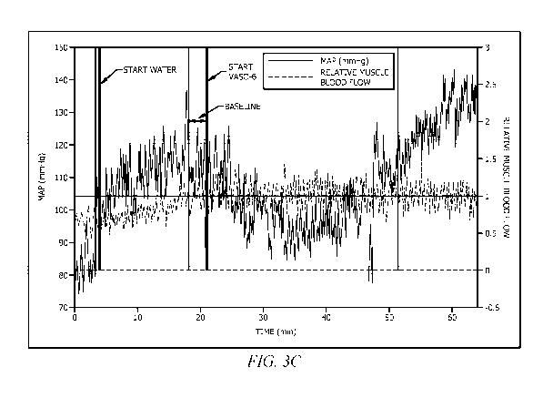
showing the start of water, start of VAS0-61'm (GEO),
FIG. 4Adepicts data showing nitric oxide production.
FIG. 4B depicts data showing nitric oxide production.
FIG. 4C depicts data showing nitric oxide production.
FIG. 5A is a plurality of L-arginine images.
FIG. 5B is a plurality of L-citrulline DL-malate 2:1 images.
FIG. 50 is a plurality of NITROSIGINE/ASI-bonded arginine silicate images.
FIG. 5D is a plurality of images of the current formulation/extract.
FIG. 6A depicts increase in nitric oxide production in RAW264.7 cells.
FIG. 6B depicts production of nitric oxide by RAW cells with increasing doses
of the current
formulation.
FIG. 7 depicts a graph showing the relative changes in blood flow and blood
pressure in
subjects using just energy drink in phase 1, and energy drink plus VASO-6 in
phase 2.
FIG. 8 depicts a graph showing the relative changes in blood flow and blood
pressure in
subjects using just energy drink in phase 1, and energy drink plus VASO-6 in
phase 2.
FIG. 9 depicts a graph showing the relative changes in blood flow in subjects
using just energy
drink in phase 1, and energy drink plus VASO-6 in phase 2 and a graph showing
relative
changes in blood pressure in subjects using just energy drink in phase 1, and
energy drink plus
VASO-6 in phase 2,
FIG. 10 depicts a graph showing the relative changes in blood flow in subjects
using just energy
drink in phase 1, and energy drink plus VASO-6 in phase 2 and a graph showing
relative
8
CA 03093912 2020-09-14
WO 2019/178378 PCT/US2019/022303
changes in blood pressure in subjects using just energy drink in phase 1, and
energy drink plus
VASO-6 in phase 2.
FIG. 11 depicts a graph showing the difference in brachial artery blood flow
between placebo
and VASO-6TM.
FIG. 12 depicts graphs showing changes in blood flow and blood pressure when a
subject is
given VASO-6 Tm.
FIG. 13 depicts skeletal muscle pump, showing the contraction of skeletal
muscles surrounding
a vein compresses the blood and increases the pressure in that area. This
action forces blood
closer to the heart where venous pressure is lower. The importance of the one-
way valves can
be seen to ensure blood flows in the proper direction.
SUMMARY
This summary is provided to introduce a selection of concepts in a simplified
form. These
concepts are described in further detail in the detailed description of
example embodiments of
the disclosure below. This summary is not intended to identify key features or
essential features
of the claimed subject matter, nor is it intended to be used to limit the
scope of the claimed
-- subject matter.
Embodiments disclosed herein include a composition to treat endovascular
dysfunction, the
composition comprising: a procyanidin having a preponderance of (-)-
epicatechins from
materials that contain polyphenols, catechins, epicatechins, and galloylated
epicatechins;
wherein the procyanidin is galloylated; wherein the epicatechins include
between two (2) and
five (5) monomers; wherein the epicatechins include isolated epicatechin-(4-8)-
epicatechin-(4-
8)-epicatechin-gallate (C1 -gallate); and a pharmaceutically acceptable
excipient or carrier.
In additional embodiments, the composition further comprising one or more of
inositol-stabilized
arginine, inositol-stabilized arginine silicate, AS1, L-arginine AKG, L-
citrulline, L-citrulline
malate, arginine HCL, sodium bicarbonate, vitamin C, ascorbic acid, sucrose,
aspailate,
magnesium, saccharomyces cerevisiae, valeriana officinalis root, alcohol, CBD
(medical and
recreational), THC (medical and recreational), acetaminophen,
dextromethorphan, doxylamine,
phenylephrine, ibuprofen, naproxen, Melissa officinal's, zinc, galphimia
glauca, luffa operculate,
sabadilla, zincum aceticum, zincum gluconicum, dioscorea pseudojaponica,
passionflower
extract,l-theanine, sceletium tortuosum, melatonin, diphenhydramine, citrus-
based extracts, or
agmatine sulfate; wherein the composition has enhanced bio-availability,
effectiveness, and
potency.
In additional embodiments, the composition wherein the procyanidin is obtained
from raw
materials; wherein the raw material is selected from a group consisting of:
green tea leaves,
apples (peel on), apricots, pecans, pistachios, almonds and hazelnuts,
cherries, peaches,
9
CA 03093912 2020-09-14
WO 2019/178378 PCT/US2019/022303
blackberries, black grapes, strawberries, concord grapes, red grapes, cocoa
beans, plums
(black diamond raw with peel on), pears, Oolong tea, milk chocolate, fava
beans, dark
chocolate, cherries, cacao beans, broadbeans (immature seeds), black tea,
peanut skins,
grape vine, blueberries and raspberries.
In further embodiments a method of treating an endovascular dysfunction,
comprising:
administering a composition to a subject, wherein the composition comprises: a
procyanidin
having a preponderance of (-)-epicatechins from materials that contain
polyphenols, catechins,
epicatechins, and galloylated epicatechins; wherein the procyanidin is
galloylated; wherein the
epicatechins include between two (2) and five (5) monomers; wherein the
epicatechins include
isolated epicatechin-(4-8)-epicatechin-(4-8)-epicatechin-gallate (C1-
gallate); a
pharmaceutically acceptable excipient or carrier; wherein the composition
comprises a
therapeutically effect amount of the galloylated procyanidins having a
preponderance of (-)-
epicatechins.
In additional embodiments, the method wherein the therapeutically effective
amount of the
galloylated procyanidins is greater than about 0.4 pM.
In additional embodiments, the method wherein the therapeutically effective
amount of the
galloylated procyanidins is 0.76 pM.
In additional embodiments, the method wherein a concentration of the
therapeutically effective
amount of the galloylated procyanidins is about 5%-45% by weight.
In additional embodiments, the method wherein the method further comprises
upregulating a
.. canonical pathway in the subject wherein the canonical pathway is selected
from the group
consisting of: Actin Cytoskeleton Signaling; 0D28 Signaling in T Helper Cells;
Chemokine
Signaling; CREB Signaling in Neurons; CXCR4 Signaling; Ephrin Receptor
Signaling;
ERK/MAPK Signaling; Fcy Receptor-mediated Phagocytosis in Macrophages and
Monocytes;
fMLP Signaling in Neutrophils; GNRH Signaling; GP6 Signaling Pathway; Ga12/13
Signaling;
Gag Signaling; Gas Signaling; IL-6 Signaling; IL-8 Signaling; Insulin Receptor
Signaling;
lntegrin Signaling; Melatonin Signaling; Nitric Oxide Signaling in the
Cardiovascular System;
Noradrenaline and Adrenaline Degradation; NRF2-mediated Oxidative Stress
Response;
Oncostatin M Signaling; Oxidative Phosphorylation; P2Y Purigenic Receptor
Signaling
Pathway; p7056K Signaling; PAK Signaling; Phospholipase C Signaling; PI3K
Signaling in B
Lymphocytes; PI3K/AKT Signaling; Production of Nitric Oxide and Reactive
Oxygen Species in
Macrophages; Protein Kinase A Signaling; Rac Signaling; RANK Signaling in
Osteoclasts;
Regulation of Actin-based Motility by Rho; RhoA Signaling; Signaling by Rho
Family GTPases;
Synaptic Long Term Potentiation; Telornerase Signaling; and a-Adrenergic
Signaling.
In additional embodiments, the method wherein administration to the subject
causes an
increase in intracellular nitric oxide production.
CA 03093912 2020-09-14
WO 2019/178378 PCT/US2019/022303
In additional embodiments, the method wherein administration to the subject
causes increased
blood flow, increased blood oxygenation, lower blood pressure, increased
cognizance, dose-
specific increase in nitric oxide production, dose-specific increase in
vasodilation, reduced fat,
increased muscle stamina, increased blood flow to muscles, increased blood
flow to brain,
decreased exercise/workout recovery time, increased exercise efficiency,
increased alertness
(e.g., aiding in treatment of narcolepsy, attention deficit disorder, chronic
fatigue syndrome,
depression, Addison's disease, or sleep deprivation), pre-performance/workout
treatment for
stimulation of workout vigor (mental and physical) and enhanced performance,
post-
performance/workout supplement for muscle recovery, male/female virility
enhancement,
increased metabolic rate, increased workout volume, reduced feeling of effort
during exercise,
increased motivation to exercise, as drug or supplement delivery mechanism, as
a nutrient
delivery mechanism, oxygenated blood delivery, as a prevention and/or
treatment of endothelial
dysfunction, reduced stress and anxiety, as a sleep aid, reduced hangover
after alcohol
consumption, increased energy, enhanced heart health, enhanced respiratory
efficiency,
increased angiogenesis, as treatment for wound closure, enhanced food and
beverage
flavoring, improved skin and hair/coat in non-humans, improved skin and hair
in humans,
enhanced matrix metalloproteinases proliferation, and as a general aid in
animal health and
wellness in the subject.
In alternative embodiments, a method of extracting or isolating galloylated
procyanidins having
a preponderance of (-)-epicatechins from a raw material, the method
comprising: selecting the
raw material that contains polyphenols, catechins, epicatechins, and
galloylated epicatechins;
extracting from the raw material polyphenols, catechins, epicatechins, and
galloylated
epicatechins (collectively the unrefined material) from the sample using hot
water at a
temperature of about 80 C to about 85 C; passing the unrefined material
through a mesh filter;
absorbing the filtered material with a macro-porous absorption resin: eluting
impurities from the
absorbed, filtered material using pure water; eluting the material in ethanol
and collecting an
ethanol eluent fraction therefrom; concentrating the ethanol eluent fraction
and recovering a
solvent using a vacuum system; pasteurizing, sterilizing, and quickly cooling
the resulting
material; spraying drying the material into a powder; sifting and v-blending
the powder to even
quality of each lot.
In additional embodiments, the method wherein the raw material is selected
from a group
consisting of: green tea leaves. apples (peel on), apricots, pecans,
pistachios, almonds and
hazelnuts, cherries, peaches, blackberries, black grapes, strawberries,
concord grapes, red
grapes, cocoa beans, plums (black diamond raw with peel on). pears, Oolong
tea, milk
chocolate, fava beans, dark chocolate, cherries, cacao beans, broadbeans
(immature seeds),
black tea, peanut skins, grape vine, blueberries and raspberries.
11
CA 03093912 2020-09-14
WO 2019/178378 PCT/US2019/022303
In additional embodiments, the method wherein the epicatechins include between
about two
(2) and about live (5) monomers.
In additional embodiments, the method wherein the epicatechins include
isolated epicatechin-
(4-8)-epicatechin-(4-8)-epicatechin-gallate (C1-gallate).
In further embodiments, a method of extracting or isolating galloylated
procyanidins having a
preponderance of (-)-epicatechins from a sample, comprising: initially
extracting polyphenols,
catechins, epicatechins, and galloylated epicatechins from the sample using
ethyl acetate;
further extracting the polyphenols, catechins, epicatechins, and galloylated
epicatechins from
the sample using water; eluting the resulting material using resin and
diluting the material with
ethanol; filtering the material using activated carbon; concentrating the
material; spraying drying
the material into a powder; v-blending, sieving, and de-ironing the powder,
DETAILED DESCRIPTION OF THE PREFERRED EMBODIMENT
In the following detailed description, reference is made to the accompanying
drawings, which
form a part thereof, and within which are shown by way of illustration
specific embodiments by
which the invention may be practiced. It is to be understood that other
embodiments may be
utilized, and structural changes may be made without departing from the scope
of the present
application. These embodiments are described in sufficient detail to enable
those of ordinary
skill in the art to practice the present disclosure, and it is to be
understood that other
embodiments may be utilized, and that structural, logical, and electrical
changes may be made
within the scope of the disclosure.
From the following descriptions, it should be understood that components of
the embodiments
as generally described and illustrated in the figures herein could be arranged
and designed in
a wide variety of different configurations. Thus, the following more detailed
description of
various embodiments, as represented in the figures, is not intended to limit
the scope of the
disclosure but is merely representative of various embodiments. While the
various aspects of
the embodiments are presented in drawings, the drawings are not necessarily
drawn to scale
unless specifically indicated.
The following description provides specific details, such as material types,
compositions,
material thicknesses, and processing conditions in order to provide a thorough
description of
embodiments of the disclosure. However, a person of ordinary skill in the art
will understand
that the embodiments of the disclosure may be practiced without employing
these specific
details. Indeed, the embodiments of the disclosure may be practiced in
conjunction with
conventional techniques employed in the industry. Only those process acts and
structures
necessary to understand the embodiments of the disclosure are described in
detail below. A
person of ordinary skill in the art will understand that some process
components are inherently
disclosed herein and that adding various conventional process components and
acts would be
12
CA 03093912 2020-09-14
WO 2019/178378 PCT/US2019/022303
in accord with the disclosure, In this description, specific implementations
are shown and
described only as examples and should not be construed as the only way to
implement the
present disclosure unless specified otherwise herein.
Illustrations presented herein are not meant to be actual views of any
particular material,
component, or system, but are merely idealized representations that are
employed to describe
embodiments of the disclosure. Referring in general to the following
description and
accompanying drawings, various embodiments of the present disclosure are
illustrated to show
its structure and method of operation. Common elements of the illustrated
embodiments may
be designated with similar reference numerals. It should be understood that
the figures
presented are not meant to be illustrative of actual views of any particular
portion of the actual
-- structure or method but are merely idealized representations employed to
more clearly and fully
depict the present invention defined by the claims below.
It should be understood that any reference to an element herein using a
designation such as
"first," "second," and so forth does not limit the quantity or order of those
elements, unless such
limitation is explicitly stated. Rather, these designations may be used herein
as a convenient
method of distinguishing between two or more elements or instances of an
element. Thus, a
reference to first and second elements does not mean that only two elements
may be employed
there or that the first element must precede the second element in some
manner. Also, unless
stated otherwise a set of elements may comprise one or more elements.
Any headings used herein should not be considered to limit the scope of
embodiments of the
invention as defined by the claims below and their legal equivalents. Concepts
described in any
specific heading are generally applicable in other sections throughout the
entire specification.
As used in this specification and the appended claims, the singular forms "a",
"an", and "the"
include plural referents unless the content clearly dictates otherwise. As
used in this
specification and the appended claims, the term "or" is generally employed in
its sense including
-- "and/or" unless the context clearly dictates otherwise.
As used herein, "about" means approximately or nearly and in the context of a
numerical value
or range set forth means - 15% of the numerical. In an embodiment, the term
"about" can
include traditional rounding according to significant figures of the numerical
value. In addition,
the phrase "about 'x' to 'y" includes "about 'x' to about 'y'",
It should be noted that ratios, concentrations, amounts, and other numerical
data may be
expressed herein in a range format. It is to be understood that such a range
format is used for
convenience and brevity, and thus, should be interpreted in a flexible manner
to include not
only the numerical values explicitly recited as the limits of the range, but
also to include all the
individual numerical values or sub-ranges encompassed within that range as if
each numerical
value and sub-range is explicitly recited. To illustrate, a concentration
range of "about 0,1% to
13
CA 03093912 2020-09-14
WO 2019/178378 PCT/US2019/022303
about 59/0" should be interpreted to include not only the explicitly recited
concentration of about
0.1 wt% to about 5 wt%, but also include individual concentrations (e.g,, 1%,
2%, 3%, and 4%)
and the sub-ranges (e.g., 0,5%, 1.1%, 2.2%, 3.3%, and 4.4%) within the
indicated range.
As used herein, "treat", "treatment", "treating", and the like refer to acting
upon a condition (e.g.,
vasoconstriction or ineffective blood vessels) with an agent (e.g.,
galloylated procyanidins) to
affect the condition by improving or altering it. The improvement or
alteration may include an
improvement in symptoms or an alteration in the physiologic pathways
associated with the
condition. The aforementioned terms cover one or more treatments of a
condition in a patient
(e.g., a mammal, typically a human or non-human animal of veterinary
interest), and includes:
(a) reducing the risk of occurrence of the condition in a subject determined
to be predisposed
to the condition but not yet diagnosed, (b) impeding the development of the
condition, and/or
(c) relieving the condition, e.g., causing regression of the condition and/or
relieving one or more
condition symptoms (e.g., vasodilation or increased nitric oxide production).
As used herein, the terms "prophylactically treat" or "prophylactically
treating" refers to
completely or partially preventing (e.g., about 50% or more, about 60% or
more, about 70% or
more, about 80% or more, about 90% or more, about 95% or more, or about 99% or
more) a
condition or symptom thereof and/or may be therapeutic in terms of a partial
or complete cure
or alleviation for a condition and/or adverse effect attributable to the
condition.
A "pharmaceutically acceptable excipient," "pharmaceutically acceptable
diluent,"
"pharmaceutically acceptable carrier," or "pharmaceutically acceptable
adjuvant" means an
excipient, diluent, carrier, and/or adjuvant that are useful in preparing a
pharmaceutical
composition that are generally safe, non-toxic and neither biologically nor
otherwise
undesirable, and include an excipient, diluent, carrier, and adjuvant that are
acceptable for
veterinary use and/or human pharmaceutical use. "A pharmaceutically acceptable
excipient,
diluent, carrier and/or adjuvant" as used in the specification and claims
includes one or more
such excipients, diluents, carriers, and adjuvants.
The term "therapeutically effective amount" as used herein describes
concentrations or
amounts of components such as agents which are effective for producing an
intended result,
including increased intracellular nitric oxide production. Compositions
according to the present
invention may be used to effect a favorable change in nitric oxide levels,
whether that change
is an improvement, relieving to some extent one or more of the symptoms of the
condition being
treated, and/or that amount that will prevent, to some extent, one or more of
the symptoms of
the condition that the host being treated has or is at risk of developing, or
a complete cure of
the disease or condition treated.
The term "administration" or "administering" is used throughout the
specification to describe the
process by which a composition comprising a galloylated epicatechin as an
active agent, are
14
CA 03093912 2020-09-14
WO 2019/178378 PCT/US2019/022303
delivered to a patient or individual for therapeutic purposes. The composition
of the subject
invention and methodology in use thereof can be administered a number of ways
including, but
not limited to, parenteral (such term referring to intravenous and intra-
arterial as well as other
appropriate parenteral routes), subcutaneous, peritoneal, inhalation, vaginal,
rectal, nasal, or
instillation into body compartments.
Administration will often depend upon the amount of compound administered, the
number of
doses, and duration of treatment. In an embodiment, multiple doses of the
agent are
administered. The frequency of administration of the agent can vary depending
on any of a
variety of factors, such as nitric oxide levels, and the like. The duration of
administration of the
agent, e.g., the period of time over which the agent is administered, can
vary, depending on
any of a variety of factors, including patient response, etc.
The amount of the agent contacted (e.g., administered) can vary according to
factors such as
the degree of susceptibility of the individual, the age, sex, and weight of
the individual,
idiosyncratic responses of the individual, the dosimetry, and the like.
Detectably effective
amounts of the agent of the present disclosure can also vary according to
instrument and film-
related factors. Optimization of such factors is well within the level of
skill in the art, unless
otherwise noted.
As used herein, the term "subject," "patient," or "organism" includes humans
and mammals
(e.g., mice, rats, pigs, cats, dogs, and horses). Typical hosts to which an
agent(s) of the present
disclosure may be administered will be mammals, particularly primates,
especially humans. For
veterinary applications, a wide variety of subjects will be suitable, e.g,,
livestock such as cattle,
sheep, goats, cows, swine, and the like; poultry such as chickens, ducks,
geese, turkeys, and
the like; and domesticated animals particularly pets such as dogs and cats.
For diagnostic or
research applications, a wide variety of mammals will be suitable subjects,
including rodents
(e.g., mice, rats, hamsters), rabbits, primates, and swine such as inbred pigs
and the like.
The phrases "connected to" and "coupled to" refer to any form of interaction
between two or
more entities, including mechanical, electrical, magnetic, electromagnetic,
fluid, and thermal
interaction. Two components may be connected or coupled to each other even
though they are
not in direct contact with each other. For example, two components may be
coupled to each
other through an intermediate component.
The use of "including," "comprising," or "having," "containing," "involving,"
and variations thereof
herein, is meant to encompass the items listed thereafter and equivalents
thereof as well as
any additional items a person of ordinary skill in the art would reasonably
understand to be
included.
Referring in general to the following description and accompanying drawings,
various
embodiments of the present disclosure are illustrated to show its structure
and method of
CA 03093912 2020-09-14
WO 2019/178378 PCT/US2019/022303
operation. Common elements of the illustrated embodiments may be designated
with similar
reference numerals. Accordingly, the relevant descriptions of such features
apply equally to the
features and related components among all the drawings. Any suitable
combination of the
features, and variations of the same, described with components illustrated in
Figure 1, can be
employed with the components of Figure 2, and vice versa. This pattern of
disclosure applies
equally to further embodiments depicted in subsequent figures and described
hereinafter. It
should be understood that the figures presented are not meant to be
illustrative of actual views
of any particular portion of the actual structure or method but are merely
idealized
representations employed to more clearly and fully depict the present
invention defined by the
claims below.
All referenced publications are incorporated herein by reference in their
entirety. Furthermore,
where a definition or use of a term in a reference, which is incorporated by
reference herein, is
inconsistent or contrary to the definition of that term provided herein, the
definition of that term
provided herein applies and the definition of that term in the reference does
not apply.
In certain embodiments, the current invention is formulations and associated
methods and
therapies for humans and other animals, in the treatment of small vessel
disease, high blood
pressure, endothelial dysfunction, and other diseases/co-morbitities
associated with small
vessel disease or with blood vessels that are no longer effective. The
formulations include
procyanidins having a preponderance of (-)-epicatechins, wherein the
procyanidins are
preferably galloylated and administered to a patient or subject in need.
Flavonoids are known
for their healthy effects and limited toxicity. The flavanol (-)-epicatechin
(Bpi) enhances exercise
capacity in mice and Bpi-rich cocoa improves skeletal muscle structure in
heart failure patients.
(-)-Epicatechin decreases myostatin and 13-galactosidase and increases levels
of markers of
muscle growth. In humans, myostatin and 13-galactosidase increase with aging
while follistatin,
MyoD and myogenin decrease. To achieve both bioavailability and potency, it is
also
contemplated that the number of epicatechins monomers forming each procyanidin
is between
two (2) and five (5). More specifically, isolated epicatechin-(4-8)-
epicatechin-(4-8)-epicatechin-
gallate (C1-gallate) is administered to the patient. Through this isolation,
formulations were
developed to maximize the large molecules responsible for 50% vasodilation and
small
molecules responsible for 15% vasodilation.
In certain embodiments, the current invention comprises a formulation, in a
kit, including a
gallate enhanced oligomer paired with one or more of inositol-stabilized
arginine, inositol-
stabilized arginine silicate, AS1, L-arginine AKG, L-citrulline, L-citrulline
malate, arginine HCL,
sodium bicarbonate, vitamin C, ascorbic acid, sucrose, aspartate, magnesium,
saccharomyces
cerevisiae, valeriana officinalis root, alcohol. CBD (medical and
recreational). THC (medical
and recreational), acetaminophen, dextromethorphan, doxylamine, phenylephrine,
ibuprofen,
naproxen, Melissa officinalis, zinc, galphimia glauca, luffa operculate,
sabadilla, zincum
16
CA 03093912 2020-09-14
WO 2019/178378 PCT/US2019/022303
aceticum, zincum gluconicum, dioscorea pseudojaponica, passionflower extract,
1-theanine,
sceletium tortuosum, melatonin, diphenhydramine, citrus-based extracts, and/or
agmatine
sulfate to boost the bio-availability, effectiveness, and potency. In other
embodiments, the
current invention is a pharmaceutical compound of the dose-specific
formulation and
combination of the ingredients listed above.
Effects or uses of embodiments of the current invention include, but are not
limited to, increased
blood flow, increased blood oxygenation, lower blood pressure, increased
cognizance, dose-
specific increase in nitric oxide production, dose-specific increase in
vasodilation, reduced fat,
increased muscle stamina, increased blood flow to muscles, increased blood
flow to brain,
decreased exercise/workout recovery time, increased exercise efficiency,
increased alertness
(e.g., aiding in treatment of narcolepsy, attention deficit disorder, chronic
fatigue syndrome,
depression, Addison's disease, or sleep deprivation), pre-performance/workout
treatment for
stimulation of workout vigor (mental and physical) and enhanced performance,
post-
performance/workout supplement for muscle recovery, male/female virility
enhancement,
increased metabolic rate, increased workout volume, reduced feeling of effort
during exercise,
increased motivation to exercise, as drug or supplement delivery mechanism, as
a nutrient
delivery mechanism, oxygenated blood delivery, as a prevention and/or
treatment of endothelial
dysfunction, reduced stress and anxiety, as a sleep aid, reduced hangover
after alcohol
consumption, increased energy, enhanced heart health, enhanced respiratory
efficiency,
increased angiogenesis, as treatment for wound closure, enhanced food and
beverage
flavoring, improved skin and hair/coat in non-humans, improved skin and hair
in humans,
enhanced matrix metalloproteinases proliferation, and as a general aid in
animal health and
wellness.
Example 1
In an embodiment, the current invention is a method of manufacture of a
formulation including
an effective amount of galloylated procyanidins having a preponderance of (-)-
epicatechins.
The method includes first selecting raw material that contains polyphenols,
catechins,
epicatechins, and galloylated epicatechins. Examples of such raw materials
include, but are
not limited to, green tea leaves (Camellia sinensis), apples (peel on),
apricots, pecans,
pistachios, almonds and hazelnuts, cherries, peaches, blackberries, black
grapes,
strawberries, concord grapes, red grapes, cocoa beans, plums (black diamond
raw with peel
on), pears, Oolong tea (Camellia sinensis), milk chocolate, fava beans, dark
chocolate,
cherries, cacao beans, broadbeans (immature seeds), black tea (Camellia
sinensis), peanut
skins, grape vine, blueberries and raspberries. Hot water, ranging from -80-85
C, is used as
an extraction method for the polyphenols, catechins, epicatechins, and
galloylated
epicatechins. The unrefined material is then run through a 200-mesh filter,
and the residue is
discarded. The filtered material is then absorbed with a macro-porous
absorption resin.
17
CA 03093912 2020-09-14
WO 2019/178378 PCT/US2019/022303
After the filtered material is absorbed, the impurities of the filtered,
absorbed material are eluted
using pure water. After elution using pure water, the material is then eluted
in 25% ethanol to
remove caffeine and some simple catechins. Thereafter, the active ingredients
are eluted using
80% ethanol, and the 80% ethanol eluent fraction is collected. The material is
concentrated,
and the solvent is recovered using a vacuum system. The material is then
pasteurized,
sterilized, and cooled down quickly, followed by being spray dried into a
powder. Finally, the
powder is sifted and v-blended to even quality of each lot.
Using the foregoing steps, a composition was generated and was tested using
liquid
chromatography-tandem mass spectrometry to identify analytes/oligomers and
quantify
concentrations of each analyte/oligomer. Results can be seen in Table 1.
Table 1. Oligomer/Analyte identification and concentrations.
Analyte
Concentration Dilution Sample
Analyte
Analyte
(ng/m14 Factor Concentration(mg/mL) (%)
Catechin Dimer G 56.8 100 0.0648
0.0876
Catechin Trimer G 6.47 5 1.30
0.000500
ECGC Dimer-1 4030 100 0.0648 6.22
Catechin Trimer G 12.3 100 0.0648
0.0190
ECGC Dimer-2 24.1 5 1.30
0.00186
Catechin Tetramer 6.50 5 1.30
0.000500
Catechin Dimer OG 93.5 100 0.0648 0.144
ECG Dimer 13200 100 0.0648 20.3
Catechin Dimer 4.93 100 0.0648
0.00760
Catechin 1100 5 1.30
0.0845
.. Example 2
In an embodiment, the current invention is an alternative method of
manufacture of the current
formulation. The method includes first selecting raw material that contains
polyphenols,
catechins, epicatechins, and galloylated epicatechins. Examples of such raw
materials include,
but are not limited to, green tea leaves, apples (peel on), apricots, pecans,
pistachios, almonds
and hazelnuts, cherries, peaches, blackberries, black grapes, strawberries,
concord grapes,
red grapes, cocoa beans, plums (black diamond raw with peel on), pears, Oolong
tea, milk
chocolate, fava beans, dark chocolate, cherries, cacao beans, broadbeans
(immature seeds),
18
CA 03093912 2020-09-14
WO 2019/178378 PCT/US2019/022303
black tea, peanut skins, grape vine, blueberries and raspberries. Ethyl
acetate is used as an
extraction method for the polyphenols, catechins, epicatechins, and
galloylated epicatechins.
A second extraction process, via water, is used to further extract the
polyphenols, catechins,
epicatechins, and galloylated epicatechins. This material is then recovered.
A separation step is performed by resin and is diluted using ethanol. This
process is repeated
twice, and the material is recovered. This material is filtered using
activated carbon, and the
filtered material is concentrated at this phase. The material is then spray
dried into a powder.
Finally, the powder is v-blended, where sieving and de-ironing takes place.
Concord Grape pumace and seed extracts provide various yield ratios based on
the extract
type is outlined in Table 2.
Table 2. Concord Grape Pumace & Seed Extracts
Serial No. Product & Spec. Batch No. Qty. Yield Ratio
Remarks
Concord Grape Seed
Extract
Most suitable for
SF-CGS001 (Type 1) 181006 10 g/bag Seeds 30:1
production
Concord Grape Seed Highest
purity,
Extract impractical
on
SF-CGS002 (Type 2) 181008 1 g/bag Seeds 200:1
actual production
Concord Grape Seed
Extract
SF-CGS003 (Type 3) 181010 15 g/bag Seeds residues
Concord Grape
Pumace Extract 45:1
SF-CGP001 (Type 1) 181011 10 g/bag (Pumace +seeds)
Concord Grape
Pumace Extract residues
SF-CGP002 (Type 2) 181012 5 g/bag (Pumace+seeds)
Concord GSE Type 1, analysis method GL-816 has provided the following resulls:
Catechin
Dimer Gallate: 668 ng/mL; Catechin Trimer Gallate 879 ng/mL; EGCG Dimer-1 ND;
Catechin
Trimer 337 ng/ML; EGCG Dimer-2 0.302 ng/mL; Catechin Tetramer 1.10 ng/mL;
Catechin
Dimer Digallate 2.18 ng/mL; ECG Dimer 3.38 ng/mL; Catechin Dimer 66 ng/mL;
Catechin 6260
ng/mL.
Concord Pumace and Seeds, analysis method GL-816 has provided the following
results:
Catechin Dimer Gallate: 54.9 ng/mL; Catechin Trimer Gallate 3.48 ng/mL; EGCG
Dimer-1 ND;
Catechin Trimer 230 ng/ML; EGCG Dimer-2 0.278 ng/mL; Catechin Tetramer 0.919
ng/mL;
Catechin Dimer Digallate 1.69 ng/mL; ECG Dimer 0.967 ng/mL; Catechin Dimer
54.9 ng/mL;
Catechin 7700 ng/mL.
19
CA 03093912 2020-09-14
WO 2019/178378 PCT/US2019/022303
Using the foregoing steps, a composition was generated and was tested using
liquid
chromatography-tandem mass spectrometry to identify analytes/oligomers and
quantify
concentrations of each analyte/oligomer. Results can be seen in Table 3.
Table 3. Oligomer/Analyte identification and concentrations.
Oligomer Oligomer Dilution Sample
Oligomer
Oligomers Concentration Concentration Factor Concentration (%)
(ngtmL) (ug/mL) (mg/mL)
Catechin Dimer 24.1 0.0241 100 0.0611
0.0395
Gallate
Catechin Trimer 9.46 0.00950 5 1.22
0.000770
Gallate
Epiga llocatech in 910 0.910 100 0.0611
1.49
gallate Dimer 1
Catechin Trimer 16.5 0.0165 100 0.0611
0.0270
Epiga llocatech in 93.1 0.0931 5 1.22
0.00762
gallate Dimer 2
Catechin Tetramer 22.7 0.0227 5 1.22
0.00186
Catechin Dimer 158 0.158 100 0.0611
0.259
Digallate
Epicatechin gallate 16,800 16.8 100
0.0611 27.4
Dimer
Catechin Dimer 0.510 0.000500 100 0.0611
0.000830
Catechin 24.1 0.0241 5 1.22
0.243
Example 3
The method of Example 1 or Example 2 was performed to generate an extract
including the
components discussed above, where this extract was studied for efficacy.
Ultimately, the in
vitro study herein compares the efficacy of certain substances, along with the
developed
formulation/extract containing the mixture of polyphenolic compounds, in the
induction of
intracellular nitric oxide production. These substances are frequently found
in sports
performance foods and beverages, and include arginine silicate inositol
complex (ASO, L-
arginine, and L-citrulline-DL-malate (2:1).
CA 03093912 2020-09-14
WO 2019/178378 PCT/US2019/022303
Methods
Drug Preparation. Allornetric scaling and the general equation of Body
Surfaced Area
Normalization Method was used to calculate an in vitro 7-dose, based upon the
generally
accepted human oral dose of these nutritional supplements. The current
formulation, L-arginine
(COMPOUND SOLUTIONS) and L-citrulline DL-rnalate 2:1 (COMPOUND SOLUTIONS) were
each prepared at 300 mg/m1 in DMEM without phenol red (CORNING) and stored at -
20 C until
use, NITROSIGINE (AS1; bonded arginine silicate; arginine silicate inositol
complex;
COMPOUND SOLUTIONS), and lipopolysaccharides from E. coil 0111: B4 (LPS; SIGMA
ALDRICH) were dissolved in DMSO to 300 mg/mland 50 pg/ml, respectively, and
stored frozen
until use. 4,5-Diaminolluorescein Diacetate (DAF-FM) was diluted from 5mM
stocks in DMSO,
Cell Culture. RAW264.7 mouse cells (ATCC) were grown at 37 C in 5% CO2 in DMEM
lacking
phenol red, supplemented with glucose, pyruvate and L-glutamine and 10% fetal
bovine serum
(GIBCO; FISHER SCIENTIFIC, LOT# 1931538). All experiments were completed with
cultures
under 8 passages, and cell densities were maintained between 0.2 x 10⁶ and 0.8
x 105 cells
per ml during maintenance. For sub-culturing, the monolayer was washed twice
with HEPES
buffered saline (HBS; 140 mM NaCI, 1.5 mM Na2HPO4.2H20, 50 mM HEPES, pH 7.2),
and
then incubated for 2 min in 0.25% Trypsin-EDTA (THERMOFISHER SCIENTIFIC).
Cells were triturated with complete growth medium. Density was determined
under phase-
contrast using 0.2% Trypan blue. Three viable cell counts were performed on
the
hernocytometer and averaged. Allometric scaling and the general equation of
Body Surface
Area Normalization method [J Basic Clin Pharrn. March 2016-May 2016; 7(2): 27-
31. doi:
10.4103/0976-0105.177703 PMC ID: PMC4804402 A simple practice guide for dose
conversion
between animals and human Anroop B. Nair and Shery Jacobi] were used to
calculate an in
vitro dose based upon the generally accepted human oral dose of these
nutritional
supplements.
Nitric Oxide Assay. Nitric oxide levels induced by the various test agents
were determined using
a free radical-sensing fluorescent dye 4,5-diaminofluorescein diacetate (DAF-
FM;
THERMOFISHER). DAF-FM diacetate is essentially non-fluorescent until it reacts
with nitric
oxide to form a fluorescent benzotriazole. DAF-FM diacetate is cell-permeant
and passively
diffuses across cellular membranes. Once inside cells, it is cleacetylated by
intracellular
esterases to become DAF-FM. Although there has been less published evidence of
use of this
dye than traditional methods such as the Griess method, this quantification
reagent DAF-FM
has exhibited extreme sensitivity to nitric oxide insofar as being able to
detect individual NO-
producing neurons in brain slices.
Here, cells (50,000) in 3 ml of complete growth medium were plated onto 35-mm
glass bottom
dishes (MATTEK) pre-coated with poly-D-lysine. Cells were grown for 24 h and
washed twice
21
CA 03093912 2020-09-14
WO 2019/178378 PCT/US2019/022303
in HBS. Three (3) ml of serum-free medium was added. Cells were then treated
with 1 pM
lipopolysaccharide or various concentrations of test agents and grown for an
additional 30 min.
DAF-FM was added to cells to a final concentration of 2 pM and incubated for
an additional 30
min.
For confocal microscopy, medium was removed, and cells were washed once in HBS
and
replaced with 3 ml Live Cell Imaging solution (INVITROGEN). Cells were
immediately imaged
on a PERKIN ELMER ULTRAVIEW ERS confocal microscopy system. Images represent
400x
final magnification and were taken using a 1500 ms exposure with a 488 nm
Argon-ion laser
and 527 nm emission filter. For cell treatments resulting in little or no
fluorescence, 4',6-
diarnidino-2-phenylindole (DAPI, MOLECULAR PROBES) was added at a final
concentration
of 300 nM to an additional sample. Images were captured and analyzed as tiff
formatted files.
Densitometry was performed using IMAGEJ software (NIH-bundled with 64-bit Java
1.8.0_112). For determining fluorescence, the entire image was analyzed for
each image taken.
Data are representative of 2 independently performed experiments.
Results
Red, green, and blue pixels were converted to brightness values using the
formula
V=(R+G+B)/3. DAPI counter staining is provided for the L-arginine and L-
citrulline-DL-rnalate
2:1 600pg/rd samples to confirm the adherence of cells in light of their
noticeably low mean
fluorescence. At corresponding doses of L-arginine. L-citrulline-DL-rnalate
2:1, and
NITROSIGINEO, the current formulation produced a greater amount of DAF-FM
fluorescence
correlating to an increase in nitric oxide levels against each comparative
ingredient/compound.
At the biologically active dose of 1 ng/rn1 incubated for 30 minutes, LPS
induced a bright and
consistent DAF-FM fluorescence indicating that nitric oxide levels increased,
in this cell line.
This demonstrates the ability for the current formulation/extract to induce
the production of
intracellular nitric oxide in RAW 264.7 mouse macrophage cells. Nitric oxide
is produced in
various mammalian tissues by three classes of nitric oxide synthase enzymes:
endothelium NO
synthase (eNOS), neural NO synthase (nNOS) and inducible NO synthase (iNOS).
It is the
iNOS enzyme that is activated in RAW cells in response to Lipopolysaccharide.
These murine
immune cells provide a static response to an infectious presence by releasing
pro-inflammatory
mediators including nitric oxide. The mediators aid in increasing blood flow
to the site of
infection and this in turn improves the invasion of leukocytes. Although a
component of immune
clearance, compounds that can safely mimic the effects of lipopolysaccharides
are sought after
since increasing blood flow to the tissues, especially over long periods of
time, can increase
endurance and protein anabolism.
Example 4
22
CA 03093912 2020-09-14
WO 2019/178378 PCT/US2019/022303
Gallate enhanced oligomer (GEO) with tradenarne VAS0-6Tm has been examined
using human
endothelial cells. The GEO chemical structure contains (-)epicatechins,
galloylated procyanidin,
epicatechin procyanidin monomers (2-5), isolated epicatechin-(4-8)-epicatechin-
(4-8)-
epicatechin-gallate (C1-gallate), and flavonoid. The GEO has effects on
vasodilatorivasorelaxor-NO production, anti-inflammation, ATP producer, muscle
growth,
angiogenesis, vasculogenesis, multiple protein/genetic controller for cancer
inhibition and
viral/bacterial inhibition.
Proanthocyanidins are a class of oligomeric polyphenol compounds composed
primarily of (+)-
catechin and (¨)-epicatechin molecules; as shown below:
OH
HO 7 8
OH
3
6
OH
5 4
OH
(+)-catechin
401 OH
OH
. ,
OH
(7)-epicatechin
Proanthocyanidins can occur as polymers of up to 50 monomer units.
Procyanidins are a class
of proanthocyanidin that consist exclusively of epicatechin and catechin
molecules (Natural
Products Report 2009, 26:1001-1043). Structural elucidation of
proanthocyanidins, such as
procyanidins, is far from trivial, and requires complex NMR analysis, usually
at low temperature.
However, it is known that catechin/epicatechin units can be linked through a
single carbon-
carbon bond: a 04-08 or a C4-C6 linkage. Alternatively, an additional ether
bond can be
present, i.e. 04-06, 02-0-----07 or 04-C8, 02-0----07. As shown below one
example of a
procyanidin tetramer, joined via 04-08 linkages:
23
CA 03093912 2020-09-14
WO 2019/178378 PCT/US2019/022303
OH
HO 101 0
OH
OH OH
OH
11 HO 0
401 OH
OH OH
OH
HO 0
110
OH
OH OH
OH
HO 0
..."
OH
OH
5 OH
The term "galloylated" is intended to mean that at least one gallic acid
molecule is attached to
the proanthocyanidin molecule. The gallic acid molecule(s) can be attached in
any position.
However, it is commonly found that the at least one gallic acid molecule is
joined to the
(epi)catechin core via an ester linkage to the hydroxyl group at the 3
position. Galloylated
10 proanthocyanidins are frequently found when the proanthocyanidins are
derived from particular
plant sources, including grapes and grape products. An example of a
galloylated epicatechin
molecule is shown below:
011
110
OH
OH 011
0
OH
OH
In one embodiment, the compositions of the present invention may be used to
enable ergogenic
15 .. effects, preferably leading to more sustained athletic performance.
Thus, in one embodiment
the compositions of the invention may be used as ergogenic aids.
In one embodiment, the compositions of the present invention may be used as
prophylactics in
order to prevent or delay the onset of endothelial dysfunction in patients at
risk thereof.
In a further embodiment, the present invention is directed to use of a
composition of the
20 .. invention for the prevention or treatment of endothelial dysfunction.
24
CA 03093912 2020-09-14
WO 2019/178378 PCT/US2019/022303
In a further embodiment, the present invention is directed to use of a
composition of the
invention in the manufacture of a medicament for use in the prevention or
treatment of
endothelial dysfunction.
In a further embodiment, the present invention is directed to a method of
treating endothelial
dysfunction comprising administering to a patient in need thereof, either
simultaneously or
sequentially, at least one procyanidin, preferably wherein the at least the
procyanidin is
galloylated. In the case of simultaneous administration, this may be in the
form of a
pharmaceutical composition of the invention.
In one embodiment, the compositions of the present invention may be used in
preventing or
treating diseases associated with endothelial dysfunction including
arteriosclerosis,
hypertension, pulmonary hypertension, coronary artery disease, chronic heart
failure,
peripheral artery disease, diabetes, chronic renal failure and erectile
dysfunction.
In an embodiment, the process of the present invention further involves the
addition of at least
one pharmaceutically acceptable excipient or carrier. Addition of the
pharmaceutically
acceptable excipient or carrier may occur simultaneously with, or separately
from, the mixing
of the galloylated procyanidin, and in any order.
The present application provides compositions comprising certain polyphenol
compounds that
may be used for the prevention or treatment of endothelial dysfunction. The
dosage regimen
for the compositions of the present invention will, of course, vary depending
upon factors such
as the route of administration, the age, sex, health, medical condition and
weight of the
recipient; the nature and extent of the symptoms; the nature of any concurrent
treatment; the
frequency of treatment; the route of administration and the effect desired. In
particular it is noted
that compositions of the present invention may be formulated for use in
therapy, or for use as
a prophylactic or as an ergogenic aid.
Compositions of this invention may be administered in a single daily dose, or
the total daily
dosage may be administered in divided doses two, three, or four times daily.
In an embodiment of the invention, desired polyphenol compounds are
rnicroencapsulated,
either separately or together, to increase stability, or bioavailability or to
mask taste Preferably,
rnicroencapsulation is carried out using water-in-oil rnicroencapsulation
technology for liquid
formulations (see U.S. Pat, No. 8,685,446 B2) or using a three-component
rnicroencapsulation
mixture of maltodextrin, mesquite gum, and zein, which is spray dried for
solid or powder
formulations (see Food and Bioprocess Technology, 2013, 6: 941-51).
In an embodiment of the invention, the compositions can be in the form of any
pharmaceutically
acceptable formulations such as tablets, capsules, buccal tablets, orally
disintegrating tablets,
oral fast dissolving tablets, dispersible tablet, masticatory, granules, dry
suspension, injection,
CA 03093912 2020-09-14
WO 2019/178378 PCT/US2019/022303
solution, slow-release formulation, controlled-release formulation, rapid-
release formulation,
and transdermal preparations.
In an embodiment of the invention, the compositions can be prepared as part of
a nutraceutical
product, for example as a snack bar or a pre-prepared drink/powdered drink
formula.
In an embodiment of the invention, the compositions of the invention may
advantageously
comprise further components such as vitamins, minerals and/or fibre. Suitable
vitamins and
minerals include, but are not limited to, the B vitamins, vitamin C, folic
acid, calcium, iron,
magnesium, zinc, selenium, niacin, vitamin D. vitamin A, vitamin E, chromium,
copper,
manganese, boron, molybdenum, omega fatty acids and co-enzyme Q10. Mixtures of
such
additional components may be advantageous. For example, in patients with
cognitive
impairment, it may be advantageous to combine compositions of the invention
with a source of
omega 3 fatty acids and vitamin B12. Where the compositions are intended for
use as ergogenic
aids, formulation with a protein source, such as whey powder, may be
desirable.
Table 4 Biolog ical activity of galloylated and non-galloylated and non-
galloylated polyphenols
Biological activity Control Compounda Effectb
Inhibition of fatty acid synthase activity Untreated enzyme GA
=
EC
EGC
EGCG t t t
ECG T t T
Inhibition of rat liver microsomal 5a-reductase Untreated enzyme EGCG
PGG t t t
TDG t t t
T3G t t
TVG T t T
Trolox-equivalent antioxidant activity C, EC EGCG t t
Untreated
Triggering of suicidal erythrocyte death erythrocytes PGG /1% t
Inhibitory effects on LNCaP and DU145 cells Untreated cells
PGG t t t
Induction of apoptosis in HL-60 cells Untreated cells GA t t
DGR t t t
Growth inhibition of MCF-7 and MDA-MB231 cells Untreated cells
PGG t T
Cytotoxicity towards K562 cells Untreated cells PGG
Reduction of pancreatic lipase activity in vitro Untreated
enzyme EGCG t t
GCG t t t
ECG t t
CG t t t
Induction of apoptosis in HTB9 cells Untreated cells 7-GSB t t
Inhibition of VEGF-induced proliferation of HUVEC Untreated cells
ECGC t t
26
CA 03093912 2020-09-14
WO 2019/178378 PCT/US2019/022303
ECG t t
EGC /1µ
EC
Untreated
Activiation of type 1 ryanodine receptor receptor EGCG t t t
ECG t t t
EGC
EC =
Cytotoxicity towards S-G cells (immortalized gum
epithelial cells) Untreated cells EGCG t t t
EGC t t
ECG t t
EC
CG t t t
Inhibition of NA+/K+ ATPase pump activity Untreated pump EGCG t t
ECG 1 1
EGC
EC
Untreated
Inhibition of NA/H exchanger activity exchanger EGCG
ECG t t
EGC t t t
EC t t '1µ
Suppression of fatty acid synthase expression n
MCF-7 cells Untreated cells C
EC
EGC
EGCG t t
Reduction of cell viability in HL-60 cells Untreated cells EGCG t
t
PGG t t
TDG
T3G T3'G
TF
By way of example, the daily dosage of procyanidins in the compositions of the
invention is
from about 100 mg to about 1000 mg, preferably about 25010 about 600 mg. The
daily dosage
of ellagitannins in the compositions of the invention is from about 50 mg up
to about 1000 mg,
preferably about 300 mg to about 600 mg.
Table 5¨ Biological activity of galloylated polyphenols compared to their non-
galloylated parent
molecules
Pchefil Galloyi-
moleculea derivativea Biological activity
Effectb
27
CA 03093912 2020-09-14
WO 2019/178378 PCT/US2019/022303
SB 7-GSB Antioxidant activity
DB1 DGB1 Cytotoxicity towards human cancer cell lines derived
from breast tc
(MDA-MB-231, MDA-MB-435, MCF-7, BT-20); prostate (LNCap,
DU-145);
stomach (SNU-1); duodenum (Hu-Tu-80); colon (HT-29); rectum
(SW-1463); lung (A549); central nervous system (U-87); bone
DB2 DGB2 (U202) and skin (SK-MEL-5) cancer
RST DGR Growth inhibition of HT-29 human colorectal carcinoma
cells
RST DGR DPPIle radical scavenging activity
Inhibition of BxPC-3 cell colony formation (pancreatic
RST DGR adenocarcinoma cells)
Quercetin 3GQ Induction of heme oxygenase 1 in RAW264,7 cells 4,
4'
Taxifolin 7GT
Quercetin 3GQ Cytotoxicity, inhibition of proliferation, migration
and tube formation
Taxifolin 7GT of HUVECo
EGC EGCG
tc
Inhibition of VEGF-induced proliferation migration and tube
EC ECG formation of human umbilical vein endothelial cells
(HUVEC)
SB 3-GSB Cytotoxicity, inhibition of proliferation, migration
and tube formation = e
7-GSB of HUVECo
20-GSB
23-GSB
OHS 3-GDHS Cytotoxicity, inhibition of proliferation, and
migration of HUVECo 4' 4,
7-GDHS t
t
20-GDHS =
23-GDHS
Example 5
Data on human subject blood flow and blood pressure were gathered with
subjects drinking
just an energy drink and an energy drink with VASO8TM. FIGS. 7 and 9 depict
the graph of the
changes in blood flow and blood pressure recorded during the time the subjects
drink the
cocktail including the time after the subjects are done drinking the cocktail.
FIGS. 8 and 10
depict the graph of the changes in blood flow and blood pressure recorded only
with the time
after the subjects are done drinking the cocktail. As can be seen in the
graphs in FIGS. 7-10
the addition of VASO8TM increased the blood flow by about 15% starting
approximately at the
ten (10) minute mark. This increase in blood flow is maintained until the end
of the test. As can
be seen in the graphs in FIGS. 7-10 the addition of VASO6TM does not appear to
change the
blood pressure in the subjects.
While clinical results have shown VASO6TM promotes nitric oxide production and
blood flow,
as well as increased vasodilation, little is known about the cellular or
molecular mechanisms
involved.
28
CA 03093912 2020-09-14
WO 2019/178378 PCT/US2019/022303
To further elucidate the molecular mechanisms affected by VASO6TM treatment,
mas
spectrometry-based proteomics was performed on cultured human aortic
endothelial cells
treated with 2 doses of VASO-6 TM (300 and 600mg) while the control was DMSO
treated.
Human aortic endothelial cells were collected, and protein was solubilized
before being
proteolytically digested with Trypsin to generate peptides for LC-MS/MS
analysis, Peptides
were separated on a 50cm 018 column (Thermo) using an Ultimate 300 UHPLC
system
(Thermo) and analyzed on a hybrid quadrupole-Orbitrap mass spectrometer (Q
Exactive Plus,
Thermo).
Raw data files were searched using MaxQuant (www.maxquantorg) against the
Uniprot Horno
Sapiens reference proteorne database. Ratios were generated by dividing the
intensity of a
given protein in the treatment group by the intensity of the protein in the
control sample.
Significant outliers were determined using the Significance A test in Perseus
(www.coxdocs.org). Proteins that were found to be significant were uploaded
into Ingenuity
Pathways Analysis software for bioinforrnatic analysis.
Over 2300 proteins were identified in each sample, of which 198 showed
significant changes
in abundance after treatment with 300mg VASO6TM, while 181 proteins showed
significant
changes following treatment with 600mg VASO-6 TM.
IPA analysis revealed several canonical pathways, as well as cellular and
molecular and
functions associated with the significantly altered proteins. Significance is
established using a
z-score.
Comparison analysis provides an indication of potential dose-dependent
changes. For
example, joint inflammation is moderately inhibited in the 300mg dose (z-score
-0.655) and is
significantly inhibited with the 600rng dose (z-score -1,982), angiogenesis,
inhibited with 300rng
(z-score -1.021), while it is increased with 600mg VASO-6 (z-score 1.447).
Increased nitric oxide in the blood stream leads to endothelial-dependent
relaxation (EDR),
which aids in the delivery of oxygen to muscles, subsequently increase ATP
production and
nutrient delivery.
The pathway involved in nitric oxide signaling in the cardiovascular system
showed a significant
positive z-score (1,342) with 300mg VASO-6TM treatment, suggesting increased
activity/NO
signaling, which is consisting with clinical findings. The proteins identified
in the pathway:
arginase 2 (Arg2), calmodulin like 5 (CALML5), heat shock protein 90
(Hsp90b1), mitogen-
activated protein kinase kinase 1 (MAP2K1), mitogen-activated protein kinase
kinase 2
(MAP2K2), and mitogen-activated protein kinase 3 (MAPK3).
29
CA 03093912 2020-09-14
WO 2019/178378 PCT/US2019/022303
MAP2K1, MAP2K2, and MAPK3 are involved in numerous pathways involved in the
regulation
of many biological functions. For example, ERK/MAPK signaling was also shown
to have
increased activity, as well as alpha-adrenergic signaling, and actin
cytoskeleton signaling.
Oxidative phosphorylation had significant positive z-score in both 300mg and
600mg doses
(2.236 and 2, respectively). Oxidative phosphorylation is the production of
ATP using energy
derived from the transfer of electrons in the electron transport in the
mitochondria.
Table 6 ¨ Proteins identified in the Oxidative Phosphorylation Canonical
Pathway. Values are
ratios of LFQ intensity values corresponding to either the 300mg or 600mg dose
compared with
untreated control samples.
Protein ID Protein Name 300mg 600mg
UQCRQ Ubiquinol-cytochrome c reductase 32.967 22.153
complex
UQCRH Ubiquinol-cytochrome c reductase hinge 2.142 4.559
NDUFB3 NADH: ubiquinone oxidoreductase 2.495 NA
subunit B3
COX7A2 Cytochrome c oxidase subunit 7A2 3.285 NA
COX5B Cytochrome c oxidase subunit 5B 2.236 NA
ATP5F1D ATP synthase Fl subunit delta NA 5.214
ATP5PF ATP synthase peripheral stalk subunit F6 NA 5.054
These human aortic endothelial cell findings are fairly consistent with the
clinical findings,
suggesting an increase in NO and ATP production, as well as the increased
activity of several
pathways related to angiogenesis.
The best-characterized extracellular functions of ATP in humans is that it
improves muscular
performance include enhanced muscular contraction, increased vasodilation and
the capacity
to decrease pain perception. These effects are triggered when ATP binds to a
specific set of
adenosine receptors embedded within the cell membrane. This interaction
between ATP and
the adenosine receptor initiates certain cellular-signaling cascades that
produce the
aforementioned ergogenic outcomes. Collectively, these recently discovered
roles of ATP
CA 03093912 2020-09-14
WO 2019/178378 PCT/US2019/022303
indicate that ATP supplementation can produce performance-enhancing effects
without
requiring a high amount of ATP in the body.
Table 7 showing the effects and z-score for different functions and their
medically relevant
categories with 600mg of VASO-6TM
Activation
Functions z-score Action Categories
Cardiovascular System
Development and
Up Function,
Organismal
angiogenesis 1.447 Regulated/Increases Development
Embryonic Development,
Organ
Development,
Organismal Development,
Skeletal and Muscular
System Development and
Up Function, Tissue
formation 1.195 Regulated/Increases Development
Cellular
Development,
Cellular Growth and
Proliferation,
Embryonic
Development, Organ
Development, Organismal
Development, Skeletal and
Muscular System
Development and
Up Function, Tissue
formation 1.387 Regulated/Increases Development
Connective Tissue
Disorders,
Inflammatory
Disease,
Inflammatory
Down
Response,
Organismal
inflammation -1.982 Regulated/Reduces
Injury and Abnormalities,
31
CA 03093912 2020-09-14
WO 2019/178378 PCT/US2019/022303
Skeletal and Muscular
Disorders
Cardiovascular System
Development and
Up Function,
Cellular
migration 2.926 Regulated/Increases Movement
Cardiovascular System
Development and
Up Function.
Organismal
vasculogenesis 1.283 Regulated/Increases Development
Table 8 showing the effects and z-score for different function and their
medically relevant
categories for 300mg of VASO-6 TM
Diseases or
Functions Activation
Claims Functions z-score Action Categories
Connective Tissue
Disorders,
Inflammatory
Disease,
Inflammatory
Response,
Organismal Injury
and Abnormalities,
Inflammation Down Skeletal and
of joint inflammation -0.655 Regulated/Reduces Muscular Disorders
Cardiovascular
System
Development and
Function,
Cellular
Proliferation of
Development,
endothelial Up
Cellular
Function
cells proliferation 0.251 Regulated/Increases
and Maintenance,
32
CA 03093912 2020-09-14
WO 2019/178378 PCT/US2019/022303
Cellular Growth and
Proliferation,
Organismal
Development,
Tissue Development
Cardiovascular
System
Migration of
Development and
endothelial Up
Function, Cellular
cells migration 0.37 Regulated/Increases Movement
Cellular
Development,
Cellular Growth and
Proliferation, Organ
Development,
Skeletal and
Muscular System
Proliferation of
Development and
smooth Up
Function, Tissue
muscle cells proliferation 1.39 Regulated/Increases
Development
Cellular Movement,
Cell Skeletal and
movement of
Muscular System
smooth cell Up
Development and
muscle cells movement 1.807 Regulated/Increases Function
Human Subjects given placebo are compared to those given VAS0-6Thi with
respect to brachial
arterial blood flow at baseline, 30 min after taking drug, 2 min after
exercise, and 45 min after
exercise is depicted in FIG. 11. VASO6TM shows a significant increase in blood
flow, in
comparison to placebo most clearly at 2 min after exercise. The approximate
78.6%
approximate increase in blood flow with VAS0-6Tm is depicted in FIG. 11. The
increase in
muscular skeletal pump can also be seen in FIG. 12 which shows VASO6TM
synergizing the
post exercise blood flow in these two subjects when given a 300mg dose. This
can be in part
due to increases in muscular skeletal pump.
33
CA 03093912 2020-09-14
WO 2019/178378 PCT/US2019/022303
The pumping action of the heart propels the blood into the arteries, from an
area of higher
pressure toward an area of lower pressure. If blood is to flow from the veins
back into the heart,
the pressure in the veins must be greater than the pressure in the atria of
the heart. Two factors
help maintain this pressure gradient between the veins and the heart. First,
the pressure in the
atria during diastole is very low, often approaching zero when the atria are
relaxed (atrial
diastole). Second, two physiologic "pumps" increase pressure in the venous
system. The use
of the term "pump" implies a physical device that speeds flow. These
physiological pumps are
less obvious.
Skeletal Muscle Pump
In many body regions, the pressure within the veins can be increased by the
contraction of the
surrounding skeletal muscle. This mechanism, known as the skeletal muscle pump
(Figure 13),
helps the lower-pressure veins counteract the force of gravity, increasing
pressure to move
blood back to the heart. As leg muscles contract, for example during walking
or running, they
exert pressure on nearby veins with their numerous one-way valves. This
increased pressure
causes blood to flow upward, opening valves superior to the contracting
muscles so blood flows
through. Simultaneously, valves inferior to the contracting muscles close;
thus, blood should
not seep back down toward the feet. Military recruits are trained to flex
their legs slightly while
standing at attention for prolonged periods. Failure to do so may allow blood
to pool in the lower
limbs rather than returning to the heart. Consequently, the brain will not
receive enough
oxygenated blood, and the individual may lose consciousness. The use of VASO-6
T" enhances
skeletal muscle pump in part as evidenced by FIG. 12.
Benefits of increased blood flow for Exercise
An increase in blood flow or circulation in the human body can help benefit
the heart and the
body's muscles and arteries throughout the body. Increased blood circulation
improves oxygen
rich blood flow to extremities, https://www.livestrong.com/article/323211-
benefits-of-increased-
blood-circulation. A subject may experience an increase in blood flow to vital
organs when
oxygen levels in blood are improved by exercising muscles and working out.
Working out
muscles and increasing aerobic activity can help with blood circulation.
Benefits of regulated blood pressure during exercise
The lower HR, blood pressure, and ventilatory responses to human exercise at a
given
workload with a trained skeletal muscle suggest that factors within the
contracting skeletal
muscle contribute to a lower sympathetic activation during exercise. Changes
in skeletal muscle
lactate and K+ concentrations and pH are likely to contribute to these changes
by altering
afferent feedback, whereas the markedly lower interstitial ATP concentrations
during exercise
with the previously immobilized leg suggest that interstitial ATP contributes
to blood flow
regulation in other ways than by simulating muscle afferents. The similar CO
and 02 delivery,
34
CA 03093912 2020-09-14
WO 2019/178378 PCT/US2019/022303
.. despite an 8% to 14% lower HR and blood pressure during exercise with the
trained leg,
suggest that adaptations within the skeletal muscles can result in =20% lower
myocardial work
during exercise without compromising 02 delivery and aerobic metabolism.
httpl/hyperahajournals.orgicontent/hypertensionahal61/5/1126.full.pdf.
Negative Effects of High Blood Pressure and Exercise
.. When the heart is put under stress during exercise, it is considered
healthy. Yet stress due to
high blood pressure is had for the heart. Researchers have obtained new
findings which
indicate that a previously undetected signal pathway causes or protects from
heart failure ¨
depending on the type of
stress.
https://www.sciencedaily,cornireleases/2018/01 /180109125224 htm.
.. The canonical pathways in which GEO leads to up regulation of the pathway
include: Actin
Cytoskeleton Signaling; CD28 Signaling in T Helper Cells; Chernokine
Signaling; CREB
Signaling in Neurons; CXCR4 Signaling; Ephrin Receptor Signaling; ERKIMAPK
Signaling; Fcy
Receptor-mediated Phagocytosis in Macrophages and Monocytes; fMLP Signaling in
Neutrophils; GNRH Signaling; GP6 Signaling Pathway; Ga12/13 Signaling; Guq
Signaling; Gus
.. Signaling; IL-6 Signaling; 1L-8 Signaling; Insulin Receptor Signaling;
Integrin Signaling;
Melatonin Signaling; Nitric Oxide Signaling in the Cardiovascular System;
Noradrenaline and
Adrenaline Degradation; NRF2-mediated Oxidative Stress Response; Oncostatin M
Signaling;
Oxidative Phosphorylation; P2Y Purigenic Receptor Signaling Pathway; p7056K
Signaling;
PAK Signaling; Phospholipase C Signaling; P13K Signaling in B Lymphocytes;
PI3K/AKT
.. Signaling; Production of Nitric Oxide and Reactive Oxygen Species in
Macrophages; Protein
Kinase A Signaling; Rac Signaling; RANK Signaling in Osteoclasts; Regulation
of Actin-based
Motility by Rho; RhoA Signaling; Signaling by Rho Family GTPases; Synaptic
Long Term
Potentiation; Telomerase Signaling; a-Adrenergic Signaling.
Actin Cytoskeleton Signaling
.. The actin cytoskeleton plays an important role in dynamic processes such as
cell motility, axon
guidance, cytokinesis and phagocytosis. Cell movements are the result of
adhesion, loss of
attachment and successive re-adhesion of filopodia and lamellipodia. These
cellular
remodeling requires precise regulation of actin filament assembly/disassembly
and
organization. Multiple signaling pathways control the rearrangements of the
actin cytoskeleton.
.. Members of the Rho family of small GTPases, including RhoA, Cdc42 and Rac,
are activated
by various classes of transmembrane receptors, such as lntegrin receptors,
Receptor tyrosine
kinase, G protein-coupled receptors, and transmit signals to their downstream
effector proteins
involved in cytoskeletal regulation. RhoA is implicated in the formation of
actin stress fibers,
focal adhesion and actinomyosin assembly. RhoA binds and activates Rho kinase
(ROCK),
.. which has several downstream cytoskeletal targets. ROCK increases myosin
light chain (MYL)
CA 03093912 2020-09-14
WO 2019/178378 PCT/US2019/022303
phosphorylation by directly phosphorylating MYL and by inhibiting the myosin
light chain
phosphatase (MLCP), leading to actinornyosin assembly. ROCK also
phosphorylates LIM-
kinase (LIMK), which subsequently phosphorylates the actin depolyrnerizing
protein, cofilin,
inhibiting its function.
Cofilin inhibition leads to stabilization of actin. In addition, ROCK
increases the activity of the
Na+11-1+ exchange protein NHE1 and the Pl4P5K, potentiating stress fiber
formation and focal
adhesion assembly. On the other hand; integrin ligation stimulate the c-Src-
dependent
activation of GRLF1, which suppresses RhoA activity. The direct binding
between integrins and
FAK leads to the activation of the FAK-CAS-CRK-DOCK1-Rac pathway, which also
antagonizes RhoA activity. Activated Rac and Cdc42 activate PAK which
disassembles stress
fibers and focal adhesion, through inactivation of MLCK and stabilizes actin
filaments, through
activation of LIMK. IQGAP is a scaffolding protein downstream of Rae and
Cdc42, which
promotes formation of alherens junctions. While RhoA causes the formation of
stress fibers,
stimulation of Rac, through the activation of WAVE and the Arp2/3-actin
complex, induces the
formation of lamellipodia and activation of Cdc42 leads to the formation of
filopodia, through
the binding to NWASP.
CD28 Signaling in T Helper Cells
CD28 is a co-receptor for the TCR/CD3 complex and is responsible for providing
the co-
stimulatory signal required for T-cell activation. Priming of naive T-cells in
lymphoid organs
depends on the interaction between CD28 on T-cells and both CD80 and CD86 on
antigen
presenting cells (AFC), and induces subsequent IL-2 production and clonal
expansion of T-
cells for an effective cell-mediated immune response. CD28 is a major positive
co-stimulatory
molecule required for T-cell activation and functional differentiation, and
upon ligation with
CD80 and CD86, CTLA4 provides a negative co-stimulatory signal for the
termination of
activation and cellular function of T-cells. One of the important receptors on
T-cells is CD45,
which occurs as a component of a complex of proteins associated with the
antigen receptor.
CD45 can regulate signal transduction by modulating the phosphorylation state
of tyrosine
kinases such as Lck. Lck and Fyn remain attached to the cytoplasmic domain of
either CD4 or
CD8. Concomitantly, activation of Lck and Fyn phosphorylates ZAP70, SYK and
Vav1.
Activated Lck in turn activates CD28 and induces activation of LAT. LAT binds
to a number of
proteins, including GADS, SLP76, ITK, Vavl and Tec. These interaction lead to
the activation
of PLC-y,RLK,CARMA1,BCD 0, CDC42 and Rac, thereby facilitating the recruitment
of key
signal transduction components to drive T-cell activation.
Further binding of CD28 to Class-I regulatory PI3K recruits PI3K to the
membrane, resulting in
generation of PIP3 and recruitment of proteins that contain a pleckstrin-
homology domain to
the plasma membrane, such as PIK3C3. P13K is required for activation of AKT,
which in turn
36
CA 03093912 2020-09-14
WO 2019/178378 PCT/US2019/022303
regulates many downstream targets that to promote cell survival. In addition
to NFAT, NF-KB
has a crucial role in the regulation of transcription of the 1L-2 promoter and
anti-apoptotic
factors. For this, PLC-y utilizes P1P2 as a substrate to generate 1P3 and
DAG.1P3 elicits release
of Ca2+ via IP3R, and DAG activates PKC-0. Under the influence of RLK, PLC-y,
and Ca2+;
PKC-0 regulates the phosphorylation state of IKK complex through direct as
well as indirect
interactions. Moreover, activation of CARMA1 phosphorylates BCL10 and
dimerizes MALT1,
an event that is sufficient for the activation of 1KKs.
The two CD28-responsive elements in the 1L-2 promoter have NF-KB binding
sites. NF-KB
dimers are normally retained in cytoplasm by binding to inhibitory 1-kBs.
Phosphorylation of 1-
kBs initiates its ubiquitination and degradation, thereby freeing NF-KB to
translocate to the
nucleus. Likewise, translocation of NFAT to the nucleus as a result of
calmodulin-calcineurin
interaction effectively promotes 1L-2 expression. Activation of Vavl by TCR-
CD28-P13K
signaling connects CD28 with the activation of Rae and CDC42, and this
enhances TCR-CD3-
CD28 mediated cytoskeletal re-organization. Rac regulates actin polymerization
to drive
lamellipodial protrusion and membrane ruffling, whereas CDC42 generates
polarity and
induces formation of filopodia and microspikes. CDC42 and Rae GTPases function
sequentially
to activate downstream effectors like WASP and PAK1 to induce activation of
ARPs resulting
in cytoskeletal rearrangements. CO28 impinges on the Rac/PAK1-mediated 1L-2
transcription
through subsequent activation of MEKK1, MKKs and JNKs. Mks phosphorylate and
activate
c-Jun and c-Fos, which is essential for transcription of 1L-2. Signaling
through CD28 promotes
cytokine 1L-2 mRNA production and entry into the cell cycle, T-cell survival,
T-Helper cell
differentiation and Immunoglobulin isotype switching.
Chemokine Signaling
The chemokines are a family of proinfiammatory cytokines that act through cell
surface
receptors to regulate numerous cellular processes. Chemokines exert their
effects through G
protein-coupled receptors (GPCRs) which are relatively non-selective in their
ligand binding.
As a result of this promiscuity, many chemokine receptors bind more than one
chemokine with
high affinity. Chemokines are classified into four subfamilies according to
the pattern of
conserved cysteines in their amino acid sequences. They include CC chemokines,
CXC
chemokines, C chemokines and CX3C chemokines. The nomenclature of the
chemokine
receptors follows the notation used for the chemokine subfamilies.
Intracellular signaling by chemokine receptors depends on coupling to
heterotrimeric G-
proteins. During ligand binding, chemokine receptors associate with G-
proteins, facilitating the
exchange of guanosine diphosphate (GDP) for guanosine triphosphate (GIP).
Activation of
CXCR4 and CCR5 receptors for e.g. couple and activate Gq proteins. In the
active state, G-
proteins dissociate into Ga and Gpsubunits; the latter are able to activate
the membrane-
37
CA 03093912 2020-09-14
WO 2019/178378 PCT/US2019/022303
associated enzyme phospholipase cp2 which in turn results in the production of
phosphatidylinositol 1, 4, 5-triphosphate (IP3) and diacyl-glycerol (DAG)).
IP3 mobilizes
calcium from intracellular stores, whereas DAG acts in conjunction with
calcium to activate
various isoforms of protein kinase C (PKC). The activation of PKC and of
various calcium-
sensitive protein kinases e.g. calmodulin kinase (CAMK)catalyze protein
phosphorylation which
triggers a series of signaling events that eventually leads to cellular
responses. One such
example is the PKC mediated activation of the focal adhesion kinase PYK2,
which in turn
triggers the mitogen activated protein kinase (MAPK) pathway, resulting in the
further
production of chemokines.
The chemokine receptor CCR3 is activated by several ligands e.g. eotaxin,
monocyte
chernotactic peptide 3 (MCP-3), MCP-4, and Regulated on activation normal T
cell expressed
and secreted (RANTES). The ligand Eotaxin plays an important role in the
inflammatory
response of eosinophils involving intracellular calcium release, production of
reactive oxygen
species and changes in actin polymerization through pathway involving Gi
proteins. The
activation of PLCy by Gi a results in the production of IP3 and DAG, which
trigger RHO kinase
and PKC respectively. RHO and its downstream kinase- Rho-associated coiled-
coil forming
protein kinase (ROCK) regulate actin stress fiber formation and are required
for eosinophil
chemotaxis. Activated PKC on the other hand is responsible for ROS production
in eosinophils.
Following CCR3 activation, extracellular signal regulated kinase (ERK) is
regulated through the
phosphatidylinositol 3 kinase-gamma-(PI3Ky)/RAS/RAF-1 pathway resulting in ROS
production.
This pathway highlights some important molecular events involved in chemokine
receptor
signaling.
CREB Signaling in Neurons
The process of consolidating a new memory and the dynamic complexity of
information
processing within neuronal networks is greatly increased by activity-dependent
changes in
gene expression within individual neurons. A key paradigm of such regulation
is the activation
of the nuclear transcription factor CREB (cAMP responsive element binding
protein) and its
family members the ATF (activating transcription factor) and OREM (cAMP
response element
modulator). CREB can form homodimers or heterodimers with other members of the
ATF
family. Heterodimerization of CREB decreases its stability and ORE (cAMP
Responsive
Element) binding affinity. Activation of CREB leads to a variety of biological
responses such as
neuronal excitation, long-term memory formation, neural cell proliferation,
and opiate tolerance.
The crucial event in the activation of CREB is the phosphorylation of Ser133
in its kinase-
inducible domain (KID). Ser133 phosphorylation of CREB can be caused by
kinases like PKA,
CaMK and p70S6K in response to electrical activity, growth factors,
neurotransmitter or
38
CA 03093912 2020-09-14
WO 2019/178378 PCT/US2019/022303
hormone action on GPCR, or by neurotrophin effects on RTKs. In the nucleus,
activated CREB
results in the recruitment of the transcriptional coactivators CBP and p300.
Elkl is a part of a
Ternary Complex Factor (TCF) that activates RSKs and binds SRF to the SRE.
Phosphorylation
of End increases its transcriptional ability to form ternary complexes with
SRF at the SRE in
the promoter region of many genes, such as c-Fos. CBP/p300 stimulates gene
expression by
interacting with components of the general transcriptional machinery or by
promoting the
acetylation of specific lysine residues in nucleosomes located near
transcriptionally active
promoters thus creating access to the gene for the basal transcriptional
machinery. The basal
transcriptional machinery includes TBP, TFIIB, and RNA P01-11. The
accumulation of cAMP in
response to activation of GPCR also induces PLC-y that catalyzes the formation
of DAG, a
PKC activator through phosphatidylinositols (PI). PI3K is responsible for
activation of Akt/PKB
which directly or indirectly affects CREB.
In the presynaptic terminal, metabotropic Glutamate Receptors Group-I (GLUR)
augment
glutamate release via interaction of PKC and PKA whereas Group-II/Ill
Receptors inhibit
glutamate release. In the postsynaptic striatal neurons, group-I receptors
increase PKC activity
as well as intracellular Ca2+ levels from internal store via PLC/DAG and
PI/IP3 pathways,
respectively. Activated PKC induces an increase in extracellular Ca2+ influx
through
phosphorylation of iGluR. Elevation of Ca2+ through calcium channels
upregulates Ca2+-
dependent CaMK-II/ ERK1/2 signaling cascades resulting in CREB and Elkl
phosphorylation.
In contrast, group-II/Ill receptors suppress the Ca2+ cascades by inhibiting
AC coupling to
GPCRs such as dopamine receptors.
The cAMP/CREB signaling pathway has been strongly implicated in cell
proliferation and
survival, glucose homeostasis, spermatogenesis, circadian rhythms and the
synaptic plasticity
that is associated with a variety of complex forms of memory including spatial
and social
learning indicating that CREB may be a universal modulator of processes
required for memory
formation.
Ephrin Receptor Signaling
The Eph receptors consist of the largest group of receptor tyrosine kinases,
which bind to the
ephrins, a family of cell surface associated ligands. The ephrin-Eph receptor
complexes
influence cell behavior such as attraction/repulsion, adhesion/de-adhesion
implicated in axon
guidance, cell migration, angiogenesis and synaptic plasticity.
The ephrins are divided into two subclasses, the ephrin A subclass contains
ephrin Al to A5,
which are tethered to the cell membrane by a GPI anchor. The ephrin B subclass
contains
ephrin BI to B3, which have transmembrane domain followed by a short
cytoplasmic tail. The
Eph receptors are also divided into two subclasses (Eph Al to A8) and (EphB1
to 84, EphB6)
based on their sequence similarity and ligand affinity. EphA receptors
typically bind to most
39
CA 03093912 2020-09-14
WO 2019/178378 PCT/US2019/022303
ephrin A ligands and EphB receptors bind most of the ephrin B ligands, with
the exception of
EphA4, which binds both ephrin A and B ligands.
These ephrin-Eph complexes are unique in the receptor tyrosine kinase family
in that their
signaling is bi-directional, propagating downstream signaling in the Eph
receptor bearing cells
(forward signaling) and in the cells expressing ephrins (reverse signaling).
Forward signaling: upon ephrin engagement, each member of the Eph receptor
dirner auto-
and transphosphorylates several tyrosine residues in their cytoplasmic domain.
This
phosphorylation creates binding sites for SH2 domains of several adaptor
proteins. EphA
receptors can directly activate Rho GTPases through the exchange factor
Ephexin. The
activation of Rho and its downstream effectors induced growth cone collapse,
axon repulsion,
and cell repulsion. EphA receptors can also inhibit or promote integrin-
mediated adhesion
through FAK. EphB receptors interact with different exchange factors,
intersectin and kalirin,
The intersectin-Cdc42-WASP- actin and kalirin-Rac-PAK-actin pathways regulate
cytoskeleton
dynamics leading to dendritic spines morphogenesis. EphB receptors can also
promote
integrin-mediated adhesion through the NCK-NIK pathway and the SHEP1-CAS-CRK-
C3G-
RAP1 pathway. Furthermore, EphB receptors activate Src, which phosphorylates
NMDA
receptors and increases calcium influx, having an effect on synaptic
plasticity. Both EphA and
EphB can negatively regulate the Ras-MAPK pathway downstream of other
receptors, such as
integrins or receptor tyrosine kinases (VEGFR, PDGFR, EGFR), affecting cell
proliferation and
axon guidance.
Reverse signaling: upon receptor engagement, ephrin A ligands activates FYN,
which regulate
cell morphology and integrin-mediated adhesion. Upon binding to an EphB
receptor, ephrin B
ligands are phosphorylated on cytoplasmic tyrosine residues by Src. Adaptor
protein GRB4 is
then recruited and initiates signaling pathways that regulate cytoskeleton
dynamics and lead to
disassembly of focal adhesions. Ephrins B binds constitutively to RGS3, which
links G-protein-
coupled receptors to ephrin-Eph receptor signaling.
ERKIMAPK Signaling
The ERK (extracellular-regulated kinase)/MAPK (rnitogen activated protein
kinase) pathway is
a key pathway that transduces cellular information on meiosis/mitosis, growth,
differentiation
and carcinogenesis within a cell. Membrane bound receptor tyrosine kinases
(RTK), which are
often growth factor receptors, are the starting point for this pathway.
Binding of ligand to RTK
activates the intrinsic tyrosine kinase activity of RTK. Adaptor molecules
like growth factor
receptor bound protein 2 (GRB2), son of sevenless (SOS) and Shc form a
signaling complex
on tyrosine phosphorylated RTK and activate Ras. Activated Ras initiates a
kinase cascade,
beginning with Raf (a MAPK kinase kinase) which activates and phosphorylates
MEK (a MAPK
kinase); MEK activates and phosphorylates ERK (a MAPK). ERK in the cytoplasm
can
CA 03093912 2020-09-14
WO 2019/178378 PCT/US2019/022303
phosphorylate a variety of targets which include cytoskeleton proteins, ion
channels/receptors
and translation regulators.
ERK is also translocated across into the nucleus where it induces gene
transcription by
interacting with transcriptional regulators like ELK-1. STAT-1 and -3, ETS and
MYC. ERK
activation of p9ORSK in the cytoplasm leads to its nuclear translocation where
it indirectly
induces gene transcription through interaction with transcriptional
regulators, CREB, c-Fos and
SRF.
RTK activation of Ras and Raf sometimes takes alternate pathways. For example,
integrins
activate ERK via a FAK mediated pathway. ERK can also be activated by a CAS-
CRK-Rapl
mediated activation of B-Raf and a PLCy-PKC-Ras-Raf activation of ERK,
Fcy Receptor-mediated Phagocytosis in Macrophages and Monocytes
Phagocytosis is a host cell endocytic response to particulate matter like
bacteria. Avidly
phagocytic cells, like macrophages and neutrophils, are an early line of
defense against
invading bacteria. The process of phagocytosis is complex and comprises of
several events
like particle binding, receptor clustering, actin nucleation, pseudopod
extension, membrane
recycling and phagosorne closure. The Fc gamma receptors (FcyR; subtypes
FcyR1A. FcyRIIA
and FcyRIIIA) of the irnmunoglobulin superfarnily are the best characterized
receptors for
phagocytosis in rnacrophages and rnonocytes. The activated receptors signal
via
irnmunoreceptor based tyrosine activation motifs (ITAM) which are present
either in the
cytosolic domain of the receptor (FcyRIIA) , in an associated y(FcyR1A and
FcyRIIIA) or C,
(Fcyll IA) subunit.
Binding of IgG opsonized particles to the FcyR results in its activation and
tyrosine
phosphorylation of the associated ITAM.This phosphorylation is probably
mediated by
members of the SRC kinase family like FGR, Phosphorylated ITAM creates a
binding site for
the SRC kinase members like LYN and HCK and the spleen tyrosine kinase (SYK).
It is thought
that many downstream effectors are triggered by these kinases. Actin assembly
is a crucial
early step in phagocytosis and is triggered by G proteins like RAC and cell
division cycle 42
protein (CDC42) by the activation of the actin related protein 2/3 (ARP2/3)
complex and p21
activated kinase (PAK1). RAC in turn can be activated by the guanine
nucleotide exchange
factor (GEF) DOCK180 and adaptor protein CRKII. Other important promoters of
actin
assembly include ADP-ribosylation factor 6 (ARF6), protein kinase B (PKB/AKT)
and protein
tyrosine kinase 2 beta (PYK2). Local polymerization of actin filaments is
required for the
protrusion of pseudopodia that eventually internalize the particle, A large
molecular complex
consisting in part of vasodilator-stimulated phosphoprotein (VASP), the Fyn-
binding/SLP-76-
associated protein (FYBiSLAP), Src-homology-2 (SH2)-domain-containing
leukocyte protein of
76 kD (SLP-76), non-catalytic region of tyrosine kinase (NCK), and the Wiskott-
Aldrich
41
CA 03093912 2020-09-14
WO 2019/178378 PCT/US2019/022303
syndrome protein (WASP) is recruited to the nascent phagosome and plays a
crucial role in
actin polymerization and pseudopod extension. In addition to actin, structural
proteins like talin,
ezrin and myosin are also recruited to the nascent phagosome.
Phagosome closure and particle internalization are important steps towards the
formation of
the mature phagosome. Phosphoinositide 3 kinase (PI3K), a down stream target
of SYK,
participates in nascent phagosome closure; this P13K signal is amplified by
Grb2 associated
binder 2 (GAB2). Another target of FcyR activation is protein kinase C (PKC),
which via the
activation of phospholipase C A2 (PLC-A2) and formation of arachidonic acid,
promotes
phagosome closure and membrane fusion. Several other FcyR activated proteins
play key roles
in the formation of the mature phagosome: e.g. phospholipase C gamma 1 (PLC-
yl) and
phospholipase D (PLD) in particle internalization; G protein RABI 1 and
vesicle-associated
membrane protein 3 (VAMPS) in membrane recycling and remodeling. Structural
proteins that
are associated with the mature, actin depleted phagosome include myosin and
ezrin.
This pathway highlights the important molecular events during FcyR activation
in macrophages
and monocytes,
fMLP Signaling in Neutrophils
Neutrophils play an important role in the host defense by invading microbial
pathogens. Upon
infection neutrophils become activated through interaction with cherno
attractants and
cytokines. These ligands bind to a variety of cell surface receptors,
including heterotrirneric
GPCR for N-forrnyl-Met-Leu-Phe (fMLP) and Platelet Activating Factor (PAF),
and tyrosine
kinase-associated receptors for GMCSF. Receptor activation triggers
intracellular signal
transduction pathways, resulting in the correct biological response, for
instance, migration,
phagocytosis, antibody-dependent cell mediated cytotoxicity, degranulation,
superoxide
production, transcriptional activation, and actin reorganization. When G-
protein is blocked by
pertussis toxin, cells do not respond to IMLP. Improper functioning of
neutrophils is implicated
in the pathogenesis of a variety of inflammatory diseases resulting in tissue
damage.
fMLP receptor expression is upregulated by various cylokines. The human fMLP
receptor
shows sequence homology to the receptor of 1L-8 (Interleukin-8). Granulocytes
and
mononuclear cells are the conventional target for fMLP actions. fMLP signal
transduction
pathways lead to biosynthesis of the prostanoid. Activation of PLC-8 results
in production of
the intracellular second messengers DAG and 1P3. These second messengers
activate PKC;
mobilize Ca2+ from intracellular stores, which regulate Calm (Calmodulin) and
calcineurin.
Calcineurin activates the transcription factor NFAT (Nuclear Factor of
Activated T-Cells), which
contributes to activation of chemokine genes. PKC leads to NF-KB activation
and I-KB (Inhibitor
of K Light Chain Gene Enhancer in B-Cells) degradation. Activation of MAPK
(Mitogen Activated
Protein Kinase) cascades leads to ERKI/2 (Extracellular Signal-Regulated
Kinase) dependent
42
CA 03093912 2020-09-14
WO 2019/178378 PCT/US2019/022303
p47Phox phosphorylation as well as activation of the Elk1 transcription factor
and chemokine
gene expression. IMLP receptor ligands also activate the multisubunit enzyme
NADPH oxidase,
which produces ROS (Reactive Oxygen Species) rapidly released in the
respiratory burst. One
of the components of the NADPH (Nicotinamide Adenine Dinucleoticle Phosphate)
oxidase is
p47Phox.
In granulocytes a short exposure to fMLP induces actin polymerization,
membrane ruffling, and
cell polarization leading to cell migration toward a concentration gradient,
The FPR (Forrnyl
Peptide Receptor) activates proteins that are implicated in actin
reorganization such as Rho
family GTPases. P13K (PhosphatidylinositioI-3 Kinase) activity is induced
during leukocyte
motility by GPCR and tyrosine kinase receptors, Activated CDC42 sets in motion
signaling
pathways leading through Rac, and presumably phosphoinositide synthesis to
actin filament
barbed-end uncapping and maximal catalytic activity of WASP (Wiskott-Aldrich
Syndrome
Protein) family proteins activated by GTP-CDC42. Active WASP proteins in turn
cause the
ARP2/3 (Actin-Related Proteins) complex to promote actin nucleation.
The receptor agonist IMLP is used as a general-purpose agent to induce cell
activation of
granulocytes. The stimulatory activity of fMLP is influenced negatively by IL-
1 and positively by
TNF-a. IMLP is a strong chemoattractant and, among other things, induces
adherence,
degranulation and production of tissue-destructive oxygen-derived free
radicals in phagocytic
cells. Endogenous fMLP is produced in both physiological and pathological
conditions. As
regards human pregnancy, fMLP causes an enhancement of amniotic Ptg
(Prostaglandin)
release. fMLP-activated granulocytes and mononuclear cells release cytokines
that, in turn,
stimulate PGE2 production from amnion cells. fMLP and fMLP antagonists
represent new tools
in the future management of premature labor, a major cause of maternal and
fetal morbidity
and mortality.
GNRH Signaling
Neutrophils play an important role in the host defense by invading microbial
pathogens. Upon
infection neutrophils become activated through interaction with chemo
attractants and
cytokines. These ligands bind to a variety of cell surface receptors,
including heterotrimeric
GPCR for N-formyl-Met-Leu-Phe (fMLP) and Platelet Activating Factor (PAF), and
tyrosine
kinase-associated receptors for GMCSF. Receptor activation triggers
intracellular signal
transduction pathways, resulting in the correct biological response, for
instance, migration,
phagocytosis, antibody-dependent cell mediated cytotoxicity, degranulation,
superoxide
production, transcriptional activation, and actin reorganization. When G-
protein is blocked by
pertussis toxin, cells do not respond to IMLP. Improper functioning of
neutrophils is implicated
in the pathogenesis of a variety of inflammatory diseases resulting in tissue
damage.
43
CA 03093912 2020-09-14
WO 2019/178378 PCT/US2019/022303
fMLP receptor expression is upregulated by various cytokines. The human fMLP
receptor
shows sequence homology to the receptor of 1L-8 (Interleukin-8). Granulocytes
and
mononuclear cells are the conventional target for fMLP actions. fMLP signal
transduction
pathways lead to biosynthesis of the prostanoid. Activation of PLC-13 results
in production of
the intracellular second messengers DAG and IP3. These second messengers
activate PKC ;
mobilize Ca2+ from intracellular stores, which regulate Calm (Calmodulin) and
calcineurin.
Calcineurin activates the transcription factor NFAT (Nuclear Factor of
Activated T-Cells), which
contributes to activation of chemokine genes. PKC leads to NF-KB activation
and I-KB (Inhibitor
of K Light Chain Gene Enhancer in B-Cells) degradation. Activation of MAPK
(Mitogen Activated
Protein Kinase) cascades leads to ERK1/2 (Extracellular Signal-Regulated
Kinase) dependent
p47Phox phosphorylation as well as activation of the Elkl transcription factor
and chemokine
gene expression. fMLP receptor ligands also activate the mullisubunit enzyme
NADPH oxidase,
which produces ROS (Reactive Oxygen Species) rapidly released in the
respiratory burst. One
of the components of the NADPH (Nicotinamide Adenine Dinucleotide Phosphate)
oxidase is
p47Phox.
.. In granulocytes a short exposure to fMLP induces actin polymerization,
membrane ruffling, and
cell polarization leading to cell migration toward a concentration gradient.
The FPR (Forrnyl
Peptide Receptor) activates proteins that are implicated in actin
reorganization such as Rho
family GTPases. P13K (Phosphatidylinositio1-3 Kinase) activity is induced
during leukocyte
motility by GPCR and tyrosine kinase receptors. Activated CDC42 sets in motion
signaling
pathways leading through Rac, and presumably phosphoinositide synthesis to
actin filament
barbed-end uncapping and maximal catalytic activity of WASP (Wiskott-Aldrich
Syndrome
Protein) family proteins activated by GTP-CDC42. Active WASP proteins in turn
cause the
ARP2/3 (Actin-Related Proteins) complex to promote actin nucleation.
The receptor agonist fMLP is used as a general-purpose agent to induce cell
activation of
granulocytes. The stimulatory activity of fMLP is influenced negatively by IL-
1 and positively by
TNF-a. fMLP is a strong chemoattractant and, among other things, induces
adherence,
degranulation and production of tissue-destructive oxygen-derived free
radicals in phagocytic
cells. Endogenous fMLP is produced in both physiological and pathological
conditions. As
regards human pregnancy, fMLP causes an enhancement of amniotic Ptg
(Prostaglandin)
release. fMLP-activated granulocytes and mononuclear cells release cytokines
that, in turn,
stimulate PGE2 production from amnion cells. fMLP and fMLP antagonists
represent new tools
in the future management of premature labor, a major cause of maternal and
fetal morbidity
and mortality.
GP6 Signaling Pathway
44
CA 03093912 2020-09-14
WO 2019/178378 PCT/US2019/022303
GPVI is a member of the immunoglobulin superfamily and is expressed in
platelets and their
precursor megakaryocytes. It serves as the major signaling receptor for
collagen, which induces
platelet activation and thrombus formation. GPVI can also be activated by
larninin, fibrin,
collagen-related peptide (CRP), convulxin, alborhagin and by low shear stress.
The
extracellular region of GPVI contains two lg-like domains, which are
responsible for collagen
binding, and a short mucin-like Ser/Thr-rich stalk. GPVI is expressed on
platelets in a mixture
of monomeric and dimeric forms. The affinity of collagen for monomeric GPVI is
too low to
mediate activation, and dimeric GPVI is the active form with increased
affinity.
Platelets abundantly express sheddases of the metalloproteinase (ADAM) family,
which is used
to regulate the function of adhesion and signaling receptors. The sheddase
responsible for
GPVI proteolysis is ADAM10. The association of calrnodulin with GPVI inhibits
the activation of
ADAM10, and is regulated by calcium signaling, which plays a key role in
overall platelet
activation.
On the platelet plasma membrane, GPVI forms a complex with the homodirneric
FcR-gamma.
Each FcR-gamma chain contains one copy of an imrnunoreceptor tyrosine-based
activation
motif (ITAM) with two Tyr residues. Src family protein kinases Fyn and Lyn
phosphorylate the
ITAM Tyr residues which triggers GPVI-mediated signaling.
Phosphorylated FcR-gamma activates tyrosine kinase Syk, which leads to a
cascade of protein
phosphorylation events, phosphorylating the transrnembrane adapter protein
LAT, cytosolic
adapter protein SLP76 and GADs and other adaptor and effector proteins, which
together form
LAT signalosome. The recruitment and phosphorylation of these proteins leads
to the activation
of phospholipase PLC-gamma-2 which cleaves phosphatidylinositol 4,5-
diphosphate (PIP2)
into the second messengers 1,2-diacylglycerol (DG) and inositol 1,4,5-
trisphosphate (IP3). IP3
binds to receptors in plasma and intracellular membranes, leading to the
release of Ca2+, while
DG is the activator molecule for protein kinase C (PKC). The accumulated Ca2+
and PKC
contribute to integrin-mediated platelet activation, which induce thrombus
formation.
The phosphoinositide 3-kinase (PI3K) phosphorylates PIP2, converting it into
phosphatidylinositol 3,4,5-triphosphate (PIP3), which leads to platelet
activation via Akt
signaling and also supports the recruitment of the tyrosine kinase BTK to the
membrane, which
undergoes autophosphorylation subsequent to phosphorylation by Lyn. BTK is
responsible for
PLC-gamma-2 phosphorylation and protein tyrosine kinase 2 (PTK2) activation.
PIK2
activation leads to dense granule secretion and also has an important role in
platelet activation
via intracellular ROS accumulation.
Ga12/13 Signaling
The G12 subfamily of heterotrirneric G proteins, comprising of Ga12 and Ga13,
has been
implicated as a signaling component in cellular processes ranging from
cytoskeletal change to
CA 03093912 2020-09-14
WO 2019/178378 PCT/US2019/022303
cell growth and oncogenesis. They stimulate mitogenic signaling pathways
leading to the
oncogenic transformation of fibroblast cell lines. Gal 2 and Gal 3 regulate
cytoplasmic as well
as nuclear signaling events through downstream targets such as Ras, Rac, Rho,
and CDC42
leading to cytoskeletal reorganization and activation of MAPK, JNK, the Na+-H+
exchanger, c-
Fos, SRE and transcriptional activation of specific primary response genes.
Gal 2 and Gal 3 induce Rho activation and Rho-dependent biological responses
including
stress fiber formation and focal adhesion assembly. Two novel RhoGEFs, PDZ-
RhoGEF and
LARG, interact with the activated a-subunits of G12/G13 and thus mediate GPCR-
induced Rho
activation. Ga12/13 stimulate small GTPases by stimulating specific GEFs,
competing with
GDIs, or inhibiting specific GAPs. Both Gal 2 and Gan can physically interact
with the RGS
motif containing RhoGEF. Signal coupling between Gal 3 and Rho involve RTKs
such as EGFR
and other non-receptor kinases. In contrast, the coupling between Gal 2 and
Rho is
independent of any tyrosine kinases. Similarly, a role for BTK family of
kinases in Gal 2/13
coupling to Rho has been observed. Activated Rho induces the formation of
actin stress fibers
and promotes the assembly of focal adhesions.
GPCRs that activate Rho and use Gal 2 or Gal 3 for signal transduction include
receptors for
lysophosphatidic acid, sphingosine 1-phosphate, thrombin, thromboxane A2 and
the orphan
receptor G2A. PYK2 is itself activated by Gal 3, and to a lesser extent by Gal
2. The RGS
domain of Lsc blocks activation of PYK2 by Gal 2 and Gal 3. Gal 2 also
physically interacts
with a novel RasGAP as well as BTK and stimulates their activity. Gal 2/13
coordinates several
critical signaling events through its interaction with the Ras and Rho family
of GTPases. These
include the regulation of different kinase modules as well as the activation
of several
transcription factors such as SRFs, TCFs, Jun and ATF2. In many cases it
appears that different
members of the MAPK family such as ERK5 or JNK are activated. This activation
leads to
regulation of gene expression. Gal 3, besides directly interacting with and
activating Rho, also
engages the PI3K pathway to activate the protein kinase Ala and regulates NF-
KB, through
the activation of PYK2.
Gal2 and Gal 3 also interact with the cytoplasmic domains of several members
of the cadherin
family of cell surface adhesion proteins, causing dissociation of the
transcriptional activator
from cadherins. Among proteins previously found to associate with the cadherin
cytoplasmic
region, p-Ctnn is a multi-functional protein that not only serves to link
cadherin to the actin
cytoskeleton, but also serves as a transcriptional activator. These findings
provide a potential
molecular mechanism for the cellular transforming ability of Gal 2/13
subfamily, and reveal a
link between heterotrimeric G-proteins and cellular processes controlling
growth and
differentiation.
Gaq Signaling
46
CA 03093912 2020-09-14
WO 2019/178378 PCT/US2019/022303
The heterotrimeric G-proteins are signaling molecules that transduce signals
from a number of
types of ligands such as hormones, neurotransmitters and chemokines. These
extracellular
signals are received by members of a large superfamily of receptors, the
GPCRs, that activate
the G-proteins, which then route the signals to several distinct intracellular
signaling pathways.
Heterotrimeric G-proteins are composed of an a, 13, and y subunit.
Classically, G-proteins are
divided into four families: G-ai/o, G-as, G-aq/11, and G-al 2/13, based on a
similarity of their a-
subunits. Each family consists of various members that often show very
specific expression
patterns. Members of one family are structurally similar and often share some
of their functional
properties. The G-aq/11 family of G proteins consists of 4 members: GNAQ,
GNA11, GNA14
and GNA15/16. The a-subunits of Gq and Gil are almost ubiquitously expressed
while the
other members of this family such as G-a14 and G-al 5/16 show a rather
restricted expression
pattern.
The G-aq pathway transduces signals from cell surface receptors that are
activated by
hormones such as angiotensin-11, endothelin-1, catecholamines, and
prostaglandin F2-a to
regulate diverse physiological functions. The most well characterized
downstream molecule of
G-aq is PLC-13, the activation of which leads to the production of
intracellular messengers IP3
and DAG. IP3, which accumulates rapidly and transiently, binds to IP3R in the
ER and activates
calcium release from the ER lumen to the cytoplasm. Calcium signaling
facilitates the activation
of NFATc and axonal growth. Calcium release also activates PKC-mediated
Raf/MEK/ERK
signaling. G-aq, working through PKC appears to regulate various isoforms of
PLD. PLDs
catalyze the hydrolysis of phosphatidylcholine to produce phosphatidic acid
and choline, which
take part in cell activation. G-aq activates the transcription factor NF-KB
through PYK2.
Receptors transmitting signals through G-aq can promote Rho activation. ROCK
acts
downstream of Rho to regulate cytoskeletal rearrangements. G-aq activates CSK
which in turn
phosphorylates GSK313 thus playing a role in glycogen metabolism. Negative
regulators of G-
aq include the RGS proteins. G-protein mediated pathways interact with one
another to form a
network that regulates metabolic enzymes, ion channels, transporters, and
other components
of the cellular machinery controlling a broad range of cellular processes,
including transcription,
motility, contractility, and secretion.
Gas Signaling
G-proteins are heterotrimers, consisting of an a, 13, and y subunit. They are
involved in signal
transduction for a number of types of ligands such as hormones,
neurotransmitters and
chemokines. These extracellular signals are received by members of a large
superfamily of
receptors, the GPCRs, that activate the G-proteins, which then route the
signals to several
distinct intracellular signaling pathways thus initiating changes in cell
behavior. In the inactive
heterotrimeric state, GDP is bound to the G-a subunit. Upon activation, GDP is
released, GTP
binds to G-a, and subsequently G-a-GTP dissociates from the G.-13y heterodimer
and from the
47
CA 03093912 2020-09-14
WO 2019/178378 PCT/US2019/022303
receptor. Both G-a-GTP and G-1.3y are then free to activate downstream
effectors. The duration
of the signal is determined by the intrinsic GTP hydrolysis rate of the G-a-
subunit and the
subsequent re-association of G-a-GDP with G-13y.
Four classes of heterotrimeric G-a proteins are found in eukaryotes: G-cii/o,
G-as, G-aqi11, and
G-a12/13. As with all G-protein a-subunits, G-as consists of two domains: a
GTPase domain
that is involved in the binding and hydrolysis of GTP and a helical domain
that buries the GTP
within the core of the protein. The G-as family of G-proteins consists of 3
members: GNAS,
GNASXL and GNAL. The most well characterized function of G-as is in the
regulation of
adenylate cyclase (AC). Once active, AC produces the second messenger cAMP.
The main
downstream targets of cAMP are PKA and the GTP-exchange protein, EPACs. cAMP
activates
RaplA through a PKA-independent and EPAC-dependent pathway. RaplA activates
the B-
Raf/MEK/ERK pathway. A major target of PKA is the calcium channel RyR1 . RyR1
function is
modulated by proteins that bind to its large cytoplasmic scaffold domain,
including PKA.
Besides activating AC, G-as also stimulates the kinase activity of Sic and
Hck, members of
Sic-family tyrosine kinases. G-as binds to the catalytic domain and changes
the conformation
of Src, leading to increased accessibility of the active site to substrates.
Sic activated by direct
interaction with GPCRs or components of the GPCR signaling machinery including
G-as is
associated with the regulation of G-protein function, receptor
desensitization, and endocytosis.
The activity of the G-as subunit can be markedly reduced by RGS proteins. RGS
proteins are
multifunctional, GTPase-accelerating proteins that promote G-as subunit GTP
hydrolysis,
thereby directly terminating a subunit signaling and indirectly terminating
the G-3y dimer
signaling through a subunit binding.
IL-6 Signaling
Interleukin 6 (IL-6) is considered a regulator of acute-phase responses and a
lymphocyte
stimulatory factor. The central role of 1L-6 in inflammation makes it an
important target for the
management of infectious and inflammatory diseases. IL-6 responses are
transmitted through
Glycoprotein 130 (GP130), which serves as the universal signal-transducing
receptor subunit
for all IL-6-related cytokines.
IL-6-type cytokines utilize tyrosine kinases of the Janus Kinase (JAK) family
and signal
transducers and activators of transcription (STAT) family as major mediators
of signal
transduction. Upon receptor stimulation by IL-6, the JAK family of kinases
associated with
GP130 are activated, resulting in the phosphorylation of GP130. Several
phosphotyrosine
residues of GP130 serve as docking sites for STAT factors mainly STAT3 and
STAT1.
Subsequently, STATs are phosphorylated, form dimers and translocate to the
nucleus, where
they regulate transcription of target genes,
48
CA 03093912 2020-09-14
WO 2019/178378 PCT/US2019/022303
In addition to the JAKISTAT pathway of signal transduction. IL-6 also
activates the extracellular
signal-regulated kinases (ERK1/2) of the mitogen activated protein kinase
(MAPK) pathway.
The upstream activators of ERK1/2 include RAS and the sic hornology-2
containing proteins
GRB2 and SHC. The SHC protein is activated by JAK2 and thus serves as a link
between the
IL-6 activated JAK/STAT and RAS-MAPK pathways.
The phosphorylation of MAPKs in response to 1L-6 activated RAS results in the
activation of
nuclear factor IL-6 (NF-1L6), which in turn stimulates the transcription of
the IL-6 gene. The
transcription of the 1L-6 gene is also stimulated by tumor necrosis factor
(TNF) and Interleukin-
1 (IL-1) via the activation of nuclear factor kappa B (NFKB).
This pathway highlights the important molecular components involved in IL-6
signaling.
IL-8 Signaling
Interleukin 8 (1L-8) is a member of the C-X-C family of chemokines that plays
a central role in
angiogenesis, tumor growth and inflammation. The cell surface receptors for 1L-
8 which are
coupled to G proteins include CXCR1 (IL-8 receptor type 1) and the CXCR2 (IL-8
receptor type
2). While the CXCR1 is selectively activated by IL-8 only, CXCR2 responds to
several additional
.. chernokines. ThelL-8 receptors are expressed on several cell types like
neutrophils, endothelial
cells, monocytes and tumor cells.
Angiogenesis is a multistep process including endothelial cell proliferation,
migration, gap
formation, capillary tube formation, endothelial cell survival and death, 1L-8
plays a key role in
many aspects during the early stages of the angiogenic process. Several
kinases like
Extracellular signal regulated kinase (ERK), p21 activated kinase (PAK) and
LIM kinase are
activated by IL-8 signaling and regulate the cytoskeletal response in
angiogenesis, 1L-8 also
induces nuclear transcription factor-kappa B (NF-KB) through a TRAF6-dependent
pathway,
leading to the transcription of proangiogenic genes like 1CAM and VCAM. The IL-
8 mediated
physical interaction between CXCR1, CXCR2 and vascular endothelial growth
factor receptor
(VEGFR) leads to the transactivation and phosphorylation of the latter, in a
VEGF-independent
manner. The formation of this complex results in the activation of Rho kinase
(ROCK) which
promotes endothelial gap formation, Similar to VEGFR, 1L-8-induced
transactivation of the
EGFR is mediated by the CXCR2 and involves cathepsin B. Stimulation of EGFR
leads to the
activation of Phosphoinositide 3 kinase (PI3K) which facilitates endothelial
cell migration. The
upregulation of matrix rnetalloproteinase (MMP2 and MMP9) expression by IL-8
is another
mechanism that leads to increased endothelial cell migration. Migration and
gap formation in
endothelial cells lead to increased vascular permeability.
Tumor growth and metastasis is related to neovascularization or angiogenesis
within the tumor
tissue. 1L-8 upregulates the expression of genes involved in tumor growth
(EGFR),
49
CA 03093912 2020-09-14
WO 2019/178378 PCT/US2019/022303
angiogenesis (VEGF) and tumor invasion (MMP2 and MMP9). Additionally, 1L-8
enhances cell
proliferation by activating cyclin D via a protein kinase B (PKB/Akt) mediated
mechanism.
Activation by 1L-8 can trigger inflammation in cells like neutrophils leading
to chernotaxis, the
respiratory burst, granule release, and increased cell adhesion. The
RAS/RAFERK1/2
pathway is activated by IL-8 resulting in neutrophil degranulation releasing
proteins like
myeloperoxidase (MPO) and defensins (HNP) that play an antimicrobial role. IL-
8 activation of
phospholipase D (PLD) triggers nucleotide adenosine phosphate dehydrogenase
(NADPH)
leading to respiratory burst. Chemotaxis is triggered by the several 1L-8
activated kinases like
PKB/Akt, focal adhesion kinase (FAK) and protein tyrosine kinase 2 (PYK2),
This pathway highlights the important components of 1L-8 signaling.
Insulin Receptor Signaling
Insulin is an anabolic hormone essential for maintenance of whole-body glucose
homeostasis,
growth and development. Insulin regulates glucose homeostasis at many sites.
It reduces
hepatic glucose output via decreased gluconeogenesis and glycogenolysis and
increases the
rate of glucose uptake into striated muscle and adipose tissue. Insulin also
profoundly affects
lipid metabolism, increasing lipid synthesis in liver and fat cells, and
controlling fatty acid release
from triglycerides in fat and muscle.
Insulin action is initiated by binding to its cell surface receptor which is
an 042 heterotetrameric
complex. Once activated, the insulin receptor tyrosine phosphorylates a number
of important
proximal substrates including members of the insulin receptor substrate family
(1RS1/2/314), the
SHC adapter protein isoforms, Grb2-associated binder-1 (GAB-1) and the adapter
protein CBL.
Tyrosine phosphorylation of the IRS proteins creates recognition sites for
additional effector
molecules containing Src homology 2 (SH2) domains. These include the small
adapter proteins
GRB2 and NCK, which can trigger the RAS/Mitogen activated protein kinase
(MAPK) pathway
leading to cell growth. However, one of the most important targets of insulin
receptor mediated
phosphorylation is phosphatidylinositol 3-kinase (PI 3K).
Two classes of serinelthreonine kinases are known to act downstream of PI 3-
kinase, namely
the serine/threonine kinase Akt, also known as protein kinase B (PKB), and the
atypical protein
kinase C isoforrrls zeta and gamma (PKCely). The activation of PKB results in
the
phosphorylation and activation of cyclic nucleotide phosphodiesterase (PDE)
which is a
regulator of cyclic adenosine rnonophosphate cAMP levels. As a result of the
lower levels of
cAMP, hormone sensitive lipase (HPL) is inhibited, thus decreasing lipolysis.
AKT also inhibits
the activity of Glycogen synthase kinase 3 (GSK3). This relieves the
inhibition of ATP citrate
lyase, thereby promoting fatty acid synthesis. In addition to its effects on
lipid homeostasis.
Insulin activated PKB phosphorylates and inhibits the tuberous sclerosis
complex (TSC), which
in turn is an inhibitor of mammalian target of raparnycin (mTOR) - a central
regulator of protein
CA 03093912 2020-09-14
WO 2019/178378 PCT/US2019/022303
synthesis. The inhibition of TSC thus leads to an enhancement of protein
synthesis. In addition,
insulin activation results in the translocation of PKB to the nucleus, where
it regulates members
of the Fork head family of transcription factors and promotes cell survival.
In the cytoplasm PKB
phosphorylates and inactivates components of the apoptotic machinery,
including BAD. Thus,
the PI3K/PKB pathway is an important component of insulin signaling.
One of the fundamental actions of insulin is to stimulate the uptake of
glucose from blood into
tissues. This uptake occurs via glucose transporters (GLUT). The most
important GLUT in
insulin action is GLUT4, which is localized in endosomal vesicles and is
induced by insulin to
translocate with the vesicle to the plasma membrane. Several proteins have
been identified in
association with the GLUT4 compartment and are known to be associated with
GLUT4 at the
plasma membrane. These include the vesicle-associated membrane protein 2
(VAMP2), which
interacts with a target membrane SNAP receptor (t-SNARE) for e.g. syntaxin.
Insulin mediated
activation of PKC induces seine phosphorylation of VAMP2 in the GLUT4
compartment,
which in turn promotes GLUT4 vesicle transport to the plasma membrane and
thereby
increases glucose uptake.
This pathway highlights the key components of insulin signaling.
lntegrin Signaling
lntegrins are cell surface glycoproteins that are involved in cell-cell and
cell-extracellular matrix
(ECM) interactions. These interactions are the basis for a number of diverse
effects that include
cell migration and anchorage, cell growth and differentiation. Integrins are a
family of more than
20 different cell surface receptors which are comprised of non-covalently
associated a and 13
subunits. The ligands for integrins include the ECM proteins vitronectin,
fibronectin and
collagen.
lntegrins have the property of attaching the cell to the ECM and the
cytoskeleton to the cell
membrane. In doing so, integrins are able to communicate changes in the
external environment
of the cell and translate them into structural changes within the cell. It is
the cytoplasmic face
of the Integrin 13 subunit that is responsible for interactions with
cytoskeletal proteins like a
actinin, talin, vinculin, zyxin and F-actin. Other key mediators of integrin
signaling include Focal
adhesion kinase (FAK) and integrin linked kinase (ILK). These proteins are
important in the
formation of focal adhesions, which are responsible for signal transduction
and assembly of
stress fibers.
Cytoskeletal remodeling is important in many cellular responses, including
cell adhesion,
spreading, and motility. Rho family members of small guanosine triphosphatases
(GTPases)--
RHO, RAC, and CDC42--have been implicated as critical regulators of
cytoskeletal
changes.The primary changes in cytoskeleton are brought about by interaction
between actin
and myosin. Myosin light chain kinase (MLCK) is the enzyme that phosphorylates
and activates
51
CA 03093912 2020-09-14
WO 2019/178378 PCT/US2019/022303
myosin light chain (MLC). MLCK is inhibited by p21 activated kinase (PAK) an
effector molecule
activated by RAC and CDC42. The inhibition of MLCK thus regulates cytoskeletal
rearrangement. On the other hand, Rho-kinase (ROCK) an effector molecule of
RHO
phosphorylates myosin light chain phosphatase (MLCP) and inhibits the
phosphatase activity.
The inhibition of MLCP increases phosphorylation and activation of MLC, which
then mediates
.. the assembly of stress fibers and other cytoskeletal changes.
Integrins also trigger the activation of mitogen activated protein kinase
(MAPK) pathways.
Integrin mediated activation of PAK leads to the phosphorylation and
activation of MAPK kinase
MEK1 The activation of MEK1 leads to the downstream activation of
Extracellular signal
regulated kinase (ERK) which in turn activates MLCK promoting stress fiber
formation, PAK is
also involved in the activation of the MAPK c Jun kinase (JNK). In addition to
PAK, another
integrin activated kinase, FAK triggers the adapter protein SHC and signaling
through the
RASIMAPK pathway leading to cell proliferation. Likewise, the Integrin
mediated triggering of
ILK leads to activation of a LIM domain containing protein PINCH which in turn
activates a
SH21SH3 domain containing protein non-catalytic region of tyrosine kinase
adaptor protein 2
(NCK2). NCK2 interacts with several growth factor pathways in addition to
interacting with
cytoskeletal proteins. Thus there are several integrin activated kinases that
could serve as sites
of convergence in the action of integrins and growth factors.
This pathway highlights the important components of integrin signaling.
Melatonin Signaling
Melatonin is a hormone secreted mainly by the pineal gland or epiphysis, and
in small quantity
by the retina. Dissemination of circadian information relies on the activation
of melatonin
receptors, which are most prominently expressed in the suprachiasmatic nucleus
(SCN), and
the hypophyseal pars tuberalis, but also in many other tissues. Melatonin can
activate or inhibit
signal transduction cascades through receptors or independent of receptors.
The hormone
binds with high affinity in the picomolar range to its plasma membrane
receptors, and/or in the
nanomolar range to nuclear receptors (RZR/ROR), as well as to calmodulin. At
higher
concentrations, melatonin exhibits a free radical scavenging function. Two of
the melatonin
receptors are GPCRs (MTNR1A and MTNR1B), while the third belongs to the family
of quinone
reductases. MTNR1A and MTNR1B can couple to multiple signal transduction
cascades,
whereas the signaling cascades mediating the responses of the third receptor
are yet to be
elucidated.
Plasma membrane melatonin receptors are expressed mainly in the CNS, whereas
RZRiROR
is prominently expressed both in the periphery and the brain, The action of
plasma membrane
receptors have been associated with circadian rhythmicity, whereas direct
effects of melatonin
in the periphery, such as immunomodulation, cellular growth, bone
differentiation, and circadian
52
CA 03093912 2020-09-14
WO 2019/178378 PCT/US2019/022303
rhythmicity mainly appear to be mediated by RZR. After binding to its plasma
membrane
receptors, melatonin changes the conformation of the a-subunit of specific
intracellular G
proteins. It regulates cell function via second messengers such as cAMP, Ca2+,
cGMP, DAG,
and arachidonic acid. Besides the cAMP-dependent cascade, MTNR1A can couple to
a PLC-
dependent signal transduction cascade directly or indirectly via G-13y
subunits for
phosphoinositide turnover, and can also activate PKC signaling. On the other
hand, activation
of MTNR1A promotes ERK/MAPK signaling. These receptors can also modulate the
formation
of arachidonic acid and activation of MK. In addition, several functional
responses to melatonin
are mediated by regulation of ion channels. Activation of MTNR1As potentiates
vasoconstriction by blocking calcium-activated potassium channels in smooth
muscle. This
blockade may result from a decrease in cAMP and in phosphorylation of the
potassium
channels by PKA. Melatonins can also induce vasoconstriction in cerebral
arteries through
inhibition of potassium channels. MTNR1As also couple to the GIRK/Kir3
channels.
Similar to MTNR1A, activation of the MTNR1B by melatonin inhibits cAMP
formation.
Additionally, MTNR1B activation leads to the inhibition of cGMP formation
through proteins
upstream of guanylate cyclase such as NOS. In the SCN, melatonin increases PKC
activity
through activation of Gag, which stimulates the PLC signaling cascade. Other
responses of
melatonin receptors include phase advance of circadian rhythms in the isolated
SCN,
enhancement of cell-mediated and humoral immunity, inhibition of leukocyte
rolling in the
microvasculature, and inhibition of proliferation of human choriocarcinoma
cells, most likely by
delay of G1 to S phase transition. Furthermore, activation of MTNR1B decreases
the
expression of the glucose transporter GLUT4, which in turn decreases glucose
uptake in human
brown adipocytes.
Melatonin binds to calmodulin with high affinity and acts as an antagonist of
calmodulin-
mediated CalmKII activation. Melatonin scavenges oxygen-centered free
radicals, especially
the highly toxic hydroxyl radical, and neutralizes them by a single electron
transfer, which
results in detoxified radicals. Melatonin has been proclaimed to be a cure-all
for a wide variety
of conditions, ranging from insomnia to cancer, to acting as an anti-aging
agent.
Nitric Oxide Signaling in the Cardiovascular System
Nitric oxide (NO) is produced in the vascular system by endothelial nitric
oxide synthase
(eNOS), a Ca+2/calmodulin (CaM)-dependent enzyme. NO production is promoted by
diverse
agonists that transiently increase intracellular Ca+2 concentration and
activate eNOS. For
example, interaction of eNOS with caveolin, the structural scaffolding protein
of caveolae
reduces eNOS activity. The calveolin-eNOS complex undergoes cycles of
association and
dissociation modulated by Ca+2 concentrations. Other regulators of eNOS action
include
53
CA 03093912 2020-09-14
WO 2019/178378 PCT/US2019/022303
HSP90 and Akt which synergistically increase eNOS activity along with
formation of a ternary
complex comprised of HSP90, Akt, and CaM-bound eNOS.
In the heart, excitation-contraction (EC) coupling is driven by an ion-channel-
mediated calcium
cycle that produces myofilament contraction and relaxation. NO in the heart is
able to regulate
the activaty of ion channels like the L-type Ca(+2). These effects are
mediated by cGMP,
through the activity of three main proteins: the cGMP-dependent protein kinase
(PKG), the
cGMP-stimulated phosphodiesterase (PDE2) and the cGMP-inhibited PDE (PDE3).
There is
also evidence that NO may modulate the function of the ryanodine receptor
Ca(2+) release
channel (RyR2) on the cardiac sarcoplasmic reticulum.
Noradrenaline and Adrenaline Degradation
General Background The catecholamines dopamine, noradrenaline, and adrenaline
function
as neurotransmitters and hormones. They have important physiological
regulatory roles and
are involved in the development of many diseases. Overall, approximately half
of the dopamine
produced in the body is not converted to noradrenaline and is degraded to
inactive metabolites
(see pathway dopamine degradation). Although the degradation of endogenous
catecholamines has been well studied, many inaccuracies based on early studies
still remain
in the literature. For example, noradrenaline degradation has been depicted as
a series of
reactions, including oxidative deamination, that form 3,4-dihydroxymandelate,
followed by 0-
methylation to form vanillyl mandelate. However, updated pathways are shown in
(Eisenhofer04) and here. Catecholamines are synthesized in both neuronal and
non-neuronal
cells, including the central nervous system, sympathetic nerves, adrenal
medulla,
gastrointestinal tract, and kidneys. They have previously been considered to
be metabolized
after their release from cells. They are now believed to be largely
metabolized in the cells in
which they are synthesized. In addition, intracellular catecholamines stored
in vesicles were
believed to be released extracellularly only upon stimulation. It is now
thought that vesicular
catecholamines are in a dynamic equilibrium with the cytoplasm. Outward
leakage from
vesicles is countered by active transport back into vesicles by monoamine
transporters. The
small amount of catecholamines remaining in the cytoplasm are a major source
of metabolites.
The metabolism of the transient (and toxic) aldehyde intermediates of
catecholamine
metabolism 3,4-dihydroxyphenylglycolaldehyde and 3,4-
dihydroxyphenylacetaldehyde is
dependent upon the presence (in noradrenaline and adrenaline) or absence (in
dopamine) of
the 13-hydroxyl group. Its absence in dopamine and 3,4-
dihydroxyphenylacetaldehyde favors
oxidation by aldehyde dehydrogenase. Its presence in noradrenaline, adrenaline
and 3,4-
dihydroxyphenylglycolaldehyde favors reduction by aldehyde reductase, or
aldose reductase.
Thus, dopamine is preferentially converted to an acid metabolite, and
noradrenaline and
adrenaline are preferentially converted to an alcohol metabolite.
54
CA 03093912 2020-09-14
WO 2019/178378 PCT/US2019/022303
About This Pathway The major route of vanillyl mandelate production from
noradrenaline and
adrenaline is currently believed to involve initial oxidative deamination to
the unstable aldehyde
intermediate 3,4-dihydroxyphenylglycolaldehyde and reduction to 3,4-
dihydroxyphenylglycol by
aldehyde reductase or aldose reductase. These reactions occur mainly in
neuronal tissue,
whereas the 0-methylation of noradrenaline and 3,4-dihydroxyphenylglycol
occurs in
extraneuronal tissues. 3,4-dihydroxyphenylglycol is 0-methylated to 3-methoxy-
4-
hydroxyphenylglycol and this alcohol is dehydrogenated to the unstable
aldehyde intermediate
3-methoxy-4-hydroxyphenylglycolaldehyde which is then dehydrogenated to
vanillyl
mandelate, the major end product of noradrenaline and adrenaline degradation.
Alcohol
dehydrogenase and aldehyde dehydrogenase play the major role in vanillyl
mandelate
production in liver. Vanillyl mandelate is excreted in urine. An alternative
route following the
oxidative deamination of noradrenaline and adrenaline to 3,4-
dihydroxyphenylacetaldehyde is
its dehydrogenation to 3,4-dihydroxymandelate, which was believed for many
years to be the
main route. It is now considered to be quantitatively insignificant under
normal conditions and
3,4-dihydroxyphenylglycol is the main product (see above). Consequently, the 0-
methylation
of 3,4-dihydroxymandelate to vanillyl mandelate is no longer considered to be
the main source
of vanillyl mandelate. Two minor routes that contribute to vanillyl mandelate
production are via
the 0-methylation of noradrenaline and adrenaline to normetanephrine and
metanephrine,
respectively.
NRF2-mediated Oxidative Stress Response
General Background The catecholamines dopamine, noradrenaline, and adrenaline
function
as neurotransmitters and hormones. They have important physiological
regulatory roles and
are involved in the development of many diseases. Overall, approximately half
of the dopamine
produced in the body is not converted to noradrenaline and is degraded to
inactive metabolites
(see pathway dopamine degradation). Although the degradation of endogenous
catecholamines has been well studied, many inaccuracies based on early studies
still remain
in the literature. For example, noradrenaline degradation has been depicted as
a series of
reactions, including oxidative deamination, that form 3,4-dihydroxymandelate,
followed by 0-
methylation to form vanillyl mandelate. However, updated pathways are shown in
(Eisenhofer04) and here. Catecholamines are synthesized in both neuronal and
non-neuronal
cells, including the central nervous system, sympathetic nerves, adrenal
medulla,
gastrointestinal tract, and kidneys. They have previously been considered to
be metabolized
after their release from cells. They are now believed to be largely
metabolized in the cells in
which they are synthesized. In addition, intracellular catecholamines stored
in vesicles were
believed to be released wdracellularly only upon stimulation. It is now
thought that vesicular
catecholamines are in a dynamic equilibrium with the cytoplasm. Outward
leakage from
vesicles is countered by active transport back into vesicles by monoamine
transporters. The
CA 03093912 2020-09-14
WO 2019/178378 PCT/US2019/022303
small amount of catecholamines remaining in the cytoplasm are a major source
of metabolites.
The metabolism of the transient (and toxic) aldehyde intermediates of
catecholamine
metabolism 3,4-dihydroxyphenylglycolaldehyde and 3,4-
dihydroxyphenylacetaldehyde is
dependent upon the presence (in noradrenaline and adrenaline) or absence (in
dopamine) of
the 13-hydroxyl group. Its absence in dopamine and 3,4-
dihydroxyphenylacetaldehyde favors
oxidation by aldehyde dehydrogenase. Its presence in noradrenaline, adrenaline
and 3.4-
dihydroxyphenylglycolaldehyde favors reduction by aldehyde reductase, or
aldose reductase.
Thus, dopamine is preferentially converted to an acid metabolite, and
noradrenaline and
adrenaline are preferentially converted to an alcohol metabolite.
About This Pathway The major route of vanillyl mandelate production from
noradrenaline and
adrenaline is currently believed to involve initial oxidative deamination to
the unstable aldehyde
intermediate 3,4-dihydroxyphenylglycolaldehyde and reduction to 3,4-
dihydroxyphenylglycol by
aldehyde reductase or aldose reductase. These reactions occur mainly in
neuronal tissue,
whereas the 0-methylation of noradrenaline and 3,4-dihydroxyphenylglycol
occurs in
extraneuronal tissues. 3,4-dihydroxyphenylglycol is 0-methylated to 3-methoxy-
4-
hydroxyphenylglycol and this alcohol is dehydrogenated to the unstable
aldehyde intermediate
3-methoxy-4-hydroxyphenylglycolaidehyde which is then dehydrogenated to
vanillyl
mandelate, the major end product of noradrenaline and adrenaline degradation.
Alcohol
dehydrogenase and aldehyde dehydrogenase play the major role in vanillyl
mandelate
production in liver. Vanillyl mandelate is excreted in urine. An alternative
route following the
oxidative deamination of noradrenaline and adrenaline to 3,4-
dihydroxyphenylacetaldehyde is
its dehydrogenation to 3,4-dihydroxymandelate, which was believed for many
years to be the
main route. It is now considered to be quantitatively insignificant under
normal conditions and
3,4-dihydroxyphenylglycol is the main product (see above). Consequently. the 0-
methylation
of 3,4-dihydroxymandelate to vanillyl mandelate is no longer considered to be
the main source
of vanillyl mandelate. Two minor routes that contribute to vanillyl mandelate
production are via
the 0-methylation of noradrenaline and adrenaline to normetanephrine and
metanephrine,
respectively.
Oncostatin M Signaling
Cytokines are the principal intercellular mediators of the tissue reaction to
trauma and infection.
Members of Interleukin 6 (IL-6) hematopoietic cytokine family include 1L-6, IL-
11 , Leukemia
Inhibitor Factor (L1F), Oncostatin M (OSM), Ciliary Neurotrophic Factor (CNF),
Cardiotrophin-
, and Neurotrophin-1 , and play a particularly prominent role in orchestrating
initiation and
progression of inflammation, hematopoiesis, acute phase response, bone and
heart
development as well as Neurogenesis. Their redundant effect is attributed to
the shared use of
the common signal transducing receptor chain GPI 30. GP130 is homodimerized by
1L-6 and
1L-11 upon binding to their ligand-specific a-receptors. The other cytokines
of this family trigger
56
CA 03093912 2020-09-14
WO 2019/178378 PCT/US2019/022303
the heterodimerization of GP130 with the LIFR or the OSMR. Human OSM has the
capability
to signal both via GP130-LIFR and GP130-0SMR heterodimers to form the high
affinity,
signaling-competent OSMR1 or OSMR11.
OSM is produced by activated monocytes and lymphocytes and acts locally on
stromal cells.
Stromal cells in turn respond prominently by enhanced production of 1L-6 and
LIF. 1L-6 and L1F
enter into circulation and participate in the recruitment of systemic
inflammatory response that
includes the acute phase reaction of the liver. In bovine and human
endothelial cells, OSM
promotes the expression of urokinase plasminogen activator, basic FGF, GCSF,
and GMCSF.
In human fibroblasts, OSM modulates not only matrix metalloproteinases but
also TIMP. OSM
binds to a receptor shared with L1FR-13 and GPI 30, and to a high affinity
OSMR-15 that binds
only OSM and also involves the subunit GPI 30. The two receptors for OSM may
be functionally
different and they can be coupled to different signal transduction pathways.
Ligand-induced
oligomerization of receptor subunits activates JAKs, which in turn
phosphorylate tyrosine
residues in the receptor cytoplasmic domain. This phosphorylated tyrosine
create docking sites
for STAT1. STAT3, and STAT5, protein-tyrosine phosphatase SHP2, and linker
proteins such
as GAB1, GRB2, SOS, or SHC, which propagate the signal to other pathways such
as MEK
(MAPIQERK Kinases), ERK1/2 (Extracellular Signal Regulated Kinase), MK and
PI3K.
Receptor signaling is manifested by the activation of genes such as acute
phase proteins or
CDK inhibitor p21NVAF1, which is primarily activated through STATs and
immediate early
response genes such as c-Fos and c-Jun, primarily through ERK1/2. OSMR-13 does
not
possess a phosphorylation site for ERK1/2 and, thus, do not appear to be
appreciably
influenced by activated ERK.
As a pleiotrophic cytokine, OSM is involved in regulation of the acute phase
reaction,
hematopoiesis, bone remodeling, and homeostasis of the extracellular matrix,
and can act as
a mediator for both the proliferation and the growth arrest of various cell
lines. OSM inhibits the
growth of many cancer cell types, including human melanoma, neuroblastoma, and
fibrosarcoma. Due to its ability to induce TIMP1 and TIMP3, profibrotic
properties have been
attributed to this cytokine. Compared with other IL-6-type cytokines, OSM
often induces
stronger effects with regard to STAT and MAPK activation, induction of
protease inhibitors or
growth inhibition. In rheumatoid arthritis, OSM levels correlate with disease
severity.
Oxidative Phosphorylation
Oxidative phosphorylation is the production of ATP using energy derived from
the transfer of
electrons in an electron transport system and occurs by chemiosmosis. The
process is
accomplished though oxidation-reduction reactions in the mitochondria. During
oxidative
phosphorylation, electrons are transferred from electron donors to electron
acceptors, referred
to as the electron transport chain. The flow of electrons from NADH to 02
through protein
57
CA 03093912 2020-09-14
WO 2019/178378 PCT/US2019/022303
complexes located in the mitochondria' inner membrane leads to the pumping of
protons out of
the mitochondrial matrix. The resulting uneven distribution of protons
generates a pH gradient
and a transmembrane electrical potential that creates a proton-motive force.
ATP is synthesized
when protons flow back to the mitochondrial matrix through an enzyme complex
(Complex V).
The oxidation of fuels and the phosphorylation of ADP are coupled by the
proton gradient
across the inner mitochondrial membrane.
Oxidative phosphorylation consists of five protein-lipid enzyme complexes
(Complex I - V)
located in the mitochondrial inner membrane that contain flavins (FMN, FAD),
quinoid
compounds (coenzyme Q10, CoQ10) and transition metal compounds (iron-sulfur
clusters,
hemes, protein-bound copper). These enzymes are designated complex I
(NADH:ubiquinone
oxidoreductase, EC 1.6. 5.3), complex II (succinate:ubiquinone oxidoreductase,
EC 1.3.5.1),
complex Ill (ubiquinol:ferrocytochrome c oxidoreductase, EC 1.10.2.2), complex
IV
(ferrocytochrome c:oxygen oxidoreductase or cytochrome c oxidase, EC 1.9.3.1),
and complex
V (ATP synthase, EC 3.6.1.34). Complex I transports electrons from NADH to
ubiquinone.
Complex II catalyzes the oxidation of succinate to fumarate and transfers
electrons to
ubiquinone pool of respiratory chain. Complex III transfers electrons from
ubiquinol to
cytochrome c coupled with the transfer of electrons across inner mitochondrial
membrane.
Complex IV, the final step in the electron transport chain, is the reduction
of molecular oxygen
by electrons derived from cytochrome c. Complex V, the final enzyme in the
oxidative
phosphorylation pathway, couples a proton gradient generated by respiratory
chain to ATP
synthesis where protons flow from intermembrane mitochondrial space to the
matrix.
P2Y Purigenic Receptor Signaling Pathway
Angiogenesis plays an important role in pathological events such as tumor
growth, wound
healing and psoriasis. Recent research reveals the contribution of purines and
pyrimidines to
this process. ATP, ADP, UTP, UDP and adenosine play pivotal signaling roles in
these long-
term events, mediated through P1 and P2 receptors. Specific to the P2
receptors, physiological
effects can be exerted via receptor P2X, which are fast ionotropic receptors
that function as
cationic-gated channels, and P2Y which are GPCRs. These receptors are coupled
to specific
cellular functions as diverse as angiogenesis, neurotransmission, wound
healing,
morphogenesis and apoptosis.
The P2Y family consists of seven functional mammalian P2Y receptors: P2Y1,
P2Y2, P2Y4,
P2Y5, P2Y6, P2Y11, and P2Y12 with each member displaying ligand preferences
and the
ability to activate a variety of downstream signaling pathways. For example,
P2Y1, P2Y2, P2Y4,
P2Y6, and P2Y11 receptors are coupled to the activation of PLC, mobilization
of intracellular
Ca2+ and activation of PKC whereas the newly cloned P2Y12 receptor couples
solely to the
inhibition of AC. The P2Y11 receptor is dually coupled to the activation of
PLC and AC.
58
CA 03093912 2020-09-14
WO 2019/178378 PCT/US2019/022303
-- P2Y receptors are expressed ubiquitously, but specific tissue responses are
achieved by cell-
specific expression profiles. For example, endothelial cells release ATP and
UTP during shear
stress and hypoxia which acts on P2Y1, P2Y2 and sometimes P2Y4 purinoceptors
leading to
the production of NO and subsequent vasodilation. ATP and UTP released from
endothelial
cells stimulate endothelial and smooth muscle cell proliferation via P2Y1,
P2Y2, and P2Y4
-- receptors. Blood-borne platelets possess P2Y1 and P2Y12 ADP-selective
purinoceptors.
Activation of the P2Y1 receptor alone causes platelet shape change but no
aggregation unless
the P2Y12 receptor is activated concomitantly. This concomitant activation
initiates signaling
pathways that ultimately trigger the activation of GI:NIB/111A which promotes
high-affinity binding
to fibrinogen and platelet aggregation. Mitogenesis/cell proliferation is
another important
function of P2Y receptors. P2Y2, P2Y4, P2Y6 and P2Y11 activate various other
downstream
signaling pathways including PI3K/AKT, PLC/Ca+2 and AC/PKA leading to the
activation of
transcription factors such as c-Fos, c-Jun, CREB and c-Myc. These factors
regulate the
expression of genes that are involved in cell proliferation. Since the P2Y
receptors are coupled
to multiple specific cellular functions, they have a tremendous potential in
therapeutic
applications.
p70S6K Signaling
The p70S6K protein is a Serine/Threonine kinase that phosphorylates the
ribosomal S6
subunit, a component of the 40S subunit of eukaryotic ribosomes. It plays a
role in protein
synthesis and in cell growth control during G1 phase via enhanced translation
of certain mRNA
species. This enzyme has a complex regulation: phosphorylation by PDK1 at the
activation loop
is required for activation. Activity is also modulated by phosphorylation by
ERK1 and ERK2 and
dephosphorylation by phosphatases. The mTOR Serine/Threonine kinase is also
required for
full activation of p70S6K.
p70S6K is activated through a complex network of signaling molecules. The
enzymatic activity
of p70S6K is stimulated by GM-CSF in hematopoietic cell and neutrophils. The
generation of
3-phosphoinositide lipid products by PI3K, which is activated in response to
ligands such as
angiotensin II, EGF, insulin and IGF1, is required for the phosphorylation of
p70S6K by PDK1,
AKT and mTOR. PI3K, which is important for activation of p7056K, can be
activated by several
proteins. In thrombin signaling, binding of thrombin to its receptor PAR-1
leads to the activation
of PI3K via Gai. In B cells, identification of a novel B cell adaptor termed
BCAP, has been
reported to activate PI3K.
Mechanical stimuli activate p70S6K via mTOR signaling through a PLD-dependent
increase in
PA. The downstream mediator of AKT/p70S6K signaling regulates mRNA translation
and cell
cycle progression. Both AKT and p70S6K are capable of phosphorylating and
inactivating BAD,
-- thus regulating cell death.
59
CA 03093912 2020-09-14
WO 2019/178378 PCT/US2019/022303
rriTOR controls multiple cellular functions in response to amino acids and
growth factors. For
effective mTOR-catalyzed p7056K phosphorylation, the disruption of the ternary
complex of
rriTOR-RAPTOR-p70S6K is necessary, AKT and p70S6K are actively involved in
mediating
cell adhesion. p7056K regulates cell growth by inducing protein synthesis in
response to
cytokines. IL-4 activates p70S6K via PI3K and PKC-O. The IL-4 receptors (IL-
4Ralpha; and IL-
4Ry) induce JAK1 to activate IRS which in turn modulates PI3K.
p7056K is also an important regulator of cell proliferation. Its activation by
growth factors
requires an ERK-dependent signal. Constitutive p7056K activation occurs in
some human
malignancies and may contribute to dysregulated cell growth. FRAP-p70S6K
signaling appears
to be necessary for GI-S phase progression and proliferation in pancreatic
cancer cells.
Raparnycin, a specific inhibitor of p7056K, inhibits functional chemotaxis
which is induced by
p70S6K through MAPK signaling.
p7056K phosphorylates the 40S ribosomal protein 56, modulating the translation
of an mRNA
subset that encodes ribosomal proteins and translation elongation factors.
p7056K is activated
in response to mitogenic stimuli and is required for progression through the G-
1 phase of the
cell cycle and for cell growth. Besides S6, other important targets of p70S6K
include the
microtubule associated protein Tau. A p70S6K-modulated up-regulation of Tau
translation
might contribute to PHF-tau accumulation in neurons with neurofibrillary
changes. p70S6K also
phosphorylates Ser366 of eEF2K, causing inactivation which also leads to
protein synthesis.
Thus p70S6K is known for its role in modulating cell cycle progression, cell
size and cell
survival. In response to mitogen stimulation, p70S6K activation up-regulates
ribosomal
biosynthesis and enhances the translational capacity of the cell.
PAK Signaling
The p21 activated protein kinases (PAK) are a growing family of
serineithreonine protein
kinases which are activated in response to extracellular signals and regulate
diverse cellular
functions including cytoskeletal actin assembly, neurite outgrowth, cell cycle
control and
apoptosis.
The GTPase family proteins Cdc42 and Rac are the major activators of PAKs. The
GTP bound
forms of Cdc42 and Rac regulate assembly of the actin cytoskeleton, in part by
stimulation of
PAKs and in part by activation of the intermediate switch proteins, WASP and N-
WASP. PAKs
respond to receptor mediated signals that direct their recruitment to the
plasma membrane
followed by their activation. Major receptors that activate PAK signaling
include RTK and
integrins. Growth factors such as EGF bind and activate RTK such as EGFR which
eventually
results in activation of Ras. Ras then activates several effectors such as the
serithr kinase Raf
and PI3K. The PI3K pathway activates Cdc42 and Rac through Vay. Activated
Cdc42 and Rac
then activate PAKs. EGFR can also be linked to PAK through an adapter protein
called NCK
CA 03093912 2020-09-14
WO 2019/178378 PCT/US2019/022303
which binds PAK to form a tertiary complex of ErbBl-NCK-PAK. Furthermore, PAKs
activate
Rat by phosphorylating ser338, leading to the activation of ERK/MAPK
signaling. Stimulation
of EGFR also enhances the level of EGFR-associated PAK1 and GRB2 although the
PAK1-
GRB2 association is itself independent of this stimulation. ECM components
interact with
integrins, which via FAK and ETK activate PAK1 which eventually activates the
Raf/MEIVERK
kinase cascade. DSCAM, a type I transmembrane protein directly interacts with
PAK1 and also
stimulates MK and p38 MAP kinases. Interaction of hPIP1 with PAK1 inhibits the
Cdc42/Rac-
stimulated kinase activity through the N-terminal regulatory domain of PAK1.
PAK2 on the other
hand is activated in response to apoptotic stimuli such as ceramide or TNF,
and by caspase
cleavage followed by autophosphorylation.
Once activated, PAKs can influence actin organization and cell polarity
through phosphorylation
of substrates such as myosin and MLCK. PAKs also activate MAPK cascades in
vertebrates
and yeast, as well as the JNK and NF-KB pathways. Activation of MK causes
phosphorylation
and activation of several transcription factors. Recently, a family of PIXs
were identified as
binding tightly to the fourth proline-rich domain in the N-terminus of PAK.
PIX can regulate PAK
activity both by catalyzing GTP exchange on Cdc42/Rac and by direct binding to
PAK. Paxillin,
a focal adhesion adaptor protein, acts as a mediator of p21 GTPase-regulated
actin cytoskeletal
reorganization through the recruitment to nascent focal adhesion structures of
an active
PAIQPIX complex, potentially via interactions with p95PKL. In contrast to the
activation of PAK2
by Rae and Cdc42, cleavage and activation of PAK2 by caspases or caspase-like
proteases is
involved in the execution of programmed cell death. Proteolytic cleavage
generates
constitutively active PAK2p34, a 34 kDa C-terminal fragment. Therefore, PAK2
appears to be
unique among the PAK isoforrns because it can stimulate cell survival or
induce cell death
depending on the mechanism of activation. Stimulation of cell growth and cell
survival by
activated PAK appears to be involved in the development of human cancer.
Phospholipase C Signaling
The phospholipase c (PLC) family is divided into six classes: PLC-13, PLC-y,
PLC-6, PLC-E,
PLC-C and PLC-q. PLC-ii is activated by the G-aa or G-I3 y subunits released
from
heterotrimeric G-proteins after ligand stimulation. They are also activated by
Rae. PLC-y, on
the other hand, is activated by receptor or non-receptor tyrosine kinases.
Polypeptide growth
factors activate PLC-y1 in a wide variety of cells. PLC-y is also activated by
BCR, TCR, the
high affinity IgE receptor and the IgG receptors.
Ligation of TCR triggers the activation of Lck and Fyn followed by ZAP70.
These proteins, then
phosphorylate various downstream substrates including membrane bound LAT and
ITK bound
SLP76, eventually activating PLC-yl . BCR engagement triggers the activation
of Lyn followed
by SYK which phosphorylates BLNK thereby inducing its translocation to the
cell membrane.
61
CA 03093912 2020-09-14
WO 2019/178378 PCT/US2019/022303
BLNK contributes to activation of BTK and PLC-y. SYK also directly activates
PLC-y. Ligation
of Fc receptors to soluble Ig and immune complexes also contributes to PLC-y
activation.
Src is responsible for the activation of PLC-y in vascular smooth muscle cells
and platelets.
Phosphatidic acid is an immediate product of phosphatidylcholine hydrolysis by
PLD, activation
of which results in the activation of PLC-y. Arachidonic acid stimulates PLC-y
activity
independent of tyrosine phosphorylation in the presence of Tau. Activation PLC-
y is also
activated by integrins via Src.
PLC-6 activation involves increases in intracellular Ca2+ concentrations. Ral,
a small GTPase,
promotes PLC-6 activity. Calrnodulin binds and inhibits PLC-6 activity and Ral
can reverse this
inhibition. PLC-E is an effector of Ras and Rap. These activated GTPases
directly stimulate
PLC-E. The phospholipase activity of PLC-E is also enhanced through direct
interaction with
GTP-RhoA. PLC-41 has an important role postnatally in the brain. In neurons,
PLC-q functions
as a Ca2+ sensor that is activated by small increases in intracellular Ca2+
concentrations under
physiological conditions.
Activation of PLC results in the hydrolysis of PIP2 to release the second
messengers DAG and
IP3. DAG is the physiological activator of PKC and IP3 stimulates release of
stored Ca2+ from
the ER. Ca2+ release activates Calm which further activates Calcineurin,
CarnKKs and CamKs.
Calneurin facilitates NFAT translocation to the nucleus, a process that is
essential for axonal
growth.
PKC phosphorylates CPI17. Phosphorylation of CPI17 enhances its ability to
bind to the
catalytic subunit of MLCP causing inhibition of MLCP activity and MLC
phosphorylation that
leads to actornyosin assembly contraction. PKC phosphorylates transcription
factors such as
NF-KB, regulating the transcription of certain genes thus controlling cell
proliferation or
apoptosis. PKC also phosphorylates MARCKS in response to integrin signaling,
which is
involved in the reorganization of the actin cytoskeleton. PKCs also activate
the ERK cascade,
including direct phosphorylation of Raf1.
PI3K Signaling in B Lymphocytes
Phosphoinositide-3-Kinases (PI3K) regulate numerous biological processes
including cell
growth, differentiation, survival, proliferation, migration and metabolism. In
the immune system,
impaired PI3K signaling leads to immunodeficiency whereas unrestrained
signaling contributes
to autoirnmunity and leukemia, The Class I and III PI3Ks facilitate B cell
development through
defined stages, resulting in at least three distinct lineages of mature B
lymphocytes, In B cells,
PI3K is activated within seconds of antigen-receptor triggering. Engagement of
BCR-antigen
complex activates intracellular protein tyrosine kinases such as SYK, BTK and
Fyn which
phosphorylate the co-receptors CD19 and BCAP at the YXXM motifs. This provides
binding
sites for PI3Ks. CD19 is one of the main regulators of PI3K activity in B
cells, CD19 has an
62
CA 03093912 2020-09-14
WO 2019/178378 PCT/US2019/022303
important, but not indispensable, role in PI3K activation as it is required
for sustained PI3K
activation after BCR stimulation. The co-receptor complex is also composed of
CD21 and
CD81. 0D21 binds opsonized antigenic particles and activates complement
component 03, a
reaction central to complement function in the immune response and sustained
BCR signaling.
For B cell development, the ability of CD19 to promote a thymus-dependent
immune response
is linked to its capacity to recruit and activate PI3K. CD19 phosphorylation
activates Lyn which
in turn recruits PI3K regulatory subunit (p85). Other molecules such as Vav
contribute to PI3K
activation in B cells by a mechanism that involves the activation of Rad which
then binds to
p85 through its RhoGAP domain. B-cell proliferation is also stimulated by IL-4
via IRS
activation, LPS stimulated TLR4 activation and CD40 activated Cbl that engages
p85a-
associated p110, thus enhancing PI3K signaling. Subsequently, PIP3 is produced
at the inner
leaflet of the plasma membrane which activates the Akt/PDK-1 Signaling pathway
leading to
the down regulation of transcription factors such as Fox03A, thereby
facilitating cell survival.
Inhibitors such as PTEN and SHIP abrogate PI3KIPIP3 signaling. The P13K signal
is taken over
by TAPP adaptor proteins, which have binding specificity for PIP2 and
influence the process of
cytoskeletal reorganization.
Generation of PIP3 and PIP2 also activates DAPP1, an adaptor protein with a
high affinity PIP3-
binding PH domain, which generates BCR-dependent calcium flux via IP3R release
of stored
calcium. One of the targets regulated by calcium elevation is the
transcription factor NFAT,
whose nuclear translocation is facilitated through its dephosphorylation by
calcineurin. pKc-p
which is activated by DAG and calcium ions phosphorylates IKK, eventually
resulting in the
translocation of NF-KB to the nucleus. PKC also activates BIMP1iBc110/MALT1
that forms a
strong and specific complex within the cell to synergize with the activation
of NF-KB. 1013K
activates the MAPK cascade via the aPKC/Rafl/MEK route where ERK regulates
cell
proliferation through induction of transcription factors ElId , ATF and CREB.
Thus 1013K affects
the concerted regulation of several transcription factors which mediate gene
transcription in B
cells.
PI3K/AKT Signaling
Phosphoinositde-3-Kinases (PI3K) regulate numerous biological processes
including cell
growth, differentiation, survival, proliferation, migration and metabolism. In
the immune system,
impaired PI3K signaling leads to immunodeficiency whereas unrestrained
signaling contributes
to autoimmunity and leukemia. The Class I and III PI3Ks facilitate B cell
development through
defined stages, resulting in at least three distinct lineages of mature B
lymphocytes. In B cells,
PI3K is activated within seconds of antigen-receptor triggering. Engagement of
BCR-antigen
complex activates intracellular protein tyrosine kinases such as SYK, BTK and
Fyn which
phosphorylate the co-receptors 0D19 and BCAP at the YXXM motifs. This provides
binding
63
CA 03093912 2020-09-14
WO 2019/178378 PCT/US2019/022303
sites for P13Ks. CD19 is one of the main regulators of P13K activity in B
cells, CD19 has an
important, but not indispensable, role in P13K activation as it is required
for sustained P13K
activation after BCR stimulation. The co-receptor complex is also composed of
CD21 and
CD81. 0D21 binds opsonized antigenic particles and activates complement
component 03, a
reaction central to complement function in the immune response and sustained
BCR signaling.
For B cell development, the ability of CD19 to promote a thymus-dependent
immune response
is linked to its capacity to recruit and activate P13K. CD19 phosphorylation
activates Lyn which
in turn recruits P13K regulatory subunit (p85). Other molecules such as Vav
contribute to P13K
activation in B cells by a mechanism that involves the activation of Racl
which then binds to
p85 through its RhoGAP domain. B-cell proliferation is also stimulated by 1L-4
via IRS
activation, LPS stimulated TLR4 activation and CD40 activated Cbl that engages
p85a-
associated p110A, thus enhancing P13K signaling. Subsequently, PIP3 is
produced at the inner
leaflet of the plasma membrane which activates the Akt/PDK-1 Signaling pathway
leading to
the down regulation of transcription factors such as Fox03A, thereby
facilitating cell survival.
Inhibitors such as PTEN and SHIP abrogate PI3K/PIP3 signaling. The P13K signal
is taken over
by TAPP adaptor proteins, which have binding specificity for PIP2 and
influence the process of
cytoskeletal reorganization.
Generation of P1P3 and P1P2 also activates DAPP1, an adaptor protein with a
high affinity PIP3-
binding PH domain, which generates BCR-dependent calcium flux via IP3R release
of stored
calcium. One of the targets regulated by calcium elevation is the
transcription factor NFAT,
whose nuclear translocation is facilitated through its dephosphorylation by
calcineurin. pKc-p
which is activated by DAG and calcium ions phosphorylates IKK, eventually
resulting in the
translocation of NF-KB to the nucleus. PKC also activates BIMP1/Bc110/MALT1
that forms a
strong and specific complex within the cell to synergize with the activation
of NF-KB. 1013K
activates the MAPK cascade via the aPKCIRafl/MEK route where ERK regulates
cell
proliferation through induction of transcription factors Elkl , ATF and CREB.
Thus 1013K affects
the concerted regulation of several transcription factors which mediate gene
transcription in B
cells.
Production of Nitric Oxide and Reactive Oxygen Species in Macrophages
Production of nitric oxide (NO) by activated macrophages is central to the
control of infections.
The inducible form of nitric oxide synthase (iNOS) is responsible for NO
production in
macrophages. Regulation of iNOS takes place at the level of transcription,
with factors such as
cytokines and bacterial products playing a prominent role.
Among the cytokines, IFNy is a major inducer of iNOS. IFNy induces the
transcription of iNOS
by activating interferon regulated factor-1 (1RF-1) as well as the JAKISTAT
pathway. In addition
to the transcriptional activation of iNOS, IFNy also induces the transcription
of TNF. The
64
CA 03093912 2020-09-14
WO 2019/178378 PCT/US2019/022303
endogenously produced TNF activates NF-KB, which in turn triggers the
transcription of iNOS.
Thus TNF and IFNy demonstrate transcriptional synergy toward the expression of
iNOS.
Several bacterial products trigger toll like receptor (TLR) signaling via
ERK/MAPK and P13K
signaling cascades. The triggering of the latter pathways culminates in the
activation of
transcription factors such as NF-KB, CREB binding protein (CBP) and AP-1
complex, which in
turn results in the transcription of the iNOS gene.
In addition to NO, the rnicrobicidal and tumoricidal properties of macrophages
are dependent
on the production of reactive oxygen species (ROS). The respiratory burst,
which is the
production of ROS, is largely attributed to the activation of the
nicotinarnide adenine
diphosphate oxidase (NADPH oxidase). The latter enzyme complex is part of the
electron
transport chain, whose major membrane-bound components are gp-91 phox and p22
phox.
The cytosolic components of NADPH oxidase include p47 phox and p67 phox.
Factors such as
bacterial products, hormones and chemicals can activate NADPH oxidase by
enhancing the
membrane translocation of its cytosolic subunits. In addition, several factors
such as TNF, IFNy
and PPARa can enhance the expression of NADPH oxidase subunits, which in turn
could lead
to the activation of the enzyme.
This pathway highlights the important molecular events that lead to NO and ROS
production in
macrophages.
Protein Kinase A Signaling
Protein kinase A (PKA) regulates processes as diverse as growth, development,
memory, and
metabolism. It exists as a tetrarneric complex of two catalytic subunits (PKA-
C) and a regulatory
(PKA-R) subunit dirner. Type-II PKA is anchored to specific locations within
the cell by AKAPs.
Extracellular stimuli such as neurotransmitters, hormones, inflammatory
stimuli, stress,
epinephrine and norepinephrine activate G-proteins through receptors such as
GPCRs and
ADR-u/13. These receptors along with others such as CRHR, GcgR and DCC are
responsible
for cAMP accumulation which leads to activation of PKA, The conversion of ATP
to cAMP is
mediated by the 9 transmembrane AC enzymes and one soluble AC. The
transmembrane AC
are regulated by heterotrimeric G-proteins, Gas, Gag and Gai. Gas and Gag
activate while Glai
inhibits AC. G13 and Gy subunits act synergistically with Gas and Gag to
activate AC11, IV and
VII. However the 13 and y subunits along with Gai inhibit the activity of Ad,
V and VI.
G-proteins indirectly influence cAMP signaling by activating PLC, which
generates DAG and
P3. DAG in turn activates PKC. 1P3 modulates proteins upstream to cAMP
signaling with the
release of Ca2+ from the ER through IP3R. Ca2+ is also released by CaCn and
CNG. Ca2+
release activates Calmodulin, CamKKs and CamKs, which take part in cAMP
modulation by
activating Ad. Gal 3 activates MEKK1 and RhoA via two independent pathways
which induce
phosphorylation and degradation of IkBa and activation of PKA. High levels of
cAMP under
CA 03093912 2020-09-14
WO 2019/178378 PCT/US2019/022303
stress conditions like hypoxia, ischernia and heat shock also directly
activate PKA. TGF-1.3
activates PKA independent of cAMP through phosphorylation of SMAD proteins.
PKA phosphorylates Phospholarnban which regulates the activity of SERCA2
leading to
myocardial contraction, whereas phosphorylation of Tnnl mediates relaxation,
PKA also
activates KDELR to promote protein retrieval thereby maintaining steady state
of the cell.
Increase in concentration of Ca2+ followed by PKA activation enhances eNOS
activity which is
essential for cardiovascular homeostasis. Activated PKA represses ERK
activation by inhibition
of Rafl PKA inhibits the interaction of 14-3-3 proteins with BAD and NFAT to
promote cell
survival.
PKA phosphorylates endothelial MLCK leading to decreased basal MLC
phosphorylation, It
also phosphorylates filarnin, adducin, paxillin and FAK and is involved in the
disappearance of
stress fibers and F-actin accumulation in membrane ruffles. PKA also controls
phosphatase
activity by phosphorylation of a specific PPtasel inhibitor, DARPP32. Other
substrates of PKA
include histone 1-11, histone H2B and CREB.
PKA phosphorylates and inactivates GSK3, thus preventing oncogenesis and
neurodegeneration. It also inactivates GYS, which prevents the futile cycling
of glucose-1
phosphate back into glycogen via UDP-glucose. HSL, an important enzyme of
lipolysis, is also
phosphorylated by PKA. PKA phosphorylates GRK1 and GRK7 which reduces the
phosphorylation of Rhodopsin. PKA also phosphorylatesp-catenin and inhibits
its ubiquitination
in intact cells. Phosphorylation of p75(NTR) by PKA facilitates the efficiency
of its signal
transduction. PKA also regulates Gli3 under the influence of hedgehog
signaling. Failure to
regulate PKA may have disastrous consequences including diseases such as
cancer.
Rac Signaling
To achieve strong adhesion to their neighbors and sustain stress and tension,
epithelial cells
develop many different specialized adhesive structures. Breakdown of these
structures occurs
during tumor progression with the development of a fibroblastic morphology
characteristic of
metastatic cells. Adhesion receptors of the cadherin family have been
implicated in
development and maintenance of the differentiated epithelial phenotype.
Cadherin mediated
cell adhesion requires the activity of the cytosolic proteins of the Rho
subfamily Rho, Rac and
Cdc42.
Rac is a small GTPase that is activated by GEF, in particular ARHGEF6. Rac
mediates key
cellular processes in response to upstream regulators such as growth factors,
integrins and
hyaluronic acid binding receptor CD44. Rac is a key downstream target of PI3K.
Rac is also
activated by integrin via FAK. Interaction between 0D44 and TIAM1 can also
activate Rae.
TIAMI is a known GDRIGTP exchange factor for Rec. TIAM1 and the cytoskeletal
protein
Ankyrin physically associate as a complex. Ankyrin binding to TIAM1 activates
Rac. Upon
66
CA 03093912 2020-09-14
WO 2019/178378 PCT/US2019/022303
activation, Rac interacts with and regulates a spectrum of functionally
diverse downstream
effectors, initiating a network of cytoplasmic and nuclear signaling cascades.
A number of proteins act as targets for Rac including PAKs, IQGAP1, CDC42,
PORI and
POSH. Rac binds p67(Phox) to increase activation of the NADPH oxidase system
and
production of reactive oxygen species (ROS), which mediates activation of NF-
KB-dependent
gene expression. Rac binds the WAVE complex to release active WAVE which
promotes actin
polymerization in larnellipodia through activation of the ARP2/3 complex. Rac
also binds to the
actin binding protein IQGAP which is implicated in regulation of cell-cell
adhesion and
microtubule orientation. Recently, a novel Rac interacting protein, PORI , has
been identified
that plays a role in membrane ruffling. p140SRA1 is another novel target for
Rac that is involved
in membrane ruffling.
Rae is also implicated in the regulation of PLD which is critical in cell
growth. Rac binds to and
activates PIP5K, which increases the amount of PIP2. Rac coordinately
activates p7056K and
JNK via MLK3 activation, Once activated, JNK enters the nucleus and
phosphorylates
transcription factors such as c-Jun, c-Fos, Elkl and Eik4. Rac also activates
DBS, which further
activates RhoA and Cdc42. In neurons, Rac acts through CDK5 and p35 to
phosphorylate and
downregulate PAK1, increasing neuronal migration. PAK1 also phosphorylates and
activates
LIMK, which phosphorylates and inhibits cofilin. Cofilin stimulates actin
depolymerization and
changes in cell structure.
Rac controls the generation of ROS, both in leukocytes and non-hernatopoietic
cells, and is
necessary for cadherin-dependent adhesion. Rac activation is required for the
fully transformed
phenotype induced by oncogenes such as TIAM1 and Ras. In addition, Rac
activation perturbs
cadherin contacts with a concomitant change in cell shape including formation
of lamellae and
conversion to a fibroblastic morphology.
RANK Signaling in Osteoclasts
To achieve strong adhesion to their neighbors and sustain stress and tension,
epithelial cells
develop many different specialized adhesive structures. Breakdown of these
structures occurs
during tumor progression with the development of a fibroblastic morphology
characteristic of
metastatic cells. Adhesion receptors of the cadherin family have been
implicated in
development and maintenance of the differentiated epithelial phenotype.
Cadherin mediated
cell adhesion requires the activity of the cytosolic proteins of the Rho
subfamily Rho, Rac and
Cdc42.
Rac is a small GTPase that is activated by GEF, in particular ARHGEF6. Rao
mediates key
cellular processes in response to upstream regulators such as growth factors,
integrins and
hyaluronic acid binding receptor 0D44. Rac is a key downstream target of PI3K.
Rac is also
activated by integrin via FAK. Interaction between CD44 and TIAM1 can also
activate Rac.
67
CA 03093912 2020-09-14
WO 2019/178378 PCT/US2019/022303
TIAMI is a known GDRIGTP exchange factor for Rac. TIAM1 and the cytoskeletal
protein
Ankyrin physically associate as a complex. Ankyrin binding to TIAM1 activates
Rac. Upon
activation, Rac interacts with and regulates a spectrum of functionally
diverse downstream
effectors, initiating a network of cytoplasmic and nuclear signaling cascades.
A number of proteins act as targets for Rac including PAKs, IQGAP1, CDC42,
PORI and
POSH. Rac binds p67(Phox) to increase activation of the NADPH oxidase system
and
production of reactive oxygen species (ROS), which mediates activation of NF-
KB-dependent
gene expression. Rac binds the WAVE complex to release active WAVE which
promotes actin
polymerization in larnellipodia through activation of the ARP2/3 complex. Rac
also binds to the
actin binding protein IQGAP which is implicated in regulation of cell-cell
adhesion and
microtubule orientation. Recently, a novel Rac interacting protein, PORI , has
been identified
that plays a role in membrane ruffling. p140SRA1 is another novel target for
Rac that is involved
in membrane ruffling.
Rac is also implicated in the regulation of PLD which is critical in cell
growth. Rac binds to and
activates PIP5K, which increases the amount of PIP2. Rac coordinately
activates p7056K and
..INK via MLK3 activation. Once activated, JNK enters the nucleus and
phosphorylates
transcription factors such as c-Jun, c-Fos, Elkl and Elk4. Rac also activates
DBS, which further
activates RhoA and Cdc42. In neurons, Rac acts through CDK5 and p35 to
phosphorylate and
downregulate PAK1, increasing neuronal migration. PAK1 also phosphorylates and
activates
LIMK, which phosphorylates and inhibits cofilin. Cofilin stimulates actin
depolymerization and
changes in cell structure.
Rac controls the generation of ROS, both in leukocytes and non-hernatopoietic
cells, and is
necessary for cadherin-dependent adhesion. Rac activation is required for the
fully transformed
phenotype induced by oncogenes such as TIAM1 and Ras. In addition, Rac
activation perturbs
cadherin contacts with a concomitant change in cell shape including formation
of lamellae and
conversion to a fibroblastic morphology.
Regulation of Actin-based Motility by Rho
The actin filament network immediately under the plasma membrane in regions of
rapid cellular
protrusion consists of short, branched filaments while those deeper in the
cortex, as well as at
focal adhesions, stress fibers and in microvilli, are much longer and rarely
branched, The
dynamic organization of the actin cytoskeleton provides the force for cell
motility and is
regulated by small GTPases of the Rho family, in particular Rac1, RhoA and
CDC42. The
microtubule cytoskeleton is also polarized in a migrating cell, and in
addition to organizing the
actin cytoskeleton; Rho GTPases also influence the organization and dynamics
of these
microtubules.
68
CA 03093912 2020-09-14
WO 2019/178378 PCT/US2019/022303
Rho family proteins regulate a broad diversity of cellular functions including
cytoskeletal
organization, membrane trafficking, cytokinesis, cell proliferation, cell
motility and
transcriptional regulation. These G-Proteins function as molecular switches in
signal
transduction pathways by cycling between an active GTP-bound and an inactive
GDP-bound
state. GEFs (Guanine Nucleotide Exchange Factors) catalyze the exchange bound
GDP for
GTP, whereas GAPs (GTP Activating Proteins) increase their intrinsic GTPase
activity and
GDIs (GDP Dissociation Inhibitors) prevent release of bound GDP. In
fibroblasts, these proteins
regulate various cytoskeletal rearrangements: RhoA controls stress fiber
formation and the
attachment of contractile bundles of actin and myosin filaments to the cell
membrane at points
of focal adhesion, where integrin clusters are present. Rac regulates the
polymerization to drive
.. lamellipodial protrusion and the formation of membrane ruffles, whereas
CDC42 generates
polarity and induces formation of filopodia and microspikes. These GTPases
function
sequentially: CDC42 stimulates Rac activity, which then activates Rho.
Activated CDC42, Rac
and Rho bind to and specifically activate their downstream effectors, which
are either kinases
such as ROCK, PAK and PI5K or scaffolding proteins such as GDIA, WASP and
IRSp53. GDIA
mediates force-induced contact formation, even if the entire ROCK-activated
pathway,
including Myosin-II activation, is eliminated. Constitutively active GDIA
lacking Rho-binding
domains cooperate with activated ROCK to form stress fibers. PAK activates LIM-
kinases
(UMK1 and UMK2) to phosphorylate ADF/cofilins. This allows signals flowing
through Rho
family GTPases to coordinate the initiation of new filaments through WASP and
ARP2/3
complex. Both LIMK1 and UMK2 are downstream effectors of the Rho GTPases. GTP-
bound
Rho also activates an enzyme known as Rho-kinase, which phosphorylates the
myosin-binding
subunit of MLCP, inactivating it and thereby preventing MLC dephosphorylation.
As a result,
Rho activation leads to an accumulation of the phosphorylated MLC and,
subsequently, to the
stimulation of actomyosin ATPase activity. Activation by WAVE1, another member
of the WASP
family, also induces actin alterations in response to Racl signals upstream.
Activated Rac,
which is known to bind and activate PI5K, stimulate biosynthesis of PIP2,
leading to promotion
of actin assembly from profilin and gelsolin.
The Rho family of GTPases comprises some 21 genes in humans, encoding at least
23
signaling proteins. Although these proteins control an amazingly diverse range
of cellular
functions, one general role is in the establishment of polarity and of
polarised structures through
dynamic regulation of the actin cytoskeleton. Rho GTPases control the
polymerization,
branching and bundling of actin, allowing them to regulate the remodeling of
the actin
cytoskeleton into distinct architectural elements.
RhoA Signaling
RhoA is a member of the Ras superfamily of small GTPases that plays a central
role in diverse
biological processes such as actin cytoskeleton organization, microtubule
dynamics, gene
69
CA 03093912 2020-09-14
WO 2019/178378 PCT/US2019/022303
transcription, oncogenic transformation, cell cycle progression, adhesion and
epithelial wound
repair. The activation state of RhoA is controlled by regulatory proteins such
as GEFs which
catalyze the exchange of GDP for GTP thereby activating Rho, GDIs which
inhibit the release
of GDP to keep Rho inactive, and GAPs which increase the rate at which Rho
hydrolyzes GTP
and becomes inactivated.
RhoA is activated by a variety of growth factors, cytokines, adhesion
molecules, hormones,
integrins, G-proteins and other biologically active substances. The major
activator of RhoA are
GPCRs which use Gall, Ga12 or Gai for signal transduction. These GPCRs include
receptors
for LPA and certain hormones. EphA receptors also directly activate RhoA
through Ephexin.
IGF activates RhoA indirectly by binding IGF1R which forms a complex with
LARG.
A number of proteins have been identified as targets of RhoA, which include
the PAK family
kinases, ROCK family kinases, MBS of myosin PPtase, PKNIPRK-1, Rhophilin,
Rhotekin,
Citron, and GDIA. RhoA is important for the organization of stress fibers and
also in the
regulation of actinornyosin contractility through myosin PPtase and MLCP
phosphorylation
through ROCK. ROCK family kinases also activate LIMK which phosphorylates and
inactivates
cofilin and regulates actin cytoskeletal reorganization. ROCKs phosphorylate
EzrinNillin,
Radixin and Moesin (ERM) proteins in vitro. ROCKs can also phosphorylate the
sodium-
hydrogen exchanger, NHE1, which interacts with ERM proteins both directly and
via EBP50.
Both Rac and RhoA bind to and activate PIP5K which increases the amount of
PIP2 and
activation of ERM proteins.
Besides ROCK, other important targets of RhoA include FAK, PRK-l/ PKN1, BORG,
Citron,
PLD and GDIA. The GTPase RhoA plays a prominent role in regulating the
organization of the
cytoskeleton by promoting the assembly of focal adhesions, actin stress fibers
and activating
FAK. PKN1/PRK-1 and PKN2 are Rho targets involved in endosomal trafficking.
Citron is a
ROCK related kinase that is critical for cytokinesis and is also implicated in
other aspects of cell
cycle progression. BORG proteins are Rho targets that connect to septins which
polymerize to
form filaments involved in cytokinesis in yeast and mammalian cells. RhoA and
Rac are also
implicated in the regulation of PLD. PLD catalyzes the hydrolysis of
phosphafidylcholine to yield
phosphatidic acid and choline. Phosphatidic acid is a second messenger
involved in membrane
remodeling events that are critical to cell growth, such as vesicle
trafficking and regulated
secretion. RhoA also activates scaffolding proteins such as GDIA, WASP and
IRSp53. RhoA
binds to Rhophilin and regulates the actin cytoskeleton. RhoA also interacts
with a Rho target
protein, Rhotekin through the RBD motif. RhoA-dependent signaling is
recognized as an
essential regulator of vascular function and seems to play an important role
in major arterial
diseases such as hypertension, atherosclerosis and pulmonary hypertension,
Role of NFAT in Cardiac Hypertrophy
CA 03093912 2020-09-14
WO 2019/178378 PCT/US2019/022303
RhoA is a member of the Ras superfamily of small GTPases that plays a central
role in diverse
biological processes such as actin cytoskeleton organization, microtubule
dynamics, gene
transcription, oncogenic transformation, cell cycle progression, adhesion and
epithelial wound
repair. The activation state of RhoA is controlled by regulatory proteins such
as GEFs which
catalyze the exchange of GDP for GTP thereby activating Rho, GDIs which
inhibit the release
of GDP to keep Rho inactive, and GAPs which increase the rate at which Rho
hydrolyzes GTP
and becomes inactivated.
RhoA is activated by a variety of growth factors, cytokines, adhesion
molecules, hormones,
integrins, G-proteins and other biologically active substances. The major
activator of RhoA are
GPCRs which use Gall, Gal 2 or Gai for signal transduction. These GPCRs
include receptors
for LPA and certain hormones. EphA receptors also directly activate RhoA
through Ephexin.
IGF activates RhoA indirectly by binding IGF1R which forms a complex with
LARG.
A number of proteins have been identified as targets of RhoA, which include
the PAK family
kinases, ROCK family kinases, MBS of myosin PPtase, PKN/PRK-1, Rhophilin,
Rhotekin,
Citron, and GDIA. RhoA is important for the organization of stress fibers and
also in the
regulation of actinomyosin contractility through myosin PPtase and MLCP
phosphorylation
through ROCK. ROCK family kinases also activate LIMK which phosphorylates and
inactivates
cofilin and regulates actin cytoskeletal reorganization. ROCKs phosphorylate
EzrinNillin,
Radixin and Moesin (ERM) proteins in vitro. ROCKs can also phosphorylate the
sodium-
hydrogen exchanger, NHE1, which interacts with ERM proteins both directly and
via EBP50.
Both Rac and RhoA bind to and activate PIP5K which increases the amount of
PIP2 and
activation of ERM proteins.
Besides ROCK, other important targets of RhoA include FAK, PRK-1/ PKN1 = BORG,
Citron,
PLD and GDIA. The GTPase RhoA plays a prominent role in regulating the
organization of the
cytoskeleton by promoting the assembly of focal adhesions, actin stress fibers
and activating
FAK. PKN1/PRK-1 and PKN2 are Rho targets involved in endosomal trafficking.
Citron is a
ROCK related kinase that is critical for cytokinesis and is also implicated in
other aspects of cell
cycle progression. BORG proteins are Rho targets that connect to septins which
polymerize to
form filaments involved in cytokinesis in yeast and mammalian cells. RhoA and
Rac are also
implicated in the regulation of PLO. PLD catalyzes the hydrolysis of
phosphatidylcholine to yield
phosphatidic acid and choline. Phosphatidic acid is a second messenger
involved in membrane
remodeling events that are critical to cell growth, such as vesicle
trafficking and regulated
secretion. RhoA also activates scaffolding proteins such as GDIA, WASP and
IRSp53. RhoA
binds to Rhophilin and regulates the actin cytoskeleton. RhoA also interacts
with a Rho target
protein, Rhotekin through the RBD motif. RhoA-dependent signaling is
recognized as an
essential regulator of vascular function and seems to play an important role
in major arterial
diseases such as hypertension, atherosclerosis and pulmonary hypertension.
71
CA 03093912 2020-09-14
WO 2019/178378 PCT/US2019/022303
Role of NFAT in Regulation of the Immune Response
NFATs are a family of transcription factors expressed in a variety of cell
types of the immune
system, and play a pivotal role in the process. NFATs are basically Calcium-
dependent
transcription factors, activated by stimulation of receptors coupled to
Calcium-Calcineurin
signals. Balanced activation of NFAT and Fos-Jun complex is known to be
required for
productive immune responses. Concomitant induction of NFAT and Fos-Jun
requires concerted
activation of two different signaling pathways: Calcium-Calcineurin, which
promotes NFAT
dephosphorylation, nuclear translocation and activation; and MAPK pathway
which promotes
the synthesis, phosphorylation and activation of membes of the Fos and Jun
families of
transcription factors, downstream of MAPK pathway.
Activation of antigen receptors of the immune cells and the subsequent
stimulation of
costimulatory receptors in response to antigen presentation leads to
activation of a series of
signal transduction events mediated by several cytosolic tyrosine kinases and
adaptor proteins
like LAT, SLP76, and GRB2, SLP65 etc. and various kinases like ITK, BTK and
SYK. These
receptors contain unique cytoplasmic domains essential for downstream
signaling, called
ITAMs. One critical protein that is recruited to the adaptor proteins upon
immunoreceptor
stimulation is PLC-y whereas, PLC-13 is activated by the GPCRs. PLC is
responsible for the
production of the second messengers DAG and IP3. This event triggers the
opening of CRAC
channels at the plasma membrane, allowing influx of extracellular Ca2+,
activating Calcineurin.
This leads to the dephosphorylation of NFAT, allowing it to enter the nucleus
for the induction
of NFAT-mediated gene transcription. Effective phosphate removal by NFATs
remain in the
nucleus while Ca2+ is in elevated concentration and are rapidly phosphorylated
and exported
to the cytoplasm upon termination of Calcium signaling. Nuclear import of
dephosphorylated
NFATs is facilitated by Importins. In stimulated cells, an increase of
intracellular Calcium ions
activates Ccalcineurin to bring about dephosphorylation of NFAT. On the other
hand, several
kinases phosphorylate NFAT proteins and control their nuclear shuttling,
including GSK3, CK1,
p38 and JNK. CK1 docks at a conserved motif that is near the N-terminus of
NFAT proteins,
and it functions as both maintenance and an export kinase for SRR1. GSK3
functions as an
export kinase. In stimulated active cells, it is inhibited by the PI3K/Akt
pathway activated by
CD28 costimulation. MAPKs differentially phosphorylate the first serine of
SRR1 in the different
NFAT proteins: p38 phosphorylates NFAT1, whereas JNK phosphorylates NFAT2.
Rephosphorylation of NFAT by protein kinases brings about exposure of its NES
and can be
exported to the cytoplasm by the exportin CRM1.
The novel PKC isoforrn, PKC-0 is selectively expressed by the integration of
TCR and CD28
costimulatory signals. Productive engagement of T-Cells by Antigen Presenting
Cells results in
recruitment of PKC-0 to the T-Cell-Antigen-Presenting Cell contact area-the
Immunological
Synapse, where it interacts with several signaling molecules like Fyn, Lck and
ZAP70 to induce
72
CA 03093912 2020-09-14
WO 2019/178378 PCT/US2019/022303
activation signals essential for the activation of transcription factors NF-
KB, c-Jun and c-Fos.
PKC-0 also cooperates with Calcineurin, in transducing signals leading to
activation of c-Fos,
c-Jun and NFAT.
NFAT1 induces T-Cell anergy if prevented from interacting with its
transcriptional partners: c-
Fos and c-Jun. Thus, a single transcription factor, NFAT, regulates two
contrasting aspects of
T-Cell function, mediating non-overlapping genetic programs of productive
activation or anergy
depending on the availability of Ca2+ and the presence or absence of its
transcriptional
partners..
Signaling by Rho Family GTPases
The GTPase family of small GTP-binding proteins comprises a group of signaling
molecules
that are activated by growth factors, cytokines, adhesion molecules, hormones
and integrins.
They regulate a wide range of biological processes, including reorganization
of the actin
cytoskeleton, transcriptional regulation, vesicle trafficking, tnorphogenesis,
neutrophil
activation, phagocytosis, rnitogenesis, apoptosis and turnorigenesis. The
mammalian GTPase
family currently consists of three subfamilies: Rho. Rae and Cdc42. Each
controls the formation
of a distinct cytoskeletal element in mammalian cells, Activation of Rac
induces actin
polymerization to form larnellipodia, whereas activation of Cdc42 stimulates
the polymerization
of actin to filopodia or microspikes. In contrast, Rho regulates bundling of
actin filaments into
stress fibers and the formation of focal adhesion complexes. The small GTPases
act as
molecular switches, cycling between an active GTP-bound state and an inactive
GDP-bound
state, a process that is regulated by Guanine nucleotide exchange factors
(GEF) and GTPase
activating proteins (GAP).
A number of proteins have been identified as targets of Rho with ROCK being a
prominent
target. ROCK phosphorylates MLC which plays an important role in actomyosin
contractility.
ROCK also activates LIMK, which results in Cofilin inactivation and leads to
actin
polymerization. Both Rac and Rho bind to and activate PIP5K which then
activates ERM
proteins. ROCK also phosphorylates intermediate filaments such as vimentin and
clesmin.
These effects of ROCK have been linked to reorganization of intermediate
filaments at
cytokinesis.
Similar to Rho, Rac and Cdc42 also affect numerous downstream molecules that
mediate
effects on the cytoskeleton and gene expression. Rac releases active WAVE,
which promotes
actin polymerization in larnellipodia through activation of the ARP2/3
complex, Rac and Cdc42
bind and activate PAK family members. PAKs have multiple substrates, including
LIMK and
0P18./Stathrnin, Rac and Cdc42 also bind to the actin-binding protein QGAP,
which is
implicated in regulation of cell-cell adhesion and microtubule orientation.
Rac and Cdc42 also
bind to PI3K, thus activating the PI3KJAKT signaling pathway.
73
CA 03093912 2020-09-14
WO 2019/178378 PCT/US2019/022303
Signaling pathways that are regulated by GTPase family members play an
important role in
several pathological conditions, including cancer, inflammation, and bacterial
infections.
Although substantial evidence indicates that the balance between the two
nucleotide-bound
states of these proteins correlates well with their ability to promote
biological responses, the
precise mechanism by which this balance is regulated is still largely unknown.
Moreover,
although it is clear that a discrete 'on-off switch is too simple a mechanism
to account for the
current experimental evidence, whether the regulated intracellular
translocation of GTPases
plays a role still needs to be elucidated.
Synaptic Long Term Potentiation
Long-term potentiation (LTP) is the increase of synaptic strength between two
neurons
following high frequency stimulation to the synapse. A majority of synapses
that experience
LTP (e.g. in the hippocampus) involve a postsynaptic increase in calcium which
is mediated
through activation of the ionotropic glutamate receptor N-methyl-D-aspartic
acid (NMDA)
receptor. Activation of NMDA receptors by glutamate released from the
presynaptic neuron
results in Ca2+ influx which coactivates the extracellular regulated signal
kinase (ERK) and
cyclic adenosine monophosphate (CAMP) signal transduction pathways. Activation
of these two
regulatory pathways increases the transcription of a family of genes via the
cAMP responsive
element binding (CREB) protein activation. CREB mediated transcriptional
activation in the post
synaptic neuron is believed to be an important event in LIP.
The NMDA receptor mediated Ca2+ flux activates Calmodulin dependent adenylyl
cyclases
which play a critical role in generating the cAMP, which in turn activates
protein kinase A (PKA).
The activation of PKA plays a major role in supporting the nuclear
translocation of ERK. ERK
activation leads to indirect activation of CREB by coupling to ribosomal
protein S6 kinase
(RSK), which then phosphorylates and activates CREB. The activation of PKA
also results in
the activation of I-I, an inhibitor of protein phosphatase 1 (PP1). In the
absence of activated l-
1. calmodulin kinase II (CaMKII) is dephosphorylated and inactivated by PP1.
The NMDA
receptor dependent PKA mediated phosphorylation of I-1 thus results in the
activation of
CaMKII, one of the most abundant proteins in neurons. Activated CaMKII plays a
role in the
activation and phosphorylation of the ionotropic glutamate receptor alpha-
amino-3-hydroxy-5-
methylisoxazole-4-propionic acid (AMPA) receptor. This covalent modification
of AMPA
receptors results in a modulation of receptor numbers and therefore to an
increased response
to glutamate - an important postsynaptic event in LIP. The NMDA mediated Ca2+
flux also
activates CaMKIV which triggers CREB/ GREG binding protein (CBP)-dependent
transcription
by phosphorylating CBP.
In addition to the ionotropic glutamate receptors, the metabotropic glutamate
receptors mGluR
also play a role in LIP. The mGluR via coupled G protein activates the
phospholipase C
74
CA 03093912 2020-09-14
WO 2019/178378 PCT/US2019/022303
(PLC)/protein kinase C (PKC) pathway which triggers the NMDA receptor, thus
increasing
Ca2+ influx. The inositol triphosphate (IP3) generated as a result of PLC
activation increases
Ca2+ release from intracellular sources, further activating calrnodulin
dependent pathways.
This pathway highlights the important components of Long term potentiation
signaling.
Telornerase Signaling
The extended growth potential of cancer cells is critically dependent upon the
maintenance of
functional telomeres, which are sections of DNA occurring at the ends of each
chromosome in
a eukaryotic cell. Telomeres consist of highly repetitive sequences of DNA
that do not code for
proteins, but function as caps to keep chromosomes from fusing together. In
order to divide, a
normal cell has to replicate the entire DNA in its chromosomes. However, the
last few bases
on the telomere are not copied with each round of DNA replication as a cell
ages, which results
in telornere shortening with each round of cell division. At one point, cells
stop going through
cell division, and this halt in growth is triggered by genes that are
activated in response to DNA
damage such as p53. A telomere that becomes too short no longer protects the
chromosome
from DNA damage. Cell replication is stopped and the cell is forced into
senescence.
Telorneric structural proteins fall into two general groups: those that bind
telorneric DNA directly,
and those that interact, directly or indirectly, with the telorneric DNA-
binding proteins. Some
telorneric DNA-binding proteins bind single-stranded telorneric DNA and others
bind duplex
telorneric DNA. The telornerase ribonucleoprotein enzyme complex binds the
protruding single-
stranded end of the G-rich telorneric DNA strand in order to extend it and
make up for the loss
of terminal sequences resulting from normal semi-conservative DNA replication,
Telornerase
synthesizes its species-specific telorneric repeat sequence by elongating a
DNA primer, It has
two essential components, the RNA component TERC and a catalytic subunit TERT.
TERC
acts in concert to elongate telorneres by reading from the RNA template
sequence carried by
the RNA subunit and synthesizing a complementary DNA strand.
.. The expression of the TERT catalytic subunit is upregulated by growth
factors such as EGF via
the Ras-Raf-MEK-ERK pathway, while it is downregulated by inhibiting factors
that promote
apoptosis or block cell division such as p53, p21CIP1, E2F and HDAC. Post-
translational
signaling events acting directly on TERT also play a role in regulation of
telornerase activity,
such as activation of TERT by AKT and HSP90 and inhibition of TERT by c-Abl,
where the
phosphorylation state of TERT modulates the catalytic activity of telornerase.
Additional
molecules that regulate the activity of hTERC-hTERT and the maintenance of
telomere
structure include TRF1,Tankyrase, TIN-2 and RAP1. These proteins interact with
the telomere
and regulate the opening and closing of the free telomere end and access to
the telomere by
other protein complexes including the telomerase components.
.. a-Adrenergic Signaling
CA 03093912 2020-09-14
WO 2019/178378 PCT/US2019/022303
The extended growth potential of cancer cells is critically dependent upon the
maintenance of
functional telomeres, which are sections of DNA occurring at the ends of each
chromosome in
a eukaryotic cell. Telomeres consist of highly repetitive sequences of DNA
that do not code for
proteins, but function as caps to keep chromosomes from fusing together. In
order to divide, a
normal cell has to replicate the entire DNA in its chromosomes. However, the
last few bases
on the telomere are not copied with each round of DNA replication as a cell
ages, which results
in telomere shortening with each round of cell division. At one point, cells
stop going through
cell division, and this halt in growth is triggered by genes that are
activated in response to DNA
damage such as p53. A telomere that becomes too short no longer protects the
chromosome
from DNA damage. Cell replication is stopped and the cell is forced into
senescence.
Telomeric structural proteins fall into two general groups: those that bind
telomeric DNA directly,
and those that interact, directly or indirectly, with the telomeiic DNA-
binding proteins. Some
telomeiic DNA-binding proteins bind single-stranded telomeric DNA and others
bind duplex
telomeiic DNA. The telomerase ribonucleoprotein enzyme complex binds the
protruding single-
stranded end of the G-rich telomeric DNA strand in order to extend it and make
up for the loss
of terminal sequences resulting from normal semi-conservative DNA replication.
Telomerase
synthesizes its species-specific telomeric repeat sequence by elongating a DNA
primer. It has
two essential components, the RNA component TERC and a catalytic subunit TERT.
TERC
acts in concert to elongate telomeres by reading from the RNA template
sequence carried by
the RNA subunit and synthesizing a complementary DNA strand.
The expression of the TERT catalytic subunit is upregulated by growth factors
such as EGF via
the Ras-Raf-MEK-ERK pathway, while it is downregulated by inhibiting factors
that promote
apoptosis or block cell division such as p53, p21CIP1, E2F and HDAC. Post-
translational
signaling events acting directly on TERT also play a role in regulation of
telomerase activity,
such as activation of TERT by AKT and HSP90 and inhibition of TERT by c-Abl,
where the
phosphorylation state of TERT modulates the catalytic activity of telomerase.
Additional
molecules that regulate the activity of hTERC-hTERT and the maintenance of
telomere
structure include TRF1,Tankyrase, TIN-2 and RAP1. These proteins interact with
the telomere
and regulate the opening and closing of the free telomere end and access to
the telomere by
other protein complexes including the telomerase components.
Table 9¨ Canonical Pathway and Effect of GEO 300 mg and GEO 600mg
Canonical Pathway 300mg 600mg Action
Actin Cytoskeleton Signaling 1 1 Up
Regulated
CD28 Signaling in T Helper Cells 1 Up
Regulated
Chemokine Signaling 1.632993162 Up
Regulated
CREB Signaling in Neurons 1 Up
Regulated
CXCR4 Signaling 0.846 Up
Regulated
76
CA 03093912 2020-09-14
WO 2019/178378 PCT/US2019/022303
Ephrin Receptor Signaling 1.341640786
0.816496581 Up Regulated
ERK/MAPK Signaling 0.377964473 Up
Regulated
Fcy Receptor-mediated Phagocytosis in 1 Up
Regulated
Macrophages and Monocytes
fMLP Signaling in Neutrophils 2 1.632993162 Up
Regulated
GNRH Signaling 0.447213595 Up
Regulated
GP6 Signaling Pathway 2 Up
Regulated
Ga12/13 Signaling 0.447213595 Up
Regulated
Gag Signaling 1 1.632993162 Up
Regulated
Gas Signaling 1 Up
Regulated
1L-6 Signaling 1.341640786 Up
Regulated
1L-8 Signaling 1.341640786 Up
Regulated
Insulin Receptor Signaling 0.447213595 Up
Regulated
Integrin Signaling 0.816496581 Up
Regulated
Melatonin Signaling 1 Up
Regulated
Nitric Oxide Signaling in the 1.341640786 Up
Regulated
Cardiovascular System
Noradrenaline and Adrenaline 1 Up
Regulated
Degradation
NRF2-mediated Oxidative Stress 1.633 1.633 Up
Regulated
Response
Oncostatin M Signaling 1 Up
Regulated
Oxidative Phosphorylation 2 2.236067977 Up
Regulated
P2Y Purigenic Receptor Signaling 1 Up
Regulated
Pathway
p70S6K Signaling 1 Up
Regulated
PAK Signaling 0.447213595 Up
Regulated
Phospholipase C Signaling 1.341640786
2.121320344 Up Regulated
P13K Signaling in B Lymphocytes 1 Up
Regulated
P13K/AKT Signaling 1 Up
Regulated
Production of Nitric Oxide and Reactive 0 816496581 Up
Regulated
Oxygen Species in Macrophages
Protein Kinase A Signaling 0.816496581
0.707106781 Up Regulated
Rac Signaling 1.341640786 Up
Regulated
RANK Signaling in Osteoclasts 1.341640786 Up
Regulated
Regulation of Actin-based Motility by Rho 2 Up
Regulated
RhoA Signaling 2.236067977 Up
Regulated
Signaling by Rho Family GTPases 1 1.889822365 Up
Regulated
Synaptic Long Term Potentiation 0.816496581 Up
Regulated
Telomerase Signaling 1 Up
Regulated
a-Adrenergic Signaling 1 Up
Regulated
References
Forstermann U, Sessa WC. Nitric oxide synthases: regulation and function.
European Heart
Journal. 2012;33(7):829-837. doi:10.1093/eurheari/ehr304.
StamlerJS, Meissner G. Physiology of Nitric Oxide in Skeletal Muscle. Physiol.
Rev.
2001;81(1):209-237. doi:10.1152/physrev.2001.81.1.209.
77
CA 03093912 2020-09-14
WO 2019/178378 PCT/US2019/022303
F. Suhr, S. Gehled, M. Grau, W. Bloch, Skeletal muscle function during
exercise-fine tuning of
diverse subsystems by nitric oxide, Int J Mol Sci. 14 (2013) 7109-7139.
S. Moncada, E.A. Higgs, Endogenous nitric oxide: physiology, pathology and
clinical relevance,
Eur J Cin Invest. 21 (1991) 361-374.
Tengan CH, Rodrigues GS, Godinho RO. Nitric Oxide in Skeletal Muscle: Role on
Mitochondrial
Biogenesis and Function. International Journal of Molecular Sciences.
2012;13(12):17160-
17184. doi:10.3390/ijms131217160.
Marechal G, Gailly P. Effects of nitric oxide on the contraction of skeletal
muscle. Cell Mol Life
Sci. 1999;55(8-9):1088-1102.
Besco R, Sureda A Tur JA, Pons A. The Effects of Nitric-Oxide-Related
Supplements on
Human Performance. Sport Med. 2012:42(2):99-117. doi:10.2165/11596860-
000000000-
00000.
Padhasarathy S, Raghavamenon A. Garelnabi MO, Santanam N. Oxidized Low-Density
Lipoprotein. Methods in Molecular Biology (Clifton, N.D. 2010;610:403-417.
doi:10.1007/978-1-
60327-029-8_24.
Steinberg, D. (1997). Low Density Lipoprotein Oxidation and Its
Pathobiological Significance.
Journal of Biological Chemistry. 272(34),
20963-20966.
http://doi.org/10.10740c.272.34.20963.
Karas D, Ulrichova J, Valentova K. Gallovlation of polyphenols alters their
biological activity.
Food Chem Toxicol. 2017;105:223-240.
doi:hftps://dolorg/10.1016/j.fct.2017.04.021.
Fitzpatrick, D. F., Fleming, R. C., Bing, B.. Maggi, D. A., & Malley. R. M. 0.
(2000). Isolation
and Characterization of Endothelium-Dependent Vasorelaxating Compounds from
Grape
Seeds, 204, 6384-6390.
Byun, E., Ishikawa, T., Suyama, A., Kano, M., & Nakashima, S. (2012).
Cardiovascular
pharmacology A procyanidin trimer, Cl, promotes NO production in rat aortic
endothelial cells
via both hyperpolarization and PI3K / Akt pathways. European Journal of
Pharmacology, 692(1-
3), 52-60. http://doi.org/10.1016/j.ejphar.2012.07.011.
Jang. H., Ridgeway, S. D., & Kim, J. (2013). Effects of the green tea
polyphenol
epigallocatechin-3-gallate on high-fat diet-induced insulin resistance and
endothelial
dysfunction. 1444-1451. http://doi.org/10.1152/ajpendo .00434.2013.
Ramirez-sanchez, I., Nogueira, L., Moreno, A., Taub, P., Perkins, G., Hogan,
M. (2012).
Stimulatory Effects of the Flavanol ( ) Epicatechin on Cardiac Angiogenesis:
Additive Effects
With Exercise, 60(5), 429-438.
78
CA 03093912 2020-09-14
WO 2019/178378 PCT/US2019/022303
Nogueira, L., Ramirez-sanchez, I., Perkins, G. A., Murphy, A., Taub, P. R.,
Ceballos, G. Malek,
M. H. (2011). (-) -Epicatechin enhances fatigue resistance and oxidative
capacity in mouse
muscle, 18, 4615-4631. http://dolorg/10.1113/jphysio1.2011.209924.
Reiter CE (2010). Green tea polyphenol epigallocatechin gallate reduces
endothelin-1
expression and secretion in vascular endothelial cells: roles for AMP-
activated protein kinase,
Akt, and FOX01. https://www.ncbi.nlm.nih.gov/pubmed/19887561
Bahadi B. (2015). Gallic acid. A versatile antioxidant with promising
therapeutic and industrial
applications.
http://pubs.rsc.org/en/content/articlelanding/2015/ra/c5ra01911g#!divAbstract
Casas M, Buvinic S and Jaimovich E. ATP signaling in skeletal muscle: from
fiber plasticity to
regulation of metabolism. Exerc Sport Sci Rev 2014;42, 110-116.
Osorio-Fuentealba C, Contreras-Ferrat AE, et al. Electrical stimuli release
ATP to increase
GLUT4 translocation and glucose uptake via Pl3Kgamma-Akt-AS160 in skeletal
muscle cells.
Diabetes 2009;62, 1519-1526.
Jorquera G, Altamirano F, et al. Cav1.1 controls frequency-dependent events
regulating adult
skeletal muscle plasticity. J Cell Sci 2013;126, 1189-1198.
Jordan AN, Jurca R, et al. Effects of oral ATP supplementation on anaerobic
power and
muscular strength. Med Sci Sports Exerc 2004;36, 983-990.
Wilson JM, Joy JM, et al. Effects of oral adenosine-5'-triphosphate
supplementation on athletic
performance, skeletal muscle hypertrophy and recovery in resistance-trained
men. Nutr Metab
(Lond) 2013;10, 57.
Charest R., Blackmore PF, and Exton JH. Characterization of responses of
isolated rat
hepatocytes to ATP and ADP. J Biol Chem 1985;260, 15789-15794.
Boynton AL, Cooney RV, et al. Extracellular ATP mobilizes intracellular Ca2+
in T51B rat liver
epithelial cells: a study involving single cell measurements. Exp Cell Res
1989;181, 245-255.
Parker JC. Metabolism of external adenine nucleotides by human red blood
cells. Am J Physiol
1970;218, 1568-1574.
Schrader J, Berne RM and Rubio R. Uptake and metabolism of adenosine by human
erythrocyte ghosts. Am J Physiol 1972;223, 159-166.
Rathmacher JA, Fuller JC Jr., et al. Adenosine-F-triphosphate (ATP)
supplementation
improves low peak muscle torque and torque fatigue during repeated high-
intensity exercise
sets. J Int Soc Sports Nutr 2012;9, 48.
Agteresch HJ, Dagnelie PC, et al. Adenosine triphosphate: established and
potential clinical
applications. Drugs 1999;58, 211-232.
79
CA 03093912 2020-09-14
WO 2019/178378 PCT/US2019/022303
Khakh BS and North RA. P2X receptors as cell-surface ATP sensors in health and
disease.
Nature 2006;442, 527-532.
Gornaa AA. Characteristics of analgesia induced by adenosine triphosphate.
Pharrnacol
Toxicol 1987;61, 199-202.
The advantages set forth above, and those made apparent from the foregoing
description, are
efficiently attained. While the disclosure is susceptible to various
modifications and
implementation in alternative forms, specific embodiments have been shown by
way of non-
limiting example in the drawings and have been described in detail herein.
Since certain
changes may be made in the above construction without departing from the scope
of the instant
application, it is intended that all matters contained in the foregoing
description or shown in the
accompanying drawings shall be interpreted as illustrative and not in a
limiting sense.
The disclosure is not intended to be limited to the particular forms
disclosed, Rather, the
disclosure is to cover all modifications, equivalents, and alternatives
falling within the scope of
the disclosure as defined by the following appended claims and their legal
equivalents.
Without further elaboration, it is believed that one skilled in the art can
use the preceding
description to utilize the present disclosure to its fullest extent. The
examples and embodiments
disclosed herein are to be construed as merely illustrative and exemplary and
not a limitation
of the scope of the present disclosure in any way. It will be apparent to
those having skill in the
art, and having the benefit of this disclosure, that changes may be made to
the details of the
above-described embodiments without departing from the underlying principles
of the
disclosure herein.
It is also to be understood that the following claims are intended to cover
all of the generic and
specific features of the invention herein described, and all statements of the
scope of the
invention that, as a matter of language, might be said to fall therebetween.