Note: Descriptions are shown in the official language in which they were submitted.
CA 03095227 2020-09-25
1
Minimally-Invasive Implantable Device and Mitral-Valve-Implant System
This invention relates to a minimally-invasive implantable device and a mitral-
valve-
implant system.
Background
Medicine is used to detect and eliminate diseases, with the purpose of
restoring the health
of patients. This disclosure relates to the field of heart surgery. In the
heart-surgery field,
instruments, devices or methods are used in order to examine the interior of
the heart and/or to
use surgical interventions. In particular, this invention relates to the
minimally-invasive
reconstruction of heart valves, whereby surgical instruments are used which,
with access to the
heart, allow various reconstructions and the insertion of inventive devices
into beating hearts to
be performed. The device is an implantable device for fastening to a tissue,
by which an opening
in the expansion is limited or constricted. This is an annuloplasty ring,
which can be used in a
cavity of a bodily organ, in particular in a heart, to correct a mitral valve
insufficiency.
The heart is a muscular hollow organ, which pumps blood through the body with
rhythmic contractions and thus ensures the supply to all organs. Heart disease
can therefore lead
to various functional disorders. For example, cardiac insufficiency is
considered to be a
functional disorder. Cardiac insufficiency is the pathological inability of
the heart to convey the
amount of blood required by the body without raising the pressure in the heart
atria. Cardiac
insufficiency is divided according to its course, according to the
predominantly-affected half of
Date Recue/Date Received 2020-09-25
CA 03095227 2020-09-25
2
the heart (right or left) and according to the mechanism. Another common
disease of the heart is
the heart valve defect. A heart valve defect is a functional disorder of one
or more heart valves.
A heart valve defect can affect each of the four heart valves, whereby the
valves in the left heart,
the aortic and mitral valves, are considerably more commonly affected than the
valves of the
right heart. The functional disorder can consist of a constriction (stenosis),
an inability to close
(insufficiency), or a combination of the two (combined heart defect).
The mitral valve acts as a non-return valve. The inability to close or the
leak of the mitral
valve of the heart during the discharge phase (systole) results in a
proportional reflux of
oxygenated blood from the left chamber of the heart (left ventricle) into the
left atrium, while the
bulk of the oxygenated blood is forced through the aortic valve into the
aorta. Mitral valve
regurgitation can develop from a large number of various mechanical defects in
the mitral valve.
The valve seal, the valve, the tendinous cords, which connect the valvular
cusp to the papillary
muscles, or the papillary muscles themselves can be damaged or can be
dysfunctional in some
other way. Usually, the valve ring can perform the function of adequately
closing a mitral valve
against the high pressure of the left ventricle. To avoid regurgitation of the
valve, i.e., a reflux of
blood from the left ventricle into the left atrium during a normal cycle of
the cardiac contraction,
various devices and methods for mitral valve reconstruction are known from the
state of the art.
Mitral valve reconstruction is the restoration of the valve function with
preservation of the mitral
valve. Surgical methods include, for example, sternotomy, and catheter-guided
and minimally-
invasive annuloplasty. As devices, annuloplasty rings of all types are used in
order to eliminate
leakage in the mitral valve between the posterior cusp (posterior leaflet) and
the anterior cusp
(anterior leaflet).
Date Recue/Date Received 2020-09-25
CA 03095227 2020-09-25
3
An abundance of various annuloplasty rings is known from the state of the art:
for
example, rigid, semi-rigid and flexible annuloplasty rings as well as closed,
half-closed or open
annuloplasty rings. Also, the shape of the annuloplasty ring is different and
can be designed
circular, D-shaped, C-shaped or kidney-shaped. Also, the materials of the
annuloplasty rings are
different. Some have all mechanical annuloplasty rings but commonly, they
consist, on the one
hand, of non-dissolvable material since they have to grow on the valve ring of
the valvular cusp
and, on the other hand, they should perform the function of the natural mitral
valve.
For example, US 8,545,414 B2 discloses such an annuloplasty ring. The
annuloplasty
ring comprises an inner material that consists of high-grade steel, e.g.,
titanium, or it consists of a
flexible material, such as silicone rubber or Dacron. The inner material is
covered by a
surrounding material, such as biocompatible tissue or cloth. During the
annuloplasty method, an
annuloplasty ring is implanted in the mitral valve annulus in order to
eliminate regurgitation.
The annuloplasty ring is designed rod-shaped and has the shape of a capital
"D." In the
relatively straight section, it has an opening and consists of plastic with a
DACRON meshwork
covering.
This annuloplasty ring is attached to the anterior and posterior valve rings
of the cusp.
The drawback of this annuloplasty ring consists in the rigid and flat
embodiment. Another
drawback consists in the fact that it can be used only with the conventional
sternotomy in the left
atrium of the heart. Also, the type of fastening is disadvantageous. The
fastening of the
annuloplasty ring is done by attaching a through-going implant seam along the
mitral valve on
the valve ring. An unsuitable attachment in the anterior segment could,
however, produce an
undesirable intratrigonal shortening of the annulus.
Date Recue/Date Received 2020-09-25
CA 03095227 2020-09-25
4
Another annuloplasty ring for implantation on a mitral valve is disclosed in
US 6,858,039
B2. In contrast to the above-mentioned rigid and flat embodiment of an
annuloplasty ring from
US 8,545,414 B2, this embodiment is designed semi-rigid. In addition, this
annuloplasty ring
has a shape change not only in the X-Y plane, but also in the Z-direction,
ensuring that it comes
significantly closer to the shape of the mitral annulus, which does not lie
just in a flat plane. The
annuloplasty ring must only preserve its rear bending against the stresses
that are generated by
the musculature of the heart during each stroke cycle. It therefore consists
of a material such as
Elgiloy (a cobalt-nickel-alloy), titanium or nitinol (a nickel-titanium
alloy). The fastening of the
closed annuloplasty ring, designed in approximately a D shape, is carried out
by attachment. The
ring encloses an inner ring element and an outer attaching sheath, which make
it possible that the
ring element can be attached in the mitral annulus. The attaching sheath is
porous and flexible
enough to make it possible for a thread to go through the ring. Also, this
annuloplasty ring can
be implanted in the heart only by applying the standard sternotomy. The
attaching of an
annuloplasty ring is carried out with a through-going implant seam along the
mitral valve on the
valve ring. An unsuitable attachment in the anterior segment could, however,
produce an
undesirable intratrigonal shortening of the annulus.
A further development of an annuloplasty ring can be deduced from EP 0 624 080
Bl.
The annuloplasty ring has pull threads, by which it can be made smaller on the
periphery. The
pull threads are able to reduce the size of the posterior section of an
annuloplasty ring.
Therefore, EP 0 624 080 B1 calls for an annuloplasty ring that can still
reduce a valve
insufficiency after fastening by attaching spaced seams to the annulus. The
reduction is done by
tightening one or more pull threads, by which the periphery of the annulus can
be further reduced
in order to correct or to minimize residual valve insufficiency that remains
after the ring
Date Recue/Date Received 2020-09-25
CA 03095227 2020-09-25
implantation. The drawback of this annuloplasty ring consists in that it can
be implanted in the
heart only using the standard sternotomy. Only with eyes on the mitral valve
is it possible to
attach this annuloplasty ring, to tighten and to knot the pull threads
appropriately. Typically,
during surgery, the previously-shown annuloplasty rings are implanted in the
open heart, in
which an annuloplasty ring can be attached to the valve annulus. Open-heart
surgery is a highly-
invasive method, which requires a heart-lung machine.
To avoid a sternotomy, US 9,433,503 B2 therefore proposes a segmented
annuloplasty
ring, which is configured in its embodiment in such a way that it can be fed
to the heart by a
catheter, using, for example, a transseptal attachment or a transapical
attachment. The above-
mentioned rigid and/or semi-rigid annuloplasty rings are not suitable to be
able to be introduced
into a heart by a catheter. The annuloplasty ring in question comprises an
outer hollow element
with a large number of movable segments. Adjacent segments interact with one
another in a
rotational movement in a limited angular range. This disclosure represents
systems and methods
for the repair of heart valves. This takes place by a percutaneous
transcatheter dispensing and
fixing of an annuloplasty ring on the heart valves. The embodiments of the
annuloplasty rings
are designed in an elongate introductory geometry for the feeding catheter.
Based on the
elongate embodiment, an annuloplasty ring can be fed by a catheter for
implantation on a valve
ring. The feeding of the catheter to the heart is done, e.g., via the inguinal
access and the
attached vena cava, e.g., via the inferior vena cava into the right atrium,
via the interatrial septum
into the left atrium; the annuloplasty ring is then positioned there on the
valve ring. The
positioning is reviewed using ultrasound, fluoroscopy, i.a., imaging methods.
During the review,
the two free ends of the annuloplasty ring are then connected to one another
via a pull tab. The
segmented annuloplasty ring, on which segments a large number of spaced
anchors are arranged,
Date Recue/Date Received 2020-09-25
CA 03095227 2020-09-25
6
then has a geometric "D shape." The anchors are curved and are driven into the
tissue via a
balloon. An additional attachment of the anchors is not necessary. Such an
annuloplasty ring
consists of biological or biocompatible material and contains a nitinol rod in
the interior. The
drawbacks of this annuloplasty ring are the complicated method of the
implantation by a catheter
and the fastening of the anchors, as well as the change in size and shape of
the annuloplasty ring
on the valve ring, not described in more detail, in order to completely
eliminate regurgitation.
Another relatively elastic annuloplasty ring as an implant on an annulus of a
mitral valve
can be deduced from US 8,945,210 B2. This implant is inserted into the heart
through a
myocardial section, whereby the implant is already complete during insertion
through the
opening into the atrium. The implant is detachably fastened to an adjusting
tool and is guided
from the latter to the annulus of the mitral valve. Based on its flexibility,
the implant can be
matched to the size and shape of the annulus. At the places provided on the
implant, the latter is
then attached to the annulus through the open surgical incision in the heart.
Subsequently, the
incision in the heart is closed again, whereby the adjusting tool still
remains on the implant. As
soon as the patient is "off pump" again and there is a normal blood flow
through the heart,
additional adaptations to the size of the implant can be carried out, if
necessary. An adaptation is
carried out by manipulating the adjusting tool, which, e.g., actuates a gear
rack system in the
circular implant. A drawback of this embodiment of the annuloplasty ring is
that the latter cannot
be implanted in the beating heart.
To repair heart valves, US 8,470,028 B2 discloses devices as implants. An
implant
relates to a valve for eliminating mitral valve regurgitation. The valve is
inserted between the
valve leaflets of the mitral valve. Another device relates to an additional
implant that is designed
as a stent. The flexible stent is fed to the mitral annulus percutaneously, as
a prestressed implant,
Date Recue/Date Received 2020-09-25
CA 03095227 2020-09-25
7
via a supply catheter that can be directed through the inguinal artery and the
interatrial septum.
At the site of the annulus, the retracted stent is opened and matched to the
latter. For attachment
to the annulus, the stent has fastening means, such as prongs, hooks, i.a. In
addition, the circular
stent can be equipped with spaced magnets. It has proven to be a drawback that
the widening
and placing, i.e., the matching of the stent to the size and shape of the
mitral annulus, is subject
to problems and therefore could not pass through this implant during heart
surgery. In addition,
the drawbacks of the catheter that is guided via the inguinal artery are to be
avoided.
US 9,072,511 B2 disclosed an annuloplasty ring or its fastening with a tissue
anchor.
Also, this annuloplasty ring that is designed "C-shaped" in the normal case is
fed into the left
atrium via a catheter for implanting in the mitral valve ring. For implanting,
it is necessary to
deploy, to position and to fasten the annuloplasty ring in the left atrium
using the catheter. The
fastening is done with three or four spiral tissue anchors, whereby a large
number of various
anchorings can be used. The annuloplasty ring is referred to as an implant
element and normally
consists of three or four arc-shaped segments. The number of segments is
determined by the size
of the valve, the size of the elongated segments and by the catheter volume.
The segments are
connected to one another via hinges and can embody a defined, but limited,
pivoting movement.
The pivoting movement can also be carried out via bending joints that are
provided. The implant
element then consists of an individual piece of material. In principle, such
an annuloplasty ring
has a rigid structure, however, which is produced from the segments. To avoid
repetitions,
reference is made to the previously-cited drawbacks of a rigid structure
(excessive bending
stiffness; insufficient matching to the shape of a valve ring; after
attachment or anchoring, the
occurrence of various stresses on the valve ring, etc.). Only with the
additional insertion of a
crossbar into the "C-shaped" implant element can a D-shaped structure be
achieved for an
Date Recue/Date Received 2020-09-25
CA 03095227 2020-09-25
8
annuloplasty ring. For attachment of an implant element that consists of three
segments, first
three or four individual tissue anchors are inserted into the heart tissue
around the valve ring.
The tissue anchors are fastened with guide wires in the provided clearance
holes to the segments
of the implant elements and generate stress on the rigid implant element and
on the tissue of the
mitral valve annulus. The fastening of a guide wire on the implant element is
done using
fastening elements. The embodiment of the segmented annuloplasty ring from US
9,072,511 B2
is fastened to the implanted spiral tissue anchors.
The tissue anchors are advanced up to the left atrium in a catheter sleeve.
The places at
which the tissue anchors are to be placed were first determined with an anchor
guide frame and
lie on a circle in the mitral annulus. For centering the anchor guide frame, a
fin is inserted into
the valve gap of the mitral valve. In another method, a localization part of
the anchor guide
frame is mounted on the mitral valve. Subsequently, the anchor guide frame is
opened, and its
arm for positioning the tissue anchor is removed. The implantation method thus
comprises the
placing of the tissue anchors via an anchor guide frame onto selected sites in
an atrium of a
mitral valve of a left atrium of a heart. Attachment of an implant element to
the annulus is then
done on the embedded tissue anchors. Since the tissue anchors are provided
with guide wires,
which reach to outside of the body, the segments of the implant element are
pushed onto these
free ends, advanced by the catheter sleeve and placed on the tissue anchors.
To this end, the
segments of the implant element contain openings that are moved over the ends
of the tissue
anchors. For guiding the ends of the tissue anchors, a first conical sleeve is
moved onto the end
of a tissue anchor. The conical counterpart is also once more a sleeve or a
conical opening in the
segment. If the counterpart is a second sleeve, the latter is moved over the
first sleeve, whereby
the two sleeves are then located in the pivot joint of two segments. Another
cylindrical
Date Recue/Date Received 2020-09-25
CA 03095227 2020-09-25
9
compression spring is also arranged above the sleeves. For fastening a tissue
anchor onto the
segment of an implant element, the end of the tissue anchor has an annular
groove. After a
segment is placed on a tissue anchor, the annular groove is located above the
fastening opening
of the segment and above the compression spring. A clamping element that is
also fed via the
guide wire is also arranged via the compression spring. The clamping element
can consist of, for
example, a lock washer, with which a segment of the implant element is
connected to a tissue
anchor. This fastening process of the segments is repeated on all embedded
tissue anchors.
Because of the large number of individual parts for fastening an annuloplasty
ring to the tissue
anchors, a drawback develops during implantation. Another drawback is that the
deformed
shape of the left ventricle, which leads to constraints when the mitral valve
is closed, cannot be
restored with the above-mentioned implant elements in order to achieve an
optimal valve
closure. Remodeling of the mitral annulus cannot be adequately achieved with
rigid and semi-
rigid annuloplasty rings. Also, the method that is used for implantation of a
rigid annuloplasty
ring, with catheter-guided support, has drawbacks, as previously indicated.
Catheters have a
great deal of lengthwise capacity specifically in the longitudinal direction,
but only slight
capacity in the lateral or radial direction. The lumen of a catheter is
limited because of the
access paths to the heart.
The surgical restoration of a mitral valve has been further developed over
recent decades.
In order to pursue this change to the mitral valve repair and to make
available new advances with
alternative and additional devices and other surgical methods, it is necessary
to avoid the above-
mentioned drawbacks of the annuloplasty rings and primarily their implantation
methods.
Diseased mitral valves were previously conventionally operated via the access
to the open
ribcage so that open-heart surgeries could be pursued; see the previously-
indicated state of the art
Date Recue/Date Received 2020-09-25
CA 03095227 2020-09-25
and Fig. 1. If this intervention were associated with a patient with too high
a risk, the
intervention would be performed using a catheter. In this case, the
annuloplasty ring with a thin
sleeve is moved through the vessels into the heart; see the previously-
indicated state of the art
and Fig. 2. The two methods of sternotomy, which require an incision in the
middle of the chest
and the medical method in which access to the internal organs is achieved via
a catheter-guided
intervention (transcatheter technology), e.g., via the inguinal artery, are
therefore not to be
applied. In addition, it is necessary at least not to use rigid designs for
annuloplasty rings. Also,
the annuloplasty rings should make possible a simple fastening to beating
hearts. The fastening
of an annuloplasty ring is to occur without attaching to the mitral valve
annulus, and there is to
be a reduction in the number of technical components in the case of the rigid
and membered
annuloplasty rings that consist of segments.
Today, various conventional and minimally-invasive surgical methods are used
in heart
valve interventions. Heart valve interventions are catheter-supported and/or
surgical
interventions on heart valves or heart valve cusps, with the purpose of
restoring the functionality
of a heart valve. For the production of functionality, various technical
methods and surgical
instruments are thus available. Such techniques comprise the repair and the
replacement of heart
valves. In order to be able to conduct a repair on the heart, there are
various access paths. A
surgical access path to the heart is carried out by, for example, the
thoracotomy in the form of a
median sternotomy, which enables access in the patient's chest cavity. To this
end, the sternum
must be cut open or sawed open according to the length. With a rib spreader,
the two halves of
the ribcage are then stretched from one another. The surgical team now gains a
clear view of the
heart and the thoracic vascular systems. Because of the good visualization and
size of the
operating field, a large number of surgical instruments can be used. In a
patient, such an opening
Date Recue/Date Received 2020-09-25
CA 03095227 2020-09-25
11
of the ribcage, however, causes a high degree of traumatization, extended
stays in the hospital
and an extended healing process. This known access method and the surgical
instruments that
are used in this respect are only shown here to document the state of the art.
In many heart diseases, such as in, e.g., cardiac insufficiency, the
intervention on the
heart is performed using catheters. The transcatheter technology as access to
the heart has to a
large extent replaced thoracotomy in some areas. Many heart valve defects can
be corrected in a
gentle way by modern catheter methods, which can occasionally prevent a more
major operation.
In particular, in this day and age, defects of the heart valves of the left
half of the heart, i.e., of
the aortic valve and mitral valve, are treated using a catheter. As also in
the case of other
catheter interventions, a plastic catheter is advanced via a blood vessel into
the groin or into the
arm up to the heart. Also, this access method (transcatheter technology) to
the heart is only
shown here in order to document the state of the art.
For a large number of heart diseases or cardiac insufficiencies, access to the
heart is
carried out using the minimally-invasive method, in particular in the case of
mitral valve surgery.
In the case of mitral valve surgery, the opening of the ribcage of a patient
and the use of a heart-
lung machine were previously still necessary.
The proportion of minimally-invasive surgery continuously increases in the
elimination
of mitral valve insufficiencies in the heart and increasingly triggers the
other surgical methods,
such as sternotomy technology and the technically-challenging transcatheter
technology. The
surgical path is moving away from open-heart surgery to the application of
minimally-invasive
surgery.
In the case of mitral valve reconstruction, it is necessary to change the
state of the art
with the application of minimally-invasive surgery in such a way that the
minimally-invasive
Date Recue/Date Received 2020-09-25
CA 03095227 2020-09-25
12
intervention can be carried out in the case of the implantation of an
annuloplasty ring in beating
hearts in order to eliminate regurgitation. That is to say, devices and
methods are to be
developed in such a way that open-heart surgery for the reconstruction of
mitral valves is no
longer necessary. Surgery is moving away from open-heart surgery and toward
minimally-
invasive surgery.
A distinction is still to be made between an aortic valve reconstruction and a
mitral valve
reconstruction. The mitral valve reconstruction is a restoration of the valve
function with
preservation of the mitral valve (bicuspid valve). For successful repair of
the valve function of a
mitral valve in the interior of a human heart, the various components of the
mitral valve are
therefore to be studied and their possible defects are to be verified. The
study is done, i.a., using
diagnostics before and during surgery, e.g., with an angiography that is
supported by contrast
media, x-ray fluoroscopy and transthoracic and transesophageal
echocardiography. Only the use
and advances in diagnostics make it possible to be able to perform operations
on beating hearts
with minimally-invasive surgery.
According to the state of the art, a mitral valve reconstruction is carried
out in principle
as follows: preliminary testing, e.g., with EKG, echocardiogram (TEE),
transesophageal
ultrasound (ultrasound probe), heart catheter, Doppler study, lung function
test and investigation
of the size of the annulus (diameter of the mitral valve) to determine the
valve ring implant that
is to be inserted, narcotization of the patient, approximately 3-cm incision
in the groin area,
connection to the heart-lung machine, connection for a contrast medium,
positioning of an
invasive access through an approximately 5- to 8-cm incision in the right
pectoral muscle
between the 4" or 5th rib, shutdown of the heart, use of endoscopy and
additional imaging
methods, opening of the left atrium with a small incision, putting artificial
threads on the
Date Recue/Date Received 2020-09-25
CA 03095227 2020-09-25
13
annulus, introducing the ring implant, attaching, knotting and cutting the
threads on the ring
implant, closing the left atrium, and closing the access on the ribcage, and
the function of the
mitral valve is reviewed directly after the surgery by a transesophageal
ultrasound. It is
necessary to avoid the connection of the heart-lung machine and the shutdown
of the heart in the
case of the implantation of ring implants in the heart and to attach them to
the mitral annulus.
In order to meet the requirements imposed by minimally-invasive surgery on the
heart
valve implants, in particular on the annuloplasty rings and related surgical
instruments, it is
necessary to develop new embodiments of heart valve implants and surgical
instruments.
The so-called seamless implantation of an annuloplasty ring by means of
minimally-
invasive surgery on beating hearts is known. The method of the minimally-
invasive surgery has
advantages in comparison to the other previously-mentioned methods, for
example lower costs
because of the shorter operating time, smaller surgical incisions and faster
recovery of the
patients. That is to say, in the case of percutaneous surgeries, the patients
benefit by the
reduction in surgical risk, the reduction of complications, and the shortening
of stays. However,
the use of the minimally-invasive technique also generates some special
challenges. It must be
possible to insert and fasten an annuloplasty ring via narrow tubes, meaning
that the requirement
regarding the complexity of the device structure could be increased since
there is no direct visual
contact with the annuloplasty ring to be implanted. On the one hand, such an
annuloplasty ring
must therefore be able to be compressed or pressed together in order to be
moved through an
access sleeve, which leads to the heart. In addition, the annuloplasty ring
can be easily guided
into the access sleeve and must not be squashed. On the other hand, the
annuloplasty ring must
expand itself in its original shape without additional help in order to be
able to easily mount the
fastening means that are implanted on the annulus of the mitral valve. In
addition, the
Date Recue/Date Received 2020-09-25
CA 03095227 2020-09-25
14
annuloplasty ring must be suitable for constricting tissue, e.g., a mitral
valve ring or a bodily
opening, e.g., an atrium. Therefore, an annuloplasty ring is equipped with
simple but effective
fastening means. That is to say, the traditional heart valve surgery and the
minimally-invasive
heart surgery are to be advantageously expanded here with another minimally-
invasive surgical
method. The guiding, placing and fastening of an annuloplasty ring as well as
the positioning of
the surgical instruments are therefore of special importance. Other important
criteria are
primarily the design of the implant and the instruments, since the design has
a major influence on
the handling during surgery without visual contact. That is to say, a large
number of factors have
to be considered in order to be able to perform a suitable operation for
mitral valve
reconstruction in a minimally-invasive manner: the age and general health of
the patient, the
extent of the damage to the valve, the type of valve and the preference of the
patient.
Additional factors, which are cited below, are to be taken into consideration.
In principle,
the mitral valve reconstruction by application of annuloplasty has led to
significant
improvements in the case of mitral valve insufficiency. The purpose of the
mitral valve
annuloplasty is to restore the mitral valve competency, e.g., in the case of
leaky mitral valves, by
reconstruction of the physiological shape and function of the normal mitral
valves. Under
normal conditions, in the entire heart cycle, the mitral valves are subject to
considerable dynamic
changes in shape and size. These changes are primarily to be attributed to the
dynamic
movement of the surrounding mitral valve ring. During the cardiac cycle, the
left atrium
undergoes a sphincter movement and constricts the opening area during the
systole in order to
facilitate the coupling of the two cusps and to widen during the diastole in
order to make possible
a simple diastolic filling of the left atrium. This movement is further
reinforced by a pronounced
three-dimensional configuration, the characteristic saddle shape of the
annulus, during the
Date Recue/Date Received 2020-09-25
CA 03095227 2020-09-25
systole. The changes during the entire cycle are considered to be key for
optimizing valve
coaptation and for minimizing tissue stresses. The challenge of the mitral
valve annuloplasty
consists in improving the diseased and/or deformed shape of the mitral valve
annulus and in
restoring the physiological configuration and in this case in achieving normal
ring dynamics.
The annuloplasty enlarges the coaptation surface of the mitral cusp and thus
reduces the tension
forces that act on the reconstructed segments of the mitral valves. It is due
to the role of the
annuloplasty that a normal ratio between valve cusp surfaces and the annular
surface is ensured
in order to restore physiological coaptation. Annuloplasty is thus an
efficient technique and in
patients leads to good results. The inventive annuloplasty ring and its type
of fastening meet
these requirements and simplify, moreover, the implantation in beating hearts.
Heart surgeries can now select from a large number of various annuloplasty
rings for
restoring the original shape of a mitral valve annulus. The discussion in the
case of the selection
of the type, the size, the material and the shape of an annuloplasty ring that
is to be inserted
remains controversial. The material property of the annuloplasty rings can be
of the flexible,
semi-rigid or rigid type and incomplete or complete, planar or saddle-shaped,
adjustable or non-
adjustable in shape. As shapes, "C-shaped," "D-shaped," "circular," "kidney-
shaped" and
"saddle-shaped" annuloplasty rings are known. The surgeon determines the
suitable size of an
annuloplasty ring before implantation. The purpose is the reconfiguration of
the length and shape
of the mitral valve annulus and thus the mitral valve space or annular space.
The material in the
case of the annuloplasty rings can consist of, for example, a titanium alloy
and the near-ring edge
of a layer of silicone rubber, or the annuloplasty ring is produced with
layers of Elgiloy and
plastic strips and in turn is coated with silicone rubber on the near-line
edge, or the inner core of
an annuloplasty ring consists of a proprietary metal alloy or polyethylene or
has a cell structure
Date Recue/Date Received 2020-09-25
CA 03095227 2020-09-25
16
design that is able to simulate the physiological 3D movement of the native
mitral valve ring and
to take into consideration the anatomical saddle shape. Here, e.g., a shape
memory alloy, such as
nitinol, is considered. The core is frequently covered with tissue, which
consists of, e.g., knit
PET and is coated with carbon film or consists of knit PTFE, which contains
one or more
radiopaque, barium-impregnated silicon markers. In the case of the rigid
embodiments of
annuloplasty rings, the core consists of, i.a., rigid titanium wire, which is
covered with highly-
flexible PTFE tubing, polyester knit material and thin PTFE sleeves. If the
annuloplasty ring
consists exclusively of PTFE and a polyester seam, this ring is fully flexible
and ensures that the
valve ring moves. Most annuloplasty rings can have markers that contain barium-
impregnated
silicon, in order to make possible a radiological visualization, and thus can
better perform the
positioning of an annuloplasty ring.
Summary
The object of the invention is to indicate a mitral valve implant, in
particular an
annuloplasty ring, which can be introduced within the framework of the
application of
minimally-invasive surgery via the right thoracic area and the left atrium of
the heart and can be
anchored there. An implant can therefore take on only the size that can be
guided by a trocar
and/or catheter to the surgical site.
Another object consists in equipping the implant with a fastening means.
Multiple
fastening means are to connect an annuloplasty ring to the threads of multiple
implanted tissue
anchors.
Date Recue/Date Received 2020-09-25
CA 03095227 2020-09-25
17
The object is achieved by a device according to Claim 1. In addition, a mitral
valve
implant system according to Claim 7 is created. Configurations are produced
from the
subclaims.
The device can be used for the use of minimally-invasive surgery on beating
hearts. The
device is inserted into an anatomical opening or another lumen, preferably on
a mitral valve
annulus for adjusting the shape and size of an anatomical opening. The
annuloplasty ring of the
device can be deformed from an original configuration into a guiding
configuration and
subsequently into an expanded configuration. In the starting position, the
annuloplasty ring has
its preselected, e.g., oval, embodiment. In its oval open form, the
annuloplasty ring is pulled
onto the threads of the tissue anchor. If all threads of the implanted tissue
anchor are drawn
through the annuloplasty ring, it is then pressed together to a specific size,
by which it obtains its
guiding configuration. In the compressed state, the annuloplasty ring is
inserted into a sleeve of
a surgical instrument, in which it is introduced compressed into the left
atrium. In the atrium, the
compressed annuloplasty ring is unfolded into an open configuration. The open
shape of the
annuloplasty ring corresponds to the original starting shape before the
compression. At the site
of the mitral valve annulus, the expanded annuloplasty ring with its original
starting shape is put
onto the implanted tissue anchor for influencing the geometry of the
anatomical opening. Then,
the annuloplasty ring is fastened to the implanted tissue anchors.
An implantable device is provided, which device can be inserted into beating
hearts with
a minimally-invasive technique and with access from the right side of the
chest. A mitral valve
implant is created, in particular for an annuloplasty ring, which is simple
and economical in
production and, on the other hand, an ergonomic method with implanting with
simple handling is
Date Recue/Date Received 2020-09-25
CA 03095227 2020-09-25
18
made possible. The device can be used for surgical restoration and better
functionality of the
mitral valve.
Different shapes and material properties for the annuloplasty rings naturally
produce
different effects on the mitral annulus and thus affect the functionality and
closing ability of the
valve of a mitral valve differently. Therefore, this disclosure is not limited
to one embodiment or
one special annuloplasty ring, but rather allows a large number of different
material properties
and differently-formed annuloplasty rings, which are suitable to be able to
fasten to tissue
anchors. This is made possible with the device, in particular with an
annuloplasty ring, which is
equipped at least with a tissue anchor, preferably with five or more tissue
anchors. The number
of tissue anchors that are to be implanted depends upon the size or the
diameter of the mitral
valve annulus in order to influence the shape and size of the mitral valve
annulus positively and
to eliminate regurgitation of the blood.
A tissue anchor can be provided. The tissue anchor can have a spiral-shaped
coil screw
and a plastic thread or can consist thereof. Such a tissue anchor can be
introduced with the same
surgical instrument into the tissue around the mitral valve annulus. A needle
can also be
arranged on the free end of the plastic thread of a tissue anchor. Further
information regarding
the needle is also given below. The tissue anchors can be introduced
individually from the right
side of the chest, in the left atrium, and can be implanted around the mitral
valve annulus.
Advantageously, approximately eight to ten tissue anchors are placed for
receiving and fastening
an annuloplasty ring. Advantageously, each free end of a plastic thread of the
implanted tissue
anchor is located outside of the thoracic space and is thus accessible to the
surgeon. Each plastic
thread can have a marking on the free end. The marking can be of the color
type and/or can
consist of an indicator or the like. Based on the marking, this shows at which
site the related
Date Recue/Date Received 2020-09-25
CA 03095227 2020-09-25
19
tissue anchor of the marked plastic thread on the mitral valve annulus is
positioned. The
positioning of the tissue anchors around the mitral valve annulus is shown in
the example of Fig.
1.
In Fig. 2, an annuloplasty ring, which is attached around a mitral valve
annulus, and the
two cusps of the mitral valve are shown in a top view. The mitral valve
annulus has an oval
shape, which is designed approximately "D-shaped." The anterior cusp AL forms
in the area of
the annulus a relatively straight section relative to a curved posterior
section of the posterior cusp
PL. Since the path length of the relatively straight section is shorter than
the path length of the
curved section, three tissue anchors are advantageously arranged on the
straight section, and five
tissue anchors are arranged on the curved section. Distances between the
tissue anchors can be
the same or else different because of the anatomical 3D forming of the
annulus. If, for example,
as shown in Fig. 1, eight tissue anchors are implanted, eight threads are also
located outside of
the ribcage. Because of the marking on the threads, each thread that lies
outside of the ribcage
can be assigned to a tissue anchor that is implanted in the heart and to its
position. The
assignment of a thread and the position of its related tissue anchor is
advantageously carried out
in that an image structure is specified for the annulus in top view. The image
structure provides
the positions of the tissue anchors that are to be implanted. A marking is
assigned to each
position, for example with simple identification numbers or the like. The
first implanted tissue
anchor contains, e.g., the identification number 1, whereby a first position
for implanting is
specified in the tissue anchor. The first position of a tissue anchor on the
annulus can, after it is
first attached, be the left transition between the curved section and the
straight section. In
clockwise direction, the additional tissue anchors are then implanted. That is
to say, the second
tissue anchor that follows the first set tissue anchor receives the
identification number 2 and is
Date Recue/Date Received 2020-09-25
CA 03095227 2020-09-25
implanted in the specified position 2, etc. Naturally, the tissue anchors can
also be implanted in
another series. If, e.g., the first tissue anchor is implanted in the position
1, then the next tissue
anchor is implanted in the position 3, whereby this tissue anchor naturally
receives the
identification number 3 and the subsequent tissue anchor that is to be
implanted comes to the
position 5 with the marking of the identification number 5 on the threads,
etc. Also, the
implanting of tissue anchors in another series is possible. It is important to
note that when a
tissue anchor is implanted in a preset position, the tissue anchor or threads
thereof is provided
with the corresponding positional data.
The implantation method thus comprises the placing of the tissue anchors at
selected sites
around the mitral valve annulus in the left atrium of a heart and the
fastening of tissue anchors by
screwing-in on the mitral valve annulus, by which the latter is surrounded
with embedded tissue
anchors. The placing and the implanting of the tissue anchors are supported by
a large number
of possible imaging measuring methods. For example, by using magnetic
resonance imaging
(MRI), intracardial echocardiography (ICE), transesophageal echography (TEE),
fluoroscopy,
CT scanning, endoscopy, intravascular ultrasound (IVUS) and/or other imaging,
the mitral valve
surgery or the implantation of the inventive device is tracked during the
entire minimally-
invasive method, thus, also while various surgical instruments are being
guided and/or while
tissue anchors are being arranged for the precise placing and embedding of the
tissue anchors to
be implanted. For example, the TEE technique for determining the position of a
tissue anchor to
be implanted can be used.
If all tissue anchors are implanted around the mitral valve annulus, an
annuloplasty ring
is introduced with the corresponding surgical instrument. Ultrasound imaging
can be used
before the medical intervention in order to determine the size of the mitral
valve annulus. Such
Date Recue/Date Received 2020-09-25
CA 03095227 2020-09-25
21
information can be used in the selection of an appropriately set annuloplasty
ring. In some cases,
the annuloplasty ring can also be selected on the basis of the actual
positions of the implanted
tissue anchors.
First, the individual threads, on whose free ends a needle is located in each
case, are
guided through the fibrous ring, e.g., consisting of PET or PTFE tissue of an
annuloplasty ring.
To position a thread on the annuloplasty ring, it is necessary to use the same
position at which
the tissue anchor on the annulus is positioned. Therefore, seen in top view,
the annuloplasty ring
that is to be implanted with respect to the positioning in the tissue anchors
has the same image
structure as the image structure of the mitral valve annulus. In order to
continue on with the
previous example of the tissue anchor positions, the first tissue anchor is
located at the first
position, at the left transition between the curved section and the straight
section of the annulus.
The thread that is related to this tissue anchor 1 bears the identification
number 1. This means
that the thread 1 of the tissue anchor 1 at the corresponding point 1 has to
be run through into the
annuloplasty ring. That is to say, in order to be able to place the
annuloplasty ring in the proper
shape on the tissue anchors of the annulus, it is necessary to assign the
identification number 1 to
the thread and the position 1 of the tissue anchor to position 1 on the
annuloplasty ring and to
guide this thread at this point through the tissue of the annuloplasty ring.
The position 1 on the
annuloplasty ring also corresponds to the first position at the left
transition between the curved
section and the straight section of the annuloplasty ring. The first position
on the annuloplasty
ring corresponds to the first position of the implanted tissue anchor. The
same applies for the
other threads, which are provided by the tissue anchors and are now drawn to
the corresponding
positions by the tissue of the annuloplasty ring. That is to say, the thread 3
of the implanted
tissue anchor 3, which is located at the position 3 on the annulus, is run
through at the position 3
Date Recue/Date Received 2020-09-25
CA 03095227 2020-09-25
22
of the annuloplasty ring, whereby the position 3 on the annulus is identical
to the position 3 on
the annuloplasty ring. The thread 5 of the implanted tissue anchor 5, which is
located at the
position 5 on the mitral valve annulus, is run through to the position 5 of
the annuloplasty ring,
etc. Thus, it is ensured that the shape of an annuloplasty ring can be
appropriately adapted to the
shape of a mitral valve annulus and fastened to the tissue anchors. The
positions on the
annuloplasty ring, at which a thread can be pushed through in each case, can
already be marked
out with positions markers on the annuloplasty ring. If an annuloplasty ring
is pulled onto all
threads that are provided by the tissue anchors, the latter is advanced onto
the threads up to a
receiving surgical instrument.
An annuloplasty ring can be made of a deformable material that can be deformed
manually. The deformation relates to a compression of the, e.g., oval shape of
the annuloplasty
ring to a minimum. The minimum of the shape with regard to geometry is
achieved when the
relatively straight anterior section has come as close as possible to the
curved posterior section
and two adjacent constrictions are formed. The diameter of such an
annuloplasty ring is then
compressed to a minimum of a few millimeters. The diameter then corresponds
somewhat more
than two times a cross-section of an annuloplasty ring. Based on the available
lumen for the
guiding configuration of the annuloplasty ring, it is not necessary to
compress the annuloplasty
ring to its minimum. The length of the annuloplasty ring in the compressed
state has no
influence when it is being guided to the site of the implantation in the
atrium. If the tissue
anchor threads are drawn through the starting shape of an annuloplasty ring,
the latter are then
pressed together or compressed. This compressed state of the annuloplasty ring
is referred to as
a guiding configuration. In the guiding configuration, the annuloplasty ring
is inserted into a
sleeve. The sleeve, which is guided through a trocar, reaches up into the left
atrium of the heart.
Date Recue/Date Received 2020-09-25
CA 03095227 2020-09-25
23
With another surgical instrument, the annuloplasty ring is then moved from the
sleeve,
while the tissue anchor threads remain in addition outside of the body. If the
annuloplasty ring
exits from the sleeve and enters into the left atrium, it thus expands from
its guiding
configuration into its original starting shape. The original starting shape
corresponds to the open
oval configuration, whereby the annuloplasty ring is also always guided by the
threads of the
tissue anchors. Along the threads, the annuloplasty ring is now moved to the
ends of the tissue
anchors and placed there, whereby as previously described, it is fastened in
the proper shape to
the mitral valve annulus on the tissue anchor. Also, in the case of the
manufacturing of tissue
anchors with an annuloplasty ring, the TEE technique can be used, as well as
the stopping of the
fastening means on the threads of an annuloplasty ring.
The fastening of an annuloplasty ring to the tissue anchors can be carried out
by a known
method, such as suturing, knotting, etc. Simple fastening means for fastening
an annuloplasty
ring to the tissue anchors can be used. Advantageously, these fastening means
are mounted on
the tissue anchor threads that lie outside of the body and are advanced up to
the annuloplasty
ring. If the fastening means, which can clamp a tissue anchor thread, are
placed on the
annuloplasty ring in the area of the tissue anchor, they are cut away in the
area of the fastening
means and optionally knotted. The threads of the tissue anchors that are cut
away are removed
from the atrium and thus from the heart, and the incision in the heart wall is
closed. Thus, the
implantation of the inventive device, with the reference "MitraRing," is
enclosed in the beating
heart with use of minimally-invasive surgery, and regurgitation is eliminated.
The mitral valve
again performs its normal function and prevents the undesirable flowing-back
of blood from the
left ventricle into the left atrium, since the normal geometry of the mitral
valve was restored.
The mitral valve cusps again perform their valve function by better contact
with one another.
Date Recue/Date Received 2020-09-25
CA 03095227 2020-09-25
24
This is successfully achieved by the implantation of the inventive device,
configured in a circular
manner, on the mitral valve annulus. This form of the mitral valve surgery
requires a minimally-
invasive attempt to avoid a chest wall incision, a cardiopulmonary bypass and
a heart and lung
shutdown. Such a method is essentially more economical, does not require as
much time and is
associated with a low mortality risk for the patients.
The device is equipped with functions for the percutaneous introduction and
change in
shape of a mitral valve annulus and the application of the superior method of
minimally-invasive
surgery for constricting tissue or a bodily opening, such as a mitral valve, a
tricuspid valve or an
aortic valve, by means of such a device. In the minimally-invasive method, the
device makes
possible the implantation of an annuloplasty ring with the related tissue
anchors and fastening
agents in the tissue around the opening of an annulus. In this description,
reference is made to
heart surgery. The described method and the device can also be used in other
operations in
which tissue is to be tightened, such as, e.g., in gastric surgery or
intestinal surgery.
The implant can have individual elements that, combined with one another,
produce the
mitral valve implant with the designation "MitraRing." The "MitraRing" is
mainly formed from
three elements. A first element is a spiral anchoring element, which consists
of a coil screw with
artificial threads. The second element is a flexible annuloplasty ring, which
is fastened to
multiple tissue anchors. The third element relates to a fastening means, in
order to connect the
annuloplasty ring to the tissue anchors. All three elements can be connected
to one another to
form a mitral valve implant after the manufacturing. A system is available for
the method for
implantation of such a device.
The system has a mitral valve implant, which is suitable for minimally-
invasive repair of
a mitral valve annulus in the beating heart of a patient. It has an outer tube
spacer I, in particular
Date Recue/Date Received 2020-09-25
CA 03095227 2020-09-25
an access cannula with lumen, for guiding an inner tube spacer II and a first
inner tube spacer II,
in particular a surgical instrument with lumen, for guiding and screwing-in a
tissue anchor. After
the implantation of the tissue anchor has taken place, the tube spacer II is
exchanged for a second
inner tube spacer III, in particular a surgical instrument with lumen, for
guiding an annuloplasty
ring. In this tube spacer III, a third inner tube spacer IV, in particular a
surgical instrument with
lumen, is inserted for receiving tissue anchor threads and for pushing the
annuloplasty ring out of
the tube spacer III until the annuloplasty ring is expanded in the atrium.
Then, the tube spacers
III and IV are removed and replaced by a fourth inner tube spacer V. The
fourth inner tube
spacer V, in particular a surgical instrument with lumen for guiding a
fastening means, in
particular a clamping means, is guided along a tissue anchor thread for
fastening an annuloplasty
ring.
Description of the Embodiments
Below, additional embodiments are explained in more detail with reference to
the figures
of a drawing. In this case:
Fig. 1 shows, in a diagrammatic depiction, the thorax of a human with access
to the
heart from the right thoracic side;
Fig. 2 shows a diagrammatic view of a top view from the state of the art of an
implanted
device, in particular an annuloplasty ring that is fastened to a mitral valve
annulus
in the left atrium of a heart;
Fig. 3 shows, in a perspective depiction, another implanted device, consisting
of a
segmented annuloplasty ring with tissue anchors as fastening means from the
state
of the art;
Date Recue/Date Received 2020-09-25
CA 03095227 2020-09-25
26
Fig. 4a shows, in a perspective depiction, an inventive implantable device
that consists
of an annuloplasty ring with tissue anchors as fastening means;
Fig. 4b shows, in a diagrammatic depiction, a cross-section from Fig. 4a with
a tissue
anchor and ring element in cross-section;
Fig. 5 shows, in a diagrammatic depiction, an implantation of the fastening
means
around the mitral valve annulus;
Fig. 6 shows, in a diagrammatic depiction, an annuloplasty ring in a guiding
configuration; and
Fig. 7 shows, in a diagrammatic depiction, a device that is implanted on the
mitral valve
annulus.
In the figures, the same or similar elements are provided with the same
reference
numbers. The sizes and relative positions of the elements in the drawings are
not necessarily
indicated true to scale. For example, the shapes of various elements and
angles are not indicated
true to scale. Some of these elements are, for better depiction and for better
understanding,
arbitrarily shown enlarged.
The thorax 1 of a human shown in Fig. 1 in a diagrammatic depiction shows a
minimally-
invasive access 2 to the heart 3 for the minimally-invasive mitral valve
surgery. Interventions on
the mitral valve 14 of the heart 3, see Fig. 5, can be performed in a
minimally-invasive manner,
i.e., without use of the heart-lung machine. For example, a hybrid OR scenario
in the case of an
anesthetized patient can be used for mitral valve repair. Then, in the case of
a collapsed right
lung, multiple lateral small access openings, not shown, are made in the right
ribcage 5 between
the 3r1 or 4' intercostal spaces. This intervention is carried out with the
minimally-invasive
Date Regue/Date Received 2020-09-25
CA 03095227 2020-09-25
27
technique (also called keyhole surgery) and includes, for example, trocars,
self-retaining
retractors, optics, an atrium top retractor, among other instruments.
The access 2 to the heart 3 is carried out, as indicated above, via a small
ribcage opening
4 on the right side 5 between the 3rd or 4th rib space 6. The ribcage opening
4 is held open with a
self-retaining retractor 7 during the operation. Additional accesses, such as,
e.g., for endoscopy,
not shown, are made in the thorax 1. The heart 3 is rotated around its
longitudinal axis in the left
thoracic space 8, so that the right half of the heart rests more on the
anterior chest wall, while the
left half of the heart preferably points toward the rear. An implantable
device 10, in particular an
annuloplasty ring 11, see Fig. 4, is provided, which when the minimally-
invasive surgery is used
in the beating heart 3 of a patient can be introduced via the right thoracic
area 5 into an
anatomical opening 9 of the heart 3 using known surgical instruments and can
be anchored there.
In order to be able to penetrate into a heart 3 with the surgical instruments
and implants
and to correct a mitral valve insufficiency, in particular regurgitation of
the blood, it is necessary
to open the left atrium 12 with a small cut, an incision, and to insert a
trocar. The trocar is used,
e.g., to accommodate one or more catheters and as an access guide for them as
well as for a
device 10 that can be implanted in the left atrium 12. Analogous reference
numbers from Fig. 1
are adopted in the figures below.
In a diagrammatic depiction and in top view, Fig. 2 shows a device 10 that is
implanted
in an open heart 3, in particular an annuloplasty ring 11 from the state of
the art, which is
attached by a mitral valve annulus 13 in the left atrium 12 of a heart 3. The
mitral valve annulus
13 has an anterior cusp 16 and a posterior cusp 17. When the annuloplasty ring
11 is implanted,
the cusps 16, 17 of the mitral valve 14 are brought closer together and are
supported so that they
meet in the gap 18 when the valve 19 is closed. An annuloplasty ring 11 thus
eliminates the
Date Recue/Date Received 2020-09-25
CA 03095227 2020-09-25
28
problem of the functional mitral regurgitation. The annuloplasty ring 11 has
an arrangement that
is oval or somewhat "D-shaped" with a relatively straight anterior section 20
relative to a curved
posterior section 21. Two markers 22.1, 22.2 refer to the borders between the
anterior section 20
and posterior section 21. Multiple knotted thread loops 23 are typically used
in order to fasten
the annuloplasty ring 11 to the mitral valve annulus 13. The annuloplasty ring
11 that is shown
is implanted in the open heart 3 by opening the ribcage 4.
In addition, in Fig. 2, the arrangement of the positioning 24.1-24.8 of tissue
anchors 15.1-
15.8 on the mitral valve annulus 13 and on the annuloplasty ring 11 is shown
in dotted lines.
Information on the positioning of eight tissue anchors 15.1-15.8 is provided
for the sake
of clarity in Fig. 2. The possible positions 24.1-24.8 of the eight tissue
anchors 15.1-15.8, which
are implanted on the mitral valve annulus 13 with the minimally-invasive
technique, are shown.
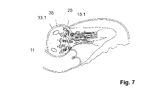
The complete inventive device 10 with an annuloplasty ring 11, shown with six
tissue anchors
15.1-15.6, is shown in Fig. 4b.
The first position 24.1 of a tissue anchor 15.1 is located at marker 22.1 on
the left border
between the anterior section 20 and the posterior section 21. The third
position 24.3 of a tissue
anchor 15.3 is located at marker 22.2 on the right border between the anterior
section 20 and the
posterior section 21. The second position 24.2 of a tissue anchor 15.2 is
located between the first
position 24.1 and the third position 24.3 in the area of the mitral valve
annulus 13 of the anterior
cusp 16, while the other positions 24.4 to 24.8 of the tissue anchors 15.4 to
15.8 are arranged in
the area of the mitral valve annulus 13 of the posterior cusp 17. The
posterior section 21 of the
annuloplasty ring 11 is formed and in general follows the changed shape of the
mitral valve
annulus 13 in the area of the posterior cusp 17. The tissue anchors 15.4 to
15.8 are implanted in
such a way that the annuloplasty ring 11 that is fastened thereto supports the
shape of the mitral
Date Recue/Date Received 2020-09-25
CA 03095227 2020-09-25
29
valve annulus 13. The annuloplasty ring 11 is not, as shown here in the state
of the art of Fig. 2,
attached directly to the mitral valve annulus 13 with knotted thread loops 23,
but rather fastened
to the tissue anchors 15.1-15.8 that are implanted on the mitral valve annulus
13, as seen from
Fig. 4a. Analogous reference numbers from this Fig. 2 are adopted in the
figures below.
Also, Fig. 3 shows in a perspective depiction, from the state of the art,
another implanted
device 10 in an unfolded configuration. The device consists of a segmented
annuloplasty ring 11
with tissue anchors 15.1-15.4 as fastening means 25. The annuloplasty ring 11
has an
approximately "C-shaped" configuration in order to reinforce an opening in the
body tissue or to
reinforce the natural valve 19. The valve 19 has the shape of a mitral valve
14; see Fig. 2.
According to the embodiment, the annuloplasty ring 11 consists of three
segments 26a, 26b, 26c.
Between the three segments 26a, 26b, 26c and on the free ends 28, 28' of the
segments 26a, 26c,
in each case a tissue anchor 15.1-15.4, altogether four tissue anchors 15.1,
15.2, 15.3, 15.4, is
arranged. The distance from the tissue anchors 15.1, 15.2, 15.3, 15.4 is
predetermined by the
length of the arc-shaped segments 26a, 26b, 26c. At the places of the tissue
anchors 15.1, 15.2,
15.3, 15.4, pivot joints 29.1-29.4 are arranged in the segments 26a, 26b, 26c,
which have a
conical mount opening (not shown) for the tissue anchors 15.1, 15.2, 15.3,
15.4. The arc shape
of the segments 26a, 26b, 26c is set in such a way that they can comprise a
portion of the mitral
valve annulus 13. Spiral tissue anchors 15.1, 15.2, 15.3, 15.4 are provided as
fastening means 25
for the annuloplasty ring 11 on the mitral valve annulus 13. The annuloplasty
ring 11 that is
shown is inserted in a catheter-guided manner into the heart 3 and implanted
there. Analogous
reference numbers from this Fig. 3 are adopted in the figures below.
In a perspective depiction, Fig. 4a shows an inventive implantable device 10,
consisting
of an annuloplasty ring 11 with fastening means 25, whereby the fastening
means 25 comprise
Date Recue/Date Received 2020-09-25
CA 03095227 2020-09-25
multiple tissue anchors 15.1, 15.2, 15.3, 15.4, 15.5, 15.6. The tissue anchors
15.1, 15.2, 15.3,
15.4, 15.5, 15.6 in turn are formed from spiral coil screws 30.1-30.6, whereby
other fastening
means can also be possible. The depiction of the heart 3 and the cusps 16, 17
of a mitral valve
14 is omitted here for the sake of clarity. This is sufficiently evident from
Figs. 5-7. The
implantation of the device 10 that is shown is carried out with use of the
minimally-invasive
surgery according to Fig. 1.
The inventive annuloplasty ring 11, according to this embodiment, has
approximately a
general circular or oval shape. In addition, the annuloplasty ring 11 has an
inner layer 43 for
stabilization and at least one outer layer 42, through which the at least one
artificial tissue anchor
thread 33 is drawn. Such an annuloplasty ring 11 comprises in cross-section a
rounded ring
element 27, which has a relatively straight anterior section 20 and an arc-
shaped or curved
posterior section 21, as also shown in Fig. 2. The anterior section 20 of an
annuloplasty ring 11
is equipped with tissue anchor positions 24.1-24.3 for an anterior side 31 of
a mitral valve
annulus 13 of the anterior cusp 16, while the posterior section 21 is equipped
with tissue anchor
positions 24.4-24.6 for a posterior side 32 of a mitral valve annulus 13 of
the posterior cusp 17.
A tissue anchor position 24.1-24.6 in the annuloplasty ring 11 is provided
with at least one tissue
anchor thread 33.1-33.6 from at least one tissue anchor 15.1-15.6. The tissue
anchors 15.1-15.6
are arranged around the mitral valve annulus 13. Each tissue anchor 15.1-15.6
that is implanted
on the mitral valve annulus 13 is equipped with a tissue anchor thread 33.1-
33.6 in order to
fasten an annuloplasty ring 11 to the tissue anchors 15.1-15.6. The tissue
anchor position 24.1 in
the annuloplasty ring lilies on the same longitudinal axis 39 as the tissue
anchor position 24'.1
on the mitral valve annulus 13. That is to say, the tissue anchor position
24.1 in the annuloplasty
ring 11 and the tissue anchor position 24'.1 on the mitral valve annulus 13
are congruent, by
Date Recue/Date Received 2020-09-25
CA 03095227 2020-09-25
31
which because of its tissue anchor position 24'.1 on the mitral valve annulus
13, a tissue anchor
thread 33.1 of a tissue anchor 15.1 can be assigned for fastening to the same
tissue anchor
position 24.1 on the annuloplasty ring 11. To avoid repetitions, the above-
mentioned example is
representative of the other tissue anchor positions 24.2-24.6 and 24' .2-24'
.6, whereby a pair of
tissue anchor positions 24.2-24'.2, 24.3-24' .3, etc., always belongs together
and is arranged on a
common longitudinal axis 39.
An annuloplasty ring 11 can be fastened based on a large number of tissue
anchor
positions 24'.1-24'.6, for example six positions on the mitral valve annulus
13 and the tissue
anchors 15.1-15.6 implanted therein. Fig. 2 shows eight tissue anchor
positions 24.1-24.8, which
are typically used to position and to fasten an annuloplasty ring 11 with its
tissue anchor
positions 24.1-24.8 on the tissue anchors 15.1-15.8 that are implanted in the
mitral valve annulus
13.
The first position 24.1 of a tissue anchor 15.1 on the annuloplasty ring 11 is
located,
viewed in top view, at marker 22.1, which characterizes the left border
between the anterior
section 20 and the posterior section 21. The third position 24.3 of a tissue
anchor 15.3 is located
at marker 22.2, which marks the right border between the anterior section 20
and the posterior
section 21. The second position 24.2 of a tissue anchor 15.2 is located
between the first position
24.1 and the third position 24.3 in the relatively straight anterior section
20 of the annuloplasty
ring 11, while the other positions 24.4 to 24.6 of the tissue anchors 15.4 to
15.6 are arranged in
the area of the curved posterior section 21. The posterior section 21 of the
annuloplasty ring 11
is formed and follows in general the changed shape of the mitral valve annulus
13 in the area of
the posterior cusp 17. The tissue anchors 15.4 to 15.8 are implanted in such a
way that the
annuloplasty ring 11 that is fastened thereto supports the shape of the mitral
valve annulus 13.
Date Recue/Date Received 2020-09-25
CA 03095227 2020-09-25
32
The same applies for the tissue anchor positions 24'.1-24'.6 of the tissue
anchors 15.1-15.6,
which are arranged around the mitral valve annulus 13. The first position
24'.1 of a tissue
anchor 15.1 is located at the mitral valve annulus 13, viewed in top view, at
the left border
between the anterior section 20 and the posterior section 21, where the
anterior cusp 16 meets the
posterior cusp 17. The same also meets the third tissue anchor position 24'.3,
which lies on the
right border between the anterior section 20 and the posterior section 21,
where the anterior cusp
16 meets the posterior cusp 17. The second position 24'.2 of a tissue anchor
15.2 is located
between the first position 24'.1 and the third position 24'.3 in the area of
the anterior cusp 16 of
the mitral valve annulus 13, while the other positions 24'.4 to 24'.6 of the
tissue anchors 15.4 to
15.6 are located in the area of the posterior cusp 17 of the mitral valve
annulus 13.
The tissue anchor positions 24'.1-24'.6 and the distances between them can be
indicated
for the tissue anchors 15.1-15.5 on the mitral valve annulus 13, including
using clock references,
viewed clockwise. By way of example, the tissue anchor position 24'.2 could be
located at 12
o'clock and the two tissue anchor positions 24'.1 and 24'.3, which border the
anterior section 20
of a mitral valve annulus 13, could be located at 2 o'clock and 10 o'clock.
The tissue anchor
positions 24'.4-24'.6 for the posterior section 21 of a mitral valve annulus
13 are located at 4
o'clock, 6 o'clock and 8 o'clock. The distances between the tissue anchors
15.1-15.6 are thus 2
hours, graphically speaking. This shows that additional tissue anchors 15,
primarily in the
posterior section 21 and the saddle area of the mitral valve annulus 13, could
be implanted on the
hour at 5 o'clock and 7 o'clock, as shown in, e.g., Fig. 2. Graphically
speaking, of course, other
time intervals are also possible, by which other angular distances between the
tissue anchors 15
would be generated.
Date Recue/Date Received 2020-09-25
CA 03095227 2020-09-25
33
Starting from the geometry of a mitral valve annulus 13, the tissue anchors
15.1-15.6 can
also be implanted on the mitral valve annulus 13 in such a way that an
annuloplasty ring 11 can
also recreate an asymmetrical opening of a mitral valve annulus 13. That is to
say, the shape of
an annuloplasty ring 11 can be changed based on multiple factors. By way of
example, Figs. 2
and 4a show two of the many possible embodiments. The shape of an annuloplasty
ring 11 can
be influenced with the implantation of additional tissue anchors 15. Also, the
distances between
the positions 24 of the tissue anchors 15 can be varied. The positioning of
the tissue anchors 15
on the mitral valve annulus 13 therefore has special importance. An
annuloplasty ring 11 that is
fastened to the implanted tissue anchors 15 thus eliminates the problem of
functional mitral
regurgitation, since the annuloplasty ring 11, together with the implanted
tissue anchors 15,
exerts a tensile force on the surrounding myocardial tissue 47. In principle,
annuloplasty rings
11 that are asymmetrical from the start can be used when a patient has a
dysplastic anatomy on
the mitral valve annulus 13. Although the material of an annuloplasty ring 11
that is used here
makes possible a manual deformation, it is stiff enough to withstand another
deformation on the
mitral valve annulus 13 as soon as it is implanted and is subject to the
normal physiological
stresses.
The outer layer 42 of an annuloplasty ring 11 should be sufficiently porous
and/or
flexible to allow it to pass through the tissue anchor threads 33. The inner
layer 43 is therefore
designed to reduce the periphery of a mitral valve annulus 13. It must
preserve its rear bending
in the posterior section 21 against the stresses that are forwarded from the
muscle tissue 47 of the
heart 3 during a stroke cycle. The materials of such an inner layer 43 were
previously laid out in
the description by way of example. Analogous reference numbers from this Fig.
4a are adopted
in the figures below.
Date Recue/Date Received 2020-09-25
CA 03095227 2020-09-25
34
In a diagrammatic depiction, Fig. 4b shows a cutaway X from Fig. 4a with a
tissue
anchor 15.1 and a ring element 27 in cross-section, by way of example of all
tissue anchors 15.1-
15.6. A tissue anchor 15.1 consists of, i.a., a spiral coil screw 30.1, which
forms the distal end
36 of a tissue anchor 15.1, while a needle 34 is arranged at the proximal end
37 of the tissue
anchor 15.1, at the free end of the tissue anchor thread 33.1. The coil screw
30.1 of the tissue
anchor 15.1 is secured in a carrier disk 38, which exits from the carrier disk
38 in the direction
toward the distal end 36. In addition, the carrier disk 38 is a holder for a
tissue anchor thread
33.1, which exits from the carrier disk 38 to the opposite side of the coil
screw 30.1. In another
embodiment, the tissue anchor thread 33.1 is fastened onto the tissue anchor
15.1, and the carrier
disk 38 is located on the common fastening site 46 of the thread 33.1 and the
anchor 15.1. The
carrier disk 38 has an attachment side 40 for the mitral valve annulus 13 and
an attachment side
41 for the ring element 27. The two attachment sides 40, 41 contain two
attachment surfaces I, II
44, 45. The first attachment surface I 44 serves the tissue anchor 15 as a
stop when it is being
screwed onto the tissue 47, while the other attachment surface II 45 serves a
ring element 27 as a
resting point. The diameter of the carrier disk 38 is designed in such a way
that screwing a tissue
anchor 15.1 too far into the myocardial tissue 47 is avoided. If all tissue
anchors 15.1-15.6, see
Fig. 4a, are implanted around the mitral valve annulus 13 in the myocardial
tissue 47, the ring
element 27 of an annuloplasty ring 11 is placed next on the carrier disk 38 of
the tissue anchor
15.1-15.6. A fastening means 25 is used to fasten a ring element 27 onto the
carrier disk 38 of a
tissue anchor 15.1. At least one tissue anchor thread 33.1 of a tissue anchor
15.1 is fastened at
least with a fastening means 25 onto the annuloplasty ring 11. The fastening
means can
preferably consist of a clamping means 35. The clamping means 35 is put onto a
tissue anchor
thread 33.1 outside of the ribcage 1. This process is carried out using the
needle 34, which is
Date Recue/Date Received 2020-09-25
CA 03095227 2020-09-25
guided through the opening of the clamping means 35. A surgical instrument
(not shown)
advances the clamping means 35 up to the ring element 27 and clamps the ring
element 27
between it and the carrier disk 38. The clamping means 35 is designed in such
a way that it can
permanently clamp a tissue anchor thread 15.1 in its opening. For example, a
clamping means
35 can also consist of two components that work against one another and exert
a clamping effect
on a tissue anchor thread 15.1. Preferably, the clamping element 35 can
consist of a cup spring,
which can be inserted relatively easily on the tissue anchor thread 33.1 into
the atrium 12 and at
the site of the fastening of the ring element 27 generates a clamping opposite
to the feeding
direction. The ring element 27, which is positioned between the clamping means
35 and the
carrier disk 38 of the tissue anchor 15.1, has an inner layer 43 and an outer
layer 42. The tissue
anchor thread 33.1 is guided through the outer layer 42 of the ring element 27
in order not to
damage the inner layer 43. The insertion of an annuloplasty ring 11 into the
atrium 12 can be
found in Fig. 6. After all clamping means 35 have been placed on the ring
element 27, the tissue
anchor threads 33.1-33.6 are severed and removed from the atrium 12 of the
heart 3. Analogous
reference numbers from this Fig. 4b are adopted in the figures below.
The heart 3 that is shown in Fig. 5 in a diagrammatic and basic depiction
lies, according
to Fig. 1, rotated around its longitudinal axis in the left thoracic space 8,
so that the right half of
the heart rests more on the anterior chest wall, while the left half of the
heart preferably points
toward the rear. A mitral valve implant, in particular an annuloplasty ring
11, is provided, which
with use of minimally-invasive surgery can be introduced into the beating
heart 3 of a patient via
the right thorax area 5 in the left atrium 12 of the heart 3, using known
surgical instruments, i.e.,
a trocar 50, and can be anchored there.
The left chamber of the heart 48 with the left atrium 12 and an access 49
through the
Date Recue/Date Received 2020-09-25
CA 03095227 2020-09-25
36
heart tissue 47 in the left atrium 12 to the mitral valve 14 is therefore
shown. The access 49 is
carried out via the indicated trocar 50 and various surgical instruments 51.1-
51.5. The various
surgical instruments for mitral valve implantation with use of the minimally-
invasive repair of a
mitral valve annulus 13 in the beating heart 3 of the patient are cited below.
A surgical
instrument consists of, for example, an outer tube spacer I 51.1, in
particular an access cannula
with lumen for guiding an inner tube spacer II 51.2. Another surgical
instrument consists of a
first inner tube spacer II 51.2 with lumen for guiding and screwing-in a
tissue anchor 15.
Another surgical instrument consists of a second inner tube spacer III 51.3
with lumen for
guiding an annuloplasty ring 11. Also, the third inner tube spacer IV 51.4 is
a surgical
instrument with lumen for receiving the tissue anchor threads 33.1-33.6 and
for pushing an
annuloplasty ring 11 out of the tube spacer III 51.3. The fourth inner tube
spacer V 51.5 is also a
surgical instrument with lumen for feeding a fastening means 25, in particular
a clamping means
35, which is guided along a tissue anchor thread 33.1 for fastening an
annuloplasty ring 11.
The left ventricle 52 is structured into an inflow and an outflow path. It is
separated from
the atrium 12 by the mitral valve 14. The mitral valve 14 is connected by
tendinous cords
(Chordae tendineae) 53 to the papillary muscles 54, which originate on the
ventricle wall 55 and
therefore ensure that the mitral valve 14 during its valve closure 19 and
during the exertion phase
(systole) of the left chamber 52 does not rebound too violently into the left
atrium 12.
A mitral valve consists of four functional components: the two cusps 16, 17
(mitral valve
leaflets), consisting of an anterior cusp 16 (cupis anterior), a posterior
cusp 17 (cupis pasterior)
and the mount of the cusps 16, 17 in the mitral valve ring 13 (mitral valve
annulus). The mitral
valve ring 13 consists of a muscle tissue, which is referred to in the
description as mitral valve
annulus 13, the tendinous cords 53 (Chordae tendineae), with which the cusps
16, 17 are fastened
Date Recue/Date Received 2020-09-25
CA 03095227 2020-09-25
37
to move on the papillary muscles 54, and the papillary muscles 54 themselves,
which protrude
inward from the myocardium 47. For reconstruction of each individual
component, different
implants, surgical instruments and/or surgical methods are available. In this
case, mitral valve
regurgitation and its elimination are considered.
To this end, in the left atrium 12, tissue anchors 15.1-15.5 are inserted into
the area
around the mitral valve annulus 13. Since the heart 3 is shown in a sectional
view, not all
possible implanted tissue anchors 15.1-15.8 from Fig. 2 can be shown here,
since only a portion
of the periphery of a mitral valve annulus 13 is shown. The depicted tissue
anchors 15.1-15.5 are
representative of all implanted tissue anchors 15.1-15.5. The tissue anchors
15.1-15.5 that are
implanted around the mitral valve annulus 13 are arranged at certain
distances. The distances
between the tissue anchors 15.1-15.5 can vary, e.g., in the saddle area of the
posterior section 21
of a mitral valve annulus 13 relative to the other distances. In addition, at
the distal end 36, the
tissue anchors 15.1-15.5 have an anchoring element 56, whereby an anchoring
element 56
consists of a corkscrew-like coil screw 30.1-30.5. The coil screws 30.1-30.5
have a distal end 36
and a proximal end 37, whereby the proximal end 37 of the coil screw 30.1-30.5
is connected to
the tissue anchor threads 33.1-33.5. The use of other anchoring means for
fastening an
annuloplasty ring 11 is conceivable from the known state of the art. The
screwed-in coil screws
30.1-30.5 are located in the myocardial tissue 47 in the area of the mitral
valve annulus 13. In
addition, the tissue anchors 15.1-15.5 at the proximal end 37 have a carrier
disk 38 and a tissue
anchor thread 33.1-33.5, which are fastened to the tissue anchors 15.1-15.5;
to this end, see Fig.
4b. The tissue anchor threads 33.1-33.5 are guided through a sleeve 51 from
the ribcage 1 for
further use and are also connected to the coil screws 30.1-30.5. The further
use of the tissue
Date Recue/Date Received 2020-09-25
CA 03095227 2020-09-25
38
anchor threads 33.1-33.5 is evident from the description of Fig. 6. Analogous
reference numbers
from the preceding Figs. 1-4 are adopted in this figure.
In a diagrammatic depiction, Fig. 6 shows an annuloplasty ring 11 in a guiding
configuration 57. In order to achieve a guiding configuration 57, the tissue
anchor threads 33.1-
33.5, which come from the tissue anchors 15.1-15.5 from the atrium 12, are
guided through to
predetermined positions 24.1-24.5 on the annuloplasty ring 11 outside of the
ribcage 1. First, the
individual tissue anchor threads 33.1-33.5, on whose free ends in each case a
needle 34 is
located, are guided through the fibrous ring of the outer layer 42 of an
annuloplasty ring 11,
which still has its starting shape 58. In order to be able to guide a tissue
anchor thread 33.1-33.5
through an annuloplasty ring 11, it is necessary to know in advance which
tissue anchor thread
33.1-33.5 is affected and at which point a tissue anchor thread 33.1-33.5 is
to be guided through
in the annuloplasty ring 11. The knowledge is necessary, since an annuloplasty
ring 11 has
various sections 20, 21: an anterior section 20, which is to be arranged on
the front cusp 16, and
a posterior section 21, which is to be arranged on the posterior cusp 17; see
Fig. 2. The mitral
valve annulus 13 also has these sections 20, 21. An annuloplasty ring 11 is to
be placed on the
mitral valve annulus 13 in such a way that their sections 20, 21 come to rest
one over another.
Along the sections 20, 21, around the mitral valve annulus 13, the tissue
anchors 15.1-15.5 are
arranged at certain distances. The distances can, however, also be irregular.
The question thus
arises as to from which tissue anchor 15.1-15.5 the tissue anchor thread 33.1-
33.5 that lies
outside of the ribcage 1 comes and at which position 24'.1-24'.5 this tissue
anchor 15.1-15.5 is
implanted on the mitral valve annulus 13. In order to be able to answer this
question, the tissue
anchor threads 33.1-33.5 therefore contain a corresponding identification.
From the
Date Recue/Date Received 2020-09-25
CA 03095227 2020-09-25
39
identification, it is clearly evident at which position 24' .1-24' .5 a tissue
anchor 15.1-15.5 is
positioned on the mitral valve annulus 13.
To position a tissue anchor thread 34 on the annuloplasty ring 11, it is
therefore necessary
to use the same position 24.1-24.5 at which the tissue anchor 15.1-15.5 is
positioned on the
mitral valve annulus 13. The annuloplasty ring 11 that is to be implanted
therefore has, seen in
top view, relative to the positioning in the tissue anchors 15.1-15.5, the
same image structure as
the image structure of the mitral valve annulus 13. If the first tissue anchor
15.1 is located at the
first position 24'.l, e.g., at the left transition between the curved section
21 and the straight
section 20 of the mitral valve annulus 13, the tissue anchor thread 33.1 that
corresponds to this
tissue anchor 15.1 thus bears the identification number 1. That is to say, the
identification
number 1 identifies the position 24'.l of a tissue anchor 15.1 on the mitral
valve annulus 13.
However, this also means that the tissue anchor thread 33.1 of the tissue
anchor 15.1 has to be
guided through to the corresponding point in the annuloplasty ring 11. That is
to say, to be able
to place the annuloplasty ring 11 in the proper shape at the tissue anchors
15.1-15.5 on the mitral
valve annulus 13, it is necessary to assign the position 24.1 on the
annuloplasty ring 11 to the
tissue anchor thread 33.1 with the identification number 1 and the position
24'.l of the tissue
anchor 15.1 and at this point to guide the outer layer 42 through the tissue.
The position 24.1 on
the annuloplasty ring 11 also corresponds to the first position 24.1 at the
left transition between
the curved section 21 and the straight section 20 of the annuloplasty ring 11.
The first position
24.1 on the annuloplasty ring 11 corresponds to the first position 24' .1 of
the implanted tissue
anchor 15.1. The same applies for the other tissue anchor threads 33.2-33.5,
which are provided
by the tissue anchors 15.2-15.5 and are now drawn into the corresponding
positions 24.2-24.5
through the tissue 42 of the annuloplasty ring 11. That is to say, the tissue
anchor thread 33.2 of
Date Recue/Date Received 2020-09-25
CA 03095227 2020-09-25
the implanted tissue anchor 15.2 has the identification number 2 and is
located at the position
24'.2 on the mitral valve annulus 13. This tissue anchor thread 33.2 is guided
through to the
position 24.2 of the annuloplasty ring 11, whereby the position 24'.2 on the
mitral valve annulus
13 in turn is identical to the position 24.2 on the annuloplasty ring 11, etc.
It is thus ensured that the shape of an annuloplasty ring 11 that matches the
shape of a
mitral valve annulus 13 can also be adapted and can be fastened onto the
tissue anchors 15.1-
15.5. At the positions 24.1-24.5 on the annuloplasty ring 11, at which in each
case a tissue
anchor thread 33.1-33.5 can be drawn through, position markers 22.1, 22.2 can,
e.g., already be
marked out on the annuloplasty ring 11. It is also conceivable that the
positions for the tissue
anchor threads 33.1-33.5 in the outer layer 42 of an annuloplasty ring 11 are
already provided
with a through opening for the needle 34. Through openings facilitate the
threading of the tissue
anchor threads 33.1-33.5 and avoid possible damage to the outer layer 42 of
the ring element 27.
If an annuloplasty ring 11 is drawn onto all tissue anchor threads 33.1-33.5
that are
provided by the tissue anchors 15.1-15.5, the latter is advanced onto the
tissue anchor threads
33.1-33.5 up to a receiving surgical instrument 51 and compressed. In this
state, the
annuloplasty ring 11 has now achieved its guiding configuration 57 in order to
be inserted into a
sleeve 51 that is guided into the trocar 50. In this phase, the tissue anchor
threads 33.1-33.5
serve as guide means for the annuloplasty ring 11. The sleeve 51, which is
guided through a
trocar 50, reaches up to the left atrium 12 of the heart 3. With another
surgical instrument, the
annuloplasty ring 11 is then moved along the tissue anchor threads 33.1-33.5
through the sleeve
51 into the atrium 12. The free ends of the tissue anchor threads 33.1-33.5
remain in addition
outside of the body 1. If the annuloplasty ring 11 exits completely from the
sleeve 51 and enters
into the left atrium 12, it expands from its guiding configuration 57 into its
original starting
Date Recue/Date Received 2020-09-25
CA 03095227 2020-09-25
41
shape 58. The original starting shape 58 corresponds, preferably according to
Figs. 2 and 4a, to
an open configuration, whereby the annuloplasty ring 11 is always still guided
by the tissue
anchor threads 33.1-33.5 of the tissue anchors 15.1-15.5. Along the tissue
anchor threads 33.1-
33.5, the annuloplasty ring 11 is now moved on the carrier disks 38, which are
arranged on the
ends of the tissue anchors 15.1-15.5 and placed there. As previously
described, the annuloplasty
ring 11 is now fastened in the proper shape to the mitral valve annulus 13, on
the tissue anchors
15.1-15.5 and as shown in Fig. 4b. To fasten an annuloplasty ring 11, a tissue
anchor thread
33.1-33.5 is provided with a clamping means 35, see Fig. 4b. Analogous
reference numbers
from the preceding Figs. 1-5 are adopted in this figure.
In a diagrammatic depiction, Fig. 7 shows a device 10 that is implanted on the
mitral
valve annulus 13, consisting of a mitral valve implant, in particular in the
form of an
annuloplasty ring 11, which in principle comprises three elements. A first
element is the
anchoring element 56, which is designed as a tissue anchor 15 with a coil
screw 30, a carrier disk
38 and a tissue anchor thread 33 and undertakes securing of the annuloplasty
ring 11 in the
myocardial tissue 47. The anchoring element 56 is not depicted in Fig. 7 for
the sake of clarity,
but it is shown in detail in Fig. 4b. The second element is the annuloplasty
ring 11 as an implant
that has an inner layer 43 and an outer layer 42, whereby the outer layer 42
receives the tissue
anchor threads 33 that produce the connection to the tissue anchor 15. The
third element forms
the fastening means 25, which consists of a clamping means 35 and is guided
along a tissue
anchor thread 33. The clamping means 35 clamps an annuloplasty ring 11 between
it and the
carrier disk 38 using a tissue anchor thread 33. In conclusion, the tissue
anchor threads 33.1-33.8
are still severed and, i.a., the surgical instruments 50, 51 are removed from
the atrium 12, and the
access 49 to the heart 3 is closed.
Date Recue/Date Received 2020-09-25
CA 03095227 2020-09-25
42
The tissue anchor positions 24.1-24.5 in the annuloplasty ring 11 now lie with
the tissue
anchor positions 24'.1-24'.5 on the mitral valve annulus 13 on the same
longitudinal axis 39 and
are thus congruent, by which a tissue anchor thread 33.1 of a tissue anchor
position 24'.1 on the
mitral valve annulus 13 corresponds to the same tissue anchor position 24.1 in
the annuloplasty
ring 11. An annuloplasty ring 11 is thus implanted in the proper shape for
eliminating mitral
valve regurgitation. Analogous reference numbers from the preceding Figs. 1-6
are adopted in
this figure.
Date Recue/Date Received 2020-09-25
CA 03095227 2020-09-25
43
Reference Symbol List
1 Thorax 31 Anterior side (of 13)
2 Access 32 Posterior side (of 13)
3 Heart 33.1-33.6 Tissue anchor thread
4 Ribcage opening 34 Needle (of 15, 33)
Right side (of 1) 35 Clamping means
6 Rib space 36 Distal end (of 15)
7 Self-retaining retractor 37 Proximal end (of 15)
8 Left thoracic space 38 Carrier disk (of 15)
9 Anatomical opening 39 Longitudinal axis (of 24, 24')
Device 40 Attachment side (of 13)
11 Annuloplasty ring 41 Attachment side (of 27)
12 Atrium 42 Outer layer (of 11, 27)
13 Mitral valve annulus 43 Inner layer (of 11, 27)
14 Mitral valve 44 Attachment surface I (of 38)
15.1-15.6 Tissue anchor 45 Attachment surface II (of 38)
16 Anterior cusp (of 14) 46 Fastening site
17 Posterior cusp (of 14) 47 Tissue (of 3)
18 Gap (of 14) 48 Left chamber of the heart
19 Valve (of 14) 49 Access (to 3)
Anterior section (of 13) 50 Trocar
21 Posterior section (of 13) 51 Instrument
22.1-22.2 Marker (of 11) 52 Left ventricle
Date Recue/Date Received 2020-09-25
CA 03095227 2020-09-25
44
23 Thread loops 53 Tendinous cords
24.1-24.8 Tissue anchor positions 54 Papillary muscles
(of 11)
24'.1-24'.8 Tissue anchor positions 55 Ventricle wall
(of 13)
25 Fastening means 56 Anchoring element
25a-26c Segments 57 Guiding configuration
27 Ring element 58 Starting shape
28 Free end
29.1-29.4 Pivot joints
30.1-30.6 Coil screws
Date Recue/Date Received 2020-09-25