Note: Descriptions are shown in the official language in which they were submitted.
CA 03095461 2020-09-28
WO 2019/217525 PCT/US2019/031275
METHODS AND APPARATUS FOR A CLEANING SHEATH FOR
AN ENDO SCOPE
CROSS-REFERENCES TO RELATED APPLICATIONS
[0001] This application claims the benefit of U.S. Provisional Patent
Application No.
62/670,205, filed May 11, 2018, and incorporates the disclosure of the
application
by reference.
BACKGROUND OF THE TECHNOLOGY
[0002] Endoscopes are used in a variety of surgical procedures
including
endscopy, laparoscopy, and thoracoscopy to investigate symptoms in the
digestive
system, cardiovascular system, and the sinuses. A common frustration
experienced
in procedures is the altered and obstructed visualization caused by debris and
bodily fluids that come into contact with the distal fiberoptic tip of the
endoscope.
In addition, upon first insertion of the endoscope into a warm body cavity,
the lens
of the optics tip may fog similar to eye goggles when swimming or snorkeling.
When this occurs, it typically requires the removal of the endoscope from the
body
cavity to remove the obstructing substance manually. Removing and reinserting
the endoscope wastes valuable O.R. time and is potentially dangerous when
visualization is compromised during a critical point of an
operation/procedure.
1
CA 03095461 2020-09-28
WO 2019/217525 PCT/US2019/031275
[0003] Existing solutions directed towards maintaining a clear field of
vision after
the tip of the endoscope has been inserted into a body cavity include devices
that
do not function with existing sizes of associated medical devices used during
the
surgical procedure such as a trocar. These devices often also require separate
pumps, motors, electronics, and switches to function properly. Each of these
devices increases the number of equipment located where the surgical procedure
is
being performed taking up space and increasing costs. Further, introducing
additional components during the surgical procedure results in increased
complexity and requires additional training by medical staff
SUMMARY OF THE TECHNOLOGY
[0004] A cleaning sheath for an endoscope according to various aspects
of the
present technology includes an open ended sheath coupled to a fluid line. The
sheath is configured to receive an endoscope within the interior of the sheath
wherein both the sheath and the endoscope may be inserted into a trocar during
use. The sheath is further configured to direct a cleaning fluid onto the
optics end
of the endoscope to clear away any visual obstructions without requiring the
removal of the endoscope from the trocar.
BRIEF DESCRIPTION OF THE DRAWINGS
[0005] A more complete understanding of the present technology may be
derived
by referring to the detailed description and claims when considered in
connection
with the following illustrative figures. In the following figures, like
reference
numbers refer to similar elements and steps throughout the figures.
2
CA 03095461 2020-09-28
WO 2019/217525 PCT/US2019/031275
[0006] Figure 1 representatively illustrates an endoscope sheath in
accordance
with an exemplary embodiment of the present technology;
[0007] Figure 2 representatively illustrates a cross-sectional view
across line 2-2
of Figure 1 in accordance with an exemplary embodiment of the present
technology;
[0008] Figure 3 representatively illustrates the endoscope sheath
coupled to a
syringe with an endoscope partially inserted into the endoscope sheath in
accordance with an exemplary embodiment of the present technology;
[0009] Figure 4 representatively illustrates a top view of a fluid hub
of the
endoscope sheath in accordance with an exemplary embodiment of the present
technology;
[0010] Figure 5 representatively illustrates a cross-sectional view
across line 5-5
of Figure 4 in accordance with an exemplary embodiment of the present
technology;
[0011] Figure 6 representatively illustrates an interior side view of a
distal end of
an alternative embodiment of the endoscope sheath in accordance with an
exemplary embodiment of the present technology;
[0012] Figure 7 representatively illustrates a cross-sectional view of
the endoscope
sheath in accordance with an exemplary embodiment of the present technology;
[0013] Figure 8 representatively illustrates a detailed view of Section
8 of Figure 7
in accordance with an exemplary embodiment of the present technology;
3
CA 03095461 2020-09-28
WO 2019/217525 PCT/US2019/031275
[0014] Figure 9 representatively illustrates a cross-sectional view
across line 9-9
of Figure 6 in accordance with an exemplary embodiment of the present
technology;
[0015] Figure 10 representatively illustrates a cross-sectional view
across line 10-
of Figure 6 in accordance with an exemplary embodiment of the present
technology;
[0016] Figure 11 representatively illustrates a detailed view of
Section 11 of
Figure 10 in accordance with an exemplary embodiment of the present technology
[0017] Figure 12 representatively illustrates an optics end of an
endoscope being
inserted into the endoscope sheath in accordance with an exemplary embodiment
of the present technology;
[0018] Figure 13 representatively illustrates the optics end of the
endoscope
inserted part way through the endoscope sheath in accordance with an exemplary
embodiment of the present technology;
[0019] Figure 14 representatively illustrates the optics end of the
endoscope being
inserted fully into the endoscope sheath in accordance with an exemplary
embodiment of the present technology;
[0020] Figure 15 representatively illustrates the optics end of the
endoscope and
the endoscope sheath being inserted into a trocar in accordance with an
exemplary
embodiment of the present technology;
[0021] Figure 16 representatively illustrates the optics end of the
endoscope and
the endoscope sheath being fully inserted into the trocar in accordance with
an
exemplary embodiment of the present technology;
4
CA 03095461 2020-09-28
WO 2019/217525 PCT/US2019/031275
[0022] Figure 17 representatively illustrates a syringe being connected
to a fluid
line of the endoscope sheath in accordance with an exemplary embodiment of the
present technology;
[0023] Figure 18 representatively illustrates a display of the optics
end of the
endoscope in accordance with an exemplary embodiment of the present
technology;
[0024] Figure 19 representatively illustrates the display of the optics
end of the
endoscope with a visual obstruction in accordance with an exemplary embodiment
of the present technology;
[0025] Figure 20 representatively illustrates the display of the optics
end of the
endoscope at the beginning of a flush cycle in accordance with an exemplary
embodiment of the present technology;
[0026] Figure 21 representatively illustrates the display of the optics
end of the
endoscope during the flush cycle in accordance with an exemplary embodiment of
the present technology; and
[0027] Figure 22 representatively illustrates the display of the optics
end of the
endoscope at the completion of the flush cycle in accordance with an exemplary
embodiment of the present technology.
[0028] Elements and steps in the figures are illustrated for simplicity
and clarity
and have not necessarily been rendered according to any particular sequence.
For
example, steps that may be performed concurrently or in a different order are
illustrated in the figures to help to improve understanding of embodiments of
the
present technology.
CA 03095461 2020-09-28
WO 2019/217525 PCT/US2019/031275
DETAILED DESCRIPTION OF EXEMPLARY EMBODIMENTS
[0029] The present technology may be described in terms of functional
block
components and various processing steps. Such functional blocks may be
realized
by any number of components configured to perform the specified functions and
achieve the various results. For example, the present technology may employ
various materials, coupling mechanisms, dimensions, and geometries, which may
carry out a variety of operations suited to a selective attachment to or use
with an
endoscope, trocar, or syringe. In addition, the technology described is merely
one
exemplary application for the invention. Further, the present technology may
employ any number of conventional techniques for flushing, cleaning, or
otherwise clearing debris such as bodily fluids and fog from optical devices.
[0030] Methods and apparatus for an endoscope sheath according to
various
aspects of the present technology may operate in conjunction with any type of
endoscope, fiberoptic video capture system, or micro-camera. Various
representative implementations of the present technology may be applied to any
type of viewing device that is insertable or otherwise intended for use within
a
body during a medical procedure that may be subjected to various types of
visual
obstructions.
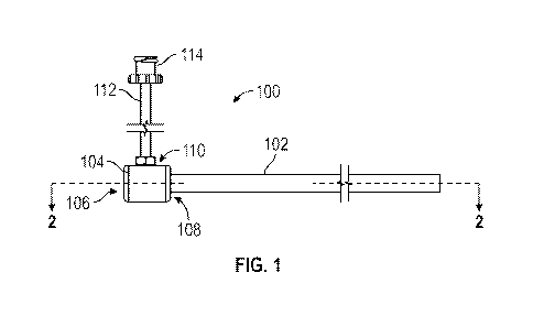
[0031] Referring to Figures 1-3, a cleaning sheath 100 may comprise a
generally
tubular shaped body 102 having a distal end 204 and a proximal end 202. At
least
a portion of an interior of the body 102 and the distal end 204 are configured
to
receive a viewing device such as an endoscope 304. The body 102 may form a
lumen extending between the proximal and distal ends 202, 204 to both receive
the
6
CA 03095461 2020-09-28
WO 2019/217525 PCT/US2019/031275
endoscope and allow flow of a cleaning fluid, such as a saline solution, from
the
proximal end 202 to the distal end 204. A fluid hub 104 may be coupled to the
proximal end 202 of the body 102 and be in fluid communication with the lumen.
A fluid line 112 may be coupled to the fluid hub 104 and be configured to
provide
a conduit path for the cleaning fluid.
[0032] The body 102 may comprise any suitable size or shape and may be
selected
according to any applicable criteria such as a size of the viewing device the
body
102 will be positioned over. For example, the body 102 may comprise an
internal
diameter of between 2 mm and 14 mm to allow both the endoscope 304 and the
body 102 to be positioned within a trocar (not shown) as commonly used in
medical procedures.
[0033] The internal diameter of the body 102 may be sized slightly
larger than the
outer diameter of the endoscope 304 to allow the cleaning fluid to flow
completely
around an outer surface 308 of the endoscope towards the distal end 204 of the
body 102. For example, and in one embodiment where the endoscope 304
comprises a 5 mm thorascope having an outer working diameter of about 5.4 mm,
the body 102 may comprise an inner diameter of between 5.42 mm and 5.75 mm.
The larger diameter of the body 102 relative to the endoscope 304 creates a
fluid
conduit 306 that extends completely around the outer surface 308 of the
endoscope
and along the length of the body 102 between the proximal and distal ends 202,
204 in which the cleaning fluid may flow.
[0034] In an alternate embodiment, and referring now to Figures 7-11,
the body
102 may comprise an outer wall 702 disposed around an inner wall 704 such that
7
CA 03095461 2020-09-28
WO 2019/217525 PCT/US2019/031275
the fluid conduit 306 is formed between the inner and outer walls 702, 704
running
along the entire length of the body 102. The total wall thickness of the body
102
may comprise the sum of the thickness of: the outer wall 702 (WO, the inner
wall
704 (W2), and the fluid conduit 306 (W3).
[0035] Referring now to Figures 6 and 9, the distal end 204 of the body
102 may
comprise a plurality of openings 602 positioned around at least a portion of
the
periphery of the inner wall 704 at predetermined locations to allow the
cleaning
fluid to be ejected inward from the body 102 towards the viewing device (not
shown). The plurality of openings 602 may be positioned at or proximate the
end
most portion of the distal end 204.
[0036] Referring now to Figures 7, 10, and 11, the inner wall 704 and
the outer
wall 702 may be coupled together at the distal end 204. The inner and outer
walls
704, 702 may be coupled together in any suitable manner to create a seal 1002
at
the distal end 204 of the body 102. The presence of the seal 1002 at the
distal end
204 forces the cleaning fluid to flow outwards from the plurality of openings
602.
[0037] The seal 1002 may comprise any suitable system or device capable
of
coupling or otherwise bonding the inner and outer walls 704, 702 together. For
example, in one embodiment, the inner and outer walls 704, 702 may be fused or
otherwise bonded together with an adhesive that creates the seal 702.
[0038] The body 102 may comprise a semi-flexible material suitable
material or
combinations of materials suitable for use inside the human body such as
natural
or synthetic polymers, thermoplastics, or metals. For example, in one
embodiment,
the body 102 may comprise a polyimide polymer adapted to withstand elevated
8
CA 03095461 2020-09-28
WO 2019/217525 PCT/US2019/031275
temperatures associated with a light source of the viewing device. In an
alternative
embodiment, the body 102 may comprise polyether ether ketone (PEEK).
[0039] The distal end 204 may be terminated in any suitable manner to
allow the
viewing device to function properly. For example, the distal end 204 may be
terminated at an angle relative to a longitudinal axis of the body 102 to
accommodate viewing devices having an angled terminal end of up to 450
.
Alternatively, the distal end 204 may be terminated at a substantially right
angle to
the body 102 to accommodate viewing devices having a visualization angle of 00
.
[0040] Referring now to Figures 1, 2, 4, and 5, the fluid hub 104
provides fluid
link between the fluid line 112 and the lumen of the body 102. The fluid hub
104
comprises a hub lumen 208 extending between opposing first and second open
ends 106, 108 that form an insertion path for the viewing device into the
lumen of
the body 102. The second open end 108 of the fluid hub 104 receives and is
coupled to the proximal end 202 of the body.
[0041] An inlet port 110 is open to the hub lumen 208 and is disposed
between the
first and second open ends 106, 108 and is coupled to the fluid line 112. The
fluid
line 112 may be coupled to the inlet port 110 by any suitable method. In one
embodiment, the inlet port 110 may be configured with a compression fitting
configured to create a seal between the fluid line 112 and the interior of the
fluid
hub 104. In an alternative embodiment, the inlet port 110 may comprise an
opening approximately equal to or slightly less than an outer diameter of a
first
end of the fluid line 112 with an edge configured to create a seal against the
fluid
line 112 when an end of the fluid line 112 is inserted through the opening.
9
CA 03095461 2020-09-28
WO 2019/217525 PCT/US2019/031275
[0042] The first open end 106 of the fluid hub 104 may be configured to
create a
seal against the viewing device when it is inserted through the fluid hub 104
to
prevent fluid from exiting out of the first open end 106 during use. For
example,
the first open end 106 may comprise a sealing element 206 positioned between
an
outer edge of the first open end 106 and an inlet port 110. The sealing
element 206
may comprise any suitable system or device for forming a seal against an outer
surface of the viewing device and the hub lumen 208. In one embodiment, the
sealing element may comprise an o-ring having an inner diameter approximately
equal to that of the diameter of the outer surface of the viewing device that
is
intended to be used with the cleaning sheath 100.
[0043] Referring again to Figures 1-3, the fluid line 112 may comprise
any
suitable system or device for creating a fluid conduit path between a cleaning
fluid
supply source and the fluid hub 104. For example, the fluid line 112 may
comprise
a flexible tube made from a polymer. The fluid line 112 may comprise any
suitable
length or diameter. For example, in one embodiment, the fluid line 112 may
comprise a length of between about 50 mm and about 300 mm and comprise a
diameter between about 2.5 mm and about 25 mm.
[0044] As described above, the first end of the fluid line 112 may be
coupled to
the inlet port 110 at or near the proximal end 202 of the body 102. A second
end of
the fluid line 112 may be configured to be connected to the cleaning fluid
supply.
For example, in one embodiment, the second end of the fluid line 112 comprise
a
leur lock 114 that may be selectively connected to an end of a syringe 302
containing the cleaning fluid.
CA 03095461 2020-09-28
WO 2019/217525 PCT/US2019/031275
[0045] Referring now to Figures 12-22, in use the cleaning sheath 100
is used in
conjunction with an endoscope 304 during the medical procedure and is
configured to direct a cleaning fluid towards the optics end 1202 of the
endoscope
304 to clear away any visual obstruction. With particular reference to Figures
12-14,
the optics end 1202 may be inserted into the first end of the fluid hub 104
and pushed
towards the distal end 204 of the body 102 until the optics end 1202 is
positioned at
the terminal opening of the distal end 204.
[0046] Referring now to Figures 15 and 16, during the medical procedure
the
combined endoscope 304 and the distal end 204 of the cleaning sheath 100 may
be
inserted through a trocar 1502 and subsequently into the body of the patient.
As shown
in Figure 17, a syringe 302 containing the fluid supply may be connected to
the
second end of the fluid line 112.
[0047] As shown in Figure 18 the optics end 1202 of the endoscope
provides an
image to a display device 1800 that may be viewed by the person performing the
medical procedure. During the procedure, the optics end 1202 may become
obstructed by coming into contact with debris or a bodily fluid or due to a
difference in temperature between the optics end 1202 and the internal
temperature
of the body. This results in an obstructed field of view on the display device
1800
as shown in Figure 19.
[0048] Referring now to Figures 19 and 20, when the field of view
becomes
obstructed, blurred, or otherwise blocked, cleaning fluid from the syringe 302
may
be injected into the body 102. The cleaning fluid may flow through the fluid
conduit 306 and out the distal end 204 of the body 102 and over the optics end
1202 of the endoscope 304. After the cleaning fluid is passed over the optics
end
11
CA 03095461 2020-09-28
WO 2019/217525 PCT/US2019/031275
1202 the obstruction may be cleared away and the field of vision restored as
shown in Figure 22 with the need to remove the endoscope 304 from the body.
[0049] These and other embodiments for methods of creating a cleaning
sheath
may incorporate concepts, embodiments, and configurations as described above.
The particular implementations shown and described are illustrative of the
technology and its best mode and are not intended to otherwise limit the scope
of
the present technology in any way. Indeed, for the sake of brevity,
conventional
manufacturing, connection, preparation, and other functional aspects of the
system
may not be described in detail. Furthermore, the connecting lines shown in the
various figures are intended to represent exemplary functional relationships
and/or
physical couplings between the various elements. Many alternative or
additional
functional relationships or physical connections may be present in a practical
system.
[0050] The description and figures are to be regarded in an
illustrative manner,
rather than a restrictive one and all such modifications are intended to be
included
within the scope of the present technology. Accordingly, the scope of the
technology should be determined by the generic embodiments described and their
legal equivalents rather than by merely the specific examples described above.
For
example, the components and/or elements recited in any apparatus embodiment
may be assembled or otherwise operationally configured in a variety of
permutations to produce substantially the same result as the present
technology
and are accordingly not limited to the specific configuration recited in the
specific
examples.
12
CA 03095461 2020-09-28
WO 2019/217525 PCT/US2019/031275
[0051] As used herein, the terms "comprises", "comprising", or any
variation
thereof, are intended to reference a non-exclusive inclusion, such that a
process,
method, article, composition or apparatus that comprises a list of elements
does
not include only those elements recited, but may also include other elements
not
expressly listed or inherent to such process, method, article, composition or
apparatus. Other combinations and/or modifications of the above-described
structures, arrangements, applications, proportions, elements, materials or
components used in the practice of the present technology, in addition to
those not
specifically recited, may be varied or otherwise particularly adapted to
specific
environments, manufacturing specifications, design parameters or other
operating
requirements without departing from the general principles of the same.
[0052] The present technology has been described above with reference
to
exemplary embodiments. However, changes and modifications may be made to the
exemplary embodiments without departing from the scope of the present
technology. These and other changes or modifications are intended to be
included
within the scope of the present technology, as expressed in the following
claims.
13