Note: Descriptions are shown in the official language in which they were submitted.
CA 03095596 2020-09-29
WO 2019/195323
PCT/US2019/025430
PRESSURE BASED BLOOD VESSEL ASSESSMENT SYSTEMS AND
METHODS
BACKGROUND OF THE INVENTION
Field of the Invention
[0001] This
application is directed to systems and methods for determining
whether and how to treat a patient based on blood pressure measurements.
Description of the Related Art
[0002]
Fractional flow reserve (FFR) is a known technique for determining
whether to treat a vascular occlusion with balloon angioplasty and/or a stent.
FFR is a test
that is performed under hyperemia. In this technique, blood pressure is
measured within
the coronary vasculature distal to and proximal of the occlusion.
Traditionally, a ratio of
these pressures has been calculated and compared with a threshold value below
which
balloon angioplasty and/or stenting was indicted and above which no such
treatment was
to be performed.
[0003] A more
recent trend has been to calculate a ratio of pressures based on
data obtained at the same locations in the vasculature relative to the
occlusion but based
only on pressures obtained during the diastolic portion of the heartbeat cycle
without
hyperemia.
SUMMARY OF THE INVENTION
[0004] Improved
apparatuses and methods for determining when and how to
treat coronary occlusions are needed. Such methods would advantageously be
able to
include data from more than just the diastolic segment and would be able to
consider data
from form one heartbeat cycle or more than one heartbeat cycles. Sampling from
multiple
heartbeat cycles and/or from multiple segments of one or multiple heartbeat
cycles can
provide more information about the condition of blood flow through the heart.
Sampling
from multiple heartbeat cycles and/or from multiple segments of one or
multiple heartbeat
cycles can enable clinicians to analyze cardiovascular condition during
resting heartbeat
cycle. Better clinical decisions flow from more comprehensive and more refined
data.
[0005] Methods
are provided for evaluating patients. A metric referred to
herein as dPRc can be calculated. The metric uses an aortic or proximal
pressure curve,
-1-
CA 03095596 2020-09-29
WO 2019/195323
PCT/US2019/025430
referred to as a Pa curve, and a distal pressure curve, referred to as a Pd
curve. The
proximal pressure curve can be provided by a guide catheter pressure sensor, a
pressure
guidewire or another device capable of sensing pressure in the aorta. The
distal pressure
curve can be provided by a pressure guidewire or other device capable of
sensing pressure
distal to a vascular occlusion. dPRc can be a multibeat metric that
incorporates data
sampling from a segment of one or more adjacent beats and from one or more
adjacent
whole beats.
[0006] In one
technique heartbeats are detected. The beats can be detected
from a continuous Pa value. The beats can be detected by Pd values. The beats
can be
detected from both Pa values and Pd values.
[0007] In one
technique, the dicrotic notch and the end of diastole (EoD)
positions are recognized from the pressure data. These positions can be or can
be used to
define a segment of a heartbeat used to calculate a heartbeat segment metric,
referred to
herein as dPR. The segment from which dPR is calculate is sometimes referred
to as the
dPR zone. A dPR value can be calculated for each heartbeat of a series of
heartbeats
detected.
[0008] A whole beat metric can be calculated. The
whole beat metric
includes data from both systolic and diastolic parts of the heartbeat. The
whole beat
metric can include a pulse transmission coefficient, referred to herein as a
PTC(B) value.
The PTC(B) value can be calculated for each heartbeat of a series of
heartbeats detected.
[0009] In some
cases, a median value of PTC(B) (referred to below as
PTC(B)med) is calculated over multiple heartbeats that are consecutive in
time. The
PTC(B)med value reduces or even in some cases minimizes the impact of signal
instabilities and artefacts. A new PTC(B)med value can be calculated for each
heartbeat
successive. The number of consecutive heartbeats used to calculate PTC(B)med
can
depend on the type of analysis being performed as discussed further below.
[0010] A ratio
of mean Pd to mean Pa is calculated at a sampling rate. The
mean Pd to mean Pa ratio can be calculated over a period matching the most
recent
heartbeats used in calculating the PCT(B)med value. One new mean Pd to mean Pa
ratio
can be calculated for each pressure sample or measurement made. Pressure
samples can
be at any suitable sample rate, such as 125 hertz (every 8 ms).
[0011] The dPRc
metric can be calculated for a time matching the duration of
the most recent group of heartbeats used to calculate the PTC(B)med value. The
dPRc
-2-
CA 03095596 2020-09-29
WO 2019/195323
PCT/US2019/025430
value can be calculated and displayed rapidly, e.g. after each pressure
sample, e.g., every
8ms.
[0012] In one
embodiment, a system is provided for assessing a vascular
condition. The system includes a pressure sensing catheter, a pressure
guidewire, and one
or more hardware processors. The pressure sensing catheter is configured to be
positioned at a proximal position within vasculature of a patient. The
pressure guidewire
is configured to be positioned at a distal position within the vasculature.
The distal
position is located distal to the proximal position. The one or more hardware
processors
is configured to detect heartbeats of the patient while the pressure sensing
catheter and the
pressure guidewire are positioned at the proximal and the distal positions in
the
vasculature respectively. The one or more hardware processors is configured to
locate a
diastolic pressure ratio (dPR) zone within a heartbeat from analysis of a
signal from at
least one of the pressure sensing catheter and the pressure guidewire. The one
or more
hardware processors is configured to calculate a dPR value including
calculating an
average of a plurality of ratios of Pa to Pd taken over time within the dPR
zone. The one
or more hardware processors is configured to calculate a multi-beat metric
including the
dPR value and a high frequency sample whole heartbeat pressure ratio. The one
or more
hardware processors is configured to output the multi-beat metric.
[0013] In one
embodiment, a method of assessing a vascular condition is
provided. A pressure sensing catheter is positioned at a proximal position,
e.g., proximal
to an occlusion within a coronary artery of a patient. A pressure guidewire is
positioned
at a distal position in the vasculature, e.g., distal to the occlusion.
Heartbeats of the
patient are detected while the pressure sensing catheter and the pressure
guidewire are in
the vasculature, including when positioned at the proximal position and at the
distal
position respectively, e.g., proximal and distal to the occlusion
respectively. A diastolic
pressure ratio (dPR) zone is located within a heartbeat from analysis of a
signal from at
least one of the pressure sensing catheter and the pressure guidewire. A dPR
value is
calculated. The calculation of the dPR value can include calculating an
average of a
plurality of ratios of Pa to Pd taken over time within the dPR zone. A multi-
beat metric is
calculated that includes the dPR value and that also includes a high frequency
sample
whole heartbeat pressure ratio. The multi-beat metric can be displayed for a
user.
BRIEF DESCRIPTION OF THE DRAWINGS
-3-
CA 03095596 2020-09-29
WO 2019/195323
PCT/US2019/025430
[0014] These
and other features, aspects and advantages are described below
with reference to the drawings, which are intended for illustrative purposes
and should in
no way be interpreted as limiting the scope of the embodiments. Furthermore,
various
features of different disclosed embodiments can be combined to form additional
embodiments, which are part of this disclosure. In the drawings, like
reference characters
denote corresponding features consistently throughout similar embodiments. The
following is a brief description of each of the drawings.
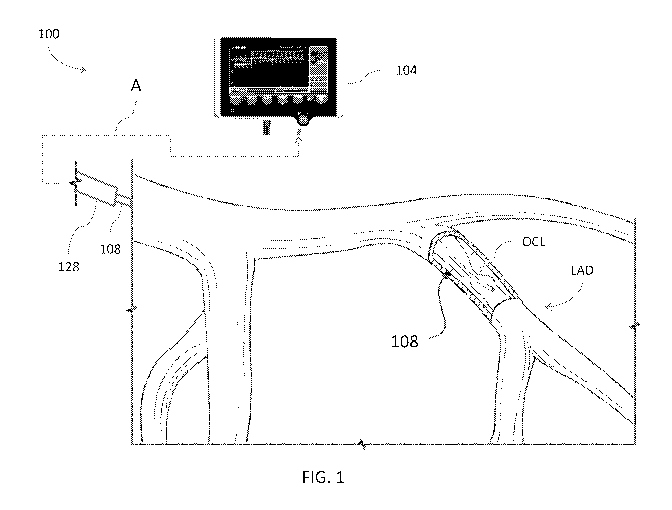
[0015] FIG. 1
is a schematic diagram showing blood vessels with a cut-out
portion in which a pressure guidewire is inserted and, spaced proximally
therefrom, a
guide catheter located proximally of the cut-out portion, e.g., in an aorta of
a patient;
[0016] FIG. 2
is a schematic diagram of an occlusion analysis system
including a pressure guidewire and a monitor assembly capable of processing
vascular
pressure data in connection with a vessel occlusion analysis;
[0017] FIG. 3
is a graphical representation of pressure signals over time
including identification of a diastolic pressure ratio zone (dPR zone) for
calculating a
metric during a segment or a portion of a heartbeat cycle;
[0018] FIG. 4
is a graphical representation similar to that of FIG. 3 in
connection with which a whole heartbeat cycle metric is described;
[0019] FIGS. 5-
6 illustrate an analysis of multiple consecutive heartbeat cycles
in calculating a multi-beat metric useful in determining whether to treat a
patient;
[0020] FIG. 7
illustrates a technique for developing a stream of data for use in
a static measurement inclusive of a high frequency sample pressure ratio
metric as well as
segment and whole heartbeat metrics over multiple consecutive heartbeats;
[0021] FIG. 8
illustrates a technique for developing a stream of data for use in
a pull-back measurement inclusive of a high frequency sample pressure ratio
metric as
well as segment and whole heartbeat metrics over multiple consecutive
heartbeats;
[0022] FIG. 8A
illustrates another technique similar to that of FIG. 8 for a
pull-back measurement;
[0023] FIGS. 9-
13 illustrate example outputs provided on a user interface of
the monitor assembly of the system of FIG. 2;
[0024] FIG. 14
is a schematic view of a blood vessel being assessed using the
methodology discussed herein; and
-4-
CA 03095596 2020-09-29
WO 2019/195323
PCT/US2019/025430
[0025] FIG. 15
is a schematic view of a blood vessel being treated following
the assessment made as illustrated in FIG. 14.
DETAILED DESCRIPTION OF THE PREFERRED EMBODIMENT
[0026] This
application is directed to systems and methods for determining
whether and how to treat a patient, where data from multiple segments of
heartbeat cycles
and/or multiple heartbeat cycles are considered. By incorporating data
indicative of both
stressed and resting heart conditions, a patient condition can be more
accurately assessed
and improved outcomes can result.
I. OVERVIEW OF PRESSURE WIRE SYSTEMS AND THEIR USE
[0027] FIGS. 1
and 2 illustrate a lesion diagnostic system 100 and the use
thereof in the vasculature of a patient. FIG. 1 illustrates the left side
coronary vasculature
with a pressure guidewire 108 disposed in a proximal portion of a left
anterior descending
artery (LAD). The pressure guidewire 108 is positioned in the left anterior
descending
artery LAD with a distal portion thereof distal to an occlusion OCL. The blood
flow in
the left anterior descending artery LAD is from proximal to distal, through
the occlusion
OCL and over the distal tip of the pressure guidewire 108. The occlusion OCL
obstructs
flow to at least some extent. The lesion diagnostic system 100 is configured
to determine
whether the extent of the obstruction is great enough to indicate that a
balloon
angioplasty, stent or other catheter intervention ought to be performed.
[0028] The
lesion diagnostic system 100 can include a monitor assembly 104
that is configured to be coupled to the pressure guidewire 108. In one
embodiment, the
lesion diagnostic system 100 includes a connection (indicated by the dashed
line A) that
facilitates connection to and disconnection of the pressure guidewire 108 from
the
monitor assembly 104. The connection to and disconnection from the monitor
assembly
104 is useful in allowing a clinician to use the pressure guidewire 108
initially for
assessing the effect of the occlusion OCL on the flow distal thereto in the
left anterior
descending artery LAD (or other coronary vessel) and to use the pressure
guidewire 108 at
a later time for delivering a treatment device such as a balloon catheter or
stent delivery
system.
[0029] The
connection indicated by the dashed arrow A also can couple a
pressure sensing component of a guide catheter assembly 128 with the monitor
assembly
-5-
CA 03095596 2020-09-29
WO 2019/195323
PCT/US2019/025430
104. The guide catheter assembly 128 can include a tubular catheter body used
to access
the vasculature. A distal tip of the guide catheter assembly 128 can be
positioned
proximal to the occlusion OCL such that pressure signals corresponding to the
pressure
proximal to the occlusion OCL, e.g., in the aorta, can be obtained. The
proximal pressure
is sometimes referred to herein as Pa.
[0030] The
pressure guidewire 108 can take any suitable form. In one
embodiment the pressure guidewire 108 includes a proximal segment that has a
proximal
end that is positioned outside the patient and a distal end that may be within
the guide
catheter assembly 128. A middle section of the pressure guidewire 108 can be
configured
to have the flexibility to navigate the tortuous vasculature of the left
anterior descending
artery LAD (or other coronary vessels) while maintaining structural integrity.
A distal
section can include a sensor housing and an atraumatic tip. Any sensing
modality can be
used. For example, an optical sensor can be configured to sense pressure when
exposed
to blood within left anterior descending artery LAD (or other coronary
vessel). The
optical sensor can be disposed within an interior space of the pressure
guidewire 108 in
fluid communication with an exterior of the pressure guidewire 108. The
optical sensor
can be selectively placed in communication with the monitor assembly 104 by a
fiber
optic signal line disposed between the sensor and a proximal end of the
pressure
guidewire 108 configured to be coupled with a fiber optic interface cable (not
shown) that
can include a guidewire connector to connect the pressure guidewire 108 with
the rest of
the system. Further details of an optical sensor based configuration of the
pressure
guidewire 108 can be found in US 2015/0057532, which is incorporated herein by
reference in its entirety.
[0031] Where
the pressure guidewire 108 is configured with an optical sensor
the ability to provide a robust optical connection with the monitor assembly
104 is of
interest. Any suitable connection structure or methodology can be used. One
approach is
described in detail in US9405078, which is incorporated by reference herein in
its
entirety.
[0032] FIG. 2
shows the flow of signal data more specifically. A clinician
attending to the patient places the guide catheter assembly 128 in the
vasculature and the
pressure guidewire 108 through the guide catheter assembly 128 into the
vasculature. The
pressure guidewire 108 provides a signal to a processor 152 which processes
the signal to
determine Pd values. The processor 152 also receives Pa values from a guide
catheter
-6-
CA 03095596 2020-09-29
WO 2019/195323
PCT/US2019/025430
signal processor 156 The Pd and Pa signals are processed in the processor 152
to generate
values of dPRc (as discussed further below). Those values can be displayed in
a dPRc
value window 144. Also, a signal trace window 148 can be provided to display
traces of
Pa, Pd, dPRc and/or any metrics that are combined into dPRc (as discussed
below). The
processor 152, the processor 152 and other processors as may be disposed in
the monitor
assembly 104 of elsewhere in the system 100 can be separate or combined into a
single
entity.
II. EXAMPLE METHODOLOGIES
A. Metrics Combining Heartbeat Segment Analysis and Whole Heartbeat Data
[0033] An
improved analysis of a patient can combine data from a segment of
a heartbeat cycle with data inclusive of a whole heartbeat cycle over one or
more than one
consecutive heartbeat cycles.
1. Heartbeat Segment Metric ¨ Diastolic Pressure Ratio (dPR)
Calculation
[0034] In one
technique, heartbeat segment data is included in a portion of a
multi-beat analysis of a patient condition. A diastolic pressure ratio (dPR)
calculation is
an example of a heartbeat segment metric. A dPR value of a given heartbeat is
determined by the mean value of a ratio of distal pressure (Pd) over proximal
pressure
(Pa) with a diastolic pressure ratio zone (dPR zone), as set forth in equation
1. As an
example, the Pd can be measured distal to the occlusion OCL and the Pa can be
measured
proximal to the occlusion OCL. Pd and Pa can be measured in un-occluded vessel
segments as well.
rx_EoD Pd(x) (Eq. 1)
L-,x=x_notch pa(x)
dPR = __________________________________
L_dPR
[0035] As noted
above, Pd is the pressure measured distal to the occlusion OCL
and is based on pressure sensed by the pressure guidewire 108. Pa can be
measured by any
suitable means, such as by the guide catheter 128. Another pressure wire or
other pressure
sensing device could also be used to measure Pa.
7
SUBSTITUTE SHEET (RULE 26)
CA 03095596 2020-09-29
WO 2019/195323
PCT/US2019/025430
[0036] FIG. 3
shows that in one technique the dPR value is calculated based
on pressure signals generated in or during a dPRzone 200. The dPRzone 200
corresponds
to a segment of a heartbeat as shown in FIG. 3. The dPRzone 200 can extend
from any of
a number of distinct portions of the heartbeat signal or a distance therefrom.
In one
embodiment the dPRzone 200 is found within a first heartbeat 204. The dPRzone
200
can end prior to a second heartbeat 208. The second heartbeat 208 is
immediately after
the first heartbeat 204. The dPRzone 200 can be defined between the dicrotic
notch 220
and the end of diastole 224 positions. FIG. 3 shows that the length of time of
the
dPRzone 200 is less than the time of the beat length 210. The beat length 210
can be
defined as the length of time between the on-set of systole of first heartbeat
204 and the
on-set of systole for the second heartbeat 208.
[0037] A new
dPR value can be obtained for every detected heartbeat, e.g., for
the first heartbeat 204, the second heartbeat 208, and as discussed further
below, a third
heartbeat 304, a fourth heartbeat 308, and a fifth heartbeat 312.
2. PTC(B) Calculation
[0038] An
analysis of a patient can include whole heartbeat data as well as
heartbeat segment data. For example, a pulse transfer coefficient (PTC) value
can be
obtained using the following method.
[0039] First a
ratio of Pd to Pa is calculated. The ratio can be calculated as a
ratio of the average distal pressure (Pd) during the entire beat divided by
the average
proximal pressure (Pa) during the entire beat. The value can be calculated
using Equation
2, shown below.
vxl_EoD Dd (x) (Eq. 2)
Z-,x=x0 EoD I
Pd pa = meanPdPaPeriod =
x1 ED D
Z-,x=x0_EoD I a kx,
[0040] The
values of Pd and Pa that are combined into the averages can be
samples taken according to a sampling frequency, such as 125 hertz. FIG. 4
shows that the
samples can be obtained throughout the first heartbeat 204. For example, the
samples
used to calculate these averages can be obtained from just after the end of
diastole 222 of
the heartbeat before the first heartbeat 204 (sometimes referred to herein as
XO_EoD) up
8
SUBSTITUTE SHEET (RULE 26)
CA 03095596 2020-09-29
WO 2019/195323
PCT/US2019/025430
to the end of diastole 224 for the first heartbeat 204 (sometimes referred to
herein as
X l_EoD).
[0041] Any
suitable approach to identify the end of diastole of the beat before
the first heartbeat 204 and the end of diastole 224 of the first heartbeat 204
can be used.
For example, an analysis of the pressure signals themselves from the pressure
guidewire
108, the guide catheter assembly 128 or both of these devices can be used to
detect the
EoD. The end of diastole 222 for the prior beat can also be calculated by
subtracting the
beat length (however calculated) from the end of diastole 224 (however
determined).
[0042] If
available, an ECG signal can be used to detect these diastolic end
points in other techniques.
[0043] A value
of a metric including the heartbeat segment data and whole
heartbeat data can thereafter be provided. In one technique a value referred
to as PTC(B)
can be calculated as a ratio of the heartbeat segment data to the whole
heartbeat data,
according to Equation 3.
dPR (Eq. 3)
PT C (B) =
Pd /
1 Pa
[0044] This
value can be calculated after the end of the first heartbeat 204 and
can be calculated for subsequent heartbeats as discussed further below.
3. PTC(B)med Calculation
[0045] FIGS. 5-
6 illustrate a further calculation of a value that considers not
only heartbeat segment data and whole heartbeat data but also considers data
from
multiple heartbeats. As discussed further below, a multi-beat metric can
include different
numbers of consecutive beats depending on the test being performed.
[0046] In one
embodiment a multi-beat metric 300 is calculated as a value of
the median of, for example, four consecutive PTC(B) values weighted based on
the
heartbeat length of the corresponding heartbeats. In another embodiment a
multi-beat
metric in connection with a pullback procedure, discussed below in connection
with FIG.
8A, is calculated as a value of the median of, for example, two consecutive
PTC(B)
values weighted based on the heartbeat length of the corresponding heartbeats.
This value
is sometimes referred to herein as PTC(B)med. The purpose of this weighted
median is to
minimize the impact of unstable signals, such as arrhythmia or other
artefacts, on metrics
9
SUBSTITUTE SHEET (RULE 26)
CA 03095596 2020-09-29
WO 2019/195323
PCT/US2019/025430
that include the PCT(B) value. One metric discussed below that includes
PTC(B)med is a dPRc
value.
[0047]
One approach to calculating PTC(B)med involves the following steps. On each
heartbeat period, there is a PTC(B)i value (PTC(B)i, PTC(B)2, ..., PTC(B)N)
and a period length Li (L1, L2, ..., LN). See FIG. 5. PTC(B)med is the
weighted median taken
on all PTC(B)i. The weight for a PTC(B)i corresponds to the heartbeat period
(Li) thereof. See
FIG. 6. This way PTC(B)med is sufficiently stable even with some PTC(B) that
correspond to
beats that are shorter than others. On FIG. 5, PTC(B)i and PTC(B)3 values
correspond to shorter
heartbeat cycles and PTC(B)2 and PTC(B)4
values
correspond to beats that are longer.
[0048]
In one methodology for static measurement, a new PTC(B)med is calculated
for every heartbeat using all four consecutive preceding heartbeats. In
another methodology for a
pullback procedure, discussed below in connection with FIG. 8A, a new
PTC(B)med is calculated
for every heartbeat using all of two consecutive preceding heartbeats.
4. dPRc calculation ¨ Static Measurement
[0049]
A metric combining heartbeat segment and whole heartbeat data, over multiple
beats can be provided in some analyses.
An example of this sort of metric is
dPRc. A dPRc value is calculated as the ratio of mean Pd to mean Pa over a
time period matching
the duration of the four consecutive heartbeats that served to calculate the
PTC(B)med, multiplied
by the PTC(B)med value previously obtained. dPRc can be calculated according
to Equation 4:
Exi+L._dPRc pd (x)
(Eq. 4)
dPRc(x) = x=xi
= PTC(B)med
rxi+L._dPRc pa(x)
Lix=xi
[0050]
In this equation L dPRc can be calculated as the sum of the length in time of
the multiple beats used to calculate the current PTC(B)med value. One static
measurement
protocol uses four consecutive beats.
[0051]
Calculating dPRc over a multiple beat (e.g., 4 beats) period provides good
stability in dPRc results. It also provides a very rapid, continuous, or rapid
and
SUBSTITUTE SHEET (RULE 26)
CA 03095596 2020-09-29
WO 2019/195323
PCT/US2019/025430
continuous stream of new dPRc values. This rapid stream of data is helpful in
measuring
conditions over time.
[0052] In case
of very stable signal, dPR and dPRc results would be similar or
even identical. However, in case of unstable signals, such as arrhythmia, dPRc
results
would be more reliable than discrete dPR values which could potentially
significantly
vary.
[0053] FIG. 7
illustrates how to determine end points (labeled as xi and x2)
over which the multi-beat ratio of pressure averages is calculated. x2 is the
position of
the current sample and xi is obtained by subtracting L_dPRc from x2. Where
L_dPRc is
the sum of heartbeat periods for the beats used in calculating PTC(B)med. In
the
illustrated case, L_dPRc = Li + L2 + L3 + L4. Because a delay is required to
detect any
heartbeat (analyzing many samples), there is always a delay between x2 and the
last
heartbeat detected.
[0054] FIGS. 9-
13 illustrate how the foregoing could be displayed on the
signal trace window 148 or in another part of the user interface 140 of the
monitor 104.
In each figure, the Pa and Pd traces are displayed and labeled. At any given
point in time
there will generally be a lower value for Pd than for Pa in the case where the
occlusion
OCL is impeding flow downstream thereof. The blue vertical lines above the
trace
represent the separate heartbeats. The horizontal line beneath the traces
labeled "dPR"
correspond to each dPR zone 200.
[0055] FIG. 9
shows an initial portion of an analysis of pressure data from the
pressure guidewire 108 and the guide catheter assembly 128. The initial
portion includes
the rising pressures associated with systole and the decreasing pressures
associated with
the on-set and initial portions of diastole in the first heartbeat 204. FIG. 9
shows only a
part of the first heartbeat 204. FIG. 10 shows the first heartbeat 204, the
second heartbeat
208, and the third heartbeat 304. For each beat the dPR value can be
calculated as
described above in the corresponding dPRzone 200.
[0056] FIG. 11
shows the first, second, and third beats and the fourth heartbeat
308. After the first heartbeat 204, second heartbeat 208, third heartbeat 304,
and fourth
heartbeat 308 have been detected and analyzed dPRc or another multi-beat
metric
combining segment and whole beat data can be calculated for these four beats.
The user
interface 140 can be configured to include a dPRc trace window 150 to display
dPRc or
another multi-beat metric combining segment and whole beat data. FIG. 10 shows
that
-11-
CA 03095596 2020-09-29
WO 2019/195323
PCT/US2019/025430
prior to sufficient consecutive beats being detected a 0 value can be
displayed for dPRc
and no trace is presented in the dPRc trace window 150. After four (or another
sufficient
number of beats) have been detected and analyzed the dPRc trace window 150 can
be
modified to display one or both of a dPRc value and a dPRc trace as shown in
FIG. 11.
[0057] FIG. 12
shows how the user interface 140 illustrates that the analysis of
dPRc is updated for fifth and subsequent consecutive beats. A new dPRc value
is
calculated based on the first heartbeat 204, the third heartbeat 304, the
fourth heartbeat
308, and a fifth heartbeat 312. The new dPRc value is generated following the
same
protocol noted above, where PTC(B)median is the weighted median of the second,
third,
fourth and fifth beats and the pressure ratio multiplier in equation 4 is
based on a new
time period of L_dPRc as the sum of the beat lengths for the second heartbeat
208, third
heartbeat 304, fourth heartbeat 308, and fifth heartbeat 312 (sum of Li, L2,
L3, and L4).
The new dPRc value and/or the dPRc trace is updated in the dPRc trace window
150 on
the user interface 140. FIG. 13 shows further calculation of the dPRc metric
later in time,
using the third heartbeat 304, the fourth heartbeat 308, the fifth heartbeat
312, and a sixth
heartbeat 316. Again, the new dPRc value and/or the dPRc trace is updated in
the dPRc
trace window 150 on the user interface 140.
[0058] Based on
the analysis, a threshold value can be established above
which a patient is not treated and below which a treatment such as angioplasty
or stenting
is performed. As shown in FIGS. 14 and 15 both the assessment of dPRc and the
treatment can be performed over the pressure guidewire 108. By updating the
dPRc value
over time the user can see the stability of the metric and gain confidence in
next clinical
steps, such as whether to treat with a balloon, a stent or other method. Also,
the output in
the dPRc trace window 150 can be updated as fast as the samples of Pa and Pd
are taken,
e.g., every 8 ms based on a sampling rate of 125 hertz. In some cases, the
screen can be
updated less frequently but still much faster than every second, e.g., 30
times per second.
This protocol provides effectively a continuous stream of data, e.g., a stream
of data
updated more often than every heartbeat, updated more than once per second,
updated
more than twice per second, updated more five times per second, updated more
than ten
times per second, updated more fifty times per second, updated more than one
hundred
times per second.
-12-
CA 03095596 2020-09-29
WO 2019/195323
PCT/US2019/025430
5. dPRc calculation ¨ Pullback Measurement
[0059] While
the foregoing has been focused largely on a static position
measurement, that is one made with at least the pressure guidewire 108 held
stationary,
another mode involves obtaining pressure data and analyzing the data while at
least the
pressure guidewire 108 is moving. Generally the movement of the guidewire 108
that is
provided is in the proximal direction from a distal position in the
vasculature toward a
proximal position adjacent to the distal end of the guide catheter assembly
128. This
motion can be provided by the clinician pulling back on the pressure guidewire
108
directly manually or using a device configured to generate a controlled
proximal
movement.
[0060] FIG. 8
illustrates one embodiment of a pullback mode analysis. In this
example, dPRc is calculated by Equation 4.
xi+xLdPRc pd (x) (Eq. 4)
dPRc(x) = xi_ 7 = PT C (B)med
vx1+1,._dPRc pa(x)
L-lx=x1
[0061] One
difference, however, is PTC(B)med can be based on the most
recent three beats. Also, L_dPRc is the average period of the three beats
(e.g., a first best
204A, a second best 208A, and a third beat 304A) used to calculate PTC(B)med.
In other
words, the first term in Equation 4 is the average distal pressure over the
time L_dPRc
divided by the average proximal pressure over the time L_dPRc. FIG. 8 shows
the
window between xl and x2 as between the time of the current pressure sample
data back
by the amount of L_dPRc.
[0062] FIG. 8A
shows another technique for conducting an analysis in a
pullback mode. This technique is similar to that if FIG. 8 except as described
differently
below. Here two beats (204A, 208A) are used in calculating PTC(B)med. This
value is
multiplied by the ratio of Pd/Pa, calculated as expressed in Equation 4.
However, in this
calculation L_dPRc is the sum of the period of the two beats, shown as the
time between
X1 and X2. This can be calculated as the time between the start of systole for
the beat
204A and the time for systole for the beat 304A. The window for calculating
the Pd/Pa
will shift out in time for each new sample, e.g., every 8 milliseconds. The
value of
L_dPRc can be calculated every time a new value of PTC(B)med is calculated,
e.g., after
the end of each full beat. One advantage of the approach discussed in
connection FIG. 8A
is that is provides faster response time than an approach requiring more than
two beats to
13
SUBSTITUTE SHEET (RULE 26)
CA 03095596 2020-09-29
WO 2019/195323
PCT/US2019/025430
present a pullback mode value. If a more stable value is desired more beats
can be used,
similar to the method of FIG. 8. Another advantage of the algorithm discussed
in
connection with FIG. 8A is that is includes an analogous calculation as is
used for the
static or stationary mode, but using two beats rather than four as used in the
static or
stationary mode.
[0063] The
foregoing approaches to dPRc provides a rapid stream of data over
time which provides more clarity for the pullback mode.
B. Advantages
[0064] The
foregoing discusses using an average of a plurality of ratios of Pd
to Pa as part of calculating a useful blood vessel occlusion evaluation
metric. The
averaging of these ratios provides advantages. For example, whenever noise is
present
the average of the ratios is more accurate than other manners of combining
multiple
measurements, such as calculating a ratio of an average of multiple distal
pressure
measurements to an average of multiple proximal pressure measurements. This is
particularly true whenever the Pa exhibits large pressure excursion caused by
pressure
tube movement or other similar sources of noise.
[0065] The dPRc
method including the calculation of PTC(B)med allows
reliable dPR calculation without the need for analyzing and removing any data
associated
with heartbeats that may actually be irregular in some way. This method thus
can be
carried out without any need to determine a priori any and all criteria that
would justify
removing or discarding data associated with irregular heartbeats.
[0066] In pull
back technique, a faster stream of data is available, allowing
rapid response of the dPRc measurement and hence, enhanced spatial resolution.
Terminology
[0067] As used
herein, the relative terms "proximal" and "distal" shall be
defined from the perspective of the user of the system. Thus, proximal refers
to the
direction toward the user of the system and distal refers to the direction
away from the
user of the system.
[0068]
Conditional language, such as "can," "could," "might," or "may,"
unless specifically stated otherwise, or otherwise understood within the
context as used, is
generally intended to convey that certain embodiments include, while other
embodiments
-14-
CA 03095596 2020-09-29
WO 2019/195323
PCT/US2019/025430
do not include, certain features, elements, and/or steps. Thus, such
conditional language
is not generally intended to imply that features, elements, and/or steps are
in any way
required for one or more embodiments.
[0069] The
terms "comprising," "including," "having," and the like are
synonymous and are used inclusively, in an open-ended fashion, and do not
exclude
additional elements, features, acts, operations, and so forth. Also, the term
"or" is used in
its inclusive sense (and not in its exclusive sense) so that when used, for
example, to
connect a list of elements, the term "or" means one, some, or all of the
elements in the
list.
[0070] The
terms "approximately," "about," "generally," and "substantially"
as used herein represent an amount close to the stated amount that still
performs a desired
function or achieves a desired result. For example, the terms "approximately,"
"about,"
"generally," and "substantially" may refer to an amount that is within less
than 10% of the
stated amount, as the context may dictate.
[0071] The
ranges disclosed herein also encompass any and all overlap, sub-
ranges, and combinations thereof. Language such as "up to," "at least,"
"greater than,"
"less than," "between" and the like includes the number recited. Numbers
preceded by a
term such as "about" or "approximately" include the recited numbers. For
example,
"about four" includes "four"
[0072] Any
methods disclosed herein need not be performed in the order
recited. The methods disclosed herein include certain actions taken by a
practitioner;
however, they can also include any third-party instruction of those actions,
either
expressly or by implication. For example, actions such as "distally moving a
locking
element" include "instructing distal movement of the locking element."
[0073] Although
certain embodiments and examples have been described
herein, it will be understood by those skilled in the art that many aspects of
the humeral
assemblies shown and described in the present disclosure may be differently
combined
and/or modified to form still further embodiments or acceptable examples. All
such
modifications and variations are intended to be included herein within the
scope of this
disclosure. A wide variety of designs and approaches are possible. No feature,
structure,
or step disclosed herein is essential or indispensable.
[0074] Some
embodiments have been described in connection with the
accompanying drawings. However, it should be understood that the figures are
not drawn
-15-
CA 03095596 2020-09-29
WO 2019/195323
PCT/US2019/025430
to scale. Distances, angles, etc. are merely illustrative and do not
necessarily bear an
exact relationship to actual dimensions and layout of the devices illustrated.
Components
can be added, removed, and/or rearranged. Further, the disclosure herein of
any particular
feature, aspect, method, property, characteristic, quality, attribute,
element, or the like in
connection with various embodiments can be used in all other embodiments set
forth
herein. Additionally, it will be recognized that any methods described herein
may be
practiced using any device suitable for performing the recited steps.
[0075] For
purposes of this disclosure, certain aspects, advantages, and novel
features are described herein. It is to be understood that not necessarily all
such
advantages may be achieved in accordance with any particular embodiment. Thus,
for
example, those skilled in the art will recognize that the disclosure may be
embodied or
carried out in a manner that achieves one advantage or a group of advantages
as taught
herein without necessarily achieving other advantages as may be taught or
suggested
herein.
[0076]
Moreover, while illustrative embodiments have been described herein,
the scope of any and all embodiments having equivalent elements,
modifications,
omissions, combinations (e.g., of aspects across various embodiments),
adaptations
and/or alterations as would be appreciated by those in the art based on the
present
disclosure. The limitations in the claims are to be interpreted broadly based
on the
language employed in the claims and not limited to the examples described in
the present
specification or during the prosecution of the application, which examples are
to be
construed as non-exclusive. Further, the actions of the disclosed processes
and methods
may be modified in any manner, including by reordering actions and/or
inserting
additional actions and/or deleting actions. It is intended, therefore, that
the specification
and examples be considered as illustrative only, with a true scope and spirit
being
indicated by the claims and their full scope of equivalents.
-16-