Note: Descriptions are shown in the official language in which they were submitted.
OPTHALMOLOGICAL IMAGING AND LASER DELIVERY DEVICE, SYSTEM AND
METHODS
TECHNICAL FIELD
[0001] The current application relates to ophthalmological devices, systems
and methods and
in particular to devices, systems and methods for imaging and laser treatment
of an eye.
BACKGROUND
[0002] Imaging of an eye is important for identifying conditions of the eye.
Various imaging
techniques may be used for capturing images of the interior compartments of
the eye. For
example, scanning laser ophthalmoscopy (SLO) imaging may provide 2-dimensional
image of
a portion of the eye, such as the retina or of cornea. Optical coherence
tomography (OCT)
imaging may provide 3- dimensional and/or cross-section images of a portion of
the retina or
cornea. Other imaging techniques may be used for capturing an image of at
least a portion of
the fundus of the eye.
[0003] Imaging of the eye may be used for identifying eye conditions requiring
treatment.
Treatment of eye conditions may be performed using lasers, with the specific
targeting
location of the laser beam or pulse determined from the captured images.
[0004] An additional, new and/or improved ophthalmological device capable of
imaging and
treating one or more eye conditions is desirable.
SUMMARY
[0005] In accordance with the present disclosure there is provided an imaging
and laser
delivery device for treatment of an eye condition comprising: a scanning laser
ophthalmoscopy (SLO) optical pathway for SLO imaging; an optical coherence
tomography
(OCT) optical pathway for OCT imaging; a treatment optical pathway for a
treatment laser;
and a delivery optical pathway comprising an objective lens for focusing light
from the SLO
optical pathway, the OCT optical pathway and the treatment optical pathway
onto a portion of
an eye being treated for the eye condition.
[0006] In a further embodiment, the imaging and laser delivery device further
comprises a
device controller for: controlling operation of components of the SLO optical
pathway, the
OCT optical pathway and the treatment optical pathway; and providing an
interface between
the laser delivery device and a computing device.
1
Date Recue/Date Received 2020-10-16
[0007] In a further embodiment, the imaging and laser delivery device further
comprises: an
SLO light source or SLO light source port for coupling the laser delivery
device to an external
SLO light source; an OCT light source or OCT light source port for coupling
the laser delivery
device to an external OCT light source; and a treatment light source or
treatment light source
port for coupling the laser delivery device to an external treatment light
source.
[0008] In a further embodiment of the imaging and laser delivery device, the
SLO light source
or external SLO light source operate at an SLO wavelength; the OCT light
source or external
OCT light source operate at an OCT wavelength; the treatment light source or
external
treatment light source operate at a treatment wavelength, and wherein each of
the SLO
wavelength, OCT wavelength and treatment wavelength are different wavelengths.
[0009] In a further embodiment of the imaging and laser delivery device, the
delivery optical
pathway comprises one or more optical devices for separating returning light
from the eye
through the objective lens and delivering a portion of the returning light to
one of the SLO
optical pathway or the OCT optical pathway based on the wavelength of the
portion of the
returning light.
[0010] In a further embodiment of the imaging and laser delivery device, the
SLO optical
pathway comprises: XY scanning optics for scanning an SLO beam across a
portion of the
eye; a detector for detecting light returning from the eye through a portion
of the SLO optical
pathway.
[0011] In a further embodiment of the imaging and laser delivery device, the
XY scanning
optics comprise one or more of: a galvonmeter; a resonant scanner; a non-
resonant scanner;
a spinning mirror; and a spinning prism.
[0012] In a further embodiment of the imaging and laser delivery device, the
OCT optical
pathway comprises: an optical splitter/combiner coupled to an OCT light source
and an OCT
detector; a sample optical pathway optically coupling the optical
splitter/combiner to the
delivery pathway; and a reference optical pathway optically coupling the
optical
splitter/combiner to a return mirror, wherein light returning from the sample
optical pathway
and the reference optical pathway are combined in the optical
splitter/combiner before being
detected by the OCT detector.
2
Date Recue/Date Received 2020-10-16
[0013] In a further embodiment of the imaging and laser delivery device, the
position of the
return mirror is adjustable in order to lengthen or shorten a length of the
reference pathway.
[0014] In a further embodiment of the imaging and laser delivery device, the
reference
pathway comprises an adjustable thickness material for compensating for
dispersion within
the eye.
[0015] In a further embodiment of the imaging and laser delivery device, the
treatment optical
pathway comprises at least one of adaptive optics, prism pair, grating pair,
dielectric mirror
coatings, and optical fiber for pre-compensating a treatment laser pulse based
on the
thickness of the adjustable thickness material in the reference pathway of the
OCT optical
pathway.
[0016] In a further embodiment, the imaging and laser delivery device further
comprises: a
second therapeutic laser.
[0017] In a further embodiment, the imaging and laser delivery device further
comprises: an
alignment system for aligning the therapeutic laser to the OCT optical
pathway.
[0018] In a further embodiment of the imaging and laser delivery device, the
alignment system
comprise a coarse alignment section and a fine alignment section.
[0019] In a further embodiment of the imaging and laser delivery device, the
coarse alignment
section comprise a pair of CMOS sensors arranged at respective ends of
different length
optical paths of a coarse alignment beam split from the therapeutic laser.
[0020] In a further embodiment of the imaging and laser delivery device, the
coarse alignment
beam is split from the therapeutic laser before injection into the OCT
pathway.
[0021] In a further embodiment of the imaging and laser delivery device, the
fine alignment
section comprises a pair of quadrature photodiodes (QPD) arranged at
respective ends of
different length optical paths of a fine alignment beam split from the
therapeutic laser.
[0022] In a further embodiment of the imaging and laser delivery device, the
alignment system
comprises positioning optics for controllably adjusting the alignment of the
therapeutic laser.
3
Date Recue/Date Received 2020-10-16
[0023] In a further embodiment of the imaging and laser delivery device, the
alignment system
uses a positive reinforcement learning algorithm to control the positioning
optics independent
of optical geometry.
[0024] In a further embodiment, the imaging and laser delivery device further
comprises a
pilot laser.
[0025] In a further embodiment of the imaging and laser delivery device, the
pilot laser passes
through a portion of the OCT pathway.
[0026] In a further embodiment of the imaging and laser delivery device, the
therapeutic laser
is a femtosecond laser.
[0027] In accordance with the present disclosure there is provided a laser
imaging and
delivery system for treatment of an eye condition comprising: an imaging and
laser delivery
device as described above; and a computing device for controlling operation of
the imaging
and laser delivery device and providing a graphical user interface to a user
of the imaging and
laser delivery system.
[0028] In a further embodiment of the imaging and laser delivery system, the
computing
device is configured to: capture SLO images and OCT images; register the
captured SLO
images and OCT images to planning images of a treatment plan for treating the
eye
condition; and controlling the treatment laser according to the treatment
plan.
[0029] In a further embodiment of the imaging and laser delivery system, the
computing
device is further configured to: track eye movement using the captured SLO
images; and
control the treatment laser according to the treatment plan and the tracked
eye movement.
[0030] In a further embodiment of the imaging and laser delivery system, the
computing
device is further configured to: identify unsafe regions for laser treatment
within the eye; and
stop the treatment laser if treatment will occur within the unsafe regions.
[0031] In a further embodiment of the imaging and laser delivery system, the
computing
device is further configured to: generate a graphical user interface (GUI)
displaying the SLO
images and OCT images.
4
Date Recue/Date Received 2020-10-16
[0032] In a further embodiment of the imaging and laser delivery system, the
GUI is used to
generate the treatment plan.
[0033] In a further embodiment of the imaging and laser delivery system, the
GUI displays
progress of a treatment plan during treatment.
[0034] In accordance with the present disclosure there is further provided a
use of the
imaging a laser delivery system as described above in the treatment of one or
more eye
conditions comprising diabetic retinopathy, age-related macular degeneration,
vitreomacular
traction, tears, detachments and holes, glaucoma, and vein occlusion.
BRIEF DESCRIPTION OF THE DRAWINGS
[0035] Further features and advantages of the present disclosure will become
apparent from
the following detailed description, taken in combination with the appended
drawings, in which:
[0036] FIG. 1 depicts components of an ocular imaging and laser treatment
system;
[0037] FIG. 2 depicts optical paths of an imaging and laser treatment system;
[0038] FIG. 3 depicts components of an SLO imaging portion of the imaging and
laser
treatment system;
[0039] FIG. 4 depicts components of an OCT imaging portion of the imaging and
laser
treatment system;
[0040] FIG. 5 depicts components of a laser delivery portion of the imaging
and laser
treatment system;
[0041] FIG. 6 depict optical components of a further ocular imaging and laser
treatment
system;
[0042] FIG. 7 depict optical components of an alignment system;
[0043] FIG. 8 depicts a method of planning and performing a treatment for an
ocular condition
using an ocular imaging and laser treatment system;
[0044] FIG. 9 depicts a graphical user interface flow for planning and
performing a treatment
for an ocular condition;
Date Recue/Date Received 2020-10-16
[0045] FIG. 10 depicts a further graphical user interface flow for performing
a treatment for an
ocular condition;
[0046] FIG. 11 depicts a method for planning an ocular treatment of an ocular
condition;
[0047] FIG. 12 depicts a method of treating an ocular condition; and
[0048] FIG. 13 depicts a convex shape surface on a fundus image and OCT image
of a
patient with vitreomacular traction.
DETAILED DESCRIPTION
[0049] An imaging and laser treatment system is described that includes both a
scanning
laser ophthalmoscopy (SLO) imaging device, and an optical coherence tomography
(OCT)
imaging device for imaging the eye simultaneously using both devices.
Additionally, the
imaging and laser delivery system includes a treatment laser that can be used
for carrying out
treatment of an ocular condition. The treatment laser may be a therapeutic
laser or surgical
laser. The SLO imaging, OCT imaging, and therapeutic laser may pass through a
common
objective lens for delivery to the eye being imaged and/or treated. Further,
the SLO imaging
device, or more particularly the images from the SLO imaging device, may be
used to identify
eye movement and account for the eye movement in the OCT imaging device and/or
the
targeting of the therapeutic laser. The combination of the SLO imaging, OCT
imaging, and
therapeutic laser can provide a system that allows for both planning and
performing a
treatment of an ocular condition using a single system. While the planning and
treatment
may be performed at separate times, which may require the individual to return
one or more
times, the planning and treatment may also be performed at a single time. It
will be
appreciated that additional components may be included in the imaging and
laser treatment
system, including for example fundus imaging components, a pilot laser system,
additional
treatment lasers, etc.
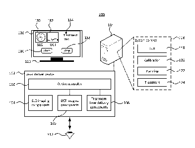
[0050] FIG. 1 depicts components of an ocular imaging and laser treatment
system. The
system 100 comprises an imaging and laser delivery device 102. The device 102
comprises
SLO imaging components 104, OCT imaging components 106 and treatment laser
delivery
components 108. The imaging and laser delivery components may be controlled by
a device
controller 110. The light for the SLO imaging, OCT imaging and treatment laser
may be
6
Date Recue/Date Received 2020-10-16
delivered to an eye 112, or possibly other target, being imaged and/or
treated. The imaging
light for SLO and OCT imaging is reflected back to the respective detectors.
[0051] The device controller 110 may provide an interface between the device
102 and a
computing device 114. The computing device 114 provides various system control
functionality 116 for operating the imaging and laser delivery device 102.
While the
computing device 114 is depicted as a separate computing device 114, it is
possible to
incorporate the computing device 114 into the imaging and laser delivery
device 102. The
device controller 110 may capture signals from respective detectors/camera of
the SLO, and
OCT imaging components 104, 106 as well as controlling other components, such
as the
sources of the imaging components, 104, 106, and treatment laser delivery
components 108,
focusing components, or other components.
[0052] The computing device 114 may comprise one or more processing units (not
depicted)
for executing instructions, one or more memory units (not depicted) storing
data and
instructions, which when executed by the one or more processing units
configure the
computing device to provide the system control functionality 116. The system
control
functionality 116 may include graphical user interface (GUI) functionality 118
that provides a
GUI for operating the imaging and laser delivery device. Calibration
functionality 120 may be
provided in order to calibrate the imaging and laser delivery device 102 and
in particular to
align and correlate the SLO imaging components 104, OCT imaging components 106
and the
treatment laser delivery components 108 so that locations in the SLO images
and OCT
images can be precisely aligned with each other and be accurately targeted by
treatment
laser. Planning functionality 122 may be provided that allows a treatment plan
to be
developed for treating a particular ocular condition. The planning
functionality 122 may use
the GUI functionality to allow a user to define the treatment plan.
Additionally or alternatively,
the planning functionality may incorporate automated, or semi-automated,
planning
functionality that may identify laser treatment locations within the captured
images.
Treatment functionality 124 may control the components of the device 102,
including the
treatment laser delivery components 108, in order to carry out the treatment
plan in order to
treat, or at least partially treat, an ocular condition.
[0053] The GUI functionality 118 may present the generated GUI on a display
126. Although
depicted as a separate display, the display could be incorporated into the
imaging and laser
7
Date Recue/Date Received 2020-10-16
delivery device 102. Although the GUI presented may vary depending upon what
information
needs to be, or may be desirable to be, displayed to the user. FIG. 1 depicts
a GUI 129 that
could be displayed during treatment. For example, the GUI may display a SLO
image 130,
and an OCT image 132. The SLO image may include an indication of the location
of the
cross section of the OCT image. The SLO image, and the OCT image may include
indications of treatment locations that have not yet been treated as well as
treatment
locations that have been treated. The GUI 134 may include other details of the
treatment
plan that may be relevant to the user as well as graphical elements for
starting 136, and
stopping 138 the treatment.
[0054] The device 102 and system 100 depicted in FIG. 1 broadly comprise the
optical
hardware, control electronics and software. The components are described in
further detail
below. The system 100 may be used for imaging eyes to identify areas for
treatment and
carrying out the treatment. The treatment may be for a wide range of different
ocular
conditions including, for example, age-related macular degeneration (AMD),
vitreomacular
traction syndrome (VTS), diabetic retinopathy, cataracts, choroidal
neovascularization,
microaneurysm, glaucoma, epiretinal membrane (ERM), retinal tears and
detachment, central
or branch vein occlusion.
[0055] FIG. 2 depicts optical paths of an imaging and laser delivery system.
As depicted, the
imaging and laser delivery device 102 comprises a SLO source 202. The SLO
source 202
may be for example a diode laser, gas laser, dye laser, solid-state laser,
continuous wave
laser, pulsed laser, ultrashort laser pulses, super radiant diode light
source, non-linearly
generated light from nonlinear optical material (e.g. supercontinuum light,
harmonic
generation light, sum or difference frequency generated light) or a port for
coupling the device
102 to the laser. The SLO laser may operate at a range of different
wavelengths, including
for example between 100nm-3000nm. The OCT source 204 may comprise a low-
coherence
light source suitable for use in OCT imaging such as for example a
superluminesccent diode,
ultrashort laser pulses, super radiant diode light source, non-linearly
generated light from
nonlinear optical material (e.g. supercontinuum light, harmonic generation
light, sum or
difference frequency generated light), or a port for coupling the device 102
to the
superluminesccent diode. The treatment laser source 206 may comprise a
treatment laser or
a port for coupling the device 102 to the treatment laser. The treatment laser
may be for
example a gas laser, fiver laser, dye laser, a fiber or free-space
8
Date Recue/Date Received 2020-10-16
femtosecond/picosecond/nanosecond laser, a solid-state laser (pulsed or
continuous
wavelength), a pulsed or continuous wavelength diode laser, an optical
parametric oscillator,
an optical amplifier optical, and optical parametric amplifier, or a coherent
light generated
form nonlinear optical processes (e.g. sum, difference, and second harmonic
light generation)
etc. The device controller 110 may provide control signals to the light
sources in order to
control them including for example, turning the lasers/lights on and off as
well as possibly
adjusting controllable parameters.
[0056] Each of the sources 202, 204, 206 is coupled to respective optical
paths 208, 210, 212
that direct the light from the sources to the target 112. Each of the optical
paths may have
various optical elements including lenses, beam splitters, combiners, mirrors,
filters
polarizers, adaptive optics, prisms, gratings, optical fibers, etc. The light
from the sources
may pass through a beam splitter/combiner 214 that combines and directs light
output from
each of sources to the eye 112 through one or more telescope lenses 216 that
focus the light
on the eye. A contact lens or a combination of contact lenses may be used on
the eye in
order to better couple the light from the telescope lenses to the eye.
[0057] Light from the treatment laser can be used for imaging of the eye,
however, the
treatment light returning from the eye does not need to be directed to a
detector. In contrast,
the light from the SLO, and OCT sources reflects of portions of the eye being
imaged and the
reflected light may be split by the beam splitter 214 and directed back to the
respective optical
paths 208, 210. The returning light may be split based on for example the
wavelengths used
for SLO, and OCT imaging, or if the same or similar wavelengths are used and
as such
splitting the returning light based on the wavelength is impossible, or
difficult, the beam may
be split based on polarization if the SLO and OCT light have different
polarization states.
[0058] The optical elements of both the SLO and OCT optical paths direct the
light from the
source to the target 112, and for the SLO imaging and OCT imaging direct
returning light of
each source to a SLO detector 218 and OCT detector 220 respectively. Each of
the optical
paths 208, 210, 212 are described in further detail below with regard to FIGs.
3 to 5.
Additional optical systems, not depicted in FIG. 2, may be included in the
imaging and
treatment system. For example, one or more fundus cameras may be incorporated
for
imaging the eye, one or more pilot laser systems, additional treatment laser
systems, etc.
9
Date Recue/Date Received 2020-10-16
[0059] FIG. 3 depicts components of an SLO imaging portion of the imaging and
laser
treatment system. The SLO source 202 outputs light useful for SLO imaging. The
SLO
optical path may comprise free-space optical elements that are arranged to
deliver the light
from the source 202 to the eye 112 and direct the returning light from the eye
to the SLO
detector. The SLO optical path includes a beam splitter, or other optical
device, such as an
optical circulator or a directional coupler, 302 capable of directing light
from the source 202 to
the eye 112 through scanning optics 304, the beam splitter 214 and the
telescope optics 216,
which may comprise one or more lenses, filters, apertures, etc. The telescope
optics 216 may
include one or more lenses capable of moving along a Z-axis, which beings the
lenses away
from or towards the eye. Moving the optics along the Z-axis can change the
focus to different
parts of the eye such as the cornea or the retina or anywhere inside the eye's
internal
compartments. The scanning optics 304 comprise optical devices capable of
scanning the
light across the eye. As depicted, the devices may include a galvanometer, or
galvo, 306 that
can scan the light through a first axis, such as the Y-axis, as well as a
resonant scanner 308
that scans the light through a second orthogonal axis, such as the X-axis.
Although described
as using a combination of a galvo and a resonant scanner, it is possible to
use other devices
to scan along either the X or Y axis. For example the scanning optics could be
provided by
an acousto-optic deflector, an electro-optic deflector, a non-resonant
scanner, spinning
mirrors, spinning prisms, micro electro-mechanical (MEM) mirrors. Further,
rather than using
two scanning devices it is possible to use a single optical scanning device
capable of
controllably scanning the optical beam in both the X and Y axis such as a 2D
MEMs mirror.
The resonant scanner 308 is capable of providing a high scanning rate as it
operates at a
significantly higher rate than the galvo. For example, in order to generate a
512x512 raster
image of the eye, the resonant scanner will need to direct the light to 512
locations each time
the galvo moves to a new row position. It is possible that the XY-scanning
optics 304 use two
galvos, although the imaging rate may be reduced. Other devices, such as micro
mirror
devices, may be used for scanning the light across the eye in a raster
pattern. The XY-
scanning optics 304 may be driven by driving circuitry 312 in the device
controller 110.
Signals from the resonant scanner may be captured by a data acquisition
circuitry 314 which
may be used to synchronize movement of the galvo with that of the resonant
scanner so that
a new row is moved to when a scan of a column is completed by the resonant
scanner.
Date Recue/Date Received 2020-10-16
[0060] Reflected light from the eye returns through the same optical path to
the beam splitter
302, which splits the returning light from the light of the source and directs
the returning light
to the SLO detector, which is depicted as an avalanche photodetector (APD)
310. The APD
signal may be captured by the data acquisition circuitry 314. Although
depicted as an APD,
other detectors are possible, including for example a tube photomultiplier of
a photodiode with
an amplifier or a semiconductor-based photo multiplier or a charged coupled
device, or a
camera. The data acquisition circuitry may operate substantial as an
electronic device that
can measure the voltage/current of relevant signals at a high enough frequency
to properly
measure the signals. The device controller may provide an interface that may
be used to
provide the captured data, including the imaging data, to the computer 114 as
well as receive
control information for controlling the SLO source and scanning optics from
the computer 114.
[0061] The optical path may include additional components including, for
example one or
more lenses, mirrors, gratings, etc. for focusing and/or directing light, one
or more filters,
apertures, etc. The additional components may provide additional functionality
such as
wavefront aberration detection and correction or compensation, intensity
detection and
correction or compensation.
[0062] The above has described using a single SLO source of a Additionally,
for example, the
SLO source may have multiple individual light sources, such as a red, green
and blue source
that are combined into a single beam. Using combined red, green and blue light
sources, and
corresponding detectors, allows true colour SLO images to be captured.
Additionally or
alternatively, it is possible to use a femtosecond laser as the SLO source, it
may be possible
to provide real time flourescin angiography. Further still, although not
depicted in FIG. 3 it is
possible for the optical source, or the optical path, may include adaptive
optics that can
significantly improve imaging resolution allowing the visualization of, for
example, single cells.
[0063] FIG. 4 depicts components of an OCT imaging portion of the imaging and
laser
treatment system. OCT imaging uses an OCT source 206 that may be, for example,
a super
luminescent diode. The source beam is split by a fiber coupler 402, or other
optical
components, that is capable of splitting and combining light to and from
different ports. The
OCT source beam is split by the fiber coupler 402 into a sample 404 that
includes scanning
optics 406, which can scan the optical beam in both an X and Y direction. The
scanning
optics 406 may comprise galvos and/or resonators similar to the XY scanning
optics 304 of
11
Date Recue/Date Received 2020-10-16
FIG. 3 or other scanning devices. The scanning optics 406 may be controlled by
the device
controller 11 in a similar manner as described above with regard to the
driving circuitry 312 of
FIG. 3. The OCT sample path delivers the OCT source beam to an eye or target
through the
beam splitter/combiner 214 that combines and directs light output from the OCT
path and the
SLO path to the eye 112 through one or more telescope lenses 216. As described
above, the
telescope lenses 216 may be moved toward or away from the eye 112, defined as
the Z-axis,
in order to change a focus point of the OCT source beam on the eye. As
described above, a
contact lens may be placed on the eye in order to deliver the OCT source beam
to the eye.
The reflected light returns through the OCT sample path back to the fiber
coupler 402 where it
is combined with light returning from an OCT reference path and the combined
light, or a
portion of the combined light, provided to the OCT detector 220. Both the OCT
source 206
and detector 220 may be controlled by the device controller 110, which can
provide an
interface to the computing device 114 to allow the computing device 114 to
control operation
of the imaging and laser delivery system as well as to receive the captured
image data from
the OCT detector.
[0064] The reference path 408 provides a path for the OCT light beam, or
portion thereof that
was split by the fiber coupler, that has the same path length as the OCT light
beam travelling
in the sample path, so that the interference of the combined light from the
sample path and
reference path provide information which can be used to provide an image of
the portion of
the eye targeted by the sample path. In order to compensate for changing path
lengths of the
sample path, which may result from, for example, different targeting/focusing
locations within
the eye, as well as changes in position of the eye, the reference path may
include a mirror
410 that is moveable in the Z-axis in order to lengthen or shorten the path
length of the
reference path. The moveable mirror 410 reflects the light back through the
reference path to
be combined with the light from the sample path in the fiber coupler 402. The
device
controller 110 may synchronize the moveable mirror with the moveable telescope
lenses so
that movement of the telescope lenses results in corresponding movement of the
mirror 410
to maintain the path lengths of the sample path and reference path.
[0065] In addition to the moveable mirror, which accounts for changing path
lengths of the
sample path, the reference path may have dispersion compensation components,
depicted as
a pair of wedges 412 that can be adjusted to provide a thicker or thinner
material for the
reference beam to pass through. The dispersion compensation components 412 can
be used
12
Date Recue/Date Received 2020-10-16
to account for the optical properties of the eye itself, which may be
particularly useful in OCT
imaging which may be used to image the back, or retina, of the eye. The
dispersion
compensation components 412 may be controlled by the device controller 110 in
coordination
with the computing device 114. In particular, the computing device 114 may
include OCT
dispersion compensation control functionality 414 that adjusts the dispersion
compensation
components, for example by moving the wedges in or out to provide a thicker or
thinner
optical component, in order to provide a focused image captured by the OCT
detector. That
is, when the dispersion compensation component is properly adjusted to account
for the
optical properties of the eye being imaged, the image captured by the OCT
detector will be in
sharp focus. The OCT dispersion compensation control functionality may be
based on
autofocus techniques which adjust the focusing optics based on a sharpness of
the captured
image. The dispersion compensation components may be adjusted until a sharp
image is
produced.
[0066] The amount of dispersion compensation provided by the dispersion
compensation
components 412 may also be used for other purposes in addition to compensating
the OCT
reference beam. Since the particular compensation provided by the dispersion
compensation
components, for example the 'thickness' of the component 412, provides an
indication of the
optical properties of the eye, the particular compensation may be used for
other
compensation, including for example, post-compensation of SLO images, which
may
comprise image processing techniques, as well as controlling optical
compensation
components in order to provide pre-compensation of the treatment laser pulse.
The temporal
pulse compression and frequency pre-compensation may be performed by, for
example,
treatment laser dispersion pre-compensation functionality 416, which may
adjust pulse pre-
compensation components in the treatment optical pathway based on the
compensation
required to provide a sharp in-focus OCT image as determined by the OCT
dispersion
compensation control functionality 414.
[0067] FIG. 5 depicts components of a treatment laser delivery portion of the
imaging and
laser treatment system. The treatment laser is depicted in FIG. 5 as being a
femtosecond,
picosecond, or a nanosecond pulsed laser 502, however other laser sources may
be used
depending upon the particular application. The treatment laser may pass
through pre-
compensation optics 504 and targeting optics 506. The targeting optics allow
the treatment
laser beam to be targeted at specific locations of the eye requiring treatment
by the treatment
13
Date Recue/Date Received 2020-10-16
laser. The targeting optics 506 may be similar to the scanning optics
described above for the
SLO and OCT optical paths and may comprise for example galvos and/or
resonators or other
scanning devices, which may be controlled by targeting control functionality
512 on the
computing device 114.. The treatment beam from the targeting optics passes
through the
beam splitter/combiner 214 and through one or more telescope lenses 216 that
direct the
treatment laser beam to the eye 112.
[0068] As described above with reference to FIG. 4, the dispersion
compensation
components 412 may provide an indication of the dispersion that occurs in the
eye.
Accordingly, the treatment laser dispersion pre-compensation functionality 416
can control,
through the device controller 110, the pre-compensation optics 504 in order to
pre-
compensate the treatment laser beam.
[0069] The above has described a system comprising optical components,
electronic
components and software components, which together provide a system capable of
imaging
an eye, or other target, using a confocal optical detection system and an
optical coherence
tomography system and targeting a location within the eye for treatment by a
therapeutic
laser system. In addition to imaging the eye, the imaging systems may also be
used to
provide real-time navigation, and eye-tracking allowing for the treatment
laser beam/pulse to
be accurately targeted.
[0070] FIG. 6 depicts optical components of a further ocular imaging and laser
treatment
system. The above system has described the three optical systems, namely the
SLO imaging
system, OCT imaging system and treatment laser system as using separate
scanning/targeting optics. It is possible to combine the targeting optics of
the treatment laser
with the scanning optics of the SLO or OCT imaging system. Further, it is
possible to
combine the scanning optics of the SLO and OCT imaging system together,
however this
may result in a slower frame rate for the SLO imaging system. The slower frame
rate may not
be sufficient to provide real-time imaging sufficient for eye tracking during
treatment laser
treatment. Accordingly, the system 600 described below provides separate
scanning optics
for the SLO imaging components while combing the scanning/targeting optics of
the OCT
imaging and treatment laser delivery together.
[0071] The system 600 comprises SLO imaging components 602, OCT imaging
components
604 and treatment laser components 606. The light for each system 602, 604,
606 is
14
Date Recue/Date Received 2020-10-16
combined/split at beam splitting device 608. The combined beam is focused onto
the eye by
one or more telescope lenses 6110, which may be moveable as depicted by arrow
610a in
order to adjust the focus point of the light in or on the eye 612 or target.
As depicted, each of
the systems may have a different wavelength. As an example the SLO wavelength
may be
approximately 658nm, the OCT wavelength may be approximately 800nm-1200nm and
the
wavelength of the treatment laser may be approximately 200nm-3000nmnm.
Although
specific wavelengths have been provided, it is possible to use different
wavelengths for each
of the SLO, OCT and treatment systems. Additionally, the SLO source could
include red,
green and blue sources and corresponding detectors or other types of SLO
imaging sources.
[0072] Regardless of the specific wavelengths, the SLO imaging system 602
comprises a light
source 614. The light source may be external to the imaging and delivery
device and coupled
to the device for example by an optical fiber or free space optics. Regardless
of how the light
source 614 is provided, it provides a light beam depicted by line 616. The
beam passes
through focusing optics, as well as scanning optics 620. The focusing optics
may include
lenses 618 positioned before the scanning optics 620 as well as lenses 622a,
622b located
after the scanning optics 620. Although only a single mirror is depicted as
the scanning
optics 620, it will be appreciated that a pair of mirrors or scanners may be
used to provide
scanning of the optical beam in both an X and Y direction. The optical beam
from the source
may also pass through a another beam spiting device, which is depicted in FIG.
6 as a
polarizing beam splitter 624 that is capable of splitting light according to
its polarization.
Although depicted as being located between the scanning optics 620 and the
source 614, it is
possible to be located in different locations of the optical pathway. The
light from the source
614 is directed towards the eye 612 through a polarizing element 626, such as
a quarter or
half waveplate, that changes the polarization of the light passing through it.
The light is
directed to the eye and the reflected off the eye returns through the same
path and again
passes through the polarizing element 626 which again changes the polarization
of the
returning light so that the returning light has a different polarization from
the source light and
so can be separated from each other by the polarizing beam splitter 624. Other
optical
devices than a polarizing beam splitter and polarizing element can be used to
separate the
returning reflected light from the source light, such as for example an
optical circulator. The
reflected light returning from the eye passes through one or more focusing
optics 630a, 630b,
which focuses the beam onto a detector 632 which may be an avalanche photo
detector or
Date Recue/Date Received 2020-10-16
similar device. It will be appreciated that the scanning optics may sweep the
beam across the
eye in the X and Y direction and the resulting output of the detector at each
coordinate can be
used to construct a raster image of the eye.
[0073] The OCT imaging system similarly comprises a light source 634, which
may be for
example one or more super luminescent diodes. The light from the source passes
through a
fiber coupler (FC) 636. The fiber coupler can split light and combine light
received on
different ports. For example incident light from ports 1 and 2 may be combined
and the
combined light split to be output from ports 3 and 4. Similarly incident light
from ports 3 and 4
is combined and output at ports 1 and 2. The FC 636 splits the light from OCT
source 634
into a sample path and a reference path. Light from the FC 636 in the sample
path may pass
through one or more focusing lenses 638. A beam splitter/combiner 640 is used
to
combine/split the light from the OCT source with/from the treatment light
source. The
combined light pass through scanning /targeting optics 642 that can scan the
light beam in
both the X and Y directions.
[0074] The light from the FC 636 is also directed to a reference path that may
pass through
one or more focusing optics 644, compensation optics 646 before reflecting off
of a mirror
648. The mirror 648 may be moveable in a direction depicted by arrow 648a in
order to
adjust the length of the reference path to match the length of the sample
path. Light returning
from both the sample path and reference path are combined together at the
fiber coupler 636
and the combined light passed to a sensor 650, which may be for example a CCD
sensor.
Additionally or alternatively, the detector may be provided by an APD may be
used with swept
source OCT. Although not depicted, one or more optical elements, including
filters, lenses,
gratings, etc. may be located in front of the sensor 650.
[0075] The treatment laser delivery system 606 comprises the treatment light
source 652, one
or more focusing optics 654 as well as pre-compensation optics 656 which may
be controlled
by the device controller (not depicted). As depicted, the combined light from
the treatment
laser and the OCT source are combined together and pass through the same
scanning/targeting optics 642. In addition to the treatment light source, the
system may
include a pilot laser 658, that may be combined with the treatment laser 652
by a beam
splitter 660. The pilot laser 658 may pass through the optical path way of the
treatment laser
and may be used to ensure the treatment laser is properly aligned and
targeted. The pilot
16
Date Recue/Date Received 2020-10-16
laser, and in particular the location of the focusing of the pilot laser
within the eye may be
detected by one or more of the imaging systems.
[0076] The imaging systems have been described above as comprising a SLO
imaging
system 602 and an OCT imaging system 604. In addition to the SLO and OCT
imaging
systems, additional imaging systems may be incorporated into the system. As
depicted, a
fundus imaging system 662 may be included. The fundus imaging system may
include a
suitable light source 664, which can be combined with other light sources by a
beam splitter
666. Although depicted as being combined with the SLO imaging light, the
fundus imaging
light may be combined with outer light sources at other locations. The
returning light is split
by a beam splitter or similar device and directed to a camera sensor 670 that
captures the
fundus image. The fundus image may be illuminated by a broad spectrum light
source and
the sensor may include red, green, and blue sensors for capturing a colour
image.
Alternatively, the fundus image may be illuminated by specific frequencies or
frequency
ranges.
[0077] The combined light from the OCT imaging and therapeutic systems, as
well as the pilot
laser and fundus imaging light, is combined with the light from the SLO
imaging system by the
beam splitter 608. The combined light from all of the systems passes through
the telescope
optics 610 which may be moved in the Z direction, towards or way from the eye,
to change
the depth of focus. Light from the treatment laser is absorb by the tissue eye
which causes
some change in the eye, such as photocoagulation, incisions in the tissue,
ablation, etc. Light
from the SLO and OCT imaging systems, as well as the fundus imaging system and
pilot
laser, are reflected back from the eye and is separated and directed to the
respective optical
path. The reflected light passes through each optical path to the respective
sensor, i.e. the
SLO sensor 632 or the OCT sensor 650.
[0078] Although numerous optical elements have been depicted above, additional
optical
elements may be included in the system. For example, one or more filters may
be provided
at different positions in the optical paths in order to block certain
wavelengths. Additionally,
apertures may be provided to further block unfocused light. Additionally, one
or more sensors
may be located along the optical paths in order to determine, and possibly
adjust alignment of
light from one or more of the sources. Additionally, while a single treatment
light source is
described, it is possible to have multiple different treatment light sources,
or to have
17
Date Recue/Date Received 2020-10-16
interchangeable light sources allowing one treatment light source to be
replaced with a
different treatment source. Additionally, although the treatment source has
been described as
being used for carrying out a particular treatment, it is possible for the
treatment source to be
used in imaging the eye along with carrying out the particular treatment.
[0079] The above has described a system capable of simultaneously imaging an
eye using
both a SLO imaging system and an OCT imaging system while also delivering a
treatment
laser to a targeted location in the eye. The system may be controlled by
software in order to
provide various imaging, treatment planning, and treatment performance
functionality.
[0080] FIG. 7 depict optical components of an alignment system. The alignment
system 700
may be incorporated into any of the embodiments described above, however is
described
below with particular reference to the components of the OCT system 604
described in
reference to FIG. 6. It is noted that the components of the OCT reference path
and sensor
have been omitted from FIG. 7 for simplicity. The alignment system 702 allows
the
therapeutic laser to be aligned, or at least substantially aligned, with the
OCT laser. When
the therapeutic laser is fully aligned with the OCT laser the two laser beams
or pulses will be
coincident with each other along their entire path. The alignment system may
also be used to
align the therapeutic laser with an input fiber or port. For example, if the
therapeutic laser is a
femtosecond laser, the source may be coupled to the imaging and treatment
system using an
optical fibre such as a hollow core fibre or kogami fibre, which may have a
small numerical
aperture which may require active alignment in order to inject the laser pulse
into the fibre.
Additionally, aligning the therapeutic laser with the OCT laser helps to align
the target
locations determined using in part the OCT image with the actual treatment
location. The
alignment system 702 may comprise a coarse alignment system that can align the
laser
source for proper injection into the coupling component, which may be an
optical fiber or free-
space optics such as an articulated arm and mirror assembly. In addition to
the coarse
alignment, fine alignment may be provided using sensors along the OCT path.
[0081] The coarse alignment components may be located at the output of the
therapeutic
source 652. The therapeutic beam passes through two adjustable mirrors or
other positioning
optics 704, 706. Although not depicted in FIG. 7, the positioning optics 704,
706 are
controllable by a controller in order to be able to control the alignment of
the therapeutic
beam. The positioning optics 704, 706 may be arranged in a Z-fold arrangement,
a figure-4
18
Date Recue/Date Received 2020-10-16
arrangement or any other type of arrangement suitable for aligning the
therapeutic beam.
After passing through the positioning optics 704, 706 the therapeutic beam
passes through a
beam splitter 708 that directs a portion of the beam to coarse alignment
sensors and the
other portion to the optical coupler of the OCT path. The beam splitter 708
may be an
asymmetric splitter so that only a small portion of the therapeutic light is
spit to the alignment
components. For example the beam splitter 708 may be 99:1 splitter. The light
split for
alignment is further split by a second beam splitter 710 for directing the
light into two separate
paths that terminate at sensors that can determine the incident location of
the light in two
orthogonal axis, such as the X and Y axis. The sensors are depicted as being
CMOS
sensors 712, 714 which provide a relatively large sensor area in order to be
able to detect the
incident location even if the beam is relatively poorly aligned. Although not
depicted in FIG 7,
the coarse alignment sensors 712, 714 are coupled to a controller that
controls the
positioning optics 704, 706 in order to move the incident location of the
laser to be centered in
both alignment sensors 712, 714. The path lengths to the two sensors should be
different,
with a longer path length providing greater alignment accuracy.
[0082] In addition to the coarse alignment, a fine alignment sensors may be
provided for
providing a more precise measurement of misalignment. A beam splitter 716 may
be located
in the OCT path an may split the beam to direct a portion of the beam to a
first quadrature
photodiode (QPD) 718, which can be used as a precise alignment sensor. A
second beam
splitter 720 may be located in the OCT path as depicted, or alternatively in
the alignment path
from the splitter 716 similar to the arrangement for the course alignment.
Regardless, a
second path to a second QPD 722 is provided. As with the coarse alignment the
path lengths
to each QPD 718, 722 should differ to ensure the path of the beam is aligned
along the path.
That is, if the path lengths were the same, the sensors would only confirm
that the path was
aligned at the particular location, but the beams could be diverging or
converging from the
point. The controller (not depicted) controls the positioning optics 704, 706
in order to
arrange the incident location on both QPD sensors to be in the middle, or as
close to the
middle as necessary to achieve the desired precision in the alignment.
[0083] It is noted that FIG. 7 only depicts the optical components and omits
the control
components. As will be appreciated, the sensors 712, 714, 718, 722 are coupled
to a
controller that determines the adjustments that need to be made in order to
align the beams
according to the sensor data. The controller may then control the operation of
the moveable
19
Date Recue/Date Received 2020-10-16
mirrors or positioning optics 706, 708 in order to align the therapeutic laser
with the OCT
laser. The controller controls the positioning optics 706, 708 so that the
incident location of
the therapeutic laser is at the center of each of the sensors 712, 714, 718,
722, or at least
attempts to position the incident location as close to the center as possible.
The alignment
system may constantly correct the alignment of the therapeutic laser.
Alternatively, the
alignment may be performed at specific times or intervals, such as before
treatment, upon
startup, daily, etc.
[0084] Control of the alignment process may be accomplished without any
knowledge of the
geometry of the optical pathway. The alignment process may use, for example, a
positive
reinforcement learning algorithm in order to control the positioning optics in
order to converge
the laser beam onto a specific point on each sensor, such as the center. The
alignment
algorithm may make adjustments to the positioning optics, measure the
resulting laser beam
position on the sensors and use the feedback to further adjust the positioning
optics
according to the alignment algorithm.
[0085] In addition to aligning the laser according to the sensor 712, 714,
716, 720 information,
the system may also be aligned using real-world feedback. For example, a model
of the eye,
such as a plastic eye or other suitable material, may be positioned within the
system and the
imaging system used to target a specific location. The therapeutic laser may
be fired at the
targeted location and the result of the therapeutic laser on the model eye
detected and any
discrepancy between the target location and the actual incident location can
be corrected for,
for example using the alignment mirrors or positioning optics 706, 708. The
real-world
alignment may be performed periodically, such as before treatment, upon
startup, daily, etc.
[0086] FIG. 8 depicts a method of planning and performing a treatment for an
ocular condition
using an ocular imaging and laser treatment system. The method 800 begins with
capturing
OCT images (802) along with other possible images, including for example SLO
images,
fundus images, flourescin angiography, or other images of the eye. The images
may be
captured by the imaging and delivery systems described above, or they may be
captured by
separate imaging systems and possibly taken at different times. The images are
registered to
each other using image processing techniques to identify corresponding
features in the
images and align or transform the images to be registered together. One or
more target
locations can be identified (804) in the registered images. The target
locations are locations
Date Recue/Date Received 2020-10-16
within the eye that are to be targeted for treatment by the treatment laser.
The target
locations can be identified manually by an ophthalmologist or other
professional. The target
locations may be identified in the registered images using drawing tools or
other techniques
that allow the treatment locations to be specified. Additionally, or
alternatively the target
locations may be identified within the images using automated processes which
if required
may be presented to a treatment provider for approval or adjustment. In
addition to the
identified target locations, the laser parameters, such as power, pulse
duration, pulse
frequency, a treatment time, repetition, etc. are also specified for each
target location. The
target locations and associated laser parameters are used to generate a
treatment plan (806)
that specifies how the laser will be operated for the treatment of an eye
condition. The
treatment plan, which may specify the treatment locations using Cartesian
coordinates, or
other 3 dimensional coordinate system, may be stored in association with one
or more
registered images, allowing the treatment plan, and so treatment locations, to
be accurately
re-aligned to the eye by registering the eye position to the images of the
treatment plan.
[0087] As described above, the treatment plan may be generated while the
individual being
treated is located in the imaging and laser delivery system, or may be
generated from
separately captured images. Regardless, at some point after generating the
treatment plan,
the individual will be located in the imaging and laser delivery system and
the system will
begin to capture SLO images (808), fundus image, and OCT images (810) the
newly
captured images are registered against the previous images of the treatment
plan (812). If
the treatment plan images were previously captured by separate imaging
systems, this may
use imaging processing techniques to identify corresponding features within
the images in
order to register them to each other. Alternatively, if the treatment plan was
generated while
the individual was located in the imaging and laser delivery system, the
registration may be
done, for example by adjusting the registration based on eye movement. After
registering the
images to the treatment plan, the alignment may be verified prior to treatment
using a pilot
laser to ensure that the pilot laser that passes through the treatment laser
optical path is
properly aligned and so the treatment laser is aligned as well. Regardless,
once the newly
captured images and treatment plan images are registered, the treatment plan
can begin
(814). The treatment plan may be presented or displayed over the real time
images and the
treatment plan confirmed prior to beginning treatment. During treatment, the
system may
continuously capture SLO and fundus images (816), which are captured in real-
time at a
21
Date Recue/Date Received 2020-10-16
relatively high frequency, to identify eye movement (818). The identified eye
movement may
be used to adjust the target location of the treatment laser in order to
target the correct
location within the eye according to the treatment plan while accounting for
the eye
movement (820). Although not depicted in FIG. 8, it is possible for the system
to also capture
OCT images during the treatment phase in order to allow the treatment to be
monitored in
real time. The monitoring may be done manually by a treatment provider, or
automatically or
semi-automatically by one or more algorithms. The monitoring may be used to
adjust
treatment parameters during the treatment, stop the treatment prematurely, or
continue the
treatment at the particular location further than specified by the treatment
plan.
[0088] In addition to identifying and tracking eye movement, the method may
also process the
captured SLO images in order to identify restricted locations within the eye
(822) that are not
safe for treatment with the treatment laser. It is possible to identify
restricted locations, such
as the optic nerve, and the macula manually during the planning of the
treatment. It will be
appreciated that different regions may be identified as restricted regions for
different
treatment types. For example, during treatment for age-related macular
degeneration the
optic nerve may be identified as a restricted location, whereas, during other
treatment such as
treatment of the optical nerve, it may not be identified as a restricted
location. Additionally or
alternatively to identifying the restricted locations during the planning
phase, the restricted
locations may be identified automatically during the treatment using image
processing and
machine learning techniques. Identifying restricted treatment locations from
the real time
captured images may allow for identifying dynamic regions that should be
restricted from
treatment as opposed to static regions or locations such as the optic nerve.
For example, a
treatment region that was considered safe for treatment during the planning
stage may
appear to be unsafe for further laser treatment, and so be identified as a
restricted region, as
a result of the treatment. For example, the treatment may cause some damage to
the tissue
which is above an acceptable threshold and as such any further treatment at
that location
would be unsafe. Once the restricted locations are identified, whether
automatically during
treatment or manually during the planning phase or possibly automatically
during the planning
phase, it is determined if the treatment is to occur in the restricted
location (824) and if it is
(Yes at 824) the treatment is stopped (826). Stopping the treatment may
involve simply
controlling the treatment source to not deliver the treatment light.
Additionally or alternatively
one or more backup redundancies may be provided, such as shutters, flip
mirrors, etc. may
22
Date Recue/Date Received 2020-10-16
be provided to ensure that the treatment light does not reach the eye. If the
treatment is not
in an unsafe location (No at 824), the treatment continues and the images may
continue to be
captured and processed.
[0089] Once the treatment plan is completed, the treatment plan can be updated
(828) with
information about the actual treatment performed as well as images captured
after the
treatment was completed. Although the treatment plan is described as being
completed in a
single session, it is possible that the treatment plan be carried out over
multiple separate
sessions, in which case the post-treatment images may be used to re-align
captured images
for the next session and verify the locations of previous treatment locations.
[0090] FIG. 9 depicts a graphical user interface flow for planning and
performing a treatment
for an ocular condition. The system may provide a user interface for allowing
a provider, such
as an ophthalmologist, to interact with and control the system, including for
example to
generate a treatment plan for a patient as well to carry out a generated
treatment plan. The
user interface may be provided in numerous ways and the interface flow
depicted in FIGs. 9
and 10 is intended only to be illustrative of one such interface. The user may
initially be
presented with options to select either planning or treatment functionality
(902). If the user
selects the planning option the interface may present the user with an option
for selecting an
existing patient or adding a new patient (904). If a new patient is to be
added, forms may be
displayed for entering patient information (906), including for example,
patient name, medical
records, images, insurance information, etc. If an existing patient is to be
selected, the
existing patients may be displayed or presented in a manner that allows
existing patients to
be searched and one selected (908). Regardless of if a new patient is entered
or an existing
patient selected, the available images for the user may be displayed (910) and
one or more
treatment options presented (912). The system may be provided with various
treatment
functionality which allows different eye conditions to be treated. Each
treatment type may
present images or information in a different manner most suited to the
particular treatment.
The user may be presented with the different treatment options for selection
(912).
Additionally or alternatively, the system may have functionality for
processing the images and
identifying a possible eye condition and then automatically select the
corresponding treatment
planning options. As depicted depending upon the treatment type selected,
different
treatment planning may be displayed, for example, for vitreomacular traction
planning (914)
which may best specify the treatment plan using a 3D image of the eye,
diabetic retinopathy
23
Date Recue/Date Received 2020-10-16
planning (916) which may best specify the treatment plan using a 2D image of
the eye, age-
related macular degeneration (AMD) planning (918), which may display 3D images
of the
eye, or 2D images with one or more cross-sectional images, or other treatment
planning
options (920). Each of the treatment options may present the user with tools
for planning a
treatment path and/or may automatically determine and present a recommended
treatment
plan. If the treatment plan is generated while a patient is in an imaging and
treatment system
as described above, it may be possible to display a simulated treatment plan
on the real time
images, using the a pilot laser instead of the actual treatment laser (922).
Regardless of how
the treatment plan is generated, the user interface may display a simulated
treatment
confirmation to the user for accepting the treatment plan (924).
[0091] If the user selects the treatment option instead of the planning option
at (902), the
interface flow proceeds to the flow depicted in FIG. 10.
[0092] FIG. 10 depicts a further graphical user interface flow for performing
a treatment for an
ocular condition. The treatment interface may begin with displaying a list of
patients with
pending treatment plans, or if the treatment is for the individual continuing
on from the
planning phase, the interface may simply display the information for the
current user. Once
the individual for the treatment is selected, live imaging using the SLO and
possibly the OCT
imaging systems starts (1006) and the registration of the newly captured
images against the
treatment plan images can be displayed (1008) along with an option to confirm
the
registration and starting of the treatment (1010). In addition to displaying
the treatment plan,
the treatment plan may be simulated using a pilot laser and displayed (1012)
for verification
that the treatment locations, simulated using the pilot laser, are targeting
correct locations on
the real time images.
[0093] Once the treatment starts, options may be displayed for pausing and/or
aborting the
treatment (1014). During treatment the live images, which may include both the
SLO and
OCT images, can be displayed along with an indication of the completed
portions of the
treatment plan (1016). Once the treatment plan is completed, or if the
treatment is completed
a confirmation of the completed treatment may be presented (1018).
[0094] It will be clear that the interface flows described with reference to
FIGs. 9 and 10 are
intended to be illustrative and more options may be presented, with a
different flow, different
information, etc. depending upon what is desired for the system.
24
Date Recue/Date Received 2020-10-16
[0095] FIG. 11 depicts a method for planning an ocular treatment of an ocular
condition. The
method 1100 may be performed by the computing device of an imaging and laser
delivery
system. The method begins with receiving patient information (1102), which may
be input by
a user, or retrieved from one or more different databases or information
sources. Images of
the patient may be imported or captured (1104). The images may vary depending
upon the
condition being treated and may include for example, SLO images, OCT images,
fundus
images, etc. The images may be captured separately or may be captured by the
imaging and
laser delivery system. The images are registered (1106) and the treatment type
selected
(1108). The particular treatment may be selected based on the treatment
functionality
available to the system. The treatment type may be selected manually from
available
treatment types or it may be selected automatically by identifying potential
eye conditions
present in the registered images and then selecting an appropriate treatment
type. The
treatment planning process may then be loaded for the particular treatment
type (1110) and
the treatment plan generated and stored along with the patient information
(1112).
[0096] Different treatment types may be planned in various ways. Further, it
may be possible
to automatically generate a treatment plan for different conditions. For
example,
vitreomacular traction may have automatic planning functionality that may be
loaded and
processes the registered images in order to identify a location or locations
that require laser
treatment in order to sever the partially attached vitreous. The automatically
generated
treatment plan may be presented for approval and/or adjusting.
[0097] Additionally or alternatively, the planning may involve manually
specifying the
treatment plan. Such a scenario is depicted in FIG. 11. The images, such as
the fundus/SLO
and/or OCT images may be displayed (1114) along with path editing tools (1116)
that allow a
user to draw or otherwise specify locations within the images. User input is
received that
specifies the target location(s) using the path editing tools (1118). The
specified locations
may be associated with laser parameters (1120) that define the particular
treatment laser
treatment to apply at the particular location. The laser parameters may be
individual specified
for each location, or the laser parameters may be specified for groups of
locations. The
generated treatment plan may be displayed to the user (1122), for example as
an overlay on
the displayed images. If the treatment plan is approved (Yes at 1124) the
treatment plan may
be stored with the patient information (1112). If the plan is not approved (No
at 1124) the
editing tools may again be presented to allow the user to continue editing the
plan.
Date Recue/Date Received 2020-10-16
[0098] In displaying the treatment plan, the system may perform one or more
checks to
determine if the plan has any possible issues, such as over-applying a laser
treatment to a
particular area, treatment in a possibly unsafe location, treatment in a
location with no
identifiable possible conditions, etc. Any possible issues that are
automatically detected may
be presented to the user for confirmation or correction.
[0099] FIG. 12 depicts a method of treating an ocular condition. The method
1200 begins with
retrieving a stored treatment plan and patient information. The treatment
processing
functionality for performing the particular treatment of the treatment may be
loaded (1204)
and the treatment carried out according to the treatment plan. Once the
treatment plan is
completed, the results of the treatment plan along with one or more images
captured during
the treatment procedure can be stored (1206). The functionality for performing
a treatment
may be relatively simple and simply comprise functionality for operating the
treatment laser
according to the specified laser parameters focused at the particular
treatment locations.
Additionally or alternatively, the treatment functionality may be more
complicated, for example
the treatment functionality may allow for the treatment process to be
monitored and/or
adjusted. The monitoring may be done automatically, for example by processing
the
captured images in order to identify when treatment of a particular location
is completed, or
reached a treatment threshold for stopping treatment in the particular
location. Additionally or
alternatively, the monitoring may be done manually by monitoring images
captured, and
displayed, in real time and allowing a user to stop and/or adjust the
treatment according to the
displayed images.
[0100] FIG. 12 depicts one illustrative treatment process (1204a), which
comprises capturing
real time images of the eye using the SLO and OCT imaging system (1206) and
loading the
images stored in association with the treatment plan (1208). The captured
images and
images associated with the treatment plan are registered to each other (1210)
and the
treatment plan displayed (1212). If the treatment should proceed (Yes at
1214), the laser
treatment begins (1216) which may include monitoring eye movement in real time
using the
SLO system to update the treatment locations to account for the eye movement
as well as
possibly identify unsafe treatment regions in the eye. The images captured in
real time may
be displayed (1218) along with the progression of the treatment plan (1220).
If the treatment
is not to proceed (No at 1214) the treatment plan may be edited (1222), which
may include for
example loading the treatment type planning functionality as described above
(1224) in order
26
Date Recue/Date Received 2020-10-16
to edit the treatment plan. Once the treatment plan is edited, it can be
approved (1226) and
stored with the patient information (1228) before again displaying the plan
for approval to
proceed (1214).
[0101] The above has described a flexible imaging and laser treatment system
that can be
used to identify and treat numerous different eye conditions. The system may
include
multiple different treatment lasers that are used to treat the different
conditions, or the system
may have an interchangeable treatment laser system that allows different
treatment laser
sources to be used. Regardless, the system can be used to identify ocular
conditions,
generate treatment plans and carry out the treatment in a single session, or
multiple sessions.
The system can be used to treat a wide range of conditions including for
example, age-
related macular degeneration (AMD), vitreomacular traction, and diabetic
retinopathy, among
other conditions.
[0102] Previous treatment of vitreomacular traction has severed the traction
causing vitreous
humor strands with focused radiation from an Nd:YAG laser. The severing may be
affected by
the pressure wave of photo disruptions which are caused by the high pulse
energies in the
mJ range at pulse durations of a few ns. These pressure waves may also damage
the
surrounding tissue, making the use of this method impossible in immediate
proximity of the
retina.
[0103] The imaging and laser deliver system may be configured with a treatment
laser
capable of making precise incision in transparent media without damaging the
surrounding
tissue, allowing the system to be used in treating vitreo macular traction.
The system may be
configured with a treatment laser that is an ultrashort pulse laser with pulse
widths in the
range of <300 fs, pulse energies in the range of 1-2 pJ, and pulse repetition
rates of
approximately >500kHz. The diameter of the laser beam in the eye pupil maybe
preferably
between 2 and 4 mm. The beam divergence can be varied in order to realize a
shift of the
focal position in axial direction (z-scan or z-axis as described above). The
treatment laser
system is coupled to a scanner/targeting system which allows the spatial
variation of the
focus in three dimensions (x, y, and z). The eye to be treated may be
mechanically, and
optically, coupled via a contact glass which can be suctioned to the cornea or
the sclera of
the eye using a vacuum. In this case, the laser radiation is coupled in the
eye via the contact
27
Date Recue/Date Received 2020-10-16
glass. A focusing optics with a numerical aperture of approximately 0.1 (0.05-
0.2) may be
provided.
[0104] In addition to the treatment laser scanner optical system, the device
furthermore
includes a navigation system which comprises a confocal optical detection
(SLO) and an
optical coherence tomography (OCT). A machine learning (ML) algorithm, or
other techniques
including manual techniques, may detect and triangulate the retina segment
that is under
traction and may also detect the region of the vitreous strands that cause
traction of the
retina. The ML algorithm may then provide a suggested treatment procedure that
will result in
the smallest amount of cutting, or other characteristics such as greater
amount of cutting but
safer cutting locations, required in order to release the tension on the
retina. The particular
shape of the cutting path may vary depending upon factors of each patient,
however the
shape of the cut used for severing the connection of the vitreous cortex to
the retina may
have a general convex shape surface that wraps around the traction region. The
specific
shape of the cuts may be determined by the ML algorithm, which may consider
what location
and path of treatment will result in the best outcome for the patient. The ML
algorithm may
estimate optimal laser parameters as well.
[0105] FIG. 13 depicts a convex shape surface on a fundus image and OCT image
of a
patient with vitreomacular traction. The fund us image and the corresponding
OCT image
depict a patient with vitreomacular traction. The convex shape surface 1302
may be used to
sever the vitreous strands. The convex shape surface 1302 describes the
pathway of
therapeutic laser for the purpose of cutting/ablating the vitreous strands, in
order to release it
from the retina, and treat macular traction.
[0106] The system as described has a control system which can provide control
data to the
treatment laser and the scanner system. When control data are generated, it is
taken into
account that in case of incisions in the vitreous humor, the radiation
exposure of the retina
does not exceed the known thresholds for damage to surrounding tissue. For
this purpose,
the energy and power density may be calculated locally on the retina using an
optical model,
and the temporal and spatial sequence of the applied pulses may be varied
during the
treatment phase so that the radiation exposure for each location on the retina
is below the
damage threshold. In addition to simply setting the laser parameters to be
below an expected
damage threshold, it is possible to use the A-scan data from the OCT images in
order to
28
Date Recue/Date Received 2020-10-16
identify a formation of a bubble in the treatment area which may be indicative
of tissue being
damaged as so the treatment to the region should be stopped or paused. That
is, the system
may stop the irradiation of each treatment spot based on the data from the OCT
interferometer.
[0107] It may be advantageous to distribute the incisions relatively evenly in
the volume of the
vitreous humor, wherein a safety distance to the retina must be observed. The
system may
provide functionality which detects at least the posterior boundary layers of
the crystalline
lens and the retina based on the data from the OCT interferometer. The
functionality may
provide for the identification of the vitreous body strand structures which
cause the tensile
loads and the reduction of the tensile forces using appropriate relief
incisions.
[0108] During the planning phase for treatment of VMT, patient images may be
imported into
the system and the doctor or specialist may choose to either manually mark the
treatment
pathway, or approve a computer-aided treatment pathway that may be generated
automatically. The patient information and treatment plan or pathway may then
be stored for
future execution.
[0109] During the execution phase, the patient may in front of the imaging and
laser delivery
device, and the contact lens is contacted to the eye. The SLO imaging obtains
a raster scan
of the patient's retina. The SLO raster scan is matched with the image, or
images, associated
with the previously generated treatment pathway plan. The system translates
the treatment
coordinates, such that they correspond with the device imaging orientation.
The SLO imaging
system continues to image the patient's retina in order to continuously track
the movement of
the patient's eye. When the system coordinates are locked, and it is safe to
perform
treatment, the doctor or specialist may provide an indication to proceed, such
as pressing
and/or holding a button. During the procedure a live stream of OCT and SLO
images may be
displayed in order to track the progress of the treatment. The OCT and SLO
images captured
during the treatment may be stored for future reference, possibly for further
patient treatment
or evaluation of the treatment. Additionally the stored images may also be
used a training
corpus for training of machine learning algorithms of the system for
identifying different
conditions.
[0110] In addition to treating vitreomacular traction as described above, the
system may also
be used in treating diabetic retinopathy. Diabetic retinopathy results in
damage to the retina
29
Date Recue/Date Received 2020-10-16
due to complications of diabetes. If left untreated, diabetic retinopathy can
eventually lead to
blindness. Diabetic retinopathy typically results from microvascular retinal
changes. For
example, diabetic induced effects may damage tissue of the eye, which may
change the
formation of the blood-retinal barrier and make the retinal blood vessels
become more
permeable. In treating such conditions, one or more light beams may be
directed into the eye
and/or onto retinal tissue to cause photocoagulation of the tissue so as to
finely cauterize
ocular blood vessels and/or prevent blood vessel growth to induce various
therapeutic
benefits.
[0111] In providing laser photocoagulation treatments, however, it is
important to avoid
damaging sensitive tissue of the eye, such as the fovea, macula, etc. In
certain instances, it
may be desired to treat tissue close to these areas while ensuring that damage
to such areas
is avoided. The current system may be used to accurately target and deliver
the treatment
laser to the desired locations. In addition to the accurate targeting along
the x, y and z axes
the system may also use the real-time imaging of the SLO and OCT imaging
systems to
ensure the laser treatment does not damage surrounding tissue. The laser beam
of the
treatment laser can be targeted as a pattern of geometric shapes to be
directed to deliver the
treatment. The geometric pattern can be either manually created by the doctor,
or
automatically generated by the computing device based on captured images.
[0112] The pattern of geometric shapes may be defined on the retinal tissue of
the eye by (i.e.
on the image of the SLO and OCT). The pattern of geometric shapes may include:
a grid
having a plurality of squares, a grid having a plurality of rectangles, a
semicircle pattern, a
pattern of circles, a hexagonal pattern, etc. The treatment pattern may
include or define a grid
having a plurality of rows and columns. The grid may include an Mx N array of
squares or
rectangles arranged in a linear or semicircular pattern. Delivering the
treatment laser
treatment causes photocoagulation of the retinal tissue. The treatment beam
may be
delivered in a series of pulses of sufficiently short duration so as to avoid
inducing traditional
photocoagulation of the retinal tissue while inducing photo activation of a
therapeutic healing
response. The planning and treatment phases for treating diabetic retinopathy
may be similar
to the planning and treatment phases described above for vitreo-retinal
traction although the
treatment locations and laser parameters for the treatment locations may be
different.
Date Recue/Date Received 2020-10-16
[0113] Other ocular conditions that may be treated in a similar manner by
accurately
targeting a treatment laser in the x, y, z directions. For example age-related
macular
degeneration (AMD) may be treated by targeting drusen locations for radiation
by the
treatment laser. Other ocular conditions may be treated in a similar manner.
Additionally, the
combination of the real time imaging and treatment may be used to correct
conditions that
may require changes to the treatment plan as treatment occurs. Tears,
detachments, and
holes may be treated using the treatment laser; however, as the treatment
occurs the position
for further treatments may move. For example, laser treatment of a tear may
cause the
remaining portions of the tear to move, the real time imaging systems may be
used to
determine the new treatment location by identifying the new locations of the
tear.
[0114] As described above, the imaging and therapeutic laser delivery system
may be used in
the treatment of one or more eye conditions, including diabetic retinopathy,
age-related
macular degeneration, vitreomacular traction, tears, detachments, holes,
glaucoma, and vein
occlusion.
[0115] It will be appreciated by one of ordinary skill in the art that the
system and components
shown in Figures 1 - 13 may include components not shown in the drawings. For
simplicity
and clarity of the illustration, elements in the figures are not necessarily
to scale, are only
schematic and are non-limiting of the elements structures. It will be apparent
to persons
skilled in the art that a number of variations and modifications can be made
without departing
from the scope of the invention as defined in the claims.
[0116] Although certain components and steps have been described, it is
contemplated that
individually described components, as well as steps, may be combined together
into fewer
components or steps or the steps may be performed sequentially, non-
sequentially or
concurrently. Further, although described above as occurring in a particular
order, one of
ordinary skill in the art having regard to the current teachings will
appreciate that the particular
order of certain steps relative to other steps may be changed. Similarly,
individual
components or steps may be provided by a plurality of components or steps. One
of ordinary
skill in the art having regard to the current teachings will appreciate that
the components and
processes described herein may be provided by various combinations of
software, firmware
and/or hardware, other than the specific implementations described herein as
illustrative
examples.
31
Date Recue/Date Received 2020-10-16
[0117] The techniques of various embodiments may be implemented using
software,
hardware and/or a combination of software and hardware. Various embodiments
are directed
to apparatus, e.g. a node which may be used in a communications system or data
storage
system. Various embodiments are also directed to non-transitory machine, e.g.,
computer,
readable medium, e.g., ROM, RAM, CDs, hard discs, etc., which include machine
readable
instructions for controlling a machine, e.g., processor to implement one, more
or all of the
steps of the described method or methods.
[0118] Some embodiments are directed to a computer program product comprising
a
computer-readable medium comprising code for causing a computer, or multiple
computers,
to implement various functions, steps, acts and/or operations, e.g. one or
more or all of the
steps described above. Depending on the embodiment, the computer program
product can,
and sometimes does, include different code for each step to be performed.
Thus, the
computer program product may, and sometimes does, include code for each
individual step
of a method, e.g., a method of operating a communications device, e.g., a
wireless terminal
or node. The code may be in the form of machine, e.g., computer, executable
instructions
stored on a computer-readable medium such as a RAM (Random Access Memory), ROM
(Read Only Memory) or other type of storage device. In addition to being
directed to a
computer program product, some embodiments are directed to a processor
configured to
implement one or more of the various functions, steps, acts and/or operations
of one or more
methods described above. Accordingly, some embodiments are directed to a
processor, e.g.,
CPU, configured to implement some or all of the steps of the method(s)
described herein.
The processor may be for use in, e.g., a communications device or other device
described in
the present application.
[0119] Numerous additional variations on the methods and apparatus of the
various
embodiments described above will be apparent to those skilled in the art in
view of the above
description. Such variations are to be considered within the scope.
32
Date Recue/Date Received 2020-10-16