Note: Descriptions are shown in the official language in which they were submitted.
CA 03096607 2020-10-08
WO 2019/234574
PCT/IB2019/054553
SYSTEMS AND METHODS FOR REFLECTION-BASED
POSITIONING RELATIVE TO AN EYE
FIELD OF THE DISCLOSURE
The present disclosure relates to systems and methods for positioning a
device, such as
a diagnostic or surgical device, with respect to an eye.
BACKGROUND
In ophthalmology, an exact positioning of a device, such as a diagnostic or
surgical
device, relative to the eye is often crucial for accurate results. It may be
important to center part
of the device in front of the eye and set part of the device at a precise
distance from the eye.
Many techniques may be used to align a device. For example, a reflection-based
technique may be used in which individual light sources are reflected on the
cornea and imaged
with a camera or eye. The size and or position of the reflection is used to
position the device.
A height-based technique may also be used. Alignment can be achieved, for
example,
by using the distance information provided by the Scheimpflug-technique or
with the volume
information of an optical coherence tomography (OCT) system.
An image-based technique may use the sharpness of the image of the eye to
determine
the distance as used, for example, in cameras to automatically focus a scene
on the sensor.
A stereo-based technique may involve photogrammetric measurement of distance
and
detecting the same eye features (e.g. pupil, limbus) at different points of
reference.
SUMMARY
Disclosed herein are systems for aligning an ophthalmic device with respect to
an eye
of a patient. The system may include an ophthalmic device. The ophthalmic
device may include
an on-axis illuminator; an on-axis camera, an off-axis illuminator; and an off-
axis camera. The
system may include an on-axis and an off-axis that intersects the on-axis, at
an acute off-axis
angle. The system may be configured such that the on-axis illuminator emits
light that is
incident on and reflected by the eye of the patient to form an on-axis
reflection having a center.
The system may be configured such that the on-axis camera is pointed along the
on-axis such
that the on-axis camera captures an on-axis image including the on-axis
reflection and produces
1
CA 03096607 2020-10-08
WO 2019/234574
PCT/IB2019/054553
on-axis image data representative of the on-axis image, including the on-axis
reflection. The
system may be configured such that the off-axis illuminator emits light along
an off-axis
incident path that is incident on and reflected by the eye along an off-axis
reflective path to
form an off-axis reflection having a center. The system may be configured such
that the off-
axis camera is pointed along the off-axis such that the off-axis camera
captures an off-axis
image including the off-axis reflection and produces off-axis image data
representative of the
off-axis image, including the off-axis reflection. The system may be
configured such that the
ophthalmic device is operable to be aligned with respect to the eye of the
patient when the on-
axis is substantially normal to the center of the on-axis reflection and the
off-axis is
substantially normal to the center of the off-axis reflection.
The system may have the following additional features, which may further be
combined
with one another in any possible combinations unless clearly mutually
exclusive:
The system may be configured such that on-axis camera is enclosed within the
on-axis
illuminator.
The system may be configured such that the off-axis camera is enclosed within
the off-
axis illuminator.
The system may be configured such that the on-axis illuminator has a shape
with a
defined center.
The system may be configured such that the system includes a processor. The
processor
may be coupled to receive the on-axis image data and the off-axis image data
and produce a
pass signal when the ophthalmic device is aligned with respect to the eye of
the patient.
The system may include an off-axis angle in the range from 0 to 90, or from
15 to
75 . For example, the off-axis angle may be set to 45 degrees.
The system may include a control interface coupled to receive an input from a
user and
produce a control signal. The system may include a motor coupled to receive
the control signal
and adjust a position of the ophthalmic device relative to an eye of a
patient.
The system may include a processor coupled to receive the on-axis image data
and the
off-axis image data and send an instruction signal. The system may include a
motor coupled to
receive the instruction signal and adjust a position of the ophthalmic device
relative to the eye.
The system may include a screen coupled to display a processed image. The
processed
image may include at least a portion of the on-axis image received by the on-
axis camera and
at least a portion of the off-axis image received by the off-axis camera.
Disclosed herein are methods for aligning an ophthalmic device with an eye of
a patient.
The method may include placing the ophthalmic device at a position relative to
the eye of the
2
CA 03096607 2020-10-08
WO 2019/234574
PCT/IB2019/054553
patient, the position comprising a x-position, y-position, and z-position. The
method may
include lighting a first portion of the eye of the patient with an on-axis
illuminator, producing
an on-axis reflection having a center. The method may include lighting a
second portion of the
eye of the patient with an off-axis illuminator, producing an off-axis
reflection having a center.
The method may include receiving with an on-axis camera pointed along an on-
axis, an on-
axis image including the on-axis reflection. The method may include receiving
with an off-axis
camera pointed along an off-axis, an off-axis image including the off-axis
reflection. The
method may include determining whether the on-axis is substantially normal to
the center of
the on-axis reflection. The method may include determining whether the off-
axis is
substantially normal to the center of the off-axis reflection. The method may
include, when the
on-axis is not substantially normal to the center of the on-axis reflection,
adjusting at least one
of the x-position and y-position of the ophthalmic device. The method may
include, when the
off-axis is not substantially normal to the center of the off-axis reflection,
adjusting the z-
position of the ophthalmic device.
The method may have the following additional steps and features, which may
further
be combined with one another in any possible combinations unless clearly
mutually exclusive:
The method may include enclosing the on-axis camera within the on-axis
illuminator.
The method may include enclosing the off-axis camera within the off-axis
illuminator.
The method may include generating on-axis image data representing the on-axis
image.
The method may include generating off-axis image data representing the off-
axis image. The
method may include sending the on-axis image data to a processor. The method
may include
sending the off-axis image data to the processor. The method may include
generating, with the
processor, an instruction signal. The method may include sending an
instruction signal to a
motor.
The method may include displaying on a screen at least a portion of the on-
axis image
superimposed with at least a portion of the off-axis image.
The method may include generating an on-axis graphical representation of at
least a
portion of the on-axis image. The method may include generating an off-axis
graphical
representation of at least a portion of the off-axis image. The method may
include displaying
on a screen at least a portion of the on-axis graphical representation. The
method may include
displaying on the screen at least a portion of the off-axis graphical
representation.
The method may include setting an allowable margin of error comprising at
least one
value corresponding to a distance from the on-axis to the center of the on-
axis reflection.
3
CA 03096607 2020-10-08
WO 2019/234574
PCT/IB2019/054553
Disclosed herein are methods for arranging components of a system for aligning
an
ophthalmic device with an eye of a patient. The method may include pointing an
on-axis camera
along an on-axis. The method may include setting an off-axis angle by pointing
an off-axis
camera along an off-axis, wherein the off-axis intersects the on-axis at an
intersection point,
the off-axis angle being the acute angle formed by the intersection of the on-
axis and the off-
axis. The method may include placing an on-axis illuminator at an on-axis
position, wherein
the on-axis illuminator is operable to emit visible and/or infrared light
towards the intersection
point. The method may include placing an off-axis illuminator at an off-axis
position relative
to the on-axis camera and the on-axis illuminator. The placing an off-axis
illuminator may be
done such that the off-axis illuminator, by being placed at the off-axis
position, is operable to
emit light towards the intersection point along an off-axis incident path. An
apex of an eye, if
placed at the intersection point, would reflect light emitted by the off-axis
illuminator forming
an off-axis reflection with a center, the off-axis substantially centered in
the off-axis reflection.
The method may have the following additional steps and features, which may
further
be combined with one another in any possible combinations unless clearly
mutually exclusive:
The method may include enclosing the on-axis camera within the on-axis
illuminator.
The method may include enclosing the off-axis camera within the off-axis
illuminator.
The method may include setting an allowable margin of error. The margin of
error may
include a first value corresponding to a distance from the on-axis to the
center of the on-axis
reflection. The margin of error may include a second value corresponding to a
distance from
the off-axis to the center of the off-axis reflection.
The above system may be operable to perform the above methods, or may result
from
the above methods. The above methods may be used with or to result in the
above system. In
addition, the above methods may be used with one another to result in and
operate an
ophthalmic device. The above disclosure further includes the use of an
ophthalmic device or
a system for aligning an ophthalmic device according to the above methods.
BRIEF DESCRIPTION OF THE DRAWINGS
For a more complete understanding of the present disclosure and its features
and
advantages, reference is now made to the following description, taken in
conjunction with the
accompanying drawings, which depict various embodiments of the disclosure.
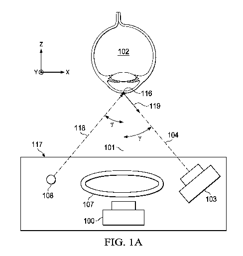
FIGURE lA is a schematic diagram of an ophthalmic system with multiple cameras
and illuminators in an aligned position with respect to an eye.
4
CA 03096607 2020-10-08
WO 2019/234574
PCT/IB2019/054553
FIGURE 1B is a schematic diagram of an on-axis image captured by on-axis
camera of
the ophthalmic system of FIGURE 1A.
FIGURE 1C is a schematic diagram of an off-axis image captured by the off-axis
camera of ophthalmic system of FIGURE 1A.
FIGURE 2A is a schematic diagram of an ophthalmic system with an on-axis
camera
and illuminator when the camera is not in an aligned position.
FIGURE 2B is a schematic diagram of an on-axis image of an eye captured by the
on-
axis camera not in an aligned position, as depicted in FIGURE 2A.
FIGURE 2C is a schematic diagram of an ophthalmic system with an on-axis
camera
and illuminator when on-axis camera is in an aligned position.
FIGURE 2D is a schematic diagram of an on-axis image captured by the on-axis
camera
in an aligned position, as depicted in FIGURE 2C.
FIGURE 3A is a schematic diagram of an ophthalmic system with an on-axis
camera,
off-axis camera, and off-axis illuminator not in an aligned position with
respect to an eye.
FIGURE 3B is a schematic diagram an off-axis image captured by an off-axis
camera
not in an aligned position with respect to an eye, as illustrated in FIGURE
3A.
FIGURE 3C is a schematic diagram of an ophthalmic system with an on-axis
camera,
off-axis camera, and off-axis illuminator in an aligned position with respect
to an eye.
FIGURE 3D is a schematic diagram of an off-axis image captured by an off-axis
camera
is in an aligned position with respect to an eye, as illustrated in FIGURE 3C.
FIGURE 4 is a schematic diagram of an ophthalmic system with a camera enclosed
by
an illuminator.
FIGURE 5A is a schematic front view an ophthalmic system.
FIGURE 5B is a schematic rear view of the ophthalmic system of FIGURE 5A with
a
screen and a control interface.
5
CA 03096607 2020-10-08
WO 2019/234574
PCT/IB2019/054553
FIGURE 6 is a block schematic diagram of a portion of an ophthalmic system
with a
control interface and motors.
FIGURE 7 is a block schematic diagram of a portion of an ophthalmic system
with a
processor and motors.
FIGURE 8 is a flow chart of a method for arranging components of a system for
aligning an ophthalmic device with an eye.
FIGURE 9 is a flow chart of a method for aligning a device with an eye.
DETAILED DESCRIPTION
In the following description, details are set forth by way of example to
facilitate
discussion of the disclosed subject matter. It should be apparent to a person
of ordinary skill in
the field, however, that the disclosed embodiments are exemplary and not
exhaustive of all
possible embodiments and furthermore than the component features of the
disclosed
embodiments may be combined with one another unless clearly mutually
exclusive, even
though every such combination is not expressly described.
As will be described in further detail, the inventors of the present
disclosure have
developed methods and systems for use in diagnostic applications and
ophthalmic surgery. The
ophthalmic systems and methods disclosed herein may be used for improved
alignment with
the eye. Better alignment allows for more accurate diagnosis and surgical
procedures.
The systems and methods for alignment disclosed herein may be fast, cost-
efficient and
simple to implement. The systems and methods may produce information that is
very easy to
interpret for the user. For example, the user may need only adjust the system
to center bright
reflections in one or two images (one off- and one on-axis image). Centering
the reflections
will position the system in a defined x, y and z position relative to the
cornea of the eye. The
methods and systems allow for accurate alignment despite variations in corneal
shape and size
because the methods and systems may rely solely on reflections from the apex.
Some figures and descriptions in this disclosure include an x-y-z orientation
for
reference purposes. The designations of the x, y, and z axes are arbitrary and
may be rearranged.
An axis labeled as a circle enclosing a point indicates a three-dimensional
system, which can
be imagined as extending at a normal from the page. In FIGURES 1-4, the y-axis
is
6
CA 03096607 2020-10-08
WO 2019/234574
PCT/IB2019/054553
perpendicular to an on-axis. Adjusting or moving a position of an ophthalmic
device or system
means moving at least a portion of the device or system.
The systems and methods disclosed herein may be used to align any number of
ophthalmic devices. For example, the systems and methods may be used to align
any
ophthalmic diagnostic device such as a keratometer, any ophthalmic surgical
devices such as a
laser, lenses, or any other ophthalmic device.
FIGURE lA depicts an ophthalmic system 117 with multiple cameras and
illuminators
in an aligned position. On-axis camera 100 is pointed toward an eye 102 of a
patient along an
on-axis 101. The on-axis may be perpendicular to a portion of the eye 102 used
to determine
the aligned position. For example, the on-axis 101 may be perpendicular to the
apex 116 of the
cornea or the on-axis 101 may pass through and be parallel to the center of
the pupil. Off-axis
camera 103 is pointed toward the eye 102 along an off-axis 104. The
intersection between the
on-axis 101 and the off-axis 104 forms an off-axis angle y. The off-axis angle
y may be acute
(between 0 and 90 , more particularly between 15 and 75 , as illustrated in
FIGURE 1A. In
some embodiments, the off-axis angle y is 45 degrees, also as illustrated in
FIGURE 1A.
On-axis camera 100 and Off-axis camera 103 may continuously record images or
may
capture images at predetermined or user-inputted intervals. Although only two
cameras (on-
axis camera 100 and off-axis camera 103) are shown in the embodiment in FIGURE
1A,
additional cameras may be used. In particular, additional off-axis cameras at
the same off-axis
angle or at different off-axis angles may be used. Off-axis camera 103 is
shown as positioned
at a distance from on-axis camera 100in the x-direction, but off-axis camera
103 may also be
positioned at the same x-position as on-axis camera 100 and may be instead
separated from on-
axis camera 100 by, for example, a distance in the y-direction.
On-axis illuminator 107 encloses and encircles on-axis camera 100. On-axis
illuminator
107 may include a fixation light, single or multiple light emitting diodes
(LEDs), organic light-
emitting diodes (OLEDs), liquid-crystal displays (LCDs), plasma displays, or
other lighting
technology such as projection or conventional light bulbs, or combinations
thereof. On-axis
illuminator 107, although shown as a circle, may take a variety of shapes for
which a center is
readily defined, such as a square, rhombus, other regular polygon, or oval, or
a dot pattern
arranged in such a shape. On-axis camera 100 is pointed along on-axis 101
through on-axis
illuminator 107. Although on-axis camera 100 is positioned behind on-axis
illuminator 107 in
7
CA 03096607 2020-10-08
WO 2019/234574
PCT/IB2019/054553
the y-direction in the depicted embodiment, on-axis camera 100 may be
positioned at the same
y-position as the on-axis illuminator 107 or in front of on-axis illuminator
107. Additionally,
on-axis illuminator 107 need not enclose on-axis camera 100. On-axis
illuminator 107 may be
shaped as a single point, for example, by using one LED, or shaped as two
intersecting lines or
other shapes. The size and shape of on-axis illuminator 107 may be independent
of corneal
shape and size.
Off-axis illuminator 108 is positioned away from the off-axis camera 103. Off-
axis
illuminator 108 and off-axis camera 103 may be placed at the same or different
distances from
on-axis camera 100. Off-axis illuminator 108 may include a fixation light,
single or multiple
light emitting diodes (LEDs), organic light-emitting diodes (OLEDs), liquid-
crystal displays
(LCDs), plasma displays, or other lighting technology such as projection or
conventional light
bulbs, or combinations thereof. Off-axis illuminator 108, although shown as a
point, may take
a may take a variety of shapes for which a center is readily defined, such as
a square, rhombus,
other regular polygon, or oval, or a dot pattern arranged in such a shape. Off-
axis illuminator
108 may enclose off-axis camera 103. Off-axis illuminator 108 may be placed at
an equal
distance from the on-axis 101 as off-axis camera 103.
The ophthalmic system 117 or portions thereof may be aligned in the x- and y-
directions as follows. On-axis illuminator 107 may emit light that is incident
on the eye 102.
Light incident on the eye 102 may be reflected to form an on-axis reflection
112, having a
center. The on-axis reflection 112 may reach on-axis camera 100 and off-axis
camera 103 .The
on-axis camera 100 and off-axis camera 103 can capture images of light
reflected from the eye
102. The on-axis camera 100 may be pointed along on-axis 101. The off-axis
camera 103 may
be pointed along off-axis 104. The images may include image data, including
digital or analog
values that represent the image. A user or computer can use the images to
determine whether
the ophthalmic system 117 or portions thereof are aligned with the eye 102.
When the on-axis
101 is normal to the center of the on-axis reflection 112, the ophthalmic
system 117 or portions
thereof are aligned in the x- and y- directions.
The ophthalmic system 117 or portions thereof may be aligned in the z-
direction as
follows. Off-axis illuminator 108 may emit light that travels along off-axis
incident path 118
such that the light is incident on the eye 102. Light incident along off-axis
incident path 118
may be reflected at the eye 102 along the off-axis reflective path 119 to form
an off-axis
reflection 113, having a center. In FIGURE 1A, the center of the apex 116 of
the eye 102 is in
8
CA 03096607 2020-10-08
WO 2019/234574
PCT/IB2019/054553
the off-axis reflective path 119. The off-axis reflection 113 may reach on-
axis camera 100 and
off-axis camera 103. The on-axis camera 100 and off-axis camera 103 can
capture images of
reflected light from the eye 102. The on-axis camera 100 may be pointed along
on-axis 101.
The off-axis camera 103 may be pointed along off-axis 104. The images may
include image
data, including digital or analog values that represent the image. A user or
processor can use
the images to determine whether the ophthalmic system 117 or portions thereof
are aligned
with the eye 102. When the off-axis 104 is normal to the center of the off-
axis reflection 113,
as shown, the ophthalmic system 117 or portions thereof are aligned in the z-
direction. Off-
axis camera 103 may be placed such that the off-axis 104 is normal to the
center of the off-axis
reflection 113.
FIGURE 1B depicts on-axis image 110 received by on-axis camera 100 in
ophthalmic
system 117 shown in FIGURE 1A. On-axis image 110 may be represented by
electronic image
data, including digital or analog values. On-axis image 110 may be displayed
against a grid
111. On-axis image 110 may depict on-axis reflection 112 of the on-axis
illuminator 107 from
the eye 102. In the image captured at this position of the ophthalmic system
117, on-axis camera
100 is in an aligned position as on-axis reflection 112 is in the center of on-
axis image 110.
On-axis 101 is normal to the center of the apex 116 of the eye 102 and the on-
axis reflection
112. On-axis reflection 112 is also in the center of grid 111. On-axis image
110 may be sent to
a processor, displayed on a screen, or both. A user or processor may determine
whether the on-
.. axis camera 100 is centered based on the on-axis image 110. Although on-
axis reflection 112
is precisely centered in on-axis image 110 in FIGURE 1B, a margin of error may
be set such
that on-axis camera 100 is in an aligned position, even when it is not
perfectly centered. Also
captured in on-axis image 110, is off-axis reflection 113 of off-axis
illuminator 108 off the eye
102of the patient. A user or processor may also gather information about the
relative position
of all or part of ophthalmic system 117 based on the position of off-axis
reflection 113 in on-
axis image 110.
FIGURE 1C depicts an off-axis image 114 received by the off-axis camera 103 in
ophthalmic system 117 of FIGURE 1A. Off-axis image 114 may be represented by
electronic
image data, including digital or analog values. Off-axis image 114 may be
displayed against a
grid 115. Off-axis image 114 may depict off-axis reflection 113 of off-axis
illuminator 108
from the eye 102. The off-axis camera 103 is in an aligned position as shown
by the position
of the off-axis reflection 113 in the center of the off-axis image 114. Off-
axis 104 is normal to
9
CA 03096607 2020-10-08
WO 2019/234574
PCT/IB2019/054553
the center of the apex 116 of the eye 102 at off-axis angle y. Off-axis 104 is
normal to the center
of off-axis reflection 113. The off-axis reflection 113 is also in the center
of grid 115. Off-axis
image 114 may be sent to a processor, displayed on a screen, or both. A user
or processor may
determine whether off-axis camera 103 is centered based on off-axis image 114.
Although off-
axis reflection 113 is precisely centered in off-axis image 114 in FIGURE 1C,
a margin of error
may be set such that off-axis camera 103 is in an aligned position, even when
it is not perfectly
centered. Also captured in off-axis image 114 is on-axis reflection 112 of on-
axis illuminator
107 off the eye 102 of the patient. A user or processor may also gather
information about the
relative position of all or part of the ophthalmic system 117 based on the
position of on-axis
reflection 112 in off-axis image 114.
FIGURE 2A depicts ophthalmic system 117 with an on-axis camera 100 and on-axis
illuminator 107 when the on-axis camera 100 is not in an aligned position with
respect to an
eye 102. On-axis camera 100 is pointed along on-axis 101 generally towards the
eye 102. On-
axis 101 is not normal to the center of the apex 116 of an eye 102. On-axis
101 is not normal
to on-axis reflection 112, so on-axis camera 100 is not in an aligned position
in FIGURE 2A.
FIGURE 2B depicts an on-axis image 110 captured by an on-axis camera 100 when
on-
axis camera 100 is not in an aligned position, as shown in FIGURE 2A. On-axis
camera 100 is
not in an aligned position as on-axis reflection 112 is in not the center of
on-axis image 110.
On-axis reflection 112 is also not in the center of grid 111. To reach an
aligned position, the
position of all or a portion of ophthalmic system 117 relative to the eye 102
needs to be adjusted
at least in the x and y directions.
FIGURE 2C depicts ophthalmic system 117 with an on-axis camera 100 and on-axis
illuminator 107 when the on-axis camera 100 is in an aligned position with
respect to an eye
102. The position of all or part of the ophthalmic system 117 in FIGURE 2C is
different in the
x and y directions as compared to FIGURE 2A. On-axis 101 is normal to the
center of the apex
116 of the eye 102. On-axis 101 is normal to on-axis reflection 112, so on-
axis camera 100 is
in an aligned position in FIGURE 2C.
FIGURE 2D depicts an on-axis image 110 captured by an on-axis camera 100 when
on-axis camera 100 is in an aligned position, as shown in FIGURE 2C. On-axis
camera 100 is
in an aligned position as on-axis reflection 112 is in the center of the on-
axis image 110. On-
axis reflection 112 is also in the center of grid 111.
CA 03096607 2020-10-08
WO 2019/234574
PCT/IB2019/054553
FIGURE 3A depicts an ophthalmic system 117 with an on-axis camera 100, off-
axis
camera 103, and off-axis illuminator 108. Ophthalmic system 117 is shown when
off-axis
camera 103 is not in an aligned position with an eye 102. Off-axis camera 103
is pointed along
off-axis 104 generally towards the eye 102. Off-axis 104 intersects on-axis
101 at an off-axis
angle 7. Off-axis illuminator 108 emits light toward the eye 102. Light
traveling along off-axis
incident path 118 is reflected at the eye 102 along the off-axis reflective
path 119 to form an
off-axis reflection 113. Off-axis 104 is not normal to the center of the apex
116 of the eye 102
or off-axis reflection 113, so off-axis camera 103 is not in an aligned
position in FIGURE 3A.
To reach an aligned position, the position of all or part of the ophthalmic
system 117 relative
to the eye 102 needs to be adjusted at least in the y direction.
FIGURE 3B depicts an off-axis image 114 captured by an off-axis camera 103
when
the off-axis camera 103 is not in an aligned position, as shown in FIGURE 3A.
The off-axis
camera 103 is not in an aligned position as the off-axis reflection 113 is in
not the center of the
off-axis image 114. The off-axis reflection 113 is also not in the center of
grid 115. To reach
an aligned position, the position of all or part of the ophthalmic system 117
relative to the eye
102 needs to be adjusted at least in the y direction.
FIGURE 3C depicts an ophthalmic system 117 with an on-axis camera 100, off-
axis
camera 103, and off-axis illuminator 108. Ophthalmic system 117 is shown when
the off-axis
camera 103 is in an aligned position with respect to an eye 102. Off-axis 104
is normal to the
center of the apex 116 of the eye 102. Off-axis 104 is normal to off-axis
reflection 113, so it is
in an aligned position in FIGURE 3C. Ophthalmic system 117 is aligned since
normal 120 at
the reflection point 121 is aligned with on-axis 301. Reflection point 121 is
determined by light
emitted from off-axis illuminator 108. Light travels along off-axis incident
path 118 and is
reflected to off-axis reflective path 119. Relative to FIGURE 3A, the position
of the ophthalmic
device has been adjusted in the y direction.
FIGURE 3D depicts an off-axis image 114 captured by an off-axis camera 103
when
the off-axis camera 103 is not in an aligned position, as shown in FIGURE 3C.
Off-axis camera
103 is in an aligned position as off-axis reflection 113 is in the center of
off-axis image 114.
Off-axis reflection 113 is in the center of grid 115.
FIGURE 4 depicts an ophthalmic system 117 with on-axis camera 100 enclosed by
on-
axis illuminator 107. On-axis camera 100 is pointed along on-axis 101. On-axis
illuminator
11
CA 03096607 2020-10-08
WO 2019/234574
PCT/IB2019/054553
107 may still enclose the on-axis camera 100, whether on-axis camera 100 is in
front, aligned,
or centered within on-axis illuminator 107 along the y direction.
Additionally, on-axis
illuminator 107 can be shaped in a may take a variety of shapes for which a
center is readily
defined, such as a square, rhombus, other regular polygon, or oval, or a dot
pattern arranged in
such a shape. On-axis illuminator 107 may still enclose on-axis camera 100 if
it is shaped to
not form an outer boundary with a defined inner area. For example, an on-axis
illuminator 107
formed as a line of LEDs may enclose on-axis camera 100 if, e.g., on-axis
camera 100 is placed
in series with the line of LEDs, with an LED on one side of the camera, and an
LED on another.
On-axis camera 100 need not be centered within the on-axis illuminator 107 in
any direction.
FIGURE 5A is a front view of an ophthalmic system 117. The ophthalmic system
117
may include ophthalmic device 105. Ophthalmic device 105 may include a
keratometer, as
shown in Figs. 5A and 5B. Ophthalmic device 105 may include any ophthalmic
diagnostic
device such as a keratometer, any ophthalmic surgical devices such as a laser,
lenses, or any
other ophthalmic device. Viewable from the front of ophthalmic system 117, are
examples of
an on-axis camera 100 pointed along an on-axis 101, an off-axis camera 103
pointed along an
off-axis 104, an on-axis illuminator 107, and an off-axis illuminator 108.
FIGURE 5B is a rear view of the ophthalmic system 117 shown in FIGURE 5A. The
ophthalmic system 117 includes a screen 518 and a control interface 519.
Screen 518 is shown
displaying a processed image 510, depicting an on-axis reflection 112 and an
off-axis reflection
113. Screen 518 may be any sort of display, e.g., LED, OLED, LCD, plasma, etc.
Screen 518
may be a touch screen. Screen 518 need not be attached to the same physical
structure as any
of the physical components shown in FIGURE 5B and FIGURE 5A.
Although processed image 510 in the depicted embodiment only shows one view,
it
may show multiple views¨such as the view from an off-axis camera 103 in
addition to a view
from on-axis camera 100. Additionally, screen 518 may display a processed
image 510 that
overlaps images taken from multiple cameras. For example, screen 518 may
display an image
captured by on-axis camera 100 and superimpose the off-axis reflection of the
light emitted an
off-axis illuminator received by an off-axis camera 103.
Screen 518 may display augmented reality or graphical representations of the
reflected
illuminator light to display reflections more clearly or to better communicate
the positional
information contained therein. Screen 518 may display graphical
representations of the on-axis
12
CA 03096607 2020-10-08
WO 2019/234574
PCT/IB2019/054553
reflection, the off-axis reflection, or both. The graphical representations
may depict portions or
the entirety of the reflections. Screen 518 may display images of the eye or
reflections or
graphical representations thereof. Screen 518 may display filtered images, not
displaying some
data generated by one or more cameras. Screen 518 may also display graphical
representations
of an eye. Screen 518 may display numerical information such as necessary
directional
adjustments to achieve alignment. Screen 518 may further display an indication
of a pass or
fail based on whether the system is aligned in any or all of the spatial
directions. A separate
light indicator, audible indicator, or physical indicator may indicate a pass
or fail indication.
Also depicted in FIGURE 5B is a control interface 519. In the depicted
embodiment,
control interface 519 includes a joystick, but may instead include, e.g.,
analog buttons,
graphical buttons, knobs, foot pedals, etc. Control interface 519 may have
virtual components
displayed on screen 518. Control interface 519 need not be attached to the
same physical
structure as any of the physical components shown in FIGURE 5B and FIGURE 5A.
Control
interface 519 may receive input from the user in the form of, e.g., a
mechanical movement of
.. a joystick or electrical input and may generate a corresponding analog or
digital signal. Control
interface 519 may have an internal processor or export a generated signal to a
motor or a
processor. Depending on the signal received, a motor or mechanical structure
(e.g., lever) may
then adjust a position of all or part of the ophthalmic system in one or more
directions.
FIGURE 6 is a block diagram of a portion of an embodiment of the ophthalmic
system
117 of FIGURES 5A and 5B with a control interface 519 and motors 605, 606, and
607. Control
interface 519 receives a user input 601 and sends one or more control signals
602, 603, and
604 to one or more motors 605, 606, and 607. User input 601 may be, e.g., a
movement by the
user of a joystick of the control interface 519. Control interface 519
converts user input 601 to
a digital or analog signal and sends a corresponding control signal 602, 603,
and 604 to a
respective motor 605, 606, and 607.
Each motor 605, 606, or 607 may receive a unique signal or may receive the
same signal
from the control interface 519 and decode it. Depending on the signal
received, a motor 605,
606, or 607 may then adjust a position of all or part of the ophthalmic system
117 in one or
more directions, which may correlate with the x-, y- and z- axes described
above with respect
to eye 102, or in directions that represent movement along two or three of the
x-, y-, and z-
axes. The user may use the control interface 519 to adjust a position of all
or part of the
ophthalmic system 117 until it is in an overall aligned position with respect
to eye 102. The
13
CA 03096607 2020-10-08
WO 2019/234574
PCT/IB2019/054553
system may have fewer or more components, such as motors, control signals, and
user inputs,
than are depicted in FIGURE 6.
FIGURE 7 is a block diagram of a portion of an embodiment of the ophthalmic
system
117 of FIGURES 5A, 5B, and 6 with a processor 700 and motors 605, 606, and
607. As part
of a control interface 519, in addition to a control interface 519, or in lieu
thereof, the
ophthalmic system 117 may have a processor 700. The processor 700 may receive
image data
701 and send one or more instruction signals 702, 703, and 704 to one or more
motors 605,
606, and 607. Image data 701 may be data obtained from an on-axis camera, off-
axis camera,
both, or additional cameras. Image data 701 may include data in any image or
video format.
Image data may also be pre-processed at an earlier stage. Processor 700
receives image data
701 and may determine, based on image data 701, whether corrective action is
needed by, e.g.,
performing digital image processing.
Processor 700 may compare information contained in the image data 701 to a
reference
stored in memory 708 to determine if corrective action is needed. User input
610 may be read
by the processor 700 and thereafter written to or read from memory 708. Memory
708 and
processor 700 may transfer information through memory signal 709.
If corrective action is needed, processor 700 may send one or more instruction
signals
702, 703, and 704 to one or more motors 605, 606, and 607. One or more motors
605, 606, and
607 are actuated according to one or more instruction signals 702, 703, and
704 generated by
the processor 700. Motors 605, 606, and 607 upon actuation may adjust a
position of all or part
of the ophthalmic system 117 in one or more directions, which may correlate
with the x-, y-
and z- axes described above respect to eye 102, or in directions that
represent movement along
two or three of the x-, y-, and z- axes. Motors 605, 606, and 607 may move
part or all of the
ophthalmic system, ultimately to an overall aligned position. The actuation
and processing may
occur automatically upon receiving the image data, in addition to or instead
of relying on user
input. Accordingly, part or all of the ophthalmic system may be moved based on
image data
received by cameras.
Additionally, a margin of error may be set by user input 610, may be stored in
memory
708, or both. The margin of error can be used to determine whether the
difference between the
image data and a reference image is within an acceptable range. The margin of
error may
include one or numerous values containing allowable distances in one or more
directions.
14
CA 03096607 2020-10-08
WO 2019/234574
PCT/IB2019/054553
The margin of error may include all or any combination of the distances from
centers
and electronic values described below. For example, the margin of error may
include an
allowable distance from the center of on-axis 101 to the center of the on-axis
reflection 112 in
one or more directions, which may correlate with the x-, y- and z- axes
described above with
respect to eye 102, or in directions that represent distances along two or
three of the x-, y-, and
z- axes. The margin of error may include an allowable distance from the center
of off-axis 104
to the center of the off-axis reflection 113 in one or more directions, which
may correlate with
the x-, y- and z- axes described above with respect to eye 102, or in
directions that represent
distances along two or three of the x-, y-, and z- axes. The margin of error
may include
electronic representations, such as digital or analog values corresponding to
an allowable
distance from the center of on-axis 101 to the center of the on-axis
reflection 112 in one or
more directions, which may correlate with the x-, y- and z- axes described
above with respect
to eye 102, or in directions that represent distances along two or three of
the x-, y-, and z- axes.
The margin of error may include electronic representations, such as digital or
analog values
corresponding to an allowable distance from the center of off-axis 104 to the
center of the off-
axis reflection 113 in one or more directions, which may correlate with the x-
, y- and z- axes
described above with respect to eye 102, or in directions that represent
distances along two or
three of the x-, y-, and z- axes. The margin of error can be input via the
user input 610,
processed by the processor 700, and stored in memory 708. The processor 700
may use a
margin of error to determine if corrective action is need. If corrective
action is needed, the
processor 700 will generate and send one or more instruction signals 702, 703,
704 to actuate
one or more motors 605, 606, and 607 to move all or part of the ophthalmic
system into an
overall aligned position. More or fewer components, such as motors,
instruction signals, paths
of image data, and user inputs may be used than are illustrated in FIGURE 7.
FIGURE 8 is a flow chart of a method for arranging components of a system for
aligning an ophthalmic device with an eye of a patient. The ophthalmic system
may be an
ophthalmic system 117 as described in FIGURES 1-7. After starting arranging at
step 800, the
method includes pointing an on-axis camera along an on-axis at step 801.
Pointing at step 801
may set a reference for positioning the other components. Pointing at step 801
may include
placing or installing a camera on a system.
The method may include setting an off-axis angle at step 802. The off-axis
angle may
be set in reference to the on-axis in which the off-axis angle is an acute
angle formed at the
CA 03096607 2020-10-08
WO 2019/234574
PCT/IB2019/054553
intersection point of the on-axis and the off-axis. The method may include
pointing an off-axis
camera along the off-axis at step 803.
The method may include placing one or more illuminators in a position at step
806. An
on-axis illuminator may be placed at an on-axis position, where it is able to
emit light in a
direction that the on-axis camera may capture. An off-axis illuminator may be
placed at an off-
axis position, where it is able to emit light in a direction that the off-axis
camera may capture.
Placing illuminators at step 806 may include installing illuminators on a
system. An illuminator
is placed at a relative position to a camera. The placing illuminators at step
806 may include
enclosing an on-axis camera within an illuminator. The placing illuminators at
step 806 may
include enclosing an off-axis camera within an illuminator.
The method may also include setting a margin of error at step 807. Setting a
margin of
error at step 807 may include adjusting values in memory or controlling user
input.
The method may also include ending the arranging at step 808. The method may
include
using components or systems as described elsewhere in this disclosure.
FIGURE 9 is a flow chart of a method for aligning an ophthalmic system with an
eye.
The ophthalmic system may be ophthalmic system 117 as described with respect
to FIGURES
1-7 or aligned using the method of FIGURE 8. After starting aligning at step
901, the method
may include setting an ophthalmic system at an initial position at step 902.
The method may include lighting a first and second portion of an eye at step
903. The
first and second portions may overlap. The first and second portions may be
caused by two
different illuminators. The method may include receiving images at step 904
with one or more
cameras.
The method may include determining if an adjustment is needed at step 905.
Determining at step 905 may be done manually by a user, or by a processor, or
by a combination
of user and processor input. Determining at step 905 answers whether a
position of all or part
of a system needs to be adjusted relative to an eye in one or more directions,
which may
correlate with the x-, y- and z- axes described above respect to eye 102, or
in directions along
two or three of the x-, y-, and z- axes. Determining at step 905 may include
sending image data
corresponding to one or more images to a processor and generating an
instruction signal.
16
CA 03096607 2020-10-08
WO 2019/234574
PCT/IB2019/054553
If an adjustment is needed, the method may include determining an amount of
adjustment needed at step 906. Determining an amount at step 906 may include
determining
the amount of adjustment to center all or part of the system, such as the on-
axis camera, relative
to an eye in x, y, and/or z directions. Determining an amount at step 906 may
be done manually
by a user, or by a processor, or by a combination of user and processor input.
Determining an
amount at step 906 may include sending image data of one or more images to a
processor and
generating an instruction signal. The method may include adjusting a position
of all or part of
a system relative to an eye at step 907. Adjusting at step 907 may include
actuating motors via
instruction signals or control signals or mechanically moving all or part of
the system.
When an adjustment is no longer needed, or when the on-axis camera 100 and off-
axis
camera 103 are substantially centered as described above within a margin of
error, the method
may include ending the aligning at step 908. The method may include using
components or
systems as described elsewhere in this disclosure.
The above disclosed subject matter is to be considered illustrative, and not
restrictive, and the
appended claims are intended to cover all such modifications, enhancements,
and other
embodiments which fall within the true spirit and scope of the present
disclosure. Thus, to the
maximum extent allowed by law, the scope of the present disclosure is to be
determined by the
broadest permissible interpretation of the following claims and their
equivalents and shall not
be restricted or limited by the foregoing detailed description.
17