Note: Descriptions are shown in the official language in which they were submitted.
CA 03096931 2020-10-09
WO 2019/202621 PCT/IT2019/050077
-1 -
"APPARATUS AND METHOD TO DETERMINE ERYTHROCYTE
SEDIMENTATION RATE AND OTHER CONNECTED PARAMETERS"
* * * * *
FIELD OF THE INVENTION
The present invention concerns an apparatus and the corresponding method
used, in the field of medical analysis, to determine erythrocyte sedimentation
rate
(ESR), as well as other parameters related thereto.
BACKGROUND OF THE INVENTION
In the field of medical analysis, pathological states, defined as
inflammatory,
are ascertained by measuring the sedimentation rate of the corpuscular part of
the
blood, in particular of the erythrocytes, or red cells. In particular, the
erythrocyte
sedimentation rate represents a non-specific diagnosis test of the state of
inflammation.
Different methods have been adopted to determine the ESR, which however
have not proven to be completely satisfactory from the point of view of speed
and practicality of detection.
In these methods, which use different measurement systems, the blood taken
from the patients is introduced into tubular containers, or test tubes, and
subsequently, after a possible centrifugation, the necessary measurements are
performed on the blood samples.
Some known systems provide to detect, at predetermined time intervals, the
position of the separation interface between the fluid plasmatic part of the
blood,
substantially clear, and the corpuscular part consisting of red and white
blood
cells and platelets, which is more turbid.
Other systems provide instead to detect the optical density or absorbance of
the blood in correspondence with the separation interface.
The methods to detelinine the ESR proposed so far are generally characterized
by an initial dead time which significantly influences the overall time of the
analysis, which therefore cannot be performed in succession with other much
faster analyses, such as for example blood count analysis.
Furthermore, known methods have to use disposable containers, which entails
an increase in the cost both of their purchase and also of their disposal.
Again, the
amount of blood needed to perform the analysis is high and this leads to
CA 03096931 2020-10-09
WO 2019/202621 PCT/IT2019/050077
-2 -
problems in particular cases, for example when the analysis has to be
performed
on children.
A method is also known which provides to take the blood to be analyzed from
the container in which it is stored and insert the same blood in a measurement
volume, of reduced thickness, which is used for various measurements performed
on different samples. This method is based on detecting the optical density or
absorbance of the blood in a fixed point of the measurement volume, which is
rotated to accelerate the sedimentation of the blood.
The means used to detect the absorbance include electromagnetic radiation
emission/detection devices associated with the measurement volume. The
absorbance values detected are directly proportional to the number of blood
cells
present in the blood sample at the point of observation, this number varying
over
time due to the sedimentation of the blood cells themselves.
The study of the absorbance over time allows to trace the value of the ESR by
eliminating the initial dead times, eliminating the need to use disposable
containers for the analysis.
Furtheimore, the required amount of blood to be analyzed is smaller, and
therefore the analysis can also be performed without difficulty on pediatric
patients.
Despite these merits, this method is characterized by some problems that limit
its completely satisfactory use.
The size of the ESR measuring apparatus and the difficulties in managing
multiple centrifugation means limit the possibility of coupling this system
with
an integrated instrument for counting blood cells. Furthermore, the size
entails
the need to perform the analysis in laboratory and the analysis procedure
itself
requires considerable volumes of blood.
Furthermore, after each measurement, the centrifugation means and the
volume associated thereto must always be repositioned with respect to the
emission/detector means, which creates problems and anomalies in the control
of
the blood flow.
In this apparatus, after the analysis, the blood sample is discharged and a
new
blood sample is introduced into the measurement volume.
To avoid washing the measurement volume after the discharge, the residues of
CA 03096931 2020-10-09
WO 2019/202621 PCT/IT2019/050077
-3 -
the sample already analyzed are discharged by the new blood sample to be
analyzed, the hydraulic path that the blood must complete to avoid pollution
being rather long; this increases the volume of blood to be used.
A further limitation of this method is given by the fact that the acquisition
of
photometric data depends on the speed of rotation of the measurement volume
and cannot therefore be considered a continuous phenomenon.
The state of the art proposes solutions to some of these problems, for example
in US 5,827,746, in EP 1.907.819, or in EP 2.880.418, all in the name of the
Applicant. However, the need has been found to further perfect the apparatuses
of the state of the art in terms of precision, efficiency and versatility, to
provide
methodological solutions that are even more adaptable to wide ranges of
patients
and in different operating conditions.
One purpose of the present invention is therefore to provide a method and the
corresponding apparatus to determine the erythrocyte sedimentation rate, as
well
as other related parameters, which allow a fast, easy and reliable analysis,
which
can also be performed in combination with different types of hematologic
analyses.
Another purpose of the present invention is to allow the integration of this
apparatus into existing blood cell counting systems, thus exploiting the
homogenization of the blood already performed by the same blood cell counter.
Another purpose of the invention is to provide an apparatus that is compact
and easy to transport, practical to use in any condition and environment and
which can also be used as a disposable ambulatory or hospital instrument, for
example for so-called POCs (Points of Care), and which is particularly
suitable
for analysis on pediatric patients.
The Applicant has devised, tested and embodied the present invention to
overcome the shortcomings of the state of the art and to obtain these and
other
purposes and advantages.
SUMMARY OF THE INVENTION
The present invention is set forth and characterized in the independent
claims,
while the dependent claims describe other characteristics of the invention or
variants to the main inventive idea.
In accordance with the above purposes, the apparatus to determine the ESR
CA 03096931 2020-10-09
WO 2019/202621 PCT/IT2019/050077
-4 -
according to the present invention comprises, in its general structure, a
capillary
tube, transparent to radiations in a certain field of wavelengths, having at
least a
substantially rectilinear segment with extremely reduced sizes inside which
the
blood to be analyzed is introduced and made to pass.
By radiation, here and in the following description, we intend to refer to
both
electromagnetic waves, in particular those in the visible field, and also to
different waves that follow the principles of undulatory mechanics, such as
for
example, but not limited to, sound waves, ultrasonic waves, or mechanical
vibrations, as well as any other type of radiation that can be used in this
context.
Therefore, although below, in particular in the detailed description of the
drawings, reference will be made to light radiations and to optical-type
emitters/detectors, it is understood that the invention is equally applicable
to all
types of radiations as indicated above.
The apparatus also comprises pump means able to send a blood sample inside
the capillary, so that the blood sample can be passed through, in a
measurement
zone, by a radiation emitted by emitter devices and detected by mating
detector
devices disposed in correspondence with a point of the capillary tube,
corresponding to the measurement zone, on the opposite side with respect to
the
emitter means.
The detector devices are connected to a control and processing unit able to
transform the values detected in an expression of the sedimentation speed, or
of
other parameters connected thereto, into a measurement unit compatible with
the
units normally used.
According to possible embodiments, the pump means can be suitable to
abruptly interrupt the flow of blood flowing through the capillary, so as to
cause
it to strongly decelerate (stopped-flow) and therefore an aggregation and
sedimentation of the blood corpuscles thanks to its compaction.
The compaction causes a variation of the signal detected by the detector means
with consequent acquisition of the information useful to determine the ESR.
At the end of the detection, the blood sample analyzed is discharged from the
circuit and the capillary tube is ready to receive a new blood sample to be
analyzed.
According to a variant, the capillary tube comprises a reading chamber in
CA 03096931 2020-10-09
WO 2019/202621 PCT/IT2019/050077
-5 -
correspondence with which the measurement is perfolined.
In particular, one fottnulation of the present invention provides that the
reading chamber consists of a small (capillary) measuring tube, for example
with
a cylindrical section, although this shape is not in itself limiting, and made
of
plastic material, for example, but not only, acrylic material, or of glass.
The use
of these materials allows the capillary tube to be modeled also, and in
particular,
in the entry and exit surfaces of the radiation detected by the reception
means.
The small capillary tube defines a transit channel that is coupled in fluidic
continuity with the supply pipe, normally made of Teflon, of the samples to be
analyzed.
The particular shape of the measuring tube of acrylic material or glass is
made
so that the entry zone of the light, of the sound waves, or of other suitable
radiation, has a substantially flat or suitably shaped surface, instead of a
curvilinear cylindrical surface as in the case of a conventional tube.
According to a further evolutionary characteristic, the measuring tube also
has
a flat surface at its opposite end, that is, the exit end, so that the path of
the
optical, sound or other type of radiation is not deflected/refracted by
curvatures
which alter its information content.
Thanks to these substantially flat surfaces facing the emitter/detector
devices,
the radiation emitted is less subject to perturbative factors that can
invalidate the
correctness and accuracy of the measurement.
In particular, these reading windows with a flat surface interact with the
radiation incident on it independently of their positioning inside the
standard
positioning tolerances for mechanical workings.
According to a further variant of the invention, these flat windows constitute
transparent, non-diffusive surfaces such as those of a conventional tube,
generally made of Teflon, and allow to obtain much higher optical or sound
detection sensitivity.
According to a variant of the invention, the reading chamber with measuring
tube made of acrylic material, or glass, is connected to a conventional type
of
pipe, for example made of Teflon, upstream and downstream, in which the
movement of the blood sample occurs.
In a further characteristic, the glass or acrylic reading chamber is housed
CA 03096931 2020-10-09
WO 2019/202621 PCT/IT2019/050077
-6 -
inside a rigid container, which defines the housing seatings for the upstream
and
downstream pipes that define the path of the blood sample to be analyzed.
In a further embodiment, the rigid container also has collimation means which
define the path of the optical, sound or other type of beam which passes
through
the reading chamber.
Thanks to this configuration of the measuring chamber, it is possible to
measure the flow velocity, in the stopped-flow, or stop and flow, step which
facilitates the measurement of the viscosity of the blood sample and makes it
more precise.
According to a further characteristic of the present invention, the measuring
method, thanks to the characteristics of the apparatus, and in particular of
the
measurement cell, described above, allows to avoid contamination between
sample and sample, that is, avoiding the so-called "carryover" phenomenon,
which leads to contamination between successive samples and therefore to
obtaining distorted measurements or the need for washing between samples.
The method according to the present invention provides to collect a sample of
blood in extremely small quantities, able to facilitate pediatric or capillary
sampling, for example in a range from 30 microliters to 180 microliters.
According to possible embodiments, the method can provide to use latexes to
improve the calibration and adjustment of the measuring instrument. For
example, the use of three-level turbidity latexes can be provided to calibrate
the
accuracy of the measurement.
In particular, the use of the latexes allows to detect the functionality of
the
internal sensors of the equipment to guarantee measurement and calibration
performances that certify the correct functioning of the instrumentation. This
aspect is even more important as the measurement of the ESR is not an exam
that
has external controls, as for example blood glucose analysis.
According to the invention, the measuring instrument comprising emitter and
detector devices is located at a specific point in the blood flow which
corresponds
to the end of travel of each sample read.
Through the use of the reading chamber made of acrylic or vitreous material,
located inside the rigid support, and also thanks to the collimation of the
radiation
emitted, it is possible, according to the invention, to always measure the
terminal
CA 03096931 2020-10-09
WO 2019/202621 PCT/IT2019/050077
-7 -
part of the sample, the so-called tail of the sample, which is free from
contamination of the previous sample.
Furthermore, in this way all the blood samples that follow are not, at the
measuring point, contaminated by the previous one.
In one embodiment of the invention, the volume of blood in the reading
chamber is approximately equal to 1 microliter, while the amount of blood of
each pediatric sample per individual patient can be in a range from 30
microliters
to 180 microliters.
In particular, the present invention allows the sample to be read in a volume
of
1 microliter of whole blood, whether the blood sample is analyzed at one point
of
the pipe, or whether it is analyzed in the reading chamber.
According to one characteristic of the invention, the reading and measuring
point is located in such a position, with respect to the measuring chamber,
and in
particular to the glass or acrylic tube, that the microliters of blood pass
and are
made to flow through the reading chamber as an inert passage, without any
measuring of said part.
The reading of the sample is started for a portion of 1 microliter in volume
on
the last 5 microliters of the initial volume.
The passage of the microliters of inert blood through the reading chamber of 1
microliter has the function of mechanical thrust or washing.
The thrust volume on which no measurement is made allows to offer the non-
contamination between sample and sample in the last 5 microliters. Therefore,
the passage of the sample to be analyzed has a self-washing effect with
respect to
the previous sample.
Thanks to this, the invention allows to perform measurements on drops from
capillary sampling (for example 25 microliters) and at the same time does not
require washing between samples, making it particularly suitable for use in so-
called Points of Care (POCs) and for pediatric use.
In other words, the sequential self-washing of the sample allows to avoid the
carryover phenomenon.
To summarize, the advantages offered by the present invention, and in
particular the conformation and structure of the reading chamber, are the
following:
CA 03096931 2020-10-09
WO 2019/202621 PCT/IT2019/050077
-8 -
- it is possible to perform ESR measurements with reduced sampling volumes
that are very suitable for pediatric patients and capillary samples;
- there are no reductions in the precision of the measurement due to
deviation
of the radiations caused by problems connected to the manufacture of
conventional tubes;
- both pediatric samples and also adult patient samples benefit from the
self-
washing system of the sample itself, avoiding carryover between sample and
sample;
- experimental ESR measurement tests of very high and very low alternate
samples confirm the same results even reversing the same samples.
In the apparatus according to the invention, the capillary tube, the blood
sampling means and the measuring instrument can constitute a transportable
structure which is distinct and separate from the control and processing unit,
and
from a possible system to display the results, and be connected to them by
means
of transmission cables or also via radio.
In this way, extreme flexibility and versatility of use is obtained, since the
sampling and analysis instrument can have reduced sizes that allow its use,
for
example, also directly from the bed of a patient, or in any case in
challenging
conditions.
It is also possible both to use a plurality of these apparatuses in parallel,
to
simultaneously perform the same analysis on different blood samples, and also
to
use the same apparatus in series with other devices, able to perform different
types of hematological analyses on the same sample.
Furthermore, this apparatus, also as a result of the very short time required
for
the analysis, can also be used in local clinics, in hospital rooms, in mobile
hematological units or, as we said, integrated with equipment intended for
other
types of hematological analyses.
This apparatus, allowing a continuous analysis directly after the withdrawal
of
native blood from the patient, does not require the use of anticoagulant
substances, as the blood can be analyzed before the clot has time to form.
In particular, the continuity of data acquisition by the detector means allows
a
better evaluation of the optical density of the blood sample, and therefore an
extremely precise measurement of the ESR, and allows to detect possible
CA 03096931 2020-10-09
WO 2019/202621 PCT/IT2019/050077
-9 -
abnormal states of the blood flow, for example due to air bubbles or clots.
The continuous study of the flow can also be used to determine other
parameters of blood rheology, such as density or viscosity.
According to possible embodiments, the control and processing unit can be
configured to compare the viscosity of the sample with the ESR value obtained.
In a preferred solution of the invention, the pump means are reversible and
allow to invert the flow inside the circuit; this allows the re-homogenization
of
the blood sample and the rapid repetition of the measurements thereon.
The capillary is able to be thetinostated to allow analysis at a constant
temperature which can be preset as desired.
It is therefore possible to make the same sample of blood pass through
capillary tubes thermostated at different temperatures and disposed in series,
evaluating the ESR values according to the variation in the analysis
temperature.
In this case, it is preferable that at least one of the capillaries is kept at
a
temperature around 37 C, in order to prevent the precipitation of some blood
components and to guarantee a reliable comparison model.
BRIEF DESCRIPTION OF THE DRAWINGS
These and other characteristics of the present invention will become apparent
from the following description of some embodiments, given as a non-restrictive
example with reference to the attached drawings wherein:
- fig. 1 schematically shows a first embodiment of an apparatus to
deteHnine the
erythrocyte sedimentation rate and other parameters according to the
invention;
- fig. 2 schematically shows a variant of the apparatus in fig. 1;
- fig. 3 shows a detail of the apparatus in fig. 2;
- fig. 4 shows an exploded view of the detail in fig. 3;
- fig. 5 schematically shows a measurement diagram obtained with the
apparatus
according to the present invention.
To facilitate comprehension, the same reference numbers have been used,
where possible, to identify identical common elements in the drawings. It is
understood that elements and characteristics of one embodiment can
conveniently
be incorporated into other embodiments without further clarifications.
DETAILED DESCRIPTION OF SOME EMBODIMENTS
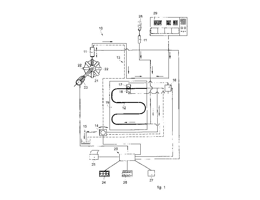
Fig. 1 schematically, and non-restrictively, shows, as a whole, an apparatus
10
CA 03096931 2020-10-09
WO 2019/202621 PCT/IT2019/050077
- 10 -
to determine the erythrocyte sedimentation rate and other parameters connected
thereto, which mainly comprises the following components:
- a sampling member 11 to take the blood sample to be analyzed;
- a pipe 12, for example made of Teflon, inside which the blood sample is
able
to be introduced, transparent to electromagnetic radiations in a field
comprised
between 100 and 2000 nm, preferably between 200 and 1000 nm;
- a circuit 13 that connects the sampling member 11 to the pipe 12 and
inside
which the blood sample circulates;
- a pump 14 associated with the circuit 13;
- a discharge pipe 15 to discharge the blood sample after the analysis;
- a measuring instrument comprising a radiation emitter device 16
associated
with a mating detector device 17, in this specific case disposed on opposite
sides
with respect to a specific point of the pipe 12;
- a control and processing unit 20 able to manage the functioning of the
apparatus 10, and
- an interface unit 18 by means of which the devices 16 and 17 are
connected
to the control and processing unit 20.
The sampling member 11, in this specific case of the syringe type, is able to
selectively take the blood sample to be analyzed from the containers, or test
tubes
22 of a storage drum 21, which can be rotated by a motor 23, for example to
perform programmed mixing cycles of the blood contained in the test tubes 22.
In
a possible solution, the motor 23 can perform the mixing by means of tilting
cycles of the storage drum 21.
An accurate mixing is essential to correctly perfolin an examination on red
cells that have not already aggregated during the time between the sampling in
the blood collection tube and the execution of the test. The mixing is
therefore
used to disaggregate the red cells and then perfolin the ESR measurement
correctly.
According to possible embodiments, the test tubes 22 can be test tubes of the
standard type, used for example for hematology measurements.
According to the embodiments shown in figs. 1 and 2, the sampling member
11 can also be used to directly take native blood from the finger 28 of a
patient,
for example perfolined with a lancing device of the finger pricking type,
CA 03096931 2020-10-09
WO 2019/202621 PCT/IT2019/050077
- 1 1 -
containing inside it the devices 16, 17 and the point of the pipe 12 in which
the
measurement is performed..
Furthermore, the blood to the pipe 12 can also come from an apparatus 29
suitable to perform other analyses, inside which the entire apparatus 10 can
be
integrated; in this way, already homogenized blood, which does not require
other
additional treatments, reaches the pipe 12.
In a variant, the sampling member 11 is provided integrally with shaking
means to homogenize the blood sample taken.
The pipe 12 is associated with a metal support 19 provided with thermostat
means which allow it to be kept at a constant temperature which can be preset
as
desired, conditioning the temperature at which the analysis is performed.
The pump 14, which can be disposed both upstream and downstream of the
pipe 12, is able to drive the sampling member 11 to circulate the blood sample
inside the circuit 13 and the pipe 12.
According to possible embodiments, the pump 14 can be a peristaltic pump,
configured to move the blood sample up to the established reading point.
The reading point is determined precisely in order to prevent the carryover
phenomenon.
In the event that the response of the examination indicates the absence of
flow,
due for example to any obstruction of the capillary tube, the peristaltic pump
can
switch the flow with a return movement of the sample taken, and repeat the
examination; or, if the response of absence of flow persists, a new cycle of
taking
a sample from a primary test tube can be activated.
According to possible embodiments, the blood sample can be read by the
measuring instrument passing through the reading point in a continuous flow,
that is, without interruption of the flow.
Alternatively, the pump 14 can be configured to interrupt the flow of the
blood
sample instantaneously, so as to cause a strong deceleration (stopped-flow)
and
therefore an aggregation of the erythrocytes.
According to possible embodiments shown in figs. 1 and 2, the pump 14 is
reversible and is able to allow blood circulation inside the circuit 13 in the
two
directions indicated respectively with a continuous line (suction) and with a
dotted line (thrust).
CA 03096931 2020-10-09
WO 2019/202621 PCT/IT2019/050077
- 12 -
The interface unit 18 is able to activate/deactivate the emitter device 16 and
to
translate the signals received by the detector device 17 into signals that can
be
read by the control and processing unit 20.
According to possible embodiments, the measuring instrument can comprise a
plurality of detector devices 17. In this way, the measuring method can detect
radiations from a plurality of detector devices 17 in a single reading of the
sample, so as to improve possible compensations.
For example, the detector devices 17 can be three detector devices 17
positioned equidistant from one another.
According to possible embodiments, the detector device 17 can be configured
to detect electromagnetic waves with a wavelength comprised between 700nm
and lmm, that is, they can be infrared waves.
According to these embodiments, the measuring instrument can be configured
to measure the temperature of the blood sample.
Furthermore, by means of the infrared wave detector device 17, it is possible
to obtain an effective mixing of the sample.
Furthermore, the aggregation of the red cells thus obtained is not affected by
low temperatures.
Embodiments of the present invention provide that the control and processing
unit 20 can be configured to compare the temperature values detected by the
detector device 17 with predetermined values, so as to possibly discard blood
samples having a temperature lower than a predetermined threshold value, for
example 18 C.
According to the present invention, the control and processing unit 20,
consisting of a microprocessor electronic processor, is programmable to manage
different functioning modes of the apparatus 10.
According to possible embodiments, the control and processing unit 20 can be
configured to compare the values detected by the detector device 17 with
preset
values, and to signal to the user the possible non-suitability of the blood
sample.
For example, if the hematocrit values are below 25%, the control and
processing
group 20 can signal to the user its non-suitability.
The control and processing unit 20 comprises a database or internal memory
27 which contains a series of parameters, in the foiiii of numerical data,
tables or
CA 03096931 2020-10-09
WO 2019/202621 PCT/IT2019/050077
- 13 -
graphs.
In particular, the database 27 can be provided with a statistical memory
comprising values of multiple of samples, for example 5,000 different samples,
so as to determine a mean of the tested population.
According to possible embodiments, the method according to the present
invention can provide a statistical control of the population based on the
values
comprised in the database 27. The database 27 can be continuously updated, and
therefore the values relating to the mean of the population tested are always
updated by the results of the tests performed.
According to possible embodiments, the control and processing unit 20 can be
configured to cooperate with a population management software, so as to
provide
a measurement graph to detect possible measurement errors, or drifts.
In particular, the control and processing unit 20 can be configured to detect
a
possible drift of the measurements of the blood samples taken from the
apparatus
10 with respect to the mean of the tested population.
Furthermore, according to possible embodiments, the measuring instrument
can be configured to perform at least one test attempt even if the control and
processing group 20 detects that the sample is not suitable.
According to this embodiment, the measuring instrument can therefore be
configured to stop after perfoHning a desired number of attempts, for example
three attempts.
According to possible embodiments, the control and processing unit 20 can be
configured to cooperate with a management software.
The management software, among its many functions, can also store the
expiry date of the latexes used in order to calibrate the accuracy of the
measurement, so as to avoid the use of latexes beyond their expiry date.
The management software can also be configured to verify that the measuring
method meets the health regulations in force in different countries. For
example,
the management software can verify that the measuring method complies with
the standards established by the Food and Drug Administration (FDA) of the
United States of America.
The control and processing unit 20 also comprises means to interface with the
user, in this specific case consisting of a keyboard 26 for data input, a
monitor or
CA 03096931 2020-10-09
WO 2019/202621 PCT/IT2019/050077
- 14 -
display 24 and a printer 25 to display the results of the analysis and to
process
them for statistical purposes.
According to possible embodiments, the measuring instrument can be
configured to detect the aggregation of the red cells and to correlate this
value
with the ESR values determined.
In particular, this correlation can also be performed for blood samples
affected
by red blood cell pathologies, such as anemia (for example sickle cell
anemia),
microcythemic samples, or suchlike.
According to possible embodiments, shown by way of example only in fig. 2,
the pipe 12 can transport the sample toward a reading chamber 50, which
comprises a capillary measuring tube 51 (figs. 3 and 4), consisting of a small
cylinder made of plastic material, for example acrylic, or glass.
The reading chamber 50 is made in a rigid container 52 (Fig. 3) which has, in
this specific case, a central through hole 54 where the capillary 51 is
housed.
According to a variant, the capillary 51 is housed in a closed volume defined
by
transparent lenses (not visible in the drawing) disposed to close the through
hole
54.
The capillary 51, by means of a pair of holes respectively front and rear 56a
and 56b, connects upstream and downstream to the pipe 12 so that the blood
sample being examined can flow forcedly through it to be passed through by the
beam of waves emitted by the emitter device 16.
According to possible embodiments, the reading chamber 50 can be
configured to allow to read the blood sample also in conditions of absence of
gravity.
In particular, in conditions of absence of gravity, the flow of the
peristaltic
pump, that is, its thrust force, is slowed down in order to adapt to the lower
resistance to which it is subjected.
According to possible embodiments, the reading chamber 50 can be associated
with the thermostat means as an alternative or in addition to the thermostat
means
possibly provided on the metal support 19.
The controlled thermostat allows to reduce the variables in performing the
ESR measurement determined by variations in the external temperature, even in
the context of variations of 2/3 C of external temperature.
CA 03096931 2020-10-09
WO 2019/202621 PCT/IT2019/050077
- 15 -
The rigid container 52 has housing seatings 55 for the corresponding segments
of the pipe 12, so as to ensure an optimal and stable fluidic connection
between
pipe 12 and capillary tube 51.
According to an advantageous embodiment, the emitter device 16 and the
mating detector device 17 are facing and opposite the capillary 51 and are
respectively able to emit and detect electromagnetic radiations with a
wavelength
advantageously comprised between 200 and 1000 nm.
The capillary 51 has opposite flat surfaces 53 facing toward the emitter
device
16 so that the path of the electromagnetic wave is not deflected/refracted by
curvatures which alter its information content.
The rigid container 52 has channels 59 which allow the beam of
electromagnetic waves to concentrate only in correspondence with the capillary
51, so that it is only a portion of the blood sample that is affected by the
measurement. In particular, as will be seen more clearly below, the reduced
portion of the sample subjected to analysis allows to obtain the important
self-
washing effect between one sample and the following one.
According to possible embodiments, the pipe 12 and/or the reading chamber
50 can be associated with a self-washing device, so that when a substantial
portion of the subsequent blood sample is made to pass, for example 20 ml of a
total of 25 ml, it is made to pass through the pipe 12 and/or the reading
chamber
50 as an inert passage, without performing any measurement, so as to perform a
mechanical thrust or washing function on the sample present in the pipe 12
and/or in the reading chamber 50.
The thrust volume of 20 microliters on which no measurement is performed
allows to offer the non-contamination between sample and sample in the last 5
microliters, avoiding the so-called "carryover" phenomenon, which leads to
obtaining distorted measurements.
Thanks to the use of the capillary 51, the incidence of geometric and
manufacturing tolerances on the precision of the measurement is reduced, if
not
eliminated, as the optical signal is perfectly collimated and is not deviated
or
altered by disturbing thicknesses or elements. It should also be considered
that
the glass or acrylic material, intrinsically, do not suffer from the problems
connected with the use of traditional Teflon tubes.
CA 03096931 2020-10-09
WO 2019/202621 PCT/IT2019/050077
- 16 -
Furthermore, the use of the capillary 51 as described above allows to suitably
design the entrance surface of the radiation emitted by the emitter device 16.
For example, in relation to the emission characteristics (type of wave,
wavelength, distance, etc.) it is possible to size the radiation entrance
surface to
obtain a plane wave of constant intensity inside the device around the passage
channel of the sample. In this way it is possible to obtain a high
insensitivity to
the positioning errors of the channel itself, so that the measurement will
guarantee a high repeatability regardless of possible assembly inaccuracies,
as
well as guaranteeing an increase in sensitivity so that the measurement can
also
be performed with quantities of sample to be analyzed in the order of the
microliter.
According to possible embodiments, the reading chamber 50 and/or the
capillary 51 can have a section of 0.8 iim2. This section advantageously
allows to
simulate the blood flow of a human vein.
Thanks to the present invention it is therefore possible to perform other
types
of measurements, for example the measurement of the refractive index of plasma
which provides indications on the protein content in the blood. This allows
the
apparatus 10 according to the present invention to be able to perform the
following functions:
- measure absorption making the measurement of optical density (imaginary
part of the refractive index) independent of the protein content (real part of
the
refractive index);
- measure the refractive index of plasma from whole blood and from plasma;
- measure a synergy of the two quantities (measure both the real and also
the
imaginary part of the refractive index) to obtain the measurement of the ZSR
(Zeta Sedimentation Rate) which represents an alternative test to the
measurement of the ESR in which the test tube containing the sample is
overturned before being subjected to measurement;
- measure the refractive index in real and imaginary part of the blood by
comparing its values during the blood flow in the polarizations of the
electric
field parallel and perpendicular to the flow.
In particular, the present invention allows to measure the ESR also from blood
samples with low hematocrit.
CA 03096931 2020-10-09
WO 2019/202621 PCT/IT2019/050077
- 17 -
According to possible embodiments, the ESR measurement can be performed
with a volume of whole blood taken from a test tube of the EDTA/Citrate type
and/or with a volume of native blood just taken from a patient.
In particular, the EDTA/Citrate type test tube has a minimum volume
comprised between 8 ml and 30 ml of whole blood.
According to possible embodiments, the method to determine the erythrocyte
sedimentation rate provides the analysis of a blood sample from a test tube
and
the perforation of the stopper of the test tube before the analysis.
In this way, the sample is aired before performing the measurement
(preventive venting), improving the stop and flow reading dynamics in the
hematic flow.
In particular, venting allows to standardize the pressure inside a tube that
has a
vacuum inside it, so that the peristaltic pump, optimized according to a fixed
number of motor steps, can present an identical position for all blood samples
.. taken and aspirated to the reading sensors.
Furthermore, it allows to create identical air bubble lengths for all blood
samples, to not obtain an oblong air bubble due to the different internal
pressure
of the sampling tube located in the mixing rotor, and to obtain identical
positions
for all samples taken in loading sequence - see reading point or head-to-tail
blood
sample, as well as to guarantee an efficient head-to-tail self-washing of the
blood
samples.
According to these possible embodiments, the detector devices 17 can be
configured to compensate for the different internal pressures of the test
tubes 22.
In some cases, the blood sample cannot be exposed to contact with air, so the
test tube is pierced without airing the sample. Therefore, in the sampling
test
tubes there is no rebalancing of the internal pressure, and the needle is
subject to
a variation in pressure.
The negative pressure inside the vacuum test tube is not constant, which does
not allow to start the movement of the sample to be analyzed in the pipe 12 in
a
precise and cadenced manner.
According to possible embodiments, the method provides to rotate the test
tube, before taking the blood sample, at a programmable mixing speed, for
example 24, 32 or 60 rpm.
CA 03096931 2020-10-09
WO 2019/202621 PCT/IT2019/050077
- 18 -
According to possible embodiments, the number of rotations can be comprised
between 1 and 1000.
According to possible embodiments, the method provides to detect the
aggregation of red cells, and hence the measurement of the ESR, by actuating
variable mixes of the sample in relation to the quantity of blood contained in
the
test tubes. Therefore, it is possible to perform the erythrocyte sedimentation
rate
(ESR) test with a well-mixed blood sample, that is, with well disaggregated
and
dispersed red cells, in order not to obtain incorrect samples which have high
ESR
determined by the formation of stacks of pre-existing rouleaux and therefore
detectable by the detector devices 17.
According to a first example, a blood sample test tube, containing between 3
ml and 7 ml of blood, can be mixed at a speed of 32 rpm for 140 rotations.
According to a further example, a blood sample test tube containing 5 mL of
blood can be mixed at a speed of 24 rpm for 140 rotations. Consequently, the
time to perform mixing in the second example is greater than the time required
in
the first example.
The blood normally tends to adhere to the bottom of the test tubes due to the
surface tension. To allow analysis of the blood adhering to the bottom of the
test
tubes, one formulation of the method according to the present invention can
comprise a first step in which the test tube is first mixed at high speed, so
as to
free the blood attached to the bottom of the test tubes, and a second step of
mixing at a conventional speed, for example 32 rpm for 140 rotations. This
advantageously allows to also carry out checks from test tubes containing a
reduced quantity of blood. In particular, the present invention can be used to
detect the ESR from a pediatric sample. For example, the blood sample can be
taken from pediatric microcuvettes whose content varies from 50 p.1 to 100 1.
According to possible embodiments, the sample can also continue to be mixed
during the analysis.
According to possible embodiments, shown by way of example in fig. 2, the
apparatus 10 according to the present invention can comprise a second external
sampling member 11 a provided with another circuit 13a, independent of the
first
circuit 13, in which the second sampling member ha is able to take the blood
sample from a test tube from the top downward. This method is particularly
CA 03096931 2020-10-09
WO 2019/202621 PCT/IT2019/050077
- 19 -
suited to pediatric samples and/or samples with urgent request.
The second sampling member allows to perform an examination of the sample
in urgent conditions without interacting with the flow of samples already
inserted
in the mixer module; in particular, it allows to perform said examination, for
example, in cups for pediatric use which are not in the external
configuration,
therefore have different sizes to sampling test tubes for adults that can be
inserted
into the mixer. These pediatric sampling cups have a small amount of blood
available and the volume of blood that can be taken on which to perform the
test
is different from the amount of blood supplied by test tubes for adults.
According to these embodiments, following the mixing, the method provides
to overturn the test tube, that is, to rotate it by 180 , so as to dispose the
part
provided with the stopper facing downward. In this way, the second sampling
member lla is inserted into the test tube from the bottom upward.
This allows, advantageously, to insert the second sampling member 11 a by a
limited length inside the test tube, for example about 2-3 mm inside the test
tube,
allowing to also collect a sample of a quantity of blood in test tubes with
critical
volume, as the needle that pierces the stopper of the test tube 22 enters it
by a
fixed amount and allows to be certain of collecting the blood sample
correctly.
In this configuration, furthermore, it is possible to collect the blood
efficiently
and quickly even from a test tube containing reduced amounts of blood, such as
a
pediatric microcuvette, even if it has been used for other analyses, such as
blood
cell count.
According to the present invention, once the sampling member 11, lla is
inserted inside the test tube, the collection of blood is performed by
aspiration of
a determined theoretical quantity of blood, for example 175 1.
According to a further formulation of the present invention, an air bubble can
be formed in the pipe 12, between the sampling member 11, 11 a and the pump
14, which, for example, can separate two successive blood samples.
According to possible embodiments, the control and processing unit 20 can
detect the position of the air bubble by means of the detector devices 17, so
as to
establish the point 0 where movement starts. In this way, the control and
processing unit 20 can regulate the drive of the pump 14 to determine a
sequential movement, that is, step by step, starting from a certain point,
thus
CA 03096931 2020-10-09
WO 2019/202621 PCT/IT2019/050077
-20 -
avoiding measurement errors due to uncertain distances between two successive
samples. The step-by-step movement then allows to read the blood sample at the
predetermined reading and measuring point in the final part of the sample,
that is,
the tail part, therefore on a certain and predetermined amount of blood,
ensuring
the reading of the sample part not contaminated by the previous sample, that
is,
eliminating the carryover effect between samples collected in load sequence.
The air bubble allows to separate the sample/sample blood flow and the
amount of blood inside the measuring tube, which allows to eliminate the
sample/sample carryover.
The movement of the blood thanks to the zero point determined by the air
bubble allows to identify with extreme precision the reading point detected by
the
fluorimetric sensors to allow the sample/sample self-washing.
The zero point of photometric reading determined by the air bubble, which
indicates the air-blood divide, allows to re-read a blood sample if the
photometric
reading (NF) indicates the absence of flow for any reason, for example due to
the
presence of a residue of rubber or a volume of blood sampled that is not
sufficient for the measurement, for example less than 30 microliters of blood
sampled. The air bubble determined by the peristaltic pump therefore allows to
calibrate the assembly procedures in order to verify the correct movement of
the
blood flow. Therefore, the air bubble has an active function and does not
simply
separate blood from bubble.
As an effect of the depression of the pump 14 which is downstream of the
measuring instrument, and the atmospheric pressure, the blood is fed toward
the
measuring instrument.
According to possible embodiments, the control and processing unit 20 is
configured to activate the pump 14 and feed the blood toward the detector
devices 17, keeping the level of the signal to detect the end of the air
bubble and
the beginning of the blood sample monitored.
In this way, the reading is performed in the tail portion of the sample to
compensate for the possible positioning error caused by the depression inside
the
test tubes.
According to possible embodiments, wherein the pump 14 is a peristaltic
pump, the control and processing unit 20 can be configured to position the
blood
CA 03096931 2020-10-09
WO 2019/202621 PCT/IT2019/050077
-21 -
sample below the first roll of the pump 14. In this way, advantageously, the
first
roll of the pump 14 acts as a closed valve and prevents the blood from moving
in
the circuit 13, 13a during the readings.
Otherwise, the blood would continue to move toward the pump 14,
compromising the execution of a correct stopped-flow procedure, as well as the
accuracy and repeatability of the measurements.
According to possible embodiments of the present invention, the detector
devices 17 can be configured to detect the air bubble which separates the
sample
under examination from the following sample.
In this way, the control and processing unit 20 can associate the detection of
the air bubble with a beginning of travel zero point, so as to time the
movement
of the sample in the pipe 12.
The method according to the present invention, therefore, provides to detect,
by means of the detector devices 17, the air bubble which separates a sample
under examination from the following sample so as to activate the movement of
the sample in the circuit 13, 13a in a desired zero point.
By means of the present invention, therefore, it is possible to obtain a
precise
and repeatable measurement of the tail portion of the sample, so as to avoid
the
carryover phenomenon even in the absence of venting of the sample.
With reference to fig. 5, a syllectogram is shown in which ESR values are
indicated at different speeds.
A first curve will now be described, that is, the top curve shown in fig. 5
and
corresponding to kinetics of aggregation of a sample with an ongoing
inflammatory pathology or process, having an ESR of 103 mm/h, obtained with a
method according to the present invention.
The segment from point A to point B - also known as OTF (Optical
Transmittance during Flux) point - represents the blood still moving in front
of
the sensor before the pump 14 stops.
The segment from point B to point C - also known as OT (Optical
Transmittance) - represents the clouding of the blood caused by the random
redistribution of red cells after the pump 14has stopped.
In the segment from point A to point B, the red cells are aligned
substantially
horizontally following the flow of blood during the suction of the pump 14,
and
CA 03096931 2020-10-09
WO 2019/202621 PCT/IT2019/050077
-22 -
when the pump 14 stops in point B they begin to dispose themselves randomly by
rotating on themselves and clouding the suspension (segment from point B to
point C).
The red cells then begin to aggregate forming stacks of rouleaux, and the
suspension becomes clearer, expressing kinetics of aggregation from point C to
point D, also called ED (End of Detection).
The second curve, indicated at the bottom of fig. 5, corresponds to a kinetics
of aggregation of a non-pathological sample, and has an erythrocyte
sedimentation rate (ESR) of 2 mm/h, perfonned with a traditional Westergren
method on a glass rod with an internal diameter of 2.55 mm, and 200 mm high.
As seen from the syllectogram shown in fig. 5, the present invention allows to
correlate the ESR values obtained in short times with the ESR values
obtainable
with conventional methods. The present invention, therefore, allows to
correlate
the kinetics of the aggregation of red cells with the final result of the
Westergren
gravitational sedimentation.
According to possible embodiments, the present invention provides to detect
the aggregation of red cells, and hence the measurement of the ESR, by using
electromagnetic radiation at 1000 pulses per second, by means of the emitter
devices 16 and the detector devices 17. This pulse measurement allows,
advantageously, to detect the aggregation process of the sample also in the
form
of a graph of the sedimentation, as shown in fig. 5.
According to possible embodiments, the test tube can be provided with an
identification code, for example a bar code, so as to interface the values
measured
by the sample contained in the test tube with the preset values in the
Laboratory
Information System (US) and/or in the database 27.
It is clear that modifications and/or additions of parts may be made to the
apparatus and to the method as described heretofore, without departing from
the
field and scope of the present invention.
For example, the emitter 16 and detector 17 devices can be positioned on the
same side of the pipe 12 or of the capillary 51 and detect the reflection of
the
emitted radiation.
Furthermore, the emitter device 16 can be suitable to emit polarized light, in
order to obtain characteristic analysis results according to the polarization.
CA 03096931 2020-10-09
WO 2019/202621 PCT/IT2019/050077
-23 -
Or, the instantaneous blocking of the flow of the blood sample can be
performed by valve means associated with the circuit 13 and/or the pipe 12.
It is also clear that, although the present invention has been described with
reference to some specific examples, a person of skill in the art shall
certainly be
able to achieve many other equivalent forms of apparatus and method, having
the
characteristics as set forth in the claims and hence all coming within the
field of
protection defined thereby.
In the following claims, the sole purpose of the references in brackets is to
facilitate reading: they must not be considered as restrictive factors with
regard to
the field of protection claimed in the specific claims.