Note: Descriptions are shown in the official language in which they were submitted.
CA 03097276 2020-10-15
WO 2019/202135 PCT/EP2019/060210
MICROFLUIDIC METHOD FOR SINGLE CELL ANALYSIS
FIELD OF THE INVENTION
The present invention is in the field of cellular and molecular biology and is
based on methods for
detecting a compound of interest produced by a single cell in a droplet. The
invention is also related
to the field of microfluidics and encompasses microfluidic devices and their
use thereof for carrying
out biological assays.
BACKGROUND
During a drug discovery program, one of the step is related to the validation
of the drug candidate
based on its expected biological effect. On that purpose, either in-vivo or in-
vitro models can be
used. On one hand, in-vivo experiments have the advantage to address the
question on a whole
living organism. However, animal models are not necessarily predictive of what
would happen in
human. Moreover, in-vivo studies are expensive and their use is limited by
ethical considerations.
On the other hand, in-vitro systems, even though failing to replicate the
precise cellular conditions
of an organism, can be performed on human cells and are particularly suitable
in case of screening
process, where a high throughput is needed. These cell-based assays are
usually performed in bulk
on cells of interest. However, in certain conditions, as it is the case for
immune cells, each of them
is unique and the need of functional cell-based assays at a single cell level
is of great interest.
Indeed, measuring immune responses in bulk populations increases the risks to
mask the unique
behavior or contribution of each single cell, especially when immune response
is highly
heterogenous, or driven by rare cell populations. Therefore, a single cell-
based assay is required to
better understand potential variations from cell to cell that would consider
individual cell
phenotypes.
Recent advances in single cell analysis methods have improved biological
understandings within
single cells by characterizing relationships between cells within a
population. Therefore, by
determining rare cell events or small changes between individual cells it is
possible to address
unresolved questions in the field of cancer, immunology, infectious disease,
stem cell and
developmental biology and neurology.
1
CA 03097276 2020-10-15
WO 2019/202135 PCT/EP2019/060210
Immune cells protect the host organism against diseases by producing
antibodies, chemokines and
cytokines. This former class of molecules are group of proteins secreted by
innate and adaptive
immune cells acting as chemical messengers. Their production by immune cells
is due to the body's
ability to raise an immune response and therefore has high clinical diagnostic
value. Thus, both the
study of antibody and cytokine secretion kinetic could give significant
information for diagnostics
of diseases and personalized therapies.
However, the absence of quantitative, single cell, high-throughput systems to
analyze individual
secreting cells limits investigation on dynamics of the immune response.
Recently, droplet based microfluidic systems have attracted significant
interest because of their
range of applications towards cell biology and based on their ability to
control the mechanical,
biological and fluidic environment at the single cell level. The technology
enables assays to be
carried out very rapidly (up to thousands of cells and/or droplet per second).
Additionally, the
system provides macroscale (pico-or nanoliter volumes of samples and reagents)
cell culture
experiments where biological samples are confined in droplets, allowing fast
detection of high
concentration of compound (from pM to M range). Moreover, the system
minimizes sample loss
and cross contamination but allows fast mixing, thermal transfer, and chemical
reaction.
Interestingly, the technology provides the possibility to perform large-scale
genotypic and
phenotypic screens at the single cell level.
In the last few years, different microfluidic devices and systems have been
proposed for single-cell
analysis (Gross et al. 2015, Int. J. Mol. Sci. 16(8):16897-16919; Reece et al.
2016, Curr. Opin.
Biotechnol. 40:90-96).
Different methods and techniques have been proposed for cells sorting in
microfluidics. Sorting
principles are mainly classified in two categories: methods based on physical
properties of the cells,
such as size, deformability, electric or optical properties, and methods based
on biomolecular
properties, notably specific surface antigens.
High purity cell separation and sorting can be achieved using a monoclonal
antibody that binds to
a cellular component. Widely used antibody-based cell analysis and/or
separation techniques
2
CA 03097276 2020-10-15
WO 2019/202135 PCT/EP2019/060210
include cell panning, magnetic cell sorting (MACS) and fluorescence-activated
cell sorting (FACS),
including fluorescence-activated droplet sorting (FADS).
In cell panning technique, cells exhibiting specific antigens can be
selectively attached on an
antibody-coated surface. Despite this technique can provide high purity, it is
affected by some
limitations such as high cell loss or impact on cell viability.
In other cell panning technique as single cell sorting by flow cytometry,
cells secreting specific
molecules can be selectively captured by an antibody bound either to cell
surface or to an extra
cellular matrix (Campbell et al., 2010 J. Immunol. 185:28-32; Manz et al.,
1995, Proc. Natl. Acad.
Sci. USA 92:1921-1925) like an antibody-coated surface. Despite this technique
can detect secreted
molecules, at the single cell level when coupled to a flow cytometer, it is
affected by some
limitations such as high background due to cell concentration (thus impacting
cell purity) and lack
of quantitative separations based on secretion and lack of real time
quantitative secretion rate
measurement.
MACS employs antibody-conjugated magnetic beads to capture specific antigens
on the cell
surface. Cell populations labeled with magnetic beads can be selectively
collected under a magnetic
field produced by a permanent magnet. MACS allows significantly higher
throughput but no single
cell sorting and lower purity than FACS. Another notable limitation is the
difficulty of detachment
and removal of the beads after separation, which may prevent subsequent
analysis.
Another exemplary of cell separation is using microfluidic method based on the
use of magnetic
beads particles, used as a beadline. The method is disclosed in international
patent application WO
2016/059182 Al, wherein each droplet is characterized by the presence of an
aggregate of particles
forming a column of magnetic beads intended to detect the occurrence of a
secreted molecule by
means of a system of capturing said molecules onto the beadline and detecting
elements onto said
beadline. The advantage of the method proposed in WO 2016/059182 Al is to be
able to assess at
the single cell level secreted molecules. However, the method disclosed in WO
2016/059182 Al is
dependent from the presence of the particle aggregate and thus prevent
sophisticated assays
requiring several cells within the same compartment. The assays suffer from
intrinsic flexibility
limitations. In addition, the sensitivity is intrinsically limited by the
binding capacity of the particles
aggregates.
3
CA 03097276 2020-10-15
WO 2019/202135 PCT/EP2019/060210
In general, limitations affecting currently available methods for analyzing
and/or separating single
cells based on secreted molecules include poor efficiency or low
yield/recovery, degradation of cell
viability/functionality in the separation process, poor reliability, poor
flexibility and/or low
throughput in terms of single cells isolated per second. Therefore, it is
evident that an improved
microfluidic method for analyzing and separating compound-secreting single
cell is highly required
to address the above-mentioned issues.
SUMMARY OF THE INVENTION
A first aspect of the present invention is directed to a method for the
detection of a compound of
interest in a microfluidic system comprising the steps of:
a. creating at least one droplet in said microfluidic system, said at least
one droplet
comprising:
i. at least one single cell,
ii. one or more first capturing agent, wherein said one or more first
capturing
agent is capable of binding said single cell as well as said compound of
interest,
iii. one or more second capturing agent comprising a label,
wherein said one
or more second capturing agent is capable of binding said compound of
interest,
b. incubating said at least one droplet capable of generating a detectable
event,
c. subjecting said at least one droplet to a direct detection,
wherein the presence or relocalization of said detectable event within said at
least one droplet
determines the presence of said compound of interest.
A second aspect of the present invention relates to the use of the method
according to the first
aspect for monitoring a biological event.
A third aspect of the present invention is directed to a method for the
detection of a compound of
interest in a droplet comprising the steps of:
a. providing a microfluidic system comprising:
i. at least one inlet,
ii. at least one outlet,
iii. one or more channels,
4
CA 03097276 2020-10-15
WO 2019/202135 PCT/EP2019/060210
b. injecting in said microfluidic system a stream of droplets, wherein at
least one droplet
comprises:
i. at least one single cells
ii. a plurality of a first capturing agents capable of binding said single
cell as well
as said compound of interest, and
iii. a plurality of second capturing agents, each comprising a label, wherein
said
plurality of second capturing agents is capable of binding said compound of
interest,
c. incubating said plurality of droplets under conditions that allow
the production of the
compound of interest, whereby if the compound of interest is produced by the
single
cell, it will be captured by said plurality of first and second capturing
agents,
d. determining the presence of the compound of interest by means of detecting
a
presence or relocalization of said label.
A fourth aspect of the present invention is directed to a microfluidic system
comprising:
a. at least one inlet,
b. at least one outlet,
c. one or more channels,
d. a module for creating at least one droplet comprising:
i. one or more single cell,
ii. a first capturing agent,
iii. a second capturing agent.
e. a detection module detecting droplet containing cells producing compound of
interest
f. an analysis module configured for the analysis of the signal.
A fifth aspect of the present invention relates to the use of a microfluidic
system according to the
fourth aspect for carrying out the method according to the first or third
aspect.
DESCRIPTION OF THE FIGURES
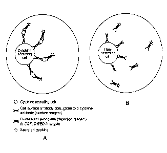
Figure 1. Single cell in-droplets secretion assay applied to cytokine
secretion detection.
While the examples presented here are focusing on cytokine and/or antibody
secretion detection
using a fluorescent detection reagent, the presented assay can be applied to
the secretion
detection of any compound of interest and using any labelled detection
reagent. PBMC are
5
CA 03097276 2020-10-15
WO 2019/202135 PCT/EP2019/060210
stimulated (either specifically using Antigen Presenting Cells labelled with
specific Antigen or non-
specifically, for example by the use of cross-linking antibodies or phorbol
esters) either on-chip (i.e.
in the droplet) or off-chip (i.e. out of the droplet, in a separate container)
are pre-labeled (either on
or off chip) with the capture reagent and encapsulated as single-cells into
droplets together with
the fluorescent detection reagent in conditions preventing cytokine secretion.
After incubation of
the droplets in conditions allowing cytokine secretion, the secreting cells
are detected by the
presence or relocalization of the detection reagent on the cell. A) Cytokine
secreting cell: the cell is
secreting the cytokine of interest, which binds to the capture reagent. The
detection reagent binds
to the secreted cytokine, thus leading to the presence or relocalization of
the fluorescent signal on
the cell. B) Non-secreting cells: the analyzed cell is not secreting the
cytokine of interest and the
detection reagent stays homogeneous in the droplet. No presence or
relocalization of fluorescence
is observed.
Figure 2. Single cell in-droplets detection of IFNy secretion is sensitive and
specific.
A) Single cell droplet-based detection of secreted IFNy specifically by
activated T-cells, compared to
non-activated. Cells that were dead before the experiment or died in the
droplets before or after
secreting the cytokine of interest are excluded from the analysis, by the
addition in droplet of
NucRed or NucGreen intercalating agent, to prevent any non-specific events,
which can
represent a substantial non-specific binder. In droplets, secretion of IFNy is
detected for 0.14% and
16.7% in droplet containing non-activated and activated cells, respectively.
B) Flow based detection
of IFNy secretion by activated T-cells. In flow cytometry, secretion of IFNy
is detected for 0% and
16% of non-activated and activated cells respectively. The shift of cells
population is a severe
limitation of the flow-based system due to high background of non-specific
capture of secreted
molecules by cells nearby during staining.
Figure 3. Single cell in-droplets detection of IFNy secretion is sensitive
(<1M), efficient (>80%)
and 100% specific.
Droplets containing single non-activated CD8+ T-cells, pre-labelled with the
capture reagent, and
co-flowed with the detection reagent, in the presence of different
concentrations of purified IFNy
were reinjected into the microfluidic device and fluorescence of each droplet
was analyzed using
proprietary software. A) Selection of droplets having the correct width and
attribution of the
different emulsions/concentration conditions. B) Selection of droplets
containing CD8+ T-cells
based on cell-labeling. C) Detection of IFNy in droplets for each
concentration of cytokine tested.
D) For each concentration of IFNy tested, the percentage of positive droplets
detected was
6
CA 03097276 2020-10-15
WO 2019/202135 PCT/EP2019/060210
determined and compared to the negative control (OnM). As low as 1nM of
cytokine was detectable
in droplets and about 80% of the cells were detected using the droplet based
single cell secretion
assay. No false positive was selected as 0% of cells/droplet were observed as
positive in the
condition containing OnM IFNy.
Figure 4. Single cell in-droplet antigen-specific activation of T cells by
antigen-presenting cell.
A) Antigen-presenting cells (APC) pulsed with a specific peptide pool and
primary CD8+ T-cells (pre-
labelled with capture reagent) were co-encapsulated in droplets. Droplets were
incubated over-
night in conditions allowing activation of T-cells by APC, which was detected
by cytokine secretion.
The following day, droplets were reinjected in the microfluidic device and
fluorescence signals were
analyzed for detection of activated T-cells having secreted and secreting
IFNy. B) Droplets of
interest were composed of one T-cell and one antigen presenting cell co-
encapsulated. Both cells
can be fluorescently labeled in different colors to enable effective selection
of droplets containing
both cells. C) A fluorescent dead cell marker was used to control viability of
cells in droplets and
exclude any false positives due to cell death, either before or during the
course of the
experiment/activation. Cells encapsulated into droplets showed high viability
after over-night
incubation as 94% of them were detected as viable. D) The droplet secretion
assay was used to
detect antigen-specific T-cell activation by APC in droplets. As anticipated,
based on responsive T-
cell frequency, 1.2% of droplets containing both a viable T-cells and a viable
APC were detected as
secreting IFNy indicating a successful, high viability, antigen-specific
activation and detection of
activated cells based on IFNy secretion of single T-cells in droplets.
Figure 5. Single cell in-droplet secretion assay applied to any secreted
molecule detection.
The method according to the present invention is highly modulable and can be
adapted to detect a
variety of biological events. While the examples presented here are focusing
on cytokine and/or
antibody secretion detection using a fluorescent detection reagent, the
presented assay can be
applied to the secretion detection of any compound of interest and using any
labelled detection
reagent. (A) Example of in-droplet detection of the secretion of diverse
compounds of interest by
the interrogated cells, including the possibility for multiplexed assays.
Here, multiplexed assay of
antibody and cytokine secretion is presented but the present invention can be
applied to any
mentioned compound of interest. Off- or on-chip stimulated PBMC are pre-
labeled with the
cytokine specific capture reagent and B cells are prelabeled with the antibody
specific capture
reagent. Both cell populations are co-encapsulated as single-cells into
droplets together with the
cytokine-specific fluorescent detection reagent and the antibody-specific
fluorescent detection
7
CA 03097276 2020-10-15
WO 2019/202135 PCT/EP2019/060210
reagent in conditions preventing cytokine and antibody secretion before they
are encapsulated as
individual cells. The labels (fluorescent in this example but can be by any
means) of both detection
reagents are selected wisely according to the assay. After incubation of the
droplets in conditions
allowing cytokine and antibody secretion, the secreting cells are detected by
the presence or
relocalization of the detection reagents on the cells. The secreted cytokine
is bound to the capture
reagent specifically bound to the cytokine-secreting cells and detected
through the presence or
relocalization of the fluorescent anti-cytokine detection reagent. The
secreted antibody is bound to
the capture reagent bound to the antibody-secreting cell and detected through
the presence or
relocalization of the fluorescent anti-antibody detection reagent on the cell.
The antibody-specific
capture reagent can be specific for all immunoglobulins allowing global
antibody response to be
detected or composed of the antigen of interest allowing antigen-specific
antibody response to be
detected. (B) Example of in-droplet cytokine secretion detection with coflowed
capture and
detection reagents. Off- or on-chip stimulated PBMC are encapsulated as single-
cells into droplets
together with the capture reagent and detection reagent (can be fluorescent as
exemplified here
or can be any other mean) in conditions preventing cytokine secretion. After
incubation of the
droplets in conditions allowing cytokine secretion, the secreting cells are
detected by the presence
or relocalization of the detection reagents on the cells. Both capture and
detection reagent
concentrations are adapted to generate the highest signal/background ratio and
enabling the
maximal fluorescent signal onto the interrogated cell. (C) Example of in-
droplet cytokine secretion
detection with the first capture reagent bound to the cells being composed of
two or more
molecules. Off- or on-chip stimulated PBMC are pre-labeled with the cytokine
specific capture
reagent composed of two or more molecules. The two or more molecules are
composed of an
antibody specific to the cell membrane of interest conjugated to a ligand A
and an antibody specific
to the cytokine of interest conjugated to a ligand B; where ligands A and B
can interact and form a
stable association. The cells are encapsulated as single-cells into droplets
together with the
fluorescent detection reagent in conditions preventing cytokine secretion.
After incubation of the
droplets in conditions allowing cytokine secretion, the secreting cells are
detected by the presence
or relocalization of the detection reagent on the cells. (D) Example of in-
droplet cytokine secretion
detection with the first capture reagent being coflowed and composed of two or
more molecules.
Off- or on-chip stimulated PBMC are encapsulated as single-cells into droplets
together with the
capture reagent and fluorescent detection reagent in conditions preventing
cytokine secretion. The
coflowed cytokine specific capture reagent is composed of two or more
molecules. The two or more
molecules are composed of an antibody specific to the cell membrane of
interest conjugated to a
ligand A and an antibody specific to the cytokine of interest conjugated to a
ligand B; where ligands
8
CA 03097276 2020-10-15
WO 2019/202135 PCT/EP2019/060210
A and B can interact and form a stable association. After incubation of the
droplets in conditions
allowing cytokine secretion, the secreting cells are detected by the presence
or relocalization of the
detection reagent on the cells. Both capture and detection reagent
concentrations are adapted to
generate the highest signal/background ratio and enabling the maximal
fluorescent signal onto the
interrogated cell. (E) Example of in-droplet cytokine secretion detection with
the first capture
reagent being composed of two molecules, one moiety being bound to the cell,
the other being
coflowed. Off- or on-chip stimulated PBMC are pre-labeled with the first
moiety of the capture
reagent composed an antibody specific of the cell membrane conjugated to a
ligand A. The cells are
encapsulated as single-cells into droplets together with the second moiety of
the capture reagent
composed of an antibody specific to the cytokine of interest conjugated to a
ligand B as well as with
the fluorescent detection reagent in conditions preventing cytokine secretion.
Ligands A and B can
interact and form a stable association. After incubation of the droplets in
conditions allowing
cytokine secretion, the secreting cells are detected by the presence or
relocalization of the
detection reagent on the cells. Both the second moiety of the capture reagent
and the detection
reagent concentrations are adapted to generate the highest signal/background
ratio and enabling
the maximal fluorescent signal onto the interrogated cell.
Figure 6. Detection of T cell activation by secreted receptor-specific
antibody.
Figure 7. Double positive detection of ADCC induced by secretion of antigen-
specific antibody and
cytotoxic factors secretion detection.
Figure 8. Double positive detection of ADCC induced by secretion of antigen-
specific antibody.
Figure 9. Description of the microfluidic system and process according to the
invention.
DETAILED DESCRIPTION OF THE INVENTION
The method according to the present invention is intended to solve the above-
mentioned issues
affecting current microfluidic techniques for single cell analysis. In
particular, the present method
provides an improved performance in detecting, analyzing and/or quantifying
the production of a
compound of interest at single cell level.
9
CA 03097276 2020-10-15
WO 2019/202135 PCT/EP2019/060210
A first advantage of the method disclosed herein is represented by its high
sensitivity. Such property
is due to the spatial confinement of a single cell producing a compound of
interest in a droplet,
wherein said single cell has freedom of mobility, allowing high viability and
thus high, yet
physiological, metabolic activity. In addition, the spatial confinement of a
single cell producing a
compound of interest in a droplet, wherein said secreted product is confined
in a constrained few
pico to nano-liter volume allows reaching high concentration in few minutes to
hours of incubation,
depending on the produced molecule.
Consequently, a second advantage emerging by using the method of the present
invention is
represented by the possibility of carrying out kinetic analysis by virtue of
monitoring a change in
the relocalization and/or intensity of a detectable event in real-time. By
extension, it is easy to
envision extending to multiple secreted compound detection, by using
differently labelled
detection reagent.
Consequently, a third advantage emerging by using the method of the present
invention is
represented by the possibility of carrying out complex, yet flexible sets of
assays by virtue of co-
encapsulating two or more cells into the droplets and monitoring the role of
cell-cell interaction for
production of said compound by one, two or more cells. Those complex assays co-
encapsulating
two or more cells also enable the detection of the secretion of two or more
compounds of interest.
A fourth advantage emerging by using the method of the present invention is
represented by the
high specificity of the detection of production of said molecule. Such
property is due to the spatial
confinement of a single cell producing a compound of interest in a droplet,
wherein said secreted
product is confined in a restrained volume, specifically captured to said
single cells and secreted
product is thus captured only by the secreting cells.
In this regard, the inventors have found that secreting cells are
advantageously detected by
monitoring the presence or relocalization of the detection reagents on the
cell within the droplet
and that cell density/concentration is not impacting the specificity of
detection.
In addition, in case of the presence of a non-secreting cell or cell not
secreting the compound of
interest, the detection reagents remain homogeneous in the droplet, thus
minimizing the false
positive hit rate.
CA 03097276 2020-10-15
WO 2019/202135 PCT/EP2019/060210
In a first aspect, the present invention relates to a method for the detection
of a compound of
interest in a microfluidic system, said method comprising the steps of:
a. creating at least one droplet in said microfluidic system, said at least
one droplet
comprising:
i. at least one single cell,
ii. one or more first capturing agent, wherein said one or more first
capturing agent is capable of binding said single cell as well as said
compound of interest,
iii. one or more second capturing agent comprising a label, wherein said
one or more second capturing agent is capable of binding said
compound of interest,
b. incubating said at least one droplet capable of generating a
detectable event,
c. subjecting said at least one droplet to a direct detection,
wherein the presence or relocalization of said detectable event within said at
least one droplet
determines the presence of said compound of interest.
In the context of the present invention, the term "microfluidic system" may
refer to one or more
integrated units or chips for performing the method disclosed herein. Said
microfluidic system is
generally represented in the form of a microfluidic chip comprising one or
more micro-channels
and one or more microfluidic devices (e.g. micropumps, microvalves).
In the context of the present invention, a "microfluidic chip" generally
refers to a set of micro-
channels made by milling, etching, ablation or molding into a material
(polymeric material such as
polydimethylsiloxane (PDMS) or polymethylmethacrylate (PMMA), polycarbonate
(PC), epoxy, COC
in particular photopolymerizable epoxy such as marketed by Norland Optical
Adhesives (NOA),
glass. silicon, plastics). A microfluidic chip may comprise a substrate and a
support, defining
together at least one channel.
As used herein, the term "droplet" refers to an isolated portion of a fluid
which is immiscible with
its surrounding. In the context of the present invention, said "droplet" may
be spherical,
substantially spherical or non-spherical in shape. Said shape may depend by
different parameters,
such as, for example, the external environment.
11
CA 03097276 2020-10-15
WO 2019/202135 PCT/EP2019/060210
Methods for preparing, generating and injecting droplets in a microfluidic
system are known to the
person skilled in the art. An exemplary method is disclosed in US 2015/0057163
Al. With reference
to the presence of a single cell in each droplet, the person skilled in the
art is aware that this
parameter can be controlled and/or estimated using the Poisson distribution.
In the context of the present invention, the expression "at least one single
cell" refers to viable and
non-viable single cell. The viability status of said at least one single
viable cell can be altered or
changed along the steps of the method according to the present invention. It
is worth noting that,
after the incubation step of a droplet according to the present method, the
capability of generating
a detectable event in said droplet refers to the possibility within the
droplet of having at least one
viable single cell.
As used herein, the term "direct detection" refers to the possibility of
detecting the compound of
interest produced by a single cell in absence of a solid support within the
droplet, wherein the solid
support would be used for capturing the compound of interest. In the context
of the present
invention, the terms "solid support" refers to any non-biological matrix, e.g.
magnetic beads, gel
matrix or affinity matrix, that has a given specificity for a target molecule
such that the target
molecule can be immobilized on said support, which allows isolation of the
target molecule from
the content comprised in the droplet.
According to an embodiment of the first aspect of the present invention, the
single cell presents a
freedom of mobility within the droplet.
In the context of the present invention, the detection of the compound of
interest is independent
from the orientation of the cell producing said compound of interest within
the droplet.
According to another embodiment of the first aspect of the present invention,
the single cell is not
captured on a solid support.
The inventors have found that the presence of a single cell with a high degree
of mobility, that is
not constrained on a solid support allow a superior sensitivity in detecting
the presence of a
compound of interest secreted by the cell because of an improved distribution
of first capturing
agents on the cellular surface.
12
CA 03097276 2020-10-15
WO 2019/202135 PCT/EP2019/060210
As used herein, the term "capturing agent" refers to a reagent, nucleic acid,
protein or peptide that
presents an affinity towards the compound of interest. In the context of the
present invention, the
method requires the presence of a first and a second capturing agent.
In the context of the present invention, the terms "first capturing agent"
and/or "second capturing
agent" may refer to a single bifunctional compound or to a complex comprising
two or more
different compounds, each characterized by a specific functionality. Examples
of first and second
capturing agents conceived for the method according to the present invention
can be a compound
or a complex formed of antibodies, antigens, cytokines, chemokines, hormones
or growth factors
or a combination of those.
As used herein, the term "relocalization" refers to a change in the spatial
disposition within a
droplet of density and/or concentration of a detectable event. As used herein,
the term "presence"
refers to the occurrence or change of the intensity of a detectable event.
An important aspect of the method according to the present invention relates
to the relocalization
of a detectable event within the droplet. In this regard, methods known in the
art cannot achieve
"relocalization" as intended herein, but only a local concentration-binding as
the excess is washed
away before doing the flow cytometry analysis. Therefore, the effect of this
feature confers to the
method according to the present invention a higher efficiency over the current
methods.
Another important step in droplet-based microfluidic assays, along with
droplet creation, pico-
injection, merging and sorting, is represented by the incubation of droplets.
In the context of the
present invention, the incubation may occur off- or on-chip. The incubation
step may also occur in
a delay line necessary for incubating droplets for a precise time allowing for
cells viability and
production of a compound of interest. An exemplary method of incubation in
delay lines is disclosed
in US 2012/0121480 Al. Typical incubation temperature before encapsulation
ranges from 0 C to
16 C, after encapsulation ranges from 16 C to 38 C, and re-injection for
analysis of secreted
molecule after incubation ranges from 0 C to 38 C. Typical incubation time
goes from milliseconds
(for kinetics analysis) to more than 24h (for cell-cell interaction mediated
compound production
regulation analysis).
In another embodiment of the first aspect of the present invention, the method
further comprises
the step of measuring cell viability in droplets after incubation. In the
context of the present
13
CA 03097276 2020-10-15
WO 2019/202135 PCT/EP2019/060210
invention, a preferred method for measuring cell viability is carried out by
using an intercalating
dye that emits fluorescence only if a dead cell is detected in the droplet,
e.g. NucRed Dead 647
ReadyProbes .
According to another embodiment of the first aspect of the present invention,
the one or more first
capturing agent binds the surface of said at least one single cell before or
after creating said at least
one single droplet.
In one embodiment of the first aspect of the present invention, the one or
more first capturing
agent binds said single cell with a density ranging from 101 to 108
molecules/cell.
According to another embodiment of the first aspect of the present invention,
the compound of
interest is produced in the droplet with a concentration of lOpM to 100 M.
In another embodiment of the first aspect of the present invention, the
droplet has a volume
ranging from 2pL to lOnL.
In another embodiment of the first aspect of the present invention, the label
is selected from the
group comprising a fluorescent label, a polymer, a protein, a peptide, a
hapten, a chemical, a nucleic
acid or a barcode label. As used herein, the term "barcode" refers to a label
that may be attached
to an analyte to convey information about said analyte. In the context of the
present invention, the
barcode label can be a mixture of labels, polymer, fluorescent label, peptide,
hapten, protein,
chemicals, nucleic acid.
In another embodiment of the first aspect of the present invention, the first
capturing agent and
said second capturing agent are independently selected from the group
comprising a protein, a
peptide, an oligonucleotide, a hapten, a nucleic acid, a fluorescent
conjugate, an enzyme conjugate,
a synthetic polymer or a barcode or a combination thereof. The barcode label
can be a mixture of
labels, said polymer, fluorescent label, peptide, haptene, protein, chemicals,
nucleic acid.
In another embodiment of the first aspect of the present invention, the first
capturing agent is an
antibody and said second capturing agent is a fluorescent anti-compound of
interest antibody.
14
CA 03097276 2020-10-15
WO 2019/202135 PCT/EP2019/060210
According to another embodiment of the first aspect of the present invention,
the first capturing
agent is a bifunctional antibody.
In another embodiment of the first aspect of the present invention, the
compound of interest is a
cell-secreted compound selected from the group including but not limited to
antibody (IgG (IgG1,
IgG2, IgG3, IgG4), IgE, IgA (IgA1,1gA2), IgMõ cytokine (1L-1-like, 1L-1a, IL-
i3, 1L-1RA, IL-2, IL-3, IL-4,
IL-5, IL-6-like, IL-6, IL-7, IL-9, 1L-10-like, IL-10,1L-n, IL-12, IL-13, IL-
14, IL-15, IL-16, IL-17, IL-18, IL-20,
Common b chain (CD131), LIE, OSM, Interferons (IFN-a, IFN-(3, IFN-y), TNF, TNF-
a, TNF-(3, CD153,
CD154, LT-(3, 4-1BBL, APRIL, CD70, CD132, CD178, GITRL, LIGHT, OX4OL, TALL-1,
TRAIL, TWEAK,
TRANCE, TGF-(3, Tpo, Flt-3L, SCE, M-CSF, MSP), chemokine (CCL1, CCL2, CCL3,
CCL4, CCL5, CCL6,
CCL7, CCL8, CCL9/CCL10, CCL11, CCL12, CCL13, CCL14, CCL15, CCL16, CCL17,
CCL18, CCL19, CCL20,
CCL21, CCL22, CCL23, CCL24, CCL25, CCL26, CCL27, CCL28, CXCL1, CXCL2, CXCL3,
CXCL4, CXCL5,
CXCL6, CXCL7, CXCL8, CXCL9, CXCL10, CXCL11, CXCL12, CXCL13,CXCL14, CXCL15,
CXCL16, CXCL17,
XCL1, XCL2, CX3CL1), hormones (estrogene, progestogens, thyroxine, steroids,
insulin, adrenaline
Epinephrine, Melatonin, Triiodothyronine, Thyroxine, Prostaglandins,
Leukotrienes, Prostacyclin,
Therocis, Adiponectin, Adrenocorticotropic hormone (or corticotropin), Amylin
(or Islet Amyloid
Polypeptide), Angiotensinogen and angiotensin, Anti-Mullerian hormone (or
MOHenan inhibiting
factor or hormone), Antidiuretic hormone (or vasopressin, arginine
vasopressin), Atrial-natriuretic
peptide (or atriopeptin), Calcitonin, Cholecystokinin, Corticotropin-releasing
hormone, Cortistatin,
Endothelin, Enkephalin, Erythropoietin, Follicle-stimulating hormone, Galanin,
Gastric inhibitory
polypeptide, Gastrin, Glucagon, Glucagon-like peptide-1, Gonadotropin-
releasing hormone,
Guanylin, Hepcidin, Human chorionic gonadotropin, Inhibin, Insulin, Insulin-
like growth factor (or
somatomedin), Leptin, Lipotropin, Melanocyte stimulating hormone, Motilin,
Orexin, Osteocalcin,
Oxytocin, Relaxin, Renin, Secretin, Somatostatin, Thrombopoietin, Uroguanylin,
Vasoactive
intestinal peptide, Steroid, estrogen, glucocorticoid, progestogen,
secosteroid), growth factors (G-
CSF, GM-CSF, Fas-ligand, Adrenomedullin (AM), Angiopoietin (Ang), Autocrine
motility factor, Bone
morphogenetic proteins (BMPs), Ciliary neurotrophic factor family, Ciliary
neurotrophic factor
(CNTF), Leukemia inhibitory factor (LIE), Interleukin-6 (IL-6), Colony-
stimulating factors,
Macrophage colony-stimulating factor (m-CSF), Granulocyte colony-stimulating
factor (G-CSF),
Granulocyte macrophage colony-stimulating factor (GM-CSF), Epidermal growth
factor ([GE),
Ephrins (A1-A5, B1-133), Erythropoietin (EPO), Fibroblast growth factor (FGF1-
FGF23), Foetal Bovine
Somatotrophin (FBS), GDNF family of ligands, Glial cell line-derived
neurotrophic factor (GDNF),
Neurturin, Persephin, Artemin, Growth differentiation factor-9 (GDF9),
Hepatocyte growth factor
(HGF), Hepatoma-derived growth factor (HDGF), Insulin, Insulin-like growth
factors, Insulin-like
CA 03097276 2020-10-15
WO 2019/202135 PCT/EP2019/060210
growth factor-1 (IGF-1 and IGF-2), Interleukins; IL-1- Cofactor for IL-3 and
IL-6, IL-2, IL-3, IL-4, IL-5,
IL-6, IL-7, Keratinocyte growth factor (KGF), Migration-stimulating factor
(MSF), Macrophage-
stimulating protein (MSP), also known as hepatocyte growth factor-like protein
(HGFLP), Myostatin
(GDF-8), Neuregulins (NRG1-NRG4), Neurotrophins, Brain-derived neurotrophic
factor (BDNF),
Nerve growth factor (NGF), Neurotrophin-3 (NT-3), Neurotrophin-4 (NT-4),
Placental growth factor
(PGF), Platelet-derived growth factor (PDGF), Renalase (RNLS) ¨ Anti-apoptotic
survival factor, T-
cell growth factor (TCGF), Thrombopoietin (TPO), Transforming growth factor
alpha (TGF-a, TGF-P
(TGF-(31, TGF-32, TGF-(33), Tumor necrosis factor-alpha (TNF-a), Vascular
endothelial growth factor
(VEGF)).
A second aspect of the present invention encompasses the use of the method
according to the first
aspect of said invention for monitoring one or several, potentially
simultaneous, biological event(s).
As used herein, the term "biological event" refers to describe an alteration
of a physiological
process and/or state occurring in a subject's body and affecting the
physiological status of living
cells. Typical example is linking secretion of a compound with induced
mortality (ADCC, CDC, ADCP,
Cytokine induced Cytolysis, Apoptosis, Chromium Release, as non-limiting
examples), another
example is activation and/or inhibition of cellular pathway by secreted
compound (G protein
coupled receptor activation, B-arrestin, caspase activation, PKC/NFKB
pathways, MAP kinases, Pi3K,
AKT pathway, Ras/Mek/Erk, PLC/Ca", as non-limiting examples).
In an embodiment of the second aspect of the present invention, the biological
event is an immune
response. Typical examples are detection of antigen recognition by T cells
inducing compound
secretion, including antigen recognition by B cells inducing compound
secretion, including as well
T cell activation monitored by secreted compound from T cells and induced by a
second secreted
compound (this example include detection of secreted compound by two different
cell types and
differentiating these using barcode specific for each one), and include B cell
activation monitored
by secreted compound from B cells and induced by a second secreted compound.
In a third aspect of the present invention, there is provided a method for the
detection of a
compound of interest in a droplet comprising the steps of:
a. providing a microfluidic system comprising:
i. at least one inlet,
ii. at least one outlet,
iii. one or more channels,
16
CA 03097276 2020-10-15
WO 2019/202135 PCT/EP2019/060210
b. injecting in said microfluidic system a stream of droplets, wherein at
least one droplet
comprises:
i. at least one single cells
ii. a plurality of a first capturing agents capable of binding said single
cell as well
as said compound of interest, and
iii. a plurality of second capturing agents, each comprising a label,
wherein said
plurality of second capturing agents is capable of binding said compound of
interest,
c. incubating said plurality of droplets under conditions that allow
the production of the
compound of interest, whereby if the compound of interest is produced by the
single
cell, it will be captured by said plurality of first and second capturing
agents,
d. determining the presence of the compound of interest by means of detecting
a
presence or relocalization of said label.
A fourth aspect of the present invention relates to a microfluidic system
comprising:
a. at least one inlet,
b. at least one outlet,
c. one or more channels,
d. a module for creating at least one droplet comprising:
i. one or more single cell,
ii. a first capturing agent,
iii. a second capturing agent,
e. a detection module detecting droplet containing cells producing a compound
of
interest.
f. an analysis module configured for the analysis of the signal.
According to an embodiment of the fourth aspect of the present invention, the
microfluidic system
is characterized by the presence of at least two modules in communication with
each other selected
from the group comprising: module for droplet production, module for droplet
detection, module
for droplet analysis, module for sorting droplets, module for tagging droplets
and module for
recovering droplets. In the context of the present invention, the module for
recovering droplets is
intended for carrying out additional process (e.g. genotyping, further
functional analysis).
17
CA 03097276 2020-10-15
WO 2019/202135 PCT/EP2019/060210
An ideal scheme of the microfluidic system and process according to the
present invention is
depicted in Figure 9.
The combination of two or more of the aforementioned modules allows the
microfluidic system
disclosed herein to achieve improved results in terms of high-
throughputability (several thousand
of droplets per second can be processed).
An important aspect of the microfluidic system according to the present
invention is that secretion
and detection steps according to the method of the first aspect of the present
invention can be
performed in the same module of the microfluidic system.
According to a fifth aspect, the microfluidic system according to the fourth
aspect is used for
carrying out the method disclosed in the first or third aspect of the present
invention.
EXAMPLES
Principle description
Healthy donor human PBMC are pre-labeled in microtubes with an excess of a bi-
functional
antibody, called "catch reagent". The catch reagent is specific for both a
leucocyte-specific
membrane protein (CD45) and the cytokine of interest. After 5 minutes
incubation in conditions
preventing cytokine secretion (i.e. at 4 C), all the leucocytes are evenly
labeled with the catch
reagent and the excess is washed away by extensive washes. Pre-labeled cells
are encapsulated as
single-cells into picoliter droplets with 1% v/v final concentration of
fluorescently-labeled anti-
cytokine antibody in conditions preventing cytokine secretion (Figure 1). The
droplets containing
single-cells are incubated for 1h20 at 37 C in a 5% CO2-controlled incubator
to enable cytokine
secretion. Droplets are reinjected and the secretion of cytokine, traduced by
the relocalization of
the detection reagent's fluorescent signal on the cell is analyzed for each
droplet. In a droplet
containing a cytokine-secreting cell, the detection reagent signal is
relocalized onto the cell, leading
to a local increase of fluorescence in the droplet. On the contrary, in a
droplet containing a non-
secreting cell, the detection reagent's fluorescent signal stays homogeneous
in the droplet and no
local increase of fluorescence is observed.
18
CA 03097276 2020-10-15
WO 2019/202135 PCT/EP2019/060210
In-droplet secretion assay was applied to the detection of IFNy (and TN Fa,
not shown) secretion by
PMA/ionomycin activated PBMC compared to non-activated PBMC (Figure 1). The
results observed
using droplet based microfluidic system and software were compared to flow
cytometry data
generated in microplates with the same cells and conditions (Figure 2). 100%
of secreting cells in
flow cytometry were detected as positive in the droplet secretion assay. False
positive cells counted
for less than 0.15% in the negative control. The droplet detection of cytokine
secretion by activated
T cell is highly efficient and specific compared to flow cytometry detection.
Quantification of cytokine secretion sensitivity and efficiency
Non-activated and non-secreting CD8+ T-cells were encapsulated in droplets
with a range of
concentration of purified IFNy following the droplet secretion assay
procedure. Four emulsions
were produced, each containing cells isolated as single cells and purified
cytokine at different
concentrations: OnM, 1nM, 5nM or 10nM final concentration of IFNy in droplets
(Figure 3). Droplets
of all four emulsions were reinjected and fluorescence signals were analyzed.
Using the droplet
secretion assay, as low as 1nM cytokine concentration was detected with no
false positive events
showing a highly sensitive and 100% specific assay. The secreting assay also
showed to be efficient
as more than 80% of the positive cells were detected in droplets.
These examples show the possibility to calibrate assay detection for
quantitative, real-time cytokine
secretion quantification in droplet by the mean of generating standard curve
samples conditions.
Antigen specific T cells identification based on cytokine secretion from co-
encapsulated APC/T-cells
in droplet
When co-cultured, antigen-presenting cells (APC) loaded with a specific
peptide can specifically
activate a subset of responding T-cells, leading to cytokine secretion. The
droplet secretion assay
was applied to detection of specific activation of T-cells by APC in droplets
(Figure 4). APC and T-
cells were co-flowed as single-cells into droplets in conditions preventing
cytokine secretion.
Droplets were incubated over-night at 37 C in 5% CO2 controlled incubator and
reinjected the
following day. Viability of both T-cells and APC was measured after over-night
incubation in
droplets, by using the NucRed , an intercalating dye. Using such dead cell
fluorescent marker, 94%
of the encapsulated cells were detected as viable. Specific activation of T-
cell by APC was detected
using the droplet secretion assay applied to IFNy secretion. Within droplets
containing viable T-cells
and APC, 1.2% secreted IFNy, demonstrating effective antigen-specific
activation of T-cells in
droplets.
19
CA 03097276 2020-10-15
WO 2019/202135 PCT/EP2019/060210
Antigen-specific and total antibody secretion detection
Antibody secretion can be assessed using the presented in-droplet secretion
assay by co-
encapsulating in droplets B cells prelabeled with the capture reagent together
with the detection
reagent. The capture reagent's first moiety is capable of recognizing a B cell
surface marker, can be
a Pan-B marker or a specific B cells marker, typical example is the CD138
marker for immunoglobulin
secreting plasma cells. The second moiety of the capture reagent either
specifically captures
antibody or consists of the antigen of interest. In the first case where the
capture reagent is
composed of a moiety capturing antibody, the detection reagent is composed of
a detectable
labeled antigen. In the second case where the capture reagent is composed of
the antigen of
interest, the detection reagent is composed of a detectable labeled anti-
antibody secondary. The
relocalization of the fluorescent signal on the B cell (labels are here
fluorescent but can be detected
by any means for people skilled in the art) indicates antigen specific
antibody secretion by the B cell
present in the interrogated droplet. The method described here can be adapted
with or without
pre-incubation of the capture reagent (Figures 5A-E). The method described
here can be adapted
to any compound of interest previously mentioned.
Detection of T cell activation by secreted receptor-specific antibody
Binding of antibodies specific to a given T cell receptor can activate the T
cell leading to, for
example, cytokine secretion. The droplet secretion assay can detect T cell
activation by a T cell
receptor-specific antibody secreted by an immunoglobulin expressing cell in
the droplet (Figure 6).
Typical example includes PBMC prelabelled with the capture reagent
encapsulated into droplets
together with an immunoglobulin expressing cell. The droplets are produced
while containing the
labeled detection reagent (labels are here fluorescent but can be by any
means) and in conditions
preventing antibody production. After incubation of the droplets in conditions
allowing antibody
production, T cell activation is detected through detection of, for example,
cytokine secretion.
Binding of the T cell receptor-specific antibody activates in turn the T cell
which then secretes, for
example, cytokines. The secreted cytokines relocalize onto the capture reagent
bound to the T cell
and the fluorescent detection reagent relocalizes onto the cytokine of
interest. Droplets containing
a T cell activated by a secreted antibody present then a detectable signal due
to the relocalization
of the detection reagent on the activated T cell. The method described here
can be adapted with
or without pre-incubation of the capture reagent. Typical examples of the
method described above
is the detection of anti-CD3 antibodies triggering the T cell activation. By
extension, the system can
be used for the identification of anti-checkpoints antibodies.
CA 03097276 2020-10-15
WO 2019/202135 PCT/EP2019/060210
Double positive detection of ADCC induced by secretion of antigen-specific
antibody and cytotoxic
factors secretion detection
The in-droplet secretion assay can be used to assess induced mortality in case
of an antigen-specific
antibody having ADCC activity is secreted (Figure 7). The double positive
assay presented here
enables the detection of both the cytotoxic factors secretion by the killing
cells (example include
primary natural killer (NK) cells, monocytes, macrophages, neutrophils,
eosinophils and dendritic
cells, as well as cell culture cell lines) and death of the target cell
induced by the compound secreted
by the killing cell. Killing cells are pre-labeled with the catch reagent
specific to the cytotoxic factors
of interest in non-saturating conditions. Non-saturating conditions of capture
reagent are
mandatory to enable both capture and detection of secreted compound of
interest secretion by
killing cell and effect of the secreted compound, yet not captured, on the
target cell ultimately
inducing cell death. The target cell is then co-encapsulated in droplet with
an immunoglobulin
producing cell and a killing cell. The encapsulated cells are coflowed with
the detection reagent
specific to the cytotoxic factors of interest in conditions preventing
antibody production before
encapsulation. After production, the droplets are incubated in conditions
allowing antibody
production. The specific antibody relocalizes on the target cell and the
killing cell binds the antibody
through the Fc receptors. Once bound to the antibody having ADCC activity, the
killing cell releases
cytotoxic factors causing the death of the target cell. Some of the secreted
cytotoxic factors are
captured by the capture reagent on the killing cell and relocalize the
detection reagent, enabling
detection of the cytotoxic factors production. Cell death is monitored by the
release of a compound
from the dying target cell expressing the antigen of interest. Alternatively,
cell death is monitored
by cell surface marker, or any other suitable marker known by people skilled
in the art. By extension,
the in-droplet assay could be applied to Complement Dependent Cytotoxicity and
Opsonophagocytosis or any other assays described above.
Alternative and/or complementary to this example is where the production of
the antibody is
detected in place (and/or in addition to) of the secreted cytotoxic factor, in
combination or without
the cell death detection (Figure 8).
21